Note: Descriptions are shown in the official language in which they were submitted.
CA 02977685 2017-08-24
WO 2016/138574
PCT/CA2016/000057
- 1 -
Title: IIOMOLOGOUS RECOMBINATION FACTORS
FIELD OF THE INVENTION
[0001] The invention relates to factors that influence or regulate
homologous
recombination, methods to monitor these factors, the use of these factors to
screen for
agents that modulate homologous recombination, and methods to modulate
homologous
recombination.
BACKGROUND OF THE INVENTION
[0002] The breast and ovarian tumour suppressors BRCA1, PALB2 and
BRCA2 promote DNA double-strand break (DSB) repair by homologous
recombination (HR) [8-10]. BRCA1 acts in this process minimally at two
discrete steps.
Firstly, it promotes DNA end resection [11, 12], the initiating step in HR
that involves
the nucleolytic processing of breaks to produce the single-stranded (ss) DNA
necessary for homology search and strand invasion [1]. Secondly, BRCA1
interacts
with PALB2 [13-15] to direct the recruitment of BRCA2 [13] and RAD51 [16, 17]
to
DSB sites. The accumulation of BRCA1 on the chromatin that flanks DSB sites is
strikingly suppressed in G1 cells [18], reminiscent of the potent inhibition
of
homologous recombination in this phase of the cell cycle. The inhibition of
BRCA1
recruitment in G1 is dependent on the 53BP1 and RIF1 proteins [18, 19], two
inhibitors of end-resection [18-22]. BRCA1 is also involved in promoting the
recruitment of BRCA2 through its interaction with PALB2 [13-15].
[0003] Tumors with compromised ability to repair double-strand DNA
breaks
by HR, including those with defects in BRCA1 and BRCA2, have been shown to be
highly sensitive to poly ADP-ribose polymerase (PARP) inhibitors. PARP
inhibitors
have also been proposed for treating other conditions such as stroke,
myocardial
infarction, inflammatory bowel disorders, head trauma, and neurodegenerative
diseases.
Inhibition of ubiquitin-specific peptidase 11 (USP11) has been shown to
hypersensitize
cells to PARP inhibitors and it has been proposed that USP11 status or the
status of
other HR-proteins in tumors may provide biomarkers for use of PARP inhibitors
(Wiltshire et al, IBC 285(19), 14565-14571, 2010).
[0004] Identification and assessment of factors that influence or regulate
homologous recombination repair proteins and the identification of events that
are both
necessary and sufficient to suppress HR in G1 cells is desirable. In addition,
identification and assessment of factors that influence or regulate USP11 may
facilitate
CA 02977685 2017-08-24
WO 2016/138574
PCT/CA2016/000057
- 2 -
the selection and monitoring of PARP inhibitor treatments, and in particular
selection
of treatments that reverse or delay emergence of PARP inhibitor resistance.
SUMMARY OF THE INVENTION
[0005] The present inventors have found that the cell cycle tightly
controls the
interaction of BRCA1 with PALB2-BRCA2 in order to constrain BRCA2 function to
the S/G2 phases. The BRCA1-interaction site on PALB2 is targeted by an E3
ubiquitin ligase composed of KEAP1, a PALB2-interacting protein [6], in
complex
with cull in 3 (CUL3)-RBX1 [7]. PALB2 ubiquitylation suppresses its
interaction
with BRC.A I and is counteracted by the deubiquitylase USP11, which is itself
under
cell cycle control. Restoration of the BRCA1-PALB2 interaction combined with
the
activation of DNA end resection was sufficient to induce HR in G1 -phase
cells, as
measured by RAD51 recruitment, unscheduled DNA synthesis and a CRISPR/Cas9-
based gene targeting assay. The mechanism prohibiting HR in GI minimally
consists
of the suppression of DNA end resection coupled to a multi-step block to BRCA2
recruitment to DNA damage sites that involves the inhibition of BRCA1-PALB2-
BRCA2 complex assembly. The ability to induce HR in G1 cells with defined
factors
may be used in gene targeting applications in non-dividing cells or cells that
are
dormant in GI phase. The findings also provide a basis for targeting USPI 1 in
combination with poly(ADP-ribose) polymerase (PARP) inhibitors.
[0006] The present inventors have also found that USP11 is regulated by a
cell
cycle-CULLIN4-RING-ligase (CRL4) and DCAFIO acts as an adaptor for the USP11
E3 ligase.
[0007] The invention provides a method for monitoring activity of USP1
I in a
sample by assaying the interaction of BRCA1 and PALB2.
[0008] 'fhe invention provides a method for monitoring activity of USP11 in
a
sample by assaying the interaction of BRCA1, PALB2, and BRCA2.
[0009] The invention provides a method for monitoring activity of USP
II in a
sample by assaying the interaction of USP11 and PALB2.
[00010] The invention provides a method for monitoring activity of
USP11 in a
sample by assaying DCAF10.
[00011] The invention provides a method for monitoring activity or
expression of
USP11 in a sample by assaying for complexes of (a) BRCA1 and PALB2; (b) BRCA1,
PALB2, and BRCA2; (c) USP11 and PALB2; and/or (d) USP11 and DCAF10.
CA 02977685 2017-08-24
WO 2016/138574
PCT/CA2016/000057
-3-
1000121 In an aspect, the invention provides a method for monitoring
activity or
expression of USP11 in a sample comprising (i) isolating complexes of (a)
BRCA1 and
PALB2; (b) BRCA1, PALB2, and BRCA2; (c) USP11 and PALB2; and/or (d) USP11
and DCAF10 in the sample; (ii) measuring the levels of the complexes; and
(iii)
detecting an increase or decrease in the activity or expression of the
complexes as
compared to a control as an indication of the activity or expression of USP11.
[00013] In an aspect, the invention provides a method for monitoring
activity or
expression of USP11 in a sample comprising (i) isolating complexes of (a)
BRCA1 and
PALB2; (b) BRCA1, PALB2, and BRCA2; (c) USP11 and PALB2; and/or (d) USP11
and DCAFIO in the sample by immunological purification; (ii) measuring the
levels of
the complexes; and (iii) detecting an increase or decrease in the activity or
expression
of the complexes as compared to a control as an indication of the activity or
expression
of USP11.
[00014] In an aspect, the invention provides a method for monitoring
activity or
expression of USPI 1 in a sample comprising (i) isolating complexes of (a)
BRCA1 and
PALB2; (b) BRCA1, PALB2, and BRCA2; (c) USP11 and PALB2; and/or (d) USP11
and DCAF10 in the sample; (ii) preparing peptides or peptide fragments from
the
isolated complexes; and (iii) subjecting the peptides or peptide fragments to
mass
spectrometry to thereby monitor the activity or expression of USP11.
[00015] The invention provides a method for monitoring activity or
expression of
USP11 in a sample by assaying ubiquitylation of PALB2, in particular
ubiquitylation of
the N-terminus of PALB2.
[00016] In an aspect, the invention provides a method for monitoring
activity or
expression of USP11 in a sample by assaying ubiquitylation of PALB2 comprising
measuring the amount of polyubiquitin bound to CRL3-KEAP1 E3 ligase in the
sample
and detecting an increase or decrease in polyubiquitin bound to CRL3-KEAP1 E3
ligase as compared to a control as an indication of the activity or expression
of USP11.
[00017] In another aspect, the invention provides a method for
monitoring
activity or expression of USP11 in a sample by assaying ubiquitylation of
PALB2
comprising measuring the activity of CRL3-KEAP1 E3 ligase, and detecting an
increase or decrease in CRL3-KEAP1 E3 ligase activity as compared to a control
as an
indication of the activity or expression of USP11.
[00018] The methods of the invention may be performed in the presence
or
absence of a test compound or agent and detection of an increase or decrease
in activity
CA 02977685 2017-08-24
WO 2016/138574
PCT/CA2016/000057
- 4 -
or expression of one or more of USP11, DCAF10, PALB2, PALB2 ubiquitylation,
BRCA1 -PALB2-BRCA2 complex, PALB2-USP11 complex, KEAP1, USP1I-DCAF
complex, CRL3-KEAP1 complex, CRL3-KEAP1-PALB2 complex, and KEAP1-
PALB2 complex, as compared to a control in the absence of the test compound or
agent
indicates that the test compound or agent may be useful as a therapeutic
agent, or for
modulating homologous recombination.
[00019] In an
aspect, the invention provides a method for identifying or
evaluating an agent for its ability to sensitize or reverse or delay emergence
of
resistance to PARP inhibitors by determining the effect of the agent on USP11
activity
or expression using a method of the invention.
[00020] In an
aspect, the invention relates to a method of identifying or
evaluating an agent for its ability to sensitize cells or reverse or delay
emergence of
resistance to PARP inhibitors by determining the effect of the agent on KEAP1,
CRL3-
KEAP1, KEAP1-PAL132 or CRL3 -KEAP1.
[00021] In an aspect, the present invention provides methods of detecting
an anti-
cancer agent comprising performing a test assay comprising contacting an
immortalized
cell with a test compound and assaying USP11 activity or expression using a
method of
the invention.
[00022] The
invention also provides a method for identifying or evaluating an
agent for its ability to modulate homologous recombination comprising
determining the
effect of a test compound or agent on one or more of USP11, DCAF10, PALB2,
PALB2 ubiquitylation, BRCAl-PALB2-BRCA2 complex, PALB2-USP11 complex,
KEAP1, USP11-DCAF10 complex, CRL3-KEAP1 complex and CRL3-ICEAP1-
PALB2 complex.
[00023] The invention provides a method of screening for a therapeutic
agent for
treatment of a disease associated with defects in HR (i.e., HR Disease),
comprising
identifying an agent that disrupts or modulates one or more of USP11, PALB2,
PALB2
ubiquitylation, DCAF10, BRCA1 -PALB2-BRCA2 complex, PALB2-USP11 complex,
KEAP1, USP11-DCAF10 complex, CRL3-KEAP1 or CRL3-ICEAP I -PALB2 complex.
[00024] The screening methods of the invention may further comprise
conducting therapeutic profiling of the identified agents or further analogs
thereof, for
efficacy and toxicity in animals; optionally formulating a pharmaceutical
composition
including one or more agents identified as having an acceptable therapeutic
profile; and
optionally administering the agent to a subject or individual.
CA 02977685 2017-08-24
WO 2016/138574
PCT/CA2016/000057
- 5 -
[00025] The invention provides methods of treating a HR Disease in an
individual comprising identifying an agent that modulates HR in accordance
with a
method of the invention and administering the agent to the individual.
[00026] In some embodiments, the invention provides a method for
sensitizing
cells to PARP inhibitors in an individual comprising identifying an agent that
sensitizes
cells to PARP inhibitors in accordance with a method of the invention and
administering the agent to the individual.
[00027] In some embodiments, the invention provides a method for
reversing or
delaying emergence of resistance to PARP inhibitors in an individual
comprising
identifying an agent that reverses or delays emergence of resistance to PARP
inhibitors
in accordance with a method of the invention and administering the agent to
the
individual.
[00028] In some embodiments, the present invention provides methods of
treating cancer in an individual comprising identifying an anti-cancer agent
identified in
accordance with a method of the invention and administering the agent to the
individual.
[00029] The invention also provides a method for predicting a response
or
categorizing a response to a PARP inhibitor in a subject comprising assaying
one or
more of USP11, DCAF10, BRCA1, BRCA2, PALB2, KEAP1, CRL3, CRL3-KEAP1,
USP11-DCAFIO complex, BRCA1-PALB2-BRCA2 complex, PALB2-USP11
complex and CRL3-KEAP1-PALB2 complex in a sample from the subject using a
method of the invention. In an aspect, a method is provided for predicting a
response or
categorizing a response to a PARP inhibitor in a subject comprising assaying
USP11
activity or expression in a sample from the subject using a method of the
invention. In
an aspect, a method is provided for predicting a response or categorizing a
response to a
PARP inhibitor in a subject comprising assaying PALB2 activity or expression
in a
sample from the subject using a method of the invention.
[00030] In an aspect, a subject is categorized as responsive to a PARP
inhibitor if
there is a decrease in one or more of USP11, DCAF10, BRCA1, BRCA2, PALB2,
KEAP1, CRL3, USP11-DCAF10, CRL3-KEAP1, BRCA1-PALB2 and BRCA1-
PALB2-BRCA2 activity or expression or PALB2 ubiquitylation compared to a
control.
In an aspect, a subject is categorized as responsive to a PARP inhibitor if
there is an
increase in one or more of USP11, DCAF10, BRCA1, BRCA2, PALB2, KEAP1,
CA 02977685 2017-08-24
WO 2016/138574
PCT/CA2016/000057
- 6 -
CRL3, USP11-DCAF10, CRL3-KEAP1, BRCAl-PALB2 and BRCA1 -PALB2-
BRCA2 activity or expression or PALB2 ubiquitylation compared to a control.
[00031] A method of predicting responsiveness to a PARP inhibitor may
further
comprise administering the PARP inhibitor to the individual.
[00032] The invention provides a method for treating a patient in need of
treatment with a PARP inhibitor comprising (a) requesting a test providing the
results
of an analysis to determine if the patient is sensitive or responsive to the
PARP
inhibitor by detecting one or more of USP11, DCAF10, BRCA1, BRCA2, PALB2,
KEAP1, USP11-DCAF10, CRL3, CRL3-KEAP1, BRCA1 -PALB2 and BRCA1-
PALB2-BRCA2, in a sample from the subject and comparing to a control to
determine
if the patient is sensitive or responsive to the PARP inhibitor; and (b)
administering the
PARP inhibitor to the patient if the patient is sensitive or responsive to the
PARP
inhibitor. In an aspect of this method of the invention, the patient has
breast cancer. In
an aspect of this method of the invention, the patient has ovarian cancer.
[00033] In an aspect, the invention provides a method for treating a
patient in
need of treatment with a PARP inhibitor comprising (a) requesting a test
providing the
results of an analysis to determine if the patient is sensitive to the PARP
inhibitor by
detecting USP11, DCAF10, BRCA1, BRCA2, PALB2, KEAP1 and/or CRL3 in a
sample from the subject and comparing to a control to determine if the patient
is
sensitive to the PARP inhibitor; and (b) administering the PARP inhibitor to
the patient
if the patient is sensitive to the PARP inhibitor. In an aspect of this method
of the
invention, the patient has breast cancer. In an aspect of this method of the
invention, the
patient has ovarian cancer.
[00034] The invention further provides a method for assigning an
individual to
one of a plurality of categories in a clinical trial for a PARP inhibitor
comprising
assaying USP11, DCAF10, PALB2, PALB2 ubiquitylation, BRCAl-PALB2-BRCA2
complex, PALB2-USP11 complex, USP11-DCAF complex, KEAP1, CRL3-KEAP1
and/or CRL3-KEAP1-PALB2 complex in a sample from the subject using a method of
the invention.
1000351 The invention also provides pharmacogenetic methods for determining
suitable treatment regimens for diseases, in particular cancer, and methods
for treating
patients, based around selection of patients based on PARP responsiveness, in
particular USP11 activity.
CA 02977685 2017-08-24
WO 2016/138574
PCT/CA2016/000057
-7-
1000361 The methods
of the invention, in particular methods for assaying USP11
activity or CRL3-ICEAP1 activity, may be used as a read out in animal model
based
screening methods for new therapeutic approaches and compounds. In an aspect,
a
method of the invention is utilized to predict the efficacy of potential new
treatments in
animal models for disease states.
[00037] The invention
provides a method for activating or modulating (e.g.,
promoting) homologous recombination in a cell comprising:
(a) promoting or stimulating the assembly or occurrence of BRCAl-PALB2 or
BRCAI-PALB2-BRCA2 complexes in the cell;
(b) activating or stimulating BRCA1 recruitment to DNA double-strand break
(DSB) sites;
(c) contacting the cell with BRCA1 -PALB2 or BRCA1 -PALB2-BRCA2
complexes;
(d) inhibiting or removing KEAP1 or CRL3-KEAP1;
(e) inhibiting the degradation of USP11 or promoting USP11 activity; and/or
(f) inhibiting or removing DCAF1 O.
[00038] The invention
provides a method for activating or modulating
homologous recombination in a cell, in particular a cell in GI phase of the
cell cycle
(G1) or GO phase of the cell cycle, comprising administering, or stimulating
assembly
of BRCAl-PALB2 or BRCA1-PALB2-BRCA2 complexes in the cell.
[00039] The invention
also provides a method for activating or modulating
homologous recombination in a cell, in particular a cell in G1 phase of the
cell cycle
(G1) or GO phase of the cell cycle (GO), comprising promoting or stimulating
the
assembly or occurrence of BRCAI-PALB2 or BRCA1 -PALB2-BRCA2 complexes in
the cell.
[00040] The invention
also provides a method for activating or modulating
homologous recombination in a cell, in particular a cell in G1 phase of the
cell cycle
(G1) or GO phase of the cell cycle (GO), comprising administering to the cell
or
contacting the cell with BRCA1-PALB2 or BRCAl-PALB2-BRCA2 complexes.
1000411 The invention also provides a method for repairing DNA double-
strand
breaks in a cell in the G1 phase of the cell cycle (G1) or GO phase of the
cell cycle
(GO), comprising promoting or stimulating the assembly or occurrence of BRCA1-
PALB2 or BRCAl-PALB2-BRCA2 complexes in the cell.
CA 02977685 2017-08-24
WO 2016/138574
PCT/CA2016/000057
- 8 -
[00042] In aspects of the invention, the assembly of BRCAl-PALB2 or
BRCA1-
PALB2-BRCA2 complexes is promoted or stimulated by administering an agent that
promotes or stimulates such assembly or an agent that promotes or stimulates
such
assembly identified using a method of the invention. In an embodiment, the
agent is
USP11 or an agonist of USP11. In an embodiment, the agent is an inhibitor of
CRL-
KEAP1. In an embodiment, the agent is an inhibitor of KEAP1. In an embodiment,
the
agent is a PALB2 mutant, In an embodiment, the agent is a PALB2 mutant that
disrupts
its interaction with KEAP1. In an embodiment, the agent is a PALB2 comprising
mutations of its Lys20, Lys25 and Lys30 residues.
[00043] A method for activating or modulating homologous recombination in a
cell may be performed in a cell wherein single strand DNA (ssDNA) generation
pathways are activated. In an aspect, ssDNA generation pathways in the cell
are
activated by DNA end resection.
[000441 The invention also provides a method for activating or
modulating
homologous recombination in a cell, in particular a cell in the GI phase of
the cell cycle
(G1) or GO phase of the cell cycle (GO) in which DNA end resection is or has
been
activated generating single-stranded DNA, comprising promoting or stimulating
the
assembly or occurrence of BRCA 1-PALB2 or BRCA1-PALB2-BRCA2 complexes in
the cell.
[00045] The invention also provides a method for repairing DNA double-
strand
breaks in a cell in the G1 phase of the cell cycle (G1) or GO phase of the
cell cycle (GO)
in which DNA end resection is or has been activated generating single-stranded
DNA,
comprising promoting or stimulating the assembly or occurrence of BRCA1 -PALB2
or
BRCAI-PALB2-BRCA2 complexes in the cell. In an embodiment, the assembly of the
complexes is promoted or stimulated by administering an agent that modulates
HR. In
an embodiment, the agent is an agent that modulates HR identified using a
method of
the invention. In an embodiment, the agent is USP1I or an agonist of USP11. In
an
embodiment, the agent is an inhibitor of CRL-KEAP1. In an embodiment, the
agent is
an inhibitor of KEAP1. In an embodiment, the agent is a PALB2 mutant. In an
embodiment, the agent is an inhibitor of DCAF10. In an embodiment, the agent
is an
inhibitor of a CULLIN4-RING-ligase.
[00046] The invention also provides a method for repairing DNA double-
strand
breaks in a cell in the G1 phase of the cell cycle (G1) or GO phase of the
cell cycle (GO)
in which DNA end resection is or has been activated generating single-stranded
DNA,
CA 02977685 2017-08-24
WO 2016/138574
PCT/CA2016/000057
- 9 -
comprising contacting the cell with BRCA1 -PALB2 or BRCA1-PALB2-BRCA2
complexes.
[00047] In an
aspect, the invention provides a method for activating or
modulating homologous recombination in a cell, in particular a cell in G1 or
GO,
comprising the step of inhibiting KEAP1 or CRL3-KEAP1 or administering an
inhibitor of KEAP1 or CRL3-KEAP1. In an aspect, the invention provides a
method for
activating or modulating homologous recombination in a cell, in particular a
cell in GI
or GO, comprising the step of blocking the degradation of USP11 or promoting
or
stimulating USP11 activity. In an embodiment, the method comprises
administering
USP11 or an agonist thereof. In an aspect, the invention provides a method for
activating or modulating homologous recombination in a cell, in particular a
cell in G1
or GO, comprising the step of inhibiting CRL-KEAP1 or administering an
inhibitor of
KEAP1 or CRL3-KEAP1 and blocking the degradation of USP11 or promoting or
stimulating USP1 activity.
[00048] The invention also
provides a method for repairing DNA double-strand
breaks in a cell in GI or GO in which DNA end resection is or has been
activated
generating single-stranded DNA, the method comprising (a) inhibiting KEAN or
CRL3-KEAP1; (b) blocking the degradation of USP11 or promoting or stimulating
USP11 activity; (c) administering USP11 or an agonist thereof; (d)
administering an
inhibitor of KEAP1 or CRL3-KEAP1; (e) administering an inhibitor of DCAF10;
and/or (e) inhibiting CRL-KEAP1 and blocking the degradation of USP11.
[00049] A method for
activating or modulating homologous recombination in a
cell may further comprise activating or promoting single-strand DNA (ssDNA)
generation pathways. In an aspect, ssDNA generation pathways are activated by
DNA
end resection.
[00050] A method for
activating or modulating homologous recombination in a
cell may further comprise a gene editing system. In an aspect the gene editing
steps
comprise contacting the cell with a nuclease. In aspects of the invention the
gene
editing system may correct a genomic modification.
[00051] The invention also
provides a method for suppressing homologous
recombination in a cell, in particular a cell in Gl, comprising suppressing
the assembly
of BRCA1 -PALB2 or BRCA1 -PALB2-BRCA2 complexes in the cell. In an
embodiment, the interaction is suppressed by administering KEAP1 or CRL3-KEAP1
or an agonist thereof. In an embodiment, the interaction is suppressed by
administering
CA 02977685 2017-08-24
WO 2016/138574
PCT/CA2016/000057
- 1 0 -
a USP11 antagonist/inhibitor (e.g., mitoxantrone). In an embodiment, the
interaction is
suppressed by administering an agent that inhibits or suppresses HR identified
using a
method of the invention.
[00052] The invention further provides kits for performing methods of
the
invention.
[00053] The invention also provides a system comprising: an assay for
determining the level of USP11 activity, complexes or biomarker levels in a
sample
obtained from the subject; a processor for processing the results; computer
coded
instructions for comparing the results with a database; and a user display for
providing
the results of the comparison. The database may comprise reference values for
USP11
activity or biomarker levels.
[00054] The invention also contemplates the use of methods, kits, and
systems of
the invention in genome modification or editing.
[00055] In an aspect, the invention further contemplates the use of
methods,
compositions, kits, and systems of the invention in genome modification or
editing,
provided that said use is not a method for treatment of the human or animal
body by
surgery or therapy, and provided that said use is not a process for modifying
the germ
line genetic identity of human beings. Genome modification may comprise
modifying a
target polynucleotide sequence in a cell, modifying expression of a
polynucleotide
sequence in a cell, generating a model cell comprising a mutated disease gene,
or
knocking out a gene. A use of the invention may further comprise repairing or
editing a
cleaved target polynucleotide by inserting an exogenous template
polynucleotide,
wherein the repair or editing results in a mutation comprising an insertion,
deletion, or
substitution of one or more nucleotides of the target polynucleotide.
1000561 Other objects, features and advantages of the present invention
will
become apparent from the following detailed description. It should be
understood,
however, that the detailed description and the specific examples while
indicating
preferred embodiments of the invention are given by way of illustration only,
since
various changes and modifications within the spirit and scope of the invention
will
become apparent to those skilled in the art from this detailed description.
DESCRIPTION OF THE DRAWINGS
[00057] The invention will now be described in relation to the drawings
in
which:
CA 02977685 2017-08-24
WO 2016/138574
PCT/CA2016/000057
- 11 -
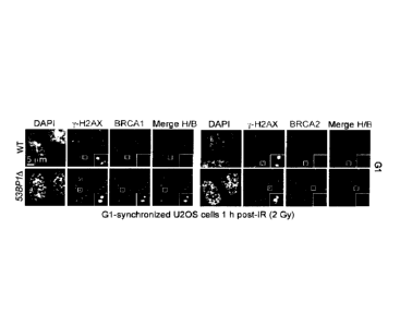
[00058] Figure 1. Inhibition of the BRCA1-PALB2 interaction in G1 is
CRL3-
KEAP1-dependent. a, Micrographs of irradiated (2 Gy) GI -synchronized U2OS
cells
processed for y-H2AX, BRCA1 and BRCA2 immunofluorescence. DAPI, 4' ,6-
diamidino-2-phenylindole; IR, ionizing radiation; WT, wild type. b,
Quantitation of the
experiment shown in a and Figure 5d. ASN, asynchronously dividing cells. WT,
wild
type (Mean + standard deviation (s.d.), N=3). c, Immunoprecipitation (IP) of
PALB2
from extracts prepared from mock- or X-irradiated 293T cells synchronized in S
or G1
phases. A normal immunoglobulin (Ig)G immunoprecipitation was performed as
control. Cyclin A staining ascertains cell cycle synchronization. Numbers on
left
indicate kDa. For gel source data see Figure 5. d, Quantitation of the
experiment shown
in Figure 7a. 53BP1A. U2OS cells transfected with the indicated GFP-PALB2
vectors
and short interfering (si)RNAs were irradiated (20 Gy) before being processed
for
microscopy. (mean s.d., N=3). e, Normal IgG and PALB2 immunoprecipitations
from
extracts prepared from synchronized and irradiated 293T cells of the indicated
genotypes. Numbers on the left indicate kDa.
[00059] Figure 2. Ubiquitylation of PALB2 prevents BRCA I -PALB2
interaction. a, Sequence of the PALB2 N terminus and mutants. [SEQ ID NOs: 1-
3] b,
GFP immunoprecipitation (IP) of extracts derived from G 1- or S-phase
synchronized
293T cells expressing the indicated GFP¨PALB2 proteins. c, In vitro
ubiquitylation of
the indicated HA-tagged PALB2 proteins by CRL.3¨KBAPI. d, Pulldown assay of
ubiquitylated HA¨PALB2 (1-103) incubated with MBP or MBP¨BRCAl-CC. I, input;
FT, flow-through; PD, pulldown. The asterisk denotes a fragment of HA¨PALB2
competent for BRCA1 binding. b¨d, Numbers on left indicate kDa.
[00060] Figure 3. USP11 opposes the activity of CRL3-KEAP1. a, Normal
IgG
or PALB2 immunoprecipitation (IP) of extracts derived from camptothecin (CPT)-
treated 293T cells of the indicated genotypes transfected with GFP¨USP11
constructs,
EV, empty vector; CS, C318S; WT, wild type. b, Clonogenie survival assays of
293T
cells of the indicated genotypes treated with olaparib (mean s.d., N > 3).
c, Normal
IgG or PALB2 immunoprecipitation of extracts derived from CPT-treated 293T
cells of
the indicated genotypes. d, Immunoblots of deubiquitylation reactions
containing
ubiquitylated HA-tagged PALB2 (1-103) and increasing concentrations of
glutathione
S-transferase (GST)¨USP11 or its C270S (CS) mutant. USP2 was used as a
control.
DUB, deubiquitylase. e, Cell cycle- synchronized U2OS cells were irradiated
(20 Gy
dose) and processed for immunoblotting. IR, ionizing radiation. f, Immunoblots
of
CA 02977685 2017-08-24
WO 2016/138574
PCT/CA2016/000057
- 12 -
extracts from irradiated U2OS cells transfected with the indicated siRNAs.
CTRL,
control. g, Fluorescence micrographs of G 1 -synchronized and irradiated (20
Gy)
53BP1A U2OS cells transfected with the indicated siRNAs. The percentage of
cells
with more than five y -H2AX-colocalizing BRCA2 foci is indicated (mean s.d.,
N=
3). Scale bars, 5 p m. a, c, d, f, Numbers to left or right indicate lcDa.
[00061] Figure 4. Reactivation of HR in G1 phase. a, Quantitation of
wild-type
(WT) and 53BP1A U2OS cells co-transfected with non-targeting (CTRL) or KEAP1
siRNAs and vectors expressing wild-type CtIP or the T847E (TE) mutant that
were
synchronized in GI, irradiated (2 Gy) and processed for y -H2AX and RAD51
immunofluorescence (mean s.d., N = 3). b, Representative micrographs from a.
IR,
ionizing radiation. c, Schematic of the gene-targeting assay. d, Gene-
targeting
efficiency at the LMNA locus in asynchronously dividing (ASN) and Gl-arrested
U2OS
cells (mean s.d., N= 3). HR, homologous recombination; sgRNA, single guide
RNA.
e, Gene targeting at the LMNA locus in Gl-arrested cells transfected with the
indicated
siRNA or a PALB2-KR expression vector (mean s.d., N = 3). f, Model of the
cell-
cycle regulation of homologous recombination.
[00062] Figure 5. Suppression of PALB2¨BRCA2 accumulation at DSB sites
in
G1 53BP1A cells. a, Schematic representation of human 53BP1 gene organization
and
targeting sites of sgRNAs used. Boxes indicate exons (E: yellow, coding
sequence;
brown, untranslated regions (UTRs)). The indels introduced by CRISPR/Cas9 and
their
respective frequencies are indicated. b, Wild-type (WT) and 53BP1A and U2OS
cells
were mock- or X-irradiated (10 Gy) before being processed for 53BP1
fluorescence
microscopy. DAPI was used to stain DNA and trace the outline of the nucleus.
c, Wild-
type (WT) and 53BP1A U2OS cells were processed for 53BP1 immunoblotting.
Tubulin was used as a loading control. d, Wild-type (WT) and 53BP1A U2OS cells
either synchronized in GI following a double-thymidine block and release or
asynchronously dividing (ASN), were irradiated (2 Gy) and processed for y-
II2AX,
PAI,B2, BRCA2 and BRCA1,immunofluorescence. The micrographs relating to
BRCA1 and BRCA2 staining in GI are found in Figure I a. e, Wild-type (WT) and
3BP1A U2OS cells synchronized in GI after release from a double-thymidine
block
were irradiated (20 Gy) and processed for y-H2AX, BRCA1 and BRCA2
immunofluorescence. On the left are representative micrographs for the Gl-
arrested
cells and the quantitation of the full experiment is shown on the right (mean
s.d.,
N=3).
CA 02977685 2017-08-24
WO 2016/138574
PCT/CA2016/000057
- 13 -
l000631 Figure 6. The BRCA1¨PALB2 interaction is cell cycle regulated.
a,
Schematic of the LacO/LacR chromatin-targeting system. b, U2OS 256 cells were
transfected with the indicated mCherry-LacR and GFP-fusions. GFP fluorescence
was
measured at the site of the lac() array-localized mCherry focus. Each circle
represents
one cell analyzed and the bar is at the median. Cells were also stained with a
cyclin A
antibody to determine cell cycle position (N=3). IR, Ionizing radiation. c,
Representative micrographs of U2OS 256 cells transfected with the indicated
mCherry-
LacR and GFP-fusions; data is quantified in d. d, Quantification of U2OS 256
cells
transfected with the indicated mCherry-LacR and GFP-fusions to tether either
BRCA1
or PALB2 to the lac0 array (N=3). e, Schematic representation of PALB2
architecture
and its major interacting proteins. f, Quantification of U2OS 256 cells
transfected with
the indicated GFP-PALB2 mutants and mCherry-LacR-BRCA1-CC. Cells were also
stained with a cyclin A antibody to determine cell cycle position (N=3).
[00064] Figure 7. Inhibition of the BRCA1¨PALB2 interaction in GI
depends on
CRL3¨KEAP1. a, Representative micrographs of the experiment shown in Figure
id. b,
Schematic representation of human KEAP1 gene organization and targeting sites
of
sgRNAs used as described in Figure 5a. The indels introduced by CR1SPR/Cas9
and
their respective frequencies are indicated. c, Immunoprecipitation (IP) of
PALB2 from
extracts prepared from irradiated 293T cells. IP with normal IgG was performed
as a
control. d, 293T cells with the indicated genotypes were transfected with the
indicated
LIA¨KEAP1 constructs, synchronized in G1 or S phases and irradiated. Cells
were
processed for PALB2 immunoprecipitation (IP). EV, empty vector; WT, wild type.
e,
Quantification of U2OS 256 cells transfected with the indicated GFP-PALB2
mutants
and mCherry-LacR-BRCA1. Cells were also stained with a cyclin A antibody to
determine cell cycle position (N=3). f, Quantification of U2OS 256 cells
transfected
with GFP-PALB2 and mCherry-LacR-BRCA1-CC (wild type or K1406R mutant).
Cells were also stained with a cyclin A antibody to determine cell cycle
position. This
panel shows that the sole lysine in the PALB2-interaction motif of BRCA1 is
not
involved in the cell cycle regulation of the PALB2-BRCA1 interaction. e, f,
Each circle
represents a cell analyzed and the bar is at the median (N = 3).
[000651 Figure 8. PALB2 is ubiquitylated by CRL3¨KEAP1. a, 11E1(293 Flp-
In
T-REX cells expressing doxycycline (DOX)-inducible His6-Ub were transfected
with
the indicated siRNAs. Cells were processed for Ni-NTA pull-down (IP). b, 293T
cells
transfected with an siRNA targeting USP11 and a Flag-PALB2 expression vector
were
CA 02977685 2017-08-24
WO 2016/138574
PCT/CA2016/000057
- 14 -
processed for Flag immunoprecipitation followed by mass spectrometry (MS).
Representative MS/MS spectra of tryptic diglycine (diG)-PALB2 peptides
identified
are shown (K16, top; K43, bottom), c, Schematic of the /acO/LacR chromatin-
targeting
system and the in vivo quantification of ubiquitylated PALB2. d,
Representative
micrographs of U2OS 256 cells transfected with the
indicated mCherry-LaeR¨PALB2 vectors. Cells were processed for FK2
immunofluorescence. EV, empty vector. Scale bar, 5 1.tm. e, Quantification of
U2OS
256 cells transfected with the indicated mCherry-LacR¨PALB2 vectors. Cells
were
processed for quantification of FK2 fluorescence at the Lac focus. Each
circle
represents a cell analyzed and the bar is at the median (N= 3). Cells were
also stained
with a cyclin A antibody to determine cell cycle position. Statistical
significance was
determined by a Kruskall¨Wallis test (***P< 0.001; **p< 0.01).
[00066] Figure 9.
Analysis of KEAP1- and USP11-dependent modulation of
PALB2 and homologous recombination. a, Site-specific chemical ubiquitylation
of
HA¨PALB2 (1-103) at residue 20 (PALB2-KC20-Ub) and 45 (PALB2-KC45-Ub) was
carried out by dichloroacetone linking. The resulting ubiquitylated PALB2
polypeptides
along with their unmodified counterparts were subjected to pulldown with a
fusion of
MBP with the coiled-coil domain of BRCA1 (MBP¨BRCA1 -CC). I, input; PD,
pulldown. Asterisk indicates a non-specific band. b, Wild-type and KEAP1A 293T
cells
were treated with cyclohcximidc (CHX) for the indicated time and then
processed for
NRF2 and KEAP1 immunoblotting. Actin levels were also determined as a loading
control. c, Immunoprecipitation (IP) of USP11 from extracts prepared from 293T
cells
that were or were not treated with camptothecin (CPT; 200 nM).
Immunoprecipitation
with normal IgG was performed as a control. d, U2OS DR-GFP cells were
transfected
with the indicated siRNAs. Twenty-four hours post-transfection, cells were
further
transfected with the indicated siRNA-resistant USP11 expression vectors (WT,
wild
type; CS, C318S and CA, C318A catalytically dead mutants) or an empty vector
(EV),
with or without an I-SceI expression vector. The percentage of GFP-positive
cells was
determined 48h post-plasmid transfection for each condition and was normalized
to the
I-SceI plus non-targeting (siCTRL) condition (mean s.d., N = 3). e,
Schematic
representation of human USP11 (top) and KEAP1 (bottom) gene organization and
targeting sites of sgRNAs (as described in Figure 5a) used to generate the
USP11A and
USP11A IKEAP1A 293T cells. The indels introduced by the CRISPR¨Cas9 and their
respective frequencies are indicated. The USP11 knockout was created first and
CA 02977685 2017-08-24
WO 2016/138574
PCT/CA2016/000057
- 15 -
subsequently used to make the USP//A IKEAP1A double mutant. f,
Immunoprecipitation of PALB2 from extracts prepared from 2931 cells
transfected
with the indicated siRNA and with or without CPT (200 nM) treatment.
Immunoprecipitation with normal IgG was performed as a control.
[00067] Figure 10. USP11 antagonizes KEAPI action on PALB2. a, U2OS DR-
GFP cells were transfected with the indicated siRNAs or left untransfected ( ¨
).
Twenty-four hours post-transfection, cells were transfected with an I-Scel
expression
vector (circle). The percentage of GFP-positive cells was determined 48 h post-
plasrnid
transfection for each condition and was normalized to the I-SceI plus non-
targeting
(CTRL) condition (mean range, N = 3). b, Parental 293T cells (wild type
(WT)) or a
USP11A derivative were transfected with the indicated GFP¨PALB2 constructs,
treated
with CPT and processed for GFP immunoprecipitation (IP). c, Parental 2931
cells (wild
type) or a USPI IA derivative were transfected with an empty vector (EV) or
the
indicated PALB2 expression vectors. Sensitivity of the cells to the PARP
inhibitor
olaparib was then determined by a clonogenic survival assay (mean s.d.,
N=3).
[00068] Figure 11. Characterization of USP11 protein stability a, U2OS
cells
synchronized in GI or S/G2 were treated with cyclohexamide (CHX) and processed
at
the indicated time points to monitor USP 11 stability. b, Immunoprecipitation
(IP) of
PALB2 from extracts prepared from 293T cells that were synchronized in GI or S
phase and treated or not with IR (20 Gy). c, U2OS cells were irradiated with a
dose of 2
or 20 Gy and processed for USP11 immunoblotting at the indicated times post-
IR.
Actin was used as a loading control. d, U2OS cells, mock-treated or incubated
with the
ATM inhibitor KU55933 (ATMi), AIR inhibitor VE-821 (ATRi) or DNA-PKcs
inhibitor NU7441 (DNAPKi), were irradiated (20 Gy) and processed for USP11 and
actin (loading control) immunoblotting. e, Similar experiment to d except that
cells
wcre exposed to ultraviolet (UV) radiation (50 in.T/cm-2). f, U2OS cells, mock-
treated or
incubated with the proteasome inhibitor MG132, were irradiated (20 Gy) and
processed
for USP11 and actin (loading control) immunoblotting. g, U2OS cells, mock-
treated or
incubated with the Cullin inhibitor MLN4924, were irradiated (20 Gy) and
processed
.. for USP11 and actin (loading control) immunoblotting.
[00069] Figure 12. Reactivation of RAD51 loading and unscheduled DNA
synthesis in G1 . a, 53BP1 U2OS cells were transfected with the indicated
siRNA,
synchronized in GI or S/G2 by release from a double-thymidine block and
irradiated
(20 Gy) before being processed for fluorescence microscopy. DAPI was used to
trace
CA 02977685 2017-08-24
WO 2016/138574
PCT/CA2016/000057
- 16 -
the nuclear boundary and cyclin A staining was used to determine cell cycle
position.
The percentage of cells with more than five y -H2AX-colocalizing PALB2 foci is
indicated as the mean s.d., N = 3. Scale bar, 5 gm. b, Representative
micrographs of
irradiated G1 -synchronized wild-type (WT) and 53BP1A U2OS cells transfected
with
the indicated siRNA and expressing wild-type CtIP. c, Representative
micrographs of
irradiated Gl-synchronized wild-type U2OS cells transfected with the indicated
siRNA
and expressing CtIP(T847E). d, U2OS 53BP1A cells were synchronized in GI,
supplemented with BrdU, irradiated (2 Gy) and processed for y -H2AX and BrdU
immunofluorescence. The percentage of cells with more than five y-H2AX-
colocalizing
BrdU foci is indicated (mean s.d., N = 3). e, Micrograph of a U2OS cell
targeted with
the CRISPR¨mClover system showing the typical perinuclear expression pattern
of
lamin A. f, Micrograph of a U2OS cell targeted with the mClover system showing
an
expression pattern characteristic of subnuclear PML foci. g, Timeline of the
gene-
targeting (LMNA) experiment presented in Figure 4d. h, Timeline of the gene
targeting
(LMNA or PML) experiment presented in Figure 4e and Figure 13.
[00070] Figure 13. Analysis of homologous recombination in Gl. a,
Quantitation
of gene targeting efficiency at the LMNA locus in asynchronously dividing U2OS
cells
transfected with increasing amount of donor template and with (grey) or
without
(white) sgRNAs. Gene-targeting events were detected by flow cytometry (mean
s.d.,
N ? 3). b, Quantitation of gene-targeting efficiency at the LMNA locus in
asynchronously dividing cells transfected with the indicated siRNA. Gene-
targeting
events were detected by flow cytometry (mean + s.d., N = 3). c, Gene-targeting
efficiency at the PML locus measured by flow cytometry in Gl-arrested 53BP1 A
U2OS
cells expressing the CtIP(T847E) mutant and co-transfected with the indicated
siRNA
or a PALB2-KR expression construct (mean s.d., N= 3). d, Gene-targeting
efficiency
at the LMNA locus measured by flow cytometry in G1 -arrested parental (wild-
type
(WT)) and 53BP1A U2OS cells transfected with KEAP1 siRNA and expressing the
CtIP(T847E) mutant (mean s.d., N = 3). e, Gene-targeting efficiency at the
LMNA
locus measured by flow cytomctry in GI-arrested parental (wild-type) and
53BP1A
U2OS cells transfected with the indicated siRNA and expressing either wild
type or the
CtIP(T847E) mutant (mean s.d., N = 3).
[00071] Figure 14 Identification of DCAF10 as a regulator of USP11
stability in
response to DNA damage. a. siRNA screen where U2OS cells were transfected with
siRNAs targeting known and predicted DCAFs along with other CUL4-interacting
CA 02977685 2017-08-24
WO 2016/138574
PCT/CA2016/000057
- 17 -
proteins. Cells were either irradiated with IR (20 Gy) or UV (50 J/m-2), let
to recover
for 3 h and then processed for USP11 immunofluorescence. Each point plotted
corresponds to the percentage of USP11 left after irradiation. The red dots
correspond
to the siRNA non-targeting controls (CTRL) and targeting USP11, whereas the
red dots
correspond to core CRL4 factors, that include CUL4 itself. b.U2OS cells were
transfected with the indicated siRNAs and then irradiated with a dose of 20 Gy
and
processed for USP11 immunoblotting at the indicated times post-ionizing
radiation.
Actin was used as a loading control.
1000721 Figure 15.
Validation of DCAF10 as a regulator of USP11. a. DCAF10
interacts with USP11. Immunoprecipitation (IP) of Flag-USP11 from extracts
prepared
from 293 Flp-IN/T-Rex cells. Cells were probed with DCAF10 and DCAF15
antibodies. b. Whole cell extracts of mouse embryo fibroblasts (MEFs) of the
indicated
genotypes were processed for USP11 immunoblotting. Tubulin was used as a
loading
control. c. U2OS DR-GFP cells were transfected with the indicated siRNAs or
expression vectors. Twenty-four hours post-transfection, cells were
transfected with an
I-SceI expression vector. The percentage of GFP-positive cells was determined
48 h
post-plasmid transfection for each condition and was normalized to the I-SceI
plus non-
targeting (CTRL) + empty vector (EV) condition.
100073] Figure 16.
KEAP1 inhibition can activate HR in G1 cells. Gene targeting
at the LMNA locus in G1 -arrested cells transfected with the indicated siRNA
and
vectors expressing either the R1 KEAP1 inhibitor or its FN3 scaffold control
(mean
s.d., N = 3).
DETAILED DESCRIPTION OF THE INVENTION
100074] The
preparation and use of the agents disclosed as well as the practice of
the methods herein employed, unless otherwise indicated, utilize conventional
techniques in molecular biology, biochemistry, chromatin structure and
analysis,
computational chemistry, cell culture, recombinant DNA and related fields as
are
within the skill of the art. The techniques are fully disclosed in the
literature. [See, for
example, Sambrook et al. Molecular Cloning: A Laboratory Manual, Second
edition,
Cold Spring Harbor Laboratory Press, 1989 and Third edition, 2001; Ausubel et
al.,
Current Protocols in Molecular Biology, John Wiley & Sons, New York, 1987 and
periodic updates; the series Methods in Enzymology, Academic Press, San Diego;
Wolffe, Chromatin Structure and Function, Third edition, Academic Press, San
Diego,
1998; Methods in Enzymology, Vol. 304, "Chromatin" (P.M. Wassarman and A. P.
CA 02977685 2017-08-24
WO 2016/138574
PCT/CA2016/000057
- 18 -
Wolffe, eds.), Academic Press, San Diego, 1999; and Methods in Molecular
Biology,
Vol. 119, "Chromatin Protocols" (P. B. Becker, ed.) Humana Press, Totowa,
1999].
[00075] Unless defined otherwise, all technical and scientific terms
used herein
have the same meaning as commonly understood by one of ordinary skill in the
art to
which this invention belongs. The following definitions supplement those in
the art and
are directed to the present application and are not to be imputed to any
related or
unrelated case. Although any methods and materials similar or equivalent to
those
described herein can be used in the practice of the invention, particular
materials and
methods are described herein.
100076] As used herein and in the appended claims, the singular forms "a",
"an",
and "the" include plural reference unless the context clearly dictates
otherwise. As used
herein, the words "comprising" (and any form of comprising, such as "comprise"
and
"comprises"), "having" (and any form of having, such as "have" and "has"),
"including"
(and any form of including, such as "includes" and "include") or "containing"
(and any
form of containing, such as "contains" and "contain") are inclusive or open-
ended and
do not exclude additional, unrecited elements or method steps.
[00077] A "gene editing system" is a system for targeting and editing
genomes,
including without limitation, a TALEN (Transcription Activator-Like Effector
Nucleases) system, a CRISPR (Clustered Regulatory Interspaced Short
Palindromic
Repeats) system and a Zinc-Finger Nucleases (ZEN) system. (See Nemudryi A.A.
et al,
Acta Naturae. 2014 Jul-Sep; 6(3): 19-40 for a review of TALEN and CRISPR
systems;
Gaj T. et al, Trends Biotechnol. 2013 Jul; 31(7): 397-405 for a review of
TALEN,
CRISPR and ZEN systems; US Published Patent Application No. 20110145940
describing a TALEN system; and Bibikova M., et al, Genetics. 2002;161(3):1169-
1175; Townsend J.A., et al, Nature 2009;459(7245):442-445; Zhang F., et at,
Proc.
Natl. Acad. Sci. USA. 2010;107(26):12028-12033; Torikai H.,
et al; Blood.
2012;119(24):5697-5705; Provasi E., et al, J.. Nat. Med. 2012;18(5):807-8151,
and
Lombardo A., et al, Nat. Methods. 2011;8(10):861-869 describing ZFN systems).
1000781 A "CRISPR system" generally refers to transcripts and other
elements
involved in the expression of, or directing the activity of Clustered
Regularly
Interspaced Short Palindromic Repeats (CRISPR)-associated ("Cas") genes. A
CRISPR
system may include without limitation, sequences encoding a Cas gene, a tracr
(trans-
activating CRISPR) sequence (e.g. tracrRNA or an active partial tracrRNA), a
tracr-
mate sequence, a guide sequence, or other sequences and transcripts from a
CRISPR
CA 02977685 2017-08-24
WO 2016/138574
PCT/CA2016/000057
- 19 -
locus. One or more elements of a CRISPR system may be derived from a type I,
type II,
or type III CRISPR system. A CRISPR system promotes the formation of a CRISPR
complex (comprising a guide sequence hybridized to a target sequence and
complexed
with one or more Cas proteins) at the site of a target sequence. A "target
sequence" or
"target polynucleotide" refers to a sequence which is sufficiently
complementary to a
designed guide sequence that the target sequence hybridizes to the guide
sequence
promoting the formation of a CRISPR complex. A target sequence may comprise
any
polynucleotide, such as DNA or RNA polynucleotides, and it may be located in
the
nucleus, cytoplasm, or an organelle, for example, mitochondria or chloroplast.
In the
context of an endogenous CRISPR system, formation of a CRISPR complex in an
endogenous CRISPR system results in cleavage of one or both strands in or near
(e.g.
within 1, 2, 3, 4, 5, 6, 7, 8, 9, 10, 20, 50, or more base pairs from) the
target sequence.
[00079i CRISPR systems are described in U.S. Pat. Nos. 8,697,359,
8,771,945,
8,795,965, 8,865,406, 8,871,445, 8,889,356, 8,889,418 and 8,895,308; US Patent
Publications US 2014-0310830, US 2014-0287938, US 2014-0273234, US2014-
0273232, US 2014-0273231, US 2014-0256046, US 2014-0248702), US 2014-
0242700, US 2014-0242699, US 2014-0242664, US 2014-0234972, US 2014-
0227787, US 2014-0189896, US 2014-0186958, US 2014-0186919, US 2014-0186843,
US 2014-0179770 and US 2014-0179006, US 2014-0170753, and US 20150232883;
European Patent Applications EP 2771468 (EP13818570.7), EP 2764103
(EP13824232.6), and EP 2784162 (EP14170383.5); and PCT Patent Publications
W02014/093661 (PCT/U52013/074743), W02014/093694 (PCT/US2013/074790),
W02014/093595 (PCT/US2013/074611), W02014/093718 (PCT/1JS2013/074825),
W02014/093709 (PCT/U S2013/074812), W 02014/093622 (PCT/U52013/074667),
W02014/093635 (PCT/US2013/074691), W02014/093655 (PCT/US2013/074736),
W02014/093712 (PCT/US2013/074819), W02014/093701 (PCT/US2013/074800),
W02014/018423 (PCT/US2013/051418) and W02014/093622 (PCT/US20131074667).
General information on CRISPR-Cas Systems is also described in the following
publications: Cong, L., et at., Science, February 15; 339(6121):819-23 (2013);
Jiang
W., et al., Nat Biotechnol March; 31(3):233-9 (2013); Wang H., et al, Cell May
9;
153(4):910-8 (2013); Konermann S, et al, Nature. 2013 Aug. 22; 500(7463):472-
6. doi:
10.1038/Nature12466. Epub 2013 Aug. 23; Ran, F A., et al, Cell August 28. pii:
S0092-
8674(13)01015-5. (2013); Hsu, P., et al, Nat Biotechnol doi:10.1038/nbt.2647
(2013);
Ran, F A., et al, Nature Protocols November; 8(11):2281-308. (2013); Shalem,
0., et
- 20 -
al., Science December 12. (2013). [Epub ahead of print]; Nishimasu, H., et al,
Cell Feb.
27. (2014). 156(5):935-49; Wu X., et al, Nat Bioteclmol. (2014) Apr. 20. doi:
10.1038/nbt.2889; Platt et al., Cell 159(2): 440-455 (2014) DOT:
10.1016/j.ce11.2014.09.014; Hsu et al. Cell 157, 1262-1278 (Jun. 5,2014)
(2014); Wang
et al., Science. 2014 Jan. 3; 343(6166): 80-84. doi: 10.1126/science. 1246981;
Doench
et al., Nature Biotechnology published online 3 Sep. 2014;
doi:10.1038/nbt.3026;
Storrs, The Scientist, Article No. 39239, March 1, 2014; and Swiech et al,
Nature
Biotechnology; published online 19 Oct. 2014; doi:10.1038/nbt.3055). Several
programs are available to design guide sequences, for example, MIT's CRISPR
Design
and E-CRISP developed by the
German Cancer Research Center. CRISPR systems also include the systems
developed
by or available from Editas Medicine (Cambridge, MA), Caribou Biosciences
(Berkeley, CA), CRIPSR Therapeutics (Basel, Switzerland), Addgene (Cambridge,
MA) and Intellia Therapeutics (Cambridge, MA),
[00080] "DNA end resection"
generally refers to nucleolytic degradation of the
5'-terminated strand of a DNA double-stranded break leading to the formation
of 3'-
terminated single-stranded DNA. DNA end resection in eukaryotes comprises two
phases: a slow initial phase, catalyzed by the Mrel 1 -Rad50-Nbs1 (MRN)
complex in
mammals, and a second and faster phase catalyzed by the exonuclease Exol or
the
helicase Bloom Syndrome Protein (BLM). DNA end resection is initiated by a
cell
cycle activation step comprising phosphorylation of the accessory protein COP
(also
known as retinoblastoma binding protein 8). Pathways involved in DNA end
resection
may be activated by stimulating or activating BRCA1 recruitment to DNA double-
strand breaks by inhibiting TP53BP1 (53BP1) or RIF, or blocking recruitment of
53BP1 or RIF to DNA double-stranded break sites. In an aspect, DNA end
resection
may be activated by inhibiting 53BP1 (or RIF) expression and/or activity and
expressing a mutated form of CUP that mimics constitutive phosphorylation, for
example CUP-Thr879G1u. In an aspect, DNA end resection is reconstituted or
activated
using inhibitors of 53BP1 and a mutated form of CUP that mimics constitutive
phosphorylation, in particular CUP-Thr879G1u. In an aspect, DNA end resection
may
be reconstituted or activated using purified human proteins: Bloom helicase
(BLM);
DNA2 helicase/nuclease; Exonuclease 1 (EX01); the complex comprising MRE11,
RAD50, and NBS1 (MRN); and Replication protein A (RPA.) (See Nimonkar A.V. et
al, Genes & Development 25:350-362, 2011; Huertas, P, Nat Struct Mol Biol,
17(10:
Date Recue/Date Received 2022-04-19
CA 02977685 2017-08-24
WO 2016/138574
PCT/CA2016/000057
-21-
11-16, doi: 10.1038/nsmb.1710, 2010; Jimeno S., et al, Nucl. Acids Res doe:
101093/nar/gkui384, 2015 for descriptions of DNA end resection).
[00081] "Homologous recombination" and "HR" refer to a type of genetic
recombination in which DNA strands of similar or identical nucleotide
sequences are
exchanged. HR can be used by cells to repair DNA double-strand breaks (DSB) by
the
following general steps. HR is initiated when the DSB is resected by nucleases
and
helicases, generating 3' single-stranded DNA (ssDNA) overhangs onto which the
RAD51 recombinase assembles as a nucleoprotein filament. This structure can
invade
homologous duplex DNA, which is used as a template for repair DNA synthesis.
The
resulting intermediates can be metabolized to yield non-crossover products
thereby
restoring the damaged DNA molecule as it existed before the double-strand
break (San
Filippo et al., Annu. Rev. Biochem. 2008. 77:229-57). The terms also include
recombination using single-stranded donor oligonucleotides (ssODNs), in
particular
recombination using single-stranded donor oligonucleotides (ssODNs) requiring
resection and which may be activated by 53BP1 inhibitors.
[00082] "HR Disease" refers to any disorder, disease, condition,
syndrome or
combination of manifestations or symptoms recognized or diagnosed as a
disorder
which may be associated with or characterized by a HR defect. Exemplary
diseases
include, for example, cancer, cardiovascular diseases including heart failure,
hypertension and atherosclerosis, respiratory diseases, renal diseases,
gastrointestinal
diseases including inflammatory bowel diseases such as Crohn's disease and
ulcerative
colitis, hepatic, gallbladder and bile duct diseases, including hepatitis and
cirrhosis,
hematologic diseases, metabolic diseases, endocrine and reproductive diseases,
including diabetes, bone and bone mineral metabolism diseases, immune system
diseases including autoimmune diseases such as rheumatoid arthritis, lupus
erythematosus, and other autoimmune diseases, musculoskeletal and connective
tissue
diseases, including arthritis, achondroplasia infectious diseases and
neurological
diseases such as Alzheimer's disease, Huntington's disease and Parkinson's
disease.
[00083] Methods of the invention may be used to monitor or treat a
disease
caused by a defect in a gene that mediates homologous recombination, for
example,
BRCA1, BRCA2, PALB2, PARP-1, USP11, RAD51, and/or DCAF10.
[00084] Embodiments of the invention provide for monitoring or
treatment of
various cancers including but not limited to carcinomas, melanomas, lymphomas,
CA 02977685 2017-08-24
WO 2016/138574
PCT/CA2016/000057
- 22 -
sarcomas, blastomas, leukemias, myelomas, osteosarcomas, neural tumors, and
cancer
of organs such as the breast, ovary, and prostate.
[000851 In embodiments, the invention provides for monitoring or
treatment of
cancer with BRCA-1 defects, BRCA-2 defects, dual BRCA-1/BRCA-2 defects, and
Fanconi anemia. In embodiments of the invention, the cancer is breast cancer,
in
particular invasive ductal carcinoma and invasive lobular carcinoma. In
embodiments
of the invention, the cancer is ovarian cancer, in particular epithelial
ovarian tumors,
germ cell ovarian tumors, and sex cord stromal tumors.
[00086] Methods of the invention for activating or modulating
homologous
.. recombination may be used to genetically modify polynucleotides associated
with a
genetic disorder. In some embodiments, the genetic disorder is a monogenetic
disorder.
In some embodiments, the genetic disorder is a multigenetic disorder. In some
embodiments, the genetic disorder is associated with one or more SNPs. In
particular
embodiments of the invention, the genomic modification corrects a point
mutation.
[00087] Examples of genetic disorders and polynucleotide sequences
associated
with the genetic disorders may be found on the World Wide Web (see for
example, the
National Center for Biotechnology Information, National Library of Medicine
(Bethesda, MA) or the McKusick-Nathans Institute of Genetic Medicine, Johns
Hopkins University (Baltimore, Md)), listed in published patents and
applications (see,
for example, US Published Application No. 2015/0247150), and in publications
(see for
example, Turitz Cox D.B. et al, Nature Medicine 21, 121-131, 2015; and
O'Connor
T.P. and R.G. Crystal, Nature Reviews/Genetics Volume 7, April 2006, pages 261-
276
including Supplementary Information, and publications cited therein).
[00088] In an aspect, the genetic disorder is a genetic disorder of
muscle. In an
aspect, the genetic disorder is myotonic dystrophy type 1. In an aspect, the
genetic
disorder is myotonic dystrophy type 2. In an aspect, the genetic disorder is
Duchenne
muscular dystrophy (DMD). In an aspect, the genetic disorder is Becker
muscular
dystrophy.
[00089] In an aspect, the genetic disorder is a genetic disorder of the
liver, for
example, alpha-1 antitrypsin deficiency, Wilson Disease, hereditary
hemochromatosis,
Type I tyrosinemia, glycogen storage disease Type IV, argininosuccinate lyase
deficiency, citrin deficiency, cholesterol ester storage disease and
hereditary fructose
intolerance.
CA 02977685 2017-08-24
WO 2016/138574
PCT/CA2016/000057
- 23 -
[00090] In an aspect, the genetic disorder is alpha-1 antitrypsin
deficiency which
is an autosomal recessive (codotninant) disease due to mutations in the
SERPINA1
gene that encodes the scrinc protease inhibitor AAT.
[00091] In an aspect, the genetic disorder is Wilson disease which
depends on
mutations in the gene encoding the ATP7B Cu translocase, a protein mainly
expressed
by the hepatocyte that regulates the levels of copper in the liver.
[00092] In an aspect, the genetic disorder is a genetic disorder of the
lungs.
[00093] In an aspect, the genetic disorder is cystic fibrosis, an
autosomal
recessive disease caused by mutations of the Cystic Fibrosis Transmembrane
Regulator
(CFTR) protein, a member of the ATP-binding cassette superfamily of
transmembrane
proteins.
[00094] In other aspects of the invention the genetic disorder may be
heamophilia, al -antitrypsin deficiency, Canavan disease, Adenosine deaminase
deficiency, X-linked severe combined immunodeficiency, familial amyloidotic
polyneuropathy, thalassemia, Tay-Sachs disease, late infantile ccroid
lipofuscinosis,
mucopolysaccharidosis, Niemann¨Pick disease, achondroplasia, Huntington
disease,
spino-cerebellar ataxia, Fredriech ataxia, Amyotrophic Lateral Sclerosis,
monogenic
hypercholesterolemia and other monogenic disorders.
1000951 In aspects of the invention the genetic disorder is sickle cell
anemia and
a method of the invention comprises correcting the mutated HBB hemoglobin gene
by
gene conversion with its paralog HBD.
[00096] An ''effective amount" refers to an amount of a compound or
composition, as described herein effective to achieve a particular biological
result. Such
results include, without limitation, the treatment of a disease or condition
disclosed
herein as determined by any means suitable in the art.
[00097] "PARP Inhibitor" refers to an inhibitor of the nuclear enzyme
poly(adenosine 5'-diphospho-ribose) polymerase ["poly(ADP-ribose) polymerase"
or
"PARP", which is also referred to as ADPRT (NAD:protein (ADP-ribosyl
transferase
(polymerising)) and PARS (poly(ADP-ribose) synthetase), PARP inhibitors have
been
described in Banasik et al., "Specific Inhibitors of Poly(ADP-Ribose)
Synthetase and
Mono(ADP-Ribosyl)-Transferase", J. Biol. Chem., 267:3, 1569-75 (1992), and in
Banasik et al., "Inhibitors and Activators of ADP-Ribosylatiort Reactions'',
Molec. Cell.
Biochern., 138, 185-97 (1994). PARP inhibitors have been disclosed and
described in
the following patents and patent applications: WO 00/42040; WO 00/39070; WO
CA 02977685 2017-08-24
WO 2016/138574
PCT/CA2016/000057
- 24 -
00/39104; WO 99/11623; WO 99/11628; WO 99/11622; WO 99/59975; WO 99/11644;
WO 99/11945; WO 99/11649; and WO 99/59973; US Patent No. 8,894,989, US Patent
No. 8,946,221; 8,778,966; 8,669,249; 8,623,884; 8,592,416; 8,546,368;
8,541,417;
8,541,403; 8,420,650; 8,362,030; 8,236,802; 8,217,070; 8,188,103; 8,188,084;
8,183,250; 8,173,682; 8,129,382; 8,088,760; 8,080,557; 8,071,623; 8,058,275;
8,012,976; 8,008,491; 7,999,117; 7,956,064; 7,875,621; 7,820,668; 7,750,008;
7,732,491; 7,728,026; 7,652,014; 7,601,719; 7,462,724; 7,087,637; 7,041,675;
6,977,298; 6,924,284; 6,737,421; 6,635,642; 6,495,541; 6,444,676; 6,395,749;
6,380,211; 6,380,193; 6,346,536; 6,197,785; 5,756,510; and Re. 36,397.
[00098] In aspects of the invention, the PARP inhibitor is Olaparib
(AstraZeneca). In aspects of the invention, the PARP inhibitor is Veliparib
(AbbVie
Inc, Chicago, IL). In aspects of the invention, the PARP inhibitor is
Rucaparib (Clovis
Oncology, Inc., Boulder, CO). In aspects of the invention, the PARP inhibitor
is IN0-
1001 (Inotek Pharmaceuticals Corp, Lexington, MA). In aspects of the
invention, the
PARP inhibitor is MK-4827 (niraparib) (Tcsaro, Waltham, MA, also see Montoni
et al,
Frontiers in Pharmacology, [4], Article 18, pages 1-7). In aspects of the
invention, the
PARP inhibitor is talazoparib (Medivation, Inc, San Francisco CA).
100099] A "sample" is a sample derived from any biological source, such
as
tissues, extracts, or cell cultures, including cells (e.g. tumor cells), cell
lines, cell
lysates, and physiological fluids, such as, for example, blood or
subpopulations thereof
(e.g. white blood cells, erythrocytes), plasma, scrum, saliva, ocular lens
fluid,
cerebrospinal fluid, sweat, urine, fecal matter, tears, bronchial lavage,
swabbings, milk,
ascites fluid, nipple aspirate, needle aspirate, synovial fluid, peritoneal
fluid, lavage
fluid, and the like. The sample can be obtained from animals, preferably
mammals,
most preferably humans. Samples can be from a single individual or pooled
prior to
analysis. The sample can be treated prior to use, such as preparing plasma
from blood,
diluting viscous fluids, and the like. Methods of treating samples can involve
filtration,
distillation, extraction, centrifugation, concentration, inactivation of
interfering
components, the addition of reagents, and the like.
[0001001 In embodiments of methods of the invention, the sample is a
mammalian
tissue sample. In another embodiment the sample is a cell lysate. In another
embodiment the sample is a cell. In another embodiment the sample is a human
physiological fluid. In a particular embodiment, the sample is human serum. In
a further
embodiment, the sample is white blood cells or erythrocytes.
CA 02977685 2017-08-24
WO 2016/138574
PCT/CA2016/000057
- 25 -
[000101] The terms "subject", "individual" or "patient" refer,
interchangeably, to
a warm-blooded animal such as a mammal. In particular, the terms refer to a
human. A
subject, individual or patient may be afflicted with or suspected of having or
being pre-
disposed to a disease as described herein. The term also includes animals bred
for food,
.. as pets, or for study including horses, cows, sheep, poultry, fish, pigs,
cats, dogs, and
zoo animals goats, apes (e.g. gorilla or chimpanzee), and rodents such as rats
and mice.
10001021 NCBI Accession Numbers for USP11, PALB2, BRCA1, BRCA2,
KEAP1, 53BP1, DCAF10, RBX1, CUL3 and CtIP are in Table 1 and the human
sequences for same are in the Sequence Listing.
Screening and Monitoring Assays
[000103] The invention provides a method for monitoring activity or
expression of
USP11 by assaying the interaction of BRCA1 and PALB2, the interaction of
BRCA1,
PALB2 and BRCA2, the interaction of USP11 and DCAF10, and/or the interaction
of
USP11 and PALB2. Routine methods known to persons skilled in the art can be
used to
assay protein interactions in a sample. For example, BRCA 1 -PALB2, BRCA1-
PALB2-
BRCA2, USP11-DCAF10, or USP11-PALB2 complexes may be isolated using affinity
techniques such as for example immunologically-based purification (e.g.
immunoaffinity chromatography), peptides may be prepared from the isolated
complexes using conventional methods (e.g. gel electrophoresis, liquid
chromatography, capillary electrophoresis, nano-reversed phase liquid
chromatography,
high performance liquid chromatography, or reverse phase high performance
liquid
chromatography), and the peptides or peptide fragments may be subjected to
mass
spectrometry (e.g., quantitative mass spectrometry such as selected reaction
monitoring
mass spectrometry (sMRM), high resolution data independent analyses (SWATH),
high
resolution multiple reaction monitoring (mRivIHR) or mSi based quantitation).
[000104] The invention also provides a method for monitoring activity of
USPI1
by assaying ubiquitylation of the N-terminus of PALB2. Routine methods known
to
persons skilled in the art can be used to assay ubiquitination in a sample.
For example,
ubiquitination or PALB2 may be assayed by measuring changes in PALB2 (e.g.,
weight; see US Patent No. 6,413,725), the amount of poly-ubiquitin bound to
CRL3-
KEAP1 E3 ligase (see for example, EP 1268847), and/or the activity of CRL3-
KEAP1
E3 ligase (see for example, US Publication No. 2013/0116152). Mass
spectrometry
techniques such as selected reaction monitoring mass spectrometry (sMRM), high
resolution data independent analyses (SWATH), high resolution multiple
reaction
CA 02977685 2017-08-24
WO 2016/138574
PCT/CA2016/000057
- 26 -
monitoring (MRMHR) or MS1 based quantitation) can also be used to monitor
ubiquitin
remnants on peptides from the PALB2 N-terminus following protease digestion.
In a
more specific example, preparation of isotopically labeled synthetic peptides
corresponding to tryptic digests of ubiquitylated PALB2, especially those that
correspond to ubiquitylation on Lys14, Ly16, Lys20, Lys25, Lys30, Lys43 or
Lys45
can be used as internal standards to quantitate the extent of PALB2
ubiquitylation.
[000105] In an aspect, the invention provides a method for assaying
ubiquitylation
of PALB2 polypeptides in a sample, thc method comprising digesting
ubiquitinated
PALB2 polypeptides in the sample with a protease, thereby generating a
plurality of
test peptides; determining the presence of at least one isopeptide bond
between
ubiquitin and a lysine residue of the test peptides by mass spectrometry to
determine the
numbers of ubiquitination sites and thereby the amount of ubiquitination of
PALB2
polypeptides in the sample. In an embodiment, the test peptides are from the
PALB2 N-
terminus. In an embodiment, the lysinc residue corresponds to Lys14, Lys16,
Lys20,
Lys25, Lys30, Lys43 or Lys45. The method may utilize peptide internal
standards
corresponding to different peptide subsequences of PALB2 to provide for
controls in a
quantitative assay. In one aspect, different synthetic peptide internal
standards
corresponding to PALB2 are generated and differentially labeled.
[000106] Proximity ligation assays (PLA) may also be used to assay
activity of
USP11 by assaying the interaction between BRCA1 and PALB2 and/or PALB2-
interacting proteins such as BRCA2, using DNA-based detection. For example,
primary
antibodies against binding partners of an interaction (e.g., PALB2 and BRCA1)
are
added to a cell lysate. A second set of antibodies, termed PLA probes or
proximity
probes, recognize the first set of primary antibodies. The PLA probes contain
DNA
strands that assemble into an assay-specific DNA molecule when in close
proximity.
This DNA molecule can then be amplified and detected using, for example,
fluorescent
probes. [See, for example, Soderberg 0. et al., Nat. Methods., 2006 December;
3(12):995-1000; Jarvius M. et al., Mol. Cell. Protcomics, 2007 September;
6(9):1500-
9)].
[000107] In an aspect, the invention provides a method for assaying BRCA1-
PALB2 or BRCA I-PALB2-BRCA2 interactions in a sample comprising: contacting
the
sample with primary antibodies to each binding partner in the interaction;
contacting
the sample with proximity probes comprising a secondary antibody that binds to
a
corresponding primary antibody, wherein each proximity probe has an
oligonucleotide
CA 02977685 2017-08-24
WO 2016/138574
PCT/CA2016/000057
- 27 -
conjugated thereto; wherein when the oligonucleotides of the proximity probes
are in
sufficient proximity to each other, the oligonucleotides of the proximity
probes interact
to form circular products that are amplified by rolling circle replication
producing
amplification products; and, measuring the amplification products to thereby
assay or
measure the interactions.
[000108] Assays that monitor PALB2 (or associated proteins such as
BRCA2) in
situ co-localization with BRCA1 can also provide a method for monitoring USP11
activity (see Example herein). For example, PALB2 localization at sites of DNA
damage (marked by BRCA1 or other markers such as y-H2AX) is dependent on USP11
activity. In such assays, cells are fixed, peimeabilized and then incubated
with
antibodies that detect PALB2, BRCA2 or their associated proteins (e.g.,
BRCA1).
Addition of labeled secondary antibodies enable the in situ visualization of
protein
accumulation at DNA damage sites in subnuclear structures termed foci.
Addition of a
genotoxic insult (such as ionizing radiation or other clastogenic treatments)
increases
the number of "foci" and can be included to augment the dynamic range of the
assay.
[0001091 It will be appreciated that proximity ligation assays and in
situ co-
localization assays may be used to assay any of the interactions disclosed
herein.
[0001101 The methods of the invention may be performed in the presence
or
absence of a test compound or agent and detection of an increase or decrease
in activity
or expression of one or more of USP11, DCAF10, PALB2, PALB2 ubiquitylation,
BRCAl-PALB2-BRCA2 complex, PALB2-USP11 complex, KEAP1, USP11-DCAF 10
complex, CRL3-KEAP1 complex, CRL3-KEAP1-PALB2 complex, and KEAP1-
PALB2 complex, as compared to a control in the absence of the test compound or
agent
indicates that the test compound or agent may be useful as a therapeutic
agent, or for
modulating homologous recombination.
[0001111 In an aspect, the invention provides a method for identifying
or
evaluating an agent for its ability to sensitize or reverse or delay emergence
of
resistance to PARP inhibitors by determining the effect of the agent on USP11
activity
using a method of the invention. In an aspect, a negative effect on USP11
indicates that
the agent is a sensitizer of cells to PARP inhibitors or can reverse or delay
emergence
of resistance to PARP inhibitors. In an aspect, a positive effect on USP11
indicates that
the agent is a poor sensitizer of cells to PARP inhibitors. In an aspect, the
ability of an
agent to sensitize or reverse or delay emergence of resistance to PARP
inhibitors is
determined by decreased levels of USP11 activity when compared to such levels
CA 02977685 2017-08-24
WO 2016/138574
PCT/CA2016/000057
- 28 -
obtained from a control. In an aspect, the ability of an agent to sensitize or
reverse or
delay emergence of resistance to PARP inhibitors is determined by increased
levels of
USP11 activity when compared to such levels obtained from a control.
10001121 The invention also relates to a method of identifying or
evaluating an
agent for its ability to sensitize cells or reverse or delay emergence of
resistance to
PARP inhibitors by determining the effect of the agent on KEAP1, CRL3-KEAP1,
or
KEAP1-PALB2. In an aspect, a negative effect on KEAP1 (loss of KEAP1 or CRL3-
KEAP1 activity) indicates that the agent is a poor sensitizer of cells to PARP
inhibitors.
In an aspect, a positive effect on KEAP1 or CRL3-KEAP1 activity indicates that
the
agent is a sensitizer of cells to PARP inhibitors or can reverse or delay
emergence of
resistance to PARP inhibitors. In an aspect, the ability of an agent to
sensitize or reverse
or delay emergence of resistance to PARP inhibitors is determined by increased
levels
of KEAP1, CRL3-KEAP1, KEAP1-PALB2 or CRL3-KEAP1 activity or expression
when compared to such levels obtained from a control. In an aspect, the
ability of an
agent to sensitize or reverse or delay emergence of resistance to PARP
inhibitors is
determined by decreased levels of KEAP1, CRL3-KEAP1, KEAPI-PALB2 or CRL3-
KEAP1 activity or expression when compared to such levels obtained from a
control.
[000113] Still further the present invention contemplates methods of
detecting an
anti-cancer agent comprising performing a test assay comprising contacting an
immortalized cell with a test compound and measuring USP11 activity or CRL3-
KEAP1 using a method of the invention and comparing to a control test assay in
the
absence of the test compound. In an aspect, USP11 activity is assayed by
measuring
USP11, PALB2, DCAF10, PALB2 ubiquitylation, BRCA1-PALB2-BRCA2 complex,
PALB2-USP11 complex, KEAP1, USP1I-DCAF10 complex, CRL3-KEAP1 and/or
CRL3-KEAP1-PALB2 complex in the cell. In an aspect, detecting a negative
effect of
the agent on 1,ISP11 activity or expression as compared with a control
indicates a
potential anti-cancer agent or PARP inhibitor sensitizer. In an aspect,
detecting a
negative effect of the agent on BRCAl-PALB2-BRCA2 complex and/or PALB2-
USP11 complex activity or expression as compared with a control indicates a
potential
anti-cancer agent or PARP inhibitor sensitizer. In an aspect, detecting a
positive effect
of the agent on KEAP1, CRL3-KEAP1 and/or CRL3-KEAP1 -PALB2 complex activity
or expression as compared with a control indicates a potential anti-cancer
agent or
PARP inhibitor sensitizer. In an aspect, decreased levels of USP11 activity
when
CA 02977685 2017-08-24
WO 2016/138574
PCT/CA2016/000057
- 29 -
compared to such levels obtained from a control are indicative that agent has
anti-
cancer activity or is a PARP inhibitor sensitizer.
[000114] The
invention provides a method for identifying or evaluating an agent
for its ability to modulate homologous recombination comprising determining
the effect
of a test compound or agent on one or more of USP11, DCAF10, PALB2, PALB2
ubiquitylation, BRCA1-PALB2-BRCA2 complex, PALB2-USP11 complex, KEAP1,
USP I 1 -DCAF I 0 complex, CRL3 -KEAP1 complex and CRL3-KEAP1-PALB2
complex. In an aspect, the invention provides a method for identifying or
evaluating an
agent for its ability to modulate homologous recombination in a cell
comprising (i)
assaying in a sample USP11, DCAF10, PALB2, PALB2 ubiquitylation, BRCA1-
PALB2-BRCA2 complex, PALB2-USP11 complex, KEAP1, USP11-DCAF10
complex, CRL3-KEAP1 and/or CRL3-KEAP1-PALB2 complex in the cell in the
presence or absence of the agent, and (ii) detecting an increase or decrease
in USP11,
DCAF10, PALB2, PALB2 ubiquitylation, BRCA1-PALB2-BRCA2 complex, PALB2-
USP11 complex, KEAP1, USP11-DCAF10 complex, CRL3-KEAP1 complex and/or
CRL3-KEAP1-PALB2 complex in the sample compared to a control as an indication
of
the ability of the agent to modulate homologous recombination.
[000115] The
invention provides a method of screening for a therapeutic agent for
treatment of a disease associated with defects in HR (i.e., HR Disease),
comprising
identifying an agent that disrupts or modulates one or more of USP11, PALB2,
PALB2
ubiquitylation, DCAF10, BRCAl-PALB2-BRCA2 complex, PALB2-USP11 complex,
KEAP1, USP11-DCAF10 complex, CRL3-KEAP1 or CRL3-KEAP1-PALB2 complex.
In an aspect, detecting a positive effect of the agent on USP11, BRCA1-PALB2-
BRCA2 complex and/or, PALB2-USP11 complex activity or expression as compared
with a control indicates a potential therapeutic agent for treatment of a HR
Disease. In
an aspect, detecting a negative cffcct of the agent on, DCFA10, KEAP1, CRL3-
KEAP1
or CRL3-KEAP1-PALB2 complex activity or expression as compared with a control
indicates a potential therapeutic agent for treatment of a HR Disease.
[0001161 Test
compounds used in the methods of the invention can be any product
in isolated form or in a mixture. The test compound may be defined by
structure or
function or it may be undefined. Examples of undefined test compounds include
without limitation tissue samples, biological fluids, cell supernatants,
vegetal
preparations; etc. Test compounds may be peptides such as soluble peptides
including
Ig-tailed fusion peptides, members of random peptide libraries and
combinatorial
CA 02977685 2017-08-24
WO 2016/138574
PCT/CA2016/000057
- 30 -
chemistry-derived molecular libraries made of D- and/or L-configuration amino
acids,
carbohydrates, nucleic acids, antisense molecules, phosphopeptidcs (including
members of random or partially degenerate, directed phosphopeptide libraries),
antibodies [e.g. polyclonal, monoclonal, humanized, anti-idiotypic, chimeric,
single
chain antibodies, fragments, (e.g. Fab, F(ab)2, and Fab expression library
fragments,
and epitope-binding fragments thereof)], small organic or inorganic molecules,
or
libraries of compounds. A test compound may be an endogenous physiological
compound or natural or synthetic compounds.
[000117] In
embodiments, the methods of the invention for identifying agents, in
particular anti-cancer agents, comprise contacting more than one test
compound, in
parallel. In some embodiments, the methods comprises contacting 2, 3, 4, 5, 6,
7, 8, 9,
10, 15, 20, 25, 30, 40, 50, 100, 1000, at least 2, at least 5, at least 10, at
least 50, at least
100, or at least 1000 test compounds in parallel. In some embodiments, high
throughput
screening of compounds and complete combinatorial libraries are assayed.
Methods for
performing high throughput screens are well known in the art. The methods can
also be
automated such that a robot can perform the experiments.
[000118] In
embodiments, the methods of the present invention for identifying
agents, in particular anti-cancer agents, comprises the step of contacting a
cell in the
presence of a test compound. The cells can then be observed to determine if
the test
compound(s) effects USP11 activity, DCAF10, PALB2, ubiquitination of PALB2,
KEAP1, CRL-KEAP1 activity, the interaction of BRCA1 and PALB2, the interaction
of USP11 and PALB2, the interaction of USP11 and DCAF10, and/or the
interaction of
BRCA1, PALB2 and BRCA2. Positive and negative controls may be performed in
which known amounts of test compound and no compound, respectively, are added
to
the assay. One skilled in the art can select and perform the appropriate
controls.
[000119] The activity
of a test compound(s) may be unknown, and the methods of
the invention may be used to identify compounds exhibiting the selected
property (e.g.,
PARP inhibitor sensitizer). In some embodiments, the activity or type of
activity of the
test compound(s) is known or expected, and the methods of the invention can be
used to
further characterize or optimize the activity (e.g., specificity, efficacy,
etc).
[0001201 A method of
the invention may also comprise assaying PARP activity in
the presence of the test compound. PARP activity may be assayed by measuring a
change of poly(ADP-ribose) polymers (PAR), and measuring NAD levels and/or ATP
levels using methods routine to one of ordinary skill in the art. In some
embodiments,
CA 02977685 2017-08-24
WO 2016/138574
PCT/CA2016/000057
-31 -
the levels of NAD are depleted in the presence of the test compound. In some
embodiments the levels of ATP are depleted in the presence of the test
compound. in
some embodiments, the levels of NAD are increased in the presence of the test
compound. In some embodiments the levels of ATP are increased in the presence
of the
test compound.
10001211 A method of the invention may comprise the step of determining
if a cell
has undergone necrosis following administration of a test compound. The
physical
characteristics of the cell can be analyzed using routine methods known to
those skilled
in the art to determine if a cell has undergone necrosis. For example,
necrosis may be
determined by measuring organelle swelling, plasma membrane disintegration,
intracellular vacuole formation, and nuclear degradation without condensation.
10001221 The screening methods of the invention may further comprise
conducting therapeutic profiling of the identified agents or further analogs
thereof, for
efficacy and toxicity in animals; optionally formulating a pharmaceutical
composition
including one or more agents identified as having an acceptable therapeutic
profile; and
optionally administering the agent to a subject or individual.
[0001231 In an aspect, the therapeutic activity of agents and
compositions
identified using a method of the invention may be evaluated in vivo using a
suitable
animal model. Thus, the screening methods of the invention may further
comprise
conducting in vivo studies comprising administering the agent to a suitable
animal
model.
[000124] The invention also provides a method of predicting a response
or
categorizing a response to a PARP inhibitor in a subject comprising assaying
one or
more of USP11, DCAFIO, BRCA1, BRCA2, PALB2, KEAP1, CRL3, CRL3-KEAP1,
BRCAl-PALB2-BRCA2 complex, PALB2-USP11 complex and CRL3-KEAP1-
PALB2 complex or PALB2 ubiquitylation in a sample from the subject using a
method
of the invention. Significantly different levels of one or more of USP11,
DCAF10,
BRCA1, BRCA2, PALB2, KEAP1, CRL3, CRL3-KEAP1, BRCA1-PALB2-BRCA2
complex, PALB2-USP11 complex and CRL3-KEAP1-PALB2 complex or PALB2
ubiquitylation compared to a control indicate responsiveness (e.g.,
sensitivity) to the
PARP inhibitor.
10001251 In an aspect, the invention provides a method of predicting a
response or
categorizing a response to a PARP inhibitor in a subject comprising detecting
USP11,
BRCA1, BRCA2, PALB2 and KEAP1 in a sample from the subject using a method of
CA 02977685 2017-08-24
WO 2016/138574
PCT/CA2016/000057
- 32 -
the invention. In an embodiment, significantly different levels of USP11,
BRCA1,
BRCA2, PALB2 and KEAP1 compared to a control indicate responsiveness (e.g.,
sensitivity) to the PARP inhibitor.
[000126] In an aspect, the invention provides a method of predicting a
response
or categorizing a response to a PARP inhibitor in a subject comprising
detecting
USP11, DCAF10, BRCA1, BRCA2, PALB, KEAP1 and CRL3 in a sample from the
subject using a method of the invention. In an embodiment, significantly
different
levels of USP11, DCAF10, BRCA1, BRCA2, PALB, KEAP1 and CRL3 compared to a
control indicate responsiveness (e.g., sensitivity) to the PARP inhibitor.
[000127] In an aspect, a subject is categorized as responsive to a PARP
inhibitor if
there is a decrease in one or more of USP11, DCAF10, BRCA1, BRCA2, PALB2,
KEAP1, CRL3, CRL3-KEAP1, BRCA1-PALB2 and BRCA1-PALB2-BRCA2 activity
or expression or PALB2 ubiquitylation compared to a control. In an aspect, a
subject is
categorized as responsive to a PARP inhibitor if there is an increase in one
or more of
USP11, DCAF10, BRCA1, BRCA2, PALB2, KEAP1, CRL3, CRL3-KEAP1, BRCA1-
PALB2 and BRCA1-PALB2-BRCA2 activity or expression or PALB2 ubiquitylation
compared to a control. In an embodiment, significantly different levels (e.g.,
lower
levels) of USPI 1 activity compared to a control indicate sensitivity to the
PARP
inhibitor.
[000128] In an aspect, the invention provides a method of predicting a
response or
categorizing a response to a PARP inhibitor in a subject comprising detecting
one or
more of USP11, DCAF10, BRCA1, BRCA2, PALB2, KEAP1, CRL3 and CRL3-
KEAP1 activity or expression or PALB2 ubiquitylation in a sample from the
subject
and comparing to a control to determine if the subject will be responsive
(e.g.,
sensitive) to the PARP inhibitor.
[000129] In an aspect, the invention provides a method of predicting a
response or
categorizing a response to a PARP inhibitor in a subject comprising detecting
USP1 I,
BRCA1, BRCA2, PALB2 and KEAP1 activity or expression in a sample from the
subject and comparing to a control to determine if the subject will be
responsive (e.g.,
sensitive) to the PARP inhibitor.
[000130] In an aspect, the invention provides a method of predicting a
response or
categorizing a response to a PARP inhibitor in a subject comprising detecting
USP11,
DCAF10, BRCA1, BRCA2, PALB2, KEAP1 and CRL3 activity in a sample from the
CA 02977685 2017-08-24
WO 2016/138574
PCT/CA2016/000057
- 33 -
subject and comparing to a control to deteilnine if the subject will bc
responsive (e.g.,
sensitive) to the PARP inhibitor.
[000131] In an aspect, the invention provides a method of predicting a
response or
categorizing a response to a PARP inhibitor in an individual comprising
assaying
USP11 activity or expression in a sample from the individual using a method of
the
invention. In an embodiment, significantly different levels (e.g., lower
levels) of USP11
activity or expression compared to a control indicate sensitivity to the PARP
inhibitor.
10001321 In an aspect, the invention provides a method of predicting a
response or
categorizing a response to a PARP inhibitor in an individual comprising
detecting
KEAP1 in a sample from the individual and comparing to a control to determine
if the
individual will be sensitive to a PARP inhibitor. In an embodiment,
significantly
different levels (e.g., higher levels) of KEAP1 compared to a control indicate
sensitivity
to the PARP inhibitor.
10001331 The invention also provides a method of predicting a response
or
categorizing a response to a PARP inhibitor in an individual comprising
detecting
CRL3-KEAP1 activity in a sample from the individual and comparing to a control
to
determine if the individual will be sensitive to a PARP inhibitor. In an
embodiment,
significantly different levels (e.g., higher levels) of CRL3-KEAP1 compared to
a
control indicate sensitivity to the PARP inhibitor.
10001341 The invention also provides a method of predicting a response or
categorizing a response to a PARP inhibitor in an individual comprising
detecting
PALB2 ubiquitylation in a sample from the individual and comparing to a
control to
determine if the individual will be sensitive to a PARP inhibitor. In an
embodiment,
significantly different levels of PALB2 ubiquitylation compared to a control
indicate
sensitivity to the PARP inhibitor.
[000135] The invention also provides a method of predicting a response
or
categorizing a response to a PARP inhibitor in an individual comprising
detecting
complexes of BRCA1, PALB2 and BRCA2 in a sample from the subject and
comparing to a control to determine if the individual will be responsive
(e.g., sensitive)
to a PARP inhibitor. In an embodiment, significantly different levels (e.g.,
absence or
low levels) of complexes of BRCA1, PALB2 and BRCA2 indicate sensitivity to the
PARP inhibitor.
[000136] The invention further provides a method for assigning an
individual to
one of a plurality of categories in a clinical trial for a PARP inhibitor
comprising
CA 02977685 2017-08-24
WO 2016/138574
PCT/CA2016/000057
- 34 -
assaying USP11, DCAF10, PALB2, PALB2 ubiquitylation, BRCAl-PALB2-BRCA2
complex, PALB2-USP11 complex, USP11-DCAFI0 complex, KEAP1, CRL3-KEAP1
or CRL3-KEAP1-PALB2 complex in a sample from the individual using a method of
the invention.
[000117] The invention further provides a method for assigning an
individual to
one of a plurality of categories in a clinical trial for a PARP inhibitor
comprising
assaying USPI I activity in a sample from the individual using a method of the
invention.
[000138] The invention further provides a method for assigning an
individual to
one of a plurality of categories in a clinical trial for a PARP inhibitor
comprising
assaying CRL3-KEAP activity in a sample from the individual.
[000139] The invention further provides a method for assigning an
individual to
one of a plurality of categories in a clinical trial for a PARP inhibitor
comprising
detecting or quantitating USP11, BRCA1, BRCA2, PALB2 and KEAP1 in a sample
from the individual.
[0001401 The invention further provides a method for assigning an
individual to
one of a plurality of categories in a clinical trial for a PARP inhibitor
comprising
detecting or quantitating USP11, DCAF10, BRCA1, BRCA2, PALB2 and KEAP1 in a
sample from the individual.
[000141] The invention further provides a method for assigning an
individual to
one of a plurality of categories in a clinical trial for a PARP inhibitor
comprising
detecting or quantitating BRCA1-PALB2-BRCA2 complex in a sample from the
individual.
[000142] The invention further provides a method for assigning an
individual to
one of a plurality of categories in a clinical trial for a PARP inhibitor
comprising
detecting or quantitating PALB2 ubiquitylation in a sample from the
individual.
10001431 In an aspect, an individual is assigned to a category for a
clinical trial for
a PARP inhibitor based on a decrease in one or more of USP11, DCAF10, BRCA1,
BRCA2, PALB2, KEAP1, CRL3, CRL3-KEAP1, BRCA1-PALB2 and BRCA1-
PALB2-BRCA2 activity or expression or PALB2 ubiquitylation compared to a
control.
In an aspect, an individual is assigned to a category for a clinical trial for
a PARP
inhibitor based on an increase in one or more of USP11, DCAF10, BRCA1, BRCA2,
PALB2, KEAP1, CRL3, CRL3-KEAP I, BRCA1-PALB2 and BRCA1-PALB2-BRCA2
activity or expression or PALB2 ubiquitylation compared to a control.
CA 02977685 2017-08-24
WO 2016/138574
PCT/CA2016/000057
- 35 -
[000144] A variety of routine methods known to a person skilled in the
art can he
employed for detecting or assaying the biomarkers USP11, DCAF10, BRCA1, BRCA2,
PALB2, KEAP1, CRL3 and/or complexes thereof in a sample. Biomarker levels
present in a sample may be determined by any suitable assay, which may
comprise the
use of any of the group comprising or consisting of immunoassays,
spectrometry, mass
spectrometry, Matrix Assisted Laser Desorption/Ionization Time-of-Flight
(MALDI-
TOF) Mass Spectrometry, microscopy, northern blot, isoelectric focussing, SDS-
PAGE, PCR, quantitative RT-PCR, gel electrophoresis, DNA microarray, and
antibody
microarray, or combinations thereof.
[000145] The invention also provides a system comprising: an assay for
determining the level of USP11 activity, complexes or biomarker levels in a
sample
obtained from the subject; a processor for processing the results; computer
coded
instructions for comparing the results with a database; and a user display for
providing
the results of the comparison. The database may comprise reference values for
USP11
activity or biomarker levels.
Treatment Methods
[000146] A method of the invention for predicting or characterizing
responsiveness to a PARP inhibitor may further comprise administering the PARP
inhibitor to the individual or subject. In an aspect, the invention relates to
a method of ,
.. treating a subject with a PARP inhibitor comprising:
a) assaying a sample from the subject for responsiveness or sensitivity to one
or more PARP inhibitors using a method of the invention;
b) identifying a PARP inhibitor that the subject effectively responds or is
sensitive to; and
c) administering the PARP inhibitor to the subject.
[000147] In an aspect, the invention provides a method for treating a
patient in
need of treatment with a PARP inhibitor comprising (a) requesting a test
providing the
results of an analysis to determine if the patient is sensitive to the PARP
inhibitor by
detecting USP11, DCAF10, BRCA1, BRCA2, PALB2, KEAP1 and/or CRL3 in a
sample from the subject and comparing to a control to determine if the patient
is
sensitive to the PARP inhibitor; and (b) administering the PARP inhibitor to
the patient
if the patient is sensitive to the PARP inhibitor. In an aspect of this method
of the
invention, the patient has breast cancer. In an aspect of this method of the
invention, the
CA 02977685 2017-08-24
WO 2016/138574
PCT/CA2016/000057
- 36 -
patient has ovarian cancer. In an aspect of the invention, the test detects
USP11
expression or activity using a method disclosed herein.
[000148] The invention further provides a method for treating cancer in
a subject
the method comprising: (i) selecting a subject who is responsive to a PARP
inhibitor
using a method of the invention, and (ii) administering to said subject the
PARP
inhibitor in an effective amount to treat the cancer. In an embodiment, the
cancer is
breast cancer. In an embodiment, the cancer is ovarian cancer.
[000149] Agents identified using the methods of the present invention
have
numerous therapeutic applications related to, for example, cancer, ischemia
reperfusion
injury, inflammatory diseases, degenerative diseases, protection from adverse
effects of
cytotoxic compounds, and potentiation of cytotoxic cancer therapy. Agents
identified
using the methods of the invention may be used to potentiate radiation and
chemotherapy by increasing apoptosis of cancer cells, limiting tumor growth,
decreasing metastasis, and prolonging the survival of tumor-bearing subjects.
In aspects
of the invention, the agents can be used to treat leukemia, colon cancer,
glioblastomas,
lymphomas, melanomas, carcinomas of the breast, ovarian cancer and cervical
carcinomas.
[000150] In other aspects of the invention, the agents can be used to
treat, without
retroviral infection, arthritis, gout, inflammatory bowel disease, CNS
inflammation, multiple sclerosis, allergic encephalitis, sepsis, septic shock,
hemmorhagic shock, pulmonary fibrosis, uveitis, diabetes, Parkinson's disease,
myocardial infarction, stroke, other neural trauma, organ transplantation,
reperfusion of
the eye, reperfusion of the kidney, reperfusion of the gut, reperfusion of
skeletal
muscle, liver toxicity following acetaminophen overdose, cardiac and kidney
toxicities
from doxorubicin and platinum based antineoplastic agents, and skin damage
secondary
to sulfur mustards.
[000151] In some embodiments, the invention provides a method for
sensitizing
an individual to treatment with PARP inhibitors comprising identifying an
agent that
sensitizes cells to PARP inhibitors in accordance with a method of the
invention and
administering the agent to the individual.
[000152] In some embodiments, the invention provides a method for
treating an
individual being treated with a PARP inhibitor comprising administering to the
individual an agent that sensitizes cells to the PARP inhibitor identified
using a method
of the invention.
CA 02977685 2017-08-24
WO 2016/138574
PCT/CA2016/000057
- 37 -
[000153] In some embodiments, the invention provides a method for
reversing or
delaying emergence of resistance to PARP inhibitors in an individual
comprising
identifying an agent that reverses or delays emergence of resistance to PARP
inhibitors
in accordance with a method of the invention and administering the agent to
the
individual.
[000154] In some embodiments, the present invention provides methods of
treating cancer in an individual comprising identifying an anti-cancer agent
in
accordance with a method of the invention and administering the agent to the
individual.
[000155] In another embodiment, the present invention provides a method of
treating leukemia, colon cancer, glioblastomas, lymphomas, melanomas,
carcinomas of
the breast, ovarian cancer or cervical carcinomas in a mammal in need of such
treatment comprising administering to the mammal a therapeutically acceptable
amount
of an agent identified using a method of the invention or a therapeutically
acceptable
salt thereof.
[000156] In another embodiment, the present invention provides a method
of
potentiation of cytotoxic cancer therapy in a mammal in need of such treatment
comprising administering to the mammal a therapeutically acceptable amount of
an
agent identified using a method of the invention that potentiates cytotoxic
cancer
therapy or a therapeutically acceptable salt thereof.
[000157] In an aspcct, the invention provides methods of treating a
disease
associated with defects in HR (i.e., HR Disease) in an individual comprising
identifying
an agent that modulates HR in accordance with a method of the invention and
administering the agent to the individual.
[000158] In another embodiment, the present invention provides a use of an
agent
identified using a method of the invention to prepare a medicament for
treating a FIR
Disease in a mammal in need of such treatment. In another embodiment, the
present
invention provides a use of an agent identified using a method of the
invention, to
prepare a medicament for inhibiting tumor growth in a mammal in need of such
treatment. In another embodiment, the present invention provides a use of an
agent
identified using a method of the invention to prepare a medicament for
treating cancer
in a mammal in need of such treatment. In another embodiment, the present
invention
provides a use of an agent identified using a method of the invention to
prepare a
medicament for treating leukemia, colon cancer, glioblastomas, lymphomas,
CA 02977685 2017-08-24
WO 2016/138574
PCT/CA2016/000057
-38 -
melanomas, carcinomas of the breast, ovarian cancer or cervical carcinomas in
a
mammal in need of such treatment. In another embodiment, the present invention
provides a use of art agent identified using a method of the invention to
prepare a
medicament for potentiation of cytotoxic cancer therapy in a mammal in need of
such
treatment comprising administering to the mammal a therapeutically acceptable
amount
of the agent.
[000159] In an embodiment, the present invention provides a
pharmaceutical
composition comprising an agent identified using a method of the invention, or
a
therapeutically acceptable salt thereof, in combination with a therapeutically
acceptable
carrier. Pharmaceutical compositions can be prepared by per se known methods
for the
preparation of pharmaceutically acceptable compositions which can be
administered to
subjects, such that an effective quantity of the active substance is combined
in a
mixture with a pharmaceutically acceptable vehicle. Suitable carriers are
described, for
example, in Remington's Pharmaceutical Sciences (Rcmington's Pharmaceutical
Sciences, Mack Publishing Company, Easton, Pa., USA 1985). On this basis, the
compositions include, albeit not exclusively, solutions of the active agents
in
association with one or more pharmaceutically acceptable vehicles or diluents,
and
contained in buffered solutions with a suitable pH and iso-osmotic with the
physiological fluids.
[000160] The pharmaceutical compositions are indicated as therapeutic
agents
either alone or in conjunction with other therapeutic agents or other forms of
treatment.
The compositions of the invention may be administered concurrently,
separately, or
sequentially with other therapeutic agents or therapies, for example PARP
inhibitors.
Homologous Recombination Methods
[000161] The invention provides a method for activating or modulating
homologous recombination in a cell comprising:
(a) promoting or stimulating the assembly or occurrence of BRCA1-PALB2 or
BRCA1-PALB2-BRCA2 complexes in the cell;
(b) promoting or stimulating BRCA1 recruitment to DNA double-strand break
(DSB) sites;
(c) contacting the cell with BRCA 1 -PALB2 or BRCA1 -PALB2-BRCA2
complexes;
(d) inhibiting KEAP1 or CRL3-KEAP1;
(e) inhibiting the degradation of USP11 or promoting USP11 activity; and/or
CA 02977685 2017-08-24
WO 2016/138574
PCT/CA2016/000057
- 39 -
(f) inhibiting DCAF10.
[000162] In an aspect the cell is in the G1 phase of the cell cycle
(G1). In an
aspect the cell is a non-dividing cell or a dormant cell in GI. In an aspect,
the cell is in
the GO phase of the cell cycle (GO). In an aspect, the methods of the
invention are used
in vilro to activate or modulate homologous recombination in cells.
[000163] In an aspect, the invention provides a method for activating or
modulating homologous recombination in a cell, in particular a cell in the GI
phase of
the cell cycle (G1) or GO phase of the cell cycle (GO), comprising (a)
promoting or
stimulating the assembly or occurrence of BRCA1-PALB2 or BRCA1-PALB2-BRCA2
complexes in the cell; and/or (b) contacting the cell with BRCA I -PALB2 or
BRCA1-
PALB2-BRCA2 complexes.
[000164] In embodiments, the assembly of BRCA1-PALB2 or BRCA1-PALB2-
BRCA2 complexes is promoted or stimulated by administering an agent that
promotes
or stimulates BRCA1-PALB2 or BRCA1-PALB2-BRCA2 complexes identified using a
method of the invention.
[000165] In an aspect, the invention provides a method for activating or
modulating homologous recombination in a cell, in particular a cell in G1 or
GO,
comprising the step of inhibiting KEAP1 or CRL3-KEAP1. In an aspect, the
invention
provides a method for activating or modulating homologous recombination in a
cell, in
.. particular a cell in G1 or GO, comprising thc step of blocking the
degradation of USP11
or promoting USP11 activity. In an embodiment, the method comprises
administering
USPI I or an agonist thereof. In an aspect, the invention provides a method
for
activating homologous recombination in a cell, in particular a cell in G1 or
GO,
comprising the step of inhibiting KEAP1 and blocking the degradation of USP11.
In an
aspect, the invention provides a method for activating homologous
recombination in a
cell, in particular a cell in G1 or GO, comprising the step of inhibiting CRL3-
KEAP1
and blocking the degradation of USP11. In an aspect, the invention provides a
method
for activating homologous recombination in a cell, in particular a cell in GI
or GO,
comprising the step of inhibiting CRL3 and blocking the degradation of USP11.
[000166] Methods of the invention may be performed in a cell, in particular
a cell
in the G1 phase of the cell cycle (Cl) or GO phase of the cell cycle (GO) in
which DNA
end resection is or has been activated generating single-stranded DNA.
[000167] The invention provides a method for activating or modulating
homologous recombination in a cell, in particular a cell in the GI phase of
the cell cycle
CA 02977685 2017-08-24
WO 2016/138574
PCT/CA2016/000057
-40 -
(G1) or GO phase of the cell cycle (GO) in which DNA end resection is or has
been
activated generating single-stranded DNA, comprising promoting or stimulating
the
assembly or occurrence of BRCA1-PALB2 or BRCA1 -PALB2-BRCA2 complexes in
the cell. In an embodiment, the assembly of the complexes is promoted or
stimulated by
administering an agent that promotes or stimulates assembly or occurrence of
BRCA1-
PALB2 or BRCA1 -PALB2-BRCA2 complexes identified using a method of the
invention.
[000168] The
invention also provides a method for repairing DNA double-strand
breaks in a cell in the G1 phase of the cell cycle (G1) or GO phase of the
cell cycle
(GO), comprising promoting or stimulating the assembly or occurrence of BRCA1-
PALB2 or BRCA I -PALB2-BRCA2 complexes in the cell.
[000169] The
invention also provides a method for repairing DNA double-strand
breaks in a cell in the GI phase of the cell cycle (G1) or GO phase of the
cell cycle (GO)
in which DNA end resection is or has been activated generating single-stranded
DNA,
comprising promoting or stimulating the assembly or occurrence of BRCA1-PALB2
or
BRCA1-PALB2-BRCA2 complexes in the cell. In an embodiment, the assembly of the
complexes is promoted or stimulated by administering an agent that promotes or
stimulates the assembly or occurrence of BRCA1-PALB2 or BRCA1 -PALB2-BRCA2
complexes identified using a method of the invention.
[000170] The invention also provides a method for repairing DNA double-
strand
breaks in a cell in the GI phase of the cell cycle (GI) or GO phase of the
cell cycle (GO)
in which DNA end resection is or has been activated generating single-stranded
DNA,
comprising contacting the cell with BRCA1-PALB2 or BRCA1 -PALB2-BRCA2
complexes.
[0001711 The invention also provides a method for repairing DNA double-
strand
breaks in a cell in GI or GO in which DNA end resection is or has been
activated
generating single-stranded DNA, the method comprising (a) inhibiting KEAP1
and/or
CRL3-ICEAP1; (b) blocking the degradation of USP11 or promoting or stimulating
USP11 activity; (c) administering USP11 or an agonist thereof; and/or (d)
inhibiting
CRL3-KEAP1 and blocking the degradation of USP11.
[0001721 A method for
activating homologous recombination in a cell may further
comprise activating or promoting single strand DNA (ssDNA) generation
pathways. In
an aspect, ssDNA generation pathways are activated by DNA end resection. In an
CA 02977685 2017-08-24
WO 2016/138574
PCT/CA2016/000057
- 41 -
embodiment, a method for activating homologous recombination in a cell further
comprises activating DNA end resection.
[000173] In an embodiment, DNA end resection is activated or promoted by
inhibiting 53BP1 (or RIF1) expression or activity (e.g., recruitment of 53BP1
to DSB
sites) and/or upregulating or expressing CtIP. In an embodiment, DNA end
resection is
activated or promoted by inhibiting 53BP1 (or RIF1) expression or activity
(e.g.,
recruitment of 53BP1 to DSB sites) and upregulating or expressing CtIP. In an
embodiment, the method involves inhibiting 53BP1 using antagonists, including
without limitation short interfering (si) RNA, short hairpin (sh) RNA and
microRNAs
(miRNAs) or an inhibitor of the histone deacetylase (HDAC) family of enzymes
(for
example, trichostatin A), and using an analog of CtIP that mimics constitutive
phosphorylation, for example CtIP-Thr879G1u.
[000174] A method for activating homologous recombination in a cell may
comprise activating BRCA1 recruitment to DNA double-strand break (DSB) sitcs.
In an
embodiment, BRCA1 recruitment is activated by inhibiting expression of 53BP1
(TP53BP1) or RIF1, or inhibiting the recruitment of 53BP1 or RIF1 to DSB
sites.
53BP1 or RIF1 may be inhibited using antagonists, including without limitation
short
interfering (si) RNA, short hairpin (sh) RNA and microRNAs (miRNAs). In an
embodiment, 53BP1 is inhibited with an inhibitor of the histone deacetylase
(HDAC)
family of enzymes, in particular a histone deacetylase inhibitor (HDACi),
preferably
trichostatin (Fukuda T. et at, Cancer Sci. 2015 Aug;106(8):1050-6. doi:
10.1111/cas.12717. Epub 2015 Jul 14).
[000175] In an aspect, a method for activating or stimulating homologous
recombination in a cell further comprises a gene editing system. In an aspect
the gene
editing system comprises contacting the cell with a nuclease. Examples of
nucleases
include without limitation, zinc finger nucleases (ZFNs), engineered
meganucicascs,
transcription activator like effector nucleases (TALENS), mega or homing
endonucleases, clustered regularly interspaced short palindromic repeats
(CRISPR)-
associated (Cas) protein, Ttago nucleases, and fusions between nucleases, such
as
mega-TALs and compact TALENs.
[000176] In an aspect, the gene editing steps comprise a TALEN system.
[000177] In an aspect, the gene editing steps comprise a ZFN system.
[000178] In an aspect, the gene editing steps comprise a CRISPR/Cas9
system.
CA 02977685 2017-08-24
WO 2016/138574
PCT/CA2016/000057
- 42 -
[000179] In aspects of the invention the gene editing system may correct
a
genomic modification. A genetic modification may comprise at least one
mutation in a
polynucleotide sequence having a locus associated with a genetic disorder. In
an aspect,
the genomic modification is selected from the group consisting of insertions,
deletions
and combinations thereof. In some embodiments, the genetic disorder is a
monogenetic
disorder. In some embodiments, the disorder is a multigenetic disorder. In
some
embodiments, the disorder is associated with one or more SNPs. In particular
embodiments of the invention, the genomic modification corrects a point
mutation.
10001801 In an aspect of a method of the invention to correct a genomic
modification, the gene editing system comprises contacting the cell with a
clustered
regularly interspaced short palindromic repeats (CRISPR)-associated (Cas)
protein and
from one to two ribonucleic acids, wherein the ribonucleic acids direct Cas
protein to
and hybridize to a selected motif of a target polynucleotide sequence
associated with a
genetic disordcr, wherein the target polynucleotide sequence is cleaved.
[000181] In an aspect, the invention provides a method for altering a
genetic
disorder associated with a target polynucleotide sequence in a cell
comprising: (1)
contacting the cell with a system which activates homologous recombination in
the cell
wherein the system comprises BRCA 1 -PALB2 or BRCA1 -PALB2-BRCA2 or agents
that maintain the BRCA1-PALB2 or BRCA1 -PALB2-BRCA2 interactions throughout
the cell cycle; and (2) contacting the target polynucleotide sequence with a
clustered
regularly interspaced short palindromic repeats-associated (Cas) protein and
from one
to two ribonucleic acids, wherein the ribonucleic acids direct Cas protein to
and
hybridize to a selected motif of the target polynucleotide sequence, wherein
the target
polynucleotide sequence is cleaved. The method may reduce expression of the
target
polynucleotide sequence, knock out the target polynucleotide sequence, or
correct the
target polynucleotidc sequence from an undesired sequence to a desired
sequence.
[000182] In an aspect, the invention provides a method for altering a
genetic
disorder associated with a target polynucicotide sequence in a cell
comprising: (1)
contacting the cell with a system which activates homologous recombination in
the cell
wherein the system comprises a kit, vector(s) or composition of the invention,
in
particular the system comprises an inhibitor of 53BP1, a KEAP1 inhibitor or
DCAF10
inhibitor, and an analog of CUP that mimics constitutive phosphorylation,
preferably
the system comprises a KEAP1 inhibitor, an inhibitor of 53BP1 chosen from
short
interfering (si) RNA, short hairpin (sh) RNA and microRNAs (miRNAs), and CtIP-
CA 02977685 2017-08-24
WO 2016/138574
PCT/CA2016/000057
- 43 -
Thr879G1u; and (2) contacting the target polynucleotide sequence with a
clustered
regularly interspaced short palindromic repeats-associated (Cas) protein and
from one
to two ribonucleic acids, wherein the ribonucleic acids direct Cas protein to
and
hybridize to a selected motif of the target polynucleotide sequence, wherein
the target
polynucleotide sequence is cleaved. The method may reduce expression of the
target
polynucleotide sequence, knock out the target polynucleotide sequence, or
correct the
target polynucleotide sequence from an undesired sequence to a desired
sequence.
[000183] The invention contemplates a method for treating or preventing
a genetic
disorder in a subject, the method comprising altering a target polynucleotide
sequence
associated with the genetic disorder in a cell by contacting the cell with a
system which
activates homologous recombination in the cell wherein the system comprises
BRCA1-
PALB2 or BRCA 1 -PALB2-BRCA2 or agents that maintain the BRCA 1 -PALB2 or
BRCA1-PALB2-BRCA2 interactions throughout the cell cycle; and contacting the
target polynucicotide sequence with a clustered regularly interspaced short
palindromic
repeats-associated (Cas) protein and from one to two ribonucleic acids,
wherein the
ribonucleic acids direct Cas protein to and hybridize to a selected motif of
the target
polynucicotide sequence, wherein the target polynucleotide sequence is
cleaved,
thereby treating or preventing the genetic disorder.
[000184] In an aspect, a method is provided for treating or preventing a
genetic
disorder in a subject, the method comprising (a) altering a target
polynucleotide
sequence associated with the genetic disorder in a cell by contacting the cell
with a
system which activates homologous recombination in the cell wherein the system
comprises a kit, vector(s) or composition of the invention, in particular the
system
comprises an inhibitor of 53BP1, a KEAP1 inhibitor or DCAF10 inhibitor, and an
analog of CtIP that mimics constitutive phosphorylation, preferably the system
comprises a KEAP1 inhibitor, an inhibitor of 53BP1 chosen from short
interfering (si)
RNA, short hairpin (sh) RNA and microRNAs (miRNAs), and CtIP-Thr879Glu; and
(b) contacting the target polynucleotide sequence with a clustered regularly
interspaced
short palindromic repeats-associated (Cas) protein and from one to two
ribonucleic
acids, wherein the ribonucleic acids direct Cas protein to and hybridize to a
selected
motif of the target polynucleotide sequence, wherein the target polynucleotide
sequence
is cleaved, thereby treating or preventing the genetic disorder.
[000185] A method for treating or preventing a genetic disorder may
comprise
introducing the cell into the subject, thereby treating or preventing the
genetic disorder
CA 02977685 2017-08-24
WO 2016/138574
PCT/CA2016/000057
-44 -
associated with the target polynucleotide sequence. The method may comprise
repairing the cleaved target polynucleotide sequence by inserting an exogenous
template polynucleotide, wherein said repair results in a mutation comprising
an
insertion, deletion, or substitution of one or more nucleotides of said target
polynucleotide sequence.
[000186] In an aspect, the target polynucleotide sequence is associated
with a
genetic disorder of the lung. In an embodiment the target polynucleotide
sequence is
associated with cystic fibrosis, in particular the polynucleotide sequence is
the cystic
fibrosis transmembrane conductor receptor (CFTR) locus. Mutations in the CFTR
(e.g., deletion of phenylalaninc at position 508 in exon 11) cause cystic
fibrosis.
[000187] In an aspect, the target polynucleotide sequence is associated
with a
genetic disorder of muscle. In an aspect, the target polynucleotide sequence
is
associated with muscular dystrophies. In an aspect, the target polynucleotide
sequence
is associated with Duchenne muscular dystrophy (DMD) (mutations in the
dystrophin
gene). In an aspect, the target polynucleotide sequence is associated with
Becker
muscular dystrophy (mutations in the dystrophin gene). In an aspect the target
polynucleotide is associated with myotonic dystrophy type 1 (mutations in the
DMPK
gene) or myotonic dystrophy type 2 (mutations in the CNBP gene). In an aspect,
the
target polynucleotide sequence is associated with sickle cell anemia (mutated
HBB
hemoglobin).
[000188] In aspects of the invention, the targeted polynucleotide
sequence is
associated with a genetic disorder of the liver. In an aspect, the target
polynucleotide
sequence is associated with alpha-1 antitrypsin deficiency (mutations in the
SERPINA1
gene). In an aspect, the targeted polynucleotide sequence is associated with
Wilson
disease (mutations in the gene encoding the ATP7B Cu translocase).
[000189] In an aspect, the methods of the invention further comprise
providing a
functional protein with enhanced characteristics as compared to its naturally
occurring
counterpart, in particular a functional protein lacking or deficient in a
subject, for
example for treating genetic disorders. In embodiments of the invention, the
methods
comprise integrating a sequence encoding a functional protein in a cell in a
subject in
need thereof by sequential administration of a gene editing system and one or
more
transgene encoding a non-naturally occurring protein with enhanced properties
as
compared to its naturally occurring counterpart. In other embodiments, the
methods
comprise administering to the subject a genetically modified cell expressing a
CA 02977685 2017-08-24
WO 2016/138574
PCT/CA2016/000057
- 45 -
functional version of one or more proteins aberrantly expressed in a subject.
Thus, an
isolated cell may be introduced into the subject (ex vivo cell therapy) or a
cell may be
modified when it is part of the subject (in vivo). In certain embodiments,
transgene(s)
are delivered using a viral vector, a non-viral vector and/or combinations
thereof
[000190] The invention also
provides a method for suppressing homologous
recombination in a cell, in particular a cell in GI, comprising suppressing
the assembly
of BRCA1-PALB2-BRCA2 complexes in the cell. In an embodiment, the interaction
is
suppressed by administering KEAP1 or CRL3-KEAP1 or an agonist thereof. In an
embodiment, the interaction is suppressed by administering a USP11
antagonist/inhibitor (e.g., mitoxantrone). In an embodiment, the interaction
is
suppressed by administering an agent that suppresses homologous recombination
identified using a method of the invention.
[000191] Components
of the methods of the invention may be delivered by
delivery systems known in the art, including without limitation viral based
systems or
non-viral based systems. Conventional viral based systems may comprise, for
example,
retroviral, lentivirus, adenoviral, adeno-associated and herpes simplex virus
vectors for
gene transfer. In an aspect, the expression vector is selected from the group
consisting
of a plasmid vector, a lentiviral vector, an adenoviral vector, and an adeno-
associated
virus vector. In an embodiment, the viral based system, is an adenoviral
vector or
adeno-associated viral vector. Examples, of non-viral based systems include
lipofection, nucleofection, microinjection, biolistics, virosomes, liposomes,
immunoliposomes, polycation or lipid:nucleic acid conjugates, naked DNA,
artificial
virons and agent-enhanced uptake of DNA.
10001921 In an
aspect, the invention provides vectors comprising activators or
modulators of homologous recombination, and optionally activators or
modulators of
DNA end resection. In an aspect, the invention provides a vector (e.g. viral
vector)
comprising one or more of the following components encoded in the vector: 1)
an
activator of DNA end resection, for example, an inhibitor of 53BP1 (or RIF)
expression
or activity and/or a CtIP compound that mimics constitutive phosphorylation;
2) a
factor that activates homologous recombination, for example, a factor that
maintains
BRCA1 -PALB2 or BRCA 1 -PALB2-BRCA2 interactions during the cell cycle; and,
optionally, 3) components of a gene editing system, in particular components
of a
CRISPR system, a TALEN system or a zinc finger nuclease system. In an
embodiment,
CA 02977685 2017-08-24
WO 2016/138574
PCT/CA2016/000057
- 46 -
the components of the gene editing system are encoded in one or more separate
expression vectors.
[000193] In another aspect, the invention provides a composition
comprising
activators or modulators of homologous recombination, and optionally
activators or
modulators of DNA end resection. In an aspect, the invention provides a
composition
comprising one or more of the following components: 1) an activator of DNA end
resection, for example, an inhibitor of 53BP1 (or RIF) expression or activity
and/or a
CtIP compound that mimics constitutive phosphorylation; 2) a factor that
activates
homologous recombination, for example, a factor that maintains BRCA1 -PALB2 or
BRCA1 -PALB2-BRCA2 interactions during the cell cycle; and, optionally, 3)
components of a gene editing system, in particular components of a CRISPR
system,
TALEN system or zinc finger nuclease system. In an embodiment, the components
of
the gene editing system are in one or more separate compositions.
[000194] Examples of activators of DNA end-resection include without
limitation,
the coding sequence of CtIP-Thr847G1u, a shRNA against the TP53BP1 mRNA, and a
shRNA against ICEAP1. The shRNA against TP53BP1 may be substituted with a
shRNA against RIP or agents that block 53BP1 recruitment to DSB sites
including a
dominant-negative 53BP1 protein. The shRNA against ICEAP1 may be substituted
with
a the coding sequence of a PALB2 mutant that contains mutations of its Lys20,
Lys25
and Lys30 residues or that contains a mutation that disrupts its interaction
with KEAP1.
[000195] Examples of factors that maintain BRCA1-PALB2 or BRCA1-PALB2-
BRCA2 interactions during the cell cycle include without limitation,
inhibitors of
ICEAP1, inhibitors of DCAF10, RNA interference agents that maintain USP11
expression in GO and GI cells or a mutated form of PALB2 that is insensitive
to
ubiquitylation by KEAP1-CUL3-RBX1 which involves the mutation of one or more
of
the Lys20, Lys25 or Lys30 residues. An example of a ICEAP1 inhibitor is the
monobody that is a potent competitive inhibitor of the ICEAP1-NRF2 interaction
disclosed in Guntas, G. et al, [44]. KEApl inhibitors are also described, for
example in
Canning P. et al, Acta Pharm Sin B., 2015 (4):285-99 and Wells, G., Biochem
Soc
Trans. 2015,43(4): 674-9.
[000196] In an embodiment, a vector of the invention comprises sequences
encoding an inhibitor of 53BP1, a ICEAP1 inhibitor or DCAF10 inhibitor, and an
analog of CtIP that mimics constitutive phosphorylation. In a particular
embodiment, a
vector of the invention comprises sequences encoding a ICEAP1 inhibitor (e.g.,
R1
CA 02977685 2017-08-24
WO 2016/138574
PCT/CA2016/000057
- 47 -
KEAP1 inhibitor; see Example 3), an inhibitor of 53BP1 and CtIP-Thr879G1u. In
a
particular embodiment, a vector of the invention comprises sequences encoding
a
KEAP1 inhibitor (e.g., 1:2.1 KEAPI inhibitor; see Example 3), an inhibitor of
53BP1
chosen from short interfering (si) RNA, short hairpin (sh) RNA and microRNAs
(miRNAs), and CtIP-Thr879G1u.
[000197] In an
embodiment, a composition of the invention comprises an inhibitor
of 53BP1, a KEAP1 inhibitor or DCAFI 0 inhibitor, and an analog of CtIP that
mimics
constitutive phosphorylation. In a particular embodiment, a composition of the
invention comprises a KEAP1 inhibitor (e.g., R1 KEAP1 inhibitor; see Example
3), an
.. inhibitor of 53BP1 and CtIP-Thr879G1u. In a particular embodiment, a
composition of
the invention comprises a KEAP1 inhibitor (e.g., R1 KEAP1 inhibitor; see
Example 3),
an inhibitor of 53BP1 chosen from short interfering (si) RNA, short hairpin
(sh) RNA
and microRNAs (miRNAs), and CtIP-Thr879G1u.
Kits
[000198] The invention further provides a kit for performing an assay or
method
disclosed herein or comprising compositions or vectors disclosed herein. In an
embodiment, a kit of the invention comprises at least one reagent for
determining
USPI 1 activity in a sample. In another embodiment, a kit of the invention
comprises at
least one reagent for determining BRCA1-PALB2-BRCA2, PALB2-KEAP1, BRCA1-
PALB2, USP11-DCAF10, or USP11-PALB2 complexes in a sample. In another
embodiment, a kit of the invention comprises at least one reagent for
determining
BRCAl-PALB2-BRCA2, PALB2-KEAP1 or USP11-PALB2 complexes in a sample.
In another embodiment, a kit of the invention comprises reagents for
determining the
levels of BRCA1, BRCA2, PALB2, USP11, DCAF10 and KEAP1 in a sample. In
another embodiment, a kit of the invention comprises at least one reagent for
determining ubiquitylation of PALB2, in particular ubiquitylation of the N-
terminus of
PALB2, in a sample. In some embodiments the reagent is an antibody or a
nucleic acid
or primers for use in a PCR reaction.
[000199] A kit may
also comprise instructions for suitable operational parameters
in the form of an insert. The instructions may inform a consumer about how to
collect
the sample. The kit may comprise samples, to be used as standard(s) for
calibration and
comparison. The kit may also comprise instructions to compare the level of
activity or
biomarkers detected in a sample with a calibration sample or chart. The kit
may also
CA 02977685 2017-08-24
WO 2016/138574
PCT/CA201 6/000057
- 48 -
include instructions indicating what level of activity or biomarkers is
diagnostic of a
disease disclosed herein.
[000200] In an
aspect, the invention provides a kit comprising one or more of the
components of a method of the invention for activating homologous
recombination and
optionally components of a gene editing system. A kit of the invention may
also include
or be used in combination with a CRISPR system, a TALEN system or zinc finger
nuclease system. In an embodiment, a kit of the invention includes or is used
in
combination with a CRISPR system. In an embodiment, a kit of the invention
includes
or is used in combination with a TALEN system. In an embodiment, a kit of the
.. invention includes or is used in combination with a zinc finger nuclease
system.
[000201] In some
embodiments, a kit of the invention comprises vector systems
and instructions for using the kit. In an aspect, the kit comprises a vector
comprising
activators of DNA end resection and activators of homologous recombination
discussed
herein. In an aspect, the kit comprises one or more vectors (e.g. viral
vectors)
comprising one or more of the following components: 1) activators of DNA end
resection, for example, inhibitors of 53BP1 (or RIF) expression or activity
and/or a
CtIP compound that mimics constitutive phosphorylation; 2) factors that
activate
homologous recombination, for example, factors that maintain BRCA1-PALB2
interactions during the cell cycle; and, optionally, 3) components of a gene
editing
system, in particular components of a CRISPR system, TALEN system or zinc
finger
nuclease system. Examples of factors that maintain BRCAl-PALB2 interactions
during
the cell cycle are described herein and include without limitation, inhibitors
of KEAP1,
for example, RNA interference agents that maintain USP11 expression in GO and
G1
cells or a mutated form of PALB2 that is insensitive to ubiquitylation by
KEAP1-
CUL3-RBX1 which involves the mutation of one or more of the Lys20, Lys25 or
Lys30 residues. Examples of activators of DNA end resection include without
limitation, the coding sequence of CtIP-Thr847G1u, a shRNA against the TP53BP1
mRNA, and a shRNA against KEAP1. The shRNA against TP53BP1 may be
substituted with a shRNA against RIF1 or agents that block 53BP1 recruitment
to DSB
sites including a dominant-negative 53BP1 protein. The shRNA against KEAP I
may be
substituted with a coding sequence of a PALB2 mutant that contains mutations
of its
Lys20, Lys25 and Lys30 residues or that contains a mutation that disrupts its
interaction
with KEAP1.
CA 02977685 2017-08-24
WO 2016/138574
PCT/CA2016/000057
- 49 -
10002021 In an embodiment, a kit of the invention comprises one or more
vectors
comprising sequences encoding an inhibitor of 53BP1, a KEAP1 inhibitor or
DCAF10
inhibitor, and an analog of CtIP that mimics constitutive phosphorylation. In
a
particular embodiment, a kit of the invention comprises one or more vectors
comprising
sequences encoding a KEAP1 inhibitor (e.g., RI KEAP1 inhibitor; see Example
3), an
inhibitor of 53BP1 and CtIP-Thr879G1u. In a particular embodiment, a kit of
the
invention comprises one or more vectors comprising sequences encoding a KEAP1
inhibitor (e.g., RI KEAP1 inhibitor; see Example 3), an inhibitor of 53BP1
chosen
from short interfering (si) RNA, short hairpin (sh) RNA and microRNAs
(miRNAs),
and CtIP-Thr879G1u.
[0002031 In some embodiments, a kit of the invention comprises a
composition of
the invention and instructions for using the kit. In an aspect the kit
comprises a
composition comprising activators or modulators of DNA end- resection and
activators
or modulators of homologous recombination discussed herein. In an aspect, the
kit
comprises a composition comprising one or more of the following components: 1)
an
activator of DNA end resection, for example, an inhibitor of 53BP1 (or RIF)
expression
or activity and/or a COP compound that mimics constitutive phosphorylation; 2)
a
factor that activates homologous recombination, for example, a factor that
maintains
BRCA 1-PALB2 or BRCA1-PALB2-BRCA2 interactions during the cell cycle; and,
optionally, 3) components of a gene editing system, in particular components
of a
CRISPR system, TALEN system or zinc finger nuclease system. In an embodiment,
the components of the gene editing system are in separate kit(s).
[000204] In an embodiment, a kit of the invention comprises a
composition
comprising an inhibitor of 53BP1, a KEAP1 inhibitor or DCAF10 inhibitor, and
an
analog of CtIP that mimics constitutive phosphorylation. In a particular
embodiment, a
kit of the invention comprises a composition comprising a KEAP1 inhibitor
(e.g., R1
KEAP1 inhibitor; see Example 3), an inhibitor of 53BP1 and CtIP-Thr879G1u. In
a
particular embodiment, a kit of the invention comprises a composition
comprising a
KEAP1 inhibitor (e.g., R1 KEAPI inhibitor; see Example 3), an inhibitor of
53BP1
chosen from short interfering (si) RNA, short hairpin (sh) RNA and microRNAs
(miRNAs), and CtIP-Thr879G1u.
[0002051 In some aspects, a kit of the invention is used in combination
with a
gene editing kit, in particular a kit for a CRISPR system, a TALEN system or
zinc
finger nuclease system. Gene editing kits are commercially available, for
example from
CA 02977685 2017-08-24
WO 2016/138574
PCT/CA2016/000057
- 50 -
Addgene (Cambridge, MA), ThermoFisher Scientific, System Biosciences Inc., and
OriGene Technologies (MD), Clontech.
[000206] The following non-limiting examples are illustrative of the
present
invention:
EXAMPLE 1
[000207] The following materials and methods were used in the study
described in
the Example.
Plasmids
10002081 The cDNA of PALB2 was obtained from the Mammalian Gene
Collection (MGC). Full length PALB2 and BRCA1 were amplified by PCR, subcloned
into pDONR221 and delivered into the pDEST-GFP, pDEST-Flag and the mCherry-
LacR vectors using Gateway cloning technology (Invitrogen). Similarly, the
coiled-coil
domain of BRCA1 (residues 1363-1437) was amplified by PCR, subcloned into the
pDONR221 vector and delivered into both mCherryLacR and pDEST-GFP vectors.
The N-terminal domain of PALB2 was amplified by PCR and introduced into the
GST
expression vector pET30-2-1-Iis-GST-TEV [31] using the EcoRI/XhoI sites. The
coiled-
coil domain of BRCA1 was cloned into pMAL-c2 using the BamHI/SalI sites.
Truncated forms of PALB2 were obtained by introducing stop codons or deletions
through site-directed mutagenesis. Full-length CtIP was amplified by PCR,
subcloned
.. into the pDONR221 and delivered into the lentiviral construct pCW57.1 (a
gift of Dr.
David Root; Addgene plasmid #41393) using Gateway cloning technology
(Invitrogen).
The USP11 cDNA was a gift of David Cortez and was amplified by PCR and cloned
into the pDsRed2-C1 vector using the EcoRI/SalI sites. The bacterial codon-
optimized
coding sequence of pig USP11 (USP11) was subcloned into the 6 x His-GST vector
pETM-30-Htb using the BamIII/EcoRI sites. siRNA-resistant versions of PALB2,
BRCA1 and USP11 constructs were generated as previously described [14], Full-
length
CUL3 and RBX1 were amplified by PCR from a human pancreas cDNA library
(Invitrogen) as previously described [32] and cloned into the dual expression
pFBDM
vector using NheI/XmaI and BssHII/NotI respectively. The NEDD8 cDNA was a gift
of Dmitris Xirodimas and was fused to a double StrepII tag at its C-terminus
in the
pET17b vector (Millipore). Human DEN1 was amplified from a vector supplied by
Aude Echalier and fused to a non-cleavable N-terminal StrepII2 x tag by PCR
and
inserted into a pET17b vector. The pCOOL-mKEAP1 plasmid was a gift from Dr.
Feng
Shao. The pcDNA3-1-1A2-KEAP1 and pcDNA3-HA2-KEAP1ABTB were gifts from
CA 02977685 2017-08-24
WO 2016/138574
PCT/CA2016/000057
- 51 -
Dr. Yue Xiong (Addgene plasmids #21556 and 21593). gRNAs were synthesized and
processed as described previously [33]. Annealed gRNAs were cloned into the
Cas9-
expressing vectors pSpCas9(BB)-2A-Puro (PX459) or pX330-U6- Chimeric_BB-CBh-
hSpCas9, a gift from Feng Zhang (Addgene plasmids #48139 and 42230). The gRNAs
targeting the LMNA or the PML locus and the mClover-tagged LMNA or PML are
previously described [45] The lentiviral packaging vector psPAX2 and the
envelope
vector VSV-G were a gift from Didier Trono (Addgene plasmids #12260 and
12259).
His6-Ubiquitin was cloned into the pcDNA5-FRT/TO backbone using the
XhoI/HindIII
sites. All mutations were introduced by site-directed mutagenesis using
QuikChange
(Stratagene) and all plasmids were sequence-verified.
Cell culture and plasmid transfection
[000209] All culture media were supplemented with 10% fetal bovine serum
(FBS). U-2-0S (U2OS) cells were cultured in McCoy's medium (Gibco). 293T cells
were cultured in DMEM (Gibco). Parental cells were tested for mycoplasma
contamination and authenticated by STR DNA profiling. Plasmid transfections
were
carried out using Lipofectamine 2000 Transfection Reagent (Invitrogen)
following the
manufacturer's protocol. Lentiviral infection was carried out as previously
described
[18]. U2OS and 2931 cells were purchased from ATCC. U2OS 256 cells were a gift
from R. Greenberg.
Antibodies
[000210] The following antibodies were employed: rabbit anti-53BP1 (A300-
273A, Bethyl), rabbit anti-53BP1 (sc-22760, Santa Cruz), mouse anti-53BP1
(#612523,
BD Biosciences), mouse anti-y-H2AX (clone JBW301, Millipore), rabbit anti-y-
H2AX
(#2577, Cell Signaling Technologies), rabbit anti-ICEAP1 (ab66620, Abeam),
rabbit
anti-NRF2 (ab62352, Abeam), mouse anti-Flag (clone M2, Sigma), mouse anti-
tubulin
(CP06, Calbiochem), mouse anti-GFP (#11814460001, Roche), mouse anti-CCNA
(M0NX10262, Monosan), rabbit anti-BRCA2 (ab9143, Abeam), mouse anti-BRCA2
(0P95, Calbiochem), rabbit anti-BRCAI (#07-434, Millipore), rabbit anti-USP11
(ab109232, Abeam), rabbit anti-USP11 (A301-613A, Bethyl), rabbit anti-RAD51
(#70-
001, Bioacademia), mouse anti-BrdU (RPN202, GE Healthcare), mouse anti-FK2
(BMLPW8810, Enzo), rabbit anti-PALB2 [34], rabbit anti-GST (sc-459, Santa
Cruz),
rabbit anti-CUL3 (A301-108A, Bethyl), mouse anti-MBP (E8032, NEB), mouse anti-
HA (clone 12CA5, a gift of Dr. M. Tyers), rabbit anti-Ubiquitin (Z0458, Dako)
and
mouse anti-actin (CP01, Calbiochem). The following antibodies were used as
- 52 -
TM
secondary antibodies in immunofluorescence microscopy: Alexa Fluor 488 donkey
anti-rabbit IgG, Alexa Fluor 488 donkey anti-goat IgG, Alexa Fluor 555 donkey
anti-
mouse IgG, Alexa Fluor 555 donkey anti-rabbit IgG, Alexa Fluor 647 donkey anti-
mouse IgG, Alexa Fluor 647 donkey anti-human IgG, Alexa Fluor 647 donkey anti-
goat IgG (Molecular Probes).
RNA interference
[000211] All siRNAs employed in this study were single duplex siRNAs
purchased from ThermoFisher. RNA interference (RNAi) transfections were
performed
using Lipofectamine RNAiMax (Invitrogen) in a forward transfection mode. The
individual siRNA duplexes used were: BRCA1 (D-003461-05), PALB2 (D-012928-
04), USP11 (D-006063-01), CUL1 (M-004086-01), CUL2 (M-007277-00), CUL3 (M-
010224-02), CUL4A (M-012610-01), CUL4B (M-017965-01), CUL5 (M-019553-01),
KEAP1 (D-12453-02), RAD51 (M-003530-04), CtIP/RBBP8 (M-001376-00), BRCA2
(D-003462-04), 53BP1 (D-003549-01) and non-targeting control siRNA (D-001210-
02). Except when stated otherwise, siRNAs were transfected 48 h prior to cell
processing.
Inhibitors and fine chemicals
[000212] The following drugs were employed at the indicated
concentrations:
cycloheximide (CHX; Sigma) at 100 ng/mL-I, camptothecin (CPT; Sigma) at 0.2
p.M,
ATM inhibitor (KU55933; Selleck Chemicals) at 10 j.tM, ATR inhibitor (VE-821;
gift
of Philip Reaper) at 10 M, DNA-PKcs inhibitor (NU7441; Genetex) at 10 p.M,
proteasome inhibitor MG132 (Sigma) at 2 1.tM, Lovastatin (S2061; Selleck
Chemicals)
at 40 ttM, Doxycycline (#8634-1; Clontech), Nedd8-activating enzyme inhibitor
(MLN4929; Active Biochem) at 5 RM and olaparib (Selleck) at the indicated
concentrations.
Immunofluoresccnce microscopy
[000213] In most cases, cells were grown on glass coverslips, fixed
with 2% (w/v)
paraformaldehyde in PBS for 20 min at room temperature, pen-neabilized with
0.3 %
(v/v) Triton X-100 for 20 min at room temperature and blocked with 5% BSA in
PBS
for 30 min at room temperature. Alternatively, cells were fixed with 100 A
cold
methanol for 10 min at -20 C and subsequently washed with PBS for 5 min at
room
temperature before PBS-BSA blocking. Cells were then incubated with the
primary
antibody diluted in PBS-BSA for 2 h at room temperature. Cells were next
washed with
PBS and then incubated with secondary antibodies diluted in PBS-BSA
supplemented
Date Recue/Date Received 2022-04-19
- 53 -
with 0.8 jig/m1 of DAPI to stain DNA for 1 h at room temperature. The
coverslips were
mounted onto glass slides with Prolong Gold mounting agent (Invitrogen).
Confocal
images were taken using a Zeiss LSM780 laser-scanning microscope. For GI vs.
S/G2
analysis of the BRCAl-PALB2-BRCA2 axis, cells were first synchronized with a
double-thymidine block, released to allow entry into S phase and exposed to 2
or 20 Gy
of X-irradiation at 5h and 12h post-release and fixed at 1 to 5 hours post-
treatment
(where indicated). For the examination of DNA replication, cells were pre-
incubated
with 30 i.t.M BrdU for 30 mM before irradiation and processed as previously
described.
CRISPR/Cas9 genome editing of USP11/ICEAP1
[000214] 293T and U2OS cells were transiently transfected with 3 distinct
sgRNAs targeting either 53BP1, USP11 or KEAP1 and expressed from the pX459
vector containing Cas9 followed by the 2A-Puromycin cassette. The next day,
cells
were selected with puromycin for 2 days and subcloned to form single colonies
or
subpopulations. Clones were screened by immunoblot and/or immunofluorescenee
to
verify the loss of 53BP1, USP11 or KEAP1 expression and subsequently
characterized
by PCR and sequencing. The genomic region targeted by the CRISPR/Cas9 was
amplified by PCR using Turbo Pfu polymerase (Agilent) and the PCR product was
cloned into the pCR2.1 TOPO vector (Invitrogen) before sequencing.
Olaparib clonogenic assay
[000215] 293T cells were incubated with the indicated doses of olaparib
(Sefleck
Chemicals) for 24 h, washed once with PBS and counted by trypan blue staining.
Five-
hundred cells were then plated in duplicate for each condition. The cell
survival assay
was performed as previously described [35].
Recombinant protein production
1000216] UST and MBP fusions proteins were produced as previously described
[36, 37]. Briefly, MBP proteins expressed in Escherichia coli were purified on
amylose
resin (New England Biolabs) according to the batch method described by the
manufacturer and stored in 1X PBS, 5% glycerol. GST proteins expressed in E.
coli
TM
were purified on glutathione sepharose 4B (GE Healthcare) resin in 50 mM Tris
HC1
pH 7.5, 300 mM NaCl, 2 mM dithiothreitol (DTT), 1 mM EDTA, 15 ug/mL-1 AEBSF
and lx Complete protease inhibitor cocktail (Roche). Upon elution from the
resin using
50 mM glutathione in 50 mM Tris HC1 pH 8, 2 mM DTT, the His6-GST tag was
cleaved off using His-tagged TEV protease (provided by F. Sicheri) in 50 mM
Tris HCl
pH 7.5, 150 mM NaC1, 10 mM glutathione, 10% glycerol, 2 mM sodium citrate and
2
Date Recue/Date Received 2022-04-19
- 54 -
mM P-mercaptoethanol. His6-tagged proteins were depleted using Ni-NTA-agarosc
beads (Qiagen) in 50 mM Tris HCl pH 7.5, 300 mM NaC1, 20 mM imidazole, 5 mM
glutathione, 10% glycerol, 1mM sodium citrate and 2 mM p-mercaptoethanol
followed
by centrifugal concentration (AmicoriCentrifugal filters, Millipore). GST-
mKEAP1 was
purified as described previously [38], with an additional anion exchange step
on a
TM
HiTrap Q HP column (GE Healthcare). The GST tag was left on the protein for in
vitro
experiments. Purification of CUL3 and RBX1 was performed as previously
described
[32]. Nedd8 and Denl were expressed in Ecoli BL21 grown in Terrific broth
media
and induced overnight with 0.5 mM isopropyl-P-D-thiogalactoside (IPTG) at 16
C.
Cells were harvested and resuspended in wash buffer (400 mM NaCl, 50 mM Tris-
HC1,
pH 8, 5% glycerol, 2 mM DTT), supplemented with lysozyme, universal nuclease
(Pierce), benzamidine, leupeptin, pepstatin, PMSF and Complete protease
inhibitor
cocktail (Roche), except for DEN1-expressing cells where the protease
inhibitors were
omitted. Cells were lysed by sonication and the lysate was cleared by
centrifugation at
20,000 rpm for 50 min. The soluble supernatant was bound to a 5 ml Strep30
Tactin
TM
Superflow Cartridge with a flow rate of 3 ml/min-1 using a peristaltic pump.
The
column was washed with 20 column volumes (CV) of washing buffer and eluted
with 5
CV washing buffer, diluted 1:2 in water to reduce the final salt
concentration, and
supplemented with 2.5 mM desthiobiotin. The elution fractions were pooled and
concentrated to a total volume of 4 ml using a 3 kDa cut-off Amicon
concentrator.
TM
DEN1 was further purified over a Superdex 75 size exclusion column, buffer
exchanged into 150 mM NaC1, HEPES, pH 7.6, 2% glycerol and 1 mM DTT. The C-
terminal pro-peptide and StrepII2x-tag were removed by incubation with
StreplI2x-
DEN1 in 1:20 molar ratio for 1 hour at room temperature. The DEN1 cleavage
reaction
was buffer exchanged on a Zeba MWCO desalting column (Pierce), to remove the
dcsthiobiotin, and passed through a Strep-Tactin Cartridge, which retains the
C-
terminal pro-peptide and DEN1. The GST-tagged Sus scrofa (pig) USP11 proteins
were
expressed in E. coli as described [39]. Cells were lysed by lysozyme treatment
and
sonication in 50mM Tris pH 7.5, 300mM NaC1, 1mM EDTA, linM AEBSF, 1 x
Protease Inhibitor mix (284 ng/ml leupeptin, 1.37 g/m1-1 pepstatin A, 170
pg/m1-1
PMSF and 330 ug/m1-1 benzamidine) and 5% glycerol. Cleared lysate was applied
to a
column packed with glutathione sepharose 4B (GE Healthcare), washed
extensively
with lysis buffer before elution in 50mM Tris pH 7.5, 150 mM NaC1, 5% glycerol
and
25 mM reduced glutathione. DUB activity was assayed on fluorogenic Ubiquitin-
AMC
Date Recue/Date Received 2022-04-19
CA 02977685 2017-08-24
WO 2016/138574
PCT/CA2016/000057
- 55 -
(Enzo life sciences), measured using a Synergy Neo microplate reader (Biotek).
His6-
TEV-Ubiquitin-G76C was purified on chelating HiTrap resin, following the
manufacturers' instructions, followed by size exclusion chromatography on a S-
75
column (GE healthcare). The protein was extensively dialysed in 1 inM acetic
acid and
lyophilised.
In vitro ubiquitylation and deubiquitylation of PALB2
[000217] HA-tagged N-terminal fragments of PALB2 (1-103) (1 uM) were in
vitro ubiquitylated using 50uM wild-type (Ubi WT, Boston Biochem) or a lysine-
less
ubiquitin (Ubi KO, Boston Biochem), 100 nM human UBA1 (El), 500 nM CDC34
(provided by F. Sicheri and D. Ceccarelli), 250 nM neddylated CUL3/RBX1, 375
nM
GST-mKEAP1 and 1.5 mM ATP in a buffer containing 50 mM Tris HC1 pH 7.5, 20
mM NaCl, 10 mM MgCl2 and 0.5 mM DTT. Ubiquitylation reactions were carried out
at 37 C for 1 hour, unless stated otherwise. For USP11-mediated
deubiquitylation
assays, HA-PALB2 (1-103) was first ubiquitylated using lysine-less ubiquitin
with
enzyme concentrations as described above in 50 1_, reactions in a buffer
containing
25mM HEPES pH 8, 150 mM NaCl, 10 mM MgCl2, 0.5 mM DTT and 1.5 mM ATP
for 1.5 h at 37 C. Reactions were stopped by the addition of 1 unit Apyrase
(New
England Biolabs). Reaction products were mixed at a 1:1 ratio with wild-type
or
catalytically inactive (C270S) USP11, or USP2 (provided by Dr. F. Sicheri and
E.
Zegiraj) using final concentrations of 100 nM, 500 nM and 2500 nM (USP11) and
500
nM (USP2) and incubated for 2 h at 30 C in a buffer containing 25 mM HEPES pH
8,
150 mM NaC1, 2 mM DTT, 0.1 mg/mL BSA, 0.03% Brij-35, 5 mM MgCl2, 0.375 mM
ATP.
Pulldown experiments between purified PALB2 and BRCA1
[000218] PALB2 in vitro ubiquitylation reaction products were diluted in a
buffer
at final concentration of 50 mM Tris-HCl pH 7.5, 150 mM NaC1, 5 mM MgCl2, 0.25
mM DTT and 0.1% NP-40. 20 jig MBP or MBP-BRCA1-CC was coupled to amylose
resin (New England Biolabs) in the above buffer supplemented with 0.1% BSA
prior to
addition of the ubiquitylation products. Pulldown reactions were performed at
4 C for 2
h, followed by extensive washing.
Co-immunoprecipitation
[000219] Cells were collected by trypsinization, washed once with PBS
and lysed
in 500 uL of lysis buffer (20 mM Tris-HC1 pH 8.0, 150 mM NaCl, 10% glycerol, 2
mM
EDTA, 1% NP-40, Complete protease inhibitor cocktail (Roche), cocktail of
CA 02977685 2017-08-24
WO 2016/138574
PCT/CA2016/000057
- 56 -
phosphatasc inhibitors (Sigma) and N-ethylmaleimide to inhibit
deubiquitination) on
ice. Lysates were centrifuged at 15 000 g for 10 min at 4 C and protein
concentration
was evaluated using absorbance at 280 nm. Equivalent amounts of proteins (-0.5-
1 mg)
were incubated with 2 pg of rabbit anti-PALB2, rabbit anti-USPI 1 antibody,
rabbit
anti-GFP antibody or normal rabbit IgG for 5 h at 4 C. A mix of protein
A/protein G-
Sepharose beads (Thellno Scientific) was added for an additional hour. Beads
were
collected by centrifugation, washed twice with lysis buffer and once with PBS,
and
eluted by boiling in 2X Laemmli buffer before analysis by SDS-PAGE and
immunoblotting. For MS analysis of Flag-PALB2, 150 x 106 transiently
transfected
HEI(293T cells were lysed in high-salt lysis buffer (50 mM Tris-HCl pII 7.5,
300 mM
NaC1, 1 mM EDTA, 1% Triton X100, 3 mM MgC12, 3 mM CaCl2), supplemented with
Complete protease inhibitor cocktail (Roche), 4 mM 1,10-Phenantroline, 50 U
benzonase and 50 U micrococcal nuclease. Cleared lysatcs were incubated with
Flag-
M2 agarose (Sigma), followed by extensive washing in lysis buffer and 50 mM
ammoniumbicarbonate.
Mass spectrometry
[000220] After
immunoprecipitation of transiently transfected Flag-PALB2 from
siCTRL-transfected or USP11 siRNA-depleted 293T cells, cysteine residues were
reduced and alkylated on beads using 10 mM DTT (30 min. at 56 C) and 15 mM 2-
chloroacetamide (1 h at room temperature), respectively. Proteins were
digested using
limited trypsin digestion on beads (1 pg trypsin (Worthington, NJ, USA) per
sample, 20
min at 37 C), and dried to completeness. For LCMS/MS analysis, peptides were
reconstituted in 5% formic acid and loaded onto a 12 cm fused silica column
with
pulled tip packed in-house with 3.5 pm Zorbax C18 (Agilent Technologies, CA,
USA).
Samples were analyzed using an Orbitrap Vclos (Thermo Scientific, MA, USA)
coupled to an Eksigent nanoLC ultra (AB SCIEX, CA). Peptides were eluted from
the
column using a 90 min linear gradient from 2% to 35% acetonitrile in 0.1%
formic
acid. Tandem MS spectra were acquired in a data-dependent mode for the top two
most
abundant multiply charged peptides and included targeted scans for five
specific N-
terminal PALB2 tryptic digest peptides (charge state 1+, 2+, 3+), either in
non-
modified form or including a diGly-ubiquitin trypsin digestion remnant. Tandem
MS
spectra were acquired using collision-induced dissociation. Spectra were
searched
against the human Refseq_V53 database using Mascot, allowing up to 4 missed
CA 02977685 2017-08-24
WO 2016/138574
PCT/CA2016/000057
- 57 -
cleavages and including carbamidomethyl (C), deamidation (NQ), oxidation (M),
GlyGly (K) and LeuArgGlyGly (K) [SEQ ID NO: 4] as variable modifications.
[000221] In vitro ubiquitylated HA-PALB2 (1-103) (50 u.L. total reaction
mix) was
run briefly onto an SDS-PAGE gel, followed by total lane excision, in-gel
reduction
using 10 mM DTT (30 min at 56 "C), alkylation using 50 mM 2-chloroacetamide
and
trypsin digestion for 16 h at 37 'C. Digested peptides were mixed with 20
1.11, of a mix
of 10 unique heavy isotope-labeled N-terminal PALB2 (AQUA) peptides (covering
full
or partial tryptic digests of regions surrounding Lys 16, 25, 30 or 43, either
in non-
modified or diG-modified form; 80-1,200 fmol u 1 per peptide, based on
individual
peptide sensitivity testing) before loading 6 ;11., onto a 12 cm fused silica
column with
pulled tip packed in-house with 3.5 pm Zorbax C18. Samples were measured on an
Orbitrap ELITE (Theinio Scientific, MA, USA) coupled to an Eksigent nanoLC
ultra
(AB SCIEX, CA, USA). Peptides were eluted from the column using a 180 min
linear
gradient from 2% to 35% acetonitrile in 0.1% formic acid. Tandem MS spectra
were
acquired in a data-dependent mode for the top two most abundant multiply
charged ions
and included targeted scans for ten specific N-terminal PALB2 tryptic digest
peptides
(charge states 1+, 2+, 3+), either in light or heavy isotope-labeled form.
Tandem MS
spectra were acquired using collision induced dissociation. Spectra were
searched
against the human Refseq_V53 database using Mascot, allowing up to 2 missed
cleavages and including carbamidomethyl (C), deamidation (NQ), oxidation (M),
GlyGly (K) and LeuArgGlyGly (K) [SEQ ID NO:4] as variable modifications, after
which spectra were manually validated.
His-Ubiquitin pull-down
10002221 293 FL1P-1N cells stably expressing Hiso-Ub were transfected
with the
indicated siRNA and treated with doxycycline (DOX) for 24 h to induce HisO-Ub
expression. Cells were pre-treated with 10 mM N-ethylmaleimide for 30 min and
lysed
in denaturating lysis buffer (6 M guanidinium-HC1, 0.1 M Na2HPO4/NaH2PO4, 10
mM
Tris-HC1, 5 mM imidazole, 0.01 M P-mercaptoethanol, complete protease
inhibitor
cocktail). Lysates were sonicated on ice twice for 10 sec with 1 min break and
centrifuged at 15 000 g for 10 min at 4 C. The supernatant was incubated with
Ni-
NTA-agarose beads (Qiagen) for 4 h at 4 C. Beads were collected by
centrifugation,
washed once with denaturating lysis buffer, once with wash buffer (8 M Urea,
0.1 M
Na2HPO4/NaH2PO4, 10 mM Tris-HC1, 5 m114 imidazole, 0.01 M [3-mercaptoethanol,
complete protease inhibitor cocktail), and twice with wash buffer supplemented
with
CA 02977685 2017-08-24
WO 2016/138574
PCT/CA2016/000057
- 58 -
0.1% Triton X-100, and eluted in elution buffer (0.2 M imidazole, 0.15 M Tris-
HC1,
30% glycerol, 0.72 M p- mercaptoethanol, 5% SDS) before analysis by SDS-PAGE
and
immunoblotting.
HR-based repair assays
10002231 Parental U2OS cells and U2OS cells stably expressing wild-type
CtIP or
CtIP(T847E) mutant were transfected with the indicated siRNA and the PALB2-KR
construct, synchronized with a single thymidine block, treated with
doxycycline to
induce CUP expression and subsequently blocked in G1 phase by adding 40 flIVI
Lovastatin. Cells were collected by trypsinization, washed once with PBS and
electroporated with 2.5 lig of sgRNA plasmid and 2.5 tig of donor template
using the
Nucleofector technology (Lonza; protocol X-001). Cells were plated in medium
supplemented with 40 M Lovastatin and grown for 24 h before flow cytometry
analysis.
PALB2 chemical ubiquitylation
[000224] PALB2 (1-103) polypeptides, engineered with only one cross-
linkable
cysteine, were ubiquitylated by cross-linking alkylation, as previously
described [40,
41], with the following modifications. Purified PALB2 cysteine mutant (final
concentration of 600 uM) was mixed with His6-TEV-Ubiquitin G76C (350 M) in
300
mM Tris pH 8.8, 120 mM NaCl and 5% glycerol. Tris(2-carboxyethyl)phosphine
(TCEP) (Sigma-Aldrich) reducing agent was added to a final concentration of
6mM to
the mixture and incubated for 30 minutes at room temperature. The hi-reactive
cysteine
cross-linker, 1,3-dichloroacetone (Sigma-Aldrich), was
dissolved in
dimethylformamide and added to the protein mix to a final concentration of
5.25 mM.
The reaction was allowed to proceed on ice for 1 h, before being quenched by
the
.. addition of 5 mM p-mercaptoethanol. Hiss-TEV-Ubiquitin-conjugated PALB2 was
enriched by passing over Ni-N'fA-agarose beads (Qiagen).
[000225] The study and the results of the study are discussed below.
[000226] DNA repair by homologous recombination (HR) [1] is highly
suppressed in GI cells [2,3] to ensure that mitotic recombination occurs
solely between
sister chromatids [4,5]. Although many HR factors are cell cycle-regulated,
the identity
of the events that are both necessary and sufficient to suppress recombination
in G1
cells is unknown. This study has found that the cell cycle tightly controls
the interaction
of BRCA1 with PALB2-BRCA2 to constrain BRCA2 function to the S/G2 phases. The
BRCA1 -interaction site on PALB2 is targeted by an E3 ubiquitin ligase
composed of
CA 02977685 2017-08-24
WO 2016/138574
PCT/CA2016/000057
- 59 -
KEAP1, a PALB2-interacting protein [6], in complex with CUL3-RBX1 [7]. PALB2
ubiquitylation suppresses its interaction with BRCA1 and is counteracted by
the
deubiquitylase USP11, which is itself under cell cycle control. Restoration of
the
BRCA1-PALB2 interaction combined with the activation of DNA-end resection is
sufficient to induce HR in Gl, as measured by RAD51 recruitment, unscheduled
DNA
synthesis and a CRISPR/Cas9-based gene targeting assay. The mechanism
prohibiting
HR in G1 minimally consists of the suppression of DNA-end resection coupled
with a
multi-step block of the recruitment of BRCA2 to DNA damage sites that involves
the
inhibition of BRCA1-PALB2-BRCA2 complex assembly. The ability to induce HR in
G1 cells with defined factors may be used in gene targeting applications in
non-
dividing cells.
[000227] The breast and ovarian tumour suppressors BRCA1, PALB2 and
BRCA2 promote DNA double-strand break (DSB) repair by HR [8-10]. BRCA1
promotes DNA-end resection to produce the single-stranded (ss) DNA necessary
for
.. homology search and strand invasion [1], and it interacts with PALB2 [13-
15] to direct
the recruitment of BRCA2 [13] and RAD51 [16, 17] to DSB sites. The
accumulation of
BRCA1 on the chromatin that flanks DSB sites is suppressed in G1 cells [18],
reminiscent of the potent inhibition of HR in this phase of the cell cycle.
Since the
inhibition of BRCA1 recruitment in G1 is dependent on the 53BP1 and RIF1
proteins
[18, 19], two inhibitors of end-resection [18-22], this regulation of BRCA1
was
originally viewed in light of its function in stimulating DNA-end processing.
[000228] However, as BRCA1 is also involved in promoting the recruitment
of
BRCA2 through its interaction with PALB2 [13-15], this study investigated
whether
inducing BRCAI recruitment to DSB sites in GI, through mutation of 53BP1 (also
known as TP53BP1) by genome editing (53BPI A; Figure 5a-c) also resulted in
BRCA2
accumulation into ionizing radiation (IR)-induced foci. In contrast with
BRCA1, neither
BRCA2 nor PALB2 are recruited to GI DSB sites in U-2-0S (U20S) cells lacking
53BP1 at IR doses ranging from 2 to 20 Gy (Figure lab and Figure 5d,e). Since
BRCA1 and PALB2 interact directly [13,14], this result suggested that GI cells
may
block BRCA2 recruitment by suppressing the BRCA1-PALB2 interaction. Indeed,
while PALB2 interacts with BRCA2 irrespective of cell cycle position, it
interacts
efficiently with BRCA1 only during S phase (Figure 1c). The presence of DNA
damage
led to the loss of the residual PALB2-BRCA1 interaction in G1 whereas it had
little
impact on the assembly of the BRCA1-PALB2-BRCA2 complex in S phase (Figure
CA 02977685 2017-08-24
WO 2016/138574
PCT/CA2016/000057
- 60 -
le). Since all proteins were expressed in GI (Figure lc), the results suggest
that the
assembly of the BRCA1 -PALB2-BRCA2 complex is controlled during the cell
cycle,
possibly to restrict the accumulation of BRCA2 at DSB sites to the S/G2
phases.
10002291 These results were confirmed using a single-cell assay
assessing the co-
localization, at an integrated Lac array [23], of a mCherry-tagged LacR-BRCA1
fusion protein with GFP-tagged PALB2 (Figure 6a). This LacRlac0 system
recapitulated the cell cycle-dependent and DNA damage-sensitive BRCA1-PALB2
interaction (Figure 6b) and enabled the finding that sequences on PALB2,
located
outside its amino-terminal BRCAI -interaction domain (residues 1-50) were
responsible
for the cell cycle-dependent regulation of its association with BRCA1 (Figure
6c,d).
Furthcr deletion mutagenesis identified a single region, encompassed within
residues
46-103 in PALB2 (Figure 6e,f) responsible for the cell cycle-dependent
regulation of
the BRCAI -PALB2 interaction. This region corresponds to the interaction site
for
KEAP1 [6], identifying this protein as a candidate regulator of the BRCA1 -
PALB2
interaction.
[000230] KEAP1 is a substrate adaptor for a CULLIN 3-RING ubiquitin (Ub)
ligase (CRL3) that targets the antioxidant regulator NRF2 for proteasomal
degradation
[24] and recognizes an "ETGE" motif on both PALB2 and NRF2 through its KELCH
domain [6]. Depletion of KEAP1 from 53BPIA cells, or deletion of the ETGE
motif in
full-length PALB2 (PALB2 AETGE) induced PALB2 IR-induced focus formation in
G1 cells (Figure Id and Figure 7a). Furthermore, in cells in which KEAPI was
inactivated by genome editing (KEAPI A, Figure 7b) a stable BRCA1-PALB2-BRCA2
complex was detected in both G1 and S phases (Figure le). KEAPI is therefore
an
inhibitor of the BRCA1-PALB2 interaction.
[000231] CUL3 also interacts with PALB2 (Figure 7c) and its depletion in
53BPI A U2OS cells de-repressed PALB2 IR-induced foci in GI (Figure Id and
Figure
7a). Furthermore, in G1-synchronized KEAPIA cells, expression of a CUL3-
binding
deficient KEAP1 protein that lacks its BTB domain (ABTB) failed to suppress
the
BRCA1-PALB2 interaction, unlike its wild type counterpart (Figure 7d). These
results
suggest that KEAPI recruits CUL3 to PALB2 to suppress its interaction with
BRCAl.
[000232] Using the LacR/Lac0 system and co-immunoprecipitation assays, a
mutant of PALB2 lacking all 8 lysine residues in the BRCAI -interaction domain
(PALB2-KR; Figure 2a) was found to interact with BRCA1 irrespective of cell
cycle
position (Figure 2b and Figure 7e,f). Further mutagenesis identified residues
20, 25 and
CA 02977685 2017-08-24
WO 2016/138574
PCT/CA2016/000057
-61 -
30 in PALB2 as critical for the suppression of the BRCA1 -PALB2 interaction
since
reintroduction of these lysines in the context of PALB2-KR (yielding PALB2-
KR/K3;
Figure 2a) led to the suppression of BRCA1- PALB2-BRCA2 complex assembly in G1
cells (Figure 2h and Figure 7e). Together, these results suggested a model
whereby
PALB2-bound KEAP1 forms an active CRL3 complex that ubiquitylates the PALB2
N-terminus to suppress its interaction with BRCA1.
[000233] While PALB2 ubiquitylation can be detected in cells (Figure
8a), the
lysine-rich nature of the PALB2 N-terminus has so far precluded unambiguously
mapping in vivo ubiquitylation sites on Lys 20, 25 or 30. However,
ubiquitylation could
.. be detected on Lys16 and Lys43 by mass spectrometry, indicating that the
PALB2 N-
terminus is ubiquitylated (Figure 8b). In a complementary set of experiments,
PALB2
targeted to the Lac0 array induced immunoreactivity to conjugated ubiquitin
(Figure
8c-e). Ub co-localization with PALB2 was highest in GI, and depended on the
KEAP1-
interaction motif and the presence of the Lys 20/25/30 residues (Figure 8d-e),
consistent with a model that PALB2 is ubiquitylated on those sites in G1
cells. Indeed,
ubiquitylation of the N-terminus of PALB2 (residues 1-103; fused to a
haemagglutinin
(HA) epitope tag) could be readily reconstituted by recombinant CRL3-KEAP1, in
a
manner that depended on the KEAP1-interaction domain of PALB2 (Figure 2c) and
Lys25 and Lys30 were unambiguously identified as being ubiquitylated by KEAP1
in
vitro by mass spectrometry.
[000234] Ubiquitylation of PALB2 by CRL3-KEAP1 inhibited its interaction
with
a BRCA1 fragment comprising residues 1363-1437 (BRCA1 -CC), an inhibition that
was more obvious with the highly modified forms of PALB2 due to the presence
of
ubiquitylated lysines outside the BRCA1-interaction domain (Figure 2d). In
order to
.. specifically test whether ubiquitylation of a single lysine residue of the
three identified
as critical inhibited the interaction with BRCA1, chemical crosslinking was
used to
install a single ubiquitin moiety at position 20 or 45 (yielding PALB2-1(c20-
1Jb and
PALB2-1(c45-Ub). Ubiquitylation of PALB2 at position 20 completely suppressed
its
interaction with BRCA1 whereas modification of residue 45 had no impact on the
interaction (Figure 9a), echoing the in vivo data (Figure 7e). Together, these
results
indicate that ubiquitylation of PALB2 at specific sites on its N-terminus
prevents its
interaction with BRCA1
[000235] Since neither the activity of the CRL3-KEAP1 E3 ligase (Figure
9b) nor
the interaction of CRL3-KEAP1 with PALB2 (Figure 7c) are regulated by the cell
CA 02977685 2017-08-24
WO 2016/138574
PCT/CA2016/000057
- 62 -
cycle, it was possible that eubiquitylation of PALB2 might be regulated in a
cell cycle-
dependent manner. KEAP1 physically interacts with USPI 1 [25], a
deubiquitylase that
also interacts with BRCA2 [26] and PALB2 (Figure 9c). USP11 depletion impairs
gene
conversion [27] (Figure 9d) and results in hypersensitivity to PARP inhibition
[27]
identifying it as an HR regulator of unknown function. Co-immunoprecipitation
experiments confirmed that USP11 and its catalytic activity were necessary for
the
formation of a stable BRCA1-PALB2-BRCA2 complex, especially in the presence of
DNA damage (Figure 3a and Figure 9e,O.
[000236] If USP11 antagonizes PALB2 ubiquitylation by CRL3-KEAP1, then
removal of KEAP1 (or CUL3) should reverse the phenotypes imparted by loss of
USP11. Indeed, deletion of KEAP1 restored resistance to PARP inhibitors
(PARPi) and
the BRCA1-PALB2 interaction in USP11 knockout cells prepared by genome editing
(USP116) (Figure 3b,c and Figure 9e). Likewise, depletion of CUL3 or KEAP1
reversed the gene conversion defect of USP11-depleted cells (Figure 10a).
Introduction
of the PALB2-KR mutant restored its interaction with BRCA1 and reversed PARPi
sensitivity in USPL IA cells in a manner that depended on Lys20/25/30 (Figure
10b,c).
Since recombinant USP11 can directly de-ubiquitylate PALB2 (1-103) (Figure
3d),
these results suggest that USP 1 I promotes the assembly of the BRCA1 -PALB2-
BRCA2 complex by reversing the inhibitory ubiquitylation on the PALB2
Lys20/25/30
residues.
10002371 It was observed that USP I I turns over rapidly in G1 cells and
interacts
poorly with PALB2 in this phase of the cell cycle (Figure 11a,b). Furthermore,
there is
a rapid loss of USP11 upon DNA damage induction, specifically in G1 phase
(Figure
3e and Figure 11b,c). The destabilization of USPI 1 following IR treatment is
dependent
on ATM signalling, whereas it is ATR-dependent following UV irradiation
(Figure
I Id,e). The drop in USP11 steady-state levels in GI is the result of
proteasomal
degradation (Figure 110, A CRL4 E3 Ub ligase is most likely responsible for
controlling the stability of USP11 as treatment with MLN4924, a pan-CRL
inhibitor
[28] (Figure 11g), or depletion of CUL4 (Figure 30 protected USP11 from DNA
damage-induced degradation. CUL4 depletion led to BRCA2 and PALB2 IR-induced
focus foiniation in G1 53BPI4 cells (Figure 3g and Figure 12a), consistent
with the
regulation of USP1 I by a CRL4 complex acting as the upstream signal that
ultimately
controls BRCA1-PALB2-BRCA2 complex assembly.
CA 02977685 2017-08-24
WO 2016/138574
PCT/CA2016/000057
- 63 -
[000238] While deletion of 53BP1 produces low levels of ssDNA in G1
cells [29],
combining the 53BPIA mutation with depletion of KEAP1 did not produce
extraction-
resistant RAD51 IR induced foci, suggesting little-to-no RAD51 nucleofilament
formation (Figure 12b). ssDNA formation remained insufficient in those cells
and thus
took advantage of the phosphomimetic T847E mutant of Ct1P that promotes
resection
in G1 cells [30]. Unlike wild type CtIP, introduction of CtIP-T847E into
53BP1A cells
depleted of KEAP1 induced RAD51 1R-induced focus formation in GI cells (Figure
4a,b and Figure 12b,c) along with unscheduled DNA synthesis (Figure 12d).
These
results suggested that the steps downstream of RAD51 nucleofilament formation,
i.e.
strand invasion, D-loop formation and DNA synthesis, could be activated in Gl.
10002391 To test whether productive HR could also be activated in GI a
CRISPR/Cas9-stimulated gene targeting assay ( Pinder I et al, Nuclei Acids
Res. 43,
9379-9392, 2015) was employed in which the insertion of the coding sequence
for the
mClover fluorescent protein at the 5' of the lamin A (LMNA) or PML genes was
monitored by microscopy or flow cytometry (Figure 4c and Figure 12e,f), with
the
latter method enabling the gating of cells with a defined DNA content (such as
G1
cells). Synchronization protocols were also established in which G1 cells
obtained after
release from a thymidine block were arrested in GI by lovastatin treatment [2]
for 24 h
(Figure 12g,h). Using this system, a concentration of donor template in the
linear range
of the assay was determined, and it was ascertained that gene targeting at the
LMNA
locus was dependent on BRCA I -PALB2-BRCA2 complex assembly (Figure 13a,b). It
was also confirmed that gene targeting by HR was highly suppressed in GI
(Figure 4d).
10002401 The combined activation of resection and BRCA1 recruitment to
DSB
sites (i.e. in 53BPIA cells expressing CtIP(T847E) was insufficient to
stimulate gene
.. targeting at either the LMNA or the PML locus in G1 cells (Figure 4e and
Figure 13c).
However, when the BRCA I -PALB2 interaction was restored in resection-
competent
G1 cells using either KEAP1 depletion or expression of the PALB2-KR mutant, a
robust increase in gene targeting events at both loci was detected (Figure 4e
and Figure
13c). However, the gene-targeting frequencies of GI cells remained lower than
those of
asynchronously dividing cells, suggesting an incomplete activation of HR.
53BP1
inactivation and the expression of CtIP(T847E) were both necessary for G1 HR
(Figure
13d,e), indicating that the simultaneous activation of end resection and BRCA2
recruitment to DSB sites were both necessary and sufficient to activate
unscheduled
recombination in this phase of the cell cycle.
CA 02977685 2017-08-24
WO 2016/138574
PCT/CA2016/000057
- 64 -
[000241] In conclusion, the regulation of BRCA1-PALB2-BRCA2 complex
assembly is a key node in the cell cycle control of DSB repair by HR. This
regulation
converges on the BRCA1 -interaction site on PALB2 and is enforced by the
opposing
activities of the E3 ligase CRL3-KEAP1 and the deubiquitylase USP11, with the
latter
being antagonized in G1 by a CRL4 complex (Figure 41). In this model, the
stabilization of USP11 in S phase licenses the recruitment of PALB2-BRCA2 and
the
subsequent loading of RAD51 at DSB sites. The studies also demonstrate that
the
suppression of HR in GI cells is largely reversible and that it involves the
combined
suppression of end resection and BRCA2 recruitment to DSB sites (Figure 40. As
most
cells in the human body are not actively cycling and are thus refractory to
HR, the
manipulations described herein give rise to the development of genome editing
methods
that enable therapeutic gene targeting in a wider variety of tissues.
EXAMPLE 2
Identification of DCAF10 as a substrate adaptor for the degradation of USP11.
[000242] CUL4-RING-Ligase (CRL4) complexes are composed of CULLIN4
(CUL4), RBX1, DDB1, DDA1 and a substrate adaptor called a DCAF [42]. To search
for the substrate adaptor that mediates the ubiquitylation of USP11, a focused
siRNA
library was assembled that depletes known and predicted DCAFs along with other
CUL4-interacting proteins. This library was screened in a high content
microscopy
assay where USP11 levels were evaluated by immunofluorescence microscopy.
Cells
were treated either with ultraviolet light (UV) or ionizing radiation (IR) to
induce
USP11 degradation. Data was normalized to the non-irradiated condition and the
mean
of two independent experiments was used to plot values after UV- and IR-
treatment.
The data, shown in Figure 14a shows that in addition to the expected
stabilization of
USPI 1 after CUL4 depletion, the depletion of DCAFIO, DCA F15 and DCAF17 also
led to USP11 stabilization. Since siRNA-mediated knockdown is prone to off-
target
effects, it was then assessed whether the knockdown of DCAFIO, DCAF15 or
DCAF17
by two independent siRNAs could stabilize USP11 in immunoblotting experiments.
It
was found that while stabilization of USP1I could only be observed with a
single
siRNA (Figure 14b), depletion of DCAFIO with both siRNAs led to a robust
stabilization of USP11 (Figure 14b). Since CRL4 substrate adaptors bind to
their
substrates [42], it was next assessed whether DCAFIO or DCAF15 could interact
with
USP11 in co-immunoprecipitation assays. It was found that when Flag-tagged
USP11
was immunoprecipitated from HEK293 cell extracts, it interacted with DCAF10
but not
CA 02977685 2017-08-24
WO 2016/138574
PCT/CA2016/000057
- 65 -
DCAF15 (Figure 15a), strongly suggesting that DCAF I 0 is a bona fide
substrate
adaptor that targets USP11 for degradation. To further assess whether DCAF10
is
indeed involved in the regulation of USP11, mouse embryo fibroblasts (MEFs)
were
generated from congenic wild type (DcaflOri), heterozygote (Dcaf10-/') and
Dcaf10-/-
mice and immunoblotted for USP11. Loss of DCAFI 0 resulted in higher steady-
state
levels of USP11 in mouse cells (Figure 15b), consistent with DCAF10 being the
adaptor of a CRL4 complex targeting USP11. Finally, it was assessed whether
DCAF10
over-expression can suppress homologous recombination in a dominant manner
using
the direct repeat (DR)-GFP assay [43]. Overexpression of DCAF10, but not of
DCAF15 led to a decrease in HR in the same magnitude as the depletion of USP I
1 and
other core HR factors (Figure 15c). Collectively, these data suggest that
DCAF10
regulates HR through the control of USP11.
EXAMPLE 3
A genetically-encoded inhibitor of KEAP1 can promote homologous recombination
in G1 cells.
[000243] The activation of gene targeting in GI cells requires the
removal of
53101, the introduction of CtIP-T847E and the interaction between PALB2 and
BRCA1, which can be achieved by the removal of KEAP1. To develop a system that
would enable activation of FIR in G1 and non-dividing cells, it was determined
whether
KEAP1 siRNAs could be replaced with inhibitors of KEAP1. A recently described
high-affinity genetically encoded inhibitor of KEAP 1, named R1, which is
based on
fibronectin-3 (FN3) scaffold was selected [44]. The LMNA gene targeting assay
[45]
was carried out in 53BP1A U2OS cells synchronized in G1 phase and it was found
that
transfection of the RI KEAP1 inhibitor, but not its FN3 control, led to a
robust
activation of gene-targeting, albeit less, and KEAP1 depletion (Figure 16).
Inhibition
of KEAP1 can be a propitious route for the activation of IIR in non-dividing
cells.
10002441 The present invention is not to be limited in scope by the
specific
embodiments described herein, since such embodiments are intended as but
single
illustrations of one aspect of the invention and any functionally equivalent
embodiments are within the scope of this invention. Indeed, various
modifications of
the invention in addition to those shown and described herein will become
apparent to
those skilled in the art from the foregoing description and accompanying
drawings.
Such modifications are intended to fall within the scope of the appended
claims.
- 66 -
[000245] All publications, patents and patent applications mentioned herein
are for the
purpose of describing and disclosing the methodologies, reagents, etc. which
are reported therein
which might be used in connection with the invention. Nothing herein is to be
construed as an
admission that the invention is not entitled to antedate such disclosure by
virtue of prior
invention.
8172710
Date Recue/Date Received 2023-02-02
CA 02977685 2017-08-24
WO 2016/138574
PCT/CA2016/000057
- 67 -
Table 1
Protein NCB! Accession SEQ ID
No. NO.
USP11 (ubiquitin carboxyl-terminal hydrolase 11) NP 004642.2 5
PALB2 (partner and localizer of BRCA2) NP_078951.2 6
BRCAI (Homo sapiens breast cancer 1) NG 005905.2 7
BRCA2 (Homo sapiens breast cancer 2) NG_012772.3 8
KEAP1 (keleh-like ECH-associated protein 1) NP 036421.2 9
53BP1 (tumor suppressor p53-binding protein 1 NP 001135452.1 10
isoform 1)
53BP1 (tumor suppressor p53-binding protein 1 NP 001135451.1 11
isoform 2)
53BP1 (tumor suppressor p53-binding protein 1 NP 005648.1 12
isoform 3)
DCAFI 0 (DDB1- and CUL4-associated factor 10 NP 077321.3 13
isoform a)
DCAF (DDB1- and CUL4-associated factor 10 isoform NP_001273739.1 14
b)
RBX1(E3 ubiquitin-protein ligase RBXI) NP 055063.1 15
NG_032169.1 16
CUL3 (Homo sapiens cullin 3)
CtIP/RBBP8 (Homo sapiens retinoblastoma binding NG_012121.1
17
protein 8)
CA 02977685 2017-08-24
WO 2016/138574
PCT/CA2016/000057
- 68 -
Full citations for publications
1. Jasin, M. & Rothstein, R. Repair of strand breaks by homologous
recombination.
Cold Spring Harb Perspect Biol 5, a012740, doi:10.1101/cshperspect.a012740
(2013).
2. Hartlerode, A., Odate, S., Shim, I., Brown, J. & Scully, R. Cell cycle-
dependent
induction of homologous recombination by a tightly regulated I-SceI fusion
protein.
PLoS One 6, e16501, doi:10.1371/journal.pone.0016501 (2011).
3. Rothkamm, K., Kruger, I., Thompson, L. H. & Lobrich, M. Pathways of DNA
doublestrand break repair during the mammalian cell cycle. Mol Cell Biol 23,
5706-
5715 (2003).
4. Kasparek, T. R. & Humphrey, T. C. DNA double-strand break repair pathways,
chromosomal rearrangements and cancer. Semi'? Cell Dev Biol 22, 886-897,
doi:10.1016/j.semcdb.2011.10.007 (2011).
5. Panier, S. & Durocher, D. Push back to respond better: regulatory
inhibition of the
DNA double-strand brcak response. Nature reviews. Molecular cell biology,
doi:10.1038/nrm3659 (2013).
6. Ma, J. ei al. PALB2 interacts with KEAP1 to promote NRF2 nuclear
accumulation
and function. Mol Cell Biol 32, 1506-1517, doi:10.1128/MCB.06271-11 (2012).
7. Genschik, P., Sumara, I. & Lechner, E. The emerging family of CULLIN3-RING
ubiquitin ligases (CRL3s): cellular functions and disease implications. EMBO J
32,
2307-2320, doi:10.1038/emboj.2013.173 (2013).
8. Roy, R., Chun, J. & Powell, S. N. BRCA1 and BRCA2: different roles in a
common
pathway of genome protection. Nat Rev Cancer 12, 68-78, doi:10.1038/nrc3181
nrc3181 [pi] (2011).
9. Li, M. L. & Greenberg, R. A. Links between genome integrity and BRCA1 tumor
suppression. Trends Biochem Sri, doi:S0968-0004(12)00092-8 [pi]
10.1016/j.tibs.2012.06.007 (2012).
10. Park, J. Y., Zhang, F. & Andreassen, P. R. PALB2: The hub of a network of
tumor
suppressors involved in DNA damage responses. Biochim Biophys Ada 1846, 263-
275,
doi:10.1016/j.bbcan.2014.06.003 (2014).
11. Schlegel, B. P., Jodelka, F. M. & Nunez, R. BRCA1 promotes induction of
ssDNA
by ionizing radiation. Cancer Res 66, 5181-5189, doi:66/10/5181 [pi]
10.1158/0008-
5472.CAN-05-3209 (2006).
CA 02977685 2017-08-24
WO 2016/138574
PCT/CA2016/000057
- 69 -
12. Stark, J. M., Pierce, A. J., Oh, J., Pastink, A. & Jasin, M. Genetic steps
of
mammalian homologous repair with distinct mutagcnic consequences. Mol Cell
Biol
24, 9305-9316, doi:24/21/9305 [pi] 10.1128/MCB.24.21.9305-9316.2004 (2004).
13. Zhang, F. et al. PALB2 links BRCA1 and BRCA2 in the DNA-damage response.
Curr Biol 19, 524-529, doi:S0960-9822(09)00723-4 [pi]
10.1016/j.cub.2009.02.018
(2009).
14. Sy, S. M., Huen, M. S. & Chen, J. PALB2 is an integral component of the
BRCA
complex required for homologous recombination repair. P Nail Acad Sci USA 106,
7155-7160, doi:10.1073/pnas.0811159106 (2009).
15. Simhadri, S. et al Male Fertility Defect Associated with Disrupted BRCA1-
PALB2
Interaction in Mice. J Biol Chem 289, 24617-24629, doi:10.1074/jbc.M114.566141
(2014).
16. Bhattacharyya, A., Ear, U. S., Koller, B. H., Weichselbaum, R. R. &
Bishop, D. K.
The breast cancer susceptibility gene BRCA1 is required for subnuclear
assembly of
Rad51 and survival following treatment with the DNA cross-linking agent
cisplatin. J
Biol Chem 275, 23899-23903, doi:10.1074/jbc.0000276200 [pi] (2000).
17. Zhang, F., Bick, G., Park, J. Y. & Andreassen, P. R. MDC1 and RNF8
function in a
pathway that directs BRCA1-dependent localization of PALB2 required for
homologous recombination. J Cell Sci 125, 6049-6057, doi:10.1242/jcs.111872
(2012).
18. Escribano-Diaz, C. et al. A Cell Cycle-Dependent Regulatory Circuit
Composed of
53BP1-RIF1 and BRCA1-CtIP Controls DNA Repair Pathway Choice. Molecular cell
49, 872-883, doi:10.1016/j.molce1.2013.01.001 (2013).
19. Feng, L., Fong, K. W., Wang, J., Wang, W. & Chen, J. RIF1 counteracts
BRCA1-
mediated end resection during DNA repair. The Journal of biological chemistry
288,11135-11143, doi:10.1074/jbc.M113.457440 (2013).
20. Chapman, J. R. etal. RIF1 Is Essential for 538P1-Dependent Nonhomologous
End
Joining and Suppression of DNA Double-Strand Break Resection. Mol Cell,
doi:S1097-
2765(13)00003-8 [pi] 10.1016/j.molce1.2013.01.002 (2013).
21. Bunting, S. F. et al. 53BP1 inhibits homologous recombination in Brcal-
deficient
cells by blocking resection of DNA breaks. Cell 141, 243-254, doi:S0092-
8674(10)00285-0[pii] 10.1016/j.ce11.2010.03.012 (2010).
22. Zimmermann, M., Lottersberger, F., Buonomo, S. B., Sfeir, A. & dc Lange,
T.
53BP1Regulates DSB Repair Using Rifl to Control 5' End Resection. Science,
doi:science.1231573 [pii]10.1126/science.1231573 (2013).
CA 02977685 2017-08-24
WO 2016/138574
PCT/CA2016/000057
-70-
23. Tang, J. et al. Acetylation limits 53BP1 association with damaged
chromatin to
promote homologous recombination. Nature structural ci'c molecular biology,
doi:10.1038/nsmb.2499 (2013).
24. Taguchi, K., Motohashi, H. & Yamamoto, M. Molecular mechanisms of the
Keapl-
Nrt2 pathway in stress response and cancer evolution. Genes Cells 16, 123-140,
doi:10.1111/j.1365-2443.2010.01473.x (2011).
25. Sowa, M. E., Bennett, E. J., Gygi, S. P. & Harper, J. W. Defining the
human
deubiquitinating enzyme interaction landscape. Cell 138, 389-403,
doi:10.1016/j.ce11.2009.04.042 (2009).
26. Schoenfeld, A. R., Apgar, S., Dolios, G., Wang, R. & Aaronson, S. A. BRCA2
is
ubiquitinated in vivo and interacts with USP11, a dcubiquitinating enzyme that
exhibits
prosurvival function in the cellular response to DNA damage. Mo/ Cell Biol 24,
7444-
7455, doi:10.1128/MCB.24.17.7444-7455.2004 24/17/7444 [pi] (2004).
27. Wiltshire, T. D. et al. Sensitivity to poly(ADP-ribose) polymerasc (PARP)
.. inhibition identifies ubiquitin-specific peptidase 11 (USP11) as a
regulator of DNA
double-strand break repair. J Biol Chem 285, 14565-14571, doi:M110.104745 [pi]
10.1074/jbc.M110.104745 (2010).
28. Enchev, R. I., Schulman, B. A. & Peter, M. Protein neddylation: beyond
cullin-
RING ligases. Nat Rev Mol Cell Biol 16, 30-44, doi:10.1038/nrm3919 (2015).
29. Yamane, A. et al. RPA accumulation during class switch recombination
represents
5'-3' DNA-end resection during the S-G2/M phase of the cell cycle. Cell Rep 3,
138-
147,doi:10.1016/j.celrep.2012.12.006 (2013).
30. Buenas, P. & Jackson, S. P. Human CtIP mediates cell cycle control of DNA
end
resection and double strand break repair. The Journal of biological chemistry
284,
9558-9565, doi:10.1074/jbc.M808906200 (2009).
31. Fradet-Turcotte, A. ct al. 53BP1 is a reader of the DNA-damage-induced
H2A
Lys 15 ubiquitin mark. Nature 499, 50-54, doi:10.1038/nature12318 (2013).
32. Enchev, R. I., Schreiber, A., Beuron, F. & Morris, E. P. Structural
insights into
the COP9 signalosome and its common architecture with the 26S proteasome lid
and
eIF3. Structure 18, 5 18-527 , doi:10.1016/j.str.2010.02.008 (2010).
33. Ran, F. A. et al. Genome engineering using the CRISPR-Cas9 system. Nat.
Protocols 8,2281-2308, doi:10.1038/nprot.2013.143 (2013).
34. Xia, B. et al. Control of BRCA2 cellular and clinical functions by a
nuclear
partner, PALB2. Mol Cell 22, 719-729, doi:10.1016/j.molce1.2006.05.022 (2006).
CA 02977685 2017-08-24
WO 2016/138574
PCT/CA2016/000057
-71-
35. Orthwein, A. et at. Mitosis inhibits DNA double-strand break repair to
guard
against telomere fusions. Science 344, 189-193, doi:10.1126/scienee.1248024
(2014).
36. Panier, S. et al. Tandem protein interaction modules organize the
ubiquitin-
dependent response to DNA double-strand breaks. Molecular cell 47, 383-395,
doi:10.1016/j.molce1.2012.05.045 (2012).
37. Juang, Y. C. et at. OTUB1 co-opts Lys48-linked ubiquitin recognition to
suppress E2 enzyme function. Mol Cell 45, 384-397,
doi:10.1016/j.molce1.2012.01.011
S1097-2765(12)00077-9 [ph] (2012).
38. Cui, J. et al. Glutamine deamidation and dysfunction of ubiquitinINEDD8
induced by abacterial effector family. Science 329, 1215-1218,
doi:10.1126/science.1193844 (2010).
39. Hendriks, 1. A., Schimmel, J., Eifler, K., Olsen, J. V. & Vertegaal, A.
C.
Ubiquitin- specific Protease II (USP11) Deubiquitinates Hybrid Small Ubiquitin-
like
Modifier (SUMO)-Ubiquitin Chains to Counteract RING Finger Protein 4 (RNF4). J
Biol Chem 290, 15526-15537, doi:10.1074/jbc.M114.618132 (2015).
40. Long, L., Furgason, M. & Yao, T. Generation of nonhydrolyzable
ubiquitin-
histone mimics. Methods 70, 134-138, doi:10.1016/j.ymeth.2014.07.006 (2014).
41. Yin, L., Krantz, B., Russell, N. S., Deshpande, S. & Wilkinson, K. D.
Nonhydrolyzable diubiquitin analogues are inhibitors of ubiquitin conjugation
and
deconjugation. Biochemistry 39, 10001-10010 (2000).
42. Jackson, S., and Xiong, Y. (2009). CRL4s: the CUL4-RING E3 ubiquitin
ligases. Trends Biochern Sci 34, 562-570.
43. Moynahan, ME., Chiu, J.W., Koller, B.H., and Jasin, M. (1999). Brcal
controls
homology-directed DNA repair. Mol Cell 4,511-518.
44. Guntas, G., Lewis, S.M., Mulvaney, K.M., Cloer, E.W., Tripathy, A.,
Lane,
T.R., Major, M.B., and Kuhlman, B. (2016). Engineering a genetically encoded
competitive inhibitor of the KEAP1-NRF2 interaction via structure-based design
and
phage display. Protein engineering, design & selection: PEDS 29, 1-9.
45. Pinder, J., Salsman, J., and Dellaire, G. (2015). Nuclear domain 'knock-
in' screen
for the evaluation and identification of small molecule enhancers of CRISPR-
based
genome editing. Nucleic Acids Res. Oct 30;43(19):9379-92. doi:
10.1093/nar/gkv993.
Epub 2015 Oct 1.