Note: Descriptions are shown in the official language in which they were submitted.
DAMPENED BIOPSY DEVICE AND METHOD OF USE
RELATED APPLICATIONS
[0001] This application claims priority to United States Provisional Patent
Application No. 62/128,166, filed on March 4, 2015 and titled "Dampened Biospy
Device and Method of Use ".
TECHNICAL FIELD
[0002] The present disclosure relates generally to medical devices. More
specifically, the present disclosure relates to biopsy devices, including
biopsy
devices configured with an impact driven or kinetic energy operation system,
including systems comprising dampening components.
BRIEF DESCRIPTION OF THE DRAWINGS
[0003] The embodiments disclosed herein will become more fully apparent
from
the following description and appended claims, taken in conjunction with the
accompanying drawings. The drawings depict only typical embodiments, which
embodiments will be described with additional specificity and detail in
connection
with the drawings in which:
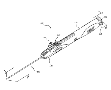
[0004] Figure 1 is a perspective view of a biopsy device in a fired
configuration.
[0005] Figure 2 is a first cross-sectional view of the biopsy device of
Figure 1,
taken through plane 2-2.
[0006] Figure 3 is a second cross-sectional view of the biopsy device of
Figure 1,
taken through plane 3-3.
[0007] Figure 4 is an exploded view of the biopsy device of Figure 1.
[0008] Figure 5A is a first enlarged cross-sectional view of a portion of
the biopsy
device of Figure 1, taken through a first plane.
[0009] Figure 5B is a second enlarged cross-sectional view of the portion
of the
biospy device of Figure 5A, taken through a second plane orthogonal to the
first
plane.
[0010] Figure 6 is an enlarged section view of a portion of Figure 2, taken
around
line 6-6.
[0011] Figure 7 is a portion of the needle assembly of the biopsy device of
Figure
1, in a primed configuration.
[0012] Figure 8A is a cross-sectional view of a portion of the needle
assembly of
Figure 7, in a primed configuration.
1
Date Recue/Date Received 2022-07-28
CA 02977811 2017-08-24
WO 2016/140937 PCT/US2016/020165
[0013] Figure 8B is a cross-sectional view of a portion of the needle
assembly of
Figure 7, in a triggered configuration.
[0014] Figure 9A is a perspective view of another embodiment of a pincer
component of a needle assembly, analogous to the needle assembly of Figure 1.
[0015] Figure 9B is a detail view of a distal end portion of the pincer of
Figure 9A,
taken through line 9B-9B.
DETAILED DESCRIPTION
[0016] Biopsy devices may be configured to retrieve tissue samples from
various
locations within a patient's body. For example, a biopsy device may comprise a
needle assembly including cannulas or other cutting members configured to
sever a
tissue sample. The needle assembly may be advanced to a location within the
body
through the skin of the patient (percutaneous access) or may be advanced
through a
body lumen or other structure.
[0017] Furthermore, a biopsy device may comprise an actuation mechanism
configured to displace the needle assembly such that the needle assembly
severs
the targeted tissue sample. Biasing mechanisms such as springs, triggers, and
so
forth may be configured to allow a practitioner to manipulate various
components of
a needle assembly through manipulating the actuation mechanism. In addition to
mechanical biasing mechanisms such as springs, compressed gas or other energy
sources may be configured to power a biopsy device. In some embodiments, for
example, a compressed CO2 cartridge may be used to power a biopsy device.
[0018] Regardless of the energy source, a mechanism may be configured such
that, once the needle assembly is disposed adjacent tissue to be biopsied,
actuation
of a single trigger may cause various components of a needle assembly to be
displaced to sever a tissue sample. Biasing elements or other energy sources
within
the actuation mechanism may provide the force required to advance the needle
assembly components, and other mechanisms may control the relative
displacement
of individual components of a needle assembly.
[0019] As further disclosed below, a biopsy device may comprise components
configured to actuate the biopsy device through transfer of kinetic energy
between
components, including instances where one or more components are displaced due
to an impact force.
[0020] Additionally, a biopsy device may comprise one or more dampening
components configured to absorb or dampen energy associated with
2
CA 02977811 2017-08-24
WO 2016/140937 PCT/US2016/020165
acceleration/deceleration of components of the device, impact between
components,
oscillation, sound, and so forth. Dampening members may comprise discrete
elements or may be a feature of any other component.
[0021] It will be readily understood that the components of the embodiments
as
generally described and illustrated in the figures herein could be arranged
and
designed in a wide variety of configurations. Thus, the following more
detailed
description of various embodiments, as represented in the figures, is not
intended to
limit the scope of the disclosure, but is merely representative of various
embodiments. While the various aspects of the embodiments are presented in
drawings, the drawings are not necessarily drawn to scale unless specifically
indicated.
[0022] The phrases "connected to" and "coupled to" refer to any form of
interaction between two or more entities, including mechanical, electrical,
magnetic,
electromagnetic, fluidic, and thermal interaction. Two components may be
coupled
to each other even though they are not in direct contact with each other. For
example, two components may be coupled to each other through an intermediate
component.
[0023] The directional terms "proximal" and "distal" are used herein to
refer to
opposite locations on a medical device. The proximal end of the device is
defined as
the end of the device closest to the practitioner when the device is in use by
the
practitioner. The distal end is the end opposite the proximal end, along the
longitudinal direction of the device, or the end furthest from the
practitioner.
[0024] Figure 1 is a perspective view of an impact biopsy device 100 in a
fired
configuration. In other words, and as further detailed below, in the
configuration of
Figure 1, elements of the biopsy device 100 are disposed in relative positions
corresponding with the state of the biopsy device 100 after it has been
actuated to
obtain a tissue sample. The biopsy device 100 may comprise a body member 110
that may be configured to be grasped by a practitioner when the biopsy device
100 is
in use. Thus, in some embodiments the body member 110 may comprise a handle
or grip. The biopsy device 100 may also comprise an actuator 120. The actuator
120 may be configured to prime and/or trigger the biopsy device 100.
Embodiments
wherein the actuator 120 comprises an assembly of subelements are also within
the
scope of this disclosure. For instance, one element of a subassembly may
comprise
a priming component while a separate element may comprise a trigger component.
3
CA 02977811 2017-08-24
WO 2016/140937 PCT/US2016/020165
In the illustrated embodiment, the actuator 120 comprises a distal input 122
and a
proximal input 124. In the illustrated embodiment, these inputs 122, 124 are
portions
of a single actuator 120 comprising an integral single member; in other
embodiments
one or both may comprise a subelement.
[0025] Additionally, and as further discussed below, displacement of the
actuator
120 with respect to the body member 110 may be configured to prime the biopsy
device 100. Further displacement of the actuator 120 with respect to the body
member 110 when the biopsy device 100 is in a primed configuration may trigger
or
release the biopsy device 100. Triggering the device may actuate elements
within
the body member 110, such as components of an needle assembly 180, in
connection with obtaining a tissue sample.
[0026] The illustrated embodiment further comprises a safety tab 126
operably
coupled to the actuator 120. Manipulation of the safety tab 126 may prevent
inadvertent triggering of the biopsy device 100 by locking the actuator 120 to
prevent
triggering when the safety tab 126 is in a locked position.
[0027] Additionally, the biopsy device 100 may comprise an adjustable stop
assembly 130. Displacement of one or more components of the adjustable stop
assembly 130 may adjust or control the length of the tissue sample severed by
the
biopsy device 100.
[0028] Figure 2 is a first cross-sectional view of the biopsy device 100 of
Figure 1,
and Figure 3 is a second cross-sectional view of the impact biopsy device 100
of
Figure 1. Figure 4 is an exploded view of the biopsy device 100 of Figure 1.
As
shown in Figures 2-4, the biopsy device 100 may include an actuation assembly
comprised of components configured to displace a needle assembly or other
cutting
members. As used herein, the actuation assembly refers generally to components
configured to transfer energy to cutting members coupled to the biopsy device
100.
Exemplary cutting members include needles, trocars, cannulas, and so forth.
[0029] In the embodiment of Figures 1-4, a needle assembly 180 is coupled
to
the biopsy device 100. It is within the scope of this disclosure to couple any
variety
of needles, cannulas, trocars, stylets, or other instruments to the biopsy
device 100.
For example, a stylet and cannula configured to sever a partial core tissue
sample
may be operably coupled to the biopsy device 100. Further, one or more
cannulas
configured to obtain a full core tissue sample may be operably coupled to the
biopsy
device 100. In some embodiments, one or more elements of a needle or cutting
4
CA 02977811 2017-08-24
WO 2016/140937 PCT/US2016/020165
assembly may be coupled to components within the body member 110 of the biopsy
device 100 and may extend from the body member 110 through a lumen in the
adjustable stop assembly 130.
[0030] In the embodiment of Figures 1-3, the biopsy device 100 is disposed
in an
fired configuration, corresponding to the state of the device after it has
been actuated
to obtain a sample. Thus, in the configuration of Figures 1-3, the biopsy
device 100
cannot be triggered to obtain a sample without first priming the biopsy device
100.
For example, the biopsy device 100 may comprise a biasing element, such as a
spring 190. In the fired configuration, the spring 190 may be uncompressed.
After
priming, and in the primed configuration, the spring 190 may be compressed or
loaded such that potential energy is stored within the spring 190. When in the
primed configuration, the biopsy device 100 is ready to be actuated.
Additionally, the
biopsy device 100 may be configured to be disposed in an initial
configuration. An
initial configuration, such as an initial shipping configuration, the spring
190 may be
unloaded though the needle assembly 180 may not be disposed in a fully
actuated
position, while in the fired configuration the spring 190 may be unloaded and
the
needle assembly 180 fully actuated, with the needle assembly 180 components in
relative positions corresponding to a state following severing of a tissue
sample.
Positions of the members of the needle assembly 180 in the fired, primed, and
initial
configurations are further detailed below.
[0031] Referring to Figures 2 and 3, the biopsy device 100 may comprise a
first
hub member, such as pincer hub 140. The pincer hub 140 may be coupled to a
pincer member 182 of the needle assembly 180. Accordingly, displacement of the
pincer hub 140 may also displace the pincer member 182. In the illustrated
embodiment, the spring 190 is disposed between the pincer hub 140 and a
housing
spring surface 162 of a housing member 160. In the depicted embodiment the
housing member 160 is coupled to the body member 110.
[0032] Again, in the illustrated fired configuration, the spring 190 is at
least
partially unloaded. As used herein, priming the biopsy device 100 refers to
displacement of various elements of the biopsy device 100 to transition the
biopsy
device 100 from the initial configuration into a primed configuration, meaning
a
configuration where the spring 190 is compressed and the biopsy device 100 may
be
triggered to obtain a sample.
CA 02977811 2017-08-24
WO 2016/140937 PCT/US2016/020165
[0033] Figure 5A is a first enlarged cross-sectional view of a the pincer
hub 140
and needle hub 150 of the dampened biopsy device 100, in the same relative
positions as shown in Figures 2-4, taken through a first plane. Figure 5B is a
second
enlarged cross-sectional view of the pincer hub 140 and needle hub 150 of
Figure
5A, taken through a second plane orthogonal to the first plane.
[0034] As shown in Figures 2-4 and Figures 5A-5B, the pincer hub 140 comprises
actuator catches 146 disposed to interact with pincer hub catches 128 on the
actuator 120. In operation, a user may draw back the actuator 120 with respect
to
the body member 110, the actuator moving in a proximal direction. This
proximal
displacement of the actuator 120 transitions the biopsy device 100 from the
fired
configuration into a primed configuration. Similarly, manipulation of the
biopsy
device 100 from an initial configuration into the primed configuration may
also be
done by priming the biopsy device. As the actuator 120 is displaced
proximally, the
pincer hub catches 128 of the actuator 120 interact with the actuator catches
146 of
the pincer hub 140, also drawing the pincer hub 140 back in a proximal
direction.
This displacement of the pincer hub 140 compresses the spring 190 between the
spring surface 162 of the housing member 160 and the pincer hub 140. The
pincer
hub 140 may comprise a pincer hub spring surface 142, which may comprise one
or
more projections from a central protrusion 141 of the pincer hub 140. The
spring
190 may be disposed at least partially around the central protrusion 141 and
compressed by interaction with the pincer hub spring surface 142 when in a
primed
configuration. When the biopsy device 100 is in a primed configuration, the
spring
190 stores potential energy that may be released when the biopsy device is
triggered.
[0035] Additionally, as the biopsy device 100 is primed, interaction
between the
pincer hub 140 and the needle hub 150 may also displace the needle hub 150.
For
example, priming the biopsy device 100 may also proximally displace the needle
hub
150. In the depicted embodiment, the needle hub 150 is coupled to the needle
186
of the needle assembly 180, thus displacement of the needle hub 150 also
displaces
the needle.
[0036] Still referencing Figures 2-5B, the pincer hub 140 comprises a
pincer hub
angled surface 143 which may interact with a needle hub angled surface 153 of
the
needle hub 150 as the pincer hub 140 is drawn back in a proximal direction. As
the
pincer hub 140 is drawn back, interaction between the pincer hub angled
surface
6
CA 02977811 2017-08-24
WO 2016/140937 PCT/US2016/020165
143 and the needle hub angled surface 153 may draw back the needle hub 150
until
needle hub stop surfaces 156 contact housing shoulder 166. Interaction between
the needle hub stop surfaces 156 and the housing shoulder 166 may prevent
further
proximal displacement of the needle hub 150.
[0037] Once proximal displacement of the needle hub 150 is arrested by the
housing shoulder 166, the pincer hub angled surface 143 and needle hub angled
surface 153 may interact to radially displace the needle hub arms 155,
allowing the
pincer hub angled surface 143 to move proximally beyond the needle hub angled
surface 153 until the pincer hub distal shoulder 144 is proximal of the needle
hub
distal catches 154. At that point, the needle hub arms 155 return from the
radially
outward position. In some instances there may be sufficient resistence to
proximal
displacement of the needle hub 150 to allow the pincer hub angled surface 143
to
move proximally beyond the needle hub angled surface 153 until the pincer hub
distal shoulder 144 is proximal of the needle hub distal catches 154 before
the
needle hub 150 contacts the housing shoulder 166. In such instances, the
needle
hub 150 will still be drawn back to into contact with the housing shoulder
166, though
the pincer hub distal shoulder distal should 144 and needle hub distal catches
154
engage prior to contact between the needle hub 150 and the housing shoulder
166.
For instance, as further detailed below, after firing the needle hub 150 may
contact
the release member 134. In some instances, interaction beween the release
member 134 and the needle hub 150 may initially resist proximal displacement
of the
needle hub, for exapmle.
[0038] The pincer hub 140 is further drawn back, creating an offset between
the
pincer hub distal shoulder 144 and the needle hub distal catches 154 when the
biopsy device 100 reaches a primed configuration.
[0039] The pincer hub 140 is drawn back proximally until the pincer hub
proximal
catches 148 engage with priming catches 168 of the housing member 160. To
accommodate proximal displacement of the pincer hub proximal catches 148 past
the priming catches 168, the pincer hub arms 147 may temporarily displace
radially
outward. Angled surfaces associated with one or both of the pincer hub arms
147
and the priming catches 168 may facilitate this displacement. Engagement of
the
pincer hub proximal catches 148 with the priming catches 168 may then prevent
distal displacement of the pincer hub 140, allowing a user to release the
actuator 120
7
CA 02977811 2017-08-24
WO 2016/140937 PCT/US2016/020165
without releasing tension on the spring 190. The biopsy device 100 is then in
a
primed configuration.
[0040] Transition of the biopsy device 100 to release the spring 190 is
referred to
as triggering the biopsy device 100. Upon triggering of the biopsy device 100,
components of the actuation assembly may, in turn, displace components of the
needle assembly 180 to obtain a tissue sample. Again, the actuation assembly
refers generally to components configured to transfer energy to cutting
members
coupled to the biopsy device 100. In the depicted embodiment, the actuation
assembly comprises the pincer hub 140, the needle hub 150, and the spring 190,
among other components.
[0041] To trigger the biopsy device 100, the actuator 120 may be distally
displaced with respect to the body member 110. When the actuator 120 is
distally
displaced, and the biopsy device 100 is in a primed configuration, trigger
surfaces
129 of the actuator 120 interact with angled arm surfaces 149 of the pincer
hub 140
such that the pincer hub arms 147 are displaced radially outward, until the
pincer
hub proximal catches 148 are no longer engaged with the priming catches 168 of
the
housing member 160. This allows the spring 190 to unload, transferring
potential
energy in the spring 190 to the pincer hub 140 as the pincer hub 140 is
accelerated
and moves in a distal direction.
[0042] As the pincer hub 140 is displaced distally, the pincer hub distal
shoulder
144 impacts the needle hub distal catches 154, accelerating the needle hub
150. As
further detailed below, acceleration of the needle hub 150 by an impact force
may
facilitate retrieval of quality tissue samples.
[0043] The interaction of the pincer hub distal shoulder 144 and needle hub
catches 154 thus couple the pincer hub 140 and needle hub 150. After impact,
the
pincer hub 140 and needle hub 150 travel distally together until interaction
between
the needle hub 150 and the adjustable stop assembly 130 stops the distal
movement
of the needle hub 150. Specifically, the release member 134 of the adjustable
stop
assembly 130 may comprise a stop surface 137 which interacts with the needle
hub
distal end 159. As further detailed below, these components may or may not
directly
interact. Specifically, a dampening element 170 may be disposed between the
stop
surface 137 and the needle hub distal end 159.
[0044] Figure 6 is an enlarged section view of a portion of Figure 2, taken
around
line 6-6. Figure 6 shows the relationship between the release member 134,
pincer
8
CA 02977811 2017-08-24
WO 2016/140937 PCT/US2016/020165
hub 140, needle hub 150, and dampening element 170 in more detail. Other
features also shown and described in connection with Figure 2 are also shown
in
Figure 6.
[0045] With reference to interaction between the needle hub 150 and the
release
member 134, the release member 134 may also interact with the pincer hub 140
to
decouple the pincer hub 140 and the needle hub 150. Specifically, and with
continued reference to Figure 6 as well as Figures 2-5B, the release member
134
may comprise one or more release surfaces 133 which interact with the needle
hub
angled surfaces 153, displacing the needle hub arms 155 radially outward and
decoupling the needle hub 150 and the pincer hub 140 by moving the needle hub
distal catches 154 out of engagement with the pincer hub distal shoulder 144.
[0046] Once decoupled from the needle hub 150, the pincer hub 140 may
continue distally beyond the needle hub 150 after interaction with the release
member 134 stops displacement of the needle hub 150. The pincer hub 140 may
continue until the pincer hub stop surface 145 contacts one or more of the
needle
hub stop surfaces 156, thus arresting the distal motion of the pincer hub 140.
Thus,
the pincer hub 140 may be configured to travel beyond the needle hub 150.
[0047] Once the biopsy device 100 has been triggered, it may be returned to
a
primed configuration by proximally displacing the actuator 120 as described
above.
Again, the actuator may comprise a distal input 122 and a proximal input 124.
Either
of these inputs 122, 124 may be manipulated in order to prime or trigger the
biopsy
device 100. The shape, grip, or position of these inputs 122, 124 may also
facilitate
or enable one-handed use of the biopsy device 100. For example, while gripping
the
body member 110, a user may displace the distal input 122 with a finger or
thumb of
the gripping hand, both to prime and to trigger the biopsy device 100.
Additionally, a
user may manipulate the safety tab 126 to prevent inadvertent triggering of
the
device during use. For example, when the biopsy device 100 is in a primed
position,
the safety tab 126 may be positioned such that distal displacement (or
triggering) of
the actuator 120 is inhibited.
[0048] The dampening element 170 may thus be disposed to dampen shock,
tactile feedback or recoil, and/or noise associated with use of the biopsy
device 100.
The dampening element may comprise any shock-absorbing material, for example,
elastomeric materials, resilient materials, foam, rubber, and so forth. Use of
one or
more dampening elements 170 may additionally reduce shock and wear on various
9
CA 02977811 2017-08-24
WO 2016/140937 PCT/US2016/020165
components of the biopsy device 100. For example, in the depicted embodiment,
the dampening element is disposed between the release member 134 and the
needle hub 150 such that the dampening element 170 absorbs energy associated
with impact of the needle hub 150 on the release member 134 to arrest the
travel of
the needle hub 150 after triggering. Use of a dampening element 170 may reduce
deformation or wear on the needle hub 150 and/or the release member 134 due to
this interaction.
[0049] In the illustrated embodiment, portions of the needle assembly 180
extend
along a longitudinal axis of the biopsy device 100. For example, a trocar 188
extends along the axis of the biopsy device 100 and may be coupled to the
housing
member 160. Other cutting elements, such as a biopsy needle associated with
the
needle hub 160 and a pincer associated with the pincer hub 140, may be
disposed
around the trocar 188. Similarly, elements such as the pincer hub 140 and/or
needle
hub 150 may comprise a central lumen and may be disposed such that one or more
of the members of the needle assembly 180 pass through the lumens of these
components. Similarly, the dampening member 170 may comprise a lumen and may
be disposed around the trocar 188 and one or more additional members of the
needle assembly 180. In some embodiments the dampening member 170 may not
be fixed coupled to any element, but rather allowed to float along the needle
assembly 180. In other embodiments, the dampening member 170 may be coupled
to the needle hub 150 or the release member 134. Still further, other
dampening
elements disposed at other positions within the biopsy device 100 are within
the
scope of this disclosure.
[0050] In some embodiments, manipulation of the adjustable stop assembly
130
may be configured to control the length of tissue sample severed by the biopsy
device 100. For example, overall length of travel of the pincer hub 140 and
needle
hub 150 may be controlled or adjusted by the position of the release member
134
with respect to the housing member 160. As the length of travel of the pincer
hub
140 and needle hub 150 is varied, the travel length of any cutting members
coupled
thereto is also varied.
[0051] The adjustable stop assembly 130 may be configured to make the
position
of the release member 134 adjustable along a continuous range. This range may
be
defined, for example, by threads on the release member 134. Interaction of
threads
on the release member 134 and mating threads coupled to the housing member 160
CA 02977811 2017-08-24
WO 2016/140937 PCT/US2016/020165
may vary the longitudinal position of the release member 134 with respect to
the
housing member 160 as the release member 134 is rotated with respect to the
housing member 160. Thus, the adjustable stop assembly 130 may be configured
such that a practitioner can adjust the length of the sample to be severed by
the
biopsy device 100, along a range related to the range of longitudinal
displacement of
the release member 134.
[0052] The adjustable stop assembly 130 may facilitate use of the biopsy
device
100 in particular therapies or procedures. Again, in some embodiments, the
adjustable stop assembly 130 may be adjustable over a continuous range,
allowing a
practitioner to configure the biopsy device 100 to sever a sample of any
length within
the range. For example, a practitioner may desire to sever a relatively short
tissue
sample, such as instances where obtaining a deeper sample would cause unwanted
trauma to adjacent tissue. Thus, the practitioner may manipulate the position
of the
adjustable stop assembly 130 in order to obtain a sample of a desired length
while
avoiding severing tissue adjacent the sample. Embodiments that utilize
distinct
catches to position the release member 134 at particular intervals are also
within the
scope of this disclosure.
[0053] The adjustable stop assembly 130 may be adjustable over a continuous
range of any length. For example, the adjustable stop assembly 130 may be
configured to allow a practitioner to adjust sample length over a continuous
range
from 2 mm to 35 mm, including from 5 mm to 30 mm, and from 10 mm to 20 mm.
Further, the sample length may be adjustable to lengths less than 2 mm or
greater
than 35 mm.
[0054] In the depicted embodiment the adjustable stop assembly 130
comprises
a an adjustment shell 132 and a release member 134. The adjustment shell 132
may be coupled to the release member 134 such that rotation of the adjustment
shell
132 causes rotation of the release member 134. Further, the components may be
disposed such that while the release member 134 is allowed to displace
longitudinally with respect to housing member 160, the longitudinal position
of the
adjustment shell 132 does not vary with respect to the housing member 160. For
example, a ridge on the release member 134 may be displaced within a slot of
the
adjustment shell 132, such that the ridge and slot may transfer rotational
displacement of the adjustment shell 132 without restraining longitudinal
displacement of the release member 134.
11
CA 02977811 2017-08-24
WO 2016/140937 PCT/US2016/020165
[0055] Such an arrangement allows the release member 134 to be
longitudinally
displaceable with respect to the housing member 160 as the adjustment shell
132
and release member 134 are rotated (via interaction of mating threads of the
release
member 134 and housing member 160, for example) without longitudinal
displacement of the adjustment shell 132. In the illustrated embodiment,
indicia on
the adjustment shell 132 correlate with the longitudinal displacement of the
release
member 134, allowing a practitioner to adjust and/or set the stroke length
through
rotation of the adjustment shell 132 and observation of the relative position
of the
indicia with respect to a reference on the release member 134. Adjustable stop
assemblies 130 comprising tactile or audible feedback associated with rotation
of the
adjustable shell 132 are also within the scope of this disclosure.
[0056] As noted above, in the illustrated embodiment, the biopsy device 100
utilizes the spring 190 to store potential energy during use. Again, in some
embodiments other energy sources, such as compressed gas, may be used in
connection with, or in place of, a spring 190.
[0057] As also noted above, the biopsy device 100 may transfer force to the
needle hub 150 through impact between the pincer hub 140 and the needle hub
150.
Again, the pincer hub 140 may be accelerated by transfer of potential energy
from
another source (such as the spring 190) directly to the pincer hub 140. A
portion of
the kinetic energy associated with the moving pincer hub 140 may be
transferred to
the needle hub 150 at impact. Accordingly, the biopsy device 100 may be
configured to quickly transfer force to a cutting member, and thus may be
configured
to limit deformation of the tissue sample during cutting. In some instances, a
needle
or other cutting member will more cleanly sever tissue when it is moving at a
threshold speed, or cutting speed. During acceleration of the needle, the
needle
may thus move through tissue by compressing or otherwise deforming the tissue,
rather than severing the tissue. An impact force may very quickly accelerate
the
needle, minimizing any such deformation. For example, by accelerating the
needle
hub 150 with an impact force, initial deformation of tissue adjacent a needle
coupled
to the needle hub 150 may be minimized.
[0058] The biopsy device 100 may thus first accelerate the pincer hub 140,
allowing the pincer hub 140 to reach a particular speed before impacting the
needle
hub 150. The spring 190 may be configured to accelerate the pincer hub 140 to
an
impact speed over a distance (such as the distance the pincer hub 140 is
displaced
12
CA 02977811 2017-08-24
WO 2016/140937 PCT/US2016/020165
prior to impact) that may allow use of a spring having a relatively small
spring
constant, as the pincer hub 140 is not required to reach impact speed prior to
impact
with the needle hub 150. The "impact speed" of the pincer hub 140 may be
defined
as the speed at which the pincer hub 140 travels in order to impart an impact
force
sufficient to accelerate the needle hub 150 to cutting speed. Thus the initial
distance
associated with accelerating the pincer hub 140, through transfer of energy
from the
spring 190, will not necessarily result in the deformation of tissue during
initial
acceleration of the biopsy device 100 components.
[0059] Additionally, a biopsy device 100 utilizing acceleration by impact
may
facilitate severing tissue samples of a variety of lengths. The impact
configuration
may accelerate cutting members associated with the device to cutting speed
without
substantially displacing the needle. Thus, the biopsy device 100 may be
configured
to sever particularly short samples, as the needle hub 150 reaches cutting
speed
without substantial displacement. By comparison, direct acceleration of a
needle
hub by a spring may require some displacement of the needle hub before the
needle
reaches cutting speed. Thus, the minimum sample length may be at least as long
as
the displacement needed to bring such a needle to cutting speed. Further, the
biopsy device 100 may be configured such that the needle maintains a
substantially
uniform cutting speed during the severing of an entire sample, rather than
accelerating during the first portion of the severing. Samples severed by
uniform
cutting speeds may be generally more uniform than samples severed by
accelerating
cutting members, which may deform a portion of the sample.
[0060] The potential energy stored in the spring 190 may be expressed by
the
equation E=(0.5)Ioe, where k is the spring constant and x the displacement of
the
spring 190 in the compressed state. The energy associated with the pincer hub
140
(and pincer components coupled thereto) after it is accelerated by the spring
190
may be expressed as E=(0.5)mV2 where m is the mass of the components coupled
to the pincer hub 140 and V is the velocity of the pincer hub 140. The
exponential
factor associated with the potential energy of the spring 190 may also
facilitate use
of springs with relatively small spring constants in the biopsy device 100.
Use of
springs with relatively small spring constants may make the biopsy device 100
easier
to prime, and may reduce shock and recoil during use.
[0061] As detailed above, the actuation assembly may be configured such
that
the pincer hub 140 travels distally a set distance after the needle hub 150
impacts
13
CA 02977811 2017-08-24
WO 2016/140937 PCT/US2016/020165
the release member 134, including embodiments wherein the dampening element
170 is disposed between the needle hub 150 and the release member 134. In some
embodiments, the needle assembly 180 may thus be designed such that a needle
associated with the needle hub 150 severs the longitudinal portion of a
sample, while
a pincer associated with the pincer hub 140 severs the distal end of the
sample after
the longitudinal portion is initially cut. Figures 7, 8A and 8B detailed below
illustrate
an exemplary configuration of a needle 186, a pincer 182, and a trocar 188.
Various
arrangements of cutting members and needle assemblies having members with
differing lengths of travel are within the scope of this disclosure. The
needle 186
may comprise a hollow cannula with a distal cutting edge configured to sever
the
longitudinal portion of a tissue sample, and the pincer 182 may comprise a
hollow
cannula with a distal cutting portion configured to sever the distal portion
of a tissue
sample, as further detailed below.
[0062] Figure 7 is a cross-sectional view of a portion of the needle
assembly 180
of the biopsy device 100 of Figure 1. In the configuration of Figure 7, the
needle
assembly 180 is disposed in a primed configuration, as opposed to the fired
configuration shown in Figures 1-3. The needle assembly 180 comprises a trocar
188, a needle 186, and a pincer 182. The trocar 188 may extend along the
longitudinal axis of the needle assembly 180 and along the longitudinal axis
of the
biopsy device (100 of Figure 2). The trocar 188 may be fixed to the housing
member
(160 of Figure 2), such that the needle 186 and pincer 182 are displaced
relative to
the trocar 188 when the needle hub (150 of Figure 2) and the pincer hub (140
of
Figure 2) are displaced with respect to the housing member (160 of Figure 2).
[0063] Again, in the configuration shown in Figure 7, the biopsy device
(100 of
Figure 2) is in a primed configuration as discussed above, meaning the pincer
hub
(140 of Figure 2) and the needle hub (150 of Figure 2) are drawn back in a
proximal
direction. (It is noted that this is a different configuration than depicted
in Figure 2 as
discussed above.) In the primed configuration the trocar 188 extends from the
distal
end of the needle assembly 180. With the trocar 188 so disposed, the needle
assembly 180 may be advanced through tissue (for example, percutaneously or
otherwise through tissue) and disposed adjacent tissue to be sampled.
[0064] When the biopsy device (100 of Figure 2) is triggered as discussed
above,
the needle 186 is advanced into the tissue, severing the longitudinal portion
of the
tissue sample. As the trocar 188 is coupled to the housing member (160 of
Figure 2)
14
CA 02977811 2017-08-24
WO 2016/140937 PCT/US2016/020165
the needle 186 extends beyond the trocar 188 as the needle hub (150 of Figure
2) is
displaced with respect to the housing member (160 of Figure 2). As detailed
above,
the needle 186 is accelerated by impact between the pincer hub (140 of Figure
2)
and the needle hub (150 of Figure 2).
[0065] Initially, after triggering, the pincer 182 advances with respect to
both the
needle 186 and the trocar 188, prior to impact between the pincer hub (140 of
Figure
2) and the needle hub (150 of Figure 2). The components may be positioned such
that during the initial advancement of the pincer 182, the pincer remains
proximal of
a annular shoulder 181 of the needle 186. The annular shoulder 181 of the
needle
186 comprises a portion of the needle 186 with a reduced inside diameter, as
shown
in the drawings and further detailed below. After impact, both the needle 186
and
the pincer 182 advance into the tissue sample.
[0066] As detailed above, the needle 186 stops prior to the pincer 182, as
the
needle hub (150 of Figure 2) contacts the release member (134 of Figure 2)
decoupling the needle hub (150 of Figure 2) and the pincer hub (140 of Figure
2) and
arresting the forward motion of the needle hub (150 of Figure 2). Again, the
dampening element (170 of Figure 2) may reduce shock as the needle 186 stops.
[0067] The pincer hub (140 of Figure 2) travels a distance after the needle
hub
(150 of Figure 2) stops, correlating to distal displacement of the pincer 182
with
respect to the needle 186 at the end of the stroke. As detailed below, this
displacement correlates to severing of a distal end of the sample by the
pincer 182.
[0068] Figure 8A shows the needle 186 and the pincer 182 in the primed
configuration, though the trocar (188 of Figure 7) is not shown in this view.
In the
primed configuration, the pincer 182 is proximal of the annular shoulder 181
of the
needle 186. This proximal offset may correlate to the distance the pincer hub
(140 of
Figure 2) travels before impact with the needle hub (150 of Figure 2) such
that the
pincer 182 remains proximal of the annular shoulder 181 of the needle 186
until the
needle 186 completes its stroke, severing the longitudinal position.
[0069] Figure 8B shows the needle 186 and the pincer 182 at the end of a
stroke,
after the needle hub (150 of Figure 2) and the pincer hub (140 of Figure 2)
have
been decoupled and the pincer hub (140 of Figure 2) has traveled distally
after the
needle hub (150 of Figure 2) contacted the release member (134 of Figure 2),
including through interaction with the dampening element (170 of Figure 2).
This
travel of the pincer hub (140 of Figure 2) correlates with displacement of the
pincer
CA 02977811 2017-08-24
WO 2016/140937 PCT/US2016/020165
182 with respect to the needle 186 such that the annular shoulder 181
displaces
portions of the pincer 182 radially inward to sever the distal end of a
sample.
[0070] Accordingly, the position of the needle assembly 180 elements in
Figure 7
and Figure 8A correspond to a primed configuration while the relative
positions
shown in Figure 8B correspond to a fired configuration. In an initial shipping
configuration, pincer 182 may be disposed such that the pincer 182 is proximal
of the
annular shoulder 181, though the spring (190 of Figures 2-3) may not be in a
loaded
configuration, as further detailed above.
[0071] In the illustrated embodiment, the annular shoulder 181 corresponds
to a
region of the needle 186 with a reduced diameter. This reduced diameter
extends
from the annular shouler 181 to the distal end of the needle 186 in the
illustrated
embodiment. In other embodiments, protrusions, an annular ring, or other
features
may be disposed to displace the portions of the pincer 182.
[0072] Repriming the biopsy device (100 of Figure 2) would return the
needle
assembly to the configuration shown in Figure 7, retracting the pincer 182 and
needle 186 such that the trocar 188 would push the sample out of the needle
186.
[0073] Figure 9A is a perspective view of another embodiment of a pincer,
and
Figure 9B is a detailed view of a distal end portion of the pincer of Figure
9A taken
through line 9B-9B that can, in certain respects, resemble components of the
pincer
182 described in connection with Figures 1-8B. It will be appreciated that all
the
illustrated embodiments may have analogous features. Accordingly, like
features are
designated with like reference numerals, with the leading digit of the
reference
numerals incremented by 1. For instance, the pincer is designated as "182" in
Figures 1-8B, and an analogous pincer is designated as "282" in Figures 9A and
9B.
Relevant disclosure set forth above regarding similarly identified features
thus may
not be repeated hereafter. Moreover, specific features of the pincer 182 and
related
components shown in Figures 9A and 9B may not be shown or identified by a
reference numeral in the drawings or specifically discussed in the written
description
that follows. However, such features may clearly be the same, or substantially
the
same, as features depicted in other embodiments and/or described with respect
to
such embodiments. Accordingly, the relevant descriptions of such features
apply
equally to the features of the pincer 282 of Figures 9A and 9B. Any suitable
combination of the features, and variations of the same, described with
respect to
the pincer 282 and components illustrated in Figures 9A and 9B can be employed
16
CA 02977811 2017-08-24
WO 2016/140937 PCT/US2016/020165
with the pincer 182 and components of Figures 1-8B, and vice versa. This
pattern of
disclosure applies equally to further embodiments depicted in subsequent
figures
and described hereafter.
[0074]
Specifically, it is within the scope of this disclosure to utilize pincer 282
in
place of pincer 182 in the biopsy device 100 and needle assembly 180 discussed
in
connection with Figures 1-8B.
[0075] As
depicted, the pincer 282 may comprise a plurality of sectioning
elements 284. Additionally, the pincer 282 may comprise one or more spiral
cuts
285 disposed along at least a portion or portions of the length of the pincer
282. In
the illustrated embodiment, the spiral cut 285 is disposed along at least a
portion of
the length of the pincer 282 at a position proximal to the sectioning elements
284. In
various embodiments, the pincer 282 may comprise a spiral cut 285 disposed
proximal of the one or more sectioning elements 284. In some embodiments, the
spiral cut 285 may be disposed at a distance sufficiently proximal in relation
to the
sectioning elements 284 such that the spiral cut 285 does not, or does not
substantially, interfere with or damage a tissue sample.
[0076] In
some embodiments, the pincer 282 may comprise one or more
sectioning elements 284 (e.g., one, two, three, four, five, six, or more
sectioning
elements 284). In the illustrated embodiment, the pincer 282 comprises six
sectioning elements 284. As discussed above, the sectioning elements 284 may
be
coupled to the pincer 282. In some configurations, the sectioning elements 284
and
the pincer 282 may be integrally formed from a single piece of material. In
certain
embodiments, at least one of the sectioning elements 284 may comprise a sharp
distal portion. As depicted in Figures 9A and 9B, the sectioning elements 284
can
comprise a pointed or tapered distal portion. At least one of the sectioning
elements
284 may also comprise at least one sharp lateral edge portion. In
some
embodiments, the at least one sharp lateral edge portion may be angled.
[0077]
With continued reference to Figures 9A and 9B, the sectioning elements
284 can comprise a plurality of angled lateral edge portions. For example, the
lateral
edge portions of the section elements 284 may be serrated or notched. Such a
configuration of the one or more sectioning elements 284 may facilitate the
cutting or
severing of body tissue by the sectioning elements 284.
[0078] As
discussed above with respect to the sectioning elements 284, the
shape of the sectioning elements 284 may also be configured such that the
17
CA 02977811 2017-08-24
WO 2016/140937 PCT/US2016/020165
sectioning elements 284 may be simultaneously, or substantially
simultaneously,
inwardly displaced toward each other to sever the second portion of the tissue
sample. Interaction with other components of a biopsy device or needle
assembly
(such as the annular shoulder 181 of Figure 7) may also be configured to
inwardly
displace the sectioning elements 284. In some embodiments, an annular shoulder
(181 of Figure 7) on the inside of a needle (186 of Figure 7) may be replaced
with an
annular ring around the inside diameter of the needle (186 of Figure 7)
discrete
protrusions, or other features.
[0079] In
some embodiments, the spiral cut 285 may extend completely through a
wall of the pincer 282. In some other embodiments, the spiral cut 285 may only
extend partially through the wall of the pincer 282. For example, the spiral
cut 285
may form a groove along a portion of the length of the pincer 282. In yet
other
embodiments, one or more portions of the spiral cut 285 may extend completely
through the wall of the pincer 282 while one or more other portions of the
spiral cut
285 may form a groove in the wall of the pincer 282.
[0080] In
certain embodiments, disposition of the spiral cut 285 along the pincer
282 can form a spring, or a spring-like portion, along the pincer 282. The
spiral cut
285 may add or provide compliance or elasticity to the pincer 282 and/or the
biopsy
needle assembly. For example, the spiral cut 285 may improve or increase
tolerances of one or more of the components of the pincer 282 and/or the
biopsy
needle assembly. Such improved tolerances may facilitate advancement or
displacement of the pincer 282 and/or the biopsy needle assembly through a
body
tissue. In various embodiments, the spiral cut 285 may absorb impact or shock
to
one or more of the pincer 282, other components of the biopsy needle assembly,
and/or the biopsy needle assembly.
For example, upon advancement or
displacement of at least a portion of the biopsy needle assembly through a
body
tissue of a patient, at least a portion of the spiral cut 285 may compress or
be
configured to compress (i.e., the spiral cut 285 may compress longitudinally,
thus
shortening the length of the pincer 282). In certain embodiments, the spiral
cut 285
can be configured to longitudinally compress in response to relative
displacement of
the outer tubular member, or another component of the biopsy needle assembly,
in
relation to the pincer 282.
[0081]
Furthermore, in connection with the dampening element (170 of Figures 2-
3) a pincer 282 comprising a spiral cut 285 may add additional compliance and
18
CA 02977811 2017-08-24
WO 2016/140937 PCT/US2016/020165
shock absorption to a biopsy device. Such shock absorption may increase sample
quality, lessen wear on components, and reduce recoil and shock.
[0082] One or more forces may result in or cause compression of the spiral
cut
285. For example, inertia of the pincer 282 as it is advanced into a body
tissue can
result in compression of the spiral cut 285. Displacement of the pincer 282 in
relation to the needle (such as 186 of Figure 2) and/or the trocar (such as
188 of
Figure 2) may also result in compression of the spiral cut 285. For example,
friction
between an outside surface of the pincer 282 and an inside surface of the
outer
tubular member may result in compression of the spiral cut 285. Furthermore,
force
used to advance or displace the sectioning elements 284 of the pincer 282 over
or
past the an annular shoulder (181 of Figure 2) or other features on the inside
diameter of a needle (such as 186 of Figure 2) can also result in compression
of the
spiral cut 285.
[0083] Additionally, at least a portion of the spiral cut 285 may rotate,
or be
configured to rotate, upon compression of the spiral cut 285. Rotation of the
spiral
cut 285 may also cause or result in rotation of the sectioning elements 284
around a
central axis of the pincer 282. This rotation may facilitate uniform, or
substantially
uniform, severing of the distal end of a tissue sample as the spiral cut 285
rotates
back to an initial position as the spiral cut uncompresses at the end of a
stroke.
[0084] In some embodiments, the spiral cut 285 and/or the sectioning
elements
284 may rotate, or be configured to rotate, between 0 and plus or minus 900.
In
some embodiments, the spiral cut 285 and/or the sectioning elements 284 may
rotate, or be configured to rotate, between 0 and plus or minus 45 ; between
0 and
plus or minus 30 , between 0 and plus or minus 15 , between 0 and plus or
minus
; or another suitable degree of rotation. Again, rotation of the sectioning
elements
284 through a body tissue may form or result in a cleaner or sharper cut in a
tissue
sample, as rotation of the sectioning elements 284 may sever along a complete,
or a
substantially complete, circumference of the distal end of the tissue sample.
[0085] Various methods and procedures are within the scope of this
disclosure.
Methods of priming the biopsy device 100 (as detailed above), advancing the
biopsy
device 100 through tissue, and triggering the biopsy device 100 (as also
detailed
above) are all within the scope of this disclosure. Further methods of
obtaining a
sample through impact acceleration of cutting members and methods of dampening
19
CA 02977811 2017-08-24
WO 2016/140937 PCT/US2016/020165
shock or recoil through interaction of the elements described above are all
within the
scope of this disclosure.
[0086] Without further elaboration, it is believed that one skilled in the
art can use
the preceding description to utilize the present disclosure to its fullest
extent. The
examples and embodiments disclosed herein are to be construed as merely
illustrative and exemplary and not a limitation of the scope of the present
disclosure
in any way. It will be apparent to those having skill in the art, and having
the benefit
of this disclosure, that changes may be made to the details of the above-
described
embodiments without departing from the underlying principles of the disclosure
herein.