Note: Descriptions are shown in the official language in which they were submitted.
CA 02978442 2017-08-31
WO 2016/141127 PCT/US2016/020583
METHODS FOR ASSESSING THE RISK OF DISEASE OCCURRENCE OR
RECURRENCE USING EXPRESSION LEVEL AND SEQUENCE VARIANT
INFORMATION
CROSS REFERENCE
[0001] This application claims priority to U.S. provisional application
62/128,463, filed
on March 4, 2015, U.S. provisional application 62/128,469, filed on March 4,
2015, and U.S.
provisional application 62/238,893, filed on October 8, 2015, each of which is
entirely
incorporated herein by reference.
BACKGROUND
[0002] A risk adapted approach to a disease therapy, such as thyroid cancer
therapy, may
minimize the risk of disease occurrence, in addition to improving disease
specific survival.
Currently, this risk adapted approach to initial subject management is based
in large part
upon post-operative classification of subjects either as high, intermediate or
low risk of
disease recurrence utilizing the 2009 American Thyroid Association (ATA)
staging system.
While this anatomic staging system has proven clinically useful, it cannot be
accurately
assessed prior to an invasive thyroidectomy, and it does not include any
molecular predictors
of disease outcome.
SUMMARY
[0003] Provided herein are various methods for assessing or stratifying
risk of disease
occurrence and/or recurrence. Transcriptional data obtained during pre-
diagnostic or
diagnostic evaluation, such as fine needle aspiration (FNA), can improve the
pre-operative
prediction of risk occurrence of a disease such as thyroid cancer, and can
provide further
individualization of subject therapy and treatment. Methods of the present
disclosure may
provide an assessment with respect to a risk of occurrence and/or recurrence
of a disease in a
relatively noninvasive manner and using low sample volumes.
[0004] An aspect of the present disclosure provides a method for evaluating
a tissue
sample of a subject to determine a risk of occurrence of disease in the
subject. The method
comprises (a) obtaining an expression level corresponding to each one or more
genes of a
first set of genes in a nucleic acid sample in a needle aspirate sample
obtained from the
subject, which first set of genes is associated with the risk of occurrence of
disease in the
subject; (b) determining a presence of a nucleic acid sequence corresponding
to each of one
or more genes of a second set of genes in the nucleic acid sample, which
second set of genes
1
CA 02978442 2017-08-31
WO 2016/141127 PCT/US2016/020583
is associated with the risk of occurrence of disease in the subject; (c)
separately comparing to
controls (i) the expression level obtained in (a) and (ii) the nucleic acid
sequence obtained in
(b) to provide comparisons of the expression level and the nucleic acid
sequence to the
controls, wherein a comparison of the nucleic acid sequence to a reference
sequence among
the controls is indicative of a presence of one or more sequence variants with
respect to a
given gene of the second set of genes; and (d) using a computer processor that
is programmed
with a trained algorithm to (i) analyze the comparisons and (ii) determine the
risk of
occurrence of the disease based on the comparisons.
[0005] In some embodiments, the needle aspirate sample is a fine needle
aspirate sample.
In some embodiments, the disease is cancer. In some embodiments, the method
further
comprises, prior to (a), obtaining the needle aspirate sample from the
subject. In some
embodiments, the method further comprises, prior to (a), determining the
expression level
from the nucleic acid sample in the needle aspirate sample. In some
embodiments, the
method further comprises, prior to (b), determining the nucleic acid sequence
from the
nucleic acid sample in the needle aspirate sample. In some embodiments, the
method further
comprises comparing the nucleic acid sequence to the reference sequence to
identify the one
or more sequence variants. In some embodiments, the reference sequence is a
housekeeping
gene from the subject. In some embodiments, the one or more genes in the first
set or second
set of genes include a plurality of genes.
[0006] In some embodiments, the needle aspirate sample has been found to be
cytologically ambiguous or suspicious. In some embodiments, the needle
aspirate sample has
a volume that is about 1 microliter or less. In some embodiments, the needle
aspirate sample
has an RNA Integrity Number (RIN) value of about 9.0 or less. In some
embodiments, RNA
purified from a needle aspirate sample has an RNA RIN value of about 9.0 or
less. In some
embodiments, the needle aspirate sample has an RIN value of about 6.0 or less.
In some
embodiments, the RNA sample has an RIN value of about 6.0 or less.
[0007] In some embodiments, the risk of occurrence of the disease includes
a risk of
recurrence of the disease in the subject. In some embodiments, the risk of
occurrence of the
cancer includes a risk of metastasis in the subject. In some embodiments, the
risk of
occurrence of cancer includes a risk of accelerated disease progression. In
some
embodiments, the risk of occurrence of cancer includes a risk of therapeutic
failure.
[0008] In some embodiments, the trained algorithm is trained employing
tissue samples
from at least 25 or at least 100 subjects having been diagnosed with the
disease. In some
2
CA 02978442 2017-08-31
WO 2016/141127 PCT/US2016/020583
embodiments, the trained algorithm is trained employing tissue samples from at
least 200
subjects having been diagnosed with the disease.
[0009] In some embodiments, (d) occurs pre-operatively. In some
embodiments, (d)
occurs prior to the subject having a positive disease diagnosis. In some
embodiments, (d)
further comprises stratifying the risk of occurrence into a low risk of
occurrence or a
medium-to-high risk of occurrence, wherein the low risk of occurrence has a
probability of
occurrence between about 50% and about 80% and wherein the medium-to-high risk
of
occurrence has a probability of occurrence between about 80% and 100%.
[0010] In some embodiments, the method further comprises applying one or
more filters,
one or more wrappers, one or more embedded protocols, or any combination
thereof to the
comparisons. In some embodiments, the one or more filters are applied to the
comparisons. In
some embodiments,the one or more filters comprise a t-test, an analysis of
variance
(ANOVA) analysis, a Bayesian framework, a Gamma distribution, a Wilcoxon rank
sum test,
between-within class sum of squares test, a rank products method, a random
permutation
method, a threshold number of misclassification (fNoM), a bivari ate method; a
correlation
based feature selection (CH) method, a minimum redundancy maximum relevance
(MRMR)
method, a Markov blanket filter method, an uncorrelated shrunken centroid
method, or any
combination thereof In some embodiments, the one or more sequence variants
comprise one
or more of a point mutation, a fusion gene, a substitution, a deletion, an
insertion, an
inversion, a conversion, a translocation., or any combination thereof. In some
embodiments,
the one or more point mutations are from about 5 to about 4000 point
mutations. In some
embodiments, the one or more fusion genes are at least two fusion genes.
[0011] In some embodiments, the stratifying has an accuracy of about 80%.
In some
embodiments, the stratifying has a specificity of about 80%. In some
embodiments, the one
or more genes of the first or second set is less than about 15 genes or less
than about 10
genes. In some embodiments, the one or more genes of the first or second set
is less than
about 75 genes. In some embodiments, the one or more genes of the first or
second set is
between about 50 and about 400 genes.
[0012] In some embodiments, the obtaining in (b) comprises sequencing a
nucleic acid
sample in the needle aspirate sample to obtain the nucleic acid sequence. In
some
embodiments, the sequencing comprises enriching for the one or more genes of a
second set
of genes, or variants thereof. In some embodiments, (a) comprises using a
microarray with
probes that are selective for the one or more genes of the first set of genes.
In some
3
CA 02978442 2017-08-31
WO 2016/141127 PCT/US2016/020583
embodiments, (a) comprises using a targeted sequencing platform (such as Ion
Torrent
Ampliseq, or Illumina TruSeq Custom Amplicon).
[0013] In some embodiments, the tissue sample is a thyroid tissue sample.
In some
embodiments, the first and second sets of genes comprise COL1A1, THBS2, or any
combination thereof In some embodiments, the second set of genes comprise
EPHA3,
COL1A1, EHF, RAPGEF5, PRICKLE1, TMEM92, ROB01, C6orf136, SPAG4, GALNT15,
LUM, NCAM2, NUP210L, NR2F1, THBS2, PSORS1C1, or any combination thereof In
some embodiments, the first set of genes comprises COL1A1, TMEM92, C1or187,
SPAG4,
EHF, COL3A1, GALNT15, NUP210L, PDZRN3, C6orf136, NA, NRXN3, COL6A3,
RAPGEF5, PRICKLE1, LUM, ROB01, BGN, AC019117.2, PRSS3P1, or any combination
thereof. In some embodiments, the second set of genes comprises EPHA3, COL1A1,
EHF,
RAPGEF5, PRICKLE1, TMEM92, ROB01, C6orf136, SPAG4, GALNT15, LUM, NCAM2,
SYNP02, NUP210L, AMZ1, NR2F1, THBS2, PSORS1C1, FTH1P24, or any combination
thereof. In some embodiments, the second set of genes comprises AKAP9, SPRY3,
SPRY3,
CAMKK2, COL1A1, FITM2, COX6C, VSIG1OL, CYCl, KDM1B, MAPK15, ARSG,
PAXIP1, DAAM1, AVL9, DMGDH, HLA-DQA1, HLA-DQB1, HLA-DRA, HLA-DRB5,
HLA-H, IRF1, MGAT1, P2RX1, PLEK, CCDC93, PPP1R12C, SLC41A3, METTL3,
CCAR2, PTPRE, SRL, SLC30A5, BMP4, ZNF133, ICE2, DCAKD, TMX1, TNFSF12,
PER2, MCM3AP, or any combination thereof
[0014] In some embodiments, the first set of genes and the second set of
genes are
different. In some embodiments, the method further comprises identifying new
genetic
biomarkers of the disease.
[0015] In some embodiments, the obtaining in (a) comprises assaying for the
expression
level corresponding to each of the one or more genes. In some embodiments, the
assaying
comprises array hybridization, nucleic acid sequencing or nucleic acid
amplification using
markers that are selected for each of the one or more genes. In some
embodiments, the
markers are primers that are selected for each of the one or more genes.
[0016] In some embodiments, the assaying comprises reverse transcription
polymerase
chain reaction (PCR). In some embodiments, the determining comprises assaying
for each of
the one or more genes of the second set of genes in the nucleic acid sample.
In some
embodiments, the assaying comprises array hybridization, nucleic acid
sequencing or nucleic
acid amplification using markers that are selected for each of the one or more
genes. In some
embodiments, the markers are primers that are selected for each of the one or
more genes. In
4
CA 02978442 2017-08-31
WO 2016/141127 PCT/US2016/020583
some embodiments, the assaying comprises reverse transcription polymerase
chain reaction
(PCR).
[0017] Another aspect of the present disclosure provides a computer-
readable medium
(e.g., memory) comprising machine-executable code that, upon execution by one
or more
computer processors, implements any of the methods above or elsewhere herein.
[0018] Another aspect of the present disclosure provides a computer system
comprising
one or more computer processors and a computer-readable medium coupled
thereto. The
computer-readable medium may comprise machine-executable code that, upon
execution by
the one or more computer processors, implements any of the methods above or
elsewhere
herein.
[0019] Additional aspects and advantages of the present disclosure will
become readily
apparent to those skilled in this art from the following detailed description,
wherein only
illustrative embodiments of the present disclosure are shown and described. As
will be
realized, the present disclosure is capable of other and different
embodiments, and its several
details are capable of modifications in various obvious respects, all without
departing from
the disclosure. Accordingly, the drawings and description are to be regarded
as illustrative in
nature, and not as restrictive.
INCORPORATION BY REFERENCE
[0020] All publications, patents, and patent applications mentioned in this
specification
are herein incorporated by reference to the same extent as if each individual
publication,
patent, or patent application was specifically and individually indicated to
be incorporated by
reference. To the extent publications and patents or patent applications
incorporated by
reference contradict the disclosure contained in the specification, the
specification is intended
to supersede and/or take precedence over any such contradictory material.
BRIEF DESCRIPTION OF THE DRAWINGS
[0021] The novel features of the invention are set forth with particularity
in the appended
claims. A better understanding of the features and advantages of the present
invention will be
obtained by reference to the following detailed description that sets forth
illustrative
embodiments, in which the principles of the invention are utilized, and the
accompanying
drawings (also "figure" and "FIG." herein), of which:
[0022] FIG. 1 shows a sample cohort of cytology data and expert
histopathology data
stratified into low risk and medium-to-high risk of occurrence of cancer;
CA 02978442 2017-08-31
WO 2016/141127 PCT/US2016/020583
[0023] FIG. 2 shows histopathology risk features and the number and percent
of samples
for each feature;
[0024] FIG. 3 shows cross validation of true positive rates plotted against
false positive
rates;
[0025] FIG. 4 shows classification performance data plotting predictive
values against
prevalence of medium-to-high risk;
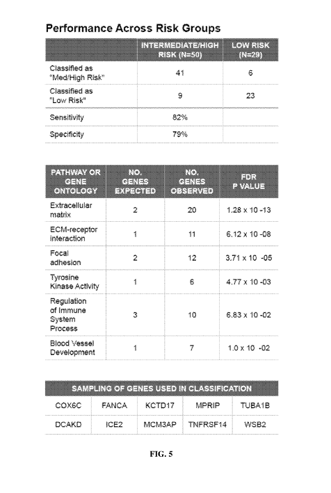
[0026] FIG. 5 shows classification performance data across low risk and
medium-to-high
risk groups;
[0027] FIG. 6 shows an example list of genes associated with a risk of
occurrence of
thyroid cancer based on gene expression level data;
[0028] FIG. 7 shows an example list of genes associated with a risk of
occurrence of
thyroid cancer based on gene expression level data obtained from ribonucleic
acid (RNA)
sequencing;
[0029] FIG. 8 shows an example list of genes associated with a risk of
occurrence of
thyroid cancer based on sequence variant data;
[0030] FIG. 9 shows a computer control system that is programmed or
otherwise
configured to implement methods provided herein;
[0031] FIG. 10 shows a flow diagram of determining accurate training
labels;
[0032] FIG. 11A shows cross validation of true positive rates plotted
against false
positive rates;
[0033] FIG. 11B shows classification performance data across
intermediate/high risk and
low risk groups;
[0034] FIG. 12 shows an example list of genes of variants selected by the
classifier in
each fold;
[0035] FIG. 13 shows an example list of genes of counts selected 8 to 10
times by the
classifier in 10 folds;
[0036] FIG. 14 shows a table of five point mutation panels and fusion
pairs;
[0037] FIG. 15 shows a graph of test performance specificity and
sensitivity across five
panels of mutations and fusion pairs;
[0038] FIG. 16 shows a table of mutation performance of panel 3 in FIGs. 14
and 15 by
cytology);
[0039] FIG. 17 shows a graph of test performance specificity and
sensitivity across five
panels of mutations and fusion pairs;
6
CA 02978442 2017-08-31
WO 2016/141127 PCT/US2016/020583
[0040] FIG. 18A shows a graphical representation; FIG. 18B shows a table
representation of mutation frequency of a Clinical Laboratory Improvement
Amendments
(CLIA) fine needle aspirate (FNA) sample;
[0041] FIG. 19A shows a graphical representation; FIG. 19B shows a table
representation of mutation frequency of a FNA sample; and
[0042] FIG. 20A shows a graphical representation; FIG. 20B shows a table
representation of mutation frequency of a tissue sample.
DETAILED DESCRIPTION
[0043] While various embodiments of the invention have been shown and
described
herein, it will be obvious to those skilled in the art that such embodiments
are provided by
way of example only. Numerous variations, changes, and substitutions may occur
to those
skilled in the art without departing from the invention. It should be
understood that various
alternatives to the embodiments of the invention described herein may be
employed.
[0044] The tertn "subject," as used herein, generally refers to an.y animal
or living
organism. Animals can be mammals, such as humans, non-human primates, rodents
such as
mice and rats, dogs, cats, pigs, sheep, rabbits, and others. Animals can be
fish, reptiles, or
others. Animals can be neonatal, infant, adolescent, or adult animals, Humans
can be more
than about 1,2, 5, 10, 20, 30, 40, 50, 60, 65, 70, 75, or about 80 years of
age. The subject
may have or be suspected of having a disease, such as cancer. The subject may
be a patient,
such as a patient being treated for a disease, such as a cancer patient. The
subject may be
predisposed to a risk of developing a disease such as cancer. The subject may
be in remission
from a disease, such as a cancer patient. The subject may be healthy.
[0045] The term "disease," as used herein, generally refers to any abnormal
or pathologic
condition that affects a subject. Examples of a disease include cancer, such
as, for example,
thyroid cancer, parathyroid cancer, lung cancer, skin cancer, and others. The
disease may be
treatable or non-treatable. The disease may be terminal or non-terminal. The
disease can be a
result of inherited genes, environmental exposures, or any combination
thereof. The disease
can be cancer, a genetic disease, a proliferative disorder, or others as
described herein,
[0046] The term "risk of occurrence of disease," as defined herein,
generally refers to a
risk or probability associated with the occurrence of a disease in a subject.
A risk of
occurrence can include a first occurrence of disease in a subject or can
include subsequent
occurrences, such as a second, third, fourth, or subsequent occurrence. A risk
of occurrence
of disease can include a) a risk of developing the disease for a first time,
b) a risk of relapse
7
CA 02978442 2017-08-31
WO 2016/141127 PCT/US2016/020583
or of developing the disease again, c) a risk of developing the disease in the
future, d) a risk
of being predisposed to developing the disease in the subject's lifetime, or
e) a risk of being
predisposed to developing the disease as an infant, adolescent, or adult. A
risk of occurrence
of a disease, such as cancer, can include a risk of the cancer becoming
metastatic. A risk of
occurrence of a disease such as cancer can include a risk of occurrence of a
stage I cancer, a
stage II cancer, a stage III cancer, or a stage IV cancer. Risk of occurrence
of cancer can
include a risk for a blood cancer, tissue cancer (e.g., a tumor), or a cancer
becoming
metastatic to one or more organ sites from other sites.
[0047] The term "sequence variant," "sequence variation," "sequence
alteration" or
"allelic variant," as used herein, generally refer to a specific change or
variation in relation to
a reference sequence, such as a genomic deoxyribonucleic acid (DNA) reference
sequence, a
coding DNA reference sequence, or a protein reference sequence, or others. The
reference
DNA sequence can be obtained from a reference database. A sequence variant may
affect
function. A sequence variant may not affect function. A sequence variant can
occur at the
DNA level in one or more nucleotides, at the ribonucleic acid (RNA) level in
one or more
nucleotides, at the protein level in one or more amino acids, or any
combination thereof The
reference sequence can be obtained from a database such as the NCBI Reference
Sequence
Database (RefSeq) database. Specific changes that can constitute a sequence
variation can
include a substitution, a deletion, an insertion, an inversion, or a
conversion in one or more
nucleotides or one or more amino acids. A sequence variant may be a point
mutation. A
sequence variant may be a fusion gene. A fusion pair or a fusion gene may
result from a
sequence variant, such as a translocation, an interstitial deletion, a
chromosomal inversion, or
any combination thereof A sequence variation can constitute variability in the
number of
repeated sequences, such as triplications, quadruplications, or others. For
example, a
sequence variation can be an increase or a decrease in a copy number
associated with a given
sequence (i.e., copy number variation, or CNV). A sequence variation can
include two or
more sequence changes in different alleles or two or more sequence changes in
one allele. A
sequence variation can include two different nucleotides at one position in
one allele, such as
a mosaic. A sequence variation can include two different nucleotides at one
position in one
allele, such as a chimeric. A sequence variant may be present in a malignant
tissue. A
sequence variant may be present in a benign tissue. Absence of a variant may
indicate that a
tissue or sample is benign. As an alternative, absence of a variant may not
indicate that a
tissue or sample is benign.
8
CA 02978442 2017-08-31
WO 2016/141127 PCT/US2016/020583
[0048] The term "mutation panel," as used herein, generally refers to a
panel designating
a specified number of genomic sites and fusion pairs that are to be detected
(or interrogated)
with a risk classifier. For example, a mutation panel may comprise 9 genomic
sites and 3
fusion pairs to be interrogated. Increasing the sensitivity of a risk
classifier by increasing the
number of point mutations and fusion pairs detected may decrease the
sensitivity of a risk
classifier.
[0049] A mutation panel may comprise one or more genomic sites and one or
more
fusion pairs. A mutation panel may comprise more than about 1, 2, 3, 4, or 5
genomic sites. A
mutation panel may comprise more than about 15 genomic sites. A mutation panel
may
comprise more than about 100 genomic sites. A mutation panel may comprise more
than
about 200 genomic sites. A mutation panel may comprise more than about 500
genomic sites.
A mutation panel may comprise more than about 1000 genomic sites. A mutation
panel may
comprise more than about 2000 genomic sites. A mutation panel may comprise
more than
about 3000 genomic sites. A mutation panel may comprise more than about 1 or 2
fusion
pairs. A mutation panel may comprise more than about 5 fusion pairs. A
mutation panel may
comprise more than about 10 fusion pairs. A mutation panel may comprise more
than about
15 fusion pairs. A mutation panel may comprise more than about 20 fusion
pairs. A mutation
panel may comprise more than about 25 fusion pairs.
[0050] The term "disease diagnostic," as used herein, generally refers to
diagnosing or
screening for a disease, to stratify a risk of occurrence of a disease, to
monitor progression or
remission of a disease, to formulate a treatment regime for the disease, or
any combination
thereof. A disease diagnostic can include a) obtaining information from one or
more tissue
samples from a subject, b) making a determination about whether the subject
has a particular
disease based on the information or tissue sample obtained, c) stratifying the
risk of
occurrence of the disease in the subject, d) confirming whether a subject has
the disease, is
developing the disease, or is in disease remission, or any combination thereof
The disease
diagnostic may infonn a particular treatment or therapeutic intervention for
the disease. The
disease diagnostic may also provide a score indicating for example, the
severity or grade of a
disease such as cancer, or the likelihood of an accurate diagnosis, such as
via a p-value, a
corrected p-value, or a statistical confidence indicator. The disease
diagnostic may also
indicate a particular type of a disease. For example, a disease diagnostic for
thyroid cancer
may indicate a subtype such as follicular adenoma (FA), nodular hyperplasia
(NI-IP),
lymphocytic thyroiditis (LCT), Htirthle cell adenoma (HA), follicular
carcinoma (FC),
papillary thyroid carcinoma (Pit), follicular variant of papillary carcinoma
(FVPTC),
9
CA 02978442 2017-08-31
WO 2016/141127 PCT/US2016/020583
medullary thyroid carcinoma (MTC), Hurthle cell carcinoma (HC), anaplastic
thyroid
carcinoma (ATC), renal carcinoma (REC.), breast carcinoma (I3CA), melanoma
(AWN), B
cell lymphoma (BCL), parathyroid (PTA), or hyperplasia papillary carcinoma
(FIPC).
Methods for evaluating a risk of occurrence or recurrence of a disease
[0051] The present disclosure provides methods for evaluating a tissue
sample of a
subject to determine a risk of occurrence or recurrence of disease in the
subject and in some
cases to determine new genetic biomarkers of the disease. Such methods can
comprise
obtaining an expression level corresponding to each of one or more genes of a
first set of
genes in a nucleic acid sample obtained from the subject. In some cases, the
expression level
is obtained using a microarray with probes that are selective for the one or
more genes of the
first set of genes. The nucleic acid sample may be obtained by the subject or
by another
individual, such as a medical professional. The first set of genes may be
associated with the
risk of occurrence of disease in the subject. In some examples, the nucleic
acid sample is
obtained by FNA, surgery (e.g., surgical biopsy), or other approaches for
obtaining a sample
from the subject. The nucleic acid sample may be in a tissue sample (such as a
thyroid tissue
sample), a blood sample, or a fluid sample obtained from the subject. In an
example, the
nucleic acid sample may be included in an FNA sample obtained from the
subject.
[0052] Next, a presence of a nucleic acid sequence corresponding to each of
one or more
genes of a second set of genes in the nucleic acid sample is determined. The
second set of
genes may be associated with the risk of occurrence of disease in the subject.
In some
examples, the presence of the sequence is determined by sequencing the nucleic
acids in the
FNA sample to obtain the nucleic acid sequence. The sequencing may also enrich
for the one
or more genes of a second set of genes, or variants thereof
[0053] Next, the obtained expression level and the obtained nucleic acid
sequence are
compared to controls to provide comparisons of the expression level and the
nucleic acid
sequence to the controls. A comparison of the nucleic acid sequence to a
reference sequence
among the controls may be indicative of a presence of one or more sequence
variants with
respect to a given gene of the second set of genes. The reference sequence can
be, for
example, a housekeeping gene obtained from the subject.
[0054] Next, the comparisons are analyzed and the risk of occurrence or
recurrence of the
disease is determined based on the comparisons. In some examples, an algorithm
implemented by one or more programmed computer processors is used to analyze
the
comparisons and determine the risk of occurrence or recurrence of the disease.
The
algorithm may be a trained algorithm (e.g., an algorithm that is trained on at
least 10, 200,
CA 02978442 2017-08-31
WO 2016/141127 PCT/US2016/020583
100 or 500 reference samples). References samples may be obtained from
subjects having
been diagnosed with the disease or from healthy subjects.
[0055] In some examples, the expression level for each of the one or more
genes of a first
set of genes can be obtained by assaying for the expression level. In some
examples, the
presence of a nucleic acid sequence corresponding to each of the one or more
genes of a
second set of genes can by determined by assaying for each of the one or more
genes. In such
examples, assaying may comprise array hybridization, nucleic acid sequencing,
nucleic acid
amplification, or others. Assaying may comprise sequencing, such as DNA or RNA
sequencing. Such sequencing may be by next generation (NextGen) sequencing.
Assaying
may comprise reverse transcription polymerase chain reaction (PCR). Assaying
may utilize
markers, such as primers, that are selected for each of the one or more genes
of the first or
second sets of genes.
[0056] Before obtaining the expression level corresponding to the one or more
genes of the
first set of genes, the sample may be obtained from the subject. The
expression level of a
plurality of genes of the nucleic acid sample may also be determined prior to
obtaining the
expression level corresponding to the one or more genes of the first set of
genes. In some
cases, before determining the presence of a nucleic acid sequence of the
second set of genes,
nucleic acid sequences of the plurality of genes in the sample can be
determined.
[0057] In some examples, the disease is cancer, such as thyroid cancer, breast
cancer or
others. Determining a risk of occurrence or recurrence can also be determined
in non-
cancerous diseases such as a genetic disorder, a hyper-proliferative disorder
or others.
[0058] The sample obtained from the subject may be cytologically ambiguous or
suspicious
(or indeterminate). In some cases, the sample may be suggestive of the
presence of a disease.
The volume of sample obtained from the subject may be small, such as about 100
microliters,
50 microliters, 10 microliters, 5 microliters, 1 microliter or less. The
sample may comprise a
low quantity or quality of polynucleotides, such as a tissue sample with
degraded or partially
degraded RNA. For example, an FNA sample may yield low quantity or quality of
polynucleotides. In such examples, the RNA Integrity Number (RIN) value of the
sample
may be about 9.0 or less. In some examples, the RIN value may be about 6.0 or
less.
[0059] The risk of occurrence of the disease may include a risk of a
subsequent
occurrence such as a second, third, fourth, or more subsequent occurrences. A
risk of
occurrence of disease can include one or more of a) a risk of developing the
disease for a first
time, b) a risk of relapse or of developing the disease again, c) a risk of
developing the
disease in the future, d) a risk of being predisposed to developing the
disease in a subject's
11
CA 02978442 2017-08-31
WO 2016/141127 PCT/US2016/020583
lifetime, e) a risk of being predisposed to developing the disease as an
infant, adolescent, or
adult. In cases where the disease is cancer, a risk of occurrence can include
a risk of the
cancer becoming metastatic.
[0060] A determination of risk can be completed pre-operatively, such as
before a patient's
surgery. A clinician may recommend that a patient be continued to be observed
rather than
recommending surgery, if the patient, for example, is determined to have a low-
risk of
papillary thyroid carcinoma. In some cases, a clinical is more likely to
recommend a patient
to have surgery, if the patient is determined to have a high-risk of papillary
thyroid
carcinoma. A determination can occur prior to the subject having a positive
disease
diagnosis, such as when a subject is suspected of having a disease or during a
routine clinical
procedure.
[0061] A determination of risk may further comprise stratifying the risk into
a low risk of
occurrence or a medium-to-high risk of occurrence. In some examples, the low
risk may be a
probability of occurrence between about 50% and about 80% and medium-to-high
risk may
be a probability of occurrence between about 80% and 100%.
[0062] Accurately stratifying the risk into low and medium-to-high risk groups
can occur in
about 80% of samples analyzed. Stratifying the risk can be accurately
determined in about
50%, 60%, 70%, 75%, 80%, 85%, 90%, 95%, 96%, 97%, 98%, or about 99% of samples
analyzed, including samples identified as cytologically ambiguous or
suspicious. Stratifying
the risk into low and medium-to-high risk groups can be at least about 80%
specific. In some
examples, the specificity of stratifying the risk can be about 50%, 60%, 70%,
75%, 80%,
85%, 90%, 95%, 96%, 97%, 98%, 99% or more, including samples identified as
cytologically
ambiguous or suspicious.
[0063] The one or more genes in the first set or second set of genes can
include a plurality
of genes, such as about 2, 10, 20, 40 genes or more. The one or more genes of
the first or
second sets can be less than about 10 genes, 20 genes, 50 genes, 60 genes, or
about 75 genes.
The one or more genes of the first or second sets can be between about 50 and
about 400
genes. The first set of genes can comprise genes from FIG. 6 or FIG. 7. The
second set of
genes can comprise genes from FIG. 8.
[0064] The first set and second set of genes can be the same set. For example,
the first and
second sets of genes may comprise COL1A1, THBS2, or any combination thereof.
[0065] The first set and second set of genes can be different sets. The second
set of genes
may comprise EPHA3, COL1A1, EHF, RAPGEF5, PRICKLE1, TMEM92, ROB01,
C6orf136, SPAG4, GALNT15, LUM, NCAM2, NUP210L, NR2F1, THBS2, PSORS1C1, or
12
CA 02978442 2017-08-31
WO 2016/141127 PCT/US2016/020583
any combination thereof The first set of genes may comprise COL1A1, TMEM92,
Cloth37,
SPAG4, EHF, COL3A1, GALNT15, NUP210L, PDZRN3, C6orf136, NA, NRXN3,
COL6A3, RAPGEF5, PRICKLE1, LUM, ROB01, BGN, AC019117.2, PRSS3P1, or any
combination thereof The second set of genes may comprise EPHA3, COL1A1, EHF,
RAPGEF5, PRICKLE1, TMEM92, ROB01, C6orf136, SPAG4, GALNT15, LUM, NCAM2,
SYNP02, NUP210L, AMZ1, NR2F1, THBS2, PSORS1C1, FTH1P24, or any combination
thereof. The second set of genes may comprise AKAP9, SPRY3, SPRY3, CAMKK2,
COL1A1, FITM2, COX6C, VSIG1OL, CYCl, KDM1B, MAPK15, ARSG, PAXIP1,
DAAM1, AVL9, DMGDH, HLA-DQA1, HLA-DQB1, HLA-DRA, HLA-DRB5, HLA-H,
IRF1, MGAT1, P2RX1, PLEK, CCDC93, PPP1R12C, SLC41A3, METTL3, CCAR2,
PTPRE, SRL, SLC30A5, BMP4, ZNF133, ICE2, DCAKD, TMX1, TNFSF12, PER2,
MCM3AP, or any combination thereof
Samples
[0066] A sample obtained from a subject can comprise tissue, cells, cell
fragments, cell
organelles, nucleic acids, genes, gene fragments, expression products, gene
expression
products, gene expression product fragments or any combination thereof. A
sample can be
heterogeneous or homogenous. A sample can comprise blood, urine, cerebrospinal
fluid,
seminal fluid, saliva, sputum, stool, lymph fluid, tissue, or any combination
thereof. A sample
can be a tissue-specific sample such as a sample obtained from a thyroid
tissue, skin, heart,
lung, kidney, breast, pancreas, liver, muscle, smooth muscle, bladder, gall
bladder, colon,
intestine, brain, esophagus, or prostate.
[0067] A sample of the present disclosure can be obtained by various
methods, such as,
for example, fine needle aspiration (RNA), core needle biopsy, vacuum assisted
biopsy,
incisional biopsy, excisional biopsy, punch biopsy, shave biopsy, skin biopsy,
or any
combination thereof.
[0068] FNA, also referred to as fine needle aspirate biopsy (FNAB), or
needle aspirate
biopsy (NAB), is a method of obtaining a small amount of tissue from a
subject. FNA can be
less invasive than a tissue biopsy, which may require surgery and
hospitalization of the
subject to obtain the tissue biopsy. The needle of a FNA method can be
inserted into a tissue
mass of a subject to obtain an amount of sample for further analysis. In some
cases, two
needles can be inserted into the tissue mass. The FNA sample obtained from the
tissue mass
may be acquired by one or more passages of the needle across the tissue mass.
In some cases,
the FNA sample can comprise less than about 6x10⁶, 5x10⁶, 4x10⁶, 3x10⁶, 2x10⁶,
1x10⁶ cells
or less. The needle can be guided to the tissue mass by ultrasound or other
imaging device.
13
CA 02978442 2017-08-31
WO 2016/141127 PCT/US2016/020583
The needle can be hollow to permit recovery of the FNA sample through the
needle by
aspiration or vacuum or other suction techniques.
[0069] Samples obtained using methods disclosed herein, such as an FNA
sample, may
comprise a small sample volume. A sample volume may be less than about 500
microliters
(uL), 400 uL, 300 uL, 200 uL, 100 uL, 75uL, 50 uL, 25 uL, 20 uL, 15 uL, 10 uL,
5 uL, 1 uL,
0.5 uL, 0.1 uL, 0.01 uL or less. The sample volume may be less than about 1
uL. The sample
volume may be less than about 5 uL. The sample volume may be less than about
10 uL. The
sample volume may be less than about 20 uL. The sample volume may be between
about 1
uL and about 10 uL. The sample volume may be between about 10 uL and about 25
uL.
[0070] Samples obtained using methods disclosed herein, such as an FNA
sample, may
comprise small sample weights. The sample weight, such as a tissue weight, may
be less than
about 100 milligrams (mg), 75 mg, 50 mg, 25 mg, 20 mg, 15 mg, 10 mg, 9 mg, 8
mg, 7 mg, 6
mg, 5 mg, 4 mg, 3 mg, 2 mg, 1 mg, 0.5 mg, 0.1 mg or less. The sample weight
may be less
than about 20 mg. The sample weight may be less than about 10 mg. The sample
weight may
be less than about 5 mg. The sample weight may be between about 5 mg and about
20 mg.
The sample weight may be between about 1 mg and about 5 ng.
[0071] Samples obtained using methods disclosed herein, such as FNA, may
comprise
small numbers of cells. The number of cells of a single sample may be less
than about
10x106, 5.5 x10⁶, 5 x10⁶, 4.5 x10⁶, 4 x10⁶, 3.5 x10⁶, 3 x10⁶, 2.5 x10⁶, 2
x106, 1.5 x10⁶, 1
x106, 0.5 x10⁶, 0.2 x10⁶, 0.1 x10⁶ cells or less. The number of cells of a
single sample may be
less than about 5 x10⁶ cells. The number of cells of a single sample may be
less than about 4
x106 cells. The number of cells of a single sample may be less than about 3
x106 cells. The
number of cells of a single sample may be less than about 2 x10⁶ cells. The
number of cells of
a single sample may be between about 1x10⁶ and about 5x10⁶ cells. The number
of cells of a
single sample may be between about lx106 and about 10x106 cells.
[0072] Samples obtained using methods disclosed herein, such as FNA, may
comprise
small amounts of deoxyribonucleic acid (DNA) or ribonucleic acid (RNA). The
amount of
DNA or RNA in an individual sample may be less than about 500 nanograms (ng),
400 ng,
300 ng, 200 ng, 100 ng, 75ng, 50 ng, 45 ng, 40 ng, 35 ng, 30 ng, 25 ng, 20 ng,
15 ng, 10 ng, 5
ng, 1 ng, 0.5 ng, 0.1 ng, or less. The amount of DNA or RNA may be less than
about 40 ng.
The amount of DNA or RNA may be less than about 25 ng. The amount of DNA or
RNA
may be less than about 15 ng. The amount of DNA or RNA may be between about 1
ng and
about 25 ng. The amount of DNA or RNA may be between about 5 ng and about 50
ng.
14
CA 02978442 2017-08-31
WO 2016/141127 PCT/US2016/020583
[0073] RNA yield or RNA amount of a sample can be measured in nanog.ram to
microgram amounts. An example of an apparatus that can be used to measure
nucleic acid
yield in the laboratory is a NANODROPO spectrophotometer, %BIM tluorometer, or
QUANTUSTm fluorometer, The accuracy of a NANODROP measurement may decrease
significantly with very low RNA concentration. Quality of data obtained from
the methods
described herein can be dependent on RNA quantity, Meaningful gene expression
or
sequence variant data or others can be generated from samples having a low or
un-
measurable RNA concentration as measured by NANODROP . In some cases, gene
expression or sequence variant data or others can be generated from a sample
having an
unmeasurable RNA concentration.
[0074] The methods as described herein can be performed using samples with
low
quantity or quality of polynucleotides, such as DNA or RNA. A sample with low
quantity or
quality of RNA can be for example a degraded or partially degraded tissue
sample. A sample
with low quantity or quality of RNA may be a fine needle aspirate (FNA)
sample. The RNA
quality of a sample can be measured by a calculated RNA Integrity Number (RIN)
value.
The RIN value is an algorithm for assigning integrity values to RNA.
measurements. The
algorithm can assign a 1 to 10 RIN value, where an RIN value of 10 can be
completely intact
RNA.. A sample as described herein that comprises RNA can have an RIN value of
about 9.0,
8.0, 7.0, 6.0, 5.0, 4.0, 3.0, 2.0, 1.0 or less. In some cases, a sample
comprising RNA can have
an RIN value equal or less than about 8,0. In some cases, a sample comprising
RNA can have
an RIN value equal or less than about 6Ø In some cases, a sample comprising
RNA can have
an MN value equal or less than about 4Ø In some cases, a sample can have an
RIN value of
less than about 2Ø
[0075] A sample, such as an FNA sample, may be obtained from a subject by
another
individual or entity, such as a healthcare (or medical) professional or robot,
A medical
professional can include a physician, nurse, medical technician or other. In
some cases, a
physician may be a specialist, such as an oncologist, surgeon, or
endocrinologist. A medical
technician may be a specialist, such as a cytologist, phlebotornist,
radiologist, pulmonolofOst
or others. A medical professional may obtain a sample from a subject for
testing or refer the
subject to a testing center or laboratory for the submission of the sample.
The medical
professional may indicate to the testing center or laboratory the appropriate
test or assay to
perform on the sample, such as methods of the present disclosure including
determining gene
sequence data, gene expression levels, sequence variant data, or any
combination thereof.
CA 02978442 2017-08-31
WO 2016/141127 PCT/US2016/020583
[0076] In some cases, a medical professional need not be involved in the
initial diagnosis
of a disease or the initial sample acquisition. An individual, such as the
subject, may
alternatively obtain a sample through the use of an over the counter kit. The
kit may contain
collection unit or device for obtaining the sample as described herein, a
storage unit for
storing the sample ahead of sample analysis, and instructions for use of the
kit.
[0077] A sample can be obtained a) pre-operatively, b) post-operatively, c)
after a cancer
diagnosis, d) during routine screening following remission or cure of disease,
e) when a
subject is suspected of having a disease, f) during a routine office visit or
clinical screen, g)
following the request of a medical professional, or any combination thereof.
Multiple samples
at separate times can be obtained from the same subject, such as before
treatment for a
disease commences and after treatment ends, such as monitoring a subject over
a time course.
Multiple samples can be obtained from a subject at separate times to monitor
the absence or
presence of disease progression, regression, or remission in the subject.
Cytological analysis
[0078] The methods as described herein, including assessment of risk of
occurrence of
disease may include cytological analysis of samples. Examples of cytological
analysis
include cell staining techniques and/or microscope examination performed by
any number of
methods and suitable reagents including but not limited to: eosin-azure (EA)
stains,
hematoxylin stains, CYTO-STA1Nrm, papanicolaou stain, eosin, nissl stain,
toluidine blue,
silver stain, azocarmine stain, neutral red, or janus green. More than one
stain can be used in
combination with other stains. In some cases, cells are not stained at all.
Cells can be fixed
and/or permeabilized with for example methanol, ethanol, glutaraldehyde or
formaldehyde
prior to or during the staining procedure. In some cases, the cells may not be
fixed. Staining
procedures can also be utilized to measure the nucleic acid content of a
sample, for example
with ethidium bromide, hematoxylin, nissl stain or any other nucleic acid
stain.
[0079] Microscope examination of cells in a sample can include smearing
cells onto a
slide by standard methods for cytological examination. Liquid based cytology
(IBC)
methods may be utilized. In some cases, LBC methods provide for an improved
approach of
cytology slide preparation, more homogenous samples, increased sensitivity and
specificity,
or improved efficiency of handling of samples, or any combination thereof. In
LBC methods,
samples can be transferred from the subject to a container or vial containing
a LBC
preparation solution such as for example CYTYC THINPREPS, SUREPATF1Tm, or
MONOPREP or any other LBC preparation solution. Additionally, the sample may
be
rinsed from the collection device with LBC preparation solution into the
container or vial to
16
CA 02978442 2017-08-31
WO 2016/141127 PCT/US2016/020583
ensure substantially quantitative transfer of the sample. The solution
containing the sample in
1,13C preparation solution may then be stored and/or processed by a machine or
by one skilled
in the art to produce a layer of cells on a glass slide. The sample may
further be stained and
examined under the microscope in the same way as a conventional cytological
preparation.
[0080] Samples can be analyzed by immuno-histochemical staining. Immuno-
histochemical staining can provide analysis of the presence, location, and
distribution of
specific molecules or antigens by use of antibodies in a sample (e.g. cells or
tissues).
Antigens can be small molecules, proteins, peptides, nucleic acids or any
other molecule
capable of being specifically recognized by an antibody. Samples may be
analyzed by
immuno-histochemical methods with or without a prior fixing and/or
permeabilization step.
In some cases, the antigen of interest may be detected by contacting the
sample with an
antibody specific for the antigen and then non-specific binding may be removed
by one or
more washes. The specifically bound antibodies may then be detected by an
antibody
detection reagent such as for example a labeled secondary antibody, or a
labeled
avidinlstreptavidin. The antigen specific antibody can be labeled directly.
Suitable labels for
immuno-histochemistry include but are not limited to fluorophores such as
fluorescein and
rhodamine, enzymes such as alkaline phosphatase and horse radish peroxidase,
or
radionuclides such as 32P and 1251.. Gene product markers that may be detected
by immuno-
histochemical staining include but are not limited to Her2/Neu, Ras, Rho,
.EGFR, VEGFR,
-UbcH10, RET/PTC1, cytokeratin 20, calcitonin, GAL-3, thyroid peroxidase, or
thyroglobulin.
[0081] Metrics associated with a risk of disease occurrence as disclosed
herein, such as
gene expression levels of a first gene set or sequence variant data of a
second gene set, need
not be a characteristic of every cell of a sample found to comprise the risk
of disease
occurrence. Thus, the methods disclosed herein can be usefill for assessing a
risk of disease
occurrence, such as a cancer, within a tissue where less than all cells within
the sample
exhibit a complete pattern of the gene expression levels or sequence variant
data, or other
data indicative of a risk of occurrence of the disease. The gene expression
levels, sequence
variant data, or others may be either completely present, partially present,
or absent within
affected cells, as well as unaffected cells of the sample. The gene expression
levels, sequence
variant data, or others may be present in variable amounts within affected
cells. The gene
expression levels, sequence variant data, or others may be present in variable
amounts within
unaffected cells. In some cases, the gene expression levels of a first set of
genes or the
presence of one or more sequence variants in a second set of genes that
correlates with a risk
17
CA 02978442 2017-08-31
WO 2016/141127
PCT/US2016/020583
of disease occurrence can be positively detected. In some instances, positive
detection can
occur in at least 70%, 75%, 80%, 85%, 90%, 95%, or 100% of cells drawn from a
sample. In
some cases, the gene expression levels of a first set of genes or the presence
of one or more
sequence variants in a second set of genes can be absent. In some instances,
absence of
detection can occur in at least 70%, 75%, 80%, 85%, 90%, 95%, or 100% of cells
of a
corresponding normal, non-disease sample.
[0082] Routine cytological or other assays may indicate a sample as
negative (without
disease), diagnostic (positive diagnosis for disease, such as cancer),
ambiguous or suspicious
(suggestive of the presence of a disease, such as cancer), or non-diagnostic
(providing
inadequate information concerning the presence or absence of disease). The
methods as
described herein may confirm results from the routine cytological assessments
or may
provide an original assessment similar to a routine cytological assessment in
the absence of
one, The methods as described herein may classify a sample as malignant or
benign,
including samples found to be ambiguous or suspicious. The methods may further
stratify
samples, such as samples known to be malignant, into low risk and medium-to-
high risk
groups of disease occurrence, including samples found to be ambiguous or
suspicious.
Diseases
[0083] A disease, as disclosed herein, can include thyroid cancer. 'Thyroid
cancer can
include any subtype of thyroid cancer, including but not limited to, any
malignancy of the
thyroid gland such as papillary thyroid cancer (PIC), follicular thyroid
cancer (FTC),
follicular variant of papillary thyroid carcinoma (FVPTC), medullary thyroid
carcinoma
(MTC), follicular carcinoma (FC), Hurthle cell carcinoma (HC), and/or
anaplastic thyroid
cancer (MX). In some cases, the thyroid cancer can be differentiated. In some
cases, the
thyroid cancer can be undifferentiated.
[0084] A thyroid tissue sample can be classified using the methods of the
present
disclosure as comprising one or more benign or malignant tissue types (e.g. a
cancer
subtype), including but not limited to follicular adenoma (FA), nodular
hyperpla.sia (NHP),
iymphocytic thyroiditis (LCT), and Hurthle cell adenoma (HA), follicular
carcinoma (FC),
papillary thyroid carcinoma (PTC), follicular variant of papillary carcinoma
(FVPTC),
medullary thyroid carcinoma (MTCI), :Hurthie cell carcinoma (:HC), and
anaplastic thyroid
carcinoma (ATC), renal carcinoma (RCC), breast carcinoma (BCA), melanoma
(MNIN), B
cell lymphoma (WL), or parathyroid (PTA).
[0085] Other types of cancer of the present disclosure can include but are
not limited to
adrenal cortical cancer, anal cancer, aplastic anemia, bile duct cancer,
bladder cancer, bone
18
CA 02978442 2017-08-31
WO 2016/141127 PCT/US2016/020583
cancer, bone metastasis; central nervous system (CNS) cancers, peripheral
nervous system
(PNS) cancers, breast cancer, Castleman's disease, cervical cancer, childhood -
Non-Hodgkin's
lymphoma, lymphoma, colon and rectum cancer, endometrial cancer, esophagus
cancer,
Ewing's family of tumors (e.g. Ewing's sarcoma), eye cancer, gallbladder
cancer,
gastrointestinal carcinoid tumors, gastrointestinal stromal tumors,
gestational trophoblastic
disease, hairy cell leukemia, Hodgkin's disease, Kaposi's sarcoma, kidney
cancer, laryngeal
and hypopharyngeal cancer, acute lymphocytic leukemia, acute myeloid leukemia,
children's
leukemia, chronic lymphocytic leukemia, chronic myeloid leukemia; liver
cancer, lung
cancer, lung carcinoid tumors, Non-Hodgkin's lymphoma, male breast cancer,
malignant
mesothelioma, multiple myeloma, myelodysplastic syndrome, myeloproliferative
disorders,
nasal cavity and parana.sal cancer, nasopharyngeal cancer, neuroblastoma, oral
cavity and
oropharyngeal cancer, osteosarcoma, ovarian cancer, pancreatic cancer; penile
cancer,
pituitary tumor, prostate cancer, retinoblastoma, rhabdomyosarcom.a, salivary
gland cancer,
sarcoma (adult soft tissue cancer), melanoma skin cancer, non-melanoma skin
cancer,
stomach cancer, testicular cancer, thymus cancer, uterine cancer (e.g. uterine
sarcoma),
vaginal cancer, vulvar cancer, or Waldenstrom's macroglobulinemia.
[0086] A disease, as disclosed herein, can include hyperproliferative
disorders. Malignant
hyperproliferative disorders can be stratified into risk groups, such as a low
risk group and a
medium-to-high risk group. Hyperproliferative disorders can include but are
not limited to
cancers, hyperplasias, or neoplasias. In some cases, the hyperproliferative
cancer can be
breast cancer such as a ductal carcinoma in duct tissue of a mammary gland,
medullary
carcinomas, colloid carcinomas, tubular carcinomas, and inflammatory breast
cancer; ovarian
cancer, including epithelial ovarian tumors such as adenocarcinoma in the
ovary and an
adenocarcinoma that has migrated from the ovary into the abdominal cavity;
uterine cancer;
cervical cancer such as adenocarcinoma in the cervix epithelial including
squamous cell
carcinoma and adenocarcinomas; prostate cancer, such as a prostate cancer
selected from the
following: an adenocarcinoma or an adenocarcinoma that has migrated to the
bone;
pancreatic cancer such as epitheliod carcinoma in the pancreatic duct tissue
and an
adenocarcinoma in a pancreatic duct; bladder cancer such as a transitional
cell carcinoma in
urinary bladder, urothelial carcinomas (transitional cell carcinomas), tumors
in the urothelial
cells that line the bladder, squamous cell carcinomas, adenocarcinomas, and
small cell
cancers; leukemia such as acute myeloid leukemia (AML), acute I ymphocytic
leukemia,
chronic 11,7mphocytic leukemia, chronic myeloid leukemia, hairy cell leukemia,
myelodysplasia, myeloproliferative disorders, acute myelogenous leukemia
(AML), chronic
19
CA 02978442 2017-08-31
WO 2016/141127 PCT/US2016/020583
myelogenous leukemia (CML), mastocytosis, chronic lymphocytic leukemia (CLL),
multiple
myeloma (MM), and myelodysplastic syndrome (MDS); bone cancer; lung cancer
such as
non-small cell lung cancer (NSCLC), which is divided into squamous cell
carcinomas,
adenocarcinomas, and large cell undifferentiated carcinomas, and small cell
lung cancer; skin
cancer such as basal cell carcinoma, melanoma, squamous cell carcinoma and
actinic
keratosis, which is a skin condition that sometimes develops into squamous
cell carcinoma;
eye retinoblastoma; cutaneous or intraocular (eye) melanoma; primary liver
cancer (cancer
that begins in the liver); kidney cancer; autoimmune deficiency syndrome
(AIDS)-related
lymphoma such as diffuse large B-cell lymphoma, B-cell immunoblastic lymphoma
and
small non-cleaved cell lymphoma; Kaposi's Sarcoma; viral-induced cancers
including
hepatitis B virus (FIBS'), hepatitis C virus (HCV), and hepatocellular
carcinoma; human
lymphotropic virus-type 1 (HTLV-1) and adult 1-cell leukemia/lymphoma; and
human
papilloma virus (HPV) and cervical cancer; central nervous system (CNS)
cancers such as
primary brain tumor, which includes gliomas (astrocytoma, anaplastic
astrocytoma, or
glioblastoma multifonne), oligodendrogliomas, ependymomas, meningiomas,
lymphomas,
schwannomas, and medulloblastomas; peripheral nervous system (PM) cancers such
as
acoustic neuromas and malignant peripheral nerve sheath tumors (MPNST)
including
neurofibromas and schwannomas, malignant fibrous cytomas, malignant fibrous
histiocytomas, malignant meningiomas, malignant mesotheliomas, and malignant
mixed
MOHenan tumors; oral cavity and oropharyngeal cancer such as hypopharyngeal
cancer,
laryngeal cancer, nasopharyngeal cancer, and oropharyngeal cancer; stomach
cancer such as
lymphomas, gastric stromal tumors, and carcinoid tumors; testicular cancer
such as germ cell
tumors (GCTs), which include seminomas and nonseminomas, and gonadal stromal
tumors,
which include Leydig cell tumors and Sertoli cell tumors; thymus cancer such
as to
thymomas, thymic carcinomas, Hodgkin disease, non-Hodgkin lymphomas carcinoids
or
carcinoid tumors; rectal cancer; and colon cancer. In some cases, the diseases
stratified,
classified, characterized, or diagnosed by the methods of the present
disclosure include but
are not limited to thyroid disorders such as for example benign thyroid
disorders including
but not limited to follicular adenomas, Hurthle cell adenomas, lymphocytic
thyroiditis, and
thyroid hypeiplasia. In some cases, the diseases stratified, classified,
characterized, or
diagnosed by the methods of the present disclosure include but are not limited
to malignant
thyroid disorders such as for example follicular carcinomas, follicular
variant of papillary
thyroid carcinomas, medullary carcinomas, and papillary carcinomas.
CA 02978442 2017-08-31
WO 2016/141127
PCT/US2016/020583
[0087] Diseases of the present disclosure can include a genetic disorder. A
genetic
disorder is an illness caused by abnormalities in genes or chromosomes.
Genetic disorders
can be grouped into two categories: single gene disorders and multifactorial
and polygenic
(complex) disorders, A single gene disorder can be the result of a single
mutated gene.
Inheriting a single gene disorder can include but not be limited to autosomal
dominant,
autosomal recessive, X-linked dominant, X-linked recessive, Y-1 inked and
mitochondrial
inheritance. Only one mutated copy of the gene can be necessary for a person
to be affected
by an autosomal dominant disorder. Examples of autosomal dominant type of
disorder can
include but are not limited to Huntington's disease, Neurofibromatosis 1,
Madan Syndrome,
Hereditary nonpolyposis colorectal cancer, or Hereditary multiple exostoses.
In autosomal
recessive disorders, two copies of the gene must be mutated for a subject to
be affected by an.
autosomal recessive disorder. Examples of this type of disorder can include
but are not
limited to cystic fibrosis, sickle-cell disease (also partial sickle-cell
disease), Tay-Sachs
disease, Niemann-Pick disease, or spinal muscular atrophy. X-linked dominant
disorders are
caused by mutations in genes on the X chromosome such as X-linked
hypophosphatemic
rickets. Some X-linked dominant conditions such as Rett syndrome, Incontinenti
a Pigmenti
type 2 and ..Nicardi Syndrome can be fatal. X-linked recessive disorders are
also caused by
mutations in genes on the X chromosome. Examples of this type of disorder can
include but
are not limited to Hemophilia A, Duchenne muscular dystrophy, red-green color
blindness,
muscular dystrophy and Androgenetic alopecia. Y-linked disorders are caused by
mutations
on the Y chromosome. Examples can include but are not limited to Male
Infertility and
hypertrichosis pinnae. The genetic disorder of mitochondrial inheritance, also
known as
maternal inheritance, can apply to genes in mitochondrial DNA such as in
Leber's Hereditary
Optic Neuropathy.
[0088] Genetic disorders may also be complex, muttifactorial or polygenic.
Polygenic
genetic disorders can be associated with the effects of multiple genes in
combination with
lifestyle and environmental factors. Although complex genetic disorders can
cluster in
families, they do not have a clear-cut pattern of inheritance. Multifactorial
or polygenic
disorders can include heart disease, diabetes, asthma, autism, autoimmune
diseases such as
multiple sclerosis, cancers, ciliopathies, cleft palate, hypertension,
inflammatory bowel
disease, mental retardation or obesity.
[0089] Other genetic disorders can include but are not limited to Ip36
deletion syndrome,
21-hydroxylase deficiency, 22q11.2 deletion syndrome, acemloplasminemia,
a,chondrogenesis, type II, achondroplasia, acute intermittent porphyria,
adenylosuccinate
21
CA 02978442 2017-08-31
WO 2016/141127 PCT/US2016/020583
lyase deficiency, Adrenoleukodystrophyõklexander disease, alkaptonuria, alpha-
I antitrypsin
deficiency, Alstrom syndrome, .Alzheimer's disease (type 1, 2, 3, and 4),
Amelogenesis
Imperfecta, amyotrophic lateral sclerosis, Amyotrophic lateral sclerosis type
2, Amyotrophic
lateral sclerosis type 4, amyotrophic lateral sclerosis type 4, androgen
insensitivity syndrome,
Anemia, Angehnan syndrome, Apert syndrome, ataxia-telangiectasia, Beare-
Stevenson cutis
gyrata syndrome, Benjamin syndrome, beta thalassetnia, biotimidase deficiency,
Birt-Hogg-
Dube syndrome, bladder cancer, Bloom syndrome, Bone diseases, breast cancer,
Camptomelic dysplasia, Canavan disease, Cancer, Celiac Disease, Chronic
Granulomatous
Disorder (CGD), Charcot-Marie-Tooth disease, Charcot-Marie-Tooth disease Type
1,
Charcot-Marie-Tooth disease Type 4, Charcot-Marie-Tooth disease Type 2,
Charcot-Marie-
Tooth disease Type 4, Cockayne syndrome, Coffin-Lowry syndrome, collagenopathy
types 11
and XI, Colorectal Cancer, Congenital absence of the vas deferens, congenital
bilateral
absence of vas deferens, congenital diabetes, congenital erythropoietic
porphyria, Congenital
heart disease, congenital hypothyroidism. Connective tissue disease, Cowden
syndrome, Cri
du chat syndrome, Crohn's disease, fibrostenosing, Crouzon syndrome,
Crouzonodermoskeletal syndrome, cystic fibrosis, De Grouchy Syndrome,
Degenerative
nerve diseases, Dent's disease, developmental disabilities, DiGeorge syndrome,
Distal spinal
muscular atrophy type V, Down syndrome, Dwarfism, Ehlers-Danlos syndrome,
Ehlers-
Danlos syndrome arthrochalasia type, Ehlers-Danlos syndrome classical type,
Ehlers-Danlos
syndrome dermatosparaxis type, Ehlers-Danlos syndrome kyphoscoliosis type,
vascular type,
erythropoietic protoporphyria, Fabry's disease, Facial injuries and disorders,
factor V Leiden
thrombophilia, familial adenornatous polyposis, familial dysautonotnia,
fanconi anemia, FG
syndrome, fragile X syndrome, Friedreich ataxia, Friedreich's ataxia, C16P1)
deficiency,
galactosemia, Gaucher's disease (type I, 2, and 3), Genetic brain disorders,
Glycine
encephalopathy, Haemochromatosis type 2, Haemochromatosis type 4, Harlequin
:lchthyosis,
Head and brain malformations, Hearing disorders and deafness, Hearing problems
in
children, hemochromatosis (neonatal, type 2 and type 3), hemophilia,
hepatoerythropoietic
porphyria, hereditary coproporphyria, Hereditary Multiple :Exostoses,
hereditary neuropathy
with liability to pressure palsies, hereditary nonpolyposis colorectal cancer,
homocystinutia,
:Huntington's disease, Hutchinson Gifford Progeria Syndrome, hyperoxaluria,
primary,
hyperphenylalaninemia, hypochondrogenesis, hypochondroplasia, idicI5,
incontinentia
pigmenti, Infantile Ciaucher disease, infantile-onset ascending hereditary
spastic paralysis,
Infertility, Jackson-Weiss syndrome, Joubert syndrome, Juvenile Primary
Lateral Sclerosis,
Kennedy disease, Klinefelter syndrome, Kni.est dysplasi a, .Krabbe disease,
Learning
22
CA 02978442 2017-08-31
WO 2016/141127 PCT/US2016/020583
disability, Lesch-Nyhan syndrome, Leukodystrophies, Li-Fraumeni syndrome,
lipoprotein
lipase deficiency, familial, :Male genital disorders, :Madan syndrome, McCune-
Albright
syndrome, McLeod syndrome, Mediterranean fever, familial, Menkes disease,
Menkes
syndrome, Metabolic disorders, methemoglobinemia beta-globin type,
Methemoglobinemia
congenital methaemoglobinaemia, methylmalonic acidemia, Micro syndrome,
Microcephaly,
Movement disorders, Mowat-Wilson syndrome, Mucopolysacchatidosis (MPS Muenke
syndrome, Muscular dystrophy, Muscular dystrophy, Duchenne and Becker type,
muscular
dystrophy, Duchenne and Becker types, myotonic dystrophy, Myotonic dystrophy
type 1 and
type 2, Neonatal hemochromatosis, neurofibromatosis, neurofibromatosis 1,
neurofibromatosis 2, Neurofibromatosis type 1, neurofibromatosis type II,
Neurologic
diseases, Neuromuscular disorders, Niemann-Pick disease, Nonketotic
hyperglycinemia,
nonsyndromic deafness, Nonsyndromic deafness autosomal recessive, Noonan
syndrome,
osteogenesis imperfecta (type I and type III), otospondylomegaepiphyseal
dysplasia,
pantothenate kinase-associated neurodegeneration, Patau Syndrome (Trisomy 13),
Pendred
syndrome, Peutz-Jeghers syndrome, Pfeiffer syndrome, phenylketonuria,
porphyria,
porphyria cutanea tarda, Prader-Willi syndrome, primary pulmonary
hypertension, prion
disease, Progeria, propionic acidemia, protein C deficiency, protein S
deficiency, pseudo-
Cia.ucher disease, pseudoxanthoma elasticum, Retinal disorders,
retinoblastoma,
retinoblastoma FA Friedreich ataxia, Rett syndrome, Rubinstein-Taybi
syndrome, Sandhoff
disease, sensory and autonomic neuropathy type III, sickle cell anemia,
skeletal muscle
regeneration, Skin pigmentation disorders, Smith Lemli Opitz Syndrome, Speech
and
communication disorders, spinal muscular atrophy, spinal-bulbar muscular
atrophy,
spinocerebel I ar ataxia, spondyloepimetaphyseal dysplasia, Strudwick type,
spondyloepiphyseal dysplasia congenita, Stickler syndrome, Stickler syndrome
COL2A1,
Tay-Sachs disease, tetrahydrobiopterin deficiency, tha.natophoric dysplasia,
thiamine-
responsive megaloblastic anemia with diabetes mellitus and sensorineural
deafness, Thyroid
disease, burette's Syndrome, Treacher Collins syndrome, triple X syndrome,
tuberous
sclerosis, Turner syndrome, Usher syndrome, variegate porphyria, von Hippel-
Lindau
disease, Waardenburg syndrome, Wei ssenbacher-Zweymuller syndrome, Wilson
disease,
IATi.plf-Hirschhorn syndrome, Xeroderma Pigmentosum, X-1 inked severe combined
immunodeficiency, X-linked sideroblastic anemia, or X-linked spinal-bulbar
muscle atrophy.
Stratifying risk of occurrence or recurrence
[0090] A risk of occurrence of disease can be stratifying samples into risk
subgroups.
Subgroups can comprise samples with a low risk of probability of disease
occurrence and
23
CA 02978442 2017-08-31
WO 2016/141127 PCT/US2016/020583
samples with a medium-to-high risk of probability of disease occurrence.
Subgroups can
comprise low risk, medium risk, and high risk groups. Low risk can comprise
samples with
about a 1%, 5%, 10%, 15%, 20%, 25%, 30%, 35%, 40%, or about 45% risk of
probability of
disease occurrence. Low risk can comprise samples with between about a 1% and
about a
25% risk probability of disease occurrence. Low risk can comprise samples with
between
about a 1% and about a 30% risk of probability of disease occurrence. Low risk
can comprise
samples with between about a 1% and about a 40% risk of probability of disease
occurrence.
Medium-to-high risk can comprise samples with about a 55%, 60%, 65%, 70%, 75%,
80%,
85% 90%, 95%, or 100% risk of probability of disease occurrence. Medium-to-
high risk can
comprise samples with between about a 50% and about a 100% risk of probability
of disease
occurrence. Medium-to-high risk can comprise samples with between about a 55%
and about
a 100% risk of probability of disease occurrence. Medium-to-high risk can
comprise samples
between about a 60% and about a 100% risk of probability of disease
occurrence.
[0091] A sample can be stratified into a low risk or a medium-to-high risk
group with an
accuracy of at least 50%, 60%, 70%, 75%, 80%, 85%, 90%, 95%, 96%, 97%, 98%,
99% or
more, including samples identified as cytologically ambiguous or suspicious or
indeterminate. A sample can be stratified with an accuracy of at least 70%. A
sample can be
stratified with an accuracy of at least 80%. A sample can be stratified with
an accuracy of at
least 90%. A sample can be identified as benign, malignant, or non-diagnostic
with an
accuracy of greater than 50%, 60%, 70%, 75%, 80%, 85%, 90%, 95%, 96%, 97%,
98%, 99?./o
or more, including samples identified as cytologically ambiguous or suspicious
or
indeterminate. Accuracy can be calculated using a classifier.
[0092] A sample can be stratified into a low risk or a medium-to-high risk
group with a
specificity of at least 50%, 60%, 700/, 75%, 80%, 85%, 90%, 95%, 96%, 97%,
98%, 99% or
more, including samples identified as cytologically ambiguous or suspicious or
indeterminate. A sample can be stratified with an accuracy of at least 70%. A
sample can be
stratified with an accuracy of at least 80%. A sample can be stratified with
an accuracy of at
least 90%. A sample can be identified as benign, malignant, or non-diagnostic
with a
specificity of greater than 50%, 60%, 70%, 75%, 80%, 85%, 90%, 95%, 96%, 97%,
98%,
99% or more, including samples identified as cytologically ambiguous or
suspicious or
indeterminate. Specificity can be calculated using a classifier.
[0093] Methods as described herein for stratifying risk of occurrence of a
disease,
classifying samples as benign, malignant, or non-diagnostic can have a
positive predictive
value of at least 95%, 95.5%, 96%, 96.5%, 97%, 97,5%, 98%, 98.5%, 99%, 99.5%
or more;
24
CA 02978442 2017-08-31
WO 2016/141127
PCT/US2016/020583
and/or a negative predictive value of at least 95%, 95.5%, 96%, 96.5%, 97%,
97.5%, 98%,
98.5%, 99%, 99.5 /0 or more. Positive predictive value (PPV), or precision
rate, or post-test
probability of disease, can be the proportion of subjects with positive test
results who are
correctly diagnosed or correctly stratified into risk groups. It can be an
important measure
because it can reflect the probability that a positive test reflects the
underlying disease being
tested for. Its value can depend on the prevalence of the disease, which may
vary. The
negative predictive value (NPV) can be the proportion of subjects with
negative test results
who are correctly diagnosed. PPV and NPV measurements can be derived using
appropriate
disease subtype prevalence estimates. For subtype specific estimates, disease
prevalence may
sometimes be incalculable because there may not be any available samples.
[0094] A. sample can be classified into one or more of the following:
benign (free of
disease), malignant (positive diagnosis for a disease), or non-diagnostic
(providing
inadequate information concerning the presence or absence of a disease). A
sample found to
be malignant can be stratified into a risk of disease occurrence such as a low
risk of disease
occurrence or medium-to-high risk of disease occurrence. Samples can be
classified into
benign versus suspicious (suspected to be positive for a disease) categories.
Samples can be
further classified for a disease subtype such as by identifying the presence
or absence of one
or more cancer subtypes. A certain molecular pathway may be indicated to be
involved in the
disease, or a certain grade or stage of a particular disease (such as I, II,
ffl, or IV cancer) can
also be indicated. In some cases, the stratified risk of occurrence may inform
an appropriate
therapeutic intervention, such as a specific drug regimen, or a surgical
intervention like a
thyroidectomy or a hetni-thyroidectomy,
[0095] The classifier or trained algorithm of the present disclose can be
used to stratify a
sample into low or medium-to-high risk groups and/or to classify a sample as
benign,
malignant, suspicious or non-diagnostic, or others. One or more selected
feature spaces such
as gene expression level and sequence variant data can be provided alone or in
combination
-to a classifier or trained algorithm. Illustrative algorithms can include but
are not limited to
methods that reduce the number of variables such as a principal component
analysis
algorithm, partial least squares method, or independent component analysis
algorithm.
Illustrative algorithms can include methods that handle large numbers of
variables directly
such as statistical methods or methods based on machine learning techniques.
Statistical
methods can include penalized logistic regression, prediction analysis of
microarrays (PAM),
methods based on shrunken centroids, support vector machine analysis, or
regularized linear
CA 02978442 2017-08-31
WO 2016/141127
PCT/US2016/020583
discriminant analysis. Machine learning techniques can include bagging
procedures, boosting
procedures, random forest algorithms, or any combination thereof.
[0096] The
classifier or trained algorithm of the present disclosure can comprise two or
more feature spaces. The two or more feature spaces can be unique or distinct
from one
another. Individual feature spaces can comprise types of information about a
sample, such as
gene expression level data or sequence variant data. Combining two or more
feature spaces in
a classifier can produce a higher level of accuracy of the risk stratifying or
classifying than
producing risk stratification using a single feature space. The dynamic ranges
of the
individual feature spaces can be different, such as at least 1 or 2 orders of
magnitude
different. For example, the dynamic range of the gene expression level feature
space may be
between 0 and about 300 and the dynamic range of sequence variant feature
space may be
between 0 and about 20.
[0097]
Individual feature spaces can comprise a set of genes, such as a first set of
genes
of the first feature space and a second set of genes of the second feature
space. A set of genes
of an individual feature space can be associated with a risk of occurrence of
risk. The first set
of genes and the second set of genes can be the same set. The first set of
genes and the second
set of genes can be different sets. The first set of genes or the second set
of genes can
comprise less than about 1000, 500, 400, 300, 200, 100, 75, 70, 65, 60, 55,
50, 45, 40, 35, 30,
25, 20, 15, 10, 5 genes or less. The first set of genes or the second set of
genes can comprise
less than about 10 genes. The first set of genes or the second set of genes
can comprise less
than about 50 genes. The first set of genes or the second set of genes can
comprise less than
about 75 genes. The first set of genes or the second set of genes can comprise
between about
50 and about 400 genes. The first set of genes or the second set of genes can
comprise
between about 50 and about 200 genes. The first set of genes or the second set
of genes can
comprise between about 10 and about 600 genes.
[0098] The
first set of genes can comprise genes listed in FIG. 6. The first set of genes
can comprise genes listed in FIG. 7. The first set of genes can comprise
COL1A1, THBS2, or
any combination thereof The first set of genes can comprise COL1A1, TMEM92,
ClorfK,
SPAG4, EHF, COL3A1, GALNT15, NUP210L, PDZRN3, C6orf136, NA, NRXN3,
COL6A3, RAPGEF5, PRICKLE1, LUM, ROB01, BGN, AC019117.2, PRSS3P12 or any
combination thereof
[0099] The
first set of genes can comprise genes listed in FIG. 13. The first set of
genes
can comprise COL1A1, NUP210L, TMEM92; C6orf136, SPAG4, EHF, RAPGEF5,
C,01,3A1, G ALNI15, PRICKLEI , LtiM, COL6 A3, ROB01, SSC5D, PSORSICI, or any
26
CA 02978442 2017-08-31
WO 2016/141127 PCT/US2016/020583
combination thereof. The first set of genes can be selected from the goup
consisting of
COL1A1, NUP210L, TMEM92, C6orf136, SPAG4, EHF, RAPGEF5, COL3A 1 , GALNT15,
PRICKLE1, UAL COL6A3, ROB01, SSC5D, PSORS1C1, and any combination thereof.
The first set of genes can comprise COL1A1. The first set of genes can
comprise NUP210L.
The first set of genes can comprise TMEM92. The first set of genes can
comprise C6orf136.
The first set of genes can comprise SPAG4. The first set of genes can comprise
EHE The
first set of genes can comprise RAPGEF5, The first set of genes can comprise
COL3 A 1, The
first set of genes can comprise GALNT15. The first set of genes can comprise
PRICKLE1.
The first set of genes can comprise LUM. The first set of genes can comprise
COL6A3. The
first set of genes can comprise ROB01. The first set of genes can comprise
SSC5D. The first
set of genes can comprise PSORS1C1.
[00100] The second set of genes can comprise those genes listed in FIG. 8. The
second set
of genes can comprise COL1A1, THBS2, or any combination thereof The second set
of
genes can comprise EPHA3, COL1A1, EHF, RAPGEF5, PRICKLE1, TMEM92, ROB01,
C6orf136, SPAG4, GALNT15, LUM, NCAM2, NUP210L, NR2F1, THBS2, PSORS1C1, or
any combination thereof The second set of genes can comprise EPHA3, COL1A1,
EHF,
RAPGEF5, PRICKLE1, TMEM92, ROB01, C6orf136, SPAG4, GALNT15, LUM, NCAM2,
SYNP02, NUP210L, AMZ1, NR2F1, THBS2, PSORS1C1, FTH1P24, or any combination
thereof. The second set of genes can comprise AKAP9, SPRY3, SPRY3, CAMKK2,
COL1A1, FITM2, COX6C, VSIG1OL, CYCE KDM1B, MAPK15, ARSG, PAXIP1,
DAAM1, AVL9, DMGDH, HLA-DQA1, HLA-DQB1, HLA-DRA, HLA-DRB5, HLA-H,
IRF1, MGAT1, P2RX1, PLEK, CCDC93, PPP1R12C, SLC41A3, METTL3, CCAR2,
PTPRE, SRL, SLC30A5, BMP4, ZNF133, ICE2, DCAKD, TMX1, TNFSF12, PER2,
MCM3AP, or any combination thereof
[00101] The second set of genes can comprise genes listed in FIG. 12. The
second set of
genes can comprise COL1A1, FITM2, AASDH, COX6C, COX10, VSIG1OL, MAPK15,
PAXIP1, AVL9, GIGYF2, HLA-DQA1, HLA-DQB1, HLA-DRA, HLA-H, MGAT1,
SLC41A3, PTPRE, SRL, SLC30A5, BMP4, ICE2, DCAKD, TMX1, HAVCR2, TNFSF12,
PER2, MCM3AP, or any combination thereof The second set of genes can be
selected from
the group consisting of COL1A1, FITM2, AASDH, COX6C, COX10, VSIG1OL, MAPK15,
PAXIP1, AVL9, GIGYF2, HLA-DQA1, HLA-DQB1, HLA-DRA, HLA-H, MGAT1,
SLC41A3, PTPRE, SRL, SLC30A5, BMP4, ICE2, DCAKD, TMX1, HAVCR2, TNFSF12,
PER2, MCM3AP, and any combination thereof. The second set of genes can
comprise
COL1A1. The second set of genes can comprise FITM2. The second set of genes
can
27
CA 02978442 2017-08-31
WO 2016/141127 PCT/US2016/020583
comprise AASDH. The second set of genes can comprise COX6C. The second set of
genes
can comprise COX10. The second set of genes can comprise VSIG1OL. The second
set of
genes can comprise MAPK15. The second set of genes can comprise PAXIP1. The
second
set of genes can comprise AVL9. The second set of genes can comprise GIGYF2.
The second
set of genes can comprise HLA-DQA1. The second set of genes can comprise HLA-
DQB1.
The second set of genes can comprise HLA-DRA. The second set of genes can
comprise
HLA-H. The second set of genes can comprise MGAT1. The second set of genes can
comprise SLC41A3. The second set of genes can comprise PTPRE. The second set
of genes
can comprise SRL. The second set of genes can comprise SLC30A5. The second set
of genes
can comprise BMP4. The second set of genes can comprise ICE2. The second set
of genes
can comprise DCAKD. The second set of genes can comprise TMX1. The second set
of
genes can comprise HAVCR2. The second set of genes can comprise TNFSF12. The
second
set of genes can comprise PER2. The second set of genes can comprise MCM3AP.
[00102] The classifier or trained algorithm of the present disclosure can
be trained using a
set of samples, such as a sample cohort. The sample cohort can comprise about
5, 10, 20, 30,
40, 50, 60, 70, 80, 90, 100, 150, 200, 250, 300, 350, 400, 450, 500, 600, 700,
800, 900, 1000,
2000, 3000, 4000, 5000 or more independent samples. The sample cohort can
comprise about
100 independent samples. The sample cohort can comprise about 200 independent
samples.
The sample cohort can comprise between about 100 and about 500 independent
samples. The
independent samples can be from subjects having been diagnosed with a disease,
such as
cancer, from healthy subjects, or any combination thereof
[00103] The sample cohort can comprise samples from about 5, 10, 20, 30, 40,
50, 60, 70,
80, 90, 100, 150, 200, 250, 300, 350, 400, 450, 500, 600, 700, 800, 900, 1000
or more
different individuals. The sample cohort can comprise samples from about 100
different
individuals. The sample cohort can comprise samples from about 200 different
individuals.
The different individuals can be individuals having been diagnosed with a
disease, such as
cancer, health individuals, or any combination thereof.
[00104] The sample cohort can comprise samples obtained from individuals
living in at
least 1, 2, 3, 4, 5, 6, 67, 8, 9, 10, 15, 20, 25, 30, 35, 40, 45, 50, 55, 60,
65, 70, 75, or 80
different geographical locations (e.g., sites spread out across a nation, such
as the United
States, across a continent, or across the world). Geographical locations
include, hut are not
limited to, test centers, medical facilities, medical offices, post office
addresses, cities,
counties, states, nations, or continents. In some cases, a classifier that is
trained using sample
28
CA 02978442 2017-08-31
WO 2016/141127 PCT/US2016/020583
cohorts from the United States may need to be re-trained for use on sample
cohorts from
other geographical regions (e.g., India, Asia, Europe, Africa, etc.).
[00105] A classifier or trained algorithm may produce a unique output each
time it is run.
For example, using different samples with the same classifier can produce a
unique output
each time the classifier is run. Using the same samples with the same
classifier can produce a
unique output each time the classifier is run. Using the same samples to train
a classifier more
than one time, may result in unique outputs each time the classifier is run.
[00106] Characteristics of a sample can be compared to characteristics of a
reference set.
The comparing can be performed by the classifier. More than one characteristic
of a sample
can be combined to formulate a risk of disease occurrence. The combining can
be performed
by the classifier. For example, sequences obtained from a sample can be
compared to a
reference set to determine the presence of one or more sequence variants in a
sample. In some
cases, gene expression levels of one or more genes from a sample can be
compared to
expression levels of a reference set of genes to determine the presence of
differential gene
expression of one or more genes. The reference set can comprise one or more
housekeeping
genes. The reference set can comprise known sequence variants or expression
levels of genes
known to be associated with a particular disease or known to be associated
with a non-disease
state. The classifier or trained algorithm can perform the comparing,
combining, statistical
evaluation, or further analysis of results, or any combination thereof
Separate reference sets
may be provided for different feature spaces. For example, sequence variant
data may be
compared to a sequence variant data reference set. A gene expression level
data may be
compared to a gene expression level reference set. In some cases, multiple
feature spaces may
be compared to the same reference set.
[00107] In some cases, sequence variants of a particular gene may or may not
affect the
gene expression level of that same gene. A sequence variant of a particular
gene may affect
the gene expression level of one or more different genes that may be located
adjacent to and
distal from the particular gene with the sequence variant. The presence of one
or more
sequence variants can have downstream effects on one or more genes. A sequence
variant of
a particular gene may perturb one or more signaling pathways, may cause
ribonucleic acid
(RNA) transcriptional regulation changes, may cause amplification of
deoxyribonucleic acid
(DNA), may cause multiple transcript copies to be produced, may cause
excessive protein to
be produced, may cause single base pairs, multi-base pairs, partial genes or
one or more
genes to be removed from the sequence.
29
CA 02978442 2017-08-31
WO 2016/141127 PCT/US2016/020583
[00108] Data from the methods described, such as gene expression levels or
sequence
variant data can be further analyzed using feature selection techniques such
as filters which
can assess the relevance of specific features by looking at the intrinsic
properties of the data,
wrappers which embed the model hypothesis within a feature subset search, or
embedded
protocols in which the search for an optimal set of features is built into a
classifier algorithm.
[00109] Filters useful in the methods of the present disclosure can include
(1) parametric
methods such as the use of two sample t-tests, analysis of variance (ANOVA)
analyses,
Bayesian frameworks, or Gamma distribution models (2) model free methods such
as the use
of Wilcoxon rank sum tests, between-within class sum of squares tests, rank
products
methods, random permutation methods, or threshold number of misclassification
(TNoM)
which involves setting a threshold point for fold-change differences in
expression between
two datasets and then detecting the threshold point in each gene that
minimizes the number of
misclassifications or (3) multivaiiate methods such as bivafi ate methods,
correlation based
feature selection methods (CFS), minimum redundancy maximum relevance methods
(MRMR), Markov blanket filter methods, and uncorrelated shrunken centroid
methods.
Wrappers useful in the methods of the present disclosure can include
sequential search
methods, genetic algorithms, or estimation of distribution algorithms.
Embedded protocols
can include random forest algorithms, weight vector of support vector machine
algorithms, or
weights of logistic regression algorithms.
[00110] Statistical evaluation of the results obtained from the methods
described herein
can provide a quantitative value or values indicative of one or more of the
following: the
likelihood of risk assessment accuracy, the likelihood of diagnostic accuracy;
the likelihood
of disease, such as cancer; the likelihood of a particular disease, such as a
tissue-specific
cancer, for example, thyroid cancer; and the likelihood of the success of a
particular
therapeutic intervention, Thus a medical professional, who may not be trained
in genetics or
molecular biology, need not understand gene expression level or sequence
variant data
results. Rather, data can be presented directly to the medical professional in
its most useful
form to guide care or treatment of the subject. Statistical evaluation,
combination of separate
data results, and reporting useful results can be performed by a classifier or
trained alsofithm.
Statistical evaluation of results can be performed using a number of methods
including, but
not limited to: the students T test, the two sided T test, pearson rank sum
analysis, hidden
markov model analysis, analysis of q-q plots, principal component analysis,
one way analysis
of variance (ANOVA), two way ANOVA, and the like. Statistical evaluation can
be
performed by the classifier or trained algorithm.
CA 02978442 2017-08-31
WO 2016/141127 PCT/US2016/020583
[00111] The methods disclosed herein may include extracting and analyzing
protein or
nucleic acid (RNA or DNA.) from one or more samples from a subject. Nucleic
acid can be
extracted from the entire sample obtained or can be extracted from a portion.
In some cases,
the portion of the sample not subjected to nucleic acid extraction may be
analyzed by
cytological examination or immuno-histochemistry. Methods for RNA or DNA
extraction
from biological samples can include for example phenol-chloroform extraction
(such as
guanidinium thiocyanate phenol-chloroform extraction), ethanol precipitation,
spin column-
based purification, or others.
[00112] General methods for determining gene expression levels may include but
are not
limited to one or more of the following: additional cytological assays, assays
for specific
proteins or enzym.e activities, assays for specific expression products
including protein or
RNA or specific RNA splice variants, in situ hybridization, whole or partial
g,enome
expression analysis, microanay hybridization assays, serial analysis of gene
expression
(SAGE), enzyme linked immuno-absorbance assays, mass-spectrometry, immuno-
histochemistry, blotting, sequencing, RNA sequencing, DNA sequencing (e.g.,
sequencing of
complementary deoxyribonucleic acid (cDNA) obtained from RNA); next generation
(Next-
Gen) sequencing, nanopore sequencing, pyrosequencing, or Nanostring
sequencing. Gene
expression product levels may be normalized to an internal standard such as
total messenger
ribonucleic acid (mRNA) or the expression level of a particular gene. There
can be a specific
difference or range of difference in gene expression between samples being
compared to one
another, for example a sample from a subject and a reference sample. The
difference in gene
expression level can be at least 5%, 10%, 15%, 20%, 25%, 30%, 35%, 40%, 45% or
50% or
more. In some cases, the difference in gene expression level can be at least
2, 3, 4, 5, 6, 7, 8,
9, 10 fold or more.
[00113] RNA Sequencing can produce two or more feature spaces such as counts
of gene
expression and presence of sequence variants of a particular sample. For
example, RNA
sequencing measures variants in genes expressed in a specific tissue or
specific sample, such
as a thyroid tissue or thyroid nodule. Next generation sequence can provide
gene expression
level data of a particular sample. Sequencing results, such as RNA sequencing
and Next
generation sequencing results, can be entered into a classifier that can
combine unique feature
spaces to determine the risk of occurrence of a disease with higher accuracy
than using a
single feature space. The classifier or trained algorithm can include
algorithms that have been
developed using a reference set of known malignant, benign, and normal
samples. The
31
CA 02978442 2017-08-31
WO 2016/141127 PCT/US2016/020583
classifier or trained algorithm can include algorithms that have been
developed using a
reference set of known low-risk, medium-risk, and high-risk samples
Markers for array hybridization, sequencing, amplification
[00114] Suitable reagents for conducting array hybridization, nucleic acid
sequencing,
nucleic acid amplification or other amplification reactions include, but are
not limited to,
DNA polymerases, markers such as forward and reverse primers, deoxynucleotide
triphosphates (dNTPs), and one or more buffers. Such reagents can include a
primer that is
selected for a given sequence of interest, such as the one or more genes of
the first set of
genes and/or second set of genes.
[00115] In such amplification reactions, one primer of a primer pair can be a
forward
primer complementary to a sequence of a target polynucleotide molecule (e.g.
the one or
more genes of the first or second sets) and one primer of a primer pair can be
a reverse primer
complementary to a second sequence of the target polynucleotide molecule and a
target locus
can reside between the first sequence and the second sequence.
[00116] The length of the forward primer and the reverse primer can depend on
the sequence
of the target polynucleotide (e.g. the one or more genes of the first or
second sets) and the
target locus. In some cases, a primer can be greater than or equal to about 5,
10, 11, 12, 13,
14, 15, 16, 17, 18, 19, 20, 21, 22, 23, 24, 25, 26, 27, 28, 29, 30, 31, 32,
33, 34, 35, 36, 37, 38,
39, 40, 41, 42, 43, 44, 45, 46, 47, 48, 49, 50, 51, 52, 53, 54, 55, 56, 57,
58, 59, 60, 65, 70, 75,
80, 85, 90, 95, or about 100 nucleotides in length. As an alternative, a
primer can be less than
about 100, 95, 90, 85, 80, 75, 70, 65, 60, 59, 58, 57, 56, 55, 54, 53, 52, 51,
50, 49, 48, 47, 46,
45, 44, 43, 42, 41, 40, 39, 38, 37, 36, 35, 34, 33, 32, 31, 30, 29, 28, 27,
26, 25, 24, 23, 22, 21,
20, 19, 18, 17, 16, 15, 14, 13, 12, 11, 10, 9, 8, 7, 6, or about nucleotides
in length. In some
cases, a primer can be about 15 to about 20, about 15 to about 25, about 15 to
about 30, about
15 to about 40, about 15 to about 45, about 15 to about 50, about 15 to about
55, about 15 to
about 60, about 20 to about 25, about 20 to about 30, about 20 to about 35,
about 20 to about
40, about 20 to about 45, about 20 to about 50, about 20 to about 55, about 20
to about 60,
about 20 to about 80, or about 20 to about 100 nucleotides in length.
[00117] Primers can be designed according to known parameters for avoiding
secondary
structures and self-hybridization, such as primer dimer pairs. Different
primer pairs can
anneal and melt at about the same temperatures, for example, within 1 C, 2 C,
3 C, 4 C,
C, 6 C, 7 C, 8 C, 9 C or 10 C of another primer pair.
[00118] The target locus can be about 10, 11, 12, 13, 14, 15, 16, 17, 18, 19,
20, 21, 22, 23,
24, 25, 26, 27, 28, 29, 30, 31, 32, 33, 34, 35, 36, 37, 38, 39, 40, 41, 42,
43, 44, 45, 46, 47, 48,
32
CA 02978442 2017-08-31
WO 2016/141127 PCT/US2016/020583
49, 50, 100, 150, 200, 220, 230, 240, 250, 260, 270, 280, 290, 300, 310, 320,
330, 340, 350,
360, 370, 380, 390, 400, 410, 420, 430, 440, 450, 460, 470, 480, 490, 500,
510, 520, 530,
540, 550, 560, 570, 580, 590, 600, 650, 700, 750, 800, 850, 900 or 1000
nucleotides from the
3' ends or 5' ends of the plurality of template polynucleotides.
[00119] The markers (i.e., primers) for the methods described can be one or
more of the
same primer. In some instances, the markers can be one or more different
primers such as
about 2, 3, 4, 5, 6, 7, 8, 9, 10, 11, 12, 13, 14, 15, 16, 17, 18, 19, 20, 30,
40, 50, 60, 70, 80, 90,
100, 200, 300, 400, 500, 600, 700, 800, 900, 1000 or more different primers.
In such
examples, each primer of the one or more primers can comprise a different
target or template
specific region or sequence, such as the one or more genes of the first or
second sets.
[00120] The one or more primers can comprise a fixed panel of primers. The one
or more
primers can comprise at least one or more custom primers. The one or more
primers can
comprise at least one or more control primers. The one or more primers can
comprise at least
one or more housekeeping gene primers. In some instances, the one or more
custom primers
anneal to a target specific region or complements thereof The one or more
primers can be
designed to amplify or to perform primer extension, reverse transcription,
linear extension,
non-exponential amplification, exponential amplification, PCR, or any other
amplification
method of one or more target or template polynucleotides.
[00121] Primers can incorporate additional features that allow for the
detection or
immobilization of the primer but do not alter a basic property of the primer
(e.g., acting as a
point of initiation of DNA synthesis). For example, primers can comprise a
nucleic acid
sequence at the 5' end which does not hybridize to a target nucleic acid, but
which facilitates
cloning or further amplification, or sequencing of an amplified product. For
example, the
sequence can comprise a primer binding site, such as a PCR priming sequence, a
sample
barcode sequence, or a universal primer binding site or others.
[00122] A universal primer binding site or sequence can attach a universal
primer to a
polynucleotide and/or amplicon. Universal primers can include -47F (M13F),
alfaMF,
A0X3', A0X5', BGHr, CMV-30, CMV-50, CVMf, LACrmt, lamgda gt1OF, lambda gt 10R,
lambda gt11F, lambda gt11R, M13 rev, Ml3Forward(-20), Ml3Reverse, male,
p 10SEQPpQE, pA-120, pet4, pGAP Forward, pGLRVpr3, pGLpr2R, pKLAC14, pQEFS,
pQERS, pucUl, pucU2, reversA, seqIREStam, seqIRESzpet, seqori, seqPCR,
seqpIRES-,
seqpIRES+, seqpSecTag, seqpSecTag+, seqretro+PSI, SP6, T3-prom, T7-prom, and
T7-
termInv. As used herein, attach can refer to both or either covalent
interactions and
noncovalent interactions. Attachment of the universal primer to the universal
primer binding
33
CA 02978442 2017-08-31
WO 2016/141127 PCT/US2016/020583
site may be used for amplification, detection, and/or sequencing of the
polynucleotide and/or
amplicon.
Uses of risk determination
[00123] Results of the classifier, such as a tisk of disease occurrence or
data from methods
disclosed herein, such as gene expression levels or sequence variant data can
be entered into a
database for access by representatives or agents of a molecular profiling
business, an
individual, a medical professional, or insurance provider. A computer or
algorithmic analysis
of the data can be provided automatically. Results can be presented as a
report on a computer
screen or as a paper record. Results can be uploaded, in some cases
automatically, to a
database or remote server. The report can include, but is not limited to, such
information as
one or more of the following: suitability of the original sample, the name
andlor number of
genes differentially expressed, the name and/or number of genes with sequence
variants, the
types of sequence variants, the expression level of genes differentially
expressed, a numetical
classifier score, a diagnosis for the subject, a statistical confidence for
the diagnosis, a risk of
occurrence of the disease, indicated therapies, or any combination thereof.
[00124] A subject may be monitored at a single time point or over multiple
time points
using the methods described herein. For example, a subject may be diagnosed
with a disease
such as cancer or a genetic disorder using the methods described herein. In
some cases, this
initial diagnosis may not involve the use of the methods described herein. The
subject having
a positive disease diagnosis, such as thyroid cancer, may then be prescribed a
therapeutic
intervention such as a thyroidectomy or to begin a drug regime, such as
chemotherapy. The
results of the therapeutic intervention may be monitored on an ongoing basis
by using the
methods described herein to detect the efficacy of the therapeutic
intervention. In another
example, a subject whom otherwise does not have cancer may be diagnosed with a
risk of
occurrence of cancer and may be monitored on an ongoing basis by the methods
described
herein to detect any changes in the state of their health status to determine
whether cancer
may become present at a later point in time or to influence the frequency of
which to perform
screening methods.
[00125] The methods as described herein may also be used to ascertain the
potential
efficacy of a specific therapeutic intervention prior to administering to a
subject. For
example, a subject may be diagnosed with cancer. The methods as described
herein may
indicate high levels of a gene expression in a gene product known to be
involved in cancer
malignancy, such as for example the RAS oncogene. A sample from the subject
haying the
high levels may be obtained and cultured in vitro. The application of various
inhibitors of the
34
CA 02978442 2017-08-31
WO 2016/141127 PCT/US2016/020583
aberrantly activated or dysregulated pathway, or drugs known to inhibit the
activity of the
pathway may then be tested against the tumor cells of the sample for growth
inhibition.
Molecular profiling may also be used to monitor the effect of these inhibitors
on for example
down-stream targets of the implicated pathway. Molecular profiling may also be
used to
predict the efficacy of these inhibitors.
[00126] The methods described herein may be used as a research tool to
identify new
markers for diagnosis of a disease such as cancer; to monitor the effect of
drugs or candidate
drugs on samples such as tumor cells, cell lines, tissues, or organisms; or to
uncover new
pathways for disease progression or repression such as cancer oncog,enesis
and/or tumor
suppression.
1001271 The methods described herein can provide: 1) gene expression analysis
of samples
containing low amount and/or low quality of nucleic acid; 2) a significant
reduction of false
positives and false negatives, 3) a determination of the underlying genetic,
metabolic, or
signaling pathways responsible for a resulting pathology, 4) the ability to
assign a statistical
probability to the accuracy of the diaposis of disease such as genetic
disorders, 5) the ability
to resolve ambiguous results, 6) the ability to distinguish between sub-:types
of a disease such
as cancer, and 7) the ability to distinguish between a low risk of occurrence
of a disease and a.
medium-to-high risk of occurrence of a disease,
[00128] Predication may rely on accurate training labels. For example, as
shown in FIG.
10, samples labeled or classified as histologically malignant in an Afirma
Gene Expression
Classifier (GEC) version 1, are further labeled or classified using the
American Thyroid
Association (ATA) staging system as either low risk of occurrence or
medium/high risk of
occurrence. For a sample to be labelled as a low risk of occurrence, a
histopathology report
may describe absence of one or more risk features. For a sample to be labelled
as a
medium/high risk of occurrence, a histopathology report may describe one or
more risk
features as being positively present. A risk feature may be a lymph node
metastasis, a
vascular invasion, an extra-thyroid extension, or any combination thereof.
[00129] A risk classifier may be trained using a single tissue sample
comprising a specific
subtype of cancer, for example, a tissue sample comprising papillary thyroid
carcinoma
(PTC). In some cases, a risk classifier is trained using a single tissue
sample comprising two,
three; four, or more subtypes of cancer, for example, PTC, LET, HA, and FC. In
some cases,
a risk classifier may be trained using more than one tissue sample, for
example two tissue
samples, wherein the two tissue samples comprising two, three, four, or more
subtypes of
cancer, for example, PTC, rx,T, HA, and FE.
CA 02978442 2017-08-31
WO 2016/141127 PCT/US2016/020583
Kits
[00130] The disease diagnostic business, molecular profiling business,
pharmaceutical
business, or other business associated with patient healthcare may provide a
kit for
performing the determining the risk of occurrence of a disease. The kit may
include a
classifier, a sample cohort for training the algorithm, and a list of genes
for each feature
space, such as a first set of genes and second set of genes. In some cases,
the kit may include
a classifier and a list of genes for each feature space. The kit may be a
general kit for all
disease types. The kit may be a specific kit for a specific disease such as
cancer, or a specific
kit to a disease subtype such as thyroid cancer. The kit may provide a
classifier that has
already been trained used a sample cohort not provided in the kit. The kit may
provide
periodic updates of sample cohorts or lists of genes for feature spaces to use
with the
classifier. The kit may provide software to automate a summary of results that
can be
reported or displayed or downloaded by the medical professional and/or entered
into a
database. The summary of results can include any of the results disclosed
herein, including
recommendations of treatment options for the patient and risk occurrence of a
disease. The
kit may also provide a unit or device for obtaining a sample from a subject
(e.g., a device
with a needle coupled to an aspirator). The kit may also provide instructions
for performing
methods as disclosed herein, and include all necessary buffers and reagents
for RNA
sequencing and next generation (NextGen) sequencing. The kit may also include
instructions
for analyzing the results. Such instructions may include directing the user to
software (e.g.,
software with a trained algorithm) and databases for analyzing the results.
Computer control systems
[00131] The present disclosure provides computer control systems that are
programmed to
implement methods of the disclosure. FIG. 9 shows a computer system 9001 that
is
programmed or otherwise configured to implement the methods provided herein.
The
computer system 9001 can regulate various aspects of stratifying risk of
occurrence of
disease of the present disclosure, such as, for example, running a classifier
or training
algorithm and reporting the stratified risk of occurrence. The computer system
9001 can be
an electronic device of a user or a computer system that is remotely located
with respect to
the electronic device. The electronic device can be a mobile electronic
device.
[00132] The computer system 9001 includes a central processing unit (CPU, also
"processor" and "computer processor" herein) 9005, which can be a single core
or multi core
processor, or a plurality of processors for parallel processing. The computer
system 9001
also includes memory or memory location 9010 (e.g., random-access memory, read-
only
36
CA 02978442 2017-08-31
WO 2016/141127 PCT/US2016/020583
memory, flash memory), electronic storage unit 9015 (e.g., hard disk),
communication
interface 9020 (e.g., network adapter) for communicating with one or more
other systems,
and peripheral devices 9025, such as cache, other memory, data storage and/or
electronic
display adapters. The memory 9010, storage unit 9015, interface 9020 and
peripheral devices
9025 are in communication with the CPU 9005 through a communication bus (solid
lines),
such as a motherboard. The storage unit 9015 can be a data storage unit (or
data repository)
for storing data. The computer system 9001 can be operatively coupled to a
computer
network ("network") 9030 with the aid of the communication interface 9020. The
network
9030 can be the Internet, an internet and/or extranet, or an intranet and/or
extranet that is in
communication with the Internet. The network 9030 in some cases is a
telecommunication
and/or data network. The network 9030 can include one or more computer
servers, which
can enable distributed computing, such as cloud computing. The network 9030,
in some
cases with the aid of the computer system 9001, can implement a peer-to-peer
network,
which may enable devices coupled to the computer system 9001 to behave as a
client or a
server.
[00133] The CPU 9005 can execute a sequence of machine-readable instructions,
which
can be embodied in a program or software. The instructions may be stored in a
memory
location, such as the memory 9010. The instructions can be directed to the CPU
9005, which
can subsequently program or otherwise configure the CPU 9005 to implement
methods of the
present disclosure. Examples of operations performed by the CPU 9005 can
include fetch,
decode, execute, and writeback.
[00134] The CPU 9005 can be part of a circuit, such as an integrated circuit.
One or more
other components of the system 9001 can be included in the circuit. In some
cases, the
circuit is an application specific integrated circuit (ASIC).
[00135] The storage unit 9015 can store files, such as drivers, libraries
and saved
programs. The storage unit 9015 can store user data, e.g., user preferences
and user
programs. The computer system 9001 in some cases can include one or more
additional data
storage units that are external to the computer system 9001, such as located
on a remote
server that is in communication with the computer system 9001 through an
intranet or the
Internet.
[00136] The computer system 9001 can communicate with one or more remote
computer
systems through the network 9030. For instance, the computer system 9001 can
communicate with a remote computer system of a user (e.g., service provider).
Examples of
remote computer systems include personal computers (e.g., portable PC), slate
or tablet PC's
37
CA 02978442 2017-08-31
WO 2016/141127 PCT/US2016/020583
(e.g., Apple iPad, Samsung Galaxy Tab), telephones, Smart phones (e.g.,
Apple iPhone,
Android-enabled device, Blackberry ), or personal digital assistants. The user
can access the
computer system 9001 via the network 9030.
[00137] Methods as described herein can be implemented by way of machine
(e.g.,
computer processor) executable code stored on an electronic storage location
of the computer
system 9001, such as, for example, on the memory 9010 or electronic storage
unit 9015. The
machine executable or machine readable code can be provided in the form of
software.
During use, the code can be executed by the processor 9005. In some cases, the
code can be
retrieved from the storage unit 9015 and stored on the memory 9010 for ready
access by the
processor 9005. In some situations, the electronic storage unit 9015 can be
precluded, and
machine-executable instructions are stored on memory 9010.
[00138] The code can be pre-compiled and configured for use with a machine
having a
processer adapted to execute the code, or can be compiled during runtime. The
code can be
supplied in a programming language that can be selected to enable the code to
execute in a
pre-compiled or as-compiled fashion.
[00139] Aspects of the systems and methods provided herein, such as the
computer system
9001, can be embodied in programming. Various aspects of the technology may be
thought
of as "products" or "articles of manufacture" typically in the form of machine
(or processor)
executable code and/or associated data that is carried on or embodied in a
type of machine
readable medium. Machine-executable code can be stored on an electronic
storage unit, such
as memory (e.g., read-only memory, random-access memory, flash memory) or a
hard disk.
"Storage" type media can include any or all of the tangible memory of the
computers,
processors or the like, or associated modules thereof, such as various
semiconductor
memories, tape drives, disk drives and the like, which may provide non-
transitory storage at
any time for the software programming. All or portions of the software may at
times be
communicated through the Internet or various other telecommunication networks.
Such
communications, for example, may enable loading of the software from one
computer or
processor into another, for example, from a management server or host computer
into the
computer platform of an application server. Thus, another type of media that
may bear the
software elements includes optical, electrical and electromagnetic waves, such
as used across
physical interfaces between local devices, through wired and optical landline
networks and
over various air-links. The physical elements that carry such waves, such as
wired or
wireless links, optical links or the like, also may be considered as media
bearing the
software. As used herein, unless restricted to non-transitory, tangible
"storage" media, terms
38
CA 02978442 2017-08-31
WO 2016/141127 PCT/US2016/020583
such as computer or machine "readable medium" refer to any medium that
participates in
providing instructions to a processor for execution.
[00140] Hence, a machine readable medium, such as computer-executable code,
may take
many forms, including but not limited to, a tangible storage medium, a carrier
wave medium
or physical transmission medium. Non-volatile storage media include, for
example, optical
or magnetic disks, such as any of the storage devices in any computer(s) or
the like, such as
may be used to implement the databases, etc. shown in the drawings. Volatile
storage media
include dynamic memory, such as main memory of such a computer platform.
Tangible
transmission media include coaxial cables; copper wire and fiber optics,
including the wires
that comprise a bus within a computer system. Carrier-wave transmission media
may take
the form of electric or electromagnetic signals, or acoustic or light waves
such as those
generated during radio frequency (RF) and infrared (IR) data communications.
Common
forms of computer-readable media therefore include for example: a floppy disk,
a flexible
disk, hard disk, magnetic tape, any other magnetic medium, a CD-ROM, DVD or
DVD-
ROM, any other optical medium, punch cards paper tape, any other physical
storage medium
with patterns of holes, a RAM, a ROM, a PROM and EPROM, a FLASH-EPROM, any
other
memory chip or cartridge, a carrier wave transporting data or instructions,
cables or links
transporting such a carrier wave, or any other medium from which a computer
may read
programming code and/or data. Many of these forms of computer readable media
may be
involved in carrying one or more sequences of one or more instructions to a
processor for
execution.
[00141] The computer system 9001 can include or be in communication with an
electronic
display 9035 that comprises a user interface (UI) 9040 for providing, for
example, an output
or readout of the classifier or trained algorithm. Examples of UI' s include,
without
limitation, a graphical user interface (GUI) and web-based user interface.
[00142] Methods and systems of the present disclosure can be implemented by
way of one
or more algorithms. An algorithm can be implemented by way of software upon
execution
by the central processing unit 9005. The algorithm can, for example,
stratifying risk of
occurrence of a disease or classifying a sample as benign, malignant,
suspicious, or non-
diagnostic.
Example 1: Risk stratification of sample using risk classifier
[00143] Current risk adapted approaches to initial management of thyroid
cancer is based
upon post-operative classification of subjects as either high-intermediate
risk or low risk of
occurrence utilizing the 2009 American Thyroid Association staging system
(ATA). While
39
CA 02978442 2017-08-31
WO 2016/141127 PCT/US2016/020583
this anatomic staging system can be clinically useful, it cannot be accurately
assessed prior to
thyroidectomy, and it cannot include any molecular predictors of subject
outcome. This study
determines if transcriptional data obtained during diagnostic fine needle
aspiration (FNA) of
malignant thyroid nodules could be used to augment risk stratification prior
to thyroid
surgery.
[00144] FNA material from samples is preoperatively collected (n=79) and post-
surgically
diagnosed by a panel of experts as papillary thyroid carcinoma (PTC),
including classic
histologic subtypes (FIG. 1 and FIG. 2). Each patient is categorized as either
"low risk" or
"medium-to-high risk" using established guidelines for occurrence risk
stratification.
Genome-wide RNA Sequence (RNASeq) data (80 million reads per sample) is
obtained and
supervised learning is used to train classifiers; including Support Vector
Machine (SVM),
Random Forest (RF), penalized logistic regression (PLR), and an ensemble of
the three.
Classifier performance is measured using 10-fold cross-validation on the same
sample cohort.
[00145] Classifiers are built using 320 genes and open source software DESeq
models that
controlled for BRAF gene status. Maximum classification performance of "low
risk" vs.
"medium-to-high risk" is observed for an support vector machine (SVM)
classifier with a
maximal area under the receiver operating characteristic (ROC) curve (AUC) of
0.86 (FIG. 3
and FIG. 4). All classifiers achieve similar AUCs: RF 0.82, PLR 0.82, and
ensemble 0.84.
Genes discovered to be useful in classification belong to a variety of
transmembrane
signaling pathways including ECM-receptor interaction, focal adhesion, and
cell adhesion
molecules (FIG. 5). The classifiers evaluated use a threshold that optimized
total accuracy,
favoring neither sensitivity nor specificity. When applied to the sample
cohort, the support
vector machine (SVM) classifier correctly identifies 79.3% (23/29) of American
Thyroid
Association (ATA) low risk tumors and 82.0% (41/50) of ATA medium-to-high risk
tumors
(FIG. 5).
Example 2: Cross-Validation Model
[00146] Indeterminate thyroid nodules are tested employing a Gene Expression
Classifier
(GEC) with mutational panels to determine whether pre-operative risk
stratification is
augmented by employing machine learning. FIG. 10 is a flow diagram showing the
determination of training labels. Afirma GEC version 1 training labels are
employed to
distinguish between histological benign samples and histologically malignant
samples. The
histologically malignant samples are further distinguished between low risk of
occurrence
and medium/high risk of occurrence using the American Thyroid Association
(ATA) Risk
training labels. Medium/high risk features include lymph node metastasis,
vascular invasion,
CA 02978442 2017-08-31
WO 2016/141127 PCT/US2016/020583
extra-thyroid extension, or any combination thereof The risk training sample
cohort is shown
in FIG. 1. The percent of samples having the medium/high risk of occurrence
histological
features is shown in FIG. 2. A 10-fold cross-validation is performed to
evaluate the Area
Under the Curves (AUCs) for different learning models including a linear
support vector
machine (SVM), Random Forest, GLMNet, and Ensemble Classifier. In this
example, the
best model is the Ensemble Classifier which has an AUC of 0.871 (as shown in
FIG. 11A), a
sensitivity of 86% (as shown in FIG. 11B), and a specificity of 86% (as shown
in FIG. 11B),
a positive predictive value (PPV) of 91.3%, and a negative predictive value
(NPV) of 78.3%.
The initial feature space is 850 initial features, including 50 counts and 800
variants. The best
performance is using 240 combined features. The top features from the variants
selected by
the classifier in every fold are shown in FIG. 12. The top features from the
counts selected 8
to 10 times by the classifier in 10 folds are shown in FIG. 13.
Example 3: Mutational Analysis
[00147] Fine needle aspirate (FNA) samples (n=81) are collected and post-
surgically
diagnosed by a panel of experts as malignant (papillary thyroid carcinoma
(PTC), multifocal
papillary thyroid carcinoma (mPTC), follicular variant of papillary thyroid
carcinoma
(FVPTC), papillary thyroid carcinoma with tall-cell features (PTC-TCV),
medullary thyroid
cancer (MTC), well-differentiated carcinoma-not otherwise specified (WDC-NOS),
hepatocellular cancer (HCC), follicular cancer (FC)) or benign (benign
familial neutropenia
(BFN), fibroadenoma (FA), hepatocellular adenoma (HCA), hyalinizing trabecular
adenoma
(HTA), Leydig cell tumour (LCT)). Surgical tissue samples (n=57) having
histopathology
truth are also analyzed. A consecutive series of indeterminate FNAs (n=101)
from a Clinical
Laboratory Improvement Amendments (CLIA) lab without histopathology are also
analyzed.
Samples are subjected to Next Generation Sequencing (NGS) and 14 genes (FIG.
14) are
evaluated with increasing numbers of interrogated genomic sites and fusion
pairs in the five
different mutational panels. As shown in FIG. 14, the upper table indicates
the number of
genomic sites and the number of fusion pairs for each of the five mutation
panels. Mutation
panel 1 is comprised of 9 genomic sites and 3 fusion pairs. Mutation panel 2
is comprised of
19 genomic sites and 25 fusion pairs. Mutation panel 3 is comprised of 208
genomic sites and
25 fusion pairs. Mutation panel 4 is comprised of 929 genomic sites and 25
fusion pairs.
Mutation panel 5 is comprised of 3670 genomic sites and 25 fusion pairs. The
lower table of
FIG. 14 shows the 14 genes targeted in one or more of the mutation panels.
[00148] Several filters are applied to score the data. Samples are scored
negative when no
fusions or point mutations are present. Samples are scored positive if at
least one fusion or
41
CA 02978442 2017-08-31
WO 2016/141127 PCT/US2016/020583
point mutation is detected, except for guanine nucleotide binding protein,
alpha stimulating
(GNAS) mutations, markers of which are considered to be markers of benignity.
[00149] Sensitivity to detect malignancy improves in all sample cohorts
with increasing
number of loci. Specificity shows the opposite trend, decreasing in all sample
cohorts with
increasing number of loci. In FNA samples in FIG. 15, the smallest 9 site
panel renders a
sensitivity of 53% and a specificity of 93%. The largest panel (3670 sites) in
FIG. 15 renders
a sensitivity of 100% and a specificity of 10%.
[00150] In surgical tissues (n=38) in FIG. 17, a similar trend is observed.
A total of 57
tissues are evaluated. However, only 38 tissues have definitive histologically
benign or
histologically malignant pathology to be used in the test performance
calculations. In the
smallest 9 site panel of FIG. 17, 89% specificity is associated with 45%
sensitivity. In the
densest panel (3670 sites) of FIG. 17, a sensitivity of 100% is associated
with 0% specificity.
[00151] Overall, the two larger panels of FIG. 15 and FIG. 17 wrongly called
87-90% of
histology benign FNAs as malignant, while the two smaller panels of FIG. 15
and FIG. 17
miss 48-58% of known cancers. The frequency of mutations and fusions in the
CLIA FNA
samples across the five panels is 13%, 4%, 21%, 89% and 92%, respectively.
Sensitivity
gained by detecting increasingly larger numbers of point mutations and fusions
come at the
cost of specificity and run the risk of overcalling malignancy in truly benign
samples.
[00152] The mutation performance by cytology in panel 3, having 208 sites, is
shown in
FIG. 16. The groups are divided by the Bethesda Cytology Category which
includes
cytologically benign (Cyto B), Atypia of Undetermined Significance/Follicular
Lesion of
Underdetermined Significance (AUS/FLUS), follicular neoplasm/suspicious for
follicular
neoplasm (FN/SFN), suspicious for malignancy (SFM), cytologically malignant
(Cyto M),
and all the samples. Several parameters including the total number of samples,
the number of
histologically benign mutations per total, the number of histologically
malignant mutations
per total, the sensitivity, the specificity are shown for each group in FIG.
16.
[00153] A graphical representation of mutation frequency observed for the CLIA
FNA
samples is shown in FIG. 18A. Mutation positive samples (Panel 3) are
indicated in a dark
gray color. GNAS positive nodules are indicated in a light gray color. Percent
mutation
frequency is subdivided into different groups including an overall group, an
AUS/FLUS
group, and an FN/SFN group. FIG. 18B shows a table of genes and mutations that
were
detected with panel 3 in the various subgroups also shown in FIG. 18A.
[00154] A graphical representation of mutation frequency observed for the FNA
samples
is shown in FIG. 19A. Mutation positive nodules (Panel 3) are indicated in
dark gray.
42
CA 02978442 2017-08-31
WO 2016/141127 PCT/US2016/020583
Nodules are depicted size proportional with the smallest nodule = 1 centimeter
(cm). Percent
mutation frequency is subdivided into different groups including an overall
group, a
histologically malignant group, and a histologically benign group. FIG. 19B
shows a table of
genes and mutations that are detected with panel 3 in the various subgroups
also shown in
FIG. 19A.
[00155] A graphical representation of mutation frequency observed for the
tissue samples
is shown in FIG. 20A. Mutation positive samples (Panel 3) are indicated in
dark gray. GNAS
positive nodules are indicated in light gray. Percent mutation frequency is
subdivided into
different groups including an overall group, a histologically malignant group,
a histologically
benign group, and a histologically unsatisfactory or nondiagnostic group. FIG.
20B shows a
table of genes and mutations that are detected with panel 3 in the various
subgroups also
shown in FIG. 20A.
[00156] While preferred embodiments of the present invention have been shown
and
described herein, it will be obvious to those skilled in the art that such
embodiments are
provided by way of example only. It is not intended that the invention be
limited by the
specific examples provided within the specification. While the invention has
been described
with reference to the aforementioned specification, the descriptions and
illustrations of the
embodiments herein are not meant to be construed in a limiting sense. Numerous
variations,
changes, and substitutions will now occur to those skilled in the art without
departing from
the invention. Furthermore, it shall be understood that all aspects of the
invention are not
limited to the specific depictions, configurations or relative proportions set
forth herein which
depend upon a variety of conditions and variables. It should be understood
that various
alternatives to the embodiments of the invention described herein may be
employed in
practicing the invention. It is therefore contemplated that the invention
shall also cover any
such alternatives, modifications, variations or equivalents. It is intended
that the following
claims define the scope of the invention and that methods and structures
within the scope of
these claims and their equivalents be covered thereby.
43