Note: Descriptions are shown in the official language in which they were submitted.
DEVICES AND METHODS OF VISUALIZING AND DETERMINING DEPTH OF
PENETRATION IN CARDIAC TISSUE
[0001]
BACKGROUND
[0002] Many technologies have been employed in order to visualize or locate
cardiac tissue
in the beating heart, including ultrasound, contrast enhanced fluoroscopy,
electrical sensors,
and direct visualization via a small camera or endoscope surrounded by a
transparent fluid
such as saline.
[0003] Each of these technologies is has its limitations, including
resolution, contrast-
induced nephropathy (CIN), and/or fluid overload, among others. Resolution
with
transesophageal echo (TEE) can be insufficient, while at the same time it is
difficult to obtain
an absolute orientation, given the degrees of freedom of the probe. Electrical
sensors can be
effective to signal contact with tissue, but are prone to error when used to
determine depth of
penetration below the surface of the tissue. In addition to CIN mentioned
above, fluoroscopic
interpretation is made more difficult by the transient nature of the contrast
injection, and can
be exacerbated further by the shape of the heart chamber. For example,
fluoroscopic short
axis views of the left ventricle can be difficult to interpret for patients
with conditions such as
heart failure or mitral valve regurgitation. These conditions can necessitate
the use of a larger
volume of contrast to obtain an adequate image. The inability to precisely
assess the cardiac
tissue in a beating heart renders it difficult to perform procedures with the
precision needed to
adequately treat these patients.
BRIEF SUMMARY
[0004] Disclosed herein are devices and methods for assessing the surface of a
target
cardiac tissue and for delivering a tissue anchor to cardiac tissue at a
preselected depth within
the endocardium. In one variation, a delivery device may comprise an elongate
body, a tissue
anchor disposed within a first longitudinal lumen of the elongate body, and a
tissue depth
1
Date recue / Date received 2021-11-22
CA 02978599 2017-09-01
WO 2016/141358 PCT/US2016/021065
indicator slidable within a second longitudinal lumen of the elongate body.
The tissue depth
indicator may have a first configuration where the tissue depth indicator
extends tangentially
toward and/or past the distal tip/end of the elongate body and a second
configuration where
the tissue depth indicator points or extends sharply away from the distal tip
of the elongate
body. In the first configuration, the tissue depth indicator may be capable of
delineating the
boundary and/or surface structures of the target tissue. The tissue depth
indicator may
transition to the second configuration when the distal tip of the elongate
body has been
advanced to a preselected depth into the target tissue. Optionally, some
delivery catheters
may further comprise a penetration depth limiter that resists or limits
penetration of the
delivery catheter into the tissue after a preselected depth has been reached.
In some
variations, a tissue depth indicator may also be configured to resist or limit
the penetration of
the delivery catheter into tissue.
[0005] One variation of a tissue anchor delivery device may comprise an
elongate body
comprising a proximal end, a distal end, a first longitudinal lumen that
terminates at a first
distal opening located at the distal end of the elongate body, and a second
longitudinal lumen
that terminates at a second distal opening located proximal to the distal end
of the elongate
body, a tissue anchor disposed in the first longitudinal lumen and configured
to exit the first
distal opening when deployed into tissue, and a tissue depth indicator. The
anchor delivery
catheter may comprise a push member slidably disposed within the first
longitudinal lumen
and configured to contact and distally advance the tissue anchor, and a stop
structure located
within the first longitudinal lumen and configured to restrict sliding the
push tube past a
selected location along the first longitudinal lumen. The tissue depth
indicator may be
slidable within the second longitudinal lumen such that a distal portion of
the tissue depth
indicator exits the second distal opening. The distal portion of the tissue
depth indicator may
comprise a first configuration where the distal portion extends toward the
distal end of the
elongate body and a second configuration where the distal portion extends away
from the
distal end of the elongate body, and where the tissue depth indicator is
configured to
transition from the first configuration to the second configuration after the
distal end of the
elongate body has penetrated a tissue surface at a pre-selected depth. In the
first
configuration, the distal portion of the indicator wire may form an obtuse
angle with respect
to the second longitudinal lumen, and in the second configuration, the distal
portion of the
2
CA 02978599 2017-09-01
WO 2016/141358 PCT/US2016/021065
indicator wire may form an acute angle with respect to the second longitudinal
lumen. For
example, the obtuse angle may be from about 90 degrees to about 180 degrees
(e.g., about
120 degrees), and the acute angle may be from about 0 degrees to about 89
degrees (e.g.,
about 80 degrees). In the first configuration, sliding the tissue depth
indicator within the
second longitudinal lumen may vary the length of the distal portion of the
indicator that exits
the second longitudinal lumen. At least the distal portion of the indicator
wire may be
radiopaque.
[0006] A distance between the second distal opening and the distal end of the
elongate
body may correspond to the pre-selected penetration depth. In some variations,
the tissue
depth indicator may comprise a radiopaque indicator wire having a proximal
portion, and the
distal portion of the indicator wire may be more compliant or flexible (e.g.,
less stiff) than the
proximal portion. For example, the proximal portion of the indicator wire may
have a first
stiffness and the distal portion of the indicator wire may have a second
stiffness, and the
second stiffness may be about 5% to about 50% of the first stiffness. The
distal portion may
have a length from about 1 cm to about 5 cm, e.g., about 3 cm. The distal
portion of the tissue
depth indicator may extend beyond the distal end of the elongate body. In some
variations,
the first longitudinal lumen may be distinct from the second longitudinal
lumen. For example,
the first longitudinal lumen and the second longitudinal lumen may be
separated by a wall.
[0007] Another variation of a tissue anchor delivery device may comprise an
elongate body
comprising a proximal end, a distal end, a first longitudinal lumen that
terminates at a first
distal opening located at the distal end of the elongate body, a second
longitudinal lumen that
terminates at a second distal opening located proximal to the distal end of
the elongate body,
and a tissue depth limiter located within the second longitudinal lumen such
that a distal
portion of the tissue depth limiter exits the second distal opening and the
distal end of the
limiter is rotatably attached along an outer wall of the elongate body at a
location proximal to
the first distal opening, and a tissue anchor disposed in the first
longitudinal lumen and
configured to exit the first distal opening when deployed into tissue. The
distal portion of the
tissue depth limiter may comprise a first configuration where at least a
length of the distal
portion is substantially straight, and a second configuration wherein the
distal portion has a
preformed curve. Contacting the preformed curve with tissue may cause the
preformed curve
to rotate with respect to the elongate body and may help to prevent the distal
end of the
3
CA 02978599 2017-09-01
WO 2016/141358 PCT/US2016/021065
elongate body from penetrating a tissue surface beyond a pre-selected depth.
The distal end
of the tissue depth limiter may be radiopaque and/or may be rotatably attached
along the
outer wall of the elongate body at a hinge. The hinge may comprise a wire pin.
In some
variations, the distal end of the tissue depth limiter may comprise a loop,
and the wire pin
may be disposed through the loop such that in the second configuration, the
tissue depth
limiter rotates (e.g., with respect to the elongate body) by translating along
the pin. A
distance between the attachment location of the depth limiter to the elongate
body and the
distal end of the elongate body corresponds to the pre-selected penetration
depth.
[0008] Also disclosed herein is a method of deploying a tissue anchor. One
variation of a
method may comprise advancing an anchor delivery device to a surface of a
target tissue,
where the anchor delivery device may comprise an elongate body comprising a
proximal end,
a distal end, a first longitudinal lumen that terminates at a first distal
opening located at the
distal end of the elongate body, and a second longitudinal lumen that
terminates at a second
distal opening located at a distance proximal to the distal end of the
elongate body, a tissue
anchor disposed in the first longitudinal lumen, and a tissue depth indicator
located within the
second longitudinal lumen. The method may further comprise sliding the tissue
depth
indicator within the second longitudinal lumen such that a distal portion of
the tissue depth
indicator exits the second distal opening to contact the surface of the target
tissue, urging a
distal portion of the tissue depth indicator along a length of the target
tissue surface to
delineate the tissue surface (e.g., the edge), advancing the distal end of the
anchor delivery
device into the target tissue until the distal portion of the tissue depth
indicator deflects away
from the tissue surface, and deploying the tissue anchor from the first distal
opening into the
target tissue. Urging the distal portion of the tissue depth indicator may
comprise sliding the
tissue depth indicator within the second longitudinal lumen. When the distal
portion of the
tissue depth indicator is urged along the target tissue surface, the distal
portion may form an
obtuse angle with respect to the second longitudinal lumen and when the distal
portion of the
tissue depth indicator deflects away from the tissue surface, the distal
portion may form an
acute angle with respect to the second longitudinal lumen. In some variations,
the obtuse
angle may be from about 90 degrees to about 180 degrees (e.g., about 120
degrees), and the
acute angle may be from about 0 degrees to about 89 degrees (e.g., about 80
degrees).
Deploying the tissue anchor may comprise advancing a push member to contact
the anchor
4
CA 02978599 2017-09-01
WO 2016/141358 PCT/US2016/021065
such that it exits the first distal opening. The method may further comprise
advancing a
tunnel catheter to the surface of the target tissue before advancing the
anchor delivery device.
The tunnel catheter may comprise one or more side apertures along a distal
length of the
tunnel catheter, and advancing the anchor delivery device may comprise
advancing the
anchor delivery device through a first side aperture of the tunnel catheter.
In some variations,
the method may further comprise withdrawing the anchor delivery device after
deploying the
tissue anchor and advancing a second anchor delivery device within the tunnel
catheter
through a second side aperture of the tunnel catheter. The target tissue may
be cardiac tissue,
such as ventricular tissue and/or endocardium of the left ventricle.
Fluoroscopy may be used
to visualize the steps of advancing the anchor delivery device, sliding the
tissue depth
indicator, urging the distal portion of the tissue depth indicator and
deploying the tissue
anchor.
[0009] Similar devices and methods may be used for the percutaneous delivery
of a tissue
anchor to any region of the body, including, but not limited to, blood vessels
(e.g., arteries,
veins), heart valves. Although the examples herein are described in the
context of delivering
anchors to myocardium of the left ventricle as part of a beating heart
procedure, it should be
understood that similar devices and methods may also be used for the delivery
of anchors to
myocardium of any of the heart chambers, valves, trabeculae, chordae
tendineae, papillary
muscles, or any cardiac structures.
BRIEF DESCRIPTION OF THE DRAWINGS
[0010] Figure lA depicts a side view of one variation of an anchor delivery
catheter. Figure
B depicts a top view of the anchor delivery catheter of Figure l B. Figure -1C
depicts a cross-
sectional view of one region of an anchor delivery catheter. Figure ID depicts
a cross-
sectional view of another region of an anchor delivery catheter. Figures lE
and IF depict the
operation of a proximal portion of the anchor delivery catheter of Figure IA.
[0011] Figure 2A is a top view of a distal portion of one variation of an
anchor delivery
catheter comprising a tissue depth indicator. Figure 2B is a side view of the
distal portion of
the anchor delivery catheter of Figure 2A. Figure 2C is an end view of the
anchor delivery
catheter of Figure 2A. Figure 2D depicts the anchor delivery catheter of
Figures 2A-2C with
the depth indicator in a first configuration. Figure 2E depicts the anchor
delivery catheter of
CA 02978599 2017-09-01
WO 2016/141358 PCT/US2016/021065
Figures 2A-2C with the depth indicator in a second configuration. Figures 2F-
2H depicts
angular variations of the depth indicator in the first configuration.
[0012] Figure 3A is a side view of a distal portion of another variation of an
anchor
delivery catheter comprising a tissue depth limiter in an expanded
configuration. Figure 3B is
a top view of the distal portion of the anchor delivery catheter of Figure 3A.
Figure 3C is an
end view of the anchor delivery catheter of Figure 3A. Figure 3D depicts a
close-up view of
one variation of a rotatable attachment mechanism between the depth limiter
and the elongate
body of the delivery catheter. Figure 3E is a side view of a distal portion of
another variation
of the anchor delivery catheter of Figure 3A, where the tissue depth limiter
is in a compressed
configuration.
[0013] Figure 4A is a flowchart depiction of one variation of a method for
delivering
anchors using a delivery catheter comprising a depth indicator. Figure 4B is a
flowchart
depiction of another variation of method for delivering anchors using a
delivery catheter
comprising a depth indicator.
[0014] Figures 5A-5K depict a short axis view of the left ventricle (LV),
showing the aortic
outflow tract and the LV chamber and the steps of a method for delivering
tissue anchors
using a delivery catheter comprising a depth indicator in accordance with the
method
depicted in Figure 4A.
[0015] Figures 6A and 6B are schematic fluoroscopic depictions of a tissue
depth indicator
in a first configuration and a second configuration, respectively.
DETAILED DESCRIPTION
[0016] Disclosed herein are devices and methods for assessing the surface of a
target
cardiac tissue and for delivering one or more tissue anchors at a preselected
depth within the
cardiac tissue. Visualization of cardiac tissue is often complicated by the
presence of blood
and moving tissue, and as such, it can also be challenging to effectively
deliver tissue anchors
to a desired depth of into tissue. Penetration depth itself, without effective
visualization, may
be difficult to interpret, especially in diseased hearts or myocardium, which
may have
additional anatomical irregularities. For example, as a delivery catheter is
advanced into
myocardium, the actual depth achieved for a given displacement of the delivery
catheter is a
6
CA 02978599 2017-09-01
WO 2016/141358 PCT/US2016/021065
function of apposition between a reference starting point and the endocardium,
any tissue
tenting, and surface topology or trabeculations. Each of these variables can
contribute
significant challenges to accurately determining the actual penetration depth
of the delivery
catheter (and therefore, the actual delivery location of the anchor).
[0017] The devices and methods described herein allow the boundary of the
cardiac surface
to be visualized in a beating heart, and facilitate providing an indication as
to the depth of
penetration into that tissue by, for example, an anchor delivery catheter.
Optionally, the
devices herein below may also limit the penetration depth of a delivery
catheter tip beyond a
preselected depth. These features may allow a practitioner to identify the
dynamic cardiac
surface in real time with a degree of certainty, thereby allowing for the
delivery of a tissue
anchor at a preselected depth with greater precision.
[0018] One variation of an anchor delivery device may comprise an elongate
body having a
proximal end, a distal end, a first longitudinal lumen that terminates at a
first distal opening
located at the distal end and a second longitudinal lumen that terminates at a
second distal
opening. A tissue depth indicator may be provided within the second
longitudinal lumen. The
tissue depth indicator may be inserted into the second longitudinal lumen
during
manufacturing or may be inserted by a practitioner just prior to inserting the
delivery catheter
into a patient. In some variations, a tissue anchor may be disposed within the
first
longitudinal lumen, and may, for example, be located near a distal segment of
the first
longitudinal lumen. The anchor may be preloaded during manufacturing of the
delivery
catheter, or may be loaded by a practitioner just prior to inserting the
delivery catheter into a
patient. The anchor may be located just proximal to the first distal opening
so that distally
translating a pushing member within the first longitudinal lumen and
contacting the anchor
may push or advance the anchor out from the delivery catheter through the
first distal
opening. The tissue depth indicator may be an elongate element, such as a wire
or guidewire,
that has a proximal portion and a distal portion, configured to be slidably
disposed within the
second longitudinal lumen of the elongate body. Alternatively, in some
variations, a tissue
depth indicator may be disposed within the first longitudinal lumen of the
elongate body (i.e.,
in the same longitudinal lumen as the anchor). The proximal portion may be
stiffer than the
distal portion, which may provide sufficient column strength so that the
tissue depth indicator
can be pushed from its proximal end and advanced through the second
longitudinal lumen
7
CA 02978599 2017-09-01
WO 2016/141358
PCT/US2016/021065
without looping or twisting. In contrast, the distal portion may be less stiff
and/or more
compliant than the proximal portion. For example, the distal portion of the
depth indicator
may be deflected, bent, angled, curved, bowed, and/or turned when it
encounters a tissue
surface. The compliant distal portion of the depth indicator may exit the
second distal
opening. The tissue depth indicator may have two configurations. In the first
configuration,
the distal portion may form an obtuse angle with respect to the second
longitudinal lumen
and/or may extend along and/or toward the distal end of the elongate body. In
some
variations, the distal tip of the tissue depth indicator may point towards, or
in the same
direction as, the distal tip of the elongate body. In the first configuration,
the distal-most
length of the depth indicator may extend or track along the surface of a
target tissue. In the
second configuration, the tissue depth indicator may form an acute angle with
respect to the
second longitudinal lumen and/or may deflect backward away from the distal end
of the
elongate body and/or the surface of the target tissue. In some variations, the
distal portion of
the depth indicator in the first configuration may have a relatively smooth or
gradual curve
(i.e., a relatively large radius of curvature), without any acute or sharp
curves or angles. For
example, there may not be an acute angle or sharp turn or inflection where the
tissue depth
indicator exits the second distal opening. In contrast, the distal portion of
the depth indicator
in the second configuration may comprise an inflection or discontinuity, such
as a sharp
curve, bend or angle, with a relatively abrupt change in curvature (i.e., a
relatively small
radius of curvature). For example, there may be an acute angle or sharp turn
of inflection
where the tissue depth indicator exits the second distal opening. This sharp
bend may be
readily visible by various imaging techniques, including fluoroscopy or
transesophageal
echocardiogram (TEE), which may provide a visual signal or indication of when
the depth
indicator has transitioned from the first configuration to the second
configuration. The
distance between the second distal opening (where the depth indicator exits
the second
longitudinal lumen) and the first distal opening may (where the anchor exits
the first
longitudinal lumen) may correspond to a preselected depth in tissue where the
anchor is
desired to be delivered. In some variations, the length of the distal portion
of the depth
indicator may be longer than the distance between the first and second distal
openings, such
that if desired, the tissue depth indicator may be advanced such it extends
beyond the distal
end of the elongate body.
8
CA 02978599 2017-09-01
WO 2016/141358 PCT/US2016/021065
[0019] A tissue depth indicator may comprise a radiopaque material. For
example, at least
the distal portion of a depth indicator wire may be radiopaque, while the
proximal portion of
the depth indicator may or may not be radiopaque. A depth indicator having at
least a
radiopaque distal portion may allow the conformational changes of the distal
portion to be
visualized using fluoroscopy techniques. Changes in the geometry or
orientation of the depth
indicator (e.g., changes in the curves of a depth indicator wire) as it
interacts with myocardial
tissue may help a practitioner to identify the location of the tissue surface
with respect to the
delivery catheter. For example, as the distal tip of the delivery catheter is
advanced towards
and into the target tissue, the tissue depth indicator may be in the first
configuration. When
the distal tip of the delivery catheter reaches a desired, preselected tissue
depth, the tissue
may press against the tissue depth indicator, thereby deflecting it away from
the tissue
surface and transitioning it to the second configuration. In the second
configuration, the depth
indicator may deflect away from the tissue at a sharp curve or discontinuity,
as previously
described.
[0020] A tissue depth indicator may be made of one or more materials. For
example, a
tissue depth indicator may comprise a wire or guidewire where the proximal
portion is made
of small diameter Nitinol or stainless steel, and the distal, tissue-
contacting portion is made of
a coil of platinum, platinum-iridium, tungsten or gold wound around a core
wire of Nitinol or
stainless steel. Alternatively, the proximal and distal portions of the
indicator wire may be
made of the same material(s), such as nickel titanium alloy, stainless steel,
etc. Alternatively
or additionally, a depth indicator wire may have a radiopaque core (e.g., a
nickel titanium
alloy core, stainless steel core, or scitanium core) and a polymeric exterior.
For example, a
depth indicator wire may have a proximal portion having a stainless steel core
and a distal
portion having a nickel titanium alloy core, with either the same or different
polymeric
exterior. In another example, a depth indicator wire may have a nickel
titanium alloy core
throughout its entire length, but the proximal portion may have a PTFE
exterior while the
distal portion may have a polymeric hydrophilic exterior. In some variations,
the overall
length of the indicator wire may be from about 120 cm to about 600 cm, e.g.,
from about 130
cm to about 300 cm, about 180 cm, about 195 cm, about 200 cm, about 300 cm,
about 450
cm. The proximal portion may have a length from about 115 cm to about 595 cm,
e.g., about
145 cm. The distal portion may have a length from about 1 cm to about 8 cm,
e.g., about 2.5
9
CA 02978599 2017-09-01
WO 2016/141358 PCT/US2016/021065
cm, about 3 cm, about 3.5 cm, about 5 cm. The proximal portion may be
constructed from a
circular cross section wire with an area moment of inertia of I = (nr4)/4,
where r is the radius
of the circular section, and a large modulus of elasticity (E), such that the
stiffness is
proportional to I*E. For example, the proximal portion may be constructed from
a 300 series
stainless steel wire with a radius of 0.15 mm (0.006 in) such that the
stiffness is proportional
to PE = 84.7 Nmm2 (0.03 lbf*in2). The distal, tissue-contacting portion may be
relatively
softer or more flexible, having, for example, a coil construction using a
lower modulus
material such as titanium, and stiffness only 5 % to 50% as great as the
proximal portion. In
some variations, the distal portion may be about 5%, about 10%, about 25%,
about 40%,
about 50%, as stiff as the proximal portion. The diameter of the depth
indicator wire may be
from about 0.005 in to about 0.050 in, e.g., about 0.008 in, about 0.010 in.
about 0.012 in,
about 0.014 in, about 0.018 in, about 0.035 in, etc. In some variations, the
distal portion of
the depth indicator wire may have a preformed curve (e.g., a J curve) while in
other
variations, the distal portion may not have a preformed curve.
[0021] Optionally, some anchor delivery catheters may comprise a tissue depth
limiter,
which may help to resist or prevent advancing a delivery catheter beyond a
certain tissue
depth. This may be a safety feature to help ensure that the delivery catheter
does not puncture
or cut through the target tissue. In some variations, there may be a structure
separate from the
tissue depth indicator that resists or stops further advancement of the
delivery catheter past a
certain tissue depth while in other variations, the tissue depth indicator
itself may resist
advancement of the delivery catheter past a certain tissue depth. In one
variation, an anchor
delivery catheter may comprise an elongate body having a proximal end, a
distal end, and a
first longitudinal lumen that terminates at a first distal opening located at
the distal end and a
second longitudinal lumen that terminates at a second distal opening located
proximal to the
distal end. The tissue delivery catheter may further comprise a tissue depth
limiter disposed
within the second longitudinal lumen. The tissue depth limiter may have a
protrusion, such as
a shoulder or curved surface, that may abut against tissue and resist distal
travel of the
delivery catheter into tissue. The curvature of the protrusion, especially the
tissue-contacting
surfaces of the protrusion, may be selected such that the protrusion does not
cause tissue
trauma (e.g., may be an atraumatic tissue-contacting surface). In some
variations, a tissue
anchor may be disposed within the first longitudinal lumen, and may, for
example, be located
CA 02978599 2017-09-01
WO 2016/141358 PCT/US2016/021065
near a distal segment of the first longitudinal lumen. The anchor may be
preloaded during
manufacturing of the delivery catheter, or may be loaded by a practitioner
just prior to
inserting the delivery catheter into a patient. The anchor may be located just
proximal to the
first distal opening so that distally translating a pushing member and
contacting the anchor
may push or advance the anchor out from the delivery catheter through the
first distal
opening.
[0022] In one variation, a tissue depth limiter may comprise a first, low-
profile
configuration and a second, expanded configuration. One variation of a tissue
depth limiter
may comprise an elongate member, such as a wire (e.g., a flat wire), disposed
within the
second longitudinal lumen of the elongate body, where a distal portion of the
elongate
member exits the second distal opening and the distal tip of the elongate
member is attached
along an outer surface of the elongate body. The attachment location of the
depth limiter may
be at a preselected distance proximal to the distal end of the elongate body.
In some
variations, the preselected distance may correspond to the maximum tissue
depth at which the
anchor delivery catheter tip may be advanced. The distal-most end of a depth
limiter may be
rotatably attached to the elongate body. For example, the distal-most end of a
depth limiter
may be coupled to an attachment member that is attached to the elongate body
such that there
is a rotational degree of freedom between the distal-most end of the depth
limiter and the
attachment member. For example, the depth limiter may be movably coupled to
the
attachment member such that movement along the attachment member would cause
the depth
limiter to rotate around the elongate body. Alternatively or additionally, the
distal-most end
of the depth limiter and the attachment member may be coupled as a ball-and-
socket
arrangement, which may allow the depth limiter to pivot around the attachment
member. In
the first configuration, the depth limiter may be flush against the outer
surface of the elongate
body. This may be a desired configuration for navigating the anchor delivery
catheter through
the vasculature (and/or within the lumen of an outer catheter) before it
reaches the
mydocardium. Before the delivery catheter contacts the surface of the
myocardium, the depth
limiter may be transitioned to the second expanded configuration. The depth
limiter may be
expanded into the second configuration by distally advancing the depth limiter
within the
second longitudinal lumen. The distally-directed pushing force on the depth
limiter wire may
cause the distal portion of the wire to curve or rotate away from the outer
surface of the
11
CA 02978599 2017-09-01
WO 2016/141358 PCT/US2016/021065
elongate body, thereby having a profile and stiffness that may act as a
shoulder or protrusion
to abut against tissue. In some variations, the distal portion of the depth
limiter wire may have
a preformed or preshaped curve such that when the depth limiter wire is pushed
distally, the
distal portion is biased toward having the preformed or preshaped curve. For
example, at least
the distal portion of the depth limiter wire may be made of a shape memory
and/or elastic
material such that the natural or low-energy state is the expanded or curved
shape, and
withdrawing the depth limiter wire within a lumen constrains the wire in a
straightened, high-
energy state. Once the distal portion of depth limiter wire is advanced
distally through the
second longitudinal lumen and the second distal aperture, the depth limiter
wire automatically
transitions to the second, expanded configuration. At least a portion of the
depth limiter in the
expanded configuration is substantially perpendicular to the longitudinal axis
of the delivery
catheter and/or the surface of the target tissue. In some variations, a
shoulder of the depth
limiter extends away from the longitudinal axis of the delivery catheter. Once
the distal end
or tip of the delivery catheter has attained a certain tissue depth, the depth
limiter contacts the
tissue surface (e.g., at the shoulder of the limiter) and resists further
advancement of the
delivery catheter into the tissue. When the practitioner experiences this
tactile indication (e.g.,
resistance against further advancement or distal movement), the practitioner
may confirm the
location of the anchor delivery catheter and/or penetration depth, and proceed
to deliver the
anchor from the distal end of the elongate body. After the anchor has been
delivered, the
depth limiter may be transitioned to the first, collapsed, configuration
before the delivery
catheter is withdrawn.
[0023] The distal-most end of a tissue depth limiter may be rotatably or
pivotably attached
to the elongate body (e.g., along the outer surface of the elongate body). In
some variations,
the distal portion of a tissue depth limiter may be more flexible than a
proximal portion of the
limiter to facilitate rotational or pivotal motion with respect to the
elongate body. This may
allow the depth limiter to rotate, pivot, or twist when it is in the expanded
configuration. The
degree and/or orientation direction of the rotation may depend on, for
example, the amount of
force exerted on the limiter by the tissue as the practitioner advances the
delivery catheter
into the tissue. That is, the deeper the penetration, the more the depth
limiter may rotate. The
depth limiter may rotate anywhere from about 1 degree to about 180 degrees,
e.g., about 45
degrees, about 90 degrees, etc. In some variations, the rotation of the depth
limiter may
12
CA 02978599 2017-09-01
WO 2016/141358 PCT/US2016/021065
provide a visual signal (in addition or alternatively to a tactile signal)
that a preselected depth
into tissue has been attained. For example, when viewing a left ventricle from
a short axis
view, rotation of the depth limiter by about 90 degrees may provide a distinct
visual cue (e.g.,
the limiter sweeping out to have a larger cross-sectional area or sweeping
inward to have a
smaller cross-sectional area) that the delivery catheter tip is at the
preselected depth and/or
that the depth limiter is pressed against the tissue surface. In variations
where the tissue depth
limiter comprises a radiopaque material, the conformational, rotational and/or
orientation
changes of the limiter can be visualized using fluoroscopy and/or
transesophageal
echocardiogram techniques. Alternatively, the distal-most end of a tissue
depth limiter may
be fixedly attached along the length of the elongate body. For example, the
distal-most end
may be attached to the elongate body by welding, soldering, and the like,
and/or one or more
adhesives.
[0024] The distal-most end of the limiter may be attached to the elongate body
by any
suitable rotational mechanisms, including, but not limited to, hinges, pivots,
ball-and-socket
joints, ball bearings, and the like. In one variation, the distal-most end of
the depth limiter
may comprise a loop and the rotatable attachment mechanism may comprise a
curved
wireform shaft or pin attached along the outer surface of the elongate body.
The curved
wireform shaft or pin may be in the form of a ring, or a partial ring (e.g., a
U-shaped curve
where the two ends are attached to the elongate body). The ring and the loop
may mutually
engage, thereby allowing the depth limiter to rotatably slide along the curve
of the ring. In
this variation, the depth limiter may sweep about 90 degrees counterclockwise
with respect to
a vertical axis perpendicular to the longitudinal axis of the elongate body
and/or about 90
degrees clockwise with response to the vertical axis, depending on the
direction and
magnitude of force applied on the depth limiter by the surface of the target
tissue. The
distance of the attachment mechanism from the distal end of the elongate body
may
correspond to a preselected tissue penetration depth. For example, if it is
desired that the tip
of the anchor delivery catheter is not to be advanced past a tissue depth of
about 6 mm, then
the attachment mechanism may be located at about 6 mm away from the distal end
of the
elongate body.
[0025] The tissue depth limiter may be made of any of the materials described
above for
the tissue depth indicator. The stiffness of the tissue depth limiter
constructed from a Nitinol
13
CA 02978599 2017-09-01
WO 2016/141358 PCT/US2016/021065
flat wire of width of about 0.006 in and thickness of about 0.011 inches may
be about 0.04
lbf*in (275 Nmm2). In some variations, the tissue depth limiter may comprise a
flattened
nickel titanium alloy wire, while in other variations, the tissue depth
limiter may comprise a
hypodermic tube. A depth limiter comprising a flattened wire may have a width
from about
0.010 in to about 0.04 in, e.g., about 0.015 in, about 0.025 in, about 0Ø030
in, about 0.035
in, etc., and a thickness from about 0.005 in to about 0.015 in. The second
longitudinal lumen
of the elongate body within which the depth limiter is disposed may have a
width of about
0Ø018 in to about 0.043 in, e.g.. about 0.02 in, about 0.025 in, about 0.04
in, etc. When the
depth limiter is in the first collapsed configuration, the overall height of a
distal section of the
anchor delivery catheter (i.e., the sum of the diameter of the elongate body
and the second
longitudinal lumen) may be from about 0.06 in to about 0.10 in for example,
about 0.09 in to
about 0.11 in, e.g.. about 0.098 in, about 0.1 in, etc. In the second expanded
configuration,
the overall height of the distal section of the anchor delivery catheter
(i.e., the sum of the
diameter of the elongate body and the height of the expanded depth limiter)
may be from
about 0.15 in to about 0.35 in for example, about 0.2 in to about 0.5 in,
e.g., about 0.283 in,
about 0.3 in, about 0.38 in, about 0.45 in, etc.
[0026] Any of the anchor delivery catheters comprising a tissue depth
indicator and/or a
tissue depth limiter described herein may further comprise a push member, such
as a push
tube, within the first longitudinal lumen of the elongate body to deploy an
anchor disposed
within that lumen. The elongate body may optionally have one or more curves,
where the one
or more curves define one or more distinct planes that may be located at one
or more angles
with respect to each other. Alternatively or additionally, the elongate body
of the anchor
delivery catheter may be steerable. The actuation of the push member, along
with the control
of the tissue depth indicator and/or tissue depth limiter, and/or any steering
mechanisms of
the delivery catheter, may be controlled at a proximal handle of the delivery
catheter. In some
variations, one or more tethers or sutures may be threaded through the anchor
to be delivered
(e.g., where the implanted device comprises a series of tethered anchors), and
the proximal
end of the one or more tethers or sutures may be coupled to the proximal
handle of the
delivery catheter. For example, the proximal handle may comprise a suture
holder that is
configured to releasably retain a suture, a push tube actuator, and a tissue
depth indicator
port. Optionally, the proximal handle may comprise a tissue depth limiter
port. A practitioner
14
may control the length of the depth limiter or indicator that exits the distal
opening of the
second longitudinal lumen by advancing or retracting/withdrawing the proximal
portion of
the limiter and/or indicator at these proximal ports. Optionally, the location
of the depth
indicator and/or limiter may be locked at a proximal portion. In some cases, a
tissue depth
indicator wire may be withdrawn entirely from the delivery catheter. The push
tube actuator
may comprise a locking mechanism so that the position of the push tube may be
secured once
it has been advanced to the desired location. Any portion of these components
may be
radiopaque, as may be desirable for fluoroscopic monitoring of the progress of
the procedure.
For example, the distal tip of the elongate body and/or a distal length of the
tissue depth
indicator and/or limiter may be radiopaque.
[0027] Any of the anchor delivery catheters described herein may also comprise
a push
tube stop structure within the first longitudinal lumen of the elongate body.
The push tube
stop structure may prevent the push tube from being over-advanced, e.g.,
advanced out of the
elongate body. In some variations, an anchor delivery catheter may be one
catheter in a
system of catheters used in a multi-step intravascular procedure. In these
procedures, an
anchor delivery catheter may be advanced within the lumen of one or more other
catheters,
and some anchor delivery catheters may comprise features to limit relative
motion between
nested catheter elements, as well as to help direct orientation of the anchor
delivery catheter
with respect to outer catheter elements. For example, an anchor delivery
catheter may
optionally comprise stop elements that limit its travel within a guide
catheter and/or a multi-
window catheter so that the length of the delivery catheter that extends out
from these
catheters is restricted. These stop elements may be external to the anchor
delivery catheter,
but internal to the outer guide catheter and/or multi-window guide catheter
(e.g., the stop
elements do not contact the target tissue). In some variations, a stop element
may be a flat
ribbon, wire loop, spring, protrusion, wing or petal. Additional details
regarding anchor
delivery catheters with stop elements that limit its travel within a guide
catheter and/or multi-
window catheter without contacting tissue are provided in co-pending U.S. Pat.
Appin. Pub.
No. 2014/0142619, filed October 11,2013.
[0028] One variation of an anchor delivery catheter comprising a tissue depth
indicator is
depicted in Figures 1A-1F. Anchor delivery catheter 100 may comprise a
proximal handle
Date recue / Date received 2021-11-22
CA 02978599 2017-09-01
WO 2016/141358 PCT/US2016/021065
102, an elongate body 104, and a tissue depth indicator 106. The distal
section 108 of the
elongate body 104 may comprise one or more curves. The proximal handle 102 may
comprise a deployment handle 110, a separable suture holder 112 configured to
retain a
suture 113, a push tube 114, a safety retainer clip 117, and a tissue depth
indicator port 116.
The proximal handle 102 may also comprise a touhy borst valve 118 with a flush
port. One
variation of a cross-sectional view of the distal section 108 of the elongate
body 104 is
depicted in Figure 1C. As depicted there, the elongate body 104 comprises a
first longitudinal
lumen 120 and a second longitudinal lumen 122. The first longitudinal lumen
120 may
extend from the proximal handle (e.g., in communication with a port 115
through which the
push tube 114 is inserted) and terminate at a distal opening 105. The second
longitudinal
lumen may extend from the proximal handle (e.g., in communication with depth
indicator
port 116) and terminate at a second distal opening 125 (Figure 1A). The push
tube 114 is
disposed within the first longitudinal lumen 120 and is configured to contact
an anchor (not
shown) in order to deploy it from a distal opening 105 of the elongate body
104. The push
tube 114 may be coupled to the deployment handle 110 such that manual
actuation of the
handle 110 distally translates the push tube 114 to deploy the anchor, as
depicted in Figures
lE and 1F. Figure lE depicts the deployment handle 110 prior to anchor
deployment (e.g.,
the anchor-stowed configuration). Figure 1F depicts the deployment handle 110
as it is
depressed by a practitioner to deploy the anchor (e.g., the anchor-deployed
configuration).
Although the variation in Figure 1C has a second longitudinal lumen 112 that
is located
within the boundaries of an outer wall 107 of the elongate body 104, the
second longitudinal
lumen may be located outside of the outer wall of the elongate body. For
example, the
variation depicted in Figure 1D has a second longitudinal lumen 122' that is
located external
to the outer wall 107' of the elongate body 104'. In some variations, the
arrangement of the
first and second longitudinal lumens depicted in Figure 1D may be used at a
distal-most
section of the elongate body (e.g., the section of the elongate body that may
contact and/or
penetrate tissue) while the arrangement of the first and second longitudinal
lumens depicted
in Figure 1C may be used at a more proximal section of the elongate body.
[0029] Figures 2A-2H depict one variation of an anchor delivery catheter
comprising a
tissue depth indicator. Anchor delivery catheter 200 comprises an elongate
body 204 having a
first longitudinal lumen 206 that terminates at a distal opening 208. An
anchor 207 may be
16
CA 02978599 2017-09-01
WO 2016/141358 PCT/US2016/021065
disposed within the first longitudinal lumen 206 such that it exits the distal
opening 208 when
deployed. A push tube 210 may located proximal to the anchor 207 and may be
distally
advanced to contact and deploy the anchor. Optionally, a push tube stop member
211 may be
disposed within the first longitudinal lumen 206 to limit the distal travel of
the push tube 210.
For example, the push tube 210 may comprise a radiopaque marker band (not
shown) that
circumscribes the tube. The outer diameter of the marker may be larger than
the inner
diameter of stop member 211 such that when the push tube has been advanced to
a selected
distal location, the marker band abuts the stop member 211. In some
variations, the marker
band may be located about 13 mm proximally from the distal end of the push
tube 210. The
elongate body 204 may optionally comprise a side slot 212 in communication
with the first
longitudinal lumen 206 and distal opening 208. The anchor 207 may comprise a
loop or
eyelet 213 through which a tether or suture may be disposed. For example, the
anchor may be
one of a plurality of tethered anchors in a tethered anchor assembly, and
tensioning the tether
across the anchors may cause the length of the tissue to which these anchors
are attached to
shorten. The anchor 207 may be oriented within the first longitudinal lumen
206 such that the
anchor loop 213 is aligned with the opening of the slot 212, which may help to
facilitate
threading of a tether (not shown) therethrough. The elongate body 204 may also
comprise a
second longitudinal lumen 214, which may extend from the proximal handle and
terminate at
a second distal opening 216. In some variations, the edge of the distal
opening 216 may be
beveled at an angle. A beveled angle at any lumen opening may help to provide
an atraumatic
leading edge to the second longitudinal lumen. A tissue depth indicator 230
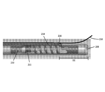
may be disposed
within the second longitudinal lumen 214 and a distal portion of the depth
indicator may exit
the second distal opening 216. As described previously, the length of the
depth indicator that
exits and/or extends from the second distal opening may be varied by advancing
or retracting
the depth indicator at the proximal handle. In some variations, the second
longitudinal lumen
214 may have a diameter from about 0.012 in to about 0.02 in, e.g., about
0.018 in, which
may be selected based on the diameter of the tissue depth indicator. The
location of the
second distal opening 216 may be determined at least in part by the desired
penetration depth
of the delivery catheter tip (i.e., the desired anchor deployment depth into
tissue). In some
variations, the distance D1 between the first distal opening 208 and second
distal opening 216
may correspond to the desired penetration depth and/or anchor delivery depth.
The desired
penetration depth and/or anchor delivery depth may vary according to a number
of factors,
17
CA 02978599 2017-09-01
WO 2016/141358 PCT/US2016/021065
include the targeted tissue region, the anchor depth required to withstand
pull-out forces to
which the anchor may be subjected during/after implantation, toughness of the
tissue,
thickness of the tissue, etc. For example, the desired penetration and/or
anchor delivery depth
may be from about 1 mm to about 15 mm, e.g., about 3 mm, about 4 mm, about 5.5
mm.
about 6 mm, about 8 mm, about 10 mm, about 12.5 mm, etc. Accordingly, distance
D1 may
vary from about 1 mm to about 15 mm, e.g., about 3 mm, about 4 mm, about 5.5
mm, about 6
mm, about 8 mm. about 10 mm, about 12.5 mm, etc.
[0030] Figures 2D and 2E depict one variation of a tissue depth indicator 230
having a
distal portion 234 that is more compliant (e.g., softer, less stiff) than a
proximal portion (view
blocked by the wall of the second longitudinal lumen) of the indicator. The
tissue depth
indicator 230 may comprise an indicator wire with a distal portion 234 that is
more compliant
than its proximal portion. The indicator wire 230 may have a first
configuration (Figure 2D)
and a second configuration (Figure 2E). The angle Al between the indicator
wire 230 and the
second longitudinal lumen 214 may vary between the two configurations. The
vertex 232 of
the angle Al may be co-localized with the second distal opening 216 (e.g., the
location where
the indicator wire exits the second distal opening). In the first
configuration, the angle Al
may be obtuse, and in the second configuration, the angle Al may be acute. For
example, the
angle Al in the first configuration may be from about 120 degrees to about 180
degrees (see
Figures 2D, 2F-2H), while the angle Al in the second configuration may be from
about 80
degrees to about 120 degrees (see Figure 2E). More generally, the curvature of
the indicator
wire in the second configuration has a discontinuity or inflection at the
point of exit that is
not present in the first configuration. The indicator wire 230 may be in the
first configuration
when the distal tip (i.e., the distal opening 208) of anchor delivery catheter
is not located at
the desired tissue depth. In this first configuration, the distal portion 234
of the indicator wire
230 may extend past the distal end of the elongate body, may contact a length
of the distal
section of the elongate body, and/or may generally have smoother curves (i.e.,
the radius of
the curvature does not change abruptly). The flexibility of the distal portion
234 allows the
indicator wire to interrogate, track along, delineate, or otherwise be urged
along the surface
of the target tissue without puncturing or abrading the tissue. The curvature
of a distal portion
that comprises a radiopaque material may be visualized using fluoroscopic
techniques, and
may allow a practitioner to identify the boundary of the tissue surface, along
with the
18
CA 02978599 2017-09-01
WO 2016/141358 PCT/US2016/021065
topology of the tissue surface. For example, curvature of the distal portion
along the surface
of the tissue may also delineate any irregularities, protrusions, trabeculae,
etc. on the surface
of the myocardium. Advancing the distal portion 234 of the indicator wire 230
ahead of the
delivery catheter may facilitate the navigation of the delivery catheter
towards the tissue
surface. It may also help facilitate the proper orientation of the delivery
catheter with respect
to the tissue surface. For example, the delivery catheter may be navigated so
that the direction
of its travel is substantially oriented at an angle of 20 to 80 degrees, e.g..
about 30 degrees to
about 45 degrees with respect to the surface of the target tissue as
delineated by the distal
portion of the indicator wire. As the delivery catheter is advanced into the
target tissue (e.g.,
endocardium of a left ventricle), the tip of the delivery catheter may
penetrate more deeply
into the tissue, and when the penetration depth is approximately the same as
the distance DI,
the tissue surface may press against the compliant distal portion of the
indicator, thereby
causing it to deflect away from the tissue surface and/or the distal end of
the elongate body
(i.e., the depth indicator transitions from the first configuration to the
second configuration).
Since this deflection results in an acute change or discontinuity in the
curvature of the distal
portion, it is readily visualized using fluoroscopic techniques. This
conformational change
provides a visual indication to the practitioner that the distal tip of the
delivery catheter has
penetrated the tissue at a depth that is approximately the same as the
distance Dl. The anchor
may then be deployed from the delivery catheter.
[0031] Some variations of an anchor delivery catheter may alternatively or
additionally
comprise a tissue depth limiter, which may resist penetration of the distal
tip of the delivery
catheter past a preselected depth. This may provide a tactile signal to a
practitioner that a
desired (or maximum) penetration depth has been attained. Figures 3A-3D depict
one
variation of an anchor delivery catheter 300 comprising an elongate body 304
and a tissue
depth limiter 330. The elongate body 304 may have a first longitudinal lumen
306 that
extends from the proximal handle and terminates at a first distal opening 308
and a second
longitudinal lumen 314 that extends from the proximal handle and terminates at
a second
distal opening 316. A tissue depth limiter 330 may be disposed within the
second longitudinal
lumen 314 and a distal portion 331 of the depth limiter 330 may exit the
second distal
opening 316. The distal end 333 of the depth limiter 330 may be attached to
the elongate
body 304. The distance D2 between the attachment location of the depth limiter
330 and the
19
CA 02978599 2017-09-01
WO 2016/141358 PCT/US2016/021065
distal tip/opening of the elongate body may correspond to the desired or
maximum tissue
penetration depth. Distance D1 may be, for example, from about 1 mm to about
15 mm, e.g.,
about 3 mm. about 4 mm, about 5.5 mm, about 6 mm, about 8 mm, about 10 mm,
about 12.5
mm, etc. In some variations, the distal portion 331 of the depth limiter may
comprise a
preformed curve, while in other variations, the distal portion does not have a
preformed
curve. The depth limiter 330 may have a first collapsed configuration (Figure
3E) and a
second expanded configuration (Figure 3A). In the first configuration, the
depth limiter may
be retracted proximally such that the distal portion is substantially flush
with (e.g., extends
along) the outer surface 305 of the elongate body 304. In the second
configuration, the depth
limiter may be advanced distally such that a longer length of the distal
portion exits the
second distal opening and allows for the distal portion to form a curve or
loop, as shown in
Figure 3A. In some variations, the distal portion 331 may have a preformed
curve that
automatically expands when a sufficient length of the depth limiter has exited
the second
longitudinal lumen. For example, the depth limiter 330 may comprise a shape-
memory
material that is preformed with a distal curve. In the first configuration,
the depth limiter is
compressed to a straightened form within the second longitudinal lumen. In the
second
configuration, the distal portion with the preformed curve automatically
assumes its expanded
shape when it exits the second distal opening and is released from the second
longitudinal
lumen. The shape of the atraumatic curve may be selected such that a larger
surface of the
curve is located generally perpendicularly to the longitudinal axis of the
elongate body. This
may help to provide sufficient resistance to forward-travel of the delivery
catheter when the
desired or maximum penetration depth is attained, but also help to provide an
atraumatic
tissue-contacting surface. In some variations, a depth limiter may comprise a
flat wire that
may help to provide resistance to forward travel while reducing the likelihood
of cutting
through the tissue surface.
[0032] The distal end of the depth limiter may be attached to the elongate
body of the
delivery catheter by any suitable method. For example, the distal end may be
attached to the
elongate body (e.g., the outer surface of the elongate body) by welding,
soldering, and the
like, and/or using one or more adhesives. These types of attachment mechanisms
may allow
the distal end of the depth limiter to be rigidly attached to the elongate
body. In other
variations, other types of attachment mechanisms may allow the distal end of
the depth
CA 02978599 2017-09-01
WO 2016/141358 PCT/US2016/021065
limiter to pivot, rotate, slide and/or otherwise deflect with respect to the
elongate body. One
variation of a rotatable attachment mechanism 332 is depicted in Figure 3D,
which is an
enlarged view of the attachment of the distal end 333 of the depth limiter and
the elongate
body 304 depicted in Figure 3A. In this variation, the attachment mechanism
332 may
comprise a curved pin/shaft or ringed structure 336 disposed within a groove
305 of the
elongate body 304. The ringed structure 336 may be a closed ring or an open
ring constructed
from a single component (e.g., a unibody component having a C-shape or a U-
shape) or two
components (e.g., two L-shaped components that are joined together). The
distal end 333 of
the depth limiter 330 may comprise a loop 334, which may be slidably or
rotatably coupled
with the ringed structure 336. In some variations, the loop may be integrally
formed with the
proximal portion of the depth limiter 330, while in other variations, the loop
may be a
hypodermic tubing that may be attached to the proximal portion of the depth
limiter. For
example, the loop of a depth limiter may be formed from a hypodermic tube may
have an
inner diameter of about 0.008 in, an outer diameter of about 0.016 in, and a
wall thickness of
about 0.004 in that is soldered or welded to a proximal portion of the depth
limiter. The loop
334 of the depth limiter may track around the circumference of the ringed
structure 336
counterclockwise or clockwise (depicted in Figure 3C along arrows 340L and
340R,
respectively). In this variation, the plane defined by the curve of the distal
portion 331 may
sweep between about 1 degree to about 90 degrees counterclockwise, and/or may
sweep
between 1 degree to about 90 degrees clockwise. The rotation or pivoting of
the curved distal
portion may provide a visual indication (in addition to a tactile indication)
that a preselected
tissue depth has been attained.
[0033] Also disclosed herein are methods of visualizing the surface of a
target tissue and
determining the penetration depth of an anchor delivery catheter into the
target tissue. In
some variations, these devices and methods may be used to deliver anchors to a
particular
depth in the endocardium of the left ventricle during a beating heart
procedure. One example
of a method for visualizing and determining the depth of delivery catheter
penetration into
tissue is outlined in the flow diagram of Figure 4A and depicted in Figures 5A-
5K. The
method 400 is one in which tissue anchors are delivered to endocardium of the
left ventricle
(LV), though it should be appreciated that this method may be employed in many
other
procedures as well. Such method may comprise using fluoroscopy imaging to
locate the
21
CA 02978599 2017-09-01
WO 2016/141358 PCT/US2016/021065
myocardium, position the catheters, and deliver one or more tethered anchors.
Fluoroscopic
images or video may be taken from a short axis view of the LV or other views
of the LV.
Figure 5A shows the short axis of the left side of the heart 10 with the
surrounding
myocardium 11, endocardium 12, LV chamber 13 and aortic outflow tract and
aortic valve
14.
[0034] The method 400 may comprise advancing a guide catheter (step 402) to
subvalvular
tissue in the LV. Figure 5B depicts a guide catheter 20 that may extend across
the aortic valve
(AV) and tangent to the LV wall, with a distal opening 21 that may be inserted
across the
aortic valve 14 and placed tangent to the endocardium 12. The method 400 may
then
comprise advancing a tunnel catheter through the guide catheter (step 404)
such that a length
of the tunnel catheter is positioned against or near the endocardium. Figure
5C depicts
placing a tunnel catheter 30 deployed from the guide catheter against or near
the endocardium
12. The tunnel catheter may extend around and alongside the LV wall.
Radiopaque markers
and windows may be disposed along the outer radius of the tunnel catheter
wall, through
which delivery catheters can be deployed. For example, the tunnel catheter may
be a template
device, using windows 31 and radiopaque markers 32 to direct the placement of
devices, such
as anchors, into the myocardium of the LV. Next, the method 400 may comprise
advancing a
delivery catheter through the tunnel catheter (step 406) to a selected window
of the tunnel
catheter.
[0035] If a tissue depth indicator is not used, the method would comprise the
steps depicted
in Figures 5D-5G. As depicted there, a delivery catheter 40 may be advanced
through the
tunnel catheter 30 such that the distal tip 41 exits a preferred window 31 and
contacts
endocardium 12. Advancing delivery catheter 40 further causes it to penetrate
endocardium
12 to a desired depth. Figure 5E schematically depicts a fluoroscopic image of
the guide
catheter, tunnel catheter and delivery catheter positioned along endocardium
of the LV. The
information revealed/depicted in a fluoroscopic image is relatively sparse,
especially with
respect to tissue location (e.g., boundary of the myocardium surface) and
depth of
penetration. Specifically, there is little or no information regarding
apposition between
endocardium 12 and tunnel catheter 30, tenting of endocardium 12 at tip 41 of
the delivery
catheter 40 or trabeculations that may be present on endocardium 12 near the
window 31.
Even after several anchors are deployed into the endocardium, with links 60
between them
22
CA 02978599 2017-09-01
WO 2016/141358 PCT/US2016/021065
that are located adjacent to endocardium 12 (as depicted in Figure 5F), the
resulting
fluoroscopic image (represented in Figure 5G) remains remarkably void of
landmarks that
locate the boundary of the endocardium 12, the boundary of the myocardium,
and/or any
other curves of structures located on the target cardiac tissue.
[0036] Using a delivery catheter with a tissue depth indicator may help to
provide
information about the location of the endocardium as well as the penetration
depth of the
delivery catheter tip. To continue from step 406 of the method 400, the next
step may
comprise advancing the tissue depth indicator out of a lumen of the delivery
catheter (step
408) ahead of the distal tip of the delivery catheter, as depicted in Figure
5H. Figure 5H
depicts a delivery catheter comprising a radiopaque depth indicator wire 70.
Depth indicator
wire 70 may be translatable to extend distally or proximally with respect to
the distal end 41
of delivery catheter 40, though in some variations the depth indicator wire
may be fixed and
not translatable along the longitudinal axis of the delivery catheter. The
method 400 may
comprise delineating the boundary of the ventricular wall or myocardium
surface using the
depth indicator wire (step 410). The depth indicator wire may be in the first
configuration, as
described previously and also depicted in Figure 6A. In the first
configuration, the depth
indicator wire may be used to interrogate the surface of endocardium 12 and
may provide a
durable localization and visualization of endocardium 12 where distal tip 41
penetrates. The
delineation of the location and/or surface textures or structures by the
deflections and curves
of the depth indicator wire may be evident in the resulting fluoroscopic image
(Figure 51).
This may facilitate the identification of the location of endocardium 12. The
method 400 may
then comprise advancing the delivery catheter such that the distal tip of the
delivery catheter
penetrates through the endocardium and into the myocardium (step 412). When a
desired
and/or preselected penetration depth is attained, the depth indicator wire may
transition to the
second configuration. That is, an inflection or discontinuity may be
introduced in the depth
indicator wire resulting from the tissue pushing on the depth indicator wire,
thereby
deflecting the distal portion of the wire away from the tissue surface.
Figures 5J and 6B
schematically depict the distal segment 81 of the lumen in which depth
indicator wire 70, and
its location relative to distal tip 41 of the delivery catheter 40, as well as
the inflection or
discontinuity 71 in the curvature of the distal portion of the indicator wire.
This discontinuity
71 and the straight distal portion 72 of the indicator wire 70 may provide a
clear visual signal
23
CA 02978599 2017-09-01
WO 2016/141358 PCT/US2016/021065
that the desired depth of penetration below endocardium 12 has been achieved.
Figure 5K is a
depiction of the fluoroscopic image of the arrangement in Figure 5J. After the
depth indicator
wire has transitioned to the second configuration and the inflection or
discontinuity is
identified, the practitioner may stop advancing the delivery catheter into the
endocardium
(step 414) and deliver the anchor into the endocardium (step 416). In some
variations, all of
the anchors may be delivered with the guidance provided by a tissue depth
indicator. For
example, the first and second anchors depicted in Figures 5D-5G may be
deployed into tissue
using method 400. Furthermore, fluoroscopic images may be acquired at any
point during
method 400, from any view (e.g., short axis view (SAX), long axis view (LAX),
A/P view,
oblique views, etc.).
[0037] Although the method 400 uses the anchor delivery catheter with
additional catheters
(e.g., a guide catheter and a tunnel catheter), it should be understood that
the anchor delivery
catheter may be used to deliver anchors without other catheters, with fewer
catheter, or with
more catheters. One example of a method for delivering anchors is depicted in
Figure 4B.
The method 420 may comprise advancing a delivery catheter to the surface of
the target
tissue (step 422), advancing the depth indicator out of a lumen of the
delivery catheter ahead
of the distal tip of the delivery catheter (step 424), delineating the
boundary of the surface of
the target tissue using the depth indicator (step 426), and advancing the
delivery catheter to
penetrate the target tissue (step 428) until a desired penetration depth is
reached. The method
420 may then comprise stopping the advancement of the delivery catheter when
the distal
portion of the depth indicator deflects away from the surface of the target
tissue (i.e.,
transitions from the first configuration to the second configuration, which
has an inflection or
discontinuity) and delivering the anchor into the tissue at the preselected
depth. Furthermore,
fluoroscopic images may be acquired at any point during method 420, from any
view.
[0038] An anchor delivery catheter comprising a tissue depth limiter may also
be used to
perform the methods of Figures 4A and 4B. However, the tissue depth limiter
may not be
capable of delineating the boundary of the surface of the target tissue.
Instead, prior to
contacting the tissue, the depth limiter may be transitioned from the first
collapsed
configuration to the second expanded configuration. In the expanded
configuration, the depth
limiter may provide a tactile signal that the tip of the delivery catheter has
attained a
preselected or maximum penetration depth. Optionally, an anchor delivery
catheter
24
CA 02978599 2017-09-01
WO 2016/141358 PCT/US2016/021065
comprising a tissue depth limiter that has a distal end rotatably attached to
the elongate body
of the delivery catheter may provide a visual cue that the preselected
penetration depth has
been attained. For example, the rotation of the depth limiter when it abuts
the target tissue
surface may provide a visual change in a fluoroscopic image that indicates a
desired or
maximum penetration depth has been attained. Furthermore, fluoroscopic images
may be
acquired at any point during these procedures, from any view.
[0039] Also disclosed herein are kits comprising an anchor delivery catheter
and a tissue
depth indicator and/or tissue depth limiter. In one variation, a kit may
comprise an anchor
delivery catheter comprising an elongate body with a first longitudinal lumen
terminating at a
first distal opening and a second longitudinal lumen terminating at a second
distal opening.
The kit may further comprise a depth indicator wire configured to be disposed
within the
second longitudinal lumen of the elongate body, where the depth indicator wire
may have a
proximal portion and a distal portion that is relatively more compliant or
flexible than the
proximal portion. The depth indicator wire may be pre-assembled during
manufacturing so
that it is disposed within the second longitudinal lumen, or may be kept
separate from the
elongate body and inserted by the practitioner just prior to use. Optionally,
the kit may
comprise an anchor disposed within the first longitudinal lumen. In another
variation, a kit
may comprise an anchor delivery catheter comprising an elongate body with a
first
longitudinal lumen terminating at a first distal opening and a second
longitudinal lumen
terminating at a second distal opening, and an anchor disposed within the
first longitudinal
lumen. The tissue depth indicator may be in a separate kit. Alternatively, a
kit may comprise
an anchor delivery catheter comprising an elongate body with a first
longitudinal lumen
terminating at a first distal opening and a second longitudinal lumen
terminating at a second
distal opening and a tissue depth limiter disposed within the second
longitudinal lumen and
attached at a distal end of the elongate body. The kit may or may not include
an anchor
disposed within the first longitudinal lumen.