Note: Descriptions are shown in the official language in which they were submitted.
CA 02979403 2017-09-11
WO 2016/183439 PCT/US2016/032349
ANATOMICALLY RELIEVED AUGMENTS
BACKGROUND
[0001] Embodiments of the present application generally relate to
orthopedic augments.
More particularly, but not exclusively, embodiments of the present application
relate to
anatomically shaped orthopedic augments that are configured to address unequal
loading
conditions and provide enhanced flexibility in placement within the associated
bone canal.
[0002] Metaphyseal and/or diaphyseal augments typically assist in
preventing loosening
and/or subsidence of an articular implant/component, such as, for example, an
implanted tibia
baseplate. Such augments can help distribute loads exerted on or by the
articular implant
through the bone, with the articular component maintaining fixation, which can
result in a longer
implant life.
[0003] One of the primary forces attributed to early failures of
orthopedic implants,
particularly in the tibia, is torsional stress. Moreover, torsional stresses
can shear the articular
implant-bone interface (cemented or un-cemented) apart, which can facilitate
premature or early
failure of the implant. Other forces, such as shear forces, can also
contribute to similar
premature or early failure of the articular implant-bone interface.
Additionally, compressive
loads, particularly unequal loads to a median plane (i.e. medial loading) of
the articular implant-
bone interface, can also cause subsidence and early failures of the articular
implant.
[0004] Additionally, too much cortical contact with the augment can, as a
consequence of
carrying too much of the load, stress shield the articular components of the
bone interface. Such
situations can result in bone resorption, which can contribute to early
failure of the implant.
Additionally, unequal cortical contact due to lack of conformity or fit can
load a particular region
of the bone, and thereby relieve the articular implant-bone interface in a
similar region. In at
least certain situations, such unequal loads or contact can act as a fulcrum,
which can facilitate
bone-interface failures for both the augment and the articular implant.
BRIEF SUMMARY
[0005] An aspect of the present application is an augment for
implantation in association
with an orthopedic implant device in a bone, the augment having an augment
wall that includes
an outer portion, an inner portion, a distal end, and a proximal end. The
inner portion of the
1
CA 02979403 2017-09-11
WO 2016/183439 PCT/US2016/032349
augment wall defines an inner region of the augment that is sized to receive
placement of one or
more components of the orthopedic implant device. The distal end at the outer
portion has a first
shape that is configured to generally conform to the shape of a metaphyseal-
diaphyseal junction
of a canal of the bone. Additionally, the proximal end at the outer portion
has a second shape
that is configured to generally conform to a shape of the metaphyseal region
of the canal of the
bone. Further, the first shape has a different shape and size than the second
shape. The augment
further includes at least one relief that extends from at least one of the
proximal end or the distal
end of the augment wall. Additionally, at least one relief is adapted to
prevent, when the
augment is implanted in the bone, contact between a portion of the augment
wall and an adjacent
wall of the bone.
BRIEF DESCRIPTION OF THE DRAWINGS
[0006] The description herein makes reference to the accompanying figures
wherein like
reference numerals refer to like parts throughout the several views.
[0007] Figure 1 illustrates a medial-lateral view of a tibial articular
implant having an
anatomically relieved tibial augment according to an embodiment of the present
application.
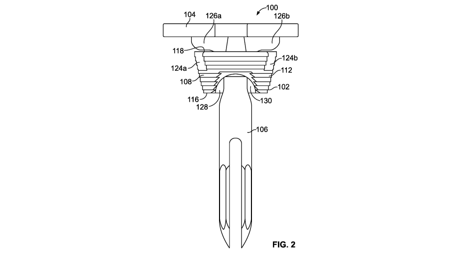
[0008] Figure 2 illustrates a posterior-anterior view of the tibial
implant device and
anatomically relieved tibial augment shown in Figure 1.
[0009] Figure 3 illustrates an isometric view of an anatomically relieved
tibial augment
according to an embodiment of the present application.
[00010] Figure 4 illustrates a medial-lateral view of the anatomically
relieved tibial
augment shown in Figure 3.
[00011] Figure 5 illustrates a posterior-anterior view of the anatomically
relieved tibial
augment shown in Figure 3.
[00012] Figure 6 illustrates a posterior-anterior view of a tibial implant
device on a
prepared implant bone.
[00013] Figure 7 illustrates a cross sectional view, taken along line A-A
of Figure 6, of the
bone and tibial implant device, including the anatomically relieved tibial
augment.
[00014] Figure 8 illustrates a posterior-anterior view of a femoral
implant device having
an anatomically relieved femoral augment according to an embodiment of the
present
application.
2
CA 02979403 2017-09-11
WO 2016/183439 PCT/US2016/032349
[00015]
Figure 9 illustrates a medial-lateral view of the femoral implant device and
anatomically relieved femoral augment shown in Figure 8.
[00016]
Figure 10 illustrates an isometric view of an anatomically relieved femoral
augment according to an embodiment of the present application.
[00017]
Figure 11 illustrates a medial-lateral view of the anatomically relieved
femoral
augment shown in Figure 10.
[00018]
Figure 12 illustrates a posterior-anterior view of the anatomically relieved
femoral
augment shown in Figure 10.
[00019]
Figure 13 illustrates a medial-lateral view of a portion of an exemplary
femoral
implant device having an anatomically relieved femoral augment positioned on a
prepared
formal bone.
[00020]
Figure 14 illustrates an anterior-posterior view of the portion of the femoral
implant device and the anatomically relieved femoral augment shown in Figure
13 positioned on
the prepared formal bone.
[00021]
Figure 15 illustrates a medial-lateral cross sectional view, taken along line
A-A of
Figure 14, of the portion of the femoral implant and anatomically relieved
femoral augment on
the prepared formal bone.
[00022]
The foregoing summary, as well as the following detailed description of
certain
embodiments of the present application, will be better understood when read in
conjunction with
the appended drawings in which like reference numbers indicate like features,
components and
method steps. For the purpose of illustrating the invention, there is shown in
the drawings,
certain embodiments. It should be understood, however, that the present
invention is not limited
to the arrangements and instrumentalities shown in the attached drawings.
DESCRIPTION OF THE ILLUSTRATED EMBODIMENTS
[00023]
Certain terminology is used in the foregoing description for convenience and
is
not intended to be limiting. Words such as "upper," "lower," "top," "bottom,"
"first," and
"second" designate directions in the drawings to which reference is made. This
terminology
includes the words specifically noted above, derivatives thereof, and words of
similar import.
Additionally, the words "a" and "one" are defined as including one or more of
the referenced
item unless specifically noted. The phrase "at least one of' followed by a
list of two or more
3
CA 02979403 2017-09-11
WO 2016/183439 PCT/US2016/032349
items, such as "A, B or C," means any individual one of A, B or C, as well as
any combination
thereof
[00024] Figures 1 and 2 illustrate medial-lateral and posterior-anterior
views, respectively,
of a tibial implant device 100 having an anatomically relieved tibial augment
102 according to an
embodiment of the present application. In the depicted embodiment, the tibial
implant device
100 is a tibial articular assembly that includes a tibial (articular)
baseplate 104, the tibial augment
102, and a stem 106. The stem 106, which can extend along a central stem axis
107, can be
directly or indirectly coupled to the tibial baseplate 104, such as, for
example, coupled to a tray
stem 109 (Figure 7). According to certain embodiments, the tibial implant
device 100 can also
include an offset/angled coupler, which can offset at least the central stem
axis 107 relative to a
central tray stem axis 111 of the tray stem 109 of the base plate 104. The
tibial implant device
100 can also include other components, such as, for example, intramedullary
stems and other
augments that can be assembled to the tibial implant device 100.
[00025] The depicted tibial implant device 100 is structured to be
cemented into and
through the tibial augment 102 and onto a prepared proximal tibia of a
patient. Further, while
Figures 1 and 2 illustrate the tibial augment 102 positioned on or about a
tibial implant device
100 in a non-implanted state or condition, the tibial augment 102 can be
implanted in a bone of
the patient prior to implantation of the remainder of the tibial implant
device 100.
[00026] Figures 3-5 illustrate isometric, medial-lateral, and posterior-
anterior views,
respectively, an exemplary tibial augment 102 according to certain embodiments
of the present
application. A variety of different augments can be used for the tibial
augment 102, including,
for example, a cone or sleeve augment, among other augments. Further, the
tibial augment 102
can have a variety of shapes and sizes. The tibial augment 102 includes an
augment wall 108
that has an inner portion 110 and an outer portion 112. The inner portion 110
of the augment
wall 108 can generally define an inner region 114 of the tibial augment 102,
which can extend
between at least a portion of the distal and proximal ends 116, 118 of the
tibial augment 102. As
indicated by at least Figures 1, 2, and 7, the inner portion 110 of the
augment wall 108 can be
sized to receive passage and/or placement of at least a portion of the stem
106, a tray stem 109 of
the baseplate 104, an offset/angled coupler, and/or other components of the
tibial implant device
100 during implantation of the tibial implant device 100 in a patient.
4
CA 02979403 2017-09-11
WO 2016/183439 PCT/US2016/032349
[00027] The outer portion 112 of the tibial augment 102 can have a variety
of shapes and
sizes. For example, according to certain embodiments, an augment wall 108 of
the tibial
augment 102 can have a generally cylindrical or conical shape as the augment
wall 108 extends
between a distal end 116 and a proximal end 118 of the tibial augment 102.
However, according
to other embodiments, the augment wall 108 can be constructed to generally
conform to the
shapes of different portions of the bone, such as, for example, the conical
shape of the tibia bone,
and/or to the shape of the inner wall of the intramedullary canal or prepared
opening in the bone
in which the tibial augment 102 will be implanted. Thus, variations among
and/or along at least
the augment wall 108 of the tibial augment 102 that accommodate such shapes of
the bone,
intramedullary canal, and/or the prepared opening can enhance the flexibility
in the placement of
the tibial augment 102 in the bone, and reduce or minimize the tibial augment
102 from
hindering the ability to position an associated articular component relative
to a joint line, while
also not hindering joint balance (flexion-extension balance) and rotation of
each component
relative to the patella-femoral joint. Additionally, according to certain
embodiments, the tibial
augment 102 can be symmetrical about at least one midline that is generally
perpendicular to a
central augment axis 120 of the tibial augment 102.
[00028] To generally accommodate the cortical shape(s) of the tibia bone,
the
intramedullary canal of the tibia, and/or the shape of the prepared opening in
the tibia bone in
which the tibia augment 102 is to be implanted, the shape of various portions
or sides of the
augment wall 108 at the distal and/or proximal ends 116, 118, as well as the
shapes of the sides
of the augment wall 108 therebetween, can be different and/or vary. According
to such
embodiments, such variances or inconsistencies among and/or along the sides or
areas of the
tibial augment 102 can preclude the augment wall 108 of the tibial augment 102
from having a
generally uniform cylindrical or conical shape. Further, according to certain
embodiments, the
outer portion 112 of the augment wall 108 of the tibial augment 102 can be
configured such that
at least the distal end 116, or diaphyseal end, of the tibial augment 102
generally conforms to the
general shape of the metaphyseal-diaphyseal junction of the tibia bone 122,
and at least the
proximal end 118 of the tibial augment 102 generally conforms to the general
shape or profile of
the metaphyseal region of the tibia bone 122. According to other embodiments,
the distal end
116 and/or proximal end 118 can be shaped to provide other cross-sectional
shapes that facilitate
the ability of the tibial augment 102 to conform to the size and/or shape of
at least a portion of
CA 02979403 2017-09-11
WO 2016/183439 PCT/US2016/032349
the intramedullary canal 124 of the tibia bone 122 and/or of the prepared
opening in the tibia
bone 122. Such conforming of the tibial augment 102 may not be limited to the
physical
shape(s) of each section of the outer portion 112 of the tibial augment 102
mating or matching
the shape of the adjacent portion of the wall of the intramedullary canal 124,
but instead can
include being shaped to operably align a central augment axis 120 of the
tibial augment 102
with, or at a selected position away from, a reference axis, including, for
example, a longitudinal
axis of the intramedullary canal 124, and/or the central stem axis 107, among
other reference
axes. Additionally, the portion of the tibial augment 102 that is shaped to
generally conform to
the shape or profile of the metaphyseal region can be located at distance
away, in the
metaphyseal direction, from the portion of the tibial augment 102 that
conforms to the general
shape or profile of the metaphyseal-diaphyseal junction that is about the same
as the distance
between the metaphyseal region and metaphyseal-diaphyseal junction of the
tibia bone 122.
[00029] As shown in at least Figures 1-5, according to certain
embodiments, the augment
wall 108 can further include at least one opening 124a, 124b that is
configured to accommodate
placement of a component of the tibial augment 102. For example, according to
the illustrated
embodiment, the tibial augment 102 can include two openings 124a, 124b that
are sized to
accommodate at least the passage and/or placement of at least a portion of the
keel(s) 126a, 126b
of the tibial baseplate 104.
[00030] The outer portion 112 of the augment wall 108 can also include one
or more
reliefs 128 that are positioned at least around the distal end 116 and/or the
proximal end 118 of
the tibial augment 102. According to the illustrated embodiment, the relief
128 can remove at
least a portion of the augment wall 108 so as to reduce or otherwise alter the
shape of at least a
profile of the tibial augment 102. For example, dashed lines in Figure 3
illustrate a portion of the
tibial augment 102 that can be removed by the recess 128, and the resulting
profile provided by
inclusion of the relief 128. As discussed below, removing, altering, and/or
contouring the shape
and/or size of the tibial augment 102 via inclusion of one or more reliefs 128
that can increase
the degree of freedom that can be attained in the placement and/or sizing of
the tibial augment
102 in the tibia bone 122, intramedullary canal, and/or a shaped or prepared
opening in the tibia
bone 122.
[00031] According to certain embodiments, the recess 128 can be configured
to extend
through the augment wall 108 so as to include an aperture 130 that exposes at
least a portion of
6
CA 02979403 2017-09-11
WO 2016/183439 PCT/US2016/032349
the inner region 114. Further, the relief 128 can also include one or more
relief walls 132, such
as, for example, opposing sidewalls 136a, 136b and an upper wall 138 that
extends around the
aperture 130. The relief walls 132 can reduce the thickness of the augment
wall 108 at or around
the aperture 130. Moreover, the augment wall 108 can have a material thickness
between the
relief wall 132 and the opposing inner portion 110 of the augment wall 108
that is less than the
thickness between opposing outer and inner portions 110, 112 of the augment
wall 108.
[00032] The sidewalls 136a, 136b and upper wall 138 of the relief wall 132
can have a
variety of different shapes and orientations that can, in at least certain
situations, increase the
degree of freedom in the positioning and/or sizing of the tibial augment 102
in the bone 122 that
can be attained via use of the relief 128. For example, in the illustrated
embodiment, the upper
wall 138 has a generally curved or arced shape, while the sidewalls 136a, 136b
generally extend
toward each other from opposite directions before reaching the upper wall 138.
Additionally, as
shown in Figure 3, according to the illustrated embodiment, the relief 128 can
be configured
such that a portion of the relief wall 132 has an angled or tapered profile
that extends inwardly
toward the distal end 116, and which provides a larger or stepper incline than
can have otherwise
been provided by the augment wall 108 without the inclusion of the relief 128
(as indicated by a
comparison of the adjacent solid and dashed lines in Figure 3).
[00033] Figures 4 and 5 illustrate a relief 128 having an aperture 130
that extends through
a portion of the distal end 116 of the tibial augment 102, and a relief wall
132 that extends along
a portion of the tibial augment 102 and about the aperture 130. However,
although the relief 128
of the depicted embodiment includes an aperture 130 in the augment wall 108,
according to
certain embodiments, the relief 128 can extend into the augment wall 108 to a
degree that
prevents the formation of such an aperture 130 in the relief 128. Further, the
aperture 130 of the
relief 128 and at least a portion of the relief wall 132 can extend along a
central relief axis 133
that is generally parallel to the adjacent portion of the augment wall 108 in
which the aperture
130 is positioned. Further, the central relief axis 133 can be non-
perpendicular to the central
augment axis 120 and/or the central stem axis 133 that can extend into/through
the inner region
114 of the tibial augment 102.
[00034] The degree to which the relief 128 extends along the augment wall
108 can vary.
For example, in the illustrated embodiment, the relief 128 extends from the
distal end 116 of the
augment wall 108 to generally a mid-region 134 of the augment wall 108, the
mid region 134
7
CA 02979403 2017-09-11
WO 2016/183439 PCT/US2016/032349
being located a midpoint or area between the distal and proximal ends 116, 118
of the tibial
augment 102. Again, while the relief 128 depicted in Figures 4 and 5 extends
from the distal end
116 of the tibial augment 102, according to other embodiments, a relief, in
addition to or in lieu
of the relief 128 depicted in Figures 4 and 5, can extend from the proximal
end 118 of the tibial
augment 102.
[00035] Figure 7 provides an example of a relief 128 of a tibial augment
102 being
configured to accommodate the cortical shape and/or configuration of the tibia
bone 122 and/or
intramedullary canal, as depicted in Figure 6. As illustrated, the reduction
in the size of the
profile of at least a portion of the tibial augment 102, and, moreover, the
resulting adjustment in
the shape of the tibial augment 102, as provided by the relief 128, can be
configured to at least
assist in the tibial augment 102 being anatomically shaped and/or to assist in
contouring or
otherwise shaping the tibial augment 102 avoid cortical bone contact, such as,
for example,
avoiding contact with the cortical bone in the metaphyseal-diaphyseal junction
140. Further, the
relief 128 can be sized or otherwise configured to prevent the tibial augment
102 from engaging
the asymmetric morphology of the tibia bone 122. Additionally, as also shown
by Figure 7, the
inclusion of the relief 128 can at least assist in the tibial augment 102 from
avoiding contact with
the implant construct, including, for example, contact with the tibial implant
device 100 that can
be associated with misalignment of the intramedullary canal with the
metaphyseal and/or the
diaphyseal region(s) of the tibia bone 122.
[00036] Figures 8 and 9 illustrate posterior-anterior and medial-lateral
views, respectively,
of a femoral implant device 200. The illustrated femoral implant device 200
includes a femoral
articular component 202, an intramedullary stem 206, and a femoral augment 206
according to
an illustrated embodiment of the present application. The femoral implant
device 200 can
include other components, including, but not limited to, a distal augment
and/or a posterior
augment. The intramedullary stem 206, which can extend along a central stem
axis 208, can be
directly or indirectly coupled to the femoral articular component 202, such
as, for example,
coupled to a component stem of the femoral articular component 202. According
to certain
embodiments, the femoral implant device 200 can include an offset/angled
coupler, which can
offset at least the central stem axis 208 relative to an axis the component
stem.
[00037] The depicted femoral implant device 200 is structured to be
cemented into and
through the femoral augment 206 and onto a prepared distal femur of a patient.
Further, while
8
CA 02979403 2017-09-11
WO 2016/183439 PCT/US2016/032349
Figures 8 and 9 illustrate the femoral augment 206 positioned on or about a
femoral implant
device 200 in a non-implanted state or condition, the femoral augment 206 can
be implanted in a
bone of the patient prior to implantation of the remainder of the femoral
implant device 200.
Thus, an inner region 219 of the femoral augment 206 can be sized to receive
passage and/or
placement of at least a portion of the intramedullary stem 206 and/or other
components of the
femoral implant device 200, including, for example, an offset/angled coupler
and/or a component
stem of the femoral articular component 202, during implantation of the
femoral implant device
200 in a patient.
[00038] Figures 10-12 illustrate an example of a femoral augment 206
according to an
illustrated embodiment of the present application. A variety of different
augments can be used
for the femoral augment 206, including, for example, a cone or sleeve augment,
among other
augments. Further, the femoral augment 206 can have a variety of shapes and
sizes. The
femoral augment 206 can include an augment wall 212 that extends about a
central augment axis
214 of the femoral augment 206. The augment wall 212 has an inner portion 216
and an outer
portion 218. The inner portion 216 of the augment wall 212 can generally
define an inner region
219 of the femoral augment 206. At least a portion of the inner region 219 can
extend between a
distal end 220 and a proximal end 222 of the femoral augment 206. The inner
region 219 can be
sized to receive placement of at least one or more components of the femoral
augment 206, such
as, for example, the intramedullary stem 206, an offset/angled coupler, and/or
the component
stem of the femoral articular component 202, and junctions there between,
among other
components.
[00039] The outer portion 218 of the augment wall 212 can be shaped to
generally fit the
cortical shape of a distal femur and/or a portion of the intramedullary canal
of the femur. Thus,
according to certain embodiments, a diaphyseal or distal end 220, of the
femoral augment 206
can be shaped to generally conform to the general shape of the metaphyseal-
diaphyseal junction
of femoral bone. Further, the opposing proximal end 222 of the femoral augment
206 can be
configured to generally conform to the general shape or profile of the
metaphyseal region of the
femoral bone. According to other embodiments, the distal end 220 and/or
proximal end 222 can
be shaped to provide other cross-sectional shapes that facilitate the ability
of the femoral
augment 206 to conform to the size and/or shape of at least a portion of the
femur and/or the
intramedullary canal of the femur. Such conforming may not be limited to the
physical shape(s)
9
CA 02979403 2017-09-11
WO 2016/183439 PCT/US2016/032349
of each section of the outer portion 218 of the augment mating or matching the
shape of the
adjacent portion of the inner wall of the intramedullary canal of the femoral
bone, but instead can
include being shaped to generally align with a central augment axis 214 of the
femoral augment
206, or at a selected position away from a reference axis, including, for
example, a longitudinal
axis of the intramedullary canal of the femur and/or the central stem axis
208, among other
reference axes. Additionally, the portion of the femoral augment 206 that is
shaped to generally
conform to the shape or profile of the metaphyseal region of the femur and/or
the intramedullary
canal of the femur can be located at distance away, in the metaphyseal
direction, from the portion
of the femoral augment 206 that conforms to the general shape or profile of
the metaphyseal-
diaphyseal junction that is about the same as the distance between the
metaphyseal region and
metaphyseal-diaphyseal junction of the femur.
[00040] To generally accommodate the cortical shape(s) of femur and/or the
medullary
canal of the femur, including, for example, the shape at both the metaphyseal-
diaphyseal junction
and at metaphyseal region of the femur, as well as shapes therebetween,
different areas or sides
of the outer portion 218 of the augment wall 212 can have different shapes.
Additionally, the
shapes along such different areas or sides of the outer portion 218 of the
augment wall 212 can
also vary between the distal and proximal ends 220, 222 of the femoral augment
206. Such
variances or inconsistencies among and/or along the sides or areas of the
femoral augment 206
can preclude the augment wall 212 of the femoral augment 206 from having a
generally uniform
cylindrical or conical shape. However, according to other embodiments, the
femoral augment
206 can have a generally cylindrical or conical shape.
[00041] As shown by at least Figures 10-12, the outer portion 218 of the
augment wall 212
can include one or more reliefs 224a, 224b that are positioned at least around
a portion of the
distal end 220 and/or the proximal end 222 of the femoral augment 206.
According to the
illustrated embodiment, the reliefs 224a, 224b can provide a recess and/or
aperture 226a, 226b in
the augment wall 212, and a relief wall 228 that reduces the thickness of the
augment wall 212 at
or around the apertures 226a, 226b. As shown in Figures 10-12, in the depicted
embodiment, the
femoral augment 206 includes a first relief 224a that extends from the distal
end 220 and toward
the proximal end 222 of the femoral augment 206, and another, second relief
224b on a generally
opposing side of the augment wall 212 that extends in an opposite direction,
and more
specifically, extends from the proximal end 222 toward the distal end 220 of
the femoral
CA 02979403 2017-09-11
WO 2016/183439 PCT/US2016/032349
augment 206. As illustrated, in the depicted example, each relief 224a, 224b
extends to an area
adjacent, or in relatively close proximity to, the opposing distal or proximal
end 220, 222 of the
augment 206.
[00042] The relief wall 228 for each relief 224a, 224b can extend along
the femoral
augment 206 and at least about the aperture 226a, 226b. Further, the augment
wall 212 can have
a material thickness between the relief walls 228 and the inner portion 216 of
the augment wall
212 that is less than the thickness between opposing inner and outer portions
216, 218 of the
augment wall 212. Further, although the reliefs 224a, 224b of the depicted
embodiment each
include an aperture 226a, 226b in the augment wall 212, according to certain
embodiments, one
or both of the reliefs 224a, 224b can extend into the augment wall 212 to a
degree that prevents
the formation of such an aperture 226a, 226b.
[00043] According to the illustrated embodiment, the apertures 226a, 226b
and/or at least
a portion of the relief walls 228 can extend along an associated central
relief axis 230a, 230b that
is generally parallel to the adjacent portion of the augment wall 212 in which
the apertures 226a,
226b and/or relief walls 228 is/are positioned. Further, the central relief
axes 230a, 230b can be
non-perpendicular to the central augment axis 214 of the femoral augment 206
and/or to the
central stem axis 208 of the intramedullary stem 204 that can extend
into/through the inner
region 219 of the femoral augment 206.
[00044] As shown in Figures 10-12, according to the illustrated
embodiment, the relief
walls 228 of the reliefs 224a, 224b can each include opposing sidewalls 232a,
232b and an
adjoining upper wall 234. The sidewalls 232a, 232b and upper wall 234 can have
a variety of
different shapes and orientations that can, in at least certain situations,
facilitate the freedom of
positioning and/or sizing that is attained via use of the reliefs 224a, 224b.
For example, as
shown by at least Figure 11, in the illustrated embodiment, the upper wall 234
for the first relief
224a can have a generally curved or arced shape, while the upper wall 234 of
the second relief
224b includes a generally flat section 236. Further, as shown in at least
Figure 12, at least a
portion of the sidewalls 232a, 232b of the reliefs 224a, 224b can have angled
or tapered profiles
that extend inwardly toward the associated distal end 220 or proximal end 222,
which can assist
in providing the femoral augment 206 with a narrower or thinner profile in
those regions than
would be provided in the absence of the reliefs 224a, 224b (as indicated by
the dashed lines in
Figure 12).
11
CA 02979403 2017-09-11
WO 2016/183439 PCT/US2016/032349
[00045] The anatomical shape of the tibial or femoral augments 102, 206,
as well as the
inclusion of the reliefs 128, 224a, 224b, can increase the available choices
or freedom in the
positioning and/or sizing of the augments 102, 206 in the corresponding
prepared tibial or femur
bone, or shaped opening in the tibial or femur bone and/or the associated
intramedullary canal in
which the tibial or femoral augment 102, 206 is implanted. As discussed, the
inclusion of the
reliefs 128, 224a, 224b can reduce the profile and/or size of the tibial
femoral augment 102, 206
at least at the distal end 116, 220 and proximal end 118, 222, and/or along
the opposing sides of
the tibial or femoral augment 102, 206. Further, the reliefs 128, 224a, 224b
can be configured
such that the augments 102, 206 are configured to accommodate certain
characteristics in the
shape of the bone or bone canal in which the augments 102, 206 can be placed.
For example, the
inclusion of the reliefs 128, 224a, 224b can at least assist in the augments
102, 206 avoiding
contact with certain portions of the bone, such as, for example, preventing
the femoral augment
206 from engaging the asymmetric morphology of the femur.
[00046] When an anatomically shaped tibial or femoral augment 102, 206
that includes a
relief(s) 128, 224a, 224b, as discussed herein, is subjected to placement at
relatively shallow
depths in the shaped or prepared tibial or femur, such as when an implant
device 100, 200 is near
the epiphysis of the bone, cancellous bone can be the primary, and possibly
only, contact to the
load bearing surfaces of the tibial or femoral augment 102, 206. Further, as
the depth of the
prepared opening in the tibial or femoral bone increases conformity, proximity
of the prepared
opening and placement of the anatomically shaped augments 102, 206 having the
reliefs 128,
224a, 224b to the cortical bone can also increase. Such conformity and
consistency of
cancellous and/or cortical bone contact throughout a depth variation of
deployment of the tibial
or femoral augments 102, 206 can at least assist in enhancing the evenness in
load distribution,
as well as enhance resistance implant failure, that can otherwise be
attributed to loosening and/or
subsiding due to one or more of the forces, such as, for example, compressive,
shear, and/or
torsion forces, that can be associated with implant devices and associated
components.
Accordingly, the anatomically shaped tibial and femoral augment augments 102,
206 can be
configured, including shaped and/or via the inclusion of reliefs 128, 224a,
224b, to prevent or
minimize the occurrence of point contact between the augments 102, 206 and the
adjacent
cortical wall of the bone. The prevention of such point contact can include
preventing
misaligned or unequal circumferential load sharing about the cortical wall.
Further, by
12
CA 02979403 2017-09-11
WO 2016/183439 PCT/US2016/032349
preventing point contact, the augments 102, 206 can prevent or otherwise
minimize the potential
for the augment 102, 206 to penetrate through, or otherwise violate, the
adjacent cortical wall of
the bone.
[00047] Shaping the tibial and femoral augments 102, 206 to generally
conform to, or
accommodate, changes and/or variances in the shape of the tibia and femoral
bone, respectively,
and/or the intramedullary canal 124 of those bones, can prevent or minimize
the extent to which
the tibial or femoral augments 102, 206 are subjected to unequal loading
conditions. Further, by
shaping different portions or areas of the tibial and femoral augments 102,
206 to generally
conform to or otherwise accommodate the shape of at least an adjacent inner
wall of the
associated bone canal or cavity, the generally anatomically shaped augments
102, 206 discussed
herein can reduce the impact forces on the corresponding articular implant-
bone interface by
distributing such forces or loads over a relatively larger surface area. More
specifically, for
example, such conforming configurations of the augments 102, 206 can improve
resistance to
torsional stress by equally distributing such forces circumferentially.
[00048] Figures 13 and 14 illustrate medial-lateral and anterior-posterior
views,
respectively, of a femoral articular component 202 of an exemplary femoral
implant device 202
having an anatomically relieved femoral augment 206, and which is positioned
on a prepared
femoral bone 240. Further, Figure 15 illustrates a medial-lateral cross
sectional view of the
portion of the femoral articular component 202 and the anatomically relieved
femoral augment
206 on the prepared femoral bone 240, as taken along line A-A of Figure 14. As
shown in
Figure 13, a first relief 224a can be configured to generally conform the
shape or profile of the
femoral augment 206 to the shape of the femoral bone 240 at the metaphyseal-
diaphyseal
junction 242, and moreover, to avoid contact with the cortical bone in the
metaphyseal-
diaphyseal junction 242. Further, as shown, the inclusion of the reliefs 224a,
224b can at least
assist in the femoral augment 206 avoiding contact with the implant construct,
including, for
example, contact with the femoral articular component 202 that can be
associated with
intramedullary canal misalignment with the metaphyseal and/or the diaphyseal
region(s) of the
femoral bone 240. For example, as shown, the second relief 224b of the femoral
augment 206
can be shaped to prevent or otherwise minimize the femoral augment 206 from
contacting an
inner portion 216 of the femoral implant device 200, such as, for example, an
inner portion of the
articular implant construct, while still providing a segment of the femoral
augment 206 at the
13
CA 02979403 2017-09-11
WO 2016/183439 PCT/US2016/032349
distal end 220 of the femoral bone 240 that can be implanted at a positioned
in a prepared portion
or cavity of the bone 240. Thus, the reliefs 224a, 224b, as illustrated, can
be constructed to allow
for a degree of rotational freedom in the angular position of the implanted
femoral augment 206
about at least the central augment axis 214 while still allowing the femoral
augment 206 to
generally conform to the shape of the femoral bone 240 and still prevent, if
desired, contact
between the femoral augment 206 and the inner portion 216 of the femoral
implant device 200.
[00049] While the invention has been described in connection with what is
presently
considered to be the most practical and preferred embodiment, it is to be
understood that the
invention is not to be limited to the disclosed embodiment(s), but on the
contrary, is intended to
cover various modifications and equivalent arrangements included within the
spirit and scope of
the appended claims, which scope is to be accorded the broadest interpretation
so as to
encompass all such modifications and equivalent structures as permitted under
the law.
Furthermore it should be understood that while the use of the word preferable,
preferably, or
preferred in the description above indicates that feature so described may be
more desirable, it
nonetheless may not be necessary and any embodiment lacking the same may be
contemplated as
within the scope of the invention, that scope being defined by the claims that
follow. In reading
the claims it is intended that when words such as "a," "an," "at least one"
and "at least a portion"
are used, there is no intention to limit the claim to only one item unless
specifically stated to the
contrary in the claim. Further, when the language "at least a portion" and/or
"a portion" is used
the item may include a portion and/or the entire item unless specifically
stated to the contrary.
14