Note: Descriptions are shown in the official language in which they were submitted.
CA 02980713 2017-09-22
WO 2016/159999 PCT/US2015/023720
OCULAR DELIVERY SYSTEMS AND METHODS
FIELD
[0001] Described here are systems and methods for accessing Schlemm's canal in
an eye and
for delivering an ocular device, tool, or fluid composition therein. The
ocular devices may
maintain the patency of Schlemm's canal without substantially interfering with
transmural,
transluminal, circumferential, or longitudinal aqueous humor fluid flow across
the canal. The
tools delivered may be used to disrupt the trabecular meshwork. The fluid
composition may be a
viscoelastic fluid that is delivered into the canal or aqueous collector
channels to facilitate
drainage of aqueous humor by dilating the canal, disrupting juxtacanalicular
meshwork and the
adjacent wall of Schlemm's canal, and/or increasing aqueous permeability
through the
trabeculocanalicular, or transmural, outflow pathway. Minimally invasive
methods for treating
medical conditions associated with elevated intraocular pressure, including
glaucoma, are also
described.
BACKGROUND
[0002] Glaucoma is a potentially blinding disease that affects over 60 million
people
worldwide, or about 1-2% of the population. Typically, glaucoma is
characterized by elevated
intraocular pressure. Increased pressure in the eye can cause irreversible
damage to the optic
nerve which can lead to loss of vision and even progress to blindness if left
untreated. Consistent
reduction of intraocular pressure can slow down or stop progressive loss of
vision associated
with glaucoma.
[0003] Increased intraocular pressure is generally caused by sub-optimal
efflux or drainage of
fluid (aqueous humor) from the eye. Aqueous humor or fluid is a clear,
colorless fluid that is
continuously replenished in the eye. Aqueous humor is produced by the ciliary
body, and then
ultimately exits the eye primarily through the trabecular meshwork. The
trabecular meshwork
extends circumferentially around the eye at the anterior chamber angle, or
drainage angle, which
is formed at the intersection between the peripheral iris or iris root, the
anterior sclera or scleral
spur and the peripheral cornea. The trabecular meshwork feeds outwardly into
Schlemm's canal,
a narrow circumferential passageway generally surrounding the exterior border
of the trabecular
meshwork. Positioned around and radially extending from Schlemm's canal are
aqueous veins or
collector channels that receive drained fluid. The net drainage or efflux of
aqueous humor can be
1
CA 02980713 2017-09-22
WO 2016/159999 PCT/US2015/023720
reduced as a result of decreased facility of outflow, decreased outflow
through the trabecular
meshwork and canal of Schlemm drainage apparatus, increased episcleral venous
pressure, or
possibly, increased production of aqueous humor. Flow out of the eye can also
be restricted by
blockages or constriction in the trabecular meshwork and/or Schlemm's canal
and its collector
channels.
[0004] Glaucoma, pre-glaucoma, and ocular hypertension currently can be
treated by reducing
intraocular pressure using one or more modalities, including medication,
incisional surgery, laser
surgery, cryosurgery, and other forms of surgery. In general, medications or
medical therapy are
the first lines of therapy. If medical therapy is not sufficiently effective,
more invasive surgical
treatments may be used. For example, a standard incisional surgical procedure
to reduce
intraocular pressure is trabeculectomy, or filtration surgery. This procedure
involves creating a
new drainage site for aqueous humor. Instead of naturally draining through the
trabecular
meshwork, a new drainage pathway is created by removing a portion of sclera
and trabecular
meshwork at the drainage angle. This creates an opening or passage between the
anterior
chamber and the subconjunctival space that is drained by conjunctival blood
vessels and
lymphatics. The new opening may be covered with sclera and/or conjunctiva to
create a new
reservoir called a bleb into which aqueous humor can drain. However,
traditional
trabeculectomy procedures carry both short and long term risks. These risks
include blockage of
the surgically-created opening through scarring or other mechanisms, hypotony
or abnormally
low intraocular pressure, expulsive hemorrhage, hyphema, intraocular infection
or
endophthalmitis, shallow anterior chamber angle, macular hypotony, choroidal
exudation,
suprachoroidal hemorrhage, and others.
[0005] One alternative is to implant a device in Schlemm's canal that
maintains the patency of
the canal or aids flow of aqueous humor from the anterior chamber into the
canal. Various
stents, shunts, catheters, and procedures have been devised for this purpose
and employ an ab-
externo (from the outside of the eye) approach to deliver the implant or
catheter into Schlemm's
canal. This method of placement is invasive and typically prolonged, requiring
the creation of
tissue flaps and deep dissections to access the canal. Additionally, it is
very difficult for many
surgeons to find and access Schlemm's canal from this external incisional
approach because
Schlemm's canal has a small diameter, e.g., approximately 50 to 250 microns in
cross-sectional
diameter, and it may be even smaller when collapsed. One such procedure, ab-
externo
canaloplasty, involves making a deep scleral incision and flap, finding and
unroofing Schlemm's
2
CA 02980713 2017-09-22
WO 2016/159999 PCT/US2015/023720
canal, circumnavigating all 360 degrees of the canal with a catheter from the
outside of the eye,
and either employing viscoelastic, a circumferential tensioning suture, or
both to help maintain
patency of the canal. The procedure is quite challenging and can take anywhere
from forty-five
minutes to two hours. The long-term safety and efficacy of canaloplasty is
very promising, but
the procedure remains surgically challenging and invasive.
[0006] Another alternative is viscocanalostomy, which involves the injection
of a viscoelastic
solution into Schlemm's canal to dilate the canal and associated collector
channels. Dilation of
the canal and collector channels in this manner generally facilitates drainage
of aqueous humor
from the anterior chamber through the trabecular meshwork and Schlemm's canal,
and out
through the natural trabeculocanalicular outflow pathway. Viscocanalostomy is
similar to
canaloplasty (both are invasive and ab-externo), except that viscocanalostomy
does not involve a
suture and does not restore all 360 degrees of outflow facility. Some
advantages of
viscocanalostomy are that sudden drops in intraocular pressure, hyphema,
hypotony, and flat
anterior chambers may be avoided. The risk of cataract formation and infection
may also be
minimized because of reduced intraocular manipulation and the absence of full
eye wall
penetration, anterior chamber opening and shallowing, and iridectomy. A
further advantage of
viscocanalostomy is that the procedure restores the physiologic outflow
pathway, thus avoiding
the need for external filtration, and its associated short and long term
risks, in the majority of
eyes. This makes the success of the procedure partly independent of
conjunctival or episcleral
scarring, which is a leading cause of failure in traditional trabeculectomy
procedures. Moreover,
the absence of an elevated filtering bleb avoids related ocular discomfort and
potentially
devastating ocular infections, and the procedure can be carried out in any
quadrant of the
outflow pathway.
[0007] However, current viscocanalostomy and canaloplasty techniques are still
very invasive
because access to Schlemm's canal must be created by making a deep incision
into the sclera,
creating a scleral flap, and un-roofing Schlemm's canal. In their current
forms, these procedures
are both "ab-extemo" procedures. "Ab-externo" generally means "from the
outside" and it is
inherently more invasive given the location of Schlemm's canal and the amount
of tissue
disruption required to access it from the outside. On the other hand, "ab-
interno" means "from
the inside" and is a less invasive approach because of the reduced amount of
tissue disruption
required to access it from the inside. Consequently, an ab-interno approach to
Schlemm's canal
offers the surgeon easier access to the canal, but also reduces risk to the
patient's eye and
3
CA 02980713 2017-09-22
WO 2016/159999 PCT/US2015/023720
reduces patient morbidity. All of these lead to improved patient recovery and
rehabilitation. The
ab-extemo viscocanalostomy and canaloplasty procedures also remain challenging
to surgeons,
because as previously stated, it is difficult to find and access Schlemm's
canal from the outside
using a deep incisional approach due to the small diameter of Schlemm's canal.
A further
drawback still is that at most, viscocanalostomy typically dilates up to 60
degrees of Schlemm's
canal, which is a 360 degree ring-shaped outflow vessel-like structure. The
more of the canal
that can be dilated, the more total aqueous outflow can be restored.
[0008] Accordingly, it would be beneficial to have systems that easily and
atraumatically
provide access to Schlemm's canal using an ab-intemo approach for the delivery
of ocular
devices, tools, and compositions. It would also be useful to have systems that
deliver devices,
tools, and compositions into Schlemm's canal expeditiously to decrease
procedure time and the
risk of infection without compromising safety and precision of the delivery
procedure. It would
also be useful to have systems that deliver devices, tools, and fluid
compositions into Schlemm's
canal using an ab-interno approach so that cataract surgery and glaucoma
surgery can both be
accomplished during the same surgical sitting using the very same corneal or
scleral incision.
Such incisions are smaller and allow for less invasive surgery and more rapid
patient recovery.
This approach allows for accessing Schlemm's canal through the trabecular
meshwork from the
inside of the eye, and thus it is called "ab-intemo." Methods of delivering
ocular devices, tools,
and compositions that effectively disrupt the juxtacanalicular meshwork and
adjacent wall of
Schlemm's canal, also known as the inner wall of Schlemm's canal, maintain the
patency of
Schlemm's canal, increase outflow, decrease resistance to outflow, or
effectively dilate the canal
and/or its collector channels using the systems in a minimally invasive, ab-
intemo manner would
also be desirable.
BRIEF SUMMARY
[0009] Described here are systems and methods for easily and reliably
accessing Schlemm's
canal with minimal or reduced trauma and for delivering an ocular device
(e.g., an implant)
therein. Other systems and methods may be implant-free, and/or rely on the
delivery and
removal of a therapeutic (disruptive) tool and/or the delivery of a fluid
composition into
Schlemm's canal to improve flow through the trabeculocanalicular outflow
system, which
consists of the trabecular meshwork, juxtacanalicular tissue, Schlemm's canal,
and collector
channels. When an ocular device is implanted, the ocular device may maintain
the patency of
Schlemm's canal without substantially interfering with transmural fluid flow
across the canal.
4
CA 02980713 2017-09-22
WO 2016/159999 PCT/US2015/023720
Transmural flow, or transmural aqueous humor flow, is defined as flow of
aqueous humor from
the anterior chamber across the trabecular meshwork into the lumen of
Schlemm's canal, across
and along the lumen of Schlemm's canal, and ultimately into aqueous collector
channels
originating in the outer wall of Schlemm's canal. When a fluid composition is
delivered into the
canal, the fluid composition, e.g., a viscoelastic fluid, delivered into the
canal may facilitate
drainage of aqueous humor by dilating the canal, rendering the trabecular
meshwork and inner
wall of Schlemm's canal more permeable to aqueous humor, and also dilating
aqueous collector
channels. When a therapeutic tool is delivered, the tool may facilitate
drainage of aqueous
humor by dilating the canal, dilating the collector channels, disrupting or
stretching the
trabecular meshwork, disrupting or stretching the juxtacanalicular tissue,
tearing or cutting the
trabecular meshwork or juxtacanalicular tissue, or completely removing the
trabecular
meshwork or juxtacanalicular tissue. Any or all of these actions may reduce
resistance to
outflow, increase aqueous outflow and drainage, and reduce intraocular
pressure.
[0010] One of the beneficial features of the system may be a cannula
configured with a distal
curved portion that defines a radius of curvature, where the radius of
curvature directly engages
the bevel at the distal tip of the cannula. However, in some variations, the
system may comprise
a straight cannula. The specific configuration of the handle of the system may
also be useful.
The handle may be sized and shaped so that it is easily manipulated with one
hand. Furthermore,
the handle may be designed for universal manipulation. By "universal" it is
meant that the
handle is ergonomically configured for both right-handed and left-handed use,
for use to access
any quadrant of the eye, and for use in advancing a cannula or elongate member
into Schlemm's
canal in a clockwise or counterclockwise fashion. Such a configuration may
include a drive
assembly that can be easily actuated in a first orientation (e.g., to deliver
an implant, tool, and/or
fluid in a clockwise fashion) and that can be easily actuated in a second,
flipped orientation (e.g.,
to deliver an implant, tool, and/or fluid in a counterclockwise fashion). Such
a configuration may
allow the drive assembly to be actuated using either a left hand or a right
hand, and may allow
the drive assembly to be used with either the left eye or the right eye.
Alternatively, in some
variations the cannula itself can be rotated to the extent needed (e.g., 180
degrees) to provide
ambidextrous ease of use in a clockwise or counterclockwise advancement
direction.
[0011] The ocular delivery systems described herein generally include a
universal handle
having a grip portion and a housing that has an interior and a distal end. A
cannula is typically
coupled to and extends from the housing distal end. The cannula may include a
proximal end
CA 02980713 2017-09-22
WO 2016/159999 PCT/US2015/023720
and a distal curved portion, where the distal curved portion has a proximal
end and a distal end,
and a radius of curvature defined between the ends. The cannula may also be
configured to
include a body; a distal tip having a bevel; and a lumen extending from the
proximal end through
the distal tip. The bevel may directly engage the distal end of the curved
portion of the cannula
(i.e., the bevel may directly engage the radius of curvature). The systems may
also generally
include a drive assembly substantially contained within the housing comprising
gears that
translate rotational movement to linear movement.
[0012] When an ocular device is to be implanted into Schlemm's canal, the
system may
further include a slidable positioning element having a proximal end and a
distal end that is
coaxially disposed within the cannula lumen. The distal end of the slidable
positioning element
may comprise an engagement mechanism for positioning (including manipulating)
the ocular
device within the canal. Exemplary engagement mechanisms that may be employed
comprise
hooks, jaws, clasps, forceps, or complimentary mating elements for releasable
attachment of the
ocular devices.
[0013] The system may be configured to include a fluid assembly in the handle
and an
elongate member comprising a lumen coaxially disposed within the cannula lumen
when a fluid
composition is to be delivered into Schlemm's canal. The fluid composition may
be delivered
through the distal end of the lumen of elongate member or through openings
spaced along the
axial length of the elongate member. Additionally, the fluid assembly may be
coupled to a
loading component configured to transfer fluid compositions into a reservoir
at least partially
defined by the assembly. Some variations of the system may have the fluid
composition
preloaded in the reservoir. Exemplary fluid compositions include without
limitation, saline,
pharmaceutical compounds, and viscoelastic fluids. The viscoelastic fluids may
comprise
hyaluronic acid, chondroitin sulfate, cellulose, or salts, derivatives, or
mixtures thereof. Use of
sodium hyaluronate as the viscoelastic fluid may be beneficial. Some systems
may be
configured to deliver a therapeutic (disruptive) tool to Schlemm's canal,
without the delivery of
an implant or fluid. In these variations, the handle may or may not include a
fluid reservoir, and
the tool may have various configurations to disrupt tissue. An exemplary
system may comprise
an elongate member comprising an atraumatic distal tip configured to be
advanced through
Schlemm's canal, and configured such that the body of the elongate member
tears or cuts
through the trabecular meshwork when the system is removed from the eye.
6
CA 02980713 2017-09-22
WO 2016/159999 PCT/US2015/023720
[0014] Methods for implanting an ocular device within Schlemm's canal are also
described.
Using the ocular delivery systems disclosed herein, the method generally
includes the steps of
creating an incision in the ocular wall that provides access to the anterior
chamber of the eye;
advancing a cannula of the system through the incision, across a portion of
the anterior chamber,
to the trabecular meshwork, and piercing the trabecular meshwork; accessing
Schlemm's canal
with the cannula; and implanting the device within the canal. The cannula will
typically
comprise a proximal end and a distal curved portion, the distal curved portion
having a proximal
end and a distal end and a radius of curvature defined between the ends; a
body; a distal tip
having a bevel, the bevel directly engaging the distal end of the curved
portion of the cannula;
and a lumen extending from the proximal end through the distal tip. A
positioning element
slidable within the cannula lumen may be employed during the step of
implanting the device
within the canal. The device may be implanted to reduce intraocular pressure
or to treat a
medical condition such as glaucoma, pre-glaucoma, or ocular hypertension.
[0015] Methods for delivering a fluid composition into Schlemm's canal are
further described.
Using the ocular delivery systems disclosed herein, the method generally
includes the steps of
creating an incision in the ocular wall that provides access to the anterior
chamber of the eye;
advancing a cannula of the system through the incision to the trabecular
meshwork; accessing
Schlemm's canal with the cannula; and delivering the fluid composition into
Schlemm's canal
using a elongate member comprising a lumen andslidable within the cannula
lumen. The cannula
will typically comprise a proximal end and a distal curved portion, the distal
curved portion
having a proximal end and a distal end and a radius of curvature defined
between the ends; a
body; a distal tip having a bevel, the bevel directly engaging the distal end
of the curved portion
of the cannula; and a lumen extending from the proximal end through the distal
tip. The fluid
composition may be delivered into Schlemm's canal through the distal end of
the elongate
member or through openings spaced along the axial length of the elongate
member. Fluids such
as saline and viscoelastic solutions may be delivered into the canal to dilate
the canal and
collector channels and/or to disrupt the juxtacanalicular meshwork or inner
wall of Schlemm's
canal to enhance permeability to aqueous humor, reduce resistance to aqueous
outflow, or
increase aqueous outflow. Examples of viscoelastic solutions are those that
include hyaluronic
acid, chondroitin sulfate, cellulose, and derivatives and mixtures thereof. As
previously stated,
the use of sodium hyaluronate as the viscoelastic solution may be beneficial.
Drugs for treating
glaucoma, steroids, anti-neovascularization (e.g., anti-vascular endothelial
growth factor (anti-
VEGF) antibodies and derivatives), anti-inflammatory, or antifibrotic drugs
may also be
7
CA 02980713 2017-09-22
WO 2016/159999 PCT/US2015/023720
combined with the viscoelastic solutions. The drugs may also be delivered
alone without
viscoelastic if desired.
[0016] When the fluid composition is delivered, the delivery step may include
actuation of the
drive assembly so that retraction of at least a portion of the gears (or
reversal of gear movement)
pressurizes the reservoir in an amount sufficient to force the fluid
composition through the
lumen of the elongate member. The fluid composition may be delivered to dilate
Schlemm's
canal. The fluid composition may also be delivered to reduce intraocular
pressure or to treat a
medical condition such as glaucoma.
[0017] The systems, devices, and methods described herein may also employ
varying degrees
of force to disrupt trabeculocanalicular tissues, e.g., the trabecular
meshwork, juxtacanalicular
tissue, Schlemm's canal, walls of Schlemm's canal, septae, obstructions, or
nanowings inside
Schlemm's canal, and collector channels, to improve drainage of aqueous humor
and in turn,
reduce intraocular pressure and treat conditions of the eye. The disruptive
force may be
generated by implant-free methods, e.g., by delivering a disruptive volume of
viscoelastic fluid
which may expand the canal and collector channels and may also stretch the
trabecular
meshwork, advancing disruptive tools, e.g., cannulas, conduits, catheters,
dilation probes,
balloons, etc., which may or may not include one or more disruptive components
on their distal
portions, or both. Depending on factors such as the type or severity of the
condition being
treated, the disruptive force may be generated to partially cut, tear,
stretch, dilate, destroy, or
completely destroy and/or remove, the trabecular meshwork and/or
juxtacanalicular tissue, and
may be adjusted by varying the volume of viscoelastic fluid delivered, or by
varying the tool
configuration, as further discussed below.
[0018] The viscoelastic or aqueous fluid may be delivered using a unitary and
single-handed,
single-operator controlled system. Advancement of the disruptive tools may
also be provided by
a unitary and single-handed, single-operator controlled system. By "unitary"
it is meant that one
systemis employed to advance an elongate member through at least a portion of
Schlemm" s
canal, and in some instances to also deliver a viscoelastic fluid, tool, or
implant into Schlemm's
canal. By "single-operator controlled" it is meant that all features of the
system, e.g., cannula,
elongate member, and tool advancement and retraction, ocular device delivery,
fluid delivery,
etc., can be performed by one user. This is in contrast to other systems that
use forceps to
advance a delivery catheter into Schlemm's canal and/or devices containing
viscoelastic fluid
that are separate or independent from a delivery catheter, and which require
connection to the
8
CA 02980713 2017-09-22
WO 2016/159999 PCT/US2015/023720
delivery catheter during a procedure by an assistant or assistants while the
delivery catheter is
held by the surgeon. Following delivery of a disruptive volume of fluid or a
tool, an implant,
e.g., a helical support or scaffold, may be advanced into Schlemm's canal to
maintain its
patency, or energy delivered to modify the structure of Schlemm's canal and/or
the surrounding
trabeculocanalicular tissues.
[0019] The single-handed, single-operator controlled systemfor delivering
fluids may include
a cannula; an elongate member comprising a lumen and slidably disposed within,
and
advanceable distally from, the cannula; and a handle coupled to the cannula,
where a portion of
the handle defines a fluid reservoir, and where the handle is capable of being
operated with a
single-hand to deliver the fluid from the reservoir through the lumen of the
elongate member.
[0020] Alternatively, a system for delivering viscoelastic fluids may include
a cannula; a
elongate member comprising a lumen and slidably disposed within, and
advanceable distally
from, the cannula; a handle coupled to the cannula, where a portion of the
handle defines a fluid
reservoir; and a linear gear moveable to advance a fluid from the fluid
reservoir through the
lumen of the elongate member.
[0021] The system for delivering viscoelastic fluids may also be configured to
include a
universal handle having a proximal end and a distal end; a cannula extending
from the distal end
and having a proximal portion and a distal portion; a slidable elongate member
comprising a
lumen and disposed within the cannula; a housing having an interior and upper
and lower
surfaces; and a wheeled drive assembly; where the wheeled drive assembly
extends past the
upper and lower surfaces of the housing. Such a system having a universal
handle may further
include a rotating cannula that can be rotated, e.g., from a left to right
position, and a wheeled
drive assembly that comprises a single wheel (rotatable component) configured
to slide the
elongate member. Instead of a wheel, a button, slide, foot pedal, or motorized
mechanism could
also be configured to slide the elongate member.
[0022] In all variations of the viscoelastic fluid delivery systems, the
elongate member may
comprise a lumen and may have an outer diameter ranging from about 25 microns
to about 1000
microns, from about 25 microns to about 500 microns, from about 50 microns to
about 500
microns, from about 150 microns to about 500 microns, from about 200 microns
to about 500
microns, from about 300 microns to about 500 microns, from about 200 microns
to about 250
microns, or from about 180 microns to about 300 microns. In some instances it
may be
9
CA 02980713 2017-09-22
WO 2016/159999 PCT/US2015/023720
beneficial for the elongate member to have an outer diameter of about 240
microns. The
elongate member may also comprise a plurality of openings spaced along at
least a portion of its
axial length or have a distal end with a cut out configured as a half tube.
[0023] In addition to disrupting Schlemm's canal and the surrounding
trabeculocanalicular
tissues using a disruptive volume of viscoelastic fluid, the outer diameter of
the elongate
member may be sized to disrupt those tissues. For example, an elongate member
having an outer
diameter ranging from about 200 microns to about 500 microns may be beneficial
for disrupting
tissues. Furthermore, a distal portion of the elongate member may include a
disruptive
component, e.g., a notch, hook, barb, balloon, or combinations thereof, that
disrupts tissues.
However, the systems may not need to include both features, i.e., deliver a
disruptive volume of
viscoelastic fluid and also have a elongate member sized for disruption. An
elongate member
configured for disruption of Schlemm's canal and surrounding tissues may be
used alone to
reduce intraocular pressure, without the delivery of fluids. Such an elongate
member may or
may not have a lumen. In some variations, the elongate member may be
configured such that the
body of the elongate member cuts or tears the trabecular meshwork as the
system is removed
from the eye. Elongate members may also be configured to comprise a balloon or
be otherwise
inflatable or expandable to a size that disrupts tissues as it is advanced.
[0024] The handle of the viscoelastic fluid delivery systems described herein
may include a
drive assembly capable of causing the fluid to be delivered from the reservoir
through the lumen
of the elongate member. The drive assembly may be a wheeled drive assembly
that includes one
rotatable component or a plurality of rotatable components. The reservoir may
be preloaded with
the viscoelastic fluid. Exemplary viscoelastic fluids may comprise hyaluronic
acid, chondroitin
sulfate, cellulose, polymers, or salts, derivatives, or mixtures thereof. It
may be beneficial to use
sodium hyaluronate as the viscoelastic fluid.
[0025] In some variations, the systems for introducing a fluid composition
into Schlemm's
canal described here may comprise a housing, a cannula, a flexible elongate
member, a
reservoir, and a drive assembly. The cannula may be attached to the distal end
of the housing
and may comprise a distal tip. The flexible elongate member may comprise a
lumen and a distal
end, and the distal end may be slidable within the cannula between a retracted
position and an
extended position. The distal end may be within the cannula in the retracted
positioned and
distal to the distal tip of the cannula in the extended position. The
reservoir may comprise a fluid
composition and the reservoir may be fluidly connected to the lumen of the
flexible elongate
CA 02980713 2017-09-22
WO 2016/159999 PCT/US2015/023720
member. The drive assembly may be configured to simultaneously move the
flexible elongate
member from the extended position to the retracted position and may deliver
the fluid
composition from the reservoir through the lumen of the flexible elongate
member. In some
variations, the system may further comprise a lock that may be configured to
resist movement of
the reservoir relative to the housing. In some instances, the system may be
configured to prevent
movement of the flexible elongate member toward the extended position after
the flexible
elongate member has been retracted a fixed cumulative distance. In some of
these instances, the
fixed cumulative distance may be about 40 mm.
[0026] In some instances, the drive assembly may comprise a linear gear. The
translation of
the linear gear in a first direction may move the flexible elongate member
toward the retracted
configuration and may deliver the fluid composition from the reservoir through
the lumen of the
elongate member. In some of these instances, translation of the linear gear in
a second direction
may move the flexible elongate member toward the extended configuration. The
volume of fluid
composition delivered from the reservoir may correspond to a distance of
movement of the
flexible polymeric elongate member toward the extended configuration. In some
variations, the
drive assembly may further comprise a rotatable component and rotation of the
rotatable
component may cause translations of the linear gear. In some instances, the
volume of fluid
composition delivered from the reservoir may correspond to a distance of
translation of the
linear gear in the first direction.
[0027] Also described here is a device for introducing a fluid composition
into Schlemm's
canal. The device may comprise a housing, a reservoir, a flexible polymeric
elongate member,
and a drive assembly. The reservoir may hold the fluid composition and may be
located within
the housing. The flexible polymeric elongate member may comprise a lumen
fluidly connected
to the reservoir. The drive assembly may be configured to cause a volume of
fluid composition
to be delivered from the reservoir to Schlemm's canal via the lumen of the
flexible polymeric
elongate member and may cause the flexible polymeric elongate member to
translate by a
distance relative to the housing. The volume of fluid composition delivered
may be fixed relative
to the distance translated by the flexible elongate member. In some
variations, the drive
assembly may comprise a rotatable wheel and the volume of fluid composition
delivered and the
distance translated by the flexible polymeric elongate member may be fixed
relative to an
amount of rotation of the wheel.
11
CA 02980713 2017-09-22
WO 2016/159999 PCT/US2015/023720
[0028] The implant-free methods for treating conditions of the eye may include
advancing an
elongate member into Schlemm's canal, where the elongate member has been
loaded with a
volume of viscoelastic fluid, and delivering the viscoelastic fluid into
Schlemm's canal at a
volume sufficient to disrupt the trabeculocanalicular tissues to reduce
intraocular pressure.
However, the implant-free methods for treating conditions of the eye may not
necessarily
include delivery of viscoelastic fluids. In these instances, the method may
comprise advancing
an elongate member into Schlemm's canal, where the elongate member has a
diameter between
about 200 and about 500 microns, and where advancement, retraction, or removal
of the
elongate member into Schlemm's canal disrupts the trabeculocanalicular tissues
sufficient to
reduce intraocular pressure. In some instances, the method may comprise
removing the system
from the eye, and in doing so cutting or tearing through the trabecular
meshwork with the body
of the elongate member.
[0029] Other methods for treating conditions of the eye may be single-handed,
single-operator
methods for introducing viscoelastic fluid into Schlemm's canal that include
advancing an
elongate member into Schlemm's canal, where the elongate member has been
loaded with a
volume of viscoelastic fluid, and delivering the viscoelastic fluid into
Schlemm's canal, where
delivering the volume of viscoelastic fluid is accomplished by a single-handed
system used by a
single operator.
[0030] When viscoelastic fluids are delivered in the methods disclosed herein,
the disruptive
volume may be between about 2 I-II (microliters) to about 16 ill
(microliters), or between about
2 pl to about 8 .1. In some variations of the methods, the volume of fluid
capable of disrupting
trabeculocanalicular tissues is about 2 1, about 3 vtl, about 4 vtl, about 5
1, about 6 vtl, about
7 tl, about 8 jti, about 9 jil, about 10 Ill, about 11 !al, about 12 jil, 13
tit about 14 Ill, about 15
or about 16 pl. It may be beneficial to deliver a volume of about 4 il of
viscoelastic fluid in
certain instances. In yet further variations, the volume of fluid delivered
ranges from about 1 Ill
per 360 degrees of the canal to about 50 Ill per 360 degrees of the canal. In
yet further variations,
the volume of fluid delivered ranges from about 0.5 pi per 360 degrees of the
canal to about
500 ill per 360 degrees of the canal. The viscoelastic fluid may be delivered
while advancing the
elongate member of a single-handed, single-operator controlled system from
Schlemm's canal in
the clockwise direction, counterclockwise direction, or both, and/or during
withdrawal of the
elongate member from Schlemm's canal. The volume of viscoelastic fluid
delivered may be
fixed relative to the distance traveled by the elongate member, and the
viscoelastic fluid may be
12
CA 02980713 2017-09-22
WO 2016/159999 PCT/US2015/023720
delivered to the same distance around Schlemm's canal as the elongate member
is advanced
around the canal. As previously stated, the viscoelastic fluid may be
delivered to disrupt
Schlemm's canal and surrounding trabeculocanalicular tissues. For example, the
delivered
viscoelastic fluid may cause disruption by dilating Schlemm's canal,
increasing the porosity of
the trabecular meshwork, stretching the trabecular meshwork, forming
microtears or perforations
in juxtacanalicular tissue, removing septae from Schlemm's canal, dilating
collector channels, or
a combination thereof. The elongate member may be loaded with the viscoelastic
fluid at the
start of an ocular procedure so that a single-operator can use a single hand
to manipulate the
system (e.g., advance and retract the elongate member or any associated tool)
and deliver the
fluid into the trabeculocanalicular tissues.
[0031] The methods disclosed herein may also include advancement of the
elongate member
about a 360 degree arc of Schlemm's canal, a 180 degree arc of Schlemm's
canal, a 90 degree
arc of Schlemm's canal, or other degree arc (e.g., between about a 5 degree
arc and about a 360
degree arc). Advancement may occur from a single access point in Schlemm's
canal or from
multiple access points in the canal. The disclosed methods may also be used to
treat a variety of
eye conditions, including, but not limited to, glaucoma, pre-glaucoma, and
ocular hypertension.
[0032] Methods for ab-interno trabeculotomy and goniotomy are also disclosed
using the
system and steps disclosed herein, including advancing a cannula at least
partially through the
anterior chamber of the eye, entering Schlemm's canal at a single access point
using the cannula,
and delivering a volume of a viscoelastic fluid through a lumen of an elongate
member slidable
within, and extendable from, the cannula, sufficient to disrupt the structure
of Schlemm's canal
and surrounding trabeculocanalicular tissues to reduce intraocular pressure.
Another method that
may be useful in treating conditions of the eye includes entering Schlemm's
canal using an
elongate member extendable from a single-operator controlled handle, the
handle comprising a
fluid reservoir, and delivering a volume of a viscoelastic fluid from the
fluid reservoir through a
lumen of the elongate member by increasing pressure within the fluid
reservoir, where the
volume of delivered viscoelastic fluid is sufficient to disrupt the structure
of Schlemm's canal
and surrounding tissues to reduce intraocular pressure. Other methods for ab-
interno
trabeculotomy and goniotomy may include cutting, tearing, and/or removing
trabecular
meshwork without the delivery of a viscoelastic fluid. In such methods, an
elongate member
configured to mechanically tear or cut and remove trabecular meshwork may be
employed. In
some methods, the elongate member is configured to mechanically tear or cut
the trabecular
13
CA 02980713 2017-09-22
WO 2016/159999 PCT/US2015/023720
meshwork when the delivery system is removed from the eye after advancing the
elongate
member into Schlemm's canal. In other methods, the elongate member may
comprise a larger
diameter, cutting features, and/or tool along or at the distal portion of the
elongate member. For
example, if the trabecular meshwork were being both cut and removed, the
conduit might pull
excised tissue back into the cannula during retraction.
[0033] The methods for treating conditions of the eye described here may
comprise advancing
an elongate member into Schlemm's canal and retracting the elongate member.
The elongate
member may comprise a lumen having a distal opening at a distal tip of the
elongate member,
and retracting the elongate member may include simultaneously delivering a
fluid composition
out of the distal opening of the lumen. In some variations, retracting the
elongate member and
delivering the fluid composition may both be actuated by rotation of a wheel.
In some instances,
the elongate member may be advanced a first length around Schlemm's canal and
the fluid
composition may be delivered the same first length around Schlemm's canal. In
some of the
methods described here, the elongate member may be advanced about 180 degrees
around
Schlemm's canal in a first direction. Some of these methods may further
comprise advancing the
elongate member about 180 degrees around Schlemm's canal in a second
direction, and
retracting the elongate member and simultaneously delivering a fluid
composition out of the
distal opening of the lumen.
[0034] In some variations, the methods described here for delivering a fluid
composition into
Schlemm's canal using a device comprising a reservoir, a plunger comprising a
lumen and a
proximal end, and a flexible elongate member comprising a lumen, with the
reservoir fluidly
connected to the lumen of the flexible elongate member via the lumen of the
plunger and with
the proximal end of the plunger located slidably within the reservoir, may
comprise moving the
proximal end of the plunger proximally within the reservoir from an extended
position to a
depressed position within the reservoir such that the plunger displaces fluid
composition from
the reservoir. The displaced fluid composition may travel through the lumen of
the plunger to
the lumen of the flexible elongate member.
[0035] In other variations, the methods described here for treating conditions
of the eye using
a delivery system comprising a housing, a drive mechanism comprising a first
wheel having a
portion extending out of a first side of the housing and a second wheel having
a portion
extending out of a second side of the housing, a cannula extending form a
distal end of the
housing, and a slidable elongate member located slidably within the cannula,
may comprise
14
CA 02980713 2017-09-22
WO 2016/159999 PCT/US2015/023720
piercing trabecular meshwork of the eye with the cannula, proximally moving
the portion of the
first wheel extending out of the first side of the housing to extend the
slidable elongate member
distally from a retracted position within the cannula such that it advances
around Schlemm's
canal in a first direction, and distally moving the portion of the first wheel
extending out of the
first side of the housing to retract the slidable elongate member proximally
back to the retracted
position. In some variations, distally moving the portion of the first wheel
extending out of the
first side of the housing may also cause a fluid composition to be delivered
into Schlemm's
canal. In some instances, the methods may further comprise proximally moving
the portion of
the second wheel extending out of the second side of the housing to extend the
slidable elongate
member distally from the retracted position within the cannula such that it
advances around
Schlemm's canal in a second direction, and distally moving the portion of the
second wheel
extending out of the second side of the housing to cause the slidable elongate
member to retract
proximally back to the retracted position. In some instances, distally moving
the portion of the
second wheel extending out of the second side of the housing may also cause a
fluid
composition to be delivered to Schlemm's canal.
[0036] Methods for disrupting trabecular meshwork of an eye using a device
comprising a
cannula, a flexible tool slidable within the cannula between a retracted
position within the
cannula and an extended position, and a drive assembly, may comprise advancing
the cannula
into an anterior chamber through a corneal or scleral incision, piercing the
trabecular meshwork
of the eye with the cannula, extending the flexible tool from the retracted
position to the
extended position, and retracting the cannula from the anterior chamber
without retracting the
flexible tool. The drive assembly may be configured to advance the flexible
tool a first
maximum distance without being retracted and may be configured to limit the
cumulative
advancement of the flexible tool to a maximum total distance. In some
variations, the first
maximum distance may be between 15 mm and 25 mm, and the maximum total
distance may be
between 35 mm and 45 mm.
[0037] In some variations, methods for disrupting trabecular meshwork of an
eye using a
device comprising a cannula, a flexible tool comprising a body and slidable
within the cannula
between a retracted position within the cannula and an extended position, may
comprise
advancing the cannula into an anterior chamber through a corneal or scleral
incision, piercing the
trabecular meshwork of the eye with a distal tip of the cannula, extending the
flexible tool from
the retracted position to the extended position, and tearing the trabecular
meshwork with the
CA 02980713 2017-09-22
WO 2016/159999 PCT/US2015/023720
body of the flexible tool progressively from a proximal end of the body to a
distal end of the
body.
[0038] The kits described here may comprise a first device and a second
device. The first
device may comprise a housing, a cannula, a flexible polymeric elongate
member, a reservoir,
and a drive assembly. The cannula may be attached to the distal end of the
housing and may
comprise a distal tip. The flexible polymeric elongate member may comprise a
lumen and a
distal end, and the distal end may be slidable within the cannula between a
retracted position and
an extended position. The distal end may be within the cannula in the
retracted positioned and
distal to the distal tip of the cannula in the extended position. The
reservoir may comprise a fluid
composition and the reservoir may be fluidly connected to the lumen of the
flexible polymeric
elongate member. The drive assembly may be configured to simultaneously move
the flexible
polymeric elongate member from the extended position to the retracted position
and may deliver
the fluid composition from the reservoir through the lumen of the flexible
polymeric elongate
member.
[0039] The second device may also comprise a housing, a cannula, a flexible
polymeric
elongate member, and a drive assembly. The cannula may be attached to the
distal end of the
housing and may comprise a distal tip. The flexible polymeric elongate member
may comprise a
lumen and a distal end. The distal end may be slidable within the cannula
between a retracted
position and an extended position and the distal end may be within the cannula
in the retracted
position and distal to the distal tip of the cannula in the extended position.
The drive assembly
may be configured to move the flexible polymeric elongate member from the
extended position
to the retracted position. The second device may not comprise a reservoir.
[0040] In some variations, the kits described here may comprise a device and a
tray. The
device may comprise a housing, a cannula, and a flexible polymeric elongate
member. The
cannula may be attached to the distal end of the housing and may comprise a
distal tip. The
flexible polymeric elongate member may comprise a lumen and a distal end, and
the distal end
may be slidable within the cannula between a retracted position and an
extended position. The
distal end may be within the cannula in the retracted position and distal to
the distal tip of the
cannula in the extended position. The tray may be configured to removably
receive the device.
The tray may comprise a first set of pinch points and a second set of pinch
points and when the
device is in the tray, the cannula may not contact the tray.
16
CA 02980713 2017-09-22
WO 2016/159999 PCT/US2015/023720
[0041] In some instances, the device may further comprise a drive assembly and
the drive
assembly may be configured to advance the flexible polymeric elongate member a
first
maximum distance without being retracted. The device may be configured to
limit the
cumulative advancement of the flexible polymeric elongate member to a maximum
total
distance. In some of these instances, the first maximum distance may be
between 15 mm and 25
mm and the maximum total distance may be between 35 mm and 45 mm.
[0042] As described here are methods of manufacturing a cannula for accessing
Schlemm's
canal. The methods may comprise creating a bevel at a distal tip of the
cannula, sharpening the
cannula, and smoothing a portion of the cannula. The distal tip of the cannula
may comprise
inner and outer circumferential edges and the cannula may comprise a lumen
therethrough. The
bevel may traverse the lumen and creating the bevel may create proximal and
distal ends of the
distal tip. Sharpening the cannula may include sharpening the distal end of
the distal tip of the
cannula thereby creating a sharpened piercing tip. Smoothing a portion of the
cannula may
include smoothing a portion of the inner or outer circumferential edges. In
some variations, the
cannula may comprise stainless steel, Nitinol, or titanium hypodermic tubing.
[0043] In some variations, sharpening the distal end of the distal tip may
comprise grinding a
portion of an external surface of the cannula and/or a portion of the outer
circumferential edge.
In some variations, the sharpened piercing tip may be configured to pierce
trabecular meshwork
of an eye. In some instances, the sharpened piercing tip may comprise two
angled surfaces. In
some of these instances, an angle between the two angled surfaces may be
between 50 degrees
and 100 degrees.
[0044] In some instances, smoothing a portion of the inner or outer
circumferential edges may
comprise smoothing the inner circumferential edge at the proximal end of the
distal tip. In some
variations, smoothing a portion of the inner or outer circumferential edges
may comprise
smoothing the outer circumferential edge at the proximal end of the distal
tip. In some instances,
smoothing a portion of the inner or outer circumferential edges may comprise
smoothing both
the inner and outer circumferential edges at the proximal end of the distal
tip. In some
variations, smoothing a portion of the inner or outer circumferential edges
may comprise
smoothing the inner circumferential edge at the distal end of the distal tip.
In some instances,
smoothing a portion of the inner or outer circumferential edges may comprise
smoothing the
entire inner circumferential edge and smoothing the outer circumferential
edges at the proximal
17
CA 02980713 2017-09-22
WO 2016/159999 PCT/US2015/023720
end of the distal tip. In any of these variations or instances, smoothing may
comprise abrasively
blasting with a soda media.
[0045] In some variations, the methods of manufacturing may further comprise
applying a
protective covering to the sharpened piercing tip prior to the smoothing step.
In these variations,
the sharpened piercing tip may comprise angled surfaces and the angles
surfaces may be covered
by the protective covering.
[0046] In some instances, the methods of manufacturing may further comprise
polishing the
distal tip. In some of these instances, polishing may comprise
electropolishing. In some
variations, the methods may further comprise passivating the cannula. In some
of these
variations, passivating may remove iron oxide from the cannula. Additionally,
in some of these
variations, passivating may comprise passivating with acid. In some instances,
the methods may
further comprise roughening at least a portion of the cannula proximal to the
distal tip. In some
of these instances, roughening may comprise abrasively blasting with a soda
media.
[0047] In variations of the methods of manufacturing described here the
methods may further
comprise cutting the cannula to a length between 50 mm and 70 mm. In some of
these
variations, cutting the cannula may comprise cutting the cannula to a length
of 60 mm.
[0048] In some instances, the methods of manufacturing may further comprise
bending a distal
portion of the cannula along a longitudinal axis of the cannula. In some of
these instances,
bending a distal portion of the cannula may comprise bending the distal
portion to an angle
between 100 degrees and 125 degrees. In some of these instances, bending the
distal portion of
the cannula may comprise bending the distal portion to a 118 degree angle.
[0049] In some variations, the methods of manufacturing a cannula for
accessing Schlemm's
canal may comprise cutting a cannula to a working length, roughening an outer
surface of the
cannula, creating a bevel at a distal tip of the cannula, grinding the distal
end of the distal tip,
applying a protective covering, smoothing a portion of the cannula, bending
the cannula,
electropolishing the cannula, and passivating the cannula. In some variations,
the cannula may
comprise a proximal portion, a central portion, a distal portion, and a lumen
therethrough and the
distal portion may comprise a distal tip. In some instances, toughening an
outer surface of the
cannula may include roughening an outer surface of the central portion of the
cannula. In some
variations, the distal tip of the cannula may comprise inner and outer
circumferential edges, and
18
CA 02980713 2017-09-22
WO 2016/159999 PCT/US2015/023720
the cannula may comprise a lumen therethrough. In some instances, the bevel
may traverse the
lumen and creating the bevel may create proximal and distal ends of the distal
tip. In some
variations, grinding the distal end of the distal tip may thereby further
sharpen the distal end of
the distal tip to create a sharpened piercing tip. In some instances, applying
a protective covering
may include applying a protective covering to the sharpened piercing tip and
smoothing a
portion of the cannula may include smoothing a portion of the inner or outer
circumferential
edge. In some variations, bending the cannula may include bending the distal
portion of the
cannula along a longitudinal axis of the cannula and electropolishing the
cannula may include
electropolishing the distal tip. In some instances, passivating the cannula
may include
passivating the cannula with acid.
BRIEF DESCRIPTION OF THE DRAWINGS
[0050] FIG. 1 shows a stylized, cross-sectional view of the eye and some of
the structures
involved in the flow of aqueous humor out of the eye.
[0051] FIG. 2 depicts a perspective view of an exemplary delivery system for
implanting an
ocular device.
[0052] FIG. 3 depicts a side view of an exemplary cannula of the delivery
system.
[0053] FIGS. 4A-4B depict perspective views of an exemplary drive assembly.
FIG. 4A shows
the drive assembly in the handle of the system in a first orientation and FIG.
4B shows the
handle in a second, flipped orientation.
[0054] FIGS. 5A-5B show perspective views of an exemplary engagement mechanism
for
delivery of an illustrative ocular implant.
[0055] FIG. 6 shows a perspective view of an engagement mechanism for delivery
of an
illustrative ocular implant according to one variation.
[0056] FIGS. 7A-7B show perspective views of engagement mechanisms for
delivery of an
illustrative ocular implant according to other variations.
[0057] FIG. 8A-8B depict perspective views of an engagement mechanism for
delivery of an
illustrative ocular implant according to yet a further variation.
19
CA 02980713 2017-09-22
WO 2016/159999 PCT/US2015/023720
[0058] FIG. 9 depicts a perspective view of another exemplary engagement
mechanism for
delivery of an illustrative ocular implant.
[0059] FIGS. 10A-10B show an exemplary delivery system for delivering a fluid
composition
into Schlemm's canal. FIG. 10A is a perspective view of the system. FIG. 10B
is a partial cross-
sectional view of the system.
[0060] FIGS. 11A-11C illustrate an exemplary method of delivering a fluid
composition out of
the delivery system.
[0061] FIG. 12 depicts an exemplary slidable elongate member for delivering a
fluid
composition.
[0062] FIGS. I 3A-13C show side or perspective views of slidable elongate
members
according to other variations.
[0063] FIG. 14 is a stylized depiction of an ab-interno method for accessing
Schlernm's canal
with the cannula of an exemplary delivery system.
[0064] FIG. 15 depicts an exemplary cannula according to another variation.
[0065] FIG. 16 is a stylized depiction of an ab-interno method of accessing
Schlemm's canal
from a single point, and delivering a viscoelastic fluid while advancing a
fluid delivery elongate
member along a 360 degree arc of the canal.
[0066] FIG. 17 is a stylized depiction of an ab-interno method of accessing
Schlemm's canal
from a single point, and delivering a viscoelastic fluid while advancing a
fluid delivery elongate
member in both the clockwise and counterclockwise directions along a 180
degree arc of the
canal.
[0067] FIGS. 18A-18C illustrate an exemplary ab-interno method of cutting or
tearing the
trabecular meshwork.
[0068] FIG. 19 is a flow-chart illustrating an exemplary manufacturing method
for a cannula
that may be used with the devices, systems, and methods described here.
[0069] FIG. 20 is a perspective view of a variation of a distal tip of a
cannula.
CA 02980713 2017-09-22
WO 2016/159999 PCT/US2015/023720
[0070] FIGS. 21A and 21B are perspective and front views, respectively, of a
variation of a
distal tip of a cannula.
[0071] FIGS. 22A-22B depict perspective views of an exemplary drive assembly
of a delivery
system. FIG. 22C shows a perspective view of the delivery system with an
extended slidable
elongate member. FIG. 22D shows a perspective view of the delivery system
without a top
portion of the housing with an extended slidable elongate member.
[0072] FIG. 23A shows a perspective view of an exemplary delivery system for
delivering a
fluid. FIG. 23B shows a cutaway view of the delivery system of FIG. 23A. FIGS.
23C-23D
show perspective views of the delivery system of FIG. 23A without the housing.
FIG. 23E
shows a close-up cutaway view of the proximal end of the delivery system of
FIG. 23A. FIG.
23F shows a perspective view of the delivery system of FIG. 23A without a top
portion of the
housing with an extended slidable elongate member.
[0073] FIG. 24 depicts a perspective view of another exemplary delivery system
for delivering
a fluid.
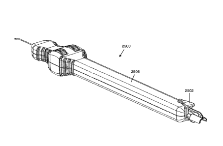
[0074] FIGS. 25A-25B depict perspective views of an exemplary delivery system
with a lock
removed (FIG. 25A) and inserted (25B) into the handle. FIGS. 25C-25D show
perspective and
cut-away views, respectively, of the lock rotated to allow loading of the
reservoir.
[0075] FIGS. 26A-26B show perspective views of an exemplary tray for a
delivery system
with a delivery system (FIG. 26A) and with a delivery system and loading tool
(FIG. 26B). FIG.
26C shows an exploded view of an exemplary packaged kit.
[0076] FIGS. 27A-27B show exemplary kits comprising multiple delivery systems.
[0077] FIG. 28A is a flow-chart illustrating an exemplary method for
delivering a fluid to
Schlemm's canal. FIGS. 28B-D depict delivery of fluid as a slidable elongate
member is
retracted as part of the method of FIG. 28A.
[0078] FIG. 29A is a flow-chart illustrating an exemplary method for
disrupting trabecular
meshwork. FIGS. 29B-D depict disruption of the trabecular meshwork as part of
the method of
FIG. 29A.
21
CA 02980713 2017-09-22
WO 2016/159999 PCT/US2015/023720
DETAILED DESCRIPTION
[0079] Described here are systems and methods for accessing Schlemm's canal
and for
delivering an ocular device, tool, and/or fluid composition therein to reduce
intraocular pressure
and thereby treat conditions of the eye. The fluids and certain components of
the system, e.g.,
the slidable elongate member, may be used to provide a force for disrupting
trabeculocanalicular
tissues, which include the trabecular meshwork, juxtacanalicular tissue,
Schlemm's canal, and
the collector channels. As used herein, the term "disrupting" refers to the
delivery of a volume of
fluid or a system component that alters the tissue in a manner that improves
flow through the
trabeculocanalicular outflow pathway. Examples of tissue disruption include,
but are not limited
to, dilation of Schlemm's canal, dilation of collector channels, increasing
the porosity of the
trabecular meshwork, stretching the trabecular meshwork, forming microtears or
perforations in
juxtacanalicular tissue, removing septae from Schlemm's canal, cutting,
tearing, or removal of
trabeculocanalicular tissues, or a combination thereof.
[0080] To better understand the systems and methods described here, it may be
useful to
explain some of the basic eye anatomy. FIG. 1 is a stylized depiction of a
normal human eye.
The anterior chamber (100) is shown as bounded on its anterior surface by the
cornea (102). The
cornea (102) is connected on its periphery to the sclera (104), which is a
tough fibrous tissue
forming the protective white shell of the eye. Trabecular meshwork (106) is
located on the outer
periphery of the anterior chamber (100). The trabecular meshwork (106) extends
360 degrees
circumferentially around the anterior chamber (100). Located on the outer
peripheral surface of
the trabecular meshwork (106) is Schlemm's canal (108). Schlemm's canal (108)
extends 360
degrees circumferentially around the meshwork (106). At the apex formed
between the iris
(110), meshwork (106), and sclera (104), is the anterior chamber angle (112).
[0081] The systems are generally configured for single-handed manipulation and
for control
by a single operator, and include one or more features useful for easily
accessing Schlemm's
canal with minimal trauma. Once access to the canal has been obtained, the
system may deliver
an ocular device, a tool, and/or a fluid composition. In some variations, the
system advances a
tool that disrupts Schlemm's canal and surrounding tissues without delivery of
an ocular device
or a fluid composition. For example, the tool may be an elongate member,
slidable within, and
extendable from, the cannula used to access the canal, having an outer
diameter sized to disrupt
the canal and surrounding tissues. The body of the elongate member may be in
some instances
configured to cut or tear through the trabecular meshwork if the system is
removed from the eye
22
while the elongate member is within Schlemm's canal, and/or the distal end of
the elongate
member may be provided with a disruptive component to aid in the disruption of
trabeculocanalicular tissues.
[0082] When a device is implanted into the canal, it will generally be
configured to maintain
the patency of Schlemm' s canal without substantially interfering with
transmural fluid flow
across the canal. This may restore, enable, or enhance normal physiologic
efflux of aqueous
humor through the trabeculocanalicular tissues. Ocular implants such as those
disclosed in U.S.
Patent Serial No. 7,909,789, and such as those disclosed in U.S. Patent Serial
No. 8,529,622,
In some
variations, the implants in U.S. Patent Serial No. 7,909,789 and U.S. Patent
Serial No. 8,529,622
include a support having a least one fenestration that completely traverses a
central core of
Schlemm's canal without substantially interfering with transmural fluid flow
or longitudinal
fluid flow across or along the canal. The ocular device may also disrupt the
juxtacanalicular
trabecular meshwork or adjacent inner wall of Schlemm's canal. The ocular
devices may also be
coated with a drug useful for treating ocular hypertension, glaucoma, or pre-
glaucoma, infection,
or scarring, neovascularizati on, fibrosis, or inflammation postoperatively.
The ocular device may
also be formed to be solid, semi-solid, or bioabsorbable.
[0083] The systems may also be used to deliver a fluid composition, e.g.,
saline or a
viscoelastic fluid. The saline may be used for irrigation. The viscoelastic
fluid may be employed
in ab-interno versions of viscocanalostomy or canaloplasty procedures to
disrupt the canal and
surrounding tissues.
I. SYSTEMS/DEVICES
[0084] The systems described herein may be single-handed, single-operator
controlled devices
that generally include a universal handle having a grip portion and a housing
that has an interior
and a distal end. A cannula is typically coupled to and extends from the
housing distal end. The
cannula may include a proximal end and a distal curved portion, where the
distal curved portion
has a proximal end and a distal end, and a radius of curvature defined between
the ends. In other
variations, the cannula may be straight and may not comprise a distal curved
portion. The
cannula may also be configured to include a body; a distal tip having a bevel;
and a lumen
extending from the proximal end through the distal tip. The bevel may directly
engage the distal
end of the curved portion of the cannula (i.e., the bevel may directly engage
the radius of
23
Date Recue/Date Received 2021-08-06
CA 02980713 2017-09-22
WO 2016/159999 PCT/US2015/023720
curvature). The systems may also generally include a drive assembly partially
contained within
the housing comprising gears that translate rotational movement to linear
movement. When an
ocular device is to be implanted into Schlemm's canal, the systems may further
include a
slidable positioning element having a proximal end and a distal end that is
coaxially disposed
within the cannula lumen. The system may also be configured to include a
slidable elongate
member comprising a lumen that is coaxially disposed within the cannula lumen.
When a fluid
composition is to be delivered into Schlemm's canal, the system may also be
configured to
include a fluid assembly in the handle. Fluid compositions such as saline,
viscoelastic fluids,
including viscoelastic solutions, air, and gas may be delivered using the
system. Suitable
markings, colorings, or indicators may be included on any portion of the
system to help identify
the location or position of the distal end of the cannula, the positioning
element, the engagement
mechanism, the ocular device, or the slidable elongate member. In some
instances, the systems
described herein may be used to perform ab-interno trabeculotomy, ab-interno
transluminal
trabeculotomy, clear corneal trabeculotomy, clear corneal transluminal
trabeculotomy, ab-
interno canaloplasty, and/or clear corneal canaloplasty, and may be used to
deliver a fluid
composition into the anterior or posterior segment of the eye.
[0085] An exemplary ocular delivery system is depicted in FIG. 2. In the
figure, delivery
system (200) includes a universal handle (202) having a grip portion (204) and
a housing (206).
The housing has a proximal end (208) and a distal end (210). A cannula (212)
is coupled to and
extends from the housing distal end (210). A drive assembly (214) is
substantially contained
within the housing (206) that actuates movement of a positioning element (not
shown). Port
(216) is provided on the distal end of the housing (210) for removable
connection to a source of
irrigation fluid.
[0086] The delivery systems described herein may in some variations be fully
disposable. In
other variations, a portion of the delivery system may be reusable (e.g., non-
patient contact
materials, such as the handle), while a portion of the delivery system may be
disposable (e.g.,
patient-contact materials, such as the cannula and elongate member). In yet
other variations, the
delivery systems described herein may be fully reusable.
Universal Handle
[0087] The ocular delivery systems described herein may include a universal
handle capable
of single-handed use. For example, the handle may be configured to be capable
for use with the
24
CA 02980713 2017-09-22
WO 2016/159999 PCT/US2015/023720
left or right hand, for use on the left or right eye, or in the clockwise or
counterclockwise
direction. That is, the handle may be configured such that the ability to use
the delivery system is
independent of which hand is used, which eye a procedure is performed on, or
which direction
around the canal an ocular device, tool, or fluid composition is delivered.
For example, the
delivery system may be used to deliver an ocular device, elongate member,
and/or fluid
composition in a clockwise direction in an eye, and then with a simple flip of
the handle (or by
rotating the cannula itself 180 degrees in another variation) to a second
orientation, may be used
to deliver an ocular device, elongate member, and/or fluid composition in the
counterclockwise
direction. However, it should be appreciated that in other variations, the
delivery systems
described herein may be configured to be used in a particular configuration
(e.g., with a single
side up, only in a clockwise direction, only in a counterclockwise direction,
etc.). The handle
generally includes a grip portion and a housing. The grip portion may be
raised, depressed, or
grooved in certain areas, or textured to improve hold of the handle by the
user or to improve
comfort of the user. The housing may include an interior portion and a distal
end. The interior
portion of the housing may contain a drive assembly and a positioning element
(both further
described below). In some variations, the distal end of the housing includes a
fluid port that can
provide fluids for irrigation of the operative field or to purge air from the
system.
[0088] The universal handle may be made from any suitable material, including
without
limitation, fluoropolymers; thermoplastics such as polyetheretherketone,
polyethylene,
polyethylene terephthalate, polyurethane, nylon, and the like; and silicone.
In some variations,
the housing or portions thereof may be made from transparent materials.
Materials with suitable
transparency are typically polymers such as acrylic copolymers, acrylonitrile
butadiene styrene
(ABS), polycarbonate, polystyrene, polyvinyl chloride (PVC), polyethylene
terephthal ate glycol
(PETG), and styrene acrylonitrile (SAN). Acrylic copolymers that may be
particular useful
include, but are not limited to, polymethyl methacrylate (PMMA) copolymer and
styrene methyl
methacrylate (SMMA) copolymer (e.g., Zylar 631 acrylic copolymer). In
variations in which
the universal handle is reusable, the handle may be made from a material that
can be sterilized
(e.g., via autoclaving), such as a heat-resistant metal (e.g., stainless
steel, aluminum, titanium).
[0089] The length of the universal handle may generally be between about 1
inch (2.5 cm) to
about 20 inches (50.8 cm). In some variations, the length of the universal
handle may be
between about 4 inches (10.2 cm) and 10 inches (25.4 cm). In some variations,
the length of the
universal handle is about 7 inches (17.8 cm).
CA 02980713 2017-09-22
WO 2016/159999 PCT/US2015/023720
Cannula
[0090] The cannula of the ocular delivery system is typically coupled to and
extends from the
housing distal end, and is generally configured to provide easy and minimally
traumatic access
to Schlemm's canal using a minimally invasive ab-intemo approach. The cannula
may be
fixedly attached to the distal end of the housing, or in other variations it
may be rotatably
attached to the distal end of the housing. In variations of the delivery
systems where the handle
is reusable and the cannula is disposable, the cannula may be removably
attached to the distal
end of the housing. Some variations of the cannula may include a proximal end
and a distal
curved portion, where the distal curved portion has a proximal end and a
distal end, and a radius
of curvature defined between the ends. However, it should be appreciated that
in other variations
the cannula may be straight and may not comprise a distal curved portion. The
cannula may also
be configured to include a body; a distal tip having a bevel and a sharpened
piercing tip; and a
lumen extending from the proximal end through the distal tip. When the cannula
comprises a
distal curved portion, the bevel may directly engage the distal end of the
curved portion of the
cannula (i.e., the bevel may directly engage the radius of curvature). In some
variations, the
sharpened piercing tip may comprise one or more angled surfaces, as is
described in more detail
below.
[0091] The cannula may be made from any suitable material with sufficient
stiffness to allow
it to be advanced through the eye wall and anterior chamber. For example, the
cannula may be
formed of a metal such as stainless steel, nickel, titanium, aluminum, or
alloys thereof (e.g.,
Nitinol metal alloy), a polymer, or a composite. Exemplary polymers include
without limitation,
polycarbonate, polyetheretherketone (PEEK), polyethylene, polypropylene,
polyimide,
polyamide, polysulfone, polyether block amide (PEBAX), and fluoropolymers. In
some
instances, it may be advantageous to coat the cannula with a lubricious
polymer to reduce
friction between the ocular tissue and the cannula during the procedure.
Lubricious polymers are
well known in the art, and include, without limitation, polyvinyl alcohol,
polyethylene glycol,
polyvinyl pyrrolidone, fluorinated polymers (including polytetrafluoroethylene
(PTFE or
Teflon )), and polyethylene oxide. In variations in which the cannula is
reusable, the cannula
may be made from a material that can be sterilized (e.g., via autoclaving),
such as a heat-
resistant metal (e.g., stainless steel, aluminum, titanium).
[0092] The cannula generally has an outer diameter sized to gain access to the
lumen of
Schlemm's canal while minimally obstructing the surgeon's view. Accordingly,
the outer
26
CA 02980713 2017-09-22
WO 2016/159999 PCT/US2015/023720
diameter may range from about 50 microns to about 1000 microns. In some
variations, the outer
diameter may range from about 150 microns to about 800 microns. The cannula
also has an
inner diameter, which may range from about 50 microns to about 400 microns.
The cannula may
also be formed to have any suitable cross-sectional profile, e.g., circular,
elliptical, triangular,
square, rectangular, etc.
[0093] The cannula may be configured to include multiple portions or parts. A
cannula having
a body, a distal curved portion having a proximal end and a distal end, a
radius of curvature
defined between the ends, and a bevel at the distal tip of the cannula that
directly engages the
distal end of the curved portion of the cannula may be particularly useful for
accessing the
lumen of Schlemm's canal. Here the body (straight portion of the cannula) may
have a length
ranging from about 5 mm to about 50 mm, about 10 mm to about 30 mm, or from
about 14 mm
to about 20 mm. In some variations, the body may have a length of about 18 mm.
The distal
curved portion of the cannula may be uniform in cross-sectional shape or it
may taper closer to
the distal end to facilitate entry into Schlemm's canal. The radius of
curvature of the distal
curved portion may be adapted to facilitate tangential entry, as well as
precise and minimally
traumatic entry into Schlemm's canal, and may range from about 1 mm to about
10 mm or from
about 2 mm to about 5 mm. In one variation, the radius of curvature is about
2.5 mm. The
cannula may also have an angular span suitable for facilitating entry into
Schlemm's canal, and
may range from about 70 degrees to about 170 degrees, or about 100 degrees to
about 150
degrees. In one variation, the angular span is about 120 degrees.
[0094] The size, shape, geometry, and the like, of the bevel at the distal end
of the curved
portion of the cannula may be beneficial in allowing easy and minimally
traumatic access to
Schlemm's canal. In this respect, and as described in further detail below,
having a bevel that
directly engages the radius of curvature of the distal end of the cannula may
be particularly
useful.
[0095] In other variations, the cannula may include a short straight segment
coupled to the
distal end of the distal curved portion of the cannula (e.g., at the end of
the radius of curvature).
Here the bevel engages the straight segment and not the radius of curvature.
The length of the
straight segment may range from about 0.5 mm to about 5 mm. In some
variations, the length of
the straight segment ranges from about 0.5 mm to about 3 mm, or from about 0.5
mm to about 1
mm. The length of the straight segment may also be less than about 0.5 mm,
e.g., it may be
about 0.1 mm, about 0.2 mm, about 0.3 mm, or about 0.4 mm. In variations where
the bevel
27
CA 02980713 2017-09-22
WO 2016/159999 PCT/US2015/023720
directly engages the distal end of the curved portion of the cannula (i.e.,
the bevel directly
engages the radius of curvature), the cannula lacks a straight segment (length
of the straight
segment is zero).
[0096] It may also be useful to have a bevel that is sharp and short to
minimize the distance
that any ocular device will have to travel when being implanted into the
canal. Exemplary bevel
angles may range from about 10 degrees to about 90 degrees. In some instances,
the bevel angle
may range from about 10 degrees to about 50 degrees. In one variation, the
bevel angle is about
35 degrees, while in another variation the bevel is about 25 degrees. The
bevel may also be
oriented in any suitable direction. For example, the bevel may be oriented so
that it opens up
towards the surgeon, or it may be reversed to open away from the surgeon or in
any plane in
between.
[0097] As is described in more detail below, in yet some variations, the
cannula is configured
to include one section that is sharp, and another section that is blunt (e.g.,
deburred). The dual
surface configuration of such a cannula may be advantageous, since it may
provide easier canal
access by piercing the meshwork while also providing a gentle, dispersed force
on the elongate
member during elongate member retraction into the cannula to avoid cutting or
breaking the
elongate member due to retraction force. For example, as shown in FIG. 15, the
distal end of
cannula (1500) may have a sharpened piercing tip (1502) and a smooth edge
(1504) that define
portions of opening (1506), through which a slidable elongate member (not
shown) may be
advanced and retracted. As is described in more detail with respect to FIGS.
19, 20A-20B, and
21, the sharp tip (1502) may be formed by compounding multiple bevels, and the
smooth edge
(1504) may be created by smoothing or deburring inner and/or outer
circumferential edges of the
distal tip. Additionally, in some embodiments, the internal and/or external
surfaces of the
elongate member adjacent to the opening (1506) may also be smoothed. Methods
of making the
cannula are described in more detail below.
[0098] The cannula of an exemplary delivery system is shown in more detail in
FIG. 3. Here
the cannula (300) comprises a proximal end (302) a distal curved portion
(304), a body (314),
and a distal tip (306). The distal curved portion (304) has a proximal end
(308) and a distal end
(310), and a radius of curvature (R) that is defined between the ends (308,
310). The distal
curved portion (304) also has an inner radius (320) defined by the surface of
the cannula closest
to the center of the radius of curvature (R), and an outer radius (322)
defined by the surface of
cannula further away from the center. A bevel (312) at the distal tip (306)
directly engages the
28
CA 02980713 2017-09-22
WO 2016/159999 PCT/US2015/023720
distal end of the curved portion of the cannula (310). In other words, the
bevel (312) may be
contiguous with the distal end of the curved portion of the cannula (310). As
previously stated,
this configuration of the distal curved portion (304) and bevel (312) may be
beneficial or
advantageous for allowing easy, atraumatic, and controlled access into
Schlemm's canal. The
angle of the bevel may also be important. In general, a short bevel may be
beneficial. The bevel
(312) may comprise an angle (A) between about 5 degrees and about 85 degrees.
In some
variations, the angle (A) may be about 5, 10, 15, 20, 25, 30, 35, 40, 45, 50,
55, 60, 65, 70, 75, 80,
or 85 degrees. In some variations, the angle (A) may be between about 23
degrees and about 27
degrees. In the variation shown in FIG. 3, the bevel angle (A) is about 25
degrees.
[0099] FIG. 20 depicts a perspective view of a distal tip (2002) of a cannula
(2000)
comprising a bevel (2014). The distal tip (2002) may be cut or ground at an
angle to create the
bevel (2014). As shown, the beveled distal tip (2002) comprises a proximal end
(2008), a distal
end (2010), and an elongated opening (2012) having an elliptical, rather than
a circular, shape.
The distal tip (2002) may comprise an elliptical shaped lumen opening that is
angled such that
the top of the elliptical opening is closer to the proximal portion of the
cannula than the bottom
of the elliptical opening. Also shown in FIG. 20 are inner and outer
circumferential edges (2004,
2006).
[0100] FIGS. 21A and 21B depict perspective and front views, respectively, of
a variation of a
distal tip (2100) of a cannula comprising a bevel (2102) and a sharpened
piercing tip (2114). As
shown there, the distal tip (2100) also comprises a proximal end (2108), a
distal end (2110),
inner and outer circumferential edges (2104, 2106), and a lumen opening
(2112). The sharpened
piercing tip (2114) may comprise two angled surfaces (2116) that converge to
form a sharp
point. The angled surfaces (2116) may have any suitable angle that results in
a sharpened
piercing tip (2114). For example, in some instances, the angle surfaces (2116)
may have an
angle (B) relative to the longitudinal axis of the distal tip (2100) of about
20, 25, 30, 35, 40, 45,
or 50 degrees, between about 25 and about 50 degrees, or between about 37.5
and about 42.5
degrees. In some instances, the angle (B) may be about 40 degrees.
Accordingly, in some
variations, the angle between the two angled surfaces (2116) may be between
about 50 and
about 100 degrees, and in some instances, the angle between the two angled
surfaces (2116) may
be about 80 degrees. It should be appreciated that although the distal tip
(2100) is depicted with
two angled surfaces, a distal tip with a single angled surface may also be
used.
29
CA 02980713 2017-09-22
WO 2016/159999 PCT/US2015/023720
Elongate Member
[0101] The delivery systems described herein may comprise a slidable elongate
member
coaxially disposed within the cannula lumen. The elongate member employed with
the systems
described herein may be of various configurations, and may or may not comprise
a lumen. The
elongate member may or may not be configured to deliver a fluid composition.
[0102] The elongate member may be coaxially disposed and slidable within the
cannula lumen
of the delivery systems described here. When the elongate member is in a
retracted position
relative to the cannula, the distal end of the elongate member may be located
within (i.e.,
proximal to) the distal tip of the cannula. When the elongate member is in an
extended position
relative to the cannula, the distal end of the elongate member may be located
outside of (i.e.,
distal to) the distal tip of the cannula. The length of extension of the
elongate member beyond
the distal tip of the cannula may correspond to the distance around Schlemm's
canal that may be
traversed by the elongate member (e.g., in order to disrupt Schlemm's canal
and/or surrounding
trabeculocanalicular tissues, and/or to deliver a fluid composition). In
variations in which the
delivery system is configured to deliver a fluid composition, the length
traversed by the elongate
member may correspond to the length around Schlemm's canal to which the fluid
composition is
delivered. In variations in which the delivery system is configured to tear or
cut the trabecular
meshwork, the length traversed by the elongate member may correspond to the
length of
trabecular meshwork that is cut or torn. In some variations, this length may
be between about 1
mm and about 50 mm. In some of these variations, the length may be between
about 10 mm and
about 40 mm, between about 15 mm and about 25 mm, between about 16 mm and
about 20 mm,
between about 18 mm and about 20 mm, between about 19 mm and about 20 mm,
between
about 18 mm and about 22 mm, about 20 mm, between about 30 mm and about 50 mm,
between
about 35 mm and about 45 mm, between about 38 mm and about 40 mm, between
about 39 min
and about 40 mm, or about 40 mm. The elongate member may be moved between
extended and
retracted positions using a drive assembly of the delivery system, described
in more detail
below.
[0103] The elongate member may be sized so that it can be advanced through the
cannula and
into a portion of Schlemm's canal (e.g., 0 to 360 degrees of the canal) to
disrupt
trabeculocanalicular tissues, stent, and/or apply tension to the canal, and/or
to deliver a fluid
composition. The elongate member may be made from any suitable material that
imparts the
desired flexibility and pushability for introduction through the eye wall,
accessing Schlemm's
CA 02980713 2017-09-22
WO 2016/159999 PCT/US2015/023720
canal, and/or navigation through other ocular tissue structures. For example,
the elongate
member may comprise a polymer (e.g., nylon, polypropylene); a polymer
reinforced with metal
wire, braid or coil; composites of polymers and metal; or metals such as
stainless steel, titanium,
shape-memory alloy (e.g., Nitinol), or alloys thereof. In variations in which
the elongate member
is reusable, the elongate member may be made from a material that can be
sterilized (e.g., via
autoclaving), such as a heat resistant metal (e.g., stainless steel, aluminum,
titanium). The
elongate member may be straight with enough flexibility and pushability to
navigate the ring-
shaped Schlemm's canal or may be pre-shaped to about a 2-10 mm radius of
curvature or about
a 6 mm radius of curvature (i.e., the approximate radius of curvature of
Schlemm's canal in an
adult human) to more easily circumnavigate Schlemm's canal, partially or in
its entirety. In
some variations, the elongate member may be configured to be advanced over or
along a
guidewire.
[0104] It may in some variations be desirable for the elongate member to have
one or more
features to improve visualization of the elongate member. For example, the
elongate member
may be colored (e.g., red, orange, yellow, green, blue, purple, etc.).
Additionally or alternatively,
visualization may be improved using an illuminated beacon, a fiber optic, side
illuminating fiber
optic, luminescence, fluorescence, or the like. For example, a fiber optic may
travel along the
body of the elongate member to deliver light to the distal tip of the elongate
member, which may
improve visualization of the distal tip of the elongate member as it is
advanced or retracted about
Schlemm's canal.
[0105] In some variations, the elongate member may be sized to have an outer
diameter
sufficient to disrupt Schlemm's canal and surrounding trabeculocanalicular
tissues. The outer
diameter may range from about 25 microns to about 1000 microns, from about 25
microns to
about 500 microns, from about 50 microns to about 500 microns, from about 150
microns to
about 500 microns, from about 200 microns to about 500 microns, from about 300
microns to
about 500 microns, from about 200 microns to about 250 microns, from about 150
microns to
about 200 microns, or from about 180 microns to about 300 microns. In some
instances it may
be beneficial for the elongate member to have an outer diameter of about 240
microns.
[0106] In some variations, the distal end of the elongate member may be
configured as a blunt
bevel, an atraumatic tip, an enlarged atraumatic tip, or the like, to help the
elongate member be
advanced through Schlemm's canal. In some of these variations, the distal end
may comprise a
blunt parasol-shaped atraumatic tip. In other variations, a distal portion of
the elongate member
31
CA 02980713 2017-09-22
WO 2016/159999 PCT/US2015/023720
may optionally include a disruptive component, e.g., a notch, hook, barb, a
rough surface, or
combination thereof, to disrupt the juxtatrabecular portion of Schlemm's canal
or juxtatrabecular
meshwork. One or more projections emanating from the elongate member may
further disrupt
the juxtatrabecular portion of Schlemm's canal or juxtatrabecular meshwork and
thus increase
permeability of aqueous humor through the trabecular meshwork into Schlemm's
canal. In some
instances, the elongate member may also deliver energy to the
trabeculocanalicular tissues (e.g.,
ultrasonic energy, radiofrequency energy (e.g., for electrocautery,
electroablati on),
electromagnetic radiation, light energy (e.g., via a fiber optic)).
[0107] In some instances, the elongate member may comprise a filament (e.g., a
filament
comprising nylon, polypropylene, metal, or the like). For example, the
elongate member may
comprise a nylon monofilament. An exemplary range of filament size may range
from about 50
microns to about 300 microns. The filament may be configured to be advanced
through all or a
portion of Schlemm's canal. In some instances the body of the filament may be
configured to cut
or tear through the trabecular meshwork when the cannula is removed from the
eye. In other
instances the filament may be configured to disrupt trabeculocanalicular
tissue upon
advancement into or retraction from Schlemm's canal. In yet other instances
the filament may be
configured to be left within the canal to continuously deliver tension on the
meshwork and
maintain patency of the canal.
[0108] In some variations the elongate member may comprise a lumen. For
example, in one
variation the elongate member may comprise a microcatheter (e.g., a nylon
microcatheter). In
some of the instances in which the elongate member comprises a lumen, the
elongate member
may be configured to deliver a fluid composition. The fluid composition may
travel through a
lumen of the elongate member and may be delivered through an opening of the
lumen. For
example, as shown in FIG. 12, the elongate member (1200) may be a flexible
tube having a
lumen in fluid communication with an opening at the distal tip (1202). In some
variations, the
distal end of the elongate member may be configured or modified to aid
delivery of the fluid
composition into Schlemm's canal. For example, the distal end of the elongate
member may
comprise a cut out configured as a half tube. Additionally or alternatively to
an opening at the
distal tip, the elongate member may optionally comprise a plurality of
openings through its wall
that are spaced along the axial length of the elongate member. In this
variation, the fluid
composition may be delivered from the reservoir through the openings in the
elongate member
and into Schlemm's canal. This lateral ejection of fluid (e.g., a viscoelastic
fluid) may in some
32
CA 02980713 2017-09-22
WO 2016/159999 PCT/US2015/023720
instances enhance disruption of outflow tissues and enhance permeability to
aqueous humor. It is
understood that the openings can be of any suitable number, size and shape,
and spaced along
the axial length of the elongate member (including the distal tip) in any
suitable manner.
Drive Assembly
[0109] The delivery systems generally include a drive assembly. The drive
assembly of the
delivery system is generally configured to move an ocular device, elongate
member, and/or fluid
composition into Schlemm's canal. The drive assembly may also in some
variations be
configured to position an ocular device within the canal, including advancing
the device into the
canal and retracting the device from the canal. The drive assembly may be at
least partially
contained within the housing and may include any suitable component or
combination of
components capable of providing the handle with universal functionality.
[0110] The drive assembly may convert an external input (e.g., motion of a
user's thumb or
finger) into motion of one or more components of the delivery system. More
specifically, the
drive assembly may cause a slidable elongate member to be extended distally
out of a cannula,
and/or it may cause a slidable elongate member to be retracted proximally into
a cannula. The
drive assembly may also optionally cause a fluid composition to be delivered
from a reservoir
through the elongate member and/or cannula.
[0111] Two or more of these effects (i.e., extension of the slidable elongate
member, retraction
of the slidable elongate member, and/or delivery of a fluid composition) may
be actuated using
the same actuation mechanism. This may allow for single-handed use of the
delivery system. For
example, if the actuation mechanism comprises a rotatable element (such as one
or more wheels,
as in variations described herein), rotating the rotatable element in a first
direction may cause
extension of the slidable elongate member, and rotating the rotatable element
in a second
direction may cause retraction of the slidable elongate member. If the
delivery system is
configured to deliver a fluid composition, rotating the rotatable element
(e.g., in the second
direction) may also cause delivery of a fluid composition. The delivery of the
fluid composition
may be simultaneous with movement (e.g., retraction) of the slidable elongate
member. In some
of these instances, the fluid composition may be delivered to the portion of
Schlemm's canal in
which the slidable elongate member is advanced; that is, the fluid composition
may be delivered
to the same angle and length of Schlemm's canal as the extension of the
elongate member. When
the fluid composition is simultaneous with retraction of the elongate member,
fluid composition
33
CA 02980713 2017-09-22
WO 2016/159999 PCT/US2015/023720
may take the place of the slidable elongate member as it is retracted and may
dilate Schlemm's
canal and/or the collector channels at that location in Schlemm's canal.
Furthermore, the
quantity of fluid delivered may be tied to the amount of movement of the
elongate member; that
is, a certain predetermined, fixed volume of fluid composition may be
delivered via the elongate
member (e.g., delivered out of the distal end of the elongate member) for a
fixed amount of
movement of the elongate member (e.g., a retraction distance) and for a fixed
amount of rotation
of the rotatable element.
[0112] In some variations, the drive mechanism may be configured to allow the
delivery
system to be used only once ¨ that is, the drive mechanism may prevent, for
example, re-
extension of the slidable elongate member after a predetermined amount of
extension and/or
retraction. Exemplary mechanisms by which external input may be converted into
motion of one
or more components of the delivery system are described in more detail below.
[0113] In some variations, the drive assembly includes components that
translate rotational
motion into linear motion. For example, the drive assembly may include a
linear gear and a pair
of pinion gear mechanisms. The linear gear may have teeth on its surface that
engage
corresponding teeth on the pinion gears. Each of the pinion gear mechanisms
may also be
coupled to a rotatable component (e.g., a wheel). Such coupling may be
accomplished with a pin
that can be threaded through a central opening in the rotatable component and
pinion gear, and a
nut that secures the rotatable component and pinion gear in a manner so that
rotation of the
rotatable component also rotates the pinion gear and vice versa. The wheels
may be attached to
the pinion gear by one of the following methods, for example: 1) the wheels
and pinion gears are
molded as one part using plastic injection molding technology; 2) the wheels
slide onto the
pinion gear and are secured with adhesive; or 3) the wheels slide on the
pinion gear and are
mechanically fixed with a fastener or a "press fit," where the wheels are
forced onto the pinion
gear and friction holds them secure. In all of the mentioned situations, the
wheels and pinion
gears may rotate coaxially, in the same direction, and at the same angular
rate. In some
variations, each of the pinion gear mechanisms is coupled to at least two
rotatable components.
In other variations, the drive assembly may be configured to include a single
rotatable
component, a plurality of rotatable components, or no rotatable component. The
wheel may have
markings or colorings to indicate degree of advancement or direction of
advancement.
[0114] One variation of the drive assembly useful to include in the universal
handle comprises
a linear gear, a pair of pinion gear mechanisms, and two rotatable components
coupled to each
34
CA 02980713 2017-09-22
WO 2016/159999 PCT/US2015/023720
pinion gear (for a total of four rotatable components). In other variations,
the drive assembly
includes a linear gear and a single pinion gear mechanism with two associated
wheels. In
variations with a pair of pinion gear mechanisms, the pinion gear mechanisms
and associated
wheels would be disposed on either side of the linear gear. The pinion gears
and linear gear
would contact each other, i.e., the teeth of the pinion gears would directly
engage corresponding
teeth on the linear gear, and the wheels on one side of the linear gear would
contact the wheels
on the opposite side of the linear gear. At least a portion of the wheels on
each side of the linear
gear would extend outside of the housing. In this variation, the drive
assembly can be
manipulated with one hand when in a first configuration, and then manipulated
with the same or
the other hand when flipped over to a second configuration. A drive assembly
having such
flexible capability can be easily used by a surgeon who is right hand dominant
or left hand
dominant, and may also be used in a procedure in which the handle is flipped
during a procedure
such that the cannula is facing a first direction in a first portion of the
procedure, and facing a
second direction in a second portion of the procedure. In a further variation,
the drive assembly
may include one rotatable component on one side of the handle and the
"universal" feature of
the handle provided by a cannula that itself can rotate instead of flipping
the handle.
[0115] In the variation shown in FIG. 4A, delivery system (400) includes a
drive assembly
(402) having a linear gear (e.g., a rack) (404) and a pair of pinion gear
mechanisms (406). Both
the linear gear and the pinion gear mechanisms have teeth that engage each
other to translate
rotational motion (of the pinion gear mechanisms (406)) to linear motion (of
the linear gear
(404)). Each of the pinion gear mechanisms (406) is coupled to two rotatable
components,
shown in the figure as wheels (408), for a total of four rotatable components.
The wheels (408)
extend outside of the housing (414) of the delivery system (400), and as such,
may be rotated by
one or more of the surgeon's fingers to correspondingly rotate the pinion gear
mechanism (406)
and thus advance or retract the linear gear (404). The wheels (408) are
coaxial with the pinion
gear mechanism (406) and rotate in unison with the pinion gear mechanism.
Movement of the
linear gear (404) advances or retracts a positioning element (410) that is
coaxially disposed and
slidable within cannula (412). FIG. 4B shows the system of FIG. 4A in a
second, flipped
orientation. In the orientation of FIG. 4A, the cannula is oriented with the
curvature facing
clockwise, while in the orientation of FIG. 4B, the cannula is oriented with
the curvature facing
counterclockwise. The extension of wheels (408) outside of the housing (414)
on either side may
allow the delivery system (400) to be used in either orientation, with either
hand, and on either
of the patient's eyes. That is, the orientation of FIG. 4B can be used with
the opposite hand or
CA 02980713 2017-09-22
WO 2016/159999 PCT/US2015/023720
the same hand as the orientation of FIG. 4A, but when a different direction of
cannulation is
desired (e.g., clockwise cannulation if counterclockwise cannulation was
performed with the
system in FIG. 4A).
[0116] Another variation of a drive assembly is shown in two different
perspective views in
FIGS. 22A-22B. As depicted there, a drive assembly (2202) may comprise a
linear gear (e.g., a
rack) (2204) and a pair of pinion gear mechanisms (2206). Both the linear gear
and the pinion
gear mechanisms have teeth that engage each other to translate rotational
motion (of the pinion
gear mechanisms) to linear motion (of the linear gear). More specifically, the
linear gear (2204)
may comprise teeth on both a first side (2220) and a second side (2222), where
the teeth on the
first side engage the first pinion gear mechanism, and the teeth on the second
side engage the
second pinion gear mechanism. Each of the pinion gear mechanisms (2206) is
coupled to two
rotatable components, shown in the figure as wheels (2208), for a total of
four rotatable
components. The wheels (2208) are coaxial with the pinion gear mechanisms
(2206) and rotate
in unison with the pinion gear mechanisms. The drive assembly may comprise one
or more
features to stabilize the pinion gear mechanisms or otherwise keep them in
place. For example,
the drive assembly (2202) may comprise wheel spacers (2216) configured to sit
between axles
(2218) of the pinion gear mechanisms. Rotation of one or more wheels (2208)
may cause
translation of the linear gear (2204).
[0117] As shown in FIGS. 22C and 23A-23B, the wheels (2208) may extend out of
the
housing (2334) of the delivery system, such that the wheels may be rotated by
a surgeon to
correspondingly rotate the pinion gear mechanisms (2206) and thus advance or
retract the linear
gear (2204). The cannula (2344) may be slidable within the linear gear (2204),
such that the
cannula and wheels are fixed relative to each other and relative to the
housing (2334), while the
linear gear (2204) translates relative to the housing. Because linear motion
of the linear gear
(2204) may be generated by rotational motion of either of the two pinion gear
mechanisms
(2206), which may in turn be generated by rotating any of the wheels (2208)
extending from the
housing (2334), the delivery system (2300) may be easily operated using a
single hand with
either the first side or the second side facing upwards, and thus with the
cannula (2344) facing a
first direction or a second direction.
[0118] In other variations, one or both pinion gear mechanisms may be able to
be disengaged
from the linear gear by biasing their position off axis from the linear gear.
This action de-couples
the pinion gear teeth to the linear gear teeth to prevent linear gear
movement. The pinion gear
36
CA 02980713 2017-09-22
WO 2016/159999 PCT/US2015/023720
mechanism may also be able to be locked to prevent rotation by engaging an
intersecting pin or
feature that prevents wheel rotation.
[0119] Further variations of the drive assembly may not employ translation of
rotational
motion to linear motion. For example, a slide (e.g., a finger slide) on the
handle that is fixed or
detachably coupled to a gear within the housing of the handle (e.g., a linear
gear as previously
described). Here the drive assembly may be configured so that advancement or
retraction of the
slide causes advancement or retraction of an ocular device and/or elongate
member, and/or
delivery of a fluid composition into Schlemm's canal. In yet further
variations, a button that can
be pressed or squeezed could be employed instead of a slide, or a foot pedal
could be employed
to deliver an ocular device, tool, and/or fluid composition.
Extending and Retracting the Elongate Member
[0120] In some variations, a proximal end of the elongate member may be fixed
relative to a
portion of a drive assembly (e.g., the linear gear (2204)), while the distal
end may be slidably
and coaxially disposed within the cannula lumen. When the elongate member does
not comprise
a lumen (e.g., is a filament), the elongate member may in some instances be
attached to the drive
assembly via crimping. When the elongate member comprises a lumen, the
elongate member
may in some instances be bonded to the drive assembly (e.g., via an adhesive)
in order to leave
the lumen of the elongate member unobstructed. The cannula, in turn, may be
fixedly attached to
the housing. In variations of the delivery systems in which the handle is
reusable and the cannula
and elongate member are disposable, a disposable assembly comprising the
elongate member
pre-loaded within the cannula may be attached to the reusable handle via any
suitable
mechanism, such as a threaded fastener or snap-in feature.
[0121] When the portion of the drive assembly is moved proximally or distally
within the
housing, this may cause corresponding movement of the elongate member relative
to the
cannula. That is, movement of the portion of the drive assembly toward the
cannula (i.e., toward
the distal end of the housing) may cause the elongate member to move from a
retracted position
to an extended position, and movement of the portion of the drive assembly
away from the
cannula (e.g., toward the proximal end of the housing) may cause the elongate
member to move
from an extended position to a retracted position. An example of an elongate
member in an
extended position is shown in FIGS. 22C-22D. As shown in the view in FIG. 22D
with the top
37
CA 02980713 2017-09-22
WO 2016/159999 PCT/US2015/023720
portion of the housing (2334) removed, the linear gear (2204) is in a distal
position. As such, the
elongate member (2346) is extended from cannula (2344).
Reservoir
[0122] The systems generally include a reservoir when a fluid composition is
to be delivered
into Schlemm's canal. The reservoir may contain various fluid compositions for
delivery.
Exemplary fluid compositions include saline and viscoelastic fluids. The
viscoelastic fluids may
comprise hyaluronic acid, chondroitin sulfate, cellulose, derivatives or
mixtures thereof, or
solutions thereof. In one variation, the viscoelastic fluid comprises sodium
hyaluronate. In
another variation, the viscoelastic composition may further include a drug.
For example, the
viscoelastic composition may include a drug suitable for treating glaucoma,
reducing or
lowering intraocular pressure, reducing inflammation, and/or preventing
infection, fibrosis,
scarring, clotting, thrombosis, bleeding, or neovascularization. Drugs such as
antimetabolites,
vasoconstrictors, anti-VEGF agents, steroids, heparin, anti-inflammatories,
nonsteroidal anti-
inflammatories (NSAIDs), other anticoagulants, fibrinolytic compounds,
biologic agents, and
gene therapy drugs may also be delivered in combination with the viscoelastic
composition.
Examples of glaucoma drugs include prostaglandins, beta blockers, miotics,
alpha adrenergic
agonists, or carbonic anhydrase inhibitors. Anti-inflammatory drugs such as
NSAIDs,
corticosteroids or other steroids may be used. For example, steroids such as
prednisolone,
prednisone, cortisone, cortisol, triamcinolone, or shorter acting steroids may
be employed.
Examples of antimetabolites include 5-fluoruracil or mitomycin C. Examples of
drugs or
antibodies that prevent neovascularization include bevacizumab, ranibizumab,
and others. In still
another variation, the system delivers the drug alone, without the
viscoelastic composition.
Saline solution may also be the fluid employed. In yet other variations, the
system may be
configured to deliver a gas, such as but not limited to air, an expansile gas
(e.g., SF6, C3F8).
[0123] In some variations, the reservoir may be at least partially defined by
a fluid assembly
and the housing, and the linear gear within the handle. The fluid assembly may
be made from
any suitable material previously mentioned for the cannula and the housing.
The volume of fluid
(in microliters) contained within the reservoir may range from about 2 IA to
about 1000 IA, or
from about 2 IA to about 500 IA In some variations, the reservoir volume may
range from about
50 pi to about 100 pl.
38
CA 02980713 2017-09-22
WO 2016/159999 PCT/US2015/023720
[0124] The fluid composition may be preloaded in the reservoir or loaded into
the reservoir
prior to use of the system, e.g., at the start of an ocular procedure, so that
the fluid can be
delivered by a single device and by a single user. Again, this is in contrast
to other systems that
use forceps or other advancement tools to advance a fluid delivery catheter
into Schlemm's
canal and/or devices containing viscoelastic fluid that are separate or
independent from a
delivery catheter or catheter advancement tool, and which require connection
to the delivery
catheter or catheter advancement tool during a procedure by, e.g., an
assistant, or by the hand of
the surgeon while the delivery catheter or catheter advancement tool is held
by another hand of
the surgeon. For example, a loading component may be provided on the fluid
assembly for
transfer of a fluid composition into the reservoir. The loading component may
have any suitable
configuration that provides reversible securement of a fluid container, e.g.,
a syringe, cartridge,
etc., to the system, and loading of a fluid composition into the reservoir.
The loading component
may be a luer fitting or include a one-way valve.
[0125] An exemplary delivery system comprising a reservoir is shown in FIGS.
23A-23F.
Shown there with (FIGS. 23A, 23B, and 23E), without (FIG. 23C and 23D), and
partially
without (FIG. 23F) a housing (2334), the delivery system (2300) comprises a
fluid assembly
(2316) comprising a reservoir (2302). In an exemplary method, a fluid
composition may be
loaded into the reservoir (2302) through a proximal opening (2328) via a
proximal seal (2318).
As best shown in FIG. 23E, the distal end of the reservoir (2302) may be
formed by a plunger
(2338) (explained in more detail below) and a distal seal (2354).
[0126] The proximal seal (2318) may be a mechanical seal located at the
proximal end of the
reservoir (2302) and comprising a ball bearing (2324) spring-biased against an
o-ring or gasket
(2330) to seal closed the reservoir. A loading tool (2326) (e.g., a nozzle)
may be used to open
the seal by pressing against the ball bearing (2324) to move it proximally
toward an open
position. While the proximal seal (2318) is open, the fluid composition may be
loaded into the
reservoir. After loading of the fluid composition, the loading tool (2326) may
be removed,
allowing the ball bearing (2324) to return to its closed position. The close-
up cross-sectional
view in FIG. 23E shows the proximal opening (2328) and ball bearing (2324). An
o-ring or
gasket (2330) sits between the ball bearing (2324) and a spring (2332), such
that force from the
spring presses the ball bearing into the gasket to form a seal between the
ball bearing and gasket
in the closed position. The loading tool (2326) is configured to fit into the
proximal opening
(2328) to press against the ball bearing (2324). The distally oriented force
against the ball
39
CA 02980713 2017-09-22
WO 2016/159999 PCT/US2015/023720
bearing (2324) moves it distally into the open position, compressing the
spring (2332), and
creating an opening between the ball bearing and the gasket (2330), through
which the fluid
composition may flow. When the loading tool (2326) is removed from the
proximal opening
(2328), the spring (2332) pushes the ball bearing (2324) proximally back into
the closed
position.
[0127] It should be appreciated that in other variations, the reservoir may
comprise other types
of seals allowing a fluid composition to be loaded into the reservoir. For
example, FIG. 24
depicts an alternative variation of a delivery system (2400), wherein the seal
comprises a
membrane (e.g., a silicone membrane). As shown, a fluid composition may be
loaded (after
movement of the optional lock (2404)) into the reservoir by puncturing the
membrane with a
needle (2402) (e.g., a 25 gauge needle). In yet other variations, the delivery
systems described
herein may be configured to receive a prefilled cartridge comprising a fluid
composition. For
example, the handle and fluid assembly may be configured such that a prefilled
cartridge can be
inserted into the fluid assembly.
[0128] In order to load the reservoir, it may be desirable to at least
temporarily secure the fluid
assembly in place in order to allow application of distal force to the seal.
In some variations, the
delivery system may comprise a lock configured to hold the fluid assembly in
place while a fluid
composition is injected into the reservoir. However, it should be appreciated
that in other
variations the delivery system may not comprise a lock. In variations having a
lock, it may be
desirable for the lock to be removable from the delivery system (or to
otherwise release the fluid
assembly) in order to allow the fluid assembly to translate relative to the
housing after the
reservoir is loaded. Translation of the fluid assembly may allow for extension
of the slidable
elongate member and/or injection of the fluid composition during the
procedure, as is described
in more detail herein.
[0129] In variations of the delivery systems having a lock, the lock may
optionally
additionally act as a cap to protect a distal opening to the reservoir. In
these variations, the lock
may comprise a first configuration in which it both holds the reservoir in
place and covers the
proximal opening to the reservoir, and a second configuration in which it
holds the reservoir in
place but allows the proximal opening to the reservoir to be accessed, such
that the reservoir can
be loaded with a fluid composition. In some instances, the lock may rotate
from the first position
to the second position.
CA 02980713 2017-09-22
WO 2016/159999 PCT/US2015/023720
[0130] FIGS. 25A-25D illustrate an exemplary lock (2502). As shown there, the
lock (2502)
may comprise a pin (2508) configured to fit into an opening (2504) in the
handle (2506) of the
delivery system (2500). In a first configuration, shown in FIG. 25B, the lock
(2502) may be
inserted into the opening (2504) in the handle and may cover the proximal
opening (2510). The
lock (2502) may comprise a protrusion (2518) configured to interface with the
proximal opening
(2510) to stabilize the lock in the first configuration. In a second
configuration, shown in FIGS.
25C-25D, the lock (2502) may remain inserted into the opening (2504) but may
pivot within the
opening to expose the proximal opening (2510) to allow loading of the
reservoir (2512). When
the pin (2508) is inserted into the opening (2504), the pin may restrict
movement of the reservoir
(2512) relative to the housing. This may allow a loading tool (2514) to apply
force through the
proximal opening (2510) to open the proximal seal (2516) of the reservoir
(2512), without the
reservoir sliding distally within the handle (2506). Restricting movement of
the reservoir (2512)
relative to the handle (2506) may prevent motion of the reservoir or other
internal components of
the delivery system before use (e.g., during transit). Once loading of the
reservoir (2512) is
complete, the lock (2502) may be removed from the opening (2504), as shown in
FIG. 25A, at
which point the reservoir (2512) may no longer be restricted by the lock from
moving relative to
the housing.
Delivering a Fluid Composition
[0131] The delivery systems described herein may be configured to deliver
fluid to Schlemm's
canal. The fluid may be delivered in a volume that provides sufficient force
to disrupt
Schlemm's canal and surrounding trabeculocanalicular tissues. Exemplary
disruptive volumes
may be about 1 I-11, about 2 tl, about 3 il, about 4 tl, about 5 pl, about 6
vtl, about 7 1, about 8
about 9 jil, about 10 1, about 11 Lill, about 12 Ill, about 13 jil, about 14
pi, about 15 pl, about
16 p.1, about 17 pl, about 18 pl, about 19 pl, or about 20 pl. In some
variations, the disruptive
volume fluid may range from about 1 pl to about 50 pl, or from about 20 pi to
about 50 pl.
[0132] As mentioned above, a elongate member may be coaxially disposed within
the cannula
lumen. In variations of the delivery system configured to deliver a fluid
composition, the
elongate member may comprise a lumen. The lumen of the elongate member may be
operatively
connected to a reservoir for delivery of a fluid composition into Schlemm's
canal. The elongate
member generally has a proximal end, a distal end, and a wall that defines the
lumen extending
therethrough. However, in some instances, the delivery system lacks an
elongate member
conduit, and the fluid composition is delivered solely through the cannula. In
other instances,
41
CA 02980713 2017-09-22
WO 2016/159999 PCT/US2015/023720
two elongate members may be employed that each simultaneously advance through
the canal in
both clockwise and counterclockwise directions to more rapidly cannulate
Schlemm's canal and
deliver therapy.
[0133] When the delivery systems are employed to deliver a fluid composition,
the fluid
composition may be preloaded in a reservoir of the system or loaded into the
reservoir prior to
use of the system. An exemplary delivery system for delivering a fluid
composition into
Schlemm's canal is shown in FIGS. IOA and 10B. Referring to FIG. 10A, delivery
system
(1000) includes a universal handle (1002) having a grip portion (1004) and a
housing (1006).
Housing (1006) has a proximal end (1008) and a distal end (1010). A cannula
(1012) is coupled
to and extends from the housing distal end (1010). A drive assembly (1014) is
substantially
contained within the housing (1006) that actuates movement of a slidable
elongate member (not
shown). The cannula (1012) and drive assembly (1014) have the same
configuration as that
shown and described in FIGS. 3 and 4A-4B for the system tailored for ocular
device
implantation, and thus are not described in detail here.
[0134] The delivery system (1000) also includes a fluid assembly (1016) (shown
in FIG. 10B)
within the handle (1002) having a loading component (1018) that is configured
to allow transfer
of a fluid composition from an external source into a reservoir defined by the
fluid assembly and
linear gear (1020). A slidable elongate member (1022) is coaxially disposed
within the cannula
lumen that is in fluid communication with the reservoir. As previously stated,
in a tool-based
system that does not deliver an implant or a fluid, the system may not include
a reservoir.
[0135] In an exemplary method, as illustrated by FIGS. 11A-11C, a fluid
composition may be
transferred into a reservoir (1102) of system (1100) via loading through
loading component
(1104). As shown in the figures, reservoir (1102) is defined by the fluid
assembly (1106) and the
linear gear (1108). Linear gear (1108) has a proximal end (1110) and a distal
end (1112), and a
lumen (1114) extending from the proximal end (1110) to the distal end (1112).
Lumen (1114) is
in fluid communication with the lumen (not shown) of the slidable elongate
member (1118). To
deploy the fluid composition out of the reservoir (1102), linear gear (1108)
is retracted in the
direction of the arrow (FIG. 11B) so that reservoir (1102) becomes
pressurized. Retraction can
be accomplished by rotation of pinion gear mechanisms (1120). Once a
sufficient amount of
pressure has been created in the reservoir (1102) the fluid composition
contained therein is
injected through linear gear lumen (1114) and the lumen of elongate member
(1118) into
Schlemm's canal.
42
CA 02980713 2017-09-22
WO 2016/159999 PCT/US2015/023720
[0136] Here, any fluid that is delivered flows through the distal end (1202)
to reach
Schlemm's canal. In other variations, the slidable elongate member (1300) may
be configured to
include a plurality of openings spaced along its axial length. The openings
may have any
suitable shape, e.g., slots (1302) (FIG. 13A) or circles (1304) (FIG. 13B).
Fluid compositions
delivered using the elongate members depicted in FIGS. 13A and FIG. 13B may
partially flow
out of the elongate member through the openings and partially out through the
distal end of the
elongate member. The distal end of the elongate member may also be configured
as a half tube
(1306) (FIG. 13C).
[0137] Some variations of the fluid assembly include a locking mechanism for
preventing
movement of the assembly within the handle, e.g., when the linear gear is
being advanced or
retracted. The locking mechanism may comprise a ratchet pawl, a combination of
ratchet pawls
or any other suitable mechanism that can be locked to prevent movement of the
fluid assembly,
and unlocked to allow movement of the fluid assembly.
[0138] Referring back to FIGS. 23A-23F, another exemplary delivery system
(2300) for
delivering a fluid composition into Schlemm's canal shown there may comprise a
housing
(2334) and a cannula (2344) extending from the distal end of the housing. A
drive assembly
(2202) (described above with respect to FIGS. 22A-22D) may be located within
the housing
(2334), as may be a fluid assembly (2316) (described in more detail above). As
described above,
the drive assembly (2202) may comprise a linear gear (2204) and a pair of
pinion gear
mechanisms (2206) coupled to wheels (2208). The delivery system (2300) may
comprise a
slidable elongate member (2336). A proximal end of the elongate member may be
fixed relative
to the linear gear (2204), while the distal end of the elongate member may be
slidably and
coaxially disposed within the lumen of the cannula (2334). A reservoir (2302)
of the fluid
assembly (2316) may be fluidly connected to a lumen of the elongate member.
For example, a
plunger (2338) comprising a lumen may fluidly connect the reservoir (2302) to
the lumen of the
elongate member. The proximal end (2350) of the plunger (2338) may be located
slidably within
the reservoir (2302), and the distal end (2352) of the plunger may be fixedly
attached to the
linear gear (2204) of the drive assembly (2202).
[0139] The fluid assembly (2316) and the drive assembly (2202) may be
connected via a
linkage (2348), as best shown in FIG. 23D. (The delivery system (2300) is
shown without the
linkage assembly in FIG. 23A in order to better show other components.) The
linkage (2348)
may be configured to allow the fluid assembly (2316) and drive assembly (2202)
to be moved as
43
CA 02980713 2017-09-22
WO 2016/159999 PCT/US2015/023720
a unit, and may allow limited movement of the fluid assembly and drive
assembly relative to
each other. In some variations, the linkage (2348) may allow the fluid
assembly (2316) to be
moved closer but not farther from the drive assembly (2202). For example, as
best shown in
FIG. 23D, the proximal end (2340) of the linkage (2348) may be fixedly
attached to the fluid
assembly (2316). The distal end (2342) of the linkage (2348) may be attached
via a one-way
ratchet to the linear gear (2204) of the drive assembly (2202). The distal end
(2342) may be able
to be moved distally along a track in the linear gear (2204), but teeth in the
track may resist
proximal movement of the distal end (2342) along the track. As such, the fluid
assembly (2316)
may be able to be moved distally toward the linear gear (2204), such that the
fluid assembly and
linear gear are brought closer together (via shortening of the portion of the
linkage (2348)
between the fluid assembly and the linear gear), but the fluid assembly may
not be able to be
moved proximally away from the linear gear. It should be appreciated that in
other variations, a
linkage may be fixedly attached to the linear gear and slidably attached to
the fluid assembly.
[0140] Thus, the linear gear (2204) and the fluid assembly (2316) may be
movable relative to
each other and may be movable within the housing (2334). Movement of the
linear gear (2204)
and fluid assembly (2316) relative to each other, as well as relative to the
housing (2334), may
cause one or more effects, including extension and retraction of the slidable
elongate member
and/or delivery of a fluid composition. More specifically, because the
proximal end (2350) of
the plunger (2338) may be located slidably within the reservoir (2302) and the
distal end (2352)
of the plunger may be fixedly attached to the linear gear (2204), movement of
the reservoir
closer to linear gear may cause proximal movement of the plunger within the
reservoir. This
may cause the length of the plunger (2338) located within the reservoir (2302)
to increase. The
portion of the plunger (2338) within the reservoir (2302) may displace fluid
within the reservoir.
The displaced fluid may move distally through the lumen of the plunger (2338),
through the
lumen of the elongate member (2336), and may be delivered out through a distal
opening of the
lumen of the elongate member.
[0141] Additionally, as mentioned above, movement of the linear gear (2204)
relative to the
housing (2334) may cause the slidable elongate member (2336) to extend or
retract. The linear
gear (2204) may be moveable between proximal and distal positions via rotation
of the wheels
(2208), while the wheels (2208) remain fixed relative to the housing (2334).
Because the
proximal end of the elongate member may be fixed relative to the linear gear
(2204) and the
distal end of the elongate member may be slidable within the lumen of the
cannula (2344), when
44
CA 02980713 2017-09-22
WO 2016/159999 PCT/US2015/023720
the drive assembly (2202) is in a proximal position, the elongate member may
correspondingly
be in a retracted position relative to the cannula (2344). When the elongate
member is in the
retracted position, the distal end of the elongate member may be located
within the cannula
(2344) (e.g., proximal to the distal tip of the cannula). When the drive
assembly (2202) is in a
distal position, the elongate member may correspondingly be in an extended
position relative to
the cannula (2344). When the elongate member (2336) is in the extended
position, the distal end
of the elongate member may extend out of the cannula (e.g., distal to the
distal tip of the
cannula).
[0142] Relative motion of the drive assembly (2202), fluid assembly (2316),
and housing
(2334) may thus be used to extend the slidable elongate member (2336) within
Schlemm's canal,
and to retract the elongate member within Schlemm's canal while simultaneously
delivering
fluid. The delivery system (2300) may start in a configuration where the fluid
assembly (2316)
and linear gear (2204) are separated by the full distance of the linkage
(2348), the fluid assembly
is located at the proximal end of the housing (2334), and the slidable
elongate member is in a
retracted position within the cannula (2344). This configuration is shown in
FIGS. 23A-23D.
The wheels (2208) may be rotated in a first direction to advance the linear
gear (2204) distally
within the housing (2334). The linkage (2348) may cause the fluid assembly
(2316) to move an
equal distance distally within the housing, maintaining the spacing between
the fluid assembly
and linear gear (2204). As the linear gear (2204) advances, the elongate
member (2336) may
move from the retracted position to an extended position. This may cause the
elongate member
(2336) to travel though Schlemm's canal. This configuration is shown in FIG.
23F, which
depicts the delivery system (2300) without a top portion of the housing (2334)
to show the linear
gear (2204) in a distal position. As can be seen there, the elongate member
(2336) is in an
extended position relative to the cannula (2344), and the fluid assembly
(2316) is also in a distal
position within the housing (2334).
[0143] The wheels (2208) may then be rotated in a second direction to retract
the linear gear
(2204) proximally within the housing (2334). This may cause the slidable
elongate member
(2336) to move from the extended position to the retracted position. However,
the fluid
assembly (2316) may not correspondingly move proximally within the housing
(2334). The
housing (2334) may comprise interior teeth (2446) near the fluid assembly
(2316) configured to
engage exterior teeth on the fluid assembly. These teeth may allow the fluid
assembly (2316) to
move distally within the housing (2334) but not proximally within the housing.
As such, when
CA 02980713 2017-09-22
WO 2016/159999 PCT/US2015/023720
the linear gear (2204) is retracted within the housing (2334), the fluid
assembly (2316) may
remain fixed relative to the housing. The linear gear (2204) and fluid
assembly (2316) may
therefore move closer together, with the linkage (2348) moving distally along
a track in the
linear gear (2204) to accommodate this movement. As the linear gear (2204) and
fluid assembly
(2316) move closer together, the plunger (2338) may displace fluid within the
reservoir (2302),
as described in more detail above. The fluid may then travel through a lumen
of the plunger
(2338) and be delivered through the lumen of the elongate member (2336).
[0144] In this way, as the elongate member is retracted, fluid may be
delivered simultaneously
out of the elongate member. The fluid may take the place of the elongate
member as it is
retracted, and as such, the fluid may be delivered to an angle and length of
Schlemm's canal that
is the same as the angle and length about which the elongate member was
advanced. A fixed,
predetermined volume of fluid may be delivered for a given amount of
retraction of the elongate
member, due to displacement of the fluid in the reservoir by the plunger, and
both the retraction
of the elongate member and the delivery of a fluid composition may be
effectuated by a single
user motion (rotation of a wheel (2208)). In some instances, full retraction
of the elongate
member may result in the delivery of between about 2 ul and about 9 pi of
fluid. In some of
these instances, full retraction of the elongate member may result in the
delivery of about 4.5 pi
of fluid. As the elongate member (2336) is retracted, the delivery system
(2300) may produce
audible and/or tactile clicks at increments. These clicks may, for example, be
due to the
ratcheting of the distal end (2342) of the linkage (2348) distally relative to
the linear gear (2204).
Each click may correspond to a fixed, predetermined volume of fluid, in some
cases, about
0.5 ul.
[0145] In some variations, the delivery systems may be configured to allow for
a fixed
cumulative amount of extension and/or retraction of the slidable elongate
member. The fixed
cumulative amount of extension/retraction may correspond, for example, to the
full
circumference of Schlemm's canal, two full circumferences of Schlemm's canal,
or any desired
distance. Exemplary fixed cumulative amounts may be, but are not limited to,
about 39 mm to
about 41 mm, about 38 mm to about 40 mm, about 35 mm to about 45 mm, about 78
mm to
about 82 mm, about 76 mm to about 80 mm, or about 70 mm to about 90 mm. The
delivery
systems may additionally or alternatively be configured to allow for a fixed
cumulative delivery
of fluid (e.g., in some variations about 9 tl of fluid). For example, in
delivery system (2300), as
described above the fluid assembly (2316) may be able to move distally within
the housing
46
CA 02980713 2017-09-22
WO 2016/159999 PCT/US2015/023720
(2334) but not proximally within the housing, and the fluid assembly may be
able to be moved
toward but not away from the linear gear (2204). As such, with each extension
of the slidable
elongate member, the linear gear (2204) and the fluid assembly (2316) may move
distally; but
with each retraction of the elongate member, the linear gear may move
proximally while the
fluid assembly remains fixed. The delivery system (2300) may comprise a stop
(e.g., a
protrusion on the interior wall of the housing) that may prevent the fluid
assembly (2316) from
moving distally beyond a certain point. Once the fluid assembly (2316) has
reached its distal-
most position, neither the fluid assembly nor the linear gear (2204) may be
moved distally or
proximally, and the wheels (2208) may no longer rotate. The distance between
the initial
position of the fluid assembly (2316) and its final distal-most position may
dictate the fixed
cumulative amount of extension/retraction of the slidable elongate member and
the fixed
cumulative delivery of fluid. It should be appreciated, however, that other
variations of the
delivery systems may not have a limited cumulative amount of extension and/or
retraction of the
elongate member; that is, some delivery systems may be able to be repeatedly
extended and
retracted without a fixed limit.
[0146] It should be appreciated that the delivery system (2300) may allow the
slidable
elongate member to be advanced and retracted multiple times, so long as the
total, cumulative
amount is below the limit. Indeed, in some variations, the maximum amount that
the elongate
member may be advanced without retraction may be less than that total,
cumulative amount. For
example, the elongate member may be advanced a first time approximately
halfway around
Schlemm's canal (i.e., 180 degrees, or approximately 19 mm to about 20 mm) in
a first
direction, which may be the maximum amount that the elongate member may be
advanced
without retraction. The elongate member may then be fully retracted (during
which fluid may be
delivered). After this first extension, the fluid assembly (2316) may have
moved half of its
maximum distance, and its distance to the linear gear (2204) may have
decreased by
approximately half of its total possible decrease. The delivery system (2300)
may then be rotated
about the handle, and the elongate member may be advanced a second time
approximately
halfway around Schlemm's canal in a second direction. The elongate member may
then be
retracted (during which fluid may be delivered). At the conclusion of the
second extension, the
fluid assembly (2316) may be located as its distal-most position, and its
distance to the linear
gear (2204) may be at its minimum. At this point, the elongate member may no
longer be
advanced, no further fluid may be deliverable, and the wheels may no longer
rotate.
47
CA 02980713 2017-09-22
WO 2016/159999 PCT/US2015/023720
Devices Not Configured to Deliver a Fluid
[0147] It should be appreciated that not all delivery systems described herein
may be
configured to deliver a fluid composition. Devices not configured to deliver a
fluid composition
may operate similarly to delivery systems configured to deliver a fluid
composition, but
retraction or advancement of the elongate member may not cause simultaneous
delivery of a
fluid composition. In some instances, the delivery systems may be identical to
those configured
to deliver a fluid composition, but may not be loaded with fluid composition.
In other instances,
the elongate member of delivery systems not configured to deliver a fluid
composition need not
comprise a lumen. Similarly, delivery systems not configured to deliver a
fluid composition need
not comprise a reservoir or plunger. In place of the reservoir, the delivery
system may comprise
a solid placeholder component having a similar exterior shape to the fluid
assembly. The
placeholder component may be connected to the linear gear of the delivery
system via a linkage,
which may or may not be integral to the placeholder component. This may allow
many of the
components between delivery systems configured to deliver a fluid composition
and not
configured to deliver a fluid composition to be interchangeable, which may
simplify
manufacturing. Thus, like delivery systems configured to deliver a fluid,
systems not configured
to deliver a fluid may or may not be configured to allow for a fixed
cumulative amount of
extension and/or retraction, as described in more detail herein.
[0148] Some delivery systems not configured to deliver a fluid composition may
be
configured such that the elongate member disrupts the trabecular meshwork. In
some variations,
the elongate member may be configured such that advancement and/or retraction
of the elongate
member may disrupt the trabecular meshwork, and the elongate member may
comprise one or
more features to promote disruption of the trabecular meshwork upon
advancement or retraction,
such as disruptive components on the distal end of the elongate member, such
as barbs, hooks,
balloons, or the like. In other variations, the elongate member may be
configured such that the
body of the elongate member is configured to cut or tear the trabecular
meshwork. For example,
the delivery system may be configured such that the elongate member may be
advanced out of
the cannula and around Schlemm's canal; if the cannula is then removed from
the eye without
retracting the elongate member, the body of the elongate member may cut or
tear the trabecular
meshwork as the cannula is removed. The body of the elongate member may be
configured to
"unzip" the meshwork, cutting or tearing from a first location of the
trabecular meshwork close
to the cannula tip (i.e., at the proximal end of the elongate member) and
continuing around the
48
CA 02980713 2017-09-22
WO 2016/159999 PCT/US2015/023720
trabecular meshwork toward the distal end of the elongate member. The elongate
member may
be configured to apply a disruptive force to cut or tear the meshwork at one
location of the
meshwork at a time, sequentially around Schlemm's canal, rather than a
disruptive force that
simultaneously cuts or tears the meshwork throughout all of the trabecular
meshwork being cut
or torn.
Implanting an Ocular Device
[0149] The cannula of the systems described herein may also deliver various
surgical tools by
ab-interno methods. For example, catheters, wires, probes, and other tools may
also be employed
ab-interno to access Schlemm's canal and then to create holes, partial
thickness disruptions, or
perforations in discreet locations or all along the trabecular meshwork or
inner wall of
Schlemm's canal. The surgeon may also advance the tools all the way across the
canal and
through the collector channel outer wall to access the sclera and
subconjunctival space (again all
from an ab-interno approach) to make incisions that create a scleral lake into
which aqueous can
drain to the scleral veins or subconjunctival space or to deliver an ocular
device ab-interno that
resides and drains into the scleral lake or sub conjunctival space from the
anterior chamber or
Schlemm's canal.
[0150] When the delivery system is used to implant an ocular device, the
cannula may have a
slidable positioning element coaxially disposed within the cannula lumen. The
slidable
positioning elements generally include an engagement mechanism for
manipulating, e.g.,
releasably engaging, advancing and/or retracting, an ocular device. Exemplary
engagement
mechanisms are depicted in FIGS. 5-9.
[0151] In FIG. 5A, the engagement mechanism (500) comprises a first jaw (502)
and a second
jaw (504). ln their closed configuration (as shown in FIG. 5A), the jaws (502,
504) are
constrained within cannula (512) and hold an ocular device (506) comprising a
support (508)
and at least one fenestration (510). When the jaws (502, 504) are advanced out
of cannula (512)
they are no longer constrained, and thus take the form of their open
configuration, as shown in
FIG. 5B. Opening of the jaws (502, 504) releases ocular device (506) from the
engagement
mechanism (500). At least one tine (514) may be provided in the first jaw
(502) and at least one
aperture (516) may be provided in the second jaw (504) to help secure a
fenestrated ocular
device when the jaws are in their closed configuration. In FIG. 6, a variation
of an engagement
49
CA 02980713 2017-09-22
WO 2016/159999 PCT/US2015/023720
mechanism (600) is shown where a first jaw (602) and a second jaw (604)
include both a tine
(606) and an aperture (608) to help grasp a fenestrated ocular device (610).
[0152] Referring to FIGS. 7A-7B, further exemplary engagement mechanisms are
depicted. In
FIG. 7A, engagement mechanism (700) comprises complementary mating elements.
Specifically, engagement mechanism (700) includes a female element, notch
(702) that is
configured to interface with a complimentary male element (704), shown as a
hook-like
projection on the ocular device (706). Here the notch (702) may be fabricated
at the end of a
hypodermic tube (708) (which would serve as the positioning element). Instead
of notch (702),
the female element of the engagement mechanism (710) may include an opening
(712), as
shown FIG. 7B, which interfaces with male element (704) on the ocular device
(706). In FIG.
7B, the positioning element (714) may be fabricated from a metal wire or rod
and the opening
(712) created via laser machining or other processes known in the art.
[0153] In other variations, the engagement mechanism may be configured as
shown in FIGS.
8A and 8B. In those figures, engagement mechanism (800) comprises a looped
portion (802). It
may be beneficial to use this particular engagement mechanism with an ocular
device (804)
including a clasp (806) with arms or tabs (808) having a closed configuration
and an expanded
configuration. Similar to the variation shown in FIGS. 5A and 5B, tabs (808)
are constrained in
their closed configuration within the cannula (810) prior to advancement out
of the cannula
(810). In their constrained configuration, tabs (808) engage the looped
portion (802) of the
engagement mechanism (800) to prevent release of the ocular device (804) from
the system.
When the looped portion (802) of the engagement mechanism (800) is advanced
sufficiently so
that tabs (808) are no longer constrained by cannula (810), tabs (808) take on
their expanded
configuration to thus release the ocular device (804) from the looped portion
(802) and into
Schlernm's canal, as shown in FIG. 8B.
[0154] Another exemplary engagement mechanism (900) is shown in FIG. 9
comprising a
coiled portion (902) and a hook (904). When an ocular device (906) having at
least one
fenestration (908) (e.g., a proximal fenestration) is to be implanted, the
hook (904) may be
releasably engaged to the fenestration (908). The ocular device (906) may be
disengaged from
the hook by the application of gentle force on the coil (902) or by another
component (not
shown) that can be advanced over the coil (902) to push the device (906) off
the hook (904). It
may be advantageous to use the hook (904) when retraction of the ocular device
(906) is desired.
CA 02980713 2017-09-22
WO 2016/159999 PCT/US2015/023720
[0155] The ocular delivery systems may further include a slidable positioning
element
coaxially disposed within the lumen of the cannula for controlled implantation
of an ocular
device within Schlemm's canal. The positioning element generally comprises a
proximal end, a
distal end, and an engagement mechanism at the distal end. The ocular device
is generally
releasably coupled to the engagement mechanism. The positioning element may be
advanced to
deploy an ocular device within the cannula into Schlemm's canal, or it may be
retracted to help
with positioning and/or repositioning of an ocular device, or disengagement of
an ocular device
from the engagement mechanism.
[0156] Some variations of the engagement mechanism include a proximal coiled
portion and a
distal hook. When an implant having at least one fenestration (e.g., a
proximal fenestration) is to
be implanted, the hook may be releasably engaged to the fenestration. The
ocular device may be
disengaged from the hook by the application of gentle force on the coil or by
another component
that can be advanced over the coil to push the device off the hook or by using
shape memory
materials that passively disengages when exiting the cannula. It may be
advantageous to use the
hook when retraction of the ocular device is desired. The surgeon may simply
move the delivery
system and engagement mechanism so that it disengages any fenestration or
notch on the
implant.
[0157] In another variation, the engagement mechanism includes opposing jaws.
Here the
engagement mechanism may include a first jaw and a second jaw, where the jaws
have a closed
configuration and an open configuration. The jaws may be used to grip and
manipulate the
ocular device, and releasably couple the ocular device to the positioning
element. The jaws may
be formed by splitting or bifurcating the distal end of a wire, e.g., by laser
cutting. The grasping
force of the jaws may be achieved by constraining the jaws within the cannula.
The ocular
device may be released once the jaws are advanced out of the cannula and
expand. The jaws
may also be pivotably connected. In yet another variation, the first jaw may
include at least one
tine, and the second jaw may include at least one aperture for receiving the
tine when the jaws
are in the closed configuration.
[0158] In further variations, the engagement mechanism comprises a looped
portion. This
variation of the engagement mechanism will typically be used with an ocular
device comprising
a spring-like clasp at its proximal end, where the clasp has a collapsed
configuration and an
expanded configuration. The clasp is generally fabricated in the expanded
position. Thus, when
a device having a clasp is disposed within the cannula, the first and second
arms or tabs of the
51
CA 02980713 2017-09-22
WO 2016/159999 PCT/US2015/023720
clasp may collapse around the looped portion of the engagement mechanism. Once
the clasped
portion of the device has exited the cannula, the arms or tabs may expand to
release the ocular
device from the looped portion.
[0159] Still another variation of the engagement mechanism includes a female
to male
interface. For example, the engagement mechanism may comprise a notch
configured to
interface with a complimentary mating element (e.g., a tab) on the ocular
device. The notch
(female component) may be formed within hypodermic tubing or may be made by
creating a
fenestration through the distal end of a positioning element made from a solid
wire or element,
and the tab or hook (male component) may formed as part of the ocular device
and may be
inserted into the fenestration or notch in the positioning element. With this
configuration, the
ocular device may be released from the positioning element as it is advanced
out of the cannula
either by the surgeon's manipulation or by shape setting of the positioning
element that causes it
to passively detach from the ocular device or both.
II. KITS
[0160] The delivery systems described herein may be placed in specialized
packaging. The
packaging may be designed to protect the systems, and in particular, to
protect the cannula. It
may be desirable for the packaging to prevent contact between the distal tip
of the cannula and
any other object or surface. In order to do so, the packaging may comprise one
or more elements
configured to secure a delivery system to the packaging at one or more
locations proximal to the
distal tip of the cannula. Securing the delivery system at at least two
locations proximal to the
distal tip of the cannula may be desirable to limit the ability of the
delivery system to pivot
relative to the packaging.
[0161] In one exemplary variation, the packaging may comprise a tray
comprising a recess
having a shape generally corresponding to the shape of the delivery system and
comprising one
or more pinch points configured to secure the delivery system at locations
proximal to the
cannula. FIG. 26A shows an exemplary tray (2604) for a delivery system (2600).
Tray (2604)
may comprise a recess (2606) configured to receive the delivery system (2600).
The tray (2604)
may comprise first (2608) and second (2610) distal pinch points and first
(2612) and second
(2614) proximal pinch points configured to secure the delivery system (2600)
within the recess
(2606). When the delivery system (2600) is secured within the tray (2604), the
cannula (2602) of
the delivery system may be suspended such that the cannula is not in contact
with the tray, and
52
CA 02980713 2017-09-22
WO 2016/159999 PCT/US2015/023720
the pinch points may limit pivoting of the delivery system (2600) in a way
that could cause the
cannula (2602) to come into contact with the tray. The pinch points may be
configured to safely
secure the delivery system (2600) within the tray (2604), while also allowing
a user to remove
the delivery system from the tray in a controlled fashion. In variations in
which the kits
described here comprise additional components, the packaging may be designed
to hold these
additional components. For example, FIG. 26B shows an exemplary tray (2626)
comprising a
recess (2628) configured to hold a loading tool (2624) and a delivery system
(2620). As shown
in FIG. 26C, a tray (2640) may be configured to be sealed with a lid (2642)
(e.g., heat sealed)
and placed within a box (2644). The box (2644) may optionally further contain
instructions for
use (2646). The lid (2642) and/or box (2644) may optionally have labels (2648)
affixed thereto.
[0162] It should be appreciated that the packaging may have other
configurations that protect
the distal tip of the cannula. For example, in another variation, the
packaging may comprise a
stiff planar sheet to which the delivery system may be attached in an
orientation such that the
cannula is not in contact with the planar sheet. The delivery system may be
attached (e.g., via
ties or other materials wrapped around the housing) at two or more points
along the housing in
order to prevent movement of the delivery system relative to the planar sheet.
It may be
desirable to protect the cannula on at least two sides; for example, a portion
of the planar sheet
near the cannula may be bent around the cannula to protect the cannula on at
least two sides, or a
second stiff planar sheet may be attached to the delivery system opposite the
first planar sheet.
[0163] Some kits described herein may comprise multiple delivery systems. For
example, a kit
may comprise two delivery systems. In some variations, the kit may comprise
two of the same
system, such that, for example, the first delivery system may be used in a
first eye of the patient
and the second delivery system may be used in the second eye of the patient.
In other variations,
the kit may comprise two different systems. For instance, the first delivery
system may be
configured to deliver a fluid composition, and the second delivery system may
not be configured
to deliver a fluid composition, but may instead be configured to disrupt the
trabecular meshwork
using the elongate member. Kits comprising multiple systems may be packaged in
any suitable
way. For example, FIG. 27A shows a kit comprising two delivery systems (2700,
2702) in a
stacked configuration (shown without outer packaging), and FIG. 27B shows a
kit comprising
two delivery systems (2704, 2706) in a side-by-side configuration (shown
without outer
packaging). Again, delivery systems (2700, 2702) may both be configured to
deliver a fluid
composition, may both be configured not to deliver a fluid composition (e.g.,
may be configured
53
to deliver an elongate member to disrupt the trabecular meshwork), or one may
be configured to
deliver a fluid composition and the other may not. Similarly, delivery systems
(2704, 2706) may
both be configured to deliver a fluid composition, may both be configured not
to deliver a fluid
composition, or one may be configured to deliver a fluid composition and the
other may not.
[0164] Some kits may comprise ocular implants in addition to one or more
delivery systems as
described herein. For example, a kit may comprise one or more devices
configured to be
implanted into Schlemm's canal, which may be generally configured to maintain
the patency of
Schlemm's canal without substantially interfering with transmural fluid flow
across the canal.
The kits may comprise one or more ocular implants such as, but not limited to,
stents for
placement in Schlemm's canal. In some variations, the ocular implants may be
one or more of
those disclosed in U.S. Patent Serial No. 7,909,789,
and U.S. Patent Serial No. 8,529,622,
In one variation, the device may comprise a twisted
ribbon member comprising a double helix comprising a first elongated edge and
a second
elongated edge and a plurality of struts extending between the elongated edges
in a direction
substantially normal to a central longitudinal axis of the twisted ribbon
member. The struts may
define a plurality of fenestrations spaced along at least a portion of the
length of the twisted
ribbon member.
III. METHODS
[0165] Methods for treating conditions of the eye and/or methods for
implanting an ocular
device, delivering a fluid composition into Schlemm's canal, and/or delivering
a tool into
Schlemm's canal using the systems described above are also provided. In some
instances,
treating conditions of the eye may result in increased aqueous humor drainage,
reduced
resistance to aqueous outflow, and/or reduced intraocular pressure. Some
methods described
herein may dilate Schlemm's canal, dilate the collector channels, and/or break
any septae that
may obstruct circumferential flow through Schlemm's canal. Dilation of
Schlemm's canal may
disrupt obstructed inner walls of the canal, stretch the trabecular meshwork,
and/or increase the
trabecular meshwork's porosity. This may improve the natural aqueous outflow
pathway. The
dilation may be performed by advancement of a tool (e.g., a slidable elongate
member as
described herein). Additionally or alternatively, the dilation may be
performed by delivery of a
fluid composition (e.g., a viscoelastic fluid as described herein).
Additionally or alternatively,
some methods described here may comprise performing a trabeculotomy to cut
trabecular
54
Date Recue/Date Received 2021-08-06
CA 02980713 2017-09-22
WO 2016/159999 PCT/US2015/023720
meshwork. Additionally or alternatively, some methods described here may
comprise implanting
an ocular device within Schlemm's canal. In some instances, the systems
described herein may
be used in performing ab-interno trabeculotomy, ab-interno transluminal
trabeculotomy, clear
corneal trabeculotomy, clear corneal transluminal trabeculotomy, ab-interno
canaloplasty, and/or
clear corneal canaloplasty. The delivery systems may also in some instances be
used for lysing
of anterior chamber synechiae, viscogonioplasty, assisting with intraocular
lens exchange,
levitating a dropped lens or foreign body, and/or repositioning of prolapsed
iris tissue.
[0166] The methods are generally single-handed, single-operator controlled
methods that are
minimally invasive, e.g., they are tailored for an ab-interno procedure, which
as previously
mentioned, can be advantageous over the more invasive ab-externo approach.
However, use of
the ocular systems in an ab-externo method may be contemplated in some
instances and thus, are
not excluded here. The methods for delivering an ocular device or fluid, or
for providing a
disruptive force, may be used to treat or prevent glaucoma, pre-glaucoma, or
ocular
hypertension. When treating glaucoma, the methods may also be used in
conjunction with a
cataract surgery (before or after) using the same incision during the same
session or at another
time.
[0167] Some of the methods, described in more detail below, may comprise
dilating
Schlemm's canal and/or aqueous collector channels (e.g., with viscoelastic
fluid) using the
delivery systems described herein. Others of the methods, also described in
more detail below,
may comprise tearing or cutting the trabecular meshwork of Schlemm's canal.
These methods
may be carried out separately, or they may be combined into a single
procedure. For example, in
some instances a portion (e.g., half) of Schlemm's canal may be dilated
(either using a fluid
composition or a tool, or both, for example), and the trabecular meshwork of
the same or a
different portion of Schlemm's canal may be torn or cut, within the same eye.
As another
example, all of Schlemm's canal may be dilated, and then all or a portion of
the trabecular
meshwork may subsequently be torn or cut. This may be desirable, for example,
in order to both
dilate the collector channels and tear or cut the trabecular meshwork.
[0168] In some of these variations, dilation and tearing or cutting may be
performed using a
single delivery system, such as one described herein configured to deliver a
fluid composition.
For example, the elongate member of a delivery system configured to deliver a
fluid
composition may first be used to deliver a fluid composition to a portion of
Schlemm's canal
(e.g., about an 180 degree arc of the canal, about a 90 degree arc of the
canal) as described
CA 02980713 2017-09-22
WO 2016/159999 PCT/US2015/023720
herein, and subsequently to tear or cut the trabecular meshwork in the same
portion of the canal
as described herein. As another example, the elongate member of a delivery
system configured
to deliver a fluid composition may first be used to deliver a fluid
composition to a portion of
Schlemm's canal (e.g., about an 180 degree arc of the canal, about a 90 degree
arc of the canal,
etc.) and subsequently to tear or cut the trabecular meshwork in another
portion of the canal
(e.g., the other about-180 degree arc, another 90 degree arc, etc.). As yet
another example, the
elongate member of a delivery system configured to deliver a fluid composition
may first be
used to deliver fluid composition to all of Schlemm's canal (e.g., by
delivering about 180
degrees of fluid composition in a first direction and then delivering about
180 degrees of fluid
composition in a second direction), and then subsequently to tear or cut the
full 360 degrees of
trabecular meshwork (e.g., by tearing or cutting about 180 degrees of
trabecular meshwork in a
first direction and then tearing or cutting about 180 degrees of trabecular
meshwork in a second
direction).
[0169] In other variations, dilation and tearing or cutting may be performed
using different
delivery systems (e.g., the dilation may be performed using a delivery system
configured to
deliver a fluid composition, and the tearing or cutting may be performed using
a delivery system
not configured to deliver a fluid). As yet another example, in some instances
dilation may be
performed in one eye of a patient, while the trabecular meshwork is torn or
cut in the other eye
of the patient.
[0170] Procedures dilating Schlemm's canal and/or tearing or cutting the
trabecular meshwork
may also be combined with procedures delivering an ocular device (described in
more detail
herein), either in the same eye or in different eyes of the same patient. For
example, all or a
portion of Schlemm's canal may be dilated, followed by insertion of an ocular
device. As
another example, a portion of the trabecular meshwork may be torn or cut,
while an ocular
implant may be delivered to another portion of Schlemm's canal. As yet another
example, a
portion of Schlemm's canal may be dilated, while an ocular implant may be
delivered to another
portion of Schlemm's canal. As yet another example, an ocular implant may be
delivered to a
portion of Schlemm's canal, and then Schlemm's canal may be subsequently
dilated to improve
the function of the ocular implant.
56
Ocular Device Delivery
[0171] In general, the methods for implanting an ocular device within
Schlemm's canal first
include the step of creating an incision in the ocular wall (e.g., the sclera
or cornea or
corneoscleral limbus or junction) that provides access to the anterior chamber
of the eye. As
shown in the stylized depiction of an eye in FIG. 14, the cannula (1400) of
the ocular delivery
system is then advanced through the incision and at least partially across the
anterior chamber
(1402) to the trabecular meshwork (not shown). Schlemm's canal (i.e., the
lumen of Schlemm's
canal) (1404) is then accessed with the distal curved portion of the cannula
(1406) and a slidable
positioning element, (or, e.g., a slidable tool or guidewire), or elongate
member (represented
generically by element 1408) is advanced from the cannula to implant an ocular
device within
Schlemm's canal, perform a procedure within Schlemm's canal or on any of the
neighboring
trabeculocanalicular tissues, or deliver a fluid into the canal. However, in
some instances, a
elongate member may not be employed so that any fluid to be delivered is
delivered through the
cannula. In yet further variations, just the trabecular meshwork is punctured
and the fluid
composition is delivered without circumnavigation of Schlemm's canal.
[0172] As previously stated, in some variations the cannula may be configured
to include a
proximal end and a distal curved portion, where the distal curved portion has
a proximal end, a
distal end, and a radius of curvature defined between the ends. Here the
cannula may also
include a body and a distal tip having a bevel that directly engages the
radius of curvature, e.g.,
it is contiguous with the radius of curvature. In other variations, Schlemm's
canal may be
accessed with a straight cannula (i.e., one not having a distal curved
portion). The method may
also include the step of flushing the system with fluid (e.g., to remove air
from the system)
and/or the step of irrigating the operative field to clear away blood or
otherwise improve
visualization of the field.
[0173] Any suitable ocular device that maintains the patency of Schlemm's
canal or improves
outflow of aqueous humor may be implanted by the systems described herein. For
example,
ocular devices that maintain the patency of Schlemm's canal without
substantially interfering
with fluid flow across, along, or out of the canal may be implanted. Such
devices may comprise
a support having at least one fenestration, as disclosed in U.S. Patent Serial
No. 7,909,789, and
U.S. Patent Serial No. 8,529,622,
Ocular devices that disrupt the juxtacanalicular trabecular meshwork or
adjacent inner
wall of Schlemm's canal may also be implanted. In addition to ocular devices
made from metal
57
Date Recue/Date Received 2021-08-06
CA 02980713 2017-09-22
WO 2016/159999 PCT/US2015/023720
or metal alloys, the use of sutures, modified sutures, modified polymers,
polymeric filaments, or
solid viscoelastic structures may be delivered. Fluid compositions such as
saline, viscoelastic
fluids, air, drug mixtures or solutions, and gas may also be delivered.
[0174] When a fluid composition is delivered into Schlemm's canal, the methods
generally
include the steps of creating an incision in the ocular wall (e.g., the sclera
or cornea) that
provides access to the anterior chamber of the eye; advancing a cannula of the
ocular delivery
system through the incision and at least partially across the anterior chamber
to the trabecular
meshwork; accessing Schlemm's canal with the cannula; and delivering the fluid
composition
into the canal using a elongate member slidable within the cannula lumen. The
cannula may be
configured to include a proximal end and a distal curved portion, where the
distal curved portion
has a proximal end, a distal end, and a radius of curvature defined between
the ends. Here the
cannula may also include a body and a distal tip having a bevel that directly
engages the radius
of curvature, e.g., it is contiguous with the radius of curvature. Further
advantageous cannula
features may also be included, which are described above. The method may also
include the step
of flushing the system with fluid (e.g., to remove air from the system) and/or
the step of
irrigating the operative field to clear away blood or otherwise improve
visualization of the field.
[0175] When an ab-intemo method is employed for implanting an ocular device,
the method
may include the following steps. The surgeon may first view the anterior
chamber and trabecular
meshwork (with underlying Schlemm's canal) using an operating microscope and a
gonioscope
or gonioprism. Using a 0.5 mm or greater corneal, limbal, or sclera incision,
the surgeon may
then gain access to the anterior chamber. A saline solution or viscoelastic
composition may then
be introduced into the anterior chamber to prevent its collapse. Here the
saline solution or
viscoelastic composition may be delivered through the delivery system cannula
or by another
mode, e.g., by infusion through an irrigating sleeve on the cannula. The
surgeon, under direct
microscopic visualization, may then advance the cannula of the delivery system
through the
incision towards the anterior chamber angle. When nearing the angle (and thus
the trabecular
meshwork), the surgeon may apply a gonioscope or gonioprism to the cornea to
visualize the
angle. The application of a fluid (e.g., a viscous solution or a viscoelastic
composition as
previously described) to the cornea and/or gonioscope or gonioprism may help
to achieve good
optical contact and negate total internal reflection thereby allowing
visualization of the anterior
chamber angle. As the surgeon visualizes the trabecular meshwork, the cannula
may then be
advanced so that the bevel of at the distal end of the curved distal portion
of the cannula pierces
58
CA 02980713 2017-09-22
WO 2016/159999 PCT/US2015/023720
the meshwork and is in communication with the lumen of Schlemm's canal. The
surgeon may
irrigate saline or a viscoelastic composition into the canal or into the
anterior chamber to either
prevent collapse of chamber, dilate Schlemm's canal, or wash away any blood
that may obscure
visualization of cannula and ocular device delivery. Next, when the ocular
device is advanced to
the extent desired by the surgeon, it is released from the engagement
mechanism so that it can
reside in Schlemm's canal. If repositioning of the ocular device is needed or
desired, the surgeon
may retract and/or reposition the ocular device using the positioning element
of the delivery
system. The surgeon may then withdraw the delivery system from the eye.
[0176] Other variations of the ab-intemo method for implanting an ocular
device include the
use of an endoscope. Similar to the method above, access to the anterior
chamber is first made
by incising the cornea, limbus, or sclera. Again, this may be done in
combination with cataract
surgery in one sitting, either before or after cataract surgery, or
independently. The anterior
chamber may be infused with saline solution or a viscoelastic composition may
be placed in the
anterior chamber to prevent its collapse. The saline or viscoelastic may be
delivered as a
separate step or it may be infused with the elongate member of the delivery
system, an irrigating
sleeve on the elongate member or cannula, or with a separate infusion cannula.
The surgeon,
under direct microscopic visualization, then advances the endoscope through
the incision and
towards the angle and trabecular meshwork. As the surgeon visualizes the
trabecular meshwork
using the endoscope or any associated video display, the bevel of the cannula
is advanced to
pierce the meshwork. The ocular device is then advanced using the positioning
element under
endoscopic visualization. The surgeon may irrigate saline or a viscoelastic
composition into the
canal or into the anterior chamber to either prevent collapse of chamber,
dilate Schlemm's canal,
or wash away any blood that may obscure visualization of cannula and ocular
device delivery.
When the ocular device is advanced to the extent desired by the surgeon, it is
released from the
engagement mechanism so that it can reside in Schlemm's canal. If
repositioning of the ocular
device is needed or desired, the surgeon may retract and/or advance the ocular
device using the
positioning element of the delivery system. The surgeon may then withdraw the
delivery system
from the eye.
Fluid Composition Delivery
[0177] Some methods described herein may comprise delivering fluid composition
into the
eye, such as into Schlemm's canal. In some methods, an elongate member
comprising a lumen
may be advanced into Schlemm's canal and the fluid composition may be
delivered via the
59
CA 02980713 2017-09-22
WO 2016/159999 PCT/US2015/023720
elongate member. Both the elongate member and fluid delivery may dilate
Schlemm's canal, and
fluid delivery may additionally dilate the collector channels. With respect to
the delivery of a
fluid composition, the methods are similar to the implantation of an ocular
device. However,
instead of using a positioning element, the delivery system may employ a
slidable elongate
member to infuse a fluid composition into Schlemm's canal.
[0178] The fluid compositions may be delivered to dilate Schlemm's canal. The
entire length
of Schlemm's canal or a portion thereof may be dilated by the fluid. For
example, at least 75%,
at least 50%, at least 25%, at least 10% of the canal, or at least 1% of the
canal may be dilated.
The fluid compositions may also be delivered to treat various medical
conditions of the eye,
including but not limited to, glaucoma, pre-glaucoma, anterior or posterior
segment
neovascularization diseases, anterior or posterior segment inflammatory
diseases, ocular
hypertension, uveitis, age-related macular degeneration, diabetic retinopathy,
genetic eye
disorders, complications of cataract surgery, vascular occlusions, vascular
disease, or
inflammatory disease.
[0179] The surgeon may first view the anterior chamber and trabecular meshwork
(with
underlying Schlemm's canal) using an operating microscope and a gonioscope or
gonioprism.
Using a 0.5 mm or greater corneal, limbal, or sclera incision, the surgeon may
then gain access
to the anterior chamber. A saline solution or viscoelastic composition may
then be introduced
into the anterior chamber to prevent its collapse. Here the saline solution or
viscoelastic
composition may be delivered through the delivery system cannula or by another
mode, e.g., by
infusion through an irrigating sleeve on the cannula. The surgeon, under
direct microscopic
visualization, may then advance the cannula of the delivery system through the
incision towards
the anterior chamber angle. When nearing the angle (and thus the trabecular
meshwork), the
surgeon may apply a gonioscope or gonioprism to the cornea to visualize the
angle. The
application of a viscous fluid (e.g., a viscoelastic composition as previously
described) to the
cornea and/or gonioscope or gonioprism may help to achieve good optical
contact and negate
total internal reflection thereby allowing visualization of the anterior
chamber angle. As the
surgeon visualizes the trabecular meshwork, the cannula may then be advanced
so that the bevel
of at the distal end of the curved distal portion of the cannula pierces the
meshwork and is in
communication with the lumen of Schlemm's canal.
[0180] Next, a slidable elongate member coaxially disposed within the cannula
lumen may be
advanced into the canal under gonioscopic visualization. The elongate member
may be advanced
CA 02980713 2017-09-22
WO 2016/159999 PCT/US2015/023720
any suitable amount and direction about the canal. For example, the elongate
member may be
advanced between about 1 degree and about 360 degrees about the canal, between
about 10
degrees and about 360 degrees about the canal, between about 150 and about 210
degrees about
the canal, or any suitable distance, about 360 degrees about the canal, about
270 degrees about
the canal, about 180 degrees about the canal, about 120 degrees about the
canal, about 90
degrees about the canal, about 60 degrees about the canal, about 30 degrees
about the canal, or
about 5 degrees about the canal. In some variations, the elongate member may
be advanced in
two steps, e.g., first in a clockwise direction (e.g., about 180 degrees,
about 90 degrees, etc.) and
second in a counterclockwise direction (e.g., about 180 degrees, about 90
degrees, etc.) about the
canal (e.g., to thereby achieve a 360 or 180 degree ab-interno
viscocanalostomy or canaloplasty).
Fluid may be injected upon advancement or retraction of the elongate member.
Once the slidable
elongate member has been positioned within the canal, a fluid composition,
e.g., a viscoelastic
solution, may be continuously or intermittently delivered through the lumen of
the elongate
member. The fluid composition may exit the lumen of the elongate member
through its distal
end (e.g., the through the distal tip), or through openings or fenestrations
provided along its
shaft, or a combination of both. The openings or fenestrations may be spaced
along the axial
length of the elongate member in any suitable manner, e.g., symmetrically or
asymmetrically
along its length. Other substances such as drugs, air, or gas may delivered be
in the same manner
if desired.
[0181] In some variations, the slidable elongate member may be repositioned by
retraction or
repeated advancement and retraction. In some variations of the method, the
same or different
incision may be used, but the delivery system cannula is employed to access
and dilate
Schlemm's canal from a different direction (e.g., counterclockwise instead of
clockwise). Once a
sufficient amount of fluid has been delivered, the surgeon may retract the
slidable elongate
member into the cannula and remove the delivery system from the eye. It should
be appreciated
that the cannulas described here may be specifically manufactured to comprise
a dual-surface
configuration at the distal tip (i.e., sharp and smooth surfaces), which may
allow the elongate
member to be advanced, repositioned, and/or retracted without severing it on
the distal tip of the
cannula. It should also be understood that these steps may be used alone or in
combination with
cataract surgery (in one sitting).
[0182] Some of the delivery systems described herein may be configured such
that the
cumulative amount of advancement and/or retraction of the slidable elongate
member is limited.
61
CA 02980713 2017-09-22
WO 2016/159999 PCT/US2015/023720
For example, as described above, after the elongate member is advanced and
retracted a
particular cumulative distance (e.g., about 39 mm to about 40 mm each of
advancement and
retraction, corresponding to the approximate circumference of Schlemm's canal;
or about 78 mm
to about 80 mm each of advancement and retraction, corresponding to
approximately twice the
circumference of Schlemm's canal; or any other suitable distance), it may no
longer be able to
be advanced. This advancement and retraction may occur over multiple
advancement-retraction
cycles. For example, the elongate member may be advanced about 20 mm, then
retracted by
about 20 mm, then advanced by about 20 mm, then retracted by about 20 mm. When
the
cumulative distance is limited to about 40 mm, after these two cycles of
advancement and
retraction, the elongate member may no longer be able to be advanced.
[0183] In some variations of the ab-interno method, the fluid composition may
be delivered
simultaneously with retraction of the elongate member (i.e., the fluid
compositions may be
delivered in a manner where retraction of a system component allows
advancement of the fluid
out of the system cannula). Referring again to FIGS. 11A-11C, linear gear
(1108) is retracted in
the direction of the arrow (FIG. 11B) so that reservoir (1102) becomes
pressurized. Retraction
can be accomplished by rotation of pinion gear mechanisms (1120). Once a
sufficient amount of
pressure has been created in the reservoir (1102) the fluid composition
contained therein is
injected through linear gear lumen (1114) and elongate member (1118) into
Schlemm's canal. It
should be understood that the ocular delivery systems may be configured so
that the fluid
compositions are delivered continuously, passively, automatically, or actively
by the surgeon.
The fluid compositions may also be delivered to the canal independent of the
gear shaft
movement with a pump or auxiliary plunger. In some variations, retraction of
the elongate
member may correspond to a fixed volume of fluid composition being delivered
via the lumen of
the elongate member. The fluid composition may be delivered via the distal
opening of the
lumen of the elongate member as it is retracted, and thus, the fluid may be
evenly delivered
throughout the portion of the canal through which the elongate member was
advanced.
[0184] The fluid compositions that may be delivered by the ocular systems
described herein
include but are not limited to saline and viscoelastic fluids. The
viscoelastic fluids may comprise
hyaluronic acid, chondroitin sulfate, cellulose, derivatives or mixtures
thereof, or solutions
thereof. In one variation, the viscoelastic fluid comprises sodium
hyaluronate. In another
variation, the viscoelastic composition may further include a drug. For
example, the viscoelastic
composition may include a drug suitable for treating glaucoma, reducing or
lowering intraocular
62
CA 02980713 2017-09-22
WO 2016/159999 PCT/US2015/023720
pressure, reducing inflammation, fibrosis neovascularization or scarring,
and/or preventing
infection. The viscoelastic composition may also include agents that aid with
visualization of the
viscoelastic composition. For example, dyes such as but not limited to
fluorescein, trypan blue,
or indocyanine green may be included. In some variations, a fluorescent
compound or
bioluminescent compound is included in the viscoelastic composition to help
with its
visualization. In other variations, the system delivers the drug alone,
without the viscoelastic
composition. In this case, the drug may be loaded onto or into a sustained
release biodegradable
polymer that elutes drug over a period of weeks, months, or years. It is also
contemplated that air
or a gas could be delivered with the systems, as described herein.
[0185] Other variations of the ab-interno method for delivering a fluid
composition include the
use of an endoscope. Similar to the method described directly above, access to
the anterior
chamber is first made by incising the cornea, limbus, or sclera. Again, this
may be done in
combination with cataract surgery in one sitting, either before or after
cataract surgery, or
independently. The anterior chamber may be infused with saline solution or a
viscoelastic
composition may be placed in the anterior chamber to prevent its collapse. The
saline or
viscoelastic may be delivered as a separate step or it may be infused with the
elongate member
of the delivery system, an irrigating sleeve on the elongate member or
cannula, or with a
separate infusion cannula. The surgeon, under direct microscopic
visualization, then advances
the endoscope through the incision and towards the angle and trabecular
meshwork. As the
surgeon visualizes the trabecular meshwork via the endoscope or any associated
display, the
bevel of the cannula is advanced to pierce the meshwork. The elongate member
is then advanced
under endoscopic visualization. The elongate member may be advanced any
suitable amount and
direction about the canal. For example, the elongate member may be advanced
between about 10
degrees to about 360 degrees about the canal, or it may be advanced in two
steps, e.g., 180
degrees in a clockwise direction and 180 degrees in a counterclockwise
direction about the canal
(to thereby achieve a full 360 degree ab-interno viscocanalostomy). Once the
elongate member
has been positioned within the canal, a fluid composition, e.g., a
viscoelastic fluid, may be
continuously or intermittently delivered through the lumen of the elongate
member. The fluid
composition may exit the lumen of the elongate member through its distal end
(e.g., the through
the distal tip), or through openings or fenestrations provided along its
shaft, or a combination of
both. The openings or fenestrations may be spaced along the axial length of
the elongate
member in any suitable manner, e.g., symmetrically or asymmetrically along its
length. Other
substances such as drugs, air, or gas may be delivered in the same manner if
desired. The
63
CA 02980713 2017-09-22
WO 2016/159999 PCT/US2015/023720
elongate member may be repositioned by retraction or repeated advancement and
retraction. In
some variations of the method, the same or different incision may be used, but
the delivery
system cannula is employed to access and dilate Schlemm's canal from a
different direction
(e.g., counterclockwise instead of clockwise). Once a sufficient amount of
fluid has been
delivered, the surgeon may retract the slidable elongate member into the
cannula and remove the
delivery system from the eye.
[0186] One variation of the methods described here is illustrated in FIG. 28A-
D, and may be
carried out using a delivery system as described with respect to FIGS. 23A-
23F. FIG. 28A
shows a flow chart illustrating the method. The method shown there may allow
for single-
handed, manually operated delivery of fluid (e.g., viscoelastic fluid or gel)
into Schlemm's canal
via a slidable elongate member comprising a lumen (e.g., a microcatheter). The
delivery of
viscoelastic fluid may be metered, such that controlled, small amounts of
viscoelastic can be
delivered to the eye. The method may allow for catheterization and
transluminal viscodilation of
360 degrees of Schlemm's canal using a single clear corneal incision for
access. This may, for
example, reduce intraocular pressure in patients with glaucoma (e.g., open-
angle glaucoma).
[0187] First, the delivery system may be removed from its packaging. Next, the
delivery
system may be pre-loaded with viscoelastic fluid. A loading tool (e.g., a
nozzle), which may be
supplied with the delivery system in a kit, may be attached to a viscoelastic
cartridge. Suitable
commercially available viscoelastics include but are not limited to HealonTM,
HealonGVTM,
AmviscTM, and PROVISCIm. The loading tool may then be flushed with
viscoelastic. The lock
on the proximal end of the delivery system may then be rotated (while
remaining attached to the
handle of the device device) to expose a proximal opening in the device. The
nozzle may then be
inserted into the proximal opening and viscoelastic fluid injected from the
viscoelastic cartridge
into the reservoir of the delivery system. It may be desirable to hold the
delivery system and
viscoelastic cartridge upright during injection. The viscoelastic fluid may be
injected until
viscoelastic flow from the distal tip of the cannula is visualized. The lock
may then be removed
from the delivery system.
[0188] To deliver viscoelastic fluid into the eye, the cannula may be advanced
into the anterior
chamber through an existing corneal or scleral incision. It may be desirable
for the incision to be
at least about 1 mm wide. The distal tip of the cannula may be used to pierce
the trabecular
meshwork to enter Schlemm's canal. The cannula may be held securely against
the angle while
the elongate member is advanced into Schlemm's canal. An exposed portion of
one or more of
64
CA 02980713 2017-09-22
WO 2016/159999 PCT/US2015/023720
the wheels of the drive assembly may be rotated proximally to advance the
elongate member up
to about 180 degrees around Schlemm's canal (about 18 mm, about 19 mm, about
20 mm, about
18 mm to about 20 mm, or about 15 mm to about 25 mm of circumferential canal
travel). At this
point, the elongate member may be fully extended, and the wheel may no longer
be able to be
rotated. During this procedure, direct microscopic or gonioscopic
visualization of the cannula tip
may be maintained, and the anterior chamber may be maintained with
viscoelastic or continuous
balanced salt solution infusion.
[0189] One or more wheels may then be rotated distally to retract the elongate
member. As the
elongate member is retracted, a specific predetermined volume of viscoelastic
may be steadily
delivered out of the lumen of the elongate member in a metered fashion, which
may cause
transluminal viscodilation of Schlemm's canal and/or collector channels. In
some variations, full
retraction of the elongate member results in the delivery of between about 2
pl and about 9 pi of
viscoelastic fluid (e.g., about 4.5 pi of viscoelastic fluid). The wheels may
be configured to be
incrementally rotated with audible and/or tactile clicks at incremental
rotation; in some cases,
about 0.5 iLt1 of viscoelastic fluid may be delivered with each click. The
delivery of viscoelastic
(2800) to Schlemm's canal (2802) and collector channels (2804) during
retraction of the
elongate member (2806) into the cannula (2808) is shown in FIGS. 28B-28D. As
can be seen in
FIGS. 28C-28D, the angle and length of delivery of viscoelastic (2800) to
Schlemm's canal
corresponds to the angle and length of advancement of the elongate member into
the canal. In
some instances, viscoelastic may be used to tamponade any blood reflux back
into the anterior
chamber.
[0190] Viscoelastic may then optionally be delivered to the other half of
Schlemm's canal.
The cannula tip may be removed from Schlemm's canal and the delivery system
may be flipped,
such that the cannula tip is rotated 180 degrees to face the opposite
direction. In some instances,
the delivery system may be flipped in the anterior chamber, without removing
the cannula from
the eye. In other instances, the delivery system may be removed from the eye,
flipped, and
reinserted into the incision. The cannula tip may then be reinserted into
Schlemm's canal via the
same incision in the trabecular meshwork, and advancement, retraction, and
delivery of
viscoelastic fluid as described above may be repeated to viscodilate the
remaining 180 degrees
of Schlemm's canal. The complete procedure may deliver between about 4 ul and
about 18 ul of
viscoelastic fluid in total to the eye (e.g., about 9 pi of viscoelastic
fluid).
CA 02980713 2017-09-22
WO 2016/159999
PCT/US2015/023720
[0191] At the end of the procedure, the anterior chamber may be irrigated
(e.g., with balanced
salt solution) through the corneal wound (either manually or automated). A
balanced salt
solution or viscoelastic may be used to reform the anterior chamber as needed
to achieve
physiologic pressure and further tamponade any blood reflux from the collector
channels back
into the anterior chamber. If necessary, a suture may be used to seal the
corneal or scleral
incision. Postoperatively, an antibiotic or antiseptic, mydriatic agent, or a
miotic agent, may be
used as appropriate. For example, a miotic eye drop may be used for weeks or
months to help
prevent synechiae formation and angle closure.
[0192] More generally, in methods described herein, exemplary volumes of
viscoelastic fluid
that may be delivered may in some instances be between about 1 pl and about
200 pl, or in some
instances be between about 11J1 and about 100 In some
instances, sufficient volumes to
provide a disruptive force may range from about 1 1-11 to about 50 pi, from
about 1 il to about 30
pl, or from about 2 [1.1 to about 16 1. In one variation, a volume of about 4
pi is sufficient to
disrupt Schlemm's canal and/or the surrounding tissues. In other variations,
the volume of
viscoelastic fluid sufficient to disrupt trabeculocanalicular tissues may be
about 2 pi, about 3
about 4 [11, about 5 pi, about 6 pl, about 7 iLtl, about 8 pi, about 9 tl,
about 10 pi, about 11 1,
about 12 pi, about 13 [1.1, about 14 1, about 15 il, about 16 pl, about 17
pi, about 18 pi, about
19 .1, about 20 1, about 25 pi, about 30 pi, about 35 pl, about 40 pi, about
45 pi, or about 50
[0193] Tissue disruption may occur by viscodilating excessively and
intentionally with at least
about 1 pl, at least about 2 [1.1, at least about 3 1, at least about 4 pl,
at least about 5 Ill, at least
about 6 pl, at least about 7 pi, at least about 8 pi, at least about 9 tl, at
least about 10 pi, at least
about 11 Ill, at least about 12 Ill, at least about 13 IA, at least about 14
tl, at least about 15 pi, at
least about 16 pl, at least about 17 pl, at least about 18 pl, at least about
19 pl, or at least about
20 Ill of viscoelastic fluid per 360 degree arc of the canal. In some
variations, at least about
20 pl, at least about 25 pi, at least about 30 pi, at least about 35 pl, at
least about 404 at least
about 45iul, or at least about 50 IA of viscoelastic fluid may be delivered.
[0194] Depending on factors such as the type or severity of the condition
being treated, the
disruptive force may be generated to partially or completely destroy and/or
remove the
trabecular meshwork, and may be adjusted by varying the volume of viscoelastic
fluid delivered.
For example, 8 pl may be used to perforate or gently tear the meshwork, while
16 ul may be
used to maximally cut or tear the meshwork. More specifically, about 1 to 2 pl
may be used to
66
CA 02980713 2017-09-22
WO 2016/159999 PCT/US2015/023720
dilate Schlemm's canal and collector channels; about 2 to 4 ul may be used to
dilate Schlemm's
canal and collector channels, and stretch juxtacanalicular tissues; and about
4 to 6 ul may be
used for all the foregoing and for the creation of microtears or
microperforations in the
trabecular meshwork and juxtacanalicular tissues (further increasing porosity
and outflow). A
volume of about 8 to 16 [1.1 may be used for all the foregoing and for
substantial
perforation/tearing of the trabecular meshwork and juxtacanalicular tissues. A
volume of about
16 to 50 ii may be used for substantial or complete tearing or cutting of the
trabecular
meshwork.
[0195] The total volume of viscoelastic fluid may be delivered along a 360
degree arc (1600)
of Schlemm's canal during a single advancement from a single access point
(1602) in the canal
(e.g., as shown in FIG. 16) or withdrawal of the elongate member (1604), or
along lesser degrees
of arc in multiple advancements or withdrawals of the elongate member. For
example, as shown
in FIG. 17, a elongate member (1700) may be advanced along a 180 degree arc of
the canal in
both clockwise (1702) and counterclockwise (1704) directions to deliver fluid
from, e.g., a
single access point (1706) in the canal. Referring to FIGS. 16 and 17, an
exemplary disruptive
volume of between about 4 iLt1 and about 18 IA may be delivered along a 360
degree arc of the
canal while the elongate member is advanced from a single access point in the
canal, or between
about 2 Hi and about 9111 may be delivered along a 180 degree arc of the canal
during two
advancements (one in the clockwise direction and the other in the
counterclockwise direction) of
the elongate member from a single access point in the canal. More
specifically, an exemplary
disruptive volume may be about 9 I-11 delivered along a 360 degree arc of the
canal, or about
4.5 il delivered along a 180 degree arc of the canal during each of two
advancements. The
elongate member may access the canal from a single point or from multiple
points.
[0196] Additionally, the fluid compositions may be delivered to restore the
tubular anatomy of
Schlemm's canal, to clear obstructions within the canal, to disrupt
juxtacanalicular trabecular
meshwork or the inner wall of Schlemm's canal within the canal, or to expand
the canal. Here
the delivery systems may include wires, tubes, balloons, instruments that
deliver energy to the
tissues, and/or other features to help with these methods. It is contemplated
that glaucoma may
be treated using such systems with additional features. The surface of these
systems may also be
roughened or have projections to further disrupt the inner wall of Schlemm's
canal and
juxtacanalicular trabecular meshwork to enhance aqueous humor outflow or
permeability.
67
CA 02980713 2017-09-22
WO 2016/159999 PCT/US2015/023720
[0197] The viscoelastic fluid may be delivered while advancing the elongate
member of a
single-handed, single-operator controlled device from Schlemm's canal in the
clockwise
direction, counterclockwise direction, or both, or during withdrawal of the
elongate member
from Schlemm's canal. As previously stated, the viscoelastic fluid may be
delivered to disrupt
Schlemm's canal and surrounding trabeculocanalicular tissues. For example, the
delivered
viscoelastic fluid may cause disruption by dilating Schlemm's canal,
increasing the porosity of
the trabecular meshwork, stretching the trabecular meshwork, forming
microtears or perforations
in juxtacanalicular tissue, removing septae from Schlemm" s canal, dilating
collector channels, or
a combination thereof. The elongate member may be loaded with the viscoelastic
fluid at the
start of an ocular procedure so that the fluid can be delivered by a single
device. This is in
contrast to other systems that use forceps or other advancement tool to
advance a fluid delivery
catheter into Schlemm's canal and/or devices containing viscoelastic fluid
that are separate or
independent from a delivery catheter or catheter advancement tool, and which
require
connection to the delivery catheter or catheter advancement tool during a
procedure by an
assistant while the delivery catheter or catheter advancement tool is held by
the surgeon.
Tool Delivery
[0198] Prior to the introduction of goniotomy and trabeculotomy (both of which
are typically
used to treat an obstructed trabecular meshwork, often genetically-driven at a
young age),
congenital glaucoma uniformly resulted in blindness. Despite the invasiveness
of goniotomy
(which is performed ab-intemo, but a sharp scalpel is used to cut 30-60
degrees of meshwork to
improve outflow) and trabeculotomy (ab-externo method where deep scleral
incisions unroof
Schlemm's canal and the meshwork is cut with a probe), the procedures are
viewed as being
effective and have allowed many pediatric patients to possibly avoid an entire
lifetime of
blindness. In 1960, Burian and Smith each independently described
trabeculotomy ab-extemo.
In this highly invasive ab-externo operation, the surgeon makes a deep scleral
incision, finds
Schlemm's canal, cannulates all 360 degrees of Schlemm's canal externally with
a catheter or
specially designed probe called a trabeculotome, and finally tensions both
ends of the catheter or
probe to the point where the trabeculotome cuts through the entire trabecular
meshwork into the
anterior chamber to improve drainage.
[0199] More recent attempts at decreasing the invasiveness of ab-extemo
trabeculotomy have
been developed by NeoMedix, which commercializes a device called "Trabectome."
The
Trabectome attempts to make trabeculotomy easier by using an ab-interno
approach. The
68
CA 02980713 2017-09-22
WO 2016/159999 PCT/US2015/023720
instrument and methods involve removal of the trabecular meshwork ab-intemo by
electrocautery using an instrument that also provides infusion and aspiration.
The disadvantages
of the Trabectome are three-fold: 1) the device employs an energy-based
mechanism to ablate
trabecular meshwork, which is believed to cause inflammation and scarring in
the eye, which in
turn can adversely impact outflow and pressure; 2) the device/procedure is
ergonomically
limited ¨ it requires a foot pedal and power cords to activate electrocautery
and irrigation in
addition to being limited to 60-120 degrees of meshwork therapy per corneal or
sclera] entry
incision; and 3) because it involves energy-based ablation and irrigation,
there is capital
equipment required.
[0200] The methods (as well as systems and devices) described herein,
including the method
for providing a disruptive force to trabeculocanalicular tissues, may be
highly suitable for ab-
interno trabeculotomy and goniotomy given that they avoid the use of
electrocautery, and are
capable of advancing elongate members over larger degrees of arc of Schlemm's
canal. When
the systems and devices are tailored to provide a disruptive force to the
trabeculocanalicular
tissues, implant-free methods may be employed, e.g., by delivering a
disruptive volume of
viscoelastic fluid, advancing disruptive tools, e.g., cannulas, elongate
members, catheters, etc.,
or both. In some instances, disruptive tools may comprise disruptive
components on their distal
portions. Exemplary disruptive components include, without limitation,
notches, hooks, barbs,
balloons, or combinations thereof. In other instances, the disruptive tools
may not comprise
disruptive components on their distal portions, and indeed may have atraumatic
blunt distal
portions. Exemplary atraumatic distal portions include, without limitation,
parasol or dome
shaped distal portions.
[0201] In some variations of the ab-intemo trabeculotomy and goniotomy
methods, the
procedure includes advancing a cannula at least partially through the anterior
chamber of the
eye, entering Schlemm's canal at a single access point using the cannula, and
delivering a
volume of a viscoelastic fluid through a elongate member comprising a lumen
and slidable
within, and extendable from, the cannula, sufficient to disrupt the structure
of Schlemm's canal
and surrounding tissues to reduce intraocular pressure. Other methods that may
be useful in
treating conditions of the eye include the steps of entering Schlemm's canal
using a elongate
member extendable from a single-operator controlled handle, the handle
comprising a fluid
reservoir, and delivering a volume of a viscoelastic fluid from the fluid
reservoir through the
elongate member by increasing pressure within the fluid reservoir, where the
volume of
69
CA 02980713 2017-09-22
WO 2016/159999 PCT/US2015/023720
delivered viscoelastic fluid is sufficient to disrupt the structure of
Schlemm's canal and
surrounding tissues to reduce intraocular pressure. The disruptive volume may
be between about
2 ul to about 16 1. In one variation, the disruptive volume is about 4 ul of
viscoelastic fluid. As
previously stated, in some instances the disruptive volume may range anywhere
between about
20 I-11 to about 50 ul. Methods based on fluid delivery are described in more
detail above.
[0202] When fluids are not used, and only a disruptive tool is employed, the
outer diameter of
the elongate member or tool may be variously sized for disruption of tissues,
analogous to how
fluid volumes may be varied to vary the level of disruption. For example, an
elongate member or
tool having an outer diameter ranging from about 50 to about 100 microns may
be advanced
through the canal to slightly dilate the canal and break or remove septae
obstructing
circumferential canalicular flow. An elongate member or tool having an outer
diameter ranging
from about 100 to 200 microns may be employed to perform the foregoing, and
may also to
begin to stretch the trabecular meshwork and juxtacanalicular tissues. An
elongate member or
tool having an outer diameter ranging from about 200 to about 300 microns may
be able to
perform the above, but may also create microtears in the trabecular meshwork
and
juxtacanalicular tissues, and may maximally dilate the collector channels. An
elongate member
or tool having an outer diameter ranging from about 300 to about 500 microns
may maximally
disrupt the tissues and may create tears or perforations all along the
trabecular meshwork and
juxtacanalicular tissues. Additionally, the further the advancement of the
elongate member or
tool through the canal, the greater the efficacy of the procedure. For
example, the elongate
member or tool may be advanced out from the tip of the cannula and into the
canal about a
30 degree arc of the canal (e.g., advanced about 3 to 4 mm out of the
cannula), advanced about a
60 degree arc of the canal (e.g., advanced about 6 to 8 mm out of the
cannula), advanced about a
90 degree arc of the canal (e.g., advanced about 10 mm out of the cannula),
advanced about a
120 arc of the canal (e.g., advanced about 15 mm out of the cannula), advanced
about a
180 degree arc of the canal (e.g., advanced about 20 mm out of the cannula),
or advanced about
a full 360 degrees of the canal (e.g., advanced about 36 to 40 mm out of the
cannula), for
maximal efficacy and maximal intraocular pressure reduction. In some
variations, the elongate
member may have a non-uniform outer diameter. For example, the elongate member
may have a
tapered outer diameter, such that the outer diameter increases from the distal
to proximal end.
[0203] In some variations, the methods disclosed herein may include
advancement of the
elongate member (or a tool) between about a 5 degree arc of Schlemm's canal
and about a 360
CA 02980713 2017-09-22
WO 2016/159999 PCT/US2015/023720
degree arc. In some variations, the methods may include advancement of the
elongate member
(or tool) about a 360 degree arc of Schlemm's canal, about a 270 degree arc of
Schlemm's canal,
about a 120 degree arc of Schlemm's canal, about a 180 degree arc of Schlemm's
canal, or about
a 90 degree arc of Schlemm's canal. In yet further variations, advancement of
the elongate
member (or a tool) may be about a 0 to 5 degree arc of Schlemm's canal, about
a 30 degree arc
of Schlemm's canal, or about a 60 degree arc of Schlemm's canal. Advancement
may occur
from a single access point in Schlemm's canal or from multiple access points
in the canal. When
a disruptive force is to be provided, it may be beneficial to advance the
elongate member in both
clockwise and counterclockwise directions about a 180 degree arc of Schlemm's
canal from a
single access point in the canal.
[0204] Depending on factors such as the type or severity of the condition
being treated, the
disruptive force may be generated to partially or completely destroy and/or
remove the
trabecular meshwork, and may be adjusted by varying the tool configuration. In
some methods,
the trabecular meshwork may be disrupted during advancement of the slidable
elongate member.
Customizing a body segment of the elongate member proximal to the tip with one
or more
notches, barbs, or balloons that catch the meshwork as the distal tip is being
guided and
advanced along Schlemm's canal could also be used, thereby disrupting,
partially tearing, fully
tearing, and/or removing trabecular meshwork upon advancement. Additionally,
an implant with
edges specifically designed to cut the meshwork could be used.
[0205] In yet other methods, the trabecular meshwork may be disrupted during
retraction of
the slidable elongate member. Still other methods for disrupting tissues may
involve customizing
the system (e.g., the elongate member, any catheters or wires, probe tips,
etc.) to catch or grasp
the meshwork upon retraction after complete advancement through the canal.
This may be done
using a wire with a bent tip, hook, notch, or barb on its end that is advanced
through the lumen
of the catheter that then snags the meshwork upon retraction, tearing it along
its length or
removing it altogether, or solely with a metal or polymer wire or suture (no
catheter) whose tip
(and/or body) is hooked, notched, or barbed in such a way that it can be
advanced into
Schlemm's canal without tearing the meshwork but snags the meshwork upon
retraction, tearing
the meshwork and/or removing it completely. Alternatively, as shown in FIG.
18C, the elongate
member (1806) may be provided with a disruptive tool, e.g., a sharp-edged
element (1808), that
can cut or tear the trabecular meshwork while being retracted into the cannula
(1800), which is
held stationary. Exemplary sharp-edged elements may be a hook, wire, or any
other suitable
71
CA 02980713 2017-09-22
WO 2016/159999 PCT/US2015/023720
shape memory component that can extend from the cannula to tear, cut, or
remove trabecular
meshwork.
[0206] Another method for disrupting tissues may include using oversized
elongate members
(e.g., having an outside diameter of 300-500 microns) to tear the meshwork
upon delivery, or
inflating or expanding the elongate member once it has been fully advanced
into Schlemm's
canal to stretch, disrupt, rupture, or fully tear the meshwork. For example, a
catheter/elongate
member, probe, or wire (with or without a lumen) whose tip is 200-250 microns
in outer
diameter, but having a shaft that begins to flare outwards after 3 clock hours
of Schlemm's canal
(i.e., at about the 5 or 10 mm mark on the catheter/elongate member) up to
about 300, up to
about 400, or up to about 500 microns, may be used, so that as the tip
advances comfortably
within Schlemm's canal, the enlarged shaft trails behind and ruptures the
trabecular meshwork
as it is advanced.
[0207] In another method, cutting, destruction, removal, or the like of the
trabecular
meshwork may be accomplished by removing the cannula from the eye while
leaving the
elongate member in the canal, thereby tearing through the meshwork. Referring
to FIG. 18A, a
cannula (1800) may be inserted into the anterior chamber (1802) and Schlemm's
canal (1804),
and a tool (e.g., a slidable elongate member (1806)) may be advanced within
the canal (1804).
As shown in FIG. 18B, the cannula (1800) can be removed from the anterior
chamber (1802)
without retracting the elongate member (1806). This action by itself may tear
the trabecular
meshwork. As the cannula (1800) is removed from the anterior chamber (1802),
the elongate
member (1806) may begin tearing the trabecular meshwork from the point at
which the cannula
(1800) was inserted into Schlemm's canal (1804), and may continue tearing
around the
trabecular meshwork toward the distal end of the elongate member.
[0208] A variation of the methods described here is illustrated in FIGS. 29A-
29D, and may be
carried out using a delivery system such as one described with respect to
FIGS. 23A-23F.
FIG. 29A shows a flow chart illustrating the method. The method may be used to
access the
trabecular outflow system using a single clear corneal incision, and may allow
for transluminal
trabeculotomy of up to 360 degrees. The method may use a flexible elongate
member that may
be advanced and retracted using a single-handed disposable manual instrument.
First, the device
may be removed from its packaging. The lock may then be removed from the
delivery system.
The cannula may be advanced into the anterior chamber through an existing
corneal or scleral
incision. It may be desirable for the incision to be at least about 1 mm wide.
The distal tip of the
72
CA 02980713 2017-09-22
WO 2016/159999 PCT/US2015/023720
cannula may be used to pierce the trabecular meshwork to enter Schlemm's
canal. The cannula
may be held securely against the angle while the flexible elongate member is
advanced into
Schlemm's canal. An exposed portion of one or more of the wheels may be
rotated proximally to
advance the flexible elongate member up to about 180 degrees around Schlemm's
canal (about
20 mm of circumferential canal travel). At this point, the flexible elongate
member may be fully
extended, and the wheel may no longer be able to be rotated. During this
procedure, direct
microscopic or gonioscopic visualization of the cannula tip may be maintained,
and the anterior
chamber may be maintained with viscoelastic or continuous balanced salt
solution infusion.
[0209] Once the flexible elongate member is advanced, the cannula may be
removed from the
eye through the incision without retracting the flexible elongate member. This
may cause the
flexible elongate member to cut through the trabecular meshwork. In some
instances, it may be
desirable to bias the distal tip of the cannula toward the trabecular meshwork
being cut; this may
in some instances help to prevent the flexible elongate member from slipping
out of the canal
during cannula removal. Removal of the cannula (2900) without retraction of
the flexible
elongate member (2902) to tear the trabecular meshwork (2904) is illustrated
in FIGS. 29B-29D.
As can be seen there, the elongate member (2902) transmits the force from
removing the cannula
(2900) into a force that tears the trabecular meshwork. Removal of the cannula
(2900) results in
an "unzipping" effect to tear the trabecular meshwork. That is, the trabecular
meshwork is torn
by the body of the elongate member (2902) from its proximal to distal end.
First, force on the
trabecular meshwork from the proximal end of the body of the elongate member
(2902) causes
the trabecular meshwork to tear near the insertion point of the cannula
(2900). As the cannula
(2900) continues to be withdrawn from the eye, as shown in FIGS. 29C and 29D,
the body of the
elongate member (2902) continues to tear through the trabecular meshwork,
toward the distal tip
of the elongate member. It should be noted that this method causes the
trabecular meshwork to
be progressively torn from a first location (the proximal end of the extended
elongate member,
near the insertion point of the cannula) to a second location (the distal end
of the extended
elongate member), as opposed to being cut or torn simultaneously along the
distance from the
first location to the second location. Furthermore, it should be noted that in
this method each
portion of the trabecular meshwork is not torn by a single feature of the
elongate member (e.g., a
distal end of the elongate member upon advancement or retraction); rather,
each portion of the
trabecular meshwork is torn by the portion of the elongate member adjacent to
it after the
elongate member has been advanced.
73
CA 02980713 2017-09-22
WO 2016/159999 PCT/US2015/023720
[0210] After the delivery system is fully removed from the eye, the flexible
elongate member
may be retracted back into the cannula by rotating one or more of the wheels
distally. Once the
flexible elongate member is fully retracted, the delivery system may be
flipped, such that the
cannula tip is rotated 180 degrees to face the opposite direction. The cannula
tip may then be
advanced into the anterior chamber through the corneal or scleral incision,
and the distal tip may
be advanced into the same entry into Schlemm's canal. The method described
above may then
be repeated on the second half of Schlemm's canal to cut through the
trabecular meshwork. In
some instances, viscoelastic may be used to tamponade any blood reflux back
into the anterior
chamber.
[0211] At the end of the procedure, the anterior chamber may be irrigated
(e.g., with balanced
salt solution) through the corneal wound (either manually or automated). A
balanced salt
solution or viscoelastic may be used to reform the anterior chamber as needed
to achieve
physiologic pressure and further tamponade any blood reflux from the collector
channels back
into the anterior chamber. If necessary, a suture may be used to seal the
corneal or scleral
incision. Postoperatively, an antibiotic or antiseptic, mydriatic agent, or a
miotic agent, may be
used as appropriate. For example, a miotic eye drop may be used for weeks or
months to help
prevent synechiae formation and angle closure.
[0212] In yet further methods, tissue disruption may be accomplished by the ab-
interno
delivery of a suture throughout SchlemnY s canal, which is then sufficiently
tensioned to stretch
the canal, disrupt the trabecular meshwork, and/or tear through the meshwork
("ab-interno
suture trabeculotomy"). Here a tool including a grasping element may be
employed for pulling
the distal suture tip inwards as the cannula is being withdrawn from the eye,
severing all 360
degrees or a segment of the trabecular meshwork, or for tying the suture ends
together to provide
tension on the meshwork without necessarily tearing it.
Ab-Externo Approach
[0213] An ab-externo approach to implanting an ocular device or delivering a
fluid
composition may include additional or slightly different steps. For example,
the creation of
tissue flaps, suturing, etc., may be part of the ab-externo method. In
general, the ab-externo
method for implanting an ocular device may include the following steps. First,
under
microscopic visualization, conjunctiva is incised, a scleral flap is created
and tissue is dissected
to identify the ostia into Schlemm's canal. The anterior chamber may be
separately infused with
74
CA 02980713 2017-09-22
WO 2016/159999 PCT/US2015/023720
saline or may have a viscoelastic composition placed in it to prevent collapse
of the anterior
chamber angle. The operation may be done as a standalone procedure or in
combination with
cataract surgery in one sitting. It may also be done before the cataract
surgery portion or after it.
[0214] Using the delivery system described herein, the cannula may be advanced
into
Schlemm's canal and the ocular device advanced using the positioning element
under direct
microscopic visualization or through a gonioscope or gonioprism. When the
ocular device is
advanced the desired amount, the surgeon may release the ocular device from
the positioning
element by actuating the engagement mechanism and remove the delivery system
from the eye
and operating field. The sclera' wound may be self-sealing, or it may then be
closed, using for
example, sutures or tissue adhesive. If repositioning of the ocular device is
needed or desired,
the surgeon may retract and/or advance the ocular device using the positioning
element of the
delivery system.
[0215] With respect to the delivery of a fluid composition, the ab-externo
method is similar to
ab-interno delivery. However, instead of using a positioning element, the
delivery system
employs a slidable elongate member to infuse a fluid composition into
Schlemm's canal. First,
under microscopic visualization, conjunctiva is incised, a scleral flap is
created and tissue is
dissected to identify the ostia into Schlemm's canal. The anterior chamber may
be separately
infused with saline or may have a viscoelastic composition placed in it to
prevent collapse of the
anterior chamber angle. The operation may be done as a standalone procedure or
in combination
with cataract surgery in one sitting. It may also be done before the cataract
surgery portion or
after it.
[0216] Using the delivery system described herein, the cannula may be advanced
into
Schlemm's canal and a elongate member coaxially disposed within the cannula
lumen may be
advanced into the canal under gonioscopic visualization. Once the elongate
member has been
positioned within the canal, a fluid composition, e.g., a viscoelastic fluid,
may be continuously
or intermittently delivered through the elongate member. The fluid composition
may exit the
lumen of the elongate member through its distal end (e.g., the through the
distal tip), or through
openings or fenestrations provided along its shaft, or a combination of both.
The openings or
fenestrations may be spaced along the axial length of the elongate member in
any suitable
manner, e.g., symmetrically or asymmetrically along its length. Other
substances such as drugs,
air, or gas may be delivered in the same manner if desired. The elongate
member may be
CA 02980713 2017-09-22
WO 2016/159999 PCT/US2015/023720
repositioned by retraction or repeated advancement and retraction. The
delivery system may then
be removed from the eye.
[0217] The configuration of the ocular delivery system may be advantageous in
many
different respects. In one aspect, the delivery system is capable of being
used in an ab-interno
method of implanting an ocular device in Schlemm's canal or an ab-interno
method of
delivering a fluid composition or a tool into the canal. In another aspect,
the delivery system
cannula is configured to allow easy and atraumatic access to Schlemm's canal.
Furthermore, the
delivery system is configured in a manner that gives the surgeon greater
freedom of use, all in a
single instrument. For example, the handle of the system is configured so that
it can be used with
either side up (i.e., by flipping over the handle or rotating the cannula).
Thus, the delivery
system is designed to be used in a clockwise or counterclockwise direction
with either hand and
in either eye. For example, the delivery system is capable of being used with
the right or left
hand to access Schlemm's canal in a counterclockwise fashion, or used with the
right left hand
to access the canal in a counterclockwise fashion, in either eye. Thus, access
to the canal from
all four quadrants of the eye can be achieved. In yet a further respect, the
delivery system
comprises single-handed, single-operator controlled devices configured to
provide a force
sufficient to disrupt Schlemm's canal and surrounding tissues to improve flow
through the
trabeculocanalicular outflow pathway. The systems generally combine access
cannulas, delivery
elongate members, elongate member advancement mechanisms, disruptive tools,
and
viscoelastic fluids into a single device so that one person or one hand can
advance the elongate
member or tool, or deliver the fluid.
Methods of Manufacturing the Cannula
[0218] As mentioned above, the cannulas described here may be configured to
both pierce the
trabecular meshwork or other tissue, and reversibly deliver the elongate
member without cutting,
breaking, or otherwise damaging the elongate member. In order to accomplish
this dual purpose,
the cannulas may be manufactured to comprise distal ends with both sharp and
dull or blunt
portions. Generally, methods of manufacturing the cannulas described here may
comprise
creating a bevel at a distal tip of the cannula, sharpening the distal end of
the distal tip to create a
sharpened piercing tip, smoothing a portion of the distal tip of the cannula,
and bending a
portion of the cannula along a longitudinal axis of the cannula. In some
variations, methods may
also comprise acquiring a cannula of an appropriate working length, roughening
an outer surface
76
CA 02980713 2017-09-22
WO 2016/159999 PCT/US2015/023720
of the cannula, applying a protective covering to a portion of the distal tip,
polishing a portion of
the cannula, and cleaning the cannula.
[0219] FIG. 19 depicts an exemplary method of manufacturing a cannula for use
with the
devices, systems, and methods described here. As shown there, a method of
manufacturing the
cannula (1900) may comprise acquiring a cannula of an appropriate working
length (1902),
roughening an outer surface of the cannula (1904), creating a bevel at a
distal tip of the cannula
(1906), sharpening the distal tip of the cannula (1908), applying a protective
covering to a
portion of the distal tip of the cannula (1910), smoothing a portion of the
distal tip of the cannula
(1912), bending the cannula (1914), polishing the cannula (1916), and cleaning
the cannula
(1918). It should be appreciated that while the method steps in FIG. 19 are
depicted in a
particular order, many of the steps may be completed in a different order, and
some of the steps
may be optional all together, as is discussed in more detail below.
[0220] To begin the process, a cannula of a suitable working length may be
acquired (1902).
The cannulas may be purchased pre-cut to a desired working length, or the raw
material used to
create the cannulas, for example, stainless steel hypodermic tubing, may be
purchased in bulk
quantities and cut to the appropriate length during the cannula manufacturing
process. The
cannulas may be examined for damage or other visual defects upon acquisition
and throughout
the manufacturing process. In some variations, the working length (i.e., a
length suitable for
handling the cannula during manufacturing) may correspond to the final desired
length of the
cannula. In other variations, for ease of manufacturing for example, the
working length may be
longer than the desired length, and the cannula may be cut or shortened to the
final desired
length at any point during the manufacturing process (e.g., by cutting the
proximal end of the
cannula), including as the last step of the process. Exemplary working lengths
include, but are
not limited to, between about 50 mm and about 70 mm, between about 40 mm and
about 90 mm,
and more specifically, about 60 mm.
[0221] The proximal end of the cannula may be cut, treated, and/or finished at
any time during
the manufacturing process. In some instances, the proximal end of the cannula
may be square cut
(i.e., cut substantially perpendicular to the longitudinal axis of the
cannula). The edges of the
proximal end may be smoothed or rounded using any suitable method, for
example, by media
blasting. This smoothing of the proximal end of the cannula may prevent
cutting, tearing, or
otherwise damaging the elongate member. For example, smoothing the proximal
end may
remove any sharp edges or jagged surfaces therefrom, and may remove any debris
or deposits
77
CA 02980713 2017-09-22
WO 2016/159999 PCT/US2015/023720
remaining in the proximal end of the lumen from the cutting process. The
proximal end of the
cannula may be inspected after smoothing, and if sharp or serrated edges
remain, the proximal
end may be further smoothed.
[0222] In some variations, an outer surface of the cannula may optionally be
roughened (1904)
or texturized, which may assist in adhering the cannula to the handle. For
example, in some
instances, a proximal or central portion of an outer surface of the cannula
may be abrasively
blasted to create a textured or rough surface to which adhesive may be
applied. Abrasively
blasting an outer surface of the cannula may increase the surface area of the
abrasively blasted
portions, which may provide for better adhesion between the handle and the
cannula.
[0223] As described above, the distal end of the cannula may be beveled. The
bevel may be
created (1906) by cutting or grinding the distal end of the cannula at an
angle relative to the
longitudinal axis of the cannula. More specifically, the bevel may be
installed such that it
traverses and is transverse to the lumen of the cannula. FIG. 3 depicts a side
view of a cannula
(300) comprising a bevel (312) at its distal tip (306). The bevel (312) may
comprise an angle (A)
between about 5 degrees and about 85 degrees. As mentioned above, the angle
(A) may be
important to properly puncture the trabecular meshwork and access Schlemm's
canal without
damaging other surrounding tissue, and/or to adequately visualize advancement
and retraction of
the elongate member. In some variations, the angle (A) may be about 5, 10, 15,
20, 25, 30, 35,
40, 45, 50, 55, 60, 65, 70, 75, 80, or 85 degrees. In some variations, the
angle (A) may be
between about 23 degrees and about 27 degrees. In some of these variations,
the angle (A) may
be about 25 degrees.
[0224] FIG. 20 depicts a perspective view of a distal tip (2002) of a cannula
(2000) after a
bevel has been created. As shown, the beveled distal tip (2002) now comprises
a proximal end
(2008) and a distal end (2010). Additionally, creating the bevel at the distal
tip (2002) may
elongate the opening (2012) at the distal tip (2002) creating an elliptical,
rather than circular,
shaped opening. Thus, beveling the distal tip (2002) may yield an elliptical
shaped lumen
opening that is angled such that the top of the elliptical opening is closer
to the proximal portion
of the cannula than the bottom of the elliptical opening. Also shown in Figure
20 are inner and
outer circumferential edges (2004, 2006).
[0225] Although installing the bevel may create sharp edges, and in some
instances, a sharp
distal tip, it may be desirable to further sharpen a portion of the distal tip
of the cannula to
78
CA 02980713 2017-09-22
WO 2016/159999 PCT/US2015/023720
achieve easier access into Schlemm's canal with higher precision. Accordingly,
in some
instances, after the bevel has been created, the distal tip may be further
sharpened (1908) to
create a sharpened piercing tip that may further assist in piercing the
trabecular meshwork. The
distal tip may be sharpened using any suitable means, for example, by grinding
or otherwise
removing a portion of the external surface and/or a portion of the outer
circumferential edge of
the distal end of the distal tip of the cannula. To minimize unwanted sharp
edges that may
damage the elongate member, it may be desirable to maintain as much of the
wall thickness at
the distal tip as possible, and to ensure that the thickness of the wall is
uniform. It may also be
beneficial to prevent cannula material or other sharpening byproducts from
forming, building-
up, adhering to, or otherwise being deposited on an internal surface of the
cannula in the lumen.
Such materials may become debris create raised or sharp surfaces or edges that
may cut or
damage the elongate member when the delivery system is in use.
[0226] FIGS. 21A and 21B depict perspective and front views, respectively, of
a variation of a
distal tip (2100) of a cannula comprising both a bevel (2102) and a sharpened
piercing tip
(2114). The distal tip (2100) also comprises a proximal end (2108), a distal
end (2110), inner
and outer circumferential edges (2104, 2106), and a lumen opening (2112). The
sharpened
piercing tip (2114) may be created by grinding the distal end (2110) of the
distal tip (2100),
thereby creating two angled surfaces (2116) that converge to form a sharp
point. The angled
surfaces (2116) may be formed at any suitable angle that results in a
sharpened piercing tip
(2114). For example, in some instances, the angle surfaces (2116) may have an
angle (B)
relative to the longitudinal axis of the distal tip (2100) of about 20, 25,
30, 35, 40, 45, or 50
degrees, between about 25 and about 50 degrees, or between about 37.5 and
about 42.5 degrees.
Accordingly, in some variations, the angle between the two angled surfaces
(2116) may be
between about 50 and about 100 degrees. It should be appreciated that although
the distal tip
(2100) is depicted with two angled surfaces, a distal tip with a single angled
surface may also be
used.
[0227] Turning back to FIG. 19, the method for manufacturing the cannula
(1900) may further
comprise smoothing a portion of the distal tip (1912) of the cannula. In
variations in which the
distal tip of the cannula is sharpened, as described above with respect to
FIGS. 21A and 21B, the
method may further comprise applying a protective covering (1910) over the
sharpened portion
of the distal tip, for example, the distal end of the sharpened piercing tip
(2114) and/or the
angled surfaces (2116), prior to smoothing the distal tip (1912). In
variations in which the distal
79
CA 02980713 2017-09-22
WO 2016/159999 PCT/US2015/023720
tip is not sharpened after it is beveled, it may still be desirable to apply a
protective covering
over the distal end of the distal tip (as described with respect to FIG. 20
above). Applying a
protective covering may help to maintain the sharp edge(s) during smoothing.
[0228] As mentioned above, the distal tip of the cannula may be configured to
both pierce
tissue, and to deliver a elongate member. The elongate member itself may be
susceptible to
being pierced, cut, severed, or otherwise damaged by the cannula. In order to
protect the
elongate member, it may be important to smooth or deburr the surfaces and/or
edges of the distal
tip of the cannula that the elongate member may contact. For example,
referring again to FIGS.
21A and 21B, in some variations, a portion of the inner and/or outer
circumferential edges
(2104, 2106), the surface between the edges (2118), and/or the internal and/or
external surfaces
of the cannula adjacent to the opening (2112), may be smoothed. This may even
out and/or dull
these edges and surfaces. For example, it may be desirable to smooth a portion
of the inner
circumferential edge (2104) at the proximal or distal end (2108, 2110) of the
distal tip (2100), or
to smooth the entire inner circumferential edge. In some instances, a portion
of the outer
circumferential edge (2106) may also be smoothed while maintaining the sharp
edges of the
distal tip (e.g., the sharpened piercing tip). For example, a portion of the
outer circumferential
edge (2106) may be smoothed at the proximal end (2108) of the distal tip
(2100), or the entire
outer circumferential edge (2106), up to the angled surfaces (2116) may be
smoothed.
Additionally, it may be desirable to smooth or deburr the surface between the
edges (2118)
and/or the internal or external surface of the cannula adjacent to the opening
(2112) at the
proximal end (2108) or distal end (2110) of the distal tip (2100), or
circumferentially around the
opening (2112).
[0229] Portions of the distal tip (2100) of the cannula may be deburred,
smoothed, evened,
rounded, dulled, or the like, using any suitable mechanism. For example,
smoothing portions of
the distal tip of the cannula may comprise mechanical and/or manual debuning,
abrasive or soda
media blasting, sanding, grinding, wire brushing, laser ablating, polishing
(e.g.,
electropolishing), a combination thereof, or the like.
[0230] Turning back to FIG. 19, the method of manufacturing a cannula (1900)
may further
comprise bending a distal potion of the cannula (1914) to form the distal
curved portion
described above. Bending the catheter may properly orient the distal tip such
that it may
atraumatically puncture the trabecular meshwork. Referring back to FIG. 3, in
some variations,
the distal portion of the cannula may be bent such that the sharpened piercing
tip is located along
CA 02980713 2017-09-22
WO 2016/159999 PCT/US2015/023720
the outer radius (322) of the curved cannula. In some instances, the distal
portion of the cannula
may be bent to an angle between about 100 and about 125 degrees, about 115 and
about 125
degrees, or to about 118 degrees relative to an external surface of a proximal
portion of the
cannula.
[0231] The distal portion of the cannula may be bent using any suitable
mechanical or manual
bending process. It may be important to select a bending process that does not
alter the cross-
sectional size and shape of the cannula during the bending process.
Additionally, it should be
appreciated that the cannula may be bent at any point in the manufacturing
process, and bending
need not occur after the distal tip of the cannula is smoothed, as depicted in
the method (1900) in
FIG. 19.
[0232] The method of manufacturing a cannula (1900) may optionally comprise
polishing
(1916) all or a portion of the cannula, for example, the distal tip of the
cannula. In variations in
which the cannula is polished, polishing the cannula (1916) may remove debris,
markings,
indentations, grooves, or the like, left on the surfaces of the cannula. These
markings may be
remnants from any part of the manufacturing process, and specifically may be
from creating the
bevel at the distal tip of the cannula (1906), sharpening the distal tip of
the cannula (1908),
and/or smoothing a portion of the distal tip of the cannula (1912). Polishing
the cannula (1916)
may be especially useful in variations in which smoothing a portion of the
distal tip of the
cannula (1912) comprises a process that generally leaves debris or markings
behind, for
example, laser ablation. Polishing the cannula (1916) may be completed using
any suitable
method, for example, electropolishing, staged media blasting using media with
increasing grain
size, or the like.
[0233] If desired, the cannula may be cleaned (1918) prior to its installation
into the delivery
systems described here. For example, in some variations, the cannula may be
passivated to
remove iron oxide or other contaminants. In some instances, the cannula may be
passivated
using an acid like, for example, nitric oxide. In other variations, the
cannula may be cleaned
using cleansers, ultrasonic baths, or any other suitable cleaning process.
[0234] The cannula and/or the assembled delivery system may be sterilized, for
example,
using gamma irradiation. The gamma irradiation dose range may be, for example,
between 25-
40kGy. Other irradiation energies may be used for sterilization, for example e-
beam irradiation.
Alternative sterilization methods include gas sterilization, for example
ethylene oxide gas
81
CA 02980713 2017-09-22
WO 2016/159999 PCT/US2015/023720
sterilization. In variations in which all or a portion of the systems are
reusable, as described
herein, these portions may be sterilized and reused. For example, in
variations in which the
handle is reusable and the cannula and elongate member are disposable, after
use the used
cannula and elongate member may be removed, the handle sterilized, and a new
cannula and
elongate member attached to the sterile handle.
[0235] While the inventive devices, systems, kits, and methods have been
described in some
detail by way of illustration, such illustration is for purposes of clarity of
understanding only. It
will be readily apparent to those of ordinary skill in the art in light of the
teachings herein that
certain changes and modifications may be made thereto without departing from
the spirit and
scope of the appended claims.
82