Note: Descriptions are shown in the official language in which they were submitted.
CA 02980852 2017-09-25
WO 2016/174604 PCT/1B2016/052407
MEANS AND METHODS FOR GENERATION OF BREAST STEM CELLS
BACKGROUND
[1] The present invention is in the field of stem cell biology, in
particular in the field of
developmental and regenerative biology. The invention generally relates to a
method of
generating cells capable of differentiating to a multicellular organoid unit
that morphologically
and/or functionally resembles the terminal ductal-lobular unit. More
precisely, said cells are
generated by dissociating mammary epithelial tissue, thereby gaining cells and
culturing said
cells in presence of a compound which elevates cAMP levels in a collagen gel.
Under said
culturing conditions said cells form a multicellular organoid unit
facilitating to obtain a breast
stem cell by isolating a single cell from said multicellular organoid unit.
The present invention
also relates to enriching said cells and differentiating them to a
multicellular organoid that
morphologically and/or functionally resembles the terminal ductal-lobular unit
and use of said
cells or said multicellular organoid in testing a compound. Furthermore, the
present invention
relates to a composition comprising said breast stem cells or the
multicellular organoid.
[2] The mammary gland (MG) is a compound tubulo-alveolar gland that is
composed of a
series of branched ducts that, during lactation, drain sac-like alveoli
(lobules) and develops
from the anlage, a cluster of specified cells derived from the ectoderm that
form a
rudimentary ductal tree before birth (Stemlicht, 2006, Breast Cancer Res. 8,
201). Puberty
induces outgrowth into an expansive network of ducts, which drain the milk-
producing units
of the breast, called terminal ductal lobular units (TDLU, Brisken and
O'Malley, 2010, Cold
Spring Herb Perspect Biol 2, a003178). The extensive proliferation and
remodeling during
every menstrual cycle and pregnancy, and the ability of single murine mammary
epithelial
cells (MEG) to reconstitute a functional MG in transplantation assays, suggest
the existence
of adult mammary stem cells (MaSC, Brisken and Duss, 2007, Stem Cell Rev and
Rep 3,
147¨ 156; Fridriksdottir et al., 2011, Int. J. Dev. Biol. 55, 719-729;
Visvader and Sting!, 2014,
Genes Dev. 28, 1143-1158). However, presence and clonal output of these MaSC
appear to
depend on developmental stage (van Amerongen et al., 2012, Stem Cell 11, 387-
400), and
whether homeostasis or regeneration is required (Rios et al., 2014, Nature 1-
19; Van
Keymeulen et al., 2012, Nature 479, 189-193; Wang at al., 2014, Nature 517, 81-
84), the
latter being induced by transplantation assays (Shackleton et al., 2006,
Nature 439, 84-88;
Stingl et al., 2006, Nature 439, 993-997).
1
CA 02980852 2017-09-25
WO 2016/174604 PCT/1B2016/052407
[3] The mammary epithelium is composed of two lineages of epithelial cells:
the luminal
cells (which make milk during lactation) and basal positioned myoepithelial
cells. Generation
and maintenance of the mammary epithelium is via the MaSC. The MaSC is of
interest to the
breast cancer biologist since cancer theory suggests that it is the stem cell,
and possibly
some of its more immediate descendants that have decreased stem cell potential
but still
have proliferative potential that are the targets for malignant
transformation. As well, recent
publications in the literature demonstrate that malignancies themselves have a
stem cell
component that propagates the tumor (Al-Hajj et al., Proc Natl Acad Sci U S A.
2003;100:3983-8). This has huge implications in the treatment of cancer since
it suggests
that in order for cancer to be successfully contained or eradicated, it is the
tumor stem cell
component that has to be the therapeutic target. The ability to identify and
purify mammary
stem cells would be invaluable to the study of breast cancer.
[4] Breast cancer is the most common malignancy to affect women, accounting
for
approximately one quarter of all female cancers. Despite a significant
improvement in the
management of breast cancer over the last few years, about 25% of women
diagnosed will
die from the disease, revealing that those tumor cells have intrinsic
properties that are
refractory to current treatment strategies. The heterogeneous nature of breast
cancer
suggests the involvement of multiple genetic factors and cell types but these
are poorly
understood.
A prerequisite to understanding breast oncogenesis is the study of the
regulation of normal
breast epithelial development.
[5] Consequently, defining the molecular identity of MaSC and their precise
contribution
to different stages of MG development and maintenance remains an active area
of
investigation. Moreover, elucidation of mechanisms that govern regenerative
potential is
crucial not only for understanding normal MG biology, but also for tissue
engineering
approaches (Nigam, 2013, Stem Cells Transl Med 2,993-1000) and cancer
research, where
such pathways are dysregulated (Magee et al., 2012, Cancer Cell 21,283-296).
[6] Importantly, significant differences in cellular and matrix composition
between the
mouse and human mammary stroma hamper assessment of human MaSC-activity in the
mouse (Parmar and Cunha, 2004, Endocrine Related Cancer 11,437-458). Limited
in vivo
growth of human mammary epithelial cells (HMEC) has been achieved by
humanization of
the mouse fat pad (Proia and Kuperwasser, 2006, Nat Protoc 1,206-214) or
transplantation
under the renal capsule (Eirew et al., 2008, Nat. Med. 14,1384-1389).
Alternatively, MaSC
potential of HMEC has been assessed in vitro, but relied on previously
cultured cells,
established cell lines and support from non-mammary gland derived stromal
cells (Dontu et
2
CA 02980852 2017-09-25
WO 2016/174604 PCT/1B2016/052407
al., 2003, Genes Dev. 17, 1253-1270; Eirew et al., 2008, Nat. Med. 14, 1384-
1389;
Gudjonsson et al., 2002, Genes Dev. 16, 693-706; Stingl et al., 2005, Methods
Mol. Biol.
290, 249-263). However, up to now people have failed to get hands on isolated
human
MaSCs.
[7] The above being said, breast (cancer) cell lines are not a suitable
equivalent for
studying breast stem cells, since such cell lines do not behave as primary
stem cells.
Moreover, up to now and to the best knowledge breast stem cells have not been
made
technically available though there is a high demand for them.
[8] In sum, attempts of the prior art to provide primary mammary, in
particular human
epithelial cells have the following disadvantages: no recapitulation of
branching
morphogenesis with generation of secondary and tertiary branches (lack of
physiological
relevance), use of cell lines and non-physiological stroma and matrix in
culture conditions, no
direct functional readout for stem cells, no quantification of stem cell
function and no readout
for de-differentiation of luminal progenitors, the latter are believed to be
cells-of-origin for
breast cancer.
[9] Consequently, there is an unsatisfied need, for making available and
thus providing a
substantially homogenous population of MaSCs from a source of freshly isolated
(i.e.
primary) human mammary gland tissue and recapitulating mammary gland
development,
homeostasis and disease-development.
[10] The present invention meets this need by providing an organoid assay that
enables
quantification of regenerative potential at the single-cell level in freshly
isolated HMEC
populations, isolation of human MaSCs from primary mammary gland tissue and
generation
of multicellular organoid units that morphologically and/or functionally
resembles the terminal
ductal-lobular unit. As such, the present invention achieved a breakthrough in
providing cells
which are capable of differentiating to a multicellular organoid that
morphologically and/or
functionally resembles the terminal ductal-lobular unit which is the
functional unit of the
mammary gland. Such cells have not been provided before the present invention
and thus
pave the way for assessing the regenerative potential of such cells, influence
of compounds
of interest on such cells as well as interaction with the physical environment
of these cells.
This achievement became possible, since the present inventors recognized
functional tests
which allow them to identify and specifically excerpt these cells from primary
tissue .
Therefore, single cells dissociated from mammalian epithelial tissue are
cultivated and
screened for their ability to generate multicellular TDLU-like structures.
Cells that exhibit the
ability to do are thought to have regenerative stem-cell potential and are
hence designated
"breast stem cells". In addition, the present inventors also identified a
combination of surface
3
CA 02980852 2017-09-25
WO 2016/174604 PCT/1B2016/052407
markers described in detail herein, which allows them to enrich such cells
which may then be
further investigated by means of the functional tests described herein in
detail. Finally, the
present inventors also identified a population of cells by making use of
another specific
combination of cell surface markers. These cells are luminal progenitor cells.
They offer the
possibility of investigating cellular responses, in particular induction or
inhibition of
differentiation and for identifying spontaneous de-differentiation.
Specifically, without being
bound by theory, de-differentiation of luminal progenitor cells to a
multicellular organoid unit
that morphologically and/or functionally resembles the terminal ductal-lobular
unit, which is
otherwise formed by the breast stem cells provided herein, is indicative of
cancerogenesis.
Hence, the luminal progenitor cells provided herein provide preferably a tool
for, inter alia,
testing compounds for their potential to cause such cells to de-differentiate.
[11] The present inventors developed an organoid assay where single, freshly
isolated
HMEC, cultured in collagen gels, generate organoids that resemble TDLU. The
TDLU-like
organoids comprise ductal structures and/or multiple branch-points and/or
alveolar buds.
They express multi- lineage markers at correct positions and/or display
contractility, which is
deemed to be required for alveologenesis. Remarkably, an increase in matrix
compliance by
switching collagen gels from an adherent, rigid state to free floatation
suffices to trigger
alveologenesis, emphasizing the importance of physical parameters in directing
differentiation of the MG (Bainer and Weaver, 2013, Science 341, 965¨ 966;
Schedin and
Keely, 2011, Cold Spring Harb Perspect Biol 3, a003228¨a003228). Importantly,
TDLU are
considered the functional unit of the breast, as they contain most of the
cells that proliferate
in response to hormones during the menstrual cycle, pregnancy and lactation
(Anderson et
al., 1998, J Mammary Gland Biol Neoplasia 3, 23-35). Therefore, the present
inventors
reasoned that generation of TDLU-like structures represents a suitable readout
for
regenerative capacity of HMEC. In line with the assumption that MaSC reside in
the basal
subpopulation, the present inventors determined that TDLU-like structure
formation is
enriched in the CD49fh/EpCArvr population, commonly referred to as basal.
However, by
performing extreme limiting dilution analysis (ELDA), the membrane metallo-
endopeptidase
CD10 was identified as a marker to enrich for TDLU-like structure-forming
cells and reveal
the presence of heterogeneous stromal cells within the CD49P/EpCAM-
population.
Together, these data highlight the diversity and plasticity of cell
populations in the normal
human MG while revealing remarkable robustness of functional and phenotypic
qualities in
isolated subpopulations, regardless of age and parity of donor tissue.
[12] To this end, the chemically and physically defined in vitro assay system
of the present
invention will be particularly useful: stromal components can be added for co-
culture studies.
Moreover, HMEC with distinct genetic backgrounds can be tested for changes in
their
4
CA 02980852 2017-09-25
WO 2016/174604 PCT/1B2016/052407
regenerative potential. Finally, the assay enables quantification of
regenerative capacity by
ELDA and/or systematic investigation of mechanotransduction at distinct steps
of
morphogenesis.
***
[13] It must be noted that as used herein, the singular forms "a", "an",
and "the", include '
plural references unless the context clearly indicates otherwise. Thus, for
example, reference
to "an expression cassette" includes one or more of the expression cassettes
disclosed
herein and reference to "the method" includes reference to equivalent steps
and methods
known to those of ordinary skill in the art that could be modified or
substituted for the
methods described herein.
[14] All publications and patents cited in this disclosure are incorporated by
reference in
their entirety. To the extent the material incorporated by reference
contradicts or is
inconsistent with this specification, the specification will supersede any
such material.
[15] Unless otherwise indicated, the term "at least" preceding a series of
elements is to be
understood to refer to every element in the series. Those skilled in the art
will recognize, or
be able to ascertain using no more than routine experimentation, many
equivalents to the
specific embodiments of the invention described herein. Such equivalents are
intended to be
encompassed by the present invention.
[16] Throughout this specification and the claims which follow, unless the
context requires
otherwise, the word "comprise", and variations such as "comprises" and
"comprising", will be
understood to imply the inclusion of a stated integer or step or group of
integers or steps but
not the exclusion of any other integer or step or group of integer or step.
When used herein
the term "comprising" can be substituted with the term "containing" or
sometimes when used
herein with the term "having".
[17] When used herein "consisting of' excludes any element, step, or
ingredient not
specified in the claim element. When used herein, "consisting essentially of"
does not
exclude materials or steps that do not materially affect the basic and novel
characteristics of
the claim. In each instance herein any of the terms "comprising", "consisting
essentially of"
and "consisting of" may be replaced with either of the other two terms.
[18] The term "about" or "approximately" as used herein means within 20%,
preferably
within 10%, and more preferably within 5% of a given value or range. It
includes also the
concrete number, e.g., about 20 includes 20.
CA 02980852 2017-09-25
WO 2016/174604 PCT/1B2016/052407
[19] Unless otherwise defined herein, scientific and technical terms used in
connection
with the present invention shall have the meanings that are commonly
understood by those
of ordinary skill in the art. Further, unless otherwise required by context,
singular terms shall
include pluralities and plural terms shall include the singular. The methods
and techniques of
the present invention are generally performed according to conventional
methods well-known
in the art. Generally, nomenclatures used in connection with techniques of
biochemistry,
enzymology, molecular and cellular biology, microbiology, genetics and protein
and nucleic
acid chemistry and hybridization described herein are those well-known and
commonly used
in the art.
[20] The methods and techniques of the present invention are generally
performed
according to conventional methods well-known in the art and as described in
various general
and more specific references that are cited and discussed throughout the
present
specification unless otherwise indicated. See, e. g., Sambrook et al.,
Molecular Cloning: A
Laboratory Manual, 3rd ed., Cold Spring Harbor Laboratory Press, Cold Spring
Harbor, N. Y.
(2001); Ausubel et al., Current Protocols in Molecular Biology, J, Greene
Publishing
Associates (1992, and Supplements to 2002); Handbook of Biochemistry: Section
A
Proteins, Vol 11976 CRC Press; Handbook of Biochemistry: Section A Proteins,
Vol 11 1976
CRC Press. The nomenclatures used in connection with, and the laboratory
procedures and
techniques of, molecular and cellular biology, protein biochemistry,
enzymology and
medicinal and pharmaceutical chemistry described herein are those well-known
and
commonly used in the art.
[21] Several documents are cited throughout the text of this specification.
Each of the
documents cited herein (including all patents, patent applications, scientific
publications,
manufacturer's specifications, instructions, etc.), whether supra or infra,
are hereby
incorporated by reference in their entirety. Nothing herein is to be construed
as an admission
that the invention is not entitled to antedate such disclosure by virtue of
prior invention.
6
CA 02980852 2017-09-25
WO 2016/174604 PCT/1B2016/052407
SUMMARY
[22] The invention generally relates to a method of generating cells capable
of
differentiating to a multicellular organoid unit that morphologically and/or
functionally
resembles the terminal ductal-lobular unit. According to the inventive method,
said cells are
generated by dissociating healthy or diseased mammary epithelial tissue,
thereby gaining
cells and culturing said cells in the presence of a compound which elevates
cAMP levels in a
collagen gel for at least 7 days. The collagen gel can be a collagen-I gel
that is attached or
free-floating. The compound that elevates cAMP levels can be an
adenylylcyclase agonist,
such as Forskolin. Under said culturing conditions said cells form a
multicellular organoid unit
facilitating to obtain a breast stem cell by isolating a single cell from said
multicellular
organoid unit. The culture medium may also comprise a ROCK inhibitor such as Y-
27632 or
Thiazovivin. Determination of whether a multicellular organoid unit is formed
is envisaged to
involve assessing the presence of ductal structures and multiple branch-points
and/or alveoli.
The method may also comprise a step of determining the capability of the
multicellular
organoid unit to contract a floating collagen gel, which may be indicative of
alveologenesis. It
is envisaged that the multicellular organoid unit can be responsive to
hormones and/or
growth factors. The present invention also relates to enriching cells from
mammary epithelial
tissue and differentiating them to a multicellular organoid that
morphologically and/or
functionally resembles the terminal ductal-lobular unit and use of said cells
and said
multicellular organoid in testing a compound. The cells can be enriched by
sorting them for
the surface marker combination CD31", CD45". EpCAM-, CD49r and CD10+.
Enrichment of
cells can also be accomplished by determining their capability to form a
multicellular
organoid unit in a collagen gel in the presence of a compound that elevates
cAMP levels
after at least 7 days and/or determining whether the multicellular organoid
unit is capable of
contracting a floating collagen-I gel. Furthermore, the present invention
relates to a
composition comprising said cells or the multicellular organoid. Such cells
which are capable
of differentiating to a multicellular organoid unit that morphologically
and/or functionally
resembles the terminal ductal-lobular unit are preferably breast stem cells,
preferably human
breast stem cells.
7
CA 02980852 2017-09-25
WO 2016/174604 PCT/1B2016/052407
FIGURE LEGENDS
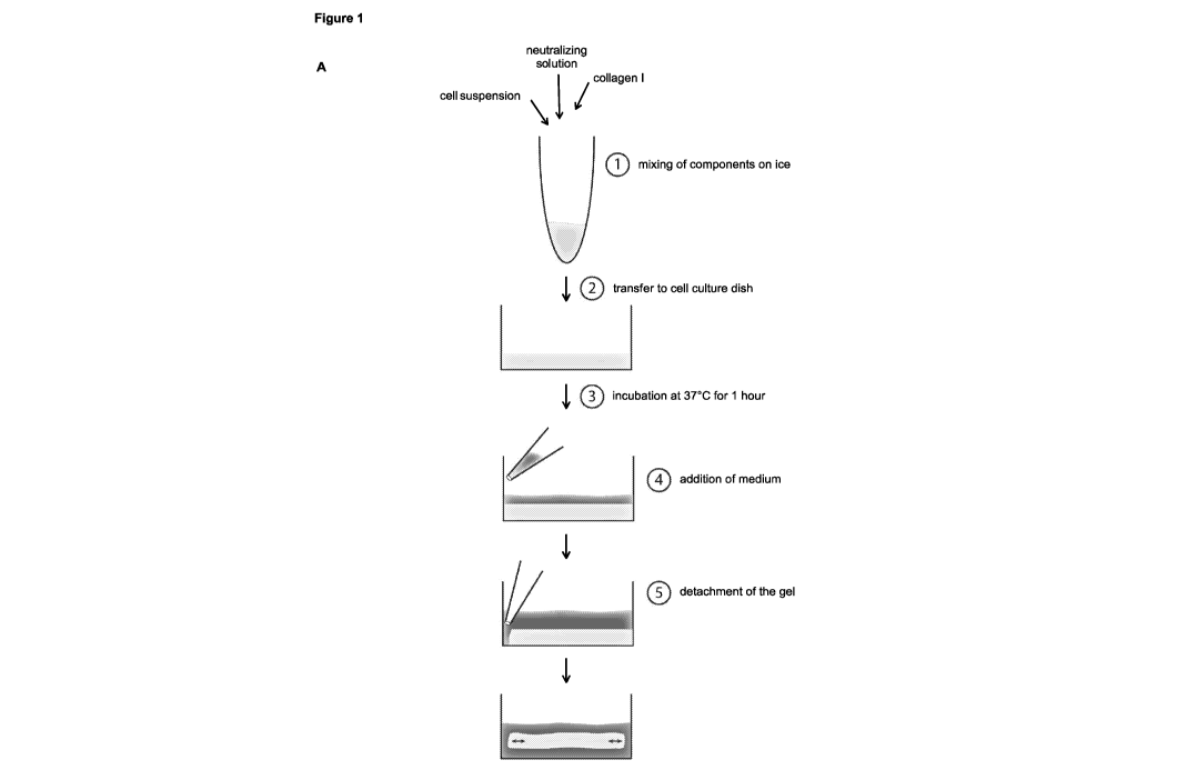
Fig. 1. Identification of culture conditions that promote generation of TDLU-
like
structures by freshly dissociated HMEC
(A) Experimental setup: generation of floating collagen gels.
(B) Bright-field microscopy: Carmine-stained representative images of
different types
of branched and non-branched structures (donor M8). Scale bar: 200 pm.
(C) Bright-field microscopy: haematoxylin-eosin stained section of a
terminal ductal
lobular unit (TDLU) from a healthy woman. Scale bar: 100 pm.
(D) Improvement of culture conditions: effect of one-time treatment with 3
pM Y-27632
at day 0 of culture and continuous treatment with 10 pM Forskolin on the
generation of
branched-type structures in floating collagen I gels at day 14 of culture.
Star-like structures
were not detected and therefore excluded from quantification. n=3
gels/condition. Structure
formation per 100 seeded cells is shown (donor M8).
(E) Quantification of monoclonal and polydonal structures formed by eGFP,
mCherry
and unlabeled passage 1 cells in floating collagen I gels (Donor M5). 500,
1500 and 13500
cells were seeded per well (24-well plate) and 3, 6 and 8 eGFP/mCherry
positive structures
among 17, 18 and 12 unlabeled structures were analyzed, respectively.
Monoclonal:
complete structure eGFP or mCherry positive. Polyclonal: eGFP/mCherry positive
and
negative areas.
(F) Confocal microscopy: representative images of monoclonal and polyclonal
structures (refer to E). Scale bar: 100 pm.
Data are shown as mean standard deviation (SD).
Fig. 2. Maintenance and expansion of TDLU-like structure formation during
passaging
and 2D-culture
(A) Experimental setup: freshly isolated HMEC (Donor M4) were cultured in
2D in the
absence or presence of 10 pM Forskolin for 5 passages, and transferred to
floating collagen
I gels in limiting dilution at passage (p) 1, 3 and 5.
(B) Extreme limiting dilution analysis (ELDA): determination of Branched
Structure-
Forming Units (B-SFU) of cells cultured in the presence of Forskolin (donor
M4).
(C) Confocal microscopy: representative TDLU-like structures generated in
floating
collagen I gels after 2D-culture in the presence of Forskolin (refer to A).
Vimentin (red), E-
cadherin (green), integrin-a6 (red), DAPI for cell nuclei (blue). Scale bar:
100 pm.
(D) ELDA: determination of B-SFU of cells cultured without Forskolin (donor
M4).
(E) Confocal microscopy: representative clusters of cells generated in
floating collagen
I gels after 20-culture without Forskolin, and transferred to floating
collagen I gels at passage
8
CA 02980852 2017-09-25
WO 2016/174604 PCT/1B2016/052407
1, 3 and 5 (refer to A). Vimentin (red), E- cadherin (green), integrin-a6
(red), DAPI for cell
nuclei (blue). Scale bar: 100 pm.
Data are shown as mean and 95 % confidence intervals (Cl).
Fig. 3. Frequency of TDLU-like structure-forming cells varies between donors
and is
increased by 20-culture
(A) Bright-field microscopy: Carmine-stained representative images of TDLU-
like
structures from freshly isolated cells of 9 donors (M1-M4, M6-M10) in floating
collagen I gels.
Scale bar: 200 pm.
(B) TDLU-like structure formation per 100 seeded HMEC from freshly isolated
cells of
9 donors at day 9 of culture. n=2. Data are shown as mean standard deviation
(SD).
(C) Sphere formation per 100 seeded HMEC from freshly isolated cells of 9
donors
(refer to A) at day 9 of culture. n=2 gels/donor. Data are shown as mean
standard deviation
(SD).
(D) Extreme limiting dilution analysis (ELDA): determination of Sphere and
Branched
Structure-Forming Units (S-SFU and B-SFU) of HMEC in floating collagen I gels
at passage
0 (Donor M8). Data are shown as mean and 95 % confidence intervals (Cl).
(E) Bright-field microscopy: Carmine-stained representative images of TDLU-
like
structures from cells of 9 donors (M1-4, M6-M10) cultured in 2D for 12 days
prior transfer to
floating collagen I gels. Scale bar: 200 pm.
(F) TDLU-like structure formation per 100 seeded HMEC from cells of 9
donors
established in 2D-culture (refer to E) at day 9 of culture. n=2 gels/donor.
Data are shown as
mean standard deviation (SD).
(G) Sphere formation per 100 seeded HMEC from cells of 9 donors established
in 20-
culture at day 9 of culture. n=2 gels/donor. Data are shown as mean standard
deviation
(SD).
(H) Analysis of viability by Fluorescence-Activated Cell Sorting (FAGS),
using 7-AAD:
n=10 donors (M1-M10). Data are shown as mean standard deviation (SD).
Fig. 4. TDLU-like structure-forming potential is contained within a
CD10+/CD49fhi/EpCAM- basal population
(A) Fluorescence Activated Cell Sorting (FACS) of freshly isolated HMEC:
dead cells
were excluded (7AAD- = live), hematopoietic and endothelial cells were
excluded
(CD45-/CD31- = Lin-), EpCAM, and CD49f were used to depict the following
populations:
Stroma (CD49f-/EpCAM-), Lumina! mature (LM, CD491/EpCAM+), Luminal progenitors
9
CA 02980852 2017-09-25
WO 2016/174604 PCT/1132016/052407
(LP, CD49f+/EpCAM+), Basal (B, CD49f1"/EpCAM-). LP and B populations were
isolated.
The B population was further subdivided into B- (CD107CD49f1"/EpCAM--) and B+
(CD10+/CD49fhi/EpCAM--).
(B) Linear correlation between sphere formation (per 100 freshly isolated
HMEC) and
the size of the LP within Lin- population (blue dots), or the size of the B
population (pink
dots). One dot represents one donor. r = correlation co- efficient.
(C) Linear correlation between TDLU-like structure formation (per 100
freshly isolated
HMEC) and the size of the B+ within Lin- population (red dots) or the B
population (pink
dots). One dot represents one donor. r = correlation co-efficient.
(D) Bright-field Microscopy: Carmine-stained representative whole collagen
I gels
containing B+, B- or LP cells (Donor M3). Scale bar: 0.5 mm.
(E) Extreme limiting dilution analysis (ELDA): determination of Branched
Structure-
Forming Units (B-SFUs) of 4 populations (B+,B-,B, LP) of freshly isolated HMEC
(Donors
M8, M9, M10) sorted by FACS according to (A) prior cultivation in floating
collagen I gels.
Data are shown as mean and 95 % confidence intervals (Cl).
Fig. 5. CD10-staining reveals a stromal component within the CD49fhi/EpCAM-
population
(A) Gene expression profiling: RNA for microarray analysis was derived from
3
subpopulations (B+, B- and LP, as indicated) purified by FACS using freshly
isolated HMEC
from 6 donors (M3, M6, M8, M9, M10, M12). Following unsupervised clustering of
all
samples, Principal Component Analysis (PCA) was conducted.
(B) Heatmap: shown are the expression values of up- and downregulated
luminal and
basal signature genes in all samples. Fold change was derived by comparing B+
versus LP
expression levels. Red (high) and blue (low) indicates log2 expression values.
Scale bar in
log2.
RT-PCR: MME/CD10, TP63, SNAI2, GATA3, ELF5, KRT8 mRNA expression in B+ and LP
cells from 3 donors (M3, M8, M10). n.d., not detectable, n=3.
(C) Heatmap: shown are the expression values of the top-20 significantly
(FDR<10%)
upregulated genes in B- samples versus B+ samples with corresponding fold
changes. Red
(high) and blue (low) indicates log2 expression values. Scale bar in log2.
(D) GO term analyses: shown are selected significantly enriched terms
(p<0.01)
associated with genes differentially regulated between B- and B+ populations
(FDR<10%,
FC>3x). Shown are gene symbols of the top-20 genes from (D).
(E) Representative flow cytometry analysis showing the fraction of CD10+
cells within
CA 02980852 2017-09-25
WO 2016/174604 PCT/1B2016/052407
the four populations defined by CD49f/EpCAM.
(F) Quantification of the percentage of CD10+ cells within the different
EpCAM/CD49f
populations as in (F). Average of 10 donors (M1-M10).
Data are shown as mean standard deviation (SD).
Fig. 6. TDLU-like structures derived from B+ cells recapitulate functional
aspects of
the mammary gland
(A) Immunohistochemistry: expression of p63, GATA-3 and CK18 in
representative
sections of structures derived from LP or B+ cells (Donor M10), fixed at
culture day 20. For
LP and B+, 6 and 5 fields of view were analyzed, respectively. Scale bar: 50
pm.
(B) Quantification of the size of floating collagen I gels containing HMEC
(Donors M3,
M8, M10). Gel size at day 13 (M3), day 14 (M8) and day 15 (M10) of culture is
given as
percentage of day 0. n=6 gels (M3, M10), n=9 gels (M8).
(C) Contraction of collagen I gels: Size of floating collagen I gels
containing LP or B+
cells (Donors M3, M10) was determined at day 12 of culture (indicated as day
0), and
imaged for two more days. Gel size is plotted relative to day 0. Half of the
gels were treated
with 2.0 ng/ml TGF-I31 once at day 0. Lower panel: Bar graphs of gel size at
day 2 as
percentage of day 0. n=12 gels/condition.
(D) Bright-field microscopy: Representative images of control and TGF431
treated gels
containing B+ cells from (C) (Donor M10).
(E) Quantification of the average number of cells per gel at the end of
analysis shown
in (C). Gels containing LP cells from donor M10 were pooled and counted,
therefore no
standard deviation is given.
(F) Contraction of individual cells. Confocal microscopy (left): B+ cell
derived
structures (Donor M8) were treated with TGF-I31 as in (C), and stained with
Phalloidin for F-
actin (white) and DAPI for cell nuclei (blue). Scale bar: 100 pm. Cell size
was determined per
condition for 30 cells of 3 different structures using ImageJ area tool.
n.s., not significant; n.a., not applicable.
Data are shown as mean standard deviation (SD).
Fig. 7. Matrix compliance in floating collagen gels is necessary for
alveologenesis and
luminal differentiation of TDLU-like structures.
(A) Experimental layout: Freshly isolated HMEC were seeded into collagen I
gels,
which were immediately detached to float (left) or left attached to the cell
culture dish
(middle, right). Once branched structures had formed, half of the attached
gels were
detached (right).
11
CA 02980852 2017-09-25
WO 2016/174604 PCT/IB2016/052407
(B) Bright-field microscopy: representative images of HMEC-derived branched
structures (Donor M8), cultured according to (A), and imaged for 60 hours,
starting at day 13
of culture. Smaller pictures are details of areas indicated with asterisk.
Scale bar: 500 pm.
(C) Quantification of side branches. Left: representative image showing
primary,
secondary and tertiary side branches indicated by red, blue and yellow lines,
respectively.
Right: The number of side branches in attached and floating collagen gels at
day 13 of
culture was quantified for 5 structures per condition (Donor M8).
(D) Confocal microscopy: representative images of HMEC-derived branched
structures
(Donor M8), cultured according to (A,B): p63 (red), GATA-3 (green), integrin-
a6 (red), laminin
(green), DAPI for cell nuclei (blue). Scale bar: 50 pm.
Data are shown as mean standard deviation (SD).
Fig. 8. referring to Fig. 1. Identification of culture conditions that promote
generation
of TDLU-like structures by freshly dissociated HMEC
(A) Effect of culture conditions on the generation of branched structures:
HMEC
(Donor M8) were cultured in presence of different concentrations of Forskolin
(continuous
treatment), Y-27632 and Thiazovivin (both one-time treatment at day 0 of
culture) in floating
collagen gels for 14 days. n=3 gels/condition. Structure formation is given
per 100 seeded
cells.
(B) Effect of culture conditions on the ratio of branched structure
subtypes, refer to (A).
n=3 gels/condition.
(C) Effect of culture conditions on the generation of non-branched
structures, refer to
(A). n=3 gels/condition. Structure formation is given per 100 seeded cells.
Fig. 9. referring to Fig. 2. Maintenance and expansion of TDLU-like structure
formation
during passaging and 2D-culture
(A) Bright-field: representative images of HMEC-derived branched structures
(donor
M8), at subsequent passages in 3D. Scale bar: 500 pm.
(B) Phase contrast microscopy: representative images of HMEC cultured in 2D
in the
absence or presence of 10 pM Forskolin at passage 1,3 and 5 (donor M4). Scale
bar: 100
pm.
(C) 2D-Immunofluorescence: representative images of HMEC cultured in 2D, as
described in (B). integrin-a6 (red), vimentin (green), I3-catenin (red), E-
cadherin (green),
fibronectin (red), Zeb1 (green), DAPI (blue). Scale bar: 100 pm.
(D) RT-PCR: ZEB1, CDH2 (N-cadherin),V/M (vimentin), FN1 (fibronectin) and
CDH1
(E-cadherin), mRNA expression of HMEC cultured in 2D, as described in (B).
n=3.
(E) RT-PCR: OVOL2 and ITGA6 (integrin-a6) mRNA expression of HMEC cultured,
as
12
CA 02980852 2017-09-25
WO 2016/174604 PCT/1B2016/052407
described in (B). n=-3.
(F) Flow cytometry analysis of CD49f and EpCAM expression in Lin- HMEC
cultured in
2D, as described in (B).
p, passage.
Fig. 10 referring to Fig. 4. TDLU-like structure-forming potential is
contained within a
CD104./CD49f/EpCAM" basal population
(A) Flow cytometry analysis of CD49f, EpCAM and CD10 expression in the 7-
AAD-,
Lin- subset of freshly isolated HMEC from 9 donors (M1-M4, M6-M10) used in
Figures 3 and
4. Determined population sizes were used for correlation analysis in Figures
4B,C and
10B,C.
(B) Correlation between branched structure formation and the size of the LP
population. One dot represents one donor.
(C) Correlation between branched structure formation and the size of the
CD10+
stromal population (CD10+/CD49f-/EpCAM-, green dots), the CD10 LP population
(CD10+/CD49f+/EpCAM+, blue dots), and CD10+ LM population (CD1017CD49(/EpCAM+,
dark blue dots). One dot represents one donor.
(D) Reanalysis of the purity of sorted LP cells from donor M8, used for
extreme limiting
dilution analysis in Figure 4E.
r, correlation coefficient.
Fig. 11 referring to Fig. 7. Matrix compliance in floating collagen gels is
necessary for
alveologenesis and luminal differentiation of TDLU-like structures.
(A) Confocal microscopy: representative images of HMEC-derived branched
structures
(Donor M8), cultured according to Figure 7A, B. p63 (red), ZO-1 (green), DAPI
(blue). Arrows
point to ZO-1 expression. Scale bar: 50 pm.
(B) RT-PCR: ELF5 and TJP1 (ZO-1) mRNA expression in B+ and LP cell derived
structures from donors M3, M8 and M10, cultured in attached and floating
collagen gels. n=3.
(C) Confocal microscopy: representative images of HMEC-derived spheres
(Donor
M8), cultured in floating and attached collagen gels, at day 14 of culture.
p63 (green), ZO-1
(red), integrin-a6 (red), laminin (green), DAPI (blue). Scale bar: 100 pm.
(D) Contraction of collagen gels: HMEC from donor M10 were grown in
attached
collagen gels. Once branched structures had formed, gels were detached (day 13
of culture)
and treated with 10 pM Blebbistatin or 5 pM Y-27632 every 24 hours. The size
of the gels
was determined directly after detachment (0 hours), and after 24, 60 and 110
hours. Gel size
13
CA 02980852 2017-09-25
WO 2016/174604 PCT/1B2016/052407
is plotted relative to the timepoint of detachment (0 hours). n=16
gels/condition.
(E) Quantification of the average number of cells per gel at the end of
analysis shown
in (D), n=4.
(F) Bright-field microscopy: representative images of HMEC-derived branched
structures (Donor M10) cultured in attached collagen gels for 12 days,
detached on day 13 of
culture, and treated with 10 pM Blebbistatin or 5 pM Y-27632 every 24 hours.
Structures
were imaged for 60 hours. Smaller pictures are details of areas indicated with
asterisk. Scale
bar: 500 pm.
n.d., not detectable
n.s., not significant
14
CA 02980852 2017-09-25
WO 2016/174604 PCTAB2016/052407
DETAILED DESCRIPTION
[23] The present inventors pioneered in providing an organoid assay that
enables single
cells from mammary epithelial tissue to recapitulate mammary gland
development,
homeostasis and disease-development. In particular, the present inventors have
developed
means and methods, i.a. culturing conditions, that allow cells freshly
isolated from primary
mammary epithelial tissue to form structures that resemble the terminal ductal-
lobular unit
(TDLU), the functional unit of the breast.
[24] The means and methods provided herein enables detection, isolation and
manipulation of breast-stem cell-containing cell populations, in particular
such isolated from
primary tissue, and studying of key aspects of tissue architecture and
function. It also allows
for quantification of regenerative potential on a single-cell level. The assay
is highly
quantitative and scalable, and provides a highly sensitive and specific, thus
reproducible
functional readout that is suitable for high-throughput screening.
[25] Accordingly, the present invention provides a method of generating cells
capable of
differentiating to a multicellular organoid unit that morphologically and/or
functionally
resembles the terminal ductal-lobular unit, comprising
(i) culturing dissociated cells from mammary epithelial tissue in a collagen
gel for at
least 7 days, said culture medium comprising a compound which elevates cAMP
levels;
(ii) determining whether a multicellular organoid unit is formed in step (i);
and
(iii) obtaining a single cell from said multicellular organoid unit of (ii).
(i) Cells and Cultivation
[26] In step (i) of the above-described method of the invention, dissociated
cells from
mammary epithelial tissue are cultured. It is in general conceivable to use
cells obtained from
any of a wide variety of sources, e.g. the cells may be primary cells, cells
of a cell line,
untransformed cells, transformed cells, genetically modified cells, or non-
genetically modified
cells. Induced pluripotent stem cells are also envisaged. In general, any type
of cell that can
be obtained from mammary epithelial tissue can be used in the methods of the
invention.
The use of primary cells (i.e., directly derived from mammary epithelial
tissue) can be
particularly advantageous when it is desired to most accurately reflect cell
behaviour in vivo.
Primary cells dissociated from mammary epithelial tissue include, for example,
mammary
epithelial cells (MEC), including e.g. myoepithelial and luminal mammary
epithelial cells,
myoepithelial and luminal mammary progenitor cells, and adult mammary stem
cells (MaSC).
CA 02980852 2017-09-25
WO 2016/174604 PCT/1B2016/052407
In particular, the term "cells dissociated from mammary epithelial tissue"
includes any type of
stem cells obtainable from mammary epithelial tissue using means and methods
known in
the art. In general, "stem cells" are undifferentiated cells that have the
ability to go through
numerous cycles of cell division while maintaining the undifferentiated state
(self-renewal)
and can differentiate into specialized cell types (potency).The term in
particular also includes
"breast stem cells" as defined elsewhere herein.
[27] It is further also conceivable to use cells dissociated from other
tissues, e.g. epithelial
tissues of the pancreas, lung, or kidney. Particularly envisaged in this
regard are cells, in
particular stem cells, having the ability of forming a multicellular organoid
unit comprising
ductal structures and/or multiple branch-points and/or alveoli and/or may also
be capable of
contracting a collagen gel, preferably a free-floating collagen-I gel. Said
cells can be primary
cells, cells of a cell line, untransformed cells, transformed cells,
genetically modified cells,
non-genetically modified cells, or induced pluripotent stem cells.
[28] For example, primary human mammary epithelial cells (HMEC) can be derived
from
fresh breast reduction tissue (reduction mammoplasty) by mechanical and/or
enzymatic
dissociation and, if desired, can be further purified by methods such as
fluorescence
activated cell sorting (FACS). Human and murine breast cancer-derived
established cell
lines, such as MCF7, MDA-MB-231 and 4T1 cells can also be used. One of skill
in the art
would be aware of other cell lines (e.g., derived from other cancer types)
that may be used in
embodiments of the invention. The term "dissociated" means that individual
cells have been
released from a cell compound, cell agglomeration or tissue.
[29] It is envisaged that "dissociated cells" are derived from healthy or
diseased mammary
epithelial tissue. "Diseased tissue" in particular refers to tissue comprising
cells with germline
or somatic mutations, e.g. in proto-oncogenes. The term includes tissue
comprising
cancerous and/or pre-cancerous cells and/or tissue derived from a patient
diagnosed with
breast cancer. "Healthy tissue", on the other hand, refers to tissue from
healthy donors that
preferably does not comprise germline or somatic mutations, cancerous and/or
pre-
cancerous cells.
[30] In order to obtain dissociated cells, mammary epithelial tissue can be
dissociated
mechanically and/or enzymatically. Means and methods for mechanical and
enzymatical
tissue dissociation are well-known in the art. E.g., the tissue can be minced
using scalpels or
other suitable tools. Other means of mechanical tissue dissociation are also
conceivable, e.g.
sonication or others.. Further, tissue dissociating agents may be used,
typically including
tissue degrading enzymes such as collagenase, trypsin, neutral protease or
dispase, and
other proteolytic enzymes. However, the tissue dissociating agents are not
necessarily
16
CA 02980852 2017-09-25
WO 2016/174604 PCT/1B2016/052407
limited to enzymes. Other examples of tissue dissociating agents are chelating
agents. The
length of time required for treatment will vary depending on the sonication
frequency, type of
the agent, the concentration of agent, and the temperature at which treatment
is conducted.
Treatment is allowed to proceed until a sufficient amount of tissue has
dissociated without
causing undue damage to released cells or cellular aggregates. Dissociation
advantageously
also comprises obtaining a single-cell suspension of the dissociated cells as
described in the
appended examples.
[31] Next, dissociated cells are plated in collagen gels. The collagen gel may
be
composed of one collagen type or a mixture of collagen types. A collagen type
is, for
example, type I, II, Ill, IV of V, with the type I being preferred. The
collagen concentration
may be in the range of about 0.5 to 2 mg/ml, preferably of about 0.8 to 1.8
mg/ml and even
more preferred of about 1.0 to 1.5 mg/ml. The term comprises attached and free-
floating
collagen gels.
[32] The term "attached gel" as used herein, refers to a rigid collagen gel
that sticks to the
surface of the cell culture dish. This is in contrast to a 'floating gel" that
has been
mechanically detached from the cell culture dish after polymerization of the
gel and is
thereby able to float in the cell culture medium. A floating gel is therefore
more compliant
than an attached gel and can e.g. contract or expand.
[33] E.g., the gel can be a collagen-I gel that is attached or free-floating
in growth medium.
[34] The growth medium is advantageously supplemented with a compound which
elevates cAMP levels. Optionally, the growth medium may be supplemented with a
ROCK
inhibitor.
[35] A "compound which elevates CAMP levels" can in general be any compound
that is
capable of increasing levels of cyclic adenosine monophosphate (cAMP). The
capability of
compounds to do so can be assessed e.g. by commercially available test kits
such as the
Promega cAMP-GloTm Assay which is based on the principle that cyclic AMP
(CAMP)
stimulates protein kinase A (PKA) holoenzyme activity, decreasing available
ATP and
leading to decreased light production in a coupled luciferase reaction.
Without wishing to be
bound by theory, addition of a compound which elevates cAMP levels is thought
to promote
formation of TDLU-like branched structures and/or alveologenesis. The compound
can for
example be an activator of adenylylcyclase, or the compound can be cAMP, or a
cAMP
mimetic (i.e. having cAMP functionality). The term "activator of
adenylylcyclase" comprises
compounds that elevate cAMP levels by directly activating adenylylcyclase
(e.g. by binding to
adenylylcyclase). Said compounds are designated "adenylylcyclase agonists"
herein. The
17
CA 02980852 2017-09-25
WO 2016/174604 PCT/1B2016/052407
term "activator of adenylylcyclase" also comprises compounds that elevate cAMP
levels by
indirectly activating adenylylcyclase, e.g. by activating stimulators of
adenylylcyclase (such
as activating G-protein coupled receptor subunits) or by inactivating
inhibitors of
adenylylcyclase (such as inhibitory G-protein coupled receptor subunits).
Exemplary
compounds include choleratoxin and pertussistoxin. However, particularly
envisaged
compounds for elevating cAMP levels are adenylylcyclase agonists, such as
Forskolin.
[36] The present inventors also discovered that addition of a ROCK inhibitor
can increase
formation of TDLU-like branched structures. Thus a ROCK inhibitor can be added
to improve
cell culture conditions. However, supplementing a ROCK inhibitor for more than
about 5 days
may result in dissociation of cell-cell adhesion, thereby perturbing
morphogenesis. Hence, it
is envisaged that the ROCK inhibitor may be removed after about 5 days from
the culture
medium. Changes in cell-cell adhesion and morphology can be monitored macro-
and
microscopically, in order to determine the need and time point of removing the
ROCK
inhibitor.
[37] A "ROCK inhibitor" as used herein is compound that acts as an inhibitor
of Rho-
associated protein kinase, i.e. reduces or even abolishes ROCK functionality.
The capability
of a compound to act as a ROCK inhibitor can be assessed by various means,
e.g. by
determining its ability to compete with ATP for binding to ROCK and/or by
assessing its
effects on cell morphology, G1-S Transition and cytokinesis as described in
Ishizaki T Mol
Pharmacol. 2000 May;57(5):976-83. The inhibitor may be either unspecific or
specific for
either of the ROCK isoforms ROCK1 and/or ROCK2. ROCK inhibitors known in the
art have
been reviewed in Liao et al. J Cardiovasc Pharmacol. 2007 Jul; 50(1): 17-24
and include
Fasudil, Y-27632, Thiazovivin, Y39983, Wf-536, SLx-2119, Azabenzimidazole-
aminofurazans, DE-104, Olefins, Isoquinolines, Indazoles, pyridinealkene
derivatives, H-
1152P, ROKa inhibitor, XD-4000, 4-(1-aminoalkyl)-N-(4-pyridyl)cyclohexane-
carboxamides,
HMN-1152, Rhostatin , BA-210, BA-207, BA-215, BA-285, BA-1037, Ki-23095, VAS-
012,
with Y-27632 or Thiazovivin being particularly envisaged for use in the method
of the
invention.
[38] The present inventors have observed that culture medium comprising Y-
27632 or
Thiazovivin as a ROCK inhibitor and an adenylylcyclase agonist such as
Forskolin as a
compound which elevates cAMP levels is one particularly useful culture medium
for use in
the methods of the present invention. E.g., the culture medium may comprise Y-
27632 in a
concentration of about 1-5 pM, about 2-4 pM or about 3 pM, and Forskolin in a
concentration
of about 5-15 pM, about 6-14 pM, about 7-13 pM, about 8-12 pM, about 9-11 pM
or about 10
pM. It is however to be noted that the ROCK inhibitor may be removed after a
while from the
culture medium as described herein.
18
CA 02980852 2017-09-25
WO 2016/174604 PCT/1B2016/052407
(ii) Multicellular organoid unit
[39] Next, it is determined whether a multicellular organoid unit has been
formed in step
(i).
[40] A "multicellular organoid unit" is a multicellular structure that is
formed by a single
cell. It is in particular envisaged that the single cell is a stem cell,
preferably a breast stem
cell as described herein. The multicellular organoid unit morphologically
and/or functionally
resembles the terminal ductal-lobular unit (TDLU) and is therefore also termed
"TDLU-like
(branched) structure" herein. The term "terminal ductal-lobular unit" or
"TDLU" as used herein
is a structure of the breast. Each breast lobe is drained by a collecting duct
terminating in the
nipple. The collecting duct has several branches, which end in a terminal
ductal-lobular unit
(TDLU), the basic functional and histopathological unit of the breast. The
TDLU is composed
of a small segment of terminal duct and a cluster of ductules, which are the
effective
secretory units. The functional structures are surrounded by specialized
connective tissue. A
normal terminal ductal lobular unit ranges from 1-4 mm. The TDLU is composed
of the
extralobular terminal duct, intralobular terminal duct, lobule (functional
unit of the breast)
[41] However, though a multicellular organoid unit is ideally morphologically
and/or
functionally identical to a TDLU, it cannot be excluded that there may be
differences. These
differences are reflected in the term "organoid" meaning it is an organ
structure (i.e. an entire
organ or functional part thereof) that is formed and grown ex vivo which
ideally
morphologically and/or functionally resembles an organ structure. The same is
true for the
term "resemble". It means that a multicellular organoid unit is/behaves like
an organ structure
and thus morphologically and/or functionally behaves like a (natural) organ
structure.
However, in contrast to a (natural or in vivo) organ, an organoid structure is
formed and
grown ex vivo. An example for a difference between a TDLU and a multicellular
organoid unit
is lactation. While a TDLU being part of the (natural) breast is able to
secrete milk, a
multicellular organoid unit is, to the best of the knowledge of the present
inventors, not able
to do so. However, nonetheless, a multicellular organoid unit shares identity
with the natural
TDLU as regards morphology in that it comprises ductal structures, multiple
branch-points
and advantageously alveoli. From a functional perspective, a multicellular
organoid unit is,
like a natural TDLU, capable of contraction. Contraction may be tested as
described herein.
[42] A multicellular organoid unit is in particular considered to
morphologically and/or
functionally resemble the TDLU when it comprises ductal structures and/or
multiple branch-
points. It may also comprise alveoli at the tip of the ducts. Presence of the
aforementioned
features in a multicellular organoid unit can be easily assessed by the
skilled person using
visual examination, e.g. bright-field microscopy as described in the appended
examples.
19
CA 02980852 2017-09-25
WO 2016/174604 PCT/1B2016/052407
[43] It is further envisaged that the multicellular organoid unit is
responsive to hormones
and/or growth factors. Hormones include steroid hormones: estrogen,
progesterone and
androgens, pituitary hormones: prolactin, human growth hormone, other peptide
hormones:
gluco- and mineralcorticoids, insulin. Growth factors and morphogenes include
the following
families: EGF (Epidermal Growth Factors), IGF (Insulin-like growth Factors),
FGF (Fibroblast
Growth Factors), Wnt (Wingless), TGF-beta (Transforming Growth Factor beta),
Notch, shh
(sonic hedgehog). Included are endogenous and recombinant factors, precursors
and
derivatives, as well as endogenous, recombinant and synthetic agonists and
antagonists.
Responsiveness to hormones and growth factors renders the multicellular unit
of the present
invention a suitable substrate to test compounds for their ability to elicit a
physiologically
response.
(iii) Single cell
[44] In step (iii) of the method of the invention, a single cell is
obtained from the
multicellular organoid unit formed in step (ii) of the method.
[45] It is envisioned that said cell is a single breast stem cell. Over the
course of at least
days single breast stem cells will generate complex gland structures, i.e. a
multicellular
organoid unit that morphologically and/or functionally resembles the terminal
ductal-lobular
unit. It can be determined whether said multicellular organoid unit comprises
ductal
structures and/or multiple branch-points and/or alveoli as described herein.
Also or
alternatively, it can be determined whether said multicellular organoid unit
is capable of
contracting a floating collagen gel, preferably a free-floating collagen-I
gel. Such contraction
may then be indicative of alveologenesis of said multicellular organoid unit.
[46] The term "breast stem celr as used herein thus refers to a cell capable
of forming a
multicellular organoid unit comprising ductal structures and/or multiple
branch-points and/or
alveoli and/or may also be capable of contracting a collagen gel, preferably a
free-floating
collagen-I gel, such a cell is a breast stem cell. In particular, the breast
stem cell is
envisioned to be CD31-, CD45", EpCAM", CD49f+ and CD10+.
[47] Such a breast stem cell can be obtained as a single cell by means and
methods
known in the art from said multicellular organoid unit. Indeed, the present
inventors
demonstrated that such a breast stem cell obtained from a multicellular
organoid unit of the
present invention will again, when plated in a collagen gel, form another
multicellular
organoid unit. This is the proof for such a cell to be a breast stem cell.
CA 02980852 2017-09-25
WO 2016/174604 PCT/1B2016/052407
(iv) Gel contraction
[48] The inventors have further observed that multicellular organoid
structures were able
to contract floating gels, presumably reflecting the contraction of the TDLU
ducts during
lactation. The method of the invention may further comprise a step of
determining whether
the obtained multicellular organoid unit is capable of contracting a floating
collagen gel.
Without wishing to be bound by theory, the present inventors observed that
alveoli preferably
developed when cells were cultivated in compliant, floating collagen gels, and
that
alveologenesis further was dependent on and/or triggered by contraction of the
collagen gel.
Thus, contraction of a floating gel by a multicellular organoid unit is
envisaged to be
indicative of alveologenesis.
[49] Contraction of the collagen gel may be quantified by measurement of the
gel size at
various times with a ruler or with image analysis software, such as NIH Image
or Image Pro-
Plus (MediaCybemetics) and can be correlated to breast stem cell content.
[50] As set out herein, the present inventors have discovered that
alveologenesis may be
triggered by contraction of the collagen gel. Consequently, the present
invention also
provides a method for influencing the behaviour, i.e. triggering cell
differentiation and hence
alveologenesis, by providing the mechanic stimulus via detachment of an
attached collagen
gel. It is therefore possible to synchronize alveologenesis in a multitude of
multicellular
organoids.
(v) Enrichment
[51] As set out elsewhere herein, the present inventors identified a
combination of surface
markers that can be used to enrich cells, in particular breast stem cells,
from a population of
mammary epithelial cells. Without wishing to be bound by theory, the present
inventors noted
that the cell surface marker combination of CD31-, CD45, EpCAM-, CD49r and
CD10+
correlated to multicellular organoid unit formation capacity. It is speculated
that the
aforementioned combination of surface markers is specific for breast stem
cells of the basal
mammary epithelial cell population. Accordingly, the method may further
comprise a step of
enriching a population of cells by sorting the cells for the cell surface
marker combination
CD31", CD45", EpCAM-, CD49r and CD10+ prior to culturing said cells in a
collagen gel.
[52] Enrichment of cells with the desired surface markers can be accomplished
using
methods known in the art, e.g. by fluorescence-activated cell sorting (FACS)
as described in
the appended examples.
[53] This step can advantageously be used to enrich cells capable of
differentiating to a
multicellular organoid unit that morphologically and/or functionally resembles
the terminal
21
CA 02980852 2017-09-25
WO 2016/174604 PCT/IB2016/052407
ductal-lobular unit, but is not a mandatory prerequisite to obtain a single
breast stem cell
from said multicellular organoid unit, since such a breast stem cell can
readily be obtained as
described above, i.e., without prior enrichment, but merely on the basis that,
when plated in a
collagen gel, preferably a collagen-I gel, it is capable of differentiating to
a multicellular
organoid unit that morphologically and/or functionally resembles the terminal
ductal-lobular
unit as described herein.
(vi) Pre-cultivation
[54] The method of the invention may further comprise a step of culturing the
dissociated
cells in 2D-culture (or other methods) prior to transferring them to collagen
gels. This step is
also referred to as "pre-cultivation" herein.
[55] Without wishing to be bound by theory, it is thought that 2D-pre-
cultivation may
increase the ability of primary mammary epithelial cells to form multicellular
organoid units.
Pre-cultivation, in particular 2D pre-cultivation, further allows genetic
manipulation of the cells
prior to cultivation in the collagen gel. Pre-cultivation can be accomplished
using standard
protocols known in the art, depending on the type of cell, length of
cultivation, desired cell
morphology and density and other parameters. An exemplary protocol for pre-
cultivation of
human primary epithelial cells can be found in the appended examples.
Breast stem cell
[56] Furthermore, the present invention relates to a breast stem cell
obtainable by the
methods of the invention, in particular using a collagen-I gel for
cultivation. Said breast stem
cell is envisaged to be capable of differentiating in a collagen gel to a
multicellular organoid
unit that morphologically and/or functionally resembles the terminal ductal-
lobular unit,
wherein said multicellular organoid unit comprises ductal structures and
multiple branch-
points and/or is capable of contracting a floating collagen gel. In
particular, the breast stem
cell may be CD31", CD45", EpCAM", CD49f+ and CD10+.
[57] It is further envisioned that the breast stem cell of the present
invention may be
genetically modified. Said genetic modification can be caused by stable or
transient
introduction of various genetic elements, (e.g., viral vectors, plasmids,
extrachromosomal
replicating vectors, etc.) encoding one or more genes, e.g. the catalytic
subunit of the human
telomerase holoenzyme (hTERT) to generate immortalized cell lines. Such cell
lines can be
further genetically modified and transformed, e.g. by introducing the Simian
Virus 40 (SV40),
Large T antigen encoding gene, and the haRAS oncogene. In some embodiments,
gene
expression of one or more genes may be knocked-out by insertional mutagenesis
using e.g.
restriction enzymes or genetic elements which are inserted in the coding
region or down-
22
CA 02980852 2017-09-25
WO 2016/174604 PCT/1B2016/052407
regulated by genetically modifying cells to express a short hairpin RNA
(shRNA), microRNA
(miRNA) or miRNA precursor, miRNA sponge, etc. It will be appreciated that a
variety of
different oncogenes and/or tumor suppressor genes can be used to genetically
modify cells.
One of skill in the art would be aware of suitable vectors and genetic
elements (e.g.,
regulatory elements such as promoters, enhancers, etc.) for transfection of
mammalian cells.
In some embodiments, a regulatable (e.g., inducible and/or repressible)
expression control
element (e.g., promoter) is used to achieve regulatable expression of an RNA
or protein of
interest in cells.
[58] The invention thus also provides a multicellular organoid unit that
morphologically
and/or functionally resembles the terminal ductal-lobular unit, comprising
breast stem cells of
the present invention.
Compound testing
[59] The breast stem cell or the multicellular organoid unit obtainable by the
methods of
the present invention can advantageously be used to test a variety of
compounds for their
potential to elicit a cellular response on said breast stem cell or
multicellular organoid unit. A
"cellular response" can be the frequency of a certain type of cell, cell
growth (size of cell), cell
proliferation, growth arrest, cell survival, apoptosis, necrosis, autophagy,
senescence, DNA
damage, differentiation, de-differentiation, trans-differentiation, migration,
invasion, self-
renewal, oncogenesis, and changes in the morphology of cells in the
multicellular structure
pertaining to: cell-cell adhesion, cell-matrix adhesion, apical-basal
polarity, planar polarity as
well as gene expression, regulatory RNA expression, protein expression,
changes in
metabolism, andothers. Cellular responses can be assessed using standard
protocols
known in the art. Compounds that can be tested for their ability to provoke a
cellular
response include a drug, hormone, growth factor, antibody, nucleotide
molecule, peptide,
protein or (co-cultured) cell.
[60] A method for testing a compound for its ability to elicit a cellular
response according
to the invention comprises the following steps:
(i) bringing a breast stem cell or a multicellular organoid unit obtained by
the above-
described methods of the invention into contact with said compound; and
(ii) determining whether said compound elicits a cellular response.
23
CA 02980852 2017-09-25
WO 2016/174604 PCT/1B2016/052407
Pharmaceutical composition
[61] Further, the present invention relates to a composition comprising a
breast stem cell
or the multicellular organoid unit as disclosed herein.
[62] Said composition can be a pharmaceutical composition. The term
"pharmaceutical
composition" particularly refers to a composition suitable for administering
to a human or
animal, i.e., a composition containing components which are pharmaceutically
acceptable. In
particular, a pharmaceutical composition comprises a breast stem cell or a
multicellular
organoid unit as described herein together with a carrier, diluent or
pharmaceutical excipient
such as buffer, preservative and tonicity modifier. Pharmaceutical
compositions of the
invention comprise a therapeutically effective amount of a breast stem cell or
a multicellular
organoid unit and can be formulated in various forms, e.g. in solid, liquid,
gaseous or
lyophilized form and may be, inter alia, in the form of an ointment, a cream,
transdermal
patches, a gel, powder, a tablet, solution, an aerosol, granules, pills,
suspensions,
emulsions, capsules, syrups, liquids, elixirs, extracts, tincture or fluid
extracts or in a form
which is particularly suitable for topical or oral administration.
[63] The pharmaceutical composition may further comprise a solvent such as
water, a
buffer for adjusting and maintaining the pH value, and optionally further
agents for stabilizing
the breast stem cell or multicellular organoid unit C or preventing
degradation of the same. It
may additionally comprise further breast stem cells or multicellular organoid
units, other
pharmaceutically active agents, such as adjuvants etc.
[64] By "therapeutically effective amount" is meant an amount of breast stem
cells or
multicellular organoid units that elicit the desired therapeutic effect. The
exact amount dose
will depend on the purpose of the treatment, and will be ascertainable by one
skilled in the
art using known techniques. As is known in the art and described above,
adjustments for
age, body weight, general health, sex, diet, drug interaction and the severity
of the condition
may be necessary, and will be ascertainable with routine experimentation by
those skilled in
the art.
[65] A variety of routes are applicable for administration of the
pharmaceutical
composition, including, but not limited to, orally, topically, transdermally,
subcutaneously,
intravenously, intraperitoneally, intramuscularly or intraocularly. However,
any other route
may readily be chosen by the person skilled in the art if desired.
Binding molecules
[66] As set out elsewhere herein, the present inventors have for the first
time found a
combination of surface markers that allow for enrichment of breast stem cells
having TDLU-
24
CA 02980852 2017-09-25
WO 2016/174604 PCT/1B2016/052407
like structure formation potential. Said markers can be detected by binding
molecules. The
present invention thus also relates to the use of binding molecules directed
against CD31,
CD45, EpCAM, CD49f and CD10 for enriching breast stem cells from a population
of primary
mammary epithelial cells.
[67] The term "binding molecule" as used herein in general refers to any
molecule able to
recognize and bind to CD31, CD45, EpCAM, CD49f or CD10, and in particular
includes
antibodies or functional fragments thereof such as Fab or F(ab)2 or antibody
derivatives such
as bispecific antibodies (for example, scFvs), chimeric antibodies, humanized
antibodies,
single domain antibodies such as VHH antibodies (also known as Nanobodies) or
domain
antibodies (dAbs) or an lipocalin muteins (also known as anticalins) and
others.
[68] It is in particular envisaged that the binding molecules are employed to
enrich CD31,
CD45-, EpCAM-, CD49f' and CD10+ cells. As described elsewhere herein,
enrichment of
cells having the desired combination of markers can be accomplished using
standard
protocols known in the art such as FACS as described in the appended examples.
[69] Accordingly, the present invention also provides a method of enriching
breast stem
cells from a population of primary mammary epithelial cells, comprising
(i) sorting cells for the cell surface marker combination CD31-, CD45-, EpCAM-
,
CD49r and CD10+.
[70] Said method may further comprise the following steps:
(ii) culturing sorted cells in a collagen gel for at least 7 days, said
culture medium
comprising a compound which elevates CAMP levels;
(iii) determining whether a multicellular organoid unit is formed in step
(ii); and
(iv) obtaining a single cell form said multicellular organoid unit of (iii).
[71] It will be appreciated that method steps (ii)-(iv) correspond to
method steps (i)-(iii) of
the method for generating cells capable of differentiating into multicellular
organoid structures
also described in detail elsewhere herein. Hence, the definitions and
explanations with
regard to the latter are also applicable to the method for enrichment of
breast stem cells,
mutatis mutandis.
[72] Breast stem cells can also be enriched from a population of cells from
mammary
epithelial tissue by a method comprising the step(s) of
CA 02980852 2017-09-25
WO 2016/174604 PCT/1B2016/052407
(I) determining whether cells from said population of cells from mammary
epithelial
tissue are capable of forming a multicellular organoid unit in a collagen-I
gel in the
presence of a compound which elevates cAMP levels after at least 7 days and/or
(ii) determining whether said multicellular organoid unit is capable of
contracting a
floating collagen-I gel.
[73] Again, in step (i) determination of whether a multicellular organoid unit
is formed is
accomplished by assessing the presence of ductal structures and multiple
branch-points in
said multicellular organoid unit. As described elsewhere herein, it is
contemplated that
capability of the multicellular organoid unit to contract the floating gel
(step (ii)) is indicative of
alveologenesis.
[74] As described in greater detail in the context of other methods of the
invention, the
culture medium may comprise a Rho-kinase (ROCK) inhibitor, said ROCK inhibitor
being
either unspecific or specific for either ROCK1 and/or ROCK2. It is in
particular envisaged that
the ROCK inhibitor is Y-27632 or Thiazovivin and the compound which elevates
cAMP
levels, is an adenylylcyclase agonist such as Forskolin.
Progenitor cells
[75] Furthermore, the inventors discovered that luminal progenitor cells may
be cultured,
similar to breast stem cells, in a collagen gel. Without wishing to be bound
by theory, luminal
progenitor cells may be the cells-of-origin for breast cancer. In contrast to
breast stem cells,
luminal progenitor cells typically form spheres when cultured in a collagen
gel. However, in
rare cases luminal progenitor cells de-differentiate spontaneously and thereby
acquire stem-
cell attributes resulting in the generation of branched structures, in
particular multicellular
organoid units, in a collagen gel. Upon de-differentiation luminal progenitor
cells down-
regulate the expression of the cell lineage markers CK8, CK18, GATA3 and up-
regulate the
expression of Vimentin. De-differentiation of luminal progenitor cells is
indicative of an
abnormality and may be a first step in the development of breast cancer.
Hence, the de-
differentiation capacity of luminal progenitor cells may indicate an increased
breast cancer
risk.
[76] Accordingly, the present invention relates to a method for determining
the rate of
spontaneous de-differentiation of luminal progenitor cells, comprising:
(I) enriching a luminal progenitor cell containing population by sorting the
cells for the
cell surface marker combination CD31", CD45", EpCAM+, CD49.1+;
(ii) culturing said cells in a collagen gel, in particular a collagen I gel;
and
26
CA 02980852 2017-09-25
WO 2016/174604 PCT/1B2016/052407
(iii) determining whether a multicellular organoid unit is formed in step
(ii).
[77] Means and methods for determining whether a multicellular organoid unit
is formed
have been described elsewhere herein and are applicable mutatis mutandis.
[78] Furthermore, the present invention relates to a method for generating a
de-
differentiated luminal progenitor cell, comprising:
(i) enriching luminal progenitor cell containing population by sorting the
cells for the
cell surface marker combination CD31", CD45", EpCAM+, CD49f;
(ii) culturing said cells in a collagen gel;
(iii) determining whether a multicellular organoid unit is formed in step
(ii); and
(iv) obtaining a single cell from the multicellular organoid unit.
[79] The culture medium used for luminal progenitor cells may comprise a Rho-
kinase
(ROCK) inhibitor, said ROCK inhibitor being either unspecific or specific for
either ROCK1
and/or ROCK2 and/or a compound which elevates cAMP levels, as described
herein.
[80] Furthermore, the luminal progenitor cells may be dissociated cells from
mammary
epithelial tissue, wherein said epithelial tissue is healthy or diseased
tissue, wherein said
diseased mammary epithelial tissue comprises germ-line or somatic mutations.
[81] Luminal progenitor cells, obtainable as described herein, can be used for
testing a
compound, such as a drug, hormone, growth factor, antibody, nucleotide
molecule, peptide,
protein or (co-cultured) cell and others. Upon treatment, the de-
differentiated luminal
progenitor cells may show a cellular response, e.g., frequency of a certain
type of cell, cell
growth (size of cell), cell proliferation, growth arrest, cell survival,
apoptosis, necrosis,
autophagy, senescence, DNA damage, differentiation, de-differentiation, trans-
differentiation,
migration, invasion, self-renewal, oncogenesis, and changes in the morphology
of cells in the
multicellular structure pertaining to: cell-cell adhesion, cell-matrix
adhesion, apical-basal
polarity, planar polarity as well as gene expression, regulatory RNA
expression, protein
expression, changes in metabolism, and others. The luminal progenitor cells
can thus be
used a tool for testing compounds for their potential to modulate cellular
responses as
described herein. Provided herein is therefore the use of luminal progenitor
cells for testing
compounds, e.g. for their potential to induce or inhibit differentiation
and/or de-differentiation,
thereby e.g. assessing their carcinogenic potential. For example, compounds
capable of
inhibiting differentiation and/or inducing de-differentiation may be
potentially cancerogenous
compounds. Methods for determining the cellular responses such as
differentiation and de-
27
CA 02980852 2017-09-25
WO 2016/174604 PCT/1B2016/052407
differentiation are well-known in the art and include, e.g., microscopy, PCR
techniques such
as real-time PCR or digital PCR, cell sorting/flow cytometry,
immunocytochemistry, western
blotting, and biomarker analysis.
Another potential use for the de-differentiated luminal progenitor cells is
their use as a
preclinical model of invasive breast cancer.
28
CA 02980852 2017-09-25
WO 2016/174604 PCT/1B2016/052407
EXAMPLES
The following Examples illustrate the invention, but are not to be construed
as limiting the
scope of the invention.
I. Experimental Procedures
1. Isolation and culture of human mammary epithelial cells
[82] Mammary gland tissue was obtained from healthy women undergoing reduction
mammoplasty at the Nymphenburg Clinic for Plastic and Aesthetic Surgery
(Christian
Gabka), in accordance with the regulations of the ethics committee of the
Ludwig-Maximilian
University Munich (proposal 397-12). Single cell suspensions of primary HMEC
were
generated as previously described with minor modifications (Stingl et al.,
2005). Briefly, the
ductal tree was minced into about 1.0 mm3 pieces and digested in collagenase I
and
hyaluronidase (both Sigma), and subsequently with Trypsin-EDTA and dispase
(Life
Technologies), and then cryopreserved. Before further processing, cells were
filtered through
a 40 pm strainer, to remove residual tissue fragments and cell aggregates.
Cells were
seeded in 20 on polystyrene cell culture plates or in collagen I gels in
Mammary Epithelial
Cell Growth Medium (MECGM, PromoCell) supplemented with 1% Pen/Strep
(Invitrogen),
0.5% FCS (Pan Biotech), 3 pM Y-27632 (Biomol) and 10 pM Forskolin (Biomol),
unless
stated otherwise. After an establishment period of 5 days, medium was changed
to MECGM
supplemented with 1% Pen/Strep and 10 pM Forskolin, unless stated otherwise.
2. 3D-Collagen I gels
[83] Single cell suspensions containing the desired amount of cells were
quickly mixed
with neutralizing solution, and acidified rat tail collagen I (Coming) was
added, resulting in a
final collagen I concentration of 1.3 mg/ml. Next, the gel mixture was plated
into siloxane-
coated 24-well or 48-well plates. After polymerization of the gel, medium with
supplements
was carefully added and gels were detached from the well. Attached and
attached-to-floating
gels were prepared in uncoated 24-well plates. Cells were maintained for 8 up
to 20 days
(shorter periods for quantification and longer periods for long term
treatments).
3. Extreme limiting dilution analysis (ELDA)
[84] For determination of structure-forming units (SFU), limiting dilution
collagen gels with
at least 6 gels per cell-dose were prepared in 48-well plates, as described
above. Structures
were stained with Carmine solution and were imaged on a Zeiss SteREO Lumar.V12
microscope with a NeoLumar S 0.8x objective (10-20 x Zoom). Gels with at least
one
29
CA 02980852 2017-09-25
WO 2016/174604 PCT/1B2016/052407
branched structure were counted as positive. Branched structures were defined
as
containing branching points and being 0.057 mm2 in size. Limiting
dilutions were
analyzed using a webtool, as described previously (Hu and Smyth, 2009).
4. Immunofluorescence
[85] Cells were fixed with 4% paraformaldehyde. For immunofluorescence, cells
were
perrneabilized with 0.2% Triton X-100 and blocked with 10% goat or donkey
serum in 0.1%
BSA. Primary and secondary antibodies used for stainings are listed in Tables
3 and 4,
respectively. Cell nuclei were visualized with DAPI.
5. Flow cytometry and Fluorescence-Activated Cell Sorting (FACS)
[86] Single cell suspensions of HMEC were stained with CD31-PB, CD45-V450,
CD49f-
PE, EpCAM-FITC, and CD10-APC antibodies (see Table 5). Prior to sorting, 7AAD
(BD
Biosciences) was added to distinguish dead and live cells. After excluding
7AAD+ and
CD31+/CD45+ (Lin+) cells, HMEC were sorted into three or four populations (LP:
CD49f+/EpCAM+, B: CD49fhi/EpCAM-, B-: CD10-/CD49fhi/EpCAM- and B+:
CD10+/CD49fhi/EpCAM-) using a FAGS Aria III (BD Biosciences). The separated
populations were re-analyzed to ensure the purity of the sort. FlowJo V10 was
used for post-
analysis.
6. Expression profiling and statistical transcriptome analysis
[87] Total RNA from freshly sorted HMEC from Donors M3, M6, M8, M9, M10 and
M12
was amplified using the Ovation Pico WTA System V2 in combination with the
Encore Biotin
Module (Nugen). Amplified cDNA was hybridized on Affymetrix Human Gene 2.0 ST
arrays.
Array data has been submitted to GEO (G5E64248).
7. Statistical analysis
[88] Data are presented as mean standard deviation (SD) except for SFUs
which are
shown as mean and 95 % confidence intervals (Cl). The student's t test (two-
tailed,
unpaired) was used to compare two groups. A p-value p<0.05 was considered
significant;
*p<0.05, "p<0.005, ***p<0.0005.
CA 02980852 2017-09-25
WO 2016/174604 PCT/1B2016/052407
8. Expanded procedure: isolation and culture of human mammary epithelial cells
[89] Mammary gland tissue was obtained from healthy women undergoing reduction
mammoplasty at the Nymphenburg Clinic for Plastic and Aesthetic Surgery (Prof.
Christian
Gabka), in accordance with the regulations of the ethics committee of the
Ludwig-Maximilian
University Munich (proposal 397-12). Single cell suspensions of primary HMEC
were
generated as previously described with minor modifications (Stingl et al.,
2005). Briefly, the
ductal tree was minced into about 1 mm3 pieces and enzymatically digested in
tissue
digestion buffer (F12:DME/HEPES, 1,5% w/v BSA) supplemented with 1 pg/ml
insulin, 300
U/m1 collagenase and 100 Ll/m1 hyaluronidase (all Sigma) at 37 C over night.
The stromal
compartment was optionally separated by differential centrifugation and
cryopreserved. The
pellet enriched for epithelial cells was further dissociated in 0.15% Trypsin-
EDTA and 5
mg/ml dispase (Life Technologies) and then cryopreserved. Before further
processing, cells
were filtered through a 40 pm strainer, to remove residual tissue fragments
and cell
aggregates. Freshly isolated primary HMEC were seeded in Mammary Epithelial
Cell Growth
Medium (MECGM, PromoCell) supplemented with 1% Pen/Strep (Invitrogen), 0.5%
FCS
(Pan Biotech), 3 pM Y-27632 (Biomol) and 10 pM Forskolin (Biomol), unless
otherwise
stated. After an establishment period of 5 days, medium was changed to MECGM
supplemented 1% Pen/Strep and 10 pM Forskolin, unless otherwise stated. Upon
establishment, medium was replaced every 3-4 days. Cells were maintained in 5%
CO2, 3%
02 for the whole culture period.
9. Expanded procedure: 3D-collagen gels
[90] In case of floating collagen gels tissue culture plastics were siloxane-
coated by
pretreatment with a solution of 25 g/I dichloro-octamethyltetrasiloxane (Santa
Cruz, sc-
229834) in n-heptane (Applichem, #1948) for approximately 30 seconds and
subsequently
rinsed one time each with PBS and water. Siloxane-coating facilitates
detachment of gels.
For attached or attached-to-floating collagen gels the culture plates were
left uncoated.
[91] Three-dimensional floating collagen gels were prepared based on a
published
protocol (Wozniak and Keely, 2005) with modifications described below.
[92] Neutralizing solution (11x PBS, 550 mM HEPES, comprising 1/10th of the
volume of
collagen) was added to a single cell suspension in growth medium containing
the desired
amount of cells. Quickly, acidified rat tail collagen type I (Corning) was
added, resulting in a
final concentration of collagen of 1.3 mg/ml. Next, the gel mixture was
quickly plated into 24-
well (400 pi) or 48-well (200 pi) tissue culture plastics on ice and left to
polymerize at 37 C
31
CA 02980852 2017-09-25
WO 2016/174604 PCT/1B2016/052407
for 1 hour after which 600 pl (24-well plate) or 300 pl (48-well plate) medium
with
supplements was carefully added. The concentrations of supplements were
calculated for
the total volume of the gel with medium.
[93] In case of floating collagen gels, the gels were detached from the well
by encircling
them with a pipet tip followed by gently shaking the plate. Cells were
cultured for 8 up to 20
days.
[94] For improvement of culture conditions, 1x10⁴ HMEC/400 pl collagen gel
were
seeded.
[95] For comparison of structure formation by 9 different donors in passage 0
and in
passage 2, 2x10⁴ HMEC/400 pt collagen gel and 8x10² HMEC/400 pl collagen gel
were
plated, respectively.
[96] For contraction assays, 5x10³ HMEC were plated or 3x10³ sorted B+
cells/400 pl
collagen gel and 1x10⁴ LP cells/400 pl collagen gel. At day 12 of culture, 2
ng/pl TGF- 131
(R&D Systems) was added to the culture medium once. For inhibition of
contraction
experiments, 3x10³ HMEC (Donor M10) were plated/400 pl collagen gel, and the
gels were
left attached to the culture dish. At day 13 of culture, when structures had
formed, gels were
detached and 10 pM Blebbistatin or 5 pM Y-27632 were added to the culture
medium every
24 hours. To determine the number of cells per gel, collagen gels were minced
using a
scalpel, digested with 300 Wm! collagenase I (Sigma) for 1 hour at 37 C,
followed by 0.15%
trypsin (5 minutes at 37 C), and filtered to obtain single cells. Cells were
counted with a
hemocytometer. Images of structures in the gels were acquired on a Leica DM IL
LED
microscope equipped with a HiPlan 10x/0.22 PHI objective and images of whole
gels were
taken with a Zeiss SteREO Lumar.V12 microscope with a NeoLumar S 0.8x
objective (6.4 x
Zoom).
10. 3D-Matrigel culture
[97] Single cells were resuspended in Growth Factor Reduced Matrigel
(Corning), plated
into 24-well plates on ice (400 p1/well) and Matrigel was left to polymerize
at 37 C for 1 hour.
After this, medium was added and gels were treated like the 3D-collagen gels.
32
CA 02980852 2017-09-25
WO 2016/174604 PCT/1B2016/052407
11. Expanded procedure: 2D-immunofluorescence
[98] Cells grown on poly-D-lysine-coated glass coverslips were fixed with 4%
paraformaldehyde for 15 minutes, permeabilized with 0.2% Triton X-100 for 2
minutes, and
then blocked with 10% goat or donkey serum in 0.1% BSA for 1 hour. Slides were
incubated
with primary antibodies in 0.1% BSA for 1 hour, followed by incubation with
secondary
antibodies in 0.1% BSA for 2-3 hours. Cell nuclei were stained with 167 ng/ml
DAPI.
Coverslips were mounted with AQUA-POLY/MOUNT mounting medium (Polysciences).
All
steps were performed at room temperature. Images were acquired on an Axioplan
2 imaging
light/fluorescence microscope using a 20x objective and processed with
Axiovision Rel 4.7
and Gimp 2.8.2/Adobe Photoshop CS5 software.
12. Expanded procedure: 3D-immunofluorescence
[99] Cells in 3D collagen gels were washed with PBS for 10 minutes, fixed with
4%
paraformaldehyde for 15 minutes, washed with PBS for 10 minutes, quenched with
0.15 M
Glycine for 10 minutes, and washed again with PBS for 10 minutes. Then, cells
were
permeabilized with 0.2% Triton-X-100 for 10 minutes and washed with PBS for 10
minutes.
Cells were blocked with 10% goat or donkey serum (both Biozol) in 0.1% BSA for
3 hours at
room temperature or overnight at 4 C. After washing with PBS for 10 minutes,
gels were
incubated with primary antibodies in 0.1% BSA at 4 C overnight. Gels were
washed with
PBS three times for 10 minutes and incubated with secondary antibodies in 0.1%
BSA for 2-
3 hours at room temperature, followed by further two times washing with PBS
for 10 minutes
(for antibodies, see Tables 3,4). Cell nuclei were stained with 167 ng/ml DAPI
(Sigma) for 2
minutes. Then, gels were washed with PBS three times for 10 minutes and with
water two
times for 5 minutes. The fixation, quenching, permeabilization, and all
washing steps were
performed at room temperature on a shaker. Collagen gels were transferred to a
microscope
slide, excess liquid was removed with a tissue, and mounted with AQUA-
POLY/MOUNT
mounting medium (Polysciences). Samples were imaged on an inverted confocal
laser
scanning microscope equipped with 4 laser lines (405, 488, 543, and 633 nm)
and
UPLSAPO 60x, 40x and 20x objective lenses. FV-10-ASW 1.7 Viewer and Gimp
2.8.2/Adobe
Photoshop CS5 software were used to adjust brightness across the entire image
field.
13. Immunohistochemistry
[100] For immunohistochemistry, collagen gels were fixed in 4%
paraformaldehyde and
embedded in paraffin. Staining was performed on 2 pm thick sections according
to
manufacturer's recommendations and standard protocols. Antibodies are listed
in Table 3
33
CA 02980852 2017-09-25
WO 2016/174604 PCT/1B2016/052407
and were detected with the ultraView Universal DAB Detection Kit (Roche). For
hematoxylin
and eosin staining, formalin-fixed and paraffin-embedded (FFPE) breast tissues
from
cosmetic breast reduction surgeries were selected from the tissue archives of
the Institute of
Pathology, Ludwig-Maximilians-University Munich, Munich, Germany. 2 pm thick
H&E-
stained sections were examined by two pathologists for no evidence of
dysplasia or
malignancy. Tissue samples had been anonymized according to the local ethics
committee
regulations.
14. Carmine staining
[101] Carmine-alum solution was prepared according to standard protocols.
Collagen gels
were fixed with 4% paraformaldehyde, as described above, and were incubated in
Carmine
solution on a shaker overnight at room temperature and then mounted with Roti-
Aqua Mount
(Roth). Structures in gels were imaged on a Leic,a DM IL LED microscope with a
HiPlan
10x/0.22 PHI objective and whole mount pictures were taken with a Zeiss SteREO
Lumar.V12 microscope with a NeoLumar S 0.8x objective (10-20x Zoom).
15. RNA preparation and quantitative PCR analysis
[102] After homogenization using the QIAshredder, RNA was isolated with the
RNeasy Mini
Kit in combination with the RNase-Free DNase Set (all Qiagen), according to
manufacturer's
instructions. RNA was reverse transcribed using the EasyScript Plus cDNA
Synthesis Kit
(Abm) according to the manufacturer's Oligo(dT) protocol. In case of small
amounts of RNA,
total RNA was amplified using the Ovation Pico WTA System V2 in combination
with the
Encore Biotin Module (Nugen). Real-Time quantitative PCR was performed with
the Power
SYBR Green PCR Master Mix (Life Technologies) on a QuantStudio 12K Flex qPCR
System. Data were analyzed using the ACt method to present data as fold change
expression compared to the housekeeping gene RPL32 (Schmittgen and Livak,
2008)
Primers are listed in Table 2.
16. Morphological analysis of gels, structures and cells
[103] Size of gels, structures, and cells was determined with the ImageJ tool
for
measurement of areas. Quantification of structures was carried out using the
ImageJ cell
counter. Structures with at least two branching points were considered as
branched. For
branching point analysis, branches were traced, one main branch was set, and
one
branching point was counted for each side-branch.
34
CA 02980852 2017-09-25
WO 2016/174604 PCT/1B2016/052407
17. Plasmids, virus production and infection of target cells
[104] The mCherry coding sequence was amplified using primers mCherry Xbal_FW
(ttICTAGAcaggatcccgccaccatg) and mCherry_Sall_RV
(ttGTCGACttacttgtacagctcgtccatgc)
and cloned into pRRL.SIN.cPPT.CMV-GFP.WPRE (gift from Timm Schroder, ETH
Basel,
Switzerland) using Xbal and Sall. HEK293T high performance cells (ATCC) were
transfected
with pMD2.G (Addgene plasmid 12259), psPAX2 (Addgene plasmid 12260), and pRRL
coding either for GPF or mCherry. Cell- free supernatants were collected
during 48 hours
and 1 ml of lentiviral suspension were applied to a 10 cm dish of HMEC passage
0, in the
presence of 3.3 pg/ml protamine sulfate. After 4 hours, cells were trypsinized
and seeded
into floating collagen gels.
18. Expanded procedure: Expression profiling and statistical transcriptome
analysis
[105] Total RNA from freshly sorted HMEC from donors M3, M6, M8, M9, M10, M12
was
amplified using the Ovation Pico WTA System V2 in combination with the Encore
Biotin
Module (Nugen). Amplified cDNA was hybridized on Affymetrix Human Gene 2.0 ST
arrays.
Staining and scanning was done according to the Affymetrix expression protocol
including
minor modifications as suggested in the Encore Biotion protocol. Expression
console
(v.1.3Ø187, Affymetrix) was used for quality control and to obtain annotated
normalized
RMA gene-level data (standard settings including median polish and sketch-
quantile
normalization). Statistical analyses were performed by utilizing the
statistical programming
environment R (R Development Core Team, 2008) implemented in CARMAweb (Rainer
et
al., 2006). Genewise testing for differential expression was done employing
the (limma) t-test
and Benjamini-Hochberg multiple testing correction (FDR
[106] <10%). To reduce the background, sets of regulated genes were filtered
for average
expression >10 in at least one of the three groups. Heatmaps were generated
with
CARMAweb and GO term and pathway enrichment analyses (p<0.01) were done with
GePS
(Genomatix). Array data has been submitted to GEO (G5E64248).
CA 02980852 2017-09-25
WO 2016/174604 PCT/1B2016/052407
II. Results
Example 1: Identification of culture conditions that promote generation of
TDLU-like
structures by freshly dissociated HMEC
[107] To develop a 3D-culture system in which HMEC recapitulate morphogenesis,
collagen type I was chosen as a substrate. On the one hand, collagen I
constitutes a main
component of extracellular matrix in the human MG and provides an environment
of defined
composition. On the other hand, its physical properties can be modified and
supplemented to
model different microenvironments. To generate hydrogels for 3D-culture, it
was built on
observations that a breast carcinoma cell line generated tubular structures
when cultured in
collagen gels that freely float in the growth medium (Fig. 1A; Experimental
Procedures;
(Wozniak and Keely, 2005). Within a period of 10- 12 days, freshly isolated
single-cell
suspensions of HMEC cultured in freely floating collagen gels gave rise to a
variety of
multicellular structures that were subdivided into 3 types of branched (TDLU-
like, thin, star)
and 3 types of non- branched structures (stick, sphere, multi-sphere, Fig.
1B). The TDLU-like
structures were labeled as such, because they displayed side-branched ducts
with rounded,
alveolar tips, similar to the morphology of TDLU in situ (Fig. 1B,C). TDLU are
histological
units of the breast consisting of a cluster of up to 100 alveoli, i.e. round
buds at the tips of
branches, and a small segment of the terminal duct that drains into larger
ducts, leading to
the nipple. Because TDLU are the functional units of the MG, the focus was on
characterizing cells and conditions enabling the formation of these particular
organoids.
[108] Since only about 1 in 2000 primary HMEC plated into the gels was able to
generate
any of the branched-type structures (Fig. 1D), firstly, culture conditions
were sought to be
improved. Recent studies have shown that inhibitors of Rho- associated kinase
(ROCK)
increase colony formation in 2D- and 3D-culture, and allow for the acquisition
of regenerative
capacity by mouse MEC (Guo et al., 2012; Makarem et al., 2013; Prater et al.,
2014). Thus,
ROCK inhibitor Y- 27632 was added to the growth medium upon plating of freshly
dissociated cells to promote initial survival. After a period of 5 days, the
growth medium was
replaced and the ROCK inhibitor removed. It could be observed that treatment
with 3 pM of
the ROCK inhibitor Y-27632 increased branched structure formation by
approximately 5-fold
(Figs 1D, 8A). Similar observations were made with Thiazovivin, another ROCK
inhibitor (Fig.
8A). Importantly, higher concentrations of ROCK inhibitors led to formation of
star-like
agglomerations and loss of TDLU-like branched structures (Fig. 8B). Continuous
treatment
with Y-27632 after 5 days of initial culture resulted in dissolution of cell-
cell adhesion, thereby
perturbing morphogenesis (data not shown).
36
CA 02980852 2017-09-25
WO 2016/174604 PCT/1B2016/052407
[109] Though addition of ROCK inhibitors increased formation of branched
structures, it
was visible that these were thin in diameter with few alveoli at their tips
(Fig. 1D). To increase
alveologenesis, Forskolin was added to the growth medium, an agonist of
Protein Kinase A,
to increase intracellular cAMP levels (Fradkin et at., 1982). Compounds that
raise cAMP
levels are in widespread use for epithelial cultures (Stampfer, 1982) and
promote polarization
and lumen formation in spheres derived from MCF10A mammary epithelial cells
(Nedvetsky
et at., 2012). Indeed, the addition of 10 iM Forskolin promoted the formation
of TDLU-like
branched structures by approximately 12-fold, while overall branched structure-
forming
potential was increased 3-fold (Figs 1D, 8A,B). Formation of non-branched
structures (mostly
spheres) was only slightly increased (approximately 1.5-fold, Fig. 8C).
Together, these
results indicated that Forskolin promotes the formation of alveolar buds in
branched
structures. In conclusion, treatment with 3 pM Y-27632 during initial
establishment of the
organoid cultures and continuous treatment with 10 pM Forskolin was used as
standard
condition for all experiments, unless stated otherwise. Under these
conditions, the
predominant types of structures generated by freshly isolated HMEC were TDLU-
like
branched structures and spheres.
[110] Matrigel, a basement membrane protein mixture derived from murine
sarcoma cells,
is a commonly used substrate for the 3D-culture of mammary epithelial cells
(Benton et al.,
2014; Mailleux et al., 2008). To determine whether experiments performed in
collagen gels
were comparable to those performed in Matrigel, HMEC was seeded into Matrigel
while not
changing any of the other parameters. Strikingly, Matrigel did not support the
growth of
freshly isolated HMEC (data not shown). Indeed, it has been argued that
primary HMEC
need to be established in 20-culture before cultivation in Matrigel (Dontu et
al., 2003) or,
alternatively, need support by stromal cells (Eirew et at., 2008).
Example 2: Single HMEC give rise to TDLU-like structures in floating collagen
gels
[111] Single murine MEC are able to repopulate a mouse mammary fat pad cleared
of
endogenous epithelium (Shackleton et al., 2006; Stingl et al., 2006). HMEC
with comparable
repopulating potential were identified by transplantation under the renal
capsule of NOD-
SCID mice, where they generated epithelial structures (Eirew et al., 2008).
Thus, to test for
donality of TDLU-like structures, a portion of freshly isolated HMEC was
labeled with eGFP
or mCherry fluorescent protein by lentiviral transduction before plating cells
in decreasing
concentrations. After 8 days of culture, nuclei were stained with DAPI to
determine the
frequency of clonal (complete overlap of eGFP or mCherry with DAPI) and
polyclonal
structures (eGFP or mCherry with areas of DAPI-only staining) by confocal
microscopy. In
gels containing 500 cells, 67% of the positive structures showed a complete
overlap of eGFP
37
CA 02980852 2017-09-25
WO 2016/174604 PCT/1B2016/052407
or mCherry with DAPI, suggesting that they were derived from a single cell. At
higher cell
densities (4500-13500 cells/gel), up to 100% of structures were positive for
cells labeled with
eGFP or mCherry together with DAPI-only areas, and thus were derived from more
than one
cell (Fig. 1E). Interestingly, in the majority of multicolored structures, one
part of the structure
was uniformly positive for eGFP or mCherry, whereas the other part was DAPI-
only,
suggesting that they were the result of two monoclonal structures that had
merged (Fig. 1F,
middle). A minority of multicolored structures exhibited eGFP or mCherry
positive areas
intermingled with DAPI-only areas, suggesting that multiple cells had merged
at the very
beginning of structure formation (Fig. 1F, right). Together, these
observations demonstrated
that single HMEC give rise to TDLU-like structures in floating collagen gels
when seeded at
low densities and that generally, TDLU-like structures do not arise from
collaboration of cells
at the beginning of structure formation.
Example 3: Maintenance and expansion of TDLU-like structure formation during
passaging and 2D-culture
[112] To test for the presence of HMEC with regenerative capacity over
multiple passages,
collagen gels were enzymatically digested to yield single cell suspensions,
which were re-
plated into floating collagen gels. Formation of branched structures over 2
such passages
could be observed (Fig. 9A). After passage 2, 3D-cultured HMEC predominantly
generated
spheres, suggesting a loss of regenerative, but not proliferative capacity
(data not shown).
Genetic manipulation is facilitated by cultivation of cells in 2D- rather than
3D- culture.
Therefore, it was tested whether the capacity to form TDLU-like structures is
maintained in
2D-culture. For this purpose, cells were cultured on polystyrene cell culture
dishes and plated
into floating collagen gels at passage 1, 3 and 5 to determine TDLU-like
structure-forming
units by Extreme Limiting Dilution Analysis (ELDA; Fig. 2A; (Hu and Smyth,
2009). In
passage 1 and 3, branching potential was comparable, with -1/290 and -1/250
cells giving
rise to a TDLU-like structure, respectively (Fig. 2B,C). However, TDLU-like
structure
formation dramatically decreased by passage 5. It could be noted that HMEC
exhibited an
epithelial morphology in 20-culture (Fig. 9B,C). Since mesenchymal, rather
than epithelial
traits, have been associated with stemness in basal populations of murine and
human MEC
(Mani et al., 2008; Morel et al., 2008), induction of an Epithelial-
Mesenchymal Transition
(EMT) might promote regenerative capacity. Forskolin promoted an alveolar,
more
differentiated phenotype of branched structures in floating collagen gels.
Therefore, it was
tested whether it inhibited mesenchymal attributes in 2D-culture. Indeed, in
the absence of
Forskolin, HMEC cultured in 2D spontaneously acquired mesenchymal attributes,
as
evidenced by acquisition of front-to-back polarization, downregulation of E-
cadherin
38
CA 02980852 2017-09-25
WO 2016/174604 PCT/1B2016/052407
expression at the protein level and upregulation of mesenchymal markers at the
protein and
transcript level (Fig. 9B-D).
[113] To assess whether the mesenchymal phenotype led to increased formation
of TDLU-
like structures, cells were again transferred from 2D-culture to floating
collagen gels in
limiting dilution for passage 1, 3 and 5. Surprisingly, while proliferating
vigorously in 2D-
culture without Forskolin, cells transferred to floating collagen gells
generated only a few
loose cell agglomerations (Fig. 2D,E). Immunofluorescence revealed that cells
maintained a
mesenchymal phenotype, remained negative for E-cadherin and positive for
vimentin
expression (Fig. 2E). These results suggested that HMEC depend on repression
of
mesenchymal transdifferentiation to manifest regenerative potential. The
transcription factor
OVOL2, a negative regulator of EMT- associated genes, has recently been found
essential
for morphogenesis and regeneration in the mouse MG (Watanabe et al., 2014). At
passage
1, transcript levels of OVOL2 were similar in HMEC cultured both with and
without Forskolin.
However, after passage 1, the expression of OVOL2 started to decrease
dramatically in
HMEC cultured without Forskolin (Fig. 9E). Similar dynamics of repression at
the transcript
and protein level were observed for /TGA6/integrin-a6 (CD49f), a cell surface
marker for
basal and lumina! progenitors (Fig. 9E,F). In summary, HMEC cultured in 2D
without
Forskolin upregulate expression of mesenchymal genes, followed by
downregulation of the
epithelial gatekeeper OVOL2 and 1TGA6. Together, these results indicate that
upregulation
of mesenchymal genes during spontaneous EMT in 2D-culture may directly
interfere with
regenerative capacity of HMEC, as loss of TDLU-like structure formation
precedes the
downregulation of epithelial cell-fate determinants.
Example 4: Frequency of TDLU-like structure-forming cells varies between
donors and
is increased by 2D-culture
[114] HMEC from individual donors may behave differently due to genetic
background, age
and parity, and may be particularly responsive to changes in hormone status
(Tanos et al.,
2012). To determine the level of reproducibility for observations made with
cells from one
donor tissue to another, TDLU-like structure formation of 9 donors were
compared,
representing different ages (17-71 years) and parity (0-2, Table 1). As
expected, structure-
forming potential was very heterogeneous (Fig. 3A-C). For example, cells from
donor tissue
M1 almost exclusively formed spheres, whereas M7 exhibited high TDLU-like
structure but
relatively low sphere formation (Fig. 3A-C). To quantify representative TDLU-
like-Structure
Forming Units (B(ranched)-SFU) and Sphere-Structure Forming Units (S-SFU), an
ELDA
was performed with a moderately TDLU-like structure- and sphere-forming donor
tissue.
Thereby, a B-SFU of 1/1005 and S-SFU of 1/55 were determined (Fig. 3D). In
summary,
39
CA 02980852 2017-09-25
WO 2016/174604 PCT/IB2016/052407
these results show that heterogeneity between donors is reflected by
differences in the
frequency of cells that generate TDLU-like structures and spheres. Within the
limited number
of donor tissues analyzed so far, these effects appeared to be independent of
age or parity.
[115] Next, it was determined whether HMEC from all donors maintained
structure- forming
ability following establishment in 20-culture. For this purpose, HMEC were
established in 2D-
culture and then transferred to floating collagen gels. Both freshly isolated
and 2D-cultured
HMEC generated TDLU-like structures with similar morphologies, suggesting that
short-term
2D-culture did not significantly change cell behavior (Fig. 3A,E).
Interestingly, it could be
observed that 20-culture increased the formation of TDLU-like structures on
average by -12
fold and the formation of spheres by -4 fold (Fig. 3F,G). This observation
might be largely
due to the fact that approximately 50% of freshly isolated HMEC are not
viable, as
determined by 7-AAD labeling (Fig. 3H). Given the overnight processing of
tissue required for
dissociation of the human MG, this amount of cell death is expected.
Consequently, many of
the freshly isolated HMEC plated will not generate structures, resulting in
underestimation of
TDLU-like structure-forming potential. In addition, these data suggest that
either TDLU-like
structure-forming cells expand preferentially or, alternatively, some HMEC
acquired
structure-forming ability de novo. In line with the latter hypothesis, it has
recently been
proposed that mouse myoepithelial cells acquire regenerative potential during
2D-culture in
the presence of ROCK-inhibitor (Prater et al., 2014).
Example 5: TDLU-like structure-forming potential is contained within a
CD10+/CD49fhi/EpCAM- basal population
[116] MaSC have been shown to reside within the basal MEC population in both
the human
and murine MG (Shackleton et al., 2006; Stingl et al., 2006). Therefore, it
was determined
whether the size of the basal and luminal cell population, respectively,
predicts the frequency
of TDLU-like structure and sphere formation in floating collagen gels. Using
Fluorescence
Activated Cell Sorting (FACS), viable CD45-/CD31- (Lin-) cells were further
subdivided
based on C049f and EpCAM expression, as previously described (Fig. 4A, (Eirew
et at.,
2008; Lim et al., 2009a). In line with existing data, mature luminal cells
(termed LM, CD49f-
/EpCAM+) did not show clonogenic activity in floating collagen gels (data not
shown and (Lim
et al., 2009b). Therefore, the focus was on the lumina! progenitor (termed LP;
CD49f+/EpCAM+) and basal population (termed B; CD49f111/EpCAM-). Thus,
respective
proportions of LP and B populations within the Lin- compartment of 9 donors
(Fig. 10A) were
correlated with organoid formation by freshly isolated bulk HMEC (Fig. 4B,C).
CA 02980852 2017-09-25
WO 2016/174604 PCT/1B2016/052407
[117] It was found that sphere formation correlated with the size of the LP
population, but
not with the size of the B population (Fig. 4B). This observation suggests
that spheres
predominantly arise from LP. However, neither the size of the LP, nor the size
of the B
population was predictive of TDLU-like structure formation (Figs 4C, 10B).
Considering that
regenerative capacity was shown to reside within the CD49fhi/EpCAM- population
(Eirew et
al., 2008; Lim et al., 2009), it could be concluded that heterogeneity within
the B population
probably accounted for the missing correlation between size of the B
population and
TDLU-like structure formation.
[118] To unravel regenerative capacity within the B population, expression of
the cell
surface metalloendopeptidase CD10 was analyzed, which was previously suggested
as a
potential MaSC-marker (Bachelard-Cascales et al., 2010). Two distinct
subpopulations were
found within the B population; the majority of cells were CD10- (referred to
as B-) and a
smaller subset was CD10+ (referred to as B+, Fig. 4A). CD10+ cells were also
found among
the stromal, LM and LP populations. However, TDLU-like structure formation
correlated
better with the size of the B+ population, than with the percentage of CD10+
cells within
these other populations (Figs 4C, 10C). To determine whether CD10 expression
within the B
population enriches for branching potential, sorted B+, B-, B and LP cell
populations were
plated in floating collagen gels for ELDA. Indeed, B-SFUs were enriched -7-
fold in the B+
population over the B population and -30-fold over the B- population (Fig.
4D,E).
[119] Together, these data demonstrate that CD10 enriches for B-SFU within the
B
population. It should be noted that the result of ELDA is likely a stark
underestimation of true
B-SFU. Furthermore, it can be estimated that at least 50% of viable HMEC die
after sorting
due to stress inflicted by the FACS-procedure, as was recently described for
murine MEG
(Prater et al., 2014).
Example 6: CD49f+lEpCAM+ luminal progenitor cells predominantly form spheres
in
floating collagen gels
[120] LP cells from two donors, M9 and M10, gave rise to spheres, but did not
generate
any branched structures, as determined by ELDA. Interestingly, LP cells from
donor M8
displayed TDLU-like structure-forming ability (Fig. 4E). The appearance of
TDLU-like
structures in LP-derived cultures could not be explained by contamination with
other cells
during the sorting procedure (Fig. 10D). Considering that this phenomenon was
observed for
one donor only, these data suggest that the LP population of this particular
donor was more
41
CA 02980852 2017-09-25
WO 2016/174604 PCT/1B2016/052407
plastic and thereby able to acquire branching ability. Plasticity of LP cells
has been described
before: when transplanted under the renal capsule or into a humanized fat pad
of immune
compromised mice, human LP cells can give rise to structures containing both
luminal and
basal cells (Keller et al., 2010; Shehata et al., 2012). In conclusion, LP
cells cultured in
floating collagen gels predominantly give rise to spheres. However, under
certain conditions,
LP cells might become plastic and acquire the ability to generate more complex
structures.
Example 7: CD10-staining reveals a stromal component within the CD49fhi/EpCAM-
population
[121] ELDA demonstrated that sorting for the CD10+ population within the B
population
enriched for cells with regenerative ability. To assess differences between B-
and B+ cells at
the phenotypic level, gene expression profiling was performed. For this
purpose, freshly
isolated cells from 6 donors of various age and parity were separated into B+,
B- and LP
populations by FACS (see Fig. 4A, Table 1). Principal component analysis (PCA)
of global
gene expression revealed 3 distinct clusters corresponding to these different
populations
(Fig. 5A). Thus, while sizes of the B+, B- and LP populations vary greatly
between different
donors (Fig. 10A), isolated populations cluster tightly across donors at the
transcriptional
level. In conclusion, robustness in function, i.e. structure formation, is
reflected at the
transcriptional level. Together, these results support the applicability of
employing cell
surface markers to isolate distinct subpopulations from primary HMEC in order
to determine
regenerative potential.
[122] PCA confirmed that B- and B+ cells represent distinct populations. To
understand the
cellular identity of these populations, transcript levels of luminal and basal
cell fate
determinants were compared (Fig. 5B). As expected, basal genes (such as TP63
and
ACTA2) were strongly upregulated within B+ compared to LP cells. Conversely,
lumina!
genes (such as KRT19, MUC1, ELF5) were highly upregulated in LP cells compared
to B+
cells. Gene expression levels of MME (CD10), TP63, SNAI2, GATA3, ELF5 and KRT8
were
confirmed by qPCR for three donors, strongly suggesting that B+ cells are
basal/myoepithelial (Fig. 5C). Surprisingly, the expression of both basal and
luminal cell-fate
determinants was low in B- cells compared to B+ and LP cells (Fig. 5B). In
particular, the
comparatively lower expression of both basal (KRT14) and lumina! cytokeratins
(KRT8, 18,
19) by the B- population, together with the absence of structure formation in
floating collagen
gels, called into question the epithelial identity of these cells (Fig. 5B,C).
Indeed, the 20 most
highly upregulated transcripts (FDR<10%) in the B- versus B+ population
included IGK
(encoding immunoglobulin chains), LYVE1 and CDH5 (encoding VE-cadherin),
indicative of
42
CA 02980852 2017-09-25
WO 2016/174604 PCT/1B2016/052407
B-cells, T-cells, as well as lymph- and vascular-endothelial cells (Fig. 5D).
In support of these
data, GO-term analysis revealed groups of genes associated with circulatory
system
development, cytokine receptor binding, antigen binding, VEGF and angiogenesis
to be
significantly over-represented within the B- compared to the B+ gene
expression profile (Fig.
5E). These results suggested that the CD49fhi/EpCAM- population, commonly
referred to as
basal, contains stromal cells, including hematopoietic and endothelial cells.
Importantly, a
systematic analysis of cell fate markers in the human MG by
immunohistochemistry recently
revealed that all cells at basal positions express CD10, supporting the
conclusion that the B-
population contains non-basal cells (Santagata et al., 2014). CD31 and CD45,
as employed
in the study, are commonly used markers to exclude endothelial and
hematopoietic cells
from sorted cell populations. However, the data suggest that these markers do
not allow for
complete exclusion of such cells. Indeed, it has been shown that certain types
of endothelial
cells, such as in spleen and kidney capillaries, are negative for CD31
(Pusztaszeri at al.,
2006). The gene expression profile of the B- population also suggested the
presence of B-
cells or T-cells: Indeed, transitional B-cells as well as plasmablasts and
plasma cells are
known to downregulate CD45 and might therefore be included in the B-
population
(Zikherman et al., 2012).
[123] Thus, using CD10 as a cell surface marker within the CD49fhi/EpCAM-
population
does not merely enrich regenerative cells within the basal cell population,
but rather yields a
purified basal population. To analyze whether CD10 can replace CD49f in the
FAGS
protocol, its expression was determined in the different subpopulations
derived by staining
with CD49f and EpCAM. Importantly, CD10 was not only expressed within the
CD49f1i/EpCAM- population. Among 10 donors, on average 1% of LM
(CD49r/EpCAM+),
10% of LP (CD49f+/EpCAM+) and 47% of stromal cells (CD49r/EpCAM-) were found
to
express CD10 (Fig. 5F,G). Taken together, the results indicate that stromal
cells have the
following cell-surface marker profiles: CD1044./CD49f4+/EpCArvr. Therefore,
only sorting of
CD10 /CD49f+/EpCAM- cells allows for exclusion of different stromal
populations in order to
purify basal cells.
Example 8: Branched structures derived from the B+ population display markers
of
the lumina! lineage
[124] Since B+ cells were able to form structures in floating collagen gels
that resemble
TDLU in situ, it was hypothesized that they might give rise to cells of the
luminal lineage,
analogous to bipotential progenitors or MaSC. By contrast, LP cells, which
mainly formed
43
CA 02980852 2017-09-25
WO 2016/174604 PCT/1B2016/052407
spheres in floating collagen gels, were expected to be mostly restricted to a
luminal cell fate.
Therefore, B+ and LP populations were sorted of freshly isolated HMEC, the
cells were
plated into floating collagen gels, and they were cultured for a period of 20
days, to allow for
differentiation. Next, immunohistochemistry was performed on serial sections
for nuclear
expression of the transcription factors p63 and GATA-3, critical determinants
of basal and
luminal cell fate, respectively (Asselin-Labat et al., 2007; Kouros-Mehr et
al., 2006). It could
be observed that all TDLU-like structures derived from B+ cells contained p63-
positive cells
in basal positions and were also GATA-3-positive in luminal positions,
suggesting that B+
cells gave rise to cells expressing markers of the lumina! lineage (Fig. 6A).
However,
expression of the luminal marker cytokeratin (CK)18 could not be detected in
structures
derived from B+ cells, suggesting that CK18 might be induced later in the
differentiation
process. As expected, spheres derived from LP cells were p63-negative, but
GATA-3- and
CK18- positive (Fig. 6A). In conclusion, the data suggest that B+ cells
exhibit bipotent
features in floating collagen gels by giving rise to GATA-3 positive cells.
The observation that
LP cells did not give rise to any p63-positive cells suggests that they are
largely lineage-
restricted.
Example 9: TDLU-like structures derived from B+ cells recapitulate functional
aspects
of the mammary gland
[125] A major function of the basal/myoepithelial cells in the MG is
contraction of the ducts
during lactation, supporting milk ejection. Indeed, it could be observed that
gels containing
TDLU-like structures began to contract after approximately 12 days of culture,
thus shrinking
in diameter (Fig. 6B). To determine which cells exerted contractility, sorted
B+ and LP cells
were cultured in floating collagen gels for 12 days to allow for generation of
TDLU-like
structures and spheres, respectively. Gels were photographed from this time
point on every
24 hours for 2 more days. At this point, gels containing B+ cells were
contracted to about half
of their initial size (Fig. 6C,D). These observations suggest that B+ cells,
which give rise to
TDLU-like structures, exert contractile activity in floating collagen gels,
whereas LP cells,
which generate spheres, do not.
[126] The morphogen TGF-01 promotes contractility (Scharenberg et al., 2014).
Indeed,
one-time treatment with 2.0 ng/ml recombinant TGF-I31 increased contraction of
the gels
containing B+ derived TDLU-like structures by approximately 2-fold (Fig.
6C,D). By contrast,
TGF-131 did not have an effect on the size of gels containing LP-derived
spheres from donor
M10, which is in accordance with the non-contractile function of these cells
in situ.
Interestingly, TGF-I31 did induce a slight contraction of collagen gels
containing M3 luminal
cells. However, this contraction was much less pronounced than the contraction
of gels
44
CA 02980852 2017-09-25
WO 2016/174604 PCT/1B2016/052407
containing B+ cells. Importantly, determining the average number of cells per
gel revealed
that contraction was not correlated with differences in proliferation (Fig.
6E). To confirm the
increase in contraction after TGF-01-treatment at a cellular level, detection
of F-actin was
performed using phalloidin, and the average cell size was determined. In
accordance with
the decreased gel size, single cells were significantly smaller in diameter in
TGF-I31-treated
structures as compared to controls (Fig. 6F).
[127] In conclusion, contractility, an essential function of myoepithelial
cells in the adult MG,
is recapitulated in floating collagen gels and can be further stimulated by
TGF-131-treatment.
Indeed, it was recently shown that murine MaSC are myoepithelial and thus,
contractile
(Prater et al., 2014). Therefore, determining contractility in floating
collagen gels might serve
as a functional assay for the identification and characterization of human
MaSC.
Example 10: Matrix compliance in floating collagen gels is necessary for
alveologenesis and luminal differentiation of TDLU-like structures
[128] To test whether contraction of gels is required for formation of TDLU-
like structures,
HMEC were either cultured in floating collagen gels or in gels that remained
attached to the
bottom and walls of the polystyrene culture dish, thereby preventing gel-
contraction.
Additionally, HMEC were plated into attached collagen gels that were detached
to float once
branched structures had formed (Fig. 7A). Substantial differences in
morphology were
displayed in floating versus attached gels: while cells in floating gels
developed alveoli at the
tips of branched structures, cells in attached gels formed thin and elongated
ducts with a
significantly increased number of side branches and complete lack of
alveologenesis (Fig.
7B,C). Remarkably, formation of alveoli could be induced within 24 hours in
attached gels
that were detached to float (Fig. 7B). Together, these results indicated that
a rigid collagen
matrix that cannot be contracted by B+ cells promotes elongation and side
branching
whereas a compliant matrix in floating gels promotes alveologenesis.
[129] To further investigate whether switching from a rigid to a compliant
matrix
environment promoted differentiation at the cellular level, confocal
immunofluorescence was
performed. In floating/compliant collagen gels, cells of the outer layers
adjacent to the
collagen matrix expressed the basal marker p63. In contrast, cells in the
inner layer were
p63-negative and expressed the transcription factor GATA-3 and the tight-
junction protein
ZO-1 at lumina! positions (Figs 7D, 11A), consistent with the earlier
observations (Fig. 6A)
and similar to lineage marker expression in situ. Furthermore, integrin-a6
(CD49f) was
exclusively localized at the basal position and co-localized with its ligand
laminin, indicating
deposition of basement membrane components by the basal cell layer. By
contrast,
CA 02980852 2017-09-25
WO 2016/174604 PCT/IB2016/052407
branched structures in attached/rigid gels did not form round buds and showed
no polarized
expression of p63, integrin- a6, and low to undetectable levels of laminin
(Fig. 7B,D).
Furthermore, ZO-1 was not detectable in branched structures within attached
collagen gels,
whereas GATA-3 staining was only observed in rare cells that were localized at
both basal
and lumina! positions (Figs 70, 11A). These observations were further
supported by the
finding that mRNA levels of ELF5 and TJP120-1 were not detectable or lower in
B+
cell-derived branched structures grown in attached compared to floating
collagen gels (Fig.
11B). However, expression of GATA3 mRNA was detected in all conditions (data
not shown).
As expected by the non-contractile function of luminal cells, attachment of
the gels did not
have any detectable effect on the morphology, as well as on the expression of
ELF5 and
TJP120-1 in LP-derived spheres (Fig. 11B,C). Taken together, these results
indicated that
culture within a floating/compliant collagen matrix promotes alveologenesis
and luminal
differentiation of basal HMEC.
[1301 To test whether contractility of basal cells was required for
alveologenesis, freshly
isolated HMEC were again plated into attached collagen gels. Once branched
structures had
formed, gels were detached and simultaneously treated with either the myosin-
II inhibitor
Blebbistatin (Prater et at., 2014) or the ROCK-inhibitor Y-27632 to prevent
cellular
contraction (Fig. 11D,E). While structures in the control condition acquired
an alveolar
morphology after detachment, this was prevented by treatment with either of
the compounds
(Fig. 11F). Together, these results indicate that the contractile function of
basal cells is
crucial for alveologenesis and differentiation.
46
CA 02980852 2017-09-25
WO 2016/174604
PCT/1B2016/052407
Tables
Table 1. Reduction mammoplasty donors
Donor Age (years) Parity
M1
44 1
M2
68 1
M3
71 2
M4
68 2
M5
48 2
M6
69 1
M7
35 2
M8
53 2
M9
17 0
M10
42 1
M12 54 0
47
CA 02980852 2017-09-25
WO 2016/174604
PCT/1B2016/052407
Table 2. Primers used for qPCR
Target Sequence (Fw, Rv)
CDH1 TGCCCAGAAAATGAAAAAGG (SEQ ID No: 1),
GTGTATGTGGCAATGCGTTC (SEQ ID No: 2)
ELF5 TAGGGAACAAGGAATTTTTCGGG (SEQ ID No: 3),
GTACACTAACCTTCGGTCAACC (SEQ ID No: 4)
FN1 CAGTGGGAGACCTCGAGAAG (SEQ ID No: 5),
TCCCTCGGAACATCAGAAAC (SEQ ID No: 6)
GATA3 GCCCCTCATTAAGCCCAAG (SEQ ID No: 7),
TTGTGGTGGTCTGACAGTTCG (SEQ ID No: 8)
KRT8 TCCTCAGGCAGCTATATGAAGAG (SEQ ID No: 9),
GG1TGGCAATATCCTCGTACTGT (SEQ ID No: 10)
RPL32 CAGGGTTCGTAGAAGATTCAAGGG (SEQ ID No: 11),
CTTGGAGGAAACATTGTGAGCGATC (SEQ ID No: 12)
MME TGGATCTI-GTAAGCAGCCTCA (SEQ ID No: 13),
GCACAACGTCTCCAAGTTGC (SEQ ID No: 14)
CDH2 ACAGTGGCCACCTACAAAGG (SEQ ID No: 15),
CCGAGATGGGGTTGATAATG (SEQ ID No: 16)
OVOL2 ACAGGCATTCGTCCCTACAAA (SEQ ID No: 17),
CGCTGCTTATAGGCATACTGC (SEQ ID No: 18)
TP63 AGAGAGAGGGACTTGAGTTCT (SEQ ID No: 19),
TGGTCGATGCTGTTCAGGAGC (SEQ ID No: 20)
SNAI2 GGGGAGAAGCCTTTTTCTTG (SEQ ID No: 21),
TCCTCATGTTTGTGCAGGAG (SEQ ID No: 22)
VIM GAGAACTTTGCCGTTGAAGC (SEQ ID No: 23),
GCTTCCTGTAGGTGGCAATC (SEQ ID No: 24)
ZEB1 GCACAAGAAGAGCCACAAGTAG (SEQ ID No: 25),
GCAAGACAAGTTCAAGGGTTC (SEQ ID No: 26)
TJP1 CTTACCACACTGTGCGTCCAT (SEQ ID No: 27),
AGGAGTCGGATGATTTTAGAGCA (SEQ ID No: 28)
48
CA 02980852 2017-09-25
WO 2016/174604
PCT/1B2016/052407
Table 3. Primary antibodies for immunohistochemistry and immunofluorescence
Immunohistochemistry
Epitope [Clone] Conjugation Host Supplier
GATA3 [L50-823] mouse Biocare Medical (CM405)
CK18 [Ks18.04] mouse Progen (61028)
p63 [BC4A4] mouse Biocare Medical
(PM163AAK)
Immunofluorescence
Epitope [Clone] Conjugation Host Supplier
E-cadherin [24E10] Alexa 488 rabbit NEB, Whitby, Canada
E-cadherin [EP700Y] rabbit Biozol, Eching
GATA-3 [L50-823] mouse Biocare Medical (CM405)
integrin-a6 [G0H31 rat Santa Cruz, Dallas, USA
laminin rabbit Sigma, Steinheim
p63 [BC4A4] mouse Abcam, Cambridge, UK
49
CA 02980852 2017-09-25
WO 2016/174604
PCT/1B2016/052407
p63 [H-137] - rabbit Santa Cruz, Dallas, USA
Phalloidin Atto 647N - Sigma, Steinheim
vimentin [D21H31 XP - rabbit Biozol, Eching
vimentin [V9]- mouse Abcam, Cambridge, UK
ZO-1 Alexa 594 mouse lnvitrogen, Karlsruhe ZO-
1 [1Al2]- mouse Life Technologies
Table 4. Secondary antibodies
Host Epitope Conjugation Supplier
Goat Mouse IgG Alexa 594 Life Technologies, Darmstadt
Goat Rabbit IgG Alexa 488 Life Technologies, Darmstadt
Donkey Mouse IgG Alexa 488 Life Technologies, Darmstadt
Donkey Rabbit IgG Alexa 546 Life Technologies, Darmstadt
Donkey Rabbit IgG Alexa 488 Life Technologies, Darmstadt
Donkey Rabbit IgG Alexa 594 Life Technologies, Darmstadt
Donkey Rat IgG Cy3 Dianova, Hamburg
CA 02980852 2017-09-25
WO 2016/174604
PCT/1B2016/052407
Table 5. Antibodies used for flow cytometry and fluorescence activated cell
sorting
Epitope [Clone] Conjugation Host Supplier
7-AAD - - BD, Heidelberg
CD10 [HIC10a] APC mouse Biozol, Eching
CD31 [WM59] PB mouse Biozol, Eching
CD326/EpCAM [VU- FITC mouse Biozol, Eching
109]
CD45 (H130] V450 mouse BD, Heidelberg
CD49f [GoH3] PE rat BD, Heidelberg
51
CA 02980852 2017-09-25
WO 2016/174604 PCT/1B2016/052407
References
Alitalo, A. and Detmar, M. (2012). Interaction of tumor cells and lymphatic
vessels in cancer
progression. Oncogene 31, 4499-4508.
Anderson, E., Clarke, R. B. and Howell, A. (1998). Estrogen responsiveness and
control of
normal human breast proliferation. J Mammary Gland Biol Neoplasia 3, 23-35.
Asselin-Labat, M.-L, Sutherland, K. D., Barker, H., Thomas, R., Shackleton,
M., Forrest, N.
C., Hartley, L., Robb, L., Grosveld, F. G., van der Wees, J., et al. (2007).
Gata-3 is an
essential regulator of mammary-gland morphogenesis and luminal-cell
differentiation. Nat. Cell
Biol. 9, 201-209.
Bachelard-Cascales, E., Chapellier, M., Delay, E., Pochon, G., Voeltzel, T.,
Puisieux, A.,
Caron de Fromentel, C. and Maguer-Satta, V. (2010). The CD10 enzyme is a key
player to
identify and regulate human mammary stem cells. Stem Cells 28, 1081-1088.
Bainer, R. and Weaver, V. (2013). Cell biology. Strength under tension.
Science 341, 965¨ 966.
Benton, G., Arnaoutova, I., George, J., Kleinman, H. K. and Koblinski, J.
(2014). Matrigel:
From discovery and ECM mimicry to assays and models for cancer research. Adv.
Drug Deliv.
Rev. 79-80C, 3-18.
Betterman, K. L., Paquet-Fifield, S., Asselin-Labat, M.-L., Visvader, J. E.,
Butler, L. M.,
Stacker, S. A., Achen, M. G. and Harvey, N. L. (2012). Remodeling of the
lymphatic
vasculature during mouse mammary gland morphogenesis is mediated via
epithelial- derived
lymphangiogenic stimuli. Am. J. Pathol. 181, 2225-2238.
52
CA 02980852 2017-09-25
WO 2016/174604 PCT/1132016/052407
Brisken, C. and Duss, S. (2007). Stem cells and the stem cell niche in the
breast: an integrated
hormonal and developmental perspective. Stem Cell Rev and Rep 3, 147¨ 156.
Brisken, C. and O'Malley, B. (2010). Hormone action in the mammary gland. Cold
Spring Harb
Perspect Bid l 2, a003178.
Celia-Terrassa, T., Meca-Cortes, 0., Mateo, F. de Paz, A. M., Rubio, N., Arnal-
Estape, A.,
Ell, B. J., Bermudo, R., Diaz, A., Guerra-Rebollo, M., et al. (2012).
Epithelial- mesenchymal
transition can suppress major attributes of human epithelial tumor-initiating
cells. J. Clin. Invest.
122, 1849-1868.
Chaudhuri, 0., Koshy, S. T., Branco da Cunha, C., Shin, J.-W., Verbeke, C. S.,
Allison,
K. H. and Mooney, D. J. (2014). Extracellular matrix stiffness and composition
jointly regulate
the induction of malignant phenotypes in mammary epithelium. Nature Materials
13, 970-978.
Cheung, K. J., Gabrielson E., Welt Z. and Ewald A.J. (2013). Collective
invasion in breast
cancer requires a conserved basal epithelial program. Cell 155, 1639-1651.
Dontu, G., Abdallah, W. M., Foley, J. M., Jackson, K. W., Clarke, M. F.,
Kawamura, M. J.
and Wicha, M. S. (2003). In vitro propagation and transcriptional profiling of
human mammary
stem/progenitor cells. Genes Dev. 17, 1253-1270.
Ehmann, U. K., Peterson, W. D. and Misfeldt, D. S. (1984). To grow mouse
mammary
epithelial cells in culture. J. Cell Biol. 98, 1026-1032.
53
CA 02980852 2017-09-25
WO 2016/174604 PCT/1B2016/052407
Eirew, P., Sting!, J., Raouf, A., Turashvili, G., Aparicio, S., Emerman, J. T.
and Eaves, C. J.
(2008). A method for quantifying normal human mammary epithelial stem cells
with in vivo
regenerative ability. Nat. Med. 14, 1384-1389.
Fradkin, J. E., Cook, G. H., Kilhoffer, M. C. and Wolff, J. (1982). Forskolin
stimulation of
thyroid adenylate cyclase and cyclic 3",5-"adenosine monophosphate
accumulation.
Endocrinology 111, 849-856.
Fridriksdottir, A. J. R., Petersen, 0. W. and Rnnov-Jessen, L. (2011). Mammary
gland stem
cells: current status and future challenges. mt. J. Dev. Biol. 55, 719-729.
Gudjonsson, T., Villadsen, R., Nielsen, H. L., Ronnov-Jessen, L., Bissell, M.
J. and
Petersen, 0. W. (2002). Isolation, immortalization, and characterization of a
human breast
epithelial cell line with stem cell properties. Genes Dev. 16, 693-706.
Guo, W., Keckesova, Z., Donaher, J. L., Shibue, T., Tischler, V., Reinhardt,
F., Itzkovitz, S.,
Noske, A., ZOrrer-Hardi, U., Bell, G., et al. (2012). Slug and Sox9
Cooperatively Determine the
Mammary Stem Cell State. Cell 148, 1015-1028.
Hu, Y. and Smyth, G. K. (2009). ELDA: extreme limiting dilution analysis for
comparing
depleted and enriched populations in stem cell and other assays. J. Immunot
Methods 347, 70-
78.
Keller, P. J., Lin, A. F., Arendt, L. M., Klebba, I., Jones, A. D., Rudnick,
J. A., DiMeo, T. A.,
Gilmore, H., Jefferson, D. M., Graham, R. A., et al. (2010). Mapping the
cellular and molecular
54
CA 02980852 2017-09-25
WO 2016/174604 PCT/1B2016/052407
heterogeneity of normal and malignant breast tissues and cultured cell lines.
Breast Cancer Res.
12, R87.
Kouros-Mehr, H., Slorach, E. M., Sternlicht, M. D. and Werb, Z. (2006). GATA-3
maintains
the differentiation of the luminal cell fate in the mammary gland. Cell 127,
1041-1055.
Levental, K. R., Yu, H., Kass, L., Lakins, J. N., Egeblad, M., Erler, J. T.,
Fong, S. F. T.,
Csiszar, K. Giaccia, A., Weninger, W., et al. (2009). Matrix crosslinking
forces tumor
progression by enhancing integrin signaling. Ce//139,891-906.
Lim, E., Valliant, F., Di Wu, Forrest, N. C., Pal, B., Hart, A. H., Asselin-
Labat, M.-L., Gyorki,
D. E., Ward, T., Partanen, A., et al. (2009a). Aberrant luminal progenitors as
the candidate
target population for basal tumor development in BRCA1 mutation carriers. Nat.
Med. 1-9.
Lim, E., Valliant, F., Wu, D., Forrest, N. C., Pal, B., Hart, A. H., Asselin-
Labat, M.-L., Gyorki,
D. E., Ward, T., Partanen, A., et al. (2009b). Aberrant luminal progenitors as
the candidate
target population for basal tumor development in BRCA1 mutation carriers. Nat.
Med. 15, 907-
913.
Lu, P., Stemlicht, M. D. and Werb, Z. (2006). Comparative mechanisms of
branching
morphogenesis in diverse systems. J Mammary Gland Biol Neoplasia 11,213-228.
Magee, J. A., Piskounova, E. and Morrison, S. J. (2012). Cancer Stem Cells:
Impact,
Heterogeneity, and Uncertainty. Cancer Cell 21, 283-296.
CA 02980852 2017-09-25
WO 2016/174604 PCT/1B2016/052407
Maguer-Safta, V., Chapellier, M., Delay, E. and Bachelard-Cascales, E. (2011).
CD10: a tool
to crack the role of stem cells in breast cancer. PMC. Natl. Acad. Sci. U.S.A.
108, E1264¨author
reply E1265.
Mailleux, A. A., Overholtzer, M. and Brugge, J. S. (2008). Lumen formation
during mammary
epithelial morphogenesis: insights from in vitro and in vivo models. cc 7, 57¨
62.
Makarem, M., Kannan, N., Nguyen, L. V., Knapp, D. J. H. F., Balani, S.,
Prater, M. D., Sting!,
J., Raouf, A., Nemirovsky, 0., Eirew, P., et al. (2013). Developmental changes
in the in vitro
activated regenerative activity of primitive mammary epithelial cells. Plos
Biol 11, e1001630.
Mani, S. A., Guo, W., Liao, M.-J., Eaton, E. N., Ayyanan, A., Zhou, A. Y.,
Brooks, M.,
Reinhard, F., Zhang, C. C., Shipitsin, M., et al. (2008). The epithelial-
mesenchymal transition
generates cells with properties of stem cells. Cell 133, 704-715.
Morel, A.-P., Lievre, M., Thomas, C., Hinkal, G., Ansieau, S. and Puisieux, A.
(2008).
Generation of breast cancer stem cells through epithelial-mesenchymal
transition. PLoS ONE 3,
e2888.
Muschler, J. and Streuli, C. H. (2010). Cell-matrix interactions in mammary
gland development
and breast cancer. Cold Spring Harb Perspect Biol 2, a003202¨a003202.
Nedvetsky, P. I., Kwon, S.-H., Debnath, J. and Mostov, K. E. (2012). Cyclic
AMP regulates
formation of mammary epithelial acini in vitro. MoL Biol. Cell 23, 2973-2981.
56
CA 02980852 2017-09-25
WO 2016/174604 PCT/1B2016/052407
Nigam, S. K. (2013). Concise review: can the intrinsic power of branching
morphogenesis be
used for engineering epithelial tissues and organs? Stem Cells Trans! Med 2,
993-1000.
cane, 0. H., Corcoles, R., Fabra, A., Moreno-Bueno, G., Acloque, H., Vega, S.,
Barrallo-
Gimeno, A., Cano, A. and Nieto, M. A. (2012). Metastatic Colonization Requires
the
Repression of the Epithelial-Mesenchymal Transition Inducer Prrx1. Cancer Cell
1-16.
Parmar, H. and Cunha, G. R. (2004). Epithelial-stromal interactions in the
mouse and human
mammary gland in vivo. Endocrine Related Cancer 11, 437-458.
Paszek, M. and Weaver, V. (2010). Biophysics. Enforcing order on signaling.
Science 327,
1335-1336.
Paszek, M. J., Zahir, N., Johnson, K. R., Lakins, J. N., Rozenberg, G. I.,
Gefen, A., Reinhart-
King, C. A., Margulies, S. S., Dembo, M., Boettiger, D., et al. (2005).
Tensional homeostasis
and the malignant phenotype. Cancer Cell 8, 241-254.
Prater, M. D., Petit, V., Alasdair Russell, I., Giraddi, R. R., Shehata, M.,
Menon, S., Schulte,
R., Kalajzic, I., Metzger, D., Faraldo, M. M., et al. (2014). Mammary stem
cells have
myoepithelial cell properties. Nat. Cell Biol.
Proia, D. A. and Kuperwasser, C. (2006). Reconstruction of human mammary
tissues in a
mouse model. Nat Protoc 1,206-214.
Provenzano, P. P. and Keely, P. J. (2009). The role of focal adhesion kinase
in tumor initiation
and progression. Cell Adh Migr 3, 347-350.
57
CA 02980852 2017-09-25
WO 2016/174604 PCT/1B2016/052407
Pusztaszeri, M. P., Seelentag, W. and Bosman, F. T. (2006).
lmmunohistochemical
expression of endothelial markers CD31, CD34, von Willebrand factor, and Fli-1
in normal
human tissues. J. Histochem. Cytochem. 54, 385-395.
Rainer, J., Sanchez-Cabo, F., Stocker, G., Sturn, A. and Trajanoski, Z.
(2006). CARMAweb:
comprehensive R- and bioconductor-based web service for microarray data
analysis. Nucleic
Acids Res. 34, W498-503.
Rios, A. C., Fu, N. Y., Lindeman, G. J. and Visvader, J. E. (2014). In situ
identification of
bipotent stem cells in the mammary gland. Nature 1-19.
Santagata, S., Thakkar, A., Ergonui, A., Wang, B., Woo, T., Hu, R., Harrell,
J. C.,
McNamara, G., Schwede, M., Culhane, A. C., et al. (2014). Taxonomy of breast
cancer based
on normal cell phenotype predicts outcome. J. Clin. Invest. 124, 859-870.
Scharenberg, M. A., Pippenger, B. E., Sack, R., Zingg, D., Ferralli, J.,
Schenk, S., Martin, I.
and Chiquet-Ehrismann, R. (2014). TGF-I3-induced differentiation into
myofibroblasts involves
specific regulation of two MKL1 isoforms. Journal of Cell Science 127, 1079-
1091.
Schedin, P. and Keely, P. J. (2011). Mammary gland ECM remodeling, stiffness,
and
mechanosignaling in normal development and tumor progression. Cold Spring Harb
Perspect
Biol 3, a003228-a003228.
58
CA 02980852 2017-09-25
WO 2016/174604 PCT/1B2016/052407
Scheel, C., Eaton, E. N., Li, S. H.-J., Chaffer, C. L., Reinhardt, F., Kah, K.-
J., Bell, G., Guo,
W., Rubin, J., Richardson, A. L., et al. (2011). Paracrine and autocrine
signals induce and
maintain mesenchyrnal and stem cell states in the breast. Cell 145, 926-940.
Schmittgen, T. D. and Livak, K. J. (2008). Analyzing real-time PCR data by the
comparative
C(T) method. Nat Protoc 3, 1101-1108.
Shackleton, M., Valliant, F., Simpson, K. J., Sting!, J., Smyth, G. K.,
Asselin-Labat, M.- L.,
Wu, L., Lindeman, G. J. and Visvader, J. E. (2006). Generation of a functional
mammary gland
from a single stem cell. Nature 439, 84-88.
Shehata, M., Teschendorff, A., Sharp, G., Novcic, N., Russell, I. A., Avril,
S., Prater, M.,
Eirew, P., Caldas, C., Watson, C. J., et al. (2012). Phenotypic and functional
characterisation
of the luminal cell hierarchy of the mammary gland. Breast Cancer Res. 14,
R134.
Stampfer, M. R. (1982). Cholera toxin stimulation of human mammary epithelial
cells in culture.
In Vitro 18, 531-537.
Sternlicht, M. D. (2006). Key stages in mammary gland development: the cues
that regulate
ductal branching morphogenesis. Breast Cancer Res. 8, 201.
Stingl, J., Eirew, P., Ricketson, I., Shackleton, M., Valliant, F., Choi, D.,
Li, H. I. and Eaves,
C. J. (2006). Purification and unique properties of mammary epithelial stem
cells. Nature 439,
993-997.
59
CA 02980852 2017-09-25
WO 2016/174604 PCT/1B2016/052407
Sting!, J., Emerman, J. T. and Eaves, C. J. (2005). Enzymatic dissociation and
culture of
normal human mammary tissue to detect progenitor activity. Methods MoL Biol.
290, 249-263.
Tanos, T., Rojo, L., Echeverria, P. and Brisken, C. (2012). ER and PR
signaling nodes during
mammary gland development. Breast Cancer Res, 14, 210.
Tanos, T., Sflomos, G., Echeverria, P. C., Ayyanan, A., Gutierrez, M.,
Delaloye, J.-F.,
Raffoul, W., Fiche, M., Dougall, W., Schneider, P., at al. (2013).
Progesterone/RANKL is a
major regulatory axis in the human breast. Sci Trans! Med 5, 182ra55-182ra55.
Tran, H. D., Luitel, K., Kim, M., Zhang, K., Longmore, G. D. and Tran, D. D.
(2014). Transient
SNAIL1 Expression is Necessary for Metastatic Competence in Breast Cancer.
Cancer Res. 74,
6330-6340.
Tsai, J. H., Donaher, J. L., Murphy, D. A., Chau, S. and Yang, J. (2012).
Spatiotemporal
regulation of epithelial-mesenchymal transition is essential for squamous cell
carcinoma
metastasis. Cancer Cell 22, 725-736.
van Amerongen, R., Bowman, A. N. and Nusse, R. (2012). Developmental Stage and
Time
Dictate the Fate of Wntabeta;-Catenin-Responsive Stem Cells in the Mammary
Gland. Stem
Cell 11,387-400.
Van Keymeulen, A., Rocha, A. S., Ousset, M., Beck, B., Bouvencourt, G., Rock,
J., Sharma,
N., Dekoninck, S. and Blanpain, C. (2012). Distinct stem cells contribute to
mammary gland
development and maintenance. Nature 479, 189-193.
CA 02980852 2017-09-25
WO 2016/174604 PCT/1B2016/052407
Visvader, J. E. and Sting!, J. (2014). Mammary stem cells and the
differentiation hierarchy:
current status and perspectives. Genes Dev. 28, 1143-1158.
Wang, D., Cai, C., Dong, X., Yu, Q. C., Zhang, X.-0., Yang, L. and Zeng, Y. A.
(2014).
Identification of multipotent mammary stem cells by protein C receptor
expression. Nature.
Watanabe, K., Villarreal-Ponce, A., Sun, P., Salmans, M. L., Fallahi, M.,
Andersen, B. and
Dal, X. (2014). Mammary Morphogenesis and Regeneration Require the Inhibition
of EMT at
Terminal End Buds by Ovol2 Transcriptional Repressor. Dev. Cell 29, 59-74.
Wozniak, M. A. and Keely, P. J. (2005). Use of three-dimensional collagen gels
to study
mechanotransduction in T47D breast epithelial cells. Biol Proced Online 7, 144-
161.
Wozniak, M. A., Desai, R., Solski, P. A., Der, C. J. and Keely, P. J. (2003).
ROCK- generated
contractility regulates breast epithelial cell differentiation in response to
the physical properties of
a three-dimensional collagen matrix. J. Cell Biol. 163, 583-595.
Zikherrnan, J., Doan, K., Parameswaran, R., Reschke, W. and Weiss, A. (2012).
Quantitative
differences in CD45 expression unmask functions for CD45 in B-cell
development, tolerance,
and survival. Proc. NatL Acad. Sol. U.S.A. 109, E3-12.
61