Note: Descriptions are shown in the official language in which they were submitted.
CA 02981061 2017-09-26
WO 2016/161025 PCT/US2016/025080
HYDROGEL IMPLANTS WITH POROUS MATERIALS AND METHODS
INCORPORATION BY REFERENCE TO ANY PRIORITY APPLICATIONS
[0001] The present application claims priority benefit of U.S. Provisional
Patent App. No. 62/141,059, filed on March 31, 2015, which is incorporated
herein by
reference in its entirety for all purposes.
BACKGROUND
Field
[0002] This disclosure relates generally to implants, and, more
specifically, to
hydrogel joint implants and various tools, devices, systems, and methods
related thereto.
Description of Related Art
[0003] Implants can be used to replace deteriorated or otherwise damaged
cartilage within a joint. Such devices can be used to treat osteoarthritis,
rheumatoid
arthritis, other inflammatory diseases, generalized joint pain, joints damaged
in an
accident, while damaged participating in athletics, joints damaged due to
repetitive use,
and/or other joint diseases.
SUMMARY
[0004] In some embodiments, an implant configured for implantation in a
joint comprises, or alternatively consists essentially of, a first portion, a
second portion,
and a third portion. The first portion comprises a hydrogel. The second
portion
comprises a porous material (e.g., ceramic, metal, plastic) and the hydrogel
in pores of the
porous material. The third portion comprises the porous material. The second
portion is
between the first portion and the second portion. The first portion is free or
substantially
free of the porous material. The third portion is free or substantially free
of the hydrogel.
[0005] The hydrogel may comprise polyvinyl alcohol (PVA). The hydrogel
may comprise water. The hydrogel may comprise saline. The porous material may
comprise an oxide material. The porous material may comprise at least one of
aluminum,
alumina, zirconia, titanium, titania, stainless steel, PEEK, and steatite. The
porous
material may have a porosity between 45 ppi and 80 ppi. Pores of the porous
material
may have a dimension between 100 p.m and 500 p.m. The first portion may
comprise a
-1-
CA 02981061 2017-09-26
WO 2016/161025 PCT/US2016/025080
contoured surface. The first portion may comprise an annular flange. The third
portion
may comprise threads. The implant may be load bearing and non-biodegradable.
The
implant may be configured to be placed in at least one of a toe, finger,
ankle, knee,
shoulder, hip, or other joint. A lateral dimension of the first portion may be
between 6
mm and 10 mm. A lateral dimension of the first portion may be between 5% and
15%
larger than a lateral dimension of the third portion. A ratio of a lateral
dimension of the
first portion to a lateral dimension of the third portion may be between 1.05
and 1.3.
[0006] In some embodiments, a method of treatment comprises, or
alternatively consists essentially of, aligning an implant deployment tool
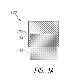
with a recess in
a bone, the recess comprising an opening facing a joint, and deploying the
implant out of
the implant deployment tool, through the opening, and at least partially in
the recess
[0007] After deployment, the implant may be 1 mm to 3 mm proud. The
method may further comprise radially compressing the first portion of the
implant in the
implant deployment tool. The method may further comprise forming the recess.
Forming
the recess may comprise using a drill bit. Deploying the implant may comprise
urging
the implant through an interior of the implant deployment tool using a
plunger.
Deploying the implant may be manual. Deploying the implant may be mechanically
assisted. Deploying the implant may comprise screwing the implant into the
recess.
[0008] In some embodiments, a method of manufacturing the implant
comprises, or alternatively consists essentially of, positioning hydrogel
material in a well
of a mold, positioning porous material in an upper portion of the well and
protruding
from the well, and freezing and thawing the hydrogel material at least once.
[0009] Positioning the porous material may comprise anchoring the porous
material.
[0010] In some embodiments, a method of manufacturing the implant
comprises, or alternatively consists essentially of, aligning a well of a
second mold
portion with a well of a first mold portion, the well of the first mold
portion comprising a
porous material, positioning hydrogel material in the well of the second mold
portion and
partially in the well of the first mold portion, and freezing and thawing the
hydrogel
material at least once.
[0011] The method may further comprise positioning the porous material in
the well of the first mold portion. Positioning the hydrogel material may be
through a
closable port, and further comprising closing the closable port. The method
may
-2-
CA 02981061 2017-09-26
WO 2016/161025 PCT/US2016/025080
comprising forming flash between the first mold portion and the second mold
portion.
The method may further comprise removing the flash. The porous material may
comprise
a disc shape.
[0012] In some embodiments, an implant system configured for implantation
in a joint comprises, or alternatively consists essentially of, a first part
and a second part.
The first part comprises an implant. The implant comprises, or alternatively
consists
essentially of, a first portion comprising a hydrogel, a second portion
comprising a porous
material and the hydrogel in pores of the porous material, and a third portion
comprising
the porous material. The first portion is free of or lacks the porous
material. The third
portion is free of or lacks the hydrogel. The second part comprises sidewalls,
a bottom, a
cavity at least partially defined by the sidewalls and the bottom, and an
anchoring
element. The cavity is configured to at least partially receive the implant.
One of the
porous material and the sidewalls of the second part comprises a detent and
the other of
the porous material and the sidewalls of the second part comprises a groove
configured to
interact with the detent when the implant is at least partially in the cavity
of the second
part.
[0013] The porous material may comprise a toroidal shape. The porous
material may comprise a detent extending radially inward. The anchoring
element may
selected from the group consisting of a barb, and anchor, and a hole in the
bottom of the
second part and a screw configured to extend through the hole in the bottom of
the second
part. The anchor may comprise an insert, a finger extending radially outwardly
and
towards a top of the implant system, a wire threaded through holes in the
bottom of the
second part, and a knot configured to be tightened upon pulling of ends of the
wire.
[0014] In some embodiments, an implant system configured for implantation
in a joint comprises, or alternatively consists essentially of, a first part
and a second part.
The first part comprises an implant comprising a first portion comprising a
hydrogel, a
second portion comprising a porous material and the hydrogel in pores of the
porous
material, and a third portion comprising the porous material. The first
portion is free of
or lacks the porous material. The third portion is free of or lacks the
hydrogel. The
second part comprises sidewalls, a bottom, and a cavity at least partially
defined by the
sidewalls and the bottom. The cavity is configured to at least partially
receive the
implant.
-3-
CA 02981061 2017-09-26
WO 2016/161025 PCT/US2016/025080
[0015] One of the porous material and the sidewalls of the second part may
comprise a detent and the other of the porous material and the sidewalls of
the second
part may comprise a groove configured to interact with the detent when the
implant is at
least partially in the cavity of the second part. The second part may further
comprise an
anchoring element. The anchoring element may comprise a barb. The anchoring
element
may comprise an anchor comprising an insert, a finger extending radially
outwardly and
towards a top of the implant system, a wire threaded through holes in the
bottom of the
second part, and a knot configured to be tightened upon pulling of ends of the
wire. The
ends of the wire may form a loop. The anchoring element may comprise a hole in
the
bottom of the second part and a screw configured to extend through the hole in
the
bottom of the second part. The anchoring element may comprise a hole in the
sidewalls
of the second part and a second screw configured to extend through the hole in
the
sidewalls of the second part.
[0016] In some embodiments, an implant system configured for implantation
in a joint comprises a first portion comprising a hydrogel, a second portion
comprising a
porous material and the hydrogel in pores of the porous material, and a third
portion
comprising the porous material. The first portion is free of or lacks the
porous material.
The third portion is free of or lacks the hydrogel. The third portion is
configured to
contact bone. Pores of the porous material are configured to allow bone
infiltration.
[0017] The first portion may comprise a contoured surface. The contoured
surface may be customized for a particular subject based on scan data. The
scan data may
comprise at least one of computerized tomography, computerized axial
tomography,
positron emission tomography, and magnetic resonance imaging. The porous
material
may comprise at least one of aluminum, titanium, and stainless steel. The
porous material
may comprise titanium mesh. The porous material may comprise printed titanium.
The
porous material may comprise at least one of alumina, zirconia, titania, and
steatite. The
porous material may comprise PEEK. The porous material may have a porosity
between
45 ppi and 80 ppi. Pores of the porous material may have a dimension between
100 p.m
and 500 p.m. The first portion may comprise a hemispherical shape. The first
portion
may comprise a wedge shape.
[0018] In some embodiments, an implant system configured for implantation
in a joint comprises an implant comprising a first portion comprising a
hydrogel, a second
portion comprising a porous material and the hydrogel in pores of the porous
material,
-4-
CA 02981061 2017-09-26
WO 2016/161025 PCT/US2016/025080
and a third portion comprising the porous material. The first portion is free
of or lacks
the porous material. The third portion is free of or lacks the hydrogel.
[0019] The second portion may be between the first portion and the second
portion. The hydrogel may comprise polyvinyl alcohol (PVA). The hydrogel may
comprise water. The hydrogel may comprise saline. The porous material may
comprise
an oxide ceramic. The porous material may comprise at least one of aluminum,
titanium,
and stainless steel. The porous material may comprise titanium mesh. The
porous
material may comprise printed titanium. The porous material may comprise PEEK.
The
porous material may comprise at least one of alumina, zirconia, titania, and
steatite. The
porous material may have a porosity between 45 ppi and 80 ppi. Pores of the
porous
material may have a dimension between 100 p.m and 500 p.m. The first portion
may
comprise an annular flange. The third portion may comprise threads.
[0020] The first portion may comprise a contoured surface. The contoured
surface may be customized for a particular subject based on scan data. The
scan data may
comprise at least one of computerized tomography, computerized axial
tomography,
positron emission tomography, and magnetic resonance imaging.
[0021] The first portion may comprise a hemispherical shape. The second
portion may comprise a hemispherical shape. The third portion may comprise a
cylindrical shape. The first portion may comprise a wedge shape. The third
portion may
comprise a wedge shape. The porous material may comprise a disc shape. The
porous
material may comprise a toroidal shape. The porous material may comprise a
detent
extending radially inward. The porous material may comprise an aperture
through a
sidewall of the porous material. The hydrogel may at least partially extend
through the
aperture. The porous material may comprise a finger extending radially
outwardly and
towards a top of the implant system. The porous material may comprise a barb.
[0022] The implant may be load bearing. The implant may be non-
biodegradable. The implant system may be configured to be placed in at least
one of a
toe, finger, ankle, knee, shoulder, hip, or other joint. A lateral dimension
of the first
portion may be between 6 mm and 10 mm. A lateral dimension of the first
portion may
be between 5% and 15% larger than a lateral dimension of the third portion. A
ratio of a
lateral dimension of the first portion to a lateral dimension of the third
portion may be
between 1.05 and 1.3.
-5-
CA 02981061 2017-09-26
WO 2016/161025 PCT/US2016/025080
[0023] The implant system may further comprise a second part comprising
sidewalls, a bottom, and a cavity at least partially defined by the sidewalls
and the
bottom. The cavity may be configured to at least partially receive the
implant. The
porous material may comprise a groove extending radially inward and the second
part
may comprise a detent extending radially inward from the sidewalls of the
second part.
The detent may be configured to interact with the groove when the implant is
at least
partially in the cavity of the second part. The porous material may comprise a
detent
extending radially outward and the second part may comprise a groove extending
radially
outward into the sidewalls of the second part. The detent may be configured to
interact
with the groove when the implant is at least partially in the cavity of the
second part.
[0024] The second part further may comprise an anchoring element. The
anchoring element may comprise a barb. The barb may comprise a plurality of
barbs.
The plurality of barbs may be vertically stacked. The anchoring element may
comprise
an anchor comprising an insert, a finger extending radially outwardly and
towards a top
of the implant system, a wire threaded through holes in the bottom of the
second part, and
a knot configured to be tightened upon pulling of ends of the wire. The ends
of the wire
may form a loop. The anchoring element may comprise a hole in the bottom of
the
second part and a screw configured to extend through the hole in the bottom of
the second
part. The anchoring element may comprises a plurality of holes in the bottom
of the
second part and a plurality of screws configured to extend through the
plurality of holes
in the bottom of the second part. The anchoring element may comprise a hole in
the
sidewalls of the second part and a second screw configured to extend through
the hole in
the sidewalls of the second part. The anchoring element may comprise a
plurality of
holes in the sidewalls of the second part and a plurality of second screws
configured to
extend through the plurality of holes in the sidewalls of the second part.
[0025] In some embodiments, a method of treatment comprises, or
alternatively consists essentially of, aligning an implant deployment tool
with a recess in
a bone and deploying the implant out of the implant deployment tool, through
the
opening, and at least partially in the recess. The recess comprises an opening
facing a
joint.
[0026] After deployment, the implant may be 1 mm to 3 mm proud. The
method may further comprise radially compressing the first portion of the
implant in the
implant deployment tool. The method may further comprise forming the recess.
Forming
-6-
CA 02981061 2017-09-26
WO 2016/161025 PCT/US2016/025080
the recess may comprise using a drill bit. Deploying the implant may comprise
urging
the implant through an interior of the implant deployment tool using a
plunger.
Deploying the implant may be manual. Deploying the implant may be mechanically
assisted. Deploying the implant may comprise screwing the implant into the
recess.
[0027] In some embodiments, a method of manufacturing the implant
comprises positioning hydrogel material in a well of a mold, positioning
porous material
in an upper portion of the well and protruding from the well, and freezing and
thawing the
hydrogel material at least once. Positioning the porous material may comprise
anchoring
the porous material.
[0028] In some embodiments, a method of manufacturing the implant
comprises aligning a well of a second mold portion with a well of a first mold
portion.
The well of the first mold portion comprises a porous material. The method
further
comprises positioning hydrogel material in the well of the second mold portion
and
partially in the well of the first mold portion and freezing and thawing the
hydrogel
material at least once. The method may further comprising positioning the
porous
material in the well of the first mold portion. Positioning the hydrogel
material may be
through a closable port. The method may further comprise closing the closable
port.
[0029] The method may comprising forming flash between the first mold
portion and the second mold portion. The method may further comprise removing
the
flash. The porous material may comprises a disc shape. The porous material may
comprises a toroidal shape.
BRIEF DESCRIPTION OF THE DRAWINGS
[0030] Certain features, aspects, and advantages of the disclosure are
described with reference to drawings, which are intended to illustrate, but
not to limit, the
various inventions disclosed herein. It is to be understood that the attached
drawings are
for the purpose of illustrating concepts and embodiments of the disclosure and
may not be
to scale.
[0031] Figure 1A schematically illustrates an example implant;
[0032] Figure 1B schematically illustrates an example implant;
[0033] Figure 1C schematically illustrates an example implant;
[0034] Figure 2 is a photo of an example implant;
-7-
CA 02981061 2017-09-26
WO 2016/161025 PCT/US2016/025080
[0035] Figures 3A and 3B schematically illustrate an example method of
positioning an example implant;
[0036] Figure 3C schematically illustrates an example method of positioning
the example implant of Figure 1B;
[0037] Figure 4 schematically illustrates an example method of
manufacturing
example implants;
[0038] Figures 5A-5C schematically illustrate an example method of
manufacturing example implants;
[0039] Figure 6 schematically illustrates another example implant
[0040] Figure 7A schematically illustrates an example implant;
[0041] Figure 7B schematically illustrates an example method of positioning
an example implant;
[0042] Figure 7C schematically illustrates an example method of positioning
an example implant;
[0043] Figure 8A schematically illustrates an example implant;
[0044] Figure 8B schematically illustrates an example method of positioning
an example implant;
[0045] Figure 8C schematically illustrates an example method of positioning
an example implant;
[0046] Figure 9A is a side view of an example implant;
[0047] Figure 9B is a cross-sectional view of the implant of Figure 9A;
[0048] Figures 9C and 9D are top and side perspective exploded views of the
implant of Figure 9A;
[0049] Figure 9E is a cross-sectional view of an example implant;
[0050] Figure 10A is a side view of an example implant;
[0051] Figure 10B is a cross-sectional view of the implant of Figure 10A;
[0052] Figures 10C-10E are top and side perspective exploded views of the
implant of Figure 10A;
[0053] Figure 1OF is a cross-sectional view of an example implant;
[0054] Figure 11A is a side view of an example implant;
[0055] Figure 11B is a cross-sectional view of the implant of Figure 11A;
[0056] Figure 11C is a top and side perspective view of the implant of
Figure
11A;
-8-
CA 02981061 2017-09-26
WO 2016/161025 PCT/US2016/025080
[0057] Figure 11D is a top and side perspective exploded view of the
implant
of Figure 11A;
[0058] Figure 11E is a cross-sectional view of an example implant;
[0059] Figure 12A is a side cross-sectional view of an example implant;
[0060] Figure 12B is a side cross-sectional view of an example implant;
[0061] Figure 12C is a side cross-sectional view of an example implant;
[0062] Figure 12D is a side cross-sectional view of example implants;
[0063] Figure 13A is a top and side perspective view of an example implant;
and
[0064] Figure 13B is plan view of an example device for manufacturing
example implants.
DETAILED DESCRIPTION
[0065] The discussion and the figures illustrated and referenced herein
describe various embodiments of a cartilage implant, as well as various tools,
systems,
and methods related thereto. A number of these devices and associated
treatment
methods are particularly well suited to replace deteriorated or otherwise
damaged
cartilage within a joint. Such implants are configured to remain within the
patient's joint
on a long-term basis (e.g., for most or all of the life of the patient or
subject), and as such,
are configured, in some embodiments, to replace native cartilage. In some
embodiments,
an implant is configured to be substantially non-biodegradable and/or non-
erodable. In
some embodiments, an implant is configured to remain within the patient's
joint or other
portion of the anatomy for a minimum of 10 to 100 years (e.g., about 10 years,
about 20
years, about 25 years, about 30 years, about 35 years, about 40 years, about
45 years,
about 50 years, about 55 years, about 60 years, about 65 years, about 70
years, about 75
years, about 80 years, about 85 years, about 90 years, about 95 years, about
100 years,
duration ranges between the foregoing values, etc.) without losing structural
and/or
physical properties and/or without losing ability to function as a cartilage
replacement
component or device. In some embodiments, an implant is configured to remain
within
the anatomy for greater than 100 years without losing structural and/or
physical
properties and/or without losing ability to function as a cartilage
replacement component.
Certain implants described herein can be used to treat osteoarthritis,
rheumatoid arthritis,
other inflammatory diseases, generalized joint pain, joints damaged in an
accident, joints
-9-
CA 02981061 2017-09-26
WO 2016/161025 PCT/US2016/025080
damaged while participating in athletics, joints damaged due to repetitive
use, and/or
other joint diseases. However, the various devices, systems, methods, and
other features
of the embodiments disclosed herein may be utilized or applied to other types
of
apparatuses, systems, procedures, and/or methods, including arrangements that
have non-
medical benefits or applications.
[0066] Certain
embodiments described herein may be advantageous because
they include one, several, or all of the following benefits: (i) improved
osseointegration
compared to implants having a hydrogel surface; (ii) improved coupling of
disparate
implant materials; (iii) improved cavity wall apposition compared to
substantially
cylindrical implants; (iv) reduced implant height; (v) reduced depth of a bone
cavity
configured to receive an implant; (vi) improved structural stability; and/or
(vii) increased
manufacturing flexibility.
[0067] Figure 1A
schematically illustrates an example implant 100. The
implant 100 comprises, or alternatively consists essentially of, a first
portion 102, a
second portion 104, and a third portion 106. The first portion 102 and the
second portion
104 of the implant 100, as well as other implants disclosed herein (as is the
case for each
implant feature unless described otherwise), comprises, or alternatively
consists
essentially of, a hydrogel (e.g., a hydrogel or other formulation comprising
polyvinyl
alcohol (PVA) hydrogel). The third portion 106 comprises, or alternatively
consists
essentially of, a porous material (e.g., a material or section comprising
porous ceramic
material (e.g., oxide-ceramic), metal (e.g., titanium (e.g., titanium mesh,
printed
titanium), stainless steel (e.g., stainless steel wool)), plastic (e.g.,
polyaryl ether ketone
(PAEK) (e.g., polyether ether ketone (PEEK))), other biocompatible materials,
combinations thereof, and the like).
[0068] The first portion
102 and the second portion 104 of the implant 100
can comprise one or more other materials, either in addition to or in lieu of
PVA, such as,
for example, other hydrogels, other polymeric materials, additives, and/or the
like. As
discussed herein, the second portion 104 comprises porous material. In
some
embodiments, the PVA content of a hydrogel is about 40% by weight. The PVA
content
of hydrogel in an implant 100 can be less than or more than about 40% by
weight (e.g.,
about 10%, about 15%, about 20%, about 25%, about 30%, about 32%, about 34%,
about
36%, about 37%, about 38%, about 39%, about 41%, about 42%, about 43%, about
44%,
about 46%, about 48%, about 50%, about 55%, about 60%, about 65%, about 70%,
less
-10-
CA 02981061 2017-09-26
WO 2016/161025 PCT/US2016/025080
than about 10%, more than about 70%, ranges between such values, etc.), as
desired or
required.
[0069] The hydrogel of the implant 100, as well as other implants disclosed
herein, can comprise water, saline, other liquids, combinations thereof,
and/or the like. In
some embodiments, saline may be preferred over water, because, under certain
circumstances, saline can help maintain osmotic balance with surrounding
anatomical
tissues following implantation. The exact composition of hydrogel in an
implant 100
(e.g., PVA or other hydrogel materials, water, saline or other liquids, other
additives, etc.)
can be selected so as to provide the implant 100 with the desired or required
strength,
load bearing capacity, compressibility, flexibility, longevity, durability,
resilience,
coefficient of friction, and/or other properties and characteristics. Thus, in
some
embodiments, any hydrogel portion of the implants disclosed herein consist
essentially of
saline and PVA. In some embodiments, such hydrogel portions of the implants do
not
comprise any additional additives (e.g., growth factors, surface or other
coatings, etc.). In
addition, according to some embodiments, the hydrogel portions of any of the
implant
configurations disclosed herein comprises a consistent concentration (e.g., no
concentration gradients), density and/or other chemical and/or physical
properties
throughout.
[0070] In some embodiments, the implant 100, as well as other implants
disclosed herein, is configured for drug delivery and/or is seeded with growth
factors
and/or cells. In some embodiments, the implant 100 comprises one or more of
the
following: chondrocytes, growth factors, bone morphogenetic proteins,
collagen,
hyaluronic acid, nucleic acids, and stem cells. Such factors and/or any other
materials
included in the implant 100 and selectively delivered to an implant site can
help facilitate
and/or promote the long-term fixation of the implant 100 at the joint or other
target area
of the anatomy.
[0071] In some embodiments, the hydrogel comprises PVA and/or any other
polymeric material. In some embodiments, the content of PVA in the hydrogel is
between about 35% and about 45% by weight (e.g., about 35%, about 36%, about
37%,
about 38%, about 39%, about 40%, about 41%, about 42%, about 43%, about 44%,
about
45%, ranges between such values, etc.). In some embodiments, the content of
PVA in the
hydrogel is greater than about 45% by weight (e.g., about 45%, about 50%,
about 55%,
about 60%, about 65%, about 70%, greater than about 70%, ranges between such
values,
-11-
CA 02981061 2017-09-26
WO 2016/161025 PCT/US2016/025080
etc.) or less than about 35% by weight (e.g., about 5%, about 10%, about 15%,
about
20%, about 25%, about 30%, about 35%, ranges between such values, less than
about
5%, etc.). In some embodiments, the content of PVA or other component in the
hydrogel
is about 40% by weight.
[0072] In some embodiments, the implant 100 is load bearing and generally
non-biodegradable (e.g., non-bioerodable). In some embodiments, the implant
100 is
configured for placement in at least one of a toe, finger, ankle, knee,
shoulder, hip, or any
other joint. In some embodiments, a transition between the upper surface and
the
sidewalls is generally curved or otherwise smooth.
[0073] In some embodiments, the first portion 102 of the implant may have a
lateral dimension (e.g., diameter) between about 6 mm and about 10 mm (e.g.,
about 6
mm, about 7 mm, about 8 mm, about 9 mm, about 10 mm, ranges between such
values,
etc.), as measured in an uncompressed state. Lateral dimensions smaller than
about 6 mm
(e.g., between about 2 mm and about 6 mm) and larger than about 10 mm (e.g.,
between
about 10 mm and about 14 mm) are also possible for use in subjects with small
or large
bones, respectively, and/or for use in joints with small or large bones,
respectively.
[0074] The third portion 106 of the implant can comprise a porous material,
such as, for example, a porous ceramic (e.g., oxide-ceramic), metal (e.g.,
titanium (e.g.,
titanium mesh, printed titanium), stainless steel (e.g., stainless steel
wool)), plastic (e.g.,
polyaryl ether ketone (PAEK) (e.g., polyether ether ketone (PEEK))), other
biocompatible materials, combinations thereof, and the like). The third
portion 106 may
be free or substantially free from the hydrogel of the first portion 102. In
some
embodiments, the third portion 106 is substantially rigid or non-deformable.
In some
embodiments, the third portion 106 is at least partially deformable. The pores
and/or
other openings of the third portion 106 may promote osseointegration of the
implant 100
in a bone. Compared to an implant consisting essentially of hydrogel, an
implant
comprising one or more porous materials (e.g., porous ceramic, metal, plastic,
etc.) may
have a reduced height because the porous ceramic and/or other porous material
may
provide structural stability and/or because the porous ceramic or other porous
material
may provide better osseointegration such that less contact with bone provides
at least as
much osseointegration.
[0075] The third portion 106 is illustrated in Figure 1A as a disc,
although
other shapes of the third portion 106 are also possible. In some embodiments,
the third
-12-
CA 02981061 2017-09-26
WO 2016/161025 PCT/US2016/025080
portion 106 may be toroidal, wedge-shaped, etc., for example as described in
further
detail herein. In some embodiments, the third portion 106 is substantially
rigid, semi-
rigid, and/or non-deformable. In some embodiments, the second portion 104
comprises
the hydrogel of the first portion 102 within pores of the porous material of
the third
portion 106. According to some embodiments, the diameter or other lateral
dimension of
the second portion 104 and/or third portion 106 is smaller than the diameter
or other
lateral dimension of the first portion 102 of the implant. As discussed
herein, this can
permit the implant 100 to be radially compressed (e.g., during delivery into a
target
anatomical site of a subject), especially in embodiments where the first
portion 102 is
more readily radially compressible than the second portion 104 and/or the
third portion
106 (e.g., because of the material(s) included in each portion). For example,
in some
embodiments, the diameter or other lateral dimension of the second portion 104
and/or
third portion 106 is between about 70% and about 95% (e.g., about 70%, about
75%,
about 80%, about 85%, about 90%, about 95%, ranges between the foregoing
percentages, etc.) of the diameter or other lateral dimension of the first
portion 102.
[0076] According to some embodiments, the second portion 104 and the
third
portion 106 may comprise an oxide ceramic, for example oxide ceramics from
CeramTec
of Laurens, South Carolina, as provided in Tables 1 and 2, although other
materials and
combinations of materials are also possible (e.g., non-oxide ceramics, non-
ceramics).
Table 1
Alumina
Alumina Alumina Alumina Alumina
Property Units
(99.5%)
(92%) (94%) (96%) (99.5%) I
Density g/cm3 3.65 3.6 3.7 3.9 3.9
Hardness HV 0.5 1300 1200 1350 1700 1700
Flexural MPa 240 290 296 310 310
Strength (k PSI) (34.8) (42) (43) (45) (45)
Fracture
mpaxmin 5 3 4 4 4
Toughness
Young's GPa 300 289 303 372 376
Modulus (x106 PSI) (44) (42) (44) (54) (54)
-13-
CA 02981061 2017-09-26
WO 2016/161025
PCT/US2016/025080
Table 1
Alumina
Alumina Alumina Alumina Alumina
Property Units
(99.5%)
(92%) (94%) (96%) (99.5%) I
II
Shear GPa 120 121 127 152 152
Modulus (x106 PSI) (17) (17.5) (18.5) (22) (22)
Polsson -- 0.24 0.21 0.21 0.21 0.21
Thermal
Expansion x10-6/ C 7.0 6.6 6.5 6.8 6.7
(300 C)
Thermal
Expansion x10-6/ C 7.3 7.6 7.6 7.9 7.8
(700 C)
Thermal
Expansion x10-6/ C 7.5 8.2 8.1 8.3 8.2
(1,000 C)
Thermal
Conductivity W/mK 21.0 21.0 24.0 30.0 30.0
at 25 C
Volume
ohmxcm >1014 >1014 >1014 >1014 >1014
Resistivity
Specific
J/gK 0.96 0.8 1.1 0.8 0.8
Heat
Dielectric
V/mil -- 200 210 230 220
Strength
Dielectric
Constant at -- -- 9.0 9.3 9.8 9.8
1 MHz
Dissipation
Factor at 1 -- 9.0x10⁻⁴ 3.0x10⁻⁴ 3.0x10⁻⁴ 1.0x10-
4 1.0x10⁻⁴
MHz
-14-
CA 02981061 2017-09-26
WO 2016/161025
PCT/US2016/025080
Table 1
Alumina
Alumina Alumina Alumina Alumina
Property Units
(99.5%)
(92%) (94%) (96%) (99.5%) I
II
Loss Factor
__ -- 3.0x10⁻³ 3.0x10⁻³ 1.0x10⁻³ 1.0x10-
3
at 1 MHz
Table 2
Toughene
Steatite
Property Units Zirconia Titania Steatite I
d Alumina II
Density g/cm3 4.0 6.0 4.0 2.7 2.8
Hardness HV 0.5 1600 1150 800 450 420
Flexural MPa 448 752 138 131 145
Strength (k PSI) (65) (109) (20) (19) (21)
Fracture
MPaxm1/2
4 10 3 -- --
Toughness
Young's GPa 186 227 108 112
__
Modulus (x106 PSI) (27) (33) (16) (16)
Shear GPa 80 90 43 45
__
Modulus (x106 PSI) (11.6) (13.0) (6.3) (6.5)
Polsson -- -- 0.33 0.27 0.23 0.25
Thermal
Expansion x10-6/ C 7.9 -- 8.3 8.2 6.9
(300 C)
Thermal
Expansion x10-6/ C 8.5 10.0 9.0 8.9 7.8
(700 C)
Thermal
Expansion x10-6/ C 9.6 11.0 9.0 9.4 8.0
(1,000 C)
-15-
CA 02981061 2017-09-26
WO 2016/161025 PCT/US2016/025080
Table 2
Toughene
Steatite
Property Units Zirconia Titania Steatite I
d Alumina II
Thermal
Conductivity W/mK 25.0 2.7 11.9 5.5 5.9
at 25 C
Volume
ohmxcm 9.0x10¹³ >1012 >1014 >1014
Resistivity
Specific
J/gK 0.96 0.4 0.7 1.1 1.1
Heat
Dielectric
V/mil 100 210 230
Strength
Dielectric
Constant at 28 85 5.8 6.1
1 MHz
Dissipation
Factor at 1 9.0x10⁻⁴
5.0X 10⁻⁴
1.9 x 10⁻³
8.0x10⁻⁴
MHz
Loss Factor
1.1x10⁻² 5.0x10-
3
at 1 MHz
[0077] According to some embodiments, the second portion 104 and the third
portion 106 may comprise a metal, for example titanium mesh, printed titanium,
stainless
steel, etc. According to some embodiments, the second portion 104 and the
third portion
106 may comprise a plastic, for example PAEK, PEEK, etc.
[0078] In some embodiments, the porous material can have a porosity
between about 45 pores per inch (ppi) and about 80 ppi (e.g., about 45 ppi,
about 50 ppi,
about 55 ppi, about 60 ppi, about 65 ppi, about 70 ppi, about 75 ppi, about 80
ppi, ranges
between such values, etc.). The pores of the porous material may have a
diameter or
other dimension between about 100 micrometers (microns; p.m) and about 500 p.m
(e.g.,
about 100 p.m, about 150 p.m, about 200 p.m, about 250 pm, about 300 pm, about
350
p.m, about 400 p.m, about 450 p.m, about 500 p.m, ranges between such values,
etc.), as
desired or required.
-16-
CA 02981061 2017-09-26
WO 2016/161025 PCT/US2016/025080
[0079] In some embodiments, pores of the porous material in the second
portion 104 are different than pores of the porous material in the third
portion 106. For
example, the pores of the porous material in the second portion 104 may be
configured to
allow hydrogel infiltration while the pores of the porous material in the
third portion 106
may be configured to allow osseointegration. In some embodiments, the porous
material
in the second portion 104 is different than the porous material in the third
portion 106.
For example, the porous material in the second portion 104 may comprise a
first material
having a property and the porous material in the third portion 106 may
comprise a second
material having a property different than the property of the first material.
The property
may comprise, for example, the material itself (e.g., whether ceramic, metal,
plastic, etc.),
porosity, pore size, dimensions, deformability, etc.
[0080] Overlap of hydrogel material of the first portion 102 and porous
material of the third portion 106 in the second portion 104, for example by
the hydrogel
material filling pores of the porous material, may securely anchor the first
portion 102 to
the third portion 106, for example compared to an implant in which a surface
of a
hydrogel material is adhered to a surface of another material. In some
embodiments, a
ratio of a height of the second portion 104 to a height of the third portion
106 is between
about 1:5 and about 5:1 (e.g., about 1:5, about 1:4, about 1:3, about 1:2,
about 1:1, about
2:1, about 3:1, about 4:1, about 5:1, ranges between such values, etc.). In
some
embodiments, a ratio of a height of the second portion 104 to a height of the
ceramic
material (e.g., a height of the second portion 104 and a height of the third
portion 106) is
between about 1:5 and about 1:1.1 (e.g., about 1:5, about 1:4, about 1:3,
about 1:2, about
1:1.5, about 1:1.4, about 1:1.3, about 1:1.2, about 1:1.1, ranges between such
values,
etc.). In some embodiments, a ratio of a height of the third portion 106 to a
height of the
ceramic material (e.g., a height of the second portion 104 and a height of the
third portion
106) is between about 1:5 and about 1:1.1 (e.g., about 1:5, about 1:4, about
1:3, about 1:2,
about 1:1.5, about 1:1.4, about 1:1.3, about 1:1.2, about 1:1.1, ranges
between such
values, etc.).
[0081] Compared to an implant consisting essentially of hydrogel, an
implant
comprising porous material (e.g., porous ceramic, metal, plastic, etc.) may
have a reduced
height. For example, compared to implants consisting only or essentially of a
hydrogel
material, such hybrid implants can have a height that is reduced by between
about 5% and
about 30% (e.g., about 5%, about 10%, about 15%, about 20%, about 25%, about
30%,
-17-
CA 02981061 2017-09-26
WO 2016/161025 PCT/US2016/025080
ranges between the foregoing percentages, etc.). In some embodiments, the
third portion
106 of the implant 100 may provide improved or enhanced structural stability
to the
implant 100. Such improved or enhanced structural stability may be beneficial
for use
with large bones, although use with small bones is also possible.
[0082] Although the implant 100 is schematically illustrated as a
cylindrical
plug, other shapes of the implant 100 are also possible. For example, an upper
surface of
the first portion 102 may be contoured to abut particular anatomy (e.g.,
planar (e.g., flat),
non-planar (e.g., curved, concave, convex, undulating, fluted)). The implant
100 can
include a generally circular or oval cross-sectional shape. In some
embodiments, the
implant 100 is generally shaped like a cylinder or a mushroom. The overall
shape of any
of the implants disclosed herein can vary depending on the specific
application or use.
For example, the shape of at least part of a portion 102, 104, 106 can be
generally
polygonal (e.g., rectangular, round, hexagonal), irregular, and/or the like.
[0083] A molding process, for example as described herein with respect to
Figures 4 and/or with respect to Figures 5A-5C, may be used to form particular
shape of
an implant 100.
[0084] In some embodiments, means for treating a joint (e.g., the implant
100)
comprises, or alternatively consists essentially of, means for providing a
lubricious
surface (e.g., the first portion 102) and means for promoting osseointegration
(e.g., the
third portion 106).
[0085] Figure 1B schematically illustrates an example implant 150. The
implant 150 comprises, or alternatively consists essentially of, a first
portion 152, a
second portion, and a third portion 156. The first portion 152 and the second
portion of
the implant 150 comprises, or alternatively consists essentially of, a
hydrogel (e.g., a
hydrogel or other formulation comprising PVA hydrogel). The second portion is
not
illustrated due to the opacity of the hydrogel material of the first portion
152. The third
portion 156 comprises, or alternatively consists essentially of, a porous
material (e.g., a
material or section comprising porous ceramic material (e.g., oxide-ceramic) ,
metal (e.g.,
titanium (e.g., titanium mesh, printed titanium), stainless steel (e.g.,
stainless steel wool)),
plastic (e.g., polyaryl ether ketone (PAEK) (e.g., polyether ether ketone
(PEEK))), other
biocompatible materials, combinations thereof, and the like). The first
portion 152, or the
hydrogel material, comprises a contoured upper surface 162. The upper surface
162 may
be rounded at the edges and then flat (e.g., as illustrated in Figure 1B),
contoured to
-18-
CA 02981061 2017-09-26
WO 2016/161025 PCT/US2016/025080
correspond to an opposing surface, etc. The hydrogel material of the implant
150 also
includes a taper 164 towards the porous material of the third portion 156.
Other shapes,
surface contours, and combinations thereof are also possible.
[0086] Figure 1C schematically illustrates an example implant 180. The
implant 180 comprises, or alternatively consists essentially of, a first
portion 182, a
second portion 184, and a third portion 186. The third portion 186 comprises
threads
188, which can allow the implant to be screwed into bone and/or a hole in
bone. The
implant 180 may take the shape of a screw. The threads 188 may comprise a same
material as the third portion 186 (e.g., porous material) or a different
material than the
third portion 186 (e.g., a non-porous ceramic, metal, plastic, etc.). Aspects
of orthopedic
screws, dental implants, etc. such as coatings, surface features, etc. may be
integrated into
the threads 188 and/or the third portion 186. In some embodiments, the second
portion
184 may comprise threads. Threads in the second portion 184 may help, for
example, to
anchor the hydrogel material to the porous material and/or inhibit relative
longitudinal
movement therebetween. Threads of the second portion 184 may be the same or
different
than the threads 188 of the third portion 186.
[0087] Figure 2 illustrates one embodiment of an implant 200 comprising a
hydrogel section and a porous material section. Similar to the implant 100
discussed
above, the illustrated implant 200 comprises a first hydrogel portion 202, a
second
overlap portion 204, and a third porous material portion 206. In the depicted
arrangement, the third portion 206 is substantially free from the hydrogel of
the first
portion 202, as highlighted by the dotted line 208 between the second portion
204 and the
third portion 206. More or less overlap in the second portion 204 is also
possible, for
example by using less hydrogel material and/or less porous material, by
adjusting height
of the implant 200, etc. In some embodiments, a ratio of a height of the
second portion
204 (e.g., measured at an average of the hydrogel level) to a height of the
implant 200 is
between about 5% and about 40% (e.g., about 5%, about 10%, about 15%, about
20%,
about 25%, about 30%, about 35%, about 40%, ranges between such values, etc.).
In
some embodiments, a ratio of a height of the second portion 204 to a height of
the first
portion 202 is between about 15% and about 75% (e.g., about 15%, about 25%,
about
35%, about 45%, about 55%, about 65%, about 75%, ranges between such values,
etc.).
In some embodiments, a ratio of a height of the second portion 204 to a height
of the third
portion 206 is between about 10% and about 90% (e.g., about 10%, about 20%,
about
-19-
CA 02981061 2017-09-26
WO 2016/161025 PCT/US2016/025080
30%, about 40%, about 50%, about 60%, about 70%, about 80%, about 90%, ranges
between such values, etc.).
[0088] Figures 3A and 3B schematically illustrate an example method of
positioning an example implant 300. Similar to the implants 100, 200, the
implant 300
comprises a first hydrogel portion 302, a second overlap portion 304, and a
third porous
material portion 306.
[0089] According to some embodiments, the bone portion 308 in which the
implant 300 will be positioned has been drilled to form a hole or aperture or
recess or
cavity or crater or pit or pocket 310. In some embodiments, the lateral
dimension (e.g.,
diameter) of the hole 310 is less than the lateral dimension (e.g., diameter)
of the third
portion 306, which is rigid. In some embodiments, a lateral dimension and/or
cross-
sectional area of the hole 310 is about 5% to about 15% (e.g., about 5%, about
6%, about
7%, about 8%, about 9%, about 10%, about 11%, about 12%, about 13%, about 14%,
about 15%, ranges between such values, etc.) wider or otherwise larger than
the lateral
dimension and/or cross-sectional area of the third portion 306. The lateral
dimension
(e.g., diameter) of the hole 310 may be smaller than the lateral dimension
(e.g., diameter)
of the first portion 302, which may flex radially inwardly. Although
illustrated as a
cylindrical hole 310, other shapes are also possible (e.g., trapezoidal
tapering inwards
towards the upper surface). In some embodiments, a lateral dimension and/or
cross-
sectional area of the hole 310 is about 5% to about 15% (e.g., about 5%, about
6%, about
7%, about 8%, about 9%, about 10%, about 11%, about 12%, about 13%, about 14%,
about 15%, ranges between such values, etc.) narrower or otherwise smaller
than the
lateral dimension and/or cross-sectional area of the first portion 302. The
hole 310 may
be coated or otherwise treated prior to positioning of the implant 300.
[0090] As a result of the shape of the implant 300 and the corresponding
implant site (e.g., in the hole 310), the implant 300 may be inwardly radially
compressed
in order to insert the implant 300 in the hole 310. A delivery system or
introducer 312
and/or other delivery tools can be used to facilitate positioning of the
implant 300.
Radially inward compressive forces may facilitate delivery of an implant 300
that is at
least partially radially oversized relative to the hole 310, as discussed
further herein. The
degree to which the implant 300 can be compressed (e.g., circumferentially,
radially
inwardly, etc.) may depend on one or more factors, properties, characteristics
and/or other
considerations of the first portion 302, such as, for example, implant size,
water content,
-20-
CA 02981061 2017-09-26
WO 2016/161025 PCT/US2016/025080
ingredients and other components, strength, elasticity, surrounding
temperature, method
of manufacturing, and/or the like. Although described herein as generally
rigid, the
second portion 304 and the third portion 306 may also have some degree of
compressibility. Radial compression of an implant 300 can affect the overall
height, the
shape and/or contours of outer surfaces (e.g., top or articulating surface,
base or bottom
surface, sides, etc.), and/or one or more other properties or characteristics
of the implant
300. In some embodiments, radial compression of an implant 300 causes the
height of
the implant 300 to increase (e.g., relative to the height of the implant 300
when not
radially compressed). Consequently, careful consideration may be given to the
design of
the implant 300 based on, among other things, the expected level of radial
compression
that may occur once the implant 300 has been properly secured in the hole 310,
prior to
implantation. Otherwise, in some embodiments, upon implantation, an implant
300 may
not properly align with adjacent cartilage or other tissue surfaces in a joint
or other
anatomical location.
[0091] According to some embodiments, the implant 300 is loaded into a
delivery system 312; only the distal end of the delivery system 312 is
illustrated in Figure
3A. The delivery system 312 can comprise an outer body 314 and a plunger or
pusher
member 316. The outer body 314 may be cylindrical or may taper radially
inwardly
towards the distal end of the delivery system 312. In the illustrated
embodiment, the
plunger 316 abuts the first portion 302 of the implant 300. The delivery
system 312 can
be aligned with the hole 310, and then a user such as a surgeon can depress
the plunger
316. The plunger 316 is translatable along the longitudinal axis of the
delivery system
312 to push the implant 300 out of the distal end of the delivery system 312
into the hole
310. Depression of the plunger 316 and/or deployment of the implant 300 may be
manual, mechanically assisted, combinations thereof, and the like.
[0092] Figure 3B illustrates the implant 300 in the bone portion 308. The
hole
310 preferably has a depth that is greater than or equal to the height of the
second portion
304 and the third portion 306 such that the part of the implant 300 prolapsing
from the
bone portion 308, the load-bearing surface, comprises hydrogel and is free or
substantially free of the relatively more rigid porous material. In some
embodiments, an
upper surface of the implant 300 is about 1 millimeter (mm) to about 3 mm
above an
upper surface of the bone portion 308 (e.g., the bone of the bone portion,
remaining
cartilage, etc.), also termed "proud," designated in Figure 3B by the
measurement p,
-21-
CA 02981061 2017-09-26
WO 2016/161025 PCT/US2016/025080
which can provide a desired contour of the damaged joint surface. In some
embodiments,
such a raised or otherwise protruding configuration can assist in creating a
smoother
transition between the exposed surface of the implant 300 and adjacent native
surfaces.
[0093] The first portion 302 may have a larger lateral dimension (e.g.,
diameter) than the third portion 306 to create a "mushroom" shape, as
illustrated in
Figure 3B, as well as in Figures 1A-2 and other examples herein. In some
embodiments,
for any of the implants disclosed herein, a ratio of a lateral dimension
(e.g., diameter)
and/or cross-sectional area of the first portion 302 or a portion thereof to a
lateral
dimension (e.g., diameter) and/or cross-sectional area of the third portion
306 or a portion
thereof is between about 1 and about 1.3 (e.g., greater than or equal to about
1.05, about
1.06, about 1.07, about 1.08, about 1.09, about 1.1, about 1.11, about 1.12,
about 1.13,
about 1.14, about 1.15, about 1.16, about 1.17, about 1.18, about 1.19, about
1.2, about
1.21, about 1.22, about 1.23, about 1.24, about 1.25, about 1.26, about 1.27,
about 1.28,
about 1.29, about 1.3, ranges between such values, etc.). In other
embodiments, the ratio
is between about 1 and 1.05 (e.g., greater than or equal to about 1.01, about
1.02, about
1.03, about 1.04, about 1.05, ranges between such values, etc.), or greater
than about 1.3
(e.g., greater than or equal to about 1.3, about 1.35, about 1.4, about 1.45,
about 1.5,
about 1.55, about 1.6, etc.), as desired or required.
[0094] The smaller third portion 306 can slide into the hole 310 of the
bone
portion 308, although preferably making contact with the sidewalls or
perimeter of the
hole 310, and the larger first portion 302 can be wedged into the hole 310 of
the bone
portion 308 due to its flexibility. Referring again to Figure 3A, the implant
300 may be
held in the delivery system 312 by radial compression of the implant 300. The
substantially rigid porous material of the second portion 304 and the third
portion 306
might not be susceptible to radial compression. The larger first portion 302
can be
radially compressed or wedged into the outer body 314 due to its flexibility
while the
smaller third portion 306 may slide within the outer body 314.
[0095] Figure 3C schematically illustrates an example method of positioning
the example implant 150 of Figure 1B. The implant 150 in a bone portion 358,
for
example by the method of Figures 3A and 3B or another method. The third
portion 156
makes contact with the perimeter of the hole in the bone portion 358. The
hydrogel
material of the second portion and usually the first portion 152 is radially
compressed in
the hole in the bone portion 358. Figure 3C illustrates a radially compressed
segment 366
-22-
CA 02981061 2017-09-26
WO 2016/161025 PCT/US2016/025080
and an uncompressed segment 368. The uncompressed segment 368 is over the
surface
of the bone portion 358. Referring again to Figure 3B, the segment of the
first portion
302 that is proud may be radially larger than the segment of the first portion
302 that is in
the hole 310 in the bone portion 308.
[0096] Figure 4 schematically illustrates an example method of
manufacturing
example implants 400. A mold 408 comprises a plurality of wells or cavities or
recesses
or holes 410. Bottoms of the wells 410 may be contoured, for example for a
specific
anatomy location in which an implant 400 will be placed, and/or other factors
or
considerations. For example, an implant 400 can be configured to generally or
specifically match the slopes, contours, and/or other features of the existing
cartilaginous
and/or bone tissue (e.g., planar (e.g., flat), non-planar (e.g., curved,
concave, convex,
undulating, fluted)), a recess or hole or cavity created in the bone, and/or
the like.
Accordingly, the function of a rehabilitated joint or other targeted
anatomical region
being treated can be improved. The bottom surfaces of the implants 400 in the
mold 408
will be the upper load bearing surfaces of the implants 400 in use. In some
embodiments,
the mold 408 further comprises a plurality of anchors 412 configured to
inhibit or prevent
third portions 406 from sinking into first portions 402 during the
manufacturing process.
The anchors 412 may comprise wire, clamps, releasable adhesive, combinations
thereof,
and the like. Hydrogel material of the first portion 402 fills pores of the
porous material
of the third portion 406 in the second portion 404. The upper surface of the
hydrogel may
be generally planar, although other shapes are also possible. A mold
configured to make
one implant 400 at a time is also possible.
[0097] Figures 5A-5C schematically illustrate an example method of
manufacturing example implants 500. As shown in Figure 5A, in some
embodiments, a
first mold portion 508 comprises a plurality of wells or cavities or recesses
or holes 510.
The first mold portion 508 may be the same or different than the mold 408. In
contrast to
the method of Figure 4, the implants 400 are made "upside down" in that the
bottom
surfaces of the implants 500 in the mold 508 will not be the upper load
bearing surfaces
of the implants 500 in use. Porous material 505, for example in the shape of
discs,
grommets, etc., are inserted into the wells 510. Since the porous material 505
are
substantially rigid, they will not conform to any contours at bottoms of the
wells 510.
The fit of the porous ceramic 505 in the mold 508 is preferably tight enough
that hydrogel
-23-
CA 02981061 2017-09-26
WO 2016/161025 PCT/US2016/025080
material subsequently inserted into the wells 510 is inhibited or prevented
from flowing
to bottoms of the wells 510.
[0098] As shown in Figure 5B, a second mold portion 512 comprises a
plurality of wells 514 configured to be aligned with the plurality of wells
510 of the first
mold portion 508. Bottoms of the wells 514 (or tops of the wells 514 in the
orientation of
Figure 5B) may be contoured, for example for a specific anatomical location in
which an
implant 500 will be placed, and/or other factors or considerations. For
example, an
implant 500 can be configured to generally or specifically match the slopes,
contours,
and/or other features of the existing cartilaginous and/or bone tissue (e.g.,
planar (e.g.,
flat), non-planar (e.g., curved, concave, convex, undulating, fluted)), a
recess or hole or
cavity created in the bone, and/or the like. The upper surfaces of the
implants 500 will be
the upper load bearing surfaces of the implants 500 in use.
[0099] As shown in Figure 5C, the second mold portion 512 is aligned with
the first mold portion 508. Hydrogel material can then be inserted into the
wells 510,
512, for example through closable port or holes or apertures 516. The hydrogel
material
fills some but preferably not all of the pores of the porous material 505.
Porous material
505 free or substantially free of hydrogel material form the third portions
506, porous
material 505 having pores at least partially filled or filled by hydrogel
material form the
second portions 504, and hydrogel material free or substantially free of
porous material
505 form the first portions 502.
[0100] The first mold portion 508 and the second mold portion 512 meet at
intersection 518. Similar to blow molding processes, any spacing between the
mold
portions 508, 512 may result in flashing. The molds 508, 512 may be configured
to
reduce or minimize flashing, for example by being precisely corresponding,
tightly
joined, etc. The molds 508, 512 may be configured to not reduce flashing, for
example
by using a flash removal process or by allowing the implants 500 to have
flashing.
[0101] One or more of the mold portions described with respect to Figures 4
and 5A-5C, and modifications thereof, may be tailored to, or designed or
customized for,
a specific subject's anatomy. For example, a surface of a bone and/or an
opposing bone
may be scanned (e.g., via computerized tomography (CT), computerized axial
tomography (CAT), positron emission tomography (PET), magnetic resonance
imaging
(MRI), combinations thereof, etc.), which can be used to make a mold (e.g.,
via 3D
printing, CAD-CAM milling, etc.) to match specific anatomical features of a
specific
-24-
CA 02981061 2017-09-26
WO 2016/161025 PCT/US2016/025080
patient or subject. For example, with reference to Figure 4, the bottom of the
well 410
may be customized such that hydrogel of the first portion 402 takes a certain
shape. For
another example, with reference to Figures 5A-5C, the bottom of the well 514
may be
customized such that hydrogel of the first portion 402 takes a certain shape.
Other parts
of the molds may also be modified (e.g., sides of the wells 410, 514, wells
510, etc.). A
custom implant can be advantageous, for example, when the anatomy has been
damaged
or otherwise includes unique characteristics.
[0102] In some embodiments, a scan may reveal that a plurality of implants
may be used for treatment. For example, compared to one implant, a plurality
of implants
may be better able to treat a large defect, be better provide a load bearing
surface to key
points, and/or provide better access to a physician. The scan can be used to
select
locations and/or sizes for a plurality of implants. For example, taking a knee
joint as an
example, a user may select in a scan a portion of a lateral condyle for a
first implant and a
portion of a medial condyle for a second implant. If the implant would provide
an
advantage if the portion is a little more anterior, a little more posterior, a
little more
medial, a little more lateral, etc., the implant can be customized to that
particular location
using the scan, which may result in, for example, different load bearing
surface features,
different dimensions, different protrusion amounts, combinations thereof, and
the like.
[0103] Any of the implant embodiments disclosed herein, or equivalents
thereof, can be manufactured using freeze/thaw cycling and/or any other
appropriate
production method. For example, a hydrogel formulation comprising water,
saline, PVA
(and/or other hydrogel materials), other polymeric materials, other additives
and/or the
like can be cooled, heated, and/or otherwise treated as part of a freeze/thaw
manufacturing process. In some embodiments, a hydrogel solution comprising
saline and
about 40% PVA by weight is heated to approximately 121 C under elevated
pressure
conditions (e.g., to effect dissolution of the polymer). For example, such a
solution can
be autoclaved to facilitate complete or substantially complete dissolution of
the PVA in
the saline, water, and/or other liquid. Next, the temperature and/or pressure
of the
solution can be lowered to permit entrapped air and/or other gases to escape.
In some
embodiments, after the autoclaving or similar step, the solution is generally
maintained at
a temperature of approximately 95 C and atmospheric pressure for a
predetermined time
period. The solution can then be transferred (e.g., pumped, poured, etc.) into
a mold or
-25-
CA 02981061 2017-09-26
WO 2016/161025 PCT/US2016/025080
mold portions (e.g., as described with respect to Figures 4 and 5C) where,
once set, form
at least part of the shape of the implant.
[0104] The molded implant can be removed either after initial formation or
after undergoing additional treatment (e.g., freeze/thaw cycling, other heat
and/or
pressure treatment, etc.). The molded implant may optionally be cut, altered,
or
otherwise processed after molding. In some embodiments, flashing may be
excised and
discarded as part of a subsequent reshaping step.
[0105] In some embodiments, due in part to the remaining production steps,
accommodation of any changes in size (e.g., expansion, contraction, etc.) that
may occur
or are likely to occur to the implant can be considered during manufacturing
by properly
sizing and otherwise designing the mold or mold portions. The amount of
contraction or
expansion of the implant can be based on one or more factors or conditions,
such as, for
example, the number of freeze/thaw cycles, the temperature and/or pressure
ranges
associated with the remaining steps, and/or the like.
[0106] Other methods can also be used to form the implants described
herein.
For example, an implant can be formed, at least in part, using an injection
molding
process and/or any other molding or casting procedure. In such injection or
transfer
molding techniques, once the hydrogel or other implant solution has been
prepared, it can
be loaded into an injection cylinder or other container of a molding press.
The solution
can then be forcibly transferred into a closed mold assembly using a pneumatic
or
hydraulic ram or any other electromechanical device, system, or method. In
some
embodiments, the hydrogel and/or other solution or implant component is
injected into a
corresponding closed mold assembly through a standard runner and gate system.
Injection molding of implants can provide one or more benefits relative to
open mold
assemblies. For instance, an implant formed as part of an injection molding
technique
may be or may essentially be in a final shape immediately after the injection
molding step
has been completed such that the manufacturing process may be free or may be
substantially free of steps such as post-mold cutting, reshaping, resizing,
and/or
processing.
[0107] Regardless of how the implant is molded or otherwise shaped or
manufactured, the implant can be subsequently subjected to one or more
freeze/thaw
cycles, as desired or required. In some embodiments, the implant, while in a
cavity of a
mold, is cooled using a total of four freeze/thaw cycles in which the
temperature is
-26-
CA 02981061 2017-09-26
WO 2016/161025 PCT/US2016/025080
sequentially varied between about -20 C and about 20 C. In some embodiments,
the
number of freeze/thaw cycles, the temperature fluctuation, and/or other
details can be
different than disclosed herein, in accordance with a specific production
protocol and/or
implant design.
[0108] Following freeze/thaw cycling, the implant can be at least partially
removed (e.g., including fully removed) from the mold and placed in one or
more saline
and/or other fluid (e.g., other liquid) baths where the implant can be
subjected to
additional cooling and/or other treatment procedures (e.g., to further
stabilize the physical
properties of the implant). In some embodiments, the implant undergoes an
additional
eight freeze/thaw cycles while in saline. In some embodiments, such follow-up
cooling
procedures can be either different (e.g., more or fewer freeze/thaw cycles,
different type
of bath, etc.) or altogether eliminated from the production process, as
desired or required.
[0109] When the cooling (e.g., freeze/thaw cycling) and/or other
manufacturing processes have been completed, the implants can be inspected for
any
manufacturing flaws or other defects. At least some of the implants can be
subjected to
selective testing for physical and other characteristics, in accordance with
the original
design goals and/or target parameters. The implant may be cut or otherwise
processed to
remove any excess portions (e.g., flash). In some embodiments, one or more
completed
implant is packaged in hermetically sealed plastic trays or other containers
comprising
foil or other types of lids or covering members. A volume of saline and/or
other liquid
can be included within such trays or other containers to provide hydration of
the
implant(s) during storage and/or any other steps preceding use. In some
embodiments,
the implant tray or other container is terminally sterilized using e-beam
exposure between
about 25 kilogray (kGy) and about 40 kGy.
[0110] Additional details related to implants comprising hydrogels,
including
methods of manufacturing and use, can be found in U.S. Patent No. 5,981,826,
U.S.
Patent No. 6,231,605, and PCT Patent Application Publication No. WO
2012/162552,
each of which is hereby incorporated by reference in its entirety for all
purposes.
[0111] Figure 6 schematically illustrates another example implant 600.
Similar to the implant 100, the implant 600 comprises a first hydrogel portion
602, a
second overlap portion 604, and a third porous portion (e.g., comprising
porous material)
606. In some embodiments, the implant 600 comprises an outer contour or rim or
flange
608. As described with respect to Figure 5C, the use of a two-part mold may
result in
-27-
CA 02981061 2017-09-26
WO 2016/161025 PCT/US2016/025080
flashing where the mold portions 508, 512 meet at intersection 518. The outer
contour
608 may comprise unremoved flashing. The outer contour 608 may increase
apposition
of the first portion 602 in a hole, which can help to anchor the implant 600
in the hole.
[0112] Figure 7A schematically illustrates an example implant 700. Similar
to
the implant 100, the implant 700 comprises a first hydrogel portion 702, a
second overlap
portion 704, and a third porous portion (e.g., comprising porous material)
706. The
implant 700 comprises outer sidewalls having an angle to the longitudinal axis
of the
implant 700. In some embodiments, the implant 700 comprises a frustoconical
shape. In
some embodiments, the implant 700 comprises a pyramid shape. In some
embodiments,
the porous material in the second portion 704 and the third portion 706
comprises a disc
shape, for example as described with respect to the implant 100. In some
embodiments,
the porous material in the second portion 704 and the third portion 706
comprises a
frustoconical or pyramid shape, for example as shown in Figure 7A. The shape
of the
porous material and the shape of the hydrogel may be the same (e.g., as
illustrated in
Figure 7A) or different (e.g., comprising a disc-shaped porous material).
[0113] Implant dimensions, shapes, angles, tooling used to make non-
cylindrical bone apertures, tooling to deploy non-cylindrical implants,
potential
advantages, etc. may be the same as or similar to (e.g., including appropriate
modification
to include porous material as understood from the present application) the
hydrogel
implants comprising wedge shapes are described in U.S. Patent No. 9,155,543,
which is
hereby incorporated by reference in its entirety for all purposes.
[0114] In some embodiments, the porous material may be selected based on
bone infiltration characteristics and/or dimensions of the third portion 706.
In certain
such embodiments, the height and/or shape of the second portion 704 may be at
least
partially based on a porosity of the porous material. For example, if the
porous material
is more porous, then hydrogel infiltration into the porous material will be
greater, so less
porous material may be used. Conversely, if the porous material is less
porous, then
hydrogel infiltration into the porous material will be less, so more porous
material may be
used.
[0115] Figure 7B schematically illustrates an example method of positioning
an example implant 700. More specifically, Figure 7B illustrates the implant
700 in a
bone portion 708. According to some embodiments, the bone portion 708 in which
the
implant 700 will be positioned has been drilled to form a hole or aperture or
recess or
-28-
CA 02981061 2017-09-26
WO 2016/161025 PCT/US2016/025080
cavity or crater or pit or pocket 710. The hole 710 comprises a shape
corresponding to
the shape of the implant 700. For example, the hole 710 may be frustoconical,
pyramidal,
etc. As described in further detail in U.S. Patent No. 9,155,543, a
cylindrical pilot hole
may be formed in the bone segment, then a secondary tool may be used to shape
the hole
710 such that the hole 710 can take a wide variety of shapes. In some
embodiments, the
lateral dimension (e.g., diameter) of the top of the hole 710 is greater than
the lateral
dimension (e.g., diameter) of the bottom of the third portion 706, which is
rigid. In some
embodiments, a lateral dimension and/or cross-sectional area of the top of the
hole 710 is
about 5% to about 15% (e.g., about 5%, about 6%, about 7%, about 8%, about 9%,
about
10%, about 11%, about 12%, about 13%, about 14%, about 15%, ranges between
such
values, etc.) wider or otherwise larger than the lateral dimension and/or
cross-sectional
area of the bottom of the third portion 706. The lateral dimension (e.g.,
diameter) of the
hole 710 may be smaller than the lateral dimension (e.g., diameter) of the
bottom of the
first portion 702, which may flex radially inwardly. In some embodiments, a
lateral
dimension and/or cross-sectional area of the top of the hole 710 is about 5%
to about 15%
(e.g., about 5%, about 6%, about 7%, about 8%, about 9%, about 10%, about 11%,
about
12%, about 13%, about 14%, about 15%, ranges between such values, etc.)
narrower or
otherwise smaller than the lateral dimension and/or cross-sectional area of
the bottom of
the first portion 702. The hole 710 may be coated or otherwise treated prior
to
positioning of the implant 700.
[0116] The hole 710 preferably has a depth that is greater than or equal to
the
height of the second portion 704 and the third portion 706 such that the part
of the
implant 700 prolapsing from the bone portion 708, the load-bearing surface,
comprises
hydrogel and is free or substantially free of the relatively more rigid porous
material. In
some embodiments, an upper surface of the implant 700 is about 1 millimeter
(mm) to
about 7 mm above an upper surface of the bone portion 708 (e.g., the bone of
the bone
portion, remaining cartilage, etc.), which can provide a desired contour of
the damaged
joint surface. In some embodiments, such a raised or otherwise protruding
configuration
can assist in creating a smoother transition between the exposed surface of
the implant
700 and adjacent native surfaces.
[0117] As a result of the shape of the implant 700 and the corresponding
implant site (e.g., in the hole 710), the implant 700 may be inwardly radially
compressed
in order to insert the implant 700 in the hole 710. A delivery system or
introducer and/or
-29-
CA 02981061 2017-09-26
WO 2016/161025 PCT/US2016/025080
other delivery tools can be used to facilitate positioning of the implant 700.
Radially
inward compressive forces may facilitate delivery of the implant 700 that is
at radially
oversized relative to the top of the hole 710. The degree to which the implant
700 can be
compressed (e.g., circumferentially, radially inwardly, etc.) may depend on
one or more
factors, properties, characteristics and/or other considerations of the first
portion 702,
such as, for example, implant size, water content, ingredients and other
components,
strength, elasticity, surrounding temperature, method of manufacturing, and/or
the like.
Although described herein as generally rigid, the second portion 704 and the
third portion
706 may also have some degree of compressibility. Radial compression of an
implant
700 can affect the overall height, the shape and/or contours of outer surfaces
(e.g., top or
articulating surface, base or bottom surface, sides, etc.), and/or one or more
other
properties or characteristics of the implant 700. In
some embodiments, radial
compression of an implant 700 causes the height of the implant 700 to increase
(e.g.,
relative to the height of the implant 700 when not radially compressed).
Consequently,
careful consideration may be given to the design of the implant 700 based on,
among
other things, the expected level of radial compression that may occur once the
implant
700 has been properly secured in the hole 710, prior to implantation.
Otherwise, in some
embodiments, upon implantation, an implant 700 may not properly align with
adjacent
cartilage or other tissue surfaces in a joint or other anatomical location.
[0118] Interaction
between the sidewalls of the hole 710 and the edges of the
implant 700 can create a downward force, which can create a more secure
implantation
(e.g., resisting dislodge forces). Interaction between the sidewalls of the
hole 710 and the
edges of the implant 700 can create a downward force, which can help the third
portion
706 make contact with the bottom of the hole 710, which can improve bone
infiltration
into the third portion 706.
[0119] In some
embodiments, the third portion and the hole may have non-
uniform lateral cross-sections. For example, the bottom of the third portion
may have an
ellipse shape having a length greater than a width, and the top of the hole
may have an
oval shape having a length greater than a width. During implantation, the
implant may be
positioned such that the length and width of the third portion are aligned
with the length
and width of the hole. The generally rigid third portion may fit through the
hole when
aligned, but the generally rigid third portion may not fit through the hole
when the
implant is rotated. For example, after rotation, the length of the third
portion may not be
-30-
CA 02981061 2017-09-26
WO 2016/161025 PCT/US2016/025080
able to fit through a width of the hole. If the length of an ellipse compared
to a circle in a
third portion for an otherwise same implant may increase the area of contact
between the
bottom of the third portion and the bottom of the hole by about 10% to about
50% (e.g.,
about 10%, about 20%, about 30%, about 40%, about 50%, ranges between such
values,
etc.). In some embodiments, the implant may be rotated until sides of the
third portion
make contact with the hole. Contact between sides of the third portion and
sides of the
hole may provide increased area for bone infiltration and/or increase downward
force.
[0120] Figure 7C schematically illustrates an example method of positioning
an example implant 720. Similar to the implant 700, the implant 720 comprises
a first
hydrogel portion 722, a second overlap portion 724, and a third porous portion
(e.g.,
comprising porous material) 726. The implant 700 further comprises an anchor
728. The
anchor 728 is illustrated as comprising barbs, but implants comprising other
types of
anchors compatible with the implant 720 and other implants (e.g., the implants
100, 150,
180, 200, 300, 600, 700, and modifications thereof) are described in further
detail herein.
For example, the porous material may comprise a grommet shape including an
eyelet.
The eyelet may be in a center of the porous material or elsewhere, including
superficial
eyelets in lateral sides of the porous material. The anchor may extend through
the eyelet.
The porous material may comprise a plurality of eyelets, which may be used for
a
plurality of anchors and/or for securing a single anchor.
[0121] When the implant 720 is inserted into a hole 740 in a bone segment
732 that comprises a secondary hole 742, the anchor 728 can fit into the
secondary hole
742. The anchor 728 can flex inwardly during insertion and then is resistant
to retraction.
The anchor 728 can maintain a downward force on the third portion 726 against
the
bottom of the hole 740. The force may be advantageous at least until bone
infiltration,
which may be complete enough to anchor the implant without the anchor 728 in
about six
to eight weeks. In some embodiments, the secondary hole 742 can be formed
while
forming a pilot hole (e.g., using a dual diameter drill bit). In some
embodiments, the
secondary hole 742 can be formed before or after forming a pilot hole (e.g.,
using a
different drill bit), before or after forming a shape such as a wedge. In some
embodiments in which a guide pin is used for procedures like drill bit
alignment, the
secondary hole 742 may be a result of removal of the guide pin.
[0122] Figure 8A schematically illustrates an example implant 800. Similar
to
the implant 100, the implant 800 comprises a first hydrogel portion 802, a
second overlap
-31-
CA 02981061 2017-09-26
WO 2016/161025 PCT/US2016/025080
portion 804, and a third porous portion (e.g., comprising porous material)
806. As
illustrated in Figure 8A, the porous material comprises a titanium mesh and/or
stainless
steel wool comprising a bundle of intertwined filaments. The implant 800
comprises a
mushroom shape in which the first portion 802 and the second portion 804 are
generally
hemispherical or dome shaped and the third portion 806 is generally
cylindrical. The
shape of the porous material in the second portion 802 and the shape of the
hydrogel may
be the same (e.g., as illustrated in Figure 8A) or different (e.g., the porous
material
continuing as a cylinder, being disc shaped in the second portion 804, etc.).
In some
embodiments, the shape of the first portion 802 corresponds to a load bearing
surface, for
example a condyle.
[0123] Figure 8B schematically illustrates an example method of positioning
an example implant 800. More specifically, Figure 8B illustrates the implant
800 in a
bone portion 808. According to some embodiments, the bone portion 808 in which
the
implant 800 will be positioned has been drilled to form a hole or aperture or
recess or
cavity or crater or pit or pocket 810. The hole 810 comprises a shape
corresponding to
the shape of the implant 800. For example, the hole 810 may comprise a
generally
cylindrical lower portion and a wedge shape or spherical segment shape upper
portion.
As described in further detail in U.S. Patent No. 9,155,543, a cylindrical
pilot hole may
be formed in the bone segment, then a secondary tool may be used to shape the
hole 810
such that the hole 810 can take a wide variety of shapes. In some embodiments,
the
lateral dimension (e.g., diameter) of the top of the hole 810 is greater than
the lateral
dimension (e.g., diameter) of the bottom of the second portion 804, which is
relatively
rigid. In some embodiments, a lateral dimension and/or cross-sectional area of
the top of
the hole 810 is about 5% to about 15% (e.g., about 5%, about 6%, about 7%,
about 8%,
about 9%, about 10%, about 11%, about 12%, about 13%, about 14%, about 15%,
ranges
between such values, etc.) wider or otherwise larger than the lateral
dimension and/or
cross-sectional area of the bottom of the second portion 804. The lateral
dimension (e.g.,
diameter) of the hole 810 may be smaller than the lateral dimension (e.g.,
diameter) of the
bottom of the first portion 802, which may flex radially inwardly. In some
embodiments,
a lateral dimension and/or cross-sectional area of the top of the hole 810 is
about 5% to
about 15% (e.g., about 5%, about 6%, about 7%, about 8%, about 9%, about 10%,
about
11%, about 12%, about 13%, about 14%, about 15%, ranges between such values,
etc.)
narrower or otherwise smaller than the lateral dimension and/or cross-
sectional area of
-32-
CA 02981061 2017-09-26
WO 2016/161025 PCT/US2016/025080
the bottom of the first portion 802. In some embodiments, the lateral
dimension (e.g.,
diameter) of the lower segment of the hole 810 is about the same (e.g., about
5%, about
10%, about 15%, about 20%, about 25%) as the lateral dimension (e.g.,
diameter) of
the third portion 806, which is generally rigid but may be flexible or
compressible enough
to squeeze radially inwardly. In some embodiments, as the third portion 806 is
longitudinally compressed, a lateral dimension (e.g., diameter) of the third
portion 806
may increase. The hole 810 may be coated or otherwise treated prior to
positioning of the
implant 800.
[0124] The hole 810
preferably has a depth that is greater than or equal to the
height of the second portion 804 and the third portion 806 such that the part
of the
implant 800 prolapsing from the bone portion 808, the load-bearing surface,
comprises
hydrogel and is free or substantially free of the relatively more rigid porous
material. In
some embodiments, an upper surface of the implant 800 is about 1 millimeter
(mm) to
about 7 mm above an upper surface of the bone portion 808 (e.g., the bone of
the bone
portion, remaining cartilage, etc.), which can provide a desired contour of
the damaged
joint surface. In some embodiments, such a raised or otherwise protruding
configuration
can assist in creating a smoother transition between the exposed surface of
the implant
800 and adjacent native surfaces.
[0125] As a result of the
shape of the implant 800 and the corresponding
implant site (e.g., in the hole 810), the implant 800 may be inwardly radially
compressed
in order to insert the implant 800 in the hole 810. A delivery system or
introducer and/or
other delivery tools can be used to facilitate positioning of the implant 800.
Radially
inward compressive forces may facilitate delivery of the implant 800 that is
at radially
oversized relative to the top of the hole 810. The degree to which the implant
800 can be
compressed (e.g., circumferentially, radially inwardly, etc.) may depend on
one or more
factors, properties, characteristics and/or other considerations of the first
portion 802,
such as, for example, implant size, water content, ingredients and other
components,
strength, elasticity, surrounding temperature, method of manufacturing, and/or
the like.
Although described herein as generally rigid, the second portion 804 and the
third portion
806 may also have some degree of compressibility. Radial compression of an
implant
800 can affect the overall height, the shape and/or contours of outer surfaces
(e.g., top or
articulating surface, base or bottom surface, sides, etc.), and/or one or more
other
properties or characteristics of the implant 800. In
some embodiments, radial
-33-
CA 02981061 2017-09-26
WO 2016/161025 PCT/US2016/025080
compression of an implant 800 causes the height of the implant 800 to increase
(e.g.,
relative to the height of the implant 800 when not radially compressed).
Consequently,
careful consideration may be given to the design of the implant 800 based on,
among
other things, the expected level of radial compression that may occur once the
implant
800 has been properly secured in the hole 810, prior to implantation.
Otherwise, in some
embodiments, upon implantation, an implant 800 may not properly align with
adjacent
cartilage or other tissue surfaces in a joint or other anatomical location.
[0126] Interaction between the sidewalls of the hole 810 and the edges of
the
implant 810 can create a downward force, which can create a more secure
implantation
(e.g., resisting dislodge forces). Interaction between the sidewalls of the
hole 810 and the
edges of the implant 800 can create a downward force, which can help the third
portion
806 make contact with the bottom of the hole 810, which can improve bone
infiltration
into the third portion 806. The sides of the third portion 806 may appose the
sidewalls of
the lower segment of the hole 810 and contact the bottom of the hole 810,
which can
provide a large area for bone infiltration.
[0127] Figure 8C schematically illustrates an example method of positioning
an example implant 800. In contrast to Figure 8B, the hole 820 in the bone
segment 818
is generally cylindrical throughout. The bottom of the first portion 802 can
appose
sidewalls of the hole 818. The hole 820 may be easier and/or faster to form
than the hole
810 such that the hole 820 may be desired even if sidewall apposition by the
third portion
806 is reduced or eliminated and Interaction between the sidewalls of the hole
810 and
the edges of the implant 810 do not create a downward force. In some
embodiments, a
hole comprises a lower cylindrical portion having a first lateral dimension
(e.g., diameter)
sized to correspond to a lateral dimension (e.g., diameter) of the third
portion 806 and an
upper cylindrical portion having a second lateral dimension (e.g., diameter)
larger than
the first lateral dimension, which may be easier to form than the hole 810 and
still provide
sidewall apposition by the third portion 806.
[0128] Figure 9A is a side view of an example implant 900. Figure 9B is a
cross-sectional view of the implant 900 of Figure 9A. Figures 9C and 9D are
top and side
perspective exploded views of the implant 900 of Figure 9A. Like several of
the implants
described herein, the implant 900 comprises, in a first part 901, hydrogel 902
and porous
material 904. The hydrogel 902 may infiltrate pores of the porous material
904. As best
seen in Figures 9B and 9D, the porous material 904 is generally annular. In
some
-34-
CA 02981061 2017-09-26
WO 2016/161025 PCT/US2016/025080
embodiments, the porous material 904 could be a solid disc (e.g., like the
porous material
of the implant 100).
[0129] The implant 900 also comprises a second part 908. The second part
908 comprises an annular rim, a bottom, and a barb 910. The rim and the bottom
at least
partially define a cavity configured to receive the first part 901. In some
embodiments,
the barb 910 is monolithic (formed from a single piece of material) with the
remainder of
the second part 908. In some embodiments, the barb 910 is formed separately
from the
remainder of the second part 908 and coupled to the remainder of the second
part 908.
The second part 908 may comprise a rigid material such as metal, ceramic,
plastic, etc.
The second part 908 may be formed, for example, by metal casting, injection
molding,
milling, printing, combinations thereof, etc.
[0130] The barb 908 may be inwardly compressible when longitudinally
advanced, for example into a hole in a bone site, but configured to catch when
longitudinally retracted, for example from a hole in a bone site. The second
part 908 may
comprise a porous material (e.g., to allow bone infiltration) and/or non-
porous material
(e.g., the second part 908 being anchored by the barb 908).
[0131] The second part 908 may be inserted at an implant site (e.g., a hole
in a
bone site) and the first part 901 may be inserted into the second part 908,
and thus also
into the implant site. As best seen in Figures 9B-9D, the porous material 904
comprises
an annular groove 905 and the second part 908 comprises a detent 912. When the
first
part 901 is inserted into the second part 908, the porous material 904 and/or
the detent
912 can flex until the detent 912 interacts with the groove 905, at which
point the first
part 901 is inhibited from being dislodged from the second portion 908. The
groove 905
may be partially or fully annular. In some embodiments, the second part 908
comprises a
plurality of detents 912 (e.g., two detents, three detents, four detents, five
detents, six
detents, ranges therebetween, or more than six detents). The circumferential
spacing
between detents 912 may be the same (e.g., spaced by about 360 divided by the
number
of detents) or may vary between pairs of detents 912.
[0132] Figure 9E is a cross-sectional view of an example implant 920. The
implant 920 shares some features with the implant 900, for example comprising
hydrogel
922, porous material 924, and a second part 928. The second part 928 comprises
a barb
930 comprising two radially outward protrusions, which may provide better
dislodge
resistance than a single barb. Converse to the implant 900, in which the
porous material
-35-
CA 02981061 2017-09-26
WO 2016/161025 PCT/US2016/025080
904 comprises a groove 905 and the second part 908 comprises a detent 912, in
the
implant 920, the porous material 924 comprises a detent 925 and the second
part 928
comprises a groove 932. The groove 925 may be partially or fully annular. In
some
embodiments, the porous material 924 comprises a plurality of detents 925
(e.g., two
detents, three detents, four detents, five detents, six detents, ranges
therebetween, or more
than six detents). The circumferential spacing between detents 925 may be the
same
(e.g., spaced by about 360 divided by the number of detents) or may vary
between pairs
of detents 925.
[0133] Figure 10A is a side view of an example implant 1000. Figure 10B is
a cross-sectional view of the implant 1000 of Figure 10A. Figures 10C-10E are
top and
side perspective exploded views of the implant 1000 of Figure 10A. Like the
implant
900, the implant 1000 comprises, in a first part 1001, hydrogel 1002 and
porous material
1004. The hydrogel 1002 may infiltrate pores of the porous material 1004. As
best seen
in Figures 10B and 10E, the porous material 1004 is generally annular. In some
embodiments, the porous material 1004 could be a solid disc (e.g., like the
porous
material of the implant 100).
[0134] The implant 1000 also comprises a second part 1008. The second part
1008 comprises an annular rim, a bottom, and an anchor 1010. The rim and the
bottom at
least partially define a cavity configured to receive the first part 1001. The
second part
1008 may comprise a rigid material such as metal, ceramic, plastic, etc. The
second part
1008 may be formed, for example, by metal casting, injection molding, milling,
printing,
combinations thereof, etc. The second part 1008 may comprise a porous material
(e.g., to
allow bone infiltration) and/or non-porous material (e.g., the second part
1008 being
anchored by the barb 1008).
[0135] The anchor 1010 comprises an insert 1022 and radially outwardly
extending fingers 1024. The anchor 1010 may be coupled to the second part 1008
by a
wire 1026 extending through holes 1009 and forming a knot 1028. The anchor
1010 may
be pushed into a hole (e.g., a hole in a bone site). The fingers 1024 may flex
radially
inwardly during advancement into the hole and flex radially outward to inhibit
dislodgement from the hole. Ends of the wire 1026 may optionally form a loop.
When
ends of the wire 1026 are pulled, the knot 1028 tightens to draw the second
part 1008 and
the anchor 1010 closer together. The anchor 1010 is inhibited from retracting,
so the
tightening pushes the second part 1008 into the hole. In some embodiments, the
anchor
-36-
CA 02981061 2017-09-26
WO 2016/161025 PCT/US2016/025080
1010 shares features with the anchors described in U.S. Patent Pub. No.
2015/0351815,
which is incorporated herein by reference in its entirety for all purposes.
[0136] The second part 1008 may be inserted at an implant site (e.g., a
hole in
a bone site) and tightened, and the first part 1001 may be inserted into the
second part
1008, and thus also into the implant site. As best seen in Figures 10B-10E,
the porous
material 1004 comprises an annular groove 1005 and the second part 1008
comprises a
detent 1012. When the first part 1001 is inserted into the second part 1008,
the porous
material 1004 and/or the detent 1012 can flex until the detent 1012 interacts
with the
groove 1005, at which point the first part 1001 is inhibited from being
dislodged from the
second portion 1008. The groove 1005 may be partially or fully annular. In
some
embodiments, the second part 1008 comprises a plurality of detents 1012 (e.g.,
two
detents, three detents, four detents, five detents, six detents, ranges
therebetween, or more
than six detents). The circumferential spacing between detents 1012 may be the
same
(e.g., spaced by about 360 divided by the number of detents) or may vary
between pairs
of detents 1012.
[0137] Figure 1OF is a cross-sectional view of an example implant 1040. The
implant 1040 shares some features with the implant 1000, for example
comprising
hydrogel 1042, porous material 1044, and a second part 1048. The second part
1048
comprises an anchor 1050 including a loop 1060 proximal to a knot 1058.
Converse to
the implant 1000, in which the porous material 1004 comprises a groove 1005
and the
second part 1008 comprises a detent 1012, in the implant 1040, the porous
material 1044
comprises a detent 1045 and the second part 1048 comprises a groove 1052. The
groove
1052 may be partially or fully annular. In some embodiments, the porous
material 1044
comprises a plurality of detents 1045 (e.g., two detents, three detents, four
detents, five
detents, six detents, ranges therebetween, or more than six detents). The
circumferential
spacing between detents 1045 may be the same (e.g., spaced by about 360
divided by the
number of detents) or may vary between pairs of detents 1045.
[0138] Figure 11A is a side view of an example implant 1100. Figure 11B is
a cross-sectional view of the implant 1100 of Figure 11A. Figure 11C is a top
and side
perspective view of the implant 1100 of Figure 11A. Figure 11D is a top and
side
perspective exploded view of the implant 1100 of Figure 11A. Like the implant
900, the
implant 1100 comprises, in a first part 1101, hydrogel 1102 and porous
material 1104.
The hydrogel 1102 may infiltrate pores of the porous material 1104. As best
seen in
-37-
CA 02981061 2017-09-26
WO 2016/161025 PCT/US2016/025080
Figures 11B and 11D, the porous material 1104 is generally annular. In some
embodiments, the porous material 1104 could be a solid disc (e.g., like the
porous
material of the implant 100).
[0139] The implant 1100 also comprises a second part 1108. The second part
1108 comprises an annular rim and a bottom. The rim and the bottom at least
partially
define a cavity configured to receive the first part 1101. The second part
1108 may
comprise a rigid material such as metal, ceramic, plastic, etc. The second
part 1108 may
be formed, for example, by metal casting, injection molding, milling,
printing,
combinations thereof, etc. The second part 1108 may comprise a porous material
(e.g., to
allow bone infiltration) and/or non-porous material (e.g., the second part
1108 being
anchored by the barb 1108).
[0140] The bottom of the second part 1108 comprises a plurality of holes
1109 configured to receive screws 1110. In some embodiments, the bottom of the
second
part 1108 comprises one hole 1109, two holes 1109, three holes 1109, four
holes 1109
(e.g., as shown in Figure 11D), five holes 1109, six holes 1109, ranges
therebetween, or
more than six holes.
[0141] The second part 1108 may be inserted at an implant site (e.g., a
hole in
a bone site). One or more screws 1010 may be inserted through holes 1109 to
tighten the
second part 1108 against the hole. The first part 1101 may be inserted into
the second
part 1108, and thus also into the implant site. As best seen in Figures 11B
and 11D, the
porous material 1104 comprises an annular groove 1105 and the second part 1108
comprises a detent 1112. When the first part 1101 is inserted into the second
part 1108,
the porous material 1104 and/or the detent 1112 can flex until the detent 1112
interacts
with the groove 1105, at which point the first part 1101 is inhibited from
being dislodged
from the second portion 1108. The groove 1105 may be partially or fully
annular. In
some embodiments, the second part 1108 comprises a plurality of detents 1112
(e.g., two
detents, three detents, four detents, five detents, six detents, ranges
therebetween, or more
than six detents). The circumferential spacing between detents 1112 may be the
same
(e.g., spaced by about 360 divided by the number of detents) or may vary
between pairs
of detents 1112.
[0142] Figure 11E is a cross-sectional view of an example implant 1120. The
implant 1120 shares some features with the implant 1100, for example
comprising
hydrogel 1122, porous material 1124, and a second part 1128. Like the implant
1100, the
-38-
CA 02981061 2017-09-26
WO 2016/161025 PCT/US2016/025080
second part 1128 comprises a bottom including holes configured to receive
screws 1110
therethrough. The second part 1128 also comprises holes in the sidewalls
configured to
receive screws 1140 therethrough. Inserting the screws 1140 in the sidewalls
of the
second part can provide more options for fixation of the second part 1128 at
an implant
site and/or better fixation than a second part comprising only bottom holes.
Converse to
the implant 1100, in which the porous material 1104 comprises a groove 1105
and the
second part 1108 comprises a detent 1112, in the implant 1120, the porous
material 1124
comprises a detent 1125 and the second part 1128 comprises a groove 1132. The
groove
1132 may be partially or fully annular. In some embodiments, the porous
material 1124
comprises a plurality of detents 1125 (e.g., two detents, three detents, four
detents, five
detents, six detents, ranges therebetween, or more than six detents). The
circumferential
spacing between detents 1125 may be the same (e.g., spaced by about 360
divided by the
number of detents) or may vary between pairs of detents 1115.
[0143] Figure 12A is a side cross-sectional view of an example implant
1200.
Like the implant 900, the implant 1200 comprises hydrogel 1202 and porous
material
1204. The hydrogel 1202 may infiltrate pores of the porous material 1204. The
porous
material 1204 is generally annular. In some embodiments, the porous material
1204
could be a solid disc (e.g., like the porous material of the implant 100). The
porous
material 1204 comprises a detent 1206 configured to interact with or "bite"
the hydrogel
1202. The detent 1206 may be partially or fully annular. In some embodiments,
the
porous material 1204 comprises a plurality of detents 1206, for example at a
variety of
positions along the inner walls of the porous material 1204. In some
embodiments, the
porous material 1204 may comprise a non-porous material, for example because
the bite
of the detent 1206 provides sufficient interaction with the hydrogel 1202 to
inhibit
dislodgement of the hydrogel 1202. The porous material 1204 further comprises
a barb
1208. The barb 1208 may be inwardly compressible (e.g., due to being thin
and/or
porous) when longitudinally advanced, for example into a hole in a bone site,
but
configured to catch when longitudinally retracted, for example from a hole in
a bone site.
The barb 1208 may be partially or completely annular. The porous material 1204
may
comprise barbs 1208 at different longitudinal positions. In some embodiments,
the
porous material 1204 may be 3D printed, machined, etc.
[0144] The height of the porous material 1204 may be at least partially
based
on the intended use of the implant 1200. For example, if the intended use is a
small joint
-39-
CA 02981061 2017-09-26
WO 2016/161025 PCT/US2016/025080
(e.g., in a hand or foot), a larger profile and a less proud hydrogel 1202
generally reduces
the chances of dislocation.
[0145] Figure 12B is a side cross-sectional view of an example implant
1210.
Like the implant 1200, the implant 1210 comprises hydrogel 1212 and porous
material
1214 comprising a barb 1218. The porous material 1214 comprises a groove into
which
the hydrogel 1212 can radially outwardly extend to form a detent 1216 or a
flange if fully
annular. The interaction between the detent 1216 and the porous material 1214
provides
sufficient bite that the porous material 1214 may comprise a non-porous
material.
Depending on the mold, desirability of the detent 1216 extending radially
outward of the
porous material 1214, etc., the detent 1216 may be trimmed.
[0146] Figure 12C is a side cross-sectional view of an example implant
1220.
Like the implant 1200, the implant 1220 comprises hydrogel 1222 and porous
material
1224 comprising a detent 1226. The porous material 1224 lacks or is free of a
barb, but
the porous material 1224 may interact with sidewalls of a hole at an implant
site to allow
bone infiltration. The implant 1220 may comprise a hydrogel detent like the
implant
1210. Any of the implants 1200, 1210, 1220 may comprise porous material
detents,
hydrogel detents, or combinations thereof.
[0147] Figure 12D is a side cross-sectional view of example implants 1230,
1240. The implant 1230 comprises hydrogel 1232 and porous material 1234. The
hydrogel 1232 infiltrates the pores of the porous material 1234. The porous
material
1234 comprises a detent 1236 configured to engage a groove, for example in a
second
part and/or in a bone hole at an implant site. The porous material 1234 has a
height h1.
The hydrogel 1232 is proud over the porous material 1234. The implant 1240
comprises
hydrogel 1242 and porous material 1244. The hydrogel 1242 infiltrates the
pores of the
porous material 1244. The porous material 1244 comprises a detent 1246
configured to
engage a groove, for example in a second part and/or in a bone hole at an
implant site.
The porous material 1244 has a height h2 less than the height h1. The hydrogel
1242 is
proud over the porous material 1244. The implant 1240 may provide more
hydrogel 1242
to appose sidewalls of a second part and/or a bone hole at an implant site. If
the bone
stock and/or density is limited, the height of the porous material may be
increased, which
can provide column strength in the implant. If bone stock and/or density is
favorable, the
height of the porous material may be reduced, which can allow more hydrogel
apposition
of sidewalls.
-40-
CA 02981061 2017-09-26
WO 2016/161025 PCT/US2016/025080
[0148] With respect to any of the implants described herein in which the
porous material comprises metal, the implant may advantageously be visible
under x-ray.
[0149] With respect to any of the implants described herein, comprising
porous material allowing fixation due to bone infiltration and/or comprising a
fixation
device such as a barb, anchor, screw, etc., an overall height can be reduced
versus and
implant, for example, consisting essentially of hydrogel (e.g., lacking porous
material
and/or a fixation device), which generally uses interaction between a long
bone hole and
a large height to provide anti-dislodging force. In some embodiments, an
anchored
implant can have a height that is less than a height of an equivalent but
unanchored
implant by about 10% to about 60% (e.g., about 10%, about 20%, about 30%,
about 40%,
about 50%, about 60%, ranges between such values, etc.). For example, if a
hydrogel
implant has a diameter of 10 mm and a height of 10 mm, then an implant as
described
herein may have a diameter of 10 mm and a height of 5 mm such that the height
is 50%
less.
[0150] Figure 13A is a top and side perspective view of an example implant
1300. The implant 1300 comprises hydrogel 1302 and porous material 1304. The
hydrogel 1302 infiltrates pores of the porous material 1304. The porous
material 1304
comprises radially outward and upward extending fingers 1306. Similar to the
anchor
1010 of the implant 1000 described herein, the fingers 1306 may flex radially
inwardly
during advancement of the implant 1300 into a bone hole at an implant site and
flex
radially outward to inhibit dislodgement from the hole. In some embodiments,
the fingers
1306 and general structure of the device 1300 share features with the anchors
described in
U.S. Patent Pub. No. 2015/0351815. The hydrogel 1302 may extend radially
outward
towards the fingers 1306.
[0151] Figure 13B is plan view of an example device 1310 for manufacturing
example implants such as the implant 1300. In some embodiments, the device
1310
comprises a hydrogel mold. The device 1310 comprises a plurality of radially
outwardly
extending arms 1312 configured to accommodate hydrogel that can infiltrate
cutouts
forming the fingers 1306. For example, with reference to Figures 4-5C and 13A,
a
porous material 1304 comprising a plurality of fingers 1306 can be placed into
the device
1310. The device 1310 may be filled with hydrogel 1302, which can infiltrate
pores of
the porous material 1304 to form the implant 1300.
-41-
CA 02981061 2017-09-26
WO 2016/161025 PCT/US2016/025080
[0152] Although several
embodiments and examples are disclosed herein, the
present application extends beyond the specifically disclosed embodiments to
other
alternative embodiments and/or uses of the various inventions and
modifications, and/or
equivalents thereof. It is
also contemplated that various combinations or
subcombinations of the specific features and aspects of the embodiments may be
made
and still fall within the scope of the inventions. Accordingly, various
features and aspects
of the disclosed embodiments can be combined with or substituted for one
another in
order to form varying modes of the disclosed inventions. Thus, the scope of
the various
inventions disclosed herein should not be limited by any particular
embodiments
described above. While the embodiments disclosed herein are susceptible to
various
modifications, and alternative forms, specific examples thereof have been
shown in the
drawings and are described in detail herein. However, the inventions of the
present
application are not limited to the particular forms or methods disclosed, but,
to the
contrary, cover all modifications, equivalents, and alternatives falling
within the spirit and
scope of the various embodiments described and the appended claims. Further,
the
disclosure herein of any particular feature, aspect, method, property,
characteristic,
quality, attribute, element and/or the like in connection with an
implementation or
embodiment can be used in all other implementations or embodiments set forth
herein.
[0153] In any methods
disclosed herein, the acts or operations can be
performed in any suitable sequence and are not necessarily limited to any
particular
disclosed sequence and not be performed in the order recited. Various
operations can be
described as multiple discrete operations in turn, in a manner that can be
helpful in
understanding certain embodiments; however, the order of description should
not be
construed to imply that these operations are order dependent. Additionally,
any structures
described herein can be embodied as integrated components or as separate
components.
For purposes of comparing various embodiments, certain aspects and advantages
of these
embodiments are described. Not necessarily all such aspects or advantages are
achieved
by any particular embodiment. Thus, for example, embodiments can be carried
out in a
manner that achieves or optimizes one advantage or group of advantages without
necessarily achieving other advantages or groups of advantages.
[0154] The methods
disclosed herein include certain actions taken by a
practitioner; however, they can also include any third-party instruction of
those actions,
either expressly or by implication. For example, actions such as "deploying an
implant"
-42-
CA 02981061 2017-09-26
WO 2016/161025 PCT/US2016/025080
include "instructing deployment of an implant." The ranges disclosed herein
also
encompass any and all overlap, sub-ranges, and combinations thereof Language
such as
"up to," "at least," "greater than," "less than," "between," and the like
includes the
number recited. Numbers preceded by a term such as "about" or "approximately"
include
the recited numbers and should be interpreted based on the circumstances
(e.g., as
accurate as reasonably possible under the circumstances, for example 5%,
10%, 15%,
etc.). For example, "about 1 mm" includes "1 mm." Phrases preceded by a term
such as
"substantially" include the recited phrase and should be interpreted based on
the
circumstances (e.g., as much as reasonably possible under the circumstances).
For
example, "substantially rigid" includes "rigid" and "substantially parallel"
includes
"parallel."
-43-