Note: Descriptions are shown in the official language in which they were submitted.
CA 02981159 2017-09-27
WO 2017/001362
PCT/EP2016/064920
METHODS OF TREATMENT WITH TASELISIB
CROSS REFERENCE TO RELATED APPLICATIONS
This non-provisional application filed under 37 CFR 1.53(b), claims the
benefit
under 35 USC 119(e) of US Provisional Application Serial No. 62/186,236 filed
on 29 June
2015, which is incorporated by reference in entirety.
FIELD OF THE INVENTION
The invention relates generally to treatment of cancer with PI3K inhibitor
compound,
taselisib (GDC-0032). The invention also relates to methods of using taselisib
for in vitro, in
situ, and in vivo diagnosis or treatment of mammalian cells, or associated
pathological
conditions.
BACKGROUND OF THE INVENTION
Upregulation of the phosphoinositide 3-kinase (PI3K)/Akt signaling pathway is
a
common feature in most cancers (Yuan and Cantley (2008) Oncogene 27:5497-510).
Genetic
deviations in the pathway have been detected in many human cancers (Osaka et
al (2004)
Apoptosis 9:667-76) and act primarily to stimulate cell proliferation,
migration and survival.
Activation of the pathway occurs following activating point mutations or
amplifications of
the PIK3CA gene encoding the p110a PI3K isoforms (Samuels et al (2004) Science
304:554;
Hennessy et al (2005) Nat. Rev. Drug Discov. 4:988-1004). Genetic deletion or
loss of
function mutations within the tumor suppressor PTEN, a phosphatase with
opposing function
to PI3K, also increases PI3K pathway signaling (Zhang and Yu (2010) Clin.
Cancer Res.
16:4325-30).
1
CA 02981159 2017-09-27
WO 2017/001362
PCT/EP2016/064920
These aberrations lead to increased downstream signaling through kinases such
as Akt
and mTOR and increased activity of the PI3K pathway has been proposed as a
hallmark of
resistance to cancer treatment (Opel et al (2007) Cancer Res. 67:735-45; Razis
et al (2011)
Breast Cancer Res. Treat. 128:447-56).
The phosphatidylinositol 3-kinase (PI3K) signaling pathway is one of the most
dysregulated pathways in hormone receptor (HR)-positive metastatic breast
cancer (mBC)
(Bachman KE, et al. Cancer Biol Ther. 2004; 3:772-775; Stemke-Hale K, et al.
Cancer Res.
2008; 68:6084-6091; Koboldt DC, et al. Nature 2012; 490:61-70).
Phosphatidylinosito1-4,5-
bisphosphate 3-kinase, catalytic subunit alpha (PIK3CA) encodes the PI3Ka
isoform (p110)
of the PI3K catalytic subunit,(Samuels Y, et al. Science 2004; 304:554) with
mutations in this
gene detected in ¨40% of HR-positive BC (Arthur LM, et al. Breast Cancer Res
Treat. 2014;
147:211).
Phosphatidylinositide 3-Kinase (PI3K) is a major signaling node for key
survival and
growth signals for lymphomas and is opposed by the activity of the phosphatase
PTEN. The
PI3K pathway is dysregulated in aggressive forms of lymphoma (Abubaker (2007)
Leukemia
21:2368-2370). Eight percent of DLBCL (diffuse large B-cell lymphoma) cancers
have
PI3KCA (phosphatidylinositol-3 kinase catalytic subunit alpha) missense
mutations and 37%
are PTEN negative by immunohistochemistry test.
Phosphatidylinositol is one of a number of phospholipids found in cell
membranes,
and which participate in intracellular signal transduction. Cell signaling via
3'-
phosphorylated phosphoinositides has been implicated in a variety of cellular
processes, e.g.,
malignant transformation, growth factor signaling, inflammation, and immunity
(Rameh et al
(1999) J. Biol Chem. 274:8347-8350). The enzyme responsible for generating
these
phosphorylated signaling products, phosphatidylinositol 3-kinase (also
referred to as PI 3-
kinase or PI3K), was originally identified as an activity associated with
viral oncoproteins
and growth factor receptor tyrosine kinases that phosphorylate
phosphatidylinositol (PI) and
its phosphorylated derivatives at the 3'-hydroxyl of the inositol ring
(Panayotou et al (1992)
Trends Cell Biol 2:358-60). Phosphoinositide 3-kinases (PI3K) are lipid
kinases that
phosphorylate lipids at the 3-hydroxyl residue of an inositol ring (Whitman et
al (1988)
Nature, 332:664). The 3-phosphorylated phospholipids (PIP3s) generated by P13-
kinases act
as second messengers recruiting kinases with lipid binding domains (including
plekstrin
homology (PH) regions), such as Akt and PDK1, phosphoinositide-dependent
kinase-1
2
CA 02981159 2017-09-27
WO 2017/001362
PCT/EP2016/064920
(Vivanco et al (2002) Nature Rev. Cancer 2:489; Phillips et al (1998) Cancer
83:41).
The PI3 kinase family comprises at least 15 different enzymes sub-classified
by
structural homology and are divided into 3 classes based on sequence homology
and the
product formed by enzyme catalysis. The class I PI3 kinases are composed of 2
subunits: a
110 kd catalytic subunit and an 85 kd regulatory subunit. The regulatory
subunits contain
SH2 domains and bind to tyrosine residues phosphorylated by growth factor
receptors with a
tyrosine kinase activity or oncogene products, thereby inducing the PI3K
activity of the p110
catalytic subunit which phosphorylates its lipid substrate. Class I PI3
kinases are involved in
important signal transduction events downstream of cytokines, integrins,
growth factors and
immunoreceptors, which suggests that control of this pathway may lead to
important
therapeutic effects such as modulating cell proliferation and carcinogenesis.
Class I PI3Ks
can phosphorylate phosphatidylinositol (PI), phosphatidylinosito1-4-phosphate,
and
phosphatidylinosito1-4,5-biphosphate (PIP2) to produce phosphatidylinosito1-3-
phosphate
(PIP), phosphatidylinosito1-3,4-biphosphate, and phosphatidylinosito1-3,4,5-
triphosphate,
respectively. Class II PI3Ks phosphorylate PI and phosphatidylinosito1-4-
phosphate. Class
III PI3Ks can only phosphorylate PI. A key P13-kinase isoform in cancer is the
Class I P13-
kinase, p110a as indicated by recurrent oncogenic mutations in p110a (Samuels
et al (2004)
Science 304:554; US 5824492; US 5846824; US 6274327). Other isoforms may be
important in cancer and are also implicated in cardiovascular and immune-
inflammatory
disease (Workman P (2004) Biochem Soc Trans 32:393-396; Patel et al (2004)
Proc. Am.
Assoc. of Cancer Res. (Abstract LB-247) 95th Annual Meeting, March 27-31,
Orlando,
Florida, USA; Ahmadi K and Waterfield MD (2004) "Phosphoinositide 3-Kinase:
Function
and Mechanisms" Encyclopedia of Biological Chemistry (Lennarz W J, Lane M D
eds)
Elsevier/Academic Press).
After Ras, PI3K is the second most mutated oncogene in cancer. Oncogenic
mutations
of p110 alpha have been found at a significant frequency in colon, breast,
brain, liver,
ovarian, gastric, lung, and head and neck solid tumors. About 35-40% of
hormone receptor
positive (HR+) breast cancer tumors harbor a PIK3CA mutation. PTEN
abnormalities are
also found in glioblastoma, melanoma, prostate, endometrial, ovarian, breast,
lung, head and
neck, hepatocellular, and thyroid cancers. Phosphatase and tensin homolog
(PTEN) is a
protein that, in humans, is encoded by the PTEN gene (Steck PA, et al (1997)
Nat. Genet. 15
(4): 356-62). Mutations of this gene are a step in the development of many
cancers. PTEN
3
CA 02981159 2017-09-27
WO 2017/001362
PCT/EP2016/064920
acts as a tumor suppressor gene through the action of its phosphatase protein
product. This
phosphatase is involved in the regulation of the cell cycle, preventing cells
from growing and
dividing too rapidly (Chu EC, et al (2004) Med. Sci. Monit. 10 (10): RA235-
41).
PI3 kinase is a heterodimer consisting of p85 and p110 subunits (Otsu et al
(1991)
Cell 65:91-104; Hiles et al (1992) Cell 70:419-29). Four distinct Class I PI3K
isoforms have
been identified, designated PI3K cc (alpha), 13 (beta), 8 (delta), and y
(gamma), each
consisting of a distinct 110 kDa catalytic subunit and a regulatory subunit.
Three of the
catalytic subunits, i.e., p110 alpha, p110 beta and p110 delta, each interact
with the same
regulatory subunit, p85; whereas p110 gamma interacts with a distinct
regulatory subunit,
p101. The patterns of expression of each of these PI3Ks in human cells and
tissues are
distinct. In each of the PI3K alpha, beta, and delta isoform subtypes, the p85
subunit acts to
localize PI3 kinase to the plasma membrane by the interaction of its SH2
domain with
phosphorylated tyrosine residues (present in an appropriate sequence context)
in target
proteins (Rameh et al (1995) Cell, 83:821-30; Volinia et al (1992) Oncogene,
7:789-93).
Measuring levels of biomarkers (e.g., expression levels or functional protein
levels of
secreted proteins in plasma) can be an effective means to identify patients
and patient
populations that will respond to specific therapies including, e.g., treatment
with
chemotherapeutic agents. There is a need for more effective means for
determining which
patients with hyperproliferative disorders such as cancer will respond to
which treatment with
chemotherapeutic agents, and for incorporating such determinations into more
effective
treatment regimens for patients, whether the chemotherapeutic agents are used
as single
agents or combined with other agents.
The phosphoinositide 3-kinase (PI3K) signaling cascade, a key mediator of
cellular
survival, growth, and metabolism, is frequently altered in human cancer.
Activating
mutations in PIK3CA, the gene which encodes the a-catalytic subunit of PI3K,
occur in
approximately 30% of breast cancers. These mutations result in constitutive
activity of the
enzyme and are oncogenic. Expression of mutant PIK3CA H1047R in the luminal
mammary
epithelium evokes heterogeneous tumors that express luminal and basal markers
and are
positive for the estrogen receptor. The PIK3CA H1047R oncogene targets a
multipotent
progenitor cells and recapitulates features of human breast tumors with PIK3CA
H1047R
(Meyer et al (2011). Cancer Res; 71(13):4344-51). Hyperactivation of PI3K can
occur as a
result of somatic mutations in PIK3CA, the gene encoding the p110a subunit of
PI3K. The
4
CA 02981159 2017-09-27
WO 2017/001362
PCT/EP2016/064920
HER2 oncogene is amplified in 25% of all breast cancers and some of these
tumors also
harbor PIK3CA mutations. PI3K can enhance transformation and confer resistance
to HER2-
directed therapies. PI3K mutations E545K and H1047R introduced in MCF10A human
mammary epithelial cells that also overexpress HER2 conferred a gain of
function to
MCF10A/HER2 cells. Expression of H1047R PI3K but not E545K PI3K markedly
upregulated the HER3/HER4 ligand heregulin (HRG) (Chakrabarty et al (2010)
Oncogene
29(37):5193-5203).
The PI3 kinase/Akt/PTEN pathway is an attractive target for cancer drug
development
since such agents would be expected to inhibit cellular proliferation, repress
signals from
stromal cells that provide for survival and chemoresistance of cancer cells,
reverse the
repression of apoptosis, and surmount intrinsic resistance of cancer cells to
cytotoxic agents.
Certain thienopyrimidine compounds have p110 alpha binding, PI3 kinase
inhibitory activity,
and inhibit the growth of cancer cells (Wallin et al (2011) Mol. Can. Ther.
10(12):2426-2436;
Sutherlin et al (2011) Jour. Med. Chem. 54:7579-7587; US 2008/0207611; US
7846929; US
7781433; US 2008/0076758; US 7888352; US 2008/0269210. GDC-0941 (pictilisib,
CAS
Reg. No. 957054-30-7, Genentech Inc.), is a selective, orally bioavailable
inhibitor of PI3K
with promising pharmacokinetic and pharmaceutical properties (Folkes et al
(2008) Jour. of
Med. Chem. 51(18):5522-5532; US 7781433; US 8324206; Belvin et al, American
Association for Cancer Research Annual Meeting 2008, 99th:April 15, Abstract
4004; Folkes
et al, American Association for Cancer Research Annual Meeting 2008,
99th:April 14,
Abstract LB-146; Friedman et al, American Association for Cancer Research
Annual
Meeting 2008, 99th:April 14, Abstract LB-110; Wallin et al (2012) Clin. Cancer
Res.18:3901-3911; Yuan et al (2013) Oncogene 32:318-326; O'Brien eta! (2010)
Clin
Cancer Res. 16:3670-3683; Salphati et al (2010) Drug Metab. and Disp.
38(9):1436-1442;
Edgar et al (2010) Cancer Res. 70:1164-1172) and shows synergistic activity in
vitro and in
vivo in combination with certain chemotherapeutic agents against solid tumor
cell lines (US
8247397; US 8604014; US 8536161).
Taselisib (GDC-0032, Genentech Inc., Roche RG7604, CAS Reg. No. 1282512-48-
4), named as 2-(4-(2-(1-isopropy1-3-methy1-1H-1,2,4-triazol-5-y1)-5,6-
dihydrobenzofflimidazo[1,2-d][1,4]oxazepin-9-y1)-1H-pyrazol-1-y1)-2-
methylpropanamide,
is a selective, potent, orally bioavailable inhibitor of PI3K alpha (a) with a
Ki = 0.2 nM, and
with reduced inhibitory activity against PI3K beta (13) (Ndubaku et al (2013)
Jour. Med.
5
CA 02981159 2017-09-27
WO 2017/001362
PCT/EP2016/064920
Chem. 56(11):4597-4610; Staben et al (2013) Bioorg. Med. Chem. Lett. 23 2606-
2613; WO
2011/036280; US 8242104; US 8343955; US 8586574; US 2014/0044706). Taselisib
is
being studied in patients with locally advanced or metastatic solid tumors.
This selectivity
profile, and excellent pharmacokinetic and pharmaceutical properties, allowed
for greater
efficacy in vivo at the maximum tolerated dose relative to the pan Class I
PI3K inhibitor,
GDC-0941 (Genentech Inc., pictilisib) in PIK3CA mutant xenografts. Mutations
in the
phosphoinositide-3 kinase alpha isoform (PIK3CA) are frequent in breast cancer
and activate
the PI3K signaling pathway (Kang et al (2005) Cell Cycle 4(4):578-581.
Mutations increase
lipid binding where helical mutations activate by weakening inhibitory
interactions with the
p85 subunit (Zhao and Vogt (2008) Oncogene 27(41):5486-5496). Kinase domain
mutations
activate by changing protein conformation (Burke et al (2012) Proc. Natl.
Acad. Sci.
109(38):15259-15264. Notably, GDC-0032 preferentially inhibits PIK3CA mutant
cells
relative to cells with wild-type PI3K. GDC-0032 potently inhibits signal
transduction
downstream of PI3K and induces apoptosis at low concentrations in breast
cancer cell lines
and xenograft models that harbor PIK3CA mutations. The mutant-bias of GDC-0032
is
linked to unique properties of GDC-0032, including cellular potency against
the mutant
isoform and reduction of receptor tyrosine kinase (RTK) signaling.
Taselisib is a potent and selective inhibitor of class I PI3Ka, -13, and -y
isoforms, and
displays greater selectivity for mutant PI3Ka isoforms than wild-type PI3Ka
(Olivero A, et al.
American Association for Cancer Research annual meeting, Washington, DC, USA,
April 6-
10, 2013; Wallin J, et al. 36th San Antonio Breast Cancer Symposium, San
Antonio, TX,
USA, December 10-14, 2013). In PIK3CA-mutant breast cancer (BC) models,
taselisib
enhanced the efficacy of standard-of-care therapeutics, including the ER
antagonist
fulvestrant (Sampath D, et al, 36th San Antonio Breast Cancer Symposium
(SABCS), San
Antonio, TX, USA, Dec. 10-14, 2013). PIK3CA mutations are one of the most
frequent
genomic alterations in breast cancer (BC), being present in about 40% of
estrogen receptor
(ER)-positive, HER2-negative breast tumors. PIK3CA mutations promote growth
and
proliferation of tumors and mediate resistance to endocrine therapies in BC.
Taselisib
displays greater selectivity for mutant PI3Ka than wild-type PI3Ka (alpha).
Taselisib has
enhanced activity against PIK3CA-mutant breast cancer cell lines, and clinical
data include
confirmed partial responses in patients with PIK3CA-mutant BC treated with
taselisib either
as a single agent or in combination with fulvestrant.
6
CA 02981159 2017-09-27
WO 2017/001362
PCT/EP2016/064920
SUMMARY OF THE INVENTION
Taselisib (GDC-0032, Genentech Inc., Roche RG7604, CAS Reg. No. 1282512-48-
4), named as 2-(4-(2-(1-isopropy1-3-methy1-1H-1,2,4-triazol-5-y1)-5,6-
dihydrobenzofflimidazo[1,2-d][1,4]oxazepin-9-y1)-1H-pyrazol-1-y1)-2-
methylpropanamide),
induces the degradation of mutant-p110 alpha protein. The ability of PI3K
inhibitors to
degrade p110 is correlated with the clinical response. Depletion of p110 alpha
protein may
identify a patient who will respond to treatment with taselisib.
o
H2 N-'-r il---
N 0
.1 N
Ni......
i\J-=-N GDC-0032 (taselisib)
Taselisib specifically promotes the degradation of the mutant pllOalpha
subunit in a
dose, time, ubiquitin and proteasome-dependent manner. This effect observed
for all p110
mutations tested to date may be mediated through the destabilization of p110-
p85 interaction.
Degradation of mutant, but not wildtype PI3K protein appears to prevent RTK-
driven
PI3K pathway reactivation, possibly enabling a wider therapeutic window for
the use of PI3K
inhibitors.
An aspect of the invention is a method of selecting a patient for treatment
with
taselisib comprising:
(a) treating a biological sample obtained from the patient with taselisib;
and
(b) detecting the depletion of p110 alpha protein;
wherein the depletion of p110 alpha protein in the biological sample
identifies a
patient who will respond to treatment with taselisib. Depletion of p110 alpha
protein indicates
therapeutic responsiveness by a patient to the compound, and may be measured
by binding to
an anti-p110 alpha antibody. Binding of the anti-p110 alpha antibody to the
p110 alpha
protein in the sample is determined by western blot analysis, enzyme-linked
immunosorbent
7
CA 02981159 2017-09-27
WO 2017/001362
PCT/EP2016/064920
assay (ELISA), radioimmunoas say (RIA), immunohistochemistry (IHC),
fluorescence-
activated cell sorting (FACS), or reverse-phase protein array.
An aspect of the invention is a method of treating a patient comprising:
(a) testing a biological sample obtained from the patient for PIK3CA
mutation
status, wherein the PIK3CA mutation status comprises a mutation selected from
H1047R,
C420R, H1047L, E542K, E545K and Q546R;
(b) contacting the biological sample from a patient with a PIK3CA mutation
with
taselisib and detecting depletion of p110 alpha isoform; and
(c) administering taselisib to the patient with a PIK3CA mutation. The
biological
sample may be a circulating tumor cell. In combination with taselisib, the
patient may be
administered a chemotherapeutic agent selected from 5-FU, docetaxel, eribulin,
gemcitabine,
GDC-0973, GDC-0623, paclitaxel, tamoxifen, fulvestrant, dexamethasone,
pertuzumab,
trastuzumab emtansine, trastuzumab and letrozole. The patient may have a HER2
expressing
breast cancer or estrogen receptor positive (ER+) breast cancer. The cancer
may be
metastatic. Taselisib may be administered to a patient in an adjuvant setting.
An aspect of the invention is a method of selecting patients with a PIK3CA
mutation
for treatment with taselisib comprising:
(a) detecting a PIK3CA mutation in a biological sample obtained
from the patient;
and
(b) comparing the level of p110 alpha in a biological sample obtained from
the
patient prior to administration of taselisib with the level of p110 alpha in
the biological
sample obtained from the patient after administration of taselisib,
wherein a depletion in the level of p110 alpha in the biological sample
obtained from
the patient after administration of taselisib identifies a patient who will
respond to treatment
with taselisib.
8
CA 02981159 2017-09-27
WO 2017/001362
PCT/EP2016/064920
An aspect of the invention is a method of treating cancer comprising:
(a) comparing the level of p110 alpha in a biological sample
obtained from a
patient with cancer prior to administration of taselisib with the level of
p110 alpha in a
biological sample obtained from the patient after administration of taselisib,
and
(b) altering the dosage, the frequency of dosing, or the course of
taselisib therapy
administered to the patient.
An aspect of the invention is a method of monitoring therapeutic efficacy in
patients
with cancer comprising:
(a) administering taselisib to the patient;
(b) measuring p110 alpha in a biological sample obtained from the patient
after
administration of taselisib; and
(c) altering the dosage, the frequency of dosing, or the course of
taselisib therapy
administered to the patient.
An aspect of the invention is a method of selecting a treatment regimen for a
patient
diagnosed as having cancer, the method comprising contacting a cancer cell of
the patient
with an effective amount of taselisib, and detecting the level of p110 alpha
in response to
taselisib, wherein detection of depletion of p110 alpha indicates that the
cancer is susceptible
to treatment with taselisib, and wherein the treatment regimen comprises
administering
taselisib to the patient if the cancer is determined to be susceptible to
treatment with taselisib.
The cancer cell may be a PIK3CA mutant cancer cell.
An aspect of the invention is a method of treating cancer comprising:
a) administering taselisib to a patient;
b) measuring a change in the level of p110 alpha or a biomarker correlated
to the
level of p110 alpha in a biological sample obtained from the patient; and
c) selecting a dosage, frequency of dosing, or the course of taselisib
therapy to be
administered to the patient which shows depletion of p110 alpha in a
biological sample
9
CA 02981159 2017-09-27
WO 2017/001362
PCT/EP2016/064920
obtained from the patient. The change in the level of p110 alpha may be
depletion in the level
of p110 alpha.
An aspect of the invention is a method of identifying a biomarker for
monitoring
responsiveness to taselisib in the treatment of cancer, the method comprising:
(a) detecting the expression, modulation, or activity of a biomarker
correlated to
the level of p110 alpha in a biological sample obtained from a patient who has
received at
least one dose of taselisib; and
(b) comparing the expression, modulation, or activity of the
biomarker to the
status of the biomarker in a reference sample wherein the reference sample is
a biological
sample obtained from the patient prior to administration of taselisib;
wherein the modulation of the biomarker changes by at least 2 fold lower or
higher
compared to the reference sample is identified as a biomarker useful for
monitoring
responsiveness to taselisib. The cancer may be HER2 expressing breast cancer.
An aspect of the invention is a method of treating cancer in a patient,
comprising
administering a therapeutically effective amount of taselisib to the patient,
wherein treatment
is based detecting a biomarker correlated to the level of p110 alpha in a
biological sample
obtained from the patient. The biological sample may be a tumor biopsy sample
or a
circulating tumor cell.
BRIEF DESCRIPTION OF THE DRAWINGS
Figure 1 shows Western blot analysis of p110a protein degradation by taselisib
is
dose-dependent, specific to PI3Ka mutant cells. This mechanism of action
allows taselisib to
diminish the impact of feedback through RTKs, which otherwise attenuates anti-
tumor
activity.
Figure 2 shows Western blot analysis of HCC1954 cells (PIK3CA H1047R) 24 hrs
treated with taselisib, pictilisib (GDC-0941, Genentech), and alpelisib
(BYL719, Novartis
CAS#: 1217486-61-7). Other oral PI3K inhibitors, pictilisib and alpelisib in
clinic
development do not degrade mutant p110a protein.
CA 02981159 2017-09-27
WO 2017/001362
PCT/EP2016/064920
Figure 3 shows Western blot analysis of PIK3CA wildtype HDQP1 breast cancer
cells
and mutant HCC1954 (PIK3CA H1047R) breast cancer cells treated with taselisib.
Taselisib
leads to p110a depletion in PIK3CA mutant cell line without effecting p85
level.
Figure 4 shows Western blot analysis of SW48 isogenic lines including SW48
parental PIK3CA wildtype, mutant isogenic SW48 E545K heterozygote (het), and
PIK3CA
mutant isogenic SW48 H1047R heterozygote cells with taselisib at various
concentrations;
0.2 ILEM, 1 M, 504, plus control (DMSO vehicle).
Figure 5A shows a plot of p110 alph mRNA expression in cells measured by
relative
pllOalpha mRNA expression versus 18S RNA levels. Drug does not change the mRNA
expression of p110a.
Figure 5B shows Western blot analysis of CRISPR (clustered regularly
interspaced
short palindromic repeats) generated SW48 E545K hemizygous lines (two clones
ran in
duplicates). This western blot shows significantly reduced mutant p110a level
compare to
SW48 E545K heterozygous line which suggests mutant p110a may be less stable
than WT
p110a. The lanes from left to right are SW48 E545K hemizygous clonel, SW48
E545K
hemizygous clone2, SW48 parental, SW48 E545K heterozygous, SW48 E545K
heterozygous.
Figure 6 shows Western blot analysis of PIK3CA mutant HCC1954 (PIK3CA
H1047R) breast cancer cells treated with taselisib at liuM and 504, plus
control (DMSO
vehicle). P110 alpha (p110a) is depleted in a time dependent manner.
Figure 7A shows real time QPCR results in measuring relative RNA levels versus
18S
control in HCC1954 wildtype (left) and H1047R mutant (right) p110 alpha cells
treated with
taselisib (GDC0032).
Figure 7B shows real time QPCR results in measuring p110a mRNA expression
relative to RPL19 control in HCC1954 p110 alpha wildtype (left) and p110 alpha
H1047R
mutant (right) p110 alpha cells treated with taselisib (GDC0032).
Figure 7C shows real time QPCR results in measuring p110a mRNA expression
relative to RPL19 control in HCC1954 p110 alpha wildtype (left) and p110 alpha
H1047R
mutant (right) p110 alpha cells treated with alpelisib (BYL-719).
11
CA 02981159 2017-09-27
WO 2017/001362
PCT/EP2016/064920
Figure 8A shows Western blot analysis of mutant HCC1954 (PIK3CA H1047R)
breast cancer cells treated with taselisib at 1.6 M for the indicated time. At
4 hours prior to
harvest, 10 ILEM MG132 was added (right lanes).
Figure 8B shows Western blot analysis of lysates of mutant HCC1954 (PIK3CA
H1047R) breast cancer cells treated with taselisib at 1.6 M for the indicated
time. At 4 hours
prior to harvest, 10 ILEM MG132 was added (middle lanes) and 10 ILEM UAE1
inhibitor.
Figure 8C shows Western blot analysis of mutant HCC1954 (PIK3CA H1047R)
breast cancer cells treated with taselisib at 1.6 M for the indicated time. At
4 hours prior to
harvest, 10 ILEM MG132 was added (right lanes).
Figure 8D shows Western blot analysis of mutant HCC1954 (PIK3CA H1047R)
breast cancer cells treated with taselisib at 1.6 M for the indicated time. At
4 hours prior to
harvest, 10 ILEM MG132 was added (right lanes).
Figure 8E shows Western blot analysis of mutant HCC1954 (PIK3CA H1047R)
breast cancer cells treated with taselisib at 1.6 M for the indicated time. At
4 hours prior to
harvest, 10 ILEM MG132, chloroquine, or ammonium chloride was added (right
lanes).
Figure 8F shows a pathway diagram to ubiquitination of p110 describing two
different
mechanism by which animal cells degrade proteins with pathway specific
inhibitors.
Figure 9A shows Western blot analysis of PI3K wildtype, BRAF mutant SW982
cells
treated with taselisib and alpelisib (BYL719). Taselisib does not degrade or
deplete p110
delta.
Figure 9B shows Western blot analysis of PI3K wildtype, B cell lymphoma SU-DHL-
10 cells treated with G-102 (Table 1, US8242104) and GDC-0032 (taselisib).
Figure 10 shows Western blot analysis of PIK3CA wildtype HDQP1 breast cancer
cells at 1 hr and 24 hr with PI3K inhibition by a PI3K inhibitor at various
concentrations;
100nM, 1 M, 5 M, plus control (DMSO vehicle).
Figure 11A shows Western blot analysis of MDA-MB 453 (H1047R) cells at 1 hr
and
24 hr with PI3K inhibition by G-102 (Table 1) at various concentrations; 3 nM,
16 nM, 60
nM, 400 nM, 2 ILEM, plus control (DMSO vehicle).
12
CA 02981159 2017-09-27
WO 2017/001362
PCT/EP2016/064920
Figure 11B shows Western blot analysis of MDA-MB 453 (H1047R) cells at 1 hr
and
24 hr with PI3K inhibition by taselisib (GDC-0032) at various concentrations;
3 nM, 16 nM,
60 nM, 400 nM, 2 ILEM, plus control (DMSO vehicle).
Figure 12 shows Western blot analysis of SW48 H1047R cells at 1 hr and 24 hr
with
PI3K inhibition by taselisib (GDC-0032) and G-102 (Table 1) at various
concentrations; 1
nM, 10 nM, 100 nM, plus control (DMSO vehicle).
Figure 13 shows Western blot analysis of SW48 PIK3CA H1047R cells with and
without growth factor ligand NRG treated with PI3K inhibition by taselisib
(GDC-0032) and
G-102 (US 8242104) at various concentrations; 1 nM, 10 nM, 100 nM, plus
control (DMSO
vehicle).
Figure 14A shows in vitro cellular proliferation of pPRAS40 in isogenic mutant
(E545K, H1047R) versus wildtype parental PI3K cells at 24 hours at various
concentrations
of G-181, to establish parental/mutant selectivity EC50 values.
Figure 14B shows in vitro cellular proliferation of pPRAS40 in isogenic mutant
(E545K, H1047R) versus wildtype parental PI3K cells at 24 hours at various
concentrations
GDC-0032, to establish parental/mutant selectivity EC50 values.
Figure 14C shows in vitro cellular proliferation of pPRAS40 in isogenic mutant
(E545K, H1047R) versus wildtype parental PI3K cells at 24 hours at various
concentrations
G-102, to establish parental/mutant selectivity EC50 values.
Figure 15 shows a plot of in vitro cellular proliferation data with 5W48
isogenic
wildtype and mutant (E545K, H1047R) cell lines and treatment with dose
titrations of:
taselisib and pan-PI3K inhibitor, pictilisib (GDC-0941).
Figure 16 shows plots of efficacy (IC50 micromolar) of pictilisib (GDC-0941),
BKM120 (buparlisib, Novartis AG, CAS Reg. No. 944396-07-0), taselisib (GDC-
0032) and
BYL719 (alpelisib, Novartis CAS#: 1217486-61-7) in a 4 day cell proliferation
(Cell-Titer
GloR, Promega) assay against PIK3CA mutant cell lines. Each dot represents a
different
cancer cell line.
Figure 17 shows plots of in vitro cellular proliferation data with PIK3CA
wildtype
and mutant (E545K, H1047R) cell lines and treatment with dose titrations of
taselisib and
13
CA 02981159 2017-09-27
WO 2017/001362
PCT/EP2016/064920
PI3K alpha selective inhibitors GDC-0326 (US 8242104), and BYL719 in a 72 hr
study with
Cell Death-Nucleosome ELISA detection.
Figure 18A shows the fitted tumor volume change over 21 days in cohorts of 8-
10
immunocompromised mice bearing HCC1954.xl breast tumor xenografts harboring
PIK3CA
H1047R (PI3Kcc) mutation dosed once daily by 100 microliter (ul) PO (oral)
administration
with Vehicle (MCT; 0.5% methycellulose/0.2% Tween 80), 150 mg/kg pictilisib
(GDC-
0941), and 25 mg/kg taselisib (GDC-0032). The term uL means microliter.
Figure 18B shows the fitted tumor volume change over 21 days in cohorts of 8-
10
immunocompromised mice bearing HCC1954.xl breast tumor xenografts harboring
PIK3CA
H1047R (PI3Kcc) mutation dosed once daily by 100 microliter (ul) PO (oral)
administration
with Vehicle (MCT; 0.5% methycellulose/0.2% Tween 80), 40 mg/kg alpelisib (BYL-
719),
and 15 mg/kg taselisib (GDC-0032).
Figure 18C shows the fitted tumor volume change over 28 days in cohorts of 8-
10
immunocompromised mice bearing WHIM20 hormone receptor positive patient-
derived
breast tumor xenografts harboring PIK3CA E542K (PI3Kcc) mutation dosed once
daily by
100 microliter (ul) PO (oral) administration with Vehicle (MCT; 0.5%
methycellulose/0.2%
Tween 80) and 15 mg/kg taselisib (GDC-0032).
Figure 18D shows the fitted tumor volume change over 27 days in cohorts of 8-
10
immunocompromised mice bearing HCI-003 hormone receptor positive patient-
derived
breast tumor xenografts harboring PIK3CA H1047R (PI3Kcc) mutation dosed once
daily by
100 microliter (ul) PO (oral) administration with Vehicle (MCT; 0.5%
methycellulose/0.2%
Tween 80), 40 mg/kg alpelisib (BYL-719) and 2.5, 5.0, 15 mg/kg taselisib (GDC-
0032).
Figure 19 shows Western blot analysis of mutant HCC1954 (PIK3CA H1047R) breast
cancer cells treated with taselisib at various concentrations; 16 nM, 80 nM,
400 nM, plus
control (DMSO vehicle).
Figure 20 shows a model of a conformation of the kinase domain of H0147R
mutant
p110 alpha PI3K isoform.
Figure 21 shows a plot of GDC-0032 potency (IC50s) in a four day viability
assay
across a cell line panel harboring PIK3CA mutants. Data is shown according to
the location
14
CA 02981159 2017-09-27
WO 2017/001362
PCT/EP2016/064920
of the mutation in PIK3CA.
Figure 22 shows Western blot (WB) analysis of p85 co-immunoprecipitation (Co-
IP)
with p110a and the level parallel with p110a suggesting that stable p110a is
in complex with
p85 and significant dose dependent p110a degradation induced by taselisib.
Figure 23 shows steady state p110a mRNA expression.
Figure 24A shows trypsin cleavage of wild-type PIK3CA HCC-1954 (top) and
H1047R mutation expressing PIK3CA HCC-1954 (bottom), according to Example 7.
Figure 24B shows liquid chromatography-tandem mass spectrometry (LC-MS/MS)
analysis on the wild-type PIK3CA HCC-1954 (left) and H1047R mutation
expressing
PIK3CA HCC-1954 (right) after digestion and pllOalpha (PIK3CA) protein
immunoprecipitation, according to Example 7.
DETAILED DESCRIPTION OF EXEMPLARY EMBODIMENTS
Reference will now be made in detail to certain embodiments of the invention,
examples of which are illustrated in the accompanying structures and formulas.
While the
invention will be described in conjunction with the enumerated embodiments, it
will be
understood that they are not intended to limit the invention to those
embodiments. On the
contrary, the invention is intended to cover all alternatives, modifications,
and equivalents
which may be included within the scope of the present invention as defined by
the claims.
One skilled in the art will recognize many methods and materials similar or
equivalent to
those described herein, which could be used in the practice of the present
invention. The
present invention is in no way limited to the methods and materials described.
In the event
that one or more of the incorporated literature, patents, and similar
materials differs from or
contradicts this application, including but not limited to defined terms, term
usage, described
techniques, or the like, this application controls.
DEFINITIONS
The words "comprise," "comprising," "include," "including," and "includes"
when
used in this specification and claims are intended to specify the presence of
stated features,
integers, components, or steps, but they do not preclude the presence or
addition of one or
CA 02981159 2017-09-27
WO 2017/001362
PCT/EP2016/064920
more other features, integers, components, steps, or groups thereof.
The terms "treat" and "treatment" refer to both therapeutic treatment and
prophylactic
or preventative measures, wherein the object is to prevent or slow down
(lessen) an undesired
physiological change or disorder, such as the growth, development or spread of
cancer. For
purposes of this invention, beneficial or desired clinical results include,
but are not limited to,
alleviation of symptoms, diminishment of extent of disease, stabilized (i.e.,
not worsening)
state of disease, delay or slowing of disease progression, amelioration or
palliation of the
disease state, and remission (whether partial or total), whether detectable or
undetectable.
"Treatment" can also mean prolonging survival as compared to expected survival
if not
receiving treatment. Those in need of treatment include those already with the
condition or
disorder as well as those prone to have the condition or disorder or those in
which the
condition or disorder is to be prevented.
The phrase "therapeutically effective amount" means an amount of a compound of
the
present invention that (i) treats the particular disease, condition, or
disorder, (ii) attenuates,
ameliorates, or eliminates one or more symptoms of the particular disease,
condition, or
disorder, or (iii) prevents or delays the onset of one or more symptoms of the
particular
disease, condition, or disorder described herein. In the case of cancer, the
therapeutically
effective amount of the drug may reduce the number of cancer cells; reduce the
tumor size;
inhibit (i.e., slow to some extent and preferably stop) cancer cell
infiltration into peripheral
organs; inhibit (i.e., slow to some extent and preferably stop) tumor
metastasis; inhibit, to
some extent, tumor growth; and/or relieve to some extent one or more of the
symptoms
associated with the cancer. To the extent the drug may prevent growth and/or
kill existing
cancer cells, it may be cytostatic and/or cytotoxic. For cancer therapy,
efficacy can be
measured, for example, by assessing the time to disease progression (TTP)
and/or
determining the response rate (RR).
The term "biological sample" comprises all samples of tissue, cells and body
fluid
taken from an animal or a human being.
An "effective response" of a patient or a patient's "responsiveness" to
treatment with a
medicament and similar wording refers to the clinical or therapeutic benefit
imparted to a
patient at risk for, or suffering from, a disease or disorder, such as cancer.
In one
embodiment, such benefit includes any one or more of: extending survival
(including overall
16
CA 02981159 2017-09-27
WO 2017/001362
PCT/EP2016/064920
survival and progression free survival); resulting in an objective response
(including a
complete response or a partial response); or improving signs or symptoms of
cancer. In one
embodiment, a biomarker (e.g., mutant p110 alpha, for example, as determined
using IHC) is
used to identify the patient who is predicted to have an increase likelihood
of being
responsive to treatment with drug, e.g. taselisib (GDC-0032), relative to a
patient who does
not express the biomarker. In one embodiment, the biomarker, as determined
using IHC) is
used to identify the patient who is predicted to have an increase likelihood
of being
responsive to treatment with a drug, relative to a patient who does not
express the biomarker
at the same level. In one embodiment, the presence of the biomarker is used to
identify a
patient who is more likely to respond to treatment with a drug, relative to a
patient that does
not have the presence of the biomarker. In another embodiment, the presence of
the
biomarker is used to determine that a patient will have an increase likelihood
of benefit from
treatment with a drug, relative to a patient that does not have the presence
of the biomarker.
The "amount" or "level" of a biomarker associated with an increased clinical
benefit
to a cancer (e.g. breast or NSCLC) patient refers to a detectable level in a
biological sample
wherein the level of biomarker is associated with increased patient clinical
benefit. These can
be measured by methods known to the expert skilled in the art and also
disclosed by this
invention. The expression level or amount of biomarker assessed can be used to
determine
the response to the treatment. In some embodiments, the amount or level of
biomarker is
determined using IHC (e.g., of patient tumor sample from biopsy or blood). In
some
embodiments, amount or level of a biomarker associated with an increased
clinical benefit in
a cancer patient is an IHC score of 2, an IHC score of 3, or an IHC score of 2
or 3. In some
embodiments, amount or level of a c-met biomarker associated with an increased
clinical
benefit in a cancer patient is 50% or more tumor cells with moderate staining
intensity,
combined moderate/high staining intensity or high staining intensity. In some
embodiments,
amount or level of a biomarker associated with an increased clinical benefit
in a cancer
patient is 50% or more of tumor cells with moderate or high staining
intensity.
The term "detection" includes any means of detecting, including direct and
indirect
detection.
The term "diagnosis" is used herein to refer to the identification or
classification of a
molecular or pathological state, disease or condition. For example,
"diagnosis" may refer to
identification of a particular type of cancer, e.g., a lung cancer.
"Diagnosis" may also refer to
17
CA 02981159 2017-09-27
WO 2017/001362
PCT/EP2016/064920
the classification of a particular type of cancer, e.g., by histology (e.g., a
non small cell lung
carcinoma), by molecular features (e.g., a lung cancer characterized by
nucleotide and/or
amino acid variation(s) in a particular gene or protein), or both.
The term "prognosis" is used herein to refer to the prediction of the
likelihood of
cancer-attributable death or progression, including, for example, recurrence,
metastatic
spread, and drug resistance, of a neoplastic disease, such as cancer.
The term "prediction" (and variations such as predicting) is used herein to
refer to the
likelihood that a patient will respond either favorably or unfavorably to a
drug or set of drugs.
In one embodiment, the prediction relates to the extent of those responses. In
another
embodiment, the prediction relates to whether and/or the probability that a
patient will
survive following treatment, for example treatment with a particular
therapeutic agent and/or
surgical removal of the primary tumor, and/or chemotherapy for a certain
period of time
without cancer recurrence. The predictive methods of the invention can be used
clinically to
make treatment decisions by choosing the most appropriate treatment modalities
for any
particular patient. The predictive methods of the present invention are
valuable tools in
predicting if a patient is likely to respond favorably to a treatment regimen,
such as a given
therapeutic regimen, including for example, administration of a given
therapeutic agent or
combination, surgical intervention, chemotherapy, etc., or whether long-term
survival of the
patient, following a therapeutic regimen is likely.
The term "increased resistance" to a particular therapeutic agent or treatment
option,
when used in accordance with the invention, means decreased response to a
standard dose of
the drug or to a standard treatment protocol.
The term "decreased sensitivity" to a particular therapeutic agent or
treatment option,
when used in accordance with the invention, means decreased response to a
standard dose of
the agent or to a standard treatment protocol, where decreased response can be
compensated
for (at least partially) by increasing the dose of agent, or the intensity 5
of treatment.
"Patient response" can be assessed using any endpoint indicating a benefit to
the
patient, including, without limitation, (1) inhibition, to some extent, of
tumor growth,
including slowing down or complete growth arrest; (2) reduction in the number
of tumor
cells; (3) reduction in tumor size; (4) inhibition (e.g., reduction, slowing
down or complete
stopping) of tumor cell infiltration into adjacent peripheral organs and/or
tissues; (5)
18
CA 02981159 2017-09-27
WO 2017/001362
PCT/EP2016/064920
inhibition (e.g., reduction, slowing down or complete stopping) of metastasis;
(6)
enhancement of anti-tumor immune response, which may, but does not have to,
result in the
regression or rejection of the tumor; (7) relief, to some extent, of one or
more symptoms
associated with the tumor; (8) increase in the length of survival following
treatment; and/or
(9) decreased mortality at a given point of time following treatment.
A "biomarker" is a characteristic that is objectively measured and evaluated
as an
indicator of normal biological processes, pathogenic processes, or
pharmacological responses
to a therapeutic intervention. Biomarkers may be of several types: predictive,
prognostic, or
pharmacodynamics (PD). Predictive biomarkers predict which patients are likely
to respond
or benefit from a particular therapy. Prognostic biomarkers predict the likely
course of the
patient's disease and may guide treatment. Pharmacodynamic biomarkers confirm
drug
activity, and enables optimization of dose and administration schedule.
"Change" or "modulation" of the status of a biomarker, including a PIK3CA
mutation
or set of PIK3CA mutations, as it occurs in vitro or in vivo is detected by
analysis of a
biological sample using one or more methods commonly employed in establishing
pharmacodynamics (PD), including: (1) sequencing the genomic DNA or reverse-
transcribed
PCR products of the biological sample, whereby one or more mutations are
detected; (2)
evaluating gene expression levels by quantitation of message level or
assessment of copy
number; and (3) analysis of proteins by immunohistochemistry,
immunocytochemistry,
ELISA, or mass spectrometry whereby degradation, stabilization, or post-
translational
modifications of the proteins such as phosphorylation or ubiquitination is
detected.
The terms "cancer" and "cancerous" refer to or describe the physiological
condition in
mammals that is typically characterized by unregulated cell growth. A "tumor"
comprises
one or more cancerous cells. Examples of cancer include, but are not limited
to, carcinoma,
lymphoma, blastoma, sarcoma, and leukemia or lymphoid malignancies. More
particular
examples of such cancers include squamous cell cancer (e.g., epithelial
squamous cell
cancer), lung cancer including small- cell lung cancer, non-small cell lung
cancer
("NSCLC"), adenocarcinoma of the lung and squamous carcinoma of the lung,
cancer of the
peritoneum, hepatocellular cancer, gastric or stomach cancer including
gastrointestinal
cancer, pancreatic cancer, glioblastoma, cervical cancer, ovarian cancer,
liver cancer, bladder
cancer, hepatoma, breast cancer, colon cancer, rectal cancer, colorectal
cancer, endometrial or
uterine carcinoma, salivary gland carcinoma, kidney or renal cancer, prostate
cancer, vulval
19
CA 02981159 2017-09-27
WO 2017/001362
PCT/EP2016/064920
cancer, thyroid cancer, hepatic carcinoma, anal carcinoma, penile carcinoma,
as well as head
and neck cancer. Gastric cancer, as used herein, includes stomach cancer,
which can develop
in any part of the stomach and may spread throughout the stomach and to other
organs;
particularly the esophagus, lungs, lymph nodes, and the liver.
The term "hematopoietic malignancy" refers to a cancer or hyperproliferative
disorder
generated during hematopoiesis involving cells such as leukocytes,
lymphocytes, natural
killer cells, plasma cells, and myeloid cells such as neutrophils and
monocytes.
Hematopoietic malignancies include non-Hodgkin's lymphoma, diffuse large
hematopoietic
lymphoma, follicular lymphoma, mantle cell lymphoma, chronic lymphocytic
leukemia,
multiple myeloma, acute myelogenous leukemia, and myeloid cell leukemia.
Lymphocytic
leukemia (or "lymphoblastic") includes Acute lymphoblastic leukemia (ALL) and
Chronic
lymphocytic leukemia (CLL). Myelogenous leukemia (also "myeloid" or
"nonlymphocytic")
includes Acute myelogenous (or Myeloblastic) leukemia (AML) and Chronic
myelogenous
leukemia (CML).
A "chemotherapeutic agent" is a biological (large molecule) or chemical (small
molecule) compound useful in the treatment of cancer, regardless of mechanism
of action.
The term "mammal" includes, but is not limited to, humans, mice, rats, guinea
pigs,
monkeys, dogs, cats, horses, cows, pigs and sheep.
The term "package insert" is used to refer to instructions customarily
included in
commercial packages of therapeutic products, that contain information about
the indications,
usage, dosage, administration, contraindications and/or warnings concerning
the use of such
therapeutic products.
The phrase "pharmaceutically acceptable salt" as used herein, refers to
pharmaceutically acceptable organic or inorganic salts of a compound of the
invention.
Exemplary salts include, but are not limited, to sulfate, citrate, acetate,
oxalate, chloride,
bromide, iodide, nitrate, bisulfate, phosphate, acid phosphate, isonicotinate,
lactate, salicylate,
acid citrate, tartrate, oleate, tannate, pantothenate, bitartrate, ascorbate,
succinate, maleate,
gentisinate, fumarate, gluconate, glucuronate, saccharate, formate, benzoate,
glutamate,
methanesulfonate "mesylate", ethanesulfonate, benzenesulfonate, p-
toluenesulfonate, and
pamoate (i.e., 1,1'-methylene-bis -(2-hydroxy-3-naphthoate)) salts. A
pharmaceutically
acceptable salt may involve the inclusion of another molecule such as an
acetate ion, a
CA 02981159 2017-09-27
WO 2017/001362
PCT/EP2016/064920
succinate ion or other counter ion. The counter ion may be any organic or
inorganic moiety
that stabilizes the charge on the parent compound. Furthermore, a
pharmaceutically
acceptable salt may have more than one charged atom in its structure.
Instances where
multiple charged atoms are part of the pharmaceutically acceptable salt can
have multiple
counter ions. Hence, a pharmaceutically acceptable salt can have one or more
charged atoms
and/or one or more counter ion.
The desired pharmaceutically acceptable salt may be prepared by any suitable
method
available in the art. For example, treatment of the free base with an
inorganic acid, such as
hydrochloric acid, hydrobromic acid, sulfuric acid, nitric acid,
methanesulfonic acid,
phosphoric acid and the like, or with an organic acid, such as acetic acid,
maleic acid,
succinic acid, mandelic acid, fumaric acid, malonic acid, pyruvic acid, oxalic
acid, glycolic
acid, salicylic acid, a pyranosidyl acid, such as glucuronic acid or
galacturonic acid, an alpha
hydroxy acid, such as citric acid or tartaric acid, an amino acid, such as
aspartic acid or
glutamic acid, an aromatic acid, such as benzoic acid or cinnamic acid, a
sulfonic acid, such
as p-toluenesulfonic acid or ethanesulfonic acid, or the like. Acids which are
generally
considered suitable for the formation of pharmaceutically useful or acceptable
salts from
basic pharmaceutical compounds are discussed, for example, by P. Stahl et al,
Camille G.
(eds.) Handbook of Pharmaceutical Salts. Properties, Selection and Use. (2002)
Zurich:
Wiley-VCH; S. Berge et al, Journal of Pharmaceutical Sciences (1977) 66(1)
119; P. Gould,
International J. of Pharmaceutics (1986) 33 201 217; Anderson et al, The
Practice of
Medicinal Chemistry (1996), Academic Press, New York; Remington's
Pharmaceutical
Sciences, 18th ed., (1995) Mack Publishing Co., Easton PA; and in The Orange
Book (Food
& Drug Administration, Washington, D.C. on their website). These disclosures
are
incorporated herein by reference thereto.
The phrase "pharmaceutically acceptable" indicates that the substance or
composition
must be compatible chemically and/or toxicologically, with the other
ingredients comprising
a formulation, and/or the mammal being treated therewith.
The term "synergistic" as used herein refers to a therapeutic combination
which is
more effective than the additive effects of the two or more single agents. A
determination of
a synergistic interaction between a mutant selective, PI3K-binding compound,
or a
pharmaceutically acceptable salt thereof, and one or more chemotherapeutic
agent may be
based on the results obtained from the assays described herein. The results of
these assays
21
CA 02981159 2017-09-27
WO 2017/001362
PCT/EP2016/064920
can be analyzed using the Chou and Talalay combination method and Dose-Effect
Analysis
with CalcuSyn software in order to obtain a Combination Index (Chou and
Talalay, 1984,
Adv. Enzyme Regul. 22:27-55). The combination therapy may provide "synergy"
and prove
"synergistic", i.e., the effect achieved when the active ingredients used
together is greater
than the sum of the effects that results from using the compounds separately.
A synergistic
effect may be attained when the active ingredients are: (1) co-formulated and
administered or
delivered simultaneously in a combined, unit dosage formulation; (2) delivered
by alternation
or in parallel as separate formulations; or (3) by some other regimen. When
delivered in
alternation therapy, a synergistic effect may be attained when the compounds
are
administered or delivered sequentially, e.g., by different injections in
separate syringes or in
separate pills or tablets. In general, during alternation therapy, an
effective dosage of each
active ingredient is administered sequentially, i.e., serially, whereas in
combination therapy,
effective dosages of two or more active ingredients are administered together.
Combination
effects may also be evaluated using the BLISS independence model and the
highest single
agent (HSA) model (Lehar et al. 2007, Molecular Systems Biology 3:80).
"ELISA" (Enzyme-linked immunosorbent assay) is a popular format of a "wet-lab"
type analytic biochemistry assay that uses one sub-type of heterogeneous,
solid-phase
enzyme immunoassay (EIA) to detect the presence of a substance in a liquid
sample or wet
sample (Engvall E, Perlman P (1971). "Enzyme-linked immunosorbent assay
(ELISA).
Quantitative assay of immunoglobulin G". Immunochemistry 8 (9): 871-4; Van
Weemen
BK, Schuurs AH (1971). "Immunoassay using antigen-enzyme conjugates". FEBS
Letters 15
(3): 232-236). ELISA can perform other forms of ligand binding assays instead
of strictly
"immuno" assays, though the name carried the original "immuno" because of the
common
use and history of development of this method. The technique essentially
requires any
ligating reagent that can be immobilized on the solid phase along with a
detection reagent
that will bind specifically and use an enzyme to generate a signal that can be
properly
quantified. In between the washes only the ligand and its specific binding
counterparts remain
specifically bound or "immunosorbed" by antigen-antibody interactions to the
solid phase,
while the nonspecific or unbound components are washed away. Unlike other
spectrophotometric wet lab assay formats where the same reaction well (e.g. a
cuvette) can be
reused after washing, the ELISA plates have the reaction products immunosorbed
on the solid
phase which is part of the plate and thus are not easily reusable. Performing
an ELISA
involves at least one antibody with specificity for a particular antigen. The
sample with an
22
CA 02981159 2017-09-27
WO 2017/001362
PCT/EP2016/064920
unknown amount of antigen is immobilized on a solid support (usually a
polystyrene
microtiter plate) either non-specifically (via adsorption to the surface) or
specifically (via
capture by another antibody specific to the same antigen, in a "sandwich"
ELISA). After the
antigen is immobilized, the detection antibody is added, forming a complex
with the antigen.
The detection antibody can be covalently linked to an enzyme, or can itself be
detected by a
secondary antibody that is linked to an enzyme through bioconjugation. Between
each step,
the plate is typically washed with a mild detergent solution to remove any
proteins or
antibodies that are not specifically bound. After the final wash step, the
plate is developed by
adding an enzymatic substrate to produce a visible signal, which indicates the
quantity of
antigen in the sample.
"Immunohistochemistry" (IHC) refers to the process of detecting antigens
(e.g.,
proteins) in cells of a tissue section by exploiting the principle of
antibodies binding
specifically to antigens in biological tissues. Immunohistochemical staining
is widely used in
the diagnosis of abnormal cells such as those found in cancerous tumors.
Specific molecular
markers are characteristic of particular cellular events such as proliferation
or cell death
(apoptosis). IHC is also widely used to understand the distribution and
localization of
biomarkers and differentially expressed proteins in different parts of a
biological tissue.
Visualising an antibody-antigen interaction can be accomplished in a number of
ways. In the
most common instance, an antibody is conjugated to an enzyme, such as
peroxidase, that can
catalyze a color-producing reaction (see immunoperoxidase staining).
Alternatively, the
antibody can also be tagged to a fluorophore, such as fluorescein or rhodamine
(see
immunofluorescence).
"Immunocytochemistry" (ICC) is a common laboratory technique that uses
antibodies
that target specific peptides or protein antigens in the cell via specific
epitopes. These bound
antibodies can then be detected using several different methods. ICC can
evaluate whether or
not cells in a particular sample express the antigen in question. In cases
where an
immunopositive signal is found, ICC also determines which sub-cellular
compartments are
expressing the antigen.
"Isogenic" cell lines and human disease models are a family of cells that are
selected
or engineered to accurately model the genetics of a specific patient
population, in vitro ( "in
the lab", in an artificial environment). They are provided with a genetically
matched 'normal
cell' to provide an isogenic system to research disease biology and novel
therapeutic agents
23
CA 02981159 2017-09-27
WO 2017/001362
PCT/EP2016/064920
(Bardelli A, et al (2003) Science 300 (5621): 949). Isogenic cell lines can be
used to model
any disease with a genetic foundation. Cancer is one such disease for which
isogenic human
disease models have been widely used. Isogenic cell lines are created via a
process called
homologous gene-targeting. Targeting vectors that utilize homologous
recombination are the
tools or techniques that are used to knock-in or knock-out the desired disease
causing
mutation or SNP (single nucleotide polymorphism) to be studied. Although
disease mutations
can be harvested directly from cancer patients, these cells usually contain
many background
mutations in addition to the specific mutation of interest, and a matched
normal cell line is
typically not obtained. Subsequently, targeting vectors are used to 'knock-in'
or 'knock out'
gene mutations enabling a switch in both directions; from a normal to cancer
genotype; or
vice versa; in characterized human cancer cell lines.
The terms "adjuvant" and "adjuvant setting" refer to care or treatment that is
given in
addition to the primary, main or initial treatment. The surgeries and complex
treatment
regimens used in cancer therapy have led the term to be used mainly to
describe adjuvant
cancer treatments. An example of adjuvant therapy is the additional treatment
usually given
after surgery where all detectable disease has been removed, but where there
remains a
statistical risk of relapse due to occult disease. If known disease is left
behind following
surgery, then further treatment is not technically adjuvant. For example,
radiotherapy or
systemic therapy is commonly given as adjuvant treatment after surgery for
breast cancer.
Systemic therapy consists of chemotherapy, immunotherapy or biological
response modifiers
or hormone therapy. Oncologists use statistical evidence to assess the risk of
disease relapse
before deciding on the specific adjuvant therapy. The aim of adjuvant
treatment is to improve
disease-specific symptoms and overall survival. Because the treatment is
essentially for a
risk, rather than for provable disease, it is accepted that a proportion of
patients who receive
adjuvant therapy will already have been cured by their primary surgery.
Adjuvant systemic
therapy and radiotherapy are often given following surgery for many types of
cancer.
The term "wild type PI3K p110 alpha isoform" means that no mutation exists in
the
PI3K p110 alpha gene.
The term "mutant PI3K p110 alpha isoform" means that one or more activating
mutations lie within an allele of PI3K p110 alpha.
The parameter "IC50" means the half maximal inhibitory concentration and is a
24
CA 02981159 2017-09-27
WO 2017/001362
PCT/EP2016/064920
measure of the effectiveness of a substance in inhibiting a specific
biological or biochemical
function. This quantitative measure indicates how much of a particular drug or
other
substance (inhibitor) is needed to inhibit a given biological process (or
component of a
process, i.e. an enzyme, cell, cell receptor or microorganism) by half. It is
commonly used as
a measure of antagonist drug potency in pharmacological research. IC50
represents the
concentration of a drug that is required for 50% inhibition in vitro and is
comparable to an
EC50 for agonist drugs. EC50 also represents the plasma concentration required
for obtaining
50% of a maximum effect in vivo. The parameter Ki is correlated with IC50 (Cer
RZ et al
(2009) Nucl. Acids Res. 37:W441-W445). Whereas Ki is the binding affinity of
the inhibitor,
IC50 is the functional strength of the inhibitor.
p110 ALPHA DEPLETION BY TASELISIB
Degradation of p1 10alpha (p110a) by taselisib occurs in a dose-dependent,
time-
dependent, proteasome dependent, and ubiquitin dependent manner. Degradation
by taselisib
is specific to p110a in PIK3CA mutant cells; no degradation of p85, p110 delta
isoform, or
p110a in wildtype cells is observed. The surprising and unexpected benefits of
pathway
suppression in the face of feedback is measured by pAKT and pPRAS40 levels at
1 and 24
hours. These correlative observations may widen the therapeutic window
(increased
therapeutic index) for treatment options with taselisib.
Degradation of p110a by taselisib can be tested by immunoprecipitation (IP) of
p85/western blot for p110, or vice-versa, in the presence of increasing doses
of taselisib.
Since negligible p110 degradation occurs at 2 hours of treatment this is a
reasonable window
in which to evaluate p110/p85 dissociation without significant p110
degradation to
complicate interpretation, and thus provides a diagnostic opportunity to
predict cancer
patients that will respond to treatment with taselisib.
Quantification of mutant versus wildtype p110 alpha degradation may be
performed
by proteomic techniques, including establishing the half-life of wildtype and
mutant proteins,
off-rates of taselisib in mutant and wildtype cell lines, and identification
of ubiquitination
sites and cellular machinery. Degradation of p110a may thus be measured as a
depletion of
p110a.
CA 02981159 2017-09-27
WO 2017/001362
PCT/EP2016/064920
Taselisib is more effective than other PI3K inhibitors in mutant cells,
because it
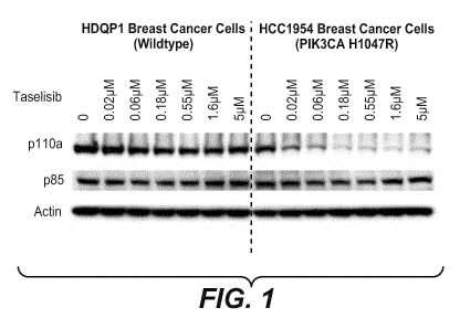
uniquely degrades mutant-p110a protein. Figure 1 shows Western blot analysis
of p110a
protein degradation by taselisib is dose-dependent, specific to PI3Ka mutant
cells. While the
invention is not limited by any particular mechanism of action, this rationale
for activity
allows taselisib to diminish the impact of feedback through RTKs, which
otherwise
attenuates anti-tumor activity. Figure 2 shows Western blot analysis of
HCC1954 cells
(PIK3CA H1047R) 24 hrs treated with taselisib, pictilisib (GDC-0941,
Genentech), and
alpelisib (BYL719, Novartis CAS#: 1217486-61-7). Other oral PI3K inhibitors,
pictilisib and
alpelisib in clinic development do not degrade mutant p110a protein.
Taselisib leads to p110a depletion in PIK3CA mutant cell lines without
effecting p85
level, consistent with a mechanism of dissociation of p110a/p85 that results
in p110a
monomer degradation (Figure 3). When p110 alpha is dissociated from p85, as a
monomer
p110 alpha is unstable and rapidly turned over (Yu et al (1998) Mol. Cell Bio.
18:1379-1387;
Wu et al (2009) Proc. Natl. Acad. Sci. 106(48):20258-20263). The p110a half-
life is
approximately 1 hr, whereas the p110a/p85 dimer is significantly more stable,
with a half-life
of approximately 5 hr.
Mutant p110a is more susceptible than wild type to degradation by taselisib.
Figure 4
shows Western blot analysis of SW48 isogenic lines including SW48 parental
PIK3CA
wildtype, mutant isogenic SW48 E545K heterozygote (het), and PIK3CA mutant
isogenic
SW48 H1047R heterozygote cells with taselisib at various concentrations; 0.2
ILEM, 1 M,
504, plus control (DMSO vehicle).
The p110a E545K mutant protein appears to be less stable than wildtype protein
(Figures 5A and 5B). Mutant p110 alpha RNA expression is unchanged in the
E545K
engineered cells. Figure 5A shows a plot of p110 alpha mRNA expression in
cells measured
by relative pllOalpha mRNA expression versus 18S RNA levels. Drug does not
change the
mRNA expression of p110a. Figure 5B shows Western blot analysis of CRISPR
(clustered
regularly interspaced short palindromic repeats) generated SW48 E545K
hemizygous lines
(two clones ran in duplicates). This western blot shows significantly reduced
mutant p110a
level compare to SW48 E545K heterozygous line which suggests mutant p110a may
be less
stable than WT p110a. The lanes from left to right are SW48 E545K hemizygous
clone 1,
SW48 E545K hemizygous clone2, SW48 parental, SW48 E545K heterozygous, SW48
E545K heterozygous.
26
CA 02981159 2017-09-27
WO 2017/001362
PCT/EP2016/064920
P110 alpha is depleted in a time dependent manner. Figure 6 shows Western blot
analysis of mutant HCC1954 (PIK3CA H1047R) breast cancer cells treated with
taselisib at
liuM and 5 M. Since taselisib has a long clinical pharmacokinetic half-life of
about 40 hours,
as measured from patient samples, mutant p110a degradation should be occurring
in tumors.
Taselisib does not decrease p110a RNA, although protein is decreased. Figure
7A
shows real time QPCR results in measuring relative RNA levels versus 18S
control in
HCC1954 wildtype and mutant p110 alpha cells. No difference in p110a mRNA
levels for
DMSO vs. GDC-0032 treated cells was detected. There was approximately 8-fold
higher
expression of mutant allele. The DNA copy number PIK3CA is 4-5, with 1 WT and
3-
4mutant alleles (exome seq). The Ratio of mutant to wildtype RNA predicts
amount of drug-
induced p110a degradation. Reduction of p110a does not occur at the
transcriptional stage.
Figure 7B shows real time QPCR results in measuring p110a mRNA expression
relative to
RPL19 control in HCC1954 p110 alpha wildtype (left) and p110 alpha H1047R
mutant
(right) p110 alpha cells treated with taselisib (GDC0032). Figure 7C shows
real time QPCR
results in measuring p110a mRNA expression relative to RPL19 control in
HCC1954 p110
alpha wildtype (left) and p110 alpha H1047R mutant (right) p110 alpha cells
treated with
alpelisib (BYL-719). The ratio of mRNA levels in wild type and mutant cells
confirm that
reduction of p110a does not occur at the transcriptional stage.
Assay Name: PIK3CA.H1047R.WT
FAM probe sequence: ATGATGCACATCATGGT (SEQ ID
NO.:1)
Forward Primer Sequence: GGCTTTGGAGTATTTCATGAAACA
(SEQ ID NO.:2)
Reverse Primer Sequence: GAAGATCCAATCCATTTTTGTTGTC
(SEQ ID NO.:3)
Assay Name: PIK3CA.H1047R.Mutant
FAM probe sequence: TGATGCACGTCATGGT
(SEQ ID NO.:4)
Forward Primer Sequence: GGCTTTGGAGTATTTCATGAAACA (SEQ ID NO.:5)
Reverse Primer Sequence: GAAGATCCAATCCATTTTTGTTGTC
(SEQ ID NO.:6)
27
CA 02981159 2017-09-27
WO 2017/001362 PCT/EP2016/064920
Depletion of p110a by taselisib is proteasome mediated (Figures 8A-8E) and
requires
El ubiquitin-activating enzyme, illustrated in Figure 8F. Figure 8A shows
Western blot
analysis of mutant HCC1954 (PIK3CA H1047R) breast cancer cells treated with
taselisib at
1.6 M for the indicated time. At 4 hours prior to harvest, 10 ILEM of
proteasome inhibitor
MG132 (N-(benzyloxycarbonyl)leucinylleucinylleucinal Z-Leu-Leu-Leu-al, CAS
Reg. No.
133407-82-6) was added (right lanes). MG-132 proteasome inhibitor rescues
degradation of
p110a by taselisib (GDC-0032). Adding proteasome inhibitor at 24 hrs is too
late to protect
from drug-induced degradation.
P110 alpha depletion is proteasome mediated and require El ubiquitin-
activating
enzyme. Figure 8B shows Western blot analysis of lysates of mutant HCC1954
(PIK3CA
H1047R) breast cancer cells treated with taselisib at 1.6 M for the indicated
time. At 4 hours
prior to harvest, 10 ILEM MG132 was added (middle lanes) and 10 ILEM UAE1
inhibitor,
((2R,3S,4R,5R)-5-(6-(((S)-2,3-dihydro-1H-inden-1-yl)amino)-9H-purin-9-y1)-3,4-
dihydroxytetrahydrofuran-2-yl)methyl sulfamate, CAS Reg. No. 905578-77-0,
having the
structure:
H
N
0 -,A
H2N-S-0 "OH
0 HO
Figure 8C shows Western blot analysis of mutant HCC1954 (PIK3CA H1047R)
breast cancer cells treated with taselisib at 1.6 M for the indicated time. At
4 hours prior to
harvest, 10 ILEM MG132 was added (right lanes). Taselisib mediates p110a poly-
ubiquitination and poly-ubiquitinated p110a accumulates with MG132. Treatment
with the
El inhibitor (UAE1 inhibitor), see Figure 8E, collapsed high molecular weight
bands in the
autoradiogram, confirming specificity of antibody.
Figure 8D also shows Western blot analysis of mutant HCC1954 (PIK3CA H1047R)
breast cancer cells treated with taselisib at 1.6 M for the indicated time. At
4 hours prior to
harvest, 10 ILEM MG132 was added (right lanes). Comparison of measurements
conducted
from the cell membrane and cytosol demonstrated that ubiquitination of p110a
occurs
28
CA 02981159 2017-09-27
WO 2017/001362
PCT/EP2016/064920
primarily at the membrane. Membrane associated p110a is more efficiently
ubiquitinated by
taselisib than cytosolic p110a. Taselisib rapidly mediates degradation of
mutant p110a at the
plasma membrane. The degradation rate of membrane-associated p110a is much
faster than
cytosolic p110a degradation. Short term treatment of cells with taselisib
mediates dose
dependent degradation of membrane but not cytosolic p110a. In comparison,
alpelisib (BYL-
719) only has a weak effect at membrane and no effect in total lysate.
Alpelisib showed a
weak initial response in membrane but did not cause degradation of p110a over
time.
Taselisib is a superior degrader of p110a than alpelisib.
Figure 8E shows Western blot analysis of mutant HCC1954 (PIK3CA H1047R)
breast cancer cells treated with taselisib at 1.6 M for the indicated time. At
4 hours prior to
harvest, 10 ILEM MG132, chloroquine, or ammonium chloride was added (right
lanes).
Taselisib mediated p110a depletion is not affected by lysosome inhibitors,
suggesting that
p110a degradation is not endosome/lysosome mediated, where proteasome
inhibitor MG132
is a positive control.
Figure 8F shows a pathway diagram to ubiquitination of p110 alpha describing
two
different mechanism by which animal cells degrade proteins with pathway
specific inhibitors
(Jadhav, T. et al (2009) "Defining an Embedded Code for Protein
Ubiquitination" J.
Proteomics Bioinform, Vol 2(7):316-333; Wang, G. et al (2012) ("K63-linked
ubiquitination
in kinase activation and cancer" Frontiers in Oncology, Vol 2(5):1-13).
Because proteasome
inhibitor MG132 but not lysosome inhibitors, chloroquine or ammonium chloride
NH4C1,
was able to rescue p110a degradation, GDC-0032 mediated p110a degradation is a
ubiquitin
proteasome dependent degradation pathway rather than dependent on a lysosome
degradation
machinery.
Thus, taselisib-mediated p110a protein degradation is time-dependent, dose-
dependent, non-transcriptional, specific to mutant cells, ubiquitin dependent
and mediated by
the proteasome, and rapid for membrane-associated p110a.
Neither taselisib or alpelisib affect liver p110a at the membrane.
Taselisib does not degrade or deplete p110 delta (Figures 9A and 9B). Figure
9A
shows Western blot analysis of PI3K wildtype, BRAF mutant 5W982 cells treated
with
taselisib and alpelisib (BYL719, Novartis, CAS#: 1217486-61-7, (S)-N1-(4-
methy1-5-(2-
(1,1,1-trifluoro-2-methylpropan-2-yl)pyridin-4-yl)thiazol-2-yl)pyrrolidine-1,2-
29
CA 02981159 2017-09-27
WO 2017/001362
PCT/EP2016/064920
dicarboxamide). Taselisib does not degrade or deplete p110 delta. Figure 9B
shows Western
blot analysis of PI3K wildtype, B cell lymphoma SU-DHL-10 cells treated with
taselisib and
G-102 (Table 1, US8242104).
In the PI3K signaling pathway, PI3K inhibitors relieve negative feedback and
prime
the pathway for reactivation. When negative feedback is blocked, the phospho-
RTK (ErbB2,
EGFR, ErbB3) activity increases, effectiveness of PI3K inhibition is reduced,
and pAKT is
increased. Figure 10 shows Western blot analysis of PIK3CA wildtype HDQP1
cells at 1 hr
and 24 hr with PI3K inhibition with a PI3K inhibitor at various
concentrations; 100nM, 1 M,
51iM, plus control (DMSO vehicle).
Taselisib is better than another PI3K inhibitor G-102 (US 8242104) at
maintaining
signaling suppression at late timepoint. Figure 11A shows Western blot
analysis of MDA-
MB 453 (H1047R) cells at 1 hr and 24 hr with PI3K inhibition by G-102 at
various
concentrations; 3 nM, 16 nM, 60 nM, 400 nM, 2 ILEM, plus control (DMSO
vehicle). Figure
11B shows Western blot analysis of MDA-MB 453 (H1047R) cells at 1 hr and 24 hr
with
PI3K inhibition by taselisib (GDC-0032) at various concentrations; 3 nM, 16
nM, 60 nM,
400 nM, 2 ILEM, plus control (DMSO vehicle).
Taselisib protects against RTK-driven pathway reactivation. At 1 hr the pAkt
knockdown is approximately equivalent for both PI3K inhibitors. At 24 hrs
there is increased
pRTK. Taselisib is better at suppressing signaling at 24 hrs than G-102, a non-
degrader PI3K
inhibitor. Figure 12 shows Western blot analysis of 5W48 H1047R cells at 1 hr
and 24 hr
with PI3K inhibition by taselisib (GDC-0032) and G-102 at various
concentrations; 1 nM, 10
nM, 100 nM, plus control (DMSO vehicle).
In cells stimulated with growth factor ligand, taselisib is better than non-
degrader G-
102 at suppressing signaling. A rationale for this effect is mutant PI3Ka is
more susceptible
to degradation when pRTK is increased. pRTK binding is thought to shift p85
relative to
p110a, leading to more active p110a (alpha). Hotspot p110a mutations can occur
on the
interface between p110a and p85, and may loosen p85/p110 interaction. Figure
13 shows
Western blot analysis of 5W48 PIK3CA H1047R cells with and without growth
factor ligand
NRG treated with PI3K inhibition by taselisib (GDC-0032) and G-102 at various
concentrations; 1 nM, 10 nM, 100 nM, plus control (DMSO vehicle).
CA 02981159 2017-09-27
WO 2017/001362
PCT/EP2016/064920
Measuring in vitro cellular proliferation, pPRAS40 in isogenic mutant vs
wildtype
cells at 24 hrs is useful to select PI3K inhibitor compounds in a degradation
assay. Figures
14A-C shows in vitro cellular proliferation of pPRAS40 in isogenic mutant
(E545K,
H1047R) versus wildtype parental PI3K cells at 24 hours at various
concentrations of G-181,
GDC-0032 and G-102 to establish parental/mutant selectivity EC50 values.
Increased cellular potency of taselisib is observed relative to pan-PI3K
inhibitor,
pictilisib (GDC-0941) in mutant PI3Ka knock-in cells. Figure 15 shows a plot
of in vitro
cellular proliferation data with SW48 isogenic wildtype and mutant (E545K,
H1047R) cell
lines and treatment with dose titrations of taselisib and pictilisib.
Taselisib has enhanced potency in PIK3CA mutant cancer lines. Figure 16 shows
plots of efficacy (IC50 micromolar) of pictilisib (GDC-0941), BKM120
(buparlisib, Novartis
AG, CAS Reg. No. 944396-07-0, 5-(2,6-dimorpholinopyrimidin-4-y1)-4-
(trifluoromethyl)pyridin-2-amine), taselisib (GDC-0032) and BYL719 (alpelisib,
Novartis
CAS#: 1217486-61-7) in a 4 day cell proliferation (Cell-Titer GloR, Promega)
assay against
PIK3CA mutant cell lines. Each dot represents a different cancer cell line.
Pictilisib,
BKM120 (buparlisib, Novartis AG, CAS Reg. No. 944396-07-0), and BYL719 are non-
degraders of PI3K.
Taselisib has enhanced potency in an apoptosis assay. Figure 17 shows plots of
in
vitro cellular proliferation data with PIK3CA wildtype and mutant (E545K,
H1047R) cell
lines and treatment with dose titrations of taselisib and PI3K alpha selective
inhibitor GDC-
0326 (US 8242104), and BYL719 in a 72 hr study with Cell Death-Nucleosome
ELISA
detection.
Taselisib has greater maximal in vivo efficacy than other non-degrader drugs,
in a
PI3Ka mutant xenograft in mice. At the maximum tolerable dose (MTD), taselisib
can cause
tumor shrinkage. Figure 18A shows the fitted tumor volume change over 21 days
in cohorts
of 8-10 immunocompromised mice bearing HCC1954.xl breast tumor xenografts
harboring
PIK3CA H1047R (PI3Kcc) mutation dosed once daily by 100 microliter (ul) PO
(oral)
administration with Vehicle (MCT; 0.5% methycellulose/0.2% Tween 80), 150
mg/kg
pictilisib (GDC-0941), and 25 mg/kg taselisib (GDC-0032). The term uL means
microliter.
At maximum tolerated doses of both drugs, GDC-0032 is more efficacious than
GDC-0941
and induces tumor regressions. Thus, GDC-0032 is more efficacious than a non-
mutant
31
CA 02981159 2017-09-27
WO 2017/001362
PCT/EP2016/064920
selective PI3Kcc inhibitor in a PI3K mutant xenograft model. Figure 18B shows
the fitted
tumor volume change over 21 days in cohorts of 8-10 immunocompromised mice
bearing
HCC1954.xl breast tumor xenografts harboring PIK3CA H1047R (PI3Kcc) mutation
dosed
once daily by 100 microliter (ul) PO (oral) administration with Vehicle (MCT;
0.5%
methycellulose/0.2% Tween 80), 40 mg/kg alpelisib (BYL-719), and 15 mg/kg
taselisib
(GDC-0032). Oral and daily dosing of GDC-0032 for 21 days resulted in tumor
regressions
over the treatment (Rx) period. Alternatively, oral and daily dosing of the
non-mutant
selective PI3Kcc inhibitor, BYL-719, over 21 days induced tumor stasis. Thus,
GDC-0032 is
more efficacious than a non-mutant selective PI3Ka inhibitor in a PI3K mutant
xenograft
model. Treatment with GDC-0032 and BYL-719 was well tolerated based on minimal
changes in mouse body weight when compared to vehicle controls or from the
initiation of
the study.
The compound known as alpelisib (BYL719, Novartis, CAS#: 1217486-61-7) is an
oral, selective inhibitor of the PI3K alpha isoform, and is in clinical trials
for the potential
treatment of a variety of tumor types, including a phase III study in
combination with
fulvestrant for second-line hormone receptor-positive, HER2- advanced
metastatic breast
cancer (Furet, P. et al (2013) Bioorg. Med. Chem. Lett. 23:3741-3748; US
8227462; US
8476268; US 8710085). Alpelisib is named as (S)-N1-(4-methy1-5-(2-(1,1,1-
trifluoro-2-
methylpropan-2-yl)pyridin-4-yl)thiazol-2-yl)pyrrolidine-1,2-dicarboxamide) and
has the
structure:
ON FNI N
E /
H2N0 o S ______________________
F3C ¨N
alpelisib (BYL-719)
Figure 18C shows the fitted tumor volume change over 28 days in cohorts of 8-
10
immunocompromised mice bearing WHIM20 hormone receptor positive patient-
derived
breast tumor xenografts harboring PIK3CA E542K (PI3Kcc) mutation dosed once
daily by
100 microliter (ul) PO (oral) administration with Vehicle (MCT; 0.5%
methylcellulose/0.2%
Tween 80) and 15 mg/kg taselisib (GDC-0032). Oral and daily dosing of GDC-0032
for 28
days resulted in tumor regressions over the treatment period that was
sustained after dosing
32
CA 02981159 2017-09-27
WO 2017/001362
PCT/EP2016/064920
had ended. Treatment with GDC-0032 was well tolerated based on minimal changes
in
mouse body weight when compared to vehicle controls or from the initiation of
the study.
Figure 18D shows the fitted tumor volume change over 27 days in cohorts of 8-
10
immunocompromised mice bearing HCI-003 hormone receptor positive patient-
derived
breast tumor xenografts harboring PIK3CA H1047R (PI3Kcc) mutation dosed once
daily by
100 microliter (ul) PO (oral) administration with Vehicle (MCT; 0.5%
methylcellulose/0.2%
Tween 80), 40 mg/kg alpelisib (BYL-719) and 2.5, 5.0, 15 mg/kg taselisib (GDC-
0032).
Oral and daily dosing of GDC-0032 for 27 days resulted in a dose-dependent
increase in
tumor regressions over the treatment (Rx) period. Alternatively, oral and
daily dosing of the
non-mutant selective PI3Kcc inhibitor, BYL-719, over 27 days induced tumor
stasis. Thus,
GDC-0032 is more efficacious than a non-mutant selective PI3Ka inhibitor in a
PI3K mutant
xenograft model. Treatment with GDC-0032 and BYL-719 was well tolerated based
on
minimal changes in mouse body weight when compared to vehicle controls or from
the
initiation of the study.
Efficacy of GDC-0032 was evaluated in the WHIM20 hormone receptor positive
patient-derived breast cancer xenograft model that harbors the PIK3CA E542K
hot-spot
mutation. Oral and daily dosing of GDC-0032 for 28 days resulted in tumor
regressions over
the treatment (Rx) period that was sustained after dosing had ended. Treatment
with GDC-
0032 was well tolerated based on minimal changes in mouse body weight when
compared to
vehicle controls or from the initiation of the study.
Efficacy of GDC-0032 was evaluated in the HCI-003 hormone receptor positive
patient-derived breast cancer xenograft model that harbors the PIK3CA H1047R
hot-spot
mutation. Oral and daily dosing of GDC-0032 for 27 days resulted in a dose-
dependent
increase in tumor regressions over the treatment (Rx) period. Alternatively,
oral and daily
dosing of the non-mutant selective PI3Ka inhibitor, BYL-719, over 27 days
induced tumor
stasis. Thus, GDC-0032 is more efficacious than a non-mutant selective PI3Ka
inhibitor in a
PI3K mutant xenograft model. Treatment with GDC-0032 and BYL-719 was well
tolerated
based on minimal changes in mouse body weight when compared to vehicle
controls or from
the initiation of the study.
Signaling rapidly returns to normal after taselisib washout, and p110a level
begins to
return at 8-24 hrs later in mutant HCC1954 (PIK3CA H1047R) breast cancer cells
treated
33
CA 02981159 2017-09-27
WO 2017/001362
PCT/EP2016/064920
with taselisib. Figure 19 shows Western blot analysis of mutant HCC1954
(PIK3CA
H1047R) breast cancer cells treated with taselisib at various concentrations;
16 nM, 80 nM,
400 nM, plus control (DMSO vehicle).
In one embodiment, depletion of p110 alpha protein can be measured by cellular
proliferation, cell signaling, or apoptosis levels.
In one embodiment, depletion of p110 alpha protein is correlated with a
measurable
biomarker from a biological sample obtained from a patient. Depletion of p110
alpha protein
may be detected in a clinical setting by a Protein Simple IEF (isoelectric
focusing)
technology. With adequate available patient tissue, western blot analysis, or
mass
spectroscopy could measure pllOalpha protein levels. Mass spectrometry may
allow
interrogation of the proteome of single cells, including detection of
ubiquitination. NMR
(nuclear magnetic resonance) spectroscopy is another biophysical tool to
detect and measure
pllOalpha and p85 dissociation or pllOalpha degradation.
Alternatively a specific anti-pllOalpha antibody may be useful in
immunohistochemistry (IHC) or immunofluorescence (IF) based tests.
Immunoprecipitation (IP) and protein localization of PI3K proteins may detect
changes in dissociation of p85 and pllOalpha or when pllOalpha is degraded
before and after
treatment by taselisib may identify and predict patient responders.
The activity of taselisib in a mutant isogenic PI3K cell line is greater than
its activity
in a wild type isogenic PI3K cell line. Isogenic PI3K mutant cell line may
have a mutation
selected from H1047R, C420R, H1047L, E542K, E545K and Q546R.
Taselisib differentially affects wild type p85/p110alpha complex relative to
ATP-Km
of mutant p85/p110alpha complex, as measured or detected by ATP-Km, PIP2-Km,
the rate
or extent of conversion of PIP2 to PIP3, membrane localization, lipid membrane
affinity, and
receptor tyrosine kinase binding.
Taselisib differentially induces conformational changes of wild type
p85/p110alpha
complex relative to mutant p85/p110alpha complex. Conformational changes
include a
binding interaction with mutant p85/p110alpha complex which is not present
between
taselisib with wild type p85/p110alpha complex.
34
CA 02981159 2017-09-27
WO 2017/001362
PCT/EP2016/064920
In one embodiment, taselisib selectively binds a mutant form of isolated PI3K
alpha
and the IC50 for binding to mutant-PI3K alpha is less than the IC50 for
binding to wild type-
PI3K alpha.
In one embodiment, taselisib is more active in inhibiting a mutant-PI3K alpha
isogenic cancer cell line than inhibiting a wild type form of the PI3K alpha
isogenic cancer
cell line.
In one embodiment, taselisib is selective for binding to the alpha-subunit of
mutant-
PI3K wherein the PI3K mutations are selected from H1047R, C420R, H1047L,
E542K,
E545K and Q546R, and has inhibitory activity, as measured by IC50, in a mutant
PI3K p110
alpha isoform cell line which is lower than the IC50 inhibitory activity of
taselisib in a wild
type PI3K p110 alpha isoform cell line. The mutant PI3K p110 alpha isoform is
selected
from H1047R, C420R, H1047L, E542K, E545K and Q546R.
TRYPSIN CLEAVAGE AND MASS SPECTROSCOPY DETECTION OF P110 ALPHA
DEPLETION
In addition to the methods above to detect and measure depletion of p110 alpha
in
biological samples treated with taselisib, direct detection can be achieved by
mass
spectrometry. HCC1954 breast cancer cells, expressing a mixture of wild type
and H1047R
mutant p110a protein, were treated with 500 nM taselisib (GDC-0032) in DMSO
for 24 hours.
Immunoprecipitation of p110 alpha protein was performed and captured protein
separated by
SDS-PAGE and Coomassie stained. Gel bands containing p110 alpha were subjected
to in gel
trypsin digestion to generate tryptic peptides for analysis. Tryptic peptides
from cells treated
with DMSO (control) or taselisib showed equivalent levels of the
QM(ox)NDAHHGGWTTK
peptide sequence (SEQ ID NO. :7), based on quantification of the corresponding
749.8282
m/z (+/- 10 ppm) ion.. Tryptic peptides from taselisib treated cells showed
loss of mutant-
specific peptide HGGWTTK (SEQ ID NO. :8), characterized by an ion of 393.6983
m/z (+/-
10 ppm), relative to DMSO treated cells. Thus, neo-tryptic peptides specific
to mutant
tumors, such as p110a H1047R, can be used to demonstrate depletion of mutant
p110a
relative to wild type form.
CA 02981159 2017-09-27
WO 2017/001362
PCT/EP2016/064920
wild type tryptic peptide QM (ox) NDAHHGGWTTK
(SEQ ID NO.:7)
mutant tryptic peptide HGGWTTK
(SEQ ID NO.:8)
Figure 24A shows trypsin cleavage of wild-type PIK3CA HCC-1954 (top) and
H1047R mutation expressing PIK3CA HCC-1954 (bottom), according to Example 7.
Figure 24B shows liquid chromatography-tandem mass spectrometry (LC-MS/MS)
analysis on the wild-type PIK3CA HCC-1954 (left) and H1047R mutation
expressing
PIK3CA HCC-1954 (right) after digestion and pllOalpha (PIK3CA) protein
immunoprecipitation, according to Example 7.
MUTANT SELECTIVITY
Table 1 compiles biological properties for several PI3K-binding compounds. GDC-
0032 (Ndubaku et al (2013) Jour. Med. Chem. 56(11):4597-4610; Staben et al
(2013) Bioorg.
Med. Chem. Lett. 23 2606-2613; WO 2011/036280; US 8242104; US 8343955) is more
potent against PI3K alpha mutant cancer cells relative to the pan-PI3K
inhibitor compound
GDC-0941 (Folkes et al (2008) Jour. of Med. Chem. 51(18):5522-5532; US
7781433; US
8324206). The four PI3K inhibitors of Table 1 differ in biochemical potency
(Ki values) in
binding to PI3K alpha and relative potency against the four wild type Class 1
isoforms of
PI3K. GDC-0326 is an alpha selective PI3K inhibitor compound and binds weakly
to the
beta, delta, and gamma isoforms (US 8242104). GDC-0941 is a "pan" inhibitor,
binding
relatively well to all four isoforms. GDC-0032 is "beta sparing", binding well
to the alpha,
delta, and gamma isoforms and weakly to beta.
36
CA 02981159 2017-09-27
WO 2017/001362
PCT/EP2016/064920
Table 1 PI3K binding compound activity
Comp wt-PI3K mut- mut- PI3K PI3K 6 PI3K 7 Wild MCF PC3
ound a pllOaH pllOaE type- 7 IC50 IC50
1047R 545K PI3K a! [tm [tm
K.1 K.1 K.1
Mut-
K, nm
PI3K a
K. nm K. nm
03/0c) (6/a) (7/0)
G-102 0.14 39.7 3.7 16 0.056 1.6
(282) (27) (111)
GDC- 0.23 31 4.8 12 1
0326
(133) (20) (51)
GDC- 0.29 0.11 0.14 9.1 0.092 0.89 2.6 0.025 not
0032 active
(31) (0.36) (3.5) (Mut
E545
K)
GDC- 3 33 3 75
0941
(11) (1) (25)
ON 0
H 2 N 0 --\
H3C
H3C W G-102
37
CA 02981159 2017-09-27
WO 2017/001362
PCT/EP2016/064920
ONF121 N
1\11
N.:::' GDC-0326
o
H2 N-'-r il---
N 0
.1 N
Ni......
NN-=-N GDC-0032 (taselisib)
o
( )
N
\ I 'NH
(N\ N 110
,c),s
\ GDC-0941 (pictilisib)
Knock-in of mutant PI3K alpha increases cellular potency for taselisib (GDC-
0032)
as shown in SW48 isogenic lines, PI3K alpha wild-type (parental) and helical
domain mutant
E545K and kinase domain mutant H1047R, whereas GDC-0941 shows no such mutant
selectivity effect. It can be deduced that GDC-0032 interacts with the mutant
protein
differently than GDC-0941 does. This unexpected result implies a unique
mechanism or
mode of binding of certain potent PI3K inhibitors, but not others. The mutant
selectivity
property of GDC-0032, lacking in GDC-0941, gives GDC-0032 greater maximal
efficacy
than GDC-0941 in a PI3K alpha mutant xenograft tumor model, HCC1954 breast
cancer with
kinase domain H1047R mutation. Following daily oral dosing for 21 days, at the
maximum
tolerated dose of 25 mg/kg, GDC-0032 induced tumor regressions whereas at the
maximum
tolerated dose of 150 mg/kg, GDC-0941 caused tumor growth inhibition.
In vitro tumor cell proliferation was measured in cancer cell lines treated
with GDC-
0032, GDC-0326 (US 8242104) or GDC-0941. GDC-0032 induces apoptosis in PI3K
mutant
cells at low concentrations. Increased mutant potency of GDC-0032 is not
correlated with
38
CA 02981159 2017-09-27
WO 2017/001362
PCT/EP2016/064920
biochemical binding potency against wild-type PI3K alpha. Increased potency
against PI3K
mutant cell lines and tumors by PI3K inhibitor compounds may be affected by:
physicochemical or permeability properties, intracellular levels, isoform
selectivity, and
absolute potency against wild type p110 alpha or wild type p110 delta. In a
cell viability
assay, p110 alpha-selective inhibitor GDC-0326 (Table 1) was 6X less potent
against the
H1047R p110 alpha cell line, HCC1954 than GDC-0032. GDC-0032 is less alpha
selective
against the beta, gamma, and delta isoforms than GDC-0326 (US 8242104). Knock-
in of
mutant PI3K alpha increases cellular potency for mutant selective GDC-0032,
but not for
PI3K alpha-specific inhibitors, such as G-102. A similar cell viability assay
determined that
inhibition of p110 delta does not decrease viability in PI3K mutant cell
lines. Also,
comparable intracellular levels of GDC-0032 were measured (pmol/mg) in various
wild type
and PI3K mutant cells. Results indicate intracellular accumulation does not
explain increased
mutant potency of GDC-0032.
Autoradiography of gel electrophoresis of radiolabelled lysates from mutant
isogenic
5W48 PI3K alpha H1047R cell line and wild type 5W48 parental cells treated
with GDC-
0032, GDC-0326 or GDC-0941 measured cleaved PARP, pS6(S235/236), pAKTT308,
pAKTs473,
beta-Actin and GAPDH by Western blot analysis. PI3K pathway knockdown
correlates with
the induction of apoptosis in a dose-dependent manner. Taselisib induces
apoptosis in cells
harboring PI3K mutations at very low compound concentrations. Similar effects
were seen in
isogenic cells from MCF10A breast cell line and HCC1954, PI3K alpha H1047R
breast
cancer cell line. Despite comparable compound properties, pathway knockdown is
stronger
with mutant selective GDC-0032 than non-mutant selective GDC-0326. Increased
mutant
potency of GDC-0032 is not explained by biochemical potency against wild type
PI3K alpha.
By these assays, compound can be assessed to examine the impact of structural
changes on PI3K alpha mutant selectivity. Changes in size and hydrogen bonding
capability
in a specific region may correlate with improved selectivity.
Preliminary clinical trial data showed that taselisib achieved partial
responses in five
out of ten patients with PIK3CA mutant tumors, and four out of five patients
with PIK3CA
mutant breast tumors (Olivero and Juric (2013) AACR).
39
CA 02981159 2017-09-27
WO 2017/001362
PCT/EP2016/064920
INTERACTIONS OF TASELISIB WITH PI3K
Taselisib (GDC-0032) is more selective for PI3K alpha mutant isoforms than
PI3K
wild type isoforms due to key interactions with PI3K alpha mutant isoforms
that differ from
interactions with the PI3K wild type isoform, and may include a precise
positioning and
arrangement of atoms and functional groups to interact with key mutant-
specific features of
PI3K alpha mutant isoforms. Such interactions may be achieved by functional
groups acting
as hydrogen bond-donors, hydrogen bond-acceptors and/or Van der Waals force
partners with
the PI3K alpha mutant isoform protein (Staben et al (2013) Bioorg. Med. Chem.
Lett.
23:2606-2613; Ndubaku et al (2013) Jour. Med. Chem. 56(11):4597-4610).
Taselisib may
adopt binding topologies in low energy conformations and make efficient polar
and van der
Waals interactions in the ligand binding site.
It has been established that PIK3CA mutations increase lipid binding and PI3K
basal
activity (Burke et al (2012) Proc. Natl. Acad. Sci. 109:15259-15264).
Mutations destabilize
the closed, cytosolic inactive form of PI3K alpha, promoting increased lipid
binding. The
mutant selective, PI3K-binding compounds of the invention increase
stabilization of the
closed form of PI3K alpha, preventing conformational changes that increase
lipid binding.
Hotspot mutations induce regions of the kinase domain to be more deuterated in
hydrogen-
deuterium exchange of the Hi 047R mutant indicating they become more dynamic
(destabilized) and available for exchange. Those changes were accompanied by
increased
affinity for lipid membrane and may account for increased activation and
downstream
signaling. This dynamic region, residues 848-859 (Figure 20), may provide key
binding
interactions for the compounds of the invention.
Figure 21 shows a plot of binding potency of GDC-0032 viability in cell lines
with
PIK3CA mutants, according to the location of the mutation in PIK3CA. GDC-0032
is potent
against cell lines harboring PI3K mutations regardless of the location of the
mutation. Some
of the cell lines have additional mutations such as B-Raf and Ras as
resistance markers.
Figure 22 shows Western blot (WB) analysis of p85 co-immunoprecipitation (Co-
IP)
with p110a and the level parallel with p110a suggesting that stable p110a is
in complex with
p85 and significant dose dependent p110a degradation induced by taselisib.
Cells were
treated with GDC-0032 for 24h, a point at which p110a is clearly being
degraded. Alternative experiments may employ treatment of cells with sampling
at shorter
CA 02981159 2017-09-27
WO 2017/001362
PCT/EP2016/064920
time points (2 hr, 4 hr, etc) where pllOalpha is not yet degraded but is
dissociated from p85.
Detection of dissociation of mutant but not wild type pllOalpha from p85 after
treatment
with taselisib may be a predictive biomarker for patients with mutant PI3K
tumors likely to
respond to treatment with taselisib.
Figure 23 shows steady state p110a mRNA expression. Cell lines most sensitive
to
p110a degradation by GDC0032 harbor more H1047R mutation than WT p110a. The
ratio of
Mutant to WT p110a allele may determine sensitivity to GDC-0032 mediated p110a
degradation.
X-ray structures of PI3K beta (Zhang, X. et al (2011) Mol. Cell 41:567-5789),
PI3K
alpha (Huang, C.-H. et al (2007) Science 318:1744), PI3K delta (Berndt, A. et
al (2010) Nat.
Chem. Biol. 6:11), and PI3K gamma ("Structural determinants of
phosphoinositide 3-kinase
inhibition by wortmannin, LY294002, quercetin, myricetin, and staurosporine",
Walker, E.H.
et al (2000) Mol.Cell 6:909) have been reported. One difference of note is the
conformation
of a tryptophan residue present in all isoforms (alpha-Trp780, beta-Trp781,
delta-Trp760,
gamma-TRP812). This is the same tryptophan residue that is thought to be a
crucial for
obtaining isoform specificity for PI3K delta as the result of an 'induced fit'
movement of
adjacent residues or specific interaction by inhibitors. The conformation of
the indole of this
Trp residue is unique in PI3K beta and may provide basis for design of "beta-
sparing" PI3K
inhibitors with low, weak binding to PI3K beta. Rotation along Ca¨CI3 of beta-
Trp781
presents an alternate orientation. Differences in second shell residues
promote a unique
orientation of this Trp in PI3K beta. First, the indole N¨H donates an H-bond
to acidic
residues in both PI3K alpha (G1u798) and PI3K gamma (G1u814) that occupy a
similar region
of the binding site but are not present in PI3K beta or delta. This
interaction could favor the
observed orientation of the indole in these isoforms. In place of Glu814, PI3K
beta and delta
possess neutral residues with nonpolar side chains (beta-Va1783 and delta-
Met762
respectively) and thus the indole can occupy other energetically favorable
orientations. It is
possible that the branched valine residue of PI3K beta disfavors orientations
similar to those
observed in alpha, delta and gamma; the C gamma methyl instead approaches van
der Waals
distance to the indole 4/5 position. Under this interpretation of structure-
activity relationship
(SAR), in this unique conformation the Trp sidechain is more easily insulted
by steric bulk of
the inhibitor. Also of note is the differential angle and atoms presented for
pi-stacking by the
indole in PI3K alpha, delta and gamma relative to beta. For PDB analysis of pi-
interaction
41
CA 02981159 2017-09-27
WO 2017/001362
PCT/EP2016/064920
between tryptophan rings and aromatic amino acid side chains (Phe, Tyr, His)
see: Samanta,
U. et al (1999) Biol. Crystallogr., D55:1421. This observation could partially
explain why
non-aromatic substitution in the same region results in molecules with lower
overall
selectivity over PI3Kb (Staben et al (2013) Bioorg. Med. Chem. Lett. 23:2606-
2613).
BIOMARKER DETECTION OF MUTANT P110 ALPHA
In certain embodiments, the presence and/or expression level/amount of
biomarker
proteins in a sample is examined using immunohistochemistry (IHC) and staining
protocols.
IHC staining of tissue sections has been shown to be a reliable method of
determining or
detecting presence of proteins in a sample. In some embodiments of any of the
methods,
assays and/or kits, a relevant biomarker is mutant p110 alpha protein. In some
embodiments,
mutant p110 alpha is detected by immunohistochemistry. In some embodiments,
elevated
expression of a mutant p110 alpha biomarker in a sample from an individual is
elevated
protein expression and, in further embodiments, is determined using IHC. In
one
embodiment, expression level of biomarker is determined using a method
comprising: (a)
performing IHC analysis of a sample (such as a subject cancer sample) with an
antibody; and
b) determining expression level of a biomarker in the sample. In some
embodiments, IHC
staining intensity is determined relative to a reference. In some embodiments,
the reference is
a reference value. In some embodiments, the reference is a reference sample
(e.g., control cell
line staining sample or tissue sample from non-cancerous patient).
IHC may be performed in combination with additional techniques such as
morphological staining and/or fluorescence in-situ hybridization. Two general
methods of
IHC are available; direct and indirect assays. According to the first assay,
binding of antibody
to the target antigen is determined directly. This direct assay uses a labeled
reagent, such as a
fluorescent tag or an enzyme-labeled primary antibody, which can be visualized
without
further antibody interaction. In a typical indirect assay, unconjugated
primary antibody binds
to the antigen and then a labeled secondary antibody binds to the primary
antibody. Where
the secondary antibody is conjugated to an enzymatic label, a chromogenic or
fluorogenic
substrate is added to provide visualization of the antigen. Signal
amplification occurs because
several secondary antibodies may react with different epitopes on the primary
antibody. The
primary and/or secondary antibody used for IHC typically will be labeled with
a detectable
moiety. Numerous labels are available which can be generally grouped into the
following
42
CA 02981159 2017-09-27
WO 2017/001362
PCT/EP2016/064920
categories: (a) Radioisotopes, such as 35S, 14C, 1251, 3H, and 1311; (b)
colloidal gold
particles; (c) fluorescent labels including, but are not limited to, rare
earth chelates (europium
chelates), Texas Red, rhodamine, fluorescein, dansyl, Lissamine,
umbelliferone,
phycocrytherin, phycocyanin, or commercially available fluorophores such
SPECTRUM
ORANGE7 and SPECTRUM GREEN7 and/or derivatives of any one or more of the
above;
(d) various enzyme-substrate labels are available (US 4275149; US 4318980).
Examples of
enzymatic labels include luciferases (e.g., firefly luciferase and bacterial
luciferase (US
4737456), luciferin, 2,3-dihydrophthalazinediones, malate dehydrogenase,
urease, peroxidase
such as horseradish peroxidase (HRPO), alkaline phosphatase, .beta.-
galactosidase,
glucoamylase, lysozyme, saccharide oxidases (e.g., glucose oxidase, galactose
oxidase, and
glucose-6-phosphate dehydrogenase), heterocyclic oxidases (such as uricase and
xanthine
oxidase), lactoperoxidase, microperoxidase, and the like. Examples of enzyme-
substrate
combinations include, for example, horseradish peroxidase (HRPO) with hydrogen
peroxidase as a substrate; alkaline phosphatase (AP) with para-Nitrophenyl
phosphate as
chromogenic substrate; and .beta.-D-galactosidase (beta-D-Gal) with a
chromogenic substrate
(e.g., p-nitrophenyl-.beta.-D-galactosidase) or fluorogenic substrate (e.g., 4-
methylumbellifery1-.beta.-D-galactosidase). In some embodiments of any of the
methods, a
biomarker such as mutant p110 alpha protein is detected by
immunohistochemistry using an
anti-mutant p110 alpha diagnostic antibody (i.e., primary antibody). In some
embodiments,
the anti-mutant p110 alpha diagnostic antibody specifically binds human mutant
p110 alpha.
In some embodiments, the anti-mutant p110 alpha antibody is a nonhuman
antibody, such as
a rat, mouse, or rabbit antibody. In some embodiments, anti-mutant p110 alpha
diagnostic
antibody is a monoclonal antibody. In some embodiments, the anti-mutant p110
alpha
diagnostic antibody is directly labeled.
Specimens thus prepared may be mounted and coverslipped. Slide evaluation is
then
determined, e.g., using a microscope, and staining intensity criteria,
routinely used in the art,
may be employed. In one embodiment, it is understood that when cells and/or
tissue from a
tumor is examined using IHC, staining is generally determined or assessed in
tumor cell
and/or tissue (as opposed to stromal or surrounding tissue that may be present
in the sample).
In some embodiments, it is understood that when cells and/or tissue from a
tumor is
examined using IHC, staining includes determining or assessing in tumor
infiltrating immune
cells, including intratumoral or peritumoral immune cells. In some
embodiments, the
presence of a mutant p110 alpha biomarker is detected by IHC in >0% of the
sample, in at
43
CA 02981159 2017-09-27
WO 2017/001362
PCT/EP2016/064920
least 1% of the sample, in at least 5% of the sample, in at least 10% of the
sample. In some
embodiments, the mutant p110 alpha biomarker is detected by IHC in tumor
cells.
In alternative methods, the biological sample from a patient, e.g. tumor
biopsy or
circulating tumor cell, may be contacted with an antibody specific for a
biomarker under
conditions sufficient for an antibody-biomarker complex to form, and then
detecting said
complex. The presence of the biomarker may be detected in a number of ways,
such as by
Western blotting and ELISA procedures for assaying a wide variety of tissues
and samples,
including plasma or serum. A wide range of immunoassay techniques using such
an assay
format are available (US 4016043; US 4424279; US 4018653), including single-
site and two-
site or "sandwich" assays of the non-competitive types, as well as in the
traditional
competitive binding assays. These assays also include direct binding of a
labeled antibody to
a target biomarker. Presence and/or expression level/amount of a selected
biomarker in a
tissue or cell sample may also be examined by way of functional or activity-
based assays. For
instance, if the biomarker is an enzyme, one may conduct assays known in the
art to
determine or detect the presence of the given enzymatic activity in the tissue
or cell sample.
The anti-mutant p110 alpha antibody or antigen binding fragment thereof, may
be
made using methods known in the art, for example, by a process comprising
culturing a host
cell containing nucleic acid encoding any of the previously described anti-
mutant p110 alpha
antibodies or antigen-binding fragment in a form suitable for expression,
under conditions
suitable to produce such antibody or fragment, and recovering the antibody or
fragment.
METHODS OF TREATMENT
Taselisib (GDC-0032) is useful in treating hyperproliferative disorders
including
cancer. In one embodiment, a patient with a mutant PI3K tumor is treated with
taselisib. The
mutant PI3K tumor may be a breast tumor, a lung tumor, or a tumor found in
other organs.
An embodiment of the invention is a method for the treatment of cancer
comprising
administering taselisib to a patient, wherein the activity of taselisib in a
mutant isogenic PI3K
cell line is greater than the activity of taselisib in a wild type isogenic
PI3K cell line.
Another embodiment of the invention is a method for the treatment cancer
comprising
administering taselisib to a patient, wherein the IC50 binding activity of
taselisib to a mutant
44
CA 02981159 2017-09-27
WO 2017/001362
PCT/EP2016/064920
PI3K p110 alpha isoform is lower than the IC50 binding activity of taselisib
to wild type
PI3K p110 alpha isoform.
In one embodiment, the PI3K p110 alpha isoform mutant is selected from H1047R,
H1047L, E542K, E545K and Q546R.
In one embodiment, a biological sample obtained from the patient prior to
administration of taselisib has been tested for PIK3CA or PTEN mutation
status, and wherein
PIK3CA or PTEN mutation status is indicative of therapeutic responsiveness by
the patient to
taselisib.
In one embodiment, the mutation status includes detecting mutants selected
from
H1047R, H1047L, C420R, E542K, E545K or Q546R.
In one embodiment, the hyperproliferative disorder is HER2 expressing breast
cancer
or estrogen receptor positive (ER+) breast cancer, where the breast cancer may
be metastatic.
In one embodiment, taselisib is administered to a patient in an adjuvant
setting.
In one embodiment, the patient has been previously treated with tamoxifen,
fulvestrant, or letrozole.
The methods of the invention also include:
= methods of diagnosis based on the identification of a biomarker;
= methods of determining whether a patient will respond to taselisib, or a
combination of taselisib and a chemotherapeutic agent;
= methods of optimizing therapeutic efficacy by monitoring clearance of
taselisib,
or a combination of taselisib and a chemotherapeutic agent;
= methods of optimizing a therapeutic regime of taselisib, or a combination
of
taselisib and a chemotherapeutic agent, by monitoring the development of
therapeutic resistance mutations; and
= methods for identifying which patients will most benefit from treatment
taselisib
or a combination of taselisib and a chemotherapeutic agent therapies and
CA 02981159 2017-09-27
WO 2017/001362
PCT/EP2016/064920
monitoring patients for their sensitivity and responsiveness to treatment with
taselisib or a combination of taselisib and a chemotherapeutic agent
therapies.
The methods of the invention are useful for inhibiting abnormal cell growth or
treating a hyperproliferative disorder such as cancer in a mammal (e.g., human
patient). For
example, the methods are useful for diagnosing, monitoring, and treating
multiple myeloma,
lymphoma, leukemias, prostate cancer, breast cancer, hepatocellular carcinoma,
pancreatic
cancer, and/or colorectal cancer in a mammal (e.g., human).
Therapeutic combinations of: (1) taselisib and (2) a chemotherapeutic agent
are useful
for treating diseases, conditions and/or disorders including, but not limited
to, those
characterized by activation of the PI3 kinase pathway. Accordingly, another
aspect of this
invention includes methods of treating diseases or conditions that can be
treated by inhibiting
lipid kinases, including PI3K. In one embodiment, a method for the treatment
of a solid
tumor or hematopoietic malignancy comprises administering a therapeutic
combination
including taselisib as a combined formulation or by alternation to a mammal,
wherein the
therapeutic combination comprises a therapeutically effective amount of
taselisib, and a
therapeutically effective amount of one or more chemotherapeutic agents
selected from 5-FU,
docetaxel, eribulin, gemcitabine, cobimetinib (GDC-0973, XL-518, CAS Reg. No.
934660-
93-2), GDC-0623 (CAS Reg. No. 1168091-68-6), paclitaxel, tamoxifen,
fulvestrant,
dexamethasone, pertuzumab, trastuzumab emtansine, trastuzumab and letrozole.
Therapeutic
combinations of: (1) taselisib and (2) a chemotherapeutic agent may be
employed for the
treatment of a hyperproliferative disease or disorder, including hematopoietic
malignancy,
tumors, cancers, and neoplastic tissue, along with pre-malignant and non-
neoplastic or non-
malignant hyperproliferative disorders. In one embodiment, a human patient is
treated with a
therapeutic combination and a pharmaceutically acceptable carrier, adjuvant,
or vehicle,
wherein taselisib, or metabolite thereof, of said therapeutic combination is
present in an
amount to detectably inhibit PI3 kinase activity.
Hematopoietic malignancies include non-Hodgkin's lymphoma, diffuse large
hematopoietic lymphoma, follicular lymphoma, mantle cell lymphoma, chronic
lymphocytic
leukemia, multiple myeloma, AML, and MCL.
Another aspect of this invention provides a pharmaceutical composition or
therapeutic
combination for use in the treatment of the diseases or conditions described
herein in a
46
CA 02981159 2017-09-27
WO 2017/001362
PCT/EP2016/064920
mammal, for example, a human, suffering from such disease or condition. Also
provided is
the use of a pharmaceutical composition in the preparation of a medicament for
the treatment
of the diseases and conditions described herein in a warm-blooded animal, such
as a mammal,
for example a human, suffering from such disorder.
Another aspect of this invention provides taselisib for use in the treatment
of cancer
wherein the patients to be treated have a PIK3CA mutation, wherein the
mutation comprises
a mutation selected from H1047R, C420R, H1047L, E542K, E545K and Q546R.
Another aspect of this invention provides taselisib for use in the treatment
of cancer
wherein the patients to be treated have a PIK3CA mutation with depletion of
p110 alpha
isoform after contacting taselisib with a biological sample with PIK3CA
mutation status,
wherein the mutation comprises a mutation selected from H1047R, C420R, H1047L,
E542K,
E545K and Q546R.
Another aspect of this invention provides the use of taselisib in the
manufacture of a
medicament for the treatment of cancer, wherein the subject to be treated have
a PIK3CA
mutation.
Another aspect of this invention provides the use of taselisib in the
manufacture of a
medicament for the treatment of cancer wherein the patients to be treated have
a PIK3CA
mutation with depletion of p110 alpha isoform after contacting taselisib with
a biological
sample with PIK3CA mutation status, wherein the mutation comprises a mutation
selected
from H1047R, C420R, H1047L, E542K, E545K and Q546R.
Another aspect of this invention provides taselisib for use in the treatment
of cancer,
wherein the patients have a depletion of p110 alpha isoform after contacting
taselisib.
Another aspect of this invention provides the method of determining the
responsiveness to taselisib comprising the steps:
a) administering taselisib; and
b) measuring a change in the level of p110 alpha or a biomarker
correlated to the
level of p110 alpha in a biological sample obtained from the patient.
47
CA 02981159 2017-09-27
WO 2017/001362
PCT/EP2016/064920
Another aspect of this invention provides the invention as hereinabove
described.
PHARMACEUTICAL COMPOSITIONS AND FORMULATIONS
Pharmaceutical compositions or formulations of the compounds of the invention
comprise taselisib and one or more of a pharmaceutically acceptable carrier, a
glidant, a
diluent, and an excipient.
Pharmaceutical compositions or formulations of the compounds of the invention
further comprise a second chemotherapeutic agent.
Mutant selective, PI3K-binding compounds and chemotherapeutic agents of the
present invention may exist in unsolvated as well as solvated forms with
pharmaceutically
acceptable solvents such as water, ethanol, and the like, and it is intended
that the invention
embrace both solvated and unsolvated forms.
The compounds of the present invention may also exist in different tautomeric
forms,
and all such forms are embraced within the scope of the invention. The term
"tautomer" or
"tautomeric form" refers to structural isomers of different energies which are
interconvertible
via a low energy barrier. For example, proton tautomers (also known as
prototropic
tautomers) include interconversions via migration of a proton, such as keto-
enol and imine-
enamine isomerizations. Valence tautomers include interconversions by
reorganization of
some of the bonding electrons.
Pharmaceutical compositions encompass both the bulk composition and individual
dosage units comprised of more than one (e.g., two) pharmaceutically active
agents including
a mutant selective, PI3K-binding compound and a chemotherapeutic agent
selected from the
lists of the additional agents described herein, along with any
pharmaceutically inactive
excipients, diluents, carriers, or glidants. The bulk composition and each
individual dosage
unit can contain fixed amounts of the aforesaid pharmaceutically active
agents. The bulk
composition is material that has not yet been formed into individual dosage
units. An
illustrative dosage unit is an oral dosage unit such as tablets, pills,
capsules, and the like.
Similarly, the methods of treating a patient by administering a pharmaceutical
composition is
also intended to encompass the administration of the bulk composition and
individual dosage
units.
48
CA 02981159 2017-09-27
WO 2017/001362
PCT/EP2016/064920
Pharmaceutical compositions also embrace isotopically-labeled compounds of the
present invention which are identical to those recited herein, but for the
fact that one or more
atoms are replaced by an atom having an atomic mass or mass number different
from the
atomic mass or mass number usually found in nature. All isotopes of any
particular atom or
element as specified are contemplated within the scope of the compounds of the
invention,
and their uses. Exemplary isotopes that can be incorporated into compounds of
the invention
include isotopes of hydrogen, carbon, nitrogen, oxygen, phosphorus, sulfur,
fluorine, chlorine
and iodine, such as 2H, 3H, 11c, 13c, 14c, 13N, 15N, 150, 170, 180, 32p, 33p,
35s, 18F, 36a, 1231
and 1251. Certain isotopically-labeled compounds of the present invention
(e.g., those labeled
with 3H and 14C) are useful in compound and/or substrate tissue distribution
assays. Tritiated
4 i
1
(3H) and carbon-14 ( C) sotopes are useful for their ease of preparation and
detectability.
Further, substitution with heavier isotopes such as deuterium (2H) may afford
certain
therapeutic advantages resulting from greater metabolic stability (e.g.,
increased in vivo half-
life or reduced dosage requirements) and hence may be preferred in some
circumstances.
Positron emitting isotopes such as 150, 13N, lic and 18F are useful for
positron emission
tomography (PET) studies to examine substrate receptor occupancy. Isotopically
labeled
compounds of the present invention can generally be prepared by following
procedures
analogous to those disclosed in the Examples herein below, by substituting an
isotopically
labeled reagent for a non-isotopically labeled reagent.
Taselisib is formulated in accordance with standard pharmaceutical practice
for use in
a therapeutic combination for therapeutic treatment (including prophylactic
treatment) of
hyperproliferative disorders in mammals including humans. The invention
provides a
pharmaceutical composition comprising a mutant selective, PI3K-binding
compound in
association with one or more pharmaceutically acceptable carrier, glidant,
diluent, additive,
or excipient.
Suitable carriers, diluents, additives, and excipients are well known to those
skilled in
the art and include materials such as carbohydrates, waxes, water soluble
and/or swellable
polymers, hydrophilic or hydrophobic materials, gelatin, oils, solvents, water
and the like.
The particular carrier, diluent or excipient used will depend upon the means
and purpose for
which the compound of the present invention is being applied. Solvents are
generally
selected based on solvents recognized by persons skilled in the art as safe
(GRAS) to be
administered to a mammal. In general, safe solvents are non-toxic aqueous
solvents such as
49
CA 02981159 2017-09-27
WO 2017/001362
PCT/EP2016/064920
water and other non-toxic solvents that are soluble or miscible in water.
Suitable aqueous
solvents include water, ethanol, propylene glycol, polyethylene glycols (e.g.,
PEG 400, PEG
300), dimethylsulfoxide (DMSO), cremophor (e.g. CREMOPHOR EL , BASF), and
mixtures thereof. The formulations may also include one or more buffers,
stabilizing agents,
surfactants, wetting agents, lubricating agents, emulsifiers, suspending
agents, preservatives,
antioxidants, opaquing agents, glidants, processing aids, colorants,
sweeteners, perfuming
agents, flavoring agents and other known additives to provide an elegant
presentation of the
drug (i.e., a compound of the present invention or pharmaceutical composition
thereof) or aid
in the manufacturing of the pharmaceutical product (i.e., medicament).
Pharmaceutical formulations of the compounds of the present invention may be
prepared for various routes and types of administration. For example, a mutant
selective,
PI3K-binding compound having the desired degree of purity may optionally be
mixed with
pharmaceutically acceptable diluents, carriers, excipients or stabilizers
(Remington's
Pharmaceutical Sciences (1995) 18th edition, Mack Publ. Co., Easton, PA), in
the form of a
lyophilized formulation, milled powder, or an aqueous solution.
The pharmaceutical formulations of the invention will be dosed and
administered in a
fashion, i.e., amounts, concentrations, schedules, course, vehicles and route
of administration,
consistent with good medical practice.
The initial pharmaceutically effective amount of taselisib administered orally
or
parenterally per dose will be in the range of about 0.01-1000 mg/kg, namely
about 0.1 to 20
mg/kg of patient body weight per day, with the typical initial range of
compound used being
0.3 to 15 mg/kg/day. The dose of taselisib and the dose of the
chemotherapeutic agent to be
administered may range for each from about 1 mg to about 1000 mg per unit
dosage form, or
from about 10 mg to about 100 mg per unit dosage form. The doses of taselisib
and the
chemotherapeutic agent may be administered in a ratio of about 1:50 to about
50:1 by weight,
or in a ratio of about 1:10 to about 10:1 by weight.
Acceptable diluents, carriers, excipients and stabilizers are nontoxic to
recipients at
the dosages and concentrations employed, and include buffers such as
phosphate, citrate and
other organic acids; antioxidants including ascorbic acid and methionine;
preservatives (such
as octadecyldimethylbenzyl ammonium chloride; hexamethonium chloride;
benzalkonium
chloride, benzethonium chloride; phenol, butyl or benzyl alcohol; alkyl
parabens such as
CA 02981159 2017-09-27
WO 2017/001362
PCT/EP2016/064920
methyl or propyl paraben; catechol; resorcinol; cyclohexanol; 3-pentanol; and
m-cresol); low
molecular weight (less than about 10 residues) polypeptides; proteins, such as
serum albumin,
gelatin, or immunoglobulins; hydrophilic polymers such as
polyvinylpyrrolidone; amino
acids such as glycine, glutamine, asparagine, histidine, arginine, or lysine;
monosaccharides,
disaccharides and other carbohydrates including glucose, mannose, or dextrins;
chelating
agents such as EDTA; sugars such as sucrose, mannitol, trehalose or sorbitol;
salt-forming
counter-ions such as sodium; metal complexes (e.g., Zn-protein complexes);
and/or non-ionic
surfactants such as TWEENTm, CREMOPHOR EL , PLURONICSTM or polyethylene glycol
(PEG). The active pharmaceutical ingredients may also be entrapped in
microcapsules
prepared, for example, by coacervation techniques or by interfacial
polymerization, for
example, hydroxymethylcellulose or gelatin-microcapsules and poly-
(methylmethacylate)
microcapsules, respectively, in colloidal drug delivery systems (for example,
liposomes,
albumin microspheres, microemulsions, nano-particles and nanocapsules) or in
macroemulsions. Such techniques are disclosed in Remington's Pharmaceutical
Sciences
18th edition, (1995) Mack Publ. Co., Easton, PA.
Sustained-release preparations of taselisib and chemotherapeutic compounds may
be
prepared. Suitable examples of sustained-release preparations include
semipermeable
matrices of solid hydrophobic polymers containing taselisib, which matrices
are in the form
of shaped articles, e.g., films, or microcapsules. Examples of sustained-
release matrices
include polyesters, hydrogels (for example, poly(2-hydroxyethyl-methacrylate),
or poly(vinyl
alcohol)), polylactides (US 3773919), copolymers of L-glutamic acid and gamma-
ethyl-L-
glutamate, non-degradable ethylene-vinyl acetate, degradable lactic acid-
glycolic acid
copolymers such as the LUPRON DEPOTTm (injectable microspheres composed of
lactic
acid-glycolic acid copolymer and leuprolide acetate) and poly-D (-) 3-
hydroxybutyric acid.
The pharmaceutical formulations include those suitable for the administration
routes
detailed herein. The formulations may conveniently be presented in unit dosage
form and
may be prepared by any of the methods well known in the art of pharmacy.
Techniques and
formulations generally are found in Remington's Pharmaceutical Sciences 18th
Ed. (1995)
Mack Publishing Co., Easton, PA. Such methods include the step of bringing
into association
the active ingredient with the carrier which constitutes one or more accessory
ingredients. In
general the formulations are prepared by uniformly and intimately bringing
into association
51
CA 02981159 2017-09-27
WO 2017/001362
PCT/EP2016/064920
the active ingredient with liquid carriers or finely divided solid carriers or
both, and then, if
necessary, shaping the product.
Formulations of taselisib and/or chemotherapeutic agent suitable for oral
administration may be prepared as discrete units such as pills, hard or soft
e.g., gelatin
capsules, cachets, troches, lozenges, aqueous or oil suspensions, dispersible
powders or
granules, emulsions, syrups or elixirs each containing a predetermined amount
of taselisib
and/or a chemotherapeutic agent. The amount of taselisib and the amount of
chemotherapeutic agent may be formulated in a pill, capsule, solution or
suspension as a
combined formulation. Alternatively, taselisib and the chemotherapeutic agent
may be
formulated separately in a pill, capsule, solution or suspension for
administration by
alternation.
Formulations may be prepared according to any method known to the art for the
manufacture of pharmaceutical compositions and such compositions may contain
one or
more agents including sweetening agents, flavoring agents, coloring agents and
preserving
agents, in order to provide a palatable preparation. Compressed tablets may be
prepared by
compressing in a suitable machine the active ingredient in a free-flowing form
such as a
powder or granules, optionally mixed with a binder, lubricant, inert diluent,
preservative,
surface active or dispersing agent. Molded tablets may be made by molding in a
suitable
machine a mixture of the powdered active ingredient moistened with an inert
liquid diluent.
The tablets may optionally be coated or scored and optionally are formulated
so as to provide
slow or controlled release of the active ingredient therefrom.
Tablet excipients of a pharmaceutical formulation of the invention may
include: Filler
(or diluent) to increase the bulk volume of the powdered drug making up the
tablet;
Disintegrants to encourage the tablet to break down into small fragments,
ideally individual
drug particles, when it is ingested and promote the rapid dissolution and
absorption of drug;
Binder to ensure that granules and tablets can be formed with the required
mechanical
strength and hold a tablet together after it has been compressed, preventing
it from breaking
down into its component powders during packaging, shipping and routine
handling; Glidant
to improve the flowability of the powder making up the tablet during
production; Lubricant to
ensure that the tabletting powder does not adhere to the equipment used to
press the tablet
during manufacture. They improve the flow of the powder mixes through the
presses and
minimize friction and breakage as the finished tablets are ejected from the
equipment;
52
CA 02981159 2017-09-27
WO 2017/001362
PCT/EP2016/064920
Antiadherent with function similar to that of the glidant, reducing adhesion
between the
powder making up the tablet and the machine that is used to punch out the
shape of the tablet
during manufacture; flavor incorporated into tablets to give them a more
pleasant taste or to
mask an unpleasant one, and colorant to aid identification and patient
compliance.
Tablets containing the active ingredient in admixture with non-toxic
pharmaceutically
acceptable excipient which are suitable for manufacture of tablets are
acceptable. These
excipients may be, for example, inert diluents, such as calcium or sodium
carbonate, lactose,
calcium or sodium phosphate; granulating and disintegrating agents, such as
maize starch, or
alginic acid; binding agents, such as starch, gelatin or acacia; and
lubricating agents, such as
magnesium stearate, stearic acid or talc. Tablets may be uncoated or may be
coated by
known techniques including microencapsulation to delay disintegration and
adsorption in the
gastrointestinal tract and thereby provide a sustained action over a longer
period. For
example, a time delay material such as glyceryl monostearate or glyceryl
distearate alone or
with a wax may be employed.
Pharmaceutical compositions may be in the form of a sterile injectable
preparation,
such as a sterile injectable aqueous or oleaginous suspension. This suspension
may be
formulated according to the known art using those suitable dispersing or
wetting agents and
suspending agents which have been mentioned above. The sterile injectable
preparation may
be a solution or a suspension in a non-toxic parenterally acceptable diluent
or solvent, such as
a solution in 1,3-butanediol or prepared from a lyophilized powder. Among the
acceptable
vehicles and solvents that may be employed are water, Ringer's solution and
isotonic sodium
chloride solution. In addition, sterile fixed oils may conventionally be
employed as a solvent
or suspending medium. For this purpose any bland fixed oil may be employed
including
synthetic mono- or diglycerides. In addition, fatty acids such as oleic acid
may likewise be
used in the preparation of injectables.
The formulations may be packaged in unit-dose or multi-dose containers, for
example
sealed ampoules and vials, and may be stored in a freeze-dried (lyophilized)
condition
requiring only the addition of the sterile liquid carrier, for example water,
for injection
immediately prior to use. Extemporaneous injection solutions and suspensions
are prepared
from sterile powders, granules and tablets of the kind previously described.
Preferred unit
dosage formulations are those containing a daily dose or unit daily sub-dose,
as herein above
recited, or an appropriate fraction thereof, of the active ingredient.
53
CA 02981159 2017-09-27
WO 2017/001362
PCT/EP2016/064920
The invention further provides veterinary compositions comprising at least one
active
ingredient as above defined together with a veterinary carrier therefore.
Veterinary carriers
are materials useful for the purpose of administering the composition and may
be solid, liquid
or gaseous materials which are otherwise inert or acceptable in the veterinary
art and are
compatible with the active ingredient. These veterinary compositions may be
administered
parenterally, orally or by any other desired route.
COMBINATION THERAPY
Taselisib may be employed in combination with certain chemotherapeutic agents
for
the treatment of a hyperproliferative disorder, including solid tumor or
hematopoietic
malignancy, along with pre-malignant and non-neoplastic or non-malignant
hyperproliferative disorders. In certain embodiments, taselisib is combined
with a
chemotherapeutic agent in a single formulation as a single tablet, pill,
capsule, or solution for
simultaneous administration of the combination. In other embodiments,
taselisib and the
chemotherapeutic agent are administered according to a dosage regimen or
course of therapy
in separate formulations as separate tablets, pills, capsules, or solutions
for sequential
administration of taselisib and the chemotherapeutic agent selected from 5-FU,
docetaxel,
eribulin, gemcitabine, GDC-0973, GDC-0623, paclitaxel, tamoxifen, fulvestrant,
dexamethasone, pertuzumab, trastuzumab emtansine, trastuzumab and letrozole.
The
chemotherapeutic agent has anti-hyperproliferative properties or is useful for
treating the
hyperproliferative disorder. The combination of taselisib and chemotherapeutic
agent may
have synergistic properties. The chemotherapeutic agent of the pharmaceutical
combination
formulation or dosing regimen preferably has complementary activities
taselisib, and such
that they do not adversely affect each other. Such compounds of the
therapeutic combination
may be administered in amounts that are effective for the purpose intended. In
one
embodiment, a pharmaceutical formulation of this invention comprises taselisib
and a
chemotherapeutic agent such as described herein. In another embodiment, the
therapeutic
combination is administered by a dosing regimen wherein the therapeutically
effective
amount of taselisib is administered in a range from twice daily to once every
three weeks
(q3wk), and the therapeutically effective amount of the chemotherapeutic agent
is
administered separately, in alternation, in a range from twice daily to once
every three weeks.
54
CA 02981159 2017-09-27
WO 2017/001362
PCT/EP2016/064920
Therapeutic combinations of the invention include taselisib, and a
chemotherapeutic
agent selected from 5-FU, docetaxel, eribulin, gemcitabine, GDC-0973, GDC-
0623,
paclitaxel, tamoxifen, fulvestrant, dexamethasone, pertuzumab, trastuzumab
emtansine,
trastuzumab and letrozole, for separate, simultaneous or sequential use in the
treatment of a
hyperproliferative disorder.
The combination therapy may be administered as a simultaneous or sequential
regimen. When administered sequentially, the combination may be administered
in two or
more administrations. The combined administration includes coadministration,
using
separate formulations or a single pharmaceutical formulation, and consecutive
administration
in either order, wherein preferably there is a time period while both (or all)
active agents
simultaneously exert their biological activities.
Suitable dosages for any of the above coadministered agents are those
presently used
and may be lowered due to the combined action (synergy) of the newly
identified agent and
other chemotherapeutic agents or treatments, such as to increase the
therapeutic index or
mitigate toxicity or other side-effects or consequences.
In a particular embodiment of anti-cancer therapy, the therapeutic combination
may
be combined with surgical therapy and radiotherapy, as adjuvant therapy.
Combination
therapies according to the present invention include the administration of
taselisib and one or
more other cancer treatment methods or modalities. The amounts of taselisib
and the
chemotherapeutic agent(s) and the relative timings of administration will be
selected in order
to achieve the desired combined therapeutic effect.
ADMINISTRATION OF PHARMACEUTICAL COMPOSITIONS
The therapeutic combinations of the invention may be administered by any route
appropriate to the condition to be treated. Suitable routes include oral,
parenteral (including
subcutaneous, intramuscular, intravenous, intraarterial, inhalation,
intradermal, intrathecal,
epidural, and infusion techniques), transdermal, rectal, nasal, topical
(including buccal and
sublingual), vaginal, intraperitoneal, intrapulmonary and intranasal. Topical
administration
can also involve the use of transdermal administration such as transdermal
patches or
iontophoresis devices. Formulation of drugs is discussed in Remington's
Pharmaceutical
Sciences, 18th Ed., (1995) Mack Publishing Co., Easton, PA. Other examples of
drug
CA 02981159 2017-09-27
WO 2017/001362
PCT/EP2016/064920
formulations can be found in Liberman, H. A. and Lachman, L., Eds.,
Pharmaceutical Dosage
Forms, Marcel Decker, Vol 3, 2nd Ed., New York, NY. For local
immunosuppressive
treatment, the compounds may be administered by intralesional administration,
including
perfusing or otherwise contacting the graft with the inhibitor before
transplantation. It will be
appreciated that the preferred route may vary with for example the condition
of the recipient.
Where the compound is administered orally, it may be formulated as a pill,
capsule, tablet,
etc. with a pharmaceutically acceptable carrier, glidant, or excipient. Where
the compound is
administered parenterally, it may be formulated with a pharmaceutically
acceptable
parenteral vehicle or diluent, and in a unit dosage injectable form, as
detailed below.
A dose to treat human patients may range from about 1 mg to about 1000 mg of
taselisib, such as about 5 mg to about 20 mg of the compound. A dose may be
administered
once a day (QD), twice per day (BID), or more frequently, depending on the
pharmacokinetic
(PK) and pharmacodynamic (PD) properties, including absorption, distribution,
metabolism,
and excretion of the particular compound. In addition, toxicity factors may
influence the
dosage and administration dosing regimen. When administered orally, the pill,
capsule, or
tablet may be ingested twice daily, daily or less frequently such as weekly or
once every two
or three weeks for a specified period of time. The regimen may be repeated for
a number of
cycles of therapy.
ARTICLES OF MANUFACTURE
In another embodiment of the invention, an article of manufacture, or "kit",
containing taselisib useful for the treatment of the diseases and disorders
described above is
provided. In one embodiment, the kit comprises a container comprising
taselisib. The kit
may further comprise a label or package insert, on or associated with the
container. The term
"package insert" is used to refer to instructions customarily included in
commercial packages
of therapeutic products, that contain information about the indications,
usage, dosage,
administration, contraindications and/or warnings concerning the use of such
therapeutic
products. Suitable containers include, for example, bottles, vials, syringes,
blister pack, etc.
The container may be formed from a variety of materials such as glass or
plastic. The
container may hold taselisib or a formulation thereof which is effective for
treating the
condition and may have a sterile access port (for example, the container may
be an
intravenous solution bag or a vial having a stopper pierceable by a hypodermic
injection
56
CA 02981159 2017-09-27
WO 2017/001362
PCT/EP2016/064920
needle). At least one active agent in the composition is taselisib. The label
or package insert
indicates that the composition is used for treating the condition of choice,
such as cancer. In
one embodiment, the label or package inserts indicates that the composition
comprising a
Formula I compound can be used to treat a disorder resulting from abnormal
cell growth.
The label or package insert may also indicate that the composition can be used
to treat other
disorders. Alternatively, or additionally, the article of manufacture may
further comprise a
second container comprising a pharmaceutically acceptable buffer, such as
bacteriostatic
water for injection (BWFI), phosphate-buffered saline, Ringer's solution and
dextrose
solution. It may further include other materials desirable from a commercial
and user
standpoint, including other buffers, diluents, filters, needles, and syringes.
The kit may further comprise directions for the administration of taselisib
and, if
present, the second pharmaceutical formulation. For example, if the kit
comprises a first
composition comprising taselisib and a second pharmaceutical formulation, the
kit may
further comprise directions for the simultaneous, sequential or separate
administration of the
first and second pharmaceutical compositions to a patient in need thereof.
In another embodiment, the kits are suitable for the delivery of solid oral
forms of
taselisib, such as tablets or capsules. Such a kit preferably includes a
number of unit dosages.
Such kits can include a card having the dosages oriented in the order of their
intended use.
An example of such a kit is a "blister pack". Blister packs are well known in
the packaging
industry and are widely used for packaging pharmaceutical unit dosage forms.
If desired, a
memory aid can be provided, for example in the form of numbers, letters, or
other markings
or with a calendar insert, designating the days in the treatment schedule in
which the dosages
can be administered.
According to one embodiment, a kit may comprise (a) a first container with
taselisib
contained therein; and optionally (b) a second container with a second
pharmaceutical
formulation contained therein, wherein the second pharmaceutical formulation
comprises a
second compound with anti-hyperproliferative activity. Alternatively, or
additionally, the kit
may further comprise a third container comprising a pharmaceutically-
acceptable buffer, such
as bacteriostatic water for injection (BWFI), phosphate-buffered saline,
Ringer's solution and
dextrose solution. It may further include other materials desirable from a
commercial and
user standpoint, including other buffers, diluents, filters, needles, and
syringes.
57
CA 02981159 2017-09-27
WO 2017/001362 PCT/EP2016/064920
Where the kit comprises taselisib and a second therapeutic agent, i.e. the
chemotherapeutic agent, the kit may comprise a container for containing the
separate
compositions such as a divided bottle or a divided foil packet, however, the
separate
compositions may also be contained within a single, undivided container.
Typically, the kit
comprises directions for the administration of the separate components. The
kit form is
particularly advantageous when the separate components are preferably
administered in
different dosage forms (e.g., oral and parenteral), are administered at
different dosage
intervals, or when titration of the individual components of the combination
is desired by the
prescribing physician.
EXAMPLES
Example 1 pllOcc (alpha) PI3K Binding Assay
Binding Assays: Initial polarization experiments were performed on an Analyst
HT
96-384 (Molecular Devices Corp, Sunnyvale, CA.). Samples for fluorescence
polarization
affinity measurements were prepared by addition of 1:3 serial dilutions of
pllOalpha PI3K
(Upstate Cell Signaling Solutions, Charlottesville, VA) starting at a final
concentration of
20ug/mL in polarization buffer (10 mM Tris pH 7.5, 50 mM NaC1, 4mM MgC12,
0.05%
Chaps, and 1 mM DTT) to 10mM PIP2 (Echelon-Inc., Salt Lake City, UT.) final
concentration. After an incubation time of 30 minutes at room temperature, the
reactions
were stopped by the addition of GRP-1 and PIP3-TAMRA probe (Echelon-Inc., Salt
Lake
City, UT.) 100 nM and 5 nM final concentrations respectively. Read with
standard cut-off
filters for the rhodamine fluorophore (kex = 530 nm; kern = 590 nm) in 384-
well black low
volume Proxiplates (PerkinElmer, Wellesley, MA.) Fluorescence polarization
values were
plotted as a function of the protein concentration. EC50 values were obtained
by fitting the
data to a four-parameter equation using KaleidaGraph software (Synergy
software, Reading,
PA). This experiment also establishes the appropriate protein concentration to
use in
subsequent competition experiments with inhibitors.
Inhibitor IC50 values were determined by addition of the 0.04mg/mL pllOalpha
PI3K
(final concentration) combined with PIP2 (10mM final concentration) to wells
containing 1:3
serial dilutions of the antagonists in a final concentration of 25mM ATP (Cell
Signaling
Technology, Inc., Danvers, MA) in the polarization buffer. After an incubation
time of 30
58
CA 02981159 2017-09-27
WO 2017/001362 PCT/EP2016/064920
minutes at room temperature, the reactions were stopped by the addition of GRP-
1 and PIP3-
TAMRA probe (Echelon-Inc., Salt Lake City, UT.) 100nM and 5nM final
concentrations
respectively. Read with standard cut-off filters for the rhodamine fluorophore
(kex = 530
nm; kern = 590 nm) in 384-well black low volume Proxiplates (PerkinElmer,
Wellesley,
MA.) Fluorescence polarization values were plotted as a function of the
antagonist
concentration, and the IC50 values were obtained by fitting the data to a 4-
parameter equation
in Assay Explorer software (MDL, San Ramon, CA.).
Alternatively, inhibition of PI3K was determined in a radiometric assay using
purified,
recombinant enzyme and ATP at a concentration of liuM (micromolar). The
compound was
serially diluted in 100% DMSO. The kinase reaction was incubated for 1 h at
room
temperature, and the reaction was terminated by the addition of PBS. IC50
values were
subsequently determined using sigmoidal dose-response curve fit (variable
slope).
Example 2 In Vitro Cell Proliferation Assay
Cell Culture: Cell lines were grown under standard tissue culture conditions
in RPMI
media with 10% fetal bovine serum, 100 U/mL penicillin, and 100 i.tg/mL
streptomycin.
HCC-1954 and HDQ-P1 are breast cancer cell lines (American Type Culture
Collection;
Manassas, VA. HCC-1954 and HDQ-P1 cells were placed in each well of a 6-well
tissue
culture plate at 800,00 cells/well and incubated at 37 C overnight. Cells were
incubated with
the indicated concentrations of each compound for 24 hours. Following
incubation, cells
were washed once with cold phosphate-buffered saline (PBS) and lysed in
BiosourceTM Cell
Extraction Buffer (Invitrogen; Carlsbad, CA) supplemented with protease
inhibitors
(F. Hoffman-LaRoche; Mannheim, Germany), 1 mM phenylmethylsulfonyl fluoride,
and
Phosphatase Inhibitor Cocktails 1 and 2 (Sigma-Aldrich; St. Louis, MO).
Protein
concentrations were determined using the Pierce BCA Protein Assay Kit (Thermo
Fisher
Scientific; Rockford, IL).
Efficacy of PI3K-binding compounds was measured by a cell proliferation assay
employing the following protocol (Mendoza et al (2002) Cancer Res. 62:5485-
5488).
The CellTiter-Glo Luminescent Cell Viability Assay is a homogeneous method to
determine the number of viable cells in culture based on quantitation of the
ATP present,
which signals the presence of metabolically active cells. The CellTiter-Glo
Assay is
59
CA 02981159 2017-09-27
WO 2017/001362
PCT/EP2016/064920
designed for use with multiwell plate formats, making it ideal for automated
high-throughput
screening (HTS), cell proliferation and cytotoxicity assays. The homogeneous
assay
procedure involves adding a single reagent (CellTiter-Glo Reagent) directly
to cells
cultured in serum-supplemented medium. Cell washing, removal of medium or
multiple
pipetting steps are not required. The Cell Titer-Glo Luminescent Cell
Viability Assay,
including reagents and protocol are commercially available (Promega Corp.,
Madison, WI,
Technical Bulletin TB288).
The assay assesses the ability of compounds to enter cells and inhibit cell
proliferation.
The assay principle is based on the determination of the number of viable
cells present by
quantitating the ATP present in a homogenous assay where addition of the Cell
Titer-Gb
reagent results in cell lysis and generation of a luminescent signal through
the luciferase
reaction. The luminescent signal is proportional to the amount of ATP present.
Procedure: Day 1 ¨ Seed Cell Plates (384-well black, clear bottom,
microclear, TC
plates with lid from Falcon #353962), Harvest cells, Seed cells at 1000 cells
per 54 1 per well
into 384 well Cell Plates for 3 days assay. Cell Culture Medium: RPMI or DMEM
high
glucose, 10% Fetal Bovine Serum, 2mM L-Glutamine, P/S. Incubate 0/N
(overnight) at 37
C, 5% CO2.
Day 2 ¨ Add Drug to Cells, Compound Dilution, DMSO Plates (serial 1:2 for 9
points). Add 20 jul of compound at 10 mM in the 2nd column of 96 well plate.
Perform serial
1:2 across the plate (10 1 + 20 1 100% DMSO) for a total of 9 points using
Precision Media
Plates 96-well conical bottom polypropylene plates from Nunc (cat.# 249946)
(1:50 dilution).
Add 147 1 of Media into all wells. Transfer 3 1 of DMSO + compound from each
well in
the DMSO Plate to each corresponding well on Media Plate using Rapidplate
(Caliper, a
Perkin-Elmer Co.). For 2 drug combination studies, transfer one drug 1.5 1 of
DMSO +
compound from each well in the DMSO Plate to each corresponding well on Media
Plate
using Rapidplate. Then, transfer another drug 1.5 jul to the medium plate.
Drug Addition to Cells, Cell Plate (1:10 dilution): Add 6 1 of media +
compound
directly to cells (54 jul of media on the cells already). Incubate 3 days at
37 C, 5% CO2 in an
incubator that will not be opened often.
Day 5 ¨ Develop Plates, Thaw Cell Titer Glo Buffer at room temperature: Remove
Cell Plates from 37 C and equilibrate to room temperature for about 30
minutes. Add Cell
CA 02981159 2017-09-27
WO 2017/001362
PCT/EP2016/064920
Titer-Glo Buffer to Cell Titer-Glo Substrate (bottle to bottle). Add 30 1
Cell Titer-Glo
Reagent (Promega cat.# G7572) to each well of cells. Place on plate shaker for
about 30
minutes. Read luminescence on Analyst HT Plate Reader (half second per well).
Cell viability assays and combination assays: Cells were seeded at 1000-2000
cells/well in 384-well plates for 16 h. On day two, nine serial 1:2 compound
dilutions were
made in DMSO in a 96 well plate. The compounds were further diluted into
growth media
using a Rapidplate robot (Zymark Corp., Hopkinton, MA). The diluted compounds
were
then added to quadruplicate wells in 384-well cell plates and incubated at 37
C and 5% CO2.
After 4 days, relative numbers of viable cells were measured by luminescence
using Cell
Titer-Glo (Promega) according to the manufacturer's instructions and read on
a Wallac
Multilabel Reader (PerkinElmer, Foster City). EC50 values were calculated
using Prism
4.0 software (GraphPad, San Diego). Drugs in combination assays were dosed
starting at 4X
EC50 concentrations. If cases where the EC50 of the drug was >2.5 M, the
highest
concentration used was 10 M. The PI3K-binding compound and chemotherapeutic
agents
were added simultaneously or separated by 4 hours (one before the other) in
all assays.
An additional exemplary in vitro cell proliferation assay includes the
following steps:
1. An aliquot of 100 1 of cell culture containing about 104 cells (see
Table 3 for
cell lines and tumor type) in medium was deposited in each well of a 384-well,
opaque-
walled plate.
2. Control wells were prepared containing medium and without cells.
3. The compound was added to the experimental wells and incubated for 3-5
days.
4. The plates were equilibrated to room temperature for approximately 30
minutes.
5. A volume of CellTiter-Glo Reagent equal to the volume of cell culture
medium present in each well was added.
6. The contents were mixed for 2 minutes on an orbital shaker to induce
cell lysis.
61
CA 02981159 2017-09-27
WO 2017/001362
PCT/EP2016/064920
7. The plate was incubated at room temperature for 10 minutes to stabilize
the
luminescence signal.
8. Luminescence was recorded and reported in graphs as RLU = relative
luminescence units.
9. Analyze using the Chou and Talalay combination method and Dose-Effect
Analysis with CalcuSyn software (Biosoft, Cambridge, UK) in order to obtain a
Combination Index.
Alternatively, cells were seeded at optimal density in a 96 well plate and
incubated for
4 days in the presence of test compound. Alamar Blue n4 was subsequently added
to the
assay medium, and cells were incubated for 6 h before reading at 544 nm
excitation, 590nm
emission. EC50 values were calculated using a sigmoidal dose response curve
fit.
Alternatively, Proliferation/Viability was analyzed after 48 hr of drug
treatment using
Cell Titer-Glo reagent (Promega Inc., Madison, WI). DMSO treatment was used
as control
in all viability assays. IC50 values were calculated using XL fit software
(IDBS, Alameda,
CA)
The cell lines were obtained from either ATCC (American Type Culture
Collection,
Manassas, VA) or DSMZ (Deutsche Sammlung von Mikroorganismen und Zellkulturen
GmbH, Braunschweig, DE). Cells were cultured in RPMI 1640 medium supplemented
with
10% fetal bovine serum, 100 units/ml penicillin, 2 mM L-glutamine, and 100
mg/ml
streptomycin (Life Technology, Grand Island, NY) at 37 C under 5% CO2.
Example 3 SW48 Isogenic Cell Line Viability Assay
Cell culture. Cell lines were obtained from the American Type Culture
Collection
(ATCC, VA) or from the Deutsche Sammlung von Mikroorganismen und Zellkulturen
GmbH (DMSZ, Germany). Lines were cultured in DMEM or RPMI supplemented with
10%
fetal bovine serum, 100 units/ml penicillin, and 100 1..tg/m1 streptomycin at
37 C under 5%
CO2. MCF7-neo/HER2 is an in vivo selected tumor cell line developed at
Genentech and
derived from the parental MCF7 human breast cancer cell line. Isogenic cell
lines (SW48
Parental, SW48 E545K, and SW48 H1047R) were licensed from Horizon Discovery
Ltd.
62
CA 02981159 2017-09-27
WO 2017/001362
PCT/EP2016/064920
(Cambridge, UK) and cultured in McCoy's 5A supplemented with 10% fetal bovine
serum,
100 units/ml penicillin, and 100 [tg/m1 streptomycin at 37 C under 5% CO2.
Cell viability assays. 384-well plates were seeded with 1000 to 2000
cells/well in a
volume of 54 jul per well followed by incubation at 37 C under 5% CO2
overnight (-16
hours). Compounds were diluted in DMSO to generate the desired stock
concentrations then
added in a volume of 6 ILEL per well. All treatments were tested in
quadruplicate. After 4
days incubation, relative numbers of viable cells were estimated using
CellTiter-Glo
(Promega, Madison, WI) and total luminescence was measured on a Wallac
Multilabel
Reader (PerkinElmer, Foster City,CA). The concentration of drug resulting in
50% inhibition
of cell viability (IC50) or 50% maximal effective concentration (EC50) was
determined using
Prism software (GraphPad, La Jolla, CA). For cell lines that failed to achieve
an IC50 the
highest concentration tested (101.4,M) is listed.
Example 4 In Vivo Mouse Tumor Xenograft Efficacy
Mice: Female severe combined immunodeficiency mice (Fox Chase SCID , C.B-
17/IcrHsd, Harlan) or nude mice (Taconic Farms, Harlan) were 8 to 9 weeks old
and had a
BW range of 15.1 to 21.4 grams on Day 0 of the study. The animals were fed ad
libitum
water (reverse osmosis, 1 ppm Cl) and NIH 31 Modified and Irradiated Lab Diet
consisting
of 18.0% crude protein, 5.0% crude fat, and 5.0% crude fiber. The mice were
housed on
irradiated ALPHA-Dri bed-o'cobs Laboratory Animal Bedding in static
microisolators on
a 12-hour light cycle at 21-22 C (70-72 F) and 40-60% humidity. PRC
specifically
complies with the recommendations of the Guide for Care and Use of Laboratory
Animals
with respect to restraint, husbandry, surgical procedures, feed and fluid
regulation, and
veterinary care. The animal care and use program at PRC is accredited by the
Association for
Assessment and Accreditation of Laboratory Animal Care International (AAALAC),
which
assures compliance with accepted standards for the care and use of laboratory
animals.
Tumor Implantation: Xenografts were initiated with cancer cells. Cells were
cultured
in RPMI 1640 medium supplemented with 10% fetal bovine serum, 2 mM glutamine,
100
units/mL penicillin, 100 pg/mL streptomycin sulfate and 25 pg/mL gentamicin.
The cells
were harvested during exponential growth and resuspended in phosphate buffered
saline
(PBS) at a concentration of 5 x 10⁶ or 10 x 106cells/mL depending on the
doubling time of
the cell line. Tumor cells were implanted subcutaneously in the right flank,
and tumor growth
63
CA 02981159 2017-09-27
WO 2017/001362
PCT/EP2016/064920
was monitored as the average size approached the target range of 100 to 150
mm3. Twenty-
one days after tumor implantation, designated as Day 0 of the study, the mice
were placed
into four groups each consisting of ten mice with individual tumor volumes
ranging from 75-
172 mm3 and group mean tumor volumes from 120-121 mm3 (see Appendix A). Volume
was calculated using the formula:
Tumor Volume (mm3) = (w2 x 1)/2, where w = width and 1= length in mm of a
tumor.
Tumor weight may be estimated with the assumption that 1 mg is equivalent to 1
mm3 of
tumor volume.
Therapeutic Agents: A PI3K-binding compound was supplied as a dry powder in
salt form, which contained 73% active agent, and was stored at room
temperature protected
from light. Drug doses were prepared weekly in 0.5% methylcellulose: 0.2%
Tween 80 in
deionized water ("Vehicle") and stored at 4 C. The salt form containing 73%
active agent
was accounted for in the formulation of G-033829 doses. Doses of the PI3K-
binding
compound were prepared on each day of dosing by diluting an aliquot of the
stock with
sterile saline (0.9% NaC1). All doses were formulated to deliver the stated
mg/kg dosage in a
volume of 0.2 mL per 20 grams of body weight (10 mL/kg).
Treatment: All doses were scaled to the body weights of the
individual animals
and were provided by the route indicated in each of the figures.
Endpoint: Tumor volume was measured in 2 dimensions (length and
width),
using Ultra Cal IV calipers (Model 54 10 111; Fred V. Fowler Company), as
follows: tumor
volume (mm3) = (length x width2) x 0.5 and analyzed using Excel version 11.2
(Microsoft
Corporation). A linear mixed effect (LME) modeling approach was used to
analyze the
repeated measurement of tumor volumes from the same animals over time
(Pinheiro J, et al.
nlme: linear and nonlinear mixed effects models. R package version 3.1 92.
2009; Tan N, et
al. Navitoclax enhances the efficacy of taxanes in non-small cell lung cancer
models. Clin.
Cancer Res. 2011;17(6):1394-1404). This approach addresses both repeated
measurements
and modest dropouts due to any non¨treatment-related death of animals before
study end.
Cubic regression splines were used to fit a nonlinear profile to the time
courses of log2 tumor
volume at each dose level. These nonlinear profiles were then related to dose
within the
mixed model. Tumor growth inhibition as a percentage of vehicle control (%
TGI) was
calculated as the percentage of the area under the fitted curve (AUC) for the
respective dose
64
CA 02981159 2017-09-27
WO 2017/001362
PCT/EP2016/064920
group per day in relation to the vehicle, using the following formula: % TGI =
100 x (1 -
AUCdosei AUCven). Using this formula, a TGI value of 100% indicates tumor
stasis, a TGI
value of >1% but <100% indicates tumor growth delay, and a TGI value of >100%
indicates
tumor regression. Partial response (PR) for an animal was defined as a tumor
regression of
>50% but <100% of the starting tumor volume. Complete response (CR) was
defined as
100% tumor regression (i.e., no measurable tumor) on any day during the study.
Toxicity: Animals were weighed daily for the first five days of
the study and
twice weekly thereafter. Animal body weights were measured using an Adventurer
Pro
AV812 scale (Ohaus Corporation). Percent weight change was calculated as
follows: body
weight change (%) = Rweightday new - weightday 0)/weightday 0] x 100. The mice
were
observed frequently for overt signs of any adverse, treatment- related side
effects, and clinical
signs of toxicity were recorded when observed. Acceptable toxicity is defined
as a group
mean body weight (BW) loss of less than 20% during the study and not more than
one
treatment-related (TR) death among ten treated animals. Any dosing regimen
that results in
greater toxicity is considered above the maximum tolerated dose (MTD). A death
is classified
as TR if attributable to treatment side effects as evidenced by clinical signs
and/or necropsy,
or may also be classified as TR if due to unknown causes during the dosing
period or within
10 days of the last dose. A death is classified as NTR if there is no evidence
that death was
related to treatment side effects.
Example 5 Western Blot Analysis of p110a protein
Protein Assays: Protein concentration was determined using the Pierce BCA
Protein Assay Kit (Rockford, IL). For immunoblots, equal protein amounts were
separated by electrophoresis through NuPage Bis-Tris 4-12% gradient gels
(Invitrogen;
Carlsbad, CA); proteins were transferred onto Nitrocellulose membranes using
the IBlot
system and protocol from InVitrogen. Antibodies to pl 10alpha, and phospho-Akt
(Ser473), were obtained from Cell Signaling (Danvers, MA). Antibodies to beta-
actin
and GAPDH were from Sigma.
For Western blots, equal amounts of protein were separated by electrophoresis
through
NuPage Tris-acetate 3-18% gradient gels (Invitrogen). Proteins were
transferred onto
nitrocellulose pore membranes using the iBlot system and protocol from
Invitrogen (Carlsbad,
CA 02981159 2017-09-27
WO 2017/001362
PCT/EP2016/064920
CA). pAkt (Ser473),and p110 alpha, and p85 antibodies were obtained from Cell
Signaling
Technology (Danvers, MA). Beta Actin antibody was obtained from Sigma-Aldrich
(St. Louis,
MO). Specific antigen-antibody interaction was detected with a horseradish
peroxidase-
conjugated secondary antibody IgG using enhanced chemiluminescence Western
blotting
detection reagents (GE Healthcare Life Sciences, Pittsburgh, PA).
Western blot analysis using polyclonal rabbit anti-PI3K p110 alpha antibody
was
conducted following the manufacturer's protocol (PI3 Kinase p110a Antibody
#4255, PI3
Kinase p110a (C73F8) Rabbit mAb #4249, Cell Signaling Technology). Monoclonal
and
polyclonal PI3K p110 alpha antibodies are commercially available (Santa Cruz
Biotechnology). See Popkie et al (2010) J Biol Chem.; 285(53):41337-47;
Yoshioka et al
(2012) Nat Med. Oct;18(10):1560-9; Biswas et al (2013) J Biol Chem. Jan
25;288(4):2325-
39; Ramadani et al (2010) Sci Signal. Aug 10;3(134).
Western Blotting Protocol (Cell Signaling Technology):
For western blots, incubate membrane with diluted primary antibody in 5% w/v
BSA,
1X TBS, 0.1% Tween 20 at 4 C with gentle shaking, overnight.
Dilutions:
Western Blotting, 1:1000
Immunoprecipitation, 1:50
Immunohistochemistry, 1:400
Supplied in 10 mM sodium HEPES (pH 7.5), 150 mM NaC1, 100 pg/m1 BSA, 50%
glycerol and less than 0.02% sodium azide. Store at ¨20 C. Do not aliquot the
antibody.
A. Solutions and Reagents
NOTE: Prepare solutions with reverse osmosis deionized (RODI) or equivalent
grade
water.
1. 20X Phosphate Buffered Saline (PBS): (#9808) To prepare 1 L 1X PBS: add
50 ml 20X PBS to 950 ml dH20, mix.
66
CA 02981159 2017-09-27
WO 2017/001362
PCT/EP2016/064920
2. 10X Tris Buffered Saline (TBS): (#12498) To prepare 1 L 1X TBS: add 100
ml 10X to 900 ml dH20, mix.
3. lx SDS Sample Buffer: Blue Loading Pack (#7722) or Red Loading Pack
(#7723) Prepare fresh 3X reducing loading buffer by adding 1/10 volume 30X DTT
to 1
volume of 3X SDS loading buffer. Dilute to 1X with dH20.
4. 10X Tris-Glycine SDS Running Buffer: (#4050) To prepare 1 L lx running
buffer: add 100 ml 10X running buffer to 900 ml dH20, mix.
5. 10X Tris-Glycine Transfer Buffer: (#12539) To prepare 1 L lx Transfer
Buffer: add 100 ml 10X Transfer Buffer to 200 ml methanol + 700 ml dH20, mix.
6. 10X Tris Buffered Saline with Tween 20 (TBST): (#9997) To prepare 1 L
1X TBST: add 100 ml 10X TBST to 900 ml dH20, mix.
7. Nonfat Dry Milk: (#9999).
8. Blocking Buffer: 1X TBST with 5% w/v nonfat dry milk; for 150 ml, add 7.5
g nonfat dry milk to 150 ml 1X TBST and mix well.
9. Wash Buffer: (#9997) 1X TBST.
10. Bovine Serum Albumin (BSA): (#9998).
11. Primary Antibody Dilution Buffer: lx TBST with 5% BSA; for 20 ml, add
1.0 g BSA to 20 ml 1X TBST and mix well.
12. Biotinylated Protein Ladder Detection Pack: (#7727).
13. Prestained Protein Marker, Broad Range (Premixed Format): (#7720).
14. Blotting Membrane and Paper: (Cell Signaling Technology #12369) This
protocol has been optimized for nitrocellulose membranes. Pore size 0.2 jtm is
generally
recommended.
15. Secondary Antibody Conjugated to HRP: Anti-rabbit IgG, HRP-linked
Antibody (#7074).
67
CA 02981159 2017-09-27
WO 2017/001362
PCT/EP2016/064920
16. Detection Reagent: SignalFireTM ECL Reagent (#6883).
B. Protein Blotting
A general protocol for sample preparation.
1. Treat cells by adding fresh media containing regulator for desired time.
2. Aspirate media from cultures; wash cells with 1X PBS; aspirate.
3. Lyse cells by adding 1X SDS sample buffer (100 I per well of 6-well plate
or
500 I for a 10 cm diameter plate). Immediately scrape the cells off the plate
and transfer
the extract to a microcentrifuge tube. Keep on ice.
4. Sonicate for 10-15 sec to complete cell lysis and shear DNA (to reduce
sample viscosity).
5. Heat a 20 I sample to 95-100 C for 5 min; cool on ice.
6. Microcentrifuge for 5 min.
7. Load 20 I onto SDS-PAGE gel (10 cm x 10 cm).
NOTE: Loading of prestained molecular weight markers (#7720, 10 l/lane) to
verify electrotransfer and biotinylated protein ladder (#7727, 10 1/lane) to
determine
molecular weights are recommended.
8. Electrotransfer to nitrocellulose membrane (#12369).
C. Membrane Blocking and Antibody Incubations
NOTE: Volumes are for 10 cm x 10 cm (100 cm2) of membrane; for different sized
membranes, adjust volumes accordingly.
I. Membrane Blocking
1. (Optional) After transfer, wash nitrocellulose membrane with 25 ml TBS for
5
min at room temperature.
2. Incubate membrane in 25 ml of blocking buffer for 1 hr at room temperature.
68
CA 02981159 2017-09-27
WO 2017/001362
PCT/EP2016/064920
3. Wash three times for 5 min each with 15 ml of TBST.
II. Primary Antibody Incubation
1. Incubate membrane and primary antibody (at the appropriate dilution and
diluent as recommended in the product datasheet) in 10 ml primary antibody
dilution
buffer with gentle agitation overnight at 4 C.
2. Wash three times for 5 min each with 15 ml of TBST.
3. Incubate membrane with Anti-rabbit IgG, HRP-linked Antibody (#7074 at
1:2000) and anti-biotin, HRP-linked Antibody (#7075 at 1:1000-1:3000) to
detect
biotinylated protein markers in 10 ml of blocking buffer with gentle agitation
for 1 hr at
room temperature.
4. Wash three times for 5 min each with 15 ml of TBST.
5. Proceed with detection (Section D).
D. Detection of Proteins
Directions for Use:
1. Wash membrane-bound HRP (antibody conjugate) three times for 5 minutes in
TBST.
2. Prepare 1X SignalFireTM ECL Reagent (#6883) by diluting one part 2X
Reagent A and one part 2X Reagent B (e.g. for 10 ml, add 5 ml Reagent A and 5
ml
Reagent B). Mix well.
3. Incubate substrate with membrane for 1 minute, remove excess solution
(membrane remains wet), wrap in plastic and expose to X-ray film.
* Avoid repeated exposure to skin.
Western Blot Reprobing Protocol
Reprobing of an existing membrane is a convenient means to immunoblot for
multiple
proteins independently when only a limited amount of sample is available. It
should be noted
69
CA 02981159 2017-09-27
WO 2017/001362
PCT/EP2016/064920
that for the best possible results a fresh blot is always recommended.
Reprobing can be a
valuable method but with each reprobing of a blot there is potential for
increased background
signal. Additionally, it is recommended that you verify the removal of the
first antibody
complex prior to reprobing so that signal attributed to binding of the new
antibody is not
leftover signal from the first immunoblotting experiment. This can be done by
re-exposing
the blot to ECL reagents and making sure there is no signal prior to adding
the next primary
antibody.
A. Solutions and Reagents
NOTE: Prepare solutions with reverse osmosis deionized (RODI) or equivalently
purified water.
1. Wash Buffer: Tris Buffered Saline with Tween 20 (TBST-10X) (#9997)
2. Stripping Buffer: To prepare 100 ml, mix 0.76 g Tris base, 2 g SDS and
7001.1,1
P-mercaptoethanol. Bring to 100 ml with deionized H20. Adjust pH to 6.8 with
HC1.
B. Protocol
1. After film exposure, wash membrane four times for 5 rnin each in TBST. Best
results are obtained if the membrane is not allowed to dry.
2. Incubate membrane for 30 min at 50 C in stripping buffer (with slight
agitation).
3. Wash membrane six times for 5 min each in TBST.
4. (Optional) To assure that the original signal is removed, wash membrane
twice
for 5 min each with 10 ml of TBST. Incubate membrane with LumiGLO with gentle
agitation for 1 min at room temperature. Drain membrane of excess developing
solution.
Do not let dry. Wrap in plastic wrap and expose to x-ray film.
5. Wash membrane again four times for 5 min each in TBST.
6. The membrane is now ready to reuse. Start detection at the "Membrane
Blocking and Antibody Incubations" step in the Western Immunoblotting
Protocol.
CA 02981159 2017-09-27
WO 2017/001362
PCT/EP2016/064920
Example 6 Immunohistochemistry (IHC) detection of mutant p110
alpha
To determine mutant p110 alpha levels in patient tumor biopsy samples, various
diagnostic assays are available. In one embodiment, mutant p110 alpha levels
may be
analyzed by immunohistochemistry (IHC). Paraffin-embedded tissue sections from
a tumor
biopsy may be subjected to the IHC assay and accorded a staining intensity
criteria as
follows:
Score 0 - no staining is observed or membrane staining is observed in less
than 10%
of tumor cells.
Score 1+ - a faint/barely perceptible membrane staining is detected in more
than 10%
of the tumor cells. The cells are only stained in part of their membrane.
Score 2+ - a weak to moderate complete membrane staining is observed in more
than
10% of the tumor cells.
Score 3+ - a moderate to strong complete membrane staining is observed in more
than
10% of the tumor cells.
Tumor samples may be characterized according to their scores.
In some embodiments, the mutant p110 alpha biomarker is detected using an anti-
mutant p110 alpha antibody. In some embodiments, the mutant p110 alpha
biomarker is
detected as a weak staining intensity by IHC. In some embodiments, the mutant
p110 alpha
biomarker is detected as a moderate staining intensity by IHC. In some
embodiments, the
mutant p110 alpha biomarker is detected as a strong staining intensity by IHC.
The Ventana Benchmark XT or Benchmark Ultra system may be used to perform
IHC staining.
71
CA 02981159 2017-09-27
WO 2017/001362 PCT/EP2016/064920
Example 7 Mass spectrometry analysis of p110 alpha cell lines
Liquid chromatography-tandem mass spectrometry (LC-MS/MS) analysis was
performed on pllOalpha (PIK3CA) protein immunoprecipitated from three cell
lines each
treated for 24 hr with either DMSO (vehicle) or GDC-0032 (500 nM): HCC1954,
HCC202
and HDQP1. Each experiment was performed beginning with 4-6 mg protein lysate
per cell
line/treatment (total of 6 samples/experiment) for n=4 biological replicates.
One gel region
per sample, corresponding to the expected migration of PIK3CA was excised
based on the
migration of purified pllOalpha protein in an adjacent lane. Gel pieces were
diced into ¨1
mm3 pieces and subjected to in-gel digestion as follows. Gel pieces were
destained with
50mM ammonium bicarbonate/50% acetonitrile and dehydrated with 100%
acetonitrile prior
to reduction and alkylation using 50mM dithiothreitol (30 min, 50 C) and 50mM
iodoacetamide (20 min, room temperature), respectively. Gel pieces were again
dehydrated,
then allowed to reswell in a 2Ong/u1 trypsin in 50mM ammonium bicarbonate/5%
acetonitrile
digestion buffer on ice for 2 hours, and then transferred to a 37 C oven for
overnight
incubation. Digested peptides were transferred to microcentrifuge tubes and
gel pieces were
extracted twice, once with 50% acetonitrile/0.5% trifluoroacetic acid, and a
second round
with 100% acetonitrile. Extracts were combined with digested peptides and
speed-vac dried
to completion. Samples were reconstituted in 5% formic acid/0.1%
heptafluorobutyric
acid/0.01% hydrogen peroxide 30 minutes prior to LC-MS/MS analysis.
Samples were analyzed by LC-MS/MS with duplicate injection (with the exception
of
the first replicate where samples were injected once) on a Thermo LTQ Orbitrap
Elite
coupled to a Waters nanoAcquity UPLC. Peptides were loaded onto a 0.1mm X
100mm
Waters Symmetry C18 column packed with 1.7um BEH-130 resin and separated via a
two-
stage linear gradient where solvent B (98% acetonitrile, 2% water) was ramped
from 5% to
25% over 20 minutes and then from 25% to 50% over 2 minutes. Data was acquired
in data
dependent mode with Orbitrap full MS scans collected at 60,000 resolution and
the top 15
most intense precursors selected for CID MS/MS fragmentation in the ion trap.
M52 spectra
were searched using Mascot, both against a concatenated target-decoy Uniprot
database of
human proteins as well as against a small database containing wild type, E545K
and H1047R
mutant PIK3CA sequences in order to identify mutant peptides. Spectral matches
for the
72
CA 02981159 2017-09-27
WO 2017/001362
PCT/EP2016/064920
Uniprot search were filtered at a permissive false discovery rate of 10% using
linear
discriminant analysis prior to manual inspection.
Extracted ion chromatograms and peak area integration for PIK3CA peptides were
generated with 10 ppm mass tolerances using in-house software (MSPlorer). Peak
area data
for each of 14 peptides were normalized to the most abundant peak area among
the six
samples on a per block basis. In cases where duplicate injections (technical
replicates) were
available, normalized data for the two replicates were averaged to generate a
single
normalized peak area per peptide-condition-experiment (i.e. quantified
feature) to generate
the protein sequence plots.
For statistical analysis, the original, non-normalized peak areas across the
four
biological replicates were consolidated in R using linear mixed effects
modeling (lme4
package) to determine the relative ratio and p-value for the comparison of
DMSO (vehicle)
versus GDC-0032 (500 nM) treatments per cell line for each of total PIK3CA,
wild-type
PIK3CA, and mutant PIK3CA. Total PIK3CA was determined based on the data
generated
from the following peptides:
EATLITIK (residues 39-46; 444.77481 m/z) (SEQ ID NO.:9)
DLNSPHSR (155-162; 463.22945 m/z) (SEQ ID NO.:10)
LCVLEYQGK (241-249) (SEQ ID NO.:11)
VCGCDEYFLEK (254-264; 710.30246 m/z) (SEQ ID NO.:12)
VPCSNPR (376-382) (SEQ ID NO.:13)
EAGFSYSHAGLSNR (503-516; 748.35281 m/z ) (SEQ ID NO.:14)
YEQYLDNLLVR (641-651; 713.37540 m/z) (SEQ ID NO.:15)
FGLLLESYCR (684-693; 629.32042 m/z) (SEQ ID NO.:16)
LINLTDILK (712-720; 521.83039 m/z). (SEQ ID NO.:17)
QMNDAR (1042-1047; 375.66360 m/z) (SEQ ID
NO.:18)
DPLSEITEQEK (538-548; 644.81917 m/z) (SEQ ID NO.:19)
DPLSEITK (538-545; 451.74627 m/z) (SEQ ID NO.:20)
For cell line(s) bearing the H1047R mutation (i.e. HCC-1954), wild-type PIK3CA
was determined based on the QMNDAHHGGWTTK (1042-1054; 749.82824 m/z) peptide
(SEQ ID NO.:7) and mutant PIK3CA based on QMNDAR (1042-1047; 375.66360 m/z)
(SEQ ID NO.:18) and HGGWTTK (1048-1054; 393.6983 m/z) (SEQ ID NO.:8) peptides
covering the 1047-locus (Figures 24A and 24B). For cell line(s) bearing the
E545K mutation
73
CA 02981159 2017-09-27
WO 2017/001362
PCT/EP2016/064920
(i.e. HCC-202), wild-type PIK3CA was determined based on the DPLSEITEQEK (538-
548;
644.81917 m/z) (SEQ ID NO.:19) peptide and mutant PIK3CA based on DPLSEITK
(538-
545; 451.74627 m/z) (SEQ ID NO.:20) peptide covering the 545-locus. Peptides
from these
two loci were not considered when determining total PIK3CA. Whenever
applicable, cysteine
residues within PIK3CA peptides were analyzed in their carbamidomethylated
form (+57.021
Da) and methionine residues quantified based on their singly oxidized (Met-
sulfoxide) form
(+15.9949 Da). Log2 ratios (GDC-0032/DMS0) and the corresponding p-values for
total
PIK3CA, wild-type PIK3CA, and mutant PIK3CA were used to determine the
relative
abundances (and associated 95% confidence intervals) of wild-type and mutant
PIK3CA in
each condition and cell line by applying the conservation of mass principle.
Explicitly:
WT_PI3KCA_DMS0 + MUT_PI3KCA_DMS0 = total_PI3KCA_DMS0
WT_PI3KCA_GDC + MUT_PI3KCA_GDC = total_PI3KCA_GDC
proportion WT_PI3KCA = p = WT_PI3KCA / total_PI3KCA
proportion MUT_PI3KCA = 1-p = MUT_PI3KCA / total_PI3KCA
Data are plotted as relative intensity values where total_PI3KCA_DMS0 is
normalized to 1.0 and error bars represent the 95% confidence intervals for
each
measurement based on the linear effects model.
Although the foregoing invention has been described in some detail by way of
illustration and example for purposes of clarity of understanding, the
descriptions and
examples should not be construed as limiting the scope of the invention. The
disclosures of
all patent and scientific literature cited herein are expressly incorporated
by reference in their
entirety.
74