Note: Descriptions are shown in the official language in which they were submitted.
METHOD FOR MULTIPLEXED SAMPLE ANALYSIS BY
PHOTOIONIZING SECONDARY SPUTTERED NEUTRALS
BACKGROUND
Methods for imaging biological samples, such as tissue sections, are important
for
many medical applications, including diagnostics, disease monitoring,
prognosis, and drug
discovery. With the current growth and future potential of personalized
medicine, there is an
increasing demand for rapid, high-throughput and sensitive methods to detect a
large number
of disease- and individual-specific biomarkers in order to provide
personalized diagnoses and
therapies to patients. However, current imaging methods are limited in their
multiplexing
capabilities, speed, resolution and sensitivity, and by high cost.
Fluorescence microscopy is a well-known method for imaging cells and detecting
biomarkers based on optical properties of fluorescently labeled samples.
However,
fluorescence microscopy is limited in the number of fluorescent labels that
can be used
simultaneously because of the spectral overlap between different labels, and
is limited in
resolution by the diffraction limit of light (at about 0.2 gm).
As an alternative to detecting optical signals from a sample, methods to
detect
molecular mass signatures of a sample using mass spectrometry are known. For
example, in
matrix assisted laser desorption ionization (MALDI) mass spectrometry, a
sample is
embedded in an appropriate matrix and irradiation of the sample with a laser
beam causes
desorption and ionization of molecules in the sample due to absorption of
photon energy by
the matrix. The released ions are extracted from the source and detected in a
mass
spectrometer. However, MALDI has low ionization efficiency on the order of 10'
to 10,
which limits sensitivity, as well as a complex process for sample preparation,
and therefore is
not amenable to high-throughput analysis. Another mass spectrometry imaging
method is
secondary ion mass spectrometry (SIMS), in which a primary ion beam is applied
to the
sample to sputter secondary ions, which can be detected using a mass
spectrometer. However,
the efficiency of ionization depends on the
1
Date Recue/Date Received 2022-09-23
CA 02981496 2017-09-29
WO 2016/172215
PCT/US2016/028444
primary ion species, and is on average only 1% of the total sputtered species,
which include
secondary ions and neutral species. Such low ionization efficiency limits the
speed with which a
sample may be imaged at a given sensitivity. On the other hand, primary ions
that are more
efficient at ionization, such as oxygen, require bulky, expensive setups to
generate the ion beam.
In addition, the number of endogenous targets that can be detected
simultaneously by
mass spectrometry imaging techniques is limited by the ability to resolve mass
signatures of the
ionized species.
Thus there is a need for improved, cost effective methods for highly
multiplexed, high-
throughput and high-resolution imaging of biological samples.
SUMMARY
Described herein is a method of generating an image of a mass tag-labeled
cellular
sample on a substrate using photoionization of neutral species sputtered from
the sample by a
primary ion beam. In general terms, the present method involves irradiating a
plume of mass tag-
derived neutral species sputtered using a continuous or near-continuous
primary ion beam to
ionize the neutral species and render them detectable by mass spectrometry.
Photoionization
allows mass tag-derived neutral species that were undetectable in other
imaging methods, such as
secondary ion mass spectrometry. Depending on how it is implemented, the
present method may
be a rapid, highly multiplexed and sensitive method for generating a high-
resolution image of the
sample.
An implementation of the present method may include the steps of i) labeling a
cellular
sample with at least one mass tag, thereby producing a labeled sample in which
a biological
feature of interest is associated with the at least one mass tag, ii) scanning
the sample with a
continuous or near-continuous primary ion beam to generate sputtered secondary
ions and
sputtered neutral species, iii) photoionizing the sputtered neutrals to
generate ionized neutral
species, wherein the sputtered neutrals are photoionized at a site that is
proximal to their source
on the sample, iv) detecting the ionized neutral species by mass spectrometry,
thereby obtaining
spatially addressed measurements of the abundance of the at least one mass tag
across an area of
the sample, and v) producing an image of the sample using the measurements.
2
CA 02981496 2017-09-29
WO 2016/172215
PCT/US2016/028444
In certain embodiments, the at least one mass tag is a plurality of
distinguishable mass
tags, and the method includes obtaining spatially addressed measurements of
the abundance of
the plurality of distinguishable mass tags across an area of the sample by
detecting the ionized
neutral species by mass spectrometry.
The photoionizing step in some instances includes irradiating the neutral
species with
radiation produced by a radiation source, e.g. a high power-density optical
radiation produced
by, e.g., a laser or a light emitting diode (LED), thereby photoionizing the
neutral species. In any
embodiment the radiation may have a wavelength for ultraviolet, visible, or
infrared radiation,
e.g., radiation having a wavelength in the range of 100nm to lmm or 150nm to
10p.m. In some
cases, the radiation may have an average power in the range of 1 mW to 100 W.
In certain
embodiments, the radiation source, e,g., laser or LED, operates in continuous
wave (CW), quasi-
continuous wave (quasi-CW), or pulsed modes of operation. In some embodiments,
the radiation
is produced by a single LED or an LED array.
In any embodiment, the photoionizing step may include using resonant or
nonresonant
ionization to ionize the neutral species.
In any embodiment the photoionizing step may include applying radiation whose
path is
parallel to a surface of the sample and over a region of the sample impinged
upon by the primary
ion beam to ionize the sputtered neutral species.
In any embodiment the radiation produced by a radiation source, e.g., a laser,
may be
intensified by an optical resonator. In certain embodiments, the optical
resonator is configured to
maximize optical resonance of the radiation over a region of the sample
impinged upon by the
primary ion beam. In any embodiment, the radiation may be intensified by a
multipass
spectroscopic absorption cell.
In any embodiment, the method may include applying a voltage to conductive
members
.. disposed on the sample, thereby controlling the electric potential of the
sample.
In any embodiment the primary ion beam may include a beam of oxygen, cesium,
gold,
argon, bismuth, xenon, C60, SF6, indium, gallium ions, or a combination
thereof. In any
embodiment, the primary ion beam may have an ion current density of 1 nA/cm2
or more, an ion
density of 1 x 10¹³ primary ions/cm2 or more, and/or an energy of 1 keV or
more.
Also provided herein is a system that finds use in practicing the present
method.
3
CA 02981496 2017-09-29
WO 2016/172215
PCT/US2016/028444
BRIEF DESCRIPTION OF THE FIGURES
The skilled artisan will understand that the drawings, described below, are
for illustration
purposes only. The drawings are not intended to limit the scope of the present
teachings in any
way.
Fig. 1 is a schematic diagram showing an embodiment of the present disclosure.
The
figure is not drawn to scale, and the relative positions of each component may
vary.
DEFINITIONS
Unless defined otherwise, all technical and scientific terms used herein have
the same
meaning as commonly understood by one of ordinary skill in the art to which
this disclosure
belongs. Although any methods and materials similar or equivalent to those
described herein can
also be used in the practice or testing of the present teachings, some
exemplary methods and
materials are now described.
"Binding," as used herein, refers to a specific interaction between any two
members,
e.g., two proteins, two nucleic acids, a protein and a nucleic acid, etc.,
where the affinity between
a two specific binding members is characterized by a KD (dissociation
constant) of 10-5 M or
less, 10-6 M or less, such as 10-7 M or less, including 10-8 M or less, e.g.,
10-9 M or less, 10-10 M
or less, 10-11 M or less, 10-12 M or less, 10-13 M or less, 10-14 M or less,
10-15 M or less, including
10-16 M or less. "Affinity" refers to the strength of binding, increased
binding affinity being
correlated with a lower KD =
The term "specific binding" refers to the ability of a binding reagent to
preferentially bind
to a particular analyte that is present in a homogeneous mixture of different
analytes. In certain
embodiments, a specific binding interaction will discriminate between
desirable and undesirable
analytes in a sample, in some embodiments more than about 10 to 100-fold or
more (e.g., more
than about 1000- or 10,000-fold).
As used herein, the term "specific binding reagent" refers to a labeled
reagent that can
specifically bind to one or more sites in a specific molecular target (e.g., a
specific protein,
phospholipid, DNA molecule, or RNA molecule) in or on a cell. Specific binding
reagents
include antibodies, nucleic acids, and aptamers, for example. A used herein,
an "aptamer" is a
4
CA 02981496 2017-09-29
WO 2016/172215
PCT/US2016/028444
synthetic oligonucleotide or peptide molecule that specifically binds to a
specific target
molecule.
By "antibody" is meant a protein of one or more polypeptides that specifically
binds an
antigen and that are substantially encoded by all or part of the recognized
immunoglobulin
genes. The recognized immunoglobulin genes, for example in humans, include the
kappa (10,
lambda (X), and heavy chain genetic loci, which together contain the myriad
variable region
genes, and the constant region genes mu (p.), delta (6), gamma (y), sigma (a),
and alpha (a)
which encode the IgM, IgD, IgG, IgE, and IgA antibody "isotypes" or "classes"
respectively.
Antibody herein is meant to include full length antibodies and antibody
fragments, and may refer
to a natural antibody from any organism, an engineered antibody, or an
antibody generated
recombinantly for experimental, therapeutic, or other purposes. The term
"antibody" includes
full length antibodies, and antibody fragments, as are known in the art, such
as Fab, Fab',
F(ab')2, Fv, scFv, or other antigen-binding subsequences of antibodies, either
produced by the
modification of whole antibodies or those synthesized de novo using
recombinant DNA
technologies. Methods for generating antibodies that bind specifically to a
target protein or
antigen of interest are known. See, e.g., Greenfield, infra.
The terms "polynucleotide", "nucleotide", "nucleotide sequence", "nucleic
acid",
"nucleic acid molecule", "nucleic acid sequence" and "oligonucleotide" are
used
interchangeably, and can also include plurals of each respectively depending
on the context in
which the terms are utilized. They refer to a polymeric form of nucleotides of
any length, either
deoxyribonucleotides (DNA) or ribonucleotides (RNA), or analogs thereof.
Polynucleotides may
have any three-dimensional structure, and may perform any function. The
following are non-
limiting examples of polynucleotides: coding or non-coding regions of a gene
or gene fragment,
loci (locus) defined from linkage analysis, exons, introns, messenger RNA
(mRNA), transfer
RNA (tRNA), ribosomal RNA, ribozymes, small interfering RNA, (siRNA), microRNA
(miRNA), small nuclear RNA (snRNA), cDNA, recombinant polynucleotides,
branched
polynucleotides, plasmids, vectors, isolated DNA (A, B and Z structures) of
any sequence, PNA,
locked nucleic acid (LNA), TNA (treose nucleic acid), isolated RNA of any
sequence, nucleic
acid probes, and primers. LNA, often referred to as inaccessible RNA, is a
modified RNA
nucleotide. The ribose moiety of an LNA nucleotide is modified with an extra
bridge connecting
5
CA 02981496 2017-09-29
WO 2016/172215
PCT/US2016/028444
the 2' and 4 carbons. The bridge "locks" the ribose in the 3'-endo structural
conformation, which
is often found in the A-form of DNA or RNA, which can significantly improve
thermal stability.
A "plurality" contains at least 2 members. In certain cases, a plurality may
have at least
10, at least 100, at least 1000, at least 10,000, at least 100,000, at least
106, at least 107, at least
108 or at least 109 or more members.
The term "mixture", as used herein, refers to a combination of elements, e.g.,
cells, that
are interspersed and not in any particular order. A mixture is homogeneous and
not spatially
separated into its different constituents. Examples of mixtures of elements
include a number of
different cells that are present in the same aqueous solution in a spatially
unaddressed manner.
A "cellular sample" includes any biological sample that contains cells or a
structurally
intact portion thereof. A cellular sample may include extracellular
structures, such as
extracellular matrix. In some embodiments, the sample may be substantially
planar. Examples of
cellular samples include tissue samples, e.g. formalin fixed paraffin embedded
tissue samples;
cell monolayers, such as cells grown in culture as a monolayer; or dissociated
cells deposited on
a planar surface, etc.
As used herein, the term "biological feature of interest" refers to any part
of a cell that
can be stained or indicated by binding to an antibody. For example, stains may
be used to define
and examine bulk tissues (highlighting, for example, muscle fibers or
connective tissue), cell
populations (classifying different blood cells, for instance), or organelles
within individual cells.
Stains may be class-specific (DNA, proteins, lipids, carbohydrates). Exemplary
biological
features of interest include cell walls, nuclei, cytoplasm, membrane, keratin,
muscle fibers,
collagen, bone, proteins, nucleic acid, fat, etc. A biological feature of
interest can also be
indicated by immunohistological methods, e.g., using a capture agent such as
an antibody that is
conjugated to a label. In these embodiments, the capture agent binds to an
epitope, e.g., a protein
epitope, in the sample. Exemplary epitopes include, but are not limited to
carcinoembryonic
antigen (for identification of adenocarcinomas, cytokeratins (for
identification of carcinomas but
may also be expressed in some sarcomas) CD15 and CD30 (for Hodgkin's disease),
alpha
fetoprotein (for yolk sac tumors and hepatocellular carcinoma), CD117 (for
gastrointestinal
stromal tumors), CD10 (for renal cell carcinoma and acute lymphoblastic
leukemia), prostate
specific antigen (for prostate cancer), estrogens and progesterone (for tumour
identification),
6
CA 02981496 2017-09-29
WO 2016/172215
PCT/US2016/028444
CD20 (for identification of B-cell lymphomas) and CD3 (for identification of T-
cell
lymphomas).
An "association" of a biological feature of interest with a mass tag refers to
a spatial
relationship between the biological feature and the mass tag, where they are
in close proximity to
each other, relative to the spatial relationship between another biological
feature and the mass
tag. In some cases, a specific binding interaction between an antibody or a
nucleic acid
conjugated with the mass tag and the biological feature, or a component
thereof, provides for the
mass tag to associate with the biological feature. In such cases, detection of
the mass tag at a site
on a sample, according to the method described herein, is indicative of the
presence of the
biological feature associated with the mass tag at the same site on the
sample.
As used herein, the term "mass tagged" refers to a molecule that is tagged
with either a
single kind of stable isotope that is identifiable by its unique mass or mass
profile or a
combination of the same, where the combination of stable isotopes provides an
identifier.
Combinations of stable isotopes permit channel compression and/or barcoding.
Examples of
elements that are identifiable by their mass include noble metals and
lanthanides, although other
elements may be employed. An element may exist as one or more isotopes, and
this term also
includes isotopes of positively and negatively charged metals. The terms "mass
tagged" and
"elementally tagged" may be used interchangeably herein.
As used herein, the term "mass tag" means any isotope of any element,
including
transition metals, post transition metals, halides, noble metals or
lanthanides, that is identifiable
by its mass, distinguishable from other mass tags, and used to tag a
biologically active material
or analyte. A mass tag has an atomic mass that is distinguishable from the
atomic masses present
in the analytical sample and in the particle of interest. The term
"monoisotopic" means that a tag
contains a single type of metal isotope (although any one tag may contain
multiple metal atoms
of the same type).
As used herein, the term "lanthanide" means any element having atomic numbers
58 to
71. Lanthanides are also called "rare earth metals".
As used herein, the term "noble metal" means any of several metallic elements,
the
electrochemical potential of which is much more positive than the potential of
the standard
hydrogen electrode, therefore, an element that resists oxidation. Examples
include palladium,
silver, iridium, platinum and gold.
7
CA 02981496 2017-09-29
WO 2016/172215
PCT/US2016/028444
As used herein, the term "elemental analysis" refers to a method by which the
presence
and/or abundance of elements of a sample are evaluated.
As used herein, the term "multiplexing" refers to using more than one label
for the
simultaneous or sequential detection and measurement of biologically active
material.
As used herein, the term "scanning" refers to a method by which a source of
radiation
(e.g., a laser) is zig-zagged or rastered over a surface until a substantially
two dimensional area
has been irradiated by the source of energy.
As used herein, the term "spatially addressed measurements" refers to a set of
values that
are each associated with a specific position on a surface. Spatially-addressed
measurements are
mapped to a position in a sample and are used to reconstruct an image of the
sample.
As used herein, the term "across an area", in the context of spatially-
addressable
measurements of the abundance of a mass tag across an area of a sample, refers
to measurements
of mass tags that are at or under (e.g., on or within cells that are proximal
to) the surface of the
sample. The depth of the area analyzed can vary depending on the energy of the
ion beam.
DETAILED DESCRIPTION
As summarized above, aspects of the present disclosure include a method of
generating a
high resolution image of a cellular sample, the method including i) labeling a
cellular sample
with at least one mass tag, thereby producing a labeled sample in which a
biological feature of
interest is associated with the at least one mass tag, ii) scanning the sample
with a primary ion
beam to generate sputtered secondary ions and sputtered neutral species, iii)
photoionizing the
sputtered neutrals to generate ionized neutral species, wherein the sputtered
neutrals are
photoionized at a site that is proximal to their source on the sample, iv)
detecting the ionized
neutral species by mass spectrometry, thereby obtaining spatially addressed
measurements of the
abundance of the at least one mass tag across an area of the sample, and v)
producing an image
of the sample using the measurements.
Before the various embodiments are described, it is to be understood that the
teachings of
this disclosure are not limited to the particular embodiments described, and
as such can, of
course, vary. It is also to be understood that the terminology used herein is
for the purpose of
8
CA 02981496 2017-09-29
WO 2016/172215
PCT/US2016/028444
describing particular embodiments only, and is not intended to be limiting,
since the scope of the
present teachings will be limited only by the appended claims.
The section headings used herein are for organizational purposes only and are
not to be
construed as limiting the subject matter described in any way. While the
present teachings are
described in conjunction with various embodiments, it is not intended that the
present teachings
be limited to such embodiments. On the contrary, the present teachings
encompass various
alternatives, modifications, and equivalents, as will be appreciated by those
of skill in the art.
Where a range of values is provided, it is understood that each intervening
value, to the
tenth of the unit of the lower limit unless the context clearly dictates
otherwise, between the
upper and lower limit of that range and any other stated or intervening value
in that stated range
is encompassed within the present disclosure.
The citation of any publication is for its disclosure prior to the filing date
and should not
be construed as an admission that the present claims are not entitled to
antedate such publication
by virtue of prior invention. Further, the dates of publication provided can
be different from the
actual publication dates which can need to be independently confirmed.
It must be noted that as used herein and in the appended claims, the singular
forms "a,"
"an," and "the" include plural referents unless the context clearly dictates
otherwise. It is further
noted that the claims can be drafted to exclude any optional element. As such,
this statement is
intended to serve as antecedent basis for use of such exclusive terminology as
"solely," "only"
and the like in connection with the recitation of claim elements, or use of a
"negative" limitation.
As will be apparent to those of skill in the art upon reading this disclosure,
each of the
individual embodiments described and illustrated herein has discrete
components and features
which can be readily separated from or combined with the features of any of
the other several
embodiments without departing from the scope or spirit of the present
teachings. Any recited
method can be carried out in the order of events recited or in any other order
which is logically
possible.
One with skill in the art will appreciate that the present invention is not
limited in its
application to the details of construction, the arrangements of components,
category selections,
weightings, pre-determined signal limits, or the steps set forth in the
description or drawings
herein. The invention is capable of other embodiments and of being practiced
or being carried
out in many different ways.
9
The practice of various embodiments of the present disclosure employs, unless
otherwise indicated, conventional techniques of immunology, biochemistry,
chemistry,
molecular biology, microbiology, cell biology, genomics and recombinant DNA,
which are
within the skill of the art. See Green and Sambrook, MOLECULAR CLONING: A
LABORATORY MANUAL, 4th edition (2012); CURRENT PROTOCOLS IN
MOLECULAR BIOLOGY (F. M. Ausubel, et al. eds., (1987)); the series METHODS IN
ENZYMOLOGY (Academic Press, Inc.): PCR 2: A PRACTICAL APPROACH (M. J.
MacPherson, B. D. Hames and G. R. Taylor eds. (1995)), ANTIBODIES, A
LABORATORY
MANUAL SECOND EDITION (Greenfield, ed. (2012)), and CULTURE OF ANIMAL
CELLS, 6th Edition (R. I. Freshney, ed. (2010)).
Method
In certain embodiments, the present method of generating a high resolution
image of a
cellular sample includes labeling a cellular sample with at least one mass
tag, thereby
producing a labeled sample in which a biological feature of interest is
associated with the at
least one mass tag. The cellular sample may be any convenient sample that
contains cells, or
structurally intact portions thereof. In certain embodiments, the cellular
sample is a
substantially planar sample that contains cells. In some embodiments, the
cellular sample is a
tissue slice or section, e.g., a formalin-fixed, paraffin-embedded (FFPE)
section, mounted on
a substrate. In some embodiments, the cellular sample is cultured cells grown
in a monolayer
on a substrate, or dissociated cells from a culture or tissue disposed on a
substrate. Any
suitable method may be used for preparing, e.g., labeling, mounting, etc., a
sample and a
substrate, such as those described in US Patent Application Ser. No.
14/483,999.
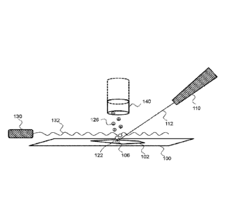
An implementation of the present method may be described with references to
Fig. 1,
which depicts a cellular sample 102 labeled with one or more mass tags, as
described below,
mounted on a substrate 100. In certain embodiments, the substrate 100 is a
flat or
substantially flat substrate. In some embodiments, the substrate 100 is a
conductive substrate.
Conductive substrates of interest include, but are not limited to, a
transparent conductive
oxide (TCO) coated glass or plastic, a conductive polymer coated glass or
plastic, or a
.. semiconductor wafer. Exemplary TCOs include indium tin oxide (ITO),
fluorine doped tin
oxide (FTO), doped zinc oxide, and the like. Exemplary conductive polymers
include, but are
not limited to, poly(3,4-
Date Recue/Date Received 2022-09-23
ethylenedioxythiophene) (PEDOT)/polystyrene sulfonic acid (PSS),
poly(thiophene)s (PT),
and the like. Exemplary semiconductor wafers may include, but are not limited
to, silicon
dioxide, gallium arsenide, and the like. In some embodiments, the substrate
100 is a non-
conductive substrate that is made conductive by, e.g., sputter coating an
insulating substrate
with a layer of metal such as Au or Pt. In some instances, an insulating
substrate is a glass or
plastic substrate.
In certain embodiments, the substrate is configured such that a voltage can be
applied
to the sample. Thus, in some embodiments, the present method includes applying
a voltage to
conductive members disposed on the sample, thereby controlling the electric
potential of the
sample. The voltage applied to the sample may vary depending on how the
present method is
implemented, and may be a positive voltage or a negative voltage. The voltage
applied to the
sample may be in the range of -100 V to 100 V, e.g., -100 V to 0 V, -80 V to -
10 V. -60 V to
-20 V, 0 V to 100 V, 10 V to 80 V, or 20 V to 60 V.
In some embodiments, the primary ion beam 112 impinges upon the labeled sample
102 at an impingement site 106, and is scanned across the sample. When a near-
continuous or
continuous ion beam (primary ions) 112 is irradiated onto the surface of a
solid sample 102 at
a high vacuum, a component of the surface is released, by a desorption-
ionization
phenomenon, into the vacuum, e.g., to form a plume of sputtered species at a
site over the
sample that is proximal to the impingement site 106 of the ion beam. The
generated sputtered
species include charged species (positively or negatively-charged secondary
ions) and neutral
species 122. When the primary ions are irradiated onto the sample, sputtered
species (neutral
and secondary ion) generated at the outermost surface of a solid sample are
released into the
vacuum, and the outermost surface (e.g., a depth of less than 1 nm, less than
2 nm, less than 5
nm, less than 10 nm, less than 20 nm, less than 50 nm, less than 100 nm, or
more than 100
nm) of the sample can be analyzed.
The primary ion beam 112 may be generated from an ion beam source 110. The ion
beam source 110 may be any convenient ion source that generates an ion beam
for sputtering
neutral species from the sample, such as an ion beam gun or a liquid metal ion
gun. Suitable
primary ion sources for performing the present method are described in, e.g.,
Applied Surface
Science, 255(4):1606-1609; Patents US8,168,957; US8,087,379; US8,076,650;
US7,670,455; and US7,241,361.
11
Date Recue/Date Received 2022-09-23
CA 02981496 2017-09-29
WO 2016/172215
PCT/US2016/028444
The primary ion beam 112 may be any suitable beam of ions for generating
sputtered
neutral species from a sample upon which the ion beam impinges. In certain
embodiments, the
primary ion beam is a continuous or near-continuous ion beam. The ion beam may
have a duty
cycle, as defined by the time the beam was on divided by the sum of the times
the beam was on
and off, of at least 1%, e.g., at least 5%, at least 10%, at least 50%, at
least 80%, or about 100%.
A near-continuous ion beam may have a duty cycle of at least 10%, e.g., at
least 50%, at least
80%, and up to 100%.
The primary ion beam 112 may include a beam of oxygen, cesium, gold, argon,
bismuth,
xenon, C60, SF6, indium or gallium ions, or a combination thereof. In certain
embodiments, the
ion beam includes ions that generate a larger number of secondary species
(neural and ionic
species) per primary ion that impinges upon a sample surface, i.e., ions that
have a higher
sputter yield, compared to, e.g., oxygen ions of equal energy. Exemplary ions
that have a higher
sputter yield than an oxygen ion beam are argon and gallium ions. In certain
embodiments, the
ion beam includes ions that generate a larger number of secondary species
(neural and ionic
species) per primary ion that impinges upon a sample surface per unit time,
i.e., ions that
generate more secondary adducts (SA), compared to, e.g., oxygen ions of equal
energy. In some
cases the ion beam includes ions that generate more SA compared to oxygen ions
by a range of 5
to 50 fold, e.g., 10 to 40 fold, including 20 to 30 fold.
In certain embodiments, the primary ion beam 112 has an ion current density of
1 nA/cm2
.. or more, e.g., 10 nA/cm2 or more, 100 nA/cm2 or more, 1 mA/cm2 or more, or
10 mA/cm2 or
more, and may be in the range of 1 nA/cm2 to 1 A/cm2, e.g., 1 nA/cm2 to 100
mA/cm2, or 10
nA/cm2 to 10 mA/cm2. In certain embodiments, the primary ion beam 112 has an
ion density of 1
x 1013 primary ions/cm2 or more, e.g., 1 x 10¹⁴ primary ions/cm2 or more, 1 x
1015 primary
ions/cm2 or more, 1 x 10¹⁶ primary ions/cm2 or more, 1 x 10¹⁷ primary ions/cm2
or more, or 1 x
1018 primary ions/cm2 or more, and may be in the range of 1 x 10¹³ to 1 x 10¹⁹
primary ions/cm2 ,
e.g., 1 x 10¹⁴ primary ions/cm2 to 1 x 10¹⁸ primary ions/cm2, or 1 x 10¹⁴
primary ions/cm2 to 1 x
1017 primary ions/cm2. In certain embodiments, the primary ion beam 112 has an
energy of 0.1
keV or more, e.g., 0.5 keV or more, 1 keV or more, 5 keV or more, or 10 keV or
more, and may
be in the range of 0.1 to 1000 keV, e.g., 0.5 to 100 keV, 1 to 50 keV,
including 1 to 10 keV. In
some embodiments, the width of the primary ion beam 112 is 1 nm or more, e.g.,
5 nm or more,
10 nm or more, 100 nm or more, or 200 nm or more, and may be 20 wri or less,
e.g., 10 p.m or
12
less, 1 gm or less, 500 nm or less, or 200 nm or less. In some embodiments,
the width of the
primary ion beam 112 is in the range of 1 nm to 20 gm, e.g., 5 nm to 10 gm, 10
nm to 1 gm,
20 nm to 500 nm, including 50 nm to 300 nm.
In certain embodiments, the scanning step includes irradiating the sample 102
with
the primary ion beam 112 to generate sputtered secondary ions and neutral
species 122 at
specific depths. Thus in certain embodiments, the scanning step includes
irradiating the
sample 102 with the primary ion beam 112 that has a primary ion current,
sputtering yield,
ionization efficiency and dwell time sufficient to generate sputtered
secondary ions and
neutral species 122 at specific depths. By "depth" is meant along the axis
perpendicular to the
surface of the substrate (z-axis) on which sample is mounted, in a proximal to
distal direction
relative to the ion beam source. In certain embodiments, the scanning step
includes
irradiating the sample 102 with the primary ion beam 112 to generate sputtered
secondary
ions and neutral species 122 at a depth resolution in the range of 1 nm to
10,000 nm, e.g., 2
nm to 1,000 nm, 5 nm to 100 nm, including 10 nm to 50 nm.
The sputtered neutral species 122 are then ionized to generate ionized neutral
species
126 at a site that is proximal to the source of the sputtered neutral species,
e.g., the
impingement site 106 of the primary ion beam on the sample. Any convenient
method may
be used to effect post-ionization of the sputtered neutral species. In certain
embodiments, the
post-ionization is done by irradiating the sputtered neutral species using a
high power-density
optical radiation 132, e.g., a laser beam. In some embodiments, the high power-
density
optical radiation 132 is generated by a radiation source 130, e.g., a laser
beam source, an
LED or an LED array.
The post-ionizing radiation source 130 may be any convenient radiation source.
Suitable method and systems for post-ionization are described in, e.g., US
Patent Nos.
4,743,804; 4,948,962; 5,146,088; 5,218,204; 5,272,338; 5,519,215; 6,072,182;
6,211,516;
and 8,410,704; and US Application Pub. Nos. 20020036363; 20060081775; and
20090008571.
In some embodiments, the sputtered neutral species 122 are photoionized by
irradiating the neutral species, to generate ionized neutral species 126. The
photoionized
neutral species may have a net positive charge or a net negative charge. In
some
embodiments, irradiating the sputtered neutral species includes applying
radiation produced
by a laser or a light emitting diode (LED) to the sputtered neutral species.
In some
embodiments, the radiation is produced by a
13
Date Recue/Date Received 2022-09-23
CA 02981496 2017-09-29
WO 2016/172215
PCT/US2016/028444
single LED or an LED array. The wavelength of the radiation may vary and in
some cases may
have a wavelength for ultraviolet, visible, or infrared radiation. In some
embodiments, the
average power of the radiation is in the range of 1 mW to 100 W, e.g., 1 mW to
100 mW, 1 mW
to 10 mW, 1 W to 100 W, 10 W to 100W, 10 mW to 10 W, including 100 mW to 1W.
Photoionizing may be done using resonant ionization or nonresonant ionization
to ionize the
neutral species.
The radiation is configured such that the sputtered neutral species 122 are
exposed to the
radiation 132 at a site that is proximal to their source 106. In certain
cases, the post-ionizing
radiation is applied such that the radiation travels parallel to a surface of
the sample and over a
region 106 of the sample impinged upon by the primary ion beam, to ionize the
sputtered neutral
species. The shortest distance between the path of the post-ionizing radiation
and the source of
the sputtered neutral species on the sample surface may vary, and in some
cases be in the range
of 0.3 to 5 mm, e.g., 0.4 to 4 mm, 0.5 to 3 mm, including 0.5 to 1.5 mm.
Where the sputtered neutral species are photoionized by a high power-density
optical
radiation 132, e.g., a laser beam, the width, or diameter, of the radiation
may vary, and may be in
the range of 0.3 to 3 mm, e.g., 0.5 to 2 mm, including 0.75 to 1.5 mm. In
certain embodiments,
the radiation source 130 operates in continuous wave (CW), quasi-continuous
wave (quasi-CW),
or pulsed modes of operation. The radiation source 130 may have a duty cycle,
as defined by the
time the radiation, e.g., laser, was on divided by the sum of the times the
radiation was on and
off, of at least 0.0001%, e.g., at least 0.001%, at least 0.01%, at least
0.1%, at least 1%, at least
10%, at least 50%, at least 80%, or about 100%. In some embodiments, the
radiation is a diode
laser, a diode-pumped solid state laser, an excimer laser, or a gas laser.
Depending on how it is implemented, the present method of ionizing sputtered
secondary
species (neutral and ionic species) produced by a primary ion beam applied to
a sample achieves
high ionization efficiency of all sputtered secondary species. The ionization
efficiency may be at
least 20%, e.g., at least 50%, at least 75%, at least 90%, or about 100% of
the sputtered
secondary species.
The method described herein employs a mass tag, i.e., a stable isotope that is
identifiable
by its mass for labeling of a biological, cellular sample, measured on an
instrument capable of
quantifying elemental composition with spatial registration using a primary
ion beam, an
ionization means for ionizing sputtered neutral species, and a mass
spectrometer.
14
The mass tag may be part of or conjugated to a stain, or conjugated to a
capture agent
such as an antibody. In certain embodiments, mass tags may be composed of a
chelating
polymer made up of repeating units of a metal chelator, such as
ethylenediaminetetraacetic
acid (EDTA) or diethylene triamine pentaacetic acid (DTPA), chelated to one or
more atoms
of a single non-biological isotope. In some embodiments the mass tags may be
substantially
uniform in size, so the abundance of specific binding reagent will be in
direct proportion with
the number of tag atoms. The tagged specific binding reagent is then contacted
with a
biological sample, washed, and measured with a mass spectrometry instrument
capable of
quantifying the number of tag atoms present in the sample with spatial
registration. The
abundance of the analyte may be inferred from the molar ratio of tag atoms per
detection
reagent.
The method described above may be multiplexed in that the assay can be done
using
multiple specific binding reagents (e.g., more than 2 specific binding
reagents, up to 5
specific binding reagents, up to 10 specific binding reagents, up to 20
specific binding
reagents, up to 50 specific binding reagents or up to 100 specific binding
reagents or more).
Each specific binding reagent may be linked to a different mass tag, where the
mass tags are
distinguishable from one another by mass spectrometry. Alternatively or in
addition,
multiplexing may involve using stains for specific features of interest.
Many elements exist in nature as multiple stable isotopes. For example, l'Eu
accounts for 52% of europium on Earth and 151Eu makes up most of the remaining
48%,
while unstable, radioactive isotopes of europium constitute less than 1%. Many
stable
isotopes are commercially available as powders or salt preparations, in
varying degrees of
purity, including 99% (2N), 99.9% (3N), 99.99% (4N), 99.999% (5N) and 99.9999%
(6N)
pure. In some embodiments, metal chelator tags may be synthesized using
enriched isotopes.
For example, mass dots may be synthesized using 151Eu (e.g. Europium 151
Oxide, 99.999%
purity, American Elements). Mass dots are described in US patent publication
2012/0178183.
Using enriched isotopes maximizes the number of unique species of isotope tags
that can be
simultaneously detected in a multiplexed analysis. In addition, spatially
distinct features of
interest may be labeled with the same metal tag to further multiplex the
analysis. Such
spatially distinct features may be distinguished based on co-localization with
one or more
other metal tags. For example, a Her2 membrane stain and an ER nuclear stain
using the
same metal
Date Recue/Date Received 2022-09-23
CA 02981496 2017-09-29
WO 2016/172215
PCT/US2016/028444
tag may be distinguished from one based on a dsDNA or histone H3 stain that
uses a different
metal tag, which would co-localize with the ER stain.
The mass tag may be part of or conjugated to a stain. In these embodiments,
the stain
may be phalloidin, gadodiamide, acridine orange, bismarck brown, barmine,
Coomassie blue,
bresyl violet, brystal violet, 4',6-diamidino-2-phenylindole (DAPI),
hematoxylin, eosin,
ethidium bromide, acid fuchsine, haematoxylin, hoechst stains, iodine,
malachite green, methyl
green, methylene blue, neutral red, Nile blue, Nile red, osmium tetroxide
(formal name: osmium
tetraoxide), rhodamine, safranin, phosphotungstic acid, osmium tetroxide,
ruthenium tetroxide,
ammonium molybdate, cadmium iodide, carbohydrazide, ferric chloride, hexamine,
indium
trichloride, lanthanum nitrate, lead acetate, lead citrate, lead(II) nitrate,
periodic acid,
phosphomolybdic acid, potassium ferricyanide, potassium ferrocyanide,
ruthenium red, silver
nitrate, silver proteinate, sodium chloroaurate, thallium nitrate,
thiosemicarbazide, uranyl acetate,
uranyl nitrate, vanadyl sulfate, or any derivative thereof. The stain may be
specific for any
feature of interest, such as a protein or class of proteins, phospholipids,
DNA (e.g., dsDNA,
ssDNA), RNA, an organelle (e.g., cell membrane, mitochondria, endoplasmic
recticulum, golgi
body, nulear envelope, and so forth), a compartment of the cell (e.g.,
cytosol, nuclear fraction,
and so forth). The stain may enhance contrast or imaging of intracellular or
extracellular
structures.
In certain embodiments, the stain may be suitable for administration to a live
subject. The
stain may be administered to the subject by any suitable means, such as
ingestion, injection (e.g.,
into the blood circulation), or topical administration (e.g., during a
surgery). Such a stain may be
specific for a tissue, biological structure (e.g., blood vessel, lesion), or
cell type of interest. The
stain may be incorporated into cells of the subject of a cellular process,
such as glucose uptake.
Examples of such stains include, without limitation, gadolinium, cisplatin,
halogenated
carbohydrates (e.g., carbohydrates which are fluorinated, chlorinated,
brominated, iodinated),
and so forth. Other injectable stains used in imaging techniques (e.g., such
as MRI, PET scans,
CT scans and so forth) may be conjugated to a mass tag if not inherently
associated with a mass
tag, and administered to a live subject. A sample may be obtained from the
subject after
administration, for use in the methods described herein.
16
CA 02981496 2017-09-29
WO 2016/172215
PCT/US2016/028444
In other embodiments, and as will be described in greater detail below, the
mass tag may
be conjugated to a capture agent, e.g., an antibody that recognizes an epitope
on the sample. In a
multiplexed assay, a combination of capture agents and stains may be used.
The mass tag used in the method may be any stable isotope that is not commonly
found
in the sample under analysis. These may include, but are not limited to, the
high molecular
weight members of the transition metals (e.g. Rh, Ir, Cd, Au), post-transition
metals (e.g. Al, Ga,
In, Ti), metalloids (e.g. Te, Bi), alkaline metals, halogens, and actinides,
although others may be
used in some circumstances. A mass tag may have a mass in the range of 21 to
238 atomic mass
units (AMU). In certain embodiments, a lanthanide may be use. The lanthanide
series of the
periodic table comprises 15 elements, 14 of which have stable isotopes (La,
Ce, Pr, Nd, Sm, Eu,
Gd, Tb, Dy, Ho, Er, Tm, Yb, Lu). Lanthanindes can be readily used because of
their rarity in the
biosphere. There are greater than 100, non-biological stable isotopes of
elements between 1 and
238 AMU. In some embodiments, tagging isotopes may comprise non-lanthanide
elements that
can form stable metal chelator tags for the applications described herein. In
the present
photoionization mass spectrornetry measurement modality, unlike some ICP-MS-
based
modalities, the elemental reporter could also consist of lower MW, transition
elements not
common in biological matrices (e.g. Al, W, and Hg).
Elements suitable for use in this method in certain embodiments include, but
are not
limited to, lanthanides and noble metals. In certain cases, an elemental tag
may have an atomic
number of 21-92. In particular embodiments, the elemental tag may contain a
transition metal,
i.e., an element having the following atomic numbers, 21-29, 39-47, 57-79, and
89. Transition
elements include the lanthanides and noble metals. See, e.g., Cotton and
Wilkinson, 1972, pages
528-530. The elemental tags employed herein are non-biological in that they
are man-made and
not present in typical biological samples, e.g., cells, unless they are
provided exogenously.
In particular embodiments, the mass tag to be linked to the binding reagent
may be of the
formula: R-MT, where R is a reactive group that can form a linkage with a
reactive group on a
specific binding reagent and MT is a mass tag. The compound may also contain a
spacer between
R and MT. In particular embodiments, R may be, e.g., a maleimide or halogen-
containing group
that is sulfydryl reactive, an N-hydroxysuccinimide (NHS)-carbonate that is
amine-reactive or an
N,N-diisopropy1-2-cyanoethyl phosphoramidite that is hydroxyl-reactive. Such
groups react with
other groups on the specific binding reagent, e.g., a cysteine or other
residue of an antibody or a
17
sulfhydryl group of an oligonucleotide). In many embodiments, the linkage
between the
reactive group and the mass tag is not selectively cleavable, e.g., is not
photo-cleavable.
In particular embodiments, MT may be a polymer of, e.g., 10-500 units, where
each
unit of the polymer contains a coordinated transition metal. Suitable reactive
groups and
.. polymers containing coordinating groups, including 1,4,7,10-
tetraazacyclododecane-1,4,7,10-
tetraacetic acid (DOTA) and DTPA-based polychelants, are described in a
variety of
publications, including: Manabe et al. (Biochemica et Biophysica Acta 883: 460-
467 (1986))
who describes attaching up to 105 DTPA residues onto a poly-L-lysine backbone
using the
cyclic anhydride method and also attaching polylysine-poly-DTPA polychelants
onto
monoclonal antibody (anti-human leukocyte antigen (HLA) IgGi) using a 2-
pyridyl
disulphide linker achieving a substitution of up to about 42.5 chelants (DTPA
residues) per
site-specific macromolecule; Torchilin (U.S. Patent 6,203,775) who describes a
generic
method for labeling antibodies that includes an antibody-reactive, lanthanide
chelating
compound of a generic formula; Sieving (U.S.5,364,614), the abstract for
describes a DOTA-
based polychelant containing a polylysine backbone that is linked to a
protein. Further
descriptions of such moieties are described in, for example: US20080003616
(Polymer
backbone element tags), US6,203,775 (Chelating polymers for labeling of
proteins),
US7,267,994 (Element-coded affinity tags), US6,274,713 (Polychelants) and
5,364,613
(Polychelants containing macrocyclic chelant moieties), as well as many
others. In addition to
.. the methods described in the references cited above, methods for making
polymer-based
elemental tags are also described in detail in Zhang et al (Agnew Chem. Int.
Ed. Engl. 2007
46: 6111-6114). In addition, any chelator able to bind to metal tags can be
used. These
include EDTA, ethylene glycol tetraacetic acid (EGTA), and Heme. These
chelators are able
to bind to +1, +2, +3, +4 ions of metal tags. Methods for linking such tags to
binding reagents
.. are known in the art. For example, the MAXPAR reagents produced by DVS
Sciences is a
maleimide-functionalized polymer of DTPA, with an average length of 30
monomers. Using
the MAXPAR protocol, it is possible to conjugate a typical IgG antibody with 6
or 7
polymers, thereby conjugating an average of 200 tagging isotope atoms per
antibody.
18
Date Recue/Date Received 2022-09-23
CA 02981496 2017-09-29
WO 2016/172215
PCT/US2016/028444
When using mass-based elemental analysis there are more than 100 non-
biological
elemental isotopic masses available between 21 and 238 atomic mass units
(arnu) that can be
simultaneously measured with virtually no overlap. Because these elements are
not usually
present in biological isolates, the only limitations of detection are the
sensitivity of the reagents
to which they are conjugated, and the sensitivity of the instrument performing
the measurement.
In particular embodiments, the method described above may be employed in a
multiplex
assay in which a heterogeneous population of cells is labeled with a plurality
of distinguishably
mass tagged binding reagents (e.g., a number of different antibodies). As
there are more than 80
naturally occurring elements having more than 200 stable isotopes, the
population of cells may
be labeled using at least 2, at least 5, at least 10, at least 20, at least
30, at least 50, or at least 100,
up to 150 or more different binding reagents (that bind to, for example
different cell surface
markers) that are each tagged with a different mass. After the population of
cells is labeled, they
are analyzed using the method described herein.
As noted above, the specific binding reagent used in the method may be any
type of
molecule (e.g., an antibody, a peptide-MHC tetramer, a nucleic acid (e.g.,
ssRNA or ssDNA), an
aptamer, a ligand specific for a cell surface receptor, etc.) that is capable
of associating with
cells, e.g., specifically binding to a binding partner in or on cells. The
binding partner may be a
protein, a nucleic acid or another type of cellular macromolecule (e.g., a
carbohydrate). The
binding partner may be on the cell surface, or it may be extracellular or
intracellular (e.g.,
associated with the nucleus or another organelle, or cytoplasmic).
In certain aspects, a specific binding reagent may be an MT conjugated to a
nucleic acid
that hybridizes to a specific RNA and/or DNA sequence. The MT conjugated
nucleic acid may
be used in combination with any suitable technique for detecting a target
(e.g., RNA, DNA,
protein or protein complex), such as standard in-situ hybridization, in-situ
hybridization utilizing
branched DNA probes (e.g., as provided by Affymetrix), proximity ligation
(PLA) and rolling
circle amplification (e.g., as provided by Olink bioscience), and so forth. In-
situ hybridization
techniques, including those employing branched DNA probes are described by
Monya Baker et
al. (Nature Methods 9, 787-790 (2012)). Briefly, in-situ hybridization using
branched DNA
probes utilizes a series of ssDNA probes, where a first set of DNA probes
specifically hybridizes
to the target DNA or RNA sequence, and a second set of DNA probes may
hybridize to a portion
of the first set of DNA probes, thus expanding the number of DNA probes that
can bind
19
(indirectly) to a single DNA or RNA molecule. A third set may bind to the
second set of DNA
probes in a likewise manner, and so forth. One or more of the sets of DNA
probes may be
conjugated to a metal tag to label the target DNA or RNA molecule. Proximity
ligation
techniques, including detection of single RNA molecules, DNA molecules, and
protein
complexes are described by Weibrecht et al. (Nature Methods 9, 787-790
(2012)). Rolling
circle amplification is described by Larsson et al. (Nat. Methods 1, 227-232
(2004)). Briefly,
in proximity ligation followed by rolling circle amplification, a nucleic acid
is hybridized to
two proximal RNA or DNA strands, after which the nucleic acid is ligated and
then
amplified, resulting in many copies of the sequence complimentary to the
nucleic acid. The
complimentary sequence is therefore present in higher copy number than the
original
proximal RNA or DNA strands, and can be more easily detected (e.g., by a MT
conjugated
nucleic acid that hybridizes to the complimentary sequence). The proximal RNA
or DNA
stands may each be conjugated to a different antibody (e.g., where the
different antibodies
may each be specific for a different protein of a protein complex).
Any of the above techniques may be used to resolve single molecular targets
(e.g.,
individual RNA molecules, DNA molecules, proteins or protein complexes). As
single
molecular targets may be resolvable as discrete puncti, a combination of metal
isotopes may
be used to uniquely label the molecular target. In one example, the specific
binding reagent
may be a nucleic acid may be conjugated to a unique combination of metal
isotopes. In
another example, a combination of MT conjugated nucleic acids (e.g., each
conjugated to a
different mass tag) may be used together to label the molecular target with a
unique
combination of metal isotopes. As such, n number of mass tags could be
combinatorially used
to label 2n different molecular targets, provided that the molecular targets
can be spatially
distinguished. The method described herein may be used to assay a sample of
biological
origin that contains cells, in which the amounts of certain components (e.g.,
protein, nucleic
acid or other molecules) need to be determined.
The sample may be labeled before or after being mounted on the substrate 100.
After
labeling the sample with one or more mass tags, the sample is scanned with a
primary ion
beam to generate sputtered secondary ions and sputtered neutral species, as
described above.
Date Recue/Date Received 2022-09-23
CA 02981496 2017-09-29
WO 2016/172215
PCT/US2016/028444
In certain embodiments, the radiation 132, e.g., high power-density optical
radiation,
produced by the radiation source 130 is intensified by an optical resonator
located outside the
radiation source. Any suitable type of resonator may be used to intensify the
post-ionizing
radiation, including a Fabry-Perot ring resonator, Michelson interferometer-
typed resonator, Fox-
Smith interferometer-typed resonator, Mach-Zehnder interferometer- typed
resonator, and the
like. The general operation of optical resonators are known, and are described
in, e.g., PCT
Application No. 2014155776; US Pat. Nos. 4,915475; 5,283,801; US Application
Pub. Nos.
20130058364; 20130064258. In certain embodiments, the optical resonator may be
configured to
be in the same compartment as the sample being imaged by the present method
and may be
.. distinct from the radiation source 130, such as the laser beam source.
Thus, in certain
embodiments, the optical resonator is configured to maximize optical resonance
of the radiation
over a region of the sample impinged upon by the primary ion beam.
In certain embodiments, the radiation 132, e.g., high power-density optical
radiation,
produced by the radiation source 130 is intensified by a multipass
spectroscopic absorption cell.
Any suitable type of multipass spectroscopic absorption cell may be used to
intensify the post-
ionizing radiation, such as a Pfund cell, White cell, Herriott cell, etc. The
general operation of
multipass spectroscopic absorption cells are known, and are described in,
e.g., US Pat. Nos.
5,818,578; 5,880,850; 7,307,716; US Application Pub. No. 20090035183.
When primary ions are irradiated onto the sample surface, sputtered neutral
species 126
.. having various masses are generated depending on the composition of the
surface of the sample.
The ionized, e.g., photoionized, neutral species 126 are focused in one
direction by an electrical
field, and detection is performed at a remote position, e.g., by mass
spectrometry. Upon
photoionization, the post-ionized neutral species 126 having a smaller mass
flies faster than an
ion having a larger mass in a time-of-flight (TOF) mass spectrometer ion
transport section 140.
Therefore, a measurement of a time between generation and detection of the
photoionized neutral
species (flight time) enables the analysis of masses of the generated
photoionized neutral species
to be performed. In certain embodiments, the TOF mass spectrometer is an
orthogonal time-of-
flight mass spectrometer. The term "orthogonal- refers to the direction of
flow of ions introduced
into a TOF mass spectrometer that is perpendicular to the direction in which
the ions are
extracted and accelerated to separate ions based on mass and charge. The
principles of
orthogonal TOF mass spectrometry is described in, e.g., Guilhaus, 1995 J Mass
Spec. 30:1519;
21
Chen et al., 1999 Int J Mass Spec. 185/186/187:221; and U.S. Patent No.
5,614,711. Thus, in
some embodiments, the present method includes detecting ionized neutral
species generated
by ionizing, e.g., photoionizing, neutral species sputtered by a primary ion
beam by TOF
mass spectrometry, e.g., orthogonal TOF mass spectrometry.
In some embodiments, a continuous or near-continuous primary ion beam will
produce a continuous or near-continuous emission of sputtered species (neutral
and
secondary ion), and the sputtered neutral species will be photoionized by a
high power-
density optical radiation 130, e.g., a laser beam, producing a continuous or
near-continuous
emission of ionized neutral species that will be focused and transferred by
the ion transport
section 140. This continuous or near-continuous ionized neutral species
current will then be
sampled over the entire range of possible masses of interest being analyzed by
pulsed optics
and time-of-flight mass spectrometry. In another embodiment of the invention,
the primary
ion source 110 will produce a pulsed primary ion beam in order to release
packets of
sputtered neutral species that are in turn ionized into packets of post-
ionized neutral species
before entering the TOF mass analyzer.
Time-of-Flight Mass Spectrometers (TOF MS) operate on the principle of
measuring
the time which ions travel over a fixed distance, the time being usually
proportional to the
square root of the mass-to-charge ratio of an ion and thus being a measure of
the mass of a
detected ion. Ions that arrive at an ion detector produce detector output
signals that usually
consists of a sequence of peaks each representing one or more ions of a
particular mass-to-
charge ratio (m/z). Generally, the duration of each peak in the mass spectrum
is less than 100
nanosecond, and the total duration of the detector output signal which
represents ions of all
masses (usually called single mass spectrum) is of the order of 100
microsecond. Such
detector output signals are usually digitized in one of two distinct ways:
time-to-digital
conversion or transient recording. In a time-to-digital converter (TDC), a
counter associated
with each arrival time window is incremented when an event of ion arrival is
detected within
this window. All events of ions arriving at a detector within a certain time
period (called
"dead time" of the TDC, typically 5-20 ns) can only be counted as one event.
As a result, the
TDC technique, being an ion counting technique, has been limited by the
measurement time
dynamic range and is not generally suitable for high dynamic range
characterization of
rapidly changing ion beams.
22
Date Recue/Date Received 2022-09-23
One example of a rapidly changing ion beam occurs when a sample is sputtered
and
produces a sputtered species cloud that rapidly changes in composition and/or
sputtered
species density. TOF MS is an example of a preferred method of analysis of
sputtered species
clouds upon ionization, e.g., photoioniziation, in an imaging instrument with
a mass
.. spectrometer detector that measures elemental composition of a planar
biological sample,
specifically for elements that are attached to antibodies or other affinity
reagents conjugated to
their specific antigens, as described in Angelo et al. Nature Medicine 2014
20:436. The
primary ion beam dwell time produces a sputtered species cloud lasting 10-
10,000
microseconds. It is desirable to be able to analyze such a short sputtered
species cloud upon
photoionization, for ions of multiple m/z with dynamic range of at least 4
orders of magnitude.
Another way of digitization of the detector output signal is the use of a
transient
recorder, in which all of the information in the signal that represents a
single TOF mass
spectrum (single transient) is captured and stored. For example, transient
recorders, based on
analog-to-digital converters (ADC), are encountered in commercial Digital
Storage
.. Oscilloscopes.
It can be desirable in some circumstances to provide information about the
change in
elemental composition of a particle-produced sputtered species cloud during
transient periods
that can last, for example, 10-1000 microseconds. In such circumstances it can
be desirable to
collect and store multiple mass spectra during such a relatively short period.
The duration of a
single mass spectrum can desirably be of the order of 10-20 microseconds,
allowing 1-1000
spectra to be collected for a single sample segment. A typical width for a
single mass window
in elemental TOF with a single mass spectrum duration of approximately 20
microsecond is
10-25 nano seconds. A sampling rate of 1 GHz or better can thus be desirable
for
characterizing post-ionized neutral species peak shapes. Such a high sampling
rate and
.. 104 dynamic range requirement results in a data generation rate well in
excess of 1 GB/s. This
is much higher than the fastest data transfer rate (-250 MB/s) achievable with
technology
known in the art. Recent advances in TOF-MS have made this measurement and
data transfer
workflow more routine. A TOF analysis data workflow as described in (Bandura
Anal Chem
2009 81:6813-22 or US patent no. US8283624) could be used herein.
The analysis of ionized, e.g., photoionized, sputtered neutral species may be
perfonned
in a similar manner to the analysis of sputtered secondary ions in Secondary
Ion Mass
Spectrometry (SIMS). In SIMS, the sputtered secondary ions are transferred
into a mass
spectrometer, where
23
Date Recue/Date Received 2022-09-23
they are mass analyzed and quantified using standard mass analyzers (e.g.,
time-of-flight,
magnetic sector, quadrupole, ion trap, or a combinations thereof). Displaying
the mass spectra
that were collected from the sample surface generates chemical images. Each
pixel in the
resulting essentially represents a mass spectrum. The principles of secondary
ion mass
spectrometry are described in, e.g., Belu et al (Biomaterials. 2003 24: 3635-
53), Pal et al
(Histochem Cell Biol. 2010 134: 423-43) and Klitzing (Methods Mol Biol. 2013
950: 483-
501). Further methods and systems for analyzing data that includes spatially
addressed
measurements of the abundance of one or more mass tags is described in, e.g.,
US Patent
Application Ser. No. 14/483,999 and US Provisional Application No. 61/974,351.
In order to reconstruct an image of the sample, the mass detector, the primary
ion
source 110 and optionally the radiation source 130 may be coordinated by a
synchronizer to
allow assignment of the detected mass information of the ionized neutral
species to their
source on the sample, i.e., the location on the sample upon which the primary
ion beam
impinged to generate the sputtered neutral species. Thus, in certain
embodiments, the mass
information from the detector signal would be integrated into single values
for each mass
channel for a sample segment. The positional information for the segment and
its
corresponding mass information would be recorded. At the same time, TOF MS
scans would
be integrated to form the mass information for the segment. For example, the
irradiation time
of the primary ion source on a single segment of the sample may be
approximately equivalent
to three sequential TOF MS scans. The coordination of this timing, the
positional information
and the digitization of the integrated mass values would be carried out by the
synchronizer.
The positional infoimation of the sample may be obtained by any suitable
method. In
some instances, the substrate 100 on which the sample 102 is mounted contains
a registration
mark, such as a mark inscribed into a microscope slide. Upon detecting the
position of the
registration mark, e.g., by optical means, the detected position may be used
to correlate the
position of the sample 102 with the position of the ion beam impingement site
106 on the
sample. The number of registration marks on the substrate may be one or more,
e.g., two or
more, three or more, 5 or more, or 10 or more. In certain embodiments, the
location of the
registration mark is determined with an accuracy of 500 pm or less, e.g., 300
pm or less, 100
pm or less, 50 pm or less, or about 10 pm.
24
Date Recue/Date Received 2022-09-23
CA 02981496 2017-09-29
WO 2016/172215
PCT/US2016/028444
After the initial data is obtained, the data is used to construct an image of
the sample. The
resolution of the image may vary, and in some cases may be at least 1,000 nm,
e.g., at least 750
nm, at least 500 nm, at least 250 nm, at least 100 nm, at least 50 nm, or at
least 10 nm. In certain
cases, the resolution of the image may be in the range of 10 to 1,000 nm,
e.g., 10 to 750 nm, 20
to 500 nm, 30 to 400 nm, or 50 to 200 nm.
In some embodiments, the method provides a two-dimensional or a three-
dimensional
image of a sample indicating the abundance of one or more mass tags used to
label the sample. A
three-dimensional image may have a depth resolution of at least 10 pm, e.g.,
at least 1 pm, at
least 100 nm, at least 10 nm, at least 1 nm, and in some cases the depth
resolution may range
.. from 1 nm to 50,000 nm, e.g., 2 nm to 10,000 nm, 5 nm to 1,000 nm, 10 nm to
500 nm, including
10 nm to 100 nm. By "depth" is meant along the axis (z-axis) perpendicular to
the surface of the
substrate 100 on which a sample 102 is attached, in a proximal to distal
orientation relative to the
ion beam source 110.
The image may be analyzed to identify the boundaries of individual cells,
and/or
subcellular features in individual cells, in the image. Computer-implemented
methods for
segmenting images of cells are known in the art and range from relatively
simple thresholding
techniques (see, e.g., Korde, et al Anal Quant Cytol Histol. 2009 31, 83-89
and Tuominen et al
Breast Cancer Res 2010 12, R56), to more sophisticated methods, such as, for
instance, adaptive
attention windows defined by the maximum cell size (Ko et al. J Digit Imaging
2009 22, 259-
274) or gradient flow tracking (Li, et al. J Microsc 2008 231, 47-58). Some
suitable image
segmentation methods may be reviewed in Ko et al (J Digit Imaging. 2009 22:
259-74) and Ong
(Comput Biol Med. 1996 26:269-79). Next the data that corresponds to each of
the individual
cells, or a subcellular feature thereof, that have been defined by the
segmenting are integrated to
provide, for each cell, values that represent the amount of each of the mass
tags within the
boundary of each cell. This step of the method results in a data set that
contains, for each cell,
measurements of the amount of each of the mass tags that are associated with
the cell. This
concept is illustrated in the table shown below.
Tag 1 Tag 2 Tag 3 Tag 4 Tag 5
Celli 0.1 0.1 5 3 1
Cell 2 0.2 0.4 4 0.1 0.1
Cell 3 10 0.1 0.2 0.3 5
CA 02981496 2017-09-29
WO 2016/172215
PCT/US2016/028444
This data allows one to categorizing the cells in the sample. For example, in
the example shown
in the table above, the three cells are likely to be different types of cells
because they have
different profiles of mass tags where the profile identifies the category. In
particular cases, this
information may be used to provide a false-color image in which each of the
cells is color-coded
by their category. As such, this method may comprise displaying an image of
the sample, in
which the cells are color-coded by their category. In particular embodiments,
in any one pixel of
the image, the intensity of the color of the pixel correlates with the
magnitude of the signals
obtained for that pixel obtained in the original scanning. In these
embodiments, the resulting
false color image may show color-code cells in which the intensity of the
color in any single
pixel of a cell correlates with the amount of specific binding reagent that is
associated with the
corresponding area in the sample.
As the original scan may only result in partial removal of the sample (e.g.,
at a depth on
the nanometer scale), the sample may be re-scanned to generate an additional
data set having
measurements of the abundance of one or more mass tags across the area that
was originally
scanned. For example, the original scan may be used to identify an area or
areas of interest in the
sample. Such a scan may be lower resolution and may therefore be more rapid,
measure the mass
tag abundance in a larger area at a time, and/or may result in removal of less
of the sample. The
re-scan may be a higher resolution scan of the abundance of metal tags in the
area or areas of
interest. Alternatively or in addition, multiple scans across the same area
may be used to produce
a 3 dimensional image (e.g., compiled from the individual 2 dimensional data
sets). In certain
aspects, areas of interest identified by an original scan may be analyzed
further after isolation of
the area of interest from the sample, e.g., such as by laser capture micro
dissection.
The methods described herein may include normalization as a means of
standardizing
data obtains across samples and/or time-points (e.g., to enable quantitative
cross-sample
comparison). In certain aspects, normalization of ionization and/or overall
measurement
efficiency may be performed using standardized metal particles or suspension
present in the
sample. The standardized metal particles or suspension may have a known amount
of one or
more mass tags, and the resulting measurement of the one or more mass tags may
be used to
normalize the measurements of other mass tags in the sample. For example,
normalization beads
may be used to calibrate the system or normalize data obtained by the present
method.
Normalization of mass cytometry data using bead standards is described by
Rachel Fink et al.
26
(Cytometry A. 83(5):483-94(2013)), and is applicable to the present method
which also utilize
time of flight mass spectrometry. Alternatively or in addition, ionization
and/or measurement
efficiency may be normalized according to any of the above-mentioned stains.
For example,
measurements of a mass tag used to stain the ER may be normalized to the
overall intensity of
that mass tag in a given area, in the cell, or across multiple cells in the
sample.
Normalization may also be used to account for the effects of, for example,
degree of
tissue fixation, retention of protein, and staining efficiency with specific
binding reagents.
Mass tags conjugated to well-characterized antibodies that bind molecular
targets stably
expressed across a wide range of cell types may be used for noimalization.
Such antibodies
include, without limitation, antibodies to housekeeping proteins (such as
GAPDH, HSP90,
beta-actin and beta-tubulin), dsDNA and histone H3.
As discussed above, the methods of the present disclosure allow for a
multiplexed
approach. Multiple mass tags may be measured to determine the abundance of
multiple
molecular targets (e.g. specific proteins, DNA, RNA, etc.) as well as biologic
features of
interest in the sample (e.g., cell or tissue structure, cellular organelles,
cellular fractions, etc.).
In addition, mass tag measurements may be normalized according to any of the
above-
described embodiments. The large number of discrete mass tags enables
multiplexing of more
than 2, 5, 10, 20, 30, 40, 50, 60, 70, 80, 100 or more different mass tags in
a single area.
Multiple mass tags (e.g., conjugated to antibodies against complementary
epitopes of the same
.. molecular target) may be used for redundancy so as to increase confidence
in a measurement
of a specific molecular target. Further multiplexing may be achieved by using
identical mass
tags to label two or more spatially distinct targets or features of interest.
Alternatively or in
addition, a unique combination of metal tags may be used to identify a
spatially distinct target
or feature of interest.
Depending on how it is implemented, the present method of generating a high-
resolution image of a cellular sample is a highly sensitive, rapid method. In
some cases, the
sensitivity of detection achieved by the present method is higher by a factor
in the range of 10
to 200, e.g., 50 to 150, 75 to 120, or 80 to 100, than a comparable method
that does not use
post-ionization of sputtered neutral species. In certain embodiments, the
speed at which a
sample is analyzed is higher by a factor in the range of 5 to 50, e.g., 10 to
40, 10 to 30, or 15
to 25, than a comparable method that does not use post-ionization of sputtered
neutral species.
In some embodiments, the
27
Date Recue/Date Received 2022-09-23
CA 02981496 2017-09-29
WO 2016/172215
PCT/US2016/028444
gain in signal per unit time is higher by a factor in the range of 50 to
5,000, e.g., 100 to 3,000,
200 to 1,500, or 500 to 1,000, than a comparable method that does not use post-
ionization of
sputtered neutral species.
Utility
The above-described method can be used to analyze a cells from a subject to
determine,
for example, whether the cell is normal or not or to determine whether the
cells are responding to
a treatment. In one embodiment, the method may be employed to determine the
degree of
dysplasia in cancer cells. In these embodiments, the cells may be from a
sample of from a
multicellular organism or a microbe. A biological sample may be isolated from
an individual,
e.g., from a soft tissue or from a bodily fluid, or from a cell culture that
is grown in vitro. A
biological sample may be made from a soft tissue such as brain, adrenal gland,
skin, lung, spleen,
kidney, liver, spleen, lymph node, bone marrow, bladder stomach, small
intestine, large intestine
or muscle, etc. Bodily fluids include blood, plasma, saliva, mucous, phlegm,
cerebral spinal
fluid, pleural fluid, tears, lactal duct fluid, lymph, sputum, cerebrospinal
fluid, synovial fluid,
urine, amniotic fluid, and semen, etc. Biological samples also include cells
grown in culture in
vitro. A cell may be a cell of a tissue biopsy, scrape or lavage or cells. In
particular embodiments,
the cell may of a cell in a formalin fixed paraffin embedded (FFPE) sample. In
particular cases,
the method may be used to distinguish different types of cancer cells in FFPE
samples.
Also provided is a method for identifying a hydroxymethylation pattern that
correlates with
phenotype, e.g., a disease, condition or clinical outcome, etc. In some
embodiments, this method
may comprise (a) performing the above-described method on a plurality of cfDNA
samples,
wherein the cfDNA samples are isolated from patients having a known phenotype,
e.g., disease,
condition or clinical outcome, thereby detennining which sequences are
hydroxymethylated in
cfDNA from each of the patients; and (b) identifying a hydryoxymethylation
signature that is
correlated with the phenotype.
In some embodiments, the method may be used to produce a signature that may be
diagnostic (e.g., may provide a diagnosis of a disease or condition or the
type or stage of a
disease or condition, etc.), prognostic (e.g., indicating a clinical outcome,
e.g., survival or death
within a time frame) or theranostic (e.g., indicating which treatment would be
the most
effective).
28
CA 02981496 2017-09-29
WO 2016/172215
PCT/US2016/028444
The method described above finds particular utility in examining tissue
sections using
panels of antibodies, examples of which are provided in the table below.
CD3, CD7, CD20, CD34, CD45, CD56, CD117,
Acute Leukemia MC Panel
MPO, PAX-5, and TdT.
Adenocarcinoma vs. Mesothelioma Pan-CK, CEA, MOC-31, BerEP4, TTF1,
calretinin,
IHC Panel and WT-1.
Bladder vs. Prostate Carcinoma [HC
CK7, CK20, PSA, CK 903, and p63.
Panel
ER, PR, Ki-67, and HER2. Reflex to HER2 FISH
Breast IHC Panel
after HER2 IHC is available.
Burkitt vs. DLBC Lymphoma IHC
BCL-2, c-1VIYC, Ki-67.
panel
Carcinoma Unknown Primary Site, CK7, CK20, mammaglobin, ER, TTF1, CEA, CA19-
Female (CUPS IHC Panel - Female) 9, Si , synaptophysin, and WT-1.
Carcinoma Unknown Primary Site, CK7, CK20, TTF1, PSA, CEA, CA19-9, Si , and
Male (CUPS IHC Panel - Male) synaptophysin.
GIST IHC Panel CD117, DOG-1, CD34, and desmfit.
Hepatoma/Cholangio vs. Metastatic HSA (HepPar 1), CDX2, CK7, CK20, CAM 5.2,
Carcinoma IHC Panel TTF-1, and CEA (polyclonal).
BOB-1, BCL-6, CD3, CD10, CD15, CD20, CD30,
Hodgkin vs. NHL IHC Panel CD45 LCA, CD79a, MUM1, OCT-2, PAX-5, and
EBER ISH.
Lung Cancer IHC Panel chromogranin A, synaptophysin, CK7, p63, and TTF-
29
CA 02981196 2017-09-29
WO 2016/172215
PCT/US2016/028444
1.
Lung vs. Metastatic Breast TTF1, mammaglobin, GCDFP-15 (BRST-2), and
Carcinoma IHC Panel ER.
BCL-2, BCL-6, CD3, CD4, CD5, CD7, CD8, CD10,
Lymphoma Phenotype IBC Panel CD15, CD20, CD30, CD79a, CD138, cyclin D1,
Ki67, MUMI., PAX-5, TdT, and EBER ISH.
Lymphoma vs. Carcinoma IHC CD30, CD45, CD68, CD117, pan-keratin, MPO,
Panel S100, and synaptophysin.
Lymphoma vs. Reactive Hyperplasia BCL-2, BCL-6, CD3, CD5, CD10, CD20, CD23,
IHC Panel CD43, cyclin D1, and Ki-67.
CD68, Factor XIIIa, CEA (polyclonal), S-100,
Melanoma vs. Squatnous Cell
melanoma cocktail (HMB-45, MART- l/Melan-A.
Carcinoma. 1HC Panel
tyrosinase) and Pan-CK.
Mismatch Repair Proteins 1HC Panel
MLH1, MSH2, MSH6, and PMS2.
(MMR/Colon Cancer)
Neuroendocrine Neoplasm IHC CD56, synaptophysin, chromogranin A, TTF-1, Pan-
Panel CK, and CEA (polyclonal).
CD19, CD20, CD38, CD43, CD56, CD79a, CD138,
Plasma Cell Neoplasm IHC Panel
cyclin D1, EMA, kappa, lambda, and MUM1.
Prostate vs. Colon Carcinoma INC CDX2, CK 20, CEA (monoclonal), CA19-9,
PLAP,
Panel CK 7, and PSA.
Pan-CK, SMA, desmin, S100, CD34, vimentin, and
Soft Tissue Tumor IHC Panel
CD68.
CA 02981496 2017-09-29
WO 2016/172215
PCT/US2016/028444
ALK1. CD2. CD3. CD4. CD5, CD7. CD8, CD 10.
T-Cell Lymphoma IFIC panel
CD20, CD21, CD30. CD56, TdT, and EBER 1SH.
T-LGL Leukemia II-IC panel CD3, CD8, granzyme B. and TIA-1.
Undifferentiated Tumor 1HC Panel Pan-CK. S100, CD45, and vimentin.
In some embodiments, the method may involve obtaining an image as described
above
(an electronic form of which may have been forwarded from a remote location)
and may be
analyzed by a doctor or other medical professional to determine whether a
patient has abnormal
cells (e.g., cancerous cells) or which type of abnormal cells are present. The
image may be used
as a diagnostic to determine whether the subject has a disease or condition,
e.g., a cancer. In
certain embodiments, the method may be used to determine the stage of a
cancer, to identify
metastasized cells, or to monitor a patient's response to a treatment, for
example.
In any embodiment, data can be forwarded to a "remote location", where "remote
location," means a location other than the location at which the image is
examined. For
example, a remote location could be another location (e.g., office, lab, etc.)
in the same city,
another location in a different city, another location in a different state,
another location in a
different country, etc. As such, when one item is indicated as being ''remote"
from another, what
is meant is that the two items can be in the same room but separated, or at
least in different
rooms or different buildings, and can be at least one mile, ten miles, or at
least one hundred miles
apart. "Communicating" information references transmitting the data
representing that
information as electrical signals over a suitable communication channel (e.g.,
a private or public
network). "Forwarding" an item refers to any means of getting that item from
one location to the
next, whether by physically transporting that item or otherwise (where that is
possible) and
includes, at least in the case of data, physically transporting a medium
carrying the data or
communicating the data. Examples of communicating media include radio or infra-
red
transmission channels as well as a network connection to another computer or
networked device,
and the intemet or including email transmissions and information recorded on
websites and the
like. In certain embodiments, the image may be analyzed by an MD or other
qualified medical
31
CA 02981496 2017-09-29
WO 2016/172215
PCT/US2016/028444
professional, and a report based on the results of the analysis of the image
may be forwarded to
the patient from which the sample was obtained.
In some cases, the method may be employed in a variety of diagnostic, drug
discovery,
and research applications that include, but are not limited to, diagnosis or
monitoring of a disease
or condition (where the image identifies a marker for the disease or
condition), discovery of drug
targets (where the a marker in the image may be targeted for drug therapy),
drug screening
(where the effects of a drug are monitored by a marker shown in the image),
determining drug
susceptibility (where drug susceptibility is associated with a marker) and
basic research (where is
it desirable to measure the differences between cells in a sample).
In certain embodiments, two different samples may be compared using the above
methods. The different samples may be composed of an "experimental" sample,
i.e., a sample of
interest, and a "control" sample to which the experimental sample may be
compared. In many
embodiments, the different samples are pairs of cell types or fractions
thereof, one cell type
being a cell type of interest, e.g., an abnormal cell, and the other a
control, e.g., normal, cell. If
two fractions of cells are compared, the fractions are usually the same
fraction from each of the
two cells. In certain embodiments, however, two fractions of the same cell may
be compared.
Exemplary cell type pairs include, for example, cells isolated from a tissue
biopsy (e.g., from a
tissue having a disease such as colon, breast, prostate, lung, skin cancer, or
infected with a
pathogen etc.) and normal cells from the same tissue, usually from the same
patient; cells grown
in tissue culture that are immortal (e.g., cells with a proliferative mutation
or an immortalizing
transgene), infected with a pathogen, or treated (e.g., with environmental or
chemical agents such
as peptides, hormones, altered temperature, growth condition, physical stress,
cellular
transformation, etc.), and a noitnal cell (e.g., a cell that is otherwise
identical to the experimental
cell except that it is not immortal, infected, or treated, etc.); a cell
isolated from a mammal with a
cancer, a disease, a geriatric mammal, or a mammal exposed to a condition, and
a cell from a
mammal of the same species, preferably from the same family, that is healthy
or young; and
differentiated cells and non-differentiated cells from the same mammal (e.g.,
one cell being the
progenitor of the other in a mammal, for example). In one embodiment, cells of
different types,
e.g., neuronal and non-neuronal cells, or cells of different status (e.g.,
before and after a stimulus
on the cells) may be employed. In another embodiment of the invention, the
experimental
material is cells susceptible to infection by a pathogen such as a virus,
e.g., human
32
CA 02981496 2017-09-29
WO 2016/172215
PCT/US2016/028444
immunodeficiency virus (HIV), etc., and the control material is cells
resistant to infection by the
pathogen. In another embodiment, the sample pair is represented by
undifferentiated cells, e.g.,
stem cells, and differentiated cells.
Cells any organism, e.g., from bacteria, yeast, plants and animals, such as
fish, birds,
reptiles, amphibians and mammals may be used in the present method. In certain
embodiments,
mammalian cells, i.e., cells from mice, rabbits, primates, or humans, or
cultured derivatives
thereof, may be used.
Systems
Also provided herein is a system that find use in performing the present
methods, as
described above, to generate a high resolution image of a cellular sample. An
implementation of
the system may include a) holder for retaining a substrate mounted with a
sample, b) a
continuous or near-continuous primary ion beam source configured to scan the
sample and
sputter secondary ions and neutral species from the sample, c) a radiation
source configured to
photoionize the neutral species at a site that is proximal to their source on
the sample, d) an
orthogonal time-of-flight mass spectrometer configured to detect the
photoionized neutral
species and obtain spatially addressed measurements of the abundance of at
least one mass tag
associated with the sample, and e) a computer comprising an image analysis
module that
processes the measurements to produce an image of the sample.
As described above, the sample may be labeled with at least one mass tag using
any
convenient method to produce a labeled sample in which a biological feature of
interest is
associated with the at least one mass tag. In certain embodiments, the sample
is labeled with a
plurality of (i.e., two or more, e.g., three or more, four or more, 5 or more,
10 or more, up to
about 100, or more) distinguishable mass tags, and the system is configured to
obtain spatially
addressed measurements of the abundance of the plurality of mass tags
associated with the
sample.
In certain embodiments, the radiation source may be any suitable radiation
source,
including a laser or LED/LED array, for ionizing the sputtered neutral
species, as described
above. The present system may also include an optical resonator, which may be
configured to
maximize optical resonance of the radiation over a region of the sample
impinged upon by the
primary ion beam. In such cases, the optical resonator may be configured to be
in the same
33
compartment as the sample being imaged in the present system and may be
distinct from the
radiation source, such as a laser. In certain embodiments, the system includes
a multipass
spectroscopic absorption cell.
In certain embodiments, the system includes a synchronizer that coordinates
the mass
spectrometer and the primary ion source, and optionally the sample holder
and/or the radiation
source, to allow assignment of the detected mass information of the ionized
neutral species
with their source on the sample, i.e., the location on the sample upon which
the primary ion
beam impinged to generate the sputtered neutral species, as described above.
In certain embodiments, the system includes a registration means configured to
determine the position of the sample such that the position of the sample can
be correlated
with positioning of the ion beam source, and hence, with the position of the
ion beam
impingement site, as described above.
In certain embodiments, the image analysis module of the present system is
configured
to analyze the image, e.g., identify the boundaries of individual cells and/or
subcellular
features in individual cells, integrate mass tag information for an individual
cell, normalize the
analyzed image, and/or display the image of the sample, wherein the cells may
be color-coded
according to category that reflects the mass tags associated with a cell, as
described above.
The citation of any publication is for its disclosure prior to the filing date
and should
not be construed as an admission that the present invention is not entitled to
antedate such
publication by virtue of prior invention.
34
Date Recue/Date Received 2022-09-23