Note: Descriptions are shown in the official language in which they were submitted.
ASSAYS USING SURFACE-ENHANCED RAMAN SPECTROSCOPY
(SERS)-ACTIVE PARTICLES
TECHNICAL FIELD
The presently disclosed subject matter relates to diagnostic assays using
surface
enhanced Raman spectroscopy (SERS)-active particles, including liquid-based
assays;
magnetic capture assays; microparticle-nanoparticle satellite structures for
signal
amplification in an assay; composite SERS-active particles useful for enhanced
detection of targets; and sample tubes and processes for using the same.
BACKGROUND
Various techniques have been developed to detect the presence of one or more
analytes in an assay. For example, fluorescent, luminescent, chemiluminescent,
or
electrochemiluminescent techniques have been used to detect analytes within a
biological sample. In many biological assays, including assays where micro- or
nanoparticles are used for detecting the presence and/or amount of one or more
analytes
in a biological sample, the generation of a signaling event is used to detect
the presence
of the analyte. Such biological assays known in the art, however, have
limitations.
Thus, it might be advantageous to provide an assay having one or more enhanced
characteristics, including, but not limited to, enhanced sensitivity,
specificity,
accuracy, repeatability, and combinations thereof.
BRIEF SUMMARY
In some embodiments, the presently disclosed subject matter provides liquid-
based assays including magnetic capture particles having attached thereto a
binding
member having an affinity for one or more analytes of interest and SERS-active
nanoparticles, also having attached thereto a binding member having an
affinity for
the one or more analytes of interest. When contacted with a biological sample
containing one or more analytes, a magnetic capture particle-analyte-SERS-
active
nanoparticle complex is formed. The magnetic properties of the magnetic
capture
-1-
CA 2981992 2017-10-11
particles can be used to localize the magnetic capture particle-analyte-SERS-
active
nanoparticle complex in a predetermined area within an assay vessel for
detecting the
SERS signal.
In other embodiments, the presently disclosed subject matter provides a
magnetic capture/liquid-based assay incorporating a reference label. In such
embodiments, magnetic particles used for the magnetic pull-down are labeled
with a
reference label capable of producing a detectable signal, in addition to the
binding
member specific to the analyte of interest. The signal emitted by the SERS-
active
nanoparticle of the magnetic capture particle-analyte-SERS-active nanoparticle
complex can be referenced to that of the reference label attached to the
magnetic
capture particle to compensate for variations in pellet size, shape, or
positioning.
In a further embodiment, a lysis reagent can be used in an assay, such as a
liquid-based assay, with or without magnetic pull-down. The use of a lysis
reagent
can provide an increased signal and/or improved limit of detection for
analytes of
interest, for example, in biological matrices, such as human blood, plasma, or
serum,
or in cells.
In yet another embodiment, a method for amplifying a signal in a liquid-based
assay is provided. Such methods include adding a second aliquot of a reporter
molecule having the same signal-producing capabilities as the reporter
molecule, e.g.,
a SERS-active nanoparticle, already present in the assay solution before the
magnetic
capture complexes are localized. This second aliquot of reporter molecule has
attached thereto one or more binding members having an affinity for the
binding
member of the reporter molecule present in the assay solution. The second
reporter
molecule therefore can bind to the first reporter molecule, resulting in a
higher signal
per magnetic capture particle-analyte-reporter molecule complex.
In other embodiments, methods for improving Raman reference spectra and
spectral analysis in magnetic capture/liquid-based assays are provided. In one
embodiment, reference spectra of one or more analytes of interest are obtained
with
the analyte disposed in a magnetized pellet, as opposed to using reference
spectra of
the analyte obtained in solution. In another embodiment, methods are provided
for
improving the SERS spectral analysis, including selecting the wavelength
region
- 2 -
CA 2981992 2017-10-11
within which the analysis is performed and selecting the components of the
spectral
fitting procedure, e.g., a least squares fitting technique, based on results
from an initial
analysis.
In some embodiments, the presently disclosed subject matter provides composite
nanostructures, including a composite structure, referred to herein as a
"satellite"
structure, comprising a plurality of signal-bearing particles, e.g.,
nanoparticles, bound to a
core particle. In other embodiments, a composite structure, referred to herein
as a "core-
shell" structure, which includes a core particle, an active material, such as
a Raman-
active material, surrounding the core particle, and one or more shells, such
as a metal
shell, surrounding the active material is provided. The presently disclosed
satellite and
core-shell structures can be used to amplify or otherwise enhance a signal in
an assay,
such as a SERS assay.
In some embodiments, a sample tube designed to form a magnetic particle
pellet having a consistent size, shape, and density is provided, wherein the
sample
tube has dimensions to physically constrain a magnetic particle pellet to a
desired
size. The sample tube can include an optical window allowing for the detection
of
optical signals generated from the magnetic particle pellet. A system is
provided for
forming a magnetic particle pellet that uses a magnet positioned adjacent and
below
the sample tube. The system can be used in a magnetic capture assay. The
presently
disclosed subject matter further relates to a method of reliably forming a
smaller,
denser magnetic particle pellet in a sample tube.
Also provided herein, are representative systems and instrumentation suitable
for carrying out the presently disclosed assays.
Certain aspects of the presently disclosed subject matter having been stated
hereinabove, which are addressed in whole or in part by the presently
disclosed
subject matter, other objects will become evident as the description proceeds
when
taken in connection with the accompanying Examples and Drawings as best
described
herein below.
- 3 -
CA 2981992 2017-10-11
BRIEF DESCRIPTION OF THE SEVERAL VIEWS OF THE DRAWING(S)
Having thus described the presently disclosed subject matter in general terms,
reference will now be made to the accompanying drawings, which are not
necessarily
drawn to scale, and wherein:
Figure 1 is a schematic diagram of a representative presently disclosed
magnetic capture assay;
Figure 2 is a schematic diagram depicting the use of a reference label, e.g.,
a
SERS-active nanoparticle, as a reference on a magnetic capture particle;
Figure 3 is a graphical representation of a comparison of a non-referenced
signal and a referenced signal. The non-referenced signal (B) is the peak
intensity at
1590 cm' of a trans-1,2-bis(4-pyrridypethylene (BPE) Raman reporter and the
referenced signal (A) is the ratio of the peak intensities at 1590 cm-1 and
1180 cm-1,
which is the peak intensity corresponding to a 4,4'-dipyridyl (DPY) reporter.
Note
the scale of the left side of the graph corresponds to the referenced signal
(A),
whereas the scale on the right side of the graph corresponds to the non-
referenced
signal (B);
Figure 4 is a schematic diagram of an example of an optical system suitable
for use with the presently disclosed assays;
Figures 5A-5D are schematic representations of pellet formation by sample
tube rotation;
Figure 6 is a graphical representation of a comparison of the 4,4'-dipyridyl
(DPY) reporter signal in buffer, plasma, and lysed blood with and without
lysing
reagent (rgts);
Figure 7 is a representative schematic diagram of a presently disclosed assay
using a signal amplification method;
Figures 8A and 8B are a comparison of liquid-based immunoassays run with
identical reagents in the absence of (8A) and the presence of (8B) the
presently
disclosed signal amplification method;
Figure 9 is a representative schematic diagram of the presently disclosed
amplification method in a polynucleotide detection format;
- 4 -
CA 2981992 2017-10-11
Figures 10A and 10B present results from a DNA hybridization assay in the
absence of (10A) and presence of (10B) the presently disclosed signal
amplification
method;
Figures 1 1A and 11B show that a random signal can be erroneously assigned
to input variables due to spurious alignment of features. Normally distributed
random
noise with a standard deviation of 10,000 was fit using a least-squares
routine. In this
example, marker 4 was assigned a weight of 0.5 to balance negative weights of
other
markers;
Figures 12A and 12B show representative reference spectra obtained in
solution (A) and in a magnetic particle pellet (B). The marker 5 peak (near
865 nm)
relative to marker 1 is higher in the pellet, and the shape of the peak at 880
nm is
slightly different.
Figure 13 is a graphical representation of concentration estimates for one of
five markers in a multiplexing experiment. Concentration level 20 corresponds
to
2.5E8 marker particles/mL. Estimates using pellet-based reference spectra
(closed
diamonds) show better accuracy and precision, especially at lower
concentrations,
than solution-based reference spectra (open circles). The straight line shows
a 1:1
relation. (Solution-based data points are offset from the pellet-based points
on the x-
axis for clarity.);
Figure 14 shows a transmission electron micrograph (TEM) of a satellite
structure according to one embodiment of the presently disclosed subject
matter;
Figure 15 illustrates a sandwich assay using a satellite structure for
amplifying
an analyte signal according to an embodiment of the presently disclosed
subject
matter;
Figure 16A depicts a sandwich assay using a satellite structure for
amplifying an analyte signal according to an embodiment of the presently
disclosed
subject matter; and Figure 16B shows the sandwich assay of Figure 16A
following
the application of a magnetic field;
Figure 17 illustrates a cross-section of a core-shell composite particle
according to an embodiment of the presently disclosed subject matter;
Figure 18A is a drawing showing a side view of a sample tube according to
one embodiment of the presently disclosed subject matter;
- 5 -
CA 2981992 2017-10-11
Figure 18B is a drawing showing a cross-sectional side view taken along
Section A-A of the sample tube shown in Figure 18A;
Figure 18C is a drawing showing a bottom view of the sample tube shown in
Figure 18A;
Figure 19A is a drawing showing a side view of a magnet positioned adjacent
and below a sample tube according to one embodiment of the presently disclosed
subject matter;
Figure 19B is a drawing showing a top view of the magnet and sample tube
shown in Figure 19A; and
Figure 20 is a graph illustrating a thyroid-stimulating hormone (TSH) assay
binding curve using a sample tube according to one embodiment of the presently
disclosed subject matter.
DETAILED DESCRIPTION
The presently disclosed subject matter now will be described more fully
hereinafter with reference to the accompanying Drawings, in which some, but
not all
embodiments of the presently disclosed subject matter are shown. Many
modifications and other embodiments of the presently disclosed subject matter
set
forth herein will come to mind to one skilled in the art to which the
presently
disclosed subject matter pertains having the benefit of the teachings
presented in the
foregoing descriptions and the associated Drawings. Therefore, it is to be
understood
that the presently disclosed subject matter is not to be limited to the
specific
embodiments disclosed and that modifications and other embodiments are
intended to
be included within the scope of the appended claims. Although specific terms
are
employed herein, they are used in a generic and descriptive sense only and not
for
purposes of limitation.
The terms "a," "an," and "the" refer to "one or more" when used in this
application, including the claims. Thus, for example, reference to "a sample"
includes
a plurality of samples, unless the context clearly is to the contrary (e.g., a
plurality of
samples), and so forth.
- 6 -
CA 2981992 2017-10-11
Throughout this specification and the claims, the words "comprise,"
"comprises," and "comprising" are used in a non-exclusive sense, except where
the
context requires otherwise.
As used herein, the term "about," when referring to a value is meant to
encompass variations of, in some embodiments 50%, in some embodiments 20%,
in some embodiments 10%, in some embodiments 5%, in some embodiments
1%, in some embodiments 0.5%, and in some embodiments 0.1% from the
specified amount, as such variations are appropriate to perform the disclosed
methods
or employ the disclosed compositions.
Further, when an amount, concentration, or other value or parameter is given
as
either a range, preferred range, or a list of upper preferable values and
lower preferable
values, this is to be understood as specifically disclosing all ranges formed
from any pair
of any upper range limit or preferred value and any lower range limit or
preferred value,
regardless of whether ranges are separately disclosed. Where a range of
numerical
values is recited herein, unless otherwise stated, the range is intended to
include the
endpoints thereof, and all integers and fractions within the range. It is not
intended that
the scope of the presently disclosed subject matter be limited to the specific
values
recited when defining a range.
I. ASSAYS USING SERS-ACTIVE PARTICLES
In some embodiments, the presently disclosed subject matter provides
diagnostic assays for determining the presence or amount of an analyte or
ligand of
interest in a biological sample. Accordingly, in some embodiments, the
presently
disclosed subject matter provides assay methods, compositions, systems,
instruments,
and kits for performing diagnostic assays using SERS-active nanoparticles.
As described in more detail herein below, the detection capabilities of the
presently disclosed assays are improved in one or more ways over assays known
in
the art. These improvements include, but are not limited to an increase in
signal
intensity, enhanced specificity, higher accuracy, improved repeatability, and
combinations thereof The presently disclosed assays also can provide
diagnostic
results in a shorter period of time than assays known in the art. Such
improvements,
either alone or in combination, allow for the use of the presently disclosed
methods in
- 7 -
CA 2981992 2017-10-11
applications, such as diagnostic assays using Raman spectroscopy as a
detection
method, optical imaging of tissues, and other applications, where such
enhancements
are required.
When used in a diagnostic assay, the enhanced characteristics observed for the
presently disclosed methods enable detection of biomarkers, including, but not
limited
to, proteins, polynucleotides, and metabolites, at lower concentrations than
those
measurable using SERS methods known in the art, and also enable detection of
cells
(e.g., whole organisms). These enhanced characteristics also are beneficial in
applications where the Raman signal has to pass through, i.e., is transmitted
through, a
complex medium, such as whole blood or serum. Further, diagnostic assays with
a
enhanced characteristics can be required for early detection of a condition or
a disease
state in a subject.
A. General Overview: Surface Enhanced Raman Spectroscopy
When a molecule is irradiated with photons of a particular frequency, the
photons are scattered. The majority of the incident photons are elastically
scattered
without a change in frequency (Rayleigh scattering), whereas a small fraction
of the
incident photons (approximately 1 in every 106) interact with a vibrational
mode of
the irradiated molecule and are inelastically scattered. The inelastically
scattered
photons are shifted in frequency and have either a higher frequency (anti-
Stokes) or a
lower frequency (Stokes). By plotting the frequency of the inelastically
scattered
photons against their intensity, a unique Raman spectrum of the molecule is
observed.
The low sensitivity of conventional Raman spectroscopy, however, has limited
its use
for characterizing biological samples in which the target analyte(s) typically
are
present in small quantities.
When a Raman-active molecule is adsorbed on or in close proximity to, e.g.,
within about 50 A of, a metal surface, the intensity of a Raman signal arising
from the
Raman-active molecule can be enhanced. For example, increases in the Raman
signal
by a factor of about 103 to about 106, or in some cases, 1014 have been
reported to
date. This enhancement is referred to as the surface-enhanced Raman scattering
(SERS) effect. The SERS effect was first reported in 1974 by Fleishman et al.,
who
observed intense Raman scattering from pyridine adsorbed on a roughened silver
- 8 -
CA 2981992 2017-10-11
electrode surface. See Fleishman et al., "Raman spectra of pyridine adsorbed
at a
silver electrode," Chem. Phys. Lett., 26, 163 (1974); see also Jeanmaire, D.
L., and
Van Dyne, R. P., "Surface Raman spectroelectrochemistry. 1. Heterocyclic,
aromatic, and aliphatic-amines absorbed on anodized silver electrode." J.
Electroanal. Chem., 84(1), 1-20 (1977); Albrecht, M. G., and Creighton, J. A.,
"Anomalously intense Raman spectra of pyridine at a silver electrode,"
J.A.C.S., 99,
5215-5217 (1977). Since then, SERS has been observed for a number of different
molecules adsorbed on the surface of metal surfaces. See, e.g., A. Campion, A.
and
Kambhampati, P., "Surface-enhanced Raman scattering," Chem. Soc. Rev., 27, 241
(1998).
The magnitude of the SERS enhancement depends on a number of parameters,
including the position and orientation of various bonds present in the
adsorbed
molecule with respect to the electromagnetic field at the metal surface. The
mechanism by which SERS occurs is thought to result from a combination of (i)
surface plasmon resonances in the metal that enhance the local intensity of
the
incident light; and (ii) formation and subsequent transitions of charge-
transfer
complexes between the metal surface and the Raman-active molecule.
The SERS effect can be observed with Raman-active molecules adsorbed on
or in close proximity to metal colloidal particles, metal films on dielectric
substrates,
and metal particle arrays, including metal nanoparticles. For example, Kneipp
et al.
reported the detection of single molecules of a dye, cresyl violet, adsorbed
on
aggregated clusters of colloidal silver nanoparticles. See Kneipp, K. et al.,
"Single
molecule detection using surface-enhanced Raman scattering (SERS), Phys. Rev.
Lett., 78(9), 1667-1670 (1997). That same year, Nie and Emory observed the
surfaced enhanced resonance Raman spectroscopy (SERRS) signal, wherein the
resonance between the absorption energy of the Raman-active molecule and that
of
the nanoparticle yield an enhancement as large as about 1010 to about 1012, of
a dye
molecule adsorbed on a single silver nanoparticle, where the nanoparticles
ranged
from spherical to rod-like add had a dimension of about 100 nm. See Nie, S.,
and
Emory, S. R., "Probing single molecules and single nanoparticles by surface-
- 9 -
CA 2981992 2017-10-11
enhanced Raman scattering," Science, 275, 1102-1106 (1997); Emory, S. R., and
Nie,
S., "Near-field surface-enhanced Raman spectroscopy on single silver
nanoparticles,"
Anal. Chem., 69, 2631 (1997).
A Raman enhancing particle having associated therewith, e.g., adsorbed on or
attached to, a SERS-active molecule(s) is referred to herein as a "SERS-active
particle." More particularly, a SERS-active particle, as referred to herein,
includes a
particle have a surface that induces, causes, or otherwise supports surface-
enhanced
Raman light scattering (SERS) or surface-enhanced resonance Raman light
scattering
(SERRS). A number of surfaces are capable of producing a SERS signal,
including
roughened surfaces, textured surfaces, and other surfaces, including smooth
surfaces.
"Raman scattering" generally refers to the inelastic scattering of a photon
incident on a molecule. Photons that are inelastically scattered have an
optical
frequency (vi), which is different than the frequency of the incident light
(v0). The
difference in energy (AE) between the incident light and the inelastically
scattered
light can be represented as (AE) = hlvo - vil, wherein h is Planck's constant,
and
corresponds to energies that are absorbed by the molecule. The incident
radiation can
be of any frequency vo, but typically is monochromatic radiation in the
visible or near-
infrared spectral region. The absolute difference Ivo - vii is an infrared,
e.g.,
vibrational, frequency. More particularly, the process that produces light of
frequency
other than vo is referred to as "Raman scattering." The frequency v1 of the
"Raman
scattered" radiation can be greater than or less than vo, but the amount of
light with
frequency v1 <v0 (Stokes radiation) is greater than that with frequency vi >
vo (anti-
Stokes radiation).
As used herein, the term "radiation" refers to energy in the form of
electromagnetic radiation that can induce surface-enhanced Raman scattering in
a
sample under test, e.g., a sample comprising a SERS-active nanoparticle having
one
or more SERS-active reporter molecules associated therewith. More
particularly, the
term "radiation" refers to energy in the form of electromagnetic radiation
that causes
the surface of a nanoparticle to induce, emit, support, or otherwise cause
light
scattering, e.g., Raman scattering, in a reporter molecule proximate to the
nanoparticle
surface. As used herein, a "reporter molecule" refers to any molecule or
chemical
compound that is capable of producing a Raman spectrum when it is illuminated
with
- 10 -
CA 2981992 2017-10-11
radiation of a proper wavelength. A "reporter molecule" also can be referred
herein
as a "label," a "dye," a "Raman-active molecule," or "SERS-active molecule,"
each of
which can be used interchangeably.
"Surface-enhanced Raman scattering" or "SERS" refers to the phenomenon
that occurs when the Raman scattering signal, or intensity, is enhanced when a
Raman-active molecule is adsorbed on or in close proximity to, e.g., within
about 50
A of, a metal surface. "Surface-enhanced resonance Raman scattering" or
"SERRS"
refers to an increased SERS signal that occurs when the reporter molecule in
close
proximity to the SERS-active nanoparticle surface is in resonance with the
excitation
wavelength.
B. Representative Nanoparticles Suitable for Use with the
Presently
Disclosed Methods
1. Nanoparticles Generally
Any SERS-active particle is suitable for use in the presently disclosed
methods. Such SERS-active particles typically are nanoparticles and also are
referred
to as "nanotags." As used herein, the terms "nanoparticle," "nanostructure,"
"nanocrystal," "nanotag," and "nanocomponent," are used interchangeably and
refer
to a particle having at least one dimension in the range of about 1 nm to
about 1000
nm, including any integer value between 1 nm and 1000 nm (including about 1,2,
5,
10, 20, 50, 60, 70, 80, 90, 100, 200, 500, and 1000 nm). In some embodiments,
the
nanoparticle is a metallic nanoparticle. In some embodiments, the nanoparticle
is a
spherical particle, or substantially spherical particle having a core diameter
between
about 2 nm and about 200 nm (including about 2, 5, 10, 20, 50, 60, 70, 80, 90,
100,
and 200 nm). In some embodiments, the nanoparticle has a core diameter between
about 2 nm and about 100 nm (including about 2, 5, 10, 20, 30, 40, 50, 60, 70,
80, 90,
and 100 nm) and in some embodiments, between about 20 nm and 100 nm (including
about 20, 21, 22, 23, 24, 25, 26, 27, 28, 29, 30, 31, 32, 33, 34, 35, 36, 37,
38, 39, 40,
41, 42, 43, 44, 45, 46, 47, 48, 49, 50, 51, 52, 53, 54, 55, 56, 57, 58, 59,
60, 61, 62, 63,
64, 65, 66, 67, 68, 69, 70, 71, 72, 73, 74, 75, 76, 77, 78, 79, 80, 81, 82,
83, 84, 85, 86,
87, 88, 89, 90, 91, 92, 93, 94, 95, 96, 97, 98, 99, and 100 nm). One of
ordinary skill
in the art, upon review of the presently disclosed subject matter, would
recognize that
- 11 -
CA 2981992 2017-10-11
a nanoparticle suitable for use with the presently disclosed assays can
include a core,
e.g., a metal core, which induces the Raman effect, and can further include
one or
more layers of SERS-active materials, encapsulants, and/or outer shell
structures that
also can contribute to the size, e.g., total diameter of the nanoparticle
structure.
SERS-active nanoparticles suitable for use with the presently disclosed
methods typically comprise at least one metal, i.e., at least one element
selected from
the Periodic Table of the Elements that is commonly known as a metal. Suitable
metals include Group 11 metals, such as Cu, Ag, and Au, or any other metals
known
by those skilled in the art to support SERS, such as alkali metals. In some
embodiments, the nanoparticle substantially comprises a single metal element.
For
example, the preparation of gold nanoparticles is described by Frens, G., Nat.
Phys.
ScL, 241, 20 (1972). In other embodiments, the nanoparticle comprises a
combination
of at least two elements, such as an alloy, for example, a binary alloy. In
some
embodiments, the nanoparticle is magnetic.
In other embodiments, the metal includes an additional component, such as in
an Au2S/Au core-shell particle. Au2S/Au core-shell particles have been
reported to
have widely tunable near-IR optical resonance. See Averitt, R. D., et al.,
"Ultrafast
optical properties of gold nanoshells," JOSA B, 16(10), 1824-1832 (1999).
Further,
Ag core/Au shell particles, such as those described by Cao, Y.W., et al., "DNA-
modified core-shell Ag/Au nanoparticles," J. Am. Chem. Soc., 123(32), 7961-
7962
(2001), or Au core/Ag shell particles, or any core-shell combination involving
SERS-
active metals, can be used. Other combinations suitable for use in core-shell
particles
also are suitable for use with the presently disclosed methods, including Au-
or Ag-
functionalized silica/alumina colloids, Au- or Ag-functionalized TiO2
colloids, Au
nanoparticle capped-Au nanoparticles (see, e.g., Mucic, et al., "DNA-directed
synthesis of binary nanoparticle network materials," J. Am. Chem. Soc.,
120(48),
12674 (1998)); Au nanoparticle-capped TiO2 colloids; and particles having a Si
core
with a metal shell (i.e., "nanoshells"), such as silver-capped Si02 colloids
or gold-
capped Si02 colloids. See, e.g., Jackson, et al., Proc. NatL Acad. ScL U.S.A.
101(52):17930-5 (2004); see also U.S. Patent Nos. 6,344,272 and 6,685,986 to
- 12 -
CA 2981992 2017-10-11
Oldenburg et al. The use of such nanoshells in biosensing applications has
been
described. See U.S. Patent No. 6,699,724 to West et al.
Another class of nanoparticles suitable for use with the presently disclosed
methods includes nanoparticles having an internal surface. Such nanoparticles
include hollow particles and hollow nanocrystals or porous or semi-porous
nanoparticles. See e.g., U.S. Patent No. 6,913,825 to Ostafin et al.
Accordingly, the presently disclosed
subject matter also provides a nanoparticle comprising a core-shell particle
active for
SERS or a hollow nanoparticle active for SERS. In some embodiments, such
nanoparticles can exhibit an improved SERS signal.
While it is recognized that particle shape and aspect ratio can affect the
physical, optical, and electronic characteristics of nanoparticles, the
specific shape,
aspect ratio, or presence/absence of internal surface area does not bear on
the
=
qualification of a particle as a nanoparticle. Accordingly, nanoparticles
suitable for
use with the presently disclosed methods can have a variety of shapes, sizes,
and
compositions. Further, the nanoparticle can be solid, or in some embodiments,
as
described immediately hcreinabove, hollow. Non-limiting examples of suitable
nanoparticles include colloidal metal hollow or filled nanobars, magnetic,
paramagnetic, conductive or insulating nanoparticles, synthetic particles,
hydrogels
(colloids or bars), and the like. It will be appreciated by one of ordinary
skill in the
art that nanoparticles can exist in a variety of shapes, including but not
limited to
spheroids, rods, disks, pyramids, cubes, cylinders, nanohelixes, nanosprings,
nanorings, rod-shaped nanoparticles, arrow-shaped nanoparticles, teardrop-
shaped
nanoparticles, tetrapod-shaped nanoparticles, prism-shaped nanoparticles, and
a
plurality of other geometric and non-geometric shapes.
Further, nanoparticles suitable for use with the presently disclosed methods
can be isotropic or anisotropic. As referred to herein, anisotropic
nanoparticles have a
length and a width. In some embodiments, the length of an anisotropic
nanoparticle is
the dimension parallel to the aperture in which the nanoparticle was produced.
In
some embodiments, the anisotropic nanoparticle has a diameter (width) of about
350
nm or less. In other embodiments, the anisotropic nanoparticle has a diameter
(width)
- 13 -
CA 2981992 2017-10-11
of about 250 nm or less and in some embodiments, a diameter (width) of about
100
nm or less. In some embodiments, the width of the anisotropic nanoparticle is
between about 15 nm to about 300 nm. Further, in some embodiments, the
anisotropic nanoparticle has a length, wherein the length is between about 10
nm and
350 nm.
Much of the SERS literature (both experimental and theoretical) suggests that
anisotropic particles (rods, triangles, prisms) can provide an increased
enhancement of
the Raman signal as compared to spheres. For example, the so-called "antenna
effect"
predicts that Raman enhancement is expected to be larger at areas of higher
curvature.
Many reports of anisotropic particles have been recently described, including
silver
(Ag) prisms and "branched" gold (Au) particles.
Anisotropic Au and Ag nanorods can be produced by electrodeposition into
preformed alumina templates, in a manner similar to the production of
Nanobarcodes0 particles. See, e.g., Nicewarner-Pena, S. R., et al.,
"Submicrometer
metallic barcodes," Science, 294, 137-141 (2001); Walton, I. D., et al.,
"Particles for
multiplexed analysis in solution: detection and identification of striped
metallic
particles using optical microscopy," Anal. Chem. 74, 2240-2247 (2002). These
particles can be prepared by the deposition of alternating layers of
materials, typically
Au and Ag, into preformed alumina templates, and can have a diameter of about
250
nm and a length of about 6 microns.
2. Encapsulated SERS-Active Nanoparticles
SERS-active metal nanoparticles have a tendency to aggregate in aqueous
solution and once aggregated are difficult to re-disperse. Further, the
chemical
composition of some Raman-active molecules is incompatible with chemistries
used
to attach other molecules, such as proteins, to metal nanoparticles. These
characteristics can limit the choice of Raman-active molecule, attachment
chemistries,
and other molecules to be attached to the metal nanoparticle.
Accordingly, in some embodiments, a SERS-active reporter molecule when
affixed, e.g., either adsorbed or covalently attached to a nanoparticle, can
be coated or
encapsulated, for example, in a shell, of a different material, including a
polymer,
glass, or ceramic material. Such embodiments are referred to herein as
"encapsulated
- 14 -
CA 2981992 2017-10-11
SERS-active nanoparticles." Methods for preparing encapsulated SERS-active
nanoparticles are described in U.S. Patent No. 6,514,767 to Natan.
Examples of suitable particles for use with the presently disclosed methods
include Oxonica Nanotags (Oxonica Inc., Mountain View, California). In one
embodiment, the nanotags comprise a gold core having a diameter of about 50
nm, are
coated with a distinct reporter molecule, and are encapsulated, in some
embodiments,
in a 10 nm to 50 um protective glass coating, in some embodiments, a 15 nm to
40 nm
protective glass coating, in some embodiments, a 30 nm to 40 nm protective
glass
coating, and, in some embodiments, a 35 nm protective glass coating, as
described
hereinabove. Nanotags are further described, for example, in U.S. Patent No.
6,514,767 to Natan; U.S. Published Patent Application Nos. 2003/0166297 to
Natan
and 2005/0158870 Al to .Natan.
The presently disclosed encapsulated SERS-active nanoparticles can include a
metal nanoparticle, a submonolayer, monolayer, or multilayer of one or more
reporter
molecules in close proximity to the surface of the metal nanoparticle. The
term "in
close proximity" is intended to mean within about 50 nm or less of an outer
surface of
the nanoparticle. A nanoparticle having a submonolayer, monolayer, or
multilayer of
one or more reporter molecules attached to an outer surface of the
nanoparticle core
also can include an encapsulating shell. In such embodiments, the reporter
molecule
is positioned at an interface between the outer surface of the metal
nanoparticle and an
interior surface of the encapsulating shell.
The nanoparticle core comprising the encapsulated nanoparticle can be a metal
sphere, e.g., a gold, silver, or copper sphere, having a diameter of about 20
rim to
about 200 nm. In some embodiments, the nanoparticle core comprises an oblate
or
prolate metal spheroid. The diameter of the nanoparticle core can be selected
based,
in part, on the wavelength of incident light. In some embodiments, the
encapsulating
shell comprises a dielectric material, such as a polymer, glass, metal, metal
oxides,
such as TiO2 and Sn02, metal sulfides or a ceramic material. In some
embodiments,
the encapsulant is glass, e.g., SiOx. To encapsulate the presently disclosed
SERS-
active nanoparticles in glass, the metal nanoparticle cores can be treated
with a glass
- 15 -
CA 2981992 2017-10-11
primer, i.e., a material that can lead to a growth of a uniform coating of
glass, or can
improve adhesion of the glass coat to the particle, or both. Glass can then be
grown
over the metal nanoparticle by standard techniques known in the art.
The encapsulation process can be carried out after, or during, attaching or
adsorbing one or more reporter molecules to the core nanoparticle. In this
way, the
dye is sequestered from the surrounding solvent as a coating on the surface of
the
metal nanoparticle core. Such a configuration provides the metal nanoparticle
core
with a stable SERS activity. The dye can form a sub-monolayer, a complete
monolayer, or a multilayer assembly on the surface of the metal nanoparticle
core.
The dye layer can comprise a single dye or can be a mixture of different dyes.
Thus, in some embodiments, a SERS-active reporter molecule forms a layer
on the outer surface of the nanoparticle core, wherein the layer at least
partially covers
the outer surface of the nanoparticle core and is defined by an inner surface
and an
outer surface. The encapsulant is disposed on at least one of the outer
surface of the
nanoparticle core and the outer surface of the layer of the SERS-active
reporter
molecule to at least partially surround the nanoparticle core, which is at
least partially
covered with a layer of the SERS-active reporter molecule.
Further, in some embodiments, the encapsulant can be modified, e.g.,
derivatized by standard techniques known in the art, to attach molecules,
including
biomolecules, to its outer surface. This characteristic allows the presently
disclosed
encapsulated SERS-active nanoparticles to be conjugated to molecules,
including
biomolecules, such as proteins and nucleic acids, or to solid supports without
interfering with the Raman activity of the dye. Glass and other materials
suitable for
use as an encapsulating shell contain functional groups amenable to molecular
attachment. For example, immersion of glass in a suitable base allows for the
covalent attachment of alkyl trichlorosilanes or alkyl trialkoxysilanes, with
additional
functionality available on the end of the alkyl group of the alkyl
trichlorosilane or
alkyl trialkoxysilane group. In some embodiments, one or more of an
aminoalkyltrialkyloxysilane group, a mercaptoalkyltrialkoxysilane group, or a
carboxyalkyltrialkoxysilane group can be covalently attached to the glass
surface.
Thus, glass surfaces can be modified with many forms of biomolecules and
biomolecular superstructures, including cells, as well as oxides, metals,
polymers, and
- 16 -
CA 2981992 2017-10-11
the like. Likewise, surfaces of glass can be modified with well-organized
monomolecular layers. Accordingly, glass coatings support many types of
chemical
functionalization (also referred to herein as "derivatization"). Other forms
of
encapsulants also can be functionalized, as well. Accordingly, the presently
disclosed
nanoparticles can be affixed to any species known in the art having a
chemically-
reactive functionality.
The thickness of the encapsulant can be varied depending on the physical
properties required of the SERS-active nanoparticle. The physical properties,
such as
the sedimentation coefficient can be affected by the thickness of the
encapsulant. In
general, the thicker the encapsulant, the more effective the sequestration of
the SERS-
active dyes on the metal nanoparticle core from the surrounding solvent.
In embodiments wherein the encapsulant is glass, the thickness of the glass
typically can range from about 1 nm to about 50 nm. In exemplary, non-limiting
embodiments, the encapsulated SERS-active nanoparticles comprise gold
nanoparticles having a diameter ranging from about 50 nm to about 100 nm
encapsulated in a sphere of glass having a thickness ranging from about, in
some
embodiments, from about 10 nm to about 50 nm; in some embodiments, from about
15 nm to about 40 nm; and, in some embodiments, about 35 nm. The optimization
of
the dimensions of the presently disclosed encapsulated SERS-active
nanoparticles can
be accomplished by one of ordinary skill in the art. For example, it is known
in the
art that core-shell nanoparticles (e.g., Au/AuS nanoparticles) support SERS
and have
different optical properties as compared to pure metal nanoparticles.
Likewise, it is
known in the art that SERS from prolate spheroids can be enhanced relative to
spheres
with the same major axis. Further, it is known that single particle
enhancements are
wavelength-dependent. Thus, the particle size can be "tuned" to achieve a
maximum
SERS signal for a given excitation wavelength. Accordingly, the composition of
the
particle, or its size or shape can be altered in accordance with the presently
disclosed
subject matter to optimize the intensity of the SERS signal.
The presently disclosed encapsulated SERS-active nanoparticles are easy to
handle and store. Further, they also are aggregation resistant, stabilized
against
- 17 -
CA 2981992 2017-10-11
decomposition of the dye in solvents and air, are chemically inert, and can be
concentrated, e.g., by magnetic pull down techniques, and redispersed without
loss of
SERS activity.
As described in more detail herein below, the presently disclosed subject
matter also provides more specialized nanoparticles capable of enhancing an
assay
using SERS-active particles.
3. Reporter Molecules
The reporter molecules can be any molecule that provides a Raman signal
upon exposure to appropriate irradiation. A "reporter molecule" refers to any
molecule or chemical compound that is capable of producing a Raman signal. A
"reporter molecule" also can be referred herein as a "label," a "dye," a
"Raman-active
molecule," or "SERS-active molecule," each of which can be used
interchangeably.
A number of distinct reporter molecules with strong Raman spectra are known
and
can be used to create distinct "flavors" of SERS-active particles to enable
multiplexing capabilities (the term "flavors" indicates particles that provide
distinct
Raman signatures upon irradiation). Such particles typically are able to
function in
the near-infrared (NIR) wavelength region, are detectable in whole blood, and
are
photostable. Further, a number of different "flavors" can be excited with a
single
wavelength.
C. Representative Capture Probes
Capture probes, such as antibodies or DNA probes, can be immobilized onto
the protective glass coating using known bioconjugation techniques. An
advantage of
this approach is that the SERS signal-generating reporter molecule is secured
in close
proximity to the gold surface and protected by the glass coating from
biological or
chemical attack. In addition, competitive binding between the reporter
molecule and
the capture probe is eliminated, allowing for maximum surface coverage of the
reporter molecule on the nanoparticle core surface and the capture probe on
the glass
surface, respectively.
More generally, SERS-active nanoparticles can be functionalized with a
molecule, such as a specific binding member of a binding pair, which can bind
to a
- 18 -
CA 2981992 2017-10-11
target analyte. The binding event creates a detectable signal, which is
indicative of
the presence and/or amount of an analyte. The detectable signal can correspond
to a
localized detection of a SERS tag or can be represented by a detectable
wavelength
shift in the SERS spectrum.
The use of a functionalized SERS-active nanoparticle has several advantages
over non-functionalized nanoparticles. First, the functional group provides a
degree
of specificity to the nanoparticle by providing a specific interaction with a
target
analyte. Second, the target analyte does not have to be Raman active itself;
its
presence can be determined by measuring the SERS spectrum of the Raman-active
dye attached to the nanoparticle. Such measurements are referred to herein as
"indirect detection," in which the presence or absence of a target analyte or
ligand in a
biological sample is determined by detecting a SERS signal that does not
directly
emanate from the target analyte or ligand of interest.
SERS-active nanoparticles suitable for use with the presently disclosed
methods can be functionalized to bind to a target analyte in at least two
different
ways. In some embodiments, the SERS-active reporter molecule, i.e., a SERS-
active
dye, can be conjugated with a specific binding member of a binding pair,
whereas in
other embodiments, a specific binding member of a binding pair can be attached
directly to the nanoparticle. In embodiments in which the nanoparticle core is
at least
partially surrounded by an encapsulating shell, the binding member can be
attached to
an outer surface of the encapsulating shell.
As used herein, the term "conjugate" refers to a molecule comprising two or
more subunits bound together, optionally through a linking group, to form a
single
molecular structure. The binding can be made either by a direct chemical bond
between the subunits or through a linking group. Such binding in a conjugate
typically is irreversible. As used herein, the term "affinity" refers to the
strength of
the attraction between one binding member to another member of a binding pair
at a
particular binding site. The term "specificity" and derivations thereof, refer
to the
likelihood that a binding member will bind to another member of a binding
pair. Such
binding between one binding member, e.g., a binding protein, to another
binding
member of a binding pair, e.g., a ligand or analyte, can be reversible.
- 19 -
CA 2981992 2017-10-11
The term "specific binding member" refers to a molecule for which there
exists at least one separate, complementary binding molecule. A specific
binding
member is a molecule that binds, attaches, or otherwise associates with a
specific
molecule. The binding, attachment, or association can be chemical or physical.
A
specific molecule to which a specific binding member binds can be any of a
variety of
molecules, including, but not limited to, antigens, haptens, proteins,
carbohydrates,
nucleotide sequences, nucleic acids, amino acids, peptides, enzymes, and the
like.
Further, a specific binding member of a particular type will bind a particular
type of
molecule. In such instances, the specific binding members are referred to as a
"specific binding pair." Accordingly, an antibody will specifically bind an
antigen.
Other specific binding pairs include avidin and biotin, carbohydrates and
lectins,
complementary nucleotide sequences, complementary peptide sequences, enzymes
and enzyme cofactors, and the like.
1. SERS-Active Nanoparticles Having a Specific Binding Member of a
Binding Pair Attached Directly Thereto
In some embodiments, a binding member of a specific binding pair, for
example, an antibody, such as a monoclonal antibody, can be attached directly
to the
surface of the nanoparticle. In an exemplary embodiment, a specific binding
member
of a binding pair, e.g., a monoclonal antibody, can be treated with linker,
e.g.,
polyethylene glycol (PEG), and attached directly to the nanoparticle through
the PEG
linker.
As would be appreciated by one of ordinary skill in the art, the selection of
the
linker can be determined by various factors depending on the objects of the
assay.
For example, the use of PEG as a linker can stabilize the orientation of the
antibody
such that the epitope of the antigen is pointed away from the surface of the
. nanoparticle. In this way, the functionalized nanoparticle can be
designed to
maximize the presentation of the epitope or other binding region to the test
solution,
thereby potentially increasing the sensitivity of the assay.
Depending on the binding member, other linkers can be used. For example,
alkanethiols can be used as linkers for antibodies and peptides. Short chain
alkanethiols, including, but not limited to, N-succinimidyl-S-
acetylthioacetate
- 20 -
CA 2981992 2017-10-11
(SATA) and N-succinimidyl-S-acetylthiopropionate (SATP) can be used as linkers
after sulfhydryl deprotection. Other properties also can determine the choice
of
linker, such as the length of the linker chain. For example, PEG can be
desirable in
that it also acts to protect the surface of the reagent and is flexible, which
can enhance
the ability of the reagent to bind to the analyte of interest.
A specific binding member, such as an antibody, also can be modified with a
linker, such as a thiolated PEG linker, and attached to the nanoparticle.
2. Representative Binding Members
In some embodiments, the binding member conjugated with the presently
disclosed SERS-active nanoparticle, either through the SERS-active reporter
molecule
or directly attached to an outer surface of the nanoparticle itself, comprises
a
polypeptide or protein, such as a glucose binding protein. Representative
binding
members include, but are not limited to, specific binding members having an
affinity
for a target analyte, including nucleic acids, protein domains, antibody
fragments,
cells, and antibodies for target analytes, such as prostate specific antigen
(P SA),
creatine kinase MB (CKMB) isoenzyme, cardiac troponin I (cTnI) protein,
thyroid-
stimulating hormone (TSH), influenza A (Flu A) antigen, influenza B (Flu B)
antigen,
and respiratory syncytial virus (RSV) antigen. Antibodies for such target
analytes are
known in the art.
The analyte and binding member can act as binding partners. The term
"associates" or "binds" as used herein refers to binding partners having a
relative
binding constant (Kd) sufficiently strong to allow detection of binding to the
protein
by a detection means. The Kd can be calculated as the concentration of free
analyte at
which half the protein is bound, or vice versa. When the analyte of interest
is glucose,
the Kd values for the binding partners are between about 0.0001 mM and about
50
mM.
D. Diagnostic Assays Generally
SERS-active nanoparticles can be used in diagnostic assays. For example,
Rohr et al. demonstrated an immunoassay with SERS detection including multiple
components and washing steps. See Rohr, T. E., et al., "Immunoassay employing
- 21 -
CA 2981992 2017-10-11
surface-enhanced Raman spectroscopy," Anal. Biochem., 182:388 (1989). Also, Ni
et
al. demonstrated reporter attachment to a gold slide in a heterogeneous
detection
assay including incubation and washing steps. See Ni. J.. et al.,
"Inununoassay
Readout Method Using Extrinsic Raman Labels Adsorbed on Immunogold Colloids,"
Anal. Chem., 71:4903 (1999). The SERS assays disclosed by Rohr et al. and Ni
et al.,
as well as others known in the art, require lengthy incubations and wash
steps.
Another example of an assay using SERS is disclosed in U.S. Patent No.
5,266,498 to Tarcha et al.
Tarcha et al. discloses the use of a multiple reagent system in which a label
or
antibody is attached to a SERS surface. A second reagent contains the
complementary pair of either label or antibody.
In some embodiments, the presently disclosed SERS-active nanoparticles can
be used as optical tags in biological assays. In some embodiments, a target
molecule,
e.g., an antigen, to be detected is captured by a first binding partner
attached to a solid
surface. A second binding partner, also specific to the target molecule, can
be
attached to a SERS-active nanoparticle. When an analyte is present, both the
first and
second binding partners will bind the target, thus forming a sandwich of SERS-
active
nanoparticle ¨ target ¨ solid surface. The solid surface can be, e.g., an
immovable
substrate or a movable particle.
E. Liquid-based Assays
Liquid-based assay approaches using SERS-active nanoparticles have been
previously disclosed. See, e.g., Hirsch et al., "A Whole Blood Immunoassay
Using
Gold Nanoshells," Anal. Chem., 75 (10), 2377-2381
(2003). Hirsch et at. discloses the optical detection of
particle aggregation in the presence of an analyte of interest by measuring
optical
absorption changes due to particle interactions. The aggregation of the
nanoparticles
in the assay disclosed by Hirsch et at, detects the plasmon resonance decrease
that
occurs as a result of the aggregation of particles. Hirsch et al., however,
does not
disclose the use of Raman signals for detection.
-22 -
CA 2981992 2017-10-11
In one embodiment of the presently disclosed assays, SERS-active particles
can be used in a so-called "no-wash" or "homogeneous" assay. In such an assay,
a
sample is collected into a container, e.g., a specimen collection container,
an assay
vessel, or other sample container suitable for use with the presently
disclosed assays,
and the assay is performed without the need to remove sample from the
container,
e.g., an assay vessel. Advantageously, the sample can be collected into a
container
that can already contain all reagents necessary to perform the assay. In some
embodiments, however, one or more reagents can be added to the container
following
specimen collection. Representative containers suitable for use with the
presently
disclosed assays are described in further detail in Section III herein below.
In other embodiments, the presently disclosed SERS-active nanoparticles can
be used in heterogeneous assays. As used herein, the term "heterogeneous
assay"
generally refers to an assay in which one or more components of the assay are
added
or removed from the assay sequentially. More particularly, a heterogeneous
assay can
rely, in part, on the transfer of analyte from a liquid sample to a solid
phase by the
binding of the analyte during the assay to the surface of the solid phase. At
some
stage of the assay, whose sequence varies depending on the assay protocol, the
solid
phase and the liquid phase are separated and the determination leading to
detection
and/or quantitation of the analyte is performed on one of the two separated
phases.
Thus, a heterogeneous assay, for example, can include a solid support coated
with an
antigen or antibody that binds an analyte of interest and thereby separates or
removes
the analyte from other components in the sample under test. These other
components
can be selectively removed from the sample by one or more washing steps and
the
analyte remains bound to the solid support, where it is detected, or can be
removed by
an additional washing step and subsequently detected.
In liquid-based assays, the sample typically is incubated, e.g., at ambient
conditions, but it also is possible to provide controlled conditions, such as
a specific
temperature or rocking of the sample. Following the incubation period, the
container
can then placed into a reader to obtain a signal from one or more SERS-active
particles that were pre-loaded or subsequently added into the container. A
Raman
signal is produced, and detected, upon interrogation by incident radiation of
a
particular wavelength, e.g., laser radiation.
- 23 -
CA 2981992 2017-10-11
1. Surface-Immobilized Target Analyte(s) of Interest
In some embodiments of liquid-based assays, the target analyte(s) of interest
is
immobilized, for example, on a localized area of a solid support, such as a
functionalized inner surface of a container, e.g., a specimen collection
container or
assay vessel. The immobilized target analyte(s) of interest can then contacted
with a
detection reagent comprising SERS-active nanoparticles conjugated with a
specific
binding member, e.g., an antibody, having an affinity for the target
analyte(s) of
interest. The SERS-active nanoparticles can interact or associate with, e.g.,
be
reversibly or irreversibly bound to, the immobilized target analyte(s) of
interest.
Following a suitable incubation time, this interaction between the SERS-active
nanoparticle and the immobilized target analyte(s) can be detected by
illuminating the
localized area of the solid support with incident radiation of the appropriate
wavelength and measuring the SERS signal emitted by the SERS-active reporter
molecule. Further, because each type of SERS-active reporter molecule exhibits
a
unique SERS spectrum, a single SERS spectrum can be used to detect a plurality
of
target analytes of interest by including SERS-active nanoparticles comprising
different SERS-active reporter molecules in the detection reagent.
Accordingly, the
presently disclosed SERS-active nanoparticles can be used in multiplexed assay
formats.
2. Reporter Selection and Usage
Reporter molecules preferably exhibit relatively simple Raman spectra with
narrow line widths. This characteristic allows for the detection of several
different
Raman-active species in the same sample volume. Accordingly, this feature
allows
multiple SERS-active nanoparticles, each including different dyes, to be
fabricated
such that the Raman spectrum of each dye can be distinguished in a mixture of
different types of nanoparticles. This feature allows for the multiplex
detection of
several different target species in a small sample volume. Thus, nanoparticles
having
the reporter molecules associated with or attached thereto also are suitable
for use in
multiplexed chemical assays, in which the identity of the SERS-active
nanoparticle
encodes the identity of the target of the assay.
- 24 -
CA 2981992 2017-10-11
Such reporter molecules, when associated with or attached to SERS-active
nanoparticles, provide spectral diversity and resolvability in multiplex
assays. Each
SERS-active nanoparticle, when coupled to a target-specific reagent, can
encode the
identity of that particular target molecule. Further, the intensity of a
particular Raman
signal can reveal the quantity of that particular target molecule.
Accordingly, SERS-
active nanoparticles can be used in multiplexed assays to yield qualitative
and/or
quantitative information regarding a target molecule without requiring
position-
sensitive localization of reagents.
A detection reagent can include more than one type of label, e.g., more than
one type of SERS-active reporter molecule, depending on the requirements of
the
assay. For example, different types of SERS-active reporter molecules can
exhibit a
Raman signal, i.e., a Raman spectrum or Raman spectral feature, at different
wavelengths and can be used to create a unique Raman "fingerprint" for a
specific
analyte of interest, thereby enhancing the specificity of the assay. Different
reporter
molecules can be attached to different specific binding members to provide a
reagent
capable of detecting more than one analyte of interest, e.g., a plurality of
analytes of
interest. Further, multiple reporter molecules can be used to create an
internal
reference signal that can be used to distinguish background noise from signal
detection, particularly in samples that exhibit or are expected to exhibit a
relatively
weak signal. Additionally, more than one SERS-reporter molecule can be used to
avoid or overcome non-specific radiation emitted from the sample solution
under test,
i.e., radiation emitted from the sample solution that cannot be attributed to
direct or
indirect measurement of an analyte of interest.
F. Magnetic Capture in Liquid-based Assays
In some embodiments of liquid-based assays, a magnetic capture reagent can
be used to facilitate localization of the particles in the assay vessel. In
such
embodiments, magnetic capture particles can be labeled with a binding member
that
has an affinity for one or more analytes of interest. Such magnetic capture
particles
can bind to one or more analytes of interest, which also can be bound to a
SERS-
active nanoparticle, to form a magnetic capture particle-analyte-SERS-active
- 25 -
CA 2981992 2017-10-11
nanoparticle complex. The magnetic properties of the magnetic capture
particles can
be used to localize the magnetic capture particle-analyte-SERS-active
nanoparticle in
a predetermined area within the assay vessel for detecting the SERS signal.
Accordingly, in some embodiments, the magnetic capture particle-analyte-
SERS-active nanoparticle complex is localized at a predetermined area within
the
assay vessel, for example, a specimen collection container or tube. Radiation
can then
be directed at the localization area and the SERS signal can be detected. The
localization of the magnetic capture particle-analyte-SERS-active nanoparticle
complex can increase the reporter molecule-surface interaction and increase
the signal
by concentrating the SERS effect to a particular area of the assay vessel.
Magnetic capture of the particles can be accomplished using any method
known in the art, including, but not limited to, placing a strong magnet or
inducing a
magnetic field at a localized area of the assay vessel. The magnetic field can
be
induced, for example, by one or more permanent magnets or electromagnets.
More particularly, in some embodiments, the presently disclosed SERS-active
nanoparticles, conjugated with a specific binding member having an affinity
for the
target analyte(s) of interest can be disposed in a container, e.g., a specimen
collection
container, either prior to, concurrent with, or subsequent to disposing
therein a
biological sample suspected of containing one or more target analytes of
interest.
Magnetic particles, also conjugated with a specific binding member having an
affinity
for the target analyte(s) of interest, can be disposed in the container.
Target analyte(s)
of interest present in the sample can bind to the SERS-active nanoparticles
and the
magnetic particles, thereby forming a complex, e.g., a magnetic capture
particle-
analyte-SERS-active nanoparticle complex, wherein the target analyte(s) is
sandwiched between the SERS-active nanoparticle and the magnetic particle.
See,
e.g., Figure 1, which shows a representative magnetic capture assay suitable
for use
with the presently disclosed subject matter.
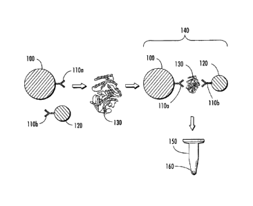
Referring now to Figure 1, a schematic diagram of a representative magnetic
capture assay for detecting the presence of one or more analytes in a
biological
sample is shown. The presently disclosed magnetic capture assay includes one
or
more magnetic capture particles 100, which have associated therewith at least
one
specific binding member 110a having an affinity for one or more analytes 130
of
- 26 -
CA 2981992 2017-10-11
interest in a biological sample. The assay also includes one or more SERS-
active
nanoparticles 120, which have associated therewith at least one binding member
110b
having an affinity for the one or more analytes 130. Binding member 110b
associated
with SERS-active nanoparticle 120 can be the same or different than binding
member
110a associated with magnetic capture particles 100. Magnetic particle 100 and
SERS-active nanoparticle 120 are contacted with a biological sample comprising
one
or more analytes 130 of interest and incubated for a period of time to form
magnetic
capture particle-analyte-SERS-active nanoparticle complex 140, e.g, an
antibody-
antigen "sandwich" structure, if the one or more analytes 130 are present in
the
biological sample. Magnetic capture particle-analyte-SERS-active nanoparticle
complex 140 is exposed to a magnetic field (not shown) to induce complex 140
to
migrate to a localized area of container 150, e.g., an assay vessel or
specimen
collection container, to form pellet 160. Pellet 160 is illuminated with
incident
radiation at one or more wavelengths, for example, in a system as shown in
Figure 4,
to induce the SERS-active reporter molecule to produce a detectable signal to
detect
the presence or amount of the one or more analytes in the biological sample.
One of
ordinary skill in the art upon review of the presently disclosed subject
matter would
recognize that magnetic capture particle 100, SERS-active nanoparticle 120,
and
combinations thereof, can be included in container 150 before the sample is
disposed
therein, or can be added to container 150 prior to, concurrent with, or
subsequent to
disposing the sample therein;
Accordingly, in some embodiments, magnetic particle enrichment can be used
advantageously in the presently disclosed assays. In one such approach, SERS-
active
particles having one or more capture probes for the analyte(s) of interest
attached
thereto can be present in a container, e.g., a specimen collection container,
prior to
sample collection, or, in some embodiments, added after collection. During the
incubation phase, target analytes are bound onto the SERS-active particle
surface by
the capture probes. Magnetic particles, also having attached thereto capture
probes to
the target(s) of interest, that have been provided in the container can attach
to
different epitopes on the same target(s), e.g., one or more analytes of
interest, and thus
form complexes where the target analyte is sandwiched between a SERS-active
nanoparticle and a magnetic particle. A magnet can then be used to concentrate
these
-27 -
CA 2981992 2017-10-11
sandwiches in a specified space, i.e., to form a pellet, in the container. The
magnet
can either be applied before the container is placed into the reader or can be
integrated
into the reader. Incident radiation of a desired wavelength, e.g., a laser
beam, can
then be focused on the pellet of concentrated SERS-active nanoparticle-target-
magnetic particle sandwich complexes and the SERS signal is obtained from the
SERS-active nanoparticles.
As also disclosed in more detail herein, the magnetic capture embodiments of
the presently disclosed assays also can include contacting multiple types of
SERS-
active nanoparticles with the sample, wherein each type of SERS-active
nanoparticle
has attached thereto a SERS-active reporter molecule that exhibits a unique
Raman
signal. Such embodiments can be used to detect a plurality of analytes of
interest,
referred to herein as multiplexing.
G. Referencing and Controls in the Magnetic Pull-Down Liquid-
based
Assay
In conventional immunoassays, detection of an antigen can occur by
"sandwiching" the antigen between two antibodies, one of which is labeled with
an
optical, colorimetric, or radiometric reporter. The measured signal, e.g., an
optical,
colorimetric, or radiometric reporter can then be used to determine the
concentration
of the antigen present in the sample. Conventional enzyme-linked immunosorbent
assay (ELISA) immunoassays are examples of this type of technology. One issue
with this technical approach, is that the magnitude of the optical signal
depends on
several factors in addition to the presence and/or amount of the antigen. For
example,
the alignment and performance of the optics can impact the measured signal.
Typically, to avoid this problem, additional control samples having known
concentrations of antigen are measured.
In one embodiment of the presently disclosed subject matter, as noted above, a
measurable signal is generated by forming an antigen-mediated complex between
a
SERS-labeled nanotag and a magnetic capture particle. The complexes can be
separated from the solution by application of a magnetic field and the optical
signal
from the resulting magnetic pellet is measured.
- 28 -
CA 2981992 2017-10-11
The position of the magnetic pellet relative to the interrogating optics can
affect the magnitude of the measured optical signal and ultimately, the
calibration of
the assay. In addition, the shape of the magnetic pellet might not always be
consistent. For example, altering the surface functionality of the magnetic
particles
could change the density and/or shape of the pellet.
According to embodiments of the presently disclosed subject matter, it is
possible to compensate for variations in pellet size, shape, or positioning.
These
methods also are applicable to other assay formats in which a pellet is
formed. In one
embodiment, magnetic particles used for the magnetic pull-down are labeled
with a
reference label in addition to the capture probes, e.g., antibodies specific
to the
antigen of interest. The reference label can be any moiety capable of
generating (by
itself or upon some type of stimulation) a detectable signal, including, but
not limited
to fluorophores, organic dyes, rare earth elements, and Raman reporters, and
also
could include particles comprising such components. Specific examples of
reference
labels include SERS-active particles of the type disclosed herein and silica
particles
having fluorophores distributed on or throughout the silica particles.
An example of incorporation of a reference label into an assay is illustrated
in
Figure 2. The presently disclosed magnetic capture assay incorporating a
reference
label includes one or more magnetic capture particles 200, which have
associated
therewith at least one specific binding member 210a having an affinity for one
or
more analytes 240 of interest in a biological sample. In this embodiment, the
one or
more magnetic capture particles 200 also have associated therewith at least
one
reference label 230 capable of generating a detectable signal. In some
embodiments,
reference label 230 comprises a second SERS-active nanoparticle having a
different
reporter molecule than the one or more SERS-active nanoparticles 220 which
form
complex 250 with the one or more analytes 240.
The assay also includes one or more SERS-active nanoparticles 220, which
have associated therewith at least one binding member 210b having an affinity
for the
one or more analytes 240. Binding member 210b associated with SERS-active
nanoparticle 220 can be the same or different than binding member 210a
associated
with magnetic capture particles 200.
-29 -
CA 2981992 2017-10-11
As with the assay depicted in Figure 1, magnetic particle 200 and SERS-active
nanoparticle 220 are contacted with a biological sample comprising one or more
analytes 240 of interest and incubated for a period of time to form magnetic
capture
particle-analyte-SERS-active nanoparticle complex 250 if the one or more
analytes
240 are present in the biological sample. Magnetic capture particle-analyte-
SERS-
active nanoparticle complex 250 is exposed to a magnetic field (not shown) to
induce
complex 250 to migrate to a localized area of a container, e.g., an assay
vessel or
specimen collection container, to form pellet as shown previously in Figure 1.
All magnetic particles present in the container (whether complexed or not) are
pulled down into the localized area of the container, e.g., an optical read
area. The
pellet comprising magnetic capture particle-analyte-SERS-active nanoparticle
complex 250 is illuminated with incident radiation at one or more wavelengths,
for
example, in a system as shown in Figure 4, to induce SERS-active nanoparticle
220 to
produce a first detectable signal and reference label 230 to produce a second
detectable signal. The first detectable signal of SERS-active nanoparticle 220
can be
compared to the second detectable signal of reference label 230 to detect the
presence
or amount of one or more analytes 240 in the biological sample.
The Raman signal from particle 220 is related to the amount of analyte 240,
e.g., an antigen, present; whereas the signal from reference label 230, e.g.,
a
nanoparticle having a different SERS-reporter molecule than particle 220, acts
as a
reference and corrects for variations in pellet shape, density, and/or
position. Thus,
calibration can be based on a comparing the intensity of Reporter 1, e.g.,
particle 220,
to the intensity of Reporter 2, e.g., reference label 230. For example, the
signal can be
calculated by as (Reporter 1 intensity)/(Reporter 2 intensity), in other
words, the ratio
of the intensity of Reporter 1 relative to the intensity of Reporter 2.
Although Figure 2 shows one SERS-active particle per magnetic capture
particle, multiple reference labels/particles per magnetic particle are
possible or,
alternatively, a fraction of the magnetic capture particles could be labeled
with one or
more references, while the remainder of the magnetic capture particles is
reference
free.
- 30 -
CA 2981992 2017-10-11
As with the assay depicted in Figure 1, one of ordinary skill in the art upon
review of the presently disclosed subject matter would recognize that magnetic
capture particle 200, SERS-active nanoparticle 220, and combinations thereof,
can be
included in the container before the sample is disposed therein, or can be
added to the
container prior to, concurrent with, or subsequent to disposing the sample
therein.
The presently disclosed subject matter also encompasses embodiments in
which the magnetic particle-SERS-active nanoparticle sandwich is pre-complexed
and
can be used with the presently disclosed methods as a reference label. This
complex
is inert, that is, it does not complex with the analyte of interest. A known
amount of
such pre-complexed particles can be added to a sample as a reference.
H. Use of Lysis Reagent in Liquid-based Assay
In a further embodiment, a lysis reagent can be used in an assay, such as a
liquid-based assay, with or without magnetic pull-down. When used in
biological
matrices, such as human blood, plasma, or serum, a lysis reagent can provide
an
increased signal and/or improved limit of detection for biomarkers. In
particular,
when used in an immunoassay using SERS-active particles, the addition of a
lysis
reagent increases the Raman signal intensity when compared to samples that do
not
contain the lysis reagent.
One lysis reagent suitable for use with the presently disclosed methods
includes one or more of the following components: (4-(2-hydroxyethyl)-1-
piperazineethanesulfonic acid) (HEPES) buffer, sodium chloride,
ethylenediaminetetraacetic acid (EDTA), beta glycerophosphate, Triton and
protease inhibitors, and combinations thereof. See, e.g., Hirsch, L. R., et
al., "A
Whole Blood Immunoassay Using Gold Nanoshells," Anal. Chem. 75, 2377-2381
(2003). Many detergents are suitable for use as a lysing reagent.
Representative
examples of anionic detergents include, but are not limited to, the salts of
cholic acid,
caprylic acid, sodium dodecyl sulfate (SDS), and deoxycholic acid.
Representative
examples of cationic detergents include, but are not limited to,
cetylpyridinium and
benzalkonium chloride. Representative examples of zwitterionic detergents
include,
but are not limited to, 3[(3-Cholamidopropyl)dimethylammonio]-propanesulfonic
acid (CHAPS) and phosphatidylcholine. Representative examples of nonionic
- 31 -
CA 2981992 2017-10-11
detergents include, but are not limited to, digitonin, Tweent 20
(polyoxyethylenesorbitan, monolaurate) and Triton X-100. The lysis reagents
also
can include protease inhibitors, including, but not limited to, aprotinin,
EDTA,
leupeptin, a-macroglobulin, pepstatin, phenylmethylsulphonyl fluoride (PMSF),
tosyl-L-lysine chloromethyl ketone (TLCK), and tosyl-L-phenylalanine
chloromethyl
ketone (TPCK), and can be selected depending on the particular protease
target.
Other lysis reagents suitable for use with the presently disclosed subject
matter
include, but are not limited to, dithiothreitol, ethylene glycol tetraacetic
acid (EGTA),
sodium cholate, sodium deoxycholate, NP-40, Glycocholic acid, sodium
taurocholate,
taurodeoxycholic acid, hexadecyltrimethylammonium bromide, Brij 35, Brij 58P,
N-
Decanoyl-N-methylglucamine, Igepal CA-630, N-Nonanoyl-N-methylglucamine,
octyl-b-D-1-thioglucopyranoside, Span 20, Triton X-114, Tween0 40, Tween0 80,
3-(4-heptyl) phenyl 3-hydroxy propyl) dimethylammonio propane sulfonate, and
amidosulfobetaine-14. Lysis reagents also can be used to lyse cells.
One of ordinary skill in the art would appreciate that other lysis reagents
known in the art also are suitable for use with the presently disclosed
methods.
Further, one of ordinary skill in the art would appreciate that the lysis
reagent is
contacted with the sample in an amount effective to lyse substantially all of
the
cellular contents of the sample. An example of this method is provided in
Example 2.
As provided in Example 2, in immunoassays detected by SERS, addition of a
lysis reagent to a sample under test results in higher signal levels and/or
increased
sensitivity for biomarker detection in biological matrices when compared to
samples
containing no lysis reagent. Increased sensitivity offers many benefits, e.g.,
the "time
to result" can be decreased and patient outcome can potentially be improved.
The
larger Raman signals observed in the presence of the lysis reagent also can
lead to
reduced instrumentation costs. The lysis reagent could be used to increase
sensitivity
for a variety of assays, including but not limited to, lateral flow assays,
enzyme linked
immunosorbant assays, and surface plasmon resonance assays. The lysis reagent
also
can be used to treat types of biological samples other than blood, serum or
plasma,
such as sputum for tuberculosis detection, or any other type of specimen used
for
diagnostics that requires the detection of a biomarker. Individual components
in the
lysis reagent could be used alone or in combinations to achieve similar
effects.
- 32 -
CA 2981992 2017-10-11
I. Representative Instrumentation For Detecting a SERS Signal
Emitted
By a Sample Under Test
Referring now to Figure 4, a representative system for use with the presently
disclosed assays is provided. System 400 includes light source 402 capable of
producing electromagnetic radiation capable of inducing a Raman signal in a
SERS-
active particle. In some embodiments, light source 402 is a laser, which in
some
embodiments, is a laser capable of operating in the near-infrared spectral
region, e.g.,
a solid-state diode laser with an emission wavelength of about 785 nm.
Electromagnetic radiation emitted from light source 402, e.g., light of one or
more
particular wavelengths, can be directed by fiber 404 to lens 406a. Fiber 404
can be
any optical fiber suitable for use with the presently disclosed system. For
example,
fiber 404 can be single fiber 404a or fiber bundle 404b.
Lens 406a expands the light transmitted through fiber 404 and directs the
light
through filter 407a, which can be, in some embodiments, a bandpass filter, and
onto
beamsplitter 408, e.g., a dielectric beamsplitter. A portion of the light
incident on
beamsplitter 408 is directed to lens 406b, which focuses the light onto sample
412,
which is contained in container 410, e.g., a specimen collection container or
an assay
vessel. Sample 412 can comprise one or more magnetic capture particle-analyte-
SERS-active nanoparticle complexes as disclosed herein. The light incident on
sample 412 is capable of inducing a SERS signal, i.e., scattered radiation,
from the
magnetic capture particle-analyte-SERS-active nanoparticle complexes
comprising
sample 412. Scattered radiation emitted from sample 412 is collected by lens
406b
and is directed to beamsplitter 408. A portion of the scattered radiation is
transmitted
through beamsplitter 408 and directed to filter 407b, e.g., a longpass filter.
After
passing through filter 407b, the scattered radiation is directed to lens 406c,
which
focuses the scattered radiation onto fiber 414. Fiber 414 can be any optical
fiber
suitable for use with the presently disclosed system. For example, fiber 414
can be
single fiber 414a or fiber array 414b. Fiber 414 directs the scattered
radiation to
spectrometer 416, which, in some embodiments, includes charge-coupled device
(CCD) 418.
- 33 -
CA 2981992 2017-10-11
In some embodiments, a laser serves as the excitation source of the incident
radiation used to detect one or more target analytes of interest. One of
ordinary skill
in the art upon review of the presently disclosed subject matter could
ascertain the
type of laser, including the strength and excitation wavelength, suitable for
use with
the SERS-active reporter molecules described herein. Radiation scattered or
emitted
from the sample can be detected using detection systems known in the art.
In some embodiments, more than one type of radiation source, or more than
= one excitation wavelength, can be used. For example, in embodiments
wherein two
analytes of interest are to be detected, the reagent can include two distinct
types of
SERS -active reporter molecules and/or two distinct types of specific binding
members. In other embodiments, incident radiation of a single wavelength can
be
used to induce different Raman spectra from two or more distinct SERS-active
reporter molecules. In some embodiments, however, incident radiation of
different
wavelengths can be used to produce distinct Raman signals for each analyte of
interest. As one of ordinary skill in the art would recognize upon review of
the
presently disclosed subject matter, the selection of the particular
wavelength(s) to be
used depends on the analyte of interest, the specific binding members used,
and the
particular SERS-active reporter molecules used.
The presently disclosed assay can be conducted with any suitable Raman
spectrometer systems known in the art, including, for example, a Multimode
Multiple
Spectrometer Raman Spectrometer (Centice, Morrisville, North Carolina, United
States of America), such as the Raman spectrometer system disclosed in U.S.
Patent
No. 7,002,679 to Brady et al.
More particularly, a system and method for a Raman spectroscopy assay using
paramagnetic particles is disclosed in U.S. Patent Application Publication No.
2006/0240572 to Carron et al., and a Raman spectrometer system suitable for
use with
the presently disclosed assays is disclosed in U.S. Patent Application
Publication No.
2005/0248758 to Carron et al.
Sensing devices, such as optical detectors, radiation sources, and computer
systems, microprocessors, and computer software and algorithms, can be used in
any
combination in practicing the methods disclosed herein. Accordingly, in some
- 34 -
CA 2981992 2017-10-11
embodiments, software, or other computer readable instructions can be used to
interpret, analyze, compile, or otherwise parse output data related to the
presently
disclosed optical assay. The software or other computer system can be used to
display, store, or transmit output data, whether in digital or other forms to
one or more
users.
A Method for Amplifring a SERS Signal in a Liquid-Based Assay
Biological assays often require accurate yet sensitive detection of
biomolecules in a variety of media. One approach toward the development of
more
sensitive assays is increasing the output signal of the assay. The presently
disclosed
method demonstrates that, in some embodiments, a signal enhancement of a
factor of
three or higher is possible. Accordingly, in some embodiments, the presently
disclosed subject matter provides a method for amplifying output signal in a
liquid-
based assay of the type described above and depicted in Figure 1.
The amplification method according to one embodiment begins with a sample
being added to a solution containing both a magnetic capture particle and a
reporter
molecule, such as, but not limited to, as SERS-active nanoparticle comprising
a SERS
reporter molecule, and incubated for a short period of time, during which a
sandwich
complex comprising the magnetic capture particle, analyte and reporter
molecule can
be formed.
The signal amplification characteristic of the presently disclosed subject
matter arises from the addition of another aliquot of reporter molecule, e.g.,
another
aliquot of SERS-active nanoparticles having the same signal-producing
capabilities,
e.g., the same Raman reporter molecule, as the first aliquot added to the
assay
solution, before the magnetic capture particles are localized. This second
aliquot of
reporter molecule, e.g., a second aliquot of SERS-active nanoparticles,
presents
antibodies (or another molecule, e.g., a specific binding member) that
recognizes the
antibody on the first aliquot of reporter molecules, e.g., SERS-active
nanoparticles,
originally present in the assay. The presence of the second aliquot of
reporter
molecules results in a higher number of reporter molecules per sandwich in the
sample under test and therefore a higher signal per sandwich complex.
- 35 -
CA 2981992 2017-10-11
For example, as depicted in Figure 7, SERS-active nanoparticles coated with
an antibody that recognizes unbound antibodies on the first aliquot of SERS-
active
nanoparticles can be added to the assay solution. When these secondary
antibodies
bind to the first aliquot of SERS-active nanoparticles, the signal level goes
from one
SERS-active nanoparticle per sandwich complex to three.
More particularly, referring now to Figure 7, a representative schematic
diagram of the presently disclosed assay using signal amplification method is
depicted. The presently disclosed assay includes one or more magnetic capture
particles 700, which have associated therewith at least one specific binding
member
710a having an affinity for one or more analytes 730 of interest in a
biological
sample. The assay also includes a first aliquot of one or more reporter
molecules 720
capable of producing a detectable signal, e.g., one or more SERS-active
nanoparticles,
which have associated therewith at least one binding member 710b having an
affinity
for the one or more analytes 730. Binding member 710b associated with reporter
molecule 720 can be the same or different than binding member 710a associated
with
magnetic capture particles 700. Magnetic particle 700 and reporter molecule
720 are
contacted with a biological sample comprising one or more analytes 730 of
interest
and incubated for a period of time to form magnetic capture particle-analyte-
reporter
molecule complex 740, e.g, an antibody-antigen "sandwich" structure, if the
one or
more analytes 730 are present in the biological sample.
The assay depicted in Figure 7, then includes a second aliquot of one or more
reporter molecules 750 capable of producing a detectable signal having
associated
therewith at least one specific binding member 710c having an affinity for the
specific
binding member 710b of the first aliquot of reporter molecules 720. The second
aliquot of reporter molecules 750 can be disposed in container 760 prior to,
concurrent with, or subsequent to disposing the sample and/or the first
aliquot of one
or more reporter molecules 720 therein, wherein the one or more reporter
molecules
of the second aliquot of reporter molecules 750 is the same as the one or more
reporter molecules of the first aliquot of reporter molecules 720.
Complex 740, comprising a sandwich complex of magnetic capture particle
700, analyte 730, reporter molecule 720, having associated therewith reporter
molecule 750, is exposed to a magnetic field (not shown) to induce complex 740
to
- 36 -
CA 2981992 2017-10-11
migrate to a localized area of container 760, e.g., an assay vessel or
specimen
collection container, to form pellet 770. Pellet 770 is illuminated with
incident
radiation at one or more wavelengths, for example, in a system as shown in
Figure 4,
to induce the reporter molecule to produce a detectable signal to detect the
presence or
amount of the one or more analytes in the biological sample.
One of ordinary skill in the art upon review of the presently disclosed
subject
matter would recognize that magnetic capture particle 700, reporter molecule
720,
reporter molecule 750, and combinations thereof, can be included in container
760
before the sample is disposed therein, or can be added to container 760 prior
to,
concurrent with, or subsequent to disposing the sample therein;
Depending on the form of the assay, the second aliquot of SERS tags can be
added at any stage, e.g., sequentially or concurrently with the first aliquot
of SERS
tags, or sequentially or concurrently with the sample. Also, to avoid a large
increase
in background noise in the assay, it is preferable that the biomolecule on the
surface
of the second SERS-active nanoparticle not recognize the biomolecule on the
surface
of the magnetic capture particle. In the case of an immunoassay, this
characteristic
can be accomplished by immobilizing biomolecules, e.g., antibodies that
originate in
different species on the magnetic capture nanoparticle and SERS-active
nanoparticle.
For example, in some embodiments, the initial assay solution could include
magnetic
capture particles having antibodies produced in goat and the SERS-active
nanoparticle
could present antibodies raised in mouse. If the second aliquot of SERS-active
nanoparticles is labeled with an anti-mouse antibody, no binding to the
magnetic
capture particle will take place. The presently disclosed amplification method
can be
used to detect virtually any analyte, including DNA, provided two binding
partners
with orthogonal epitopes are used.
An additional benefit of increased signal output in a given assay is that less
sophisticated (and less expensive) detection systems can be used to test for
an analyte.
Referring now to Figure 8, representative results of the presently disclosed
amplification strategy are provided. Figure 8A shows a typical binding curve
for a
homogenous protein assay without amplification as shown previously in Figure
1.
The y-axis is the output of an algorithm that quantifies the level of SERS
signal for
- 37 -
CA 2981992 2017-10-11
each sample. Figure 8B shows the assay performed with the identical reagents
and
concentrations with the amplification step shown in Figure 7. The graph shows
signal
increases of three-fold or greater upon inclusion of the amplification step.
The presently disclosed method can be used to detect virtually any analyte
provided two binding partners with orthogonal epitopes are used. For example,
in
some embodiments, the presently disclosed amplification method can be used to
detect a polynucleotide. The use of the term "polynucleotide" is not intended
to limit
the presently disclosed methods to polynucleotides comprising DNA. Those of
ordinary skill in the art will recognize that polynucleotides can comprise
ribonucleotides and combinations of ribonucleotides and deoxyribonucleotides.
Such
deoxyribonucleotides and ribonucleotides include both naturally occurring
molecules
and synthetic analogues. More particularly, the term "polynucleotide" is
intended to
encompass a singular nucleic acid, as well as plural nucleic acids, and refers
to an
isolated nucleic acid molecule or construct, e.g., messenger RNA (mRNA),
plasmid
DNA (pDNA), or short interfering RNA (siRNA). A polynucleotide can be single-
stranded or double-stranded, linear or circular. A polynucleotide can comprise
a
conventional phosphodiester bond or a non-conventional bond (e.g., an amide
bond,
such as found in peptide nucleic acids (PNA)). The term "nucleic acid" refers
to any
one or more nucleic acid segments, e.g., DNA or RNA fragments, present in a
polynucleotide. By "isolated" nucleic acid or polynucleotide is intended a
nucleic
acid molecule, DNA or RNA, that has been removed from its native environment.
Examples of an isolated polynucleotide include recombinant polynucleotides
maintained in heterologous host cells or purified (partially or substantially)
polynucleotides in solution. Isolated polynucleotides or nucleic acids
according to the
present invention further include such molecules produced synthetically.
Isolated
polynucleotides also can include isolated expression vectors, expression
constructs, or
populations thereof. "Polynucleotide" also can refer to amplified products of
itself, as
in a polymerase chain reaction. The "polynucleotide" can contain modified
nucleic
acids, such as phosphorothioate, phosphate, ring atom modified derivatives,
and the
like. The "polynucleotide" can be a naturally occurring polynucleotide (i.e.,
one
existing in nature without human intervention), or a recombinant
polynucleotide (i.e.,
-38 -
CA 2981992 2017-10-11
one existing only with human intervention). While the terms "polynucleotide"
and
"oligonucleotide" both refer to a polymer of nucleotides, as used herein, an
oligonucleotide is typically less than 100 nucleotides in length.
Figure 9 provides a representative schematic of the amplification method used
for polynucleotide detection. Referring now to Figure 9, the presently
disclosed
amplification method includes magnetic capture particle 900 having attached
thereto
capture probe 910a, e.g., a capture probe having 15 base pairs. Also provided
is
reporter molecule 920, e.g., a SERS-active nanoparticle, having attached
thereto
capture probe 910b, e.g., a capture probe having 15 base pairs. When contacted
with
target polynucleotide 930, e.g., a polynucleotide having 30 base pairs,
complex 940 is
formed and includes double-stranded polynucleotide 950 comprised of target
polynucleotide molecule 930 and capture probes 910a and 910b. Complex 940 can
then be contacted with reporter molecule 960, e.g., a SERS-active
nanoparticle,
having attached thereto capture probe 910c, e.g., a capture probe
complementary to
unbound capture probe 910b attached to reporter molecule 920 to form complex
980,
which includes double-stranded polynucleotide 970 comprising capture probes
910b
and 910c. Complex 980 is exposed to a magnetic field (not shown) to induce
complex 980 to migrate to a localized area of a container, e.g., an assay
vessel or
specimen collection container as shown in Figure 7, to form a magnetized
pellet. The
pellet is illuminated with incident radiation at one or more wavelengths, for
example,
in a system as shown in Figure 4, to induce the reporter molecule to produce a
detectable signal to detect the presence or amount of the one or more target
DNA in
the biological sample.
Data from the presently disclosed DNA assay are shown in Figures 10A and
10B.
K Generating Improved Raman Reference Spectra and Spectral
Analysis
in Magnetic Pull-Down Liquid-based Assay
The presently disclosed subject matter, in another embodiment, provides a
method of generating reference spectra for use in Raman Spectroscopy-based
analyses. This method can be used advantageously with magnetic particle-
coupled
nanoparticles in liquid assays of the type disclosed herein. To determine the
quantity
- 39 -
CA 2981992 2017-10-11
of a specific SERS-active nanoparticle in a sample, a Raman signal generally
must be
generated by a known amount of the specific SERS-active nanoparticle.
Typically,
that known or reference signal is generated by specific SERS-active
nanoparticles in
solution, as sample processing can be simplified.
In some assay systems, such as those disclosed herein, SERS-active particles
are complexed with magnetic particles through the reactions involving the
analyte of
interest. These combined particles are pulled into a small volume by an
external
magnet. The Raman signal generated by the nanoparticles within this pellet can
be
analyzed using the reference signal previously obtained from solution.
The presently disclosed subject matter provides, however, that the Raman
spectra from a pellet are different from the Raman spectra from solution, even
if the
same single SERS-active nanoparticle type is contained in the sample. Improved
results can be obtained if the reference spectra referred to above are
acquired of the
pellet, not from solution. The differences in these solution spectra as
compared to the
pellet spectra can cause errors in quantifying the amount of nanoparticles in
the
sample. This observation is especially true in a multiplexing environment,
where
several SERS-active nanoparticles having unique Raman signals are present in
the
sample. For example, features within the sample signal which are not contained
in the
reference spectra can be interpreted as coming from other SERS-active
nanoparticles
within the sample (see Example 4).
The differences in spectra coming from solution and pellets do not have to be
large to have an impact on the quantification of SERS-active nanoparticles
(see
Example 4). This is especially true when one SERS-active nanoparticle is
present in
large quantities and another is entirely absent. Under these circumstances, a
small
error can induce a false positive result for the absent SERS-active
nanoparticle.
According to this embodiment, Raman spectra can be analyzed by least-
squares fitting, in which case reference signals are used for each potential
component.
In least squares fitting techniques, the signal under analysis is assumed to
be
composed of a linear combination of spectra, each contribution varying by the
relative
amount of specific SERS-active nanoparticle within the sample. Features within
the
measured signal that cannot be "fit" to any of the reference signals can be
largely
partitioned into a background signal. If a feature of the measured signal,
however,
- 40 -
CA 2981992 2017-10-11
coincides with a feature of any reference signal, then the fitting will assign
some
portion of the total signal to that reference source. If spectra are
influenced by the
pelletization of the nanoparticles, for example, then those changes will
necessarily
contribute to errors in quantification. If those changes are captured in the
reference
spectra, then such errors sample analyses can be reduced. One of ordinary
skill in the
art would recognize that other multivariate analysis techniques, including,
but not
limited to, partial least squares, principal component analysis, and the like,
could be
used with the presently disclosed methods.
The presently disclosed subject matter, in a further embodiment, provides a
method of analyzing Raman spectra, especially those obtained from SERS-active
nanoparticles, and especially in a multiplexed situation where particles with
different
Raman spectra are used to identify multiple analytes. To determine the
quantity of a
specific SERS-active nanoparticle (nanoparticle with a specific SERS-active
nanoparticle which generates a unique Raman signal) in a sample, a sample
spectrum
typically is recorded over a broad range of wavelengths or wavenumbers, and
then
compared to one or more reference spectra. This comparison involves fitting
the
sample spectra to the reference spectra over a consistent and suitable
wavelength or
wavenumber range.
The presently disclosed subject matter demonstrates that an estimate of
nanoparticle components made from this typical comparison can be refined in
two
ways based on the results of the first comparison. First, because the
differences
between spectral contributions for different components can vary over a
spectral
range, specific ranges within the spectra can be weighted more heavily in the
calculations depending on the relative amounts of components in the first
estimate.
Second, components estimated to be absent or present in very low
concentrations can
be removed from the analysis, and the remaining components re-estimated. In
either
case, an increase in fit accuracy ("goodness of fit") can be calculated and
appraised.
The "goodness of fit" represents a difference between the sample spectrum and
the
fitted spectrum and can be determined by any method known in the art and can
be
reported, for example, as a residual of the least squares fitting technique.
These two
- 41 -
CA 2981992 2017-10-11
methods can be applied in combination or separately. In addition, the
presently
disclosed method is applicable to a variety of SERS-active nanoparticles that
similarly
have spectra that must be distinguished.
II. COMPOSITE NANOSTRUCTURES AND METHODS OF THEIR USE
The presently disclosed subject matter provides composite nanostructures,
including a composite structure, referred to herein as a "satellite"
structure, comprising a
plurality of signal-bearing particles, e.g., nanoparticles, bound to a core
particle, and a
composite structure, referred to herein as a "core-shell" structure, which
includes a core
particle, an active material, such as a Raman-active material, surrounding the
core
particle, and one or more shells, such as a metal shell, surrounding the
active material.
The presently disclosed satellite and core-shell structures can be used to
amplify or
otherwise enhance a signal in an assay, such as a SERS assay.
Accordingly, in some embodiments, the presently disclosed subject matter
provides a method for performing an assay, the method comprising: providing at
least
one of a satellite structure, a core-shell structure, or combinations thereof;
contacting the
satellite structures and/or core-shell structures with a sample suspected of
containing one
or more target analytes, and performing a detection step to determine the
presence or
absence of the one or more target analytes in the sample.
More particularly, in one embodiment, signal enhancement can be achieved by
providing a composite structure having a plurality of signal-bearing
satellites (e.g.,
nanoparticles) bound to a core particle (e.g., a microparticle) such that the
resulting
= composite satellite structure is capable of generating multiple signals.
Thus, the satellite
structure can improve the detection of analytes within an assay, such as for
samples
having a lower concentration of analytes.
According to various aspects of the presently disclosed subject matter, the
satellite particles can be metal, semiconductor, organic, and/or inorganic
nanoparticles.
Similarly, the core particle can be magnetic, silica, metal (e.g., gold), or
organic
microparticles or nanoparticles. The satellite and core particles can be
spherical or
non-spherical in shape. The satellite particles can be bound to each core
particle using
electrostatic, covalent, or van der Waals forces. In addition, the satellite
particles
typically comprise a reporter molecule, such as fluorescent, Raman-active, or
enzyme
- 42 -
CA 2981992 2017-10-11
reporter molecules which facilitates detection. The satellite particles and/or
the at least
one core particle can have further molecules bound thereto, such as
antibodies, nucleic
acid probes, or blocking agents.
In a further embodiment, the presently disclosed subject matter provides a
core-
shell structure comprising a core particle, an active material, such as a
Raman-active
material, surrounding the core particle, and a shell surrounding the active
material,
wherein the shell can be a contiguous layer or a plurality of nanoparticles,
which
preferably are in close proximity to one another. The core and shell materials
can be
the same or different, and are desirably chosen from materials that enhance
the spectrum
of the active material. For example, a gold core and gold shell will improve
the
detectable spectrum of a Raman-active material in a surface enhanced Raman
scattering
application.
Additional aspects of the method can include detecting a target analyte using
surface enhanced Raman scattering. A plurality of magnetic, metallic, or
semiconductor beads having, e.g., an antibody for the target analyte, can be
introduced into the assay, along with a composite structure having a similar
binding
molecule for the target analyte. The analyte, if present, becomes sandwiched
between
the composite structure and the bead. The bead can be manipulated, e.g., by
application of a magnetic field, to concentrate a plurality of the sandwiches
for detection
via Raman spectrum. A further aspect of the presently disclosed subject matter
includes contacting a plurality of the presently disclosed composite
structures with a
cell or tissue under conditions to attach the composite structure to an
analyte (e.g., a
normal or cancerous cell) and imaging the structures.
A. Satellite Nanostructures
The presently disclosed subject matter provides microparticle-nanoparticle
satellite structures, which, in some embodiments, can be used to amplify or
enhance a
signal in an assay, such as a SERS assay.
Referring now to Figure 14, there is shown a transmission electron micrograph
(I'EM) of satellite structure 1400 according to one embodiment of the
presently
disclosed subject matter. Satellite structure 1400 includes a plurality of
smaller
particles (e.g., a signaling moiety) bound to a larger particle (e.g., a
carrier), such as a
- 43 -
CA 2981992 2017-10-11
plurality of nanoparticles 1412 bound to microparticle 1414. As disclosed in
further
detail herein below, satellite structure 1400 is capable of amplifying an
analyte signal
in an assay. Thus, satellite structure 1400 can be able to amplify a signal to
detect an
analyte that can otherwise not be detected, such as in samples containing
lower
concentrations of analytes.
It is understood that satellite structure 1400 described herein can be used to
detect any number of analytes. The term "analyte" is not meant to be limiting
and can
be any molecule of interest such as a protein, nucleic acid, or metabolite.
The analyte
could alternatively be a cell, such as a cancerous cell. Furthermore, the
assay used to
analyze analytes can be homogeneous or heterogeneous. For instance, a
heterogeneous assay typically requires separation, either by washing or other
physical
means, of the reaction elements between the individual steps of the assay
procedure,
while a homogeneous assay does not require any separating steps. In addition,
any
desired assay can be used to identify and analyze the analytes. For example,
sandwich
immunoassays can be used, examples of which are provided in further detail
herein.
Further, satellite structure 1400 can be used to amplify signaling events for
a plurality of
analytes such that multiplex detection can be used to detect such plurality of
analytes.
For example, a first satellite structure with a distinct signal, e.g., a
distinct SERS
spectrum includes an antibody for a first analyte, and is mixed directly with
a second
satellite structure exhibiting a different, distinct signal, e.g., a distinct
SERS spectrum,
and having a second antibody specific for a second type of analyte.
Nanoparticles 1412 can be any suitable particle capable of generating a
detectable
signal that is used to detect the occurrence of a binding event. For example,
nanoparticles 1412 can be metal, semiconductor, organic, or inorganic
nanoparticles.
In the example shown in Figure 14, nanoparticles 1412 are gold particles.
Similarly,
microparticle 1414 can be any desired carrier that facilitates attachment of a
plurality of
nanoparticles 1412 thereto. For instance, microparticle 1414 could be
magnetic,
silica, metal (e.g., gold), and/or organic microparticles. In the example
depicted in Figure
14, the microparticle is an amine-funcfionalized silica particle.
Nanoparticles 1412
typically are able to provide detectable signals themselves, e.g., they can
be, quantum
dots or SERS-active particles. Other particles suitable for use with the
presently
disclosed methods are known to those of ordinary skill in the art. For
example, each of
- 44 -
CA 2981992 2017-10-11
nanoparticles 1412 can contain a reporter molecule such as fluorescent, Raman,
and
enzyme reporter molecules that enable the nanoparticles to act as signaling
elements if
the nanoparticles are not otherwise capable of doing so on their own. See, for
example,
U.S. Patent No. 6,514,767.
Although the term "microparticle" is used throughout, it is understood that
microparticle 1414 could be any particle that is larger than nanoparticles
1412 (where
nanoparticles are defined to have sizes of about 2 nm to about 200 nm in
diameter).
Various techniques can be used to bind nanoparticles 1412 to each
microparticle 1414,
such as using electrostatic, covalent, or van der Waals forces. Furthermore,
nanoparticles 1412 could be attached to microparticle 1414 using various
linkers, such as
polymers, DNA, amino acids, short-carbon chains, streptavidin/biotin linkage,
or the
like, which can allow for a higher density of nanoparticles about the
microparticle. The
length and type of the linker can be chosen to tune the number of
nanoparticles 1412
attached to microparticle 1414, and the spacing between the nanoparticles and
microparticles. Nanoparticles 1412 and/or microparticle 1414 also could be
coated (e.g.,
with a polymer) to control interparticle interactions.
Any number of nanoparticles 1412 can be bound to microparticle 1414, although
the precise number of nanoparticles can be dictated by the particular
experiment being
performed. For example, it can be possible to obtain satellite structure 1400
having
a uniform and maximum distribution of nanoparticles 1412 to microparticle 1414
by
optimizing the ratio of the particles and mixing conditions during the
reaction.
Furthermore, nanoparticles 1412 and microparticle 1414 can be of various sizes
and
configurations depending on the analyte to be detected, such as spherical or
non-spherical (e.g., nanorods) in shape. For example, Figure 14 shows a
transmission
electron microscopy image, wherein nanoparticles 1412 and microparticle 1414
are
spherical in shape, and the nanoparticles are 40 nm in diameter and the
microparticle
is 1 gm in diameter. In addition, microparticle 1414 can be submicron (e.g.,
0.5 gm) in
diameter.
Further, nanoparticles 1412 and/or microparticle 1414 can be labeled with a
species, such as organic or inorganic compounds, polymers, proteins,
receptors,
antibodies, nucleic acid probes, and blocking agents, to enhance or facilitate
the binding
- 45 -
CA 2981992 2017-10-11
event with the analyte or facilitate the binding event with the analyte or to
prevent
undesired interactions. Exemplary blocking agents include albumin, casein,
polyvinyl
alcohol, poly (ethylene glycol), and gammaglobulin.
Various detection techniques can be used to detect one or more analytes of
interest via satellite structure 1400, e.g., fluorescence or Raman
spectrometry. To
perform the detection, at least one binding event typically exists between
satellite
structure 1400 and an analyte (e.g., via a sandwich as described below).
Amplification
of the signal results from each of a plurality of nanoparticles 1412
generating multiple
signals based on a single binding event, rather than a single nanoparticle
generating
only one signal from that binding event.
Figure 15 depicts sandwich assay 1500 according to one embodiment of the
presently disclosed subject matter. In particular, the assay includes antibody
1522a
immobilized on support 1524, while satellite structure 1510 also includes
antibody
1522b immobilized on one of nanoparticles 1512. While this particular
embodiment
illustrates that antibody 1522b is coupled to satellite structure 1510 via
nanoparticles
1512, it is understood that in some embodiments it can be advantageous to
attach
antibody 1522b to carrier microparticle 1514. Analyte 1516 is captured between
antibodies 1522a, 1522b, which results in a binding event. Satellite structure
1510
facilitates the generation of a plurality of detectable signaling events due
to the
signaling events by each of nanoparticles 1512, which results in amplification
of the
binding event between analyte 1516 and nanoparticle 1512 capturing the
analyte. It is
understood that antibody 1522a on support 1524 and antibody 1522b on satellite
structure 1510 need not be the same.
Figure 16A illustrates sandwich assay 1600 according to another embodiment of
the presently disclosed subject matter. Assay 1600 includes assay vessel 1628
containing a plurality of satellite structures 1610, analytes 1616, and beads
1630.
Beads 1630 can be a magnetic, semiconductor, or metallic material that are
capable of
being attracted by a magnetic field, or can be structures that otherwise
facilitate
separation/enrichment, e.g., via gravity or centrifugation. There can be any
number of
beads 1630 depending on the number of satellite particles 1610 and analytes
1616,
and the beads can be any size and configuration capable of sandwiching an
analyte
between the bead and a satellite structure. For example, according to one
exemplary
- 46 -
CA 2981992 2017-10-11
embodiment, which is not meant to be limiting, sandwich assay 1600 can include
about
1,000 to 100,000 beads 1630, about 1,000 to 100,000 satellite structures 1610
per assay,
and about 50 to 10,000 nanoparticles 1612 per microparticle 1614. In addition,
beads
1630 can have located thereon species such as antibodies, nucleic acid probes,
blocking agents, or the like.
Analyte 1616 can be captured or sandwiched between satellite structure 1610
and bead 1630. In this regard, analyte 1616 can attach to nanoparticle 1612 of
a
respective satellite structure 1610, while bead 1630 can attach to the analyte
such that
the analyte is sandwiched between the nanoparticle and the bead resulting in a
binding
event. After allowing a predetermined time for a plurality of analytes 1616 to
be
captured, a magnetic field (provided, for example, by a permanent magnet or an
electromagnet) can be applied to assay 1600, which concentrates sandwiched
analytes
1616 in a particular location, for example the bottom of assay vessel 1628 as
concentrated pellet 1632, as shown in Figure 16B. Pellet 1632 can be formed at
the
bottom or the side of assay vessel 1628 depending on the direction that the
magnetic
field is applied. Pellet 1632 can then be analyzed to determine the presence
of analytes
1616 via the signals provided from the satellite structures. The signal
resulting from
each satellite structure 1610 is enhanced by the presence of the plurality of
nanoparticles
1612 as described above. Surface enhanced Raman scattering (SERS) can be used
to
detect signals from satellite structure 1610 containing Raman-active material.
A further embodiment of the presently disclosed subject matter is directed to
cellular imaging, wherein satellite structure 1610 can be used to facilitate
the analysis
of a cell. Using conventional techniques for analyzing cells, a limited number
of particles
can be capable of attaching to a cell, such as a cancerous cell, to facilitate
the imaging
of the cell for identifying normal or cancerous cells. For example, a cell
having ten
surface markers attached thereto would only be capable of binding to ten
nanoparticles
such that there can only be ten signaling events corresponding to ten surface
markers. By
sending satellite structures 1610 to attach to a cell, each having a plurality
of
nanoparticles, a plurality of signaling events can be generated by binding
each satellite
structure to the cell. For instance, continuing with the example above, there
can be ten
satellite structures 1610 that each includes ten nanoparticles 1612 such
signal is
generated from 100 nanoparticles, thus amplifying each individual binding
event by a
- 47 -
CA 2981992 2017-10-11
factor of 10. As such, the amplification of the signaling events can enhance
the
identification of the cell using cellular imaging. As discussed above,
satellite
structures 1610 can be labeled, such as with an antibody coating, to
facilitate the binding
events. The size of satellite structure 1610 can be tailored to enable
decoration of a cell
by a maximum number of nanoparticles 1612 by optimizing the size of
microparticle
1614 to which nanoparticles 1612 are attached. Thus, if microparticle 1614 is
very
small, only a handful of nanoparticles 1612 can be attached to it. On the
other hand, if
microparticle 1614 is very large, only a few satellite structures 1610 can be
bound to
the cell.
B. Composite Core-Shell Nanoparticles
In some embodiments, the presently disclosed subject matter provides composite
particles, referred to herein as "core-shell" structures, which include a core
particle, an
active material, such as a Raman-active material, surrounding the core
particle, and one
or more shells, such as a metal shell, surrounding the active material, which,
in some
embodiments, can be used to amplify or enhance a signal in an assay, such as a
SERS
assay.
Referring now to Figure 17, composite structure 1700 comprises core 1702, an
active material, such as Raman-active material 1704 around the core, and
contiguous
shell 1706 around the active material. Typically outer shell 1706 is 20 nm
thick or less.
In one specific embodiment, core 1702 is a gold core about 20 to about 200 nm
in
diameter, shell 1706 also is gold and has a thickness of 2 to 20 nm, and the
active
material is a Raman-active material such as trans-1,2-bis(4-pyridyeethylene
(SPE). In
addition, a thin, < Snm, silica layer can be disposed on the Raman-active
material
under shell 1706. The silica is advantageous in some situations to promote
fabrication
of the gold shell. Alternatively, a bi-functional molecule can be used to
promote
binding of gold to the Raman-active material. Gold is an advantageous metal in
providing surface enhanced Raman scattering, but metals such as silver and
copper
(or alloys of any such metals) also are useful.
In a further embodiment of the core-shell composite structure, the core-shell
structure is similar to Figure 17, but with the shell being made up of
nanoparticles,
typically having a size equal to or smaller than the core. In one such
embodiment, the
- 48 -
CA 2981992 2017-10-11
core is a gold core about 20 nm to about 200 nm in diameter, the active
material is a
Raman-active material such as trans-1,2-bis(4-pyrridyl)ethylene (BPE), and the
nanoparticles also are gold and have a diameter of about 2 nm up to the size
of the core.
In addition, a thin, <5nm, silica layer can be deposited on the Raman-active
material
under the nanoparticles. Alternatively, a bi-functional molecule can be used
to
promote binding of gold to the Raman-active material. Gold is an advantageous
metal in
providing surface enhanced Raman scattering, but metals such as silver and
copper (or
alloys of any such metals) also are useful.
It is understood that the embodiments shown in Figures 14-17 and described
above are not meant to be limiting. In particular, the satellite and
core/shell structures of
the presently disclosed subject matter can be used in a variety of assays for
enhancing
analyte signals, such as a homogeneous assay or a lateral flow assay. It is
possible that
a single assay would use both types of particles. The size, number, material
and
configuration of nanoparticles 1512, and microparticles 1514 can be varied
depending
on analyte 1516 being identified and a desired amplification of a signaling
event.
Similarly, for core/shell structures 1700, the size, material, and thicknesses
can be
adapted for the particular assay or environment.
As noted, a particularly advantageous application for the presently disclosed
subject matter is for surface enhanced Raman scattering. When two metallic
nanostructures are in close proximity, electromagnetic coupling of the
plasmonic fields
from the two structures takes place, producing large electromagnetic field
enhancements. When a Raman active molecule is placed in the enhanced
electromagnetic field, the intensity of the Raman signal from the molecule
shows an
increase of several orders of magnitude versus the signal when the molecule is
on an
isolated nanoparticle. Conventionally, those in the art have attempted to
aggregate
multiple metallic nanoparticles, but such aggregation is difficult and largely
uncontrollable. The presently disclosed subject matter is able to provide the
desired
electromagnetic coupling, but in a more advantageous manner.
In addition, while not limited to any particular theory, it is believed that
the two
interacting plasmonic structures (either core and shell or core and
satellites) will allow
one to tune the wavelength at which surface plasmon resonance of the composite
- 49 -
CA 2981992 2017-10-11
structure occurs. This tuning in combination with the wavelength of an
excitation laser is
expected to further enhance the Raman signal from the composite structures,
thereby
increasing the sensitivity of any assay.
The composite structures of the presently disclosed subject matter can be
manufactured by any appropriate technique. For example, the following patent
documents provide disclosure on nanoparticle formation, including silica
coatings:
U.S. Patent Nos. 6,548,168 and 6,514,767 and -U.S. Patent Application
Publication No.
2001/0002315. Other methods for forming and manipulating metallic particles
are known to those skilled in the art.
In addition to the spherical particles discussed above, other configurations
are
possible, such as rod-shaped or other elongated shaped particles, and cores
comprising multiple particles.
Ill. SAMPLE TUBE AND METHODS OF USING THE SAME
The presently sample tubes, and methods of using the same, can be used with
any of the presently disclosed particles or assay methods.
A. Sample Collection Containers Generally
In some embodiments, the sample container is selected from the group
consisting of a cuvette, a tube, such as a blood collection tube, generally,
an assay
vessel, or any other sample collection container compatible with the sample
under test
and SERS measurements. In some embodiments, the sample collection container,
e.g., a tube, can have an internal pressure that is less than the atmospheric
pressure of
the surrounding environment. Such sample collection containers are disclosed
in U.S.
= Patent Nos. 5,860,937 to Cohen; 5,906,744 to Carroll et al.; and
6,821,789 to Mono
et al. Further, in
some embodiments, the sample collection container includes a detection reagent
comprising the presently disclosed SERS-active nanoparticles. In such
embodiments,
the sample collection container has a detection reagent disposed therein
before the
user, e.g., a patient or a medical technician, collects the biological sample,
e.g., blood,
- 50 -
CA 2981992 2017-10-11
to be detected. The detection reagent, for example, can be immobilized on an
inner
surface, e.g., an inner wall, of the sample collection container or simply
otherwise
disposed within the sample container.
The sample collection container, for example, a blood collection tube, can be
shipped to the user with the detection reagent disposed therein.
Alternatively, the user
can select a suitable detection reagent and introduce the detection reagent
into the
collection device before collecting the sample specimen. Further, the
presently
disclosed subject matter can include a kit comprising one or more of a sample
collection container, such as a blood collection tube, one or more reagents,
such as
one or more detection reagents comprising nanoparticles having a SERS-active
reporter molecule attached thereto, magnetic capture particles, and individual
components thereof. Such kits can include any number of the components of the
assay, including, but not limited to, multiple reporter molecules or multiple
specific
binding members either attached to a nanoparticle or packaged separately
therefrom.
B. Presently Disclosed Sample Collection Containers
In conventional immunoassays, detection of an antigen can occur by
"sandwiching" the antigen between two antibodies, one of which is labeled with
an
optical or colorometric reporter. The measured optical signal can then be used
to
determine the concentration of the antigen present in the sample. Conventional
Enzyme-Linked Immunosorbent Assay (ELISA) immunoassays are examples of this
type of technology.
A magnetic capture assay involves coating small magnetically susceptible
particles, such as microbeads ranging in size from a few hundred nanometers to
tens
of micrometers, with a target-specific substance, for example a ligand or an
antibody.
The particles are introduced into a well containing a solution of the target
entities, and
unwanted biological molecules. The target entities then bind to the coating on
the
magnetic particles. Cells, proteins, nucleic acid sequences and the like are
examples
of target entities. Magnets are placed near the well to apply magnetic fields
on the
well and the solution.
- 51 -
CA 2981992 2017-10-11
The magnetic particles, including the target entities bound to the particles,
are
attracted to the magnets. The magnets provide a magnetic force sufficient to
accumulate the particles in an expedient manner. Depending on placement of the
magnet, the particles can be collected in the bottom or the side walls of the
wells. A
uniform particle separation profile is desirable, such as a profile in which
the beads
uniformly distribute about the base of each well to produce a "flat profile,"
or in
which the particles pull to the sides of the wells equally.
In another assay format, a magnetic capture assay using surface enhanced
Raman scattering (SERS) reporters, an optical signal is generated by forming
an
antigen-mediated complex between a SERS-labeled nanotag and a magnetic capture
particle. Referring once again to Figure 1, an example of magnetic capture
SERS
assay is illustrated. With this technical approach, the magnitude of the
optical signal
can depend on the size, density, and position of the magnetic particle pellet.
The size, density, and position of the magnetic particle pellet relative to
the
interrogating optics can affect the magnitude and reproducibility of the
measured
optical signal. For example, altering the surface functionality of the
magnetic
particles can change the shape of the pellet, which then could lead to
significantly
different optical signals. In previous sample analysis tube designs having a
large
window, the assay reaction can be quenched by holding the tube above a magnet
to
attract the magnetic particles down to the bottom of the tube. Because the
window of
the tube is relatively large in comparison to the absolute number of magnetic
particles,
a dense pellet might not be consistently formed and an additional step for the
pellet
formation can be needed. One method for forming a consistent magnetic pellet
includes using a magnet mounted below the sample tube, where the center of the
magnet is positioned off center in respect to the sample tube axis. This
method is
illustrated in Figure 5. It can typically take only a few seconds for the
magnet to
induce formation of a pellet at the bottom of the sample tube. Typically, the
pellet
takes the shape of a torus, where almost no particles can be found in the
center of the
pellet. Rotating the sample tube around its center axis can modulate the
magnetic
field experienced by the pellet in such a way that the pellet becomes denser.
Thus,
in some embodiments, the presently disclosed subject matter provides a method
for
the reliable creation of small and dense magnetic particle pellets.
- 52
CA 2981992 2017-10-11
Referring now to Figure 5, a process of reliable pellet formation by rotating
the sample tube above an off-center mounted magnet is illustrated. Figure 5A
shows
a side view of sample tube 500 and magnet 510. As shown in Figure 5A, magnet
510
(e.g., a rod) is mounted below sample tube 500, where the center of the magnet
is
positioned off center in respect to the sample tube axis (shown as dashed line
in
Figure 5A). A top view of container 500 and magnet 510 is shown in Figure 58,
where the top of magnet 510 is depicted as cross-sectional area 520. In a
liquid-based
assay of the type described herein, it typically takes a short period of time,
e.g., a few
seconds, for magnet 510 to induce formation of a pellet at the bottom of
sample tube
500. Typically, the pellet takes the shape of a torus, where almost no
particles can be
found in the center of the pellet. Figure 5C shows a top view of the formation
of
pellet 530 in the shape of a torus. Cross-sectional area 520 of magnet 510 can
be seen
through the center of pellet 530, which contains almost no particles. The
presently
disclosed subject matter provides that rotating sample tube 500 around its
center axis
modulates the magnetic field experienced by pellet 530 in such a way that
pellet 530
becomes denser. Referring now to Figure 5D, a top view of a denser pellet 540
formed after rotating sample tube 500 around its center axis is shown.
C. Pellet Formation in Liquid-based Assay using Magnetic
Particles
The small size of the magnet and the additional rotation of the sample tube
can
lead to a more reliable and dense magnetic particle pellet. Fluctuations in
the magnet
position, as well as in the rotation speed, however, can modulate the pellet
position
and density and therefore ultimately can change the measured optical signal.
Steps to
form the magnetic particle pellet must therefore be performed with care and
skill.
Accordingly, a need exists for a system for forming a magnetic particle pellet
having
a consistent density and shape in a sample tube, and which can be achieved
with a
simpler process.
Referring once again to Figures 1 and 4, an example of a magnetic capture
assay and detection system is illustrated. The assay uses capture antibodies
attached
to magnetic particles. Detection antibodies are attached to surface-enhanced
Raman
scattering (SERS) labeled nanoparticles. The capture antibody and the
detection
antibody each specifically bind to different and distinct epitopes on a target
analyte.
- 53 -
CA 2981992 2017-10-11
Consequently, an antigen-mediated complex between the SERS-labeled
nanoparticle
and the magnetic capture particle (S-A-M "sandwich") can form. The S-A-M
sandwich complexes can then be segregated from the sample solution by applying
a
magnetic field. A magnet is used to attract the sandwich complexes to the
bottom of
the tube where an optical signal from the resulting magnetic particle pellet
is
measured. The presently disclosed subject matter can be used with this process
and
technique.
With this technical approach, the magnitude of the optical signal can depend
on the size, density, and position of the magnetic particle pellet. The shape
of the
magnetic pellet might not always be consistent. For example, altering the
surface
functionality of the magnetic particles can change the density and/or shape of
the
pellet. As shown in Figure 5, alternative approaches use a magnet mounted
below a
sample tube. A small pellet can be formed in the bottom of the sample tube by
rotating the sample tube around its center axis. Also provided herein are
sample tubes
for use in the presently disclosed methods.
Referring now to Figures 18A to 18C, according to at least one embodiment of
the presently disclosed subject matter, a sample tube 1810 can comprise an
elongated
body 1812 having an open end 1814, and a conical portion 1816 having a closed
end
1818. Conical portion 1816 can further comprise a flat bottom 1820 at closed
end
1818. The sample tube 1810 can have a center longitudinal axis 1822 that is
normal
to flat bottom 1820. While the embodiments exemplified in Figures 18A-18C
illustrate a cylindrical elongated body 1812, the presently disclosed subject
matter is
not limited to a sample tube having this shape. Elongated body 1812 can have
any
desired shape, such as, for example, triangular, square, multi-sided, like
pentagonal,
hexagonal, and octagonal, and other geometrical shapes. The shape of the
elongated
body is not critical. Conical portion 1816, exemplified in Figures 18A to 18C,
also is
not limited to a circular cone and can have any tapered shape, such as an
outer shape
that matches the elongated body or a shape different from the elongated body,
as long
as the portion is tapered or otherwise leads to a reduced diameter or
dimension
compared to the diameter or dimensions of the elongated body.
- 54 -
CA 2981992 2017-10-11
According to various embodiments, sample tube 1810 can comprise a wall
1824 having a thickness 1825 ranging from about 0.1 mm to about 1.25 mm, for
example, about 0.635 mm. Any wall thickness is acceptable, and wall
thicknesses
above and below this given thickness can be used. According to various
embodiments, elongated body 1812 can have an inner diameter 1826, at open end
1814, ranging from about 2.0 mm to about 25 mm, for example, in some
embodiments, about 2.0 mm to about 12 mm, or in particular embodiments, about
10.2 mm. Any inside or inner diameter can be acceptable, and inner diameters
above
or below these given amounts can be used. Also, when a geometric shape is used
that
does not have a diameter, the recited diameters can serve as a length or
average length
or longest length across the space defined. This characteristic applies to all
aspects of
the presently disclosed subject matter.
The elongated body 1812 can taper toward conical portion 1816, for instance,
in a slight manner, such as at an angle ranging from about 0 degrees to about
5
degrees, such as from 1 degree to 3 degrees. Angles above or below this given
amount can be used. The flat bottom 1820 can have a center axis 1834, and
conical
portion 1816 can taper toward center axis 1834. This angle can be from about
10
degrees to about 50 degrees, or from about 15 degrees to about 45 degrees, or
from
about 15 degrees to 40 degrees, or from 20 degrees to 30 degrees. Angles above
or
below this given amount can be used. The angle in each case is determined
using the
center axis 1834 as the reference line.
According to various embodiments, sample tube 1810 can have any overall
length 1823, such as one ranging from about 10 mm to about 50 mm, for example,
from 20 to 30 mm, such as 25.4 mm. Any overall length is acceptable, and
overall
lengths above and below these lengths can be used. According to various
embodiments, sample tube 1810 can define an interior volume. The interior
volume
can range from about 0.1 mL to about 50 ml, in some embodiments, from about
0.1
mL to about 2.0 mL or more, or from about 0.2 mL to about 1.5 mL, for example,
about 1.4 mL, or about 0.4 mL. Amounts above or below this range can be used
and
are not critical.
- 55 -
CA 2981992 2017-10-11
According to various embodiments, conical portion 1816 can have any length,
such as one ranging from about 2.0 mm to about 10.0 mm, for example, about
6.35
mm. Lengths above or below this length can be used. According to various =
embodiments, conical portion 1816 can define an interior volume of any amount,
such
as ranging from about 10 L to about 50 mL, from about 10 1.1L to about 5 mL,
from
about 10 L to about 1 mL, from about 10 L to about 500 L, or from about 50
L
to about 400 L, for example, about 200 L. The interior volume can be above
or
below this range.
The sample tube 1810 can comprise a flat bottom 1820 having an inner
surface 1826 and an outer surface 1828. Inner surface 1826 and/or outer
surface 1828
can have an optical quality finish or polished surface. According to various
embodiments, flat bottom 1820 can have any thickness, such as ranging from
about
0.10 mm to about 2.0 mm, or from about 0.25 mm to about 1.0 mm, for example,
about 0.635 mm. Other thicknesses above or below these amounts can be used.
According to various embodiments, flat bottom 1820 can have any inner
diameter 1829 ranging, such as one from about 0.25 mm to about 10 mm, in some
embodiments, from about 0.25 mm to about 2.5 mm, or from about 0.5 mm to about
1.25 mm, for example, about 1 mm. In various embodiments, flat bottom 1820 can
have any outer diameter 1830, such as one ranging from about 1.25 mm to about
25
mm, for example, in some embodiments, about 1.25 mm to about 6 mm, and in some
embodiments, about 2.50 mm.
In general, for any dimension or volume mentioned herein, other sample tube
sizes and dimensions can be appropriate. Generally, one can take into account,
for
example, the type and volume of sample, the type of assay, the volume of
reagents,
and/or the desired size of the magnetic particle pellet.
According to various embodiments, sample tube 1810 can comprise a flange
1821 circumscribing open end 1814. Flange 1821 can have any thickness, such as
ranging from about 0.25 mm to about 1.25 mm, for example about 0.80 mm.
According to various embodiments, sample tube 1810 can further comprise a
cap or closure device (not shown) having an open position and a closed
position for
sealing open end 1814. In some embodiments, the cap can be configured to fit
tightly
into open end 1814. The cap can have a size and shape configured to protect
and
- 56 -
CA 2981992 2017-10-11
cover the perimeter of the tube opening, and help maintain the inside of the
sample
tube free of any contaminant. The caps can be sealed to the sample tubes by
pressing
them downward against a resisting frictional force. The closure can be any
shape and
can involve any closure technique, such as snap-fit, screw-fit, stopper, and
the like. In
some embodiments, the cap can be attached to sample tube 1810 by a flexible
hinge.
According to various embodiments, sample tube 1810 can comprise any
material that does not react with the components placed therein. According to
various
embodiments, the materials can have a minimal affect on the transmission of
light and
the background spectra. Sample tube 1810 can comprise glass, ceramic, and/or
polymeric material, for example, polypropylene, polyethylene, polystyrene,
polycarbonate, cyclic olefin copolymer, and the like, or any combination
thereof
Sample tube 1810 can be fabricated by standard procedures, such as, for
example,
injection molding or other molding techniques.
According to various embodiments, sample tube 1810 can comprise a
thermally conductive material and/or comprise walls having a thickness that
allows
for rapid heat transfer, such as occurs during polymerase chain reaction
thermal
cycling.
According to various embodiments, flat bottom 1820 can comprise an optical
quality window. Basic characteristics of the optical window can include
surface
quality, thickness uniformity, and/or optical purity. Optical quality refers
to minimal
or no absorption and scattering loss such that desired levels of transmission
can be
achieved. Optical quality also includes uniformity of optical properties, such
as the
index of refraction. According to various embodiments, the optical quality
window
can have no scratches or defects larger than 2J2 or no larger than 2/4,
wherein 2. is the
wavelength of emitted light.
The presently disclosed subject matter is not limited to individual samples
tubes. According to various embodiments, a sample tube can comprise a multi-
well
plate. A sample tube can comprise, for example, a 96-well plate, a 384-well
plate, or
a 1536-well plate. In various embodiments, the multi-well plates can be used
in high
throughput applications, typically involving automated systems. In such
systems, for
- 57 -
CA 2981992 2017-10-11
example, each 96-well plate can be arranged as 8x12 wells, each 384-well plate
can
be arranged as 16x24 wells, and each 1536 well plate can be arranged as 32x48
wells.
Any arrangement can be used with the presently disclosed subject matter.
According to various embodiments, a system for forming a magnetic particle
pellet in a sample tube can comprise a sample tube and a magnet positioned
adjacent
and below the sample tube. The sample tube can comprise a conical portion
having a
closed flat bottom, the sample tube capable of holding a volume of magnetic
particles
contained therein. In some embodiments, the magnet can be positioned adjacent
(e.g,
below) the flat bottom of the sample tube wherein the magnet is capable of
providing
a magnetic force sufficient to attract magnetic particles contained within the
volume
to the flat bottom, thus forming a magnetic particle pellet. According to
various
embodiments, the sample tube can contain a sample comprising magnetic
particles
disposed within the sample tube.
According to various embodiments, the sample tube can further contain
magnetic particles disposed within the sample tube. The flat bottom of the
sample
tube can have an inner diameter, and a magnetic particle pellet having a
diameter or
size substantially the same (within 10%, within 5%, within 1% of the inner
diameter)
as the inner diameter can be formed.
According to various embodiments, the magnetic particles can comprise
paramagnetic, superparamagnetic, and/or ferromagnetic materials. The magnetic
particles can be or contain any conventional magnetic material, and can be any
size/diameter. The magnetic particles can be any size, and according to
various
embodiments, can have a diameter ranging from about 0.05 micron to about 5.0
microns or smaller or larger diameters or lengths/widths. The magnetic
particles can
comprise, for example, iron oxide, magnetite, or a combination thereof. The
magnetic
particles can comprise encapsulated magnetic particles, for example, particles
comprising a magnetite-rich core encapsulated with a polymer shell. The
magnetic
particles can comprise, for example, paramagnetic microspheres, such as
approximately 1 micron in diameter, available from Bangs Laboratories, Inc.,
Fishers,
Indiana.
- 58 -
CA 2981992 2017-10-11
According to various embodiments, the surface area (e.g., Lx W) of the
magnet can be the same, greater, or less than the surface area of the flat
bottom. For
instance, referring now to Figure 19, the surface area of magnet 1940 can be
greater
than the surface area of flat bottom 1920. Magnet 1940 can be positioned
adjacent
and below flat bottom 1920 of sample tube 1910. According to various
embodiments,
the magnet entirely overlaps and encompasses the flat bottom. According to
various
embodiments, the magnet can have a surface area that is greater than the
surface area
of the flat bottom, for instance, by a range of from about 1% to about 200%,
or from
about 3% to 100% or from about 5% to 50%, or from about 5% to 25%, or from
about
5% to 15%, or from about 1% to 10%. There is no upper limit on how large the
magnet can be with respect to surface area. A magnet having a surface area
larger
than, and encompassing, the flat bottom allows sample tube 1910 to be
positioned
with a degree of flexibility with regard to the magnet and yet still produce a
consistent
magnetic particle pellet.
Although the embodiment shown in Figures 19A and 19B illustrates a circular
shaped magnet 1940, magnets of any other desirable shape can be used.
Preferably,
the magnets provide a substantially uniform parallel magnetic field that
extends into
part or all of the volume occupied by sample tube 1910 and has a uniform
gradient
directed toward the magnet surface. Magnet 1940 can have, for example, a
triangular,
square, rectangular, hexagonal, trapezoidal, or elliptical, shape, or other
geometrical
shape.
According to various embodiments, magnet 1940 can comprise any type of
external magnetic field-producing device. Magnet 1940 can comprise, for
example,
one or more ferromagnets, ferrimagnets, polymer-bonded magnets, rare earth
magnets, ceramic magnets, and/or electromagnets, or any combination thereof.
The
magnet can be an electromagnetic device. Magnet 1940 can comprise, for
example,
iron-oxide, magnetite, gadolinium, alnico, ticonal, barium-strontium,
neodymium-
iron-boron, and/or samarium cobalt, or any combination thereof and the like.
Magnet
1940 can produce a sufficient magnetic field to attract all the magnetic
particles to flat
bottom 1920, and magnet 1940 can be positioned such that sample tube 1910 is
within
the influence of the magnetic field provided by magnet 1940. If the magnetic
field is
too weak, magnetic particle pellets can either be incomplete or can take
longer to
-59 -
CA 2981992 2017-10-11
complete. According to various embodiments, the magnet can have a strength
ranging from about 1 millitesla (mT) to about 1 Tesla (T), or strengths above
this
range, for example 10 T. The magnet can be a single piece or can be multiple
pieces,
such as multiple magnets arranged in any configuration (e.g., stacked, aligned
next to
each other, and the like).
The magnets can be, for example, encased within a protective housing.
According to various embodiments, the housing can further be capable of
accepting a
plurality of sample tubes, and one or more magnets can be arranged in the
housing
such that each sample tube is positioned within the housing adjacent to at
least one
magnet. According to various embodiments, multiple electromagnets can be used
wherein each electromagnet is independently controlled, for example, using a
dedicated power supply.
According to various embodiments, the system can comprise a plurality of
sample tubes, each sample tube comprising a conical portion having a closed
flat
bottom and capable of holding a volume of magnetic particles contained
therein. The
system can further comprise one or more magnets positioned adjacent and below
the
plurality of sample tubes, wherein the one or more magnets are capable of
providing a
magnetic force sufficient to attract the magnetic particles to a respective
flat bottom of
each sample tube.
As illustrated in Figure 19A, sample tube 1910 can have a longitudinal axis
1922, and magnet 1940 can provide a magnetic field 1942 that is aligned
substantially
parallel to longitudinal axis 1922. Magnetic field 1942 can maintain its
substantially
parallel alignment into the volume encompassed by sample tube 1910. Sample
tube
1910 and magnet 1940 can be positioned adjacent such that magnetic field 1942
can
be substantially aligned with longitudinal axis 1922. Additionally, the
parallel
alignment of magnetic field 1942 can vary slightly throughout the volume,
without
departing from the presently disclosed subject matter.
As illustrated in Figures 19A and 19B, flat bottom 1920 can comprise inner
surface 1926, and magnet 1940 can provide magnetic field 1942 that is aligned
substantially normal to inner surface 1926. Magnetic field 1942 can maintain
its
- 60 -
CA 2981992 2017-10-11
substantially normal alignment when penetrating a fluid within sample tube
1910
containing magnetic particles, and can have a substantially uniform strength
along the
entire inner surface 1926.
According to at least one embodiment, in a method of forming a magnetic
particle pellet in a sample tube, a sample tube comprising a conical portion
having a
closed flat bottom, and defining an interior volume therein, and containing
magnetic
particles within the interior volume, can be provided. A magnet can be
positioned
adjacent and below the flat bottom, wherein the magnet is capable of providing
a
magnetic force sufficient to attract the magnetic particles to the flat bottom
and
forming a magnetic particle pellet in the flat bottom. According to various
embodiments, the flat bottom can have an inner diameter and the magnetic
particle
pellet can have a pellet diameter that is substantially the same as or greater
than the
inner diameter.
According to various embodiments, the magnet can be positioned adjacent and
below the flat bottom of the sample tube, and both the magnet and the sample
tube
can remain stationary (e.g., no turning needed) while the magnetic particle
pellet is
formed. According to various embodiments, the magnetic particle pellet can be
formed on the flat bottom over a time ranging from about 1 second to about 5
minutes, or more, and in some embodiments, about 1 minute.
In at least one embodiment, a plurality of sample tubes can be provided, each
sample tube comprising an elongated body and a conical portion having a closed
flat
bottom, and defining an interior volume therein. Each of the sample tubes can
contain
magnetic particles within the interior volume. The method can further comprise
positioning one or more magnets adjacent and below the plurality of sample
tubes,
wherein the one or more magnets are capable of providing a magnetic force
sufficient
to attract the magnetic particles to a respective flat bottom of each sample
tube.
According to various embodiments, the magnetic force provided to each of the
plurality of sample tubes can be substantially equivalent among each sample
tube.
According to various embodiments, the magnetic force can be provided to each
sample tube for substantially the same time period. According to various
embodiments, the magnetic force can start attracting the magnetic particles in
each
sample tube at substantially the same moment in time.
- 61 -
CA 2981992 2017-10-11
According to various embodiments, a system of detecting a signal can
comprise a sample tube comprising a conical portion having a closed flat
bottom and
capable of holding a volume of magnetic particle complex containing therein.
The
sample tube can contain a volume of magnetic particle complex capable of
generating
a detectable signal. According to various embodiments, a magnet can be
positioned
adjacent and below the flat bottom of the sample tube. The magnet can be
capable of
providing a magnetic force sufficient to attract the magnetic particle complex
contained within the volume to the flat bottom to form a magnetic particle
complex
pellet. According to various embodiments, the system can comprise a detector
capable of detecting the signal generated from the magnetic particle complex
in the
formed pellet.
According to various embodiments, the magnetic particle complex can
comprise a magnetic particle comprising a first capture probe specific to a
target
analyte, a SERS7active particle comprising a second capture probe specific to
the
target analyte, and the target analyte. The magnetic particle complex can be
capable
of generating a Raman signal. The complexes are separated from the solution by
application of a magnetic field, and the optical signal from the resulting
magnetic
pellet is measured. SERS-labeled nanotags are further described, for example,
in U.S.
Patent No. 6,514,767 Bl; U.S. Patent No. 7,192,778 B2; and U.S. Patent
Application
Pub. No. 2005/158870 Al.
Magnetic capture agents and magnetic capture assays are
further described in, for example, U.S. Patent No. 5,945,281; U.S. Patent No.
6,514,415 B2; and 0. Olsvik, "Magnetic Separation Techniques in Diagnostic
Microbiology," Clinical Microbiology Reviews, Vol. 7,43-54
(1994). These methods and/or
materials can be used in the presently disclosed subject matter.
According to various embodiments of the system, the sample tube can
comprise a flat bottom comprising an optical window. A signal generated from
the
magnetic particle complex can be detected through the optical window. The
signal
can be, for example, a Raman signal. According to various embodiments, the
detector
can comprise a laser capable of interrogating the magnetic particle complex.
The
detector can comprise, for example, a spectrometer and/or a charge couple
device.
- 62 -
CA 2981992 2017-10-11
Referring to Figure 4, an optical system for detecting a signal generated from
a
magnetic particle complex is illustrated. The system in Figure 4 can be used
with the
sample tube and method of the presently disclosed subject matter.
According to various embodiments, a method of detecting a target analyte in a
sample can comprise providing a sample tube comprising a conical portion
having a
closed flat bottom, and holding a volume of magnetic particle complex
contained
therein. The magnetic particle complex can comprise the target analyte and can
be
capable of generating a detectable signal. The method can further comprise
positioning a magnet adjacent and below the flat bottom, wherein the magnet is
capable of providing a magnetic force sufficient to attract the magnetic
particle
complex contained within the sample tube to the flat bottom and form a
magnetic
particle complex pellet. According to various embodiments, a magnetic particle
complex pellet can be formed on the flat bottom. According to various
embodiments,
a signal generated from the magnetic particle complex pellet can be detected
and
analyzed to determine the presence or absence of the target analyte. In some
embodiments, the quantitative amount and/or the identity of the analyte can be
determined.
According to various embodiments of the method, a plurality of sample tubes
can be provided. Each sample tube can contain a volume of magnetic particle
complex comprising a target analyte. The method can further comprise
positioning
one or more magnets such that the magnetic force provided to each of the
plurality of
sample tubes can be substantially equivalent among each sample tube. According
to
various embodiments, the magnetic force can be provided to each sample tube
for
substantially the same time period. According to various embodiments, the
magnetic
force can start attracting the magnetic particle complex in each sample tube
at
substantially the same moment in time.
According to various embodiments of the method, the magnetic particle
complex can comprise a magnetic particle comprising a first capture probe
specific to
the target analyte, a SERS-active particle comprising a second capture probe
specific
to the target analyte, and the target analyte. An example of the magnetic
particle
complex is illustrated in Figure 1. According to various embodiments, the
target
analyte can comprise a first epitope and a second epitope that is different
from the
-63 -
CA 2981992 2017-10-11
first epitope. The first capture probe can comprise a first moiety that
specifically
binds to the first epitope, and the second capture probe can comprise a second
moiety
that specifically binds to the second epitope. Consequently, a "sandwich"
complex
can be formed. According to various embodiments, the magnetic particle complex
can be capable of generating a Raman signal.
In at least one embodiment, a magnetic particle comprising a first capture
probe specific to a target analyte, a SERS-active particle comprising a second
capture
probe specific to the target analyte, and a sample to be analyzed are
introduced into a
sample tube. Appropriate conditions are further provided to allow a magnetic
particle
complex to form. For example, a magnetic particle complex, such as the
"sandwich"
complex illustrated in Figure 1 can be formed. According to various
embodiments,
the method can comprise a plurality of sample tubes. One or more magnets can
be
positioned such that a substantially equivalent magnetic force can be provided
to each
sample tube at substantially the same moment in time. According the various
embodiments, the magnetic particle complex formation can be quenched
equivalently.
The presently disclosed subject matter can use a light source, for example a
laser, to
interrogate the magnetic particle complex. The sample tube can comprise an
optical
window and the signal can be detected through the optical window. The signal
can be
detected using a spectrometer and/or a charge couple device.
Thus, with the presently disclosed subject matter, a magnetic pellet that is
suitable for a magnetic capture assay can be achieved with a simpler process
which
preferably involves no turning of the tube and/or magnet to achieve a magnetic
pellet
for assay purposes, such as SERS or other detection techniques. A consistent
homogenous and/or reproducible test can be achieved using the presently
disclosed
subject matter, such that a reproducible magnetic pellet in a sample tube can
be
formed having the desired characteristics and parameters sufficient for assay
purposes
as described herein. With the presently disclosed subject matter, the
alignment of the
magnet with the sample tube is not critical and a consistent homogenous and/or
reproducible magnetic particle complex can be obtained which is capable of
generating a detectable signal for assay purposes. In some past assay systems,
- 64 -
CA 2981992 2017-10-11
alignment of the magnet was important, wherein if alignment was not proper,
the
magnetic particles would not form in a consistent and desirable manner
sufficient to
be used for assay purposes.
With the presently disclosed subject matter, another benefit is the ability of
the
presently disclosed subject matter to achieve quenching all at one time (for
instance,
within 10 minutes, within 5 minutes, within 1 minute, within 30 seconds,
within 15
seconds, and the like). The design of the sample tube permits quenching to
occur at
essentially one time, such that the sandwiching is quenched and the antibody
reaction
is stopped, which permits one to achieve a better magnetic particle complex
having
the desirable contents for purposes of achieving an accurate, complete, and
reproducible assay using SERS and the like.
Referring to Figure 20, the results of a thyroid-stimulating hormone (TSH)
assay are shown. The assay was performed using a sample tube according to an
embodiment of the presently disclosed subject matter. The assay was performed
in
triplicate and the resulting binding curve is shown. The achieved minimum
detection
limit (MDL) and the reliable detection limit (RDL) are at least as good as
typically
observed using previous sample tube designs.
IV. REPRESENTATIVE TARGET ANALYTES OF INTEREST
The presently disclosed methods can be used to assess or measure the presence
or amount of one or more target analytes in a biological sample. The term
"analyte,"
as used herein, generally refers to a substance to be detected, which can be
present or
suspected of being present in a test sample. More particularly, an "analyte"
can be
any substance for which there exists a naturally occurring specific binder
partner,
such as a binding protein or receptor, or for which a specific binding partner
can be
prepared. Accordingly, an "analyte" is a substance that can bind one or more
specific
binding partners in an assay. In some embodiments, the analyte can be any
compound, such as a metabolite, to be detected or measured and which has at
least
one binding site.
The target analytes can be any molecule or compound, of which the presence
or amount is to be determined in a sample under test. Examples of classes of
analytes
that can be measured by the presently disclosed methods include, but are not
limited
- 65 -
CA 2981992 2017-10-11
to amino acids, peptides, polypeptides, proteins, carbohydrates, fatty acid,
lipids,
nucleotides, oligonucleotides, polynucleotides, glycoproteins, such as
prostate
specific antigen (PSA), proteoglycans, lipoproteins, lipopolysaccharides,
drugs, drug
metabolites, small organic molecules, inorganic molecules and natural or
synthetic
polymers. Examples of target analytes include, but are not limited to,
glucose, free
fatty acids, lactic acid, C-reactive protein and anti-inflammatory mediators,
such as
cytokines, eicosanoids, or leukotrienes. In some embodiments, the target
analytes are
selected from the group consisting of fatty acids, C-reactive protein, and
leukotrienes.
In another embodiment, the target analytes are selected from the group
consisting of
glucose, lactic acid and fatty acids.
More particularly, in some embodiments, the analyte can include glucose, as
described hereinabove, prostate specific antigen (PSA), creatine kinase MB
(CKMB)
isoenzyme, cardiac troponin I (cTnI) protein, thyroid-stimulating hormone
(TSH),
influenza A (Flu A) antigen, influenza B (Flu B) antigen, and respiratory
syncytial
virus (RSV) antigen.
Prostate specific antigen (PSA) is a protein produced by the cells of the
prostate gland and typically is present in small quantities in the serum of
normal men.
PSA can be elevated in men afflicted with prostate cancer or other prostate
disorders.
Normal PSA blood levels typically are considered to be between about 0.0 and
4.0
ng/mL, whereas PSA levels between 4 and 10 ng/mL (nanograms per milliliter)
are
considered suspicious.
Crearine kinase (CK), also known as phosphocreatine kinase or creatine
phosphokinase (CPK) is an enzyme found predominately in the heart, brain, and
skeletal muscle. Creatine kinase comprises three isoenzymes that differ
slightly in
structure: CK-BB (also referred to as CPK-1) is concentrated in the brain and
lungs;
CK-MB (also referred to as CPK-2) is found mostly in the heart; and CK-MM
(also
referred to as CPK-3) is found mostly in skeletal muscle. Diagnostic tests for
specific
CPK isoenzymes typically are performed when the total CPK level is elevated
and
can help differentiate the source of the damaged tissue. For example, an
injury to the
brain, e.g., a stroke, or lungs, e.g., a pulmonary embolism, can be associated
with
- 66 -
CA 2981992 2017-10-11
elevated levels of CK-BB. Further, CK-MM is normally responsible for almost
all
CPK enzyme activity in healthy subjects. When this particular isoenzyme is
elevated,
it usually indicates injury or stress to skeletal muscle.
CK-MB levels can be measured in subjects who have chest pain to diagnose
whether they had a heart attack and/or as an as an indication for myocardial
damage
during heart attacks. Typically, CK-MB values exhibit a significant rise in CK-
MB
values in the first two to three hours after a heart attack. If there is no
further damage
to the heart muscle, the level peaks at 12-24 hours and returns to normal 12-
48 hours
after tissue death. CK-MB levels do not usually rise with chest pain caused by
angina, pulmonary embolism (blood clot in the lung), or congestive heart
failure.
Elevated CK-MB levels also can be observed in subjects suffering from
myocarditis
(inflammation of the heart muscle, for example, due to a virus), electrical
injuries,
trauma to the heart, heart defibrillation, and open heart surgery. Blood serum
CK-MB
values measured in such assays typically range from about 0.0 to about 10
ng/mL.
CK-MB values greater than about 5 ng/mL typically confirm a diagnosis of
myocardial infarction.
Cardiac troponin I (cTnI) protein also is an independent predictor of major
cardiac events. See, e.g., Polancyzk, C. A., et al., "Cardiac troponin I as a
predictor of
major cardiac events in emergency department patients with acute chest pain,"
J. Am.
Coll. Cardiol., 32, 8-14 (1998). cTnI values in blood serum measured in
subject
suspected of having a myocardial infarction range from about 0.4 ng/mL to
about 1.5
ng/mL. Id. cTnI assays with lower detection limits of 0.1 ng/mL have the
potential,
however, to be more sensitive for detecting myocardial injury. Id.
Thyroid-stimulating hormone (TSH) is synthesized and secreted by thyrotrope
cells in the anterior pituitary gland which regulates the endocrine function
of the
thyroid gland. TSH levels are tested in the blood of subjects suspected of
suffering
from an excess (hyperthyroidism) or deficiency (hypothyroidism) of thyroid
hormone.
Normal TSH levels in adults range from about 0.4 milli-international units per
liter
(mIU/L) to about 4.5 mIU/L. Current assays for TSH include sandwich ELISA for
the measurement of TSH in blood serum or plasma, in which TSH in the sample is
bound by anti-TSH monoclonal antibodies and then detected by spectrophotometry
or
colorimetry.
- 67 -
CA 2981992 2017-10-11
The presently disclosed assays also can be used to detect influenza viruses.
Three types of influenza viruses exist: Influenzavirus A; Influenzavirus B;
and
Influenzavirus C. Influenza A (Flu A) and Influenza C (Flu C) infect multiple
species, while Influenza B (Flu B) infects almost exclusively humans. Type A
viruses
are the most virulent human pathogens among the three influenza types and
typically
cause the most severe disease. Influenza A virus can be subdivided into
different
serotypes based on the antibody response to these viruses and include HIN I
(i.e.,
"Spanish Flu"); H2N2 (i.e., "Hong Kong Flu"); H5N1 (i.e., avian influenza
strain or
"Bird Flu"); H7N7; H1N2; H9N2; H7N2; H7N3, and H1ON7. Influenza B is almost
exclusively a human pathogen and is less common than Influenza A and only
includes
one serotype. The influenza C virus infects humans and pigs and can cause
severe
illness and local epidemics, but is less common than the other types.
Diagnostic tests available for influenza include rapid immunoassay,
immunofluorescence assay, polymerase chain reaction (PCR), serology, and viral
culture. Immunofluorescence assays entail staining of specimens immobilized on
microscope slides using fluorescent-labeled antibodies for observation by
fluorescence microscopy. Culture methods employ initial viral isolation in
cell
culture, followed by hemadsorption inhibition, immunofluorescence, or
neutralization
assays to confirm the presence of the influenza virus. Antigen detection
assays to
diagnose influenza infection include DIRECTIGENTm EZ Flu A or DIRECTIGENTm
EZ Flu A+B test kits, (available from BD Diagnostic Systems, Sparks,
Maryland).
Such rapid chromatographic immunoassays can be used for the direct detection
of
influenza A or influenza A and B viral antigens from nasopharyngeal
washes/aspirates, nasopharyngeal swabs and throat swabs of symptomatic
patients.
Further, such diagnostic tests can be used to distinguish between influenza A
and
influenza B.
Respiratory syncytial virus (RSV) is the most common cause of bronchiolitis
and pneumonia among infants and children under 1 year of age. RSV is a
negative-
sense, enveloped RNA virus. Diagnosis of RSV infection can be made by virus
isolation, detection of viral antigens, detection of viral RNA, demonstration
of a rise
in serum antibodies, or a combination of these approaches. Traditional methods
for
detection of respiratory viruses have included cell culture and direct
fluorescent
- 68 -
CA 2981992 2017-10-11
antibody (DFA). Enzyme immunoassay (EIA) and rapid manual systems are
available for specific viruses such as Influenza A/B and RSV. Currently, most
clinical laboratories use antigen detection assays to diagnose RSV infection,
such as
DIRECTIGENTm EZ RSV test (available from BD Diagnostic Systems, Sparks,
Maryland), which is a rapid chromatographic immunoassay for the direct and
qualitative detection of RSV antigen in nasopharyngeal washes, nasopharyngeal
aspirates, nasopharyngeal swabs and nasopharyngeal swab/washes from subjects
suspected of having a viral respiratory infection.
Accordingly, in some embodiments, the presently disclosed subject matter
provides a method for detecting the presence or amount of a target analyte in
a
biological sample, e.g., blood serum, wherein the target analyte includes
glucose,
prostate specific antigen (PSA), creatine kinase MB (CKMB) isoenzyme, cardiac
troponin I (cTnI) protein, thyroid-stimulating hormone (TSH), influenza A (Flu
A)
antigen, influenza B (Flu B) antigen, and respiratory syncytial virus (RSV)
antigen,
the method comprising contacting the biological sample with a reagent
comprising
one or more SERS-active nanoparticles having associated therewith at least one
specific binding member having an affinity for the analyte, e.g., a specific
binding
protein or monoclonal or polyclonal antibody for the analyte of interest, and
at least
one SERS-active reporter molecule; illuminating the biological sample with
incident
radiation at a wavelength to induce the SERS-active reporter molecule to
produce a
SERS signal; and measuring the SERS signal to detect the presence or amount of
analyte in the biological sample.
As used herein, the term "carbohydrate" includes, but is not limited to
monosacchatides, disaccharides, oligosaccharides and polysaccharides.
"Carbohydrate" also includes, but is not limited to, molecules comprising
carbon,
hydrogen and oxygen that do not fall within the traditional definition of a
saccharide,
i.e., an aldehyde or ketone derivative of a straight chain polyhydroxyl
alcohol,
containing at least three carbon atoms. Thus, for example, a carbohydrate as
used
herein can contain fewer than three carbon atoms.
The term "fatty acids," as used herein include all fatty acids, including free
fatty acids (FFA) and fatty acids esterified to other molecules. Examples of
specific
fatty acids include, but are not limited to, palmitate, stearate, oleate,
linoleate,
- 69 -
CA 2981992 2017-10-11
linolenate, and arachidonate. The term "free fatty acid" is used herein as it
is known
in the art in that FFA are not part of other molecules, such as triglycerides
or
phospholipids. Free fatty acids also include non-esterified fatty acids that
are bound
to or adsorbed onto albumin. As used herein, the term "unbound free fatty
acid"
(unbound FFA) is used to denote a free fatty acid or free fatty acids that are
not bound
or adsorbed onto albumin or other serum proteins.
As used herein, the term "lipid" is used as it is in the art, i.e., a
substance of
biological origin that is made up primarily or exclusively of nonpolar
chemical groups
such that it is readily soluble in most organic solvents, but only sparingly
soluble in
aqueous solvents. Examples of lipids include, but are not limited to, fatty
acids,
triacylglycerols, glycerophospholipids, sphingolipids, cholesterol, steroids
and
derivatives thereof. For example, "lipids" include but are not limited to, the
ceramides, which are derivatives of sphingolipids and derivatives of
ceramides, such
as sphingomyelins, cerebrosides and gangliosides. "Lipids" also include, but
are not
limited to, the common classes of glycerophospholipds (or phospholipids), such
as
phosphatidic acid, phosphatidylethanolamine, phosphatidylcholine,
phosphatidylserine, phosphatidylinositol, phosphatidylglycerol, and the like.
As used herein, a "drug" can be a known drug or a drug candidate, whose
activity or effects on a particular cell type are not yet known. A "drug
metabolite" is
any of the by-products or the breakdown products of a drug that is changed
chemically into another compound or compounds. As used herein, "small organic
molecule" includes, but is not limited to, an organic molecule or compound
that does
not fit precisely into other classifications highlighted herein. More
particularly, the
term "small organic molecule" as used herein, refers to organic compounds,
whether
naturally-occurring or artificially created (e.g., via chemical synthesis)
that have
relatively low molecular weight and that are not proteins, polypeptides, or
nucleic
acids. Typically, small molecules have a molecular weight of less than about
1500
g/mol. Also, small molecules typically have multiple carbon-carbon bonds.
Further, in some embodiments, the presently disclosed subject matter provides
a method of detecting one or more of a nucleic acid, e.g., deoxyribonucleic
acid
(DNA), a DNA fragment, a nucleotide, a polynucleotide, an oligonucleotide, and
the
like. Generally, the method comprises contacting one or more of a nucleic
acid, a
- 70 -
CA 2981992 2017-10-11
DNA fragment, a nucleotide, a polynucleotide, an oligonucleotide, with a
presently
disclosed SERS-active nanoparticle having an oligonucleotide attached thereto
and
detecting the presence of or a change in the SERS spectrum thereof. In
exemplary
embodiments, the oligonucleotides attached to the presently disclosed SERS
active
nanoparticles have a sequence, or sequences, complementary to portions of the
sequence of the target nucleic acid, DNA fragment, nucleotide, polynucleotide,
or
oligonucleotide. A detectable SERS spectrum, and/or a change in the SERS
spectrum, can be observed as a result of the hybridization of the
oligonucleotide
attached to the SERS active nanoparticle and the target nucleic acid, DNA
fragment,
nucleotide, polynucleotide, or oligonucleotide.
The presently disclosed SERS-active nanoparticles, the oligonucleotides, or
both can be fiinctionalized to attach the oligonucleotides to the
nanoparticles. Such
methods are known in the art. For example, oligonucleotides functionalized
with
alkanethiols at the 3'-termini or 5'-termini readily attach to nanoparticles,
including
gold and other metal nanoparticles. See e.g., Whitesides Proceedings of the
Robert
A. Welch Foundation 39th Conference On Chemical Research Nanophase Chemistry,
Houston, Tex., pp. 109-121 (1996); see also Mucic et al., Chem. Commun. 555-
557
(1996) (describing a method of attaching 3' thiol DNA to flat gold surfaces
which
also can be used to attach oligonucleotides to nanoparticles).
Other functional groups suitable for attaching oligonucleotides to solid
surfaces include phosphorothioate groups (see, e.g., U.S. Pat No. 5,472,881 to
Beebe
et al., for the binding of
oligonucleotide-phosphorothioates to gold surfaces), substituted
alkylsiloxanes (see,
e.g., Burwell, Chemical Technology, 4, 370-377 (1974) and Mattcucci and
Caruthers,
J. Am. Chem. Soc., 103, 3185-3191(1981) for binding of oligonucleotides to
silica
and glass surfaces, and Grabar et al., Anal. Chem., 67,735-743 (1995) for
binding of
aminoalkylsiloxanes and for similar binding of mercaptoaklylsiloxanes).
Oligonucleotides terminated with a 5' thionucleoside or a 3' thionucleoside
also can
be used for attaching oligonucleotides to solid surfaces.
Other methods are known in the art for attaching oligonucleotides to
nanoparticles. Such methods are described in the following representative
references.
Nuzzo et al., J Am. Chem. Soc., 109, 2358 (1987) (disulfides on gold); Allara
and
- 71 -
CA 2981992 2017-10-11
Nuzzo, Langmuir, 1, 45 (1985) (carboxylic acids on aluminum); Allara and
Tompkins, J. Colloid Interface Sc., 49, 410-421 (1974) (carboxylic acids on
copper);
Iler, The Chemistry Of Silica, Chapter 6, John Wiley & Sons, New York (1979)
(carboxylic acids on silica); Timmons and Zisman, J. Phys. Chem., 69, 984-990
(1965) (carboxylic acids on platinum); Soriaga and Hubbard, J. Am. Chem. Soc.,
104,
3937 (1982) (aromatic ring compounds on platinum); Hubbard, Acc. Chem. Res.,
13,
177 (1980) (sulfolanes, sulfoxides and other functionalized solvents on
platinum);
Hickman et al., J. Am. Chem. Soc., 111, 7271 (1989) (isonitriles on platinum);
Maoz
and Sagiv, Langmuir, 3, 1045 (1987) (silanes on silica); Maoz and Sagiv,
Langmuir,
3, 1034 (1987) (silanes on silica); Wasserman et al, Langmuir, 5, 1074 (1989)
(silanes on silica); Eltekova and Eltekov, Langmuir, 3, 951 (1987) (aromatic
carboxylic acids, aldehydes, alcohols and methoxy groups on titanium dioxide
and
silica); Lee et al., J. Phys. Chem., 92, 2597 (1988) (rigid phosphates on
metals).
Further, oligonucleotides functionalized with a cyclic disulfide, for example,
cyclic disulfides having a 5- to 6-membered ring including at least two sulfur
atoms,
also are suitable for use with the presently disclosed subject matter.
Suitable cyclic
disulfides are available commercially or can be synthesized by known
procedures.
The reduced form of the cyclic disulfides also can be used. In some
embodiments, the
cyclic disulfide can further have a linker, for example, a hydrocarbon moiety,
such as
a steroid residue, attached thereto.
Each nanoparticle can have a plurality of oligonucleotides attached thereto.
As a result, each nanoparticle-oligonucleotide conjugate can bind to a
plurality of
oligonucleotides or nucleic acids having a complementary sequence. Methods of
making oligonucleotides of a predetermined sequence are well-known. See, e.g.,
Sambrook et al., Molecular Cloning: A Laboratory Manual (2nd ed. 1989) and F.
Eckstein (ed.) Oligonucleotides and Analogues, 1st Ed. (Oxford University
Press,
New York, 1991). Solid-phase synthesis methods can be used for
oligoribonucleotides
and oligodeoxyribonucleotides (known methods of synthesizing DNA also are
useful
for synthesizing RNA). Oligoribonucleotides and oligodeoxyribonucleotides also
can
be prepared enzymatically.
- 72 -
CA 2981992 2017-10-11
Accordingly, the presently disclosed subject matter provides a method for
detecting nucleic acids. Any type of nucleic acid can be detected by the
presently
disclosed method. Therefore, the presently disclosed methods can be used in
several
applications where the detection of a nucleic acid is required, for example,
in the
diagnosis of disease and in sequencing of nucleic acids. Examples of nucleic
acids
that can be detected by the presently disclosed methods include, but are not
limited to,
genes (e.g., a gene associated with a particular disease), viral RNA and DNA,
bacterial DNA, fungal DNA, cDNA, mRNA, RNA and DNA fragments,
oligonucleotides, synthetic oligonucleotides, modified oligonucleotides,
single-
stranded and double-stranded nucleic acids, natural and synthetic nucleic
acids, and
the like.
Representative examples of the uses of the methods of detecting nucleic acids
include, but are not limited to, the diagnosis and/or monitoring of viral
diseases (e.g.,
human immunodeficiency virus, hepatitis viruses, herpes viruses,
cytomegalovirus,
and Epstein-Barr virus), bacterial diseases (e.g., tuberculosis, Lyme disease,
H. pylori,
Escherichia coil infections, Legionella infections, Mycoplasma infections,
Salmonella
infections), sexually transmitted diseases (e.g., gonorrhea), inherited
disorders (e.g.,
cystic fibrosis, Duchene muscular dystrophy, phenylketonuria, sickle cell
anemia),
and cancers (e.g., genes associated with the development of cancer); in
forensics; in
DNA sequencing; for paternity testing; for cell line authentication; for
monitoring
gene therapy; and for many other purposes.
The nucleic acid to be detected can be isolated by known methods, or can be
detected directly in cells, tissue samples, biological fluids (e.g., saliva,
urine, blood,
serum, and the like), solutions containing PCR components, solutions
containing large
excesses of oligonucleotides or high molecular weight DNA, and other samples,
as
also known in the art. See, e.g., Sambrook et al., Molecular Cloning: A
Laboratory
Manual (2nd ed. 1989) and B. D. Hames and S. J. Higgins, Eds., Gene Probes 1
(IRL
Press, New York, 1995). Methods of preparing nucleic acids for detection with
hybridizing probes also are well known in the art. See, e.g., Sambrook et al.,
Molecular Cloning: A Laboratory Manual (2nd ed. 1989) and B. D. Hames and S.
J.
Higgins, Eds., Gene Probes 1 (IRL Press, New York, 1995). If a nucleic acid is
present in small amounts, it can be applied by methods known in the art,
including
-73 -
CA 2981992 2017-10-11
polymerase chain reaction (PCR) amplification. See, eat, Sambrook et al.,
Molecular
Cloning: A Laboratory Manual (2nd ed. 1989) and B. D. Barnes and S. J.
Higgins,
Eds., Gene Probes 1 (1RL Press, New York, 1995).
One presently disclosed method for detecting nucleic acid comprises
contacting a nucleic acid with one or more of the presently disclosed
nanoparticles
having oligonucleotides attached thereto. The nucleic acid to be detected can
have at
least two portions. The lengths of these portions and the distance(s), if any,
between
them are chosen so that when the oligonucleotides on the nanoparticles
hybridize to
the nucleic acid, a detectable SERS signal can be observed. These lengths and
distances can be determined empirically and depend on the type of particle
used and
its size and the type of electrolyte present in solutions used in the assay
(as is known
in the art, certain electrolytes affect the conformation of nucleic acids).
Also, when a nucleic acid is to be detected in the presence of other nucleic
acids, the portions of the nucleic acid to which the oligonucleotides on the
nanoparticles are to bind must be chosen so that they contain sufficient
unique
sequence so that detection of the nucleic acid will be specific. Guidelines
for doing so
are well known in the art. The contacting of the nanoparticle-oligonucleotide
conjugates with the nucleic acid takes place under conditions effective for
hybridization of the oligonucleotides on the nanoparticles with the target
sequence(s)
of the nucleic acid. These hybridization conditions are well known in the art
and can
readily be optimized for the particular system employed. az, gs,õ Sambrook et
alõ
Molecular Cloning: A Laboratory Manual (2nd ed. 1989). In some embodiments,
stringent hybridization conditions are employed.
Representative methods for detecting nucleic acids by using SERS-active
nanoparticles having oligonucleotides attached thereto are disclosed in U.S.
Patent
No. 7,169,556 to Park et al.
As used herein, the term "sample" includes any liquid or fluid sample,
including a sample derived from a biological source, such as a physiological
fluid,
including whole blood or whole blood components, such as red blood cells,
white
blood cells, platelets, serum and plasma; ascites; urine; saliva; sweat; milk;
synovial
fluid; peritoneal fluid; amniotic fluid; percerebrospinal fluid; lymph fluid;
lung
embolism; cerebrospinal fluid; pericardial fluid; cervicovaginal samples;
tissue
- 74 -
CA 2981992 2017-10-11
extracts; cell extracts; and other constituents of the body that are suspected
of
containing the analyte of interest. In addition to physiological fluids, other
liquid
samples, such as water, food products and the like, for the performance of
environmental or food production assays are suitable for use with the
presently
disclosed subject matter. A solid material suspected of containing the analyte
also can
be used as the test sample. In some instances it might be beneficial to modify
a solid
test sample to form a liquid medium or to release the analyte.
In some embodiments, the sample can be pre-treated prior to use, such as
preparing plasma from blood, diluting viscous fluids, or the like. Such
methods of
treatment can involve filtration, distillation, concentration, inactivation of
interfering
compounds, and the addition of reagents.
The sample can be any sample obtained from a subject. The term "subject"
refers to an organism, tissue, or cell from which a sample can be obtained. A
subject
can include a human subject for medical purposes, such as diagnosis and/or
treatment
of a condition or disease, or an animal subject for medical, veterinary
purposes, or
developmental purposes. A subject also can include sample material from tissue
culture, cell culture, organ replication, stem cell production and the like.
Suitable
animal subjects include mammals and avians. The term "avian" as used herein
includes, but is not limited to, chickens, ducks, geese, quail, turkeys, and
pheasants.
The term "mammal" as used herein includes, but is not limited to, primates,
e.g,
humans, monkeys, apes, and the like; bovines, e.g., cattle, oxen, and the
like; ovines,
e.g., sheep and the like; caprines, e.g., goats and the like; porcines, e.g.,
pigs, hogs,
and the like; equines, e.g., horses, donkeys, zebras, and the like; felines,
including
wild and domestic cats; canines, including dogs; lagomorphs, including
rabbits, hares,
and the like; and rodents, including mice, rats, and the like. Preferably, the
subject is
a mammal or a mammalian cell. More preferably, the subject is a human or a
human
cell. Human subjects include, but are not limited to, fetal, neonatal, infant,
juvenile,
and adult subjects. Further, a "subject" can include a patient afflicted with
or
suspected of being afflicted with a condition or disease. Thus, the terms
"subject" and
"patient" are used interchangeably herein. A subject also can refer to cells
or
collections of cells in laboratory or bioprocessing culture in tests for
viability,
differentiation, marker production, expression, and the like.
- 75 -
CA 2981992 2017-10-11
The presently disclosed methods can be used to diagnose, for the prognosis, or
the monitoring of a disease state or condition. As used herein, the term
"diagnosis"
refers to a predictive process in which the presence, absence, severity or
course of
treatment of a disease, disorder or other medical condition is assessed. For
purposes
herein, diagnosis also includes predictive processes for determining the
outcome
resulting from a treatment. Likewise, the term "diagnosing," refers to the
determination of whether a sample specimen exhibits one or more
characteristics of a
condition or disease. The term "diagnosing" includes establishing the presence
or
absence of, for example, a target antigen or reagent bound targets, or
establishing, or
otherwise determining one or more characteristics of a condition or disease,
including
type, grade, stage, or similar conditions. As used herein, the term
"diagnosing" can
include distinguishing one form of a disease from another. The term
"diagnosing"
encompasses the initial diagnosis or detection, prognosis, and monitoring of a
condition or disease.
The term "prognosis," and derivations thereof, refers to the determination or
prediction of the course of a disease or condition. The course of a disease or
condition can be determined, for example, based on life expectancy or quality
of life.
"Prognosis" includes the determination of the time course of a disease or
condition,
with or without a treatment or treatments. In the instance where treatment(s)
are
contemplated, the prognosis includes determining the efficacy of a treatment
for a
disease or condition.
As used herein, the term "risk" refers to a predictive process in which the
probability of a particular outcome is assessed.
The term "monitoring," such as in "monitoring the course of a disease or
condition," refers to the ongoing diagnosis of samples obtained from a subject
having
or suspected of having a disease or condition.
The term "marker" refers to a molecule, such as a protein, including an
antigen, that when detected in a sample is characteristic of or indicates the
presence of
a disease or condition.
The presently disclosed subject matter also provides methods for monitoring
disease states in a subject, including chronic diseases, such as, but not
limited to, heart
- 76 -
CA 2981992 2017-10-11
disease, coronary artery disease, diabetes, metabolic disorders, inflammatory
diseases,
such as rheumatoid arthritis, and cancer. The metabolic disorders can include,
but are
not limited to, hyperlipidemia, hypolipidemia, hyperthyroidism, and
hypothyroidism.
Further, the presently disclosed methods can be used to monitor specific
markers of a chronic disease. By monitoring the concentrations of molecular
artifacts,
metabolites, and deleterious and/or beneficial molecules of a disease state,
the
subject's progression, regression or stability can be assessed, and treatments
can, in
turn be adjusted or revised accordingly. For example, markers for heart
disease that
could be monitored in vivo using the presently disclosed biosensors include,
but are
not limited to, total fatty acids, lactate, glucose, free fatty acids and
various
cardiotonic agents, such as, but not limited to cardioglycosides and
sympathomimetics. Markers of diabetes include, but are not limited to,
glucose,
lactate and fatty acids. Likewise, markers for coronary artery disease
include, but are
not limited to, C-reactive peptide and free fatty acids. Generally, markers of
various
metabolic disorders include, but are not limited to, specific fatty acids.
The presently disclosed SERS-active nanoparticles also are suitable for use in
devices for monitoring drug treatment. Indeed, the SERS-active nanoparticle
can be
designed to specifically bind a drug, drug candidate or a drug metabolite. In
this
manner, the plasma concentration of the drug could be monitored and dosages
could
be adjusted or maintained based on the concentration measurements provided by
the
SERS method. Accordingly, a pharmaceutical regimen could be individualized for
a
particular subject, including the use of a SERS-active nanoparticle that can
specifically and reversibly bind the drug or drug metabolite to determine
plasma
concentrations of the drug. The concentrations provided by the SERS method can
then be used to determine the bioavailability of the drug in the subject. The
dose of
the drug administered to the subject can then be altered to increase or
decrease the
bioavailability of the drug to the subject to provide maximum therapeutic
benefits and
avoiding toxicity.
The presently disclosed SERS-active nanoparticles also can be used to
simultaneously monitor a variety of metabolites, the measurements of which
could be
used to profile the subject's metabolic or physical state. For example, during
extended periods of strenuous exercise, glucose is broken down in anaerobic
-77 -
CA 2981992 2017-10-11
processes to lactic acid. The presently disclosed SERS-active nanoparticles
can be
used to determine lactate thresholds of athletes, to maximize the benefits of
training
and decrease recovery time. Similarly, the SERS-active nanoparticles can be
used to
determine lactate thresholds in soldiers to prevent fatigue and exhaustion and
to
decrease recovery time. To that end, the presently disclosed SERS-active
nanoparticles can be used to monitor glucose levels, lactic acids levels and
other
metabolites during exercise or physical stress.
The presently disclosed SERS-active nanoparticles also can be used to monitor
a condition or disease state in a patient in an acute care facility, such as
an emergency
room or a post-operative recovery room or a hospital. For example, in
embodiments
providing a method for monitoring glucose levels in a subject, studies have
shown
that mortality can be decreased by as much as 30% in post-operative patients
when
glucose levels are monitored and kept normal. Thus, the presently disclosed
SERS-
based diagnostic assays can be used in situations where monitoring glucose or
other
metabolites is essential to recovery or the overall health of the subject.
The amount of one or more analytes present in a sample under test can be
represented as a concentration. As used herein, the term "concentration" has
its
ordinary meaning in the art. The concentration can be expressed as a
qualitative
value, for example, as a negative- or positive-type result, e.g., a "YES" or
"NO"
response, indicating the presence or absence of a target analyte, or as a
quantitative
value. Further, the concentration of a given analyte can be reported as a
relative
quantity or an absolute quantity, e.g., as a "quantitative value." The
presently
disclosed assays, in some embodiments, are capable of detecting an analyte of
interest
at a concentration range of about 5 fg/mL to about 500 ng/mL; in some
embodiments,
at a concentration range of about 10 fg/mL to about 100 ng/mL; in some
embodiments, at a concentration range of about 50 fg/mL to about 50 ng/mL.
The quantity (concentration) of an analyte can be equal to zero, indicating
the
absence of the particular analyte sought or that the concentration of the
particular
analyte is below the detection limits of the assay. The quantity measured can
be the
SERS signal without any additional measurements or manipulations.
Alternatively,
the quantity measured can be expressed as a difference, percentage or ratio of
the
measured value of the particular analyte to a measured value of another
compound
- 78 -
CA 2981992 2017-10-11
including, but not limited to, a standard or another analyte. The difference
can be
negative, indicating a decrease in the amount of measured analyte(s). The
quantities
also can be expressed as a difference or ratio of the analyte(s) to itself,
measured at a
different point in time. The quantities of analytes can be determined directly
from a
generated signal, or the generated signal can be used in an algorithm, with
the
algorithm designed to correlate the value of the generated signals to the
quantity of
analyte(s) in the sample.
The presently disclosed SERS-active nanoparticles are amenable for use with
devices capable of continuously measuring the concentrations of one or more
analytes. As used herein, the term "continuously," in conjunction with the
measuring
of an analyte, is used to mean the device either generates or is capable of
generating a
detectable signal at any time during the life span of the device. The
detectable signal
can be constant, in that the device is always generating a signal, even if a
signal is not
detected. Alternatively, the device can be used episodically, such that a
detectable
signal can be generated, and detected, at any desired time.
V. CELLULAR IMAGING
The small size of the presently disclosed SERS-active nanoparticles allow the
nanoparticles to be incorporated into cells. For example, the use of SERS to
study the
complexation of a chemotherapeutic agent with DNA has been demonstrated. $ee
Nabiev, I. R., et al., "Selective analysis of antitumor drug interactions with
living
cancer cells as probed by surface-enhanced Raman spectroscopy, Eur. Biophys.
J., 19,
311-316 (1991); Morjani, H., et al., "Molecular and cellular interactions
between
intoplicine, DNA, and topoisomerase II studied by surface-enhanced Raman
scattering spectroscopy," Cancer Res., 53, 4784-4790 (1993). SERS also has
been
used to investigate the mechanism of chemotherapeutic resistance to certain
cancers.
See Breuzard, G., et al., "Surface-enhanced Raman scattering reveals
adsorption of
mitoxantrone on plasma membrane of living cells," Biochem. Biophys. Res.
Comm.,
320, 615-621 (2004). Further, SERS has been used to characterize the
distribution of
particular chemicals within cells and to distinguish between the cytoplasm and
the
nucleus of the cell. See Kneipp, K., et al., "Surface-enhanced Raman
spectroscopy in
single living cells using gold nanoparticles," Appl. Spectrosc., 56(2), 150-
154 (2002).
- 79 -
CA 2981992 2017-10-11
Accordingly, in some embodiments, nanoparticles labeled with the reporter
molecules can be used for cellular imaging, for example, to distinguish
between
abnormal cells, for example, a cell exhibiting an anomaly, such as a cancerous
cell,
versus normal cells in a biological sample. In such embodiments, the intensity
of the
Raman signal arising from the dye is proportional to the density of cells
detected.
Further, in some embodiments, the nanoparticles labeled with the reporter
molecules
also can be labeled with another species, such as a specific binding member of
a
binding pair, for example, an antibody, to facilitate binding to a cell of
interest The
use of SERS-active nanoparticles for cellular imaging is described in U.S.
Patent
Application Publication Nos. 2006/0054506 and 2006/0046313.
Accordingly, in some embodiments, the presently disclosed subject matter
provides a method for detecting the presence of one or more target structures
in a
sample cell, the method comprising: (a) contacting one or more sample cells
with one
or more SERS-active nanoparticles labeled with one or more binding members
under
conditions suitable for binding of the one or more binding members to one or
more
target structures in the sample cell, wherein the SERS-active nanoparticle has
associated therewith a reporter molecule capable of producing a
distinguishable
Raman signal; and (b) detecting one or more distinguishable SERS signals from
the
sample cell to indicate the presence of the one or more target structures in
the sample
cell.
In some embodiments, the presently disclosed SERS-active nanoparticles can
be used for staining microstuctures within a cell. In such embodiments, the
SERS-
active nanoparticles can be labeled with at least one ligand that specifically
binds to a
known target microstructure or receptor. In some embodiments, a set of SERS-
active
nanoparticle probes can be used, wherein each member of the set comprises a
combination of a ligand that specifically binds to a known target or receptor
and one
or more SERS-active dyes that can produce a distinguishable SERS signal upon
binding with the target.
Under suitable conditions, the labeled SERS-active nanoparticles can
specifically bind to receptors and other microstuctures within the cell. The
"stained"
cells can then be imaged, for example, by using a scanning Raman microscope to
- 80 -
CA 2981992 2017-10-11
determine the presence and location of specific receptors and microstructures
in the
cells. Further, the SERS signals from individual Raman-active dyes associated
with a
particular ligand can be used to distinguish between specific receptors and
microstructures in the cell and to create a profile of the receptors and
microstructures
in the cell. The profile of a target cell assayed according to the presently
disclosed
method can be compared with a profile similarly obtained from a normal cell of
the
same type to determine the presence of an anomaly in the target cell. The
target cell
can be either living or dead.
As used herein, the term "microstructure" includes, but is not limited to,
extracellular matrix molecules, such as fibronectin and laminin; intracellular
structures, such as actin filaments and microtubes; cell nucleus structures,
such as
histone; and the like. Suitable ligands for binding to such microstructures
can be
selected from the ligands disclosed herein, and include, but are not limited
to,
antibodies, such as anti-fibronectin antibodies and anti-actin antibodies, and
other
naturally-occurring ligands, such as anti-histone protein.
Images of cells containing Raman spectral information can be obtained by a
variety of methods known in the art. For example, a microscope can be coupled
to a
charge-coupled device (CCD) camera such that complete images of the sample can
be
obtained. Typically, in such embodiments, a wavenumber (or wavelength)
filtering
device, such as a monochromator or liquid crystal tunable filter, can be
inserted
between the sample and the CCD camera. The filtering device allows only a
narrow
bandwidth of scattered radiation to reach the CCD camera at any one time.
Multiple
images can be collected by the CCD camera, wherein each image covers a
particular
spectral range of the scattered radiation. The spectra from each point in the
image can
be assembled in software. Alternatively, light from a single point of an image
can be
dispersed through a monochromator and the complete spectrum of that point can
be
acquired on an array detector. The sample can be scanned such that each point
in the
image is acquired separately. The Raman image is then assembled in software.
In
another approach, a line scan instrument can be constructed that excites the
sample
with a line of radiation. The line is imaged spatially along one axis of a CCD
camera
while simultaneously being spectrally dispersed along the orthogonal axis.
Each
readout of the camera acquires the complete spectrum of each spatial pixel in
the line.
- 81 -
CA 2981992 2017-10-11
To complete the image the line is scanned across the sample. An example of a
Raman
instrument suitable for imaging is described in Talley, et al., "Nanoparticle
Based
Surface-Enhanced Raman Spectroscopy," NATO Advanced Study Institute:
Biophotonics, Ottawa, Canada (January 6, 2005).
In some embodiments, the presently disclosed SERS-active nanoparticles can
be incorporated into a cell or tissue by a passive uptake mechanism. Another
mechanism for incorporating nanoparticles into cells is through the use of
small
peptide, which can bind to endocytotic receptors on the cell surface and draw
the
nanoparticles into the cell through endocytosis. See Tkachenko, A. G., et al.,
"Cellular trajectories of peptide-modified gold particle complexes: comparison
of
nuclear localization signals and peptide transduction domains," Bioconjugate
Chem.,
15, 482-490 (2004). Further, the SERS-active nanoparticles can be introduced
into
cells via microinjection, transfection, electroporation, and endocytosis-
mediated
approaches, including the use of amphipathic peptides, such as PEP-1, the use
of
cationic lipid-based reagents, such as LIPOFECTAMINETm (Invitrogen Corp.,
Carlsbad, California, United States of America), and the use of micelles and
transfection reagents such as transferrin, mannose, galactose, and Arg-Gly-Asp
(RGD), and other reagents such as the dendrimer-based reagent SUPERFECTTm
(Qiagen, Inc., Valencia, California, United States of America).
Intracellularly,
indirect methods can be used to show that the particles are bound to the
desired
targets. One method suitable for demonstrating the specificity of the probes
is
immunofluorescence, which can be used to verify the location of the SERS-
active
nanoparticles. A number of commercially available fluorescent probes are
useful for
labeling cellular structures (such as the mitochondria, Golgi apparatus and
endoplasmic reticulum) in living cells. By conjugating an antibody that
targets the
same structure, the fraction of nanoparticles that actively label their target
can be
determined. Likewise, what percentage of nanoparticles that are non-
specifically
bound also can be determined. Another approach to verifying the location of
the
SERS-active nanoparticles is to use fluorescent protein fusions, such as GFP
and its
analogs.
In some embodiments, imaging agents comprising the presently disclosed
SERS-active nanoparticles are provided for use in medical diagnosis. The
presently
- 82 -
CA 2981992 2017-10-11
disclosed imaging agents are useful in imaging a patient generally, and/or
specifically
diagnosing the presence of diseased tissue in a patient. As described
hereinabove, by
selecting the size, shape, and composition of the nanoparticle core; the
identity of the
dye; and the composition and thickness of encapsulant, if desired, the optimum
excitation and emission frequencies of the SERS-active nanoparticles can be
tuned to
occur between about 630 run and about 1000 nm, i.e., the minimum region for
absorption and scattering by tissues.
An imaging process can be carried out by administering an imaging agent
comprising one or more presently disclosed SERS-active nanoparticles to a
cell, a
tissue sample, or to a subject, such as a patient, and then scanning the cell,
tissue
sample, or subject using any system known in the art that can perform spectral
imaging, including, but not limited to spot scanning confocal microscopes,
line
scanning systems, and Optical Coherence tomographic systems. The presence of
the
presently disclosed SERS-active nanoparticle in a cell, tissue sample, or
subject also
can be observed by any imaging systems that detects over a single wavelength
band,
as well as any fluorescence imaging system that includes an excitation light
source
and filtered image detection. Other imaging systems suitable for use with the
presently disclosed SERS-active nanoparticles are described in Tuchin, V. V.,
Handbook of optical biomedical diagnostics, Bellingham, Wash., USA: SPIE
Press,
2002. Other imaging methods,
including time domain methods, such as dynamic light scattering spectroscopy
and
tomography, time-of-flight imaging, quasi-elastic light scattering
spectroscopy,
photon-correlation spectroscopy, Doppler spectroscopy, and diffusion wave
spectroscopy are suitable for use with the presently disclosed subject matter.
All
these techniques allow differentiation between photons and where they have
been
based on their time signatures. Because SERS-active nanoparticles can have
different
time signatures than fluorescent substances and the like, they can be
discriminated
against tissues and other labels with these methods. Useful instrument
parameters
also include a modulated light source and time sensitive detector. The
modulation can
be pulsed or continuous.
The scanning of the cell, tissue sample, or subject provides spectra or images
of an internal region of the cell, tissue sample, or subject and can be used
to detect or
- 83 -
CA 2981992 2017-10-11
diagnose the presence of a condition or a disease state. By region of a cell,
tissue
sample, or subject, it is meant the whole cell, tissue sample, or subject, or
a particular
area or portion of the cell, tissue sample, or subject. When the subject is a
patient, the
presently disclosed imaging agents can be used to provide images of internal
organs
of the patient, including vasculature, heart, liver, and spleen, and in
imaging the
gastrointestinal region or other body cavities, or in other ways as will be
readily
apparent to those skilled in the art, such as in tissue characterization,
blood pool
imaging, and the like.
The presently disclosed subject matter also provides, in some embodiments, a
method of diagnosing abnormal pathology in vivo, the method including
introducing a
plurality of SERS-active nanoparticles targeted to a molecule involved in the
abnormal pathology into a bodily fluid contacting the abnormal pathology,
wherein
the SERS-active nanoparticles can become associated with the molecule involved
in
the abnormal pathology, and imaging the associated SERS-active nanoparticles
in
vivo. The presently disclosed method is generally applicable to any organ
accessible
by the SERS-active nanoparticle probes, including the gastrointestinal tract,
heart,
lung, liver cervix, breast, and the like.
In some embodiments, the presently disclosed SERS-active nanoparticles can
be introduced into a subject via an endoscope, as in the case of a
colonoscopy, or a
needle, or used with a disposable tip or sleeve, or via endocytosis,
transfection,
microinjection, and the like. In other embodiments, the SERS-active
nanoparticle
probes can be introduced by directly introducing the imaging probe itself. In
some
embodiments, individual optical fibers, or bundles of optical fibers, can be
introduced
into live organisms for imaging. Such methods have been demonstrated for
imaging
of nerves, brain, microvessels, cells, as well as for characterizing
biodistribution.
Gel-coated optical fibers are well known in the sensor literature. The
presently
disclosed SERS-active nanoparticles can be non-covalently bound to the gel,
wherein
the nanoparticles can diffuse into the tissue upon introduction into the
tissue. A
variety of other methods to immobilize SERS-active nanoparticles on the outer
surface of fibers such that they can diffuse into liquid phases to which they
are
contacted also are suitable for use with the presently disclosed subject
matter.
- 84 -
CA 2981992 2017-10-11
In some embodiments, the presently disclosed subject matter provides a
method for labeling an animal with a SERS-active nanoparticle, the method
comprising introducing a SERS-active nanoparticle into the animal. The
presently
disclosed SERS-active nanoparticles can be introduced into an animal by any
suitable
method, including, but not limited to, any subcutaneous implantation method or
intravenously. The SERS-active nanoparticle can be detected using appropriate
instrumentation. In some embodiments, the presently disclosed subject matter
provides an identification system for animals, including livestock and
domesticated
pets, wherein the SERS-active nanoparticle is implanted under the skin (or
hide) of
the animal to enable identification.
EXAMPLES
The following Examples have been included to provide guidance to one of
ordinary skill in the art for practicing representative embodiments of the
presently
disclosed subject matter. In light of the present disclosure and the general
level of
skill in the art, those of skill can appreciate that the following Examples
are intended
to be exemplary only and that numerous changes, modifications, and alterations
can
be employed without departing from the scope of the presently disclosed
subject
matter. The following Examples are offered by way of illustration and not by
way of
limitation.
Example 1
Magnetic Capture SERS Assay with Reference Labels
NanoplexTM Biotags were purchased from Oxonica Inc. (Mountain View,
California). The particles were gold particles having a diameter of about 50
nm and
labeled with Raman reporter selected from trans-1,2-bis(4-pyridy1)-ethylene
(BPE) or
(DPY) and encapsulated with glass as described herein. The glass
encapsulant was biotinylated. Magnetic particles (approximately one micron in
diameter) were purchased from Bangs Laboratories, Inc. (Fishers, Indiana) and
labeled with streptavidin. Two solutions of biotinylated BPE- and DPY-labeled
nanoparticles were mixed each with the streptavidin-coated magnetic particles.
These
two solutions were then mixed together in a ratio of 7:3 and a magnetic field
was
-85 -
CA 2981992 2017-10-11
applied using a system of the type illustrated in Figure4, thereby forming a
pellet at
the bottom of the sample tube. The pellet was then repeatedly disrupted and
reformed, and the Raman signal was measured for each pellet configuration,
where
the Raman signal from the BPE and DPY reporter were measured simultaneously.
In
the recorded Raman spectra the peak at 1590 cm-1 corresponds to the BPE Raman
reporter and the peak at 1180 cm-' corresponds to the DPY reporter.
The signal of the BPE reporter alone provides a measure of the repeatability
of
the signal when no referencing is used. Using the DPY reporter as a reference,
the
ratio of the BPE and DPY reporter is calculated. Figure 3 shows a comparison
of the
non-referenced and referenced signal. The coefficient of variation for five
rounds of
pelleting and disruption is 8.4% for the non-referenced signal and 3.8% for
the
referenced signal. Therefore, the reduction in signal variation is reduced by
55%,
when using a second reporter as a reference.
Example 2
Magnetic Capture SERS Assay with Lysis Reagent
Oxonica NanoplexTM nanoparticles with surface thiol groups were labeled
with goat anti-human cTnI polyclonal antibodies (BiosPacific, Emeryville, CA)
using
Sulfo-SMCC (sulfosuccinimidyl -4-(N-maleimidomethyl) cyclohexane-1-
carboxylate). Separately, magnetic particles (Bioclone 1-1.tm BcMag
Carboxyterminated magnetic beads) were labeled with anti-human cTnI monoclonal
antibodies (BiosPacific, Emeryville, California) using 1-ethy1-343-
dimethylaminopropyl]carbodiimide hydrochloride (EDC). A master mix of antibody-
labeled nanoparticles and magnetic particles was created, which resulted in
each assay
of 162 jiL containing 5.1 x 10⁸ nanoparticles and 15 lig magnetic particles. A
lysing
reagent solution also was prepared, containing 10 mM HEPES, 50 mM 13-
glycerophosphate, 70 mM NaCI, 2 mM EDTA, 1% Triton X100, and 1X Sigma
Protease Inhibitor Cocktail P2714.
To test the impact of the lysing reagent on assay performance, samples were
prepared consisting of PBS-diluted buffer, plasma, or blood. The samples were
divided into four groups. In the first group, lysing reagent was added to each
of the
three media (buffer, plasma, and blood), and after approximately 10 minutes,
the
- 86 -
CA 2981992 2017-10-11
samples were spiked with cardiac Troponin I, previously dissolved in buffer,
to a final
concentration of 100 ng/mL. In the second group of samples, lysing reagent was
again added, but, instead, buffer without Troponin I was added to constitute
samples
at 0 ng/mL. The third and fourth group consisted of control samples without
lysing
reagent, containing either 0 or 100 ng/mL Troponin I. For each sample, 100 [IL
of the
sample was added to 62 !IL of the Master Mix solution and incubated for 30
minutes
(slow rotation, room temperature). The final concentration of biological fluid
was
21% in the plasma and blood samples. The reactions were stopped by forming a
magnetic pellet, and the SERS signal from the pellet in each tube was read on
a
custom instrument.
Figure 6 shows the SERS signal levels with and without lysing reagent for 0
and 100 ng/mL cardiac Troponin I. Error bars on the graph indicate standard
deviation from three replicates.
Example 3
Pellet Formation by Rotation of Sample Tube
Magnetic particles from Bangs Laboratories (approximately one micron in
diameter) dispersed in water were used to form a dense pellet as follows. A
magnet
(e.g., a rod) is mounted below a sample tube, where the center of the magnet
is
positioned off center in respect to the sample tube axis (see Figure 5). After
a few
seconds, the magnet induced formation of a pellet at the bottom of the sample
tube.
The sample tube was rotated around its center axis, thereby modulating the
magnetic
field experienced by the pellet in such a way that the pellet becomes denser.
The
schematic in Figure 5 shows the process of pellet formation by rotating the
sample
tube above an off-center mounted magnet.
After the magnet has been placed below the sample tube, particles are
captured by the magnet in a manner of seconds. The formed pellet can be
irregular in
shape and can resemble the cross section of a torus. After turning the sample
tube
around its own axis the pellet becomes denser. After some more turns, the
pellet
shape no longer changes. The resulting pellet is denser and smaller than the
pellet
formed without sample tube rotation.
- 87 -
CA 2981992 2017-10-11
Example 4
Improved Raman Reference Spectra
A multiplexing experiment was conducted where five different SERS-active
nanoparticle, i.e., SERS-active nanoparticles having different Raman dyes,
also
referred to herein as "markers," were mixed together in varying proportions. A
biotin-avidin binding technique was used to bind the nanoparticles to magnetic
beads
(approximately 1 micron in diameter). Pellets were formed as described in
Example 3
immediately hereinabove. Spectra of the pelleted mixtures were analyzed using
two
sets of reference spectra: the nanoparticles in solution and the nanoparticles
in
magnetic bead-pellets.
Calibration curves were constructed using the fit weights for pellets
containing
each SERS-active nanoparticle alone. SERS-active nanoparticle concentrations
were
then estimated for each mixture. SERS-active nanoparticle concentrations
ranged
from 1.25E7 particles/mL to 2.5E8 particles/mL. Even though the reference
spectra
in solution and in pellets can be difficult to distinguish visually (see
Figure 12), the
differences can be important for accurate estimation of low concentrations in
a
multiplexed system. The accuracy and precision for concentration estimates of
zero
and low concentrations were improved, as shown in Table 1.
Table 1
Average Error Standard Deviation of Errors
Marker 1 2 3 4 5 1 2 3 4 5
Solution References
0 1.50 0.23 0.28 0.32 0.05 1.46 2.67 0.39 0.56 0.32
1.25E+07 0.93 0.10 0.14 0.48 0.07 1.41 0.50 0.43 0.72 0.37
2.50E+08 0.53 2.81 -1.49 6.07 -1.16 4.92 6.40 5.63 6.86 5.67
Pellet References
0 0.24 0.30 0.17 -0.22 0.03 0.22 2.60 0.14 0.47 0.22
1.25E+07 0.07 0.16 0.06 0.14 0.02 0.29 0.38 0.28 0.54 0.36
2.50E+08 -1.57 2.99 -1.63 5.01 -1.20 4.29 6.37 5.61 6.47 5.55
________________________________________________________________
- 88 -
CA 2981992 2017-10-11
Concentration estimates for marker 1 also are plotted in Figure 13. Although
the above description of this reference technique is based on signals
generated by
SERS-active nanoparticles coupled to magnetic particles and pulled into a
pellet for
Raman analysis, the concept is applicable to any situation where markers are
configured in a particular configuration during detection that differs from an
earlier
configuration that might be used as a reference.
As shown in Figure 11, a random signal can be erroneously assigned to input
variables due to spurious alignment of features. Normally distributed random
noise
with a standard deviation of 10,000 was fit using a least-squares routine.
Input signal
1100 is shown in Figure 11A. The fitted spectrum 1110 reflects errors due to
the
random noise in input signal 1100. In this particular case, marker 4 was
assigned a
weight of 0.5 to balance negative weights of other markers. A fitted spectrum
1120
for marker 4 is shown in Figure 11B.
Representative reference spectra are shown in Figure 12. Referring to Figure
12A, spectrum 1200 represents a SERS spectrum of marker 1 in solution, whereas
spectrum 1210 represents a SERS spectrum of marker 5 in solution. Referring
now to
Figure 12B, spectrum 1220 represents a SERS spectrum of marker 1 in a pellet,
whereas spectrum 1230 represents a SERS spectrum of marker 5 in a pellet.
Although the foregoing subject matter has been described in some detail by
way of illustration and example for purposes of clarity of understanding, it
will be
understood by those skilled in the art that certain changes and modifications
can be
practiced within the scope of the appended claims and equivalents thereof.
It will be understood that, although a number of patent applications, patents,
and other references are referred to herein, such reference does not
constitute an
admission that any of these documents forms part of the common general
knowledge
in the art.
- 89 -
CA 2981992 2017-10-11