Note: Descriptions are shown in the official language in which they were submitted.
CA 02982456 2017-10-11
WO 2016/182822
PCT/US2016/030844
MEDICAL CONNECTORS CONFIGURED TO RECEIVE
EMITTERS OF THERAPEUTIC AGENTS
CROSS REFERENCE TO RELATED APPLICATIONS
[0001] This patent application claims the benefit of priority to U.S.
Provisional Patent Application No. 62/212,473, filed August 31, 2015, and U.S.
Provisional Patent Application No. 62/159,130, filed May 8, 2015. All of the
foregoing
applications are hereby incorporated by reference herein in their entireties.
BACKGROUND
Field
[0002] The inventions relate generally to medical connectors and
specifically
to medical connectors for use in fluid infusion or transfer systems.
Description of the Related Art
100031 Medical connectors are used to attach or interface between or
among
two or more medical components of patient fluid infusion systems, such as
fluid lines or
tubes (e.g., catheters), pumps, syringes, IV bags, drip chambers, infusion
ports, injection
sites, and/or shunts, etc.
[0004] Many different types of fluids are used in patient fluid
infusion or
transfer systems, including hydrating fluids (e.g., saline), nourishing
fluids, pain-
diminishing medications, antibiotics, antimicrobials, anti-inflammatories,
sedatives,
anticoagulants, chemotherapy drugs, bodily fluids (e.g., blood in dialysis
procedures),
and/or other types of medicinal fluids. In health clinics and hospitals, many
different
types of medicinal fluids need to be purchased, inventoried, stored, and made
available to
healthcare practitioners, which requires substantial storage space and is
expensive,
complex, and time-consuming.
[0005] In some situations, a fluid line is attached in fluid
conununication with
a patient's vascular system, such as through an injection site into a blood
vessel (e.g., an
arter3,7 or vein). During an initial infusion phase, one or more medicinal
fluids are infused
through the fluid line into the patient's bloodstream. After the initial
infusion phase is
complete, the fluid line is sometimes left in place in a standby phase for an
extended
period until one or more subsequent infusions are performed. While the fluid
line is in
-1-
CA 02982456 2017-10-11
WO 2016/182822
PCT/US2016/030844
the standby phase, with the fluid stagnant, the risk of microbial invasion and
colonization
increases.
100061 To diminish this risk, healthcare practitioners sometimes
infuse a small
amount of antimicrobial fluid into the end of a fluid line at the beginning of
a standby
phase to form a microbial block at the entrance of the fluid line. Before the
next infusion
phase, the antimicrobial fluid is generally removed by aspirating it from the
fluid line into
a syringe, and then discarding it, in order to avoid infusing the
antimicrobial fluid into the
patient. This antimicrobial block procedure is usually very effective, but
sometimes it is
not performed in clinical settings because it requires the purchase,
inventory, retrieval,
and infusion of an additional medicinal fluid and related disposables, which
further adds
to the burden of an otherwise onerous fluid supply system in the health clinic
or hospital.
100071 In some medical procedures, one or more additives are desired
to be
added to a particular medical fluid that is flowing through a fluid line for a
variety of
therapeutic purposes; however, the process for adding such additives requires
obtaining
and storing bulky liquid containers and utilizing some type of slow liquid-
additive
infusion procedure.
SUMMARY
100081 In some embodiments, a medical fluid connector is configured
to
receive an emitter of therapeutic agents to be emitted into a fluid pathway
within the
connector, the medical fluid connector comprising a proximal female end, an
intermediate
region, a distal male end, and a fluid pathway extending from the proximal
female end,
through the intermediate region, to the distal male end. A retaining structure
is positioned
within the intermediate region. In some embodiments, the retaining structure
is
configured to securely receive an emitter of one or more therapeutic agents in
a position
and orientation in which the fluid pathway is configured to convey fluid
moving
longitudinally through the fluid pathway directly into a proximal region of
the emitter,
around one or more outside lateral surfaces of the emitter, and toward the
distal male end.
In some embodiments, the retaining structure is configured to retain the
emitter by way of
only a friction fit or an interference fit between the retaining structure and
the emitter, and
not by way of other retaining methods (e.g., adhesive, sonic welding,
entrapment between
separable housing pieces, coating, molding, heat staking, solvent bonding,
chemical
bonding, etc.). In some embodiments, any retaining method can be used. In some
-2-
CA 02982456 2017-10-11
WO 2016/182822
PCT/US2016/030844
embodiments, the medical fluid connector is open from end to end in that the
connector is
configured to allow at least a portion of the fluid to travel freely into
and/or from the
proximal female end, through the intermediate region, and to and/or out of the
distal male
end.
100091 In some embodiments, a medical fluid connector comprises a
housing
with a proximal region and a distal region, with a fluid pathway extending
between the
proximal and distal regions. The fluid pathway is configured to receive and
convey fluid
through the housing. In some embodiments, the housing contains an emitter of
one or
more therapeutic agents that is securely positioned within the housing in a
location in
which the fluid pathway is configured to pass adjacent to and outside of at
least a
majority of the external surface area of the emitter. In some embodiments, the
fluid
pathway is at least partially open and configured to convey fluid freely
through the
housing about the emitter.
100101 In some embodiments, a method of manufacturing a medical fluid
connector is provided. In some embodiments, the method includes one or more of
the
following steps: (a) providing a housing comprising a proximal female end, an
intermediate region, a distal male end, in which a fluid pathway extends from
the
proximal female end, through the intermediate region, to the distal male end;
(b)
providing a retaining structure positioned within the intermediate region, the
retaining
structure comprising a retaining space and a plurality of fluid flow spaces
generally
surrounding the retaining space; and (c) inserting an emitter of one or more
therapeutic
agents into the retaining space, such that the emitter is securely retained
within the
housing and the emitter is configured to remain secured within the connector
when fluid
moves longitudinally through the fluid pathway directly into a proximal region
of the
emitter, around one or more outside lateral surfaces of the emitter, and
toward the distal
male end.
10011] Any of the embodiments described above, or described elsewhere
herein, can include one or more of the following features.
100121 In some embodiments, the medical fluid connector comprises an
emitter. In some embodiments, the medical fluid connector comprises one or
more
additional emitters. In some embodiments, the emitter is configured to emit
one or more
antimicrobial agents into the fluid pathway when fluid passes through the
connector. In
some embodiments, the emitter is substantially cylindrical, substantially
rectangular,
-3-
CA 02982456 2017-10-11
WO 2016/182822
PCT/US2016/030844
substantially spherical, substantially conical, substantially pyramidal, or
substantially
cubical. In some embodiments, the retaining structure comprises a plurality of
longitudinal struts. In some embodiments, the retaining structure comprises a
plurality of
base portions.
100131 In some embodiments, the proximal female region near the
proximal
female end comprises a connection structure. In some embodiments, the
connection
structure comprises a screw thread. In some embodiments, at least a portion of
the screw
thread is oversized. In some embodiments, the screw thread comprises a
disconnection-
resistant feature.
100141 In some embodiments, the distal region comprises a distal male
protrusion. In some embodiments, the distal male protrusion is oversized.
100151 Some embodiments pertain to a method of providing an
antimicrobial
block for a standby patient fluid infusion line. In some embodiments, the
method
comprises attaching a proximal portion of a medical connector to a syringe
containing a
liquid. In some embodiments, the medical connector comprises an emitter of one
or more
antimicrobial agents. In some embodiments, the medical connector is configured
to
securely position the emitter inside of a fluid pathway of the medical
connector. In some
embodiments, the method comprises attaching a distal portion of the medical
connector to
a proximal end of a standby fluid line of a patient. In some embodiments, the
method
comprises infusing fluid from the syringe, through the proximal portion of the
medical
connector, into contact with at least upper and lateral external surfaces of
the emitter,
thereby emitting one or more therapeutic agents into the fluid pathway. In
some
embodiments of the method, the emitter is positioned within an intermediate
region of the
connector.
100161 Some embodiments pertain to a method of providing an
antimicrobial
block for a fluid infusion line. In some embodiments, the method comprises
providing a
connector with an emitter of an antimicrobial agent, the emitter being
securely positioned
inside of a fluid pathway of the medical connector. In some embodiments, the
method
comprises instructing a user to attach a proximal portion of the medical
connector to a
syringe containing a liquid. In some embodiments, the method comprises
instructing a
user to infuse a fluid from the syringe, through the proximal portion of the
medical
connector, into contact with at least upper and lateral external surfaces of
the emitter to
thereby emit one or more therapeutic agents into the fluid pathway.
-4-
CA 02982456 2017-10-11
WO 2016/182822
PCT/US2016/030844
BRIEF DESCRIPTION OF THE DRAWINGS
[0017] Figure 1 is a front view of a medical connector that is
configured to
receive one or more emitters of one or more therapeutic agents;
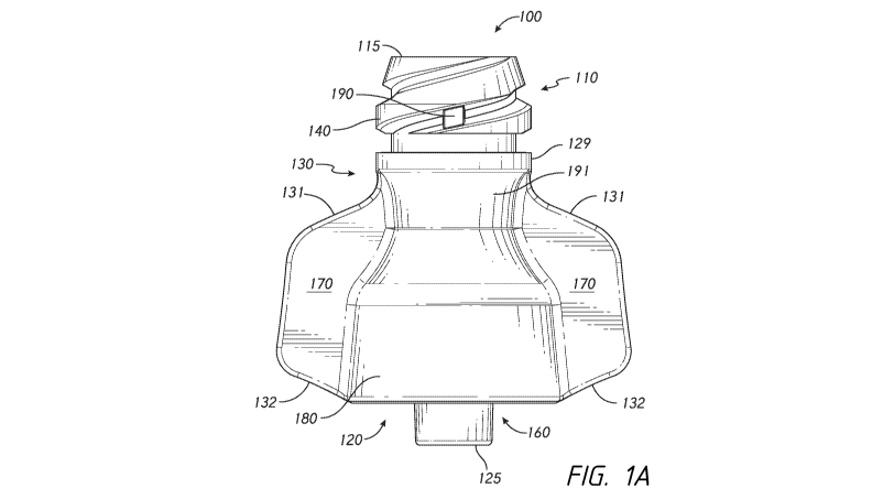
[0018] Figure 1A is a front view of another embodiment of a medical
connector that is configured to receive one or more emitters of One or more
therapeutic
agents;
100191 Figure 2 is a side view of the medical connector of Figure 1;
100201 Figure 3A is a front view of the medical connector of Figure 1
in a
vertical cross section along line 3-3 of Figure 2;
[0021] Figure 3B is the front view of Figure 3A with an emitter of
one or
more therapeutic agents being inserted into the fluid pathway;
[0022] Figure 3C is the front view of Figure 3A with the emitter
securely
positioned in the fluid pathway;
[0023] Figure 4 is a bottom view of the medical connector of Figure 1
at a
horizontal cross section along line 4-4 of Figure 2;
[0024] Figure 5 is a top view of the medical connector of Figure 1 at
a
horizontal cross section along line 5-5 of Figure 2;
[0025] Figure 6A is a top view of the medical connector of Figure 1
with a
horizontal cross section along line 6-6 of Figure 3A;
[0026] Figure 6B is the top view of Figure 6A with an emitter of one
or more
therapeutic agents inserted into the fluid pathway;
[0027] Figure 6C is a top view of Figure 6A with a different type of
emitter of
one or more therapeutic agents inserted into the fluid pathway (e.g., an
emitter with a
larger diameter or cross-sectional width than the emitter of Figure 6B);
[0028] Figure 7A is a top perspective view of the medical connector
of Figure
1;
[0029] Figure 7B is a bottom perspective view of the medical
connector of
Figure 1;
[0030] Figure 7C is a front perspective view of the medical connector
of
Figure 1 with a vertical cross section along line 4-4 of Figure 2;
[0031] Figure 8 is the front perspective view of Figure 7C with an
emitter
securely positioned in the fluid pathway and fluid flowing through the
connector; and
-5-
CA 02982456 2017-10-11
WO 2016/182822
PCT/US2016/030844
100321 Figure 8A is another front perspective view of Figure 7C
illustrating
another example of fluid flow through the connector.
100331 Nothing illustrated in these drawings or described in the
associated text
is indispensable or essential: rather, any feature, structure, material,
component, or step
illustrated or described in any embodiment can be used alone or omitted, or
can be used
with or instead of any feature, structure, material, component, or step
illustrated or
described in any other embodiment. For example, some embodiments do not
include any
emitter, but do include one or more other features illustrated or described in
this
specification. The features are illustrated and described in discrete
embodiments merely
for convenience of explanation, but not to limit the inventions or to
segregate the
inventions into isolated collections of features. The proportions and relative
sizes of
components and features illustrated in the drawings form part of this
disclosure, but
should only be interpreted to form part of a claim if recited in such claim,
either now or in
the future.
DETAILED DESCRIPTION
100341 Figures 1-7 illustrate an example of a medical connector 100.
Different embodiments of medical connectors are described in this
specification, some of
which include the features illustrated in Figure 1-7. A medical connector 100
comprises
a housing comprising a proximal region 110 with a proximal end 115, a distal
region 120
with a distal end 125, and a body 130 extending between the proximal and
distal ends
115, 125. The distal region 120 can comprise a male protrusion 160, such as a
male luer
protrusion. An internal fluid pathway 135 can extend between the proximal and
distal
regions 110, 120 of the connector 100, such as between the proximal and distal
ends 115,
125. In some embodiments, either or both of the proximal or distal regions
110, 120 can
include a connection structure 140, such as one or more threads (as shown),
clasps, arms,
latches, protrusions, and/or recesses, etc., that is configured to help guide,
attach, and/or
retain the medical connector to another device, such as another medical
connector.
100351 As shown in Figures 1 and 2, the proximal region 110 can
comprise a
threaded connection structure 140 configured to rotatably attach and detach
from a
corresponding threaded connection structure on another device, such as a male
end of a
syringe (not shown). A thread-stop or collar 129 can be positioned distally
from the
connection structure 140 to prevent or resist over-extending the threaded
connection
-6-
CA 02982456 2017-10-11
WO 2016/182822
PCT/US2016/030844
between the connector 100 and another medical device. As illustrated in
Figures 3A-C
and 4, the proximal region can include a female coupling 150 configured to
slidably
receive a corresponding male coupling of another device such as a syringe (not
shown).
In some embodiments, as shown, the female coupling 150 comprises a conduit 155
within
the proximal region 110. The conduit 155 can comprise a tapering wall
structure that
diminishes in diameter or horizontal cross-sectional width from the proximal
end 115
toward the distal end 125. In some embodiments, the taper can conform to a
standard
within the medical industry, such as any version of the ISO 594 standard
(which includes
a 6% luer taper) or any other applicable standard (e.g., DIN and EN standard
1707:1996
and/or 20594-1:1993). The conduit 155 can be configured to snugly and tightly
receive a
male coupling of another standard-compliant device with a corresponding taper
or other
shape, such as a corresponding luer taper, to produce resistance against fluid
leakage after
the male coupling is inserted fully into the female coupling 150. In some
embodiments,
as shown, the connector 100 is separate from the syringe; and in some
embodiments, the
connector 100 is integrated into or bonded to the syringe.
[0036] In some embodiments, as illustrated, the connection structure
140 can
comprise one or more disconnection-resisting features or structures configured
to resist
disconnection between the connector 100 and another medical implement (such as
a
syringe or other connector or other structure). The disconnection-resisting
features(s) or
structure(s) can have many different forms, such as one or more freely
spinning positions
or stages after connection is accomplished, one or more increased friction-
inducing anti-
rotation impediments, and/or one or more disconnection-resisting thread
shapes. For
example, in a threaded connection structure 140, as shown in Figures 1, 2, and
4, a
friction-inducing impediment can comprise one or more (e.g., at least two)
protrusions
190 positioned between multiple thread turns, the one or more protrusions 190
extending
radially outwardly from the inner surface 194 of the threading 197, thereby
providing a
region of radial space between the radially outermost surface 196 of the
protrusion 190
and the radially outermost surface 198 of the threading 197 that is smaller
than the radial
space between the inner surface 194 of the threading 197 and the outennost
surface 198
of the threading 197. In this example of an impediment, as the threading of
another
device (such as a syringe, not shown) is rotatably attached to the threading
197 of the
connection structure 140, the relative rotation of the two devices is slowed
down or
resisted through increased frictional contact between the impediment and the
threading of
-7-
CA 02982456 2017-10-11
WO 2016/182822
PCT/US2016/030844
the other device, thereby requiring greater torque to attach the two devices
and/or
requiring greater torque to detach the two devices, which diminishes the risk
of accidental
disconnection. The contact between the impediment and the threading of the
other device
may cause wedging, compaction, crushing, and/or compression of either or both
structures. Many different types of impediments can be used to resist
disconnection that
are different from those described and/or illustrated.
[0037] A disconnection-resisting thread shape can help resist or
prevent
disconnection between the connector 100 and another medical device, such as a
syringe.
For example, as illustrated in Figures 1, 2, and 7C, a helical threading 197
with multiple
thread turns can comprise a thread portion with an oversized region 201,
and/or an
outwardly flaring or outwardly tapering region 203. In some embodiments, as
shown, the
outermost diameter of a beginning thread portion 199 can be a first diameter
that is a
standard size or within a standard range of sizes, such as may be specified in
any
applicable medical device standard (e.g., any of those mentioned elsewhere in
this
specification), or slightly smaller than a standard size or range of sizes. As
the thread
progresses around the proximal region 110 in the distal direction, the
outermost diameter
of a portion of the thread can flare or taper outwardly to a non-standard
second diameter
than is larger than the first diameter and larger than the diameter or range
of diameters
specified in one or more applicable medical device standards. Since the other
medical
device to which the proximal region 110 of the connector 100 is configured to
attach
(e.g., a syringe) will typically have a standard diameter of threading, the
outward taper or
flare of the disconnection-resisting thread shape of the connector 100 can
cause the space
between the respective threads to decrease, or can cause the attachment region
of the
other medical device to stretch by a small amount, and/or can cause the
threading 197 of
the connector 100 to compress by a small amount. One or more of these effects
can
create opposing radial forces between the threading surface of the other
medical device
and the threading surface 197 of the connector 100, which can increase the
friction
between the respective surfaces and thereby resist or prevent rotational
movement and
decrease the risk of accidental disconnection between the two devices. As
shown in this
example, the connector 100 can be configured to resist disconnection from a
syringe. In
some embodiments, the connection structure 140 is configured such that the
resistance is
sufficiently high that it is not possible under normal conditions of use to
disconnect the
connector 100 from the other medical device (e.g., disconnection is
prevented). Many
-8-
CA 02982456 2017-10-11
WO 2016/182822
PCT/US2016/030844
other different types of disconnection-resisting or disconnection-preventing
features can
be used instead of or in addition to those illustrated and/or described in
this specification,
including one or more structures not including thread shapes or impediments,
or any type
of threads at all.
[0038] In some uses, it may be desirable to temporarily attach the
distal region
120 of the connector 100 to a standby fluid line that has at its proximal end
a resealable
needleless female connector, such as a Clavell) connector sold by ICU Medical,
Inc. or a
SmartSitet connector sold by CareFusion Corporation. This type of
configuration can
allow a healthcare practitioner to infuse fluid from a fluid source (such as a
syringe) into
the proximal end 115 of connector 100, through the distal end 125 of connector
100, and
into the resealable needleless female connector, the fluid line, and
ultimately the patient.
However, it may be undesirable, in some embodiments, to leave the connector
100
attached to a resealable female connector for a prolonged period, especially
when
unattended, since the connector 100 may not include a seal at its proximal end
in some
embodiments (as shown), and may therefore expose the fluid in the fluid line
to the
outside environment or even allow fluid in the fluid line to flow out of the
fluid line.
Thus, in some embodiments, there is no connection-securing structure (such as
threads) in
the distal region 120 of the connector 100 to discourage long-tern connection.
Rather,
the illustrated connector 100, without connection-securing structure, is
configured to be
rapidly and easily slidably inserted into and/or removed from a corresponding
female
connector without requiring any additional motion (e.g., twisting, rotating,
clasping, etc.)
in a non-secured connection. Also, the absence of connection-securing
structure in the
distal region 120 of the connector obviates the need to use a reverse twisting
motion to
remove the distal region 120 from the resealable needleless female connector,
which
would otherwise increase the risk that a threadably secured connection between
a syringe
and the proximal region 110 of the connector would be inadvertently partially
or
completely disconnected or backed out, potentially causing a leak.
[0039] In some embodiments (not shown), the distal region 120 can
comprise
any suitable connection structure, such as any connection structure that is
illustrated
and/or described in connection with the proximal region 110 of medical
connector 100 or
in connection with any other embodiment. If used, the connection structure can
be
included in an inner region 165 generally surrounding the male luer protrusion
160. As
illustrated, the inner region 165 can be generally surrounded by a shroud or
skirt that is
-9-
CA 02982456 2017-10-11
WO 2016/182822
PCT/US2016/030844
configured to pass over and around a corresponding female end of another fluid
connector
to which the male protrusion 160 of the connector 100 is configured to be
attached. As
illustrated in Figure 7C, in some embodiments, the proximal end of the inner
region 165
is positioned further in the distal direction than the distal end of the
intermediate region
192. In some embodiments, including those in which there is no gripping
portion 170 in
the distal region 120 of the connector 120 (or no gripping portion 170 at
all), the shroud
or skirt can be omitted, as with any other feature, structure, material, or
step disclosed or
illustrated in this specification. When the shroud or skirt is omitted, the
male protrusion
160 can be fully exposed along its length from a proximal base region to a
distal end
region.
[00401 The male
protrusion 160 can include one or more features to facilitate
temporary attachment to a resealable needleless female connector. For example,
the male
protrusion 160 may not be a standard luer, in that it may have a non-standard
size and/or
shape (e.g., a size and/or shape that does not conform with one or more
features or
requirements of one or more medical industry standards, such as the ISO 594
medical luer
standard and/or one or more other medical standards). In the illustrated
embodiment, the
male protrusion 160 has a taper that is about 6%, which comports with one or
more
medical standards, but the male protrusion 160 is oversized in that it has a
larger outer
diameter on its distal end than is specified in one or more medical standards.
For example, in some embodiments, the distal outer diameter of the male
protrusion 160
can be at least about 1/1,000 of an inch and or at least about 3/1,000 of an
inch larger than
a standard distal outer diameter. Many other sizes can be used.
10041] Since
most resealable needleless female connectors have proximal
openings with standard-size diameters, the oversized, non-standard male
protrusion 160
will have a larger diameter at its distal end than the diameter at the distal
end of the
conduit of the female opening in a standard medical device to which the
connector 100 is
configured to attach. The larger diameter on the male protrusion 160 can
enable it to fit
more tightly or snugly at a lesser penetration depth into the female opening
than would a
male luer protrusion with a standard distal outer diameter. This can help to
facilitate a
non-secured, temporary attachment of the distal region 120 of the connector
100 to a
resealable needleless female connector (not shown). Most, if
not all, resealable
needleless female connectors include a compressible elastomeric sealing
element or other
movable sealing element that can be advanced distally within such connector to
-10-
CA 02982456 2017-10-11
WO 2016/182822
PCT/US2016/030844
temporarily open it to fluid flow, such as by inserting the male protrusion
160 into a
proximal female opening on such a resealable needleless female connector. The
sealing
element is configured to rebound to a sealed position by pushing back against
an inserted
male protrusion. The amount of rebound or push-back force increases as the
penetration
depth of the inserted male protrusion increases. Since the male protrusion 160
is non-
standard, having a larger distal outer diameter, it penetrates less distance
into the
needleless female connector when fully inserted, and therefore the sealing
element exerts
less proximally-directed rebound force against it, lowering the risk that the
male
protrusion 160 will be pushed back in the proximal direction by the sealing
element and
thereby dislodged from the resealable needleless female connector.
100421 As shown, in some embodiments, the overall longitudinal length
of the
connector 100 can be relatively short. For example, either or both of the
longitudinal
length of the portion of the fluid pathway 135 within the threaded region (or
the region on
which the connection structure 140 is affixed, in some embodiments) and/or the
longitudinal length of the portion of the fluid pathway 135 within the male
protrusion 160
can be greater than the longitudinal length of the portion of the fluid
pathway 135 that
extends between the threaded region and the male protrusion 160, as shown in
Figure 3.
As illustrated, a base portion 180 of the distal region 120 of the connector
100 can be
relatively wide. For example, the external diameter and/or external horizontal
cross
sectional width of the base portion 180 can be larger than an external neck
portion 191
located between the proximal region 110 and the base portion 180. Many
different sizes
and proportions of the portions of the connector 100 can be used.
100431 The connector 100 can comprise a grasping portion 170, such as
one or
more tabs (as shown), recesses, protrusions, stripes, butnps, and/or friction-
inducing
gripping surfaces, etc. In the embodiment illustrated in Figure 1, the
grasping portion
170 enables a user to securely retain the connector 110 during connection and
disconnection with another device, such as when another device is twisted, or
swayed or
rocked back and forth, onto or away from the threaded proximal region 110, or
when the
threaded proximal region 110 is twisted, or swayed or rocked back and forth,
into or out
of another device.
100441 The grasping portion 170 can be relatively large in comparison
to the
size of the overall connector 100. For example, as shown in Figures 5 and 6,
the
horizontal cross-sectional width (e.g., extending between respective lateral
edges) of the
-11-
CA 02982456 2017-10-11
WO 2016/182822
PCT/US2016/030844
grasping portion 170 can be larger than the external diameter or horizontal
cross-sectional
width of the base portion 180, in some embodiments. As illustrated, the
longitudinal
length (in the proximal-to-distal dimension) of the grasping portion 170 can
be larger
than the longitudinal length of the connection structure 140 in the proximal
region 110 of
the connector 100. In some embodiments, as shown, the longitudinal length of
the
grasping portion 170 can extend over more than half of the overall
longitudinal length of
the connector 100. The grasping portion 170 can comprise one or more curved
lateral
edges or sides, as illustrated.
100451 As illustrated in Figure 1A, in some embodiments the grasping
portion
170 can comprise one or more upper edges 131 and/or one or more lower edges
132. As
shown, one or both of the upper edges 131 can be slanted, such as with a
downwardly
sloped slant: and/or one or both of the lower edges 132 can be slanted, such
as with an
upwardly sloped slant. In an intermediate region of the connector 100 (e.g.,
below the
thread-stop or collar 129 and above the distal region 120), the connector body
can
comprise a first region having a first cross-sectional width or diameter, a
second region
positioned distal from the first region and having a second cross-sectional
width or
diameter, and a third region positioned distal from the second region and
having a third
cross-section width or diameter. As shown in Figure 1A, the second cross-
sectional
width or diameter can be smaller than either or both of the first cross-
sectional width or
diameter and/or the third cross-sectional width or diameter. As shown, in a
region of the
connector body below the second region, the connector body can comprise a
continuously
increasing outer cross-section or diameter that produces an outward flare from
the second
region in a distal direction toward the distal region 120 of the connector
100.
100461 As shown in Figure 1A, in some embodiments one or more
friction-
inducing impediments 190 can be positioned circumferentially along the
connection
structure 140 in a region that is generally about midway between the grasping
portions
170. In some embodiments, such as shown in Figure 1, the friction-inducing
impediment
190 can be positioned circumferentially along the connection structure 140 in
general
alignment with one or more longitudinal edges of one or more grasping portions
170 (see
Figure 2).
100471 In some embodiments (not shown), the horizontal cross-
sectional
width of the grasping portion 170 is no larger than the external diameter or
horizontal
cross-sectional width of the base portion 180, and/or may comprise one or more
small
-12-
CA 02982456 2017-10-11
WO 2016/182822
PCT/US2016/030844
friction-inducing structures, such as one or more protrusions, grooves, and/or
other slide-
resistant structures or materials. In some embodiments, the grasping portion
170 can be
omitted, as with any other feature, structure, material, or step disclosed or
illustrated in
this specification.
100481 In some embodiments, all or a portion of the fluid pathway 135
inside
of the connector 100 can be straight, as illustrated in Figure 3A (either
before or after
insertion of an emitter 200, as shown in Figures 3B and 3C), from the proximal
region
110 or proximal end 115 to the distal region 120 or end 125 of the connector
100, such
that a single straight line can be drawn within the fluid pathway 135 from the
beginning
to the end of the fluid pathway 135. In some embodiments, the fluid pathway
135 can
extend along a generally straight path without one or multiple sharp, angular,
perpendicular, and/or obtuse changes in direction in the fluid pathway 135. In
some
embodiments, the fluid pathway 135 is straight or generally straight at least
along a
majority of the longitudinal length of the fluid pathway 135. The fluid
pathway 135 can
be straight or generally straight along any particular segment of the fluid
pathway, such
as along the distance between all or a majority of the proximal end 115 of the
connector
100 and the proximal end of the intermediate region 192, between all or a
majority of the
proximal end of the intermediate region 192 and the distal end of the
intermediate region
192, and/or between all or a majority of the proximal end of the portion of
the fluid
pathway 135 within the male protrusion 160 and the distal end of the portion
of the fluid
pathway 135 within the male protrusion. A straight path or a generally
straight path can
diminish turbulence and/or stagnation in one or more portions of the fluid
pathway 135
and/or can provide a high flow rate and low fluid resistance.
100491 As shown in Figure 3A, the diameter or horizontal cross
sectional
width of the fluid pathway 135 can vary along the longitudinal length of the
fluid
pathway 135. For example, as illustrated in Figure 3A, the diameter or cross
sectional
width of the fluid pathway 135 in at least a portion of the proximal region
110 can be
larger than the diameter or cross sectional width of the fluid pathway 135 in
at least a
portion of an intermediate region 192, which in turn can be larger than the
diameter or
cross sectional width of the fluid pathway 135 in at least a portion of the
distal region
120, such as the portion of the fluid pathway 135 inside of the male
protrusion 160.
100501 In some embodiments, as shown, the connector 100 can comprise
a
stationary structure without any moving external and/or internal parts during
use.
-13-
CA 02982456 2017-10-11
WO 2016/182822
PCT/US2016/030844
For example, the external and/or internal shape, orientation, position, and/or
size of the
connector 100 and its internal components before attachment to or engagement
with
another medical device can be the same as it is after attachment to or
engagement with
another medical device. In some embodiments, the connector 100 can comprise
moving
parts to facilitate connection and disconnection, opening and closing of the
connector to
form a valve, and/or regulation of pressure or volume.
100511 The connector 100 can comprise one or more additional features
that
are not shown in Figures 1-7, such as proximal and/or distal ends 115, 125
that include
resealably openable and closeable apertures with one or more resilient or
rigid sealing
elements to enable selective fluid flow; a rigid internal cannula or support
member or
spike that is configured to assist in supporting or opening a sealing element;
a body 130
that is clear or transparent or includes a clear or transparent portion and/or
one or more
other internal structures that are clear or transparent or include a clear or
transparent
portion that is or are configured to enable viewing of fluid within the
internal fluid
pathway 135 during use; a cap for selectively closing the fluid pathway;
and/or a
pressure-regulating or volume-regulating feature inside of the connector 100
to enable
neutral flow, etc. For example, any feature, structure, material, component,
or step
illustrated or described in any embodiment of U.S. Patent Nos. 5,685,866;
7,815,614;
8,454,579; and/or 8,758,306, which are each incorporated by reference herein
in their
entireties, can be used with or instead of any feature, structure, material,
component, or
step illustrated or described in any embodiment in this specification.
100521 In some embodiments, as shown, the connector 100 can be
configured
to receive or include one or more components that are configured to provide
one or more
therapeutic agents into the medicinal fluid that is inside and/or moving
through the fluid
pathway 135. For example, as illustrated in Figures 3B, 3C, and 6B, an emitter
200 of
one or more therapeutic agents can be inserted into the connector 100 in an
internal
region, such as in the intennediate region 192, of the fluid pathway 135. As
illustrated, in
some embodiments, the emitter 200 is positioned entirely within the connector
100 and
not partially or entirely positioned within another medical device, such as a
syringe. The
connector 100 can be temporarily or permanently attached to a syringe or any
other
medical device. The connector 100 with the emitter 200 can be configured to
provide
infusion of one or more therapeutic agents in a low-profile, non-bulky,
inexpensive
manner, without requiring large or complex storage or logistical requirements.
-14-
CA 02982456 2017-10-11
WO 2016/182822
PCT/US2016/030844
100531 The emitter 200 can comprise any material and/or structure
that is
configured to provide, leach out, release, diffuse, infuse, dissolve, erode
into, or
otherwise emit a therapeutic agent into the fluid pathway 135, alone or in
combination
with fluid flowing through the fluid pathway 135. In some embodiments, the
emitter 200
can comprise a non-dissolving substrate or storage material or matrix or other
base
material in which a therapeutic agent is temporarily held or captured or bound
until the
therapeutic agent is emitted within the fluid pathway 135. The emitter 200 can
have any
suitable shape. For example, the emitter 200 can be cylindrical (as shown) or
rectangular.
In some embodiments, as shown, the emitter 200 can be elongate (e.g., its
longitudinal
length, from its proximal end 201 or face to its distal end 203 or face is
larger than its
diameter or cross sectional area). As illustrated, some emitters 200 are solid
or
substantially solid or resistive to fluid flow from a proximal end or face 201
to a distal
end or face 203. For example, as shown, in some embodiments there are no
internal,
discrete, and/or generally longitudinally oriented fluid pathways within or
through the
emitter 200; rather, fluid may be permitted to soak into or be absorbed by or
pass through
the emitter 200 only in essentially random or highly tortious directions
(e.g., not a direct
or discrete pathway), and/or fluid may not be permitted to soak into or pass
through the
emitter 200 at all. In some embodiments (not shown), an emitter 200 for use
with the
connector 100, or with any other embodiment of a connector, can include one or
more
apertures, channels, tunnels, passages, and/or fluid pathways that are
configured to carry
or convey fluid through or within the emitter (e.g., from a proximal end or
face 201 to a
distal end or face 203) without substantial resistance to fluid flow.
100541 In some embodiments, all or at least a portion of the outer
housing of
the connector 100 where all or at least a portion of the emitter 200 is
contained can be
clear or transparent to permit viewing of the emitter 200 from outside of the
connector
100. In some embodiments, as shown, the emitter 200 is very small. For
example, as
shown in Figure 3a, the longitudinal length of the emitter 200, from its
proximal face to
its distal face can be less than or equal to about the longitudinal length of
the conduit 155
within the proximal region 110 and/or less than or equal to about the
longitudinal length
of the male protrusion 160; and/or the diameter or cross-sectional area of the
emitter 200
can be less than or equal to about the outer diameter of the male protrusion
160. In some
embodiments, the longitudinal length of the emitter 200 and/or the diameter of
the emitter
-15-
CA 02982456 2017-10-11
WO 2016/182822
PCT/US2016/030844
can be a few millimeters (e.g., at least about 2 millimeters or at least about
4 millimeters).
Many other sizes and shapes and configurations can be used for the emitter
200.
[0055] As illustrated, in some applications, the emitter 200 can
comprise a
compressible and/or fibrous matrix material on which a therapeutic agent has
been coated
or into which a therapeutic agent has been infused, impregnated, soaked,
absorbed, and/or
bonded. In some embodiments, the emitter 200 can include any suitable
biocompatible
binder to facilitate a temporary water-soluble or other liquid-soluble bond
between the
base material and the therapeutic agent, or the emitter 200 may not include
any binder. In
some embodiments, the emitter 200 does not include a substrate but is instead
formed of a
consumable material that gradually erodes away or dissolves into the fluid
pathway 135
during infusion until it is used up. Any type of therapeutic agent can be
used, including
but not limited to one or more nourishing agents (e.g., vitamins, minerals,
etc.), pain-
diminishing medications, antibiotics, antimicrobials (e.g., any chlorhexidine-
based
compound), anti-inflammatories, sedatives, anticoagulants (e.g., heparin),
chemotherapy
drugs, and/or other types of therapeutic agent. The size and shape of the
emitter 200
and/or of the overall connector 100 can be very different depending upon the
amount or
type of therapeutic agent that is intended to be infused. For example, a very
large
connector can be used when a large amount of therapeutic agent needs to be
infused.
Many other different types of emitters can be used instead of or in addition
to the emitter
200 as illustrated. For example, an emitter can be provided in the form of a
coating on an
interior surface of the connector 100 or a material integrated into a portion
of the base of
the body 130 of the connector 100, or any other suitable material or structure
that
provides a therapeutic agent at a desired time, in a desired dosage, and/or at
a desired
infusion rate. Among many other embodiments, an emitter for use with the
connector
100 can be provided in the form of any of the cartridges or other emitters
that are
illustrated or described in International PCT Publication No. W02013/023146 A
1 (Di
Fiore), which is incorporated by reference herein in its entirety'. Many other
types of
emitters can be used instead of or in addition to those illustrated or
described.
10056j In some embodiments in which an emitter 200 is provided in the
form
of an inserted material, such as is shown in the example of Figure 3B, the
interior region
of the connector 100 can comprise a retaining structure 210 for the emitter
200, as
illustrated in Figure 3A. Although the retaining structure 210 is illustrated
with particular
dimensions and features, the retaining structure 210 can comprise any suitable
material or
-16-
CA 02982456 2017-10-11
WO 2016/182822
PCT/US2016/030844
structure that retains the emitter 200 in such a way that the emitter 200 can
be configured
to emit one or more therapeutic agents as desired for a particular medical
therapy.
100571 As illustrated, in some examples, the retaining structure 210
can
comprise one or more retaining components 230 that extend from an internal
wall of the
connector 100 into an internal space of the connector (such as radially
inwardly). For
example, as shown, the retaining components 230 can be retaining struts that
extend
generally longitudinally along the fluid pathway 135. The retaining components
230 can
be positioned in the intermediate region 192, as shown. In some embodiments,
the
retaining structure 210 can comprise at least two or at least three or at
least four (as
shown in Figure 6B) retaining components 230 such as retaining struts. As
shown in
Figure 7C, one or more of the retaining components 230 can comprise a first
portion or
component that extends a first distance from an internal wall of the connector
100 into an
internal space of the connector and a second portion or component that extends
a second
distance from an internal wall of the connector 100 into an internal space of
the connect.
The second distance can be greater than the first distance. For example, one
or more of
the retaining struts can comprise a retaining protrusion, such as an elongate
longitudinal
portion, and a base portion 240. The longitudinal portion can be formed as a
protrusion
extending radially inwardly from the interior wall of the intermediate region
192 of the
fluid pathway 135 of the connector 100. As shown, in some embodiments, one or
more
of the base portions 240 can be radially aligned with one or more of the
longitudinal
portions 230. One or more of the base portions 240 can extend radially
inwardly from the
interior wall of the fluid pathway 135 further than one or more of the
longitudinal
portions 230, as illustrated in Figures 3A-3C, 6A, 7A, and 7C, for example.
100581 In some embodiments, as shown, the retaining structure 210 can
provide a retaining space within which the emitter 200 can be retained. For
example, the
retaining space can correspond to the outer width or thickness of the emitter
200, such as
by being about the same size as or slightly smaller than the outer width or
thickness of the
emitter 200. When an emitter 200 is inserted into a retaining space, such as
by pushing
the retainer into the proximal end 215 of the connector 100, through the
proximal portion
of the fluid pathway 135, and into the intermediate portion 192, the emitter
200 can
radially compress or contract by a small amount such that the retaining
structure 210 can
exert a radially inwardly directed retaining force against the emitter 200
that is sufficient
to produce an increase in friction that resists dislodgment of the emitter 200
from the
-17-
CA 02982456 2017-10-11
WO 2016/182822
PCT/US2016/030844
retaining space (for example, as shown in Figure 6C). In some embodiments,
as illustrated, the emitter 200 can be securely retained within the connector
100 in a
manner that resists or prevents either or both of longitudinal or lateral
movement of the
emitter 200 within the retaining space. In some embodiments (not shown), the
retaining
space is configured to be somewhat larger than the emitter 200 to pernnt the
emitter 200
to move or float within the retaining space, either before or during infusion.
100591 As shown, a plurality of longitudinal portions 230 can be
positioned
radially around the retaining space such that the plurality of longitudinal
portions 230 are
configured to contact the outer surface of the emitter 200 when inserted. In
some
embodiments, as illustrated, the longitudinal portions 230 are provided
generally equally
spaced circumferentially from each other. As illustrated, one or more of the
longitudinal
portions can comprise longitudinal faces (e.g., facing radially inwardly) that
are slightly
inwardly tapered along the longitudinal dimension in the proximal-to-distal
direction,
such that the distance between respective longitudinal portions is slightly
less on the
distal side of the longitudinal portions than on the proximal side of the
longitudinal
portions. This inward tapering can help to securely retain the emitter 200
when inserted
into the retaining space. As shown in Figure 7C, one or more of the
longitudinal portions
230 can include a proximal face or region 260 that is tapered or beveled or
slanted to
facilitate insertion of an emitter 200 into the retaining space by providing
an initially wide
but gradually narrowing region for the emitter 200 upon insertion of the
emitter 200 into
the retaining space.
100601 As shown in Figures 6B and 8, a flow space 250 can be provided
between two or more retaining components (e.g., longitudinal portions 230)
sequentially
positioned circuinferentially around the fluid pathway 135 (and/or generally
surrounding
or positioned generally around the retaining space). As shown in Figure 8 and
8A, the
one or more flow spaces 250 can be configured to permit fluid flowing through
the fluid
pathway 135 within the connector 100 to flow around the longitudinal portions
230 and
through the flow spaces 250, along one or more lateral sides or lateral
surfaces of the
emitter 200, such as between the one or more lateral sides or lateral surfaces
of the
emitter 200 and the internal wall of the fluid pathway 135. In some
embodiments, there
is at least one flow space 250, or at least two flow spaces 250, or at least
four flow spaces
250 (as shown). In some embodiments, as illustrated in Figure 8, at least a
majority of
the external surface area of the emitter 200 is spaced from the internal
surface of the fluid
-18-
CA 02982456 2017-10-11
WO 2016/182822
PCT/US2016/030844
pathway 135 so that the fluid pathway 135 can pass adjacent to and around the
outside of
the emitter 200 to permit some or all of the fluid to flow around the outside
of the emitter
200 (e.g., at least a majority of the external lateral surface area of the
emitter 200 does not
contact one or more retaining components or other surfaces inside of the
connector 100).
In some embodiments of connector 100, there are no flow spaces or only very
small
and/or very constricted flow spaces, such that all or a majority of the fluid
pathway and
the fluid flowing through the connector is configured to pass within or
through the
emitter 200 (e.g., by passing through a proximal portion or face 201 of the
emitter 200
and exiting out of a distal portion or face 203 of the emitter 200). Such an
emitter 200
can have many forms; for example, it can be solid and/or porous and/or include
one or
more apertures, channels, tunnels, passages, and/or fluid pathways for
conveying or
carrying fluid.
10061] A base retainer can be fonned in any suitable manner, such as
by a
plurality of base portions 240 (as illustrated), that can provide a lower flow
space 280
between a distal end of the intermediate region 192 and a distal end of the
emitter 200, as
shown in Figures 3C and 8. The distal end of the retaining space, as shown,
can include
an aperture 270 that is smaller in diameter than another portion of the flow
pathway 135
in the intermediate region 192 and/or that is smaller in diameter than the
retaining space.
Within the flow pathway 135, the aperture 270 can lead from the intermediate
region 192
to the interior of the male protrusion 160, as illustrated. The base retainer
can assist in
retaining the emitter 200 apart or spaced away from the distal end of the
intermediate
region and/or from the aperture 270, so as to enable fluid flowing through the
fluid
pathway 135 to flow around the distal end of the emitter 200 and out of the
aperture 270
(without causing the emitter 200 to plug up or block the aperture 270).
100621 In some embodiments, as shown in Figure 7C, the circumference
of a
circle transcribed by the longitudinal portions 230 and/or the base portions
240 of the
retaining structures 210 around the fluid path can be greater than the
circumference of the
intemal fluid pathway 135 of the distal end 125 (and/or greater than the
circumference of
the aperture 270 of the fluid pathway of the distal end 125). In some
embodiments, as
shown in Figure 7C, the distance across the intermediate region 192 between
generally
opposite facing longitudinal portions 230 and/or base portions 240 can be
greater than a
minimum diameter of the intemal fluid pathway 135 of the distal end 125 and/or
greater
than the diameter of the aperture 270 of the fluid pathway of the distal end
125. For
-19-
CA 02982456 2017-10-11
WO 2016/182822
PCT/US2016/030844
instance, as shown in Figure 7C, in some embodiments, where two retaining
structures
are positioned generally opposite one another about a circumference formed by
the
retaining structures 210 around the fluid path, the transverse distance
between the
oppositely positioned retaining structures 210 and/or base portions 240 is
greater than the
diameter of the internal fluid pathway 135 of the distal end 125 (and/or
greater than a
diameter of the aperture 270). As shown in Figures 6B and 6C, respectively, in
some
embodiments, the circumference formed by the retaining structures 210 around
the fluid
pathway is about the same size or just smaller than the circumference of an
emitter 200.
In some embodiments, when the circumference formed by the retaining structures
210
around fluid pathway is just smaller than the circumference of an emitter 200,
the
longitudinal portions 230 of the retaining structures 210 can engage (e.g.,
hold or restrain)
the emitter 200 (e.g., by friction). In some embodiments, as shown in Figure
7C, when
the internal fluid pathway 135 is smaller than the portion of the intermediate
region 192
between the longitudinal portions 230, then a distal shelf or support region
or fluid
diverting region can be formed in the internal region 192 between the
longitudinal
portions and the internal fluid pathway 135 of the distal end 125. As shown in
Figure 7C,
the shelf or support region or fluid diverting region can be generally
horizontal or
generally transverse in some embodiments.
[0063] In some embodiments, as shown in Figures 6A and 7C, a
circumference formed by the base portions 240 about the fluid path within the
of the
intermediate region 192 can be greater than the circumference of the internal
fluid
pathway 135 of the distal end 125 (and/or greater than the circumference of
the aperture
270 of the fluid pathway of the distal end 125). For instance, as shown in
Figure 7C, in
some embodiments, where two base portion 240 structures are positioned
generally
opposite from one another about a circumference formed by the base structures
240
around the fluid path, the transverse distance between the oppositely
positioned retaining
structures 210 is greater than the diameter of the internal fluid pathway 135
of the distal
end 125 (and/or greater than a diameter of the aperture 270). As shown in
Figure 7C, in
some embodiments, the intemal-most circumference formed by the base portions
240
terminates circumferentially outwardly of the circumference of the aperture
270. In some
embodiments, as shown in Figure 3A, the portion of the intermediate region 192
within
the longitudinal portions 230 is smaller in transverse width or diameter or
circumference
than the conduit 155 within the proximal region 110.
-20-
CA 02982456 2017-10-11
WO 2016/182822
PCT/US2016/030844
100641 As illustrated in Figure 8 and 8A, the connector 100 can be
configured
so that the position and orientation of the retaining structure and the
emitter 200 permits
fluid flowing through the fluid pathway 135 to flow mostly or entirely around
and/or
outside of the emitter 200. In its initial state, the emitter 200 can be dry
or not saturated
with fluid. As fluid flows around and/or outside of the emitter 200, the
emitter 200 is
wetted or the level of wetness of the emitter 200 is increased and therapeutic
agent is
emitted from the emitter 200 into the flowing fluid, first from the periphery
of the emitter
200 (which is closest to the flowing fluid) and then from the core or interior
of the emitter
200. As the fluid flowing around the emitter 200 soaks into and/or eventually
saturates
the emitter 200, therapeutic agent contained within the interior of the
emitter 200
migrates toward the periphery of the emitter 200 and is eventually emitted
into the fluid
pathway 135. By directing the fluid flow predominantly around and/or outside,
rather
than predominantly through, the emitter 200, the connector 100 does not become
plugged
up or require excessive force on the syringe to accomplish fluid infusion. In
some
embodiments (not shown), most or all of the fluid flow can be directed through
the
emitter 200, for example in embodiments in which there are no flow spaces 250,
280.
Also, by directing the fluid flow predominantly around and/or outside, rather
than
predominantly through the emitter 200, the connector 100 allows at least a
portion of
fluid to flow freely through the connector.
100651 As shown in the example of Figure 8, in some embodiments, the
proximal face of the emitter 200 can be unobstructed by the retaining
structure 210 within
the fluid pathway 135. For example, as shown, the retaining structure 210 can
be
positioned only on or along or in contact with one or more outer lateral or
longitudinal
sides of the emitter 200 and/or not on or along or in contact with or in
blocking
relationship with a proximal face of the emitter 200. In some embodiments, the
proximal
face of the emitter 200 is exposed to the full diameter or cross-sectional
width of the fluid
pathway 135 of the conduit 155 within the proximal region 110, such that the
fluid
flowing through the fluid pathway is configured to initially contact the full
proximal face
of the emitter 200 when flowing in a distally directed longitudinal direction,
without
being required to twist or turn to contact the proximal face of the emitter
200. In some
embodiments, as illustrated in Figure 8, there is no constriction or blockage
of the fluid
pathway 135 within the proximal region 110 between the conduit 155 and the
proximal
face of the emitter 200.
-21-
CA 02982456 2017-10-11
WO 2016/182822
PCT/US2016/030844
[0066] The emitter 200, in some implementations, can be positioned
within
the fluid pathway 135 a sufficient distance from the proximal end 115 of the
connector
100 that when a male protrusion (such as from a syringe) is inserted into the
proximal
region 110 of the connector, the distal end of the male protrusion does not
contact the
emitter 200.
[0067] In some embodiments, as illustrated, the portion of the fluid
pathway
135 located within the male protrusion 160 can be generally or completely
unobstructed
and/or unimpeded. For example, as shown, the emitter 200 can be located
entirely
outside of the fluid pathway 135 located within the male protrusion 160. For
example,
the emitter 200 can be configured to be positioned within the intermediate
portion 192 of
the connector 100, as shown. In some embodiments, as illustrated, the emitter
200 can be
positioned entirely inside of the connector 100, with no portion of the
emitter protruding
outside of the connector 100. As shown in Figures 8 and 8A, the connector can
be
shaped, structured, and/or contoured such that the fluid pathway 135 of the
connector 100
can be configured to convey liquid along a first portion of the fluid pathway
135 around
the outside of the emitter 200, the first portion having an outer perimeter
that is wider
than the diameter of the emitter 200, and along a second portion of the fluid
pathway 135
in a distal direction from the emitter 200 into a region having an outer
perimeter that is
narrower than the diameter of the emitter 200 (e.g., inside of the male
protrusion 160).
(0068] The connector 100 can be used in many different ways and/or in
many
different systems for providing one or more therapeutic medical effects. An
example of
using the connector 100 in a method of providing an anti-microbial block in a
patient
standby fluid line or providing an emitted therapeutic agent (such as any
agent disclosed
elsewhere in this specification) in any fluid line can include one or more of
the following
steps, and/or one or more instructions can be provided to the user (e.g.,
healthcare
practitioner or patient) to perform one or more of the following steps, in any
suitable
order:
[0069] (1) The connector 100 with an antimicrobial emitter 200 or
another
type of emitter 200 of one or more therapeutic agents can be attached to the
proximal end
of a fluid line at the end of an infusion stage to initiate the beginning of a
standby stage.
In some embodiments, the emitter 200 can comprise a dry or unsaturated,
biocompatible,
clinically safe dosage of an anti-microbial material, such as a chlorhexidine
compound, or
an anti-thrombotic material, or any other therapeutic material, that is
configured to be
-22-
CA 02982456 2017-10-11
WO 2016/182822
PCT/US2016/030844
infused into the fluid pathway 135. A standard liquid, such as water or
saline, or any
other suitable liquid, can be forced into or infused into the proximal end 115
of the
connector 100 from another medical device, such as a syringe or a pump or a
vial or a
fluid line or an IV bag, and brought into fluid communication with the emitter
200
(e.g. by passing around or through, and/or within it).
[0070] (2) An antimicrobial or other therapeutic agent can be
automatically
emitted from the emitter 200 and infused into the fluid line to form an
antimicrobial block
downstream of the emitter 200 and/or to provide any other therapeutic effect
in the fluid
line. In some embodiments, only a small amount of standard or other liquid is
passed
from the syringe into the connector 100 (e.g., less than or equal to about 10
cc or less than
or equal to about 20 cc or less than or equal to about 50 cc of water or
saline), such that
the antimicrobial or other agent remains in the fluid line during the standby
stage and
does not migrate in any appreciable amount into thc patient's bloodstream.
[0071] In some embodiments, by utilizing connectors 100 with
antimicrobial-
infused emitters 200 or other therapeutic-agent-infused emitters 200, a health
clinic or
hospital can conveniently diminish the space, expense, and logistics
associated with
providing and infusing antimicrobial liquid or other therapeutic liquid into
fluid lines to
perform antimicrobial blocks. The connector 100 can be used in many different
types of
methods.
100721 In some embodiments, as shown, the medical connector 100 is
not a
valve. In some embodiments, the medical connector 100 does not have a dynamic
sealing
mechanism. In some embodiments, for example, the medical connector 100 does
not
have both a closed mode (a position where fluids do not pass and/or are
restricted through
the medical connector 100) and an open mode (a position where fluids pass
through the
medical connector 100 freely). In some embodiments, the medical connector 100
is not
configured to stop the flow of fluid through the medical connector 100. In
some
embodiments, the medical connector is not configured to provide a low pressure
seal. In
some embodiments, the medical connector lacks a closable aperture. In some
embodiments, the medical connector 100 is open. In some embodiments, fluid can
flow
freely (and/or in unrestricted fashion) through the proximal end 115, to the
intermediate
region 192, and through the distal region 120 via the internal fluid pathway
135 (e.g.,
when the medical connector 100 lacks or has an emitter 200). In some
embodiments, the
-23-
CA 02982456 2017-10-11
WO 2016/182822
PCT/US2016/030844
medical connector 100 lacks a ring seal around the fluid path and in the
intermediate
region.
(00731 In some embodiments, the internal fluid pathway 135 of the
medical
connector 100 does not have a stretchable and/or compressible gland or
resilient seal. In
some embodiments, the internal fluid pathway 135 of the medical connector 100
is not
configured to receive a stretchable and/or compressible gland or resilient
seal. In some
embodiments, the medical connector 100 is not configured to allow the
compression of a
stretchable and/or compressible gland or resilient seal within the internal
fluid pathway
135. In some embodiments, the medical connector 100 lacks an actuator
configured to
open and close. In some embodiments, the medical connector 100 lacks a rigid
supporting or centering or piercing member (e.g., a cannula, needle, spike,
etc.). In some
embodiments, the internal fluid pathway 135 lacks a rigid member. In some
embodiments, the medical the intermediate portion 192 is not configured to
allow a rigid
member to pass into the intermediate portion 192. In some embodiments, the
base
portions 240 do not extend into the internal fluid pathway 135 of the distal
end 125. In
some embodiments, the fluid pathway 135 in the distal region 120 is of
insufficient
diameter to accommodate a rigid member.
(00741 Any terms generally associated with circles, such as "radius"
or
"radial" or "diameter" or "circtunference" or "circumferential" or any
derivatives or
similar types of terms are intended to be used to designate any corresponding
structure in
any type of geometry, not just circular structures. For example, "radial" as
applied to
another geometric structure should be understood to refer to a direction or
distance
between a location corresponding to a general geometric center of such
structure to a
perimeter of such structure; "diameter" as applied to another geometric
structure should
be understood to refer to a cross sectional width of such structure; and
"circumference" as
applied to another geometric structure should be understood to refer to a
perimeter region.
Nothing in this specification or drawings should be interpreted to limit these
terms to only
circles or circular structures.
-24-