Note: Descriptions are shown in the official language in which they were submitted.
CA 02982555 2017-10-12
WO 2016/168542 PCT/US2016/027676
1
ANTIBODIES DIRECTED AGAINST IN _______ IERLEUKIN 36 RECEPTOR (IL-36R)
INCORPORATION-BY-REFERENCE OF MATERIAL SUBMITTED ELECTRONICALLY
[0001] Incorporated by reference in its entirety herein is a computer-
readable
nucleotide/amino acid sequence listing submitted concurrently herewith and
identified as
follows: One 70,258 Byte ASCII (Text) file named "723558 5T25.TXT," created on
April 13,
2016.
BACKGROUND OF THE INVENTION
[0002] The interleukin 36 (IL-36) cytokines IL-36a, IL-36P, and IL-36y
(formerly IL-1F6,
IL-1F8, and IL-1F9) are interleukin-1 (IL-1) family members that bind to the
IL-36 receptor (IL-
36R) (formerly IL-1Rrp2 or IL-1RL2) and use IL-1 receptor accessory protein
(IL-1RAcP) as a
coreceptor to stimulate intracellular signals similar to those induced by IL-1
(Towne et al., J.
Biol. Chem., 279(14): 13677-13688 (2004)). IL-1F5 is an IL-1 family member
that has been
shown to act as an antagonist of IL-36R, and is now referred to as IL-36Ra
(Dinarello et al., Nat.
ImmunoL, 11(11): 973 (2010)).
[0003] IL-36a, IL-363, and IL-36y are highly expressed in several tissues,
including internal
epithelial tissues that have been exposed to pathogens, and in skin.
Expression of IL-36Ra and
IL-36a is significantly up-regulated in IL-1P/TNF-a-stimulated human
keratinocytes, and IL-
36Ra and IL-36y mRNAs are overexpressed in psoriasis skin lesions. Elevated IL-
36a mRNA
and protein expression also have been observed in chronic kidney disease
(Ichii et al., Lab
Invest., 90(3): 459-475 (2010)). Both murine bone marrow-derived dendritic
cells (BMDCs) and
CD4+ T lymphocytes constitutively express IL-36R and respond directly to IL-
36a, IL-363, and
IL-36y by producing proinflammatory cytokines (e.g., IL-12, IL-1P, IL-6, TNF-
a, and IL-23)
inducing a more potent stimulatory effect than other IL-1 cytokines (Vigne et
al., Blood,
118(22): 5813-5823 (2011)).
[0004] Transgenic mice overexpressing IL-36a in keratinocytes exhibit a
transient
inflammatory skin disorder at birth that renders mice highly susceptible to a
12-0-
tetradecanoylphorbol 13-acetate-induced skin pathology resembling human
psoriasis (Blumberg
et al., .I. Exp. Med., 204(11): 2603-2614 (2007); and Blumberg et al., I
Immunol., /85(7):4354-
CA 02982555 2017-10-12
WO 2016/168542 PCT/US2016/027676
2
4362 (2010)). Furthermore, IL-36R-deficient mice are protected from imiquimod-
induced
psoriasiform dermatitis (Tortola et al., I Clin. Invest., 122(11): 3965-3976
(2012)). These
results strongly suggest a role for IL-36 in certain inflammatory disorders of
the skin.
[0005] IL-36 cytokines also have been implicated in certain severe forms of
psoriasis,
including pustular psoriasis, generalized pustular psoriasis (GPP), and palmo-
plantar pustulosis
(PPP)) (see, e.g., Town, J.E. and Sims, J.E., Curr. Opin. Pharmacol., /2(4):
486-90 (2012); and
Naik, H.B. and Cowen, E.W., Dermatol Clin., 3/(3): 405-425 (2013)). Pustular
psoriasis is a
rare form of psoriasis characterized by white pustules surrounded by red skin.
Generalized
pustular psoriasis is a severe, systemic form of pustular psoriasis that has a
high risk of fatality,
while palmo-plantar pustulosis is a chronic form of pustular psoriasis that
affects the palms and
soles of the feet. Current treatments for pustular psoriasis, GPP, and PPP
include oral retinoids
and topical steroids, but these treatments exhibit poor efficacy and severe
side effects.
[0006] There is a need for antagonists of IL-36R (e.g., an antibody) that
bind IL-36R with
high affinity and effectively neutralize IL-36R activity. The invention
provides such IL-36R-
binding agents.
BRIEF SUMMARY OF THE INVENTION
[0007] The invention provides an isolated immunoglobulin light chain
polypeptide which
comprises the amino acid sequence of Gln Val Gln Xaal Xaa2 Gln Ser Gly Ala Glu
Val Lys Lys
Pro Gly Ala Ser Val Lys Val Ser Cys Lys Ala Ser Gly Phe Thr Phe Thr Ser Tyr
Asp Ile Asn Trp
Val Arg Gln Ala Pro Gly Gln Xaa3 Leu Glu Trp Met Gly Trp Ile Tyr Pro Gly Asp
Xaa4 Ser Thr
Lys Tyr Asn Glu Lys Phe Lys Gly Arg Val Thr Ile Thr Xaa5 Asp Xaa6 Ser Ala Xaa7
Thr Ala
Tyr Met Glu Leu Xaa8 Ser Leu Arg Ser Glu Asp Thr Ala Val Tyr Xaa9 Cys Thr Arg
Ser Phe
Tyr Thr Met Asp Tyr Trp Gly Gln Gly Thr Thr Val Thr Val Ser Ser (SEQ ID NO:
56), wherein
(a) Xaal is leucine (Leu) or phenylalanine (Phe), (b) Xaa2 is valine (Val),
methionine (Met), or
leucine (Leu), (c) Xaa3 is arginine (Arg) or glycine (Gly), (d) Xaa4 is
glycine (Gly), serine (Ser),
or alanine (Ala), (e) Xaa5 is arginine (Arg) or alanine (Ala), (f) Xaa6 is
threonine (Thr) or lysine
(Lys), (g) Xaa7 is serine (Ser) or asparagine (Asn), (h) Xaa8 is serine (Ser)
or alanine (Ala), and
(i) Xaa9 is tyrosine (Tyr) or phenylalanine (Phe)..
CA 02982555 2017-10-12
WO 2016/168542 PCT/US2016/027676
3
[0008] The invention provides an isolated immunoglobulin heavy chain
polypeptide which
comprises the amino acid sequence of Gln Val Gln Leu Val Gln Ser Gly Ala Glu
Val Lys Lys
Pro Gly Ala Ser Val Lys Val Ser Cys Lys Ala Ser Gly Tyr Thr Phe Thr Asn Tyr
Xaal Met Xaa2
Trp Val Arg Gln Ala Pro Xaa3 Gln Gly Leu Glu Trp Met Gly Met Phe Xaa4 Pro Xaa5
Xaa6
Xaa7 Val Thr Arg Leu Asn Gln Lys Phe Lys Asp Arg Val Thr Met Thr Arg Asp Thr
Ser Thr Ser
Thr Val Tyr Met Glu Leu Ser Ser Leu Arg Ser Glu Asp Thr Ala Val Tyr Tyr Cys
Ala Arg Thr
Thr Ser Met Ile Ile Gly Gly Phe Ala Tyr Trp Gly Gln Gly Thr Leu Val Thr Val
Ser Ser (SEQ ID
NO: 15), wherein (a) Xaal is tryptophan (Trp) or tyrosine (Tyr), (b) Xaa2 is
histidine (His),
asparagine (Asn), or tyrosine (Tyr), (c) Xaa3 is glycine (Gly) or arginine
(Arg), (d) Xaa4 is
aspartic acid (Asp), glutamic acid (Glu), or histidine (His), (e) Xaa5 is
serine (Ser), threonine
(Thr), or tyrosine (Tyr), (f) Xaa6 is asparagine (Asn) or glycine (Gly), and
(g) Xaa7 is serine
(Ser), alanine (Ala), or aspartic acid (Asp).
[0009] The invention provides an isolated immunoglobulin light chain
polypeptide which
comprises the amino acid sequence of Xaal Xaa2 Gln Xaa3 Gln Glu Ser Gly Pro
Gly Leu Val
Lys Pro Ser Gln Thr Leu Ser Leu Thr Cys Thr Val Xaa4 Xaa5 Tyr Ser Ile Thr Xaa6
Asp Phe Ala
Trp Asn Trp Ile Arg Gln Xaa7 Pro Gly Xaa8 Xaa9 Leu Glu Trp Ile Gly Tyr Ile Ser
Tyr Ser Gly
Asp Thr Asn Tyr Asn Pro Ser Leu Lys Ser Arg Val Thr Ile Xaal0 Xaall Asp Thr
Ser Lys Asn
Gln Phe Ser Leu Lys Leu Ser Ser Val Thr Ala Ala Asp Thr Ala Xaal2 Tyr Xaal3
Cys Ala Ile
Arg Gly Pro Tyr Ser Phe Thr Tyr Trp Gly Gln Gly Thr Leu Val Thr Val Ser Ser
Xaal4 (SEQ ID
NO: 57), wherein Xaal is glutamine (Gin) or aspartic acid (Asp); Xaa2 is
valine (Val) or leucine
(Leu); Xaa3 is leucine (Leu) or phenylalanine (Phe); Xaa4 is threonine (Thr)
or serine (Ser);
Xaa5 is glycine (Gly) or arginine (Arg); Xaa6 serine (Ser) or alanine (Ala);
Xaa7 is proline (Pro)
or phenylalanine (Phe); Xaa8 is lysine (Lys) or asparagine (Asn); Xaa9 is
glycine (Gly) or lysine
(Lys); Xaal0 is serine (Ser) or threonine (Thr); Xaall is valine (Val) or
arginine (Arg); Xaal2 is
threonine (Thr) or valine (Val); Xaal3 is tyrosine (Tyr) or phenylalanine
(Phe); and Xaal4 is
alanine (Ala) or absent.
[0010] The invention provides an isolated immunoglobulin heavy chain
polypeptide which
comprises the amino acid sequence of SEQ ID NO: 33, SEQ ID NO: 34, or SEQ ID
NO: 35.
[0011] The invention provides an isolated immunoglobulin light chain
polypeptide which
comprises the amino acid sequence of Asp Ile Val Met Thr Gln Ser Pro Leu Ser
Leu Pro Val Thr
CA 02982555 2017-10-12
WO 2016/168542 PCT/US2016/027676
4
Pro Gly Glu Pro Ala Ser Ile Ser Cys Arg Ser Ser Lys Ser Leu Leu His Ser Asn
Xaal Asn Thr
Tyr Leu Tyr Trp Xaa2 Leu Gln Lys Pro Gly Gln Ser Pro Gln Leu Leu Ile Xaa3 Arg
Met Ser Asn
Leu Ala Ser Gly Val Pro Asp Arg Phe Ser Gly Ser Gly Ser Gly Thr Asp Phe Thr
Leu Lys Ile Ser
Arg Val Glu Ala Glu Asp Val Gly Val Tyr Tyr Cys Met Gln His Leu Glu Tyr Pro
Phe Thr Phe
Gly Gln Gly Thr Lys Leu Glu Ile Lys (SEQ ID NO: 36), wherein (a) Xaal is
glycine (Gly) or
alanine (Ala), (b) Xaa2 is phenylalanine (Phe) or tyrosine (Tyr), and (c) Xaa3
is tyrosine (Tyr) or
serine (Ser).
[0012] The invention provides an isolated immunoglobulin light chain
polypeptide which
comprises the amino acid sequence of Asp Ile Val Met Thr Gln Thr Pro Leu Ser
Leu Ser Val Thr
Pro Gly Gln Pro Ala Ser Ile Ser Cys Arg Ser Ser Lys Ser Leu Leu His Xaal Asn
Xaa2 Ile Thr
Tyr Phe Tyr Trp Tyr Leu Xaa3 Lys Pro Gly Gln Pro Pro Gln Leu Leu Ile Tyr Gln
Met Ser Asn
Leu Ala Ser Gly Val Pro Asp Arg Phe Ser Gly Ser Gly Ser Gly Thr Asp Phe Thr
Leu Lys Ile Ser
Arg Val Glu Ala Glu Asp Val Gly Val Tyr Tyr Cys Ala Gln Asn Leu Glu Leu Pro
Leu Thr Phe
Gly Gly Gly Thr Lys Val Glu Ile Lys (SEQ ID NO: 40), (a)Xaal is serine (Ser)
or arginine
(Arg), (b) Xaa2 is glycine (Gly) or alanine (Ala), and (c) Xaa3 is glutamine
(Gin) or histidine
(His).
[0013] The invention provides an isolated immunoglobulin light chain
polypeptide which
comprises the amino acid sequence of Asp Ile Gln Met Thr Gln Ser Pro Ser Ser
Leu Ser Ala Ser
Val Gly Asp Arg Val Thr Ile Thr Cys Arg Ala Ser Gln Xaal Ile Asn Asn Tyr Leu
Asn Trp Tyr
Gln Gln Lys Pro Gly Lys Ala Pro Lys Leu Leu Ile Tyr Tyr Thr Ser Xaa2 Leu His
Ser Gly Val
Pro Ser Arg Phe Ser Xaa3 Ser Gly Ser Gly Xaa4 Asp Xaa5 Thr Phe Thr Ile Ser Ser
Leu Gln Pro
Glu Asp Ile Ala Thr Tyr Tyr Cys Gln Gln Gly His Thr Leu Pro Trp Thr Phe Gly
Gly Gly Thr
Lys Val Glu Ile Lys Xaa6 Xaa7 (SEQ ID NO: 58), wherein (a) Xaal is aspartic
acid (Asp) or
tryptophan (Trp), (b) Xaa2 is arginine (Arg) or methionine (Met), (c) Xaa3 is
glycine (Gly),
serine (Ser) or proline (Pro), (d) Xaa4 is threonine (Thr) or asparagines
(Asn), (e) Xaa5 is
phenylalanine (Phe) or tyrosine (Tyr), (f) Xaa6 is arginine (Arg) or absent,
and (g) Xaa7 is
threonine (Thr) or absent..
[0014] The invention provides an isolated immunoglobulin light chain
polypeptide which
comprises the amino acid sequence of SEQ ID NO: 48, SEQ ID NO: 49, or SEQ ID
NO: 50.
CA 02982555 2017-10-12
WO 2016/168542 PCT/US2016/027676
[0015] In addition, the invention provides isolated or purified nucleic
acid sequences
encoding the foregoing immunoglobulin polypeptides, vectors comprising such
nucleic acid
sequences, IL-36R-binding agents comprising the foregoing immunoglobulin
polypeptides,
nucleic acid sequences encoding such IL-36R-binding agents, vectors comprising
such nucleic
acid sequences, isolated cells comprising such vectors, compositions
comprising such IL-36R-
binding agents or such vectors with a pharmaceutically acceptable carrier, and
methods of
treating a disorder that is responsive to IL-36R inhibition by administering
effective amounts of
such compositions to mammals.
BRIEF DESCRIPTION OF THE SEVERAL VIEWS OF THE DRAWING(S)
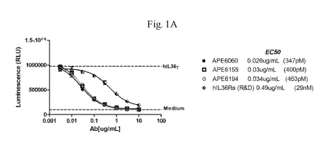
[0016] Figure 1A is a graph depicting the results of the HEK human IL-
36R/IL-8 luciferase
reporter assay described in Example 1 upon stimulation of cells with hIL-36y.
[0017] Figure 1B is a graph depicting the results of the FMK human IL-
36R/IL-8 luciferase
reporter assay described in Example 1 upon stimulation of cells with hIL-36(3.
[0018] Figure 1C is a graph depicting the results of the FMK human IL-
36R/IL-8 luciferase
reporter assay described in Example 1 upon stimulation of cells with hIL-36a.
[0019] Figure 1D is a graph depicting the results of the HEK human IL-
36R/IL-8 luciferase
reporter assay described in Example 1 upon stimulation of cells with 50 ng/mL
hIL-36a.
[0020] Figure 1E is a graph depicting the results of the FMK human IL-
36R/IL-8 luciferase
reporter assay described in Example 1 upon stimulation of cells with 20 ng/mL
hIL-36(3.
[0021] Figure 1F is a graph depicting the results of the FMK human IL-
36R/IL-8 luciferase
reporter assay described in Example 1 upon stimulation of cells with 600 ng/mL
hIL-36y.
[0022] Figure 2A is a graph depicting the results of the HEK cynomolgus IL-
36R/IL-8
luciferase reporter assay described in Example 1 upon stimulation of cells
with 2 ug/mL cynoIL-
36a.
[0023] Figure 2B is a graph depicting the results of the FMK cynomolgus IL-
36R/IL-8
luciferase reporter assay described in Example 1 upon stimulation of cells
with 10 ug/mL
cynoIL-36(3.
CA 02982555 2017-10-12
WO 2016/168542 PCT/US2016/027676
6
[0024] Figure 2C is a graph depicting the results of the FMK cynomolgus IL-
36R/IL-8
luciferase reporter assay described in Example 1 upon stimulation of cells
with 300 ng/mL
cynoIL-36y.
[0025] Figure 3A is a graph depicting experimental data which illustrate
the curve for the
antibody designated APE5281 binding to human IL-36R as determined by the
KINEXATm assay
described in Example 2.
[0026] Figure 3B is a graph depicting experimental data which illustrate
the curve for the
antibody designated APE6194 binding to human IL-36R as determined by the
BIACORETM
assay described in Example 2.
[0027] Figure 3C is a graph depicting experimental data which illustrate
the curve for the
antibody designated APE7247 binding to human IL-36R as determined by the
KINEXATm assay
described in Example 2.
[0028] Figure 4A is a graph depicting the results of the IL-8 secretion
assay in primary
human keratinocytes described in Example 3 using 10 ng/mL hIL-36a.
[0029] Figure 4B is a graph depicting the results of the IL-8 secretion
assay in primary
human keratinocytes described in Example 3 using 1 ng/mL hIL-36f3.
[0030] Figure 4C is a graph depicting the results of the IL-8 secretion
assay in primary
human keratinocytes described in Example 3 using 100 ng/mL hIL-36y.
[0031] Figure 4D is a graph depicting the results of the IL-8 secretion
assay in primary
human keratinocytes described in Example 3 using 10 ng/mL hIL-36a.
[0032] Figure 4E is a graph depicting the results of the IL-8 secretion
assay in primary
human keratinocytes described in Example 3 using 1 ng/mL hIL-36f3.
[0033] Figure 4F is a graph depicting the results of the IL-8 secretion
assay in primary
human keratinocytes described in Example 3 using 100 ng/mL hIL-36y.
[0034] Figure 4G is a graph depicting the results of the IL-8 secretion
assay in primary
human keratinocytes described in Example 3 using 10 ng/mL hIL-36a.
[0035] Figure 4H is a graph depicting the results of the IL-8 secretion
assay in primary
human keratinocytes described in Example 3 using 1 ng/mL hIL-36f3.
CA 02982555 2017-10-12
WO 2016/168542 PCT/US2016/027676
7
[0036] Figure 41 is a graph depicting the results of the IL-8 secretion
assay in primary human
keratinocytes described in Example 3 using 100 ng/mL hIL-36y.
[0037] Figure 5A is a graph depicting the results of the IL-8 secretion
assay in primary
cynomolgus keratinocytes described in Example 4 using 50 ng/mL cyno IL-36a.
[0038] Figure 5B is a graph depicting the results of the IL-8 secretion
assay in primary
cynomolgus keratinocytes described in Example 4 using 10 ng/mL cyno IL-36f3.
[0039] Figure 5C is a graph depicting the results of the IL-8 secretion
assay in primary
cynomolgus keratinocytes described in Example 4 using 250 ng/mL cyno IL-36y.
[0040] Figure 5D is a graph depicting the results of the IL-8 secretion
assay in primary
cynomolgus keratinocytes described in Example 4 using 50 ng/mL cyno IL-36a.
[0041] Figure 5E is a graph depicting the results of the IL-8 secretion
assay in primary
cynomolgus keratinocytes described in Example 4 using 10 ng/mL cyno IL-36f3.
[0042] Figure 5F is a graph depicting the results of the IL-8 secretion
assay in primary
cynomolgus keratinocytes described in Example 4 using 250 ng/mL cyno IL-36y.
[0043] Figure 6A is a graph depicting the results of the IL-8 secretion
assay in primary
human monocytes described in Example 5 using 5 ng/mL of IL-36f3.
[0044] Figure 6B is a graph depicting the results of the IL-8 secretion
assay in primary
human monocytes described in Example 5 using 500 ng/mL IL-36f3.
[0045] Figure 7A is a graph depicting the results of the IL-8 secretion
assay in primary
human peripheral blood mononuclear cells (PBMCs) described in Example 6 using
10 ng/mL of
IL-36a.
[0046] Figure 7B is a graph depicting the results of the IL-8 secretion
assay in primary
human peripheral blood mononuclear cells (PBMCs) described in Example 6 using
1 ng/mL IL-
36J3.
[0047] Figure 7C is a graph depicting the results of the IL-8 secretion
assay in primary
human peripheral blood mononuclear cells (PBMCs) described in Example 6 using
100 ng/mL
IL-36y.
CA 02982555 2017-10-12
WO 2016/168542 PCT/US2016/027676
8
[0048] Figure 8A is a graph depicting the results of the antibody/antigen
cross-competition
binding assay described in Example 8 as determined by BIACORETM assay using
APE5100 as
primary antibody.
[0049] Figure 8B is a graph depicting the results of the antibody/antigen
cross-competition
binding assay described in Example 8 as determined by BIACORETM assay using
APE6155 as
primary antibody.
[0050] Figure 9A is a graph depicting the results of the competitive
binding assay described
in Example 9 using CHO-K cells stably co-expressing human IL-36R and human IL-
1RAcP.
[0051] Figure 9B is a graph depicting the results of the competitive
binding assay described
in Example 9 using CHO-K cells stably co-expressing cynomolgus monkey IL-36R
variant 1 and
cynomolgus monkey IL-1RAcP.
[0052] Figure 10A is a graph depicting the results of the luciferase
reporter assay described
in Example 1 using FMK cynomolgus IL-36R variant 2 /IL-8 cells stimulated with
20 ng/mL
cynoIL-36y.
[0053] Figure 10B is a graph depicting the results of the luciferase
reporter assay described
in Example lusing HEK cynomolgus IL-36R variant 1/IL-8 cells stimulated with
300 ng/mL
cynoIL-36y.
[0054] Figure 10C is a graph depicting the results of the luciferase
reporter assay described
in Example 1 using FMK cynomolgus IL-36R variant 3/IL-8 cells stimulated with
100 ng/mL
cynoIL-36y.
[0055] Figure 10D is a graph depicting the results of the luciferase
reporter assay described
in Example 1 using FMK cynomolgus IL-36R variant 2/IL-8 cells stimulated with
300 ng/mL
cynoIL-36y.
[0056] Figure 10E is a graph depicting the results of the luciferase
reporter assay described
in Example 1 using FMK cynomolgus IL-36R variant 3/IL-8 cells stimulated with
300 ng/mL
cynoIL-36y.
CA 02982555 2017-10-12
WO 2016/168542 PCT/US2016/027676
9
[0057] Figure 1OF is a graph depicting the results of the luciferase
reporter assay described in
Example 1 using FMK cynomolgus IL-36R variant 4/IL-8 cells stimulated with 300
ng/mL
cynoIL-36y.
DETAILED DESCRIPTION OF THE INVENTION
[0058] The invention provides an isolated immunoglobulin heavy chain
polypeptide and/or
an isolated immunoglobulin light chain polypeptide, or a fragment (e.g.,
antigen-binding
fragment) thereof. The term "immunoglobulin" or "antibody," as used herein,
refers to a protein
that is found in blood or other bodily fluids of vertebrates, which is used by
the immune system
to identify and neutralize foreign objects, such as bacteria and viruses. The
polypeptide is
"isolated" in that it is removed from its natural environment. In a preferred
embodiment, an
immunoglobulin or antibody is a protein that comprises at least one
complementarity
determining region (CDR). The CDRs form the "hypervariable region" of an
antibody, which is
responsible for antigen binding (discussed further below). A whole
immunoglobulin typically
consists of four polypeptides: two identical copies of a heavy (H) chain
polypeptide and two
identical copies of a light (L) chain polypeptide. Each of the heavy chains
contains one N-
terminal variable (VH) region and three C-terminal constant (CH1, CH2, and
CH3) regions, and
each light chain contains one N-terminal variable (VI) region and one C-
terminal constant (CO
region. The light chains of antibodies can be assigned to one of two distinct
types, either kappa
(K) or lambda (X), based upon the amino acid sequences of their constant
domains. In a typical
immunoglobulin, each light chain is linked to a heavy chain by disulfide
bonds, and the two
heavy chains are linked to each other by disulfide bonds. The light chain
variable region is
aligned with the variable region of the heavy chain, and the light chain
constant region is aligned
with the first constant region of the heavy chain. The remaining constant
regions of the heavy
chains are aligned with each other.
[0059] The variable regions of each pair of light and heavy chains form the
antigen binding
site of an antibody. The VH and VL regions have the same general structure,
with each region
comprising four framework (FW or FR) regions. The term "framework region," as
used herein,
refers to the relatively conserved amino acid sequences within the variable
region which are
CA 02982555 2017-10-12
WO 2016/168542 PCT/US2016/027676
located between the hypervariable or complementary determining regions (CDRs).
There are
four framework regions in each variable domain, which are designated FR1, FR2,
FR3, and FR4.
The framework regions form the (3 sheets that provide the structural framework
of the variable
region (see, e.g., C.A. Janeway et al. (eds.), Immunobiology, 5th Ed., Garland
Publishing, New
York, NY (2001)).
[0060] The framework regions are connected by three complementarity
determining regions
(CDRs). As discussed above, the three CDRs, known as CDR1, CDR2, and CDR3,
form the
"hypervariable region" of an antibody, which is responsible for antigen
binding. The CDRs form
loops connecting, and in some cases comprising part of, the beta-sheet
structure formed by the
framework regions. While the constant regions of the light and heavy chains
are not directly
involved in binding of the antibody to an antigen, the constant regions can
influence the
orientation of the variable regions. The constant regions also exhibit various
effector functions,
such as participation in antibody-dependent complement-mediated lysis or
antibody-dependent
cellular toxicity via interactions with effector molecules and cells.
[0061] The isolated immunoglobulin heavy chain polypeptide and the isolated
immunoglobulin light chain polypeptide of the invention desirably bind to the
interleukin 36
receptor (IL-36R), formerly known as IL-1Rrp2. IL-36R is a receptor of the IL-
1R family, and
binds to the ligands IL-36a (formerly IL-1F6), IL-36f3 (formerly IL-1F8), and
IL-36y (formerly
IL-1F9) (see, e.g., Vigne et al., Blood, 118(22): 5813-5823 (2011)). IL-36a,
IL-3613, and IL-36y
are members of the IL-1 family of cytokines and bind to IL-36R and use IL-1
receptor accessory
protein (IL-1RAcP) as a coreceptor to stimulate intracellular signals similar
to those induced by
IL-1 (Towne et al., J. Biol. Chem., 279(14): 13677-13688 (2004)). IL-36
cytokines and IL-36R
are highly expressed by keratinocytes and other epithelial cell types, as well
as dendritic cells
and naive CD4+ T-cells (Towne et al., supra; Vigne et al., Blood, 118(22):
5813-5823 (2011);
and Vigne et al., Blood, 120(17): 3478-3487 (2012))
[0062] The inventive isolated immunoglobulin heavy chain polypeptide and
the inventive
isolated immunoglobulin light chain polypeptide can form an agent that binds
to IL-36R and
another antigen, resulting in a "dual reactive" binding agent (e.g., a dual
reactive antibody).
[0063] Certain other antibodies which bind to IL-36R, and components
thereof, are known in
the art (see, e.g., U.S. Patent Publication 2013/0236471). Anti-IL-36R
antibodies also are
CA 02982555 2017-10-12
WO 2016/168542 PCT/US2016/027676
11
commercially available from sources such as, for example, Abcam (Cambridge,
MA), and R&D
Systems, Inc. (Minneapolis, MN).
[0064] The invention provides an isolated immunoglobulin light chain
polypeptide which
comprises, consists of, or consists essentially of the amino acid sequence of
Gln Val Gln Xaal
Xaa2 Gln Ser Gly Ala Glu Val Lys Lys Pro Gly Ala Ser Val Lys Val Ser Cys Lys
Ala Ser Gly
Phe Thr Phe Thr Ser Tyr Asp Ile Asn Trp Val Arg Gln Ala Pro Gly Gln Xaa3 Leu
Glu Trp Met
Gly Trp Ile Tyr Pro Gly Asp Xaa4 Ser Thr Lys Tyr Asn Glu Lys Phe Lys Gly Arg
Val Thr Ile
Thr Xaa5 Asp Xaa6 Ser Ala Xaa7 Thr Ala Tyr Met Glu Leu Xaa8 Ser Leu Arg Ser
Glu Asp Thr
Ala Val Tyr Xaa9 Cys Thr Arg Ser Phe Tyr Thr Met Asp Tyr Trp Gly Gln Gly Thr
Thr Val Thr
Val Ser Ser (SEQ ID NO: 56), wherein (a) Xaal is leucine (Leu) or
phenylalanine (Phe), (b)
Xaa2 is valine (Val), methionine (Met), or leucine (Leu), (c) Xaa3 is arginine
(Arg) or glycine
(Gly), (d) Xaa4 is glycine (Gly), serine (Ser), or alanine (Ala), (e) Xaa5 is
arginine (Arg) or
alanine (Ala), (f) Xaa6 is threonine (Thr) or lysine (Lys), (g) Xaa7 is serine
(Ser) or asparagine
(Asn), (h) Xaa8 is serine (Ser) or alanine (Ala), and (i) Xaa9 is tyrosine
(Tyr) or phenylalanine
(Phe). In some embodiments, the isolated immunoglobulin heavy chain
polypeptide comprises,
consists of, or consists essentially of the amino acid sequence Gln Val Gln
Xaal Xaa2 Gln Ser
Gly Ala Glu Val Lys Lys Pro Gly Ala Ser Val Lys Val Ser Cys Lys Ala Ser Gly
Phe Thr Phe Thr
Ser Tyr Asp Ile Asn Trp Val Arg Gln Ala Pro Gly Gln Xaa3 Leu Glu Trp Met Gly
Trp Ile Tyr
Pro Gly Asp Xaa4 Ser Thr Lys Tyr Asn Glu Lys Phe Lys Gly Arg Val Thr Ile Thr
Xaa5 Asp
Xaa6 Ser Ala Ser Thr Ala Tyr Met Glu Leu Xaa7 Ser Leu Arg Ser Glu Asp Thr Ala
Val Tyr
Xaa8 Cys Thr Arg Ser Phe Tyr Thr Met Asp Tyr Trp Gly Gln Gly Thr Thr Val Thr
Val Ser Ser
(SEQ ID NO: 1), wherein (a) Xaal is leucine (Leu) or phenylalanine (Phe), (b)
Xaa2 is valine
(Val), methionine (Met), or leucine (Leu), (c) Xaa3 is arginine (Arg) or
glycine (Gly), (d) Xaa4
is glycine (Gly), serine (Ser), or alanine (Ala), (e) Xaa5 is arginine (Arg)
or alanine (Ala), (f)
Xaa6 is threonine (Thr) or lysine (Lys), (g) Xaa7 is serine (Ser) or alanine
(Ala), and (h) Xaa8 is
tyrosine (Tyr) or phenylalanine (Phe).
[0065] The inventive heavy chain polypeptide can comprise, consist of, or
consist essentially
of the amino acid sequence of SEQ ID NO: 56 or SEQ ID NO: 1 with any one of
the
aforementioned amino acid substitutions in any suitable combination. In one
embodiment, the
immunoglobulin heavy chain polypeptide comprises, consists of, or consists
essentially of an
CA 02982555 2017-10-12
WO 2016/168542 PCT/US2016/027676
12
amino acid sequence of any one of SEQ ID NO: 2, SEQ ID NO: 3, SEQ ID NO: 4,
SEQ ID NO:
5, SEQ ID NO: 6, SEQ ID NO: 7, SEQ ID NO: 8, SEQ ID NO: 9, SEQ ID NO: 10, SEQ
ID NO:
11, SEQ ID NO: 12, SEQ ID NO: 13, and SEQ ID NO: 14.
[0066] The invention also provides an isolated immunoglobulin heavy chain
polypeptide that
comprises, consists of, or consists essentially of the amino acid sequence Gln
Val Gln Leu Val
Gln Ser Gly Ala Glu Val Lys Lys Pro Gly Ala Ser Val Lys Val Ser Cys Lys Ala
Ser Gly Tyr Thr
Phe Thr Asn Tyr Xaal Met Xaa2 Trp Val Arg Gln Ala Pro Xaa3 Gln Gly Leu Glu Trp
Met Gly
Met Phe Xaa4 Pro Xaa5 Xaa6 Xaa7 Val Thr Arg Leu Asn Gln Lys Phe Lys Asp Arg
Val Thr
Met Thr Arg Asp Thr Ser Thr Ser Thr Val Tyr Met Glu Leu Ser Ser Leu Arg Ser
Glu Asp Thr
Ala Val Tyr Tyr Cys Ala Arg Thr Thr Ser Met Ile Ile Gly Gly Phe Ala Tyr Trp
Gly Gln Gly Thr
Leu Val Thr Val Ser Ser (SEQ ID NO: 15), wherein (a) Xaal is tryptophan (Trp)
or tyrosine
(Tyr), (b) Xaa2 is histidine (His), asparagine (Asn), or tyrosine (Tyr), (c)
Xaa3 is glycine (Gly)
or arginine (Arg), (d) Xaa4 is aspartic acid (Asp), glutamic acid (Glu), or
histidine (His), (e)
Xaa5 is serine (Ser), threonine (Thr), or tyrosine (Tyr), (f) Xaa6 is
asparagine (Asn) or glycine
(Gly), and (g) Xaa7 is serine (Ser), alanine (Ala), or aspartic acid (Asp).
[0067] The inventive heavy chain polypeptide can comprise, consist of, or
consist essentially
of the amino acid sequence of SEQ ID NO: 15 with one of the aforementioned
amino acid
substitutions in any suitable combination. In one embodiment, the
immunoglobulin heavy chain
polypeptide comprises, consists of, or consists essentially of an amino acid
sequence of any one
of SEQ ID NO: 16, SEQ ID NO: 17, SEQ ID NO: 18, SEQ ID NO: 19, SEQ ID NO: 20,
SEQ ID
NO: 21, SEQ ID NO: 22, SEQ ID NO: 23, or SEQ ID NO: 24.
[0068] The invention also provides an isolated immunoglobulin light chain
polypeptide
which comprises, consists of, or consists essentially of the amino acid
sequence of Xaal Xaa2
Gln Xaa3 Gln Glu Ser Gly Pro Gly Leu Val Lys Pro Ser Gln Thr Leu Ser Leu Thr
Cys Thr Val
Xaa4 Xaa5 Tyr Ser Ile Thr Xaa6 Asp Phe Ala Trp Asn Trp Ile Arg Gln Xaa7 Pro
Gly Xaa8 Xaa9
Leu Glu Trp Ile Gly Tyr Ile Ser Tyr Ser Gly Asp Thr Asn Tyr Asn Pro Ser Leu
Lys Ser Arg Val
Thr Ile Xaal 0 Xaal 1 Asp Thr Ser Lys Asn Gln Phe Ser Leu Lys Leu Ser Ser Val
Thr Ala Ala
Asp Thr Ala Xaal2 Tyr Xaal3 Cys Ala Ile Arg Gly Pro Tyr Ser Phe Thr Tyr Trp
Gly Gln Gly
Thr Leu Val Thr Val Ser Ser Xaal4 (SEQ ID NO: 57), wherein Xaal is glutamine
(Gin) or
aspartic acid (Asp); Xaa2 is valine (Val) or leucine (Leu); Xaa3 is leucine
(Leu) or phenylalanine
CA 02982555 2017-10-12
WO 2016/168542 PCT/US2016/027676
13
(Phe); Xaa4 is threonine (Thr) or serine (Ser); Xaa5 is glycine (Gly) or
arginine (Arg); Xaa6
serine (Ser) or alanine (Ala); Xaa7 is proline (Pro) or phenylalanine (Phe);
Xaa8 is lysine (Lys)
or asparagine (Asn); Xaa9 is glycine (Gly) or lysine (Lys); Xaal 0 is serine
(Ser) or threonine
(Thr); Xaall is valine (Val) or arginine (Arg); Xaal2 is threonine (Thr) or
valine (Val); Xaal3 is
tyrosine (Tyr) or phenylalanine (Phe); and Xaal4 is alanine (Ala) or absent.
In some
embodiments, the isolated heavy chain immunoglobulin polypeptide comprises,
consists of, or
consists essentially of the amino acid sequence Xaal Val Gln Xaa2 Gln Glu Ser
Gly Pro Gly Leu
Val Lys Pro Ser Gln Thr Leu Ser Leu Thr Cys Thr Val Xaa3 Gly Tyr Ser Ile Thr
Ser Asp Phe
Ala Trp Asn Trp Ile Arg Gln Xaa4 Pro Gly Xaa5 Xaa6 Leu Glu Trp Ile Gly Tyr Ile
Ser Tyr Ser
Gly Asp Thr Asn Tyr Asn Pro Ser Leu Lys Ser Arg Val Thr Ile Xaa7 Xaa8 Asp Thr
Ser Lys Asn
Gln Phe Ser Leu Lys Leu Ser Ser Val Thr Ala Ala Asp Thr Ala Val Tyr Xaa9 Cys
Ala Ile Arg
Gly Pro Tyr Ser Phe Thr Tyr Trp Gly Gln Gly Thr Leu Val Thr Val Ser Ser (SEQ
ID NO: 25),
wherein (a) Xaal is glutamine (Gin) or aspartic acid (Asp), (b) Xaa2 is
leucine (Leu) or
phenylalanine (Phe), (c) Xaa3 is threonine (Thr) or serine (Ser), (d) Xaa4 is
proline (Pro) or
phenylalanine (Phe), (e) Xaa5 is lysine (Lys) or asparagine (Asn), (f) Xaa6 is
glycine (Gly) or
lysine (Lys), (g) Xaa7 is serine (Ser) or threonine (Thr), (h) Xaa8 is valine
(Val) or arginine
(Arg), and (i) Xaa9 is tyrosine (Tyr) or phenylalanine (Phe).
[0069] The inventive heavy chain polypeptide can comprise, consist of, or
consist essentially
of the amino acid sequence of SEQ ID NO: 57 or SEQ ID NO: 25 with one or more
of the
aforementioned amino acid substitutions in any suitable combination.In one
embodiment, the
immunoglobulin heavy chain polypeptide comprises, consists of, or consists
essentially of an
amino acid sequence of any one of SEQ ID NO: 26, SEQ ID NO: 27, SEQ ID NO: 28,
SEQ ID
NO: 29, SEQ ID NO: 30, SEQ ID NO: 31, SEQ ID NO: 32, SEQ ID NO: 51, SEQ ID NO:
52,
SEQ ID NO: 53, or SEQ ID NO: 54.
[0070] In another embodiment, the invention provides an isolated
immunoglobulin heavy
chain polypeptide which comprises, consists of, or consists essentially of the
amino acid
sequence of SEQ ID NO: 33, SEQ ID NO: 34, or SEQ ID NO: 35
[0071] When the inventive immunoglobulin heavy chain polypeptide consists
essentially of
an amino acid sequence of any one of SEQ ID NO: 1-SEQ ID NO: 35, additional
components
can be included in the polypeptide that do not materially affect the
polypeptide, e.g., by
CA 02982555 2017-10-12
WO 2016/168542
PCT/US2016/027676
14
influencing affinity of the inventive heavy chain polypeptide to IL-36R.
Examples of such
components include, for example, protein moieties such as biotin that
facilitate purification or
isolation, passenger mutations, sequences free of problematic sites including
free cysteines,
additional glycosylation sites, and high-likelihood deamidation or
isomerization sites.
[0072] When
the inventive immunoglobulin heavy chain polypeptide consists of an amino
acid sequence of any one of SEQ ID NO: 1-SEQ ID NO: 35, the polypeptide does
not comprise
any additional components (i.e., components that are not endogenous to the
inventive
immunoglobulin heavy chain polypeptide).
[0073] The
invention provides an isolated immunoglobulin heavy chain polypeptide which
comprises an amino acid sequence that is at least 90% identical (e.g., at
least 91%, at least 92%,
at least 93%, at least 94%, at least 95%, at least 96%, at least 97%, at least
98%, at least 99%, or
100% identical) to any one of SEQ ID NO: 1-SEQ ID NO: 35. Nucleic acid or
amino acid
sequence "identity," as described herein, can be determined by comparing a
nucleic acid or
amino acid sequence of interest to a reference nucleic acid or amino acid
sequence. The percent
identity is the number of nucleotides or amino acid residues that are the same
(i.e., that are
identical) as between the sequence of interest and the reference sequence
divided by the length of
the longest sequence (i.e., the length of either the sequence of interest or
the reference sequence,
whichever is longer). A number of mathematical algorithms for obtaining the
optimal alignment
and calculating identity between two or more sequences are known and
incorporated into a
number of available software programs. Examples of such programs include
CLUSTAL-W, T-
Coffee, and ALIGN (for alignment of nucleic acid and amino acid sequences),
BLAST programs
(e.g., BLAST 2.1, BL2SEQ, and later versions thereof) and FASTA programs
(e.g., FAS TA3x,
FASTM, and SSEARCH) (for sequence alignment and sequence similarity searches).
Sequence
alignment algorithms also are disclosed in, for example, Altschul et al., J.
Molecular Biol.,
215(3): 403-410 (1990), Beigert et al., Proc. Natl. Acad. Sci. USA, 106(10):
3770-3775 (2009),
Durbin et al., eds., Biological Sequence Analysis: Pro babalistic Models of
Proteins and Nucleic
Acids, Cambridge University Press, Cambridge, UK (2009), Soding,
Bioinformatics, 21(7): 951-
960 (2005), Altschul et al., Nucleic Acids Res., 25(17): 3389-3402 (1997), and
Gusfield,
Algorithms on Strings, Trees and Sequences, Cambridge University Press,
Cambridge UK
(1997)).
CA 02982555 2017-10-12
WO 2016/168542 PCT/US2016/027676
[0074] In another embodiment, the invention provides an immunoglobulin
light chain
polypeptide that comprises, consists of, or consists essentially of the amino
acid sequence Asp
Ile Val Met Thr Gln Ser Pro Leu Ser Leu Pro Val Thr Pro Gly Glu Pro Ala Ser
Ile Ser Cys Arg
Ser Ser Lys Ser Leu Leu His Ser Asn Xaal Asn Thr Tyr Leu Tyr Trp Xaa2 Leu Gln
Lys Pro Gly
Gln Ser Pro Gln Leu Leu Ile Xaa3 Arg Met Ser Asn Leu Ala Ser Gly Val Pro Asp
Arg Phe Ser
Gly Ser Gly Ser Gly Thr Asp Phe Thr Leu Lys Ile Ser Arg Val Glu Ala Glu Asp
Val Gly Val Tyr
Tyr Cys Met Gln His Leu Glu Tyr Pro Phe Thr Phe Gly Gln Gly Thr Lys Leu Glu
Ile Lys (SEQ
ID NO: 36), wherein (a) Xaal is glycine (Gly) or alanine (Ala), (b) Xaa2 is
phenylalanine (Phe)
or tyrosine (Tyr), and (c) Xaa3 is tyrosine (Tyr) or serine (Ser).
[0075] The inventive light chain polypeptide can comprise, consist of, or
consist essentially
of the amino acid sequence of SEQ ID NO: 36 with one or more of the
aforementioned amino
acid substitutions in any suitable combination. In one embodiment, the
isolated immunoglobulin
light chain polypeptide comprises, consists of, or consists essentially of an
amino acid sequence
of any one of SEQ ID NO: 37, SEQ ID NO: 38, or SEQ ID NO: 39.
[0076] The invention also provides an immunoglobulin light chain
polypeptide that
comprises, consists of, or consists essentially of the amino acid sequence Asp
Ile Val Met Thr
Gln Thr Pro Leu Ser Leu Ser Val Thr Pro Gly Gln Pro Ala Ser Ile Ser Cys Arg
Ser Ser Lys Ser
Leu Leu His Xaal Asn Xaa2 Ile Thr Tyr Phe Tyr Trp Tyr Leu Xaa3 Lys Pro Gly Gln
Pro Pro
Gln Leu Leu Ile Tyr Gln Met Ser Asn Leu Ala Ser Gly Val Pro Asp Arg Phe Ser
Gly Ser Gly Ser
Gly Thr Asp Phe Thr Leu Lys Ile Ser Arg Val Glu Ala Glu Asp Val Gly Val Tyr
Tyr Cys Ala
Gln Asn Leu Glu Leu Pro Leu Thr Phe Gly Gly Gly Thr Lys Val Glu Ile Lys (SEQ
ID NO: 40),
(a) Xaal is serine (Ser) or arginine (Arg), (b) Xaa2 is glycine (Gly) or
alanine (Ala), and (c)
Xaa3 is glutamine (On) or histidine (His).
[0077] The inventive light chain polypeptide can comprise, consist of, or
consist essentially
of the amino acid sequence of SEQ ID NO: 40 with one or more of the
aforementioned amino
acid substitutions in any suitable combination. In one embodiment, the
isolated immunoglobulin
light chain polypeptide comprises, consists of, or consists essentially of an
amino acid sequence
of any one of SEQ ID NO: 41, SEQ ID NO: 42, SEQ ID NO: 43, or SEQ ID NO: 44.
[0078] The invention provides an isolated immunoglobulin light chain
polypeptide which
comprises, consists of, or consists essentially of the amino acid sequence of
Asp Ile Gln Met Thr
CA 02982555 2017-10-12
WO 2016/168542 PCT/US2016/027676
16
Gin Ser Pro Ser Ser Leu Ser Ala Ser Val Gly Asp Arg Val Thr Ile Thr Cys Arg
Ala Ser Gin
Xaal Ile Asn Asn Tyr Leu Asn Trp Tyr Gin Gin Lys Pro Gly Lys Ala Pro Lys Leu
Leu Ile Tyr
Tyr Thr Ser Xaa2 Leu His Ser Gly Val Pro Ser Arg Phe Ser Xaa3 Ser Gly Ser Gly
Xaa4 Asp
Xaa5 Thr Phe Thr Ile Ser Ser Leu Gin Pro Glu Asp Ile Ala Thr Tyr Tyr Cys Gin
Gin Gly His Thr
Leu Pro Trp Thr Phe Gly Gly Gly Thr Lys Val Glu Ile Lys Xaa6 Xaa7 (SEQ ID NO:
58),
wherein (a) Xaal is aspartic acid (Asp) or tryptophan (Trp), (b) Xaa2 is
arginine (Arg) or
methionine (Met), (c) Xaa3 is glycine (Gly), serine (Ser) or proline (Pro),
(d) Xaa4 is threonine
(Thr) or asparagines (Asn), (e) Xaa5 is phenylalanine (Phe) or tyrosine (Tyr),
(f) Xaa6 is
arginine (Arg) or absent, and (g) Xaa7 is threonine (Thr) or absent. In some
embodiments, the
immunoglobulin light chain polypeptide comprises, consists of, or consists
essentially of the
amino acid sequence Asp Ile Gin Met Thr Gin Ser Pro Ser Ser Leu Ser Ala Ser
Val Gly Asp Arg
Val Thr Ile Thr Cys Arg Ala Ser Gin Asp Ile Asn Asn Tyr Leu Asn Trp Tyr Gin
Gin Lys Pro
Gly Lys Ala Pro Lys Leu Leu Ile Tyr Tyr Thr Ser Arg Leu His Ser Gly Val Pro
Ser Arg Phe Ser
Xaal Ser Gly Ser Gly Thr Asp Xaa2 Thr Phe Thr Ile Ser Ser Leu Gin Pro Glu Asp
Ile Ala Thr
Tyr Tyr Cys Gin Gin Gly His Thr Leu Pro Trp Thr Phe Gly Gly Gly Thr Lys Val
Glu Ile Lys
(SEQ ID NO: 45), wherein (a) Xaal is serine (Ser) or proline (Pro), and (b)
Xaa2 is
phenylalanine (Phe) or tyrosine (Tyr).
[0079] The inventive light chain polypeptide can comprise, consist of, or
consist essentially
of the amino acid sequence of SEQ ID NO: 58 or SEQ ID NO: 45 with one or more
of the
aforementioned amino acid substitutions in any suitable combination. In one
embodiment, the
isolated immunoglobulin light chain polypeptide comprises, consists of, or
consists essentially of
an amino acid sequence of any one of SEQ ID NO: 46, SEQ ID NO: 47, or SEQ ID
NO: 55.
[0080] In another embodiment, the invention provides an isolated
immunoglobulin light
chain polypeptide which comprises, consists of, or consists essentially of the
amino acid
sequence of SEQ ID NO: 48, SEQ ID NO: 49, or SEQ ID NO: 50
[0081] When the inventive immunoglobulin light chain polypeptide consists
essentially of an
amino acid sequence of any one of SEQ ID NO: 36-SEQ ID NO: 50, additional
components can
be included in the polypeptide that do not materially affect the polypeptide,
such as those
described herein. When the inventive immunoglobulin light chain polypeptide
consists of an
amino acid sequence of any one of SEQ ID NO: 36-SEQ ID NO: 50, the polypeptide
does not
CA 02982555 2017-10-12
WO 2016/168542 PCT/US2016/027676
17
comprise any additional components (i.e., components that are not endogenous
to the inventive
immunoglobulin light chain polypeptide).
[0082] The invention provides an isolated immunoglobulin light chain
polypeptide which
comprises an amino acid sequence that is at least 90% identical (e.g., at
least 91%, at least 92%,
at least 93%, at least 94%, at least 95%, at least 96%, at least 97%, at least
98%, at least 99%, or
100% identical) to any one of SEQ ID NO: 36-SEQ ID NO: 50. Nucleic acid or
amino acid
sequence "identity" can be determined using the methods described herein.
[0083] One or more amino acids of the aforementioned immunoglobulin heavy
chain
polypeptides and/or light chain polypeptides can be replaced or substituted
with a different
amino acid. An amino acid "replacement" or "substitution" refers to the
replacement of one
amino acid at a given position or residue by another amino acid at the same
position or residue
within a polypeptide sequence.
[0084] Amino acids are broadly grouped as "aromatic" or "aliphatic." An
aromatic amino
acid includes an aromatic ring. Examples of "aromatic" amino acids include
histidine (H or
His), phenylalanine (F or Phe), tyrosine (Y or Tyr), and tryptophan (W or
Trp). Non-aromatic
amino acids are broadly grouped as "aliphatic." Examples of "aliphatic" amino
acids include
glycine (G or Gly), alanine (A or Ala), valine (V or Val), leucine (L or Leu),
isoleucine (I or Ile),
methionine (M or Met), serine (S or Ser), threonine (T or Thr), cysteine (C or
Cys), proline (P or
Pro), glutamic acid (E or Glu), aspartic acid (D or Asp), asparagine (N or
Asn), glutamine (Q or
Gln), lysine (K or Lys), and arginine (R or Arg).
[0085] Aliphatic amino acids may be sub-divided into four sub-groups. The
"large aliphatic
non-polar sub-group" consists of valine, leucine, and isoleucine. The
"aliphatic slightly-polar
sub-group" consists of methionine, serine, threonine, and cysteine. The
"aliphatic polar/charged
sub-group" consists of glutamic acid, aspartic acid, asparagine, glutamine,
lysine, and arginine.
The "small-residue sub-group" consists of glycine and alanine. The group of
charged/polar
amino acids may be sub-divided into three sub-groups: the "positively-charged
sub-group"
consisting of lysine and arginine, the "negatively-charged sub-group"
consisting of glutamic acid
and aspartic acid, and the "polar sub-group" consisting of asparagine and
glutamine.
CA 02982555 2017-10-12
WO 2016/168542 PCT/US2016/027676
18
[0086] Aromatic amino acids may be sub-divided into two sub-groups: the
"nitrogen ring
sub-group" consisting of histidine and tryptophan and the "phenyl sub-group"
consisting of
phenylalanine and tyrosine.
[0087] The amino acid replacement or substitution can be conservative, semi-
conservative,
or non-conservative. The phrase "conservative amino acid substitution" or
"conservative
mutation" refers to the replacement of one amino acid by another amino acid
with a common
property. A functional way to define common properties between individual
amino acids is to
analyze the normalized frequencies of amino acid changes between corresponding
proteins of
homologous organisms (Schulz and Schirmer, Principles of Protein Structure,
Springer-Verlag,
New York (1979)). According to such analyses, groups of amino acids may be
defined where
amino acids within a group exchange preferentially with each other, and
therefore resemble each
other most in their impact on the overall protein structure (Schulz and
Schirmer, supra).
[0088] Examples of conservative amino acid substitutions include
substitutions of amino
acids within the sub-groups described above, for example, lysine for arginine
and vice versa such
that a positive charge may be maintained, glutamic acid for aspartic acid and
vice versa such that
a negative charge may be maintained, serine for threonine such that a free -OH
can be
maintained, and glutamine for asparagine such that a free -NH2 can be
maintained.
[0089] "Semi-conservative mutations" include amino acid substitutions of
amino acids
within the same groups listed above, but not within the same sub-group. For
example, the
substitution of aspartic acid for asparagine, or asparagine for lysine,
involves amino acids within
the same group, but different sub-groups. "Non-conservative mutations" involve
amino acid
substitutions between different groups, for example, lysine for tryptophan, or
phenylalanine for
serine, etc.
[0090] In addition, one or more amino acids can be inserted into the
aforementioned
immunoglobulin heavy chain polypeptides and/or light chain polypeptides. Any
number of any
suitable amino acids can be inserted into the amino acid sequence of the
immunoglobulin heavy
chain polypeptide and/or light chain polypeptide. In this respect, at least
one amino acid (e.g., 2
or more, 5 or more, or 10 or more amino acids), but not more than 20 amino
acids (e.g., 18 or
less, 15 or less, or 12 or less amino acids), can be inserted into the amino
acid sequence of the
immunoglobulin heavy chain polypeptide and/or light chain polypeptide.
Preferably, 1-10 amino
CA 02982555 2017-10-12
WO 2016/168542 PCT/US2016/027676
19
acids (e.g., 1, 2, 3, 4, 5, 6, 7, 8, 9, or 10 amino acids) are inserted into
the amino acid sequence of
the immunoglobulin heavy chain polypeptide and/or light chain polypeptide. In
this respect, the
amino acid(s) can be inserted into any one of the aforementioned
immunoglobulin heavy chain
polypeptides and/or light chain polypeptides in any suitable location.
Preferably, the amino
acid(s) are inserted into a CDR (e.g., CDR1, CDR2, or CDR3) of the
immunoglobulin heavy
chain polypeptide and/or light chain polypeptide.
[0091] The inventive isolated immunoglobulin heavy chain polypeptide and
light chain
polypeptides are not limited to polypeptides comprising the specific amino
acid sequences
described herein. Indeed, the immunoglobulin heavy chain polypeptide or light
chain
polypeptide can be any heavy chain polypeptide or light chain polypeptide that
competes with
the inventive immunoglobulin heavy chain polypeptide or light chain
polypeptide for binding to
IL-36R. In this respect, for example, the immunoglobulin heavy chain
polypeptide or light
chain polypeptide can be any heavy chain polypeptide or light chain
polypeptide that binds to the
same epitope of IL-36R recognized by the heavy and light chain polypeptides
described herein.
Antibody competition can be assayed using routine peptide competition assays
which utilize
ELISA, Western blot, or immunohistochemistry methods (see, e.g., U.S. Patents
4,828,981 and
8,568,992; and Braitbard et al., Proteome Sci., 4: 12 (2006)).
[0092] The invention provides an IL-36R-binding agent comprising,
consisting essentially
of, or consisting of one or more of the inventive isolated amino acid
sequences described herein.
By "IL-36R-binding agent" is meant a molecule, preferably a proteinaceous
molecule, which
binds specifically to the IL-36R protein. Preferably, the IL-36R-binding agent
is an antibody or
a fragment (e.g., antigen-binding fragment) thereof. The IL-36R-binding agent
of the invention
comprises, consists essentially of, or consists of the inventive
immunoglobulin heavy chain
polypeptide and/or the inventive immunoglobulin light chain polypeptide. In
one embodiment,
the IL-36R-binding agent comprises, consists essentially of, or consists of
the inventive
immunoglobulin heavy chain polypeptide or the inventive immunoglobulin light
chain
polypeptide. In another embodiment, the IL-36R-binding agent comprises,
consists essentially
of, or consists of the inventive immunoglobulin heavy chain polypeptide and
the inventive
immunoglobulin light chain polypeptide.
CA 02982555 2017-10-12
WO 2016/168542 PCT/US2016/027676
[0093] Any amino acid residue of the inventive immunoglobulin heavy chain
polypeptide
and/or the inventive immunoglobulin light chain polypeptide can be replaced,
in any
combination, with a different amino acid residue, or can be deleted or
inserted, so long as the
biological activity of the IL-36R-binding agent is not materially diminished
(e.g., enhanced or
improved) as a result of the amino acid replacements, insertions, and/or
deletions.
[0094] The "biological activity" of an IL-36R-binding agent refers to, for
example, binding
affinity for a particular IL-36R epitope, neutralization or inhibition of IL-
36R binding to its
receptor(s), neutralization or inhibition of IL-36R activity in vivo (e.g.,
IC50), pharmacokinetics,
and cross-reactivity (e.g., with non-human homologs or orthologs of the IL-36R
protein, or with
other proteins or tissues). In certain embodiments the inventive interleukin
36 receptor (IL-
36R)-binding agent desirably exhibits one or more of the following biological
activities: (a)
inhibits the interaction between IL-36R and IL-36a, IL-36f3, and/or IL-36y,
(b) inhibits
intracellular signaling mediated by IL-36R, and/or (c) cross-reacts with and
inhibits the activity
of human and non-human primate (e.g., cynomolgus) IL-36R. Other biological
properties or
characteristics of an antigen-binding agent recognized in the art include, for
example, avidity,
selectivity, solubility, folding, immunotoxicity, expression, and formulation.
The
aforementioned properties or characteristics can be observed, measured, and/or
assessed using
standard techniques including, but not limited to, ELISA, competitive ELISA,
surface plasmon
resonance analysis (BIACORETm), or KINEXATM, in vitro or in vivo
neutralization assays,
receptor-ligand binding assays, cytokine or growth factor production and/or
secretion assays, and
signal transduction and immunohistochemistry assays.
[0095] The terms "inhibit" or "neutralize," as used herein with respect to
the activity of a IL-
36R-binding agent, refer to the ability to substantially antagonize, prohibit,
prevent, restrain,
slow, disrupt, alter, eliminate, stop, or reverse the progression or severity
of, for example, the
biological activity of IL-36R, or a disease or condition associated with IL-
36R. The IL-36R-
binding agent of the invention preferably inhibits or neutralizes the activity
of IL-36R by at least
about 20%, about 30%, about 40%, about 50%, about 60%, about 70%, about 80%,
about 90%,
about 95%, about 100%, or a range defined by any two of the foregoing values.
[0096] The IL-36R-binding agent of the invention can be a whole antibody,
as described
herein, or an antibody fragment. The terms "fragment of an antibody,"
"antibody fragment," and
CA 02982555 2017-10-12
WO 2016/168542
PCT/US2016/027676
21
"functional fragment of an antibody" are used interchangeably herein to mean
one or more
fragments of an antibody that retain the ability to specifically bind to an
antigen (see, generally,
Holliger et al., Nat. Biotech., 23(9): 1126-1129 (2005)). The IL-36R-binding
agent can contain
any IL-36R-binding antibody fragment. The antibody fragment desirably
comprises, for
example, one or more CDRs, the variable region (or portions thereof), the
constant region (or
portions thereof), or combinations thereof. Examples of antibody fragments
include, but are not
limited to, (i) a Fab fragment, which is a monovalent fragment consisting of
the VL, VH, CL, and
CHi domains, (ii) a F(ab')2 fragment, which is a bivalent fragment comprising
two Fab
fragments linked by a disulfide bridge at the hinge region, (iii) a Fv
fragment consisting of the
VL and VH domains of a single arm of an antibody, (iv) a Fab' fragment, which
results from
breaking the disulfide bridge of an F(ab')2 fragment using mild reducing
conditions, (v) a
disulfide-stabilized Fv fragment (dsFv), and (vi) a domain antibody (dAb),
which is an antibody
single variable region domain (VH or VL) polypeptide that specifically binds
antigen.
[0097] In embodiments where the IL-36R-binding agent comprises a fragment
of the
immunoglobulin heavy chain or light chain polypeptide, the fragment can be of
any size so long
as the fragment binds to, and preferably inhibits the activity of, IL-36R. In
this respect, a
fragment of the immunoglobulin heavy chain polypeptide desirably comprises
between about 5
and 18 (e.g., about 5, 6, 7, 8, 9, 10, 11, 12, 13, 14, 15, 16, 17, 18, or a
range defined by any two
of the foregoing values) amino acids. Similarly, a fragment of the
immunoglobulin light chain
polypeptide desirably comprises between about Sand 18 (e.g., about 5, 6, 7, 8,
9, 10, 11, 12, 13,
14, 15, 16, 17, 18, or a range defined by any two of the foregoing values)
amino acids.
[0098] When the IL-36R-binding agent is an antibody or antibody fragment,
the antibody or
antibody fragment desirably comprises a heavy chain constant region (Fe) of
any suitable class.
Preferably, the antibody or antibody fragment comprises a heavy chain constant
region that is
based upon wild-type IgG1 , IgG2, or IgG4 antibodies, or variants thereof. It
will be appreciated
that each antibody class, or isotype, engages a distinct set of effector
mechanisms for disposing
of or neutralizing antigen once recognized. As such, in some embodiments, when
the IL-36R-
binding agent is an antibody or antibody fragment, it can exhibit one or more
effector functions,
such as participation in antibody-dependent complement-mediated lysis or
antibody-dependent
CA 02982555 2017-10-12
WO 2016/168542 PCT/US2016/027676
22
cellular toxicity via interactions with effector molecules and cells (e.g.,
activation of the
complement system).
[0099] The IL-36R-binding agent also can be a single chain antibody
fragment. Examples of
single chain antibody fragments include, but are not limited to, (i) a single
chain Fv (scFv),
which is a monovalent molecule consisting of the two domains of the Fv
fragment (i.e., VL and
VH) joined by a synthetic linker which enables the two domains to be
synthesized as a single
polypeptide chain (see, e.g., Bird et al., Science, 242: 423-426 (1988);
Huston et al., Proc. Natl.
Acad. Sci. USA, 85: 5879-5883 (1988); and Osbourn et al., Nat. Biotechnol.,
16: 778 (1998)) and
(ii) a diabody, which is a dimer of polypeptide chains, wherein each
polypeptide chain comprises
a VH connected to a VL by a peptide linker that is too short to allow pairing
between the VH and
VL on the same polypeptide chain, thereby driving the pairing between the
complementary
domains on different VH -VL polypeptide chains to generate a dimeric molecule
having two
functional antigen binding sites. Antibody fragments are known in the art and
are described in
more detail in, e.g., U.S. Patent Application Publication 2009/0093024 Al.
[00100] The IL-36R-binding agent also can be an intrabody or fragment thereof.
An
intrabody is an antibody which is expressed and which functions
intracellularly. Intrabodies
typically lack disulfide bonds and are capable of modulating the expression or
activity of target
genes through their specific binding activity. Intrabodies include single
domain fragments such
as isolated VH and VL domains and scFvs. An intrabody can include sub-cellular
trafficking
signals attached to the N or C terminus of the intrabody to allow expression
at high
concentrations in the sub-cellular compartments where a target protein is
located. Upon
interaction with a target gene, an intrabody modulates target protein function
and/or achieves
phenotypic/functional knockout by mechanisms such as accelerating target
protein degradation
and sequestering the target protein in a non-physiological sub-cellular
compartment. Other
mechanisms of intrabody-mediated gene inactivation can depend on the epitope
to which the
intrabody is directed, such as binding to the catalytic site on a target
protein or to epitopes that
are involved in protein-protein, protein-DNA, or protein-RNA interactions.
[00101] The IL-36R-binding agent also can be an antibody conjugate. In this
respect, the IL-
36R-binding agent can be a conjugate of (1) an antibody, an alternative
scaffold, or fragments
thereof, and (2) a protein or non-protein moiety comprising the IL-36R-binding
agent. For
CA 02982555 2017-10-12
WO 2016/168542 PCT/US2016/027676
23
example, the IL-36R-binding agent can be all or part of an antibody conjugated
to a peptide, a
fluorescent molecule, or a chemotherapeutic agent.
[00102] The IL-36R-binding agent can be, or can be obtained from, a human
antibody, a non-
human antibody, or a chimeric antibody. A "chimeric" antibody is an antibody
or fragment
thereof comprising both human and non-human regions. Preferably, the IL-36R-
binding agent is
a humanized antibody. A "humanized" antibody is a monoclonal antibody
comprising a human
antibody scaffold and at least one CDR obtained or derived from a non-human
antibody. Non-
human antibodies include antibodies isolated from any non-human animal, such
as, for example,
a rodent (e.g., a mouse or rat). A humanized antibody can comprise, one, two,
or three CDRs
obtained or derived from a non-human antibody. In one embodiment of the
invention, CDRH3
of the inventive IL-36R-binding agent is obtained or derived from a mouse
monoclonal antibody,
while the remaining variable regions and constant region of the inventive IL-
36R-binding agent
are obtained or derived from a human monoclonal antibody.
[00103] A human antibody, a non-human antibody, a chimeric antibody, or a
humanized
antibody can be obtained by any means, including via in vitro sources (e.g., a
hybridoma or a cell
line producing an antibody recombinantly) and in vivo sources (e.g., rodents).
Methods for
generating antibodies are known in the art and are described in, for example,
Kohler and
Milstein, Eur. I Immunol., 5: 511-519 (1976); Harlow and Lane (eds.),
Antibodies: A
Laboratory Manual, CSH Press (1988); and Janeway et al. (eds.), Immunobiology,
5th Ed.,
Garland Publishing, New York, NY (2001)). In certain embodiments, a human
antibody or a
chimeric antibody can be generated using a transgenic animal (e.g., a mouse)
wherein one or
more endogenous immunoglobulin genes are replaced with one or more human
immunoglobulin
genes. Examples of transgenic mice wherein endogenous antibody genes are
effectively
replaced with human antibody genes include, but are not limited to, the
Medarex HUMAB-
MOUSETm, the Kirin TC MOUSETM, and the Kyowa Kirin KM-MOUSETm (see, e.g.,
Lonberg,
Nat. Biotechnol., 23(9): 1117-25 (2005), and Lonberg, Handb. Exp. Pharmacol.,
181: 69-97
(2008)). A humanized antibody can be generated using any suitable method known
in the art
(see, e.g., An, Z. (ed.), Therapeutic Monoclonal Antibodies: From Bench to
Clinic, John Wiley
& Sons, Inc., Hoboken, New Jersey (2009)), including, e.g., grafting of non-
human CDRs onto a
human antibody scaffold (see, e.g., Kashmiri et al., Methods, 36(1): 25-34
(2005); and Hou et al.,
CA 02982555 2017-10-12
WO 2016/168542 PCT/US2016/027676
24
1 Biochem., 144(1): 115-120 (2008)). In one embodiment, a humanized antibody
can be
produced using the methods described in, e.g., U.S. Patent Application
Publication
2011/0287485 Al.
[00104] In one embodiment, a CDR (e.g., CDR1, CDR2, or CDR3) or a variable
region of the
immunoglobulin heavy chain polypeptide and/or the immunoglobulin light chain
polypeptide
described herein can be transplanted (i.e., grafted) into another molecule,
such as an antibody or
non-antibody polypeptide, using either protein chemistry or recombinant DNA
technology. In
this regard, the invention provides an IL-36R-binding agent comprising at
least one CDR of an
immunoglobulin heavy chain and/or light chain polypeptide as described herein.
The IL-36R-
binding agent can comprise one, two, or three CDRs of an immunoglobulin heavy
chain and/or
light chain variable region as described herein.
[00105] In a preferred embodiment, the IL-36R-binding agent binds an epitope
of IL-36R
which blocks the binding of IL-36R to any of its ligands (e.g., IL-36a, IL-
36f3, and IL-36y) and
inhibits IL-36R-mediated signaling. The invention also provides an isolated or
purified epitope
of IL-36R which blocks the binding of IL-36R to any of its ligands in an
indirect or allosteric
manner.
[00106] The invention also provides one or more isolated or purified nucleic
acid sequences
that encode the inventive immunoglobulin heavy chain polypeptide, the
inventive
immunoglobulin light chain polypeptide, and the inventive IL-36R-binding
agent.
[00107] The term "nucleic acid sequence" is intended to encompass a polymer of
DNA or
RNA, i.e., a polynucleotide, which can be single-stranded or double-stranded
and which can
contain non-natural or altered nucleotides. The terms "nucleic acid" and
"polynucleotide" as
used herein refer to a polymeric form of nucleotides of any length, either
ribonucleotides (RNA)
or deoxyribonucleotides (DNA). These terms refer to the primary structure of
the molecule, and
thus include double- and single-stranded DNA, and double- and single-stranded
RNA. The
terms include, as equivalents, analogs of either RNA or DNA made from
nucleotide analogs and
modified polynucleotides such as, though not limited to, methylated and/or
capped
polynucleotides. Nucleic acids are typically linked via phosphate bonds to
form nucleic acid
sequences or polynucleotides, though many other linkages are known in the art
(e.g.,
phosphorothioates, boranophosphates, and the like).
CA 02982555 2017-10-12
WO 2016/168542 PCT/US2016/027676
[00108] The invention further provides a vector comprising one or more nucleic
acid
sequences encoding the inventive immunoglobulin heavy chain polypeptide, the
inventive
immunoglobulin light chain polypeptide, and/or the inventive IL-36R-binding
agent. The vector
can be, for example, a plasmid, episome, cosmid, viral vector (e.g.,
retroviral or adenoviral), or
phage. Suitable vectors and methods of vector preparation are well known in
the art (see, e.g.,
Sambrook et al., Molecular Cloning, a Laboratory Manual, 3rd edition, Cold
Spring Harbor
Press, Cold Spring Harbor, N.Y. (2001), and Ausubel et al., Current Protocols
in Molecular
Biology, Greene Publishing Associates and John Wiley & Sons, New York, N.Y.
(1994)).
[00109] In addition to the nucleic acid sequence encoding the inventive
immunoglobulin
heavy polypeptide, the inventive immunoglobulin light chain polypeptide,
and/or the inventive
IL-36R-binding agent, the vector preferably comprises expression control
sequences, such as
promoters, enhancers, polyadenylation signals, transcription terminators,
signal peptides (e.g.,
the osteonectin signal peptide), internal ribosome entry sites (TRES), and the
like, that provide
for the expression of the coding sequence in a host cell. Exemplary expression
control sequences
are known in the art and described in, for example, Goeddel, Gene Expression
Technology:
Methods in Enzymology, Vol. 185, Academic Press, San Diego, Calif. (1990).
[00110] A large number of promoters, including constitutive, inducible, and
repressible
promoters, from a variety of different sources are well known in the art.
Representative sources
of promoters include for example, virus, mammal, insect, plant, yeast, and
bacteria, and suitable
promoters from these sources are readily available, or can be made
synthetically, based on
sequences publicly available, for example, from depositories such as the ATCC
as well as other
commercial or individual sources. Promoters can be unidirectional (i.e.,
initiate transcription in
one direction) or bi-directional (i.e., initiate transcription in either a 3'
or 5' direction). Non-
limiting examples of promoters include, for example, the T7 bacterial
expression system, pBAD
(araA) bacterial expression system, the cytomegalovirus (CMV) promoter, the
5V40 promoter,
and the RSV promoter. Inducible promoters include, for example, the Tet system
(U.S. Patents
5,464,758 and 5,814,618), the Ecdysone inducible system (No et al., Proc.
Natl. Acad. Sci., 93:
3346-3351 (1996)), the T-REXTm system (Invitrogen, Carlsbad, CA), LACSWITCHTm
system
(Stratagene, San Diego, CA), and the Cre-ERT tamoxifen inducible recombinase
system (Indra
CA 02982555 2017-10-12
WO 2016/168542 PCT/US2016/027676
26
et al., Nuc. Acid. Res., 27: 4324-4327 (1999); Nuc. Acid. Res., 28: e99
(2000); U.S. Patent
7,112,715; and Kramer & Fussenegger, Methods MoL Biol., 308: 123-144 (2005)).
[00111] The term "enhancer" as used herein, refers to a DNA sequence that
increases
transcription of, for example, a nucleic acid sequence to which it is operably
linked. Enhancers
can be located many kilobases away from the coding region of the nucleic acid
sequence and can
mediate the binding of regulatory factors, patterns of DNA methylation, or
changes in DNA
structure. A large number of enhancers from a variety of different sources are
well known in the
art and are available as or within cloned polynucleotides (from, e.g.,
depositories such as the
ATCC as well as other commercial or individual sources). A number of
polynucleotides
comprising promoters (such as the commonly-used CMV promoter) also comprise
enhancer
sequences. Enhancers can be located upstream, within, or downstream of coding
sequences.
[00112] The vector also can comprise a "selectable marker gene." The term
"selectable
marker gene," as used herein, refers to a nucleic acid sequence that allow
cells expressing the
nucleic acid sequence to be specifically selected for or against, in the
presence of a
corresponding selective agent. Suitable selectable marker genes are known in
the art and
described in, e.g., International Patent Application Publications WO
1992/008796 and WO
1994/028143; Wigler et al., Proc. Natl. Acad. Sci. USA, 77: 3567-3570 (1980);
O'Hare et al.,
Proc. Natl. Acad. Sci. USA, 78: 1527-1531 (1981); Mulligan & Berg, Proc. Natl.
Acad. Sci. USA,
78: 2072-2076 (1981); Colberre-Garapin et al., I Mol. Biol., 150: 1-14 (1981);
Santerre et al.,
Gene, 30: 147-156 (1984); Kent et al., Science, 237: 901-903 (1987); Wigler et
al., Cell, 11: 223-
232 (1977); Szybalska & Szybalski, Proc. Natl. Acad. Sci. USA, 48: 2026-2034
(1962); Lowy et
al., Cell, 22: 817-823 (1980); and U.S. Patents 5,122,464 and 5,770,359.
[00113] In some embodiments, the vector is an "episomal expression vector" or
"episome,"
which is able to replicate in a host cell, and persists as an extrachromosomal
segment of DNA
within the host cell in the presence of appropriate selective pressure (see,
e.g., Conese et al.,
Gene Therapy, 11: 1735-1742 (2004)). Representative commercially available
episomal
expression vectors include, but are not limited to, episomal plasmids that
utilize Epstein Barr
Nuclear Antigen 1 (EBNA1) and the Epstein Barr Virus (EBV) origin of
replication (oriP). The
vectors pREP4, pCEP4, pREP7, and pcDNA3.1 from Invitrogen (Carlsbad, CA) and
pBK-CMV
CA 02982555 2017-10-12
WO 2016/168542 PCT/US2016/027676
27
from Stratagene (La Jolla, CA) represent non-limiting examples of an episomal
vector that uses
T-antigen and the SV40 origin of replication in lieu of EBNA1 and oriP.
[00114] Other suitable vectors include integrating expression vectors, which
may randomly
integrate into the host cell's DNA, or may include a recombination site to
enable the specific
recombination between the expression vector and the host cell's chromosome.
Such integrating
expression vectors may utilize the endogenous expression control sequences of
the host cell's
chromosomes to effect expression of the desired protein. Examples of vectors
that integrate in a
site specific manner include, for example, components of the flp-in system
from Invitrogen
(Carlsbad, CA) (e.g., pcDNATm5/FRT), or the cre-lox system, such as can be
found in the
pExchange-6 Core Vectors from Stratagene (La Jolla, CA). Examples of vectors
that randomly
integrate into host cell chromosomes include, for example, pcDNA3.1 (when
introduced in the
absence of T-antigen) from Life Technologies (Carlsbad, CA), UCOE from
Millipore (Billerica,
MA), and pCI or pFN10A (ACT) FLEXITM from Promega (Madison, WI).
[00115] Viral vectors also can be used. Representative commercially available
viral
expression vectors include, but are not limited to, the adenovirus-based
Per.C6 system available
from Crucell, Inc. (Leiden, The Netherlands), the lentiviral-based pLP1 from
Invitrogen
(Carlsbad, CA), and the retroviral vectors pFB-ERV plus pCFB-EGSH from
Stratagene (La
Jolla, CA).
[00116] Nucleic acid sequences encoding the inventive amino acid sequences can
be provided
to a cell on the same vector (i.e., in cis). A unidirectional promoter can be
used to control
expression of each nucleic acid sequence. In another embodiment, a combination
of
bidirectional and unidirectional promoters can be used to control expression
of multiple nucleic
acid sequences. Nucleic acid sequences encoding the inventive polypeptides
alternatively can be
provided to the population of cells on separate vectors (i.e., in trans). Each
of the nucleic acid
sequences in each of the separate vectors can comprise the same or different
expression control
sequences. The separate vectors can be provided to cells simultaneously or
sequentially.
[00117] The vector(s) comprising the nucleic acid(s) encoding the inventive
polypeptides can
be introduced into a host cell that is capable of expressing the polypeptides
encoded thereby,
including any suitable prokaryotic or eukaryotic cell. As such, the invention
provides an isolated
cell comprising the inventive vector. Preferred host cells are those that can
be easily and reliably
CA 02982555 2017-10-12
WO 2016/168542 PCT/US2016/027676
28
grown, have reasonably fast growth rates, have well characterized expression
systems, and can
be transformed or transfected easily and efficiently.
[00118] Examples of suitable prokaryotic cells include, but are not limited
to, cells from the
genera Bacillus (such as Bacillus subtilis and Bacillus brevis), Escherichia
(such as E. coli),
Pseudomonas, Streptomyces, Salmonella, and Erwinia. Particularly useful
prokaryotic cells
include the various strains of Escherichia coli (e.g., K12, HB101 (ATCC No.
33694), DH5a,
DH10, MC1061 (ATCC No. 53338), and CC102).
[00119] Preferably, the vector is introduced into a eukaryotic cell.
Suitable eukaryotic cells
are known in the art and include, for example, yeast cells, insect cells, and
mammalian cells.
Examples of suitable yeast cells include those from the genera Kluyveromyces,
Pichia, Rhino-
sporidium, Saccharomyces, and Schizosaccharomyces. Preferred yeast cells
include, for
example, Saccharomyces cerivisae and Pichia pastoris.
[00120] Suitable insect cells are described in, for example, Kitts et al.,
Biotechniques, 14: 810-
817 (1993); Lucklow, Cum Opin. Biotechnol., 4: 564-572 (1993); and Lucklow et
al., J. Virol.,
67: 4566-4579 (1993). Preferred insect cells include Sf-9 and HIS (Invitrogen,
Carlsbad, CA).
[00121] Preferably, mammalian cells are utilized in the invention. A number of
suitable
mammalian host cells are known in the art, and many are available from the
American Type
Culture Collection (ATCC, Manassas, VA). Examples of suitable mammalian cells
include, but
are not limited to, Chinese hamster ovary cells (CHO) (ATCC No. CCL61), CHO
DHFR-cells
(Urlaub et al., Proc. Natl. Acad. Sci. USA, 97: 4216-4220 (1980)), human
embryonic kidney
(FMK) 293 or 293T cells (ATCC No. CRL1573), and 3T3 cells (ATCC No. CCL92).
Other
suitable mammalian cell lines are the monkey COS-1 (ATCC No. CRL1650) and COS-
7 cell
lines (ATCC No. CRL1651), as well as the CV-1 cell line (ATCC No. CCL70).
Further
exemplary mammalian host cells include primate cell lines and rodent cell
lines, including
transformed cell lines. Normal diploid cells, cell strains derived from in
vitro culture of primary
tissue, as well as primary explants, are also suitable. Other suitable
mammalian cell lines
include, but are not limited to, mouse neuroblastoma N2A cells, HeLa, mouse L-
929 cells, and
BEIK or HaK hamster cell lines, all of which are available from the ATCC.
Methods for
selecting suitable mammalian host cells and methods for transformation,
culture, amplification,
screening, and purification of cells are known in the art.
CA 02982555 2017-10-12
WO 2016/168542 PCT/US2016/027676
29
[00122] In one embodiment, the mammalian cell is a human cell. For example,
the
mammalian cell can be a human lymphoid or lymphoid derived cell line, such as
a cell line of
pre-B lymphocyte origin. Examples of human lymphoid cells lines include,
without limitation,
RAMOS (CRL-1596), Daudi (CCL-213), EB-3 (CCL-85), DT40 (CRL-2111), 18-81 (Jack
et al.,
Proc. Natl. Acad. Sci. USA, 85: 1581-1585 (1988)), Raji cells (CCL-86), PER.C6
cells (Crucell
Holland By., Leiden, The Netherlands), and derivatives thereof.
[00123] A nucleic acid sequence encoding the inventive amino acid sequence may
be
introduced into a cell by "transfection," "transformation," or "transduction."
"Transfection,"
"transformation," or "transduction," as used herein, refer to the introduction
of one or more
exogenous polynucleotides into a host cell by using physical or chemical
methods. Many
transfection techniques are known in the art and include, for example, calcium
phosphate DNA
co-precipitation (see, e.g., Murray E.J. (ed.), Methods in Molecular Biology,
Vol. 7, Gene
Transfer and Expression Protocols, Humana Press (1991)); DEAE-dextran;
electroporation;
cationic liposome-mediated transfection; tungsten particle-facilitated
microparticle bombardment
(Johnston, Nature, 346: 776-777 (1990)); and strontium phosphate DNA co-
precipitation (Brash
et al., Mol. Cell Biol., 7: 2031-2034 (1987)). Phage or viral vectors can be
introduced into host
cells, after growth of infectious particles in suitable packaging cells, many
of which are
commercially available.
[00124] The invention provides a composition comprising an effective amount of
the
inventive immunoglobulin heavy chain polypeptide, the inventive immunoglobulin
light chain
polypeptide, the inventive IL-36R-binding agent, the inventive nucleic acid
sequence encoding
any of the foregoing, or the inventive vector comprising the inventive nucleic
acid sequence.
Preferably, the composition is a pharmaceutically acceptable (e.g.,
physiologically acceptable)
composition, which comprises a carrier, preferably a pharmaceutically
acceptable (e.g.,
physiologically acceptable) carrier, and the inventive amino acid sequences,
IL-36R-binding
agent, or vector. Any suitable carrier can be used within the context of the
invention, and such
carriers are well known in the art. The choice of carrier will be determined,
in part, by the
particular site to which the composition may be administered and the
particular method used to
administer the composition. The composition optionally can be sterile. The
composition can be
frozen or lyophilized for storage and reconstituted in a suitable sterile
carrier prior to use. The
CA 02982555 2017-10-12
WO 2016/168542 PCT/US2016/027676
compositions can be generated in accordance with conventional techniques
described in, e.g.,
Remington: The Science and Practice of Pharmacy, 21st Edition, Lippincott
Williams &
Wilkins, Philadelphia, PA (2001).
[00125] The invention further provides a method of treating a disorder in a
mammal that is
responsive to IL-36R inhibition or neutralization. The method comprises
administering the
aforementioned composition to a mammal having a disorder that is responsive to
IL-36R
inhibition or neutralization, whereupon the disorder is treated in the mammal.
A disorder that is
"responsive to IL-36R inhibition" or "responsive to IL-36R neutralization"
refers to any disease
or disorder in which a decrease in IL-36R levels or activity has a therapeutic
benefit in
mammals, preferably humans, or the improper expression (e.g., overexpression)
or increased
activity of IL-36R causes or contributes to the pathological effects of the
disease or disorder.
Disorders that are responsive to IL-36R inhibition include, for example,
inflammatory diseases,
autoimmune diseases, respiratory diseases, metabolic disorders, and cancer.
[00126] Inflammatory disorders include, for example, allergic inflammation of
the skin, lungs,
and gastrointestinal tract, atopic dermatitis (also known as atopic eczema),
asthma (allergic and
non-allergic), epithelial-mediated inflammation, fibrosis (e.g., idiopathic
pulmonary fibrosis,
scleroderma, kidney fibrosis, and scarring), allergic rhinitis, food allergies
(e.g., allergies to
peanuts, eggs, dairy, shellfish, tree nuts, etc.), seasonal allergies, and
other allergies.
[00127] The inventive method can be used to treat any type of autoimmune
disease (i.e., as
disease or disorder caused by immune system overactivity in which the body
attacks and
damages its own tissues), such as those described in, for example, MacKay I.R.
and Rose N.R.,
eds., The Autoimmune Diseases, Fifth Edition, Academic Press, Waltham, MA
(2014).
Examples of autoimmune diseases that can be treated by the inventive method
include, but are
not limited to, multiple sclerosis, asthma, type 1 diabetes mellitus,
rheumatoid arthritis,
scleroderma, Crohn's disease, psoriasis vulgaris (commonly referred to as
psoriasis), pustular
psoriasis, generalized pustular psoriasis (GPP), palmo-plantar pustulosis
(PPP), inflammatory
bowel disease, psoriatic arthritis, multiple sclerosis, rheumatoid arthritis,
systemic lupus
erythematosus (SLE), ulcerative colitis, and ankylosing spondylitis. In a
preferred embodiment,
the inventive method is used to treat pustular psoriasis, generalized pustular
psoriasis, palmo-
plantar pustulosis (PPP), or psoriasis vulgaris.
CA 02982555 2017-10-12
WO 2016/168542 PCT/US2016/027676
31
[00128] Pustular psoriasis is a rare form of psoriasis characterized by
white pustules
surrounded by red skin. Generalized pustular psoriasis (GPP) is a life-
threatening disease
characterized by sudden, repeated episodes of high-grade fever, generalized
rash, and
disseminated pustules, with hyperleukocytosis and elevated serum levels of C-
reactive protein,
which can be caused by a deficiency in the interleukin-36-receptor antagonist
(interleukin-36Ra)
(Marrakchi et al., N. Engl. .I. Med., 365(7):620-628 (2011)). GPP often
presents in patients with
existing or prior psoriasis vulgaris (PV); however, GPP can develop in
patients without a history
of PV (Sugiura et al., .I. Invest. Derm., /33: 2514-2521 (2013)). Palmo-
plantar pustulosis is a
chronic inflammatory skin disease characterized by sterile pustules and red,
scaly skin on the
palms and soles that considerably impairs the quality of life of affected
individuals (de Waal,
A.C. and van de Kerkhof, P.C.M., I Dermatological Treatment, 22(2): 102-105
(2011)).
[00129] Examples of respiratory diseases that can be treated by the inventive
method include,
but are not limited to, asthma, cystic fibrosis, emphysema, chronic
obstructive pulmonary disease
(COPD), and acute respiratory distress syndrome. Examples of metabolic
disorders that can be
treated by the inventive method include, but are not limited to, obesity, type
2 diabetes,
atherosclerosis, and cardiovascular disease.
[00130] The inventive method can be used to treat any type of cancer known in
the art,
including but not limited to, melanoma, renal cell carcinoma, lung cancer,
bladder cancer, breast
cancer, cervical cancer, colon cancer, gall bladder cancer, laryngeal cancer,
liver cancer, thyroid
cancer, stomach cancer, salivary gland cancer, prostate cancer, pancreatic
cancer, leukemia,
lymphoma, and Merkel cell carcinoma (see, e.g., Bhatia et al., Cum Oncol.
Rep., /3(6): 488-497
(2011)).
[00131] Administration of a composition comprising the inventive
immunoglobulin heavy
chain polypeptide, the inventive immunoglobulin light chain polypeptide, the
inventive IL-36R-
binding agent, the inventive nucleic acid sequence encoding any of the
foregoing, or the
inventive vector comprising the inventive nucleic acid sequence induces an
immune response in
a mammal. An "immune response" can entail, for example, antibody production
and/or the
activation of immune effector cells (e.g., T-cells).
[00132] As used herein, the terms "treatment," "treating," and the like refer
to obtaining a
desired pharmacologic and/or physiologic effect. Preferably, the effect is
therapeutic, i.e., the
CA 02982555 2017-10-12
WO 2016/168542 PCT/US2016/027676
32
effect partially or completely cures a disease and/or adverse symptom
attributable to the disease.
To this end, the inventive method comprises administering a "therapeutically
effective amount"
of the IL-36R-binding agent. A "therapeutically effective amount" refers to an
amount effective,
at dosages and for periods of time necessary, to achieve a desired therapeutic
result. The
therapeutically effective amount may vary according to factors such as the
disease state, age, sex,
and weight of the individual, and the ability of the IL-36R-binding agent to
elicit a desired
response in the individual. For example, a therapeutically effective amount of
an IL-36R-
binding agent of the invention is an amount which decreases IL-36R bioactivity
in a human.
[00133] Alternatively, the pharmacologic and/or physiologic effect may be
prophylactic, i.e.,
the effect completely or partially prevents a disease or symptom thereof. In
this respect, the
inventive method comprises administering a "prophylactically effective amount"
of the IL-36R-
binding agent. A "prophylactically effective amount" refers to an amount
effective, at dosages
and for periods of time necessary, to achieve a desired prophylactic result
(e.g., prevention of
disease onset).
[00134] A typical dose can be, for example, in the range of 1 pg/kg to 20
mg/kg of animal or
human body weight; however, doses below or above this exemplary range are
within the scope
of the invention. The daily parenteral dose can be about 0.00001 jig/kg to
about 20 mg/kg of
total body weight (e.g., about 0.001 lag /kg, about 0.1 lag /kg , about 1 lag
/kg, about 5 lag /kg,
about 10 jig/kg, about 100 lag /kg, about 500 jig/kg, about 1 mg/kg, about 5
mg/kg, about 10
mg/kg, or a range defined by any two of the foregoing values), preferably from
about 0.1 jig/kg
to about 10 mg/kg of total body weight (e.g., about 0.5 jig/kg, about 1
jig/kg, about 50 jig/kg,
about 150 jig/kg, about 300 jig/kg, about 750 jig/kg, about 1.5 mg/kg, about 5
mg/kg, or a range
defined by any two of the foregoing values), more preferably from about 1
jig/kg to 5 mg/kg of
total body weight (e.g., about 3 jig/kg, about 15 jig/kg, about 75 jig/kg,
about 300 jig/kg, about
900 jig/kg, about 2 mg/kg, about 4 mg/kg, or a range defined by any two of the
foregoing
values), and even more preferably from about 0.5 to 15 mg/kg body weight per
day (e.g., about 1
mg/kg, about 2.5 mg/kg, about 3 mg/kg, about 6 mg/kg, about 9 mg/kg, about 11
mg/kg, about
13 mg/kg, or a range defined by any two of the foregoing values). Therapeutic
or prophylactic
efficacy can be monitored by periodic assessment of treated patients. For
repeated
administrations over several days or longer, depending on the condition, the
treatment can be
CA 02982555 2017-10-12
WO 2016/168542 PCT/US2016/027676
33
repeated until a desired suppression of disease symptoms occurs, or
alternatively, the treatment
can be continued for the lifetime of the patient. However, other dosage
regimens may be useful
and are within the scope of the invention. The desired dosage can be delivered
by a single bolus
administration of the composition, by multiple bolus administrations of the
composition, or by
continuous infusion administration of the composition.
[00135] The composition comprising an effective amount of the inventive
immunoglobulin
heavy chain polypeptide, the inventive immunoglobulin light chain polypeptide,
the inventive
IL-36R-binding agent, the inventive nucleic acid sequence encoding any of the
foregoing, or the
inventive vector comprising the inventive nucleic acid sequence can be
administered to a
mammal using standard administration techniques, including oral, intravenous,
intraperitoneal,
subcutaneous, pulmonary, transdermal, intramuscular, intranasal, buccal,
sublingual, or
suppository administration. The composition preferably is suitable for
parenteral administration.
The term "parenteral," as used herein, includes intravenous, intramuscular,
subcutaneous, rectal,
vaginal, and intraperitoneal administration. More preferably, the composition
is administered to
a mammal using peripheral systemic delivery by intravenous, intraperitoneal,
or subcutaneous
injection.
[00136] Once administered to a mammal (e.g., a human), the biological activity
of the
inventive IL-36R-binding agent can be measured by any suitable method known in
the art. For
example, the biological activity can be assessed by determining the stability
of a particular IL-
36R-binding agent. In one embodiment of the invention, the IL-36R-binding
agent (e.g., an
antibody) has an in vivo half life between about 30 minutes and 45 days (e.g.,
about 30 minutes,
about 45 minutes, about 1 hour, about 2 hours, about 4 hours, about 6 hours,
about 10 hours,
about 12 hours, about 1 day, about 5 days, about 10 days, about 15 days, about
25 days, about 35
days, about 40 days, about 45 days, or a range defined by any two of the
foregoing values). In
another embodiment, the IL-36R-binding agent has an in vivo half life between
about 2 hours
and 20 days (e.g., about 5 hours, about 10 hours, about 15 hours, about 20
hours, about 2 days,
about 3 days, about 7 days, about 12 days, about 14 days, about 17 days, about
19 days, or a
range defined by any two of the foregoing values). In another embodiment, the
IL-36R-binding
agent has an in vivo half life between about 10 days and about 40 days (e.g.,
about 10 days, about
13 days, about 16 days, about 18 days, about 20 days, about 23 days, about 26
days, about 29
CA 02982555 2017-10-12
WO 2016/168542 PCT/US2016/027676
34
days, about 30 days, about 33 days, about 37 days, about 38 days, about 39
days, about 40 days,
or a range defined by any two of the foregoing values).
[00137] The stability of the inventive IL-36R-binding agent can be measured in
terms of the
transition mid-point value (Tõ,), which is the temperature where 50% of the
amino acid sequence
is in its native confirmation, and the other 50% is denatured. In general, the
higher the Tõõ the
more stable the protein. In one embodiment of the invention, the inventive IL-
36R binding agent
comprises a transition mid-point value (TO in vitro of about 60-100 C. For
example, the
inventive IL-36R binding agent can comprise a Tn, in vitro of about 65-80 C
(e.g., 66 C, 68 C,
70 C, 71 C, 75 C, or 79 C), about 80-90 C (e.g., about 81 C, 85 C, or
89 C), or about 90-
100 C (e.g., about 91 C, about 95 C, or about 99 C).
[00138] The stability of the inventive IL-36R binding agent can be measured
using any other
suitable assay known in the art, such as, for example, measuring serum half-
life, differential
scanning calorimetry (DSC), thermal shift assays, and pulse-chase assays.
Other methods of
measuring protein stability in vivo and in vitro that can be used in the
context of the invention are
described in, for example, Protein Stability and Folding, B.A. Shirley (ed.),
Human Press,
Totowa, New Jersey (1995); Protein Structure, Stability, and Interactions
(IViethods in Molecular
Biology), Shiver J.W. (ed.), Humana Press, New York, NY (2010); and Ignatova,
Microb. Cell
Fact., 4: 23 (2005).
[00139] The biological activity of a particular IL-36R-binding agent also can
be assessed by
determining its binding affinity to IL-36R or an epitope thereof. The term
"affinity" refers to the
equilibrium constant for the reversible binding of two agents and is expressed
as the dissociation
constant (KD). Affinity of a binding agent to a ligand, such as affinity of an
antibody for an
epitope, can be, for example, from about 1 picomolar (pM) to about 100
micromolar (1.1M) (e.g.,
from about 1 picomolar (pM) to about 1 nanomolar (nM), from about 1 nM to
about 1
micromolar ( M), or from about 1 [IM to about 100 [IM). In one embodiment, the
IL-36R-
binding agent can bind to an IL-36R protein with a KD less than or equal to 1
nanomolar (e.g.,
0.9 nM, 0.8 nM, 0.7 nM, 0.6 nM, 0.5 nM, 0.4 nM, 0.3 nM, 0.2 nM, 0.1 nM, 0.05
nM, 0.025 nM,
0.01 nM, 0.001 nM, or a range defined by any two of the foregoing values). In
another
embodiment, the IL-36R-binding agent can bind to IL-36R with a KD less than or
equal to 200
pM (e.g., 190 pM, 175 pM, 150 pM, 125 pM, 110 pM, 100 pM, 90 pM, 80 pM, 75 pM,
60 pM,
CA 02982555 2017-10-12
WO 2016/168542 PCT/US2016/027676
50 pM, 40 pM, 30 pM, 25 pM, 20 pM, 15 pM, 10 pM, 5 pM, 1 pM, or a range
defined by any
two of the foregoing values). Immunoglobulin affinity for an antigen or
epitope of interest can
be measured using any art-recognized assay. Such methods include, for example,
fluorescence
activated cell sorting (FACS), separable beads (e.g., magnetic beads), surface
plasmon resonance
(SPR), solution phase competition (KINEXATm), antigen panning, competitive
binding assays,
and/or ELISA (see, e.g., Janeway et al. (eds.), Immunobiology, 5th ed.,
Garland Publishing, New
York, NY, 2001).
[00140] The IL-36R-binding agent of the invention may be administered alone or
in
combination with other drugs. For example, the IL-36R-binding agent can be
administered in
combination with other agents for the treatment or prevention of the diseases
disclosed herein,
such as an anti-inflammatory agent including, for example, corticosteroids
(e.g., prednisone and
fluticasone), non-steroidal anti-inflammatory drugs (NSAIDs) (e.g., aspirin,
ibuprofen, and
naproxen), biologics (e.g., infliximab (REMICADETm), adalimumab (HUMIRATm), or
etanercept (ENBRELTm)), methotrexate (MTX), an oral retinoid (e.g. acitretin
(SORIATANETm)), and topical steroids.
[0100] In addition to therapeutic uses, the IL-36R-binding agent described
herein can be
used in diagnostic or research applications. In this respect, the IL-36R-
binding agent can be used
in a method to diagnose a disorder or disease in which the improper expression
(e.g.,
overexpression) or increased activity of IL-36R causes or contributes to the
pathological effects
of the disease or disorder. In a similar manner, the IL-36R-binding agent can
be used in an assay
to monitor IL-36R protein levels in a subject being tested for a disease or
disorder that is
responsive to IL-36R inhibition. Research applications include, for example,
methods that
utilize the IL-36R-binding agent and a label to detect an IL-36R protein in a
sample, e.g., in a
human body fluid or in a cell or tissue extract. The IL-36R-binding agent can
be used with or
without modification, such as covalent or non-covalent labeling with a
detectable moiety. For
example, the detectable moiety can be a radioisotope (e.g., 3H, 14C, 32-,
r 35, or 1251), a fluorescent
or chemiluminescent compound (e.g., fluorescein isothiocyanate, rhodamine, or
luciferin), an
enzyme (e.g., alkaline phosphatase, beta-galactosidase, or horseradish
peroxidase), or prosthetic
groups. Any method known in the art for separately conjugating an antigen-
binding agent (e.g.,
an antibody) to a detectable moiety may be employed in the context of the
invention (see, e.g.,
CA 02982555 2017-10-12
WO 2016/168542 PCT/US2016/027676
36
Hunter etal., Nature, 194: 495-496 (1962); David etal., Biochemistry, 13: 1014-
1021 (1974);
Pain etal., I Immunol. Meth., 40: 219-230 (1981); and Nygren, I Histochem. and
Cytochem.,
30: 407-412 (1982)).
[0101] IL-36R protein levels can be measured using the inventive IL-36R-
binding agent by
any suitable method known in the art. Such methods include, for example,
radioimmunoassay
(RIA), and FACS. Normal or standard expression values of IL-36R can be
established using any
suitable technique, e.g., by combining a sample comprising, or suspected of
comprising, IL-36R
with an IL-36R-specific antibody under conditions suitable to form an antigen-
antibody
complex. The antibody is directly or indirectly labeled with a detectable
substance to facilitate
detection of the bound or unbound antibody. Suitable detectable substances
include various
enzymes, prosthetic groups, fluorescent materials, luminescent materials, and
radioactive
materials (see, e.g., Zola, Monoclonal Antibodies: A Manual of Techniques, CRC
Press, Inc.
(1987)). The amount of IL-36R polypeptide expressed in a sample is then
compared with a
standard value.
[0102] The IL-36R-binding agent can be provided in a kit, i.e., a packaged
combination of
reagents in predetermined amounts with instructions for performing a
diagnostic assay. If the
IL-36R-binding agent is labeled with an enzyme, the kit desirably includes
substrates and
cofactors required by the enzyme (e.g., a substrate precursor which provides a
detectable
chromophore or fluorophore). In addition, other additives may be included in
the kit, such as
stabilizers, buffers (e.g., a blocking buffer or lysis buffer), and the like.
The relative amounts of
the various reagents can be varied to provide for concentrations in solution
of the reagents which
substantially optimize the sensitivity of the assay. The reagents may be
provided as dry powders
(typically lyophilized), including excipients which on dissolution will
provide a reagent solution
having the appropriate concentration.
[0103] The following examples further illustrate the invention but, of
course, should not be
construed as in any way limiting its scope.
CA 02982555 2017-10-12
WO 2016/168542 PCT/US2016/027676
37
EXAMPLE 1
[0104] This example demonstrates that the inventive immunoglobulin heavy
chain (HC) and
light chain (LC) polypeptides can form antibodies that bind to and block the
signaling of human
IL-36R in vitro.
[0105] HEK293T/17 cells (ATCC CRL-11268) were stably transfected with a
plasmid
construct encoding either human IL-36R (hIL-36R) or cynomolgus IL-36R (cynoIL-
36R)
together with an IL-8 promoter (Promega Corp., Madison, WI), and a single cell
clone was
chosen for all subsequent assays.
[0106] HEK293 cells were plated at 3x10⁶ cells/flask onto a T75 culture
flask in 10 mL of
DMEM+10% FBS and incubated overnight at 37 C. The next morning, 24 IA
FUGENETM HJJ
(Promega Corporation, Madison, WI) was added to 500 IA OPTI-MEMTm medium (Life
Technologies, Carlsbad, CA) and incubated at room temperature for five
minutes. DNA
encoding IL-36R (2 IA) and DNA encoding the IL8 promoter (2 IA) were added to
the mixture
and allowed to incubate for an additional 25 minutes at room temperature.
Cynomolgus IL-36R
allelic variation was examined by Sanger sequencing, and four distinct allelic
variants were
identified within cynomolgus monkey populations. EIEK cell lines expressing
each cynomolgus
IL-36R allelic variants were generated separately. For both human and
cynomolgus monkey IL-
8 reporter cell lines, the endogenously expressed I-IEK human IL1RAcP was
utilized. This
DNA/ FUGENETM mixture was gently added to the cells drop-wise for transfection
and
incubated overnight at 37 C. 24 hours post-transfection, cells were split and
placed in
hygromycin and puromycin containing DMEM + 10% FBS for four weeks for
selection. After 4
weeks, stabilized cells were plated at 1 cell/well on a 96-well clear bottom
plates (5 plates/cell
line). Single cell clones were screened for surface expression of IL-36R and
expanded, with low
passage number (i.e., 1-3) cells used for the assay described below.
[0107] HEK293-human IL36R/IL8 or HEK293-cynoIL36R/IL8 variant stable cell
lines were
harvested with accutase and seeded with 0.06x10⁶ cells/well in 100 IA DMEM+10%
FBS on a
96-well clear-bottomed plate overnight at 37 C, 5% CO2. The next morning,
plates were
inverted into the sink to remove media and gently tapped on a paper towel to
dry. Diluted
antibodies comprising various combinations of the inventive HC and LC
polypeptides (see Table
1), IL-36Ra (R&D Systems, Minneapolis, MN), and a negative isotype control
antibody were
CA 02982555 2017-10-12
WO 2016/168542 PCT/US2016/027676
38
prepared in DMEM+10% FBS (Life Technologies, Carlsbad, CA) to the desired
concentrations
by two-fold dilution series, immediately added to the wells (50 [11/well), and
allowed to incubate
for 20 minutes at 37 C, 5% CO2.
Table 1
Antibody Designation HC SEQ ID NO: LC SEQ ID NO:
APE3798 33 48
APE4086 3 38
APE5125/APE5100 4 39
APE5216 5 39
APE5281 6 39
APE5214/APE4881 7 39
APE5280 8 39
APE5257 9 39
APE5258/APE5076 10 39
APE5212 11 39
APE5213/5066 12 39
APE5211 13 39
APE5217/APE5060 14 39
APE3849 34 49
APE3850 16 41
APE5600 18 42
APE5598 19 42
APE5627 20 42
APE6064 21 43
APE6060 22 43
APE6157 23 43
APE6155/APE6917 22 44
APE6194 24 44
APE3847 35 50
APE5713 27 47
APE6083 32 47
APE6903/APE7247 52 55
APE6904 53 55
APE6907 54 55
[0108] Cells were subsequently stimulated with 50 n1 of IL36a, IL36f3, or
IL36y ligands
(R&D Systems, Minneapolis, MN) and allowed to incubate for an additional 24
hours at 37 C,
5% CO2. EC50s of each of the individual cytokines were determined empirically
prior to the
assay. Luciferase activity was determined by using STEADY-GLOTm Luciferase
Assay System
(Promega, Cat# E2520, Madison, WI). 100 IA of 1:1 mix of luciferase assay
substrate: buffer was
CA 02982555 2017-10-12
WO 2016/168542 PCT/US2016/027676
39
added to each well, incubated for five minutes at room temperature, and
transferred (150u1) onto
96-well black walled, clear bottom plates. Plates were read on ENVISIONTM
Plate Reader
(PerkinElmer, Waltham, WA) to determine luminescence (60-sec delay). Data was
analyzed
using GraphPad PRISMTm Software 5 (GraphPad, San Diego, CA).
[0109] The results of the IL-8 luciferase reporter assay against human and
cyno IL-36R are
shown in Figures 1A-1F (human IL-36R), Figures 2A-2C (cyno IL-36R), Figures
10A-10C
(cyno IL-36R), and Figures 10D-10F (human IL-36R). The measured potencies
(IC50) of each of
the tested antibodies are set forth in Tables 2 and 3.1 and 3.2.
Table 2 - FIEK human IL-36R IL-8 luciferase reporter assay
EC50
EC50 (nM) (nM) EC50 (nM)
HC SEQ LC SEQ
Antibody 5Ong/m1 20Ong/m1
ID NO: ID NO: 2Ong/m1
hIL-36a hIL-367
hIL-360
Chimeric 1D9 33 48 0.267 0.093
(APE3798)
Humanized 6 39 0.17 0.12
(HzD) 1D9
(APE5281)
Chimeric 5D3 34 49 1.3 3.1
(APE3849)
Hzd 5D3 22 43 0.23 0.24 0.35
(APE6060)
Hzd 5D3
22 44 0.23 0.22 0.40
(APE6155)
Hzd 5D3
24 44 0.17 0.30 0.45
(APE6194)
Chimeric 18D4
35 50 4.2 3.6
(APE3847)
Hzd 18D4
27 47 11
(APE5713)
Hzd 18D4
52 55 .066 0.114 0.104
(APE7247)
Table 3.1 - FIEK cyno IL-36R IL-8 luciferase reporter assay
EC50 EC50
EC50 (nM)
HC SEQ ID LC SEQ ID (nM) (nM)
Antibody 300 ng/ml
NO: NO: 2 jig/m1 10 jig/m1
cIL-367
cIL-36a cIL-3613
Hzd 5D3 22 43 0.067 0.17 0.29
CA 02982555 2017-10-12
WO 2016/168542 PCT/US2016/027676
(APE6060)
Hzd 5D3
22 44 0.08 0.13 0.31
(APE6155)
Hzd 5D3
24 44 0.073 0.19 0.31
(APE6194)
Chimeric 18D4
35 50 0.51
(APE3847)
Hzd 18D4
52 55 0.24
(APE7247)
Table 3.2
LC EC50 (nM) EC50 (nM) EC50 (nM)
HC SE' 300 ng/ml 300 ng/ml 300 ng/ml
Antibody SEQ ID IL-367 IL-367 IL-367
ID NO: NO: (Cyno IL-36R (Cyno IL-36R (Cyno IL-36R
variant 1) variant 2) variant 3)
Hzd 18D4
(APE7247) 52 55 0.079 0.065 0.42
Hzd 5D3
(APE6155) 22 44 0.042 0.043 0.21
[0110] The results from this example demonstrate that the inventive
immunoglobulin heavy
chain (HC) and light chain (LC) polypeptides can form antibodies that bind to
and inhibit
signaling of human IL-36R in vitro.
EXAMPLE 2
[0111] This example demonstrates that the inventive immunoglobulin heavy
chain (HC) and
light chain (LC) polypeptides can form antibodies that bind to human IL-36R in
vitro.
[0112] DNA samples encoding various immunoglobulin heavy chain (HC) and
light chain
(LC) polypeptides as described herein were prepared by combining the
following: maxi-prepped
DNA (containing 6 ug HC plasmid and 6 ug LC plasmid), 1 ml OPTIMEMTm (Life
Technologies, Carlsbad, CA), and 72 ul FUGENETM HD Transfection Reagent
(Promega,
Fitchburg, WI). All reagents were pre-warmed. Following thorough mixing and
incubation for
25 minutes at room temperature, 1 ml of reagent/DNA mix was added to
8x10⁶HEK293-c18
cells (ATCC CRL-10852) in each T225 culture flask. 18 hours prior to
transfection, the cells
were plated in T225 culture flasks with 20 ml of DMEM (Life Technologies,
Carlsbad, CA) with
CA 02982555 2017-10-12
WO 2016/168542 PCT/US2016/027676
41
10% FBS (Life Technologies, Carlsbad, CA) per flask and incubated at 37 C in
5% CO2
overnight. Following transfection, cells were returned to 37 C in 5% CO2. The
following day,
the medium in each flask was exchanged with 25 ml 293 Freestyle medium (Life
Technologies,
Carlsbad, CA), and cells were moved to an incubator at 8% CO2. Antibody
production was
carried out for 7-12 days. Supernatants were collected from each flask, spun
down at 3000 rpm
for 10 minutes, and sterile-filtered into fresh tubes.
[0113] For antibody purification, approximately 20-30 ml of cell culture
supernatants
containing the antibodies of interest were passed through a gravity column
packed with 1-2 ml
MAB SELECT SURETm LX resin (GE Healthcare, Waukesha, WI) pre-equilibrated with
PBS
buffer (11.9 mM phosphate, 137 mM NaC1, 2.7 mM KC1, pH 7.4) (Fisher
Bioreagents, Waltham,
MA). The column was washed with five column volumes of PBS buffer. Bound
antibodies
were eluted from the resin with 5-10 column volumes of 0.1 M glycine pH 3Ø
The eluate
containing the antibodies was concentrated down to an antibody concentration
of approximately
0.1-2 mg/mL in Amicon Ultra 10K concentrators (Millipore, Billerica, MA), and
buffer was
exchanged three times against PBS buffer. Antibody concentration was
determined on a
Nanodrop 2000c spectrophotometer (Thermo Fisher Scientific, Waltham, MA), and
purity was
assessed by SDS-PAGE analysis.
[0114] The binding affinities of various purified antibodies comprising
immunoglobulin
heavy chain (HC) and light chain (LC) polypeptides described herein were
evaluated using
BIACORETM T200 (Sapidyne Instruments, Boise, Idaho) assays. BIACORETM T200
evaluation
software (GE Healthcare, Buckinghamshire, United Kingdom) is used to determine
antibody-
antigen binding kinetics and affinity. The extracellular domain of human IL-
36Rwas
immobilized at approximately 100 RU onto a CMS sensor chip (GE Healthcare,
Waukesha, WI)
using amine coupling chemistry. EIBS-EP+ buffer (0.01 M HEPES, 0.15 M NaC1, 3
mM
EDTA, 0.05% Polysorbate, pH 7.6) (Teknova, Hollister, CA) was used to
reconstitute each
antibody at various concentrations Each antibody concentration was then
injected for two to
three minutes over immobilized antigen at a flow rate of 30 [IL/min, and
allowed to dissociate
for 15 minutes. The surface was regenerated with 60 [IL of 3 M MgC12 after
each cycle.
Association and dissociation kinetic constants (kon and koff) were fit
globally using a 1:1
CA 02982555 2017-10-12
WO 2016/168542 PCT/US2016/027676
42
binding model with mass transport with the BIACORETM T200 evaluation software
in order to
report on- and off-rates (ka and kd, respectively), as well as affinities
(KD).
[0115] The binding affinities of various purified antibodies comprising
immunoglobulin
heavy chain (HC) and light chain (LC) polypeptides described herein also were
evaluated using a
KINEXAO 3000 assay(Sapidyne Instruments, Boise, Idaho) assays. KINEXAO
technology
measures the amount of unbound/free antibody molecule in solution phase after
incubation with
varying concentrations of antigen. Measuring binding events in the solution
phase with micro
beads for maximized surface area avoids mass transport limitations and
mobility effects inherent
to methods that measure binding to a solid phase. For each experiment, 50 ng
of soluble human
or cyno IL-36R extracellular domain was amine-coupled to 50 mg of UltraLink
Biosupport
beads (Thermo Fisher Scientific, Waltham, MA). A constant concentration of
antibody
(sufficient to produce 0.8 V-1.2 V of signal) was incubated for a sufficient
period of time to
approach or to reach equilibrium (time of incubation varies for each antibody
and is dependent
on affinity) with titrated antigen in sample buffer (lx PBS, pH 7.4, 0.02%
NaN3, 0.1% BSA).
Antibody-antigen solution was then flowed over antigen-coupled beads at a rate
of 0.25 mL/min.
Free antibody captured by beads was detected using ALEXA FLUORTM 647-
conjugated
AffiniPure Donkey Anti-Human IgG (H+L) (Jackson ImmunoResearch, West Grove,
PA) (500
ng/ml). The KD and/or ABC (active binding concentration) of antibody was
obtained from non-
linear regression analysis using a one-site homogeneous binding model in the
KINEXATM Pro
Software.
[0116] The resulting KD values of the BIACORETM T200 and KINEXAO 3000
assays assay
are set forth in Table 4 and Figure 3A (KinExA data for Humanized 1D9), Figure
3B (Biacore
for 5D3 APE6194) and Figure 3C (KinExA data for Humanized 18D4).
Table 4
BIACORETM BIACORETM KINEXATM KD
Description Antibody KD human KD cyno IL- human IL-36R
IL-36R 36R
1D9 Humanized APE5281 77 pM 126 pM 8 pM
5D3 Chimeric APE3850 35 pM
5D3 Humanized APE6060 50 pM
5D3 Humanized APE6155 71 pM 169 pM
5D3 Humanized APE6194 22 pM
CA 02982555 2017-10-12
WO 2016/168542 PCT/US2016/027676
43
18D4 Humanized APE7247 <20 pM <20 pM 100 pM
18D4 Humanized APE6904 27 pM
[0117] These data demonstrate that antibodies comprising different
combinations of the
inventive immunoglobulin HC and LC polypeptides described herein can bind
human IL-36R
with high affinities.
EXAMPLE 3
[0118] This example demonstrates that the inventive immunoglobulin heavy
chain (HC) and
light chain (LC) polypeptides can form antibodies that bind to human IL-36R in
vitro and inhibit
cell signaling and cytokine (e.g., IL-8) release by human primary keratinocyte
cells
endogenously expressing IL-36R.
[0119] Antibodies used in this assay were produced and purified as
described above. Normal
human epidermal kerotinocytes (NHEK) were purchased from Lonza Clonetics (cat#
00192627).
Cells were cultured and expanded using recommended culture medium (Lonza KBM
Gold
medium, cat# 00192151 with Lonza KGM Gold SingleQuot supplements, cat#
0092152) in a 5%
CO2 37 C incubator. Cells were frozen in liquid nitrogen at passage 2 in
multiple single use
aliquots.
[0120] Passage 2 cells were thawed and diluted to a density of 100,000
cells per ml in above
recommended culture medium described above and 100 p1 cells per well were
plated in standard
flat-bottom 96-well tissue culture plates for a final cell density of 10,000
cells per well. Outside
wells are filled with 200p1 phosphate buffered saline per well to avoid edge
effects. Cells were
cultured overnight in a 5% CO2 37 C incubator to allow for adherence.
[0121] The following day antibodies were added at concentrations from 10
pg/m1 or 1 pg/m1
down to 0 by half-log dilutions in culture medium. After 30 minutes,
recombinant human IL-36
ligands were added at approximately EC50 concentrations (previously determined
empirically for
each ligand) in culture medium. Antibody and ligand concentrations were made
at 4x of desired
final concentrations and 500 per well were added for a final total volume in
each well of 200 pl.
Supernatants were removed approximately 48 hours later following a three-
minute centrifugation
of the plates, transferred to clean plates, and either tested immediately or
stored at -80 C until
further analysis.
CA 02982555 2017-10-12
WO 2016/168542
PCT/US2016/027676
44
[0122] Human IL-
8 levels in the cell supernatants were assessed by ELISA using R&D
Systems DUO-SET TM ELISA kit (cat# DY208) following a standard protocol
provided by the
manufacturer. Data were graphed and ICso values were calculated using GraphPad
PRISMTm
software.
[0123] The results of this assay are shown in Table 5 and Figures 4A-4I.
Table 5
HC SEQ LC SEQ EC50 (nM) EC50 (nM) EC50
(nM)
Antibody ID NO: ID NO: 10 ng/ml 1 ng/ml human 100 ng/ml
human IL-36a IL-36I3
human IL-367
Humanized
(HzD) 1D9 6 39 0.047 0.053 0.04
(APE5281)
Hzd 5D3
22 43 0.08 0.217 0.08
(APE6060)
Hzd 5D3
22 44 0.125 0.227 0.093
(APE6155)
Hzd 5D3
24 44 0.105 0.164 0.083
(APE6194)
Hzd 18D4
(APE7247) 52 55 0.142 0.336 0.074
[0124] The results of this example demonstrate that antibodies composed of
combinations of
HCs and LCs described herein inhibit inflammatory cytokine release (IL-8) from
human primary
keratinocytes expressing IL-36R and stimulated with cytokines IL-36a, IL-36f3
and IL-36y in a
dose-dependent manner.
EXAMPLE 4
[0125] This example demonstrates that the inventive immunoglobulin heavy
chain (HC) and
light chain (LC) polypeptides can form antibodies that bind to cynomolgus IL-
36R (cyno IL-
36R) in vitro and inhibit IL-36-dependent cell signaling and cytokine (e.g.,
IL-8) release by
primary keratinocyte cells endogenously expressing IL-36R.
[0126] Antibodies used in this assay were produced and purified as
described in Example 3.
Normal cynomolgus monkey epidermal kerotinocytes were purchased from
CellBiologics
(Chicago, IL; cat# MK-6066K). Cells were cultured and expanded using
recommended culture
medium (CellBiologics epithelial medium, cat# M6621 with CellBiologics
epithelial cell
CA 02982555 2017-10-12
WO 2016/168542
PCT/US2016/027676
medium supplements, cat# M6621-kit) in a 5% CO2 37 C incubator. Cells were
frozen in liquid
nitrogen at passage 2 in multiple single use aliquots.
[0127] Passage 2 cells were thawed and diluted to a density of 100,000
cells per ml in culture
medium described above, and 100 p1 cells per well were plated in standard flat-
bottom 96-well
tissue culture plates for final a cell density of 10,000 cells per well.
Outside wells were filled
with 200 p1 PBS per well to avoid edge effects. Cells were cultured overnight
in a 5% CO2 37
C incubator to allow for adherence.
[0128] The following day antibodies were added at concentrations from 10
ug/m1 or 1 ug/m1
down to 0 by half-log dilutions in culture medium. After 30 minutes,
recombinant cynomolgus
IL-36 ligands were added at approximately EC50 concentrations (previously
determined
empirically for each ligand) in culture medium. Antibody and ligand
concentrations were made
at 4x of desired final concentrations and 500 per well were added for a final
total volume in
each well of 200 pl. Supernatants were removed approximately 48 hours later
following a three-
minute centrifugation of the plates, transferred to clean plates, and either
tested immediately or
stored at -80 C until further analysis.
[0129] Cynomolgus IL-8 levels in the cell supernatants were assessed by
ELISA using a
eBioscience (San Diego, CA) monkey IL-8 platinum ELISA kit (cat# BM5640/3)
following a
standard protocol provided by the manufacturer. Data were graphed and IC50
values were
calculated using GraphPad PRISMTm software.
[0130] The results of this assay are shown in Table 6 and Figures 5A-5F.
Table 6
HC SEQ ID LC SEQ ID EC50 (nM) EC50 (nM) EC50 (nM)
Antibody NO: NO: 50 ng/ml 10 ng/ml 250
ng/ml
cyno IL-36a cyno IL-36I3 cyno IL-367
Hzd 5D3
22 43 1.4 1.4 1.0
(APE6060)
Hzd 5D3
22 44 1.1 1.4 1.1
(APE6155)
Hzd 5D3
24 44 1.2 2.0 1.2
(APE6194)
Hzd 18D4
52 55 2.3 1.8 5.6
(APE7247)
CA 02982555 2017-10-12
WO 2016/168542 PCT/US2016/027676
46
[0131] The results of this example demonstrate that antibodies composed of
combinations of
HCs and LCs described herein inhibit inflammatory cytokine release (IL-8) from
cynomolgus
primary keratinocytes expressing IL-36R and stimulated with cytokines IL-36a,
IL-36f3 and IL-
36y in a dose-dependent manner.
EXAMPLE 5
[0132] This example demonstrates the ability of antibodies composed of HCs
and LCs
described herein to block, in a dose-dependent manner, the human IL-36-
mediated release of IL-
8 from human monocytes expressing the IL-36R.
[0133] A Leukocyte Reduction System unit processed from a donor whole blood
unit was
obtained from the San Diego Blood Bank. Peripheral blood mononuclear cells
(PBMCs) were
prepped by standard methods using Ficoll density centrifugation separation
(Sigma
HISTOPAQUETm cat# 10771). Monocytes were isolated from PBMCs with human
monocyte
isolation kit II (Miltenyi Biotec, San Diego, CA; cat# 130-091-153).
[0134] Monocytes were diluted to a density of 500,000 cells/ml in RPMI 1640
medium
containing 10% fetal bovine serum and penicillin/streptomycin, and 100[11
cells per well were
plated in standard flat-bottom 96-well tissue culture plates for a final cell
density of 50,000 per
well. Outside wells were filled with 200 pi PBS per well to avoid edge
effects. Plated cells were
incubated for 2-3 hours in a 5% CO2 37 C incubator to allow for recovery.
[0135] After approximately 2-3 hours of culture, antibodies were added at
concentrations
from 10 pg/ml or 1 pg/ml down to 0 by half-log dilutions in culture medium.
After 30 minutes,
recombinant human IL-36 ligands were added at approximately EC50
concentrations (previously
determined empirically for each ligand) in culture medium. Antibody and ligand
concentrations
were made at 4x of desired final concentrations and 500 per well were added
for a final total
volume in each well of 200 pl. Supernatants were removed approximately 48
hours later
following a three-minute centrifugation of the plates, transferred to clean
plates, and either tested
immediately or stored at -80 C until further analysis.
[0136] Human IL-8 levels in the cell supernatants were assessed by ELISA
using R&D
Systems DUO-SET TM ELISA kit (cat# DY208) following a standard protocol
provided by the
CA 02982555 2017-10-12
WO 2016/168542 PCT/US2016/027676
47
manufacturer. Data were graphed and ICso values were calculated using GraphPad
PRISMTm
software.
[0137] The results of these experiments are set forth in Table 7 and
Figures 6A and 6B.
Table 7
HC SEQ ID LC SEQ ID EC50 (nM) EC50 (nM)EC50 (nM)
Antibody human IL- human IL-
NO: NO:
human IL-367
36a 3613
Chimeric 1D9
33 48
(APE3798)
HzD 1D9
(APE5281) 6 39 0.035 0.033 0.027
Chimeric 5D3
34 49
(APE3849)
Hzd 5D3
22 43 0.081 0.90 0.79
(APE6060)
Hzd 5D3
22 44 0.088 0.117 0.078
(APE6155)
Hzd 5D3
24 44 0.09 0.105 0.084
(APE6194)
Chimeric 18D4
35 50 6.0 3.0
(APE3847)
[0138] The results of this example demonstrate that antibodies composed of
combinations of
HCs and LCs described herein inhibit inflammatory cytokine release (IL-8) from
human primary
monocytes expressing IL-36R and stimulated with cytokines IL-36a, IL-36f3 and
IL-36y in a
dose-dependent manner.
EXAMPLE 6
[0139] This example demonstrates that the inventive immunoglobulin heavy
chain (HC) and
light chain (LC) polypeptides described herein can form antibodies that
inhibit IL-36-dependent
cytokine release from human primary peripheral blood mononuclear cells.
[0140] A Leukocyte Reduction System unit processed from a donor whole blood
unit was
obtained from the San Diego Blood Bank. Peripheral blood mononuclear cells
(PBMCs) were
prepped by standard methods using Ficoll density centrifugation separation
(Sigma
HISTOPAQUETm cat# 10771).
CA 02982555 2017-10-12
WO 2016/168542 PCT/US2016/027676
48
[0141] PBMCs were diluted to density of lx106 cells/ml in RPMI 1640 medium
containing
10% fetal bovine serum and penicillin/streptomycin, and 100[11 cells per well
were plated in
standard flat-bottom 96-well tissue culture plates for a final cell density of
100,000 per well.
Outside wells were filled with 200 A PBS per well to avoid edge effects.
Plated cells were
incubated for 2-3 hours in a 5% CO2 37 C incubator to allow for recovery.
[0142] After approximately 2-3 hours of culture, antibodies were added at
concentrations
from 10 pg/ml or 1 pg/ml down to 0 by half-log dilutions in culture medium.
After 30 minutes,
recombinant human IL-36 ligands were added at approximately ECso
concentrations (previously
determined empirically for each ligand) in culture medium. Antibody and ligand
concentrations
were made at 4x of desired final concentrations and 500 per well were added
for a final total
volume in each well of 200 pl. Supernatants were removed approximately 48
hours later
following a three-minute centrifugation of the plates, transferred to clean
plates, and either tested
immediately or stored at -80 C until further analysis.
[0143] Human IL-8 levels in the cell supernatants were assessed by ELISA
using R&D
Systems DUO-SET TM ELISA kit (cat# DY208) following a standard protocol
provided by the
manufacturer. Data were graphed and ICso values were calculated using GraphPad
PRISMTm
software.
[0144] The results of this assay are shown in Figures 7A-7C, and
demonstrate that antibodies
composed of combinations of HCs and LCs described herein inhibit inflammatory
cytokine
release (IL-8) from human primary peripheral blood mononuclear cells
expressing IL-36R and
stimulated with cytokines IL-36a, IL-36f3 and IL-36y in a dose-dependent
manner.
EXAMPLE 7
[0145] This example demonstrates that the inventive immunoglobulin heavy
chain (HC) and
light chain (LC) polypeptides described herein can form antibodies that
inhibit IL-36-dependent
cytokine release from cynomolgus primary peripheral blood mononuclear cells.
[0146] Peripheral blood mononuclear cells (PBMCs) were prepped by standard
methods
using Ficoll density centrifugation separation (Sigma HISTOPAQUETm; cat#
10771) from
normal cynomolgus monkey whole blood obtained from Biotox Sciences (San Diego,
CA).
CA 02982555 2017-10-12
WO 2016/168542
PCT/US2016/027676
49
[0147] PBMCs were diluted to a density of 1x10⁶ cells/ml in RPMI 1640
medium containing
10% fetal bovine serum and penicillin/streptomycin, and 100111 cells per well
were plated in
standard flat-bottom 96-well tissue culture plates for a final cell density of
100,000 per well.
Outside wells are filled with 200p1 PBS per well to avoid edge effects. Plated
cells were
incubated for 2-3 hours in a 5% CO2 37 C incubator to allow for recovery.
[0148] After approximately 2-3 hours of culture, antibodies were added at
concentrations
from 10 pg/ml or 1 pg/ml down to 0 by half-log dilutions in culture medium.
After 30 minutes,
recombinant cynomolgus IL-36 ligands were added at approximately EC50
concentrations
(previously determined empirically for each ligand) in culture medium.
Antibody and ligand
concentrations were made at 4x of desired final concentrations and 501.1.1 per
well were added for
a final total volume in each well of 200 pi Supernatants were removed
approximately 48 hours
later following a three-minute centrifugation of the plates, transferred to
clean plates, and either
tested immediately or stored at -80 C until further analysis.
[0149] Cynomolgus IL-8 levels in the cell supernatants were assessed by
ELISA using
eBioscience monkey IL-8 platinum ELISA kit (San Diego, CA; cat# BM5640/3)
following a
standard protocol provided by the manufacturer. Data were graphed and IC50
values were
calculated using GraphPad PRISMTm software. The results of this assay are set
forth in Table 8.
Table 8
HC SEQ ID LC SEQ ID EC50 (nM) EC50 (nM) EC50 (nM)
Antibody NO: NO: 50 ng/ml 10 ng/ml 250
ng/ml
cyno IL-36a cyno IL-36I3 cyno IL-367
Hzd 5D3
22 43 0.98 1.7 1.4
(APE6060)
Hzd 5D3
22 44 1.2 1.5 0.89
(APE6155)
Hzd 5D3
24 44 1.5 1.6 1.6
(APE6194)
[0150] The results of this example demonstrate that antibodies composed of
combinations of
HCs and LCs described herein inhibit inflammatory cytokine release (IL-8) from
cynomolgus
primary peripheral blood mononuclear cells expressing IL-36R and stimulated
with cytokines
IL-36a, IL-36f3 and IL-36y in a dose-dependent manner.
CA 02982555 2017-10-12
WO 2016/168542 PCT/US2016/027676
EXAMPLE 8
[0151] This example demonstrates that the inventive immunoglobulin heavy
chain (HC) and
light chain (LC) polypeptides described herein can form antibodies that cross-
compete for
binding to human IL-36R.
[0152] Cross-competition binding of the target IL-36R by antibodies
comprising various HC
and LC polypeptides described herein was determined using a BIACORETM T200
system (GE
Healthcare, Little Chalfont, Buckinghamshire, UK). In each assay, the primary
antibody was
captured on the surface of the chip, and unutilized capture sites were
subsequently blocked by
addition of saturating amounts of a negative control antibody which does not
bind human IL-
36R. This step was followed by binding of IL-36R and subsequent addition of
the secondary
antibody to determine if the antibodies were competing for the same binding
site on the
monomeric antigen. If antibodies bind the same epitope, no secondary binding
would be
observed; if different binding sites on the IL-36R are utilized, the secondary
antibody would bind
to the primary antibody/antigen complex.
[0153] Anti-human IgG (Fc-specific; GE Healthcare, Chalfont St. Giles,
United Kingdom)
was immobilized on the surface of a BIACORETM CMS chip at ¨8,000RU using EDC-
activated
coupling chemistry. Anti-IL-36R antibodies comprising various combinations of
the inventive
HC and LC polypeptides described herein (10 g/mL; 60s contact time at a flow
rate of
10 L/min) were then captured on the surface of the chip at 25 C yielding
¨500RU captured
antibody. The surface was blocked using a non-specific, isotype-matched
negative control
antibody to the target (APE4909 at 100 g/mL; 60 second contact time at a flow
rate of 10
L/min). Subsequently, IL-36R (at 1 M) diluted in running buffer (HIBS-EP+, pH
7.6; GE
Healthcare, Chalfont St. Giles, United Kingdom) was run over the surface of
the chip (300
seconds at a flow rate of 30pL/min), and was immediately followed by a
secondary antibody.
The resulting sensograms generated via surface plasmon resonance (SPR)
indirectly monitoring
mass changes on the surface of the chip were examined to determine cross
competition between
the antibodies.
[0154] The results of the competitive binding assays for the inventive anti-
IL-36R antibodies
are shown in Table 9 and Figures 8A and 8B.
CA 02982555 2017-10-12
WO 2016/168542 PCT/US2016/027676
51
Table 9
HC SEQ LC SEQ Secondary Antibodies
Primary Antibody ID NO: ID NO: APE3847 APE5100 APE6155
(18D4) (1D9) (5D3)
APE3847 (18D4) 35 50 Competition No
Competition
competition
APE5100 (1D9) 4 39 No Competition No
competition
competition
APE6155 (5D3) 22 44 Competition No
Competition
competition
[0155] The
results of this example demonstrate that the antibodies APE6155 (5D3) and
APE3847 (18D4) compete for binding to the same epitope on human IL-36R, but do
not compete
with the antibody APE5100 for binding to IL-36R, suggesting that neither
APE6155 nor
APE3847 shares an epitope with APE5100. Competition results were consistent
and
independent of the ordering of the primary and secondary antibody binding to
antigen.
EXAMPLE 9
[0156] This example demonstrates that the inventive immunoglobulin heavy
chain (HC) and
light chain (LC) polypeptides described herein can bind to cells expressing
the human and
cynomolgus monkey IL-36R with IL-1RAcP.
[0157] The binding of antibodies to CHO-K cells stably co-expressing human
IL-36R and
human IL-1RAcP was examined. Cynomolgus IL-36R allelic variation was examined
by Sanger
sequencing, and four distinct allelic variants were identified within
cynomolgus monkey
populations. The binding of antibodies to CHO-K cells stably co-expressing
cynomolgus
monkey IL-36R variant 1 and cynomolgus monkey IL-1RAcP was also examined for
APE6155
and APE7247. Each antibody was incubated with CHO cells harvested using
accutase, washed,
and seeded at 500,000 cells per well. Cells were incubated with antibodies at
concentrations
ranging from 33nM-16 pM for 30 minutes at 4 C, and washed three times with
FACS staining
buffer. Cells were spun and aspirated, and then the incubated with 100 pl
paraformaldehyde for
minutes at room temperature. Cells were again washed, aspirated, and stained
with 100 pL of
anti-human IgG Alexa 647 for 20 minutes at 4 C Cells were resuspended in 100
pL FACS
analysis buffer before analysis on the FACS Array (BD Biosciences).
CA 02982555 2017-10-12
WO 2016/168542 PCT/US2016/027676
52
[0158] The results of the competitive binding assays for the inventive anti-
IL-36R antibodies
are shown in Figures 9A and 9B. Figure 9A shows binding of APE06155 and
APE07247
antibodies to CHO cells stably expressing human IL-36R and human IL-36R, and
Figure 9B
shows the same antibodies binding to CHO cells stably expressing cynomolgus
monkey variant 1
IL-36R and IL-1RAcP. Data were fit using Graphpad Prism software, with EC50
values for
APE6155 determined as 1.5 nM and 2.4 nM to human and cynomolgus IL-36R
expressing CHO
cells respectively, and 2.8 nIVI and 3.3 nIVI for backup Ab APE7247 binding to
human and
cynomolgus IL-36R expressing CHO cells, respectively. The negative isotype
matched control
antibody APE00422 showed no binding to either cell line.
EXAMPLE 10
[0159] This example demonstrates that the inventive immunoglobulin heavy
chain (HC) and
light chain (LC) polypeptides described herein can be used in vivo in
Cynomologous monkey
with good pharmacokinetic characteristics and subcutaneous bioavailability.
Cynomologous
monkeys were dosed with ANB019 as a single dose intravenous (IV) or
subcutaneous (SC)
injection. Blood samples were collected from the monkeys in the single dose
study from 0.5 to
672 hrs (4 wks) after dosing. The derived serum samples were analyzed at
AnaptysBio, Inc.
(San Diego, CA) using an in-house ELISA-based method. Pharmacokinetic analyses
were
performed on the serum concentration of ANA020 versus time data by AnaptysBio,
Inc.
[0160] Serum concentration vs. time profiles of ANB019 behaved normally for
both dose
routes, IV and SC, with levels dropping rapidly from Tmax through the 24 hr
time point for the
IV administration, followed by a decline in line with the expected behavior of
a monoclonal
antibody in a nonhuman primate. Pharmacokinetic parameter estimates from the
ANB019 serum
concentration values were derived from a non-compartmental analysis and are
listed in Table 10.
Parameter estimates were consistent with the anticipated pharmacokinetics for
an IgG4 scaffold
monoclonal antibody in the monkey. The half-life of ANB019 was estimated to be
¨ 270 hrs
after IV injection and ¨330 hrs after SC injection. Bioavailability after SC
injection was 60 %.
CA 02982555 2017-10-12
WO 2016/168542 PCT/US2016/027676
53
Table 10
IgG4 IV IgG4 Sc
AUC0-672 hrs 70,834,325 42,680,650
(hr*ng/mL)
T1/2 (hrs) 271 331
Cmax (ng/mL) 757,588 149,518
Tmax (hrs) 0.5 28
Bioavailability (%) 60
[0161] All references, including publications, patent applications, and
patents, cited herein
are hereby incorporated by reference to the same extent as if each reference
were individually
and specifically indicated to be incorporated by reference and were set forth
in its entirety herein.
[0162] The use of the terms "a" and "an" and "the" and "at least one" and
similar referents in
the context of describing the invention (especially in the context of the
following claims) are to
be construed to cover both the singular and the plural, unless otherwise
indicated herein or
clearly contradicted by context. The use of the term "at least one" followed
by a list of one or
more items (for example, "at least one of A and B") is to be construed to mean
one item selected
from the listed items (A or B) or any combination of two or more of the listed
items (A and B),
unless otherwise indicated herein or clearly contradicted by context. The
terms "comprising,"
"having," "including," and "containing" are to be construed as open-ended
terms (i.e., meaning
"including, but not limited to,") unless otherwise noted. Recitation of ranges
of values herein are
merely intended to serve as a shorthand method of referring individually to
each separate value
falling within the range, unless otherwise indicated herein, and each separate
value is
incorporated into the specification as if it were individually recited herein.
All methods
described herein can be performed in any suitable order unless otherwise
indicated herein or
otherwise clearly contradicted by context. The use of any and all examples, or
exemplary
language (e.g., "such as") provided herein, is intended merely to better
illuminate the invention
and does not pose a limitation on the scope of the invention unless otherwise
claimed. No
CA 02982555 2017-10-12
WO 2016/168542 PCT/US2016/027676
54
language in the specification should be construed as indicating any non-
claimed element as
essential to the practice of the invention.
[0163] Preferred embodiments of this invention are described herein,
including the best
mode known to the inventors for carrying out the invention. Variations of
those preferred
embodiments may become apparent to those of ordinary skill in the art upon
reading the
foregoing description. The inventors expect skilled artisans to employ such
variations as
appropriate, and the inventors intend for the invention to be practiced
otherwise than as
specifically described herein. Accordingly, this invention includes all
modifications and
equivalents of the subject matter recited in the claims appended hereto as
permitted by applicable
law. Moreover, any combination of the above-described elements in all possible
variations
thereof is encompassed by the invention unless otherwise indicated herein or
otherwise clearly
contradicted by context.