Note: Descriptions are shown in the official language in which they were submitted.
CA 02982775 2017-10-13
WO 2016/168446
PCT/US2016/027503
METHODS FOR TYPING OF LUNG CANCER
CROSS REFERENCE TO U.S. NON-PROVISIONAL APPLICATONS
[0001] This application claims priority from U.S. Provisional Application
Serial No.
62/147,547, filed April 14, 2015, which is incorporated by reference herein in
its entirety for
all purposes.
STATEMENT REGARDING SEQUENCE LISTING
[0002] The Sequence Listing associated with this application is provided in
text format in
lieu of a paper copy, and is hereby incorporated by reference into the
specification. The
name of the text file containing the Sequence Listing is GNCN 007 01W0
ST25.txt. The
text file is 17 KB, was created on April 14, 2016, and is being submitted
electronically via
EFS -Web.
BACKGROUND OF THE INVENTION
[0003] Lung cancer is the leading cause of cancer death in the United States
and over
220,000 new lung cancer cases are identified each year. Lung cancer is a
heterogeneous
disease with subtypes generally determined by histology (small cell, non-small
cell,
carcinoid, adenocarcinoma, and squamous cell carcinoma). Differentiation among
various
morphologic subtypes of lung cancer is essential in guiding patient management
and
additional molecular testing is used to identify specific therapeutic target
markers.
Variability in morphology, limited tissue samples, and the need for assessment
of a growing
list of therapeutically targeted markers pose challenges to the current
diagnostic standard.
Studies of histologic diagnosis reproducibility have shown limited intra-
pathologist
agreement and inter-pathologist agreement.
[0004] While new therapies are increasingly directed toward specific subtypes
of lung cancer
(bevacizumab and pemetrexed), studies of histologic diagnosis reproducibility
have shown
limited intra-pathologist agreement and even less inter-pathologist agreement.
Poorly
differentiated tumors, conflicting immunohistochemistry results, and small
volume biopsies
in which only a limited number of stains can be performed continue to pose
challenges to the
current diagnostic standard (Travis and Rekhtman Sem Resp and Crit Care Med
2011; 32(1):
1
CA 02982775 2017-10-13
WO 2016/168446
PCT/US2016/027503
22-31; Travis et al. Arch Pathol Lab Med 2013; 137(5):668-84; Tang et al. J
Thorac Dis
2014; 6(S5):S489-S501).
[0005] A recent example involving expert pathology re-review of lung cancer
samples
submitted to the TCGA Lung Cancer genome project led to the reclassification
of 15-20% of
lung tumors submitted, confirming the ongoing challenge of morphology-based
diagnoses.
(Cancer Genome Atlas Research Network. "Comprehensive genomic characterization
of
squamous cell lung cancers." Nature 489.7417 (2012): 519-525; Cancer Genome
Atlas
Research Network. Comprehensive molecular profiling of lung adenocarcinoma.
Nature
511.7511(2014): 543-550, each of which is incorporated by reference herein in
its entirety).
Thus a need exists for a more reliable means for determining lung cancer
subtype. The
present invention addresses this and other needs.
SUMMARY OF THE INVENTION
[0006] In one aspect, a method of assessing whether a patient's
adenocarcinoma lung
cancer subtype is squamoid (proximal inflammatory), bronchoid (terminal
respiratory unit) or
magnoid (proximal proliferative). In one embodiment, the method comprises
probing the
levels of at least five classifier biomarkers of the classifier biomarkers of
Table 1A, Table 1B,
Table 1C, Table 2, Table 3, Table 4, Table 5 or Table 6 at the nucleic acid
level, in a lung
cancer sample obtained from the patient. The probing step, in one embodiment,
comprises
mixing the sample with five or more oligonucleotides that are substantially
complementary to
portions of cDNA molecules of the at least five classifier biomarkers of Table
1A, Table 1B,
Table 1C, Table 2, Table 3, Table 4, Table 5 or Table 6 under conditions
suitable for
hybridization of the five or more oligonucleotides to their complements or
substantial
complements; detecting whether hybridization occurs between the five or more
oligonucleotides to their complements or substantial complements; and
obtaining
hybridization values of the at least five classifier biomarkers based on the
detecting step. The
hybridization values of the at least five classifier biomarkers are then
compared to reference
hybridization value(s) from at least one sample training set, wherein the at
least one sample
training set comprises, (i) hybridization value(s) of the at least five
biomarkers from a sample
that overexpresses the at least five biomarkers, or overexpresses a subset of
the at least five
biomarkers, (ii) hybridization values from a reference squamoid (proximal
inflammatory),
bronchoid (terminal respiratory unit) or magnoid (proximal proliferative)
sample, or (iii)
hybridization values from an adenocarcinoma free lung sample. The
adenocarcinoma lung
2
CA 02982775 2017-10-13
WO 2016/168446
PCT/US2016/027503
cancer sample is classified as a squamoid (proximal inflammatory), bronchoid
(terminal
respiratory unit) or a magnoid (proximal proliferative) subtype based on the
results of the
comparing step. In one embodiment, the comparing step comprises determining a
correlation
between the hybridization values of the at least five classifier biomarkers
and the reference
hybridization values. In one embodiment, the comparing step further comprises
determining
an average expression ratio of the at least five biomarkers and comparing the
average
expression ratio to an average expression ratio of the at least five
biomarkers, obtained from
the references values in the sample training set. In one embodiment, the
probing step
comprises isolating the nucleic acid or portion thereof prior to the mixing
step. In a further
embodiment, the hybridization comprises hybridization of a cDNA to a cDNA,
thereby
forming a non-natural complex; or hybridization of a cDNA to an mRNA, thereby
forming a
non-natural complex. In even a further embodiment, the probing step comprises
amplifying
the nucleic acid in the sample. In one embodiment, the lung cancer sample
comprises lung
cells embedded in paraffin. In one embodiment, the lung cancer sample is a
fresh frozen
sample. In one embodiment, the lung cancer sample is selected from a formalin-
fixed,
paraffin-embedded (FFPE) lung tissue sample, fresh and a frozen tissue sample.
[0007] In another aspect, provided herein is a method for assessing whether a
lung tissue
sample from a human patient is a squamoid (proximal inflammatory), bronchoid
(terminal
respiratory unit) or magnoid (proximal proliferative) adenocarcinoma lung
cancer subtype. In
one embodiment, the method comprises detecting expression levels of at least
five of the
classifier biomarkers of Table 1A, Table 1B, Table 1C, Table 2, Table 3, Table
4, Table 5 or
Table 6 at the nucleic acid level by RNA-seq, a reverse transcriptase
polymerase chain
reaction (RT-PCR) or a hybridization assay with oligonucleotides specific to
the classifier
biomarkers; comparing the detected levels of expression of the at least five
of the classifier
biomarkers of Table 1A, Table 1B, Table 1C, Table 2, Table 3, Table 4, Table 5
or Table 6 to
the expression levels of the at least five of the classifier biomarkers from
at least one sample
training set. In one embodiment, the at least one sample training set
comprises, (i) expression
levels(s) of the at least five biomarkers from a sample that overexpresses the
at least five
biomarkers, or overexpresses a subset of the at least five biomarkers, (ii)
expression levels
from a reference squamoid (proximal inflammatory), bronchoid (terminal
respiratory unit) or
magnoid (proximal proliferative) sample, or (iii) expression levels from an
adenocarcinoma
free lung sample; and classifying the lung tissue sample as a squamoid
(proximal
inflammatory), bronchoid (terminal respiratory unit) or a magnoid (proximal
proliferative)
3
CA 02982775 2017-10-13
WO 2016/168446
PCT/US2016/027503
subtype based on the results of the comparing step. In one embodiment, the
comparing step
comprises applying a statistical algorithm which comprises determining a
correlation between
the expression data obtained from the lung tissue sample and the expression
data from the at
least one training set(s); and classifying the lung tissue sample as a
squamoid (proximal
inflammatory), bronchoid (terminal respiratory unit) or a magnoid (proximal
proliferative)
subtype based on the results of the statistical algorithm. In one embodiment,
the comparing
step further comprises determining an average expression ratio of the at least
five biomarkers
and comparing the average expression ratio to an average expression ratio of
the at least five
biomarkers, obtained from the references values in the sample training set. In
one
embodiment, the lung tissue sample is selected from a formalin-fixed, paraffin-
embedded
(FFPE) lung tissue sample, fresh and a frozen tissue sample.
[0008] In yet another aspect, provided herein is a method for determining a
disease outcome
for a patient suffering from lung cancer, the method comprising: determining a
subtype of the
lung cancer through gene expression analysis of a first sample obtained from
the patient to
produce a gene expression based subtype; determining the subtype of the lung
cancer
through a morphological analysis of a second sample obtained from the patient
to produce a
morphological based subtype; and comparing the gene expression based subtype
to the
morphological based subtype, wherein a presence or absence of concordance
between the
gene expression based subtype and the morphological based subtype is
predictive of the
disease outcome. In one embodiment, discordance between the gene expression
based
subtype and morphological based subtype is predictive of a poor disease
outcome. In one
embodiment, the disease outcome is overall survival. In one embodiment, the
gene
expression base subtype and/or morphological based subtype is adenocarcinoma,
squamous
cell carcinoma, or neuroendocrine. In one embodiment, the neuroendocrine
encompasses
small cell carcinoma and carcinoid. In one embodiment, the first sample and/or
the second
sample is a formalin-fixed, paraffin-embedded (FFPE) lung tissue sample,
fresh, or a frozen
tissue sample. In one embodiment, the first sample and the second sample are
portions of an
identical sample. In one embodiment, the gene expression analysis comprises
determining
expression levels of at least five classifier biomarkers in Table 1A, Table
1B, Table 1C, Table
2, Table 3, Table 4, Table 5 or Table 6 at a nucleic acid level in the first
sample by
performing RNA sequencing, reverse transcriptase polymerase chain reaction (RT-
PCR) or
hybridization based analyses. In one embodiment, the RT-PCR is quantitative
real time
reverse transcriptase polymerase chain reaction (qRT-PCR). In one embodiment,
the RT-
4
CA 02982775 2017-10-13
WO 2016/168446
PCT/US2016/027503
PCR is performed with primers specific to the at least five classifier
biomarkers; comparing
the detected levels of expression of the at least five classifier biomarkers
of Table 1A, Table
1B, Table 1C, Table 2, Table 3, Table 4, Table 5 or Table 6 to the expression
of the at least
five classifier biomarkers in at least one sample training set(s), wherein the
at least one
sample training set comprises expression data of the at least five classifier
biomarkers of
Table 1A, Table 1B, Table 1C, Table 2, Table 3, Table 4, Table 5 or Table 6
from a reference
adenocarcinoma sample, expression data of the at least five classifier
biomarkers of Table
1A, Table 1B, Table 1C, Table 2, Table 3, Table 4, Table 5 or Table 6 from a
reference
squamous cell carcinoma sample, expression data of the at least five
classifier biomarkers of
Table 1A, Table 1B, Table 1C, Table 2, Table 3, Table 4, Table 5 or Table 6
from a reference
neuroendocrine sample, or a combination thereof; and classifying the first
sample as an
adenocarcinoma, squamous cell carcinoma, or a neuroendocrine subtype based on
the results
of the comparing step. In one embodiment, the comparing step comprises
applying a
statistical algorithm which comprises determining a correlation between the
expression data
obtained from the first sample and the expression data from the at least one
training set(s);
and classifying the first sample as an adenocarcinoma, squamous cell
carcinoma, or a
neuroendocrine subtype based on the results of the statistical algorithm. In
one embodiment,
the primers specific for the at least five classifier biomarkers are forward
and reverse primers
listed in Table 1A, Table 1B, Table 1C, Table 2, Table 3, Table 4, Table 5 or
Table 6. In one
embodiment, the hybridization analysis comprises: (a) probing the levels of at
least five
classifier biomarkers of Table 1A, Table TB, Table 1C, Table 2, Table 3, Table
4, Table 5 or
Table 6 in a lung cancer sample obtained from the patient at the nucleic acid
level, wherein
the probing step comprises; (i) mixing the sample with five or more
oligonucleotides that are
substantially complementary to portions of nucleic acid molecules of the at
least five
classifier biomarkers of Table 1A, Table TB, Table 1C, Table 2, Table 3, Table
4, Table 5 or
Table 6 under conditions suitable for hybridization of the five or more
oligonucleotides to
their complements or substantial complements; (ii) detecting whether
hybridization occurs
between the five or more oligonucleotides to their complements or substantial
complements;
(iii) obtaining hybridization values of the at least five classifier
biomarkers based on the
detecting step; (b) comparing the hybridization values of the at least five
classifier
biomarkers to reference hybridization value(s) from at least one sample
training set, wherein
the at least one sample training set comprises hybridization values from a
reference
adenocarcinoma sample, hybridization values from a reference squamous cell
carcinoma
CA 02982775 2017-10-13
WO 2016/168446
PCT/US2016/027503
sample, hybridization values from a reference neuroendocrine sample, or a
combination
thereof; and (c) classifying the lung cancer sample as a adenocarcinoma,
squamous cell
carcinoma, or a neuroendocrine subtype based on the results of the comparing
step. In one
embodiment, the comparing step comprises determining a correlation between the
hybridization values of the at least five classifier biomarkers and the
reference hybridization
values. In one embodiment, the comparing step further comprises determining an
average
expression ratio of the at least five biomarkers and comparing the average
expression ratio to
an average expression ratio of the at least five biomarkers, obtained from the
references
values in the sample training set. In one embodiment, the probing step
comprises isolating the
nucleic acid or portion thereof prior to the mixing step. In one embodiment,
the hybridization
comprises hybridization of a cDNA probe to a cDNA biomarker, thereby forming a
non-
natural complex. In one embodiment, the hybridization comprises hybridization
of a cDNA
probe to an mRNA biomarker, thereby forming a non-natural complex. In one
embodiment,
the morphological analysis of the second sample is a histological analysis.
[0009] In one embodiment, the at least five of the classifier biomarkers of
any of the
aspects provided above comprise at least 10 biomarkers, at least 20 biomarkers
or at least 30
biomarkers of Table 1A, Table 1B or Table 1C. In one embodiment, the at least
five of the
classifier biomarkers comprise at least 10 biomarkers, at least 20 biomarkers
or at least 30
biomarkers of Table 2. In one embodiment, the at least five of the classifier
biomarkers
comprise at least 10 biomarkers, at least 20 biomarkers or at least 30
biomarkers of Table 3.
In one embodiment, the at least five of the classifier biomarkers comprise the
6 biomarkers of
Table 4. In one embodiment, the at least five of the classifier biomarkers
comprise the 6
biomarkers of Table 5. In one embodiment, the at least five of the classifier
biomarkers
comprise at least 10 biomarkers, at least 20 biomarkers or at least 30
biomarkers of Table 6.
In one embodiment, the at least five of the classifier biomarkers comprise
from about 10 to
about 30 classifier biomarkers, or from about 15 to about 40 classifier
biomarkers of Table
1A, Table 1B or Table 1C. In one embodiment, the at least five of the
classifier biomarkers
comprise from about 10 to about 30 classifier biomarkers, or from about 15 to
about 40
classifier biomarkers of Table 2. In one embodiment, the at least five of the
classifier
biomarkers comprise from about 10 to about 30 classifier biomarkers, or from
about 15 to
about 40 classifier biomarkers of Table 3. In one embodiment, the at least
five classifier
biomarkers comprise from about 5 to about 30 classifier biomarkers, or from
about 10 to
about 30 classifier biomarkers of Table 6. In one embodiment, the at least
five of the
6
CA 02982775 2017-10-13
WO 2016/168446
PCT/US2016/027503
classifier biomarkers comprise each of the classifier biomarkers set forth in
Table 1A, Table
1B or Table 1C. In one embodiment, the at least five of the classifier
biomarkers comprise
each of the classifier biomarkers set forth in Table 2. In one embodiment, the
at least five of
the classifier biomarkers comprise each of the classifier biomarkers set forth
in Table 3. In
one embodiment, the at least five of the classifier biomarkers comprise each
of the classifier
biomarkers set forth in Table 6. In one embodiment, the at least five of the
classifier
biomarkers comprise each of the classifier biomarkers set forth in Table 1A.
In one
embodiment, the at least five of the classifier biomarkers comprise each of
the classifier
biomarkers set forth in Table 1B. In one embodiment, the at least five of the
classifier
biomarkers comprise each of the classifier biomarkers set forth in Table 1C.
BRIEF DESCRIPTION OF THE DRAWINGS
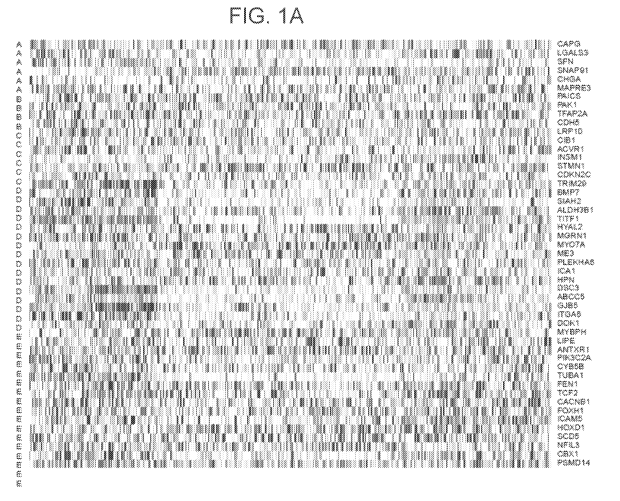
[0010] FIGs 1A-1D illustrate exemplary gene expression heatmaps for
adenocarcinoma (FIG
1A), squamous cell carcinoma (FIG 1B), small cell carcinoma (FIG 1C), and
carcinoid (FIG
1D).
[0011] FIG 2 illustrates a heatmap of gene expression hierarchical clustering
for FFPE RT-
PCR gene expression dataset.
[0012] FIG 3 illustrates a comparison of path review and LSP prediction for 77
FFPE
samples. Each rectangle represents a single sample ordered by sample number.
Arrows
indicate 6 samples that disagreed with the original diagnosis by both
pathology review and
gene expression (for sample details see Table 18).
[0013] FIGs 4-7 illustrates Kaplan Meier plots showing the predicted lung
cancer subtype
AD. SQ, or NE as a function of overall survival for 5 years for 3 independent
AD datasets:
Director's Challenge (Shedden et al; FIG. 4), TCGA RNAseq data (FIG. 5),
Tornida et al.
array data (FIG. 6) or pooled (FIG. 7) assigned a LSP gene expression subtype
across all
stages.
[0014] FIGs 8-11 illustrates Kaplan Meier plots showing the predicted lung
cancer subtype
AD, SQ, or NE as a function of overall survival for 5 years for 3 independent
AD datasets:
Director's Challenge (Shedden et al; FIG. 8), TCGA RNAseq data (FIG. 9),
Toinida et al.
array data (FIG. 10) or pooled (FIG. 11) assigned a LSP gene expression
subtype across
stages I and II.
7
CA 02982775 2017-10-13
WO 2016/168446
PCT/US2016/027503
[0015] FIG. 12 illustrates the proliferation score (11 gene PAM50 signature)
is higher in
AD-NE/SQ compared to AD-AD in all 3 datasets shown in FIGs. 4-6.
[0016] FIG, 13 illustrates gene mutation prevalence in histology-gene
expression concordant
(AD-AD) as compared to discordant (AD-NE/SQ) samples using Fisher's exact
test.
[0017] FIG. 14 illustrates reduction in lung adenocarcinoma prognostic
strength following
exclusion of histologically defined adenocarcinoma samples that are NE or SQ
by LSP gene
expression (AD-NEISQ).
[0018] FIG. 15 illustrates the Cox proportional hazard models of overall
survival (OS).
Models in the hazard ratios table in FIG. 15 used binarized risk scores (at
0.67 quantile),
calling one third of the samples high risk. Models in the p-values portion of
the table left all
risk scores continuous. All models adjusted for (T, N, Age).
DETAILED DESCRIPTION OF THE INVENTION
[0019] Gene expression based adenocarcinoma subtyping has been shown to
classify
adenocarcinoma tumors into 3 biologically distinct subtypes (Terminal
Respiratory Unit
(TRU; formerly referred to as Bronchioid), Proximal Inflammatory (PI; formerly
referred to
as Squamoid), and Proximal Proliferative (PP; formerly referred to as
Magnoid)). These
three subtypes vary in their prognosis, in their distribution of smokers vs.
nonsmokers, in
their prevalence of EGFR alterations, ALK rearrangements, TP53 mutations, and
in their
angiogenic features. The present invention addresses the need in the field for
determining a
prognosis or disease outcome for adenocarcinoma patient populations based in
part on the
adenocarcinoma subtype (Terminal Respiratory Unit (TRU), Proximal Inflammatory
(PI),
Proximal Proliferative (PP)) of the patient.
[0020] As used herein, an "expression profile" comprises one or more values
corresponding
to a measurement of the relative abundance, level, presence, or absence of
expression of a
discriminative gene. An expression profile can be derived from a subject prior
to or
subsequent to a diagnosis of lung cancer, can be derived from a biological
sample collected
from a subject at one or more time points prior to or following treatment or
therapy, can be
derived from a biological sample collected from a subject at one or more time
points during
which there is no treatment or therapy (e.g., to monitor progression of
disease or to assess
development of disease in a subject diagnosed with or at risk for lung
cancer), or can be
8
CA 02982775 2017-10-13
WO 2016/168446
PCT/US2016/027503
collected from a healthy subject. The term subject can be used interchangeably
with patient.
The patient can be a human patient.
[0021] As used herein, the term "determining an expression level" or
"determining an
expression profile" or "detecting an expression level" or "detecting an
expression profile" as
used in reference to a biomarker or classifier means the application of a
biomarker specific
reagent such as a probe, primer or antibody and/or a method to a sample, for
example a
sample of the subject or patient and/or a control sample, for ascertaining or
measuring
quantitatively, semi-quantitatively or qualitatively the amount of a biomarker
or biomarkers,
for example the amount of biomarker polypeptide or mRNA (or cDNA derived
therefrom).
For example, a level of a biomarker can be determined by a number of methods
including for
example immunoassays including for example immunohistochemistry, ELISA,
Western blot,
immunoprecipation and the like, where a biomarker detection agent such as an
antibody for
example, a labeled antibody, specifically binds the biomarker and permits for
example
relative or absolute ascertaining of the amount of polypeptide biomarker,
hybridization and
PCR protocols where a probe or primer or primer set are used to ascertain the
amount of
nucleic acid biomarker, including for example probe based and amplification
based methods
including for example microarray analysis, RT-PCR such as quantitative RT-PCR
(qRT-
PCR), serial analysis of gene expression (SAGE), Northern Blot, digital
molecular barcoding
technology, for example Nanostring Counter Analysis, and TaqMan quantitative
PCR assays.
Other methods of mRNA detection and quantification can be applied, such as
mRNA in situ
hybridization in formalin-fixed, paraffin-embedded (FFPE) tissue samples or
cells. This
technology is currently offered by the QuantiGene ViewRNA (Affymetrix), which
uses probe
sets for each mRNA that bind specifically to an amplification system to
amplify the
hybridization signals; these amplified signals can be visualized using a
standard fluorescence
microscope or imaging system. This system for example can detect and measure
transcript
levels in heterogeneous samples; for example, if a sample has normal and tumor
cells present
in the same tissue section. As mentioned, TaqMan probe-based gene expression
analysis
(PCR-based) can also be used for measuring gene expression levels in tissue
samples, and
this technology has been shown to be useful for measuring mRNA levels in FFPE
samples. In
brief, TaqMan probe-based assays utilize a probe that hybridizes specifically
to the mRNA
target. This probe contains a quencher dye and a reporter dye (fluorescent
molecule) attached
to each end, and fluorescence is emitted only when specific hybridization to
the mRNA target
occurs. During the amplification step, the exonuclease activity of the
polymerase enzyme
9
CA 02982775 2017-10-13
WO 2016/168446
PCT/US2016/027503
causes the quencher and the reporter dyes to be detached from the probe, and
fluorescence
emission can occur. This fluorescence emission is recorded and signals are
measured by a
detection system; these signal intensities are used to calculate the abundance
of a given
transcript (gene expression) in a sample.
[0022] The "biomarkers" or "classifier biomarkers" of the invention include
genes and
proteins, and variants and fragments thereof Such biomarkers include DNA
comprising the
entire or partial sequence of the nucleic acid sequence encoding the
biomarker, or the
complement of such a sequence. The biomarker nucleic acids also include any
expression
product or portion thereof of the nucleic acid sequences of interest. A
biomarker protein is a
protein encoded by or corresponding to a DNA biomarker of the invention. A
biomarker
protein comprises the entire or partial amino acid sequence of any of the
biomarker proteins
or polypeptides.
[0023] A "biomarker" is any gene or protein whose level of expression in a
tissue or cell is
altered compared to that of a normal or healthy cell or tissue. The detection,
and in some
cases the level, of the biomarkers of the invention permits the
differentiation of samples.
[0024] The biomarker panels and methods provided herein are used in various
aspects, to
assess, (i) whether a patient's NSCLC subtype is adenocarcinoma or squamous
cell
carcinoma; (ii) whether a patient's lung cancer subtype is adenocarcinoma,
squamous cell
carcinoma, or a neuroendocrine (encompassing both small cell carcinoma and
carcinoid)
and/or (iii) whether a patient's lung cancer subtype is adenocarcinoma,
squamous cell
carcinoma or small cell carcinoma. In one embodiment, as described herein, the
methods
provided herein further comprise characterizing a patient's lung cancer
(adenocarcinoma)
sample as proximal inflammatory (squamoid), proximal proliferative (magnoid)
or terminal
respiratory unit (bronchioid).
[0025] A biomarker capable of reliable classification can be one that is
upregulated (e.g.,
expression is increased) or downregulated (e.g., expression is decreased)
relative to a control.
The control can be any control as provided herein. For example, the biomarker
panels, or
subsets thereof, as disclosed in Table 1A, Table 1B, Table 1C, Table 2, Table
3, Table 4,
Table 5 and Table 6 are used in various embodiments to assess and classify a
patient's lung
cancer subtype.
CA 02982775 2017-10-13
WO 2016/168446
PCT/US2016/027503
[0026] In general, the methods provided herein are used to classify a lung
cancer sample as a
particular lung cancer subtype (e.g. subtype of adenocarcinoma). In one
embodiment, the
method comprises detecting or determining an expression level of at least five
of the
classifier biomarkers of Table 1A, Table 1B, Table 1C, Table 2, Table 3, Table
4, Table 5 or
Table 6 in a lung cancer sample obtained from a patient or subject. In one
embodiment, the
detecting step is at the nucleic acid level by performing RNA-seq, a reverse
transcriptase
polymerase chain reaction (RT-PCR) or a hybridization assay with
oligonucleotides that are
substantially complementary to portions of cDNA molecules of the at least five
classifier
biomarkers of Table 1A, Table 1B, Table 1C, Table 2, Table 3, Table 4, Table 5
or Table 6
under conditions suitable for RNA-seq, RT-PCR or hybridization and obtaining
expression
levels of the at least five classifier biomarkers based on the detecting step.
The expression
levels of the at least five of the classifier biomarkers are then compared to
reference
expression levels ofthe at least five of the classifier biomarkers of Table
1A, Table 1B, Table
1C, Table 2, Table 3, Table 4, Table 5 or Table 6 from at least one sample
training set. The at
least one sample training set can comprise, (i) expression levels(s) of the at
least five
biomarkers from a sample that overexpresses the at least five biomarkers, or
overexpresses a
subset of the at least five biomarkers, (ii) expression levels from a
reference squamoid
(proximal inflammatory), bronchoid (terminal respiratory unit) or magnoid
(proximal
proliferative) sample, or (iii) expression levels from an adenocarcinoma free
lung sample, and
classifying the lung tissue sample as a squamoid (proximal inflammatory),
bronchoid
(terminal respiratory unit) or a magnoid (proximal proliferative) subtype. The
lung cancer
sample can then be classified as an adenocarcinoma, squamous cell carcinoma, a
neuroendocrine or small cell carcinoma or even a bronchioid, squamoid, or
magnoid subtype
of adenocarcinoma based on the results of the comparing step. In one
embodiment, the
comparing step can comprise applying a statistical algorithm which comprises
determining a
correlation between the expression data obtained from the lung tissue or
cancer sample and
the expression data from the at least one training set(s); and classifying the
lung tissue or
cancer sample as a squamoid (proximal inflammatory), bronchoid (terminal
respiratory unit)
or a magnoid (proximal proliferative) subtype based on the results of the
statistical algorithm.
[0027] In one embodiment, the method comprises probing the levels of at least
five of the
classifier biomarkers of Table 1A, Table 1B, Table 1C, Table 2, Table 3, Table
4, Table 5 or
Table 6 at the nucleic acid level, in a lung cancer sample obtained from the
patient. The
probing step, in one embodiment, comprises mixing the sample with five or more
11
CA 02982775 2017-10-13
WO 2016/168446
PCT/US2016/027503
oligonucleotides that are substantially complementary to portions of cDNA
molecules of the
at least five classifier biomarkers of Table 1A, Table 1B, Table 1C, Table 2,
Table 3, Table 4,
Table 5 or Table 6 under conditions suitable for hybridization of the five or
more
oligonucleotides to their complements or substantial complements; detecting
whether
hybridization occurs between the five or more oligonucleotides to their
complements or
substantial complements; and obtaining hybridization values of the at least
five classifier
biomarkers based on the detecting step. The hybridization values of the at
least five classifier
biomarkers are then compared to reference hybridization value(s) from at least
one sample
training set. For example, the at least one sample training set comprises
hybridization values
from a reference adenocarcinoma, squamous cell carcinoma, a neuroendocrine
sample, small
cell carcinoma sample. The lung cancer sample is classified, for example, as
an
adenocarcinoma, squamous cell carcinoma, a neuroendocrine or small cell
carcinoma based
on the results of the comparing step.
[0028] The lung tissue sample can be any sample isolated from a human subject
or patient.
For example, in one embodiment, the analysis is performed on lung biopsies
that are
embedded in paraffin wax. This aspect of the invention provides a means to
improve current
diagnostics by accurately identifying the major histological types, even from
small biopsies.
The methods of the invention, including the RT-PCR methods, are sensitive,
precise and have
multianalyte capability for use with paraffin embedded samples. See, for
example, Cronin et
al. (2004) Am. J Pathol. 164(1):35-42, herein incorporated by reference.
[0029] Formalin fixation and tissue embedding in paraffin wax is a universal
approach for
tissue processing prior to light microscopic evaluation. A major advantage
afforded by
formalin-fixed paraffin-embedded (FFPE) specimens is the preservation of
cellular and
architectural morphologic detail in tissue sections. (Fox et al. (1985) J
Histochem Cytochem
33:845-853). The standard buffered formalin fixative in which biopsy specimens
are
processed is typically an aqueous solution containing 37% formaldehyde and 10-
15% methyl
alcohol. Formaldehyde is a highly reactive dipolar compound that results in
the formation of
protein-nucleic acid and protein-protein crosslinks in vitro (Clark et al.
(1986) J Histochem
Cytochem 34:1509-1512; McGhee and von Hippel (1975) Biochemistry 14:1281-1296,
each
incorporated by reference herein).
12
CA 02982775 2017-10-13
WO 2016/168446
PCT/US2016/027503
[0030] In one embodiment, the sample used herein is obtained from an
individual, and
comprises fresh-frozen paraffin embedded (FFPE) tissue. However, other tissue
and sample
types are amenable for use herein (e.g., fresh tissue, or frozen tissue).
[0031] Methods are known in the art for the isolation of RNA from FFPE tissue.
In one
embodiment, total RNA can be isolated from FFPE tissues as described by
Bibikova et al.
(2004) American Journal of Pathology 165:1799-1807, herein incorporated by
reference.
Likewise, the High Pure RNA Paraffin Kit (Roche) can be used. Paraffin is
removed by
xylene extraction followed by ethanol wash. RNA can be isolated from sectioned
tissue
blocks using the MasterPure Purification kit (Epicenter, Madison, Wis.); a
DNase I treatment
step is included. RNA can be extracted from frozen samples using Trizol
reagent according to
the supplier's instructions (Invitrogen Life Technologies, Carlsbad, Calif).
Samples with
measurable residual genomic DNA can be resubjected to DNaseI treatment and
assayed for
DNA contamination. All purification, DNase treatment, and other steps can be
performed
according to the manufacturer's protocol. After total RNA isolation, samples
can be stored at
-80 C until use.
[0032] General methods for mRNA extraction are well known in the art and are
disclosed in
standard textbooks of molecular biology, including Ausubel et al., ed.,
Current Protocols in
Molecular Biology, John Wiley & Sons, New York 1987-1999. Methods for RNA
extraction
from paraffin embedded tissues are disclosed, for example, in Rupp and Locker
(Lab Invest.
56:A67, 1987) and De Andres et al. (Biotechniques 18:42-44, 1995). In
particular, RNA
isolation can be performed using a purification kit, a buffer set and protease
from commercial
manufacturers, such as Qiagen (Valencia, Calif), according to the
manufacturer's
instructions. For example, total RNA from cells in culture can be isolated
using Qiagen
RNeasy mini-columns. Other commercially available RNA isolation kits include
MasterPure.TM. Complete DNA and RNA Purification Kit (Epicentre, Madison,
Wis.) and
Paraffin Block RNA Isolation Kit (Ambion, Austin, Tex.). Total RNA from tissue
samples
can be isolated, for example, using RNA Stat-60 (Tel-Test, Friendswood, Tex.).
RNA
prepared from a tumor can be isolated, for example, by cesium chloride density
gradient
centrifugation. Additionally, large numbers of tissue samples can readily be
processed using
techniques well known to those of skill in the art, such as, for example, the
single-step RNA
isolation process of Chomczynski (U.S. Pat. No. 4,843,155, incorporated by
reference in its
entirety for all purposes).
13
CA 02982775 2017-10-13
WO 2016/168446
PCT/US2016/027503
[0033] In one embodiment, a sample comprises cells harvested from a lung
tissue sample, for
example, an adenocarcinoma sample. Cells can be harvested from a biological
sample using
standard techniques known in the art. For example, in one embodiment, cells
are harvested
by centrifuging a cell sample and resuspending the pelleted cells. The cells
can be
resuspended in a buffered solution such as phosphate-buffered saline (PBS).
After
centrifuging the cell suspension to obtain a cell pellet, the cells can be
lysed to extract nucleic
acid, e.g, messenger RNA. All samples obtained from a subject, including those
subjected to
any sort of further processing, are considered to be obtained from the
subject.
[0034] The sample, in one embodiment, is further processed before the
detection of the
biomarker levels of the combination of biomarkers set forth herein. For
example, mRNA in a
cell or tissue sample can be separated from other components of the sample.
The sample can
be concentrated and/or purified to isolate mRNA in its non-natural state, as
the mRNA is not
in its natural environment. For example, studies have indicated that the
higher order structure
of mRNA in vivo differs from the in vitro structure of the same sequence (see,
e.g., Rouskin
etal. (2014). Nature 505, pp. 701-705, incorporated herein in its entirety for
all purposes).
[0035] mRNA from the sample in one embodiment, is hybridized to a synthetic
DNA probe,
which in some embodiments, includes a detection moiety (e.g., detectable
label, capture
sequence, barcode reporting sequence). Accordingly, in these embodiments, a
non-natural
mRNA-cDNA complex is ultimately made and used for detection of the biomarker.
In
another embodiment, mRNA from the sample is directly labeled with a detectable
label, e.g.,
a fluorophore. In a further embodiment, the non-natural labeled-mRNA molecule
is
hybridized to a cDNA probe and the complex is detected.
[0036] In one embodiment, once the mRNA is obtained from a sample, it is
converted to
complementary DNA (cDNA) in a hybridization reaction or is used in a
hybridization
reaction together with one or more cDNA probes. cDNA does not exist in vivo
and therefore
is a non-natural molecule. Furthermore, cDNA-mRNA hybrids are synthetic and do
not exist
in vivo. Besides cDNA not existing in vivo, cDNA is necessarily different than
mRNA, as it
includes deoxyribonucleic acid and not ribonucleic acid. The cDNA is then
amplified, for
example, by the polymerase chain reaction (PCR) or other amplification method
known to
those of ordinary skill in the art. For example, other amplification methods
that may be
employed include the ligase chain reaction (LCR) (Wu and Wallace, Genomics,
4:560
(1989), Landegren et al., Science, 241:1077 (1988), incorporated by reference
in its entirety
14
CA 02982775 2017-10-13
WO 2016/168446
PCT/US2016/027503
for all purposes, transcription amplification (Kwoh et al., Proc. Natl. Acad.
Sci. USA,
86:1173 (1989), incorporated by reference in its entirety for all purposes),
self-sustained
sequence replication (Guatelli etal., Proc. Nat. Acad. Sci. USA, 87:1874
(1990), incorporated
by reference in its entirety for all purposes), incorporated by reference in
its entirety for all
purposes, and nucleic acid based sequence amplification (NASBA). Guidelines
for selecting
primers for PCR amplification are known to those of ordinary skill in the art.
See, e.g.,
McPherson et al., PCR Basics: From Background to Bench, Springer-Verlag, 2000,
incorporated by reference in its entirety for all purposes. The product of
this amplification
reaction, i.e., amplified cDNA is also necessarily a non-natural product.
First, as mentioned
above, cDNA is a non-natural molecule. Second, in the case of PCR, the
amplification
process serves to create hundreds of millions of cDNA copies for every
individual cDNA
molecule of starting material. The number of copies generated are far removed
from the
number of copies of mRNA that are present in vivo.
[0037] In one embodiment, cDNA is amplified with primers that introduce an
additional
DNA sequence (e.g., adapter, reporter, capture sequence or moiety, barcode)
onto the
fragments (e.g., with the use of adapter-specific primers), or mRNA or cDNA
biomarker
sequences are hybridized directly to a cDNA probe comprising the additional
sequence (e.g.,
adapter, reporter, capture sequence or moiety, barcode). Amplification and/or
hybridization
of mRNA to a cDNA probe therefore serves to create non-natural double stranded
molecules
from the non-natural single stranded cDNA, or the mRNA, by introducing
additional
sequences and forming non-natural hybrids. Further, as known to those of
ordinary skill in
the art, amplification procedures have error rates associated with them.
Therefore,
amplification introduces further modifications into the cDNA molecules. In
one
embodiment, during amplification with the adapter-specific primers, a
detectable label, e.g., a
fluorophore, is added to single strand cDNA molecules. Amplification therefore
also serves
to create DNA complexes that do not occur in nature, at least because (i) cDNA
does not
exist in vivo, (i) adapter sequences are added to the ends of cDNA molecules
to make DNA
sequences that do not exist in vivo, (ii) the error rate associated with
amplification further
creates DNA sequences that do not exist in vivo, (iii) the disparate structure
of the cDNA
molecules as compared to what exists in nature and (iv) the chemical addition
of a detectable
label to the cDNA molecules.
CA 02982775 2017-10-13
WO 2016/168446
PCT/US2016/027503
[0038] In some embodiments, the expression of a biomarker of interest is
detected at the
nucleic acid level via detection of non-natural cDNA molecules.
[0039] In some embodiments, the method for lung cancer subtyping includes
detecting
expression levels of a classifier biomarker set. In some embodiments, the
detecting includes
all of the classifier biomarkers of Table 1 (also characterized as a lung
cancer subtype gene
panel), Table 2, Table 3, Table 4, Table 5 or Table 6 at the nucleic acid
level or protein level.
In another embodiment, a single or a subset of the classifier biomarkers of
Table 1 are
detected, for example, from about five to about twenty. The detecting can be
performed by
any suitable technique including, but not limited to, RNA-seq, a reverse
transcriptase
polymerase chain reaction (RT-PCR), a microarray hybridization assay, or
another
hybridization assay, e.g., a NanoString assay for example, with primers and/or
probes
specific to the classifier biomarkers, and/or the like. In some cases, the
primers useful for the
amplification methods (e.g., RT-PCR or qRT-PCR) are the forward and reverse
primers
provided in Table 1A, Table 1B, Table 1C, Table 2, Table 3, Table 4, Table 5
or Table 6. It
should be noted however that the primers provided in Table 1A, Table 1B, Table
1C, Table 2,
Table 3, Table 4, Table 5 and Table 6 are merely for illustrative purposes and
should not be
construed as limiting the invention.
[0040] The biomarkers described herein include RNA comprising the entire or
partial
sequence of any of the nucleic acid sequences of interest, or their non-
natural cDNA product,
obtained synthetically in vitro in a reverse transcription reaction. The term
"fragment" is
intended to refer to a portion of the polynucleotide that generally comprise
at least 10, 15, 20,
50, 75, 100, 150, 200, 250, 300, 350, 400, 450, 500, 550, 600, 650, 700, 800,
900, 1,000,
1,200, or 1,500 contiguous nucleotides, or up to the number of nucleotides
present in a full-
length biomarker polynucleotide disclosed herein. A fragment of a biomarker
polynucleotide
will generally encode at least 15, 25, 30, 50, 100, 150, 200, or 250
contiguous amino acids, or
up to the total number of amino acids present in a full-length biomarker
protein of the
invention.
[0041] In some embodiments, overexpression, such as of an RNA transcript or
its expression
product, is determined by normalization to the level of reference RNA
transcripts or their
expression products, which can be all measured transcripts (or their products)
in the sample
or a particular reference set of RNA transcripts (or their non-natural cDNA
products).
Normalization is performed to correct for or normalize away both differences
in the amount
16
CA 02982775 2017-10-13
WO 2016/168446
PCT/US2016/027503
of RNA or cDNA assayed and variability in the quality of the RNA or cDNA used.
Therefore, an assay typically measures and incorporates the expression of
certain normalizing
genes, including well known housekeeping genes, such as, for example, GAPDH
and/or (3-
Actin. Alternatively, normalization can be based on the mean or median signal
of all of the
assayed biomarkers or a large subset thereof (global normalization approach).
[0042] For example, in one embodiment, from about 5 to about 10, from about 5
to about 15,
from about 5 to about 20, from about 5 to about 25, from about 5 to about 30,
from about 5 to
about 35, from about 5 to about 40, from about 5 to about 45, from about 5 to
about 50 of the
biomarkers in any of Table 1A, Table 1B, Table 1C, Table 2, Table 3, Table 4,
Table 5 and
Table 6 are detected in a method to determine the lung cancer subtype. In
another
embodiment, each of the biomarkers from any one of Table 1A, Table 1B, Table
1C, Table 2,
Table 3, Table 4, Table 5, or from Table 6 are detected in a method to
determine the lung
cancer subtype.
Table1A
Gene symbol Gene name Forward primer SE() Reverse primer SEQ
ID
CD.H5 cadherin 5, type 2, AA GAGAGA TTG 1
TTCTTGCGACTCA.CGCT 58
VE-cadherin GATTTGGAACC
(vascular epithelium)
CLEC3B C -type lec tin domain CCAGAAGCCCA 2
GCTCCTCAAACAT 59
family 3, member B AGAAGATTGTA CTTTGTGTTCA
PAIC S phosphoribosylami AATCCTGGTGT 9 GACCACTGTGGG
60
noimidazoie CAAGCiAAG TCATTATT
carboxylase,
phosphoribosylami
Hointidazole
SUCCinocarboxamide
symbetase
PAK1 p21.101c42/Raci.- GG AC CGATTTT 4 GAAATCTCTGGC
61
activated kinase I (STEN ACCGATCC CGCTC
homolog, yeast)
PECAM1 platelet/endothelial cell ACAGTCCAGAT 5
ACTGGGCA.TCAT 62
adhesion molecule AGTCGTATGT AACiAAATCC
(CD31 antigen)
TFAP2A transciiption factor AP- GTCTCCGCCATC 6
ACTGAACAGAAG 63
2 alpha (activating CCTAT A CTTCGT
enhancer binding
protein 2 alpha)
ACVRi. activin A receptor, A CTG GTGT AA C 7
AACCTCCAAGTG 64
type I AGGAACAT G AAATTCT
17
CA 02982775 2017-10-13
WO 2016/168446
PCT/US2016/027503
Table IA
Gene symbol Gene name Forward primer SEQ Reverse primer SEQ
ED 11)
CDKN2C cyclin-depe lident k nose TTTGGAAGG AC 8 TCG
GTCTTTC AA A 65
inhibitor 2C (p18, TGCGCT TCGGCiATTA
inhibits CDK4)
CIB 1 calcium and integrin C A CGTC ATCTC C 9 CTG
CTGTC AC A G 66
binding .1 (calmyrin) CGTTC G AC AAT 66
1NSM1 insulinoma-associated 1 ATTGAACTTCCC 10
AAGGTAAAGCCA 67
ACACGA GACTCC A 67
LRFIO low density lipoprotein GG AACAGACTG ii GGGAGCGTAGGG
68
receptor-related protein TCACCAT TTAAG
STIvIN1. stathmin TC.AGAGT(iTGTG 12 CACITGTA.TTCTGC 69
lioncoprotein 18 G ACAATCAAC
TCAGGC
CAPG capping protein (actin G Ci GA CA Gm:- 13
CiTTCCACiGATG-FT 70
fiIament, ge I soli n-like AACACT GGACTTTC
CHGA chromogranin A CCTGTGAACAG GGAAAG'TGTGTC 71
(parathyfOid secretory CC CTATC1 G CiAGAT
protein 1)
EGAL S3 lectin. galac to side- TTCTGGGCACG 15
AGGCAACATCAT 72
binding, soluble, 3 GIGAACi Tucccrc
(galectist 3)
MAPRE3 rnicrombule-associated GGCCA A..ACTAG 16
GTCAAC.ACCC.AT 73
protein, RP/E13 family, AGCA.CGAATA CTTCTTCiAAA
member 3
SFN stratifin TCAGCAAGAAG 17 C GTAG ICI G AAGA 74
GA GATG CC CGG AA A
SNA P91 sy nap to so mal-a ssoc ia ted GrourcccTicrc 18 c TUG
GTIGT AGAATT 75
protein, 91 ILEA CATTAAGTA AGGAGACGTA
homolog (mouse)
AB C C5 ATP-binding cassette, CAA GT-rcAGGA 19 G GC
ATCAAGAG A 76
sub-family C(CFTR/MERP), GAACTCGA.0 GAGGC
member 5
ALDB3B 1 aldehyde de hydroge nose GGCIG'TGGITA 20
GATAAAGAGTIA 77
3 MCGATAG CAAGC1'CCTICTC1
family, member B1
ANTXR1. Anthrax toxin receptor 1 ACCCGAGGAAC 21
TCTAGGCCTTGAC 78
AA CCTTA GGAT
BMP7 Bone morphogenetic CCCTCTCCATTCC 22 TTTGGGCAAACCTCGGTA 79
protein 7 (osteogenie CTACA A
protein 1)
CACNTI1 calcium channel, CAGA G CGCCAG 2.3 G CACAGCAA
ATG 80
voltage-dependent, beta GCATTA CCACT
1 subunit
18
CA 02982775 2017-10-13
WO 2016/168446
PCT/US2016/027503
Table IA
Gene symbol Gene name Forward primer SEQ Reverse primer SEQ
ED ll-D
CBX1 chromobox homolog 1 CCACTGGCTGA 24 CTTGTCTTTC1C1CT 81
(F1P.1 he-La hornolog GGTGTTA ACTGTCTTAC
Drosophila)
CYB5B cytochrome b5 type B TGGGCGAGTCT 25 CTTGTTCCAGCAG
82
(outer initochoncliai ACGATG AACCT
mernbrane)
DOK1 docking piotein 1, 62 CTTICTGCCCTG 26 CAGTCCTCTGCAC
83
kDa (downstream of GAGATG CurrA
tyrosine kinase 1)
DSC3 desmocollin 3 GCGCC.ATTTGCT 27 CATCCAGATCCCT 84
AGAGATA CACAT
FEN1 flap structim-specific AGAGAAGATGG 28
CCAAGACACAGC 85
endonuclease 1 GCAGAAAG CAGTAAT
FOXIT I forkhead box Fll GCCCAGATCAT 29 TTTCCAGCCCTCG
86
CCGTCA TAGTC
GJB5 gap junction protein, ACCAC A AGGAC 30 GGGAC A
CA GGGA 87
beta 5 (connexin 31.1) TTCGAC AGAAC
HOXD1 homeobox DI GC'TCCGCTGCT 31 GTCTGCCACTCTG 88
ATCTTT CAAC
HPN Hepsin (transmembrane AGCGGCCAGGT 32
GTCGGCTGACCiC 89
protease, scrine 1) GGA'TTA TrfGA
HVAL2 hyaluionogiucosam ArraiGCTTIGC1 33 GAACAAcackca
90
inidase 2 GAocATA crACIGGAATAC
1CA1 islet cell auloantig,en GACCTGGATGC 34
TGOTTCGATAAG 91
1, 69 kDa CAAGCTA TCCAGACA
1CAM5 interceilulai adhesion CCGGCTCTTGG 35
CCTCTGAGGCTG 92
molecule 5, AAGITG GAAACA
telencephalin
ITGA6 integdn, alpha 6 ACC1C1GGATCGA 36 ATCCACTGATCTT
93
GTTTGATAA CCTTGC
!APE lipase, hormone -sensilive CGCAAGTCCCA 37
CAGTGCTGCTTCA 94
(1AAGAT GACACA
114E3 make enzyme 3; CGCGGATACGA 38 CCTTTCTTCAAGG 95
NADP(+)-dependent, TGTCAC GTAAAGGC
Mitochondrial
MGRN1 mahogunin, ring finger GAACTCGGCCT 39 TCGAATTTCTCTC
96
ATCGCT urc CCAT
MYBPH myosin binding protein Tc-rGAc CTCATC 40 CTGAGTCCACAC
97
IT ATCGGCAA AGGITT
MN-07A myosin VT1A GAGGTGAAGCA 41 CCCATACTTGTTG 98
AACTACGGA ATGGCAATTA
NFLI,3 nuclear factor, ACTCTCCA.CA.A 42 TCCTGCGTGTGTT
99
inter-le-akin 3 regulated AGCICG CTACI
19
CA 02982775 2017-10-13
WO 2016/168446
PCT/US2016/027503
Table IA
Gene symbol Gene name Forward primer SEQ Reverse primer SEQ
ED
PIK3C2A phosphoinosi0de-3-kinase, GGATTTCAGCT 43 AGTCATCATGTAC 100
class 2, alpha ACCAGTTACTT CCAGCA.
poly-peptide
PLEKBA6 pleckstrin homology TTCGTccruaG 44
CCCAGGA'TACTCT 101
domain containing, GATCG CTICCTT
family A member 6
PSNID14 proteasome (prosoinc, AGTGATTGATG 45
CAC'TGGATCAAC 102
macropain) 26S subunit, TGTTTGCTATCi Tau:iv
non-KIPase, 14
SCD5 stearoyl-CoA desatuase CA..AAGCCAAGC 46
CACiCTGTCACAC 103
CA.CTCACTC CCACiA GC
SI A1-12 seven in abseniia CTCGGCAGTCC 47
CGTATGGTGCAG 104
ho rao log 2 TGTTTC GGTCA
(Drosoph)la)
TCF2 transcription factor 2, AC.ACCTGGTAC 48
TCTGGACTGTCTCi 105
hepatic; LF-B3; variant GTCA.GAA GTTGAAT
hepatic nuclear factor
TC.P1 t-comple 1 ATocccAAGAG 49 CCTCiTACACCAA 106
AATCGTAAA GCTTCAT
T1'F1 thyroid transcrimion ATGAGTCCAAA 50
CcARicccAcrrT 107
Mcior I GCACACGA CTTGTA
11RIM29 tripartite motif-containing TGAGATTGAGG 51
CATTGGTCiGTGA 108
29 ATGAAGCTGAG AGCTCTTG
TUB Al tubulin, alpha 1 CCGACTCAACG 52
CGRIGACTGAGA 109
TCiAGAC TGCATT
CF1,1 cofiliit 1 (non-muscle) GroccurcuccT 53
TICATGFCGTTCiA 110
TTTCG ACACCTTCi
EEF1A1 eukatyotic translation CGTICTTITTCG 54
CATTTTGGCITTT 111
elongation factor I CAACGG AC1C1GGITAG
alpha 1
RPLIO ribosomal piorein Lit) GGTGIGCcAcT 55
GGCAGAAGCGAG 112
GAAGAT AcTIT
RP1,28 ribosomal protein L28 GTGTCGTGGTG 56
GCACATAGGAGG 113
GTCATT mocA
1114,37A ribosomal protein 1,37a GCATGAAGACA 57
GCGGAC'TTTACC 114
GroGcT GTGAC
Table 1B
Gene symbol Gene name Forward primer SEQ Reverse primer SEQ
ID ID
CDEI5 eadherin 5, type 2. AAGAGAGATTG 1
TTCTIGCGACTCACGur 58
VE-cadherin GATTTGGAACC
(vascular epithelium)
CA 02982775 2017-10-13
WO 2016/168446
PCT/US2016/027503
Table 1.B
Gene symbol Gene name Forward primer SEQ Reverse primer SEQ
ED 11)
CLEC3B C-type lectin domain CCAGAAGCCCA 2
GCTCCTCAAACAT 59
family 3, member B AG AAGA.TTGTA CTTTGTGTTCA
PA1CS phosphoribosylami AATCCTICiGTIGT 3
GACCACTCiTC1CiG 60
noiLida ZOie CAAGGAAG TCA TT ATT
carboxylase,
phosphoribosylami
noimidazole
succinocarboxamide
synthetase
PA_K1 p2 dc42/Rac 1- GGACCGAITTT 4 GAAATCTCTGGC 61
activated kinase I (STE20 ACCGATCC CGurc
homo log, yeast)
PECAN,41 plaielet/endothelial cell ACAGTCCAGAT 5
ACTGGGCATCAT 02
adhesion molecule AGTCGTATGT A AGAAATCC
(CD3 1 antigen)
TFAP2A transcription factor AP- GTCTCCGCCATC 6
AC'TGAACAGAAG 63
2 alpha (activating CCTAT Acircorr
enhancer binding
protein 2 alpha)
ACVR1 activin A receptor, AC'TGG'TGTAAC 7
AACCTCCAAGTG 64
type 1 ACiCiAACAT (1AAATTCT
CDKN2C cyclin-dependent kinase TTTGGAAGG AC 8 TCG
GTCTTTC AA A 65
inhibitor 2C (pI8, TGCCICT TCGGGA.TTA
inhibits CD1(4)
CIB 1 calcium and integrin CACGTCATCTCC 9
CTGCTGTCACAG 06
binding .1 (calmyrin) CGTTC GACAAT 06
.INSM1 insulinorna-associated 1 ATTGAACTTCCC 10
AAGGTAAAGCC.A 07
ACACGA GACTCCA 67
L.RPIO low density lipoprotein GGAACAGACTG ii GGGAGCGTAGGG
68
receptor-related protein TCACCAT TTAAG
STMNI stathmin TC.AGAGTCiTGTG 12 CAGTGTA.TTCRIC 09
lioncoprotein 18 G ACAATCAAC
'TCAGGC
CAPG capping protein (actin GGGACAGCTTC 13
GTICCAGGATGTT 70
filament:), gel solin-like AACACT GGACTTTC
CHGA chromogranin A CCTGTGAACAG GGAAAGTGTGTC 71
(parathyfOid subratoty CCCTATG GGAGAT
protein 1)
EGALS3 lectin. galactoside- TTCTGGGCACG 15
AGGCAACATCAT 72
binding, soluble, 3 GIGAACi Tucccrc
(galectin 3)
MAPRE3 rnicrombule-associated GCiCCAA..ACTAG 16
GTCAAC.ACCC.AT 73
protein, RP/E13 family, AGCA.CGAATA CTTCTTC;AAA
member 3
SFN stratifin TCAGCAAGAAG 17 CGTAGTGCiAAGA 74
GA GATGCC CC1Ci AAA
21
CA 02982775 2017-10-13
WO 2016/168446
PCT/US2016/027503
Table 1.B
Gene symbol Gene name Forward primer SEQ Reverse primer SEQ
11.1
SNAP91 syuaplsomal-associaled GTGCTC.C.CTCTC 18
CTGGTGTA G A ATT 75
protein, 91 kDa CATTAAGTA AGGAGA.CGTA
homolog (mouse)
ABCC5 ATP-binding cassene, C AAG TTCAG G A 19 GGCATCA
AG AGA 76
sub-family C(C.FT.RIMRP), G.AACTCGAC GA.GGC
member 5
ALDH3 B 1 aldehyde dehydroge nose GGCTGTGGTTA 20
GATAAAGAGTTA 77
TGCGATAG CAAGCTCCTCTG
family, member B1
ANTXR1 Anthrax toxin receptor 1 ACCCGAGGAAC 21
TCTAGGCCITGAC 78
AA CCTTA GOAT
CACNB 1 calcium channel, CAGAGCCiCCAO 23 GCACAGCAAATG
80
voltage-dependent, beta GCATT A C C ACT
1 subunit
CB X1 chromobox homolog 1 CCACTGGCTGA 21 cr-
rurcrrTcce'r 81
(1-1P1 beta homolog GOICITTA AC'TGTCY1AC
Drosophila)
CYB5B cytochrome b5 type B TGGGCGAGTCT 25 CTTGTTCCAGCAG
87
(outer mitochondria! ACGATG AA.CCT
membrane)
DOK 1 docking protein 1. 62 CTTTCTGCCCTG 26 C AG
TCCTCTGCAC 83
kria (downstream of GA GATG COTTA
tyrosine kinase 1)
DSC3 desmocollin 3 GCGCCATTTG cr 27 CATCCAGATCCCT 84
A GAG ATA CACAT
FEN1 flap stmeture-spe.cific A GAG AA GA TGG 28 CCA AG
ACACA GC 85
e.1001aUCiCaSe 1 GC AGAAAG C AG TAAT
FOXHI forkhead box fil GCCCACiA.TCAT 29
TTTCCAC1CCCTCG 86
CCGTCA TA GTC
G1135 gap junction protein, ACCACAAGGAC 30
GGGACACAGGGA 87
beta 5 (connexin 31.1) TTCGAC A GAAC
110XD1 homeobox DI GCTCCGCTGCT 31 GTCTOCCACTCRi 88
ATCTTT CAAC
HIM Heps ( tratistitenthrane AGC(IGCCAGGT 32
OTCGGCTGACCiC 89
protease, scrim 1) GGATTA TTTGA
:El-Xi\ L.2 Ityaluamogincosam ATGGGCTTTGG 33
GAACAAGTCAGT 90
inidase 2 GAGCATA. CTAGGGAATAC
1CA1 islet cell autotintigen GACCTGGATGC 34
TGCTTTCGATAAG 91
1,69 kDa C AAG CT A TCC, AG A C A
22
CA 02982775 2017-10-13
WO 2016/168446
PCT/US2016/027503
Table 1.B
Gene symbol Gene name Forward primer SEQ Reverse primer SEQ
11) 1.1)
ICA1\45 intercellular adhesion CCGGCTCTTGG 35
CCTCTGAGGCTG 92
molecule 5, AAGTTG GAAACA
telencephalin
1TGA.6 integrin, alpha 6 ACGCGGATCGA 36
ATCCACTGATCTT 93
GTTTGATAA ccTTGc
LIFE lipase, hormone-sensitive CGCAAGTCCCA 37
CAGTGCTGCTTCA 94
GAAGAT GACACA
ME3 malic enzyme 3. CGCGGA'TACGA 38 CCTTTCTTCA_AGG 95
NADP( )-dependent, TGTCAC CiTAAAGGC
Mitochondria(
MGRN1 mahop,unin, dng finger GAACTCGGCCT 39
TCGAATTTCTCTC 96
ATCGCT CTCCCAT
MYBP}I inyositt 'binding protein TCTGACCTCATC 40
CTGAGTCCACAC 97
ATCGGCAA AGGTTT
MY07A myosin VIIA GAGGTGAAGCA 41 CCCATAcTrum 98
AACTACGGA ATGGCAATTA
NFIL3 nuclear factor, ACTCTCCACAA 42 TCCTGCGTGTIGTI 99
interleukiti 3 regulated AGCTCG cTACT
P1K3C2A phospho 1rinase, GGATTTCAGCT 4.3 AGTCATCATGTAC 100
class 2, alpha ACCAGTTACTT CCAGCA
polypeptide
PLEKHA6 pleckstrin homology TTCGTCCTGGTG 41
CCCACiGATACTCT 101
domain containing. GATCG cucerr
family A member 6
FWD 11 proteasome (prosome, AGTGATTGATG 45
CACTGGATCAAC 102
macropain) 26S subunit, TGTTTGcTATG TGCCTC
non-ATPase, 14
SCD5 stearoyl-Co A. desatu rase CAA A GCCAAGC 46
CAGCTGTCA CAC 103
CACTCACTC CCAGACie
SIAH2 seven in absentia CTCGGCAGTCC 47
CGTATGGTGCAG 104
homolog 2 TGTTTC GGTCA
(Drosophila)
TCF2 transcription factor 2, ACACCTGGTAC 48
TCTGGACTGTCTG 105
hepatic; LF-B3; variant GTCAGAA GTTGAAT
hepatic nuclear factor
Tcp]. [-complex 1 ATGCCCAAGACi 49 CCIGTACACCAA 106
AATCGTAAA GCTTCAT
TTF1 thyroid transcription ATGAGTcc AAA 50
CCATGCCCACTIT 107
factor 1 GCACACGA CTTGTA
1-RW129 tripartite motif-comainitig TGAGAT-FGAGG 51
CATTGGTGGTGA 108
29 ATGAAGCTGACi AGCTCTTG
TusA1 tubul if!, alpha 1 CCGAC'TCAACG 52
CGTGGACTGAGA 109
TGAGAC TGCATT
CEA cofilin 1 (non-nmscle) GTGCcourc CT 53
TTcATIGTCGTIGA 110
TTITCG ACACCTRi
23
CA 02982775 2017-10-13
WO 2016/168446
PCT/US2016/027503
Table 1.B
Gene symbol Gene name Forward primer SEQ Reverse primer SEQ
ELI 11)
EEF1A1 enkaryotic translation CGTTCTTTTTCG 54
CATTTTGGCTTTT 111
elongation factor 1 CAACGG AGGGGT.AG
alpha 1
RPL10 ribosomal protein LIPGGTGTGCCACT 55
GGCAGAAGCGAG 112
GA A.G AT ACTTT
KPL28 ribosomal proteiri L28 GTGTCGTGGTG 56 GCA
CA TA GG AGG 113
GTCA TT TGGCA
RP1,37A ribosomal protein 1,37it GCATGAAGACA 57
GCGGACTTTACC 114
GTGGCT GTG.AC
Table 1C
Gene symbol Gene name FOMarti primer SEQ Reverse primer SEQ
113
CDI-15 cad.herin 5, type 2, A.AGAGAGATTG I
TTCTTGCGACTCACGCT 58
VE-cadherin GATTTGGAACC
(vascular epithelium)
CLEC3B C-type lectin domain CCAGAAGCCCA 2
GC'TCCTCAAACAT 59
family 3, member B AGAAGATICi'TA CTTTGIG'TTCA
PA1CS phosphoribosylami AATCCTGGTGT 3 GACCACTGTGGG 60
noimidazole CAAGGAAG TcArrATT
carboxylase,
phosphoribosylami
no imidazole
succinocatboxamide
sy nthetit se
PAK I p21/Cdc.42/Rac1- GGACCGATTTT 4 G AAA TCTCTGGC 61
activated kinase I STE20 ACCGA.TCC CGCTC
homolog, yeast)
PECAN=11. platelet/endothelial cell ACAGTCCAGA.T 5
ACTGGGCATC.AT 62
adhesion molecule AGTCCITATGI AAGAAATCC
(CD31 antigen)
TEAP2A transcription factor AP- GTCTCCGCCATC 6
ACTGAACAGAAG 63
2 alpha (activating CCTAT ACTTCGT
enhancer binding
protein 2 alpha)
ACVR 1 activin A receptor, ACTGGTGTA AC 7 A A
CCTCCAA CiTti 64
type AG CiAA.0 AT GA AATTCT
CDKN2C cyclin-dependent kinase TTTGGAAGGAC 8
TCGGTCTTTCAAA 65
inhibitor 2C (p18, TGCGCT TCGGCiATTA
inhibits CDKet)
CIB 1 calcium and integrin CACGTCATCTCC 9
CTGCTCiTCACAG 66
binding I (cattily-lin) CCi'TTC GACAAT 66
SM1 insulinoma-associated 1 ATTGA Acriv cc 10
AAGGTAAAGCCA 67
ACACGA GACTCCA 67
24
CA 02982775 2017-10-13
WO 2016/168446
PCT/US2016/027503
Table 1C
Gene symbol Gene name Forward primer SEQ Reverse primer SEQ
ED 11.)
L.RPIO low density lipoprotein GGAACAGACTG Ii GGGAGCGTAGGG
68
receptor-related protein TCACCAT TTAAG
STMN1 statiunin TCAGAGTGTGTG 12 CAGTGTATTCTGC 69
iloricoprotem 18 G AC.AATCAAC
TCAGGC
CAPG capping protein (actin GGGACAGCTTC 13
GTICCAGGATGTT 70
filament), gelsolin-like AACACT GGACTTIC
CHGA chromogranin A CCTGTGAACAG 14 GGA.AAGTGTGTC 71
(parathyroid secretory CCCTATG GGAGAT
protein I)
I,GALS3 lectin, gal acto side- TTCTGGGCACG 15
AGGCAACATCAT 72
binding, soluble, 3 G'TGAAG TCCCTC
(galectin 3)
IVIAPRE3 micrombule-associated GGCCAAACTAG 16
GTCAACACCCAT 73
protein, RP/EB family, A GCACGAAT A CTTCTTGAAA
member 3
SFN stratifin TCAGCAAGAAG 17 CG'TAGTGGAAGA 74
GAGATGCC CGGAAA
SNAP9 I synaptosomal-associated GrGurcccfcrc IS
CTGGTGTAGAATT 75
protein, 91 klia CATTAAGTA AGGAGACGTA
homolog (mouse)
ABCC5 ATP-binding cassette, CAAGTTCAGGA 19
GGCATCAAGAGA 76
sub-family C(CFIRIMRP), GAAcrcGAC GAGGC
member 5
.ALD1-13B1 aldehyde dehydrogenase GGCTGTGGTT.A
20 GAT.AAACiAGTTA 77
TGCGATAG CAAGCTCCTCTG
family, member B1
ANTXR1 Anthrax toxin receptor 1 ACCCGAGG AAC 21
TCTAGGCCTTGAC 78
AACCTTA GGAT
BMP7 Bone morphogenetic CCCTCTCCATTCC 22 TTTGGGCAAACCTCGGTA 79
protein 7 (osteogenie CTACA A
protein 1)
CACNB1 calcium channel, CAGAGCGCCAG 23 GCACAGCAAATG
80
voltage-dependent, beta GCATTA CCACT
1 subunit
CBX1 chromobox homolog 1 CCACTGGCTGA 24 CTTGTCTTTCCCT 81
(1-1P1 beta homolog GGTGTIA AC'TGTCTIAC
Drosophila)
CYB5B cytocitrome b5 type B TGGGCGAGTCT 25 CTTGTTCCAGCAG
87
(outer mitochondria! ACGATG A A.CCT
membrane)
CA 02982775 2017-10-13
WO 2016/168446
PCT/US2016/027503
Table IC
Gene symbol Gene name Forward primer SEQ Reverse primer SEQ
ED ID
DOK1 docking protein I, 62 CTTTCTGCCCTG 26
CAGTCCTCTGCAC 83
Ma (downstream of G.AGATG CGTTA
tyrosine kinase 1)
DSC3 desmocollin 3 GCGCCAITTGCT 27 CATCCAGATCCCT 84
AGAGATA CACAT
FEN1 flap structure-specific AG AGAAGATGG 28
CCAAGAC AC AGC 85
endonuclease I GC AGAAAG CAGTAAT
FOXHI fork-fiend box Hi GCCCAGATC.AT 29
TTTCCACiCCCTCG 86
CCGTCA. TAGTC
GJB5 gap junction protein. ACCACAAGGAC 30
GGGACACAGGGA 87
beta 5 (connexin 311) TTCGAC AGAAC
HOXD1 homeobox DI GCTCCGCTGur 31 GTCTGCCACTCTG 88
A TCTTT CAAC
HP NI Hepsiti (transinembrane AGCGGCCAGGT 32
(ITCGGCTGACGC 89
protease, scrim 1) GGATTA TTITGA
HYAL2 hyaluronoglucosam ATGGGCTTTGG 33
GAACAAGTCAGT 90
il0ChiSC 2 G.AGCATA CT.AGGGA..ATAC
!CAI islet cell antoitinigen GA CCTGGA TGC 34
TGCTTTCGATAAG 91
I, 69 kDa CA AGCTA TCCAGAC A
1CAM5 intercellular adhesion CCGGCTCTTGG 35
CCTCTGAGGCTG 92
molecule 5, A ACiTTG GA AACA
telencephalin
II-GA.6 integrin, alpha 6 ACGCGGATCGA 36
ATCCACTGATCTT 93
GTTTGATAA ccrrGc
LIFE lipase, hormone-sensitive CGCAAGTCCCA 37
CAGTGCTGCTTCA 94
GAAGAT GACACA
ME3 mak enzyme 3, CGCGGATACGA 38 CCTTTCTTCAAGG 95
NADP(+)-dependerit, TGTCAC GTAAAGGC
Mitochondria!
MGRN1 mahogunin, dng finger GAACTCGGCCT 39
TCGAATTTCTCTC 96
1 ATCGCT CTCCCAT
MYB PH myosin binding protein TcToAc urcATC 40
urGAGICCACAC 97
II ATCGGCAA AGGTTT
MY07A myosin VIIA GAGGTGAAGCA 41 CCCATACTTGTTG 98
A ACTACGG A A TGGCA ATTA
NFTL3 nuclear factor, ACTCTCCACAA 42 TCCTGCGTGTIGTI 99
interienkin 3 regulated AGCTCG crA CT
PIK3C2A phosphoinosnide-3 -kinase, GGATTTCAGCT 43
AGTCATCATGTAC 100
class 2, alpha ACCAGTTACTT CCAGC A
polypeptide
PLEKHA6 pleckstrin homology TTCGTCCTGGTG 44
CCCAGGATACTCT 101
domain containing. GATCG cucerr
Mmity A member 6
26
CA 02982775 2017-10-13
WO 2016/168446
PCT/US2016/027503
Table IC
Gene symbol Gene name Forward primer SEQ Reverse primer SEQ
ID 11.)
PSMD14 prot ea some (p roso me AGTG ATTGATG 45 C A
CTG GA TC A A C 102
macropain) 26S subunit, TGTTTGCTATCi TGCCTC
no n-ATPase, 14
SCD5 stearoyl-Co A de sata rase CAAAGCCAAGC 46
CA_GCTGTCACAC 103
cAurcAurc CCAGAGC
S1AH2 seven in absentia CTCGGCAGTCC 47
CG'TATGG'TGCAG 104
homolog 2 Tromc GGTCA
(Drosophila)
TCF2 transcription factor 2, ACACCTGGTAC 48
TC'TGGACTGTCTG 105
hepatic; LF-I33; variant GTCAGAA GTTGAAT
hepatic nuclear factot
TCP1 beam* x 1 ATGCCCAA GAG 49 CCTGTACACCA A 106
AA TC GT AA A G CTTC AT
TTF1 thyroid transcription ATGAGTCCA.A A 50
CCATGCCCACTTT 107
factor 1 GC.AC ACGA CTTGTA
TR1M29 tripartite motif-containing TG AG A TTG A G G Si
CATTGGTGGTGA 108
29 ATGA A GCTGAG AGCTCTTG
TUBA 1 bandit( alpha 1 CC GACTCA AC G 52 C GTGG A
CTG A G A 109
TCiAG AC TGCATT
Table 2
Gene name Forward primer SEQ Reverse primer SEQ
Gene symbol III ID
CDH5 cadherin 5, type 2; A A GAGAGA TTG 1
TTCTTGC G ACTCAC G CT 58
VE-cadherin GA TTTGG AACC
(vascular epithelium)
PA1CS phosphoribosylami AATCCTGGTGT 3 GACCACTGTGGG 60
no imida zole CAAGGAAG TCATTATT
catboxylase,
phospho aosylami
not midazole
succinocarboxamide
synthetase
P AK 1 p21../Cric42,4ac1.- GG ACCGATTTT 4 G A
AATCTCT GG C 61
activated kinase i ( STE20 ACCGATCC CGCTC
ho mo log, yeast)
PECAM1 platelet/endothelial cell ACAGTCCAGA'T 5
AC'TGGGCATCAT 62
adhesion molecule AGTCGTATGT A AGAAA'TC C
(CD31 atnigen)
TFAP2A transcription factor AP-- GTCTCCGCCATC 6 A CTGA
A CAG AA G 63
2 alpha (activating CCTAT A CTTCGT
enhancer binding
protein 2 alpha)
ACVRI activin A receptor, ACTGGTGT AAC 7 A AC
CTC CAAGTG 64
type I ACi GAAC AT GAAATTCT
CDKN2C cy din-dependent kinase TTIGGAAGGAC 8
TCGGTCTTTCAAA 65
inhibitor 2C (p18, mcGur TCGCiGATTA
inhibits CDK4)
27
CA 02982775 2017-10-13
WO 2016/168446
PCT/US2016/027503
Table 2
Gene name Fora, ard primer SEQ Reverse
primer SEQ
Gene symbol 110
C1B 1 calcium and integrin CACGTCATCTCC 9
CTGCTGTCA CACI 66
binding 1 (cahrtyrin) CG-FTC GACAAT 66
SN11 ift0 ma-a ssoc ia ted 1 ATTG AACTIC CC 10
AAGGTAAAG CC A 67
ACACGA G ACTCC A 67
LRP10 low density lipoprotein CiGAACAGAC'TG 11 GGGAGCGTAGGG
68
receptor-related protein TcACCAT TTAAG
statiunin TCAGAGTGTGTG 12 CAGTCiTA'TTCTGC 69
lioneopro te in 18 ACAATCAAC
TC AG GC
CAPG capping protein (actin GGGACAGCTTC 1.3
(3TTCCAGGATGTT 70
filament), gelsolirt-like AA CA CT GGACTTTC
CH GA chromogranin A C CTRIFGA AC ACi 14 GGAAAGTGTGTC
71
(parathyroid secretory,- CC CTA TG GGAGAT
protein 1)
LGAL S3 ctin galacto side- TTCTG GG CAC G 15
AGGCAACATCAT 72
bi mint& solubie, 3 GTGAAG TCCCTC
(galectin 3)
MAPRE3 micron:bide-associated GGCCAAACTAG 16
CiTCAACACCCAT 73
protein, RPIEB family, AGCACGAAT A CTTCITGAAA
member 3
SFN stratum n TC AGCA AG A A G 17 C GT AGTG GA
AGA 74
G.AGATGCC CGCiAAA
SNAP91 sy 'tarn so mal-associated GTGCTCCCTCTC 18
CTGGTGTAGAATT 75
protein, 91 kDa C ATTAAGT A AGGAGACGTA
homolog (mouse)
ABCC5 ATP -binding cassette, C AAG TTCAG G A 19 G
GCATCA AG AGA 76
sub-family C(C.FT.RaviRP), GAACTCGAC GAGGC
member 5
ALD1-13131 a (deity de de hydroge nase GG CTG- GGTT
A 20 G AT AAAGAG TITA 77
TGCGATAG CAAGCTCCTCTG
Family, member B1
ANTXR1 Anthrax toxin receptor 1 ACCCGAGCiAAC 21
TCTAGGCCITGAC 78
AACC1TA G GAT
CACNB 1 calcium channel, CAGAGCCiCCAG 23 GCACAGCAAATG
80
voltage-depe adept, beta GCATIA CcAcr
1 subunit
CB X1 chromobox homolog 1 CCA.CTGGCTGA. 24
CTTGTCTTTCCCT 81
(1-1P1 beta homolog GGTGTTA ACTGTCTTAC
Drosophila)
28
CA 02982775 2017-10-13
WO 2016/168446
PCT/US2016/027503
Table 2
Gene name Fo rev ard primer SEQ Reverse
primer SEQ
Gene symbol 11) ID
CYB5B cy tochro me b5 type B TGGGCGAGTCT 25 CTTGTTCCAGCAG
87
(outer initochondrial, AC GATG AA.CCT
me mb rane)
DOK 1 docking protein 1, 62 CTTTCTG CC CTG 26 C AG TCCTCTG
C AC 83
kDa (downstream of GA GATG COTTA
tyrosine kinase 1)
DSC3 desmocollin 3 GC GC CATTTG cr 27 CATCCAGATCCCT
84
A GAG ATA CACAT
FEN 1 flap stmotu re-s pe.c i lie A GAG AA GA TG G 28 C CA AG
A C A C A G C 85
e.tidOlaUCICaSe 1 GC AGAAAG C AG TAAT
FOXHI forkhead. box HI GCCCACiA.TCAT 29 TTTCCAGCCCTCG
86
C curcA TAGTC
G1135 gap junction protein, ACCACAAGGAC 30
GGGACACAGGGA 87
beta 5 (connexin 31.1) TTCCiAC A GAAC
110XD1 homeobox Dl GCTCCGCTGCT 31 GTCTGCCACTCTG 88
ATCTTT CAAC
HPN Heps in ( tratismenthrane A(ICGGCCAGGT 32
GICGGCTGACCiC 89
protease, scrim 1) GGATTA TTTG A
HY A 1.2 hyalunmoglucosam ATGGGCTTTGG 3.3 G AACA
A GTCA GT 90
inidase 2 GA GCATA. CTAGGGAATAC
ICA 1 islet cell autoantigen GACCTGGATGC 34 TG
CTTTC G ATA A G 91
1,69 kDa C AAG CT A TCC AG A C A
!CAMS intercellular adhesion CCGGCTCTTGG 35 C CTCTG
A GG CTG 92
molecule 5, A A GTTG GAAACA.
telencephalin
IT G A6 integriri, alpha 6 ACCiCGG.ATCG.A 36
ATCCA.CTCiA.TCTT 93
G'TTTGATAA CCTTGC
LIPE lipase, hormone-sensitive CGCAAG'TCCCA 37
CAGTGcrGurrrcA 94
GAAG AT G AC ACA
ME3 medic enzyme 3, CGCGGATACGA 38 CCTTTCTTCAAGG 95
NADP( )-dependent, TGTCAC GTAAAG GC
Mitoc hondri al
MGRNI ma hogi HU n, ring finger G A ACTCGG C CT 39
TCGAATTTCTCTC 96
ATCGCT CTC CC AT
MYBPH myosin binding protein TCTGACCTCATC 40 CTGAGTCCACAC
97
B. ATCG G C AA A GGTTT
MY0 7A myosin_ VII A GAGG TG AAGCA 41 C CC ATAcrroTro 98
A A CTA C GG A ATGGCAATTA
NFIL3 nuclear Deter, AcTurcc ACAA 42 Tucuman-GT(3.yr 99
niterteukin 3 regulated AGCTCG ('TACT
PIK3C2A phosphoinositide-3-kinase., GGATTTCAGCT 43 A
GTCATCATGTAC 100
class 2, alpha ACCAGTTA CTT CCAGCA.
poly-peptide
29
CA 02982775 2017-10-13
WO 2016/168446
PCT/US2016/027503
Table 2
Gene name Forward primer SEQ Reverse primer SEQ
Gene symbol ID ID
PLEKIIA 6 pleckstrin homology TTCGTCCTGGTG 44 C CC
AG GATA CTCT 101
domain containing, G.ATCG CTTCCTT
family A member 6
PSMD14 proteasome (pros me, A GTG A TTG ATG 45
CACTGGATCAAC 102
mac ropain) 268 subunit, TGTTTGCTATG TGCCTC
no n-ATPase, 14
SCD5 stea 1-CoA de sa na rase CAAAGCCAAGC 46
CAGCTGTCACAC 103
CACTCACTC CCACiAGC
S1AH2 seven in absentia CTCGGCAGTCC 47
CGTATGGTGC_AG 104
homolog 2 TGITTIC GGTCA
(Drosophila)
ICE2 transcription factor 2. ACACCTGGTAC 48
TCTGGACTGTCTG 105
hepalic, LF-B3; variant Gr FCAGAA GTITGAAT
hepatic nuclear factor
TTF1 thyroid transcription ATGAGTCCA.AA 50
CCATGCCCACTTT 107
factor 1 GC.AC ACGA CTTGTA
TR R429 tripartite motif-containing Tt]AGATTGAGG 51
CATTGGTCiGTGA. 108
29 ATGAAGCTGAG AGCTCTTG
TUBA 1 tubulitt, alpha 1 CC GACTCA AC G 52 C GTGG A
CTG A G A 109
TCiACIAC TGCATT
CFI. 1 co RIM 1 (rim-muscle) GTGCCCTCTCCT 5.3
TTCATGTCGTTGA 110
TTTCG AC.ACCTTCi
EEF1A 1 eukaryotic minsialion CGTTCTTTTTCG 54
CATTTTGGCTTTT 111
elongation factor 1 C AACCiCi A OGG GTAG
alpha 1
RP I,10 ribosomal protein 1,10 GGTGTGCCA CT 55
GGCAGAAGCGAG 112
G A A.G AT ACTTT
RP1,28 ribosomal protein L28 GTGTCGTGGTG 56 G CA
CA TA GG AG G 113
GTCA TT TGGCA
RPI,37A ribosomal protein 1,37a GCATGAAG ACA 57
GCGGACTTTA CC 114
GTGGCT GTG.AC
Table 3
Gene name Forward primer SEQ Reverse primer SEQ
Gene symbol ID
CDHS cable rin 5, type 2. AAGAGAGATTG 1
TTCITGCGACTCACGC'T 58
VE-cadherin GATTTGGAACC
(vascular epithelium)
CLEC3E C-type lectin domain C CAG AA GC CC A 2 G
CTCCTCAA AC AT 59
family 3, member B AG AAGA.TTGTA CTTTGTGTTCA
CA 02982775 2017-10-13
WO 2016/168446
PCT/US2016/027503
Table 3
Gene name Fo rev ard primer SEQ Reverse
primer SEQ
Gene symbol ID ID
PAICS phosphoribosylallti AATCCTGGTGT 3 G A C CA
CTC1TGC1G 60
noimidazole CAAGGAAG TCATTA TT
ca rho xylase,
phospho ribosylami
imidazole
succinocarboxamide
synthetase
P AK 1 p21/Cdc42/Rac 1- G G AC CGATTIT 4 GAAATCTCTGGC
61
activated kinase 1 (STE20 ACCGATCC CGCTC
homology east)
TFAP2A transcription factor AP-- GTCTCCGCCATC 6 A CTGA A
CAG AA Ci 63
2 alpha (activating CCTAT A CTTCGT
enhancer binding
protein 2 alpha)
AC VR1. activin A receptor, ACTGGTGT AAC 7 A AC CTC
CAAGTG 64
type I ACiGAACAT GAAATTCT
CDKN2C cyclin-dependent kinase TTIGGAAGGAC 8
TCGGTCTITCAAA 65
inhibitor 2C (p18, mcGcr TCGGGATTA
inhibits CDK4)
S1v11 insulinema-associated I ATTGAACTTCCC 10
AAGGTAAAGCCA 67
ACACGA G ACTCC A 67
LRP10 low density lipoprotein GGAACAGAC'TG 11 GGGAGCGTAGGG
68
receptor-related protein Tc ACCAT TTAAG
STMN1 stathmin TCAGAGTGTGTG 12 CAGTGTA'TTCTGC 69
lioncopro te in 18 ACAATCAAC
TCAGGC
CA.PG capping protein (actin GGGACAGCTTC 13
GTITCCAGGATGIT 70
filament), gelsolir,like AA CA CT GGACTTTC
CHGA chromogranin A C CTUFGA AC ACi 14 GGAAAGTGTGTC
71
tparathyloid sectetory CCCTATCi (iGAGAT
protein 1)
LGAL S3 iced n, galactoside- TICTG GG CAC Ci 15
AGGCAACATCAT 72
binding, solubie, 3 GTGAAG TCCCTC
(galectin 3)
MAPRE3 mic rotubule -as sociated GG CC AAACTACi 16
GTCAACACCCAT 73
protein, RP1EB family, AGCACGAATA CTTCITGAAA
member 3
SFN stratum rt TC AGCA AG A A G 17 C GT AGTG GA
AGA 74
GAGATGCC C GGAA A
SNAP91 synaptosomai-associated GrrGcmccrurc 18 crumiTAGAATT 75
protein, 91 kDa C ATTAAGT A AGGAGACGTA
homolog (mouse)
ABCC5 ATP -binding cassette, C AAGTTC AG GA 19 G GC
NrcA AG AGA 76
sub-family QC FTRAIRP), GAACTCGAC GAGGC
member 5
31
CA 02982775 2017-10-13
WO 2016/168446
PCT/US2016/027503
Table 3
Gene name ard primer SEQ Reverse primer SEQ
Gene symbol 110
ALD113B I aldehyde de. Ity droge 11;1Se GGCTGTGGTTA 20 GATAA
AG AGTT A 77
TGCGATAG CAA GCTCCTCTG
family, member 131
ANTXR1 Anthrax toxin receptor 1 ACCCGAGGAAC 21
TCTAGGCCTTGAC 78
AACCTTA GGAT
CA CNB 1 calcium channel; C AG AGC GC CA G 23 G CA CA
GC AAA TG 80
voltage-dependent, beta GCATTA. CCACT
1 subunit
CBX1 chromobox homolog 1 CCACTGGCTGA 24 cuarcriTccur 81
(1-1P1 beta homolog GGTGTTA ACTGTCYFAC
Drosophila)
CAT 5 B cvtochrome b5 type B TGGGCGAGTCT 25 CTTGTTC CA G
C AG 82
(outer mitochondria' ACGA.TG A AC CT
membrane)
docking protein 1, 62 CTTTCTG CC CTG 26 C A GTCCTCTGCAC 83
liDa (downstream of G.AGATG CGTTA
tyrosine }dime 1)
DSC3 de smoco li II 3 GCCiCcATTTGcr 27 CATCCAGATCCCT
84
AGAGATA CACAT
FEN I flap structure-specific AG AGAAG ATGG 28 CCAAG
AC AC AG C 85
endo nuclease 1 GCAGAAAG CAGTAA.T
Ci.1135 gap ,janction protein, ACCACAAGGA.0 30
GGGACACAGGGA 87
beta 5 (connexin 31.1) TTCGAC AGAAC
HOXD1 homeobox D1 GCTCCGC"IGC'T 31 GTCTGCCACTCTG 88
ATCTTT CAAC
HPN Hepsin (transmembrane AGCGGCCAGG'T 32
GTCGGCTGACGC 89
protease, scrim 1) GGATTA TTTGA
AL2 hyaluronoglucosani AToGGCTTTGG 33 GAAC A AG-
FC AGri. 90
inida se. 2 GAGCATA CTAGG GA ATAC
.ECA1 islet cell lutoanhigen GA CCTGGATG C 34 mcrum
GATAAG 91
1, 69 kDa CAA G CITA TCCAGACA
AM5 intercellular adhesion CCGCiCICITTGG 35
CCTCTGACiGCTCi 92
molecule 5, A AGTTG G A AAC A
telettcepbalitt
1TGA6 integrin, alpha 6 ACGCGGATCGA 36 ATCCACTGATCTT
93
GTTTG AT AA CCTTGC
114E3 make enzyme 3; CGCGGATA CG A 38 CCTTTCTTCAAGG 95
NADP(+)-dependent, TGTCAC GTA.AAGGC
Mitochondrial
MGRN1 ma hogunin, ring finger GAAcTc G(ic CT 39 Tc G
AATTFCICTC 96
ATCGCT urc CCAT
MYBPH myosin binding protein Tc-rGAc CTCATC 40 CTGAGTCCACAC
97
1-1 ATCG G C AA AGGTTT
32
CA 02982775 2017-10-13
WO 2016/168446
PCT/US2016/027503
Table 3
Gene name ard primer SEQ Reverse primer SEQ
Gene symbol ID
MY07.A myosin VIIA G AGGTGAA GC A 41
CCCATACTTGTTG 98
A ACTAC G G A ATGGCAATTA
NH1,3 nuclear factor, ACTCTCCACAA 42 TCCTGCGTGTGTT 99
intedeukin .3 regulated A Ci CTCCi CTA CT
P1K3C2A phosphoinositide-3 -kinase, GGATTTCAGCT 43
AGTCATCA.TGTAC 100
class 2, alpha ACCAGTTACTT CCAGCA
polypeptide
PLEKHA6 pleckstrin homology TIC GTCCTGGTG 44 C CC
AG GATACTCT 101
domairr containing, GATCG CTTCCTT
Family A member 6
PSMD14 pan ea some (p roso me AGTGATT(IATo 45 CAC'
FGGATCAA C 102
abaci-op:lin) 268 subunit, TGTTTGCTATG TGCCTC
no n-ATPase, 14
SCD5 stearayl-Co A de satu rase CAAAGCCAAGC 46
CAGCTGTCACAC 103
CACTCACTC CCAGAGC
SIAH2 seven in absentia CTCCiGCACITCC 47
CGTATGGTGCACi 104
honaolog 2 TcyrrTc GGTCA
(Drosophila)
TCF2 transcription factor 2, ACACCTGGTAC 48
TC'TGGACTGTCTG 105
hepatic LF-B3; variant G'TCAGAA GTICiAAT
hepatic nuclear factor
TCP1 t-complex 1 ATGCCCAA GAG 49 CCTGTACACCAA 106
AATCGTAAA G CTTC AT
TTF I thyroid transcription ATGAGTCC AAA 50 C
CATG C CC ACTTT 107
factor 1 GC.AC ACGA CTTGTA
TM-11429 tripartite motif-containing TG AG A TTG A G G Si
CATTGGTGGTGA 108
29 ATGA A GCTGAG AG CTCTTG
CFL1 cofil in_ 1 (non-muscle) Groccurcrcur 53
TICATGTCGTTCiA 110
TTTCG A CA CCTTG
EEFI Al eukaryolie translation CGTICTTITTCG 54
CATT'UTGGCTf'I'T iii
elongation factor I CAACGG AGGGGTAG
alpha 1
RPLIO ribosomal protein Lit) GGTGmc cAcT 55 G GC
AG AAG C GAG 112
G AAG A T A CTTT
RPL28 ribosomal protein L2$ GTGTCGTCiGni 56
GCACATAGGACICi 113
GTCATT TGGC A
RPL37 A ribosomal protein L37 a G C ATG AAGAC A 57 G (1G
GACTITTAC C 114
GTGG CT GTGAC
Table 4
Gene symbol Gene name Forward primer SEQ Reverse
printer SEQ
ID ID
AC VR1 activin A receptor, AC'TGG'TGTAAC 7
AACCTCCAAGTG 64
type 1 ACi CiAACAT GAAATTCT
33
CA 02982775 2017-10-13
WO 2016/168446
PCT/US2016/027503
Table 4
Gene symbol Gene name Forward primer SEQ Reverse
primer SEQ
ID ID
CDKN2C cyclin-dependent kittase TTTGGAAGG AC 8 TCG
GTCTTTC AA A 65
inhibitor 2C (p18, TGCGCT TCGGGATTA
inhibits CD1(4)
CIB 1 calcium and integrin C A CGTC ATCTC C 9 CTG
CTGTC AC A G 66
binding .1 (calmyrin) CGTTC G AC AAT 66
IN SIMI insulinoma-associated 1 ATTGAACTTCCC 10
AAGGTAAAGCCA 67
ACACGA GACTCC A 67
low density lipoprotein GG AACAGACTG Ii GGGAGCGTAGGG
68
receptor-related protein TCACCAT TTAAG
STIvIN1. stathmin TCAGAGTGTGTG 12 CAGTGTATTCRIC 69
lioncoprotein 18 G ACAATCAAC
TCAGGC
Table 5
Gene name Forward primer SEQ Reverse primer SEQ
Gene Syri3 boi 11) ID
CAPG capping protein (actin GGGACAGCTTC 13
GTTCCAGGATG'TT 70
filament), gelsolin-like AA CACT GGACTITC
CHGA chromogranin A CCTGTGAACAG 14 GGAAAGTGTGTC 71
(paradiymid secietoiy CCCTATG (IGAGAT
protein 1)
LGAL S3 lectiri, galacto side- TTCTGGGCACG 15
AGGCAACATCAT 72
binding, soluble, 3 GTGAAG Tcarre
(galectin 3)
MAPRE3 mic rombule -as sociated GGCCAAACTACi 16
GTCAACACCCAT 73
protein, RP/E9 family, AGCACGAA.TA. CTTCTTGAAA.
member 3
SFN strati]: in Tc A G C AAGAA G 17 C Gr AGTGGA
ACi A 74
GAGATGCC C GG AAA
SNAP91 synaptosomai-associated GTGCTCCCTCTC 18 CTGGIGTAGAATT 75
protein, 91 klia CATIAAGTA ACiCiAGACGTA
homolog (mouse)
Table 6
Gene Sy133 WI Gene name Forward primer SEQ Reverse primer SEQ
11)
ABCC5 ATP-binding cassette, CAAGT-rc AG GA 19 G GC
ATCAAGAG A 76
sub-family C(C.FIRNIRP), GAACTCGAC GACiGC
member 5
ALDH3B 1 aldehyde de hydroge nase GGCIG'TGGITA 20
GATAAAGAGTIA 77
3 MCGATAG CAAGCTCCTICTG
family, member B1
34
CA 02982775 2017-10-13
WO 2016/168446
PCT/US2016/027503
Table 6
Gene symbol Gene name Forty ard primer SEQ Reverse
primer SEQ
11) ID
ANTXR 1 A nth rax to xitt receptor 1 AC C CG AGG A AC 21
TCTAGGCCTTGAC 78
AACCTTA GGA.T
BMP7 bone morphogenetic CCCTCTCCATTC 22
TTTCiGGC.AAACCT 79
protein 7 (osteogenic CCTACA CGGTAA
protein 1)
CA CNB 1 calcium channel; C AG AGC GC CA G 23 G CA CA
GC AAA TG 80
voltage-dependent, beta GCATTA. CCACT
I subunit
CBX1 chromobox homolog I CCACTGGCTGA 24 cuarcriTccur SI
(1-1P1 beta homolog GGTGTTA ACTGTCTIAC
Drosophila)
CAT 5 B cytochrome b5 type B TGGGCGAGTCT 25 CTTGTTC CA G
C AG 82
(outer mitochondria' ACGA.TG A AC CT
me mbrane)
DOK1 docking protein 1, 62 CTTTCTG CC CTG 26 C A
GTCCTCTGCAC 83
liDa (downstream of GAGATG CGTTA
tyrosine }chase 1)
DSC3 de smocolliti 3 GCGCcATTTGcr 27 CATCCAGATCCCT 84
AGAGATA CACAT
FEN 1 flap struciure-specific AG AGAAG ATGG 28 CCAAG
AC AC AG C 85
endo nuclease I GCAGAAAG CAGTAA.T
FOXI-11 forkhead box Hi GCCCAGATC.AT 29
TTTCCACiCCCTCG 86
CCGTCA TAGrc
GJB 5 gap jimction protein. ACCACAAGGAC 30
GGGACACAGGGA 87
beta 5 (coitnexin 311) TTCGAC AGAAC
HOXD1 liomeobox DI GCTCCGCTGur 31 GICTGCCACTCTCi 88
A TCTTT CAAC
HP NI ilepsitt (transinembrane AGCGGCCAGGT 32
(TCGGCTGACGC 89
prol ease, serine 1) GGATTA TTTGA
HYAL2 hyalu ro nogluco sa in ATGGGCTTTGG 33
GAACAAGTCAGT 90
il0ChiSC 2 G.AGCATA CT.AGGGA.ATAC
ICA I islet cell antottinigett GA CCTG GA Tti C 34 TGCTTTC
G AT A AG 91
I, 69 kDa CAAGCTA TCCAGAC.A
IC A1145 intercellular adhesion CCGGCTCTTGG 35
CCTCTGAGGCTG 92
molecule 5, A AGTTG G A AACA
telencephal in
ITGA.6 integrin, alpha 6 ACGCGGATCGA 36 ATCCACTGATCTT
93
GTTTGATAA CCTTGC
LIFE lipase, hormone-sensitive CGCAAGTCCCA 37 CAGTGCTGCTTCA
94
GAAGAT GACACA
ME3 mak enzyme 3, CGCGGA'TACGA 38 CCTTTCTTCA_AGG 95
NADP( )-depenclent, TGTCAC GTAAAGGC
PVIiiochoncirial
CA 02982775 2017-10-13
WO 2016/168446
PCT/US2016/027503
Table 6
Gene symbol Gene name Pony ard primer SEQ Reverse
primer SEQ
-1D ID
MGRN1 mahognain, dng finger GAACTCGGCCT 39
TCGAATTTCTCTC 96
ATCGCT CTCCCAT
114YENI myosin binding protein TCTGACCTCATC 40 CTGAGTCCA
CAC 97
FI ATCGGCAA AGGTTT
MY07A myosin YHA GAGGTGAAGCA 41 CCCATAcTrum 98
A ACTACGG A A TGGCA ATTA
lINIFfL3 nuclear factor, ACTCTCCACAA 42
TCCTGCGTGTIGTI 99
interleukiti 3 regulated AGCTCG CTA CT
P1K3C2A phosphoinositide-3 -kinaso, GGATTTCAGCT 43
AGTCATCATGTAC 100
class 2, alpha ACCAGTTACTT CCAGCA
polypeptide
PLEK1-1A6 pleckstrin homology TTCGTCCTGGTG 44
CCCAGGATACTCT 101
domain containing. GA.TCG mum:
Family A member 6
PSVID14 proteasome (prosortic, AGTGATTGATo 45
cACFGGATCAAC 102
macropain) 26S subunit, TGITTTOCIATG TGCCTC
no n-ATPase, 14
SCD5 stearoyi-CoA. desatu rase CAAAGCCAAGC 46 CACI
CTGTCA CAC 103
CACTCACTC CCAGAGC
SIAM seven in absentia CTaiGCACITCC 47
CGTATGGTGCACi 104
homolog 2 TGTTTC GGTCA
(Drosophila)
TCF2 transcription factor 2, ACACCTGGTAC 48
TCTGGACTGTCTG 105
hepatic; 1-17-B3; variant G'TCAGAA GTIGAAT
hepatic nuclear factor
Tcp]. t-complex 1 ATGCCCAAGAG 49 CCTGTACACCAA 106
AATCGTAAA G CTTC AT
TTF1 thy to id transciiption AnimAcc AAA 50
CCATGCCC ACM 107
factor 1 GC AC ACGA CTTGTA
1M29 tripa ite motif-containing RiAGAT-FGAGG 51
CATTGGTGGTGA 108
29 ATGAAGCTGAG AGCTCTTG
TUBA' tubuliii, alpha 1 CCGACICAACCi 52 CGTGGACTGAG
A 109
TGACi AC TGCATT
[0043] Isolated mRNA can be used in hybridization or amplification assays that
include, but
are not limited to, Southern or Northern analyses, PCR analyses and probe
arrays, NanoString
Assays. One method for the detection of mRNA levels involves contacting the
isolated
mRNA or synthesized cDNA with a nucleic acid molecule (probe) that can
hybridize to the
mRNA encoded by the gene being detected. The nucleic acid probe can be, for
example, a
cDNA, or a portion thereof, such as an oligonucleotide of at least 7, 15, 30,
50, 100, 250, or
36
CA 02982775 2017-10-13
WO 2016/168446
PCT/US2016/027503
500 nucleotides in length and sufficient to specifically hybridize under
stringent conditions to
the non-natural cDNA or mRNA biomarker of the present invention.
[0044] As explained above, in one embodiment, once the mRNA is obtained from a
sample,
it is converted to complementary DNA (cDNA) in a hybridization reaction.
Conversion of
the mRNA to cDNA can be performed with oligonucleotides or primers comprising
sequence
that is complementary to a portion of a specific mRNA. Conversion of the mRNA
to cDNA
can be performed with oligonucleotides or primers comprising random sequence.
Conversion
of the mRNA to cDNA can be performed with oligonucleotides or primers
comprising
sequence that is complementary to the poly(A) tail of an mRNA. cDNA does not
exist in vivo
and therefore is a non-natural molecule. In a further embodiment, the cDNA is
then
amplified, for example, by the polymerase chain reaction (PCR) or other
amplification
method known to those of ordinary skill in the art. PCR can be performed with
the forward
and/or reverse primers provided in Table 1A, Table 1B, Table 1C, Table 2,
Table 3, Table 4,
Table 5, or Table 6. The product of this amplification reaction, i.e.,
amplified cDNA is
necessarily a non-natural product. As mentioned above, cDNA is a non-natural
molecule.
Second, in the case of PCR, the amplification process serves to create
hundreds of millions of
cDNA copies for every individual cDNA molecule of starting material. The
number of
copies generated is far removed from the number of copies of mRNA that are
present in vivo.
[0045] In one embodiment, cDNA is amplified with primers that introduce an
additional
DNA sequence (adapter sequence) onto the fragments (with the use of adapter-
specific
primers). The adaptor sequence can be a tail, wherein the tail sequence is not
complementary
to the cDNA. For example, the forward and/or reverse primers provided in Table
1A, Table
1B, Table 1C, Table 2, Table 3, Table 4, Table 5, or Table 6 can comprise tail
sequence.
Amplification therefore serves to create non-natural double stranded molecules
from the non-
natural single stranded cDNA, by introducing barcode, adapter and/or reporter
sequences
onto the already non-natural cDNA. In one embodiment, during amplification
with the
adapter-specific primers, a detectable label, e.g., a fluorophore, is added to
single strand
cDNA molecules. Amplification therefore also serves to create DNA complexes
that do not
occur in nature, at least because (i) cDNA does not exist in vivo, (i) adapter
sequences are
added to the ends of cDNA molecules to make DNA sequences that do not exist in
vivo, (ii)
the error rate associated with amplification further creates DNA sequences
that do not exist in
37
CA 02982775 2017-10-13
WO 2016/168446
PCT/US2016/027503
vivo, (iii) the disparate structure of the cDNA molecules as compared to what
exists in nature
and (iv) the chemical addition of a detectable label to the cDNA molecules.
[0046] In one embodiment, the synthesized cDNA (for example, amplified cDNA)
is
immobilized on a solid surface via hybridization with a probe, e.g., via a
microarray. In
another embodiment, cDNA products are detected via real-time polymerase chain
reaction
(PCR) via the introduction of fluorescent probes that hybridize with the cDNA
products. For
example, in one embodiment, biomarker detection is assessed by quantitative
fluorogenic
RT-PCR (e.g., with TaqMan probes). For PCR analysis, well known methods are
available
in the art for the determination of primer sequences for use in the analysis.
[0047] Biomarkers provided herein in one embodiment, are detected via a
hybridization
reaction that employs a capture probe and/or a reporter probe. For example,
the hybridization
probe is a probe derivatized to a solid surface such as a bead, glass or
silicon substrate. In
another embodiment, the capture probe is present in solution and mixed with
the patient's
sample, followed by attachment of the hybridization product to a surface,
e.g., via a biotin-
avidin interaction (e.g., where biotin is a part of the capture probe and
avidin is on the
surface). The hybridization assay, in one embodiment, employs both a capture
probe and a
reporter probe. The reporter probe can hybridize to either the capture probe
or the biomarker
nucleic acid. Reporter probes e.g., are then counted and detected to determine
the level of
biomarker(s) in the sample. The capture and/or reporter probe, in one
embodiment contain a
detectable label, and/or a group that allows functionalization to a surface.
[0048] For example, the nCounter gene analysis system (see, e.g., Geiss et al.
(2008) Nat.
Biotechnol. 26, pp. 317-325, incorporated by reference in its entirety for all
purposes, is
amenable for use with the methods provided herein.
[0049] Hybridization assays described in U.S. Patent Nos. 7,473,767 and
8,492,094, the
disclosures of which are incorporated by reference in their entireties for all
purposes, are
amenable for use with the methods provided herein, i.e., to detect the
biomarkers and
biomarker combinations described herein.
[0050] Biomarker levels may be monitored using a membrane blot (such as used
in
hybridization analysis such as Northern, Southern, dot, and the like), or
microwells, sample
tubes, gels, beads, or fibers (or any solid support comprising bound nucleic
acids). See, for
38
CA 02982775 2017-10-13
WO 2016/168446
PCT/US2016/027503
example, U.S. Pat. Nos. 5,770,722, 5,874,219, 5,744,305, 5,677,195 and
5,445,934, each
incorporated by reference in their entireties.
[0051] In one embodiment, microarrays are used to detect biomarker levels.
Microarrays are
particularly well suited for this purpose because of the reproducibility
between different
experiments. DNA microarrays provide one method for the simultaneous
measurement of
the expression levels of large numbers of genes. Each array consists of a
reproducible pattern
of capture probes attached to a solid support. Labeled RNA or DNA is
hybridized to
complementary probes on the array and then detected by laser scanning
Hybridization
intensities for each probe on the array are determined and converted to a
quantitative value
representing relative gene expression levels. See, for example, U.S. Pat. Nos.
6,040,138,
5,800,992 and 6,020,135, 6,033,860, and 6,344,316, each incorporated by
reference in their
entireties. High-density oligonucleotide arrays are particularly useful for
determining the
gene expression profile for a large number of RNAs in a sample.
[0052] Techniques for the synthesis of these arrays using mechanical synthesis
methods are
described in, for example, U.S. Pat. No. 5,384,261. Although a planar array
surface is
generally used, the array can be fabricated on a surface of virtually any
shape or even a
multiplicity of surfaces. Arrays can be nucleic acids (or peptides) on beads,
gels, polymeric
surfaces, fibers (such as fiber optics), glass, or any other appropriate
substrate. See, for
example, U.S. Pat. Nos. 5,770,358, 5,789,162, 5,708,153, 6,040,193 and
5,800,992, each
incorporated by reference in their entireties. Arrays can be packaged in such
a manner as to
allow for diagnostics or other manipulation of an all-inclusive device. See,
for example, U.S.
Pat. Nos. 5,856,174 and 5,922,591, each incorporated by reference in their
entireties.
[0053] Serial analysis of gene expression (SAGE) in one embodiment is employed
in the
methods described herein. SAGE is a method that allows the simultaneous and
quantitative
analysis of a large number of gene transcripts, without the need of providing
an individual
hybridization probe for each transcript. First, a short sequence tag (about 10-
14 bp) is
generated that contains sufficient information to uniquely identify a
transcript, provided that
the tag is obtained from a unique position within each transcript. Then, many
transcripts are
linked together to form long serial molecules, that can be sequenced,
revealing the identity of
the multiple tags simultaneously. The expression pattern of any population of
transcripts can
be quantitatively evaluated by determining the abundance of individual tags,
and identifying
39
CA 02982775 2017-10-13
WO 2016/168446
PCT/US2016/027503
the gene corresponding to each tag. See, Velculescu et al. Science 270:484-87,
1995; Cell
88:243-51, 1997, incorporated by reference in its entirety.
[0054] An additional method of biomarker level analysis at the nucleic acid
level is the use of
a sequencing method, for example, RNAseq, next generation sequencing, and
massively
parallel signature sequencing (MPSS), as described by Brenner et al. (Nat.
Biotech. 18:630-
34, 2000, incorporated by reference in its entirety). This is a sequencing
approach that
combines non-gel-based signature sequencing with in vitro cloning of millions
of templates
on separate 5 p.m diameter microbeads. First, a microbead library of DNA
templates is
constructed by in vitro cloning. This is followed by the assembly of a planar
array of the
template-containing microbeads in a flow cell at a high density (typically
greater than 3.0 X
106 microbeads/cm2). The free ends of the cloned templates on each microbead
are analyzed
simultaneously, using a fluorescence-based signature sequencing method that
does not
require DNA fragment separation. This method has been shown to simultaneously
and
accurately provide, in a single operation, hundreds of thousands of gene
signature sequences
from a yeast cDNA library.
[0055] Another method if biomarker level analysis at the nucleic acid level is
the use of an
amplification method such as, for example, RT-PCR or quantitative RT-PCR (qRT-
PCR).
Methods for determining the level of biomarker mRNA in a sample may involve
the process
of nucleic acid amplification, e.g., by RT-PCR (the experimental embodiment
set forth in
Mullis, 1987, U.S. Pat. No. 4,683,202), ligase chain reaction (Barany (1991)
Proc. Natl.
Acad. Sci. USA 88:189-193), self-sustained sequence replication (Guatelli et
al. (1990) Proc.
Natl. Acad. Sci. USA 87:1874-1878), transcriptional amplification system (Kwoh
et al.
(1989) Proc. Natl. Acad. Sci. USA 86:1173-1177), Q-Beta Replicase (Lizardi et
al. (1988)
Bio/Technology 6:1197), rolling circle replication (Lizardi et al., U.S. Pat.
No. 5,854,033) or
any other nucleic acid amplification method, followed by the detection of the
amplified
molecules using techniques well known to those of skill in the art. Numerous
different PCR
or qRT-PCR protocols are known in the art and can be directly applied or
adapted for use
using the presently described compositions for the detection and/or
quantification of
expression of discriminative genes in a sample. See, for example, Fan et al.
(2004) Genome
Res. 14:878-885, herein incorporated by reference. Generally, in PCR, a target
polynucleotide sequence is amplified by reaction with at least one
oligonucleotide primer or
pair of oligonucleotide primers. The primer(s) hybridize to a complementary
region of the
CA 02982775 2017-10-13
WO 2016/168446
PCT/US2016/027503
target nucleic acid and a DNA polymerase extends the primer(s) to amplify the
target
sequence. Under conditions sufficient to provide polymerase-based nucleic acid
amplification
products, a nucleic acid fragment of one size dominates the reaction products
(the target
polynucleotide sequence which is the amplification product). The amplification
cycle is
repeated to increase the concentration of the single target polynucleotide
sequence. The
reaction can be performed in any thermocycler commonly used for PCR.
[0056] Quantitative RT-PCR (qRT-PCR) (also referred as real-time RT-PCR) is
preferred
under some circumstances because it provides not only a quantitative
measurement, but also
reduced time and contamination. As used herein, "quantitative PCR (or "real
time qRT-
PCR") refers to the direct monitoring of the progress of a PCR amplification
as it is occurring
without the need for repeated sampling of the reaction products. In
quantitative PCR, the
reaction products may be monitored via a signaling mechanism (e.g.,
fluorescence) as they
are generated and are tracked after the signal rises above a background level
but before the
reaction reaches a plateau. The number of cycles required to achieve a
detectable or
"threshold" level of fluorescence varies directly with the concentration of
amplifiable targets
at the beginning of the PCR process, enabling a measure of signal intensity to
provide a
measure of the amount of target nucleic acid in a sample in real time. A DNA
binding dye
(e.g., SYBR green) or a labeled probe can be used to detect the extension
product generated
by PCR amplification. Any probe format utilizing a labeled probe comprising
the sequences
of the invention may be used.
[0057] Immunohistochemistry methods are also suitable for detecting the levels
of the
biomarkers of the present invention. Samples can be frozen for later
preparation or
immediately placed in a fixative solution. Tissue samples can be fixed by
treatment with a
reagent, such as formalin, gluteraldehyde, methanol, or the like and embedded
in paraffin.
Methods for preparing slides for immunohistochemical analysis from formalin-
fixed,
paraffin-embedded tissue samples are well known in the art.
[0058] In one embodiment, the levels of the biomarkers of Table 1A, Table 1B,
Table 1C,
Table 2, Table 3, Table 4, Table 5 or Table 6 (or subsets thereof, for example
5 to 20, 5 to 30,
to 40 biomarkers), are normalized against the expression levels of all RNA
transcripts or
their non-natural cDNA expression products, or protein products in the sample,
or of a
reference set of RNA transcripts or a reference set of their non-natural cDNA
expression
products, or a reference set of their protein products in the sample.
41
CA 02982775 2017-10-13
WO 2016/168446
PCT/US2016/027503
[0059] As provided throughout, the methods set forth herein provide a method
for
determining the lung cancer subtype of a patient. Once the biomarker levels
are determined,
for example by measuring non-natural cDNA biomarker levels or non-natural mRNA-
cDNA
biomarker complexes, the biomarker levels are compared to reference values or
a reference
sample, for example with the use of statistical methods or direct comparison
of detected
levels, to make a determination of the lung cancer molecular subtype. Based on
the
comparison, the patient's lung cancer sample is classified, e.g., as
neuroendocrine, squamous
cell carcinoma, adenocarcinoma. In another embodiment, based on the
comparison, the
patient's lung cancer sample is classified as squamous cell carcinoma,
adenocarcinoma or
small cell carcinoma. In yet another embodiment, based on the comparison, the
patient's
lung cancer sample is classified as squamoid (proximal inflammatory),
bronchoid (terminal
respiratory unit) or magnoid (proximal proliferative).
[0060] In one embodiment, expression level values of the at least five
classifier biomarkers
of Table 1A, Table 1B, Table 1C, Table 2, Table 3, Table 4, Table 5 or Table 6
are compared
to reference expression level value(s) from at least one sample training set,
wherein the at
least one sample training set comprises expression level values from a
reference sample(s).
In a further embodiment, the at least one sample training set comprises
expression level
values of the at least five classifier biomarkers of Table 1A, Table 1B, Table
1C, Table 2,
Table 3, Table 4, Table 5 or Table 6 from an adenocarcinoma sample, a squamous
cell
carcinoma sample, a neuroendocrine sample, a small cell lung carcinoma sample,
a proximal
inflammatory (squamoid), proximal proliferative (magnoid), a terminal
respiratory unit
(bronchioid) sample, or a combination thereof
[0061] In a separate embodiment, hybridization values of the at least five
classifier
biomarkers of Table 1A, Table 1B, Table 1C, Table 2, Table 3, Table 4, Table 5
or Table 6
are compared to reference hybridization value(s) from at least one sample
training set,
wherein the at least one sample training set comprises hybridization values
from a reference
sample(s). In a further embodiment, the at least one sample training set
comprises
hybridization values of the at least five classifier biomarkers of Table 1A,
Table 1B, Table
1C, Table 2, Table 3, Table 4, Table 5 or Table 6 from an adenocarcinoma
sample, a
squamous cell carcinoma sample, a neuroendocrine sample, a small cell lung
carcinoma
sample, a proximal inflammatory (squamoid), proximal proliferative (magnoid),
a terminal
respiratory unit (bronchioid) sample, or a combination thereof In another
embodiment, the at
42
CA 02982775 2017-10-13
WO 2016/168446
PCT/US2016/027503
least one sample training set comprises hybridization values of the at least
five classifier
biomarkers of Table 1A, Table 1B, Table 1C, Table 2, Table 3, Table 4, Table
5, Table 6
from the reference samples provided in Table A below.
Table A. Various sample training set embodiments of the invention
At least one sample Origin of reference Lung cancer subtyping method
training set sample hybridization values
Embodiment 1 Adenocarcinoma reference sample Assessing whether patient
and/or squamous cell carcinoma sample is adenocarcinoma or
reference sample squamous cell carcinoma
Embodiment 2 Adenocarcinoma reference Assessing whether patient
sample, squamous cell carcinoma sample is adenocarcinoma,
reference sample and/or squamous cell carcinoma or
neuroendocrine reference sample neuroendocrine sample
Embodiment 3 Adenocarcinoma reference Assessing whether patient
sample, squamous cell carcinoma sample is adenocarcinoma,
reference sample and/or small cell squamous cell carcinoma or
carcinoma reference sample small cell carcinoma sample
Embodiment 4 proximal inflammatory Assessing whether patient
(squamoid) reference sample, sample is proximal inflammatory
proximal proliferative (magnoid), (squamoid), proximal
and/or terminal respiratory unit proliferative (magnoid), or
(bronchioid) sample terminal respiratory unit
(bronchioid)
[0062] Methods for comparing detected levels of biomarkers to reference values
and/or
reference samples are provided herein. Based on this comparison, in one
embodiment a
correlation between the biomarker levels obtained from the subject's sample
and the
reference values is obtained. An assessment of the lung cancer subtype is then
made.
[0063] Various statistical methods can be used to aid in the comparison of the
biomarker
levels obtained from the patient and reference biomarker levels, for example,
from at least
one sample training set.
[0064] In one embodiment, a supervised pattern recognition method is employed.
Examples
of supervised pattern recognition methods can include, but are not limited to,
the nearest
centroid methods (Dabney (2005) Bioinformatics 21(22):4148-4154 and Tibshirani
et al.
(2002) Proc. Natl. Acad. Sci. USA 99(10):6576-6572); soft independent modeling
of class
analysis (SIMCA) (see, for example, Wold, 1976); partial least squares
analysis (PLS) (see,
for example, Wold, 1966; Joreskog, 1982; Frank, 1984; Bro, R., 1997); linear
descriminant
43
CA 02982775 2017-10-13
WO 2016/168446
PCT/US2016/027503
analysis (LDA) (see, for example, Nillson, 1965); K-nearest neighbour analysis
(KNN) (sec,
for example, Brown et al., 1996); artificial neural networks (ANN) (see, for
example,
Wasserman, 1989; Anker et al., 1992; Hare, 1994); probabilistic neural
networks (PNNs)
(see, for example, Parzen, 1962; Bishop, 1995; Speckt, 1990; Broomhead et al.,
1988;
Patterson, 1996); rule induction (RI) (see, for example, Quinlan, 1986); and,
Bayesian
methods (see, for example, Bretthorst, 1990a, 1990b, 1988). In one embodiment,
the
classifier for identifying tumor subtypes based on gene expression data is the
centroid based
method described in Mullins et al. (2007) Clin Chem. 53(7):1273-9, each of
which is herein
incorporated by reference in its entirety.
[0065] In other embodiments, an unsupervised training approach is employed,
and therefore,
no training set is used.
[0066] Referring to sample training sets for supervised learning approaches
again, in some
embodiments, a sample training set(s) can include expression data of all of
the classifier
biomarkers (e.g., all the classifier biomarkers of any of Table 1A, Table 1B,
Table 1C, Table
2, Table 3, Table 4, Table 5, Table 6) from an adenocarcinoma sample. In some
embodiments, a sample training set(s) can include expression data of all of
the classifier
biomarkers (e.g., all the classifier biomarkers of any of Table 1A, Table 1B,
Table 1C, Table
2, Table 3, Table 4, Table 5, Table 6) from a squamous cell carcinoma sample,
an
adenocarcinoma sample and/or a neuroendocrine sample. In some embodiments, the
sample
training set(s) are normalized to remove sample-to-sample variation.
[0067] In some embodiments, comparing can include applying a statistical
algorithm, such
as, for example, any suitable multivariate statistical analysis model, which
can be parametric
or non-parametric. In some embodiments, applying the statistical algorithm can
include
determining a correlation between the expression data obtained from the human
lung tissue
sample and the expression data from the adenocarcinoma and squamous cell
carcinoma
training set(s). In some embodiments, cross-validation is performed, such as
(for example),
leave-one-out cross-validation (LOOCV). In some embodiments, integrative
correlation is
performed. In some embodiments, a Spearman correlation is performed. In some
embodiments, a centroid based method is employed for the statistical algorithm
as described
in Mullins etal. (2007) Clin Chem. 53(7):1273-9, and based on gene expression
data, which
is herein incorporated by reference in its entirety.
44
CA 02982775 2017-10-13
WO 2016/168446
PCT/US2016/027503
[0068] Results of the gene expression performed on a sample from a subject
(test sample)
may be compared to a biological sample(s) or data derived from a biological
sample(s) that is
known or suspected to be normal ("reference sample" or "normal sample", e.g.,
non-
adenocarcinoma sample). In some embodiments, a reference sample or reference
gene
expression data is obtained or derived from an individual known to have a
particular
molecular subtype of adenocarcimona, i.e., squamoid (proximal inflammatory),
bronchoid
(terminal respiratory unit) or magnoid (proximal proliferative). In another
embodiment, a
reference sample or reference biomarker level data is obtained or derived from
an individual
known to have a lung cancer subtype, e.g., adenocarcinoma, squamous cell
carcinoma,
neuroendocrine or small cell carcinoma.
[0069] The reference sample may be assayed at the same time, or at a different
time from the
test sample. Alternatively, the biomarker level information from a reference
sample may be
stored in a database or other means for access at a later date.
[0070] The biomarker level results of an assay on the test sample may be
compared to the
results of the same assay on a reference sample. In some cases, the results of
the assay on the
reference sample are from a database, or a reference value(s). In some cases,
the results of
the assay on the reference sample are a known or generally accepted value or
range of values
by those skilled in the art. In some cases the comparison is qualitative. In
other cases the
comparison is quantitative. In some cases, qualitative or quantitative
comparisons may
involve but are not limited to one or more of the following: comparing
fluorescence values,
spot intensities, absorbance values, chemiluminescent signals, histograms,
critical threshold
values, statistical significance values, expression levels of the genes
described herein, mRNA
copy numbers.
[0071] In one embodiment, an odds ratio (OR) is calculated for each biomarker
level panel
measurement. Here, the OR is a measure of association between the measured
biomarker
values for the patient and an outcome, e.g., lung cancer subtype. For example,
see, I Can.
Acad. Child Adolesc. Psychiatry 2010; 19(3): 227-229, which is incorporated by
reference in
its entirety for all purposes.
[0072] In one embodiment, a specified statistical confidence level may be
determined in
order to provide a confidence level regarding the lung cancer subtype. For
example, it may
be determined that a confidence level of greater than 90% may be a useful
predictor of the
CA 02982775 2017-10-13
WO 2016/168446
PCT/US2016/027503
lung cancer subtype. In other embodiments, more or less stringent confidence
levels may be
chosen. For example, a confidence level of about or at least about 50%, 60%,
70%, 75%,
80%, 85%, 90%, 95%, 97.5%, 99%, 99.5%, or 99.9% may be chosen. The confidence
level
provided may in some cases be related to the quality of the sample, the
quality of the data, the
quality of the analysis, the specific methods used, and/or the number of gene
expression
values (i.e., the number of genes) analyzed. The specified confidence level
for providing the
likelihood of response may be chosen on the basis of the expected number of
false positives
or false negatives. Methods for choosing parameters for achieving a specified
confidence
level or for identifying markers with diagnostic power include but are not
limited to Receiver
Operating Characteristic (ROC) curve analysis, binormal ROC, principal
component analysis,
odds ratio analysis, partial least squares analysis, singular value
decomposition, least absolute
shrinkage and selection operator analysis, least angle regression, and the
threshold gradient
directed regularization method.
[0073] Determining the lung cancer subtype in some cases can be improved
through the
application of algorithms designed to normalize and or improve the reliability
of the gene
expression data. In some embodiments of the present invention, the data
analysis utilizes a
computer or other device, machine or apparatus for application of the various
algorithms
described herein due to the large number of individual data points that are
processed. A
"machine learning algorithm" refers to a computational-based prediction
methodology, also
known to persons skilled in the art as a "classifier," employed for
characterizing a gene
expression profile or profiles, e.g., to determine the lung cancer subtype.
The biomarker
levels, determined by, e.g., microarray-based hybridization assays, sequencing
assays,
NanoString assays, etc., are in one embodiment subjected to the algorithm in
order to classify
the profile. Supervised learning generally involves "training" a classifier to
recognize the
distinctions among classes (e.g., adenocarcinoma positive, adenocarcinoma
negative,
squamous positive, squamous negative, neuroendocrine positive, neuroendocrine
negative,
small cell positive, small cell negative, squamoid (proximal inflammatory)
positive,
bronchoid (terminal respiratory unit) positive or magnoid (proximal
proliferative) positive,
and then "testing" the accuracy of the classifier on an independent test set.
For new,
unknown samples the classifier can be used to predict, for example, the class
(e.g.,
adenocarcinoma vs. squamous cell carcinoma vs. neuroendocrine) in which the
samples
belong.
46
CA 02982775 2017-10-13
WO 2016/168446
PCT/US2016/027503
[0074] In some embodiments, a robust multi-array average (RMA) method may be
used to
normalize raw data. The RMA method begins by computing background-corrected
intensities for each matched cell on a number of microarrays. In one
embodiment, the
background corrected values are restricted to positive values as described by
Irizarry et al.
(2003). Biostatistics April 4 (2): 249-64, incorporated by reference in its
entirety for all
purposes. After background correction, the base-2 logarithm of each background
corrected
matched-cell intensity is then obtained. The background corrected, log-
transformed, matched
intensity on each microarray is then normalized using the quantile
normalization method in
which for each input array and each probe value, the array percentile probe
value is replaced
with the average of all array percentile points, this method is more
completely described by
Bolstad et al. Bioinformatics 2003, incorporated by reference in its entirety.
Following
quantile normalization, the normalized data may then be fit to a linear model
to obtain an
intensity measure for each probe on each microarray. Tukey's median polish
algorithm
(Tukey, J. W., Exploratory Data Analysis. 1977, incorporated by reference in
its entirety for
all purposes) may then be used to determine the log-scale intensity level for
the normalized
probe set data.
[0075] Various other software programs may be implemented. In certain methods,
feature
selection and model estimation may be performed by logistic regression with
lasso penalty
using glmnet (Friedman etal. (2010). Journal of statistical software 33(1): 1-
22, incorporated
by reference in its entirety). Raw reads may be aligned using TopHat (Trapnell
et al. (2009).
Bioinformatics 25(9): 1105-11, incorporated by reference in its entirety). In
methods, top
features (N ranging from 10 to 200) are used to train a linear support vector
machine (SVM)
(Suykens JAK, Vandewalle J. Least Squares Support Vector Machine Classifiers.
Neural
Processing Letters 1999; 9(3): 293-300, incorporated by reference in its
entirety) using the
e1071 library (Meyer D. Support vector machines: the interface to libsvm in
package e1071.
2014, incorporated by reference in its entirety). Confidence intervals, in one
embodiment,
are computed using the pROC package (Robin X, Turck N, Hainard A, et al. pROC:
an open-
source package for R and S+ to analyze and compare ROC curves. BMC
bioinformatics
2011; 12: 77, incorporated by reference in its entirety).
[0076] In addition, data may be filtered to remove data that may be considered
suspect. In
one embodiment, data derived from microarray probes that have fewer than about
4, 5, 6, 7 or
8 guanosine+cytosine nucleotides may be considered to be unreliable due to
their aberrant
47
CA 02982775 2017-10-13
WO 2016/168446
PCT/US2016/027503
hybridization propensity or secondary structure issues. Similarly, data
deriving from
microarray probes that have more than about 12, 13, 14, 15, 16, 17, 18, 19,
20, 21, or 22
guanosine+cytosine nucleotides may in one embodiment be considered unreliable
due to their
aberrant hybridization propensity or secondary structure issues.
[0077] In some embodiments of the present invention, data from probe-sets may
be excluded
from analysis if they are not identified at a detectable level (above
background).
[0078] In some embodiments of the present disclosure, probe-sets that exhibit
no, or low
variance may be excluded from further analysis. Low-variance probe-sets are
excluded from
the analysis via a Chi-Square test. In one embodiment, a probe-set is
considered to be low-
variance if its transformed variance is to the left of the 99 percent
confidence interval of the
Chi-Squared distribution with (N-1) degrees of freedom. (N-1)*Probe-set
Variance/(Gene
Probe-set Variance). about.Chi-Sq(N-1) where N is the number of input CEL
files, (N-1) is
the degrees of freedom for the Chi-Squared distribution, and the "probe-set
variance for the
gene" is the average of probe-set variances across the gene. In some
embodiments of the
present invention, probe-sets for a given mRNA or group of mRNAs may be
excluded from
further analysis if they contain less than a minimum number of probes that
pass through the
previously described filter steps for GC content, reliability, variance and
the like. For
example in some embodiments, probe-sets for a given gene or transcript cluster
may be
excluded from further analysis if they contain less than about 1, 2, 3, 4, 5,
6, 7, 8, 9, 10, 11,
12, 13, 14, 15, or less than about 20 probes.
[0079] Methods of biomarker level data analysis in one embodiment, further
include the use
of a feature selection algorithm as provided herein. In some embodiments of
the present
invention, feature selection is provided by use of the LIMMA software package
(Smyth, G.
K. (2005). Limma: linear models for microarray data. In: Bioinformatics and
Computational
Biology Solutions using R and Bioconductor, R. Gentleman, V. Carey, S. Dudoit,
R. Irizarry,
W. Huber (eds.), Springer, New York, pages 397-420, incorporated by reference
in its
entirety for all purposes).
[0080] Methods of biomarker level data analysis, in one embodiment, include
the use of a
pre-classifier algorithm. For example, an algorithm may use a specific
molecular fingerprint
to pre-classify the samples according to their composition and then apply a
correction/normalization factor. This data/information may then be fed in to a
final
48
CA 02982775 2017-10-13
WO 2016/168446
PCT/US2016/027503
classification algorithm which would incorporate that information to aid in
the final
diagnosis.
[0081] Methods of biomarker level data analysis, in one embodiment, further
include the use
of a classifier algorithm as provided herein. In one embodiment of the present
invention, a
diagonal linear discriminant analysis, k-nearest neighbor algorithm, support
vector machine
(SVM) algorithm, linear support vector machine, random forest algorithm, or a
probabilistic
model-based method or a combination thereof is provided for classification of
microarray
data. In some embodiments, identified markers that distinguish samples (e.g.,
of varying
biomarker level profiles, of varying lung cancer subtypes, and/or varying
molecular subtypes
of adenocarcinoma (e.g., squamoid, bronchoid, magnoid)) are selected based on
statistical
significance of the difference in biomarker levels between classes of
interest. In some cases,
the statistical significance is adjusted by applying a Benjamin Hochberg or
another correction
for false discovery rate (FDR).
[0082] In some cases, the classifier algorithm may be supplemented with a meta-
analysis
approach such as that described by Fishel and Kaufman et al. 2007
Bioinformatics 23(13):
1599-606, incorporated by reference in its entirety for all purposes. In some
cases, the
classifier algorithm may be supplemented with a meta-analysis approach such as
a
repeatability analysis.
[0083] Methods for deriving and applying posterior probabilities to the
analysis of biomarker
level data are known in the art and have been described for example in Smyth,
G. K. 2004
Star Appi. Genet. Mol. Biol. 3: Article 3, incorporated by reference in its
entirety for all
purposes. In some cases, the posterior probabilities may be used in the
methods of the
present invention to rank the markers provided by the classifier algorithm.
[0084] A statistical evaluation of the results of the biomarker level
profiling may provide a
quantitative value or values indicative of one or more of the following: the
lung cancer
subtype (adenocarcinoma, squamous cell carcinoma, neuroendocrine); molecular
subtype of
adenocarcinoma (squamoid, bronchoid or magnoid); the likelihood of the success
of a
particular therapeutic intervention, e.g., angiogenesis inhibitor therapy or
chemotherapy. In
one embodiment, the data is presented directly to the physician in its most
useful form to
guide patient care, or is used to define patient populations in clinical
trials or a patient
population for a given medication. The results of the molecular profiling can
be statistically
49
CA 02982775 2017-10-13
WO 2016/168446
PCT/US2016/027503
evaluated using a number of methods known to the art including, but not
limited to: the
students T test, the two sided T test, Pearson rank sum analysis, hidden
Markov model
analysis, analysis of q-q plots, principal component analysis, one way ANOVA,
two way
ANOVA, LIMMA and the like.
[0085] In some cases, accuracy may be determined by tracking the subject over
time to
determine the accuracy of the original diagnosis. In other cases, accuracy may
be established
in a deterministic manner or using statistical methods. For example, receiver
operator
characteristic (ROC) analysis may be used to determine the optimal assay
parameters to
achieve a specific level of accuracy, specificity, positive predictive value,
negative predictive
value, and/or false discovery rate.
[0086] In some cases the results of the biomarker level profiling assays, are
entered into a
database for access by representatives or agents of a molecular profiling
business, the
individual, a medical provider, or insurance provider. In some cases, assay
results include
sample classification, identification, or diagnosis by a representative, agent
or consultant of
the business, such as a medical professional. In other cases, a computer or
algorithmic
analysis of the data is provided automatically. In some cases the molecular
profiling business
may bill the individual, insurance provider, medical provider, researcher, or
government
entity for one or more of the following: molecular profiling assays performed,
consulting
services, data analysis, reporting of results, or database access.
[0087] In some embodiments of the present invention, the results of the
biomarker level
profiling assays are presented as a report on a computer screen or as a paper
record. In some
embodiments, the report may include, but is not limited to, such information
as one or more
of the following: the levels of biomarkers (e.g., as reported by copy number
or fluorescence
intensity, etc.) as compared to the reference sample or reference value(s);
the likelihood the
subject will respond to a particular therapy, based on the biomarker level
values and the lung
cancer subtype and proposed therapies.
[0088] In one embodiment, the results of the gene expression profiling may be
classified into
one or more of the following: adenocarcinoma positive, adenocarcinoma
negative, squamous
cell carcinoma positive, squamous cell carcinoma negative, neuroendocrine
positive,
neuroendocrine negative, small cell carcinoma positive, small cell carcinoma
negative,
squamoid (proximal inflammatory) positive, bronchoid (terminal respiratory
unit) positive,
CA 02982775 2017-10-13
WO 2016/168446
PCT/US2016/027503
magnoid (proximal proliferative) positive, squamoid (proximal inflammatory)
negative,
bronchoid (terminal respiratory unit) negative, magnoid (proximal
proliferative) negative;
likely to respond to angiogenesis inhibitor or chemotherapy; unlikely to
respond to
angiogenesis inhibitor or chemotherapy; or a combination thereof
[0089] In some embodiments of the present invention, results are classified
using a trained
algorithm. Trained algorithms of the present invention include algorithms that
have been
developed using a reference set of known gene expression values and/or normal
samples, for
example, samples from individuals diagnosed with a particular molecular
subtype of
adenocarcinoma. In some cases a reference set of known gene expression values
are obtained
from individuals who have been diagnosed with a particular molecular subtype
of
adenocarcinoma, and are also known to respond (or not respond) to angiogenesis
inhibitor
therapy.
[0090] Algorithms suitable for categorization of samples include but are not
limited to k-
nearest neighbor algorithms, support vector machines, linear discriminant
analysis, diagonal
linear discriminant analysis, updown, naive Bayesian algorithms, neural
network algorithms,
hidden Markov model algorithms, genetic algorithms, or any combination thereof
[0091] When a binary classifier is compared with actual true values (e.g.,
values from a
biological sample), there are typically four possible outcomes. If the outcome
from a
prediction is p (where "p" is a positive classifier output, such as the
presence of a deletion or
duplication syndrome) and the actual value is also p, then it is called a true
positive (TP);
however if the actual value is n then it is said to be a false positive (FP).
Conversely, a true
negative has occurred when both the prediction outcome and the actual value
are n (where
"n" is a negative classifier output, such as no deletion or duplication
syndrome), and false
negative is when the prediction outcome is n while the actual value is p. In
one embodiment,
consider a test that seeks to determine whether a person is likely or unlikely
to respond to
angiogenesis inhibitor therapy. A false positive in this case occurs when the
person tests
positive, but actually does respond. A false negative, on the other hand,
occurs when the
person tests negative, suggesting they are unlikely to respond, when they
actually are likely to
respond. The same holds true for classifying a lung cancer subtype.
[0092] The positive predictive value (PPV), or precision rate, or post-test
probability of
disease, is the proportion of subjects with positive test results who are
correctly diagnosed as
51
CA 02982775 2017-10-13
WO 2016/168446
PCT/US2016/027503
likely or unlikely to respond, or diagnosed with the correct lung cancer
subtype, or a
combination thereof It reflects the probability that a positive test reflects
the underlying
condition being tested for. Its value does however depend on the prevalence of
the disease,
which may vary. In one example the following characteristics are provided: FP
(false
positive); TN (true negative); TP (true positive); FN (false negative). False
positive rate
(0 )=FP/(FP+TN)-specificity; False negative rate (0 )=FN/(TP+FN)-sensitivity;
Power=
sensitivity = 1-0 0; Likelihood-ratio positive=sensitivity/(1-specificity);
Likelihood-ratio
negative=( 1 -sensitivity )/specificity. The negative predictive value (NPV)
is the proportion
of subjects with negative test results who are correctly diagnosed.
[0093] In some embodiments, the results of the biomarker level analysis of the
subject
methods provide a statistical confidence level that a given diagnosis is
correct. In some
embodiments, such statistical confidence level is at least about, or more than
about 85%,
90%, 91%, 92%, 93%, 94%, 95%, 96%, 97%, 98%, 99% 99.5%, or more.
[0094] In some embodiments, the method further includes classifying the lung
tissue sample
as a particular lung cancer subtype based on the comparison of biomarker
levels in the
sample and reference biomarker levels, for example present in at least one
training set. In
some embodiments, the lung tissue sample is classified as a particular subtype
if the results of
the comparison meet one or more criterion such as, for example, a minimum
percent
agreement, a value of a statistic calculated based on the percentage agreement
such as (for
example) a kappa statistic, a minimum correlation (e.g., Pearson's
correlation) and/or the
like.
[0095] It is intended that the methods described herein can be performed by
software (stored
in memory and/or executed on hardware), hardware, or a combination thereof
Hardware
modules may include, for example, a general-purpose processor, a field
programmable gate
array (FPGA), and/or an application specific integrated circuit (ASIC).
Software modules
(executed on hardware) can be expressed in a variety of software languages
(e.g., computer
code), including Unix utilities, C, C++, JavaTM, Ruby, SQL, SAS , the R
programming
language/software environment, Visual BasicTM, and other object-oriented,
procedural, or
other programming language and development tools. Examples of computer code
include,
but are not limited to, micro-code or micro-instructions, machine
instructions, such as
produced by a compiler, code used to produce a web service, and files
containing higher-level
instructions that are executed by a computer using an interpreter. Additional
examples of
52
CA 02982775 2017-10-13
WO 2016/168446
PCT/US2016/027503
computer code include, but are not limited to, control signals, encrypted
code, and
compressed code.
[0096] Some embodiments described herein relate to devices with a non-
transitory computer-
readable medium (also can be referred to as a non-transitory processor-
readable medium or
memory) having instructions or computer code thereon for performing various
computer-
implemented operations and/or methods disclosed herein. The computer-readable
medium
(or processor-readable medium) is non-transitory in the sense that it does not
include
transitory propagating signals per se (e.g., a propagating electromagnetic
wave carrying
information on a transmission medium such as space or a cable). The media and
computer
code (also can be referred to as code) may be those designed and constructed
for the specific
purpose or purposes. Examples of non-transitory computer-readable media
include, but are
not limited to: magnetic storage media such as hard disks, floppy disks, and
magnetic tape;
optical storage media such as Compact Disc/Digital Video Discs (CD/DVDs),
Compact Disc-
Read Only Memories (CD-ROMs), and holographic devices; magneto-optical storage
media
such as optical disks; carrier wave signal processing modules; and hardware
devices that are
specially configured to store and execute program code, such as Application-
Specific
Integrated Circuits (ASICs), Programmable Logic Devices (PLDs), Read-Only
Memory
(ROM) and Random-Access Memory (RAM) devices. Other embodiments described
herein
relate to a computer program product, which can include, for example, the
instructions and/or
computer code discussed herein.
[0097] In some embodiments, a single biomarker, or from about 5 to about 10,
from about 5
to about 15, from about 5 to about 20, from about 5 to about 25, from about 5
to about 30,
from about 5 to about 35, from about 5 to about 40, from about 5 to about 45,
from about 5 to
about 50 biomarkers (e.g., as disclosed in Table 1A, Table 1B, Table 1C, Table
2, Table 3,
Table 4, Table 5 and Table 6) is capable of classifying types and/or subtypes
of lung cancer
with a predictive success of at least about 70%, at least about 71%, at least
about 72%, about
73%, about 74%, about 75%, about 76%, about 77%, about 78%, about 79%, about
80%,
about 81%, about 82%, about 83%, about 84%, about 85%, about 86%, about 87%,
about
88%, about 89%, about 90%, about 91%, about 92%, about 93%, about 94%, about
95%,
about 96%, about 97%, about 98%, about 99%, up to 100%, and all values in
between. In
some embodiments, any combination of biomarkers disclosed herein (e.g., in
Table 1A, Table
1B, Table 1C, Table 2, Table 3, Table 4, Table 5 and Table 6 and sub-
combinations thereof)
53
CA 02982775 2017-10-13
WO 2016/168446
PCT/US2016/027503
can used to obtain a predictive success of at least about 70%, at least about
71%, at least
about 72%, about 73%, about 74%, about 75%, about 76%, about 77%, about 78%,
about
79%, about 80%, about 81%, about 82%, about 83%, about 84%, about 85%, about
86%,
about 87%, about 88%, about 89%, about 90%, about 91%, about 92%, about 93%,
about
94%, about 95%, about 96%, about 97%, about 98%, about 99%, up to 100%, and
all values
in between.
[0098] In some embodiments, a single biomarker, or from about 5 to about 10,
from about 5
to about 15, from about 5 to about 20, from about 5 to about 25, from about 5
to about 30,
from about 5 to about 35, from about 5 to about 40, from about 5 to about 45,
from about 5 to
about 50 biomarkers (e.g., as disclosed in Table 1A, Table 1B, Table 1C, Table
2, Table 3,
Table 4, Table 5 and Table 6) is capable of classifying lung cancer types
and/or subtypes with
a sensitivity or specificity of at least about 70%, at least about 71%, at
least about 72%, about
73%, about 74%, about 75%, about 76%, about 77%, about 78%, about 79%, about
80%,
about 81%, about 82%, about 83%, about 84%, about 85%, about 86%, about 87%,
about
88%, about 89%, about 90%, about 91%, about 92%, about 93%, about 94%, about
95%,
about 96%, about 97%, about 98%, about 99%, up to 100%, and all values in
between. In
some embodiments, any combination of biomarkers disclosed herein can be used
to obtain a
sensitivity or specificity of at least about 70%, at least about 71%, at least
about 72%, about
73%, about 74%, about 75%, about 76%, about 77%, about 78%, about 79%, about
80%,
about 81%, about 82%, about 83%, about 84%, about 85%, about 86%, about 87%,
about
88%, about 89%, about 90%, about 91%, about 92%, about 93%, about 94%, about
95%,
about 96%, about 97%, about 98%, about 99%, up to 100%, and all values in
between.
[0099] In some embodiments, one or more kits for practicing the methods of the
invention
are further provided. The kit can encompass any manufacture (e.g., a package
or a container)
including at least one reagent, e.g., an antibody, a nucleic acid probe or
primer, and/or the
like, for detecting the biomarker level of a classifier biomarker. The kit can
be promoted,
distributed, or sold as a unit for performing the methods of the present
invention.
Additionally, the kits can contain a package insert describing the kit and
methods for its use.
[00100] In one
embodiment, a method is provided herein for determining a disease
outcome or prognosis for a patient suffering from cancer. In some cases, the
cancer is lung
cancer. The method can comprise determining a disease outcome or prognosis for
the patient
by comparing a molecular subtype of the patient's cancer with a morphological
subtype of
54
CA 02982775 2017-10-13
WO 2016/168446
PCT/US2016/027503
the patient's cancer, whereby the presence or absence of concordance between
the molecular
and morphological subtypes predicts the disease outcome or prognosis of the
patient. In one
embodiment, discordance between the molecular subtype and the morphological
subtype
indicates a poor prognosis or poor disease outcome. The poor prognosis or
disease outcome
can be in comparison to a patient suffering from the same type of cancer
(e.g., lung cancer)
whose molecular and morphological subtype determinations are concordant. The
disease
outcome or prognosis can be measured by examining the overall survival for a
period of time
or intervals (e.g., 0 to 36 months or 0 to 60 months). In one embodiment,
survival is analyzed
as a function of subtype (e.g., for lung cancer, adenocarcinoma (TRU, PI, and
PP),
neuroendocrine (small cell carcinoma and carcinoid), or squamous). Relapse-
free and overall
survival can be assessed using standard Kaplan-Meier plots (see FIGs. 4-11) as
well as Cox
proportional hazards modeling.
[00101] In one
embodiment, the molecular subtype is determined by detecting
expression levels of classifier biomarkers, thereby obtaining an expression
profile. The
expression profile can be determined using any of the methods provided herein.
In some
cases, the patient is suffering from lung cancer and the molecular subtype of
a lung tissue
sample obtained from the patient is determined by detecting the levels of a
single biomarker,
or from about 5 to about 10, from about 5 to about 15, from about 5 to about
20, from about 5
to about 25, from about 5 to about 30, from about 5 to about 35, from about 5
to about 40,
from about 5 to about 45, from about 5 to about 50 classifier biomarkers of
Table 1A, Table
1B, Table 1C, Table 2, Table 3, Table 4, Table 5 or Table 6 using any of the
methods
provided herein for detecting the expression levels (e.g., RNA-seq, RT-PCR, or
hybridization
assay such as, for example, microarray hybridization assay).
[00102] In one
embodiment, the molecular subtype is determined by detecting
expression levels of at least five classifier biomarkers in Table 1A, Table
1B, Table 1C, Table
2, Table 3, Table 4, Table 5 or Table 6 at a nucleic acid level in a lung
tissue sample by
performing RT-PCR (or qRT-PCR) and comparing the detected expression levels to
those of
a reference sample or training set as described herein in order to determine
if the molecular
subtype of the lung tissue sample obtained from the patient is an
adenocarcinoma, squamous
cell carcinoma, or a neuroendocrine subtype. The neuroendocrine subtype can
encompass
small cell carcinoma and carcinoid. The adenocarcinoma subtype can be further
classified as
being TRU, PI, or PP. The RT-PCR can be performed with primers specific to the
at least
CA 02982775 2017-10-13
WO 2016/168446
PCT/US2016/027503
five classifier biomarkers. The primers specific for the at least five
classifier biomarkers are
forward and reverse primers listed in Table 1A, Table 1B, Table 1C, Table 2,
Table 3, Table
4, Table 5 or Table 6.
[00103] In one
embodiment, the molecular subtype is determined by probing the levels
of at least five classifier biomarkers in Table 1A, Table 1B, Table 1C, Table
2, Table 3, Table
4, Table 5 or Table 6 at a nucleic acid level in a lung tissue sample by
mixing the sample with
five or more oligonucleotides that are substantially complementary to portions
of nucleic acid
molecules of the at least five classifier biomarkers of Table 1A, Table TB,
Table 1C, Table 2,
Table 3, Table 4, Table 5 or Table 6 under conditions suitable for
hybridization of the five or
more oligonucleotides to their complements or substantial complements,
detecting whether
hybridization occured between the five or more oligonucleotides to their
complements or
substantial complements, obtaining hybridization values of the at least five
classifier
biomarkers based on the detecting step and comparing the detected
hybridization values to
those of a reference sample or training set as described herein in order to
determine if the
molecular subtype of the lung tissue sample obtained from the patient is an
adenocarcinoma,
squamous cell carcinoma, or a neuroendocrine subtype. The neuroendocrine
subtype can
encompass small cell carcinoma and carcinoid. The adenocarcinoma subtype can
be further
classified as being TRU, PI, or PP.
[00104] In one
embodiment, the morphological subtype of a tissue sample (e.g., lung
tissue sample) is a histological analysis. Histological analysis can be
performed using any of
the methods known in the art. In one embodiment, a lung tissue sample is
assigned a
histological subtype of adenocarcinoma, squamous, or neuroendocrine based on
the
histological analysis. In one embodiment, the histological subtype of a lung
tissue sample
obtained from a patient suffering from lung cancer is compared to the
molecular subtype of
the lung tissue sample, whereby the molecular subtype is determined by
examining gene
expression levels of classifier genes (e.g. from Table 1A, Table TB, Table 1C,
Table 2, Table
3, Table 4, Table 5 or Table 6). In one embodiment, the histological subtype
and molecular
subtypes are in concordance, whereby the overall survival of the patient (as
determined for
example by using standard Kaplan-Meier plots as well as Cox proportional
hazards
modeling) is substantially similar to the overall survival of other patients
with the same
subtype of cancer. In one embodiment, the histological subtype and molecular
subtype are
discordant, whereby the overall survival of the patient (as determined for
example by using
56
CA 02982775 2017-10-13
WO 2016/168446
PCT/US2016/027503
standard Kaplan-Meier plots as well as Cox proportional hazards modeling) is
substantially
dissimilar to the overall survival of other patients with concordant molecular
and histological
subtype determinations of cancer. The overall survival probability of
patient's with
discordant subtypes can be 5%, 10%, 15%, 20%, 25%, 30%, 35%, 40%, 45%, 50%,
60%,
70%, 75%, 80%, 85%, 90%, 95%, 97.5%, 99%, 99.5%, or 99.9% less or lower than
the
overall survival probability of patient's with concordant subtypes of cancer
(e.g., lung
cancer).
[00105] In one
embodiment, upon determining a patient's lung cancer subtype, the
patient is selected for suitable therapy, for example chemotherapy or drug
therapy with an
angiogenesis inhibitor. In one embodiment, the therapy is angiogenesis
inhibitor therapy, and
the angiogenesis inhibitor is a vascular endothelial growth factor (VEGF)
inhibitor, a VEGF
receptor inhibitor, a platelet derived growth factor (PDGF) inhibitor or a
PDGF receptor
inhibitor.
[00106] In
another embodiment, the angiogenesis inhibitor is an integrin antagonist, a
selectin antagonist, an adhesion molecule antagonist (e.g., antagonist of
intercellular adhesion
molecule (ICAM)-1, ICAM-2, ICAM-3, platelet endothelial adhesion molecule
(PCAM),
vascular cell adhesion molecule (VCAM)), lymphocyte function-associated
antigen 1 (LFA-
1)), a basic fibroblast growth factor antagonist, a vascular endothelial
growth factor (VEGF)
modulator, or a platelet derived growth factor (PDGF) modulator (e.g., a PDGF
antagonist).
In one embodiment of determining whether a subject is likely to respond to an
integrin
antagonist, the integrin antagonist is a small molecule integrin antagonist,
for example, an
antagonist described by Paolillo et al. (Mini Rev Med Chem, 2009, volume 12,
pp. 1439-
1446, incorporated by reference in its entirety), or a leukocyte adhesion-
inducing cytokine or
growth factor antagonist (e.g., tumor necrosis factor-a (TNF-a), interleukin-
1(3 (IL-1(3),
monocyte chemotactic protein-1 (MCP-1) and a vascular endothelial growth
factor (VEGF)),
as described in U.S. Patent No. 6,524,581, incorporated by reference in its
entirety herein.
[00107] The
methods provided herein are also useful for determining whether a subject
is likely to respond to one or more of the following angiogenesis inhibitors:
interferon gamma
113, interferon gamma 113 (Actimmune0) with pirfenidone, ACUHTR028, aV135,
aminobenzoate potassium, amyloid P, ANG1122, ANG1170, ANG3062, ANG3281,
ANG3298, ANG4011, anti-CTGF RNAi, Aplidin, astragalus membranaceus extract
with
57
CA 02982775 2017-10-13
WO 2016/168446
PCT/US2016/027503
salvia and schisandra chinensis, atherosclerotic plaque blocker, Azol, AZX100,
BB3,
connective tissue growth factor antibody, CT140, danazol, Esbriet, EXC001,
EXC002,
EXC003, EXC004, EXC005, F647, FG3019, Fibrocorin, Follistatin, FT011, a
galectin-3
inhibitor, GKT137831, GMCT01, GMCT02, GRMD01, GRMD02, GRN510, Heberon Alfa
R, interferon a-213, ITMN520, JKB119, JKB121, JKB122, KRX168, LPA1 receptor
antagonist, MGN4220, MIA2, microRNA 29a oligonucleotide, MMI0100, noscapine,
PBI4050, PBI4419, PDGFR inhibitor, PF-06473871, PGN0052, Pirespa, Pirfenex,
pirfenidone, plitidepsin, PRM151, Px102, PYN17, PYN22 with PYN17, Relivergen,
rhPTX2
fusion protein, RXI109, secretin, STX100, TGF-P Inhibitor, transforming growth
factor, P-
receptor 2 oligonucleotide,VA999260, XV615, or a combination thereof
[00108] In
another embodiment, a method is provided for determining whether a
subject is likely to respond to one or more endogenous angiogenesis
inhibitors. In a further
embodiment, the endogenous angiogenesis inhibitor is endostatin, a 20 kDa C-
terminal
fragment derived from type XVIII collagen, angiostatin (a 38 kDa fragment of
plasmin), or a
member of the thrombospondin (TSP) family of proteins. In a further
embodiment, the
angiogenesis inhibitor is a TSP-1, TSP-2, TSP-3, TSP-4 and TSP-5. Methods for
determining the likelihood of response to one or more of the following
angiogenesis
inhibitors are also provided a soluble VEGF receptor, e.g., soluble VEGFR-1
and neuropilin
1 (NPR1), angiopoietin-1, angiopoietin-2, vasostatin, calreticulin, platelet
factor-4, a tissue
inhibitor of metalloproteinase (TIMP) (e.g., TIMP1, TIMP2, TIMP3, TIMP4),
cartilage-
derived angiogenesis inhibitor (e.g., peptide troponin I and chrondomodulin
I), a disintegrin
and metalloproteinase with thrombospondin motif 1, an interferon (IFN) (e.g.,
IFN-a, IFN-P,
IFN-y), a chemokine, e.g., a chemokine having the C-X-C motif (e.g., CXCL10,
also known
as interferon gamma-induced protein 10 or small inducible cytokine B10), an
interleukin
cytokine (e.g., IL-4, IL-12, IL-18), prothrombin, antithrombin III fragment,
prolactin, the
protein encoded by the TNFSF15 gene, osteopontin, maspin, canstatin,
proliferin-related
protein.
[00109] In one
embodiment, a method for determining the likelihood of response to
one or more of the following angiogenesis inhibitors is provided is
angiopoietin-1,
angiopoietin-2, angiostatin, endostatin, vasostatin, thrombospondin,
calreticulin, platelet
factor-4, TIMP, CDAI, interferon a, interferon P,vascular endothelial growth
factor inhibitor
(VEGI) meth-1, meth-2, prolactin, VEGI, SPARC, osteopontin, maspin, canstatin,
proliferin-
58
CA 02982775 2017-10-13
WO 2016/168446
PCT/US2016/027503
related protein (PRP), restin, TSP-1, TSP-2, interferon gamma 113, ACUHTR028,
aV(35,
aminobenzoate potassium, amyloid P, ANG1122, ANG1170, ANG3062, ANG3281,
ANG3298, ANG4011, anti-CTGF RNAi, Aplidin, astragalus membranaceus extract
with
salvia and schisandra chinensis, atherosclerotic plaque blocker, Azol, AZX100,
BB3,
connective tissue growth factor antibody, CT140, danazol, Esbriet, EXC001,
EXC002,
EXC003, EXC004, EXC005, F647, FG3019, Fibrocorin, Follistatin, FT011, a
galectin-3
inhibitor, GKT137831, GMCT01, GMCT02, GRMD01, GRMD02, GRN510, Heberon Alfa
R, interferon a-2(3, ITMN520, JKB119, JKB121, JKB122, KRX168, LPA1 receptor
antagonist, MGN4220, MIA2, microRNA 29a oligonucleotide, MMI0100, noscapine,
PBI4050, PBI4419, PDGFR inhibitor, PF-06473871, PGN0052, Pirespa, Pirfenex,
pirfenidone, plitidepsin, PRM151, Px102, PYN17, PYN22 with PYN17, Relivergen,
rhPTX2
fusion protein, RXI109, secretin, STX100, TGF-P Inhibitor, transforming growth
factor, P-
receptor 2 oligonucleotide,VA999260, XV615 or a combination thereof
[00110] In yet
another embodiment, a methods for determining the likelihood of
response to one or more of the following angiogenesis inhibitors is provided:
pazopanib
(Votrient), sunitinib (Sutent), sorafenib (Nexavar), axitinib (Inlyta),
ponatinib (Iclusig),
vandetanib (Caprelsa), cabozantinib (Cometrig), ramucirumab (Cyramza),
regorafenib
(Stivarga), ziv-aflibercept (Zaltrap), or a combination thereof In yet another
embodiment,
the angiogenesis inhibitor is a VEGF inhibitor. In a further embodiment, the
VEGF inhibitor
is axitinib, cabozantinib, aflibercept, brivanib, tivozanib, ramucirumab or
motesanib. In yet a
further embodiment, the angiogenesis inhibitor is motesanib.
[00111] In one
embodiment, the methods provided herein relate to determining a
subject's likelihood of response to an antagonist of a member of the platelet
derived growth
factor (PDGF) family, for example, a drug that inhibits, reduces or modulates
the signaling
and/or activity of PDGF-receptors (PDGFR). For example, the PDGF antagonist,
in one
embodiment, is an anti-PDGF aptamer, an anti-PDGF antibody or fragment
thereof, an anti-
PDGFR antibody or fragment thereof, or a small molecule antagonist. In one
embodiment,
the PDGF antagonist is an antagonist of the PDGFR-a or PDGFR-P. In one
embodiment, the
PDGF antagonist is the anti-PDGF-13 aptamer E10030, sunitinib, axitinib,
sorefenib, imatinib,
imatinib mesylate, nintedanib, pazopanib HC1, ponatinib, MK-2461, dovitinib,
pazopanib,
crenolanib, PP-121, telatinib, imatinib, KRN 633, CP 673451, TSU-68, Ki8751,
amuvatinib,
tivozanib, masitinib, motesanib diphosphate, dovitinib dilactic acid,
linifanib (ABT-869).
59
CA 02982775 2017-10-13
WO 2016/168446 PCT/US2016/027503
EXAMPLES
[00112] The present invention is further illustrated by reference to the
following
Examples. However, it should be noted that these Examples, like the
embodiments described
above, is illustrative and is not to be construed as restricting the scope of
the invention in any
way.
Example 1¨ Methods to validate a 57 gene expression Lung Subtype Panel (LSP)
[00113] Several publically available lung cancer gene expression data sets
including
2,168 lung cancer samples (TCGA, NCI, UNC, Duke, Expo, Seoul, Tokyo, and
France) were
assembled to validate a 57 gene expression Lung Subtype Panel (LSP) developed
to
complement morphologic classification of lung tumors. LSP included 52 lung
tumor
classifying genes plus 5 housekeeping genes. Data sets with both gene
expression data and
lung tumor morphologic classification were selected. Three categories of
genomic data were
represented in the data sets: Affymetrix U133+2 (n=883) (also referred to as
"A-833"),
Agilent 44K (n=334) (also referred to as "A-334"), and Illumina RNAseq (n=951)
(also
referred to as "I-951"). Data sources are provided in Table 7 and
normalization methods in
Table 8. Samples with a definitive diagnosis of adenocarcinoma, carcinoid,
small cell, and
squamous cell carcinoma were used in the analysis.
Table 7. Data sources for publicly available lung cancer gene expression data
Source Platform(s) N Subtype Ref
RNASeq
TCGA1 528 adenocarcinomas TCGA-DCC
(LUAD)
RNASeq
TCGA2 534 Squamous TCGA-DCC
(LUSC)
CCR (2010)
UNC3 Agilent 44K 56 56 squamous
PMID: 20643781
116 PLoS One (2012)
UNC4 Agilent 44K 116
adenocarcinomas PMID: 22590557
56 adeno carcinoma,
NCI5 Agilent 44K 172 92 squamous, 10 CCR (2009)
large cell
63 adenocarcinoma, CCR (2008)
Korea 6 HG-U133+2 138
75 squamous PMID: 19010856
Expo7 HG-U133+2 130 all histology G5E2109
subtypes
CA 02982775 2017-10-13
WO 2016/168446
PCT/US2016/027503
Table 7. Data sources for publicly available lung cancer gene expression data
Source Platform(s) N Subtype Ref
Sci Transl Med
French8 HG-U133+2 307 all histology (2013)
subtypes
PMID: 23698379
Duke9 HG-U133+2 118 adenocarcinoma and Nature (2006)
squamous PMID: 16273092
PLoS One (2012)
Tokyoth HG-U133+2 246 adenocarcinomas PMID:
22080568,
23028479
ihttps ://tcga-data.nci.nih.govitcgafiles/ftp anth/distro
ftpusers/anonymonsttumor/lnad/cgcc/unc.edu/
i11umina1iiserLrnaseqv2/rnaseqv2/? C= S; 0=A
2https ://tcga-data.nci.nih.govitcgafiles/ftp anth/distro
ftpusers/anonymons/tumor/lusc/cgcc/unc.edn/
i11umina11iserLrnaseqv2/rnaseqv2/
http://www.ncbi.nlm.nih.gov/geo/quely/acc.cgi?acc=GSE17710
4http://www.ncbi.nlm.nih.gov/geo/quely/acc.cgi?acc=GSE26939
5http://re search. agendia. com/
6http://www.ncbi.nlm.nih.gov/geo/quely/acc.cgi?acc=GSE8894
7http://www.ncbi.nlm.nih.gov/geo/quely/acc.cgi?acc=GSE2109
8http://www.ncbi.nlm.nih.gov/geo/quely/acc.cgi?acc=GSE30219
9http://www.ncbi.nlm.nih.gov/geo/quely/acc.cgi?acc=GSE3141
mhttp://www.ncbi.nlm.nih.gov/geo/quely/acc.cgi?acc=GSE31210
Table 8. Normalization methods used for the 3 public gene expression datasets
Source Platforms Data Preprocessing /
Normalization
TCGA RNASeq RSEM
expression estimates are normalized to set the upper
quartile count at 1000 for gene level, 2 based log transformed,
data matrix is row (gene) median centered, column (sample)
standardized.
UNC+NKI Agilent 44K 2 based log ratio of the two channel intensities are
LOWESS
normalized, data matrix is row (gene) median centered,
column (sample) standardized.
Affy HG-U133+2 MASS
normalized one channel intensities are 2 based log
transformed, data matrix is row (gene) median centered,
column (sample) standardized.
[00114] The A-833 dataset was used as training for calculation of
adenocarcinoma,
carcinoid, small cell carcinoma, and squamous cell carcinoma gene centroids
according to
methods described previously. Gene centroids trained on the A-833 data were
then applied to
the normalized TCGA and A-334 datasets to investigate LSP's ability to
classify lung tumors
61
CA 02982775 2017-10-13
WO 2016/168446
PCT/US2016/027503
using publicly available gene expression data. For the application of A-833
training centroids
to the A-833 dataset, evaluation was performed using Leave One Out (L00) cross
validation.
Spearman correlations were calculated for tumor sample gene expression results
to the A-833
gene expression training centroids. Tumors were assigned a genomic-defined
histologic type
(carcinoid, small cell, adenocarcinoma and squamous cell carcinoma)
corresponding to the
maximally correlated centroids. A 2 class, 3 class, and 4 class prediction was
explored.
Correct predictions were defined as LSP calls matching the tumor's histologic
diagnosis.
Percent agreement was defined as the number of correct predictions divided by
the number of
all predictions and an agreement kappa statistic was calculated.
[00115] Ten lung
tumor RNA expression datasets were combined into three platform
specific data sets (A-833, A-334, and 1-951). The patient population was
diverse and included
smokers and nonsmokers with tumors ranging from Stage 1 ¨ Stage IV. Sample
characteristics and lung cancer diagnoses of the three datasets are included
in Table 9.
62
CA 02982775 2017-10-13
WO 2016/168446 PCT/US2016/027503
Table 9: Sample Characteristics
Characteristic TCGA RNA Seq Agilent Affymetrix
Total # of samples 1062 334 875
Tumor specimen
histology
Adenocarcinoma 468 174 490
Carcinoid 0 0 23
Small cell 0 0 24
carcinoma
Neuroendocrine 0 0 6
(NOS)
Squamous Cell 483 148 227
Carcinoma
Other (excluded 111 12 105
from analysis)
Gender
Female/Male/NA 285/366/300 87/85/150 272/491/7
Age at Diagnosis
Median (range) 67/(38-88) 66/(37-90) 63/(13-85)
Age not available 323 150 7
Stage
I 355 NA NA
63
CA 02982775 2017-10-13
WO 2016/168446 PCT/US2016/027503
II 146 NA NA
III 119 NA NA
IV 26 NA NA
Stage not available 305 322 770
Smoking
Smoker 386 NA NA
Nonsmoker 39 NA NA
Smoking status not 526 322 770
available
[00116] Predicted tumor type for a 2 class, 3 class, and 4 class predictor
were
compared with tumor morphologic classification and percent agreement and
Fleiss' kappa
was calculated for each predictor (Tables 10a-c).
Table 10a. A-833 dataset training gene centroids applied to 2 other publicly
available
lung cancer gene expression databases (TCGA & A-334) for a 2 class prediction
of lung
tumor type. LOO cross validation was performed for the A-833 dataset.
Prediction
Histology TCGA RNAseq Agilent Affymetrix LOO
Diagnosis
AD11 SQ11 Sum AD11 SQ 11 Sum AD11 SQ 11 Sum
Adenocarcinoma 452 11 16 11 468 151112311174 423116711490
(AD)
64
CA 02982775 2017-10-13
WO 2016/168446 PCT/US2016/027503
Squamous cell 371144611483 391110911148 411118611227
carcinoma (SQ)
Sum 4891146211951 1901113211322 4641125311717
% Agreement 94% 81% 85%
Kappa 0.89 0.61 0.66
CA 02982775 2017-10-13
WO 2016/168446 PCT/US2016/027503
Table 10b. A-833 dataset training gene centroids applied to data from 2 other
publicly
available lung cancer gene expression databases (TCGA & A-334) for a 3 class
prediction of lung tumor type. LOO cross validation was performed for the A-
833
dataset.
Prediction
Histology TCGA RNAseq Agilent Affymetrix LOO
Diagnosis
AD IINEll SQ11 Sum AD IINEll SQ11 Sum AD IINEM SQ11 Sum
Adenocarcinoma 419 11 29112011468 141116112711174 399113118811490
(AD)
Neuroendocrine NA 11 NA 11 NA 11 NA NA 11 NA 11 NA 11 NA
211491121153
(NE)
Squamous cell 2311151144511483 28113 1111711148 251171119511227
carcinoma (SQ)
Sum 44211441146511951 1691191114411322 42611591128511 770
% Agreement 91% 80% 84%
Kappa 0.82 0.61 0.69
Table 10c. A-833 dataset training gene centroids applied to data from 2 other
publicly
available lung cancer gene expression databases (TCGA & A-334) for a 4 class
prediction of lung tumor type. LOO cross validation was performed for the A-
833
dataset.
66
CA 02982775 2017-10-13
WO 2016/168446
PCT/US2016/027503
Prediction
Histology TCGA RNAseq Agilent Affymetrix LOO
Diagnosis
AD CA SC SQ Sum AD CA SC SQ Sum AD CA Sc SQ Su
m
Adeno- 428 2 20 18 468 138 2 5 29 174 389 1 3 97 490
carcinom
a (AD)
Carcinoi NA NA NA NA NA NA NA NA NA NA 1 22 0 0 23
d (CA)
Small NA NA NA NA NA NA NA NA NA NA 1 1 20 2 24
Cell (SC)
Squamou 23 2 15 443 483 27 0 3 118 148 27 1 5 194 227
s cell
carcinom
a (SQ)
Sum 451 4 35 461 951 165 2 8 147 322 418 25 28 293 764
% 92% 80% 82%
Agreeme
nt
kappa 0.84 0.60 0.65
[00117]
Evaluation of inter-observer reproducibility of lung cancer diagnosis based on
morphologic classification alone has previously been published. Overall inter-
observer
67
CA 02982775 2017-10-13
WO 2016/168446
PCT/US2016/027503
agreement improved with simplification of the typing scheme. Using the
comprehensive 2004
World Health Organization classification system inter-observer agreement was
low (k =
0.25). Agreement improved with simplification of the diagnosis to the
therapeutically
relevant 2 type differentiation of squamous/non-squamous (k = 0.55). Agreement
of inter-
observer diagnosis is compared to agreement of 2, 3 and 4 class LSP diagnosis
in this
validation study (Table 11).
68
CA 02982775 2017-10-13
WO 2016/168446
PCT/US2016/027503
Table 11. Inter-observer agreement (3) measured using kappa statistic and LSP
agreement with histologic diagnosis in multiple gene expression datasets.
WHO 2004 2 Class Squamous / 3 Class
4 Class
Classification Nonsquamous cell carcinoma
Agreement Inter- Inter-observer LSP LSP LSP
observer Agreement Agreement Agreement Agreement
Agreement w/ Hist DX w/ Hist DX w/ Hist DX
kappa 0.25 0.55 0.61-0.89 0.61-0.82 0.60-0.84
[00118]
Differentiation among various morphologic subtypes of lung cancer is
increasingly important as therapeutic development and patient management
become more
specifically targeted to unique features of each tumor. Histologic diagnosis
can be
challenging and several studies have demonstrated limited reproducibility of
morphologic
diagnoses. The addition of several immunohistochemistry markers, such as p63
and TTF-1
improves diagnostic precision but many lung cancer biopsies are limited in
size and/or
cellularity precluding full characterization using multiple IHC markers.
Agreement was
markedly better for all the classifiers (2,3, and 4 type) in the TCGA RNAseq
dataset (%
agreement range 91%-94%) as compared to the other datasets possibly due to the
greater
accuracy of the histologic diagnosis and/or the greater precision of the RNA
expression
results. Despite several limitations described below, this study demonstrates
that LSP, can be
a valuable adjunct to histology in typing lung tumors.
[00119] In
multiple datasets with hundreds of lung cancer samples, molecular profiling
using the Lung Subtype Panel (LSP) compared favorably to light microscopic
derived
diagnoses, and showed a higher level of agreement than pathologist
reassessments. RNA-
based tumor subtyping can provide valuable information in the clinic,
especially when tissue
is limiting and the morphologic diagnosis remains unclear.
69
CA 02982775 2017-10-13
WO 2016/168446
PCT/US2016/027503
[00120] The
disclosures of the following references are incorporated herein by
reference in their entireties for all purposes:
a. American Cancer Society. Cancer Facts and Figures, 2014.
b. National Comprehensive Cancer Network (NCCN) Clinical Practice Guideline in
Oncology. Non-Small Cell Lung Cancer. Version 2.2013.
c. Grilley Olson JE, Hayes DN, Moore DT, et al. Arch Pathol Lab Med 2013; 137:
32-
d. Thunnissen E, Boers E, Heideman DA, et al. Virchows Arch 2012; 461:629-38.
e. Wilkerson MD, Schallheim JM, Hayes DN, et al. J Molec Diagn 2013; 15:485-
497.
f Li B, Dewey CN. BMC Bioinformatics 2011, 12:323 doi:10.1186/1471-2105-12-323
g. Yang YH, Dudoit S, Luu P, et al. Nucleic Acids Research 2002, 30:e15.
h. Hubbell E, Liu, W, Mei R. Bioinformatics (2002) 18 (12): 1585-1592.
doi:10.1093/bioinformatics/18.12.1585.
i. Travis WD, Brambilla E, Muller-Hermelink HK, Harris CC. Pathology and
Genetics
of Tumors of the Lung, Pleura, Thymus, and Heart. 3rd ed. Lyon, France: IARC
Press; 2004. World Health Organization Classification of Tumors: vol 10.
j. Travis WD and Rekhtman N.. Sem Resp and Crit Care Med 2011; 32(1): 22-
31.
Example 2 ¨ Lun2 Cancer Subtypin2 of Multiple Fresh Frozen and Formalin Fixed
Paraffin Embedded Lun2 Tumor Gene Expression Datasets
[00121] Multiple datasets comprising 2,177 samples were assembled to evaluate
a Lung
Subtype Panel (LSP) gene expression classifier. The datasets included several
publically
available lung cancer gene expression data sets, including 2,099 Fresh Frozen
lung cancer
samples (TCGA, NCI, UNC, Duke, Expo, Seoul, and France) as well as newly
collected gene
expression data from 78 FFPE samples. Data sources are provided in the Table
12 below.
The 78 FFPE samples were archived residual lung tumor samples collected at the
University
of North Carolina at Chapel Hill (UNC-CH) using an IRB approved protocol. Only
samples
with a definitive diagnosis of AD, carcinoid, Small Cell Carcinoma (SCC), or
SQC were used
in the analysis. A total of 4 categories of genomic data were available for
analysis:
Affymetrix U133+2 (n=693), Agilent 44K (n=344), Illumina0 RNAseq (n=1,062) and
newly
collected qRT-PCR (n=78) data
CA 02982775 2017-10-13
WO 2016/168446
PCT/US2016/027503
[00122] Archived FFPE lung tumor samples (n=78) were analyzed using a qRT-PCR
gene
expression assay as previously described (Wilkerson et al. J Molec Diagn 2013;
15:485-497,
incorporated by reference herein in its entirety for all purposes) with the
following
modifications. RNA was extracted from one 10 p.m section of FFPE tissue using
the High
Pure RNA Paraffin Kit (Roche Applied Science, Indianapolis, IN). Extracted RNA
was
diluted to 5 ng/ 1_, and first strand cDNA was synthesized using gene specific
3' primers in
combination with random hexamers (Superscript III , Invitrogen0, Thermo Fisher
Scientific
Corp, Waltham, MA). An ABI 7900 (Applied Biosystems, Thermo Fisher Scientific
Corp,
Waltham, MA) was used for qRT-PCR with continuous SYBR green fluorescence
(530nm)
monitoring. ABI 7900 quantitation software generated amplification curves and
associated
threshold cycle (Ct) values. Original clinical diagnoses gathered with the
samples is in Table
13.
Table 12
Source Platforms N Subtype Normalization Method Used Data
Source
TCGA RNASeq 528 adenocarcinomas RSEM expression estimates are Ref 16
(LUAD) normalized to set the upper TCGA
quartile count at 1000 for gene
level, 2 based log transformed,
TCGA RNASeq 534 Squamous cell data matrix is row (gene) median Ref
15
(LUSC) carcinoma centered, column (sample) TCGA
standardized28
UNC Agilent_44 56 Squamous cell 2 based log ratio of the two Ref
19
carcinoma channel intensities are LOWESS GSE
normalized, data matrix is row 17710
UNC Agilent_44 116 adenocarcinomas (gene) median centered, column Ref 20
(sample) standardized29 G5E2693
9
NCI Agilent_44 172 Adenocarcinoma, Ref 22
squamous cell, & hap : //re se
large cell arch. agen
dia.com/
Korea HG-U133 + 138 Adenocarcinoma, MASS normalized one channel Ref 23
71
CA 02982775 2017-10-13
WO 2016/168446 PCT/US2016/027503
2 squamous cell intensities are 2 based log
GSE8894
carcinoma transformed, data matrix is row
Expo HG-U133 + 130 All histology (gene) median
centered, column Ref 24
2 subtypes (sample) standardized3 GSE2109
French HG-U133 + 307 All histology Ref 25
2 subtypes GSE3021
9
Duke HG-U133 + 118 Adenocarcinoma, Ref 26
2 squamous cell GSE3141
carcinoma
UNC FFPE tissue 78 Adenocarcinoma, FFPE sample gene expression Ref
27
RT-PCR squamous cell data was scaled
to align gene Supplment
carcinoma, small variance with Wilkerson et al. al File #1
cell & carcinoid data'''. A gene-specific scaling
factor was calculated that took
into account label frequency
differences between the data sets.
72
CA 02982775 2017-10-13
WO 2016/168446
PCT/US2016/027503
Table 13
Sample Label
VEL0001 Squannous.Cell.Carcinonna
VEL0002 Squannous.Cell.Carcinonna
VEL0004 Adenocarcinonna
VEL0006 Squannous.Cell.Carcinonna
VEL0007 Squannous.Cell.Carcinonna
VEL0008 Squannous.Cell.Carcinonna
VEL0010 Squannous.Cell.Carcinonna
VEL0011 Squannous.Cell.Carcinonna
VEL0012 Squannous.Cell.Carcinonna
VEL0013 Squannous.Cell.Carcinonna
VEL0014 Squannous.Cell.Carcinonna
VEL0015 Adenocarcinonna
VEL0016 Squannous.Cell.Carcinonna
VEL0017 Squannous.Cell.Carcinonna
VEL0018 Squannous.Cell.Carcinonna
VEL0019 Squannous.Cell.Carcinonna
VEL0020 Adenocarcinonna
VEL0021 Adenocarcinonna
VEL0022 Adenocarcinonna
VEL0023 Adenocarcinonna
VEL0024 Adenocarcinonna
VEL0025 Adenocarcinonna
VEL0026 Adenocarcinonna
VEL0027 Adenocarcinonna
VEL0028 Adenocarcinonna
VEL0029 Adenocarcinonna
VEL0030 Adenocarcinonna
VEL0031 Adenocarcinonna
VEL0032 Adenocarcinonna
VEL0033 Adenocarcinonna
VEL0034 Adenocarcinonna
VEL0035 Adenocarcinonna
VEL0036 Adenocarcinonna
VEL0037 Adenocarcinonna
VEL0038 Squannous.Cell.Carcinonna
VEL0039 Squannous.Cell.Carcinonna
VEL0040 Squannous.Cell.Carcinonna
VEL0042 Squannous.Cell.Carcinonna
VEL0044 Squannous.Cell.Carcinonna
VEL0046 Squannous.Cell.Carcinonna
VEL0048 Squannous.Cell.Carcinonna
VEL0049 Squannous.Cell.Carcinonna
73
CA 02982775 2017-10-13
WO 2016/168446
PCT/US2016/027503
Table 13
VEL0050 Adenocarcinonna
VEL0041 Squannous.Cell.Carcinonna
VEL0043 Squannous.Cell.Carcinonna
VEL0045 Squannous.Cell.Carcinonna
VEL0055 Neuroendocrine
VEL0056 Neuroendocrine
VEL0057 Neuroendocrine
VEL0058 Neuroendocrine
VEL0059 Neuroendocrine
VEL0060 Neuroendocrine
VEL0061 Neuroendocrine
VEL0062 Neuroendocrine
VEL0063 Neuroendocrine
VEL0064 Neuroendocrine
VEL0065 Neuroendocrine
VEL0066 Neuroendocrine
VEL0067 Neuroendocrine
VEL0068 Neuroendocrine
VEL0069 Neuroendocrine
VEL0070 Neuroendocrine
VEL0071 Neuroendocrine
VEL0072 Neuroendocrine
VEL0073 Neuroendocrine
VEL0074 Neuroendocrine
VEL0075 Neuroendocrine
VEL0076 Neuroendocrine
VEL0077 Neuroendocrine
VEL0078 Neuroendocrine
VEL0079 Neuroendocrine
VEL0080 Neuroendocrine
VEL0081 Neuroendocrine
VEL0082 Neuroendocrine
VEL0083 Neuroendocrine
VEL0084 Neuroendocrine
VEL0085 Neuroendocrine
[00123] Pathology review was only possible for the FFPE lung tumor cohort in
which
additional sections were collected and imaged. Two contiguous sections from
each sample
were Hematoxylin & Eosin (H&E) stained and scanned using an Aperioi'm
ScanScope slide
scanner (Aperio Technologies, Vista, CA). Virtual slides were viewable at
magnifications
equivalent to 32 to 320 objectives (340 magnifier). Pathologist review was
blinded to the
74
CA 02982775 2017-10-13
WO 2016/168446
PCT/US2016/027503
original clinical diagnosis and to the gene expression-based subtype
classification. Pathology
review-based histological subtype calls were compared to the original
diagnosis (n=78).
Agreement of pathology review was defined as those samples for which both
slides were
assigned the same subtype as the original diagnosis.
[00124] All statistical analyses were conducted using R 3Ø2 software
(http://cran.R-
project.org). Data analyses were conducted separately for FF and for FFPE
tumor samples.
[00125] Fresh Frozen Dataset Analysis. Datasets were normalized as described
in Table
12. The Affvmetrix dataset served as the training set for calculation of AD,
carcinoid, SCC,
and SQC gene centroids according to methods described previously (Wilkerson et
al. PLoS
ONE. 2012; 7(5) e36530. Doi:10.1371/journal.pone.0036530; Wilkerson et al. J
Molec
Diagn 2013; 15:485-497, each of which is incorporated by reference herein in
its entirety for
all purposes)
[00126] Affymetrix training gene centroids are provided in Table 14. The
training set
gene centroids were tested in normalized TCGA RNAseq gene expression and
Agilent
microarray gene expression data sets. Due to missing data from the public
Agilent dataset,
the Agilent evaluations were performed with a 47 gene classifier, rather than
a 52 gene panel
with exclusion of the following genes: CIB1 FOXI11, LIFE, PCAM1, TUBA].
Table 14.
Gene Adenocarcinoma Neuroendocrine
Squamous.Cell.Carcinoma
ABCC5 -0.453 0.3715 1.1245
ACVR1 0.0475 0.3455 -0.0465
ALDH3B1 0.4025 -0.638 -0.401
ANTXR1 -0.0705 -0.478 0.014
BMP7 -0.532 -0.6265 0.6245
CACNB1 0.024 0.157 -0.039
CAPG 0.109 -1.9355 -0.0605
CBX1 -0.2045 0.745 0.187
CDH5 0.391 0.145 -0.352
CDKN2C -0.0045 1.496 0.004
CHGA -0.143 5.7285 0.1075
CIB1 0.1955 -0.261 -0.065
CLEC3B 0.449 0.6815 -0.3085
CYB5B 0.058 1.487 -0.03
DOK1 0.233 -0.355 -0.183
DSC3 -0.781 -0.8175 4.3445
CA 02982775 2017-10-13
WO 2016/168446
PCT/US2016/027503
Table 14.
Gene Adenocarcinoma Neuroendocrine
Squamous.Cell.Carcinoma
FEN1 -0.5025 -0.0195 0.4035
FOXH1 -0.0405 0.1315 -0.0105
GJB5 -1.388 -1.5505 0.7685
HOXD1 0.17 -0.462 -0.288
HPN 0.5335 0.444 -0.736
HYAL2 0.1775 0.073 -0.143
ICA1 0.3455 1.048 -0.233
ICAM5 0.13 -0.145 -0.12
INSM1 0.0705 7.5695 -0.0245
ITGA6 -0.709 0.029 1.074
LGALS3 0.1805 -1.1435 -0.2305
LIPE 0.0065 0.5225 -0.0015
LRP10 0.2565 -0.087 -0.16
MAPRE3 -0.0245 0.6445 -0.0025
ME3 0.3085 0.3415 -0.2915
MGRN1 0.429 0.8075 -0.3775
MYBPH 0.04 -0.193 -0.054
MY07A 0.083 -0.287 -0.109
NFIL3 -0.332 -1.0425 0.3095
PAICS -0.2145 0.3915 0.2815
PAK1 -0.112 0.6095 0.0965
PCAM1 0.232 -0.256 -0.144
PIK3C2A 0.1505 0.597 -0.021
PLEKHA6 0.4465 2.0785 -0.2615
PSMD14 -0.251 0.5935 0.1635
SCD5 -0.1615 0.06 0.13
SFN -0.789 -3.026 0.91
SIAH2 -0.5795 0.1895 0.7175
SNAP91 -0.0255 3.818 0.003
STMN1 -0.0995 1.2095 0.1405
TCF2 0.2835 -0.5175 -0.4665
TCP1 -0.1685 0.9815 0.1985
TFAP2A -0.374 -0.5075 0.3645
TITF1 1.482 0.1525 -1.2755
TRIM29 -1.0485 -1.318 1.379
TUBA1 0.155 1.71 -0.07
Table 15.
Gene Adenocarcinoma Neuroendocrine
Squamous.Cell.Carcinoma
ABCC5 -1.105993 0.53584995 0.28498017
ACVR1 -0.1780792 0.27746814 -0.1331305
76
CA 02982775 2017-10-13
WO 2016/168446
PCT/US2016/027503
Table 15.
Gene Adenocarcinoma Neuroendocrine
Squamous.Cell.Carcinoma
ALDH3B1 2.21915126 -1.0930042 0.82709803
ANTXR1 0.14704523 -0.0027417 -0.1000265
CACNB1 -0.2032444 0.36015235 -0.7588385
CAPG 0.52784999 -0.6495988 -0.0218352
CBX1 -0.5905845 -0.0461076 -0.2776489
CDH5 -0.1546498 0.53564677 -0.9166437
CDKN2C -1.8382992 -0.1614815 -0.7501799
CHGA -6.2702431 8.18090411 -7.4497926
CIB1 0.29948877 -0.1804507 0.06141265
CLEC3B 0.1454466 0.86221597 -0.6686516
CYB5B -0.1957799 0.13060667 -0.2393801
DOK1 0.03629227 0.03029676 -0.2861762
DSC3 0.76811006 -2.2230482 4.45353398
FEN1 -0.4100344 -0.774919 0.19244803
FOXH1 1.36365962 -1.1539159 1.86758359
GJB5 2.19942372 -3.2908475 4.00132739
HOXD1 -0.069692 -0.3296808 0.50430984
HPN 0.62232864 -0.0416111 -0.5391064
HYAL2 0.47459315 -0.2332929 -0.0080073
ICA1 -0.8108302 1.25305275 -2.1742476
ICAM5 2.12506546 -2.2078991 2.89691121
INSM1 -2.4346556 1.92393374 -1.9749654
ITGA6 -0.7881662 0.36443897 0.54978058
LGALS3 -0.8270046 0.79512054 -0.9453521
LIPE -0.2519692 0.29291064 -0.2216243
LRP10 0.09504093 0.14082188 -0.4042101
MAPRE3 -0.6806204 1.2417945 -0.5496704
ME3 0.17668171 0.67674964 -1.581183
MGRN1 -0.0839601 0.35069923 -0.6885404
MYBPH 0.73519429 -0.9569161 1.14344753
MY07A 0.58098661 -0.2096425 0.0488886
NFI L3 0.22274434 -0.337858 0.66234639
PAICS -0.2423309 -0.1863934 0.39037381
PAK1 -0.3803406 0.15627507 0.0677904
PCAM1 0.03655586 0.32457357 -0.6957339
P1K3C2A -0.3868824 0.56861416 -0.6629455
PLEKHA6 -0.4007847 1.31002812 -1.9802266
PSM D14 -0.5115938 0.27513479 -0.2847234
SCD5 -0.4770619 -0.4338812 0.56043153
SFN 0.35719248 -1.4361124 2.34498532
SIAH2 -0.4222382 -0.3853078 0.43237756
SNAP91 -5.5499562 4.65742276 -2.5441741
77
CA 02982775 2017-10-13
WO 2016/168446
PCT/US2016/027503
Table 15.
Gene Adenocarcinoma
Neuroendocrine Squamous.Cell.Carcinoma
STMN1 -1.4075058 0.49776156 -1.017481
TCF2 1.96819785 -0.4121173 -0.6555613
TCP1 -2.9255287 2.322428 -2.3059797
TFAP2A 2.02528144 -2.9053184 3.62844763
TITF1 0.46476685 -9.82E-05 -1.7079242
TRIM29 -1.6554559 -0.6463626 2.94818107
TUBA1 1.77126501 -2.0395783 1.58902579
[00127] Evaluation of the Affymetrix data was performed using Leave One Out
(L00)
cross validation. Spearman correlations were calculated for tumor test sample
to the
Affymetrix gene expression training centroids. Tumors were assigned a genomic-
defined
histologic type (AD, SQC, or NE) corresponding to the maximally correlated
centroids.
Correct predictions were defined as LSP calls matching the tumor's original
histologic
diagnosis. Percent agreement was defined as the number of correct predictions
divided by the
number of total predictions and an agreement kappa statistic was calculated.
[00128] qRT-PCR from FFPE sample analysis: Previously published training
centroids
(Wilkerson et al. J Molec Diagn 2013; 15:485-497, incorporated by reference
herein),
calculated from qRT-PCR data of FFPE lung tumor samples, were cross-validated
in this new
sample set of qRT-PCR gene expression from FFPE lung tumor tissue. Wilkerson
et al. AD
and SQC centroids were used as published (Wilkerson et al. J Molec Diagn 2013;
15:485-
497, incorporated by reference herein). Neuroendocrine gene centroids were
calculated
similarly using published gene expression data (n=130) (Wilkerson et al. J
Molec Diagn
2013; 15:485-497, incorporated by reference herein). The Wilkerson et al. gene
centroids
(Wilkerson et al. J Molec Diagn 2013; 15:485-497, incorporated by reference
herein) for the
FFPE tissue evaluation are included in Table 15. FFPE sample gene expression
data was
scaled to align gene variance with Wilkerson et al. data. A gene-specific
scaling factor was
calculated that took into account label frequency differences between the data
sets. Gene
expression data was then median centered, sign flipped (high Ct = low
abundance), and
scaled using the gene specific scaling factor. Subtype was predicted by
correlating each
sample with the 3 subtype centroids and assignment of the subtype with the
highest
correlation centroid (Spearman correlation).
78
CA 02982775 2017-10-13
WO 2016/168446 PCT/US2016/027503
[00129] Ten lung tumor gene expression datasets including nine FF plus one new
FFPE
qRT-PCR gene expression dataset were combined into four platform-specific data
sets
(Affymetrix, Agilent, IIlumina RNAseq, and qRT-PCR). For the datasets where
clinical
information was available, the patient population was diverse and included
smokers and
nonsmokers with tumors ranging from Stage i ¨ Stage IV. Sample characteristics
and lung
cancer diagnoses of the datasets used in this study are included in Table 16.
After exclusion
of samples without a definitive diagnosis of AD, SQC, SCC, or carcinoid, and
exclusion of 1
FFPE sample that failed qRT-PCR analysis, the following samples were available
for further
data analysis: Affymetrix (n=538), Agilent (n=322), Illumina RNAseq (n=951)
and qRT-
PCR (n=77).
Table 16
OVIrro!mp.!!4RNA 222E.A.g!!!÷."!!!!!!!!!!!!!!!!!!!!!!!'ffyolgyp!!!!!!!!!!!!!!
Total # of samples 1062 344 1 693 r 78
Fresh Fresh Fresh
Tissue Preservation
Frozen Frozen Frozen FFPE
Tumor specimen histology
Adenocarcinonna 468 174 264 21
Carcinoid 0 0 23 15
Small Cell Carcinoma 0 0 24 16
Squannous Cell Carcinoma 483 148 227 25
Other(excluded from analysis) 111 22 155 01
Gender
Female/Male/NA 285/366/300
87/85/150 151/386/1 NA
Age at Diagnosis
Median/(Range) 67/(38-88)
66/(37-90) 65/(13-85) NA
Age not available 323 0 2 NA
Stage
355 NA NA NA
II 146 NA NA NA
III 119 NA NA NA
IV 26 NA NA NA
Stage not available 305 322 538 77
Smoking
Smoker 386 NA NA NA
Nonsmoker 39 NA NA NA
Smoking status not available 526 322 538 77
[00130] As a means of de novo evaluation of the new FFPE data set, we
performed
hierarchical clustering of LSP gene expression from the FFPE archived samples
(n=77); as
79
CA 02982775 2017-10-13
WO 2016/168446
PCT/US2016/027503
expected, this analysis demonstrated three clusters/subtypes corresponding to
AD, SQC, and
NE (FIG. 2). The predetermined LSP 3-subtype centroid predictor was then
applied to all 4
datasets, and results were compared with tumor morphologic classifications.
Percent
agreement and Fleiss' kappa were calculated for each dataset (Table 17). The
percent
agreement ranged from 78% - 91% and kappa's from 0.57 ¨ 0.85.
[00131] As another means of assessing independent pathology agreement, the
agreement
of blinded pathology review of the 77 FFPE lung tumors with the original
morphologic
diagnosis was found to be 82% (63/77). In 12/77 cases, blinded duplicate
slides provided
conflicting results and in 10/77 cases, at least one of the duplicates had a
non-definitive
pathological subtype classification of "Adenosquamous", "Large Cell", or "High
grade
poorly differentiated carcinoma". Comparison of the original morphologic
diagnosis,
blinded pathology review, and gene expression LSP subtype call for each of the
77 samples is
shown in FIG. 3. Details of discordant sample overlap (i.e., 6 samples where
tumor subtype
disagreed with original morphology diagnosis by both path review and gene
expression LSP
call) are provided in Table 18. Overall, these concordance values of LSP
relative to the
original pathology calls were at least as great as the concordance between any
two
pathologists (Grilley et al. Arch Pathol Lab Med 2013; 137: 32-40; Thunnissen
et al.
Virchows Arch 2012; 461(6):629-38. Doi: 10.1007/s00428-012-1234-x. Epub 2012
Oct 12;
Thunnissen et al. Mod Pathol 2012; 25(12):1574-83. Doi:
10.1038/modpathol.2012.106;
each of which is incorporated by reference herein for all purposes) thus
suggesting that the
assay described herein performs at least as well as a trained pathologist.
[00132] In this study, LSP provided reliable subtype classifications,
validating its
performance across multiple gene expression platforms, and even when using
FFPE
specimens. Hierarchical clustering of the newly assayed FFPE samples
demonstrated good
separation of the 3 subtypes (AC, SQC, and NE) based on the levels of 52
classifier
biomarkers. Concordance with morphology diagnosis when using the LSP centroids
was
greatest in the TCGA RNAseq dataset (agreement = 91%), possibly due to the
very extensive
pathology review and accuracy of the histologic diagnosis associated with TCGA
samples as
compared to other datasets. Agreement was lowest (78%) in the Agilent dataset,
which may
have been affected by the reduced number of genes that were available for that
analysis.
Overall, the LSP assay displayed a higher concordance with the original
morphology
CA 02982775 2017-10-13
WO 2016/168446
PCT/US2016/027503
diagnosis than the pathology review in all datasets except in the Agilent
dataset, in which
only 47 genes, rather than 52, were present for the analysis.
[00133] In the FFPE samples where blinded pathology re-review was possible,
results
suggested that pathology calls were not always consistent with the original
diagnosis, nor
were they necessarily consistent in the duplicate slides provided from each
sample. For a
subset of samples (n=6), both the pathology re-review and the LSP gene
expression analysis
suggested the same alternate diagnosis, leading one to question the accuracy
of the original
morphologic diagnosis, which was our "gold standard".
[00134] In this study, there were a low number of NE tumor samples in the
Affymetrix
datasei, and an absence of NE samples in both the Agilent and TCGA datasets.
This was
partially overcome by a relatively high number of NE samples in the FFPE
sample set
(31/77), thus providing a good test of the LSP signature's ability to identify
NE samples.
Another limitation of the study relates to the blinded pathology re-review.
The blinded
pathology review was based on two imaged sections and did not reflect usual
histology
standard practice where multiple sections/blocks and potentially IHC stains
would have been
available to make a diagnosis.
Incorporation by reference
[00135] The following references are incorporated by reference in their
entireties for all
purposes.
1. American Cancer Society. Cancer Facts and Figures, 2014.
2. National Comprehensive Cancer Network (NCCN) Clinical Practice Guideline in
Oncology. Non-Small Cell Lung Cancer. Version 1.2015.
3. AVASTINO (Bevacizumab) Genetech Inc, San Francisco, CA prescribing
information.
httpgene.com/download/pdf/avastin_prescribing.pdf
4. ALIMTAO (Pemetrexed disodium) Eli Lilly & Co., Indianapolis, IN prescribing
information. http://pi.lilly.com/us/alimta-pi.pdf
5. Grilley Olson JE, Hayes DN, Moore DT, et al. Validation of interobserver
agreement
in lung cancer assessment: hematoxylin-eosin diagnostic reproducibility for
non-
small cell lung cancer. Arch Pathol Lab Med 2013; 137: 32-40
81
CA 02982775 2017-10-13
WO 2016/168446
PCT/US2016/027503
6. Thunnissen E, Boers E, Heideman DA, et al. Correlation of
immunohistochemical
staining p63 and TTF-1 with EGFR and K-ras mutational spectrum and diagnostic
reproducibility in non small cell lung carcinoma. Virchows Arch 2012;
461(6):629-
38. Doi: 10.1007/s00428-012-1234-x. Epub 2012 Oct 12.
7. Thunnissen E, Beasley MB, Borczuk AC, et al. Reproducibility of
histopathological
subtypes and invasion in pulmonary adenocarcinoma. An international
interobserver
study. Mod Pathol 2012; 25(12):1574-83. Doi: 10.1038/modpathol.2012.106.
8. Rekhtman N, Ang DC, Sima CS, Travis WD, Moreira AL. Immunnohistochemical
algorithm for differentiation of lung adenocarcinoma and squamous cell
carcinoma
based on large series of whole-tissue sections with validation in small
specimens.
Modern Path. 2011; 24:1348-1359.
9. Travis WD, BrambillaE, Riley GJ, New pathologic classification of lung
cancer:
relevance for clinical practice and clinical trials. J Clin Oncol 2013; 31:992-
1001.
10. Thunnissen E, Noguchi M, Aisner S, et al. Reproducibility of
histopathological
diagnosis in poorly differentiated NSCLC: an international multiobserver
study. J
Thorac Oncol 2014; 9(9): 1354-62. doi:10. 1097/JT0.0000000000000264.
11. Travis WD and Rekhtman N. Pathological diagnosis and classification of
lung cancer
in small biopsies and cytology: strategic management of tissue for molecular
testing.
Sem Resp and Crit Care Med 2011; 32(1): 22-31.
12. Travis WD, Brambilla E, Noguchi M et al. Diagnosis of lung adenocarcinoma
in
small biopsies and cytology: implications of the 2011 International
Association for
the Study of Lung Cancer/American Thoracic Society/European Respiratory
Society
classification. Arch Pathol Lab Med 2013; 137(5):668-84.
13. Tang ER, Schreiner A.M., Bradley BP. Advances in lung adenocarcinoma
classification: a summary of the new international multidisciplinary
classification
system (IASLC/ATS/ERS). J Thorac Dis 2014; 6(S5):5489-S501.
14. The Clinical Lung Cancer Genome Project (CLCGP) and Network Genomic
Medicine (NGM). A genomics-based classification of human lung tumors. Sci
Transl Med 5, 209ra153(2013); doi: 10.1126/scitranslmed.3006802.
15. Cancer Genome Atlas Research Network. "Comprehensive genomic
characterization
of squamous cell lung cancers." Nature 489.7417 (2012): 519-525.
16. Cancer Genome Atlas Research Network. Comprehensive molecular profiling of
lung adenocarcinoma. Nature 511.7511(2014): 543-550.
82
CA 02982775 2017-10-13
WO 2016/168446
PCT/US2016/027503
17. Hayes DN, Monti S, Parmigiani G, et al. Gene expression profiling reveals
reproducible human lung adenocarcinoma subtypes in multiple independent
patient
cohorts. J Clin Oncol 2006. 24(31): 5079-5090.
18. Shedden K, Taylor JMG, Enkemann SA, et al. Gene expression-based survival
prediction in lung adenocarcinoma: a multi-site, blinded validation study:
director's
challenge consortium for the molecular classification of lung adenocarcinoma.
Nat
Med 2008. 14(8): 822-827. doi: 10.1038/nm.1790.
19. Wilkerson, Matthew D., et al. Lung squamous cell carcinoma mRNA expression
subtypes are reproducible, clinically important, and correspond to normal cell
types.
Clinical Cancer Research 16.19 (2010): 4864-4875.
20. Wilkerson M, Yin X, Walter V, et al. Differential pathogenesis of lung
adenocarcinoma subtypes involving sequence mutations, copy number, chromosomal
instability, and methylation. PLoS ONE. 2012; 7(5) e36530.
Doi:10.1371/j ournal.pone.0036530.
21. Wilkerson MD, Schallheim JM, Hayes DN, et al. Prediction of lung cancer
histological types by RT-qPCR gene expression in FFPE specimens. J Molec Diagn
2013; 15:485-497.
22. Roepman P, et al. An immune response enriched 72-gene prognostic profile
for early-
stage non¨small-cell lung cancer. Clinical Cancer Research 15.1(2009): 284-
290.
23. Lee ES, et al. Prediction of recurrence-free survival in postoperative
non¨small cell
lung cancer patients by using an integrated model of clinical information and
gene
expression." Clinical Cancer Research 14.22 (2008): 7397-7404.
24. International Genomics Consortium [http://www.intgen.org]
25. Rousseaux S, et al. Ectopic activation of germline and placental genes
identifies
aggressive metastasis-prone lung cancers. Science translational medicine 5.186
(2013): 186ra66-186ra66.
26. Bild AH, Yao G, Chang JT, et al. Oncogenic pathway signatures in human
cancers as
a guide to targeted therapies. Nature 439.7074 (2006): 353-357.
27. Faruki H, Miglarese M, Mayhew G, et al. Validation of a RT-PCR Gene
Expression
Assay for Subtyping Lung Tumor Samples. Abstract #4222. Presented at the
Association of Molecular Pathology Annual Meeting in Baltimore, MD. Nov 12-15,
2014.
83
CA 02982775 2017-10-13
WO 2016/168446
PCT/US2016/027503
28. Li B, and Dewey CN. RSEM: accurate transcript quantification from RNA-Seq
data
with or without a reference genome. BMC Bioinformatics 2011, 12:323
doi:10.1186/1471-2105-12-323
29. Yang YH, Dudoit S, Luu P, et al. Normalization for cDNA microarray data: a
robust
composite method addressing single and multiple slide systematic variation.
Nucleic
Acids Research 2002; 30(4): el5.
30. Hubbell E, Liu W, and Mei R. Robust estimators for expression analysis.
Bioinformatics (2002) 18 (12): 1585-1592.
doi:10.1093/bioinformatics/18.12.1585.
31. Rekhtman N, Tafe LJ, Chaft JE, et al. Distinct profile of driver mutations
and clinical
features in immunomarker-defined subsets of pulmonary large-cell carcinoma.
Mod
Pathol 2013; 26(4): 511-22. doi: 10.1038/modpathol.2012.195.
32. Rossi G, Mengoli MC, Cavazza A, et al. Large cell carcinoma of the lung:
clinically
oriented classification integrating immunohistochemistry and molecular
biology.
Virchows Arch. 2014; 464(1): 61-8. doi: 10.1007/s00428-013-15012-6.
33. Travis WD, Brambilla E, Noguchi M, Nicholson AG, Geisinger KR, Yatabe Y,
et al.
2011; International Association for the study of lung cancer/American Thoracic
Society/European Respiratory Society Iternational multidisciplinary
classification of
lung adenocarcinoma. J Thorac Oncol, 6:244-285.
Table 17. Subtype prediction and agreement with morphologic diagnosis for
multiple
validation datasets analyzed by the gene expression LSP gene signature.
(Results shown
below were in part based upon data generated by the TCGA Research Network:
http://cancergenome.nih.gov/).
Prediction
Histology TCGA RNAseq Agilent Affymetrix UNC FFPE
Diagnosis
AD 11NE11 SQ 11 Sum AD 11NE11 SQ 11 Sum AD 11NE11 SQ 11 Sum AD 11NE11 SQ 11
Sum
Adeno- 4191121112811468 131116113711174 248110111611264
131121161121
carcinoma
(AD)
Neuro- NA11NA11NA11NA NA11NA11NA11NA 21143 1121147 111291111131
84
CA 02982775 2017-10-13
WO 2016/168446
PCT/US2016/027503
endocrine
(NE)*
Squamous 2211111145011483 271111112011148 261101120111227
11111123 1125
cell (SQ)
Sum 44111321147811951 1581171115711322 2761143 1121911538
15113211301177
91% (869/951) 78% (251/322) 91% (492/538) 84% (65/77)
Agreemen
Kappa 0.83 0.57 0.85 0.76
*includes small cell carcinoma and carcinoid
Table 18. Original morphology diagnosis, blinded path review, and LSP subtype
result
details for 6 FFPE samples, in which both path review and LSP predicted
subtype disagreed
with the original morphologic diagnosis.
Sample # Orig Morph Path review #1 Path review #2 LSP Subtype
Diag Prediction
#021 adenocarcinoma adenosquamous adenosquamous Squamous cell
carcinoma
#023 adenocarcinoma adenocarcinoma Large cell Squamous cell
carcinoma carcinoma
#026 adenocarcinoma adenocarcinoma carcinoid neuroendocrine
#036 adenocarcinoma adenosquamous Squamous cell Squamous cell
carcinoma carcinoma
#043 Squamous cell Large cell Squamous cell neuroendocrine
carcinoma carcinoma carcinoma
#046 Squamous cell adenocarcinoma Large cell adenocarcinoma
carcinoma carcinoma
CA 02982775 2017-10-13
WO 2016/168446
PCT/US2016/027503
EXAMPLE 3¨ Survival Differences of Adenocarcinoma Lun2 Tumors with Squamous
Cell Carcinoma or Neuroendocrine Profiles by Gene Expression Subtypin2.
[00136] As shown in FIGs. 4-7, the Lung Subtype Panel (LSP) 3-class
(Adenocarcinoma
(AD), Squamous Cell Carcinoma(SQ), and Neuroendocrine (NE)) nearest centroid
predictor
developed in array data and described herein was applied to histology defined
AD samples of
all stages in the Director's Challenge (Shedder' et al.. Affy array, n=442,
FIG. 4), TCGA
(RNAseq, n=492, FIG. 5), and Tomida et al. (Agilent array, n=117, FIG. 6)
datasets. Each
histology defined AD sample was predicted as AD, SQ, or NE based on the LSP
nearest
centroid predictor, Kaplan Meier plots (FIGs. 4-7) and log rank tests for each
dataset (FIGs.
4-6) and the pooled datasets (FIG. 7) were used to assess and compare 5-year
overall survival
in two groups, those that were histologically and gene expression (GE)
concordant (AD-AD)
and those that were histologically and GE discordant (AD predicted SQ or NE
(AD-NE/SQ).
Cox proportional Hazard Models were used to assess survival differences while
controlling
for T stage, N stage, and proliferation (as measured by the PAM 50 score; FIG.
12). The
distribution of samples among the AD subtypes (Terminal Respiratory Unit(TRU),
Proximal
Proliferative(PP), and Proximal Inflammatory(PI)) was investigated.
[00137] For the analysis performed on the histology defined AD samples of all
stages, the
predictor confirmed AD subtype by GE in 80% of the histological AD samples,
while the
histological AD samples were called as GE subtypes of SQ and NE in 12% and 8%
of cases,
respectively, FThe AD-NE/SQ group (AD by histology and SQ or NE by gene
expression
LSP) had poorer survival than the AD-AD group (AD by both histology and LSP)
in each
data set (logrank n-value in RNAseq. Director's, and Tomida were 1.17e-06,
0.0009, and
0.0001, respectively). Pooling the 3 data sets and using a stratified cox
model that allowed
for different baseline hazards in each study, the hazard ratio comparing AD-
NE/SQ to AD-
AD was 1.84 (95% CI 1,48-2.30). When we fit the model adjusting for T stage, N
stage, and
proliferation score, the HR was 1.58 (95% CI 1.22-2.04). Adenosubtype
profiling of AD-
NE/SQ samples indicated that tumors were overwhelmingly of the PP or PI AD
subtypes
(209/213).
[00138] Overall, --20% histologic-defined lung adenocarcinoma (AD) differ in
gene
expression profiles. Histology-GE discordant AD tumors show worse survival
than
concordant cases. Survival differences may be partially explained by elevated
proliferation
score (see FIG. 12). Survival differences may be due to tumor biology and/or
to variable
86
CA 02982775 2017-10-13
WO 2016/168446
PCT/US2016/027503
response to standard AD management regimens. Further, gene expression tumor
subtyping
may provide valuable clinical information identifying a subset of AD samples
with poor
prognosis. Poor prognosis adenocarcinoma samples belong to the PI and PP
adenocarcinoma
subtypes, and demonstrate elevated proliferation scores. This subset of AD
tumors may be
less responsive to standard adenocarcinoma management.
Incorporation by reference
[00139] The following references are incorporated by reference in their
entireties for all
purposes.
1. Shedden K, et al. Nat Med 2008. 14(8): 822-827.
2. TCGA Cancer Nature 2014: 511(7511): 543-550
3. Tomida S, J Clin Oncol 2009; 27(17): 2793-99.
4. Neilsen TO. Clin Cancer Res 2010.
EXAMPLE 4¨ Survival Differences of Adenocarcinoma Lun2 Tumors with Snuamous
Cell Carcinoma or Neuroendocrine Profiles by Gene Expression Subtypin2
[00140] As shown in FIGs. 8-11, the Lung Subtype Panel (LSI') 3-class
(Adenocarcinoma
(AD), Squamous Cell Carcinoma(SQ), and Neuroendocrine (NE)) nearest centroid
predictor
developed in array data and described herein was applied to histology defined
AD samples of
stages I and 11 in the Director's Challenge (Shedden et al.õkffy array, n=371,
FIG. 8), TCGA
(RNAseq, n=384, FIG, 9), and Tomida et (Agilent array, n=92, FIG, 10)
data.sets. Each
histology defined AD sample was predicted as AD, SQ, or NE based on the I.SP
nearest
centroid predictor, Kaplan Meier plots (FIGs. 8-U) and log rank tests for each
dataset
(FIGs. 8-10) and the pooled datasets (FIG. 11) were used to assess and compare
5-year
overall survival in two groups, those that were histologically and gene
expression (GE)
concordant (AD-AD) and those that were histologically and GE discordant (AD
predicted St)
or NE (AD-NEISQ). Cox proportional Hazard Models were used to examine the LSP
hazard
ratio and to compare it with several other prognostic panels, Wilkerson et al
(506 genes)
Wistuba, ei al (31 genes), Kratz et al (11 genes) and Zhu et al (15 genes).
For Wistuba et al.,
genes were weighted equally. For Kratz et al, genes were weighted according to
the
coefficients in the publication. For Zhu et al., genes were weighted -Ito +1
according to the
direction of effect on OS in the TCGA AD data set. For Wilkerson et al., the
risk score was
87
CA 02982775 2017-10-13
WO 2016/168446
PCT/US2016/027503
calculated as distance to the TRU (brorichioid) centroid. Gene mutation
prevalence was
examined for significantly associated mutations of lung AD and SQ. The
predictor confirmed
AD subtype by GE in 81% of the histological AD samples, while the histological
AD
samples were called as GE subtypes of SQ and NE in 12% and 7% of cases,
respectively. The
AD-NE/SQ group (AD by histology and SQ or NE by gene expression LSP) had
poorer
survival than the AD-AD group (AD by both histology and LSP) in each data set
(see logrank
p-value in FIGs. 840). Pooling the 3 data sets and using a stratified cox
model that allowed
for different baseline hazards in each study, the hazard ratio comparing AD-
NE/SQ to AD-
AD was 2.27 (95% CI 1,71 to 3) as shown in FIG. it.
[00141] In agreement with the conclusions from Example 3, this analysis showed
that
¨20% of histologically defined lung AD differ by gene expression subtype.
Further,
histology-GE discordant AD tumors demonstrate worse survival and are
responsible for
much of the prognostic risk in multiple prognostic gene signatures as shown in
FIGs. 14 and
15. As shown in FIG. 13, mutation frequencies in Histology-GE discordant
samples differ
significantly from concordant samples for 9/48 genes evaluated. Finally,
survival differences
may be attributable to tumor biology and/or to variable response to standard
AD
management.
Incorporation by reference
[00142] The following references are incorporated by reference in their
entireties for all
purposes.
1. Wilkerson MD etal., J Molec Diag 2013; 15:485-497.
2. Faruki H, et al. Archives Path & Lab Med. October 2015.
3. Shedden K, et al. Nat Med 2008. 14(8): 822-827.
4. TCGA Lung AdenoC. Nature 2014: 511(7511): 543-550
5. Tomida S, J Clin Oncol 2009; 27(17): 2793-99.
6. Wilkerson MD etal. Clin Cancer Res 2013; 19(22): 6261-6271.
7. Kratz JR, et al. Lancet 2012: 379 (9818): 823-832.
8. Zhu CQ, et al. J Clin Oncol 2010; 28(29); 4417-4424.
9. TCGA Lung SQCC. Nature 2012; 489(7417): 519-525.
* * * * * * *
88
CA 02982775 2017-10-13
WO 2016/168446
PCT/US2016/027503
[00143] The various embodiments described above can be combined to provide
further
embodiments. All of the U.S. patents, U.S. patent application publications,
U.S. patent
application, foreign patents, foreign patent application and non-patent
publications referred to
in this specification and/or listed in the Application Data Sheet are
incorporated herein by
reference, in their entirety. Aspects of the embodiments can be modified, if
necessary to
employ concepts of the various patents, application and publications to
provide yet further
embodiments.
[00144] These and other changes can be made to the embodiments in light of the
above-
detailed description. In general, in the following claims, the terms used
should not be
construed to limit the claims to the specific embodiments disclosed in the
specification and
the claims, but should be construed to include all possible embodiments along
with the full
scope of equivalents to which such claims are entitled. Accordingly, the
claims are not
limited by the disclosure.
89