Note: Descriptions are shown in the official language in which they were submitted.
WO 2016/161506
PCT/CA2016/050344
CA 02982821 2017-10-05
METHOD FOR PREPARING MICROENCAPSULATED
HEAT-SENSITIVE BIOACTIVE MATERIAL
Background
[0001] The present invention relates to microencapsulation methods for
bioactive
materials. More specifically, the present invention relates to microparticles
containing
probiotic bacteria or other heat-sensitive bioactive materials, and to methods
of preparing
the microparticles.
[0002] Many bioactive materials, including probiotic bacteria, can be
beneficial to human
and animal health when ingested, for example, as supplements or additives to
food
products or animal feed. However, such materials are sensitive to the adverse
environmental conditions encountered when ingested, such as the acidic
conditions found
within the stomach, or high bile salt concentrations found in the upper
intestine. Thus,
these materials may undergo significant loss of viability or functionality
before they reach
their target site within the body. Encapsulating such materials can provide
protection
against such adverse environmental conditions, thereby improving viability.
International
Patent Application WO 2012/077038, US Patent 8,871,266 and US Patent
Application
Publications 2012/0189735, 2011/0008493 and 2009/0238885 describe
encapsulation of
bioactive materials.
[0003] However, bioactive materials can also undergo environmental challenges
during
the encapsulation process. For example, spray drying is a well-established
technique for
encapsulating food and feed ingredients. Spray drying is a continuous and
rapid process
with low cost and high reproducibility, and thus is highly suitable for large-
scale, industrial
applications. However, conventional spray drying procedures expose bioactive
material,
such as probiotic bacterial cells, to adverse conditions, including high
temperature, which
can reduce their viability. During spray drying, bacterial cells experience
heat stress,
dehydration, oxygen exposure and osmotic stress, which could lead to the loss
of
metabolic activity and even death of the cells. Attempts to address such
challenges
include the selection of thermal resistant bacterial strains, heat treatment
of bacteria prior
to spray drying, and the use of prebiotics or thermoprotectants such as
granular starch,
soluble fiber and trehalose. However, these methods can be difficult and time
consuming
and are not always successful.
[0004] Therefore, there is a need in the industry for an alternative method to
protect
probiotic bacterial cells and other heat-sensitive bioactive material from
damage due to
heat exposure during processing, including encapsulation procedures involving
spray
1
WO 2016/161506
PCT/CA2016/050344
CA 02982821 2017-10-05
drying. Such a method may make it possible to use spray drying techniques to
conveniently encapsulate heat-sensitive bioactive materials for which
previously known
spray drying processes are not suitable or effective.
Summary
[0005] One aspect of the present invention provides microparticles including a
matrix of
an encapsulating material, in which are dispersed smaller particles of a low
melting point
fat and a bioactive material. The particles of the low melting point fat are
substantially
separate and distinct from the bioactive material.
[0006] In another aspect, the present invention provides a method of preparing
microparticles, the method including heating a low melting point fat to form a
liquid melt;
mixing the liquid melt with an aqueous mixture of an encapsulating material to
form an
emulsion; cooling the emulsion below the melting point of the low melting
point fat;
dispersing a bioactive material into the emulsion; and spray drying the
emulsion to form
the microparticles.
[0007] In at least one embodiment, the encapsulating material comprises sodium
caseinate. In at least one embodiment, the encapsulating material further
comprises gum
arabic. In at least one embodiment, the low melting point fat has a melting
point of greater
than about 25 C. In at least one embodiment, the low melting point fat has a
melting point
of about 25 C to about 60 C. In at least one embodiment, the low melting point
fat is
selected from shortenings, cocoa butter, margarine, fatty acids, lard, suet,
palm oil,
fractionated palm oil, hydrogenated oils and mixtures thereof. In at least one
embodiment,
the low melting point fat is selected from palm oil, hydrogenated cottonseed
oil and
mixtures thereof. In at least one embodiment, the bioactive material comprises
one or
more probiotic bacteria. In at least one embodiment, the one or more probiotic
bacteria
comprise one or more Lactobacillus species.
Brief Description of the Drawings
[0008] Further features of the present invention will become apparent from the
following
written description and the accompanying figures, in which:
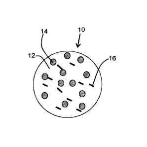
[0009] Figure 1 is a diagram of a spray-dried microparticle according to an
embodiment
of the invention including low melting point fat particles and probiotic
bacteria;
[0010] Figure 2A is a confocal light microscopy image of a spray-dried
microparticle
according to the embodiment of Figure 1, in which fat particles appear orange
due to
selective staining with Nile Red, bacterial cells are indicated by arrows and
appear blue
due to selective staining with DAPI, and sodium caseinate (NaCas) appears
green due to
selective staining with FITC;
2
WO 2016/161506
PCT/CA2016/050344
CA 02982821 2017-10-05
[0011] Figure 2B is a confocal light microscopy image of a spray-dried
microparticle
similar to the embodiment of Figure 2A but containing vegetable oil droplets
instead of
low melting point fat particles;
[0012] Figure 2C is a series of scanning electron micrographs of spray-dried
microparticles according to the embodiment of Figure 1 containing vegetable
oil and
sodium caseinate at ratios (w/w) of 0.25:1 (panels a and b), 0.50:1 (panels c
and d),
0.75:1 (panels e and f) or 1:1 (panels g and h) or low melting point fat and
sodium
caseinate at ratios (w/w) of 0.25:1 (panels i and j), 0.50:1 (panels k and l),
0.75:1 (panels
m and n) or 1:1 (panels o and p);
[0013] Figure 3A is a graph showing the thermal stability of Lactobacillus
reuteri K67 in
sodium caseinate solution;
[0014] Figure 3B is a graph showing the thermal stability of Lactobacillus
reuteri S64 in
sodium caseinate solution;
[0015] Figure 3C is a graph showing the thermal stability of Lactobacillus
zeae LB1 in
sodium caseinate solution;
[0016] Figure 4A is a graph showing the survival rate of Lactobacillus reuteri
K67
encapsulated in spray-dried microparticles including sodium caseinate (NaCas)
alone or
sodium caseinate including varying amounts of vegetable oil or low melting
point fat
(LMF);
[0017] Figure 4B is a graph showing the survival rate of Lactobacillus reuteri
S64
encapsulated in spray-dried microparticles including sodium caseinate (NaCas)
alone or
sodium caseinate including varying amounts of vegetable oil or low melting
point fat
(LMF);
[0018] Figure 4C is a graph showing the survival rate of Lactobacillus zeae
LB1
encapsulated in spray-dried microparticles including sodium caseinate (NaCas)
alone or
sodium caseinate including varying amounts of vegetable oil or low melting
point fat
(LMF);
[0019] Figure 5A is a graph showing the survival rate of a fresh culture of
Lactobacillus
reuteri K67, or Lactobacillus reuteri K67 encapsulated in spray-dried
microparticles
including sodium caseinate (NaCas) alone or sodium caseinate including varying
amounts of vegetable oil or low melting point fat (LMF), on MRS agar
supplemented with
5% NaCI;
[0020] Figure 5B is a graph showing the survival rate of a fresh culture of
Lactobacillus
zeae LB1, or Lactobacillus zeae LB1 encapsulated in spray-dried microparticles
including
3
WO 2016/161506
PCT/CA2016/050344
CA 02982821 2017-10-05
sodium caseinate (NaCas) alone or sodium caseinate including varying amounts
of
vegetable oil or low melting point fat (LMF), on MRS agar supplemented with 5%
NaCI;
[0021] Figure 5C is a graph showing the survival rate (on a logarithmic scale)
of a fresh
culture of Lactobacillus reuteri S64, or Lactobacillus reuteri S64
encapsulated in spray-
dried microparticles including sodium caseinate (NaCas) alone or sodium
caseinate
including varying amounts of vegetable oil or low melting point fat (LMF), on
MRS agar
supplemented with 5% NaCI;
[0022] Figure 6A is a graph showing the survival rate of Lactobacillus zeae
LB1
encapsulated in spray-dried microparticles including varying proportions of
sodium
caseinate (NaCas) and gum Arabic in addition to low melting point fat,
compared to
unencapsulated (free) LB1, when exposed to simulated gastric fluid (1 h - 2 h)
and
simulated intestinal fluid (3 h ¨ 6 h);
[0023] Figure 6B is a graph showing the release of Lactobacillus zeae LB1 from
the
spray-dried microparticles of Figure 6A when exposed to simulated gastric
fluid (1 h ¨
2 h) and simulated intestinal fluid (3 h ¨ 6 h);
[0024] Figure 7 is a graph showing the survival rate of Lactobacillus zeae LB1
from the
spray-dried microparticles of Figure 6A after storage at 4 C for varying
lengths of time;
[0025] Figure 8A is a graph showing the effect of outlet temperature on the
water content
of spray dried microparticles including Lactobacillus zeae LB1 and either
sodium
caseinate (NaCas) alone or a 1:1 ratio (w/w) of sodium caseinate and either
vegetable oil
or low melting point fat (LMF);
[0026] Figure 8B is a graph showing a plot of the survival rate of
Lactobacillus zeae LB1
in the spray dried microparticles of Figure 8A vs. water content;
[0027] Figure 8C is a graph showing the effect of outlet temperature on the
water activity
of the spray dried microparticles of Figure 8A;
[0028] Figure 8D is a graph showing a plot of the survival rate of
Lactobacillus zeae LB1
in the spray dried microparticles of Figure 8A vs. water activity;
[0029] Figure 8E is a graph showing a plot of the water activity of the spray
dried
microparticles of Figure 8A vs. water content;
[0030] Figure 9A is a graph showing a series of differential scanning
calorimetry (DSC)
curves of emulsions of vegetable oil in 10% (w/w) aqueous sodium caseinate
(NaCas)
solution at various ratios of oil to NaCas; the control is 10% (w/w) aqueous
NaCas
solution containing no oil;
[0031] Figure 9B is a graph showing a series of differential scanning
calorimetry (DSC)
curves of emulsions of low melting point fat (LMF) in 10% (w/w) aqueous sodium
4
WO 2016/161506
PCT/CA2016/050344
CA 02982821 2017-10-05
caseinate (NaCas) solution at various ratios of LMF to NaCas; the control is
10% (w/w)
aqueous NaCas solution containing no LMF; and pure fat represents unemulsified
LMF;
and
[0032] Figure 10 is a graph of a plot of survival of Lactobacillus zeae LB1
encapsulated
in spray-dried microparticles including sodium caseinate (NaCas) including
varying
amounts of low melting point fat (LMF) vs. the melting enthalpy of emulsions
of LMF in
10% (w/w) aqueous NaCas solution containing corresponding ratios by weight of
LMF to
NaCas.
Detailed Description
[0033] One aspect of the present invention provides microparticles. With
reference to
Figure 1, spray dried microparticles 10 include a matrix 12 of an
encapsulating material,
dispersed in which are smaller particles of a low melting point fat 14 and a
bioactive
material 16. The particles of the low melting point fat are substantially
separate and
distinct from the bioactive material. As used herein, the term "microparticle"
is intended to
mean a particle which has a diameter of from about 0.1 pm to about 100
[0034] As used herein, the term "about" or "approximately" as applied to a
numerical
value or range of values is intended to mean that the recited values can vary
within an
acceptable degree of error for the quantity measured given the nature or
precision of the
measurements, such that the variation is considered in the art as equivalent
to the recited
values and provides the same function or result. For example, the degree of
error can be
indicated by the number of significant figures provided for the measurement,
as is
understood in the art, and includes but is not limited to a variation of 1 in
the most
precise significant figure reported for the measurement. Typical exemplary
degrees of
error are within 20 percent (%), preferably within 10%, and more preferably
within 5% of a
given value or range of values. Alternatively, and particularly in biological
systems, the
terms "about" and "approximately" can mean values that are within an order of
magnitude, preferably within 5-fold and more preferably within 2-fold of a
given value.
Numerical quantities given herein are approximate unless stated otherwise,
meaning that
the term "about" or "approximately" can be inferred when not expressly stated.
[0035] As used herein, the term "substantially" refers to the complete or
nearly complete
extent or degree of an action, characteristic, property, state, structure,
item, or result. For
example, a particle that is "substantially" separate from another particle
within a matrix is
intended to mean that the particles are either completely separated by
intervening matrix
material or nearly completely separated so that some incidental contact is
possible, but
the particles do not undergo any degree of contact or intermixing which would
have a
measureable effect on their individual functions or structures. The exact
allowable degree
WO 2016/161506
PCT/CA2016/050344
CA 02982821 2017-10-05
of deviation from absolute completeness may in some cases depend on the
specific
context. However, generally speaking the nearness of completion will be so as
to have
the same overall result as if absolute and total completion were obtained.
[0036] The use of "substantially" is equally applicable when used in a
negative
connotation to refer to the complete or near complete lack of an action,
characteristic,
property, state, structure, item, or result. For example, a composition that
is "substantially
free of" particles would either completely lack particles, or so nearly
completely lack
particles that the effect would be the same as if it completely lacked
particles. In other
words, a composition that is "substantially free of" an ingredient or element
may still
actually contain such item as long as there is no measurable effect thereof.
[0037] As used herein, the term "bioactive material" is intended to mean
microorganisms,
material derived from or produced by organisms or microorganisms (including
but not
limited to tissue, genetic material, extracts, products including but not
limited to enzymes,
and the like), or organic material which has biological activity or which is
necessary or
desirable to sustain life functions (including but not limited to drugs, food
and organic
nutrients including but not limited to proteins, carbohydrates, vitamins, and
the like). As
used herein, the term "microorganisms" is intended to mean unicellular,
multicellular or
non-cellular microscopic organisms and includes but is not limited to
prokaryotic
microorganisms including but not limited to bacteria, archaea and the like;
eukaryotic
microorganisms including but not limited to algae, protists, fungi, yeasts,
molds, mites,
nematodes and the like; and infectious particles including but not limited to
viruses,
phages, prions and the like. Bioactive material can be, but need not
necessarily be, alive.
[0038] In at least one embodiment, the bioactive material is a heat-sensitive
bioactive
material whose viability can be reduced if the bioactive material is exposed
to
temperatures above a predefined range. As used herein, the term "viability" is
intended to
mean the ability to live or be sustained, or to fulfil a biological function.
Conditions under
which a bioactive material is viable need not be those under which the
bioactive material
is actively growing or functioning, but can also include conditions under
which the
bioactive material is inactive or dormant, as long as it retains at least some
potential to
live or fulfil its function. Non-living bioactive material can have viability
if it has not
decomposed or been deactivated beyond its ability to fulfil its intended
biological function.
[0039] It will be understood by the skilled person that different bioactive
materials have
different optimal temperature ranges at which viability can be maintained or
preserved.
Therefore some bioactive materials are readily damaged or destroyed, so as to
lose or
experience reduced viability, by exposure to temperatures at which other
bioactive
materials will retain full or significant viability. For example, damage can
occur at various
6
WO 2016/161506
PCT/CA2016/050344
CA 02982821 2017-10-05
sites in bacterial cells including the cell wall, cytoplasmic membrane,
ribosomes, RNA
and DNA. However, there is often a critical temperature above which the
survival of cells
decreases dramatically, and this critical temperature can be different for
different
microorganisms, including but not limited to different species, strains,
varieties or isolates.
At temperatures below the critical temperature, the cell membrane is likely to
be the main
site at which damage occurs, while at temperatures above the critical
temperature,
denaturation of ribosomes and/or proteins, as well as damage to the cell wall
can occur
and lead to thermal death of the cells.
[0040] The present microparticles include a matrix of an encapsulating
material. Suitable
encapsulating materials are well known in the art and include, but are not
limited to,
proteins such as casein or sodium caseinate, whey protein, soy protein,
gelatin and the
like, carbohydrates such as gum arabic, carrageenan, locust bean gum, gellan
gum,
xanthan gum, cellulose acetate phthalate, starch, pectin, alginate, chitosan
and the like,
and mixtures thereof.
[0041] The present microparticles include particles of a low melting point fat
dispersed in
the matrix of the encapsulating material. In at least one embodiment, the low
melting
point fat will have a melting point above normal room temperature, so as to be
in the solid
phase under normal ambient conditions. Thus, a low melting point fat would not
include
an oil which is normally liquid under normal ambient conditions, as understood
in the art.
In at least one embodiment, the low melting point fat will have a melting
point above
about 25 C. In at least one embodiment, the low melting point fat will have a
melting point
in the range of about 25 C to about 60 C. In at least one embodiment, the low
melting
point fat will have a melting point in the range of about 25 C to about 45 C.
In at least one
embodiment, the low melting point fat will have a melting point in the range
of about 30 C
to about 45 C. Suitable low melting point fats are known and include but are
not limited to
shortenings, cocoa butter, margarine, fatty acids, lard, suet, palm oil,
fractionated palm
oil, hydrogenated oils and mixtures thereof. Hydrogenated oils include but are
not limited
to hydrogenated palm oil, hydrogenated cottonseed oil and hydrogenated coconut
oil. In
at least one embodiment, the low melting point fat is selected from palm oil,
hydrogenated
cottonseed oil and mixtures thereof.
[0042] In at least one embodiment, the ratio of low melting point fat to
encapsulating
material in the microparticles varies from about 0.25:1 to about 1:1 by
weight. In at least
one embodiment, the ratio of low melting point fat to encapsulating material
in the
microparticles varies from about 0.50:1 to about 1:1 by weight. In at least
one
embodiment, the ratio of low melting point fat to encapsulating material in
the
microparticles varies from about 0.75:1 to about 1:1 by weight. In at least
one
7
WO 2016/161506
PCT/CA2016/050344
CA 02982821 2017-10-05
embodiment, the ratio of low melting point fat to encapsulating material in
the
microparticles is about 1:1 by weight.
[0043] In at least one embodiment, the present microparticles are prepared by
heating
the low melting point fat to form a liquid melt; mixing the liquid melt with
an aqueous
mixture of an encapsulating material to form an emulsion; cooling the emulsion
below the
melting point of the low melting point fat to allow solidification of the fat
particles,
dispersing the bioactive material into the emulsion; and spray drying the
emulsion to form
the microparticles.
[0044] The low melting point fat can be melted to form the liquid melt at any
temperature
above its melting point which will maintain the low melting point fat in
liquid form without
causing measurable or detrimental decomposition. Once melted, the liquid melt
can be
mixed with an aqueous mixture of an encapsulating material at a temperature at
which
the low melting point fat would remain melted, so as to form an emulsion. The
emulsion
can be prepared by using techniques well known in the art, including but not
limited to
blending and/or homogenizing the mixture of the liquid melt and the aqueous
mixture of
the encapsulating material, and treating the mixture of the liquid melt and
the aqueous
mixture of the encapsulating material with ultrasound.
[0045] In at least one embodiment, the aqueous mixture of the encapsulating
material is
an aqueous solution of the encapsulating materials described herein above. In
at least
one embodiment, the aqueous mixture further comprises one or more additives,
including
but not limited to prebiotics and protectants and antioxidants. Suitable
prebiotics and
protectants include but are not limited to sugars, oligosaccharides and
polysaccharides,
including but not limited to starch, maltodextrin, inulin, trehalose, and the
like. Suitable
antioxidants are advantageously lipid-soluble antioxidants, including but not
limited to
butylated hydroxyanisole (BHA), butylated hydroxytoluene (BHT), tert-
butylhydroquinone
(TBHQ), vitamin E, tocopherols, tocotrienols, and the like.
[0046] In at least one embodiment, the prepared emulsion of the low melting
point fat and
the aqueous mixture of the encapsulating material is cooled below the melting
point of the
low melting point fat, such that solid particles of the low melting point fat
are formed, and
the bioactive material is dispersed in the emulsion. The bioactive material
can be added
to the emulsion in any convenient form, including but not limited to a
solution or
dispersion in a suitable solvent, such as water. If the bioactive material
includes one or
more microorganisms, it can be added as a suspension in a culture medium or
diluted
culture medium. The bioactive material can be dispersed in the emulsion by any
known
technique, including but not limited to stirring and vibration.
8
WO 2016/161506
PCT/CA2016/050344
CA 02982821 2017-10-05
[0047] The emulsion containing the dispersed bioactive material is then spray
dried,
using apparatus and conditions well known in the art, to form the encapsulated
bioactive
material in the form of a powder. Advantageously, the outlet temperature of
the spray
drying apparatus is as high as possible without causing deleterious effect to
the bioactive
material, as will be understood in the art. Without being bound by theory, it
is believed
that higher outlet temperatures, where possible without deleterious effect,
will
advantageously reduce the water content of the spray dried powder and improve
the
storage stability of the spray dried powder. In at least one embodiment, the
spray drying
can be carried out at an outlet temperature of from about 65 C to about 80 C.
However,
the skilled person is readily able to select other suitable outlet
temperatures for various
bioactive materials in light of the teaching herein.
[0048] Without being bound by theory, it is believed that the droplets of low
melting point
fat dispersed through the emulsion return to the solid crystal phase when the
emulsion is
cooled below the melting point of the low melting point fat. After the
emulsion containing
the bioactive material is transferred into the spray drier, it first passes
for a short period
through a chamber in which the temperature is almost as high as the inlet
temperature.
The emulsion is then sprayed through a nozzle as micro-droplets into the
drying chamber.
The encapsulated bioactive material can be exposed to high temperatures in
these
locations. At such temperatures, the low melting point fat particles can melt
or undergo a
solid to liquid phase transition, thereby absorbing heat while maintaining a
constant
temperature. Because of this heat absorption, the temperature of any bioactive
material
embedded in the encapsulating material in the vicinity of the melting fat
particles is
prevented from increasing to the extent that it would if the fat particles
were not present.
Thus, in at least one embodiment of the present microparticles, the particles
of low
melting point fat within the matrix of encapsulating material can protect the
encapsulated
bioactive material, including but not limited to probiotic bacteria, from heat
damage during
the spray drying process. Furthermore, in at least one embodiment of the
present
microparticles, it is contemplated that the particles of low melting point fat
within the
matrix of encapsulating material can protect the encapsulated bioactive
material,
including but not limited to probiotic bacteria, from heat damage during other
processing
steps involving heat.
EXAMPLES
[0049] Other features of the present invention will become apparent from the
following
non-limiting examples which illustrate, by way of example, the principles of
the invention.
[0050] Sodium caseinate (NaCas) was purchased from Sigma-Aldrich Chemical Co.,
Ltd
(St. Louis, MO, USA). Vegetable oil and low melting point fat (LMF) were
obtained from
9
WO 2016/161506
PCT/CA2016/050344
CA 02982821 2017-10-05
UNICO Inc. (ON, Canada) and 101 Loders Croklaan Inc. (SansTransTm 39, IL,
USA),
respectively. Glassware was sterilized at 121 C for 15 min. The stains 4',6-
diamidino-2-
phenylindole (DAPI) and fluorescein isothiocyanate (FITC) were purchased from
Sigma-
Aldrich (St-Louis, MO, USA), and 9-diethylamino-5H-benzo[a]phenoxazine-5-one
(Nile
Red) was purchased from Kodak (Rochester, NY, USA).
[0051] The results of each data point in the graphs shown in the Figures
represent the
mean of triplicate experiments and the error bars indicate the standard
deviations for the
data points. All differences were considered statistically significant at a
0.05.
Example 1: Preparation of Lactobacillus isolates
[0052] Lactobacillus zeae LB1 (LB1) and Lactobacillus reuteri S64 (S64) and
K67 (K67)
are isolates from chicken or pig intestines with the capacity to inhibit
Salmonella or E. coli
infection in Caenorhabditis elegans, broiler chickens, or pigs. Isolates from
stock cultures
in 15% (v/v) aqueous glycerol at -80 C were cultured on de Man, Rogosa and
Sharpe
(MRS) agar (BD Institution, MD, USA) for recovery and single colony
purification. Each
isolate was sub-cultured twice in MRS broth at 37 C for 24 h prior to
preparation of a
fresh culture inoculated (1%, v/v) in MRS broth and grown at 37 C for 12
hours. All
cultures were grown under anaerobic atmosphere (80% N2, 15% CO2 and 5% H2) and
were harvested in the early stationary phase. A probiotic culture in the
stationary phase
often has better heat resistance than in the exponential phase. Bacterial
cells were
harvested by centrifugation (SorvallTM RC 6 Plus, Thermo Scientific Inc., MA,
USA) at
4,000 x g for 20 min (4 C) and washed twice with sterile 0.85% (w/v) sodium
chloride
solution. The pellet was then re-suspended in sterile 0.85% (w/v) sodium
chloride solution
to obtain a suspension containing approximately 10' colony-forming units
(CFU)/mL. The
bacterial suspension (101 CFU/mL) was stored at 4 C and used on the same day.
Example 2: Thermal tolerance of Lactobacillus isolates
[0053] Two 50 mL bottles containing 19 mL NaCas solution (10%, w/w) were
placed in a
water bath at test temperatures of 54 C, 57 C, 60 C, 63 C and 66 C. One of the
bottles
was a control used to monitor the temperature. When the desired temperature
was
reached, 1 mL of either Lactobacillus zeae LB1 (LB1) or Lactobacillus reuteri
S64 (564)
or K67 (K67) cell suspension (Example 1) was added to the second bottle. At
selected
intervals (between 30 s and 5 min), 1 mL aliquots were removed from the test
bottle,
serially diluted in MRS broth and plated on MRS agar for CFU counts.
Enumeration was
performed after 24 h of anaerobic incubation at 37 C. The plating and
enumeration were
accomplished using an Eddy Jet Spiral Plater (Neu-tec Group, Farmingdale, NY,
USA).
WO 2016/161506
PCT/CA2016/050344
CA 02982821 2017-10-05
Results
[0054] The heat tolerance of the three Lactobacillus isolates is shown in
Figures 3A
(K67), 3B (S64) and 3C (LB1). The viability of the three isolates was
unchanged at 54 C
for up to 5 min. At 57 C, a decrease of 0.55 log CFU mL-1 was obtained for
LB1, while the
other two isolates showed no decrease in viability up to 5 min. At 60 C, LB1
and K67
experienced decreases of 2.5 log CFU mL-1 and 0.35 log CFU mL-1, respectively,
but no
significant change was observed for S64. These results suggest that for each
isolate,
there is a critical temperature (60 C for LB1, 63 C for S64 and K67) above
which survival
decreases dramatically.
[0055] The D-values, or the time required to kill 90% of the cells at various
temperatures,
of the three different probiotic strains are presented in Table 1. D-values
can be used as
an indicator of the heat tolerance of microorganisms, such that the greater
the D-value,
the better the heat tolerance.
Table 1:
Temperature D-value (min)
( C) LB1 K67 S64
54 333.3 212.3 333.3
57 12.3 62.5 88.1
60 3.1 18.2 44.6
63 2.2 3.8 8.2
66 1.3 2.8 3.1
[0056] Relatively high D-values were found for all three strains at
temperatures below
57 C. Among the three isolates, the D-value of S64 was greater than those of
LB1 and
K67 at all temperatures investigated, indicating that S64 has the best thermal
tolerance,
while LB1 shows the poorest.
Example 3: Microencapsulation of Lactobacillus isolates
Sodium caseinate microencapsulation
[0057] Low melting point fat (LMF) was preheated at 50 C in a water bath to
melt all
crystals. Vegetable oil or LMF was then added into 100 mL aqueous sodium
caseinate
(NaCas) solution (10% w/w, 40 C) with varying ratios of lipid to NaCas
(0.25:1.00,
0.50:1.00, and 1.00:1.00 w/w). NaCas solution without vegetable oil or LMF
(0:1.00 w/w)
was used as a control. The mixtures were coarsely mixed using a blender
(Polytron(6) PT
10-35 GT-D, Kinematica Corporation, Switzerland) at 6000 rpm for 1 min (40 C)
and then
recirculated three times through a high pressure homogenizer (Nano DeBEE,
B.E.E.
International Inc., MA, USA) at 3000 psi (40 C). The prepared emulsions were
left at 0 C
11
WO 2016/161506
PCT/CA2016/050344
CA 02982821 2017-10-05
overnight, and Lactobacillus cultures (Lactobacillus reuteri K67 (K67) or S64
(S64) or
Lactobacillus zeae LB1 (LB1)) were dispersed into the emulsions and stirred at
100 rpm
for 10 min at 0 C. The final mixtures (109 CFU/g dry coating material) were
then spray
dried in a laboratory scale spray dryer (ADL 310, Yamato Scientific America
Inc., CA,
USA), at a constant inlet temperature of 170 C and outlet temperature of 80 C
and a flow
rate of 5 mUmin. Dried powder samples were collected from the base of the
cyclone and
stored in tightly sealed sterile bottles at 4 C.
Sodium caseinate-gum arabic microencapsulation
[0058] Sodium caseinate (NaCas) - gum arabic (GA) complex solutions having
ratios of
NaCas : GA of 4:0, 3:1, 2:2, 1:3, 0:4 (w/w) (total solid content 10% (w/w))
were prepared
in distilled water and stirred overnight at 4 C. The solutions were adjusted
to pH 7.0 and
pre-heated to 40 C. LMF was then added into the complex solutions at a ratio
of 1:1
(w/w). Emulsification, dispersion of Lactobacillus cultures into the emulsions
and spray
drying were carried out as described above.
Example 4: Surface and internal microstructure of the spray dried
microparticles
Confocal laser-scanning microscopy (CLSM)
[0059] Microparticles were rehydrated on a glass slide with a drop of triple
fluorescent
stain (4',6-diamidino-2-phenylindole (DAPI) 0.0005% (w/v), fluorescein
isothiocyanate
(FITC) 0.0007% (w/v) and 9-diethylamino-5H-benzo[a]phenoxazine-5-one (Nile
Red)
0.15% (w/v) in a 100 mM CaCl2 solution). A cover slip was then applied with 4
drops of
nail polish in the corners as a spacer to prevent compression of the
microparticles. Lipid
particles appear orange due to selective staining with Nile Red, bacterial
cells appear
blue due to selective staining with DAPI and sodium caseinate (NaCas) appears
green
due to selective staining with FITC. Observations of bacterial cells, protein
and lipid were
performed with a Carl Zeiss LSM 510 Duo confocal laser-scanning microscope
(Gottingen, Germany) using excitation lines at 405, 488 and 532 nm and
emission band
pass 420 - 490 nm, 515 - 550 nm and 575 - 700 nm for DAPI, FITC and Nile Red
respectively.
Results
[0060] As seen in Figures 2A and 2B, lipid particles (orange) and bacterial
cells (blue,
indicated by arrows) were dispersed throughout the NaCas matrix (green) with
no visible
differences between the oil and fat containing samples. With increasing core
(lipid) to wall
(NaCas) ratio, the density of the oil/fat globules within the particles
increased, but the
diameter of the oil/fat globules remained constant, possibly because the same
process
and parameters were applied during the preparation of emulsions and spray
drying.
12
WO 2016/161506
PCT/CA2016/050344
CA 02982821 2017-10-05
Bacterial cells were observed only in the NaCas matrix within the
microparticles, and not
within the fat particles or oil droplets, reflecting the overall hydrophilic
nature of the
bacteria surfaces. When mixed with the emulsion, the bacteria are believed to
spontaneously move into the hydrophilic phase (NaCas matrix) instead of the
hydrophobic phase (oil or fat phase).
Scanning electron microscopy (SEM)
[0061] The surface morphology of the microparticles was observed with a
scanning
electron microscope at an accelerating voltage of 20 kV. Prior to recording
microscopic
observations, carbon sticky tabs were attached to aluminum stubs and the
sticky surface
was lightly coated with gold for 45 seconds to help reduce charging in the
microscope.
Small amounts of microparticles were then dusted onto the stubs, spread with a
spatula,
and the excess particles were blown off with forced air. The stubs were then
coated with
gold for 2.5 minutes, for a final gold thickness of approximately 8.9 nm.
Results
[0062] Scanning electron micrographs are presented in Figure 2C of
microparticles
produced with varying ratios of oil (panels a to h) or low melting point fat
(panels i to p) to
sodium caseinate. The diameters of spray dried microparticles were around 15
to 20
and no bacteria were observed on the surface of the microparticles. The
microparticles
containing different lipid core materials (oil or low melting point fat) were
similar in
appearance, indicating that the lipid used did not affect the morphology of
the particles.
The shape of the particles varied from irregular to spherical, and the
surfaces of the
particles were mostly wrinkled with concavities which is believed to be
attributed to the
shrinkage of the particles caused by rapid evaporation of the water.
Example 5: Survival of spray dried microencapsulated Lactobacillus isolates
[0063] Bacterial cell viability of spray dried powders (Example 3) was
determined by the
standard plate counting method. Spray dried powders (0.5 g) were dispersed in
4.5 mL
0.2 M phosphate buffer (pH 7.0) and homogenized for 1 min at 4000 rpm
(Polytron PT
10-35 GT-D, Kinematica Corporation, Switzerland). Enumeration of cells was
carried out
by plating on MRS agar. Colony forming units (CFU) were enumerated manually
after
incubation at 37 C for 24 h.
CFU/g spray dried powder
survival rate (%)=1. ______________________________________ x 00%
CFU/g total solid in initial solution prior to spray drying
Results
[0064] Among the control samples of the three Lactobacillus isolates (i.e.
those
containing NaCas but no oil or LMF), the highest survival rate (- 95%) was
obtained with
13
WO 2016/161506
PCT/CA2016/050344
CA 02982821 2017-10-05
Lactobacillus reuteri S64 (S64), as seen in Figure 4B, which is consistent
with the higher
thermal tolerance of this isolate as noted in Example 2 above. Addition of
either vegetable
oil or LMF did not alter the survival rates of Lactobacillus reuteri K67 (K67)
(Figure 4A)
and S64 (Figure 4B) after spray drying. However, as seen in Figure 4C, the
survival rate
of Lactobacillus zeae LB1 (LB1) in the control sample was only about 16%.
Among the
samples of LB1 containing vegetable oil as core material, the survival rates
were almost
the same (around 16%), and not significantly different from that in the
control sample (p<
0.05). In contrast, addition of LMF increased the survival rate of LB1 from
16% to 63% as
the LMF to wall ratio increased from 0.25 to 1.00.
Example 6: Salt tolerance of microencapsulated Lactobacillus isolates
[0065] Fresh cultures and spray dried microparticles prepared as described in
Example 3
of Lactobacillus reuteri K67 (K67), Lactobacillus zeae LB1 (LB1) and
Lactobacillus reuteri
S64 (S64) were plated on MRS agar without NaCI or supplemented with NaCI (5%,
w/v).
The plates were incubated for up to 3 days under anaerobic conditions and
viable
numbers were recorded. The survival rate was determined using the following
equation:
Ns
Survival rate ( /0) =¨N,
where Ns and N, represent the survival number grown on MRS agar containing
NaCI and
MRS agar without NaCl, respectively.
[0066] The sensitivity of bacteria to salt was defined as follows:
Sensitivity (%) = 100 (%) ¨ Survival rate (%)
Results
[0067] Fresh cultures of the three isolates exhibited varying degrees of
tolerance to salt,
with survival rates of 96%, 76%, and 5% for K67, LB1 and S64, respectively, as
seen in
Figures 5A, 5B, and 5C, respectively. The survival rates on NaCI-MRS agar of
all spray
dried bacterial isolates encapsulated in NaCas without inclusion of oil or LMF
were
markedly lower than those of the fresh bacterial cultures: 30%, 5%, and 0.2%
for K67,
LB1 and S64, respectively. For isolates K67 and S64, spray drying induced
minimal loss
in cell viability (Example 5), but resulted in a significant decrease in salt
tolerance. This
result suggests that although the bacterial cells survived the spray drying
process, some
damage to the cell membrane may have occurred, so that the tolerance to salt
decreased. In the case of LB1, severe loss of viability was observed after
spray drying
(Example 5, Figure 4C), accompanied by further loss of salt tolerance (Figure
5B), which
suggests that the cell damage may be more extensive.
14
WO 2016/161506
PCT/CA2016/050344
CA 02982821 2017-10-05
[0068] The survival rates on NaCI-supplemented MRS agar of all three isolates
microencapsulated with vegetable oil were similar to those in the control
NaCas-only
microparticles, suggesting that inclusion of oil in the formula did not affect
the salt
tolerance of bacteria after spray drying. In contrast, significant increases
in survival rate
on NaCI-supplemented MRS agar were observed for isolates LB1 and K67 in the
presence of LMF when the ratio of LMF to wall material reached 1.0 (P<0.05),
indicating
that the addition of LMF to the microparticles can protect these isolates
against damage
experienced during spray drying which would have otherwise been expected to
further
decrease their tolerance to salt. The presence of LMF in microparticles of
encapsulated
S64 had little effect on the salt tolerance of the relatively thermally
tolerant (Example 2,
Figure 3B; Example 5, Figure 4B) but highly salt intolerant (Figure 5C) S64
isolate.
Example 7: Survival and release of microencapsulated Lactobacillus zeae LB1
(LB1) under simulated gastrointestinal conditions
[0069] Microparticles (Example 3, 0.1 g) containing Lactobacillus zeae LB1
(LB1)
encapsulated in a matrix containing varying proportions of sodium caseinate
(NaCas) and
gum Arabic, and containing low melting point fat in a 1:1 ratio by weight with
the
encapsulating matrix (Example 3), or free LB1 bacterial cells harvested as
described in
Example 1 and diluted in sterile 0.85% (w/v) sodium chloride solution to -109
CFU/mL
(0.1 mL), were added to test tubes containing 9.9 mL of pre-warmed (37 C)
freshly
prepared and filter sterilized simulated gastric fluid (SGF) (0.32 wt% pepsin,
0.2 wt%
NaCl, adjusted to pH 2.0 with 1M NCI). The samples were vortexed and incubated
at
37 C. Samples were removed at 30, 60, 90, and 120 min for bacterial counting,
and the
pH was then rapidly adjusted to 7.0 with 1M NaOH. Simulated intestinal fluid
(SIF)
(pancreatin (10 g/L) and bile salts (8 g/L) in phosphate buffer (0.2M,
pH=7.0)) (10 mL)
was added, and 1 mL aliquots were removed from each sample for bacterial
counting
after exposure to SIF for a further 1, 2, 3, and 4 h.
[0070] For the measurement of protection properties of microparticles, samples
(1 mL)
were added to 9 mL phosphate buffered saline (PBS) and homogenized for 1 min
at 4000
rpm before determination of viable cell numbers. For the measurement of
release
properties of microparticles, samples (1 mL) were withdrawn without
homogenization and
directly added into 9 mL PBS for bacterial counting. Enumeration of cells was
carried out
by plating on MRS agar. Colony forming units (CFU) were enumerated manually
after
incubation at 37 C for 24 h.
Results
[0071] Survival of encapsulated LB1 during simulated gastrointestinal
digestion (2 hours
of exposure to SGF, followed by 4 hours of exposure to SIF) is shown in Figure
6A. Free
WO 2016/161506
PCT/CA2016/050344
CA 02982821 2017-10-05
cells died very quickly and no viable bacterial cells were detected after 1 h
in SGF.
However, for encapsulated bacteria samples (NaCas with or without gum arabic
(GA)),
the survival increased significantly. Among these samples, survival rates of
bacteria
microencapsulated with only NaCas or gum arabic were similar to each other and
lower
than that of other encapsulated samples. As the gum arabic content in the wall
material
increased (from a NaCas: GA ratio of 3:1 to 1:3), the survival of encapsulated
bacteria
increased, with a loss of viability of only 1.2 log over 6 h of test time for
the sample having
a NaCas: GA ratio of 1:3.
[00721 Release of encapsulated LB1 during simulated gastrointestinal digestion
is shown
in Figure 6B. Free cells died very quickly and no live bacteria were detected
after 1 h in
SGF. For the encapsulated samples, the number of viable cells released from
the
microparticles remained constant (0-1 log CFU/g) during the first two hours of
exposure to
SGF and increased significantly when exposed to SIF. All the viable bacteria
in the
microparticles were released within 1 h when exposed to SIF.
Example 8: Storage stability of microencapsulated Lactobacillus zeae LB1 (LB1)
[0073] Samples of spray dried microparticles containing Lactobacillus zeae LB1
(LB1)
encapsulated in a matrix containing varying proportions of sodium caseinate
(NaCas) and
gum arabic, and containing low melting point fat in a 1:1 ratio by weight with
the
encapsulating matrix (Example 3) were stored at 4 C in sealed polyethylene
bags placed
in sealed glass bottles. Samples were removed at 1 week intervals for
determination of
viable bacterial count by the standard plate counting method described in
Example 5.
Results
[00741 As seen in Figure 7, minimal reduction in bacterial count was seen
during the first
4 weeks of storage at 4 C. Even after storage for 16 weeks, less than 1 log
reduction in
viability was observed.
Example 9: Water content and water activity of spray dried microparticles
[00751 Spray dried microparticles of Lactobacillus zeae LB1 (LB1) were
prepared from
sodium caseinate (NaCas) alone or mixed with vegetable oil in a 1:1 ratio
(w/w) as
described in Example 3. Spray drying was carried out at outlet temperatures of
65 C,
70 C, 75 C or 80 C. A comparison sample of spray dried microparticles of LB1
was
prepared from a 1:1 ratio (w/w) of NaCas and LMF at an outlet temperature of
80 C.
[00761 Weighing dishes were dried in an oven (105 C) to a constant weight and
then
cooled in a desiccator containing silica gel. The weight of the empty dish was
recorded
(a), approximately 3 g of powder was added, and the dish was weighed again
(b). The
loaded dish was placed in the oven at 105 C for 24 h, then cooled to room
temperature in
16
WO 2016/161506
PCT/C42016/050344
CA 02982821 2017-10-05
a desiccator and weighed again (c). The heating and cooling process was
repeated until
the weight (c) was constant. The water content was calculated as:
(b -c) x 100%
Water content - ______________________________
(c - a)
where a is the weight of the empty dish; b is the weight of the dish and the
wet powder;
and c is the weight of the dish and the dried powder.
[0077] The water activity was measured at 25 C using a water activity meter
(Aqualab
4TE, Decagon Devices Inc., USA).
Results
[0078] Microparticles spray dried at 80 C and containing NaCas only were found
to have
a water content of 6.80% by weight; whereas microparticles spray dried at 80 C
and
formulated with a 1:1 ratio of oil:NaCas or LMF:NaCas were found to have a
water
content of 3.25% by weight and 3.68% by weight, respectively. Assuming that
the water is
present in the NaCas phase only and is substantially absent from the lipid
phase, the
water content of the NaCas phase of the microparticles formulated with a 1:1
ratio of
oil:NaCas or LMF:NaCas would be 6.78% by weight and 7.67% by weight,
respectively.
As seen in Figures 2A and 2B, the LB1 cells are primarily located in the NaCas
phase of
the microparticles.
[0079] To determine whether the relatively high water content in the NaCas
phase of the
1:1 LMF:NaCas microparticles could have partially contributed to the high
survival of
bacteria in these microparticles, microparticles having similar water content
but containing
either NaCas alone or 1:1 oil:NaCas were prepared by spray drying at various
outlet
temperatures. As seen from the data presented in Figure 8A, an outlet
temperature of
about 74 C would be required to provide microparticles containing either NaCas
alone or
1:1 oil:NaCas which would have a water content of about 7.6%, similar to that
found in
1:1 LMF:NaCas microparticles spray dried at 80 C. As can be seen from the data
presented in Figure 8B, the interpolated survival rate of LB1 would be similar
in
microparticles containing either NaCas alone or 1:1 oil:NaCas and having a
water content
of about 7.6 /0.However, the interpolated survival rate of LB1 in
microparticles containing
either NaCas alone or 1:1 oil:NaCas would be much lower than the survival rate
observed
for LB1 encapsulated in 1:1 LMF:NaCas microparticles spray dried at 80 C and
having a
similar water content. This data thus indicates that the water content of the
microparticles
is not primarily responsible for the improved survival rate of LB1 cells in
microparticles
containing LMF particles.
[0080] Microparticles spray dried at 80 C and containing NaCas only were found
to have
a water activity of 0.18; whereas microparticles spray dried at 80 C and
formulated with a
17
WO 2016/161506
PCT/CA2016/050344
CA 02982821 2017-10-05
1:1 ratio of oil:NaCas or LMF:NaCas were found to have a water activity of
0.19 and 0.20,
respectively. As seen from the data presented in Figure 8C, the water activity
of
microparticles formulated with NaCas only and spray dried at an outlet
temperature of
75 C and the water activity of microparticles formulated with 1:1 ratio of
oil:NaCas and
spray dried at an outlet temperature of 72 C would be expected to be similar
to the water
content of microparticles formulated with a 1:1 ratio of LMF:NaCas and spray
dried at
80 C. However, as seen from the data presented in Figure 8D, the survival rate
of LB1 in
microparticles formulated with a 1:1 ratio of LMF:NaCas and spray dried at 80
C is
improved over the survival rate of LB1 in microparticles having similar water
activity but
formulated with NaCas only or with a 1:1 ratio of oil:NaCas.
[0081] It is known that a water activity between 0.11 and 0.23 can prevent
cell death
during storage, while water activity above this range is related to
accelerated mortality of
probiotics. As seen in Figure 8E, the water activity of the present
microparticles was
found to be in the acceptable range for maintenance of the survival of
probiotics during
storage, over a range of water content values.
Example 10: Thermal properties of emulsions containing LMF or vegetable oil
[0082] Thermal properties of emulsions containing LMF or vegetable oil in
aqueous
sodium caseinate (NaCas) solution (10% w/w) (prepared as described in Example
3)
were measured using a differential scanning calorimeter (DSC, Auto 020, TA
Instruments, DE, USA). Pure LMF (7 mg) or samples of the emulsion or the non-
emulsified 10% (w/w) aqueous sodium caseinate solution (control) (50 mg) were
weighed
and sealed in aluminum pans and loaded into the DSC. The samples were heated
from
0 C to 80 C at 1.5 C/min. All measurements were run against an empty pan and
heat
flow was recorded as a function of temperature.
Results
[0083] Differential scanning calorimetry (DSC) measures the heat capacity of
physical
states and the excess heat associated with transitions that can be induced by
temperature change. DSC profiles for the vegetable oil or LMF emulsions
prepared with
different lipid core to sodium caseinate wall ratios are presented in Figures
9A and 9B,
respectively. Neither endothermic nor exothermic peaks were observed for the
control
(10% (w/w) aqueous sodium caseinate solution) or emulsions made with vegetable
oil in
the temperature range from 0 C to 80 C (Figure 9A). As seen in Figure 9B,
however, for
the pure LMF sample, there were four peaks in the temperature range of 0 C to
80 C, at
5.46 C, 12.30 C, 21.13 C and 40.06 C. These peaks could be associated with the
four
main fatty acid components with differing chain lengths that constitute the
LMF. For the
emulsions containing LMF and NaCas at different core to wall ratios, the peak
at about
18
WO 2016/161506
PCT/CA2016/050344
CA 02982821 2017-10-05
40.06 C still existed for all samples. However, the first three peaks seen in
the pure LMF
sample were only observed in samples having a high LMF to NaCas ratio,
possibly due to
the detection limit of the DSC. With increasing LMF to NaCas ratios from 0.25
to 1.00, the
intensity of all peaks increased.
[0084] Melting enthalpy (AH) represents the energy required to melt the
crystal fat
present in the samples. The AH values of emulsions with different LMF to NaCas
ratios
are presented in Table 2.
Table 2:
H J/
LMF/NaCas (w:w) A ( g)
peak 1, 2, 3 peak 4 Total
LMF only 59.66 28.04 87.70
0.25 3.53 1.61 5.14
0.50 7.33 3.52 10.85
0.75 11.39 5.30 16.69
1.00 15.42 7.25 22.67
[0085] As the LMF to NaCas ratio increased from 0.25 to 1.00, AH increased
gradually
from 3.53 J/g to 15.42 J/g for the first three peaks and from 1.61 J/g to 7.25
J/g for the
last peak, respectively. The increased Sid in the LMF emulsion samples
suggested that
the addition of LMF would provide the emulsion with endothermic peaks at the
temperature around its melting point. The amount of absorbed heat energy
increased with
increasing LMF : NaCas ratio. For the LMF sample with core to wall ratio of
0.25, the
survival rate of LB1 was similar to those of the control and vegetable oil
samples. The
total melting enthalpy of the LMF/NaCas emulsions with different LMF to NaCas
ratios
was found to positively correlate with the survival of LB1 after spray drying
as shown in
Figure 10.
[0086] The embodiments described herein are intended to be illustrative of the
present
compositions and methods and are not intended to limit the scope of the
present
invention. Various modifications and changes consistent with the description
as a whole
and which are readily apparent to the person of skill in the art are intended
to be included.
The appended claims should not be limited by the specific embodiments set
forth in the
examples, but should be given the broadest interpretation consistent with the
description
as a whole.
19