Note: Descriptions are shown in the official language in which they were submitted.
CA 02983214 2017-10-18
WO 2016/170013 - 1 -
PCT/EP2016/058809
Treatment of Bacterial Infections in Aquaculture
Field of the Invention
The present invention relates to treatment of bacterial infections in
aquaculture,
generally farmed production of shrimp, prawns and fish. In particular the
invention
relates to compositions and methods for reducing or preventing infections
and/or for
treating existing infections in such aquaculture.
Background to the Invention
Bacteriophages are the most numerous form of life on Earth. They can be found
in
all environments where bacteria grow. Bacteriophages are detected in ground
and
surface water, soil, food (e.g., sauerkraut, wine), sewage and sludge. They
have also
been isolated from humans and animals, for example from faeces, urine, saliva,
spit,
rumen and serum. Bacteriophages are able to penetrate different organs and
tissues, including the central nervous system, and are a part of intestinal
flora
together with their bacterial hosts. They are responsible for 10-80% of total
bacterial
mortality in aquatic ecosystems and are an important factor limiting bacterial
populations.
Therapeutic applications of bacteriophage are known. WO 03/093462 discloses
methods for the immobilisation of viruses, in particular bacteriophages,
whilst
retaining their biological activity for use as antibacterial agents. Given
that the natural
environment of bacteriophages is aqueous it has been widely assumed that
stability
towards dehydration as disclosed in WO 03/093462 tends towards the natural
stability in aqueous media.
Oceans and inland waters are largely fished to their limit and the supply of
wild-
caught fish peaked in the 1990s. With the global wild fish supply stagnant and
the
human population increasing, new research shows that farmed fish and shellfish
production will have to increase by 133 percent between 2010 and 2050 in order
to
meet projected fish demand worldwide.
Nearly one-third of the world's seafood is produced by industrial aquaculture
and
production has increased by 6% per year from 8.7 million tons of fish in 1990
to 50
CA 02983214 2017-10-18
WO 2016/170013 - 2 -
PCT/EP2016/058809
million tons in 2011 and projected to reach 80 million tons by 2030. Fish
farming
plants, however, often suffer from heavy financial losses due to the
development of
infections caused by microbial pathogens, including multidrug resistant
bacteria that
are easily transmitted through water and therefore able to infect a great
variety of
fish species.
Although pathogenic species have been described in the majority of bacterial
taxonomic groups, only a relatively small number are responsible for
significant
economic losses. Vibriosis and photobacteriosis are primarily diseases of
marine
and estuarine fish, both in natural and commercial production systems
throughout
the world, occurring infrequently in freshwater fish. Both diseases can cause
significant mortality in fish, reaching values of up to 100% in infected
facilities.
Vibriosis and photobacteriosis are caused by bacteria from the family
Vibrionaceae.
Vibriosis is caused by species of Vibrio, namely by Vibrio anguillarum.
Others species of Vibrio, such as V. alginolitycus, V. carchariae, V.
salmonicida, V.
damsela, V. ordalii, V. parahemolyticus and V. vulnificus, also cause
important
infections in several species of fish. Photobacteriosis is caused by
Photobacterium
damselae subsp. piscicida which is a highly pathogenic bacterium that does not
seem to have host specificity, infecting a diverse range of fish species.
Other
bacteria including Aeromonas salmonicida, causative agent of furunculosis,
Rickettsia-like bacteria, Cytophaga marina, Flavobacterium psychrophilum and
Pseudomonas plecoglossicida are also important groups of fish pathogens.
Diseases like EMS (Early Mortality Syndrome) of shrimps are caused by
bacterial
infections in which the bacterium (Vibrio parahaemolyticus) is itself infected
by a
lysogenic bacteriophage which carries a toxin gene. On bacteriophage infection
the
bacterium incorporates the bacteriophage into its genome and expresses the
toxin
which leads to EMS in the shrimp.
Although vaccination is the ideal method to prevent many different kinds of
infectious
diseases it is not always applicable in fish species.
CA 02983214 2017-10-18
WO 2016/170013 - 3 -
PCT/EP2016/058809
Chemotherapy is a rapid and effective alternative method to treat or prevent
bacterial
infections, but the frequent use of antibiotics has resulted in an increasing
drug-
resistance in pathogenic bacteria in the aquaculture, agriculture and medical
areas
and since few chemotherapeutic drugs are licensed for use in fisheries
alternative
treatments are required.
Nakai et al. Diseases of Aquatic Organisms, vol. 37, pp. 33-41, (23 June 1999)
describes the effect of treating Lactococcus garvieae infection in yellowtail
through
the use of bacteriophage to which L. garvieae is susceptible. Nakai et al.
report that
each of the three bacteriophage isolates they tested for stability in natural
(unsterilised) sea water persisted for 3 days but had perished within 1 week.
Nakai et
al. also describe the use of fish food impregnated with 1079 PFU g-1 of
bacteriophage. Giving this food to fish that were subsequently challenged with
1085
CFU of L. garvieae by anal intubation decreased the mortality rate of the
challenged
fish.
WO 2006/047872 discloses antibacterial compositions comprising bacteriophage
that are adsorbed onto a matrix. The composition may be added to a feed for
aquatic use.
Bacteriophage have been proposed for various treatments of bacterial
infection. It is
known, however, that bacteriophages survive only for relatively short periods
in their
natural environment, i.e. in water. Average decay rates of viruses in natural
seawater
samples can be calculated, based on well-known data, e.g. in C H Suttle
(Microb.
Ecol. (1994) 28: 237-243, at about 0.48 day* The greatly reduced survival of
bacteriophages in natural aqueous environments is due to a combination of
causes,
significantly predation and sunlight.
Hence, there is a need for an alternative means to treat or reduce bacterial
infections
of farmed crustaceans, especially shrimp and prawns, and fish.
Object of the Invention
An object of the present invention is to provide compositions and uses of
those
compositions and methods using those compositions that offer an alternative
CA 02983214 2017-10-18
WO 2016/170013 - 4 -
PCT/EP2016/058809
treatment of bacterial infections in commercially reared crustaceans, e.g.
prawns and
shrimp, and/or fish. A further aim of particular embodiments is to provide
improved
such compositions, uses and methods.
Summary of the Invention
A composition comprising bacteriophage covalently attached to a particle is
for use
in treating bacterial infection in fish or crustaceans. Edible particles are
preferably
used. The present invention is based upon enhancement of stability and
viability of
bacteriophage in aqueous environments, rendering possible treatment of
bacterial
infections in aquaculture.
Feed for crustaceans or fish is provided, comprising bacteriophage covalently
attached to a particle for treating bacterial infection in fish or
crustaceans.
A method of making fish or crustacean feed comprises mixing bacteriophages
covalently attached to particles into feed components, to produce feed
comprising
said particles.
Bacteria infected with a lysogenic bacteriophage are provided and can be used
in
treating disease of fish or crustaceans.
Details of the Invention
A composition of the invention accordingly comprises bacteriophage covalently
attached to a particle for use in treating bacterial infection in fish or
crustaceans.
After administration to the fish or crustaceans, for example via feed
containing the
particles, bacterial infections are treated.
The particle can be a carrier particle, made e.g. of edible material or an
inert
material, in which case the carrier particle is typically approximately
spherical. It may
have an average diameter of up to lmm, up to 100 microns, up to 50 microns, up
to
10 microns, from mm, from 10nm, from 100nm, from 0.5 microns or any
combinations of these. In specific examples below, particles in the range 1 to
200
microns were used. The particles in general can be approximately round or
CA 02983214 2017-10-18
WO 2016/170013 - 5 -
PCT/EP2016/058809
spheroid; they are preferably smooth. Particles or fragments of edible
material may
also be of irregular shapes and sizes.
Particle size is suitably measured using methods and apparatus recognized as
standard in the art. Particle sizing in dispersions can be accomplished using
a variety
of techniques, including laser diffraction, dynamic light scattering (DLS),
disc
centrifugation, and light microscopy. All of these techniques have their
advantages
and limitations. Laser diffraction relies on a well-controlled presentation of
the
sample to the measurement region and is limited to samples with narrow range
of
particle concentrations. Dilution is often required and this may affect the
particle size,
particularly in compounds with high solubility. Examples of sizing equipment
are
made by Malvern Instruments (UK), using laser diffraction methods. For highly
irregular particles, the diameter refers to the greatest diameter in any
dimension
even if the particle is relatively non-spherical.
In embodiments of the invention, bacteriophages covalently attached to a
plurality of
particles are provided. These are preferably in relatively homogenous form, in
which
a large proportion, preferably substantially all, of the plurality of
particles have
diameters in the stated range, more preferably 80% or more, 90% or more or 95%
or
more of the particles with phage covalently attached have diameters in the
stated
range (being any range as set out above or elsewhere herein).
Particles for use in the invention to which bacteriophage are immobilised by
covalent
bonding are generally edible by or substantially inert to the animal to be
treated. In
examples, nylon particles (beads) were used. Other inert, preferably non-toxic
biocompatible material may be used. In addition, the particle may be made of a
biodegradable material. Suitable materials include polymethyl methacrylate,
polyethylene, ethylene/acrylate copolymer, nylon-12, polyurethane, silicone
resin,
silica and nylon 1010. WO 2003/093462 describes further materials that the
particles
may be made from.
Immobilisation or attachment of bacteriophage to the particle substrate may be
achieved in a number of ways. Preferably, bacteriophage are immobilised via
CA 02983214 2017-10-18
WO 2016/170013 - 6 -
PCT/EP2016/058809
covalent bonds formed between the bacteriophage coat protein and the carrier
substrate.
Further, bacteriophage are preferably immobilised to the substrate via their
head
groups or nucleocapsid by activating the substrate particle before the
addition and
bonding of bacteriophage.
The term "activated/activating/activation" is understood to mean the
activation of the
substrate such as electrically, e.g. by corona discharge, or by reacting said
substrate
with various chemical groups (leaving a surface chemistry able to bind
viruses, such
as bacteriophage head or capsid groups).
Activation of said substrate may be achieved by, for example, preliminary
hydrolysis
with an acid, preferably HCI followed by a wash step of water and an alkali to
neutralise the acid. Preferably, said alkali is sodium bicarbonate. Binding of
bacteriophage via their head groups is advantageous. In the case of complex
bacteriophage for example, binding via head groups leaves the tail groups,
which are
necessary for bacteria-specific recognition, free to infect, i.e., bind and
penetrate a
host bacterial cell. A plurality of various strain-specific bacteriophage may
be
immobilised to a substrate at any one time.
Coupling of phage to a substrate is as a result of the formation of covalent
bonds
between the viral coat protein and the substrate such as through an amino
group on
a peptide, for example a peptide bond. "Coupling Agents" that aid this process
vary,
and are dependent on the substrate used. For example, for coupling to nylon or
other polymers with amino or carboxy surface groups the coupling agents
carbodiimide or glutaraldehyde may be used.
Further details of methods and preferred methods for covalent attachment of
bacteriophage to particles or pellets or feed components, retaining phage
infectivity,
are described in more detail in WO 2003/093462 and WO 2007/072049.
A further option is to use particles that comprise one or more targeting
moiety, e.g. a
protein or ligand, to direct the particles to desired targets within fish or
crustaceans.
CA 02983214 2017-10-18
WO 2016/170013 - 7 -
PCT/EP2016/058809
For example, particles can comprise one or more lectins to target them e.g. to
fish
gills for treatment e.g. of Yersinia infection.
Suitably the present invention delivers bacteriophage via feed and the
particle is
made of edible material. Hence it is conveniently incorporated in feed for
fish /
crustaceans. Bacteriophage can be attached to particles of carbohydrate (e.g.
cellulose) or protein (including fish protein or animal protein) and this can
be
achieved using for example electric discharge methods of application to nylon
beads.
Feed comprising the particles may comprise carbohydrate, protein, lipid,
vitamin or a
mixture of one or more of all.
The invention is of use in treatment of diseases of fish and crustaceans
caused by
the following bacteria:
Bacteria Marine hosts Diseases
Vibrio species: Crustaceans Vibriosis
V. harveyi Fish Necrotising hepatopancreatitis
V. fluvialis (EMS)
V. parahaemolyticus Various other infections
V. vulnificus
V. alginolyticus
V. penaeicida
V. anguillarum
V. carchariae
V. salmonicida
V. damsela
V. ordalii
V. owensii
Aeromonas species: Fish Furunculosis
A. salmonicida
A. hydrophilla
A. punctata
Yersinia ruckeri Fish Enteric redmouth disease
Monte/la viscosa Fish Winter ulcer disease
Rickettsia salmonis Salmon Salmon rickettsial syndrome (SRS)
Piscirickettsia salmonis
Lactococcus garvieae Fish Lesions of vascular endothelium
Pseudomonas Ayu fish Haemorrhagic ascites
plecoglossicida
Flavobacterium Fish Bacterial cold water disease
psychrophilum (BCWD)
CA 02983214 2017-10-18
WO 2016/170013 - 8 -
PCT/EP2016/058809
Photobacterium Fish Photobacteriosis
damselae
In one preferred application of the invention the bacteriophage are for use in
treating
bacterial infection in crustaceans; more specifically, for treating infection
by Vibrio
bacteria species.
V. parahaemolyticus is a common inhabitant of coastal and estuarine
environments
all over the world. Hence they are often found naturally associated with
shrimp
aquaculture systems. Certain environmental conditions may be more favourable
for
the establishment, survival and growth of the organism such as temperature,
salinity,
zooplankton, tidal flushing and dissolved oxygen.
V. parahaemolyticus is closely related to shrimp pathogenic luminous bacteria
such
as V. harveyi, V. campbeffi and V. owensii. These along with other closely
related
Vibrio spp form a "V. harveyi clade". Bacteria within this clade have a very
high
degree of similarity at phenotypic and genotypic level. Certain strains of V.
parahaemolyticus can cause gastroenteritis in humans and clinical strains are
characterised by the ability to produce a thermostable direct hemolysin (TDH)
or a
TDH-related hemolysin (TRH). The genes encoding these hemolysins (tdh and trh
genes) are generally used as markers for human pathogenic strains of V.
parahaemolyticus. Human pathogenic strains possessing these markers account
for
1-2 percent of environmental strains of V. parahaemolyticus. All strains (both
clinical
and environmental) produce a thermolabile hemolysin (TLH) encoded by tlh gene
and this is generally used as a marker for V. parahaemolyticus in diagnostic
tests
(48). The tdh and trh genes encoding the virulence factors are present in
"pathogenicity islands", which are discrete genetic units present only in
virulent
strains; having a Guanine + Cytosine (G + C) content that is different from
the rest of
the chromosomal DNA and are generally acquired by horizontal gene transfer.
By use of the invention with bacteriophage specific to Vibrio species these
infections
of e.g. shrimp and prawn can now be treated.
CA 02983214 2017-10-18
WO 2016/170013 - 9 -
PCT/EP2016/058809
In another preferred application of the invention, the bacteriophage are for
treating
bacterial infection in fish, especially for treating infection by Vibrio,
Aeromonas,
Yersinia, Monte/la, Rickettsia, Piscirickettsia, Lactococcus, Pseudomonas,
Flavobacterium or Photobacterium bacteria species. Useful bacteriophage are
disclosed e.g. in US 2013/0323209.
Feed for fish and crustaceans, especially shrimp and prawns, is provided by
the
invention. One aspect of these embodiments of the invention hence provides
feed for
crustaceans or fish, comprising bacteriophages covalently attached to
particles for
treating bacterial infection in fish or crustaceans.
It is preferred that all of the feed is edible and so it is preferred that the
particle is
made of edible material, e.g. carbohydrate or protein as described elsewhere
herein.
Mixed in with the particles are other feed components that typically include
carbohydrate, protein, lipid, vitamin or a mixture of one or more of all.
Another aspect of these embodiments of the invention hence provides feed for
crustaceans or fish to which bacteriophage is covalently attached, for
treating
bacterial infection in fish or crustaceans. Typically, the feed contains
edible feed
components to which bacteriophage are covalently attached. As per previous
embodiments, bacteriophage may be covalently attached to carbohydrate or
protein
of the feed.
In particular embodiments of the invention, illustrated in the examples below,
feed
pellets are provided to which the bacteriophage are covalently attached,
generally to
the outer surface thereof by methods in which pellets are activated then have
phage
attached. Suitable and preferred pellet sizes are as described elsewhere
herein.
Specific pellets of the invention, with bacteriophage covalently attached are
for
treating infection by Vibrio bacteria species in crustaceans.
Other specific pellets of the invention, with bacteriophage covalently
attached are for
treating infection by Vibrio, Aeromonas, Yersinia, Monte/la, Rickettsia,
Piscirickettsia,
CA 02983214 2017-10-18
WO 2016/170013 - 10 -
PCT/EP2016/058809
Lactococcus, Pseudomonas, Flavobacterium or Photobacterium bacteria species in
fish.
Bacteriophage for the invention include bacteriophage in general without
limitation
provided that the bacteriophage is obtainable and its host or target bacteria
can be
cultured and infected in culture. The bacteriophage can be ssRNA, dsRNA, ssDNA
or dsDNA bacteriophage, with either circular or linear arrangement of the
genetic
material. The suitable bacteriophage include Myoviridae, Siphoviridae,
Podoviridae,
Lipothrixviridea, Rudiviridae, Ampullaviridae, Bacilloviridae, Bicaudaviridae,
Clavaviridae, Corticoviridae, Cystoviridae, Fusseloviridae, Globuloviridae,
Guttavirus,
Inoviridae, Leviviridae, Microviridae, Plasmaviridae and Tectiviridae.
Suitable phage
for use in embodiments mentioned above infect and are lytic for the bacterial
families
and species mentioned.
Examples of how to isolate desired phage are widespread in the literature,
including
just by way of illustration: Gill JJ and Hyman P," Phage choice, isolation,
and
preparation for phage therapy", Curr Pharm Biotechnol., 2010, Jan;11(1): pp2-
14,
and the previously mentioned "Bacteriophage Therapy" minireview by
Sulakvelidze
et al., Antimicrobial Agents and Chemotherapy, Mar. 2001, pp 649-659.
The invention extends the viability of both lytic and lysogenic bacteriophages
in their
natural environment, sea, fresh water or other aqueous environments, by
covalent
immobilisation. Surprisingly increased viability and stability have been
illustrated in
examples below and now make possible the bacterial treatments set out herein.
Immobilisation has been found in examples set out below in more detail to make
predation more difficult. Predation can occur through enzymatic digestion with
extracellular or intracellular enzymes from bacteria or fungi, from ingestion
by
protozoa and subsequent digestion or from the digestive processes of other
eukaryotic organisms.
In general, advantages of the invention stem from the unexpected extension of
phage viability in saline waters, fresh waters and other predominately aqueous
environments; unexpected resistance to degradation by components of natural
CA 02983214 2017-10-18
WO 2016/170013 - 11 -
PCT/EP2016/058809
environment; unexpected resistance to predation ¨ achieved by the use of
bacteriophage covalently attached to particles in feed as described herein.
A method of the invention comprises combining pellet components with particles
to
which bacteriophage are covalently attached, to form feed comprising the
particles.
That feed is then for use to deliver the bacteriophage to the target fish /
crustaceans.
In a particular method of making fish or crustacean feed, the steps comprise
mixing
bacteriophages covalently attached to particles into feed components, to
produce
feed comprising said particles.
The method may comprise:
(a) combining feed components to form a mixture,
(b) treating the mixture to (i) increase its moisture content, or (ii) heat
and
cook the mixture, or (iii) both (i) and (ii), and
(c) subsequently adding the particles to the treated mixture and, optionally,
forming pellets of feed.
Heat can be used to achieve at least partial sterilization of the pellets. One
method of
the invention comprises:
(b) heat treating the mixture,
(c) cooling the treated mixture, and
(d) subsequently adding the particles to the treated and cooled mixture.
This order of steps avoids applying heat to and thus damaging the
bacteriophage
component of the feed.
In certain methods the particles are added to formed pellets. This may be
achieved
by spraying pellets with a solution or suspension of the particles. The
sprayed pellets
can then be dried to adhere the particles thereto.
In an example of the method, preparation of the pellets comprises:
1) mixing pellet components,
2) pulverising mixed components to reduce particle size,
CA 02983214 2017-10-18
WO 2016/170013 - 12 -
PCT/EP2016/058809
3) conditioning the mixture, by exposing the pulverised components to water
and/or steam,
4) forming pellets from the conditioned mixture,
5) cooling the pellets,
6) drying the pellets,
7) adding bacteriophage on particles to the pellets, and
8) transferring the pellets to a container, typically a bag.
Typically the pellet components comprise a mixture of one or more or all of
proteins,
fats, carbohydrates, minerals, vitamins and water (e.g. meat or fish meal,
wheat
flour, rice bran, rice pollard, split peas, corn, soya meal, mill mix, fish
oil, vitamin and
mineral premix etc.). Similar mixes are used for both crustaceans and fish,
though
specific tailored mixes are also used.
The conditioning step can be used to increase the water content and/or to
partially or
completely cook pellet components. Steam is generally used, which effectively
cooks
the components and increases moisture content at the same time. Depending upon
the steam heat and step duration some degree of sterilisation may also occur
at this
time.
Pellets are generally formed by passing the conditioned material through a
pelletizing mill. Pellet size varies and the pulverising step can be of longer
duration
or more vigorous if the end pellets are to be of smaller sizes. Depending on
the size
of the fish/crustaceans, pellet diameters are typically in the range 0.1 to
30mm, more
generally 0.5mm or greater, also more generally up to 25mm, up to 20mm, up to
15mm, up to 10mm or up to 8mm. Pellet sizes under 2mm normally require fairly
extensive pulverisation to be carried out. Shrimp pellets are more commonly in
the
range approximately up to 5mm or 8mm, and can be smaller, say up to 2mm or
3mm. Fish pellets are larger and more commonly of diameter 3mm upwards.
Pellet components usually include starch. However, in water the pellets
disintegrate
due to the starch swelling. Conditioning at lower temperatures has been shown
to
reduce the starch expansion and provide a way of maintaining pellet integrity
while
wet. An optional step is to add a second heating step after the pelletizing
step.
CA 02983214 2017-10-18
WO 2016/170013 - 13 -
PCT/EP2016/058809
Heating after milling, where conventionally the cooling process can occur, has
two
main effects:
1) Starch is converted to digestible form,
2) Wheat gluten binding the pellet becomes substantially insoluble (other
glutens
have been shown to be unsuccessful).
The use of this post-milling conditioning step dramatically improves the
stability of
the pellet in water. Formulation cost is saved because less binder needs to be
added
and this tremendously helps digestibility for marine life.
Pellet buoyancy can be altered dependent on whether the marine life targeted
are
top or bottom feeders. Hollow pellets allow significantly longer flotation
times.
Still further provided by the invention are methods of making fish or
crustacean feed
comprising covalently attaching bacteriophage to feed pellets.
As per embodiments in the examples below, which contain greater details, one
such
method comprises forming feed components into pellets, and treating the
pellets to
covalently attach bacteriophage thereto. Pellet treatment is suitably
described
elsewhere herein, for activation of pellets then covalent attachment of phage.
Electrical based are especially suitable. In an example corona discharge has
been
successfully used. Activated pellets can then be combined with phage, e.g. by
bringing the pellets into contact with a solution or suspension of phage.
In a separate aspect of the invention, it is possible to take advantage of
cellular
factors that prevent superinfection of bacteria already infected with a
lysogenic
bacteriophage by a second bacteriophage of the same type. Accordingly, the
invention provides bacteria infected with a lysogenic bacteriophage for use in
treating disease of fish or crustaceans. A method of preventing disease in
fish or
crustaceans comprises infecting the same with this bacteria.
In use, fish or crustaceans are hence deliberately infected with this
bacteria, known
to be relatively prevalent but relatively innocuous (as the bacteriophage with
which it
is infected is lysogenic and does not cause disease). This step, however,
prevents
CA 02983214 2017-10-18
WO 2016/170013 - 14 -
PCT/EP2016/058809
disease caused by bacteria being subsequently infected with bacteriophage
carrying
a toxin gene. The presence of the first bacteriophage infection means
superinfection
by more pathogenic bacteriophage is reduced.
The bacteria are for example Vibrio bacteria for use in treating disease of
crustaceans.
The bacteria is for example a Vibrio, Aeromonas, Yersinia, Monte/la,
Rickettsia,
Piscirickettsia, Lactococcus, Pseudomonas, Flavobacterium or Photobacterium
bacteria species for use in treating disease of fish.
Feed for crustaceans or fish, comprising these bacteria, form further
embodiments of
the invention.
Examples
The invention is now illustrated in the following specific embodiments with
reference
to the accompanying drawings in which:-
Fig. 1 shows survival of bacteriophage 01)1in 24 in various aqueous
environments,
Fig. 2 shows immobilised bacteriophage are more resistant to UV exposure,
Fig. 3 shows storage stability of Peptobacterium single phage immobilised
onto cellulose,
Fig. 4 shows storage stability of Peptobacterium single phage immobilised
onto copolymer beads,
Fig. 5 shows survival of free and immobilised bacteriophage when exposed to
stress conditions (wet, dry, UV and high temperature),
Fig. 6 shows survival of free and immobilised bacteriophage in the presence
of the potato plant antifungal agent Neozil,
Fig. 7 shows survival of free and bacteriophage immobilised onto cellulose
strips in soil,
Fig. 8 shows survival of free and bacteriophage immobilised onto nylon strips
in soil,
CA 02983214 2017-10-18
WO 2016/170013 - 15 -
PCT/EP2016/058809
Fig. 9 shows survival of Peptobacterium phage treated cellulose powder
following incubation in non-sterile soil,
Fig. 10 shows infectious activity of Peptobacterium phage treated cellulose
powder following incubation in non-sterile soil,
Fig. 11 shows antibacterial activity displayed when multiple bacteriophage
types are immobilised on nylon in the presence of susceptible and non-
susceptible host bacteria,
Fig. 12 shows infectious activity of Salmonella bacteriophage immobilised
onto alginate sheets,
Fig. 13 shows clearing zones around pellets of the invention, and
Fig. 14 shows the survival of shrimp challenged with Vibrio parahaemolyticus
after their being fed with pellets with bacteriophage covalently attached
thereon or control pellets.
Example 1
We tested bacteriophage covalently attached to both plastic (nylon) and
carbohydrate (cellulose) particles to prove the feasibility of using
compositions of the
invention in aquaculture applications.
It was first determined whether bacteriophages immobilised on to cellulose
powder
survive longer than free bacteriophages in both sea water and fresh water.
Figure 1
shows the survival of immobilised and non-immobilised Bacteriophage 01)1in 24
in sea
water. Bacteriophage 01)1in 24 immobilised on cellulose powder survived
significantly
longer than non-immobilised bacteriophages in sea water (P 0.001).
Procedures
Media and methods
Table 1 ¨ All media was made and methods were performed in accordance with the
appropriate standard operating procedure (SOP).
Table 1
Medium / Method SOP
Nutrient Agar Agar 50
CA 02983214 2017-10-18
WO 2016/170013 - 16 -
PCT/EP2016/058809
Nutrient Broth Broth 51
Immobilisation of phage Immobilisation
Culture of bacteria Culture
"Agar 50" SOP
= Weigh desired amount of powder
= Add powder to empty bottle of adequate size
= Add distilled water to desired volume
= Cap loosely and seal with autoclave tape over the lid.
= Autoclave at 121 C for 15 minutes in accordance with the manual
= Leave media to cool
Preparation of nutrient agar plates
= Place set media in microwave.
= autoclave for 5 minutes
= check media has completely melted. If not microwave for a further 1
minute.
= Place bottle of media in water bath (50 C) and leave to cool.
= pour media into petri plates with approximately 15mL media per plate.
= allow to dry
"Broth 51" SOP
= Weigh desired amount of powder
= Place distilled water to desired volume in a beaker with a magnetic
stirrer
= Add powder to water and allow to dissolve
= Add desired amount to a clean bottle
= Cap loosely and seal with autoclave tape over the lid.
Autoclaving the media
= Place bottle of media in autoclave
= Switch on autoclave at 121 C for 15 minutes in accordance with the manual
= Leave media to cool
= Once cool label appropriately
= Leave on shelf until further use.
"Immobilisation" SOP
CA 02983214 2017-10-18
WO 2016/170013 - 17 -
PCT/EP2016/058809
Applicable bacteriophages
= (1) K
= (1) Gamma
= (1) PLS27 HER200
= (1) 7 LINDBERG HER4
= (1) 24 LINDBERG HER4
= (1) FC3-9 HER 111
= (1) K13 HER173
= (1) 68 HER49
= (1) MINCE
= (1) 235
= (1) CLYDE
= (1) 11575
= (1) 1173
= (1) T4 10360
= (1) T7 10380
= (1) Psp1
Materials required
= Bacteriophage culture
= Bunsen Burner
= Pipette for measuring 0-100p1
= Sterile 0-200p1 beveled tips
= Sterile plastic spreader
= 30 ml Universal plastic container
= Pipette controller
= Sterile 10 ml stripettes
Use of corona discharge machine (flatbed)
= Ensure corona machine is OFF before cleaning/sterilizing.
= Hook the clear cover to hold open.
= Wipe corona table with 70% alcohol to sterilise. The electrode above should
also be wiped.
= Allow the alcohol to dry off for 2 minutes. Then close the hood.
CA 02983214 2017-10-18
WO 2016/170013 - 18 -
PCT/EP2016/058809
= Start the ozone extractor.
= Switch the table on
= Switch the corona machine on
= Place the material on the middle of the corona table.
= Close the lid
= Start the corona treatment by pushing "start" on the table controls.
= The surface of the film will be treated.
= As soon as treatment is complete switch the corona machine off
= Open the cover
= Coat material in bacteriophage solution and spread using a sterile spreader.
= Place the material in a sterile plate
= Put all switches in the "off" position
= Clean the table and electrode with 70% alcohol.
Washing material
= The material should be washed 3 times in PBS
= Allow the material to air dry in a laminar hood for 2 hours.
Antibacterial activity assay
= Prepare agar overlays
= A square of treated material is carefully placed on top of the set agar
layer
= The plate is incubated face up.
= Following incubation a clearing zone around the material can be
quantified.
"Culture" SOP
Appropriate Bacteria
= Staphylococcus aureus
= Escherichia coli
= Klebsiella sp.
= Enterobacter sp.
= Pseudomonas aeruginosa
= Bacillus cereus
= Acinetobacter baumannii
CA 02983214 2017-10-18
WO 2016/170013 - 19 -
PCT/EP2016/058809
Equipment and Materials required
= Nutrient broth
= Sterile culture loop.
= Bacterial culture cultured on nutrient agar
= Black marker pen.
= 37 C rotating incubator.
= Bunsen burner.
= Sterile 30 ml Universal container
Preparation of Bacteria
= Label the side of 30 ml Universal container with the operator name, date and
microorganism cultured.
= Turn Bunsen burner onto high flame.
= Remove the lid of the Universal 30m1 container.
= Add 15 ml of sterile nutrient broth to the sterile Universal 30m1
container.
= Close lid of the Universal 30m1 container.
= Remove sterile loop from plastic wrap.
= Remove lid from petri dish containing nutrient agar and bacterial
culture.
= Remove a single colony by applying the sterile loop to the colony gently.
= Place lid on petri dish.
= Remove lid from Universal 30m1 container containing nutrient broth.
= Add culture loop containing the bacterial colony to the nutrient broth
for 2
seconds.
= Remove and discard culture loop in biohazard box.
= Close lid of Universal 30m1 container.
Storage of Bacteria
= Insert Universal 30m1 container into 37 C Stuart compact orbital
incubator
= Set incubator at 150 RPM.
= Store culture for 16 hours.
= Remove 30 ml Universal container from incubator.
= Bacterial growth is indicated by the nutrient broth solution becoming turbid
compared to sterile nutrient broth.
= If broth solution is still clear, discard and do not use.
= Broth cultures cannot be stored and must be disposed of after use.
CA 02983214 2017-10-18
WO 2016/170013 - 20 -
PCT/EP2016/058809
Bacteria and Bacteriophages
Bacteria and bacteriophages were acquired from internal stores. All bacteria
and
bacteriophages used in this study are detailed in Table 2. All bacteria were
cultured
in accordance with the instructions contained in the relevant SOP
Table 2 ¨ Bacteria and bacteriophages used in this study.
Bacteria Medium Lytic Bacteriophage
P. aeruginosa Nutrient Agar / (1)L1N24
NC2000 Broth
Source of water samples
Sea water sample was sourced from Troon beach and fresh water sample was
sourced from Drumpellier Lochs, Scotland, UK.
Preparation of Cellulose
Cellulose powder, average particle size 50 pm was utilised in this study.
Cellulose
powder to be treated with corona discharge was handled aseptically.
Immobilisation of bacteriophages onto cellulose
Cellulose powder was placed on the corona discharge table as detailed in the
Immobilisation SOP. A bacteriophage solution of concentration of 1x10⁷ PFU/ml
was
prepared for immobilisation. Cellulose was treated by 2x corona discharge
treatments at 7.5 kV and a 10 ml bacteriophage solution was aseptically
applied to
the material. Cellulose powder was vacuum filtered to remove any excess
bacteriophages in solution.
Preparation of 96 well plate with immobilised & non-immobilised bacteriophage
in
sea and fresh water environments.
Each well of the plate was filled with 200p1 final volume with equivalent
volume /
weight of free bacteriophages! immobilised bacteriophages 0.2g.
CA 02983214 2017-10-18
WO 2016/170013 - 21 -
PCT/EP2016/058809
Storage of samples
Each 96 well plate was incubated at 40 C for the duration of the study to
indicate an
accelerated time course. 96 well plates were only removed prior to sampling.
Sampling of Bacteriophage survival
Each sample was tested in triplicate by adding the contents of a single well
to 9 ml of
nutrient broth and 1 ml liquid culture of the host bacterium Pseudomonas
aeruginosa
N002000. Samples were incubated at 37 C for two hours in an orbital
incubator.
After incubation samples were filtered using 0.2 pm filters and serial diluted
1/10
using PBS for dilution to concentrations of 1x10⁻¹ ¨ 1x10⁻⁸. A plaque assay
was
performed using the soft agar overlay method, 200 pl of each concentration
including
'neat' concentration was plated on nutrient agar plates before being
inoculated.
Plates were incubated at 37 C overnight in LEEC compact incubator. Following
incubation visible plaques were counted and PFU/ml was determined.
Example 2
The data shown in Figure 2 demonstrate that immobilised bacteriophages were
more
resistant to UV exposure than free bacteriophages.
Example 3
Figures 3 to 6 show stability under storage conditions of preparations
comprising
covalently attached bacteriophage under various conditions.
Fig. 3 shows the storage stability of Peptobacterium single phage immobilised
onto
cellulose. In this example the preparation was stored in liquid (PBS), at 4 C
in
single-use aliquots.
Fig. 4 shows the storage stability of Peptobacterium single phage immobilised
onto
copolymer beads. In contrast to Fig. 3, above, the copolymer beads were stored
at
4 C in single-use aliquots but were stored under dry conditions.
Fig. 5 shows the relative degree of survival (i.e. stability) of free and
immobilised
bacteriophage when exposed to stress conditions. The stress conditions used
are
set out below:
CA 02983214 2017-10-18
WO 2016/170013 - 22 - PCT/EP2016/058809
i.Wet ¨4 weeks at 4 C
ii.Dry ¨ 4 weeks at 4 C
iii.30 seconds UV exposure
iv.1 minute at 85 C
Fig. 6 shows the storage stability of free and immobilised bacteriophage
potato
stored in the presence of the plant antifungal agent Neozil. Storage was in
PBS with
Neozil at 4 C overnight.
Table 3 shows the activity of bacteriophage covalently attached to various
substrates
after periods of storage ¨ significant activity was maintained in all cases.
Table 3
Material Storage Repeat Host Phage Start Date End
Activity
conditions use Date/Last Maintained
tested
Nylon Dry Yes S.aurues K 1 Feb 2011 4 Feb 13
Yes
squares 4 C (2 yrs 3
days)
Copolymer Moist No P. aeruginosa Si 22 Mar 12 22
May 13 Yes
1mm beads 4 C (1 yr 2
mths)
Cellulose Wet No Peptobacterium FP01 11 Mar 2011
3 Jun 13 Yes
4 C (2 yrs
3mth)
Cellulose Dry No Peptobacterium FP01 13 Sep 11 3
Sep 12 Yes
4 C (1 year 3
months)
Alginate Moist No Salmonella Shield 1 May 2012 1
Jun 12 Yes
4 C (1 months)
Example 4
Figures 7 to 11 show stability of preparations comprising covalently attached
bacteriophage in soil.
Figs 7 and 8 show the survival of free and bacteriophage immobilised onto
cellulose
and nylon strips, respectively, incubated in sterile and non-sterile soil
samples. The
tests were conducted at room temperature using single-use cellulose or nylon
strips.
Fig. 9 shows the survival of Peptobacterium phage treated cellulose powder
following incubation in non-sterile soil. Fig. 10 shows the antibacterial
activity and
CA 02983214 2017-10-18
WO 2016/170013 - 23 -
PCT/EP2016/058809
hence the efficiency/effectiveness of the surviving phage. These tests were
conducted at room temperature using Peptobacterium as a host.
Fig. 11 shows the degree of antibacterial activity displayed when multiple
bacteriophage types are immobilised on nylon in the presence of susceptible
and
non-susceptible host bacteria, i.e. both host and non-host bacteria are
exposed to
bacteriophage.
Fig. 12 shows the antibacterial activity of Salmonella bacteriophage
immobilised onto
alginate sheets. The tests were carried out using alginate sheets that were
stored
under dry conditions at 4 C.
Example 5 ¨ Shrimp Feed
Feed pellets for shrimp were made as follows:
A formulation of proteins, carbohydrates, fats, minerals and vitamins
comprising
182g/kg fish meal, 200g/kg rice pollard, 300g/kg mill mix, 118g/kg wheat
flour,
185g/kg coconut meal and 15g/kg vitamin and mineral premix was thoroughly
mixed
in a twin shaft mixer.
A pulverizer was used to grind the mixture into a fine powder.
A conditioner was then used to expose the fine powder to a high pressure (150
psi)
steam for 30 minutes. This increased the moisture content of the powder, as
well as
beginning to convert the starch into a readily digestible form.
The conditioned powder then entered a pellet mill set to produce pellets of
1.5mm
diameter.
The pellets were then subjected to a second conditioning step in order to
facilitate
binding of the starch and/or gluten in the pellet. This step dramatically
increased the
stability of the pellet in water.
CA 02983214 2017-10-18
WO 2016/170013 - 24 -
PCT/EP2016/058809
The pellets were then cooled and dried. Dried pellets were subsequently
sprayed
with an aqueous suspension of bacteriophage covalently attached to nylon
particles
of average diameter 100 microns at a concentration of 109 CFU m1-1 allowed to
dry
and then processed into containers.
Example 6 ¨ Fish Feed
Feed pellets for fish were made as follows:
A formulation of proteins, carbohydrates, fats, minerals and vitamins
comprising
201g/kg fish meal, 11g/kg fish oil, 251g/kg rice bran, 254g/kg mill mix,
150g/kg copra
meal, 118g/kg broken rice, 10g/kg wheat flour and 5g/kg vitamin and mineral
premix
was thoroughly mixed in a twin shaft mixer.
A pulverizer was used to grind the mixture into a fine powder.
A conditioner was then used to expose the fine powder to a high pressure (150
psi)
steam for 30 minutes.
The conditioned powder then entered a pellet mill set to produce pellets of
5mm
diameter.
The pellets were then cooled, and dried. Dried pellets were subsequently
sprayed
with an aqueous suspension of bacteriophage covalently attached to cellulose
particles of average diameter 50 microns at a concentration of 109 CFU m1-1
allowed
to dry and then processed into containers.
Example 7 ¨ Fish Pellets with Bacteriophage Covalently Attached
Fish food pellets based on wheat germ (composition: wheat germ, derivatives of
vegetable origin, fishmeal and fish derivatives, yeasts, vegetable protein
extracts,
molluscs and crustacean, vitamins and minerals) were subjected to two passes
through a flat bed corona machine at 7.5KV. Pellets were immediately sprayed
with
bacteriophage solution (1 x 10⁷ pfu/mL of Lin24) and air dried, and stored for
two
weeks at room temperature.
CA 02983214 2017-10-18
WO 2016/170013 - 25 -
PCT/EP2016/058809
A lawn of Camplyobacter jejuni (ATCC12851) was prepared on Petri dishes and
fragmented fish pellets retrieved from storage and placed on the surface.
These
were incubated at 370 for 36 hours.
Examination of the dishes showed clearing zones around the pellet fragments,
illustrated in Fig. 13. Pellets treated similarly without corona treatment
were inactive
(not shown) with no visible zones of clearing.
The experiment was repeated with Maize based pellets (maize derivatives of
vegetable origin, fishmeal and fish derivatives, yeasts, vitamins and minerals
and
Spirulina), with similar results (not shown).
Example 8 - Treatment of Vibrio infection of Shrimp
We developed the following protocols for treatment of Vibrio infections of
shrimp.
Isolation of bacteriophages displaying lytic activity against Vibrio
parahaemolyticus
The isolation of bacteriophages displaying lytic activity against V.
parahaemolyticus
is undertaken using 3 methods. Environmental samples are added directly to a
V.
parahaemolyticus agar overlay and also incubated at 37 C for 9h with a culture
of V.
parahaemolyticus. V. parahaemolyticus samples are also subjected to 1mg/mL of
mitomycin C to induce bacteriophage replication.
The presence of lytic bacteriophages is confirmed by the formation of clear
plaques
in a V. parahaemolyticus agar overlay.
Characterisation of bacteriophages
Each lytic bacteriophage isolated is characterised by determining the host
range,
efficiency of plating (EOP) burst size, growth curve, molecular
characterisation and
restriction analysis.
Host range and Efficiency of Plating
Each bacteriophage is added to agar overlays of each isolated V.
parahaemolyticus
to determine the host range. The EOP is determined by adding samples of a
series
of dilution factors to agar overlays of each isolated V. parahaemolyticus.
CA 02983214 2017-10-18
WO 2016/170013 - 26 -
PCT/EP2016/058809
Measurement of Burst Size and Growth Curves
The burst size of each bacteriophage is determined for each V.
parahaemolyticus
isolated by incubation of a co-culture of bacteria and bacteriophage. Samples
are
taken at different time points to establish the number of bacteria and the
number of
bacteriophages remaining in solution. The burst size is calculated using the
following
calculation:
Burst size = (Number of bacteriophages during stationary period)/(Number of
bacteriophages during lag period)
Restriction analysis
Bacteriophage DNA is subjected to digestion by restriction enzymes to ensure
that
genetically identical bacteriophages are used in the bacteriophage cocktail.
Testing of Antimicrobial activity
Tank tests are used to measure the activity of immobilised bacteriophage on V.
parahaemolyticus. This system consists of sterile salt water inoculated with a
known
bacterial concentration and a tank containing supplements to replicate pond
conditions. The bacteria are added to salt water containing a known
concentration of
immobilised bacteriophages.
Multiplicity of infection (M01) is varied to determine the impact at different
concentrations (Table 4).
Table 4: Tank test MOI
MOI Bacteriophages (pfu/mL) Bacteria (cfu/mL)
10 1 x 10⁷ 1 x 10⁶
1 1 x 10⁶ 1 x 10⁶
0.1 1 x 10⁶ 1 x 10⁶
0.01 1 x 10⁴ 1 x 10⁶
Outcomes and Success Criteria
The following are the identified outcomes:
= A bacterial culture bank containing 3 pathogenic V. parahaemolyticus
strains
CA 02983214 2017-10-18
WO 2016/170013 - 27 - PCT/EP2016/058809
= Two fully characterised bacteriophages displaying lytic activity to both
V.
parahaemolyticus strains
= Each bacteriophage and a bacteriophage cocktail immobilised onto
cellulose
and shrimp feed and tested for anti-microbial activity on an agar overlay and
tank tests.
= A minimum of a 2 log reduction in bacteria observed by immobilised
bacteriophage in each tank test.
Laboratory and Tank Testing
The aim of this stage is to further exemplify the effectiveness of the
immobilised
bacteriophage cocktail at treating V. parahaemolyticus by undertaking
additional
laboratory testing and by undertaking a tank test containing live shrimp.
Shelf life Testing
The long term shelf life of the immobilised bacteriophage formula is assessed
at
different storage temperatures using standard methods. This determines the
recommended storage conditions and shelf life of a final product.
Infection Model with Live Shrimp
Shrimp are added to separate tanks and subjected to 3 different concentrations
of V.
parahaemolyticus. This determines the concentration required to elicit EMS
pathology. Shrimps are assessed for V. parahaemolyticus infection of the
hepatopancreas by observing differences in hepatopancreas size, overall weight
vs
controls and overall mortality.
Tank test with live shrimp
To assess the efficacy of treatment options, shrimp are fed a specific
concentration
of feed containing immobilised bacteriophage and a specific concentration of
cellulose containing immobilised bacteriophage. Treated shrimps are exposed to
an
infectious dose of V. parahaemolyticus. Shrimps are assessed for V.
parahaemolyticus infection of the hepatopancreas, differences in
hepatopancreas
size and overall weight vs controls, and overall mortality.
CA 02983214 2017-10-18
WO 2016/170013 - 28 -
PCT/EP2016/058809
Procedures
Shelf life testing
Immobilised material is stored at 4 C, ambient room temperature, and at 30 C
to
represent a tropical climate. The shelf life of free bacteriophage solution
stored at
each temperature is also compared. Each material and solution are added to
agar
overlays of all V. parahaemolyticus isolates. Antimicrobial activity is
confirmed by the
presence of a zone of inhibition of bacterial growth around the material.
Material is
sampled at different time points until antimicrobial activity ceases.
Infection Model with Live Shrimp
A total of 20 L. vannamei shrimp are used for the infection model. A total of
5 shrimp
are exposed to different concentrations of V. parahaemolyticus. Each shrimp is
kept
in an individual tank. The concentrations are 1x10⁴ CFU, 1x10² CFU and 10 CFU
representing sub lethal doses. V. parahaemolyticus is introduced using
ingestion of
shrimp food particles, reverse gavage or direct injection.
Tank Test
A total of 5 replicates containing 10 post larval stage L. vannamei shrimp are
exposed to shrimp feed with immobilised bacteriophage and cellulose with
immobilised bacteriophage. A total of 5 replicates containing 10 post larval
stage L.
vannamei shrimp are also exposed to free bacteriophage added to shrimp feed
and
free bacteriophage added to cellulose. All treatments are dried and incubated
for 7
days at room temperature before treatment.
After a treatment dose, the shrimp are then exposed to an infectious dose of
V.
parahaemolyticus as determined in the infection model. V. parahaemolyticus is
delivered by ingestion of shrimp food particles, reverse gavage or through
direct
injection. Shrimp mortality is recorded daily and upon mortality the
hepatopancreas of each shrimp is measured and sampled for bacterial counts and
the presence of haemocytic nodules and hyaline necrosis of the tissue. Treated
shrimp are compared to control groups consisting of shrimp exposed to V.
parahaemolyticus alone and shrimp exposed to each bacteriophage treatment
alone.
The study is conducted for 30 days or based on the results of the infection
model.
CA 02983214 2017-10-18
WO 2016/170013 - 29 -
PCT/EP2016/058809
Outcomes and Success Criteria
The following are the identified outcomes:
= The immobilisation protocol is optimised.
= The shelf life of immobilised bacteriophage at elevated temperature, room
temperature and at 4 C is commenced and determined throughout the study.
= The effectiveness of immobilised bacteriophage as a biocontrol is
confirmed
with live shrimp.
= Success is defined as a statistically significant reduction in
differences in
hepatopancreas size and overall weight vs controls, and shrimp mortalilty or
hepatopancreas pathology when exposed to immobilised bacteriophage
treatment.
Example 9
Saltwater shrimp were exposed to Vibrio parahaemolyticus, the causative agent
of
-- AHPND (Acute hepatopancreas necrosis disorder). This infection was then
treated
by giving the shrimp a feed comprising immobilised bacteriophages active
against V.
parahaemolyticus.
Acquisition and culture of microorganisms and bacteriophages
-- V. parahaemolyticus strain designation 0004 has displayed mortality in
shrimp and is
available for immediate work. V. parahaemolyticus 0004 will be routinely
cultured
using the methods detailed above. Bacteriophage DRGS has been shown to have
lytic activity against V. parahaemolyticus 0004 and will be used for the
study.
-- Shrimp tank setup
Two 17 litre saltwater tanks were set up with a mature biological filter and
water
movement provided by a circulation pump. Salt water with a salinity of 34ppm
purified using reverse osmosis and maintained at a temperature of 26 C was
used
for the study.
Saltwater Shrimp Survival and Food Uptake
A total of 20 Thor amboinensis saltwater shrimp were acquired and 10 specimens
were added to 2 separate tanks. Shrimp feed measuring 1mm in diameter
CA 02983214 2017-10-18
WO 2016/170013 - 30 -
PCT/EP2016/058809
manufactured by OP foods was used in this study and the uptake of the feed by
shrimp was assessed for 3 days.
Immobilisation of Bacteriophage
OP feed material was disinfected by exposure to UV light for 30 min before
being
twice exposed to corona treatments at 7.5kV. A total of 10m1 of a 1x10⁸PFU
stock of
bacteriophage was applied to 20 grams of feed material. Each material was then
washed 3 times in sterile distilled water and dried in a laminar flow cabinet.
Antimicrobial activity was assessed using an agar overlay and using the
culture test
to determine antimicrobial activity.
Thor amboinensis care and feeding schedule
Shrimp were regularly fed twice a day on feed equivalent to 5% of their
estimated
body weight. One tank was fed untreated OP feed and the other tank was fed
feed
comprising immobilised bacteriophage. Feeding occurred for 3 days before
inoculation of the tanks with V. parahaemolyticus and was maintained during
inoculation.
Inoculation and assessment of shrimp health
Each tank was dosed with a culture of V. parahaemolyticus to make a final
volume of
1x10⁸ CFU/mL in the tank. Shrimp health was assessed after 6 hours of exposure
and each shrimp was given a rating using the criteria described in Table 5.
Health
was then assessed daily. A sample of tank water was taken daily to provide
counts
of bacteria in each tank. For the bacterial counts, a sample of tank water was
subjected to 8 x 1/10 serial dilutions and a sample plated onto TCBS agar. For
the
bacteriophage counts, a tank water sample was passed through a 0.2 pM filter
and
subjected to a 8 x 1/10 serial dilutions and a 100 pl sample was added to a V.
parahaemolyticus 0004 3% NaCl soft nutrient agar overlay.
CA 02983214 2017-10-18
WO 2016/170013 - 31 -
PCT/EP2016/058809
Table 5 Shrimp health assessment ratings
Rating Description
A Alive, regular movement, no observable ailments, appetite.
B Alive, limited movement, limited response to stimuli, appetite.
C Alive, unable to move/stand, very limited response to stimuli, no
appetite.
D Dead.
Results
All material containing immobilised bacteriophage displayed antimicrobial
activity
and resulted in a 2 log reduction when directly exposed to the bacteria in
solution.
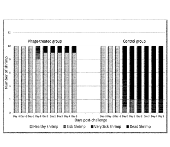
No shrimp casualties were observed before inoculation with V. parahaemolyticus
in
both tanks (Table 6; Figure 14). A total of 8 shrimp casualties were observed
in the
no treatment control and a total of 1 shrimp casualty was observed in the
treatment
tank (Table 6; Figure 14). The surviving shrimp in the no treatment control
tank were
observed to have increased morbidity compared to the surviving shrimp in the
treatment tank (Table 6; Figure 14). No significant difference was observed in
the
number of bacteria in each tank and significantly more bacteriophage was
isolated
from the treatment tank (Table 7).
CA 02983214 2017-10-18
WO 2016/170013 - 32 - PCT/EP2016/058809
Table 6¨ Health assessments of Thor amboinensis used in this study
Time No Treatment Immobilised
Control Bacteriophage
Treatment
Day 0 A ¨ 1 0 A ¨ 1 0
B ¨ 0 B ¨ 0
C ¨ 0 C ¨ 0
D ¨ 0 D ¨ 0
Day 1 A ¨ 1 0 A ¨ 1 0
B ¨ 0 B ¨ 0
C ¨ 0 C ¨ 0
D ¨ 0 D ¨ 0
Day 2 A ¨ 1 0 A ¨ 1 0
B ¨ 0 B ¨ 0
C ¨ 0 C ¨ 0
D ¨ 0 D ¨ 0
Day 3 (Inoculation) A ¨ 0 A ¨ 8
B ¨ 1 B ¨ 1
C ¨ 4 C ¨ 1
D ¨ 5 D ¨ 0
Day 4 A ¨ 0 A ¨ 9
B ¨ 2 B ¨ 0
C ¨ 0 C ¨ 0
D ¨ 8 D ¨ 1
Day 5 A ¨ 0 A ¨ 9
B ¨ 0 B ¨ 0
C ¨ 2 C ¨ 0
D ¨ 8 D ¨ 1
CA 02983214 2017-10-18
WO 2016/170013 - 33 -
PCT/EP2016/058809
Table 7 ¨ Number of Bacteria and bacteriophages recovered from each tank.
Test Tank Number of V. parahaemolyticus (CFU/ mL)
Day 3 Day 4
No Treatment Control 1 x10⁶ 1 .2x104
Immobilised Bacteriophage Tank 1.1x10⁶ 1x10⁴
Conclusions
Immobilised bacteriophage on shrimp feed confers a protective effect on Thor
amboinensis shrimp exposed to a large infectious dose of a pathogenic strain
of V.
parahaemolyticus that is known to cause AHPND in aquacultured shrimp. No
significant difference was found in V. parahaemolyticus numbers in the tank
water
which indicates the protective effect is happening locally at the site of
infection. At
the conclusion of the trial, 3 shrimp were sacrificed and the presence of
bacteriophages in the gut confirmed.
The invention hence provides compositions and methods for treatment of
bacterial
infections in aquaculture, generally of shrimp, prawns and fish.