Note: Descriptions are shown in the official language in which they were submitted.
CA 02983232 2017-10-18
Description
Title of Invention: PHARMACEUTICAL COMPOSITION FOR TREATING AND/OR
PREVENTING CANCER
Technical Field
[0001]
The present invention relates to novel medicinal use of an antibody against
CSPG5
protein or a fragment thereof as e.g., a therapeutic and/or prophylactic agent
for cancer.
Background Art
[0002]
Cancer is a disease occupying the leading position of cause of death.
Therapies
presently employed are primarily based on a surgical therapy in combination
with a radiation
therapy and a chemotherapy. Despite of recent development of new surgical
techniques and
finding of new anticancer drugs, treatment results of cancers except some
cancers have not yet
likely been improved at present. With the advancement of molecular biology and
cancer
immunology, antibodies that specifically react with cancers, cancer antigens
recognized by
cytotoxic T cells, genes encoding cancer antigens and the like have been
identified. Thus,
development of specific cancer treatments targeting cancer antigens has been
desired.
[0003]
In the cancer therapy, it is desirable that the peptides (including
polypeptides) to be
recognized as antigens are rarely present in normal cells and present
specifically in cancer
cells in order to reduce side effects. In 1991, Boon et al. of the Ludwig
Laboratory in
Belgium isolated human melanoma antigen MAGE1, recognized by CD8 positive T
cells, by
cDNA expression cloning method using an autologous tumor cell line and tumor-
reactive T
cells (Non Patent Literature 1). Subsequently, SEREX (serological
identification of antigens
by recombinant expression cloning) method was reported, which is a method of
identifying a
cancer antigen recognized by an antibody produced in response to autologous
cancer in the
1
CA 02983232 2017-10-18
body of a patient, by using a gene expression cloning method (Patent
Literature 1, Non Patent
Literature 2). Several cancer antigens, which are rarely expressed in normal
cells and
specifically expressed in cancer cells, have been isolated by this method (Non
Patent
Literature 3). Further, a cell therapy, which uses immune cells targeting a
part of the amino
acid sequence of such a cancer antigen and specifically reacting with the
cancer antigen, and a
cancer-specific immunotherapy such as a vaccine containing a cancer antigen,
are carried out
in clinical trials.
[0004]
In the meantime, various types of antibody medicines for treating cancer,
targeting a
specific antigen protein on cancer cells have been known in the world. Most of
the target
antigen proteins provide certain levels of medicinal effects as a cancer-
specific therapeutic
agent and attract attention; however they express also on a plurality of
normal cells. Because
of this, side effects are seriously concerned since not only cancer cells but
also normal cells
are damaged as a result of administration of antibodies. Accordingly, it is
expected that a
therapy with an antibody medicine having fewer side effects can be realized,
if an antigen,
which is specifically expressed only on the cancer cell surfaces and not
expressed on normal
cells, can be identified and an antibody targeting the antigen can be used as
a medicine.
Citation List
Patent Literature
[0005]
Patent Literature 1: U. S. Patent No. 5698396
Non Patent Literature
[0006]
Non Patent Literature 1: Bruggen P. et al., Science, 254: 1643-1647 (1991)
Non Patent Literature 2: Proc. Natl. Acad. Sci. USA, 92: 11810-11813 (1995)
Non Patent Literature 3: Int. J. Cancer, 72: 965-971 (1997)
Summary of Invention
2
CA 02983232 2017-10-18
Technical Problem
[0007]
An object of the present invention is to identify a cancer antigen protein
specifically
expressed on the surface of cancer cells and provide use of an antibody
targeting the cancer
antigen protein as a therapeutic and/or prophylactic agent for cancer.
Solution to Problem
[0008]
The present inventors isolated an antigen specifically expressed in cancer by
SEREX
method using cDNA library derived from a canine testicular tissue and the
serum of a breast
cancer-bearing dog, and then obtained cDNA encoding CSPG5 protein. CSPG5
protein can
bind to antibodies present in the sera derived from various cancer-bearing
living organisms.
The present inventors also found that CSPG5 protein is specifically expressed
in breast cancer,
lung cancer, brain tumor, leukemia, malignant lymphoma, adenocarcinoma,
mastocytoma,
squamous cell carcinoma, melanoma or neuroblastoma cells; and that a part of
CSPG5 protein
is specifically expressed on the surface of these cancer cells. CSPG5
(Chondroitin Sulfate
Proteoglycan 5) protein is type I transmembrane protein and one of the
neuregulin family
proteins. It is also reported that CSPG5 protein binds to ErbB3 protein to
serve as a growth
factor; and that expression of CSPG5 protein increases in ovarian cancer
having a mutation of
BRCA1 protein (Kinugasa, Y., et al., 2004, Biochem. Biophys. Res. Commun.,
321: 1045;
Press, J. Z., et al., 2010, Neoplasia., 12 (12): 993-1002). It is further
known that CSPG5
protein is highly expressed in tissues of the nervous system, such as retinal
ganglion cells,
purkinje cells and hippocampus, and serves as a proliferation/differentiation
factor of nerve
cells involved in elongation of nerve axon process (Yasuda, Y. et al., 1998,
Neurosci. Res., 32:
313; Aono, S., et al., 2006, J. Neurosci. Res., 83: 110; Nakanishi, K., et
al., 2006, J. Biol.
Chem., 281: 24970). However, there have been no reports that CSPG5 protein has
an
immunity inducing activity against cancer cells and thus is useful for
treating and preventing
cancer.
[0009]
3
CA 02983232 2017-10-18
1
A
Also, the inventors prepared CSPG5 protein molecules consisting of amino acid
sequences represented by SEQ ID NOs: 2, 4, 6, 8, 10, 12, 14 and 16 based on
the obtained
canine CSPG5 gene and its homologous genes of human, cat and mouse and
antibodies against
these CSPG5 protein molecules. Then, they found that an antibody against the
portion of
each of these CSPG5 protein molecules expressed on the surfaces of individual
cancer cells, in
other words, the extracellular region thereof, damages the cancer cell
expressing CSPG5
protein. Based on the finding, the present invention was accomplished.
[0010]
Accordingly, the present invention has the following features.
(1) A pharmaceutical composition for treating and/or preventing cancer,
comprising an
antibody or a fragment thereof having immunological reactivity with CSPG5
protein or a
fragment thereof consisting of at least 7 or more consecutive amino acid
residues, as an active
ingredient.
(2) The pharmaceutical composition according to (1), wherein the CSPG5 protein
consists of
any one of amino acid sequences represented by SEQ ID NOs: 8, 4, 6, 10 and 12,
or an amino
acid sequence having an amino acid identity of 80% or more to the amino acid
sequence.
(3) The pharmaceutical composition according to (1) or (2), wherein the cancer
is leukemia or
malignant lymphoma.
(4) The pharmaceutical composition according to any one of (1) to (3), wherein
the antibody is
a monoclonal antibody or a polyclonal antibody.
(5) The pharmaceutical composition according to any one of (1) to (4), wherein
the antibody is
a human antibody, a humanized antibody, a chimeric antibody, a single-chain
antibody or a
bispecific antibody.
[0011]
The specification incorporates the disclosure of JP Patent Application No.
2015-093640
to which the present application claims the priority.
Advantageous Effects of Invention
[0012]
4
CA 02983232 2017-10-18
The antibody against CSPG5 protein used in the present invention damages
cancer cells.
Accordingly, the antibody against CSPG5 protein is useful for treatment and/or
prevention of
cancer.
Brief Description of Drawings
[0013]
[Figure 11 Figure 1 shows expression patterns of identified CSPG5 gene in
canine tumor
tissues or cancer cell lines. In the figure, reference number 1 shows
expression patterns of
canine CSPG5 gene in individual canine tissues and cell lines; and reference
number 2 shows
expression patterns of canine GAPDH gene in individual canine tissues and cell
lines.
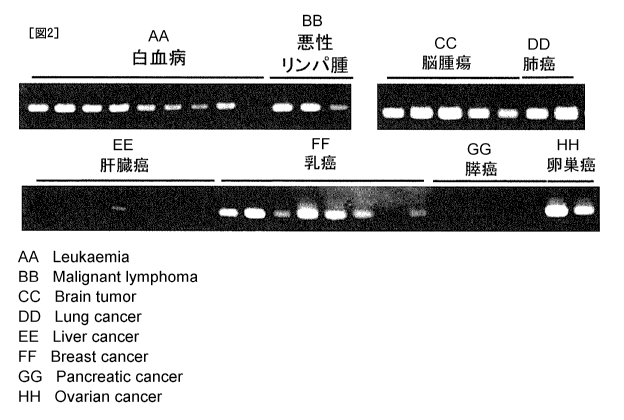
[Figure 2] Figure 2 shows expression patterns of identified CSPG5 gene in
human tumor
tissues or cancer cell lines.
[Figure 3] Figure 3 shows expression patterns of identified CSPG5 gene in
mouse tumor
tissues or cancer cell lines. Reference number 3 shows expression patterns of
mouse CSPG5
gene in individual mouse tissues and cell lines; reference number 4 shows
expression patterns
of mouse GAPDH gene in individual mouse tissues and cell lines.
[Figure 4] Figure 4 shows cytotoxic activity of a polyclonal antibody against
CSPG5 protein
(anti-CSPG5 polyclonal antibody) on a leukemia cell line (K562) and malignant
lymphoma
cells (L-1236) expressing CSPG5 gene. In the figure, reference number 5 shows
the
cytotoxic activity against K562 cells when a control polyclonal antibody was
added; reference
number 6 shows the cytotoxic activity against K562 cells when an anti-CSPG5
polyclonal
antibody was added; reference number 7 shows the cytotoxic activity against L-
1236 cells
when the control polyclonal antibody was added; and reference number 8 shows
the cytotoxic
activity against L-1236 cell when the anti-CSPG5 polyclonal antibody was
added.
[Figure 5] Figure 5 shows cytotoxic activity of a monoclonal antibody against
CSPG5 protein
(anti-CSPG5 monoclonal antibody) on a leukemia cell line (K562) and malignant
lymphoma
cells (L-1236) expressing CSPG5 gene. In the figure, reference number 9 shows
the
cytotoxic activity against K562 cells when a control monoclonal antibody was
added;
reference number 10 shows the cytotoxic activity against K562 cells when an
anti-CSPG5
CA 02983232 2017-10-18
monoclonal antibody was added; reference number 11 shows the cytotoxic
activity against L-
1236 cells when the control monoclonal antibody was added; and reference
number 12 shows
the cytotoxic activity against L-1236 cell when the anti-CSPG5 monoclonal
antibody was
added.
Description of Embodiments
[0014]
The antitumor activity of an antibody against a polypeptide consisting of the
amino
acid sequence represented by SEQ ED NO: 2, 4, 6, 8, 10, 12, 14 or 16, used in
the present
invention, can be evaluated by checking, in vivo suppression of tumor growth
in a cancer-
bearing animal or by checking, whether or not the antibody exerts in vitro
cytotoxic activity
against tumor cells expressing the polypeptide via immune cells or a
complement, as described
later.
[0015]
The nucleotide sequence of a polynucleotide encoding the protein consisting of
the
amino acid sequence represented by SEQ ID NO: 2, 4, 6, 8, 10, 12, 14 or 16 is
represented by
SEQ ID NO: 1, 3, 5, 7,9, 11, 13 or 15, respectively.
[0016]
The amino acid sequence represented by SEQ ID NO: 2 in the sequence listing
disclosed in the present invention is an amino acid sequence of CSPG5 protein
isolated as a
polypeptide binding to an antibody specifically present in the serum derived
from a cancer-
bearing dog by the SEREX method using a cDNA library derived from a canine
testicular
tissue and the serum of a breast cancer-bearing dog; the amino acid sequences
represented by
SEQ ID NOs: 4, 6, 8, 10 and 12 are isolated as human homologs of the
polypeptide; the amino
acid sequence represented by SEQ ID NO: 14 is isolated as a cat homolog of the
polypeptide;
and the amino acid sequence represented by SEQ ID NO: 16 is isolated as a
mouse homolog
of the polypeptide (see, Example 1 described later).
[0017]
6
CA 02983232 2017-10-18
a
It has been known from the amino acid sequence that CSPG5 protein is type I
transmembrane protein, and that the extracellular region predicted from its
sequence is
expressed on the surface of nerve cells. Owing to the present application, it
was confirmed
that the extracellular region of CSPG5 protein is actually expressed (present)
on the surface of
various types of cancer cells. In the present invention, an antibody binding
to the
extracellular region of CSPG5 protein on a cancer cell or binding to a
polypeptide having an
amino acid identity of 80% or more, preferably 85% or more, more preferably
90% or more,
further preferably 95% or more, 97% or more, 98% or more or 99% or more with
the amino
acid sequence of the extracellular region, is preferably used.
[0018]
The antibody against CSPG5 protein used in the present invention may be of any
type
of antibody as long as it can exert an antitumor activity. Examples of the
antibody include a
monoclonal antibody, a polyclonal antibody, a synthetic antibody, a multi-
specific antibody, a
human antibody, a humanized antibody, a chimeric antibody, a single-chain
antibody (scFV)
and an antibody fragment (for example, Fab, F(ab')2, Fv). These antibodies and
fragments
thereof can be prepared by methods known to those skilled in the art. In the
present
invention, an antibody capable of specifically binding to CSPG5 protein is
desirable and a
monoclonal antibody is preferable; however, a polyclonal antibody may be used
as long as
homogeneous antibodies can be stably produced. When the subject is a human, a
human
antibody or a humanized antibody is desirable in order to avoid or suppress a
rejection reaction.
[0019]
The phrase "specifically binding to CSPG5 protein" used herein means binding
specifically to CSPG5 protein and substantially not binding to proteins except
CSPG5 protein.
[0020]
The subject, which is a target for treatment and/or prevention of cancer by
the present
invention, is a mammal such as humans, pet animals, domestic animals and
animals for
competitive use, preferably a human.
[0021]
7
CA 02983232 2017-10-18
=
Preparation of an antigen, preparation of an antibody and a pharmaceutical
composition
according to the present invention is described below.
[0022]
<Preparation of antigen for preparing antibody>
The protein or a fragment thereof used as a sensitizing antigen for obtaining
an
antibody against CSPG5 protein (anti-CSPG5 antibody) in the present invention
may be
derived from e.g., humans, dogs, cats, mice, rats, cows, horses and chickens,
and the animal
species from which the protein or a fragment thereof is derived is not limited
thereto. The
protein or a fragment thereof is preferably selected in consideration of
compatibility to a
parent cell to be used in cell fusion. Generally, the protein derived from a
mammal is
preferable and particularly the protein derived from a human is preferable.
For example, if
the CSPG5 protein is human CSPG5 protein, human CSPG5 protein, a partial
polypeptide
thereof, a cell expressing the human CSPG5 protein, and the like can be used.
[0023]
The amino acid sequences and nucleotide sequences of CSPG5 protein and
homologs
thereof can be obtained by using, for example, GenBank (NCBI of the United
State) and by
using an algorithm such as BLAST and FASTA (Karlin and Altschul, Proc. Natl.
Acad. Sci.
USA, 90: 5873-5877,1993; Altschul et al., Nucleic Acids Res. 25: 3389-3402,
1997).
[0024]
For example, if human CSPG5 protein is used as a basis, a nucleotide sequence
(SEQ
ID NO: 3, 5, 7, 9 or 11) encoding the human CSPG5 protein and a nucleic acid
having a
nucleotide identity of 70% to 100%, preferably 80% to 100%, more preferably
90% to 100%,
further preferably 95% to 100% (for example 97% to 100%, 98% to 100%, 99% to
100% or
99.5% to 100%) to the nucleotide sequence are used as a target. Also, the
amino acid
sequence (SEQ ID NO: 4, 6, 8, 10 or 12) of the human CSPG5 protein and a
polypeptide
having an amino acid identity of 70% to 100%, preferably 80% to 100%, more
preferably 90%
to 100%, or further preferably 95% to 100% (for example, 97% to 100%, 98% to
100%, 99%
to 100% or 99.5% to 100%) to the amino acid sequence are used as a target. The
term
"nucleotide identity" used herein refers to the percentage (%) of the number
of identical
8
CA 02983232 2017-10-18
nucleotides relative to the total number of nucleotides when two nucleotide
sequences are
aligned such that they have a maximum degree of similarity by appropriately
introducing
gap(s). Similarly, the term "amino acid identity" refers to the percentage (%)
of the number
of identical amino acids relative to the total number of amino acids when two
amino acid
sequences are aligned such that they have a maximum degree of similarity by
appropriately
introducing gap(s).
[0025]
The fragment of CSPG5 protein is specified to have a length equal to or longer
than the
amino acid length of an epitope (antigen determinant) and less than the full
length of the
protein. The epitope is a polypeptide fragment which is a minimum unit
recognized by an
antibody in a mammal, preferably a human and has antigenicity or
immunogenicity, and
includes amino acid sequences having a length of about 7 to 12 amino acids,
for example, 8 to
11 amino acids.
[0026]
The human CSPG5 protein and a polypeptide containing a partial peptide thereof
can
be synthesized, for example, by a chemical synthesis method such as Fmoc
method
(fluorenylmethyloxycarbonyl method) and tBoc method (t-butyloxycarbonyl
method)
(Biochemical Experiment Course 1, Chemistry of Protein IV, Chemical
Modification and
Synthesis of Peptide, edited by the Japan Biochemical Society, Tokyo Kagaku
Dojin (Japan),
1981) or synthesized in accordance with a routine method using a commercially
available
peptide synthesizer. Alternatively, a desired polypeptide can be obtained by
genetic
engineering technique known in the art (e.g., Green, M. R. and Sambrook, J.,
2012, Molecular
Cloning: A Laboratory Manual 4th Ed., Cold Spring Harbor Laboratory Press,
Cold Spring
Harbor, New York, Ausubel et al., Short Protocols in Molecular Biology, third
edition, A
compendium of Methods from Current Protocols in Molecular Biology (1995), John
Wiley &
Sons); more specifically by preparing a polynucleotide encoding the
polypeptide, integrating
the polynucleotide into an expression vector, introducing the vector into a
host cell and
allowing the host cell to produce a polypeptide.
[0027]
9
CA 02983232 2017-10-18
The polynucleotide encoding the polypeptide can be easily prepared by a
routine
method using a genetic engineering technique known in the art or a
commercially available
nucleic acid synthesizer. For example, DNA having the nucleotide sequence of
SEQ ID NO:
3 can be prepared by carrying out PCR using a human chromosome DNA library or
a human
cDNA library as a template and a pair of primers designed so as to amplify the
nucleotide
sequence represented by SEQ ID NO: 3. The reaction conditions of the PCR can
be
appropriately defined; for example, the reaction conditions include a
condition where using a
heat-resistant DNA polymerase (for example, Taq polymerase) and a Mg2 -
containing PCR
buffer, a cycle consisting of a denaturation reaction at 94 C for 30 seconds,
an annealing
reaction at 55 C for 30 seconds to 1 minute and an elongation reaction at 72 C
for 2 minutes,
for example, is repeated 30 times and then carrying out a reaction at 72 C for
7 minutes;
however, the reaction conditions are not particularly limited thereto. The PCR
method and
conditions are described, for example in Ausubel et al., Short Protocols in
Molecular Biology,
third edition, A compendium of Methods from Current Protocols in Molecular
Biology (1995),
John Wiley & Sons (particularly Chapter 15).
[0028]
A desired DNA can be isolated by preparing an appropriate probe and primers
based on
information of the nucleotide sequence represented by SEQ ID NO: 1, 3, 5, 7,
9, 11, 13 or 15
in the sequence listing of the present application and screening a cDNA
library of a human etc.
by the probe and primers. The cDNA library is preferably prepared from the
cell, organ or
tissue expressing a protein consisting of the amino acid sequence represented
by SEQ ID NO:
2, 4, 6, 8, 10, 12, 14 or 16. Examples of such a cell or tissue include, but
are not limited to,
cells or tissues derived from cancers or tumors such as testicles or leukemia,
breast cancer,
lymphoma, brain tumor, lung cancer, colon cancer, mastocytoma, melanoma and
neuroblastoma. The aforementioned operations, such as preparation of the probe
or primers,
construction of a cDNA library, screening of a cDNA library and cloning of a
gene of interest,
are known to those skilled in the art and can be carried out in accordance
with the method
described, for example, in Green, M. R. and Sambrook (described above), Ausbel
et al.
CA 02983232 2017-10-18
(described above). From the DNA thus obtained, DNA encoding CSPG5 protein and
a
partial peptide thereof can be obtained.
[0029]
As the host cell, any type of cell may be used as long as it can express the
above
polypeptide. Examples of prokaryotic cells include Escherichia coli, and
examples of
eukaryotic cells include, but not limited to, yeast cells including budding
yeast and fission
yeast, insect cells such as silkworm cells, Xenopus egg cells and mammalian
cells such as
monkey kidney cells COS 1, Chinese hamster ovary cells CHO, human fetal kidney
cell line
HEK293 and mouse fetal skin cell line N11-13T3.
[0030]
When a prokaryotic cell is used as a host cell, an expression vector having a
replication
origin in a prokaryotic cell, a promoter, a ribosome binding site, a multi
cloning site, a
terminator, a drug resistance gene, an auxotrophic complement gene, and the
like is used as the
expression vector. Examples of the expression vector for Escherichia coli may
include pUC
system, pBluescriptIL pET expression system and pGEX expression system. The
polypeptide encoded by DNA can be expressed in the prokaryotic host cell, by
integrating
DNA encoding the polypeptide into such an expression vector; transforming a
prokaryotic
host cell with the vector; and culturing the resultant transformant. At this
time, the
polypeptide can be expressed as a part of a fusion protein with another
protein.
[0031]
When a eukaryotic cell is used as a host cell, an expression vector for a
eukaryotic cell
having a promoter, a splicing region, a poly (A) additional site, and the like
is used as the
expression vector. Examples of such an expression vector may include pKA1,
pCDM8,
pSVK3, pMSG, pSVL, pBK-CMV, pBK-RSV, EBV vector, pRS, pcDNA3.1 and pYES2.
The polypeptide encoded by DNA can be expressed in a eukaryotic host cell,
similarly to the
above, by integrating DNA encoding the above polypeptide into such an
expression vector,
transforming the eukaryotic host cell with the vector, and culturing the
resultant transformant.
The above polypeptide can be expressed as a part of a fusion protein attached
with a tag such
as a His tag (for example, (His)6 to (His)io), a FLAG tag, a myc tag, a HA tag
and a GFP,
11
CA 02983232 2017-10-18
when plNDN5-His, pFLAG-CMV-2, pEGFP-N1, pEGFP-C1 or the like is used as the
expression vector.
[0032]
Introduction of an expression vector into a host cell can be carried out by
using a
method well known in the art, such as an electroporation method, a calcium
phosphate method,
a liposome method, a DEAE dextran method, microinjection, viral infection,
lipofection and
binding to a cell membrane penetrating peptide.
[0033]
For isolation/purification of a desired polypeptide from a host cell,
separation
operations known in the art can be used in combination. Examples of the
separation
operations include, but are not limited to, a treatment with a denaturant such
as urea or a
surfactant, sonication, enzymatic digestion, salting out and solvent
fractionation precipitation,
dialysis, centrifugation, ultrafiltration, gel filtration, SDS-PAGE,
isoelectric point
electrophoresis, ion exchange chromatography, hydrophobic chromatography,
affinity
chromatography and reverse phase chromatography.
[0034]
<Antibody structure>
An antibody is a hetero-multimer glycoprotein usually containing at least two
heavy
chains and two light chains. Four types of immunoglobulins except IgM each are
a hetero-
tetramer glycoprotein of about 150 kDa primarily constituted of two identical
light (L) chains
and two identical heavy (H) chains. Typically, each of the light chains is
linked to a heavy
chain via a single disulfide covalent bond; whereas the number of disulfide
bonds between the
heavy chains varies depending on the isotypes of immunoglobulins. Each of the
heavy
chains and light chains has also an intra-strand disulfide bond. Each of the
heavy chains has
a variable domain (VH region) at an end, followed by several constant regions.
Each of the
light chains has a variable domain (VL region) at an end and a single constant
region at the
other end. The light-chain variable domain is aligned with the heavy-chain
variable domain.
The constant region of a light chain is aligned with the first constant region
following the
heavy-chain variable domain. In the variable domain of the antibody, there are
three specific
12
CA 02983232 2017-10-18
=
regions called as complementarity determining regions (CDRs), which are
variable parts and
based on which the antibody has binding specificity. In the variable region, a
portion
relatively conserved is called as a framework region (FR). Complete heavy
chain and light
chain variable domains each contain 4 FRs (FR1, FR2, FR3 and FR4 sequentially
from the N
terminal side) connected via three CDRs. The three CDRs in a heavy chain are
called
CDRH1, CDRH2 and CDRH3 sequentially from the N terminal side and the CDRs in a
light
chain are called CDRL1, CDRL2 and CDRL3. CDRH3 is the most important for
binding
specificity of an antibody to an antigen. The CDRs of each chain are held
together in close
proximity with each other via FR regions and contribute to formation of an
antigen binding
site of the antibody in concert with CDRs of the other chain. The constant
region does not
directly contribute to binding of the antibody to an antigen; however, the
constant region has
various effector functions, such as involvement in antibody-dependent cell-
mediated
cytotoxicity (ADCC), phagocytosis via binding to an Fey receptor, half-
life/clearance rate via
a neonatal Fe receptor (FcRn), and complement-dependent cytotoxicity (CDC) via
C 1 q
component of a complement cascade.
[0035]
<Preparation of antibody>
The anti-CSPG5 antibody of the present invention refers to an antibody having
immunological reactivity with a full-length CSPG5 protein or a fragment
thereof.
[0036]
The term "immunological reactivity" used herein refers to a property of an
antibody
binding to CSPG5 antigen, in-vivo. Through such a binding, an action to damage
(for
example, kill, suppress or induce regression of) a tumor is exerted. More
specifically, the
type of an antibody used in the present invention is not limited as long as it
can bind to CSPG5
protein to damage a tumor such as breast cancer, lung cancer, brain tumor,
leukemia,
malignant lymphoma, adenocarcinoma, mastocytoma, squamous cell carcinoma,
melanoma or
neuroblastoma.
[0037]
13
CA 02983232 2017-10-18
Examples of the antibody include a monoclonal antibody, a polyclonal antibody,
a
genetic recombinant antibody and an antibody fragment (for example, Fab and
F(ab1)2), as
mentioned above. Also the antibody may be any class of an irnmunoglobulin
molecule such
as IgG, IgE, IgM, IgA, IgD and IgY or any subclass thereof such as IgG 1 ,
IgG2, IgG3, IgG4,
IgAl and IgA2.
[0038]
The antibody may be further modified with acetylation, formylation, amidation,
phosphorylation, pegylation(PEG), or the like as well as glycosylation.
[0039]
Preparation examples of various antibodies are described below.
(1) Monoclonal antibody
Examples of the monoclonal antibody include a human monoclonal antibody and an
animal (non-human) monoclonal antibody (for example, a mouse monoclonal
antibody, a rat
monoclonal antibody, a rabbit monoclonal" antibody and a chicken monoclonal
antibody).
[0040]
The antibody, in the case of a monoclonal antibody, can be prepared by
carrying out
immunization in accordance with a general immunization method using a desired
antigen
(CSPG5 protein herein) or a cell expressing the desired antigen as a
sensitizing antigen, fusing
thus obtained immune cell with a parent cell known in the art in accordance
with a general cell
fusion method and screening a monoclonal antibody producing cell (hybridoma)
by a general
screening method.
[0041]
First, an animal is immunized with a sensitizing antigen in accordance with a
method
known in the art. As a general method, a sensitizing antigen is
intraperitoneally or
subcutaneously injected to a mammal, for example, a mouse. More specifically,
a sensitizing
antigen, i.e., CSPG5 protein, is diluted or suspended to an appropriate amount
of PBS
(Phosphate-Buffered Saline), physiological saline, or the like, and if
desired, a general
adjuvant, for example, Freund's complete adjuvant, is added thereto in an
appropriate amount
and emulsified. Thereafter, the emulsion is administered to a mammal, for
example, a mouse,
14
CA 02983232 2017-10-18
several times at intervals of 4 to 21 days. An appropriate carrier can be used
at the time of
immunization with a sensitization antigen. Alternatively, leukemia cell line
K562 expressing
CSPG5 gene or the like may be administered to an animal (to be immunized) in
order to
immunize the animal.
[0042]
After a mammal is immunized as described above and an increase of the level of
the
desired antibody in the serum is confirmed, immune cells are collected from
the mammal and
subjected to cell fusion in order to prepare a hybridoma producing a
monoclonal antibody.
As the preferable immune cells for preparing a hybridoma, particularly
splenocytes are
mentioned.
[0043]
Mammalian myeloma cells are used as another parent cells to be fused with the
immune cells. As the myeloma cells, various cell lines known in the art such
as P3U1 (P3-
X63Ag8U1), P3 (P3x63Ag8.653) (J. Immunol. (1979) 123, 1548-1550), P3x63Ag8U.1
(Current Topics in Microbiology and Immunology (1978) 81, 1-7), NS-1 (Kohler.
G. and
Milstein, C. Eur. J. Immunol. (1976) 6, 511-519), MPC-11 (Margulies. D. H. et
al., Cell
(1976) 8, 405-415), SP2/0 (Shulman, M. et al., Nature (1978) 276, 269-270), FO
(deSt. Groth,
S.F. et at., J. Immunol. Methods (1980) 35, 1-21), S194 (Trowbridge, I. S. J.
Exp. Med. (1978)
148, 313-323), R210 (Galfre, G. et al., Nature (1979) 277, 131-133) are
suitably used.
[0044]
The cell fusion between the immune cells and myeloma cells can be carried out
basically in accordance with a method known in the art, for example, a method
of Kohler and
Milstein et al., (Kohler, G. and Milstein, C. Methods Enzymol. (1981) 73, 3-
46).
[0045]
More specifically, the cell fusion is carried out in the presence of, for
example, a cell
fusion accelerator, in a general nutrition culture solution. As the fusion
accelerator, for
example, polyethylene glycol (PEG) or Sendai virus (HVJ) is used and, if
desired, an auxiliary
agent such as dimethylsulfoxide can be added in order to enhance fusion
efficiency.
[0046]
CA 02983232 2017-10-18
The ratio of the immune cells and myeloma cells to be used can be arbitrarily
determined. For example, the immune cells can be used in a ratio 1 to 10 times
compared
with the myeloma cells. As the culture solution to be used in the cell fusion,
for example,
RPMI 1640 culture solution or MEM culture solution suitable for proliferation
of the myeloma
cell line, and other culture solutions usually used in culturing these cells
can be used. In
addition, a serum-supplement such as fetal calf serum (FCS) can be used in
combination with
the culture solution.
[0047]
Cell fusion is carried out by sufficiently mixing predetermined amounts of the
immune
cells and myeloma cells in the culture solution and adding a PEG solution (for
example,
average molecular weight: about 1000 to 6000) previously heated to about 37 C
usually in a
concentration of 30 to 60% (w/v) followed by stirring to form desired
hybridomas.
Subsequently, an operation consisting of intermittently adding an appropriate
culture solution,
centrifuging the mixture and removing the supernatant, is repeated to remove a
cell fusion
agent unfavorable for growth of the hybridoma and the like.
[0048]
The hybridoma thus obtained is selected by culturing in a general selection
culture
solution such as HAT culture solution (culture solution containing
hypoxanthine, aminopterin
and thymidine). The culture in the HAT culture solution is continued for a
time period
(usually, several days to several weeks) sufficient for cells (non-fused
cells) other than a
desired hybridoma to die. Subsequently, a general limiting dilution method is
carried out and
screening of a hybridoma producing a desired antibody and single cloning are
carried out.
[0049]
As well as obtaining the above hybridoma by immunizing an animal except a
human
with an antigen, a hybridoma producing a human antibody having a desired
activity (for
example, cytostatic activity) can be also obtained by sensitizing human
lymphocytes such as
human lymphocytes infected with EB virus, with a protein, protein-expressing
cell or lysate
thereof in vitro, and fusing the sensitized lymphocytes with human-derived
myeloma cells, for
example U266 (registration number T1B196), having a permanent division
potential.
16
CA 02983232 2017-10-18
[0050]
Thus prepared hybridoma producing a monoclonal antibody can be sub-cultured in
a
general culture solution and stored in liquid nitrogen for a long time.
[0051]
(2) Polyclonal antibody
The antibody, in the case of a polyclonal antibody, can be prepared by
immunizing a
small animal such as a mouse, a human antibody-producing mouse or a rabbit,
with natural
CSPG5 protein, recombinant CSPG5 protein expressed in the form of a fusion
protein with
GST and the like in a microorganism such as Escherichia coil or a partial
peptide thereof to
obtain the serum; and purifying the antibody, for example, by ammonium sulfate
precipitation,
protein A, protein G column, DEAE ion exchange chromatography, an affinity
column
coupled with CSPG5 protein and a synthetic peptide. In Examples described
later, a mouse
polyclonal antibody against the extracellular region (outside cancer cell) of
the amino acid
sequence of CSPG5 protein is prepared and confirmed to have the antitumor
effect.
[0052]
As the human antibody-producing mouse used herein, for example, KM mouse
(Kirin
Pharma/Medarex) and Xeno mouse (Amgen) are known (for example, International
Publication Nos. W002/43478 and 02/092812). When such a mouse is immunized
with
CSPG5 protein or a fragment thereof, a complete human polyclonal antibody can
be obtained
from the blood.
[0053]
An antigen can be prepared, for example, in accordance with a method using an
animal
cell (JP Patent Publication (Kohyo) No. 2007-530068A) or a method using
baculovirus (for
example, International Publication No. WO 98/46777). When the immunogenicity
of an
antigen is low, the antigen may be bound to a macromolecule such as albumin
having
immunogenicity and subjected to immunization.
[0054]
(3) Recombinant antibody
17
CA 02983232 2017-10-18
The antibody, in the case of a genetic recombination antibody, can be
prepared, in
accordance with a genetic recombination technology, by cloning a gene of the
antibody from a
hybridoma, incorporating the gene into an appropriate vector, and introducing
the vector into a
host to produce a recombinant antibody (see, for example, Carl, A. K.
Borrebaeck, James, W.
Larrick, THERAPEUTIC MONOCLONAL ANTIBODIES, Published in the United Kingdom
by MACMILLAN PUBLISHERS LTD, 1990). More specifically, cDNA of a variable
region (V region) of the antibody is synthesized from mRNA of a hybridoma by
using a
reverse transcriptase. When DNA encoding a V region of a desired antibody is
obtained, it is
linked to DNA encoding a constant region (C region) of the desired antibody,
and the resultant
DNA is integrated into an expression vector. Alternatively, DNA encoding the V
region of
the antibody may be integrated into an expression vector containing DNA of the
C region of
the antibody. Said DNA may be Integrated to be expressed under the control of
an
expression regulatory region, for example, an enhancer and a promoter.
Subsequently, a host
cell is transformed with the expression vector and allowed to express the
genetic recombinant
antibody.
[0055]
Examples of the genetic recombinant antibody include a chimeric antibody, a
humanized antibody, a single-chain antibody and multi-chain antibody such as a
bispecific
antibody.
[0056]
The "chimeric antibody" is an antibody formed of sequences derived from
different
animals in combination, for example, an antibody constituted of a variable
region of a heavy-
chain and a light chain of a mouse antibody and a constant region of a heavy-
chain and a light
chain of a human antibody. A chimeric antibody can be prepared in accordance
with a
method known in the art, for example, by ligating DNA encoding an antibody V-
region and
DNA encoding a human antibody C-region, incorporating the ligate into an
expression vector,
and introducing the expression vector into a host to allow the host to produce
the antibody.
As an example, DNA encoding a human/mouse chimeric antibody can be prepared by
ligating
a DNA encoding a variable region of a light chain or a heavy chain of an
antibody derived
18
CA 02983232 2017-10-18
from a non-human animal (for example, mouse) to DNA encoding a constant region
of a light
chain or a heavy chain of an antibody derived from a human antibody.
[0057]
The "humanized antibody" is a modified antibody also called as a reshaped
human
antibody. The humanized antibody is constructed by grafting an antibody CDR
derived from
an immunized animal to a complementarity determining region of a human
antibody. A
general genetic recombination technique for preparing the humanized antibody
is also known
in the art. Specifically, the humanized antibody is obtained by cloning a DNA
encoding a
monoclonal antibody; using the resultant as a template to prepare a DNA
encoding a light-
chain variable region and a heavy-chain variable region by a RT-PCR method, or
the like;
determining the sequences of the variable regions of the light chain and heavy
chain or the
sequences of CDR1, CDR2 and CDR3 based on the Kabat EU numbering system (Kabat
et al.,
Sequences of proteins of Immunological Interest, 5thEd. Public Health Service,
National
Institute of Health, Bethesda, Md. (1991)); subsequently, synthesizing a DNA
sequence,
which is designed such that mouse antibody CDRs and framework regions
(framework region;
FR) of the human antibody can be ligated, by a PCR method using several
oligonucleotides
prepared designed to have an overlapping portion at the ends; ligating the
resultant DNA to
DNA encoding a human antibody constant region and then integrating it into an
expression
vector; and introducing the expression vector into a host and then allowing
the host to produce
a humanized antibody (see, European Patent Publication No. 239400,
International Publication
No. W096/02576). The FRs of the human antibody to be ligated via CDRs are
selected such
that the CDRs (complementarity determining regions) form a satisfactory
antigen binding site.
If necessary, the amino acids of the framework region in the variable region
of the antibody
may be substituted such that the complementarity determining regions of a
reshaped human
antibody form an appropriate antigen binding site (Sato K., et al., Cancer
Research, 1993, 53:
851-856). Further, the framework regions may be substituted with those derived
from
various human antibodies (International Publication No. W099/51743).
[0058]
19
CA 02983232 2017-10-18
The framework regions of a human antibody to be ligated via CDRs are selected
such
that the CDRs (complementarity determining regions) form a satisfactory
antigen binding site.
If necessary, the amino acids of framework regions in the variable region of
an antibody may
be substituted such that the complementarity determining regions of a reshaped
human
antibody form an appropriate antigen binding site (Sato K. et al., Cancer
Research 1993, 53:
851-856).
[0059]
An amino acid in the variable region (for example, FR) and the constant region
may be,
for example, substituted with another amino acid, after a chimeric antibody
and a humanized
antibody are formed.
[0060]
The substitution of amino acids includes substitution of amino acids of, for
example,
less than 15, less than 10, 8 or less, 7 or less, 6 or less, 5 or less, 4 or
less, 3 or less or 2 or less,
preferably 1 to 5 amino acids, and more preferably 1 or 2 amino acids. The
antibody having
a substitution should be functionally equivalent to the antibody having no
substitution.
Substitution is desirably conservative amino acid substitution, which is a
substitution between
amino acids having analogous properties in view of charge, side chain,
polarity and
aromaticity, and the like. The amino acids having analogous property can be
classified into,
for example, basic amino acids (arginine, lysine, histidine), acidic amino
acids (aspartic acid,
glutamic acid), uncharged polar amino acids (glycine, asparagine, glutamine,
serine, threonine,
cysteine, tyrosine), nonpolar amino acids (leucine, isoleucine, alanine,
valine, proline,
phenylalanine, tryptophan, methionine), branched amino acids (threonine,
valine, isoleucine)
and aromatic amino acids (phenylalanine, tyrosine, tryptophan, histidine).
[0061]
Modified antibodies include, for example, antibodies bound to various types of
molecules such as polyethylene glycol (PEG). In the modified antibody of the
present
invention, the material to be bound to the antibody is not limited. Such a
modified antibody
can be obtained by chemically modifying the obtained antibody. The chemical
modification
methods have been already established in this field.
CA 02983232 2017-10-18
[0062]
The term "functionally equivalent" used herein means that the antibody of
interest has
the same biological or biochemical activity as the antibody of the present
invention, more
specifically means that the antibody of interest has a tumor damaging action,
and a rejection
reaction does not basically occur when the antibody is applied to a human.
Such activities
include, for example, cytostatic activity or binding activity.
[0063]
A method of introducing a mutation into the polypeptide is known as a method
well
known to those skilled in the art for preparing a polypeptide which is
functionally equivalent
to a predetermined polypeptide. Those skilled in the art can employ a site-
specific
mutagenesis (Hashimoto-Gotoh, T.et al., (1995) Gene, 152,271-275: Zoller, MJ.,
and Smith,
M. (1983) Methods Enzymo1.100, 468-500; Kramer, W.et al., (1984) Nucleic Acids
Res.12,
9441-9456; Kramer, W. and Fritz, HJ., (1987) Methods Enzymo1.154, 350-367,
Kunkel, TA.,
(1985) Proc. Natl. Acad. Sci. USA., 82, 488-492; Kunkel (1988) Methods
Enzymol., 85, 2763-
2766) to appropriately introduce a mutation into the antibody of the present
invention, thereby
preparing an antibody functionally equivalent to the antibody.
[0064]
An antibody recognizing an epitope of CSPG5 protein that anti-CSPG5 antibody
recognizes can be obtained by a method known to those skilled in the art. For
example, the
antibody is prepared by determining the epitope of CSPG5 protein recognized by
anti-CSPG5
antibody by a general method (for example, epitope mapping) and preparing the
antibody
using a polypeptide having an amino acid sequence contained in the epitope as
immunogen.
In addition to this method, the antibody can be obtained by a method, for
example,
determining the epitopes of the antibodies prepared by a general method and
selecting an
antibody having the same epitope as in the anti-CSPG5 antibody.
[0065]
The affinity constant (binding constant) Ka (kon/koff) of the anti-CSPG5
antibody of
the present invention for the CSPG5 protein on a cancer cell surface is at
least 107 M-1, at least
108 M-1, at least 5 x 10⁸ M-1, at least 109 M-1, at least 5 x 10⁹ M-1, at
least 1010 M-1, at least 5 x
21
CA 02983232 2017-10-18
1 010
M', at least 1011 M-1, at least 5 x 10¹¹ M-1, at least 1012 M-1 or at least
1013 M-1. As the
binding affmity increases, a stronger antitumor activity can be obtained.
Accordingly, if an
anti-CSPG5 antibody having a high binding affinity for CSPG5 protein can be
obtained, a
stronger antitumor effect can be expected, the antibody can be applied to a
pharmaceutical
composition for treating and/or preventing cancer.
[0066]
The "single-chain antibody" is an antibody obtained by linearly ligating a
heavy-chain
variable region and a light-chain variable region via a linker. DNA encoding a
single-chain
antibody can be prepared by ligating DNA encoding a heavy-chain variable
region, DNA
encoding a linker and DNA encoding light-chain variable region. The heavy-
chain variable
region and light-chain variable region used herein are those preferably
derived from a human
antibody or those derived from a human antibody, in which CDRs alone are
replaced by those
of an antibody derived from a non-human animal (for example, mouse, rat,
chicken). A
linker consists of 12 to 19 amino acids, and includes for example, (G4S)3
consisting of 15
amino acids (G. B. Kim et al., Protein Engineering Design and Selection, 2007,
20 (9): 425-
432).
[0067]
In the case of the "bispecific antibody (diabody)", which is an antibody
capable of
specifically binding to two different epitopes, DNA encoding the bispecific
antibody can be
prepared by binding, for example, DNA encoding heavy-chain variable region A,
DNA
encoding light-chain variable region B, DNA encoding heavy-chain variable
region B and
DNA encoding light-chain variable region A sequentially in this order
(provided that DNA
encoding light-chain variable region B and DNA encoding heavy-chain variable
region B are
connected via DNA encoding a linker as mentioned above). The heavy-chain
variable
regions and light-chain variable regions each are preferably derived from a
human antibody or
derived from a human antibody, in which CDRs alone are replaced by those of an
antibody
derived from a non-human animal (for example, mouse, rat, chicken).
[0068]
22
CA 02983232 2017-10-18
A recombinant antibody can be prepared by integrating the recombinant DNA
prepared
as described above into a single or a plurality of appropriate vectors,
introducing the vector(s)
into a host cell (for example, mammalian cells, yeast cells, insect cells) and
allowing the host
cell to (co-)express the recombinant DNA (P. J. Delves., ANTIBODY PRODUCTION
ESSENTIAL l'ECHNIQUES., 1997 WILEY, P. Shepherd and C. Dean., Monoclonal
Antibodies., 2000 OXFORD UNIVERSITY PRESS; J. W. Goding., Monoclonal
Antibodies:
principles and practice., 1993 ACADEMIC PRESS).
[0069]
The antibody as mentioned above preferably has a cytotoxic activity and can
produce
an antitumor effect due to the cytotoxic activity.
[0070]
The antibody of the present invention can be conjugated with another antitumor
agent.
The antibody and the antitumor agent can be conjugated via a spacer having a
group reactive
to an amino group, a carboxyl group, a hydroxy group, a thiol group, and the
like (examples of
the reactive group include a succinimidyl group, a formyl group, a 2-
pyridyldithio group, a
maleimidyl group, an alkoxycarbonyl group and a hydroxy group).
[0071]
Examples of the antitumor agent include the following antitumor agents known
to
public by literatures etc., such as paclitaxel, doxorubicin, daunorubicin,
cyclophosphamide,
methotrexate, 5-fluorouracil, thiotepa, busulfan, improsulfan, piposulfan,
benzodopa,
carboquone, meturedopa, uredopa, altretamine,
triethylenemelamine,
triethylenephosphoramide, triethilenethiophosphoramide, trimethylolomelamine,
bullatacin,
bullatacinone, camptothecin, bryostatin, callystatin, cryptophycin 1,
cryptophycin 8, dolastatin,
duocarmycin, eleutherobin, pancratistatin, sarcodictyin, spongistatin,
chlorambucil,
chloRNAphazine, cholophosphamide, estramustine, ifosfamide, mechlorethamine,
mechlorethamine oxide hydrochloride, melphalan, novembichin, phenesterine,
prednimustine,
trofosfamide, uracilmustard, carmustine, chlorozotocin, fotemustine,
lomustine, nimustine,
ranimustine, calicheamicin, dynemycin, clodronate, esperamicin, aclacinomycin,
actinomycin,
authramycin, azaserine, bleomycin, cactinomycin, carabicin, carminomycin,
carzinophilin,
23
CA 02983232 2017-10-18
chromomycin, dactinomycin, detorbicin, 6-diazo-5-oxo-L-norleucine, ADRIAMYClN,
epirubicin, esolubicin, idarubicin, marcellomycin, mitomycin C, mycophenolic
acid,
nogalamycin, olivomycin, pepromycin, potfiromycin, puromycin, quelamycin,
rodorubicin,
streptonigrin, streptozocin, tubercidin, ubenimex, zinostatin, zorubicin,
denopterin, pteropterin,
trimetrexate, fludarabine, 6-mercaptopurine, thiamipurine, thioguanine,
ancitabine, azacitidine,
6-azauridine, carmofur, cytarabine, dideoxyuridine, doxifluridine,
enocitabine, floxuridine;
androgens such as calusterone, dromostanolone propionate, epithiostanol,
mepitiostane,
testolactone, aminoglutethimide, mitotane, trilostane, frolinic acid,
aceglatone,
aldophosphamide glycoside, aminolevulinic acid, eniluracil, amsacrine,
bestrabucil, bisantrene,
edatraxate, defofamine, demecolcine, diaziquone, elfornithine, elliptinium
acetate, epothilone,
etoglucid, lentinan, lonidamine, maytansine, ansamitocine, mitoguazone,
mitoxantrone,
mopidanmol, nitraerine, pentostatin, phenamet, pirarubicin, losoxantrone,
podophyllinic acid,
2-ethylhydrazide, procarbazine, razoxane, rhizoxin, schizophyllan,
spirogermanium,
tenuazonic acid, triaziquone, roridine A, anguidine, urethane, vindesine,
dacarbazine,
mannomustine, mitobronitol, mitolactol, pipobroman, gacytosine, doxetaxel,
chlorambucil,
gemcitabine, 6-thioguanine, mercaptopurine, cisplatin, oxaliplatin,
carboplatin, vinblastine,
etoposide, ifosfamide, mitoxantrone, vincristine, vinorelbine, novantrone,
teniposide,
edatrexate, daunomycin, aminopterin, xeloda, ibandronate, irinotecan, a
topoisomerase
inhibitor, difluoromethylomithine (DMFO), retinoic acid, capecitabine and
pharmaceutically
acceptable salts or derivatives thereof.
[0072]
, ,
A radioactive isotope known to public by literatures etc., such as 211m, 1311
1251 90y,
186Re, 188Re, 153sm, 212Bi, 32p, 175Lu and 176-rL u,
may be bound to the antibody of the present
invention. As the radioactive isotope, preferably, a radioactive isotope is
effective for
treating and diagnosing a tumor.
[0073]
The antibody of the present invention is an antibody having immunological
reactivity
with CSPG5 protein or an antibody specifically recognizing CSPG5 protein. The
antibody
should be an antibody having a structure such that no or little rejection
reaction is produced in
24
CA 02983232 2017-10-18
the target animal to which the antibody is administered. Examples of such an
antibody in the
case where a target animal is, a human etc., include a human antibody, a
humanized antibody
and a chimeric antibody (for example, a human-mouse chimeric antibody).
[0074]
A hybridoma capable of producing a human antibody or non-human animal antibody
(for example, mouse antibody) against human CSPG5 protein, is prepared. A
monoclonal
antibody produced by the hybridoma is recovered and determined as to whether
it is a desired
antibody or not based on immunological binding property to the human CSPG5
protein and
cytotoxic activity as an indicator. In this manner, a desired monoclonal
antibody-producing
hybridoma is identified and selected. Thereafter, as described above, DNA
encoding the
variable regions of the heavy chain and light chain of a desired antibody is
prepared from the
hybridoma and the nucleotide sequence thereof is determined. The information
of the
nucleotide sequence of the DNA is used for preparing another antibody.
[0075]
The present invention further provides DNA encoding the antibody of the
present
invention described above, DNA encoding the heavy chain or light chain of the
antibody
described above, or DNA encoding a variable region of the heavy chain or light
chain of the
antibody described above.
[0076]
CDRs encoded by these DNA molecules are regions determining specificity of the
antibody. The nucleotide sequence encoding the region (more specifically,
constant region
and framework region) other than CDRs of the antibody may be a nucleotide
sequence derived
from another antibody. The "another antibody" used herein may include an
antibody derived
from an organism other than a human; however, an antibody derived from a human
is
preferable in order to reduce side effects. More specifically, in the above
DNA, the regions
encoding individual framework regions of the heavy chain and the light chain
and the regions
encoding individual constant regions thereof preferably contain nucleotide
sequences encoding
the corresponding amino acid sequences derived from a human antibody.
[0077]
CA 02983232 2017-10-18
DNA of an antibody serving as an active ingredient of the present invention,
can be
obtained, for example, by the above method or the following method. First,
total RNA is
prepared from a hybridoma relating to the antibody of the present invention by
a commercially
available RNA extraction kit, and then, cDNA is synthesized with a reverse
transcriptase by
using random primers etc. Subsequently, cDNA encoding the antibody is
amplified by a
PCR method using oligonucleotides of the conserved sequences in the variable
regions of the
heavy chain gene and light chain gene of a known mouse antibody, as primers.
The constant
region-encoding sequence can be obtained by amplifying a known sequence by a
PCR method.
The nucleotide sequence of DNA can be determined by a routine method, for
example, by
integrating DNA into a sequencing plasmid or a phage etc.
[0078]
It is considered that the antitumor effect of the anti-CSPG5 antibody to be
used in the
present invention on CSPG5 protein expressing cancer cells is produced by
mechanism of
antibody-dependent cell-mediated cytotoxicity (ADCC) via effector cells and
complement
dependent cytotoxicity (CDC).
[0079]
Accordingly, the activity of the anti-CSPG5 antibody used in the present
invention can
be evaluated by measuring the ADCC activity or CDC activity against the cancer
cells
expressing CSPG5 protein in vitro, as specifically described in Examples.
[0080]
The anti-CSPG5 antibody is considered to be useful for treating or preventing
cancer,
since the antibody used in the present invention binds to the extracellular
region of CSPG5
protein present on the cancer cell surface and exerts an anti-tumor action
based on the
activity(s) mentioned above. More
specifically, the present invention provides a
pharmaceutical composition containing an anti-CSPG5 antibody as an active
ingredient for
treating and/or preventing cancer. When the anti-CSPG5 antibody is used for
administration
to a human body (antibody treatment), the anti-CSPG5 antibody is preferably
prepared as a
human antibody or a humanized antibody in order to reduce immunogenicity.
[0081]
26
CA 02983232 2017-10-18
<Binding to antigen-expressing cells>
The ability of an antibody to bind to CSPG5 protein can be specified, for
example, by a
binding assay such as ELISA, Western blotting, immunofluorescence and flow
cytometric
analysis, as described in Examples.
[0082]
<Immunohistochemical staining>
With respect to the antibody recognizing CSPG5 protein, the reactivity thereof
with
CSPG5 protein can be checked by using tissues and slices thereof in accordance
with
immunohistochemical staining method well known to those skilled in the art.
For example, a
tissue obtained from a patient during a surgical operation; or a tissue
obtained from an animal
having a heterologous tissue grafted by administering a cells expressing CSPG5
protein
naturally or after transfection; frozen tissue slice fixed with
paraformaldehyde or acetone; or
paraffin-embedded tissue slice fixed with paraformaldehyde.
[0083]
An antibody having a reactivity with CSPG5 protein can be stained with various
methods for immunohistochemical staining. For example, visualization can be
made by
reacting a horseradish peroxidase-conjugated goat anti-mouse antibody and a
horseradish
peroxidase-conjugated goat anti-rabbit antibody.
[0084]
<Pharmaceutical composition>
A target of the pharmaceutical composition for treating and/or preventing
cancer
according to the present invention is not particularly limited as long as it
is cancer (cells)
expressing CSPG5 protein on the cell surface.
[0085]
The terms "tumor" and "cancer" used herein refer to malignant neoplasms and
are
interchangeably used.
[0086]
In the present invention, a target cancer is a cancer expressing CSPG5 gene,
preferably,
in particular, cancers expressing genes encoding amino acid sequences
represented by SEQ ID
27
CA 02983232 2017-10-18
=
NOs: 2, 4, 6, 8, 10, 12, 14 and 16 or polypeptides containing partial
sequences of the amino
acid sequences consisting of at least 7 consecutive amino acids, more
preferably, cancers
except ovarian cancer, further preferably, breast cancer, lung cancer, brain
tumor, leukemia,
malignant lymphoma, mastocytoma, melanoma or neuroblastoma, more preferably,
leukemia
or malignant lymphoma.
[0087]
Examples of these specific cancers include, but are not limited to, breast
adenocarcinoma, complex breast adenocarcinoma, mammary gland malignant mixed
tumor,
ductal papillary adenocarcinoma, lung adenocarcinoma, squamous cell carcinoma,
small cell
cancer, large cell cancer, glioma which is a neuroepithelial tissue tumor,
ependymoma,
neurocytoma, fetus neuroectodermal tumor, neurinoma, neurofibroma, meningioma,
chronic
lymphocytic leukemia, Hodgkin's lymphoma, gastrointestinal-tract lymphoma,
gastrointestinal
lymphoma, small to medium cell lymphoma, cecal cancer, ascending colon cancer,
descending
colon cancer, transverse colon cancer, sigmoid colon cancer and rectal cancer.
[0088]
An animal of the interest for the pharmaceutical composition of the present
invention is
a mammal; for example, primates, pet animals, domestic animals and animals for
competitive
use, particulary preferably, humans, dogs and cats.
[0089]
When the antibody according to the present invention is used as a
pharmaceutical
composition, the antibody can be formulated by a method known to those skilled
in the art.
For example, it can be used parenterally in the form of a sterile solution or
suspension for
injection when the antibody is mixed with water or a pharmaceutically
acceptable liquid. It is
also contemplated that the antibody is appropriately mixed with a
pharmaceutically acceptable
carrier or medium; for example, sterile water, physiological saline, a
vegetable oil, an
emulsifying agent, a suspending agent, a surfactant, a stabilizer, a flavoring
agent, an excipient,
a vehicle, an antiseptic agent and/or a binder, in a unit dose required for
generally accepted
pharmaceuticals to prepare medicinal agents. The amount of active ingredient
in these
28
CA 02983232 2017-10-18
medicinal agents is specified such that the dose within a predetermined range
can be
appropriately obtained.
[0090]
The sterile composition for injection can be formulated by using a vehicle
such as
distilled water for injection in accordance with a routine manner for
preparation of medicinal
agents.
[0091]
Examples of an aqueous solution for injection include physiological saline and
isotonic
solutions containing glucose and other adjuvant(s); for example, D-sorbitol, D-
mannose, D-
mannitol and sodium chloride, and it can be used in combination with an
appropriate
solubilizing agent such as an alcohol, for example, ethanol, a polyalcohol
such as propylene
glycol and polyethylene glycol, and a nonionic surfactant such as
po1ysorbate80(Tm) and HCO-
60.
[0092]
Examples of an oily solution include sesame oil and soybean oil. A
solubilizing agent
such as benzyl benzoate and benzyl alcohol may be used in combination.
Furthermore, a
buffer such as a phosphate buffer and a sodium acetate buffer, a soothing
agent such as
procaine hydrochloride, a stabilizer such as benzyl alcohol and phenol, and/or
an antioxidant
may be blended in combination. The injection solution prepared is usually
stored in
appropriate ampoules.
[0093]
Examples of administration method include oral administration or parenteral
administration, preferably parenteral administration, in particular,
injection, nasal
administration, transpulmonary administration and transdermal administration.
Examples of
the injection include intravenous injection, intramuscular injection,
intraperitoneal injection
and subcutaneous injection for systemic administration or local
administration.
[0094]
The administration method can be appropriately selected depending upon the
age,
weight, sex and symptom of the patient. As a dose of a pharmaceutical
composition
29
CA 02983232 2017-10-18
containing an antibody or a polynucleoride encoding an antibody may be
selected from the
range of e.g., 0.0001 mg to 1000 mg per time per body-weight of 1 kg, or from
the range of
e.g., 0.001 to 100000 mg/body per patient. However, the dose is not limited by
these
numerical values. The dose and administration method may vary depending on the
body
weight, age, sex, symptom of the patient, and the like and can be
appropriately selected by
those skilled in the art.
Examples
[0095]
The present invention is more specifically described below by way of Examples;
however, the scope of the present invention is not limited by these examples.
<Example 1: Identification of novel cancer antigen protein by SEREX method>
(1) Preparation of cDNA library
Total RNA was extracted from the testicular tissue of a healthy dog in
accordance with
the Acid guanidium-Phenol-Chloroform method, and then, poly A RNA was purified
by using
Oligotex-dT30 mRNA purification Kit (manufactured by Takara Shuzo Co., Ltd.)
in
accordance with the protocol attached to the kit.
[0096]
Dog testis cDNA phage library was synthesized using the mRNA (5 jig) obtained
above. The cDNA phage library was prepared by using cDNA Synthesis Kit, ZAP-
cDNA
Synthesis Kit or ZAP-cDNA GigapackIII Gold Clonig Kit (manufactured by Agilent
Technologies) in accordance with the protocol attached to the kit. The size of
the cDNA
phage library prepared was 7.73 x 10⁵ pfu/mL.
[0097]
(2) Screening of cDNA library with serum
Immuno-screening was carried out using the dog testis cDNA phage library
prepared
above. More specifically, host Escherichia coil (XL1-Blue MRF) was infected
with phages
so as to obtain 2210 clones in a NZY agarose plate of (I) 90 x 15 mm and
culture was carried
out at 42 C for 3 to 4 hours to form plaques. The plate was covered with
nitrocellulose
CA 02983232 2017-10-18
membrane (Hybond C Extra: manufactured by GE Healthcare Bio-Scinece)
impregnated with
EPTG (isopropyl-P-D-thiogalactoside) at 37 C for 4 hours to induce protein
expression and the
protein was transferred to the membrane. Thereafter, the membrane was taken,
soaked in
TBS (10 mM Tris-HC1, 150 mM NaC1 pH7.5) containing 0.5% skim milk powder and
shaken
at 4 C overnight to suppress a nonspecific reaction. This filter was allowed
to react with the
500-fold diluted serum of a disease dog at room temperature for 2 to 3 hours.
[0098]
As the disease-dog serum mentioned above, the serum taken from a dog with
breast
cancer was used. The serum was stored at -80 C and pretreated right before
use. The
pretreatment is carried out in accordance with the following method. First,
host Escherichia
coli (XL1-Blure MRF') was infected with X ZAP Express phages having no
inserted foreign
gene and cultured on a NZY plate medium at 37 C overnight. Then, a buffer (0.2
M
NaHCO3 pH8.3) containing 0.5 M NaC1 was added to the plate, and allowed to
stand still at
4 C for 15 hours. Thereafter, the supernatant was recovered as an Escherichia
co/i/phage
extraction liquid. Subsequently, the Escherichia co/i/phage extraction liquid
recovered was
passed through a NHS-column (manufactured by GE Healthcare Bio-Science) to
allow
proteins derived from Escherichia co/i/phage to immobilize to the column. The
disease dog
serum was passed through the protein-immobilized column to react to remove the
antibody
adsorbed to Escherichia coli and phage (protein) from the serum. The serum
fraction passed
though the column was diluted 500 fold with TBS containing 0.5% skim milk
powder and
used as a sample for immuno-screening.
[0099]
The serum thus treated and the above fusion protein were blotted on a membrane
and
the membrane was washed four times with TBS-T (0.05% Tween (registered trade
mark)
20/TBS), then allowed to react with goat anti-dog IgG (Goat anti Dog IgG-h + I
HRP
conjugated; manufactured by BETHYL Laboratories) as a secondary antibody,
which was
diluted 5000-fold with TBS containing 0.5% skim milk powder, at room
temperature for one
hour. Detection was made by an enzymatic chromogenic reaction using NBT/BCIP
reaction
solution (manufactured by Roche). Colonies corresponding to chromogenic
reaction
31
CA 02983232 2017-10-18
positive-sites were picked up from the NZY agarose plate of 4)90 x 15 mm and
dissolved in
500 I, of SM buffer solution (100 mM NaC1, 10 mM MgC1SO4, 50 mM Tris-HC1,
0.01%
gelatin, pH7.5). The secondary and tertiary screening were carried out by
repeating the
above method until the chromogenic reaction positive colonies were unified.
Through
screening of 9110 phage clones reacting with IgG in the serum, a single
positive clone was
isolated.
[0100]
(3) Homology search of isolated antigen gene
In order to subject the single positive clone isolated by the above method to
nucleotide
sequence analysis, an operation for transferring from a phage vector to a
plasmid vector was
carried out. More specifically, a solution (200 }IL) containing host
Escherichia coli (XL1-
Blue MRF') prepared so as to show an absorbance at 0D600 of 1.0, a purified
phage solution
(100 ;AL) and further 1 1_11_, of ExAssist helper phage (manufactured by
Agilent Technologies)
were mixed and allowed to react at 37 C for 15 minutes. 3 mL of LB medium was
added
and culture was carried out at 37 C for 2.5 to 3 hours. Immediately after
cultivation, the
medium was kept warm in a water bath of 70 C for 20 minutes, and centrifuged
at 4 C and
1000 x g, for 15 minutes. The supernatant was recovered as a phargemid
solution.
Subsequently, a solution (200 pL) containing a phargemid host Escherichia coli
(SOLR) was
prepared so as to have an absorbance at 0D600 of 1.0 and a purified phage
solution (10 pL)
were mixed and reacted at 37 C for 15 minutes. The resultant solution (50 L)
was seeded
on an ampicillin (final concentration: 50 g/mL)-containing LB agar medium and
cultured at
37 C overnight. A single transformed SOLR colony was picked up, cultured in
ampicillin
(final concentration: 50 g/mL)-containing LB medium at 37 C and thereafter,
purified by
QIAGEN plasmid Miniprep Kit (manufactured by QIAGEN) to obtain a plasmid DNA
having
a desired insert.
[0101]
The purified plasmid was subjected to primer walking using T3 primer
represented by
SEQ ID NO: 17 and T7 primer represented by SEQ ID NO: 18 to analyze the full-
length
sequence of the insert. The gene sequence represented by SEQ ID NO: 1 was
obtained by
32
CA 02983232 2017-10-18
the sequencing. Using the nucleotide sequence and amino acid sequence of the
gene,
sequence identity search (search for identical sequence with known genes) was
carried out by
a sequence identity search program, BLAST search
(http://www.ncbi.nlm.nih.gov/BLAST/).
As a result, it was found that the gene obtained above is CSPG5 gene. In the
human CSPG5
gene, which is a human homologous factor with a canine CSPG5 gene, a
nucleotide-sequence
identity was 87%, and in human CSPG5 protein, an amino acid sequence identity
was 87%.
In cat CSPG5 gene, a nucleotide sequence identity was 92%. In cat CSPG5
protein, an
amino acid sequence identity was 91%. In mouse homologous factor, i.e., mouse
CSPG5
gene, a nucleotide sequence identity was 84%. In mouse CSPG5 protein, an amino
acid
sequence identity was 85%. The nucleotide sequences of the human CSPG5 gene
are
represented by SEQ ID NOs: 3, 5, 7, 9 and 11. The amino acid sequences of the
human
CSPG5 protein are represented by SEQ ID NO: 4, 6, 8, 10 and 12. The nucleotide
sequence
of the cat CSPG5 gene is represented by SEQ ID NO: 13. The amino acid sequence
of the
cat CSPG5 protein is represented by SEQ ID NO: 14. The nucleotide sequence of
the mouse
CSPG5 gene is represented by SEQ ID NO: 15. The amino acid sequence of the
mouse
CSPG5 protein is represented by SEQ ID NO: 16.
[0102]
(4) Gene expression analysis in tissues
Expression of the gene obtained by the above method in normal tissues and
cancer
tissues of a dog, human and mouse and a cancer cell lines was checked by a RT-
PCR (Reverse
Transcription-PCR) method. The reverse transcription reaction was carried out
as follows.
First, total RNA was extracted from individual tissues (50 to 100 mg) and
individual cell lines
(5 to 10 x 106 cells) by use of TRIZOL reagent (manufactured by Thermo Fisher
Scientific) in
accordance with the attached protocol. Using the total RNA, cDNA was
synthesized by
using Superscript First-Strand Synthesis System for RT-PCR (manufactured by
Thermo Fisher
Scientific) in accordance with the protocol attached. As the cDNA of the human
normal
tissues (brain, hippocampus, testicles, colon, placenta), gene pool cDNA
(manufactured by
Thermo Fisher Scientific), QUICK-Clone cDNA (manufactured by Clontech
Laboratories,
Inc.) and Large-Insert cDNA Library (manufactured by Clontech Laboratories,
Inc.) were used.
33
CA 02983232 2017-10-18
The PCR reaction was carried out by using the obtained gene specific primers
(canine primers
are represented by SEQ ID NOs: 19 and 20, human primers are represented by SEQ
ID NOs:
21 and 22, mouse primers are represented by SEQ ID NOs: 23 and 24) as follows.
First,
reagents and the attached buffer were added to 0.25 piL of the sample prepared
by the reverse
transcription reaction to obtain a mixture having a total amount of 25 4
containing the above
primers (2 1.1M for each), dNTPs (0.2 mM for each) and a 0.65 U ExTaq
polymerase
(manufactured by Takara Shuzo Co., Ltd.). The reaction mixture was subjected
to a Thermal
Cycler (manufactured by BIO RAD) in which a cycle consisting of a reaction at
94 C for 30
seconds, a reaction at 55 C for 30 seconds and a reaction at 72 C for one
minute, was repeated
30 times. For comparison, a GAPDH-specific primer (canine and human GAPDH
primers
are represented by SEQ ID NOs: 25 and 26, mouse GAPDH primers are represented
by SEQ
ID NOs: 27 and 28) were simultaneously used. As a result, as shown in Figure
1, the canine
CSPG5 gene was not expressed in almost all normal canine tissues, but strongly
expressed in
the canine tumor tissue. Similarly to the canine CSPG5 gene, expression of
human and
mouse CSPG5 genes in normal human and mouse tissues was rarely confirmed;
however,
expression thereof was detected in cancer cells, for example, breast cancer,
lung cancer, brain
tumor, ovarian cancer, leukemia, malignant lymphoma cells (Figures 2 and 3).
[0103]
<Example 2: preparation of human CSPG5 protein>
(1) Cloning of full-length cDNA encoding human CSPG5 and cDNA encoding the
extracellular region of human CSPG5
A full-length cDNA encoding human CSPG5 gene was obtained by cloning in
accordance with the following method based on the gene represented by SEQ ID
NO: 7
obtained Example 1. PCR was carried out as follows: Reagents and the attached
buffer were
mixed to obtain a total amount of 50 4 of mixture containing the cDNA molecule
(1 4),
which was one of those taken from various tissues and cells (prepared in
Example 1) and
whose expression was confirmed by the RT-PCR method, two types of primers (0.4
jtM for
each) having KpnI and EcoRI restriction enzyme cleavage sequences (represented
by SEQ ID
NOs: 29 and 30), 0.2 mM dNTPs, and 1.25 U PrimeSTAR HS polymerase
(manufactured by
34
CA 02983232 2017-10-18
Takara Shuzo Co., Ltd.),; and the resultant was subjected to a Thermal Cycler
(manufactured
by BIO RAD), in which a cycle (PCR) consisting of a reaction at 98 C for 10
seconds and a
reaction at 68 C for 2.5 minutes was repeated 30 times. Incidentally, the
above two types of
primers were used for amplifying a region encoding a full length amino acid
sequence
represented by SEQ ID NO: 7. After the PCR, the amplified DNA was
electrophoresed on a
1% agarose gel and a DNA fragment of about 1.7 kbp was purified by use of
QIAquick Gel
Extraction Kit (manufactured by QIAGEN). The amplified product obtained by the
above
PCR reaction was inserted in pcDNA3.1 (Thermo Fisher Scientific) (hereinafter
referred to as
CSPG5/pcDNA3.1) and confirmed to be a cDNA sequence encoding human CSPG5 gene
by
sequencing using a DNA sequencer. The sequence represented by SEQ ID NO: 7
represents
the nucleotide sequence of human CSPG5 gene and the sequence represented by
SEQ ID NO:
8 represents the amino acid sequence of human CSPG5 protein.
[0104]
A PCR reaction was carried out based on SEQ ID NO: 7 as follows. Reagents and
the
attached buffer were mixed to obtain a total amount of 50 jiL of mixture
containing two types
of primers (0.4 pM for each)(represented by SEQ ID NOs: 29 and 30) containing
KpnI and
EcoRI restriction enzyme cleavage sequences, 0.2 mM dNTPs and 1.25 U PrimeSTAR
HS
polymerase (manufactured by Takara Shuzo Co., Ltd.), and the resultant was
subjected to a
Thermal Cycler (manufactured by BIO RAD) in which the cycle consisting of a
reaction at
98 C for 10 seconds and a reaction at 68 C for 2.5 minutes was repeated 30
times.
Incidentally, the above two types of primers were used for amplifying a region
encoding the
amino acid sequence of the extracellular region of CSPG5 protein represented
by SEQ ID NO:
7. After the PCR, the amplified DNA was electrophoresed on a 1% agarose gel
and a DNA
fragment of about 1.3 kbp was purified by use of QIAquick Gel Extraction Kit
(manufactured
by QIAGEN). The amplified product obtained by the above PCR reaction was
ligated to
pcDNA3.1 to which cDNA encoding a mouse IgG2a Fc protein is inserted to obtain
an
expression vector (hereinafter referred to as pcDNA-hCSPG5 ECD-IgG2aFc)
encoding a
human CSPG5 extracellular region/mouse IgG2a Fc fusion protein (hereinafter
referred to as
hCSPG5 ECD-mIgG2aFc) and confirmed to be a cDNA sequence encoding hCSPG5 ECD-
CA 02983232 2017-10-18
mIgG2aFc by sequencing using a DNA sequencer. The sequence represented by SEQ
ID
NO: 32 represents the nucleotide sequence encoding hCSPG5 ECD-mIgG2aFc and the
sequence represented by SEQ ED NO: 33 represents the amino acid sequence of
hCSPG5
ECD-mIgG2aFc.
[0105]
(2) Preparation of hCSPG5 ECD-mIgG2aFc
As an immunizing antigen for preparing an antibody against CSPG5 protein,
hCSPG5
ECD-mIgG2aFc was prepared.
[0106]
An expression vector, pcDNA-hCSPG5 ECD-mIgG2aFc was introduced into a human
fetal kidney cell line, HEK293 cell, by a lipofection method. hCSPG5 ECD-
mIgG2aFc was
purified from the culture supernatant 7 days after introduction. The culture
supernatant was
applied to a Hi Trap proteinG HP (GE Healthcare Bioscience) column, washed
with a binding
buffer (20 mM sodium phosphate (pH 7.0)), and eluted with an elution buffer
(0.1 M glycine-
HC1 (pH 2.7)). The eluted liquid was placed in a tube containing a
neutralization buffer (1 M
Tris-HC1 (pH 9.0)) and immediately neutralized. Then, the eluted liquid
obtained by the
above method was subjected to ultrafiltration using NANOSEP 10K OMEGA
(manufactured
by PALL) and replacement with a physiological phosphate buffer solution
(manufactured by
NISSUI PHARMACEUTICAL CO., LTD.), and then aseptically filtered by HT Tuffryn
Acrodisc of 0.22 lam (manufactured by PALL) and used in the following
experiments.
[0107]
<Example 3: Preparation of polyclonal antibody binding to CSPG5 extracellular
region>
(1) Preparation of polyclonal antibody against CSPG5
To obtain an antibody binding to the extracellular region of CSPG5, 0.1 mg of
hCSPG5
ECD-mIgG2aFc prepared as described above as an antigen and an equivalent
amount of
complete Freund's adjuvant (CFA) solution were mixed and the resultant mixture
was
subcutaneously administered to a mouse 4 times every two week. Thereafter,
blood was
taken to obtain an anti-serum containing a polyclonal antibody. The anti-serum
was purified
by Protein G carrier (manufactured by GE Healthcare Bioscience) to obtain a
polyclonal
36
CA 02983232 2017-10-18
antibody against hCSPG5 ECD-mIgG2aFc. The serum of a mouse to which the
antigen was
not administered was purified by use of Protein G carrier in the same manner
as above and
used as a control antibody.
[0108]
(2) Establishment of cells constantly expressing full-length human CSPG5
CSPG5/pcDNA3.1 prepared as described above was introduced into CHO-Kl cells
(ATCC) by a lipofection method. Screening was carried out by a 500 pg/rnL G418
(Nacalai)
to establish a CHO cell line constantly expressing full-length human CSPG5
(CHO-CSPG5).
An expression vector having no cDNA encoding CSPG5 inserted therein
(hereinafter referred
to as emp/pcDNA3.1) was introduced and screened in the same manner as above to
obtain
cells to be used as control cells (hereinafter referred to as CHO-emp).
[0109]
Similarly, CSPG5/pcDNA3.1 was introduced to murine leukemia cell line EL4
(ATCC) by a lipofection method. Screening was carried out by a 500 pg/mL G418
(Nacalai)
to establish EL4 cell line constantly expressing full-length human CSPG5 gene
(EL4-CSPG5).
An expression vector having no cDNA encoding CSPG5 gene inserted therein
(hereinafter
referred to as emp/pcDNA3.1) was introduced and screened in the same manner as
above to
obtain cells to be used as control cells (hereinafter referred to as EL4-emp).
[0110]
(3) Analysis of expression of antigen protein on cell surface
It was examined whether the polyclonal antibody prepared in step (1)
specifically
reacts with CSPG5 protein expressed on the surface of the cell established in
step (2). CHO-
CSPG5 cells and CHO-emp cells (106 cells for each) were separately placed in
1.5 mL-volume
micro-centrifuge tubes and centrifuged. To each of the tubes, the polyclonal
antibody (2 jig
(5 1AL)) against CSPG5 protein prepared in the above step (1) was added. The
mixture was
further suspended with PBS (95 L) containing a 0.1% fetal bovine serum and
allowed to
stand still on ice for one hour. After washing with PBS, the mixture was
suspended with 5
}IL of an FITC-labeled goat anti-mouse IgG antibody (manufactured by
Santacruz) and 95 tL
of PBS containing a 0.1% fetal bovine serum (FBS) and allowed to stand still
on ice for one
37
CA 02983232 2017-10-18
hour. After washing with PBS, fluorescence intensity was measured by FACS
Calibur
(manufactured by BD). On the other hand, the control antibody prepared in the
above step
(1) was subjected to the same operation as above in place of the polyclonal
antibody against
CSPG5 protein and used as a control. As a result, the CHO-CSPG5 cells to which
the anti-
human CSPG5 antibody was added exhibited increase of fluorescence intensity of
about 221%
compared with the control. The same operation was applied to CHO-emp cells. As
a result,
the CHO-emp cell to which anti-human CSPG5 antibody was added exhibited the
same
fluorescence intensity as the control. It was demonstrated from these results
that the anti-
human CSPG5 antibody specifically binds to CSPG5 protein expressed on the
surface of a cell
membrane.
[0111]
The increase rate of the fluorescence intensity was expressed by an increase
rate of
mean fluorescence intensity (MFI value) of each cell and calculated in
accordance with the
following formula:
Increase rate of mean fluorescence intensity (fluorescence intensity increase
rate) (%) =
((MFI value of cells to which an anti-human CSPG5 antibody was reacted)-
(control MFI
value)) (control MFI value) x 100
[0112]
Next, it was examined whether or not CSPG5 protein is expressed on the cell
surface
with respect to two types of leukemia cell lines (K562, THP-1) and two types
of malignant
lymphoma cell lines (L-1236, P3HR-1) on which CSPG5 gene was confirmed to be
highly
expressed. Individual human cell lines (106 cells) on which gene expression
were confirmed
in the above were separately placed in 1.5 mL-volume micro-centrifuge tubes
and centrifuged.
To each of the tubes, the polyclonal antibody (2 g (5 L)) against CSPG5
protein prepared in
the above step (1) was added. The mixture was further suspended with PBS (95
L)
containing a 0.1% fetal bovine serum and allowed to stand still on ice for one
hour. After
washing with PBS, the mixture was suspended with 5 L of an FITC-labeled goat
anti-mouse
IgG antibody (manufactured by Santacruz) and 95 pt of PBS containing a 0.1%
fetal bovine
serum (FBS) and allowed to stand still on ice for one hour. After washing with
PBS,
38
CA 02983232 2017-10-18
, .
fluorescence intensity was measured by FACS Calibur (manufactured by BD). On
the other
hand, the control antibody prepared in the above step (1) was subjected to the
same operation
as above in place of the polyclonal antibody against CSPG5 protein and used as
a control.
As a result, the cell to which the anti-human CSPG5 antibody was added
exhibited increase of
fluorescence intensity of 30% or more compared with the control. More
specifically, K562
exhibited 184% increase of fluorescence intensity, THP-1 51% increase, L-1236
115%
increase, and P3 HR-1 82% increase. It was confirmed from these results that
CSPG5 protein
are expressed on the surface of cell membrane of the human cancer cell lines.
[0113]
<Example 4: Antitumor effect (ADCC activity) of polyclonal antibody against
CSPG5 protein
on cancer cells>
Next, it was examined whether a polyclonal antibody against CSPG5 protein can
damage tumor cells expressing CSPG5 protein. Evaluation was made by using the
polyclonal antibody against human CSPG5 prepared in Example 3. Human leukemia
cell
line K562 and malignant lymphoma cell line L-1236 (106 cells for each) on
which expression
of CSPG5 protein was confirmed, were separately collected in a centrifuge tube
of 50 mL in
volume. To the tube, 100 Ci chromium 51 was added and the tube was incubated
at 37 C
for two hours. Thereafter, the cells were washed three times with RPMI 1640
medium
containing a 10% fetal bovine serum and added to wells of a 96-well (with a V-
shape bottom)
plate in a ratio of 103 cells per well. To this, the above polyclonal antibody
against human
CSPG5 protein was added in a ratio of 1 pig per well and further lymphocytes
separated from
the peripheral blood of a rabbit was added in a ratio of 2 x 10⁵ cells per
well. The plate was
cultured at 37 C in a 5% CO2 condition for 4 hours. After culturing, the
amount of
chromium (Cr) 51 released from damaged tumor cells in the culture supernatant
was measured
and the ADCC activity of a polyclonal antibody against human CSPG5 protein on
each of the
cancer cells was calculated. As a result, it was confirmed that the ADCC
activities on K562
and L-1236 cells are 23.2% and 18.7%, respectively (see, Figure 4). On the
other hand, the
activity was not virtually confirmed (see, Figure 4) when the same operation
was conducted by
using the control antibody (Example 3) prepared from the peripheral blood of a
mouse not
39
CA 02983232 2017-10-18
immunized with the antigen and using a sample to which no antibody was added.
Accordingly, it was clearly demonstrated that the tumor cells expressing CSPG5
protein can
be damaged by an antibody against CSPG5 protein based on the ADCC activity.
[0114]
The cytotoxic activity was obtained by mixing the antibody against CSPG5
protein
used in the present invention, rabbit lymphocytes and the cell lines (103
cells) into which
chromium 51 was incorporated, culturing the mixture for 4 hours, measuring the
amount of
chromium 51 released in the medium after culture, and estimating the cytotoxic
activity on
each of leukemia cell lines using the following formula*.
*Formula: Cytotoxic activity (%) (the amount of chromium 51 released from K562
and L-1236 when an antibody against CSPG5 protein and rabbit lymphocytes were
added)
(the amount of chromium 51 released from target cells to which 1N hydrochloric
acid was
added) x 100.
[0115]
<Example 5: Preparation of monoclonal antibody against CSPG5 protein>
The antigen protein (hCSPG5 ECD-mIgG2aFc) (100 jag) represented by SEQ ID NO:
33 and prepared in Example 2 was mixed with the equivalent amount of MI'L +
TDM
adjuvant (manufactured by Sigma). The mixture was used as an antigen solution
per mouse.
The antigen solution was intraperitoneally administered to 6-week old Balb/c
mice
(manufactured by Japan SLC, Inc.) and further administered 4 times every week
to complete
immunization. The spleens were excised out three days after the last
immunization and
ground by sandwiching each of the spleens between two sterilized slide
glasses, washed with
PBS (-) (manufactured by Nissui) and centrifuged at 1500 rpm for 10 minutes
and then the
supernatant was removed. This operation was repeated three times to obtain
spleen cells.
The obtained spleen cells and mouse myeloma cells SP2/0 (purchased from ATCC)
were
mixed in a ratio of 5: 1. To the mixture, a PEG solution prepared by mixing
RPMI 1640
medium (200 ,L) containing 10% FBS and heated to 37 C and 800 1.it of PEG1500
(manufactured by Boehringer) heated to 37 C was added. The mixture was allowed
to stand
still for 5 minutes to perform cell fusion. The mixture was centrifuged at
1700 rpm for 5
CA 02983232 2017-10-18
minutes. After the supernatant was removed, the cells were suspended with 150
mL of RPMI
1640 medium (HAT selection medium) containing 15% FBS and a HAT solution
manufactured by Gibco in an equivalent of 2%, and seeded in fifteen 96-well
plates
(manufactured by NUNC) in an amount of 100 ,L, per well. The cells were
cultured for 7
days at 37 C in a 5% CO2 condition to obtain hybridomas, i.e., fusion cells of
spleen cells and
myeloma cells.
[0116]
A hybridoma was screened based on the binding affinity of the antibody
produced by
the hybridoma prepared for hCSPG5 ECD-mIgG2aFc. A 1 lag/mL solution of hCSPG5
ECD-mIgG2aFc protein prepared in Example 2 was added to a 96-well plate in an
amount of
100 1.1L per well and allowed to stand still at 4 C for 18 hours. After
individual wells were
washed three times with PBS-T, a 0.5% Bovine Serum Albumin (BSA) solution
(manufactured by Sigma) was added in an amount of 400 1AL per well and allowed
to stand
still at room temperature for 3 hours. The solution was removed and wells were
washed
three times with 4004 of PBS-T per well. Then each culture supernatant of the
hybridomas
obtained above was added in an amount of 100 1..IL per well and allowed to
stand still at room
temperature for 2 hours. After individual wells were washed three times with
PBS-T, FIRP-
labeled anti-mouse IgG (H + L) antibody (manufactured by Invitrogen) diluted
5000 fold with
PBS was added in an amount of 100 1..1L per well and allowed to stand still at
room
temperature for one hour. After the wells were washed three times with PBS-T,
a TMB
substrate solution (manufactured by Thermo) was added in an amount of 100 AL
per well and
allowed to stand still for 15 to 30 minutes to perform a chromogenic reaction.
After the color
was generated, 1 N sulfuric acid was added in an amount of 100 1.IL per well
to terminate the
reaction. The absorbance values at 450 nm and 595 nm were measured by an
absorption
spectrometer. As a result, hybridomas producing antibodies exhibiting high
absorbance
values were screened.
[0117]
The screened hybridomas were added to a 96-well plate in a ratio of 0.5 cell
per well
and cultured. After one week, a hybridoma forming a single colony in wells
were observed.
41
CA 02983232 2017-10-18
The cells in these wells were further cultured. A hybridoma was screened based
on the
binding affinity of the antibody produced by the hybridoma cloned for CSPG5
protein. A 1
i.tg/mL solution of hCSPG5 ECD-mIgG2aFc protein prepared in Example 2 was
added to a
96-well plate in an amount of 100 pL per well and allowed to stand still at 4
C for 18 hours.
After individual wells were washed three times with PBS-T, a 0.5% BSA solution
was added
in an amount of 400 pt per well and allowed to stand still at room temperature
for 3 hours.
The solution was removed and the wells were washed three times with PBS-T in
an amount of
400 1.IL per well. Each of the culture supernatants of hybridomas obtained
above was added
in an amount of 100 tL per well and allowed to stand still at room temperature
for 2 hours.
After individual wells were washed three times with PBS-T, HRP-labeled anti-
mouse IgG (H
+ L) antibody (manufactured by Thermo Fisher Scientific) diluted 5000 fold
with PBS was
added in an amount of 100 L per well and allowed to stand still at room
temperature for one
hour. After the wells were washed three times with PBS-T, a TMB substrate
solution
(manufactured by Thermo) was added in an amount of 100 L per well and allowed
to stand
still for 15 to 30 minutes to perform a chromogenic reaction. After color was
generated, 1 N
sulfuric acid was added in an amount of 100 I...IL per well to terminate the
reaction. The
absorbance values at 450 nm and 595 nm were measured by an absorption
spectrometer. As
a result, 312 hybridoma cell lines producing monoclonal antibodies reactive to
CSPG5 protein
were obtained.
[0118]
Subsequently, monoclonal antibodies reactive to the surface of a cell
expressing
CSPG5 protein were screened from the monoclonal antibodies. Specifically, 106
cells (CHO-
CSPG5) expressing CSPG5 protein and established in Example 2 were placed in a
1.5 mL-
volume micro-centrifuge tube and centrifuged. To this, each of the hybridoma
culture
supernatants obtained above (1001JI) was added and allowed to stand still on
ice for one hour.
After washing with PBS, FITC-labeled goat anti-mouse IgG antibody
(manufactured by
Thermo Fisher Scientific) diluted 500 fold with PBS containing 0.1% FBS was
added and the
mixture was allowed to stand still on ice for one hour. After washing with
PBS, fluorescence
intensity was measured by a FACS Calibur (manufactured by BD). CHO cells (CHO-
emp)
42
CA 02983232 2017-10-18
expressing no CSPG5 protein were subjected to the same operation as above and
used as a
control. As a result, monoclonal antibodies whose fluorescence intensities are
higher than
the control, in other words, 18 monoclonal antibodies (#1 to #18)) reacting
with the surface of
the cell expressing CSPG5 protein, were screened.
[0119]
<Example 6: Characteristics of screened antibody>
(1) Antitumor effect (ADCC activity) of monoclonal antibody against CSPG5
protein on
cancer cells
The cytotoxic activities (ADCC activity) of monoclonal antibody #1 against
CSPG5
protein screened above to cancer cells were evaluated. The hybridomas
producing a
monoclonal antibody were cultured by using hybridoma SFM (manufactured by
Thermo
Fisher Scientific) medium. The resultant supernatant was purified by use of
Hitrap proteinA
SepharoseFF (manufactured by GE Healthcare), replaced with PBS (-), and
filtered by a 0.22
gm filter (manufactured by Millipore), and the resultant product was used as
an antibody for
measuring activity. Human leukemia cell line K562 and malignant lymphoma cell
line L-
1236 (106 cells for each) were collected separately in 50 mL-volume centrifuge
tubes and 100
pCi of chromium 51 was added and incubated at 37 C for 2 hours. Thereafter,
the cells were
washed three times with RPMI 1640 medium containing a 10%FBS, and added to a
96 well
(with a V-shape bottom) plate in a ratio of 103 cells per well and used as
target cells. To this,
the above purified antibody was added in an amount of 1 lig per cell, and
mouse lymphocytes
(2 x 10⁵ cells) separated from a mouse spleen were added and cultured at 37 C
in a 5%CO2
condition for 4 hours. After culture, the amount of chromium 51 released from
the damaged
tumor cells in the culture supernatant was measured and the ADCC activity of
anti-CSPG5
monoclonal antibody to cancer cells was calculated.
[0120]
(2) Antitumor effect (CDC activity) of monoclonal antibody against CSPG5
protein on cancer
cells
The cytotoxic activity (CDC activity) of monoclonal antibody #1 against CSPG5
protein screened as described above on cancer cells was evaluated. Blood was
taken from a
43
CA 02983232 2017-10-18
rabbit, placed in an Eppendorf tube, allowed to stand still at room
temperature for 60 minutes,
and centrifuged at 3000 rpm for 5 minutes to prepare a serum for CDC activity
measurement.
Human leukemia cell line K562 and malignant lymphoma cell line L-1236 (105
cells for each)
were collected separately in 50 mL-volume centrifuge tubes and 100 Ci of
chromium 51 was
added, incubated at 37 C for 2 hours, washed three times with RPM medium
containing a
10%FBS, suspended with RPMI medium containing 50% of rabbit serum prepared as
described above and added to a 96 well (with a V-shape bottom) plate in a
ratio of 103 cells
per well. To this, the monoclonal antibody #1 used in the above step (1) was
added
individually in an amount of 1 pig and cultured at 37 C, in a 5%CO2 condition
for 4 hours.
After culture, the amount of chromium 51 released from damaged tumor cells in
the culture
supernatant was measured and the CDC activity of anti-CSPG5 monoclonal
antibody in
hybridoma supernatant to K562 and L-1236 was calculated. As a result,
monoclonal
antibody #1 has a CDC activity of 26%. The monoclonal antibody prepared in
Example 5
and reacting with CSPG5 protein itself but does not react with the surface of
cancer cells, was
subjected to the same operation. As a result, no cytotoxic activity was
observed.
Accordingly, it was demonstrated that the monoclonal antibody (#1) against
CSPG5 protein
damages tumor cells expressing CSPG5 protein also based on the CDC activity.
[0121]
<Example 7: In-vivo antitumor effect of anti-CSPG5 monoclonal antibody in
mice>
The in-vivo antitumor effect of monoclonal antibody #1 (obtained above)
against
CSPG5 protein in a cancer-bearing mouse was evaluated. The antibody used
herein was
obtained by purifying each hybridoma culture supernatant by a column, in the
same manner as
above.
[0122]
The antitumor effect of the monoclonal antibody #1 against CSPG5 protein was
examined using a cancer-bearing mouse obtained by grafting mouse-derived
leukemia cell line
EL4-CSPG5, which expresses CSPG5 protein and was established in Example 3-(2).
To the
subcutaneous portion of the back of each of thirty C57BL/6 mice (manufactured
by Japan SLC,
Inc.), EL4-CSPG5 cells (106 cells/mouse) were grafted and the mice were
allowed to grow
44
CA 02983232 2017-10-18
=
until a tumor reached a size of about 7 mm in diameter. To ten cancer-bearing
mice out of
these mice, monoclonal antibody #1 against CSPG5 protein was intraperitoneally
administered
in a dose of 100 [tg (100 pL) per mouse. To another ten mice, a monoclonal
antibody, which
was prepared in Example 5 and reacts with CSPG5 protein itself but does not
react with the
surface of cancer cells, was intraperitoneally administered in a dose of 100
jig (100 [IL) per
mouse. Thereafter, each antibody in the same dose was intraperitoneally
administered to
individual cancer-bearing mice once every three days for three times in total.
Every day, the
size of tumors was measured to observe the antitumor effect. PBS (-) was
administered in
place of the antibody to the remaining ten cancer-bearing mice, and they were
used as a
control group. As a result of observation of the antitumor effect, in the
group to which the
monoclonal antibody (#1) against CSPG5 protein was administered, the tumor
volume was
reduced to about 90% on Day 10 and about 70% on Day 20 and 60-some % on Day 30
based
on the tumor volume at the initial day of administration of the antibody as
100%. In contrast,
in the control group, the tumor volume was increased up to about 260%, 350%
and 550% on
Day 10, Day 20 and Day 30, respectively. In the group to which a monoclonal
antibody,
which reacts with CSPG5 protein itself and does not react with the surface of
cancer cells, was
administered, the antitumor effect was not obtained and the tumor volume was
increased in the
same manner as in the control group. It was demonstrated from the results that
the
monoclonal antibody (#1) against CSPG5 protein exerts a strong in-vivo
antitumor effect on
the leukemia cancer cells expressing CSPG5 protein. The size (volume) of the
tumor was
calculated in accordance with the formula:
Major axis x Minor axis x Minor axis x 0.5.
Industrial Applicability
[0123]
The antibody of the present invention is useful for treating and/or preventing
cancer.
[0124]
All publications, Patents and Patent Applications cited in the specification
are
incorporated in the specification in their entirety by reference.