Note: Descriptions are shown in the official language in which they were submitted.
PATIENT-SPECIFIC AUGMENTED GLENOID SYSTEMS AND
METHODS
CLAIM OF PRIORITY
This patent application claims the benefit of priority of Kovacs et al., U.S.
Provisional Patent Application Serial Number 62/152,304, entitled PATIENT
SPECIFIC AUGMENTED GLENOID PREP," filed on April 24, 2015.
TECHNICAL FIELD
This document pertains generally, but not by way of limitation, to systems
and methods for preparing a bone for an orthopedic implant. More particularly,
this
disclosure relates to, but not by way of limitation, preparing a bone surface
to
receive an orthopedic implant having an asymmetric bone-engaging surface.
BACKGROUND
In cases of severe glenoid wear, it can be difficult to return the joint to
near
neutral version using a standard implant. In these instances, the surgeon has
to
compromise by putting in the component at a non-ideal version angel, removing
significant amount of native bone to gain complete backside coverage of the
glenoid
base, or bone grafting to support the backside of the glenoid implant.
Recently, glenoid implants with augments have been developed as options
for these cases with severe glenoid wear. For anatomic shoulder arthroplasty,
augmented glenoid implants can include various stepped or contoured bone-
contacting surfaces. However, many of these designs still require removal of a
significant amount of bone.
Examples of glenoid implants are described in U. S. Pub. No. 2015/0150688
to Vanasse et al., U. S. Pat. No. 6,699,289 to lannotti et al., U. S. Pat. No.
9,233,003
to Roche et al., and U. S. Pat. No. 7,753,959 to Berelsman et al.
1
CA 2983650 2019-01-09
CA 02983650 2017-10-20
WO 2016/172572
PCT/US2016/028972
OVERVIEW
The present inventors have recognized, among other things, that a problem
to be solved can include the need to reduce the amount of bone removed when
implanting glenoid implants. Furthermore, the present inventors have
recognized
that another problem to be solved can include the need to simplify bone
preparation
techniques when installing glenoid implants.
The present subject matter can help provide a solution to this problem, such
as by providing augmented implants with angled, sloped and partially sloped
bone-
contacting surfaces, and instruments and methods for implanting such augmented
implants, in patient-specific and non-patient specific embodiments.
A glenoid implant can comprise a body comprising: an articular surface
configured to mate with or receive another component configured to mate with a
complimentary component; and a scapula-engaging surface opposite the articular
surface, the scapula engaging surface including first and second portions
angled
relative to each other; and a fixation feature extending from the scapula-
engaging
surface.
A method for implanting a scapular baseplate in a shoulder arthroplasty can
comprise: inserting a guide pin into a glenoid of the scapula using a guide
instrument; preparing a first portion of the glenoid to form a planar bone
surface
using the guide pin, foiming a first bore into the glenoid located
approximately at
the guide pin; forming a second bore into the glenoid offset from the first
bore;
inserting an augment ream guide into the first bore and the second bore; and
preparing a second portion of the glenoid to form an angled bone surface
relative to
the planar bone surface using the augment ream guide.
A ream guide for a shoulder arthroplasty procedure can comprise: a base
having a first surface and a second surface; a bone peg extending
perpendicularly
from the first surface; an alignment peg extending from the first surface
spaced from
the bone peg; and a guide peg extending from the second surface opposite the
bone
peg at an oblique angle to the bone peg.
2
CA 02983650 2017-10-20
WO 2016/172572
PCT/US2016/028972
This overview is intended to provide an overview of subject matter of the
present patent application. It is not intended to provide an exclusive or
exhaustive
explanation of the invention. The detailed description is included to provide
further
information about the present patent application.
BRIEF DESCRIPTION OF THE DRAWINGS
FIG 1 is a cross-sectional view of a prior art anatomic shoulder replacement
system comprising a glenoid implant for implanting in a scapula and a humeral
head
for implanting in a humerus.
FIG. 2 is a cross-sectional view of a prior art reverse shoulder replacement
system comprising a glenosphere baseplate for implanting in a scapula and a
humeral tray and humeral implant for implanting in a humerus.
FIG. 3A is a perspective view of an augmented baseplate for a reverse
shoulder implant having an angled bone surface with bores for receiving
fixation
fasteners and a glenoshpere.
FIG. 3B is a perspective view of the augmented baseplate of FIG. 3A
implanted on a scapula.
FIG. 4A is a perspective view of a standard glenoid guide instalment being
used to insert a guide pin into a glenoid of a scapula.
FIG. 4B is a perspective view of a patient-specific glenoid guide instrument
being used to insert a guide pin into a glenoid of a scapula.
FIG. 5A is a perspective view of a face reamer being advanced along the
guide pin of FIG. 4A or 4B to partially ream the scapula.
FIG. 5B is a close up view of the partially reamed scapula of FIG. 5A with a
guide pin inserted therein.
FIG. 6A is a perspective view of an augment sizer being advanced along the
guide pin of FIG. 4A or 4B to measure the size of the partially reamed
scapula.
FIG 6B is a side cross-sectional view of the augment sizer of FIG. 6A
correctly seated with a properly reamed glenoid.
3
CA 02983650 2017-10-20
WO 2016/172572
PCT/US2016/028972
FIG. 7A is a perspective view of an alignment peg drill guide being
advanced along the guide pin of FIG. 4A or 4B in order to drill an alignment
hole in
the partially reamed scapula.
FIG. 7B is a close up view of the partially reamed scapula of FIG. 5A
including an alignment hole produced using the drill guide of FIG. 7A.
FIG. 8A is an exploded view of an augment ream guide, a fixation fastener
and a driver instrument.
FIG. 8B is a perspective view of the augment ream guide seated on the
partially reamed scapula with the driver instrument inserted into the augment
ream
guide.
FIG. 8C is a side view of the augment ream guide of FIGS. 8A and 8B.
FIG. 9A is a perspective view of an augment reamer being advanced along
the augment ream guide of FIGS. 8A ¨ 8C to further ream the partially reamed
scapula.
FIG. 9B is a close up view of the completely reamed scapula of FIG. 9A.
FIG. 10A is an exploded view of an alignment post of an augmented
baseplate impactor being aligned with an augmented baseplate and the alignment
hole of the scapula.
FIG. 10B is a perspective view of an impact face of the augmented baseplate
impactor showing the alignment post, a center post and a peripheral post.
FIG. 11 is a perspective view of an augmented baseplate for an anatomic
shoulder implant having an angled bone surface with fixation posts.
FIG. 12 is a schematic view of a patient-specific glenoid guide engaging a
glenoid of a scapula to install a guide pin substantially parallel to an
anatomic axis.
FIG. 13 is a schematic view of a depth stop being installed around the guide
pin of FIG. 12.
FIG. 14 is a schematic view of a reamer being installed around the guide pin
and depth stop of FIG. 13.
FIG. 15 is a schematic view of a boss and post reamer being installed around
the guide pin of FIG. 13.
4
CA 02983650 2017-10-20
WO 2016/172572
PCT/US2016/028972
FIG. 16 is a schematic view of a peripheral post reamer guide being installed
around the guide pin of FIG. 13.
FIG. 17 is a schematic view of a patient-specific angled ream guide being
installed with a compression screw that follows the path of the guide pin of
FIG. 13.
FIG. 18 is a schematic view of a reamer being installed around the angled
ream guide of FIG. 17.
FIG 19 is a perspective view of an augmented baseplate for a reverse
shoulder implant having a slanted or sloped bone surface with fixation posts.
FIG. 20 is a schematic view of a patient-specific glenoid guide engaging a
glenoid of a scapula to install a guide pin at an angle.
FIG. 21 is a schematic view of a depth stop surrounding the guide pin of
FIG. 20.
FIG. 22 is a schematic view of a reamer being advanced onto the guide pin
of FIG. 20 to surround the depth stop and at least partially ream the glenoid.
FIG. 23 is a schematic view of a patient-specific drill guide mated to the
partially reamed glenoid of FIG. 22 to form a central post bore in conjunction
with a
reamer.
FIG. 24 is a schematic view of a patient-specific peripheral post reamer
guide being advanced into the reamed central post bore of FIG. 23 to form
peripheral bores in conjunction with a reamer.
FIG. 25 is a schematic view of the augmented baseplate of FIG. 19 mounted
onto the partially reamed glenoid so that slanted bone face and fixation posts
mate
with the prepared glenoid.
In the drawings, which are not necessarily drawn to scale, like numerals may
.. describe similar components in different views. Like numerals having
different
letter suffixes may represent different instances of similar components. The
drawings illustrate generally, by way of example, but not by way of
limitation,
various embodiments discussed in the present document.
DETAILED DESCRIPTION
5
CA 02983650 2017-10-20
WO 2016/172572
PCT/US2016/028972
FIG. 1 is a cross-sectional view of prior art anatomic shoulder implant 10
comprising implanted glenoid implant 12 and implanted humeral implant 14.
Glenoid implant 12 can include glenoid 16 and humeral implant 14 can include
humeral head 18. Glenoid implant 12 can be secured to glenoid G of scapula S
using center post 20 and peripheral post 22. Humeral implant 14 can be secured
to
humerus H using any suitable means, such as center post 24 and fasteners 26A
and
26B. Glenoid G of scapula S can typically be reamed to provide a single
surface to
engage bone surface 28 of glenoid implant 12. As can be seen, glenoid implant
12
can be typically of substantially uniform thickness and bone surface 28
typically can
comprise a single smooth surface, other than the portions associated with
center post
and peripheral post 22. These geometric features of glenoid implant 12 can
sometimes unavoidably result in some amount of healthy bone being removed.
FIG. 2 is a cross-sectional view of prior art reverse shoulder implant 30
comprising implanted humeral tray 32 and implanted glenosphere baseplate 34.
15 Humeral tray 32 can include polyethylene (PE) liner 36 and glenosphere
baseplate
34 can include glenosphere 38. Humeral tray 32 can be secured to humerus H
using
any suitable means, such as center post 40 and stem 42. Glenosphere baseplate
34
can be secured to glenoid G of scapula S using center post 44 and fasteners
46A ¨
46C. Baseplate 34 can be secured by other means, such as through the use of
four
20 peripheral screws and a center post. Glenoid G of scapula S can
typically be reamed
to provide a single surface to engage bone surface 48 of glenosphere baseplate
34.
As can be seen, glenosphere baseplate 34 can be typically of substantially
uniform
thickness and bone surface 48 typically can comprise a single smooth surface,
other
than the portions associated with center post 44 and fasteners 46A ¨ 46C.
These
geometric features of glenosphere baseplate 34 can sometimes unavoidably
result in
some amount of healthy bone being removed.
FIG. 3A is a perspective view of reverse shoulder implant 50 including
augmented baseplate 52 having angled bone surface 54. Baseplate 52 can also
include stem 56, mate face 57 and bores 58A ¨ 58E (56D shown in FIG. 10A) for
receiving fixation fasteners 60A ¨ 60E. Angled bone surface 54 can include
parallel
CA 02983650 2017-10-20
WO 2016/172572
PCT/US2016/028972
surface 54A and oblique surface 54B. Implant 50 can also include glenosphere
62,
which can include stem 64. Parallel surface 54A can be parallel to mate face
57, as
well as distal surface 66 of stem 64. In various embodiments, baseplate 52 can
be
made of a porous material, such as a highly porous metal, Trabecular Metal ,
or
tantalum.
FIG. 3B is a perspective view of the augmented baseplate 52 of FIG. 3A
implanted on scapula S. Glenoid G of scapula S can be prepared to mate with
parallel surface 54A and oblique surface 54B, such as by reaming of glenoid G
to
form obliquely oriented planar bone surfaces. Oblique surface 54B can be
located
in any orientation of glenoid G. For example, oblique surface 54B can be
located at
superior, inferior, posterior or anterior portions on glenoid G, or at any
intermediate
orientation. The methods, instruments and tools described herein, particularly
with
reference to FIGS. 4A ¨ 10B, facilitate implantation of augmented baseplate 52
onto
scapula S in such a manner so as to minimize bone removal and subsequently
align
augmented baseplate so that surfaces 54A and 54B mate flush with prepared
surfaces of glenoid G.
FIG 4A is a perspective view of standard (i.e non-patient-specific) glenoid
guide instrument 100 being used to insert guide pin 102 into glenoid G of
scapula S
Instrument 100 can include pin placement guide 104 and glenoid guide handle
106.
The appropriate pin placement guide 104 can be selected based on the
degree of glenoid erosion. For example, oblique surface 54B of augmented
baseplate 52 (FIG. 3A) can be angled relative to parallel surface 54A at an
angle of
100, 20 or 30 . Thus, pin placement guide 104 can be made to substantially
align
guide pin 102 with to the central axis of the vault of glenoid G for 10 , 20 ,
or 30
baseplates. However a 10 degree inferior tilt can be built into placement
guide 104.
The appropriate pin placement guide 104 is selected to align guide pin 102,
which
can be a Steinman pin, in the desired version and inclination. Glenoid guide
handle
106 can be attached to the appropriate augment pin placement guide 104 (100,
20 ,
or 30 ). In one example, a 3.2 mm Steinmann pin is used as guide pin 102 and
is
inserted into glenoid G at the desired angle and position, ensuring pin 102
engages
7
or perforates the medial cortical wall. A completely secure guide pin is
desired to
ensure the subsequent used reamer has a stable cannula over which to ream.
When guide pin 102 is placed correctly within guide 104, guide pin 102 can
lie flush with inferior groove 107. Pin placement guide 104 can be centered
over
the inferior portion of glenoid G. However, in glenoid deformity cases and
situations with poor bone quality, guide pin 102 can be placed into the best
possible
bone stock.
FIG. 4B is a perspective view of patient-specific glenoid guide instrument
108 being used to insert guide pin 102 into glenoid G of scapula S. Patient-
specific
glenoid guide instrument 108 can include base 110, anatomic guide sleeve 112
and
reverse guide sleeve 114. Base 110 can include patient-specific bone surface
116.
At least a portion of the scapula-engaging bone surface 116 is configured to
mirror
and conform to a surface of scapula S of a specific patient based on a three-
dimensional (3D) model of scapula S. In one embodiment, patient-specific
glenoid
guide instrument 108 can comprise a Signature guide tool commercially
available
from Zimmer Biomet. One or more examples of a Signature guide tool are
described in U.S. Pub. No. 2013/0110116 to Kehres eta!
FIG. 5A is a perspective view of face reamer 118 being advanced along
guide pin 102 of FIG. 4A or 4B to partially ream glenoid G of scapula S. Face
reamer 118 includes cannulated shaft 120 and reamer head 122.
First, the non-deficient half or portion of the native surfaces of glenoid G
can
be prepared, before the deficient, or damaged half of portion of the surfaces
of
glenoid G are prepared. Cannulated shaft 120 of face reamer 118 can be
positioned
over guide pion 102 and rotated to remove bone from glenoid G. In one example,
bone can be reamed on at least 50 percent of the face of glenoid G. Due to the
10
degree inferior tilt of guide pin 102, inferior ridge R may be evident with
bone also
prepared opposite of the glenoid erosion.
FIG. 5B is a close up view of partially reamed scapula S of FIG. 5A with
guide pin 102 inserted therein. After face reamer 118 is removed, guide pin
102
8
CA 2983650 2019-01-09
CA 02983650 2017-10-20
WO 2016/172572
PCT/US2016/028972
remains seated within glenoid G. Face reamer 118 can produce central bore 124
and first reamed surface 126. Central bore 124 can be centered around guide
pin
102 and first reamed surface 126 can include edge E that extends across
central bore
124 at the level of guide pin 102.
FIG. 6A is a perspective view of augment sizer 128 being advanced along
guide pin 102 of FIG. 4A or 4B to measure the size of partially reamed scapula
S
FIG. 6B is a side cross-sectional view of augment sizer 128 of FIG. 6A
correctly
seated with properly reamed glenoid G. Augment sizer 128 can include shaft
130,
finger 132 and base 134, and can come in different sizes (10 , 20 and/or 30 )
for
the different sized baseplates 52.
It is desirable that glenoid G be reamed to at least fifty percent to ensure
glenoid G is prepared to fully support parallel face 54A of augment baseplate
52.
Reaming beyond fifty percent can remove additional bone which is not necessary
for augment preparation. As discussed below, in one example, using augment
sizer
128 before reaming, a line can be drawn at the fifty percent line on the face
of
glenoid G (which can coincide with edge E), such as with a blue marker or
bovie,
and reaming is performed until the line disappears to ensure glenoid G is
reamed to
the desired precision level.
Augment sizer 128 can be used to measure and ensure at least fifty percent
of the face of glenoid G has been reamed. After fifty percent of the face of
glenoid
G is reamed, a central channel of shaft 130 can be slid onto guide pin 102
until
finger 132 engages the partially reamed scapula S.
After the glenoid face has been reamed at least 50 percent, the different
sized
augment sizers 128 (10 , 20 and/or 30 ) can be used to determine which size
augment baseplate 52. First the 10 augment sizer can be placed on the fifty
percent
reamed glenoid G. First, it can be evaluate whether or not the 10 finger 132
touches the non-reamed (defect) portion of the face of glenoid G or sits
proud. If
the 10 augment sizer finger 132 sits proud, off the face of the defect, then
glenoid
G can be re-evaluated with the 20 augment sizer. If the 10 augment sizer
finger
132 touches the defect yet sits proud, off the face of the fifty percent
reamed
9
CA 02983650 2017-10-20
WO 2016/172572
PCT/US2016/028972
surface, this is the size of augmented baseplate 52 that can or should be
chosen.
This algorithm can be continued until the optimal augment is found. There may
be
circumstances where the defect is in between sizes, and the surgeon can make a
judgment call as to either go to a taller augment, reaming the defect side, or
to go to
a shorter augment, reaming the high side of glenoid G. Once fifty percent of
the
face of glenoid G is reamed, face reamer 118 can be removed from guide pin
102.
As mentioned, augment sizer 128 can also be used to determine which size
(100, 20 or 30 ) augmented baseplate 52 should be used before scapula is
reamed.
Augment sizer 128 can come in three sizes (10 , 20 or 30 ) to correspond the
.. differently sized augmented baseplates 52. Augment sizer 128 can be
positioned
over guide pin 102 to engage the face of glenoid G. Augment sizer 128 can be
dialed (e.g. rotated on guide pin 102) to position finger 132 in the
appropriated
direction to allow the maximum defect to be removed and augmented baseplate 52
will lie in the desired orientation. The correctly sized augment sizer will
have both
finger 132 and base 134 engage glenoid G. As mentioned, a bovie or surgical
marker can be used to mark the fifty percent line on the face of glenoid G, as
this
will be used in the subsequent step to determine sufficient ream depth, just
described.
FIG. 7A is a perspective view of alignment peg drill guide 136 being
.. advanced along guide pin 102 of FIG. 4A or 4B in order to drill an
alignment hole
in the partially reamed scapula using drill bit 138. Alignment peg drill guide
136
can comprise handle shaft 140, baseplate 142, half-circle etch 144, windows
146
and guide hole 148. FIG. 7B is a close up view of the partially reamed scapula
S of
FIG. 5A including alignment hole 150 produced using drill guide 136 of FIG.
7A.
Base plate 142 can be positioned on glenoid G with the half-circle etch 144
in the exact location where the augment is desired. In one example, at least a
portion of the scapula-engaging surface of base plate 142 can be configured to
mirror and conform to a surface of scapula S of a specific patient based on a
three-
dimensional (3D) model of scapula S. Windows 146 can be referenced and
centered
.. on edge E of the fifty percent reamed glenoid. Once properly oriented,
drill bit 138
CA 02983650 2017-10-20
WO 2016/172572
PCT/US2016/028972
can be inserted into guide hole 148 in baseplate 142 and glenoid G can be
drilled to
form a hole for receiving an alignment finger of an inserter (discussed
below). In
one example, guide hole 148 and drill bit 138 can be sized to produce a 2.7 mm
hole. Drill bit 138 can be advanced until the drill depth is achieved by a
shoulder on
drill bit 138 bottoming out on baseplate 142. In other embodiments, an etch
can be
provided on drill bet 138 to indicate the desired drill depth. Drilling of a
2.7 mm
alignment hole can help facilitate orientation of augmented baseplate 52
during
insertion. Drill guide 136 can then be removed from guide pin 102 and guide
pin
102 can be removed from glenoid G.
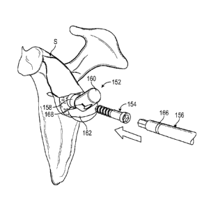
FIG. 8A is an exploded view of augment ream guide 152, fixation fastener
154 and driver instrument 156. FIG. 8B is a perspective view of augment ream
guide 152 seated on partially reamed scapula S with driver instrument 156
inserted
into augment ream guide 152. FIG. 8C is a side view of augment ream guide 152,
which can include bone peg 158, guide peg 160, base 162 and alignment post
164.
Glenoid G can include alignment hole 150 produced by drill bit 138 in the
previous
step.
Bone peg 158 can extend substantially perpendicularly from the bottom
surface of base 162, while guide peg 160 can extend at an oblique angle to
bone peg
160. Substantially perpendicular can include the central axis of bone peg 160
being
disposed ninety degrees to the bottom surface of base 162, as well as the
central axis
being within five degrees of perpendicular. Substantial perpendicularity of
bone
peg 160 can facilitate easy insertion of augment ream guide 152 into central
bore
124.
Guide pin 102 can be removed. The appropriately sized (10 , 20 or 30 )
augment ream guide 152 can be placed on the prepared glenoid G taking care to
align alignment hole 150 with alignment post 164 on ream guide 152. Next, bone
peg 160 can be inserted into central bore 124 in glenoid G, and fixation
fastener 154
can be inserted into guide peg 160 until it is fully seated within augment
ream guide
152 Fixation fastener 154 can be inserted using a hex driver under hand power.
Etch line 166 on drive instrument 156 can align with etch line 168 on ream
guide
11
CA 02983650 2017-10-20
WO 2016/172572
PCT/US2016/028972
152 when completely seated. Fixation fastener 154 can be engineered with the
same
pitch as fixation fastener 60E (FIG. 3A) to help ensure every thread of
fixation
fastener 60E will engage undisturbed bone. Fixation fastener 154 can provide
fixation of ream guide 152 during reaming for augmented baseplate 52.
FIG. 9A is a perspective view of augment reamer 170 being advanced along
augment ream guide 152 (shown in phantom) of FIGS. 8A ¨ 8C to further ream the
partially reamed scapula S. FIG. 9B is a close up view of completely reamed
scapula S of FIG. 9A including central bore 124, first reamed surface 126,
alignment hole 150 and second reamed surface 172.
The appropriately sized (100, 20 or 30 ) augment reamer 170 can be placed
over guide peg 160 of augment ream guide 152. Reamer 170 can include notch 173
that extends into shaft 176 head 174. In particular, a portion of the
circumference of
shaft 176 can be cut away at the end of shaft 176 that engages head 174, and
head
174 can include a similarly located notch that extends into the circumference
of
head 174 at the same circumferential location as the cut away portion of shaft
176.
The notch in head 174 can extend to the center of head 174 so that head 174
has a U
shape. Configured as such, reamer 170 can be slipped over guide peg 160
without
the axes of shaft 176 and guide peg 160 being coaxial. Thus, reamer 170 can be
advanced normal to the face of glenoid G. Thus, reamer 170 can enter more
directly
into an incision in the patient and avoid surrounding tissues.
Reamer 170 can be fully captured on ream guide peg 160 before beginning
to ream, such as by contacting reamer head 174 against glenoid G. Reamer shaft
176 can be rotated to remove bone. If necessary, glenoid osteophytes can be
removed to allow proper seating of reamer 170. Reaming can continue to advance
.. reamer head 174 until a shoulder within reamer shaft 176 bottoms out on
ream guide
peg 160 and the appropriate amount of bone has been prepared to accept the
selected size of augmented baseplate 52. Ream guide 152 can be designed to
allow
minimal reaming of the bone necessary to seat augmented baseplate 52. Next,
fixation fastener 154 (FIG. 8A) can be removed and augment ream guide 152 can
also be removed. Glenoid G can subsequently can include second reamed surface
12
CA 02983650 2017-10-20
WO 2016/172572
PCT/US2016/028972
172 opposite first reamed surface 126. First and second reamed surfaces 126
and
172 can adjoin at edge E. Thus, in one example, glenoid G can now accept
augmented baseplate 52. In particular, parallel surface 54A can abut first
reamed
surface 126 and oblique surface 54B can abut second reamed surface 172.
FIG. 10A is an exploded view of augmented baseplate impactor 190 and
augmented baseplate 52. Augmented baseplate impactor 190 can include alignment
post 192, which can be used to align augmented baseplate 52 with alignment
hole
150. FIG. 10B is a perspective view of impact face 194 of augmented baseplate
impactor 190 showing alignment post 192, center post 194 and peripheral post
196.
Augmented baseplate 52 can be placed onto impact face 194 of baseplate
impactor 190. For example, central post 194 can be inserted into bore 58E in
baseplate 52, while peripheral post 196 is inserted into bore 58A. Alignment
post
192 can extend through bore 58C to be inserted into alignment hole 150.
Additionally, proper orientation of impactor 190 can determined by aligning
the
augment of baseplate 52 (e.g. oblique surface 54B) with a corresponding
"augment"
label on inserter 190. When alignment post 192 is in the correct orientation,
half-
circle etch 198 on inserter 190 can align with second reamed surface 172.
Once aligned, augmented baseplate 52 can be impacted into glenoid G and
remove augmented baseplate impactor 190. Parallel and oblique surfaces 54A,
54B
of augmented baseplate 52 can or should be fully seated on first and second
reamed
surfaces 126, 172, respectively on the face of glenoid G. Fasteners 60A ¨60E
can
be used to secure baseplate 52 to scapula S, and glenosphere 62 can be
attached to
baseplate 52 via stem 64.
Visual confirmation can be attained by checking for gaps between the
reamed surface of glenoid G and baseplate 52 at bores 58A ¨ 58D. A small nerve
hook can be used to aid in confirming complete seating of baseplate 52. Due to
the
10 degree inferior to superior orientation for the baseplate preparation,
baseplate 52
may be partially or fully counter-sunk inferiorly. Guide pin 102 can be
reinserted
before impacting baseplate 52 if cannulated insertion is desired.
13
CA 02983650 2017-10-20
WO 2016/172572
PCT/US2016/028972
FIG. 11 is a perspective view of augmented baseplate 200 for an anatomic
shoulder implant having angled bone surface 202 with fixation posts 204A ¨
204C.
Baseplate 200 can also include articular surface 206, which can be configured
to
directly engage a humeral head. Angled bone surface 202 can include
crenellations
or corrugations 208 that can be used to engage bone and promote bone growth.
Angled bone surface 202 can include parallel surface 210A, which can be
parallel to
glenoid surface 206, and oblique surface 210B, which can be angled at an
oblique
angle relative to parallel surface 210A.
Glenoid G of scapula S can be prepared to mate with parallel surface 210A
and oblique surface 210B, such as by reaming of glenoid G to form obliquely
oriented planar bone surfaces. Oblique surface 210B can be located in
different
orientations on glenoid G, depending on the particular implant and particular
patient. The methods, instruments and tools described herein, particularly
with
reference to FIGS. 12 ¨ 18, facilitate implantation of augmented baseplate 200
onto
scapula S in such a manner so as to minimize bone removal and subsequently
align
augmented baseplate so that surfaces 210A and 210B mate flush with prepared
surfaces of glenoid G.
FIG. 12 is a schematic view of patient-specific glenoid guide 212 engaging
glenoid G of a scapula to install guide pin 214 substantially parallel to the
central
axis of the vault of glenoid G. In the depicted example, glenoid G is
classified as a
Walch B2 glenoid, e.g., a retroverted glenoid with posterior rim erosion, or a
bi-
concave wear pattern with an alpha angle. Patient-specific glenoid guide 212
can be
placed onto the face of glenoid G. Guide pin 214 can be inserted through
glenoid
guide 212 into the glenoid vault of glenoid G.
FIG. 13 is a schematic view of depth stop 216 installed around guide pin 214
of FIG. 12. In one example, depth stop 216 can be patient-specific in that the
length
of depth stop 216 can be sized to allow a reamer to ream glenoid G to a depth
based
on a specific patient's bone defects. Depth stop 216 can include a central
bore 218
to receive guide pin 214 The outer diameter of depth stop 216 can be sized to
receive a socket of a corresponding reamer.
14
CA 02983650 2017-10-20
WO 2016/172572
PCT/US2016/028972
FIG. 14 is a schematic view of reamer 220 installed around guide pin 214
and depth stop 216 of FIG. 13. Reamer 220 can be a standard face reamer that
includes socket 222 for receiving depth stop 216. Reamer 220 can be advanced
until end wall 224 of socket 222 engages the proximal surface of depth stop
216.
Reamer 220 can form first prepared surface 225 on glenoid G.
FIG. 15 is a schematic view of boss and post reamer 226 installed around
guide pin 214 of FIG. 13. Post reamer 226 can include central portion 228,
which
can be configured as a reamer, drill or a rasp to remove bone from glenoid G.
In
particular, central portion 228 can be stepped to provide bore 229 having
various
diameters within scapula S. In particular, central portion 228 can be stepped
to
provide progressively smaller diameter bore segments within scapula S the
deeper
bore 229 goes into the bone. Central portion 228 can be shaped to mate with
the
shape of fixation post 204A of augmented baseplate 200.
FIG. 16 is a schematic view of peripheral post guide 230 installed around
.. guide pin 214 of FIG. 13. Post guide 230 includes sockets 232A and 232B for
receiving drill or reamer 234, as well as socket 236 for receiving guide pin
214.
Sockets 232A and 232B are positioned relative to guide pin 214 in order to
place
bores 238A and 238B relative to bore 229 in locations to correspond to posts
204A
¨ 204C on baseplate 200. Thus, after post reamer 226 is removed from guide pin
214, socket 236 of peripheral post guide 230 can be slipped around guide pin
214
and reamer 234 can be used to make bores 238A and 238B using sockets 232A and
232B, respectively. Drill 234 can include stop 240 to ensure that bores 238A
and
238B are reamed to the depth of posts 204B and 204C. Bores 238A and 238B can
be shaped to mate with the shape of fixation posts 204B and 204C,
respectively.
FIG. 17 is a schematic view of patient-specific angled ream guide 242
installed with compression screw 244 that follows the path of the guide pin of
FIG.
13. Guide pin 214 can be removed and angled ream guide 242 can be inserted
into
bore 229. Compression screw 244 can be inserted into ream guide 242 and
threaded
into scapula S along the path of guide pin 214 to stabilize ream guide 242 for
reaming. Angled ream guide 242 can be configured similarly to ream guide 152
of
CA 02983650 2017-10-20
WO 2016/172572
PCT/US2016/028972
FIGS. 8A ¨ 8C. Ream guide 242 can include bone post 246 that is shaped to at
least
partially fill bore 229 and that includes a central bore for receiving
compression
screw 244. Guide post 248 can extend from bone post 246 at angle a, which can
correspond to the angle between surfaces 210A and 210B of augmented baseplate
200. Alignment post 249 can be connected to ream guide 242 to orient guide
post
248 in the correct direction. Alignment post 249 can have the shape of 204C
and
bone post 246 can have the shape of post 204A
FIG. 18 is a schematic view of reamer 250 installed around guide post 248
of angled ream guide 242 of FIG. 17. Reamer head 252 includes socket 254 to
receive guidepost 248. Using shaft 256, reamer head 252 can be rotated to form
second prepared surface 260 on glenoid G. Guide post 248 can act as a depth
stop
to limit advancement or reamer 250. Reamer 250 thereby produces second
prepared
surface 260 at angle a relative to first prepared surface 225 so that prepared
surfaces
225 and 260 mate with surfaces 210A and 210B, respectively of baseplate 200.
FIG. 19 is a perspective view of augmented glenoid implant 300 having
slanted bone surface 302. Glenoid implant 300 can also include surface 304,
center
post 308, and peripheral posts 310A and 310B. Slanted bone surface 302 can
form
first thickness ti with surface 304 at a first end and second thickness t2
with surface
304 at a second end.
Glenoid G of scapula S can be prepared to mate with slanted bone surface
302, such as by partially reaming of glenoid G at an angle to remove damaged
bone.
Slanted bone surface 302 of augmented baseplate 300 can then partially mate
flush
with the surface of glenoid G reamed at an angle and partially mate flush with
a
naturally angled, undreamed surface of glenoid G. Alternatively, substantially
all of
glenoid G can be reamed at the desired angle to mate with slanted bone surface
302.
The methods, instruments and tools described herein, particularly with
reference to
FIGS. 20¨ 25, facilitate implantation of augmented baseplate 300 onto scapula
S in
such a manner so as to minimize bone removal and subsequently align augmented
baseplate 300 so that slanted bone surface 302 mates flush with the prepared
surface
of glenoid G.
16
CA 02983650 2017-10-20
WO 2016/172572
PCT/US2016/028972
FIG. 20 is a schematic view of a patient-specific glenoid guide 312 engaging
glenoid G of scapula S to install guide pin 314 at angle [3. In the depicted
example,
glenoid G is classified as a Walch B1 glenoid, e.g., a glenoid having a narrow
posterior joint space, subchondral sclerosis and osteophytes or a sloped wear
pattern
with a beta angle. Patient-specific glenoid guide 312 is placed onto the face
of
glenoid G. Guide pin 314 is inserted through glenoid guide 312 into the
glenoid
vault of glenoid G Glenoid guide 312 mates with glenoid G to align guide pin
314
at angle (3, which can be the pathologic angle of glenoid G. Angle (3 can be
predetermined from a surgical plan to allow a reamer to engage a high side of
glenoid G having bone damage.
FIG. 21 is a schematic view of depth stop 316 surrounding guide pin 314 of
FIG. 20. In one example, depth stop 316 can be patient-specific in that the
length of
depth stop 316 can be sized to allow a reamer to ream glenoid G to a depth
based on
a specific patient's bone defects. Depth stop 316 can include central bore 318
to
receive guide pin 314. The outer diameter of depth stop 316 can be sized to
receive
a socket of a corresponding reamer.
FIG 22 is a schematic view of reamer 320 being advanced onto guide pin
314 of FIG. 20 to surround depth stop 316 and at least partially ream glenoid
G.
Reamer 320 can be a standard face reamer that includes socket 322 for
receiving
depth stop 316. Reamer 320 can be advanced until end wall 324 of socket 322
engages the proximal surface of depth stop 316. Reamer 320 can form prepared
surface 326 on glenoid G. The high side of the glenoid G can be reamed to
remove
damaged bone in glenoid G.
FIG. 23 is a schematic view of a patient-specific drill guide 328 mated to
partially reamed glenoid G of FIG. 22 to form central post bore 330 in
conjunction
with drill or reamer 332. Drill guide 328 can include base 334, which can be
patient-specific to mate with partially reamed glenoid G, and cup 336, which
can be
shaped to receive reamer 332 and positioned to align central post bore 330 in
scapula S. Base 334 is shaped to align central post bore 330 along the
anatomic axis
of scapula S while accounting for the fact that base 334 can be slanted and
non-
17
CA 02983650 2017-10-20
WO 2016/172572
PCT/US2016/028972
perpendicular to the anatomic axis. Base 334 can register on a periphery of
glenoid
G and a portion of the reamed face of glenoid G. Drill guide 328 can also
include
handle shaft 338 that can allow a surgeon to position and steady base 334 for
performing reaming. Reamer 332 can be a standard boss reamer.
FIG. 24 is a schematic view of a patient-specific peripheral post reamer
guide 340 being advanced into reamed central post bore 330 of FIG. 23 to form
peripheral bores 342A and 342B in conjunction with drill or reamer 344. Reamer
guide 340 can include center peg 346 that can be shaped to mate with central
post
bore 330 to align peripheral bores 348A and 348B with respect to glenoid G.
Reamer 344 can be a standard reamer. Alternatively, a drill may be used.
Patient-
specific peripheral post reamer guide 340 can allow for reaming of peripheral
bores
342A and 342B without the need for inserting additional guide pins into
glenoid G.
FIG. 25 is a schematic view of the augmented baseplate 300 of FIG. 19A
mounted onto partially reamed glenoid G so that slanted bone face 302 and
fixation
posts 308, 310A and 310B mate with prepared glenoid G. Before augmented
baseplate 300 is implanted, preparation of glenoid G can be performed with a
patient-specific glenoid trial having a full augment (not shown). After
confirmation
of the reaming of glenoid G, posts 308, 310A and 310B of augmented baseplate
300
can be inserted into bores 330, 342A and 342B, respectively, of scapula S. A
standard impactor can be used to insert augmented baseplate 300 and bone
cement
can also be used in bores 330, 342A and 342B. In one example, augmented
baseplate 300 will start at an anterior side of glenoid G and angle or slope
to the
posterior side, as opposed to the procedure described with reference to FIGS.
11 ¨
18 where the angle or slope starts at the midline of glenoid G.
The methods, implants and tools described herein are advantageous over
previous systems. For example, the patient-specific augment reamer guides can
allow for precise reaming of a glenoid with minimal bone removal, and can
allow
for accurate fitting with patient-specific implants. The patient-specific
guides can
allow for placement of guide pins, such as Steinmann Pins, at the angle of
pathologic glenoid for face reaming, or to place guide pins along the main
axis of
18
CA 02983650 2017-10-20
WO 2016/172572
PCT/US2016/028972
the glenoid vault. The patient-specific guides can allow for reaming of
glenoid
bosses (e.g., with standard reamers) and can be made so as to not require
placement
of a second pin in the glenoid. The patient-specific augmented implants can be
made for various types of glenoid deficiencies, such as Bl, B2 or other
glenoid
classification (anatomic or reverse).
Various Notes & Examples
Example 1 can include or use subject matter such as a glenoid implant,
comprising: a body can comprise: an articular surface configured to mate with
or
receive another component configured to mate with a complimentary component;
and a scapula-engaging surface opposite the articular surface, the scapula
engaging
surface including first and second portions angled relative to each other; and
a
fixation feature extending from the scapula-engaging surface.
Example 2 can include, or can optionally be combined with the subject
matter of Example 1, to optionally include a body and fixation feature that
can be
made from porous metal material.
Example 3 can include, or can optionally be combined with the subject
matter of one or any combination of Examples 1 or 2 to optionally include a
body
including a sidewall having a first thickness at the first portion of the
scapula-
engaging surface and a second thickness at the second portion of the scapula-
engaging surface, the second thickness can be greater than the first
thickness.
Example 4 can include, or can optionally be combined with the subject
matter of one or any combination of Examples 1 through 3 to optionally include
an
edge between the first and second portions extends across a midline of the
scapula-
engaging surface between the first and second portions.
Example 5 can include, or can optionally be combined with the subject
matter of one or any combination of Examples 1 through 4 to optionally include
first
and second portions that can each be planar.
19
CA 02983650 2017-10-20
WO 2016/172572
PCT/US2016/028972
Example 6 can include, or can optionally be combined with the subject
matter of one or any combination of Examples 1 through 5 to optionally include
first
and second portions that can be angled relative to each other at an angle in
the range
of approximately ten degrees to approximately thirty degrees.
Example 7 can include, or can optionally be combined with the subject
matter of one or any combination of Examples 1 through 6 to optionally include
a
fixation feature that comprises a center boss for receiving a fixation
fastener.
Example 8 can include, or can optionally be combined with the subject
matter of one or any combination of Examples 1 through 7 to optionally include
a
fixation feature comprising a plurality of posts.
Example 9 can include, or can optionally be combined with the subject
matter of one or any combination of Examples 1 through 8 to optionally include
a
scapula-engaging surface that can include corrugations.
Example 10 can include or use subject matter such as a method for
implanting a scapular baseplate in a shoulder arthroplasty, the method can
comprise:
inserting a guide pin into a glenoid of the scapula using a guide instrument;
preparing a first portion of the glenoid to form a planar bone surface using
the guide
pin; forming a first bore into the glenoid located approximately at the guide
pin;
forming a second bore into the glenoid offset from the first bore; inserting
an
augment ream guide into the first bore and the second bore, and preparing a
second
portion of the glenoid to form an angled bone surface relative to the planar
bone
surface using the augment ream guide.
Example 11 can include, or can optionally be combined with the subject
matter of Example 10, to optionally include forming the first bore into the
glenoid
comprises positioning a reamer over the guide pin to ream the first bore while
preparing the first portion of the glenoid; and forming the second bore into
the
glenoid comprises positioning a drill guide over the guide pin after removing
the
reamer and drilling the second bore.
Example 12 can include, or can optionally be combined with the subject
matter of one or any combination of Examples 10 or 11 to optionally include
CA 02983650 2017-10-20
WO 2016/172572
PCT/US2016/028972
forming the first bore into the glenoid by positioning a boss and post reamer
over
the guide pin to ream the first bore after preparing the first portion of the
glenoid;
and forming the second bore into the glenoid by positioning a drill guide over
the
guide pin after removing the reamer and drilling the second bore.
Example 13 can include, or can optionally be combined with the subject
matter of one or any combination of Examples 10 through 12 to optionally
include a
guide instrument that can be patient-specific to mate with the glenoid
Example 14 can include, or can optionally be combined with the subject
matter of one or any combination of Examples 10 through 13 to optionally
include a
guide instrument that positions the guide pin approximately parallel to a main
axis
of a vault of the glenoid, the planar bone surface is approximately
perpendicular to
the main axis, and the angled bone surface is angled relative to the planar
bone
surface at an angle in the range of approximately ten degrees to approximately
thirty
degrees.
Example 15 can include, or can optionally be combined with the subject
matter of one or any combination of Examples 10 through 14 to optionally
include
preparing a fist portion of the glenoid to form a planar bone surface by at
least
partially reaming the glenoid to approximately fifty percent of a surface area
of the
glenoid.
Example 16 can include, or can optionally be combined with the subject
matter of one or any combination of Examples 10 through 15 to optionally
include
preparing a first portion of the glenoid to form a planar bone surface by
inserting a
post of the augment ream guide into a notch in a reamer head and shaft of a
reamer.
Example 17 can include, or can optionally be combined with the subject
matter of one or any combination of Examples 10 through 16 to optionally
include
positioning a depth stop over the guide pin to limit a depth to which the
glenoid can
be reamed.
Example 18 can include or use subject matter such as a ream guide for a
shoulder arthroplasty procedure, the ream guide can comprise: a base having a
first
surface and a second surface; a bone peg extending substantially
perpendicularly
21
from the first surface; an alignment peg extending from the first surface
spaced from
the bone peg; and a guide peg extending from the second surface opposite the
bone
peg that can be at an oblique angle to the bone peg.
Example 19 can include, or can optionally be combined with the subject
matter of Example 18, to optionally include a guide peg that includes an
aperture
extending into the bone peg.
Example 20 can include, or can optionally be combined with the subject
matter of one or any combination of Examples 18 or 19 to optionally include a
second surface that can be angled relative to the first surface.
Each of these non-limiting examples can stand on its own, or can be
combined in various permutations or combinations with one or more of the other
examples.
The above detailed description includes references to the accompanying
drawings, which form a part of the detailed description. The drawings show, by
way of illustration, specific embodiments in which the invention can be
practiced.
These embodiments are also referred to herein as "examples." Such examples can
include elements in addition to those shown or described. However, the present
inventors also contemplate examples in which only those elements shown or
described are provided. Moreover, the present inventors also contemplate
examples
using any combination or permutation of those elements shown or described (or
one
or more aspects thereof), either with respect to a particular example (or one
or more
aspects thereof), or with respect to other examples (or one or more aspects
thereof)
shown or described herein.
In this document, the terms "a" or "an" are used, as is common in patent
documents, to include one or more than one, independent of any other instances
or
usages of "at least one" or "one or more." In this document, the term "or" is
used to
refer to a nonexclusive or, such that "A or B" includes "A but not B," "B but
not
A," and "A and B," unless otherwise indicated. In this document, the terms
"including" and "in which" are used as the plain-English equivalents of the
22
CA 2983650 2019-01-09
respective terms "comprising" and "wherein." Also, in the following claims,
the
terms "including" and "comprising" are open-ended, that is, a system, device,
article, composition, formulation, or process that includes elements in
addition to
those listed after such a term in a claim are still deemed to fall within the
scope of
that claim. Moreover, in the following claims, the terms "first," "second,"
and
"third," etc. are used merely as labels, and are not intended to impose
numerical
requirements on their objects.
23
CA 2983650 2019-01-09