Note: Descriptions are shown in the official language in which they were submitted.
CA 02984289 2017-10-27
WO 2015/175546
PCT/US2015/030398
METHOD AND SYSTEM OF ASSESSING OR ANALYZING MUSCLE
CHARACTERISTICS INCLUDING STRENGTH AND TENDERNESS USING
ULTRASOUND
CROSS-REFERENCE TO RELATED APPLICATION
[0001] This
application claims the benefit of U.S. Provisional Application Serial No.
61/992,021, entitled "Method and System of Assessing or Analyzing Muscle
Characteristics Including Strength and Tenderness Using Ultrasound" and filed
on
May 12, 2014, which is expressly incorporated by reference herein in its
entirety.
BACKGROUND
Field
[0002] The present disclosure relates generally to the field of
diagnostic sonography
imaging and techniques, more particularly, to a method and system for
analyzing
ultrasound images for muscle tissue density, tensile strength and mass, as
well as,
equating it into a rating of palatability, pliability, tenderness, strength,
and/or power.
Description of Related Art
[0003]
Carcass evaluation, whether performed "on the hoof" for live animals or "on
the
rail" for hot carcass or meat has been performed phenotypically and by using
ultrasound. A Quality grade, such as "prime," "choice," or "select," may then
be
designated to the carcass based on the pheno evaluation. Ratings may also be
designated for livestock carcass evaluation. It is very important to the meat
and
livestock industry to have a system that can provide an objective measurement
of
carcass and meat quality. Pliability or tissue tenderness is a highly desired
quality.
The United States Department of Agriculture (USDA), e.g., applies techniques
to
grade beef based on assessment of adipose tissue content within the muscle
known
as intramuscular fat (IMF), e.g., "marbling." However, marbling often fails to
accurately predict tenderness because there are two types of tissue.
Additionally,
the current USDA Quality Grade and Yield Grading system that correlates high
fat
content with tenderness can discourage meat consumption in a health conscious
consumer base.
1
CA 02984289 2017-10-27
WO 2015/175546
PCT/US2015/030398
[0004]
Although various ultrasound-based technologies have been used for carcass
evaluation, this technology has a direct correlation to the measurement of,
the
amount of back fat, rib-eye area, and IMF known as Marbling. There continues
to
be a need for a more accurate analysis pertaining to muscle density, mass,
tissue
pliability, and tissue texture separate from the assessment of taste and
palatability,
currently known as quality grade.
SUMMARY
[0005] In
light of the above described problems and unmet needs, as well as others, in
an
aspect of the disclosure, a method, a system, and/or a computer program
product are
provided that provide and/or predict a more accurate analysis of muscle
structure in
its complex form. Aspects presented herein further provide the capability to
analyze
tissue palatability, which relates to muscle pliability and tenderness,
separate from
an assessment of fat, e.g., IMF. Currently, meat, such as steaks, which have a
combination of low fat content and good pliability values, cannot be predicted
in
livestock. Aspects presented herein provide a method and system to perform non-
invasive evaluation on live, on the rail, or packaged meat to characterize
taste and
palatability, including pliability and tissue tenderness. This includes
identifying
animals that are more likely to have a desirable consumer experience, e.g.,
that
allows a consumer to enjoy a delectable delicious charbroiled, seasoned to
perfection cooked rib-eye steak.
[0006] Thus, aspects presented herein provide a method and system for
analyzing
ultrasound images in order to identify muscle tissue, density, tensile
strength and
mass, as well as, equating or otherwise correlating such analyses to a rating
of
tenderness with the quality grade. This functionality may include grading
muscle
structure of all muscle groups. The number of muscle bundles, sarcomeres,
fascicles,
fibers, sheaths around these aspects and/or muscle break down may be analyzed
and
ratings may be generated for muscle strength, growth, and/or tissue
pliability, e.g.,
tenderness.
[0007] An analysis of the muscle structure may be performed via a
simple scanning
procedure using ultrasound equipment to collect imaging data for evaluation.
For
example, the ultrasound equipment and settings that enable an accurate image
at a
depth of approximately between one to three inches of tissue, e.g.,
approximately
two inches deep into the analyzed tissue, may be performed.
2
CA 02984289 2017-10-27
WO 2015/175546
PCT/US2015/030398
[0008] The
analysis may include determining a relative number of bundles within a muscle
group, such as by using ultrasound equipment. Aspects may include gathering
data
in determining a relative number of fascicles, sarcomeres, and or fibers
within the
bundle, including measurements of the epimysium, endomysium, or sheaths around
each structure of muscle from the ultrasound image.
[0009] Thereafter, a number of muscle characteristics may be determined
or predicted
based on the analysis. For example, muscle pliability, or softness, may be
predicted
for the sample based on the analysis. Additionally, other characteristics,
including a
potential for strength in competing subjects, whether human or competition
animals,
may be predicted based on the analysis. For example, the analysis may be used
for
person involved in sports competitions and athletic events. This analysis may
be
performed regardless of the age of the subject, due to the fact that each
animal is
predisposed to have at birth a given amount of muscle tissue and may never
produce
any more for the rest of its life.
[0010] Aspects may further include using a rating system that can
identify the quality grade
for cuts of meat having a desirable amount of palatability even with moderate
or
lower fat content.
[0011] Additional advantages and novel features of these aspects will
be set forth in part in
the description that follows, and in part will become more apparent to those
skilled
in the art upon examination of the following or upon learning by practice of
this
diagnostic or sonographic analyses program and invention and aspects thereof
BRIEF DESCRIPTION OF THE DRAWINGS
[0012]
Various example aspects of the systems and methods will be described in
detail,
with reference to the following figures, wherein:
[0013] FIG. 1 is a diagram illustrating an example of muscle structure,
in accordance with
aspects of the present invention.
[0014] FIG. 2 is a diagram illustrating an example of muscle structure,
in accordance with
aspects of the present invention.
[0015] FIG. 3 is a flow chart illustrating an example method of
analyzing muscle
tenderness, in accordance with aspects of the present invention.
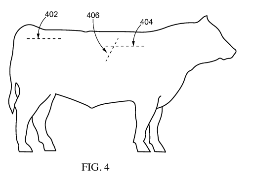
[0016] FIG. 4 is a diagram illustrating example positions for
ultrasound measurements, in
accordance with aspects of the present invention.
3
CA 02984289 2017-10-27
WO 2015/175546
PCT/US2015/030398
[0017] FIG. 5
is flow chart illustrating an example method of analyzing muscle
characteristics, in accordance with aspects of the present invention.
[0018] FIG. 6 is an example ultrasound image, in accordance with
aspects of the present
invention.
[0019] FIG. 7 is an example ultrasound image, in accordance with
aspects of the present
invention.
[0020] FIG. 8 is an example ultrasound image, in accordance with
aspects of the present
invention.
[0021] FIG. 9 presents an example system diagram of various hardware
components and
other features, for use in accordance with aspects of the present invention.
[0022] FIG. 10 is a block diagram of various example system components,
in accordance
with aspects of the present invention.
[0023] FIG. 11 is a diagram illustrating an example of muscle
structure, in accordance with
aspects of the present invention.
[0024] FIGs. 12-16 illustrate example images that may be used to assess
characteristics of
the muscle based on the muscle structure, in accordance with aspects of the
present
invention.
DETAILED DESCRIPTION
[0025] These
and other features and advantages are described in, or are apparent from, the
following detailed description of various example aspects. The detailed
description
set forth below in connection with the appended drawings is intended as a
description of various configurations and is not intended to represent the
only
configurations in which the concepts described herein may be practiced. The
detailed description includes specific details for the purpose of providing a
thorough
understanding of various concepts. However, it will be apparent to those
skilled in
the art that these concepts may be practiced without these specific details.
In some
instances, well-known structures and components are shown in block diagram
form
in order to avoid obscuring such concepts.
[0026] As presented herein muscle characteristics may be determined or
predicted based on
an analysis of ultrasound images of the muscle. For example, any of muscle
palatability, pliability, texture, tenderness, and/or softness, can be
predicted for the
sample based on the analysis. These characteristics are also referred to
herein as
tissue characteristics. Additionally, other characteristics, including a
potential for
4
CA 02984289 2017-10-27
WO 2015/175546
PCT/US2015/030398
strength and/or power in competing or sporting subjects, whether human or
competition animals, may be predicted based on the analysis.
[0027] Tenderness
[0028] Currently, livestock and on the rail product are rated based on
quality grade and
yield grade by a USDA inspector performing a phenotypical analysis of fat
found on
and within the carcass, e.g., KPH fat. However, such a measurement of fat is
not
always an accurate predictor of tenderness, texture, pliability, tissue
softness, and/or
palatability of subsequently processed meat. Additionally, breeds of livestock
that
have lower amounts of fat will incur poor ratings, even though a number of
such
livestock could have a potential to be a producer of more desirable cuts of
meat.
[0029] Meat palatability, pliability, softness, and/or tenderness relates
to the structure of the
muscle itself Thus, aspects presented herein include performing an analysis,
by the
use of ultrasound and collected imaging data, e.g. analyze the muscle
structure of,
bundle, sarcomeres, fascicles, and fibers within the deposition of the muscle,
by
collecting ultrasound images to predict tenderness of the muscle and muscle
strength. For example, the analysis may include determining or counting a
number
of bundles, a number of fascicles, a number of sarcomeres, and/or a number of
fibers
within the muscle, as well as measuring sheath(s) that surround each component
of
muscle in order to determine a score, e.g., a toughness score.
[0030] Skeletal Muscles are structured within a bundle inside of bundles
within the muscle
tissue. As illustrated in Fig. 1, a sarcomere is the basic unit of a muscle.
Muscles
are composed of tubular muscle fibers 102 (myocytes or myofibers) that are in
turn
composed of tubular myofibrils. Myofibrils are composed of repeating sections
of
sarcomeres. Sarcomeres are composed of long, fibrous proteins that slide past
each
other when the muscles contract and relax. The striated appearance of skeletal
muscle results from the regular pattern of sarcomeres within their cells. A
muscle
fiber 102 may contain any number of sarcomeres. Thus, a muscle fiber 102
comprises bundles of sarcomeres, and the muscle fibers 102 are bundled into a
group to form a fascicle 104. Bundles of fascicles 104 then form the overall
muscle
106.
[0031] Skeletal muscles, such as those found in livestock, are sheathed by
a tough layer of
connective tissue called the epimysium 108 and a matrix of the sheath. Each
epimysium 108 comprises multiple fascicle bundles 104, each of which contains
a
number of muscle fibers 102 collectively sheathed by a perimysium 110. Each
CA 02984289 2017-10-27
WO 2015/175546 PCT/US2015/030398
muscle fiber 102 comprises bundles of sarcomeres, which can be measured as an
aspect in a prediction or rating relating to tenderness.
[0032] The number of these bundled fibers varies significantly among
different subjects,
even subjects of the same species. For example, each bundle may comprise a
range
of fascicles , and fibers, right down to the sarcomeres, held together by a
matrix of
Perimysium, Endomysium, and Epimysium. e.g., approximately between 10 and
500 plus.
[0033] FIG. 2 provides two modalities of muscle structure, sagittal and
transverse.
Additional details regarding muscle structure may be found, for example,
published
on Wikipedia webpages under the designation "muscle" and "sarcomere," which is
located at http ://en.wikipedia. org/wiki/Mus c le and
http://en.wikipedia.org/wiki/Sarcomere, respectively, the entire contents of
which
are hereby incorporated herein by reference.
[0034] FIG. 11 contains an additional illustration of muscle structure
from "The Anatomy
Coloring Book" by Wynn Kapit and Lawrence M. Elson, Published in 2002 at page
44, the entire contents of which are incorporated by reference herein. Fat
deposits
flow in the same area as blood flow throughout the muscle structure. The
analysis
described herein analyzes the muscle structure, e.g., including density, size,
and
number of components that form a muscle structure. In areas that lack
fascicles,
etc., additional area may be provided for deposition of fat. The deposition of
fat is
currently used as a standard for quality grade to predict taste and
palatability. The
lack of muscle structure, e.g., fascicles within a bundle, as analyzed using
the
aspects presented herein, may be used to more accurately determine
palatability,
pliability, and tenderness.
[0035] Tough muscle typically has a coarser structure having a lower
number of larger
sized bundles, sarcomeres, fascicles, and/or fibers in comparison to tender
muscle.
The number of bundles, fascicles, sarcomeres, and/or fibers along with the
coarseness of the muscle structure create stronger or weaker muscle tissue.
For
example, a bundle having a finer (less coarse) structure and therefore a
higher
number of smaller fibers, sarcomeres, and/or fascicles in bundles of muscle
structure
may be predicted to create stronger tissue and to have a more tender quality.
For
example, livestock with a finer muscle structure may be rated to be a higher
quality
and more likely to be tender than those with a coarse muscle structure.
Coarser
muscle tissue equates to tougher pieces of meat. Therefore, meat comprising a
6
CA 02984289 2017-10-27
WO 2015/175546
PCT/US2015/030398
smaller more refined sized, finer fascicles and/or sarcomeres will be more
tender.
The number of sarcomeres and fascicles within the bundles of muscles do not
change. Animals are allotted with a certain amount at the time of birth. This
number does not change through the life of that animal. This characteristic
occurs
for all skeletal muscle tissue.
[0036] Therefore, aspects presented herein provide for a way to analyze
muscle structure
and tenderness separately from the current grading system of quality grade for
any
muscle group, which measure fat. Ultrasound is used to perform an analysis of
characteristics of the muscle structure itself As the muscle grows, e.g., the
number
of fascicles does not increase. Instead, the size of the individual fascicles
increases.
Thus, using an analysis of the relative number of fascicles and/or sarcomeres
in the
muscle structure may accurately predict which livestock will produce higher
quality
and more palatable piece of meat from a young age, because the amount of
bundles,
fascicles/sarcomeres does not change as the livestock matures.
[0037] Tenderness, as used herein, may relate both to subjective
properties of meat tissue,
such as softness, juiciness, palatability, and flavor intensity of cooked
meat, as well
as to an objective measure, e.g., relating to the shear and/or tensile force
necessary
to cut, or chew, a sample piece of meat. One example of such an objective
shear
force determination is the Warner-Bratzler shear (W-B shear or WBS) force
values
have long been used as the industry standard for an objective tenderness
scale. This
WBS testing has been performed only on the concept of cooked meat at 1600,
then
by performing the shear test without knowing the structure, volume, density,
tensile
or mass of the muscle tissue. Thus, there exists a need for a more accurate
way of
analysis with an objective determination to predict tissue palatability
pliability,
strength, and/or tenderness by providing a much needed scale with a greater
effectiveness in selecting and developing a more accurate and consistent way
of
predicting palpability and tenderness.
[0038] Aspects used herein enable a comparison of muscle structure and
density in a more
consistent form, which also enables a higher level of accuracy, consistency,
and
repeatability for objective tenderness tests, such as the WBS test. For
example,
comparisons may be made for samples having similar muscle structure, e.g., a
first
sample having approximately 20 bundles, fascicles, or sarcomeres as opposed to
a
second sample having beyond 100 within its corresponding structure. The sample
7
CA 02984289 2017-10-27
WO 2015/175546
PCT/US2015/030398
with 20 bundles, fascicles, or sarcomeres may be predicted to have less
tenderness
and less strength in comparison to the sample having 100.
[0039] FIG. 3 is a flow chart 300 of a method of determining muscle
structure, density,
mass, and volume by using ultrasound. Although described using the term
"muscle," this method may be used to predict tissue palpability pliability,
tenderness, etc., or to provide a more efficient use of the quality grading
system,
rating on-the-hoof of all livestock animals, or on-the-rail for a hot carcass
evaluation. Livestock may include, e.g., cattle, sheep, goats, bovine, equine,
swine,
etc. Although FIG. 3 is described using the example of livestock, aspects of
the
analysis, quality prediction, and rating in relation to the tenderness and
palatability
presented herein apply to any animal for which it is beneficial to evaluate
and
predict the quality of its meat, including e.g., aquatic animals, aviary,
exotic
animals, wildlife, etc. Similar to the example of evaluating livestock, it may
be
beneficial to evaluate and predict tissue characteristics of salmon, tuna,
chicken,
turkey, venison, etc. using the aspects presented herein. Additionally,
although FIG.
3 is described using the example of livestock, aspects of this analysis may
also be
used to determine muscle strength in competing animals, including humans, as
described in additional detail in connection with FIG. 5. The method may be
performed by a technician and may also be performed in an automated manner,
e.g.,
via a processor, as described infra.
[0040] At 302, an analysis is performed of muscle structure and design
using ultrasound.
This may include capturing at least one image of muscle structure using
ultrasound.
The ultrasound image may comprise musculoskeletal ultrasound data, e.g., at
least
one ultrasound image captured by presenting an ultrasound probe at a desired
location on any muscle group for which it is desired to know muscle tissue
palpability, tenderness, and/or strength, on an animal and generating an image
resultant from the ultrasound signal. Example locations are described infra,
with
examples shown in FIGs. 4-8.
[0041] For example, the ultrasound equipment and settings that enable
an accurate image at
a depth of approximately between one to three inches of tissue, e.g.,
approximately
two inches deep into the analyzed tissue, may be performed. In one example,
traditional ultrasound images may be obtained. In another example, Tissue
Doppler
Imaging (TDI) may be used to obtain an image of the tissue. TDI uses
principles
similar to Doppler echocardiography to quantify high-amplitude, lower-velocity
8
CA 02984289 2017-10-27
WO 2015/175546
PCT/US2015/030398
signals of myocardial tissue motion. For example, FIG. 16 illustrates a
Doppler
ultrasound image of muscle tissue. The Doppler ultrasound image in FIG. 16
illustrates an individual strand fiber 1602, e.g., of a ligament, can be
identified,
counted, measured, etc. within tissue in order to analyze the tissue
characteristics
such as tenderness, pliability, and strength, to determine ones susceptibility
to
injury, etc.
[0042] Additional example images are illustrated, e.g., in FIGs. 12-15.
The ultrasound
image may comprise a static image and may comprise a video of ultrasound image
data so that the analysis may be made while watching movement of the muscles.
Static images and video data may also be used in combination in order to
perform a
more accurate analysis.
[0043] The tenderness analysis in 302 may be performed for various
types of samples. For
example, the "sample" may be, among others, a cut of meat, an exposed portion
of a
carcass, or interior tissue of a live animal. The image for analysis may be
manually
obtained or electronically steered, e.g., steering of the sound waves.
[0044] The ultrasound data may be analyzed either at the site of the
ultrasound or at a
different location. If the ultrasound data is analyzed at a different
location, the
ultrasound data may be received via transmission and/or may be stored in
memory
for later analysis. For example, the analysis may be performed at a central
processing location that receives and processes ultrasound data from a number
of
samples taken remotely from the central processing location. Thus, at 306
ultrasound data may optionally be received in order to perform the analysis.
Optional aspects are illustrated using a dashed line in FIG. 3. Additional
analysis
may be performed on historical ultrasound image data in order to identify an
inherited trend in the structure of muscle.
[0045] The analysis in 302 may include presenting ultrasound data at a
display screen for
evaluation by a technician, and/or analysis of the ultrasound data performed
via a
processor. For example, a processor may analyze the image data generated by
the
ultrasound signal.
[0046] The analysis at 302 may be performed for any of a number of
ultrasound techniques.
Different locations on a sample may be used for the analysis, because
different areas
of muscle may be important for different applications. For example, different
muscles may be analyzed for palatability of meat in comparison to a strength
analysis for competing animals.
9
CA 02984289 2017-10-27
WO 2015/175546
PCT/US2015/030398
[0047] As
illustrated in FIG. 3, the analysis of muscle structure may comprise any of
determining a relative number of muscle bundles for the ultrasound data at
308,
determining a relative number of fascicles within the muscle bundles at 310,
determining a relative number of sarcomeres within the fascicles at 312, and
determining a relative number of muscle fibers for the ultrasound data at 314.
[0048] Each of the determinations in 308, 310, 312, and 314 may include
a determination
of the relative size, as well as the number, of any of the muscle bundles,
fascicles,
sarcomeres, and fibers. The analysis is of a relative number and/or size,
because any
scale or rating based on such measurements must take into account the breed,
age,
gender, size, etc. of the animal. For example a female of a first, smaller
breed may
have muscle bundles and fascicles and fibers that need to be adjusted to
correlate to
a larger animal or breed specific measurement. Although the measurements are
the
same, the measurements would correlate to different levels of tenderness or
muscle
strength in the two animals. Thus, the breed, size, age, gender, etc. of the
animal
may be taken into consideration when performing the analysis.
[0049] A measurement of the matrix of the myofibril may also be taken
using ultrasound
and this information may be included in the analysis.
[0050] Additionally, the toughness of a sheath surrounding the muscle
structure may also
correlate to the strength or tenderness of the muscle. Thus, the determination
may
include measuring a thickness of the sheath surrounding the muscle structure,
a
larger, thicker sheath may correspond to a prediction of less tender meat.
Thus,
connective tissue, e.g., collagen, contributes to meat tenderness and texture
and may
strongly affect the consumer's experience. For example, such collagen,
connective
tissue as the perimysium, endomysium, and epimysium may be identified and
measured using the hyperechoic lines within an ultrasound image. An analysis
of
the connective tissue may include not just a measurement of the amount or size
of
the tissue, but an analysis of the crosslinking of such tissue.
[0051] Any combination of determinations 308, 310, 312, 314, and 316
may be used in the
analysis 302. The determination may involve counting, identifying, and/or
sampling
the number of bundles, fascicles, sarcomeres, and/or muscle fibers in a muscle
group
within the ultrasound image and corresponding data. The size of the individual
bundles, fascicles, sarcomeres, and/or muscle fiber may be measured and a
density
may be determined. These muscle structures may be identified, measured, and
counted based on an analysis of striations in the ultrasound image of the
muscle.
CA 02984289 2017-10-27
WO 2015/175546
PCT/US2015/030398
For example, FIGs. 8 and 12-14 illustrate striations that can be analyzed in
order to
determine or estimate muscle structure. The determination may involve
estimating
an amount of bundles, fascicles, and/or sarcomeres and/or measuring the sheath
in
the ultrasound data.
[0052] As different ultrasound images may capture larger or smaller
portions of muscle, the
relative number of muscle bundles, fascicles, sarcomeres, and/or fibers may be
determined rather than the overall number shown in the ultrasound data. The
number may be determined relative to a common amount in each muscle group. For
example, a certain sample size may be considered each time. For example, the
amount of such muscle structures may be identified for a two inch square
sample.
The size of the sample may be selected depending upon the anatomical location
of
the muscle being analyzed. For example, the determinations may include a
determination of a density of bundles, fascicles, sarcomeres, and/or fibers
and/or
sheath thickness within the sample. A percentage may be generated.
Additionally,
a ratio may be generated relating any of the bundles, fascicles, sarcomeres,
and/or
fibers to one another. For example, an amount of fascicles per muscle bundle
may
be determined. An amount of sarcomeres per muscle bundle and/or fascicle may
be
determined. An amount of muscle fibers per bundle, per fascicle, and/or per
sarcomere may be determined. The determined numbers may be actual numbers,
estimated numbers, or average numbers of the muscle structure being
identified.
[0053] At 304, a prediction may be made regarding the tenderness of the
muscle based on
the analysis of the ultrasound sample performed at 302. For example, a
prediction
of the muscle quality or tenderness may be made based on the amount of,
density of,
and/or size of bundles, fascicles, sarcomeres, and/or fibers identified via
the
ultrasound data. A
tenderness scale may be provided, having a ratings
corresponding to certain ranges of bundle, fascicle, sarcomere, and/or fiber
measurements. For example, samples identified as having a lower coarseness
fascicles and/or sarcomeres may rank higher on the tenderness scale than a
sample
having a higher coarseness of fascicles and/or sarcomeres. Similarly, samples
identified as having a higher tissue texture coarseness may rank lower on a
tenderness scale indicating a desirability of the meat.
[0054] As one example, a sample having a finer texture of bundles,
fascicles, sarcomeres,
and/or fibers may be considered to have an increased amount of tenderness and
be
ranked higher on a scale of desirable pieces of meat; whereas those with
coarser
11
CA 02984289 2017-10-27
WO 2015/175546
PCT/US2015/030398
bundles, fascicles, sarcomeres, and/or fibers may be considered to have a
lesser
degree of tenderness. Finer fibers, sarcomeres, fascicles, and bundles may be
smaller in size and may also have a higher tissue density than tissue having a
coarser
structure. The finer tissue texture may be an indicator of tissue structure
providing a
more tender, palatable piece of meat, which equates to a more satisfying
eating
experience.
[0055] Similarly, the finer, denser muscle structure may also correlate
to increased strength
and a reduced susceptibility to injury.
[0056] At 318, a muscle tenderness grade and/or rating may be specified
for the sample
based on analyzed ultrasound data. The tenderness grade/rating may be a
function
of any of a density, a percentage, and a ratio of bundles, fascicles and/or
sarcomeres
illustrated in the ultrasound data. The rating may be similar to the choice,
select,
and prime designations that are used in the current quality grading system, or
the
rating may be completely separate from the current quality grading system. The
analysis 302 and prediction 304 may be used to predict quality of livestock
and yield
grade.
[0057] As an additional example, the rating may provide an additional
level to the current
grading system. For example, the rating may involve levels, e.g., 1-5 for
choice,
levels 1-5 for select, and levels 1-5 for prime. Thus, a first sample may
receive a
rating of select, level 2 based on the analysis. A second sample may receive a
rating
of select, level 3. A third may receive a rating of prime, level 5.
[0058] The analysis in 302 and the rating in 318, which is based
thereon, may consider
other factors in connection with the analysis of muscle structure performed in
302.
[0059] For example, the analysis regarding the bundles, fascicles,
sarcomeres, fibers, and/or
sheaths may be used to form the basis for tenderness grading in meat samples,
in
combination with a consideration of the species/age of the livestock and/or
traditional fat measurements, such as percentage intramuscular fat (IMF), rump
fat
thickness, 12th-13th rib fat thickness, ribeye area, marbling, etc.
[0060] For a carcass, a rating in 312 may be based on the palpability,
pliability, and
tenderness analysis 302 in combination with additional suitable factors
including
any of a type of cut of meat, electrolysis or shocking of the carcass, an
aging
process, heat processing, a storage temperature history, and any tenderizing
treatments.
12
CA 02984289 2017-10-27
WO 2015/175546
PCT/US2015/030398
[0061] The
analysis presented herein may be used to predict final carcass quality of live
animals and to predict the muscle structure, volume, and mass that may be
equated
into tenderness of particular cuts of meat after subsequent cooking.
[0062] This carcass exam can be performed anatomically on any muscle
group in any
location on an animal. As each animal is predisposed with a certain muscle
development at birth, they do not grow additional muscle bundles, sarcomeres,
fascicles, and fibers. Evaluations "on the hoof" for live animals or "on the
rail" for
a carcass or meat have been performed using ultrasound. A Quality grade, such
as
"prime," "choice," or "select," may then be designated to the carcass based on
the
evaluation of each muscle group for the determination of tissue pliability and
softness. Ratings, e.g., including a simple ranking such as a numerical base
using a
1-5 scale, may also be designated for livestock carcass evaluation. Objective
measures of carcass and meat quality may be important to the meat, but more
important is having a good experience. Consumers want to know that they are
purchasing a quality piece of meat each time and with each species within the
meat
industry. Tenderness is a highly desired quality among consumers.
[0063] Within the meat industry, the ability to accurately predict any
degree of muscle
palatability and tenderness is useful in a number of areas. It allows the
identification
of live animals having a carcass with these heritable characteristics that can
be used
for herd improvement. By making a determination of strength and tissue
pliability
based on muscle structure separately from the analysis of adipose tissue used
for
current standards, feeding programs may be altered and breeding programs may
be
designed to promote tender meat without favoring high fat content.
Importantly,
this approach also provides a more cost effective way to feed and raise
livestock,
thereby helping to keep the cost of product affordable. On the retail side,
meat
packers, butchers, processers and others involved in retail may use this
tenderness
analysis as a supplement to USDA grading to provide a more accurate prediction
of
quality grade or tenderness in meat. Thus, aspects presented herein may be
used in
combination with genetic testing and selection for breeding purposes. Aspects
may
include analyzing genetic test information on a group of animals and analyzing
tissue structure characteristics using an ultrasound imaging of the tissue, as
described herein, in order to select at least one animal from the group of
animals for
breeding purposes. The analysis of the tissue structure characteristics may
include,
e.g., analyzing the relative size, number, density, etc. of the bundles,
fascicles,
13
CA 02984289 2017-10-27
WO 2015/175546
PCT/US2015/030398
fibers, and/or sarcomeres. The animal may be selected from the group as being
predicted to have a higher meat tenderness, strength, lowered susceptibility
to
injury, ability to avoid becoming a health risk, etc. than the other animals
in the
group. Additionally, aspects presented herein may be used in order to verify
anticipated breeding results for animals identified as having a best predicted
genetic
potential.
[0064] Such a prediction of tenderness may be used to select a cooking
technique most
suitable to a specific cut of meat which may also be provided by a computer
generated analysis.
[0065] Currently, USDA quality grade is mainly based on two factors:
marbling and the
maturity of a carcass. Current ultrasound techniques to access a visual
analysis of
the marbling of a sample may involve taking ultrasound images at a variety of
reference points, including a rump fat measurement taken, e.g. at a rump
location
402 of a sample; a 12th-13th Rib fat thickness taken, e.g., at 406, a ribeye
area taken,
e.g., at 406, and a percentage of IMF, e.g., taken at 404 in FIG. 4. Although
a figure
illustrating the locations for cattle is used, the ultrasound measurements and
analysis
may be used to determine muscle volume, density, mass, and strength in
connection
with any livestock breed or species.
[0066] FIG. 6 illustrates an example transverse ultrasound image taken
at a location
between the 12th and 13th rib of a sample. FIG. 7 illustrates of a sagittal
ultrasound
image taken between a 12th and 13th rib of a sample. In FIG. 7, line 702
illustrates
an angle for a possible oblique image. A circle is indicated at 704 to
indicate the
location of the 12th and 13th rib. These anechoic areas may be used as
landmarks in
order to identify the location at which an image was taken. Line 706
illustrates the
direction of sound propagation through the tissue that was used to generate
the
image.
[0067] FIG. 8 illustrates another sagittal ultrasound image taken
between a 12th and 13th rib
of a sample. In FIG. 8, the image illustrates the hide 802, a backfat layer
804,
longissimus dorsi muscle 810, and ribs 806. The intercostals connective tissue
808
extends between the 12th and 13th rib 806. The rib creates an artifact that
may be
used as a landmark in analysis of the image. Within the longissimus dorsi
muscle
810, striations 812 provide information regarding the muscle structure.
[0068] For example, the area between the striations may be used to
determine the bundles
within the muscle structure. FIG. 6, due to the wavelength used to obtain the
image,
14
CA 02984289 2017-10-27
WO 2015/175546
PCT/US2015/030398
does not enable a view of the fibers within the bundles. However, the use of
different wavelengths may enable an analysis of muscle fiber. For example,
FIGs.
12-14 illustrate ultrasound images taken using 18 MHz, which enables an
analysis
of fiber structure within the bundle of the muscle.
[0069] Thus, the analysis may include an assessment of the distance
between striations in
order to estimate a number of fascicles within the bundle. Additional
measurements
may be taken of the thickness of the striation itself This thickness
correlates to a
thickness of the collagen, sheaths, epimysium, and/or connective tissues
surrounding
the bundle. Thus, the analysis of the muscle structure using ultrasound may
include
an analysis of the crosslinking and other characteristics of the collagen.
Layers of
collagen include the epimysium, perimysium, and endomysium. Additionally, a
count may be taken of the number of striations within the image, in order to
determine a count, mass, and/or density of the bundles.
[0070] The ultrasound data used to perform the analysis in 302 may be
obtained using
ultrasound data taken at locations similar to 402, 404, and 406 and FIGs. 5-8,
as
would be obtained in order to assess fat measurements.
[0071] For example, FIG. 12 illustrates a sagittal ultrasound image of
a sample taken near
the 12th and 13th rib. The striations 1202 in the image may be used to
estimate
muscle structure, e.g. to count and/or estimate a number of
bundles/fascicles/fibers
and an amount of connective tissue within the muscle.
[0072] FIG. 13 illustrates an ultrasound image of a sagittal image
taken at the area of the
13th rib. FIG. 13 includes striations 1302 that may be analyzed to determine
the
structure of bundles, as well as striations 1304 that may be analyzed to
determine the
fascicle structure within the bundles. Thus, the image may be analyzed to
count the
number of bundles and/or fascicles shown. Measurements may be taken of the
thickness between striations in order to determine the thickness or size of
the
different bundles and fascicles, and measurements may be taken of the
striations
themselves in order to identify the size of the connective tissue surrounding
the
bundles and/or fascicles.
[0073] The amount of bundles and/or fascicles affects tenderness and
muscle strength.
Additionally, the size and amount of connective tissue affects these
characteristics.
For example, tougher muscle includes higher amounts of connective tissue.
Thus,
tender meat will include less connective tissue and smoother, less coarse
structure
with more amounts of bundles and/or fascicles. Thus, a more palatable piece of
CA 02984289 2017-10-27
WO 2015/175546
PCT/US2015/030398
meat may be predicted using the analysis of the bundles, fascicles, and/or
connective
tissue.
[0074] In an analysis of potential strength, e.g., which may be beneficial
in
competing/sporting humans and animals, thicker connective tissue and higher
amounts of bundles and fascicles show an increased potential for building
strength.
[0075] Patterns in the striations may be breed specific. FIG. 12
illustrates a sagittal image
at the 13th rib for a first breed, while FIG. 14 shows a sagittal image taken
at the 13th
rib 1404 for a second breed. Striations 1402 may be analyzed to determine the
structure of the muscle bundles. FIG. 14 shows a higher density of bundles
than
FIG. 12. The image of FIG. 14 also shows striations within the bundles that
indicate the structure of fascicles within the bundles. The dark or anechoic
portions
1406 of the image may be used to analyze the areas between muscle structure,
e.g.,
between fascicles. These areas may include blood and/or fat, including
possible
vascular systems. These areas may be used to further analyze the structure of
the
muscle itself
[0076] Alternately, this procedure and analysis of the ultrasound data may
be taken at
different muscle groups and at any antitypical location on a sample. The
analysis
and processing is noninvasive to the animal and may be performed in a humane
manner. Additionally, the analysis does not require the ultrasound data to be
acquired at a certain age for the animal. As the muscle structure does not
change
through the lifetime of the animal, the ultrasound information may be gathered
regardless of the age of the sample. Maturity of the sample does not affect
the
accuracy of the ultrasound data.
[0077] Strength Potential and Other Muscle Characteristics
[0078] An additional example is illustrated in FIG. 5. In this example, the
muscle analysis
may be used to identify the potential for strength and/or likelihood of
susceptibility
to injury rather than tenderness. This measure may be useful, for example, in
competition animals that require strength for racing, jumping, lifting, etc.
Such an
analysis may also be useful in humans.
[0079] FIG. 5 is a flow chart 500 of a method of rating at least one muscle
characteristic
based on an analysis of muscle structure using ultrasound. The analyzed
characteristic may be potential for muscle strength or power in animals,
including
humans involved in all competitions, sports, athletics, etc. The method may be
16
CA 02984289 2017-10-27
WO 2015/175546
PCT/US2015/030398
performed by a technician and may also be performed in an automated manner,
e.g.,
via a processor, as described infra.
[0080] At step 502, an analysis may be performed of muscle structure
and design using
ultrasound. The ultrasound image may comprise musculoskeletal ultrasound data,
e.g., at least one ultrasound image captured by presenting an ultrasound probe
at a
desired location on any muscle group, as described in connection with FIG. 3.
[0081] At 506 ultrasound data may optionally be received in order to
perform the analysis.
Optional aspects are illustrated using a dashed line in FIG. 3.
[0082] As illustrated in FIG. 5, the analysis of muscle structure may
comprise determining
a relative number of muscle bundles for the ultrasound data at 508,
determining a
relative number of fascicles within the muscle bundles at 510, determining a
relative
number of sarcomeres within the fascicles at 512, and determining a relative
number
of muscle fibers for the ultrasound data at 514. Determining the relative
number
may include determining a size of the individual muscle structure components
and/or a density or coarseness of the structure, etc. Additionally, the
toughness of a
sheath surrounding the muscle structure may also correlate to the strength or
tenderness of the muscle. Thus, the determination may include measuring a
thickness of the sheath surrounding the muscle structure. An analysis of the
connective tissue or sheath may include not just a measurement of the amount
or
size of the tissue, but an analysis of the crosslinking of such tissue. Any
combination of determinations 508, 510, 512, 514, and 516 may be used in the
analysis of 502. The determination may involve counting, identifying, and/or
sampling the number of, bundles, fascicles, sarcomeres, and/or muscle fibers
in a
muscle group within the ultrasound image and corresponding data. These muscle
structures may be identified and counted via striations in the ultrasound
image of the
muscle, e.g., as described in connection with FIGs. 12-15. The determination
may
involve estimating an amount of fascicles and/or sarcomeres in the ultrasound
data.
[0083] At 504, a prediction may be made regarding a characteristic of
the muscle based on
the analysis of the ultrasound sample performed at 502. For example, a
prediction
of the muscle quality or potential for muscle strength, resistance to muscle
injury,
etc. may be made based on the amount of bundles, fascicles, sarcomeres, and/or
fibers identified via the ultrasound data. A scale may be provided, having a
rating
corresponding to certain ranges of bundle, fascicle, sarcomere, and/or fiber
measurements. For example, samples identified as having a lower amount of
17
CA 02984289 2017-10-27
WO 2015/175546
PCT/US2015/030398
fascicles and/or sarcomeres may rank lower on a scale regarding strength
potential
than a sample having a higher amount of fascicles and/or sarcomeres.
[0084] As one example, a sample having a larger amount of bundles,
fascicles, sarcomeres,
and/or fibers may be considered to have an increased amount of potential for
strength when called upon, e.g., in some competition or sporting events;
whereas
those with a fewer amount of bundles, fascicles, sarcomeres, and/or fibers may
be
considered to have a reduced potential for strength.
[0085] At 518, a muscle grade or rating based on a predicted muscle
characteristic may be
specified for the sample based on analyzed ultrasound data. For example, the
rating
may relate to a predicted strength capability or to a prediction regarding the
susceptibility to injury. The rating may be a function of any of a density, a
percentage, and a ratio of bundles, fascicles and/or sarcomeres illustrated in
the
ultrasound data.
[0086] The analysis in 502 and the rating in 512, which is based
thereon, may consider
other factors in connection with the analysis of muscle structure performed in
502.
[0087] Although the analysis may be performed by a technician, aspects
may be performed
using any combination of computer hardware and/or software. For example, in
one
example, the analysis may be performed automatically via a processor and a
tenderness rating may be output based on the analysis.
[0088] FIG. 9 presents an example system diagram of various hardware
components
and other features, for use in accordance with aspects presented herein, e.g.,
for
performing an analysis of tenderness based on muscle structure using
ultrasound.
The aspects may be implemented using hardware, software, or a combination
thereof and may be implemented in one or more computer systems or other
processing systems. In one example, the aspects may include one or more
computer
systems capable of carrying out the functionality described herein. An example
of
such a computer system 900 is shown in FIG. 9.
[0089] Computer system 900 includes one or more processors, such as
processor 904. The
processor 904 is connected to a communication infrastructure 906 (e.g., a
communications bus, cross-over bar, or network). Various software aspects are
described in terms of this example computer system. After reading this
description,
it will become apparent to a person skilled in the relevant art(s) how to
implement
the aspects presented herein using other computer systems and/or
architectures.
18
CA 02984289 2017-10-27
WO 2015/175546
PCT/US2015/030398
[0090]
Computer system 900 can include a display interface 902 that forwards
graphics,
text, and other data from the communication infrastructure 906 (or from a
frame
buffer not shown) for display on a display unit 930. Computer system 900 also
includes a main memory 908, preferably random access memory (RAM), and may
also include a secondary memory 910. The secondary memory 910 may include, for
example, a hard disk drive 912 and/or a removable storage drive 914,
representing a
floppy disk drive, a magnetic tape drive, an optical disk drive, etc. The
removable
storage drive 914 reads from and/or writes to a removable storage unit 918 in
a well-
known manner. Removable storage unit 918, represents a floppy disk, magnetic
tape, optical disk, etc., which is read by and written to removable storage
drive 914.
As will be appreciated, the removable storage unit 918 includes a computer
usable
storage medium having stored therein computer software and/or data.
[0091] In alternative aspects, secondary memory 910 may include other
similar devices for
allowing computer programs or other instructions to be loaded into computer
system
900. Such devices may include, for example, a removable storage unit 922 and
an
interface 920. Examples of such may include a program cartridge and cartridge
interface (such as that found in video game devices), a removable memory chip
(such as an erasable programmable read only memory (EPROM), or programmable
read only memory (PROM)) and associated socket, and other removable storage
units 922 and interfaces 920, which allow software and data to be transferred
from
the removable storage unit 922 to computer system 900.
[0092] Computer system 900 may also include a communications interface
924.
Communications interface 924 allows software and data to be transferred
between
computer system 900 and external devices. Examples of communications interface
924 may include a modem, a network interface (such as an Ethernet card), a
communications port, a Personal Computer Memory Card International Association
(PCMCIA) slot and card, etc. Software and data transferred via communications
interface 924 are in the form of signals 928, which may be electronic,
electromagnetic, optical or other signals capable of being received by
communications interface 924. These signals 928 are provided to communications
interface 924 via a communications path (e.g., channel) 926. This path 926
carries
signals 928 and may be implemented using wire or cable, fiber optics, a
telephone
line, a cellular link, a radio frequency (RF) link and/or other communications
channels. In this document, the terms "computer program medium" and "computer
19
CA 02984289 2017-10-27
WO 2015/175546
PCT/US2015/030398
usable medium" are used to refer generally to media such as a removable
storage
drive 980, a hard disk installed in hard disk drive 970, and signals 928.
These
computer program products provide software to the computer system 900. Aspects
presented herein may include such computer program products.
[0093] Computer programs (also referred to as computer control logic)
are stored in main
memory 908 and/or secondary memory 910. Computer programs may also be
received via communications interface 924. Such computer programs, when
executed, enable the computer system 900 to perform the features presented
herein,
as discussed herein. In particular, the computer programs, when executed,
enable
the processor 910 to perform the features presented herein. Accordingly, such
computer programs represent controllers of the computer system 900.
[0094] In aspects implemented using software, the software may be
stored in a computer
program product and loaded into computer system 900 using removable storage
drive 914, hard drive 912, or communications interface 920. The control logic
(software), when executed by the processor 904, causes the processor 904 to
perform the functions as described herein. In another example, aspects may be
implemented primarily in hardware using, for example, hardware components,
such
as application specific integrated circuits (ASICs). Implementation of the
hardware
state machine so as to perform the functions described herein will be apparent
to
persons skilled in the relevant art(s).
[0095] In yet another example, aspects presented herein may be
implemented using a
combination of both hardware and software. For example, such hardware may
comprise ultrasound scanning equipment.
[0096] FIG. 10 is a block diagram of various example system components,
for use in
accordance with aspects presented herein. FIG. 10 shows a communication system
1000 usable in accordance with the present invention. The communication system
1000 includes one or more accessors 1060, 1062 (also referred to
interchangeably
herein as one or more "users") and one or more terminals 1042, 1066. In one
aspect,
data for use in accordance aspects presented herein, for example, input and/or
accessed by accessors 1060, 1064 via terminals 1042, 1066, such as personal
computers (PCs), minicomputers, mainframe computers, microcomputers,
telephonic devices, or wireless devices, such as personal digital assistants
("PDAs")
or a hand-held wireless devices coupled to a server 1043, such as a PC,
minicomputer, mainframe computer, microcomputer, or other device having a
CA 02984289 2017-10-27
WO 2015/175546
PCT/US2015/030398
processor and a repository for data and/or connection to a repository for
data, via,
for example, a network 1044, such as the Internet or an intranet, and
couplings 1045,
1046, 1064. The couplings 1045, 1046, 1064 include, for example, wired,
wireless,
or fiberoptic links. In another aspect, the method and system presented herein
operate in a stand-alone environment, such as on a single terminal.
[0097] Additional aspects regarding ultrasound are described in attachment
1 and in
"Understanding Ultrasound Physics," Third Edition by Sindey K. Edelman, the
entire contents of which are incorporated herein by reference.
[0098] Additional details can be found in Attachment 1.
[0099] It is understood that the specific order or hierarchy of steps in
the processes
disclosed are an illustration of example approaches. Based upon design
preferences,
it is understood that the specific order or hierarchy of steps in the
processes may be
rearranged. Further, some steps may be combined or omitted. The accompanying
method claims present elements of the various steps in a sample order, and are
not
meant to be limited to the specific order or hierarchy presented.
[00100] The previous description is provided to enable any person skilled in
the art to
practice the various aspects described herein. Various modifications to these
aspects
will be readily apparent to those skilled in the art, and the generic
principles defined
herein may be applied to other aspects. Thus, the claims are not intended to
be
limited to the aspects shown herein, but is to be accorded the full scope
consistent
with the language claims, wherein reference to an element in the singular is
not
intended to mean "one and only one" unless specifically so stated, but rather
"one or
more." The word "exemplary" is used herein to mean "serving as an example,
instance, or illustration." Any aspect described herein as "exemplary" is not
necessarily to be construed as preferred or advantageous over other aspects."
Unless
specifically stated otherwise, the term "some" refers to one or more.
Combinations
such as "at least one of A, B, or C," "at least one of A, B, and C," and "A,
B, C, or
any combination thereof' include any combination of A, B, and/or C, and may
include multiples of A, multiples of B, or multiples of C. Specifically,
combinations
such as "at least one of A, B, or C," "at least one of A, B, and C," and "A,
B, C, or
any combination thereof" may be A only, B only, C only, A and B, A and C, B
and
C, or A and B and C, where any such combinations may contain one or more
member or members of A, B, or C. All structural and functional equivalents to
the
elements of the various aspects described throughout this disclosure that are
known
21
CA 02984289 2017-10-27
WO 2015/175546
PCT/US2015/030398
or later come to be known to those of ordinary skill in the art are expressly
incorporated herein by reference and are intended to be encompassed by the
claims.
Moreover, nothing disclosed herein is intended to be dedicated to the public
regardless of whether such disclosure is explicitly recited in the claims. No
claim
element is to be construed as a means plus function unless the element is
expressly
recited using the phrase "means for."
[00101] While the aspects described herein have been described in conjunction
with the
example aspects outlined above, various alternatives, modifications,
variations,
improvements, and/or substantial equivalents, whether known or that are or may
be
presently unforeseen, may become apparent to those having at least ordinary
skill in
the art. Accordingly, the example aspects, as set forth above, are intended to
be
illustrative, not limiting. Various changes may be made without departing from
the
spirit and scope of the invention. Therefore, the invention is intended to
embrace all
known or later-developed alternatives, modifications, variations,
improvements,
and/or substantial equivalents.
22