Note: Descriptions are shown in the official language in which they were submitted.
CA 02984601 2017-10-31
WO 2016/178656 PCT/US2015/028902
1
BIOPSY DEVICE
Cross-Reference To Related Applications
[0001] None.
BACKGROUND OF THE INVENTION
1. Field of the Invention
[0002] The present invention relates to biopsy devices, and, more
particularly, to a
handheld biopsy device having integrated vacuum assist to aid in tissue sample
acquisition.
2. Description of the Related Art
[0003] A biopsy device has a sample retrieval mechanism configured to sever
and
remove a tissue sample from a patient. The sample retrieval mechanism may be
in the
form of a biopsy probe assembly that is configured with a biopsy needle having
a sample
retrieval opening. Some practitioners that perform biopsy procedures prefer a
self-
contained handheld biopsy device over that of a large console system. There
are
essentially two types of self-contained handheld biopsy devices: the partially
disposable
biopsy device and the fully disposable biopsy device.
[0004] A typical partially disposable biopsy device has a reusable handheld
driver to
which a disposable probe is releasably attached. The reusable handheld driver
is typically
battery powered, and includes electrical motor drives and an on-board vacuum
pump to
aid in sample acquisition and/or retrieval. Often, such biopsy devices are
configured for
single insertion multiple sample (SIMS) procedures. The disposable probe is
used on a
single patient, and then discarded, while the handheld driver is retained for
reuse.
[0005] A typical fully disposable biopsy device has one or more mechanical
drives, such
as spring/latch arrangements, which permit the biopsy device to be manually
cocked and
fired for tissue sample acquisition. Such simple biopsy devices often are
configured to
acquire a single sample per insertion. Also, many of the fully disposable
biopsy devices
do not have vacuum to assist in sample acquisition. While some attempts have
been made
to include a vacuum assist feature in a fully disposable biopsy device, the
vacuum
produced typically is not sufficient to approach the performance of that of a
partially
disposable biopsy device as described above. Also, in a typical fully
disposable biopsy
device having vacuum assist, such vacuum is generated simultaneously with
movement of
CA 02984601 2017-10-31
WO 2016/178656 PCT/US2015/028902
2
the cutting cannula to sever the tissue sample, and thus the vacuum may be of
limited
value in acquiring the tissue sample.
[0006] What is needed in the art is a biopsy device that may be fully
disposable, and
which may generate a reserve of vacuum prior to a retraction of the cutting
cannula to
expose the sample retrieval opening of the biopsy needle, thus facilitating
efficient
vacuum application to aid in sample acquisition, and which is configured to be
easy to use.
SUMMARY OF THE INVENTION
[0007] The present invention provides a biopsy device and a method of
operating the
biopsy device.
[0008] As used herein, the terms "first", "second", "third", etc., that
precede an element
name, e.g., first latch member, second latch member, etc., are for
identification purposes to
distinguish between different elements having similar characteristic, and are
not intended
to necessarily imply order, unless otherwise specified, nor are such terms
intended to
preclude the inclusion of additional similar elements.
[0009] The invention in one form is directed to a biopsy device having a
housing, a
biopsy needle including a stylet and a cannula, a carriage assembly including
a carriage
slide, a cannula slide and a sampling slide. The carriage slide has a stylet
mount end wall
and the cannula slide has a cannula mount end wall. A charge handle is
slidably mounted
to the housing. The charge handle has a home position and a retracted
position. The
biopsy device further includes a vacuum system positioned in the housing and
carried by
the carriage assembly. The vacuum system is charged to generate a vacuum when
a
sampling spring is compressed. The vacuum system includes a first vacuum pump,
a
second vacuum pump, a manifold and a control valve. The first vacuum pump has
a first
vacuum port. The second vacuum pump has a second vacuum port. The manifold has
a
first vacuum draw port, a second vacuum draw port, and a first vacuum
application port.
The control valve has a third vacuum draw port and a second vacuum application
port.
The first vacuum port of the first vacuum pump is coupled in fluid
communication with
the first vacuum draw port of the manifold. The second vacuum port of the
second
vacuum pump is coupled in fluid communication with the second vacuum draw port
of the
manifold. The first vacuum application port of the manifold is coupled in
fluid
communication with the third vacuum draw port of the valve. The second vacuum
draw
port of the control valve is coupled in fluid communication with a first lumen
of the stylet.
CA 02984601 2017-10-31
WO 2016/178656 PCT/US2015/028902
3
The manifold has a first one-way valve coupled in fluid communication with the
first
vacuum draw port and a second one-way valve coupled in fluid communication
with the
second vacuum draw port. Each of the first one-way valve and the second one-
way valve
is configured to release positive pressure to the atmosphere and to close upon
establishment of vacuum. The control valve is operated by actuation of a
cannula retract
button of an actuator mechanism to apply the vacuum to a side sample port of
the stylet
simultaneously with movement of the cannula in a proximal direction by a force
generated
by a cannula retract spring to open the side sample port of the stylet.
[0010] The invention in another form is directed to a biopsy device that
includes a
housing having an actuator mechanism. A carriage assembly is movable relative
to the
housing. The carriage assembly includes a stylet mount wall that mounts a
stylet having a
sample port, a cannula slide that mounts a cutting cannula, a sampling slide
movably
interposed between the stylet mount wall and the cannula slide, and a carriage
latch cover
member. The cannula slide is longitudinally spaced from and movable relative
to the
stylet mount wall. The cannula slide has a first latch member, the sampling
slide has a
second latch member, and the carriage latch cover member has a third latch
member. A
charge handle is slidably mounted to the housing. The charge handle has a home
position
and a retracted position. A sampling spring is interposed between the stylet
mount wall
and the sampling slide. A cannula retract spring is interposed between, and
connected to
each of, the sampling slide and the cannula slide. A prime pierce spring is
interposed
between the carriage assembly and a portion of the housing. A vacuum system is
configured to selectively supply a vacuum to the sample port of the stylet.
The biopsy
device is configured such that a first retraction of the charge handle moves
the cannula
slide and the sampling slide in unison in a proximal direction to charge the
sampling
spring, to latch the second latch member of the sampling slide with the
carriage latch
cover member to retain the sampling spring in a charged state, and to charge
the vacuum
system to generate the vacuum. A first return of the charge handle returns the
charge
handle to the home position. A second retraction of the charge handle moves
the charge
handle to the retracted position. A second return of the charge handle to the
home position
moves the cannula slide in a distal direction away from the sampling slide to
charge the
cannula retract spring and to latch the first latch member of the cannula
slide with the
carriage latch cover member to retain the cannula retract spring in a charged
state. A third
CA 02984601 2017-10-31
WO 2016/178656 PCT/US2015/028902
4
retraction of the charge handle moves the carriage assembly as a whole in the
proximal
direction to charge the prime pierce spring and to latch the third latch
member of the
carriage latch cover member with the actuator mechanism to retain the prime
pierce spring
in a charged state.
[0011] The invention in another form is directed to a biopsy device that
includes a stylet
positioned to extend on a longitudinal axis. The stylet has a first side wall
configured to
define a first lumen and a side sample port that extends through the first
side wall to the
first lumen. A cannula is coaxial with the stylet. The cannula has a second
side wall
configured to define a second lumen and a distal cutting edge. A housing has a
proximal
end wall, an intermediate wall, and a distal end portion spaced along the
longitudinal axis.
The distal end portion has a needle opening. The housing is configured to
define a
housing chamber between the proximal end wall and the distal end portion. The
intermediate wall is interposed between the proximal end wall and the distal
end portion.
The stylet and the cannula are received through the needle opening. A proximal
direction
is from the distal end portion toward to the proximal end wall and a distal
direction is from
the proximal end wall toward the distal end portion. A charge handle is
slidably mounted
to the housing. The charge handle is configured to move between a home
position and a
retracted position. An actuator mechanism has a pierce button, a cannula
retract button,
and a sample acquisition button, and has a carriage latch strike. A carriage
assembly is
positioned in the housing chamber. The carriage assembly is configured to move
longitudinally as a whole relative to the housing. The carriage assembly
includes a
carriage slide, a carriage latch cover member, a cannula slide, and a sampling
slide. Each
of the cannula slide and the sampling slide is configured to be movable
relative to the
carriage slide. The carriage slide has a stylet mount end wall configured to
mount the
stylet. The cannula slide has a cannula mount end wall configured to mount the
cannula
and has a first latch arm that extends in the proximal direction from the
cannula mount end
wall. The sampling slide is movably interposed between the stylet mount end
wall of the
carriage slide and the cannula mount end wall of the cannula slide. The
sampling slide has
a second latch arm that extends in the distal direction. The carriage latch
cover member
has a first latch strike, a second latch strike, and a carriage latch arm. The
first latch strike
is configured to releasably engage the first latch arm. The second latch
strike is
configured to releasably engage the second latch arm. The carriage latch arm
is
CA 02984601 2017-10-31
WO 2016/178656
PCT/US2015/028902
configured to releasably engage the carriage latch strike of the actuator
mechanism. A
sampling spring is interposed between the stylet mount end wall and the
sampling slide.
The sampling spring is configured to store mechanical energy when in a
compressed state
and configured to bias the sampling slide in the distal direction. The
sampling spring is
held in the compressed state when the second latch arm is engaged with the
second latch
strike. A vacuum system is positioned in the housing and carried by the
carriage
assembly. The vacuum system is charged to generate a vacuum when the sampling
spring
is compressed. A cannula retract spring is interposed between, and is
connected to each
of, the sampling slide and the cannula slide. The cannula retract spring is
configured to
store mechanical energy in an extended state to bias the cannula slide in the
proximal
direction. The cannula retract spring is releasably held in the extended state
when the first
latch arm is engaged with the first latch strike and the second latch arm is
engaged with
the second latch strike. A prime pierce spring is interposed between the
intermediate wall
of the housing and the stylet mount end wall. The prime pierce spring is
configured to
store mechanical energy when in a compressed state and is configured to bias
the carriage
assembly as a whole in the distal direction. The prime pierce spring is held
in the
compressed state when the carriage latch arm is engaged with the carriage
latch strike of
the actuator mechanism.
[0012] The biopsy device also may include an indexing mechanism that is
movably
coupled to the cannula mount end wall of the cannula slide. The cannula mount
end wall
has an indexing window. The indexing mechanism is configured to selectively
cover a
portion of the indexing window. The charge handle has a charge handle latch
arm
configured to pass through the indexing window when the charge handle is moved
to the
retracted position, and when the indexing mechanism is positioned to cover a
portion of
the indexing window, a subsequent movement of the charge handle in the distal
direction
toward the home position causes the charge handle latch arm to engage the
indexing
mechanism to move the cannula slide in the distal direction away from the
sampling slide
to charge the cannula retract spring.
[0013] The invention in another form is directed to a biopsy device that
includes a
housing having a longitudinal axis. The housing is configured to define a
housing
chamber. An actuator mechanism has a cannula retract button, a sample
acquisition
button, and a carriage latch strike. A carriage assembly is positioned in the
housing
CA 02984601 2017-10-31
WO 2016/178656 PCT/US2015/028902
6
chamber. The carriage assembly includes a carriage slide having a carriage
base and a
stylet mount wall. The carriage assembly further includes a sampling slide, a
cannula
slide and a carriage latch cover member. The cannula slide is longitudinally
spaced from
and movable relative to the stylet mount wall. The cannula slide has a first
latch arm. The
carriage latch cover member has a first latch strike and a second latch
strike. The first
latch arm is configured to releasably engage the first latch strike. A stylet
is fixedly
connected to stylet mount wall. The stylet is configured to extend along the
longitudinal
axis, and has a side sample port. A vacuum source is carried by the carriage
assembly.
The vacuum source is configured to selectively apply a vacuum to the side
sample port of
the stylet. A cannula is fixedly connected to the cannula slide. The cannula
is coaxial
with the stylet. The cannula has a distal cutting edge. The sampling slide is
movably
interposed between the stylet mount wall and the cannula slide. The sampling
slide has a
second latch arm and a latch arm deflection member. The second latch arm is
configured
to releasably engage the second latch strike of the carriage latch cover
member. The latch
arm deflection member is configured to engage the first latch arm of the
cannula slide and
deflect the first latch arm toward the carriage base. A sampling spring is
interposed
between the stylet mount wall and the sampling slide. The sampling spring is
held in the
compressed state when the second latch arm is engaged with the second latch
strike. A
cannula retract spring is interposed between, and is connected to each of, the
sampling
slide and the cannula slide. The cannula retract spring is releasably held in
an extended
state to store mechanical energy when the first latch arm is engaged with the
first latch
strike and the second latch arm is engaged with the second latch strike. A
cocking
mechanism has a charge handle, a biasing spring, and an indexing mechanism.
The charge
handle is slidably mounted to the housing and biased by the biasing spring in
the distal
direction to a home position. The charge handle is configured to move between
the home
position and a retracted position. The charge handle and the indexing
mechanism in
combination are configured to selectively move each of the sampling slide and
the cannula
slide based on sequential actuations of the charge handle, wherein: a first
retraction of the
charge handle moves the sampling slide and the cannula slide in unison in the
proximal
direction to compress the sampling spring, to engage the second latch arm with
the second
latch strike to retain the sampling spring in the compressed state, and to
charge the
vacuum source, the charge handle configured to return to the home position by
force
CA 02984601 2017-10-31
WO 2016/178656 PCT/US2015/028902
7
exerted by the biasing spring and to sequence the indexing mechanism to a next
selection
position; and a second retraction of the charge handle moves the charge handle
to the
retracted position, and during a return of the charge handle to the home
position by force
exerted by the biasing spring, the charge handle engages the cannula slide and
the cannula
slide is moved in the distal direction which in turn extends the cannula
retract spring to the
extended state and the first latch arm releasably engages the first latch
strike to retain the
cannula retract spring in the extended state, the cannula being positioned to
close the side
sample port of the stylet. The actuator mechanism is configured such that an
actuation of
the cannula retract button releases the first latch arm from the first latch
strike to in turn
release the cannula retract spring to exert a retraction force to move the
cannula in the
proximal direction to open the side sample port of the stylet and to
simultaneously apply
the vacuum to the side sample port; and an actuation of the sample acquisition
button
releases the second latch arm from the second latch strike to release the
sampling spring to
exert a force to move the cannula in the distal direction to close the side
sample port.
[0014] The invention in another form is directed to a method of operating a
biopsy
device which includes providing a housing having an actuator mechanism;
providing a
carriage assembly movable relative to the housing, the carriage assembly
including a stylet
mount wall that mounts a stylet, a cannula slide that mounts a cutting
cannula, a sampling
slide movably interposed between the stylet mount wall and the cannula slide,
and a
carriage latch cover member, the cannula slide being longitudinally spaced
from and
movable relative to the stylet mount wall; providing a charge handle to
sequentially move
at least one of the cannula slide, the sampling slide, and the carriage
assembly as a whole,
the charge handle having a home position and a retracted position; providing a
sampling
spring interposed between the stylet mount wall and the sampling slide;
providing a
cannula retract spring interposed between, and connected to each of, the
sampling slide
and the cannula slide; providing a prime pierce spring interposed between the
carriage
assembly and a portion of the housing; providing a vacuum system to
selectively supply a
vacuum to a sample port of the stylet; retracting the charge handle a first
time to move the
cannula slide and the sampling slide in unison in a proximal direction to
charge a sampling
spring, to latch the sampling slide with the carriage latch cover member to
retain the
sampling spring in a charged state, and to charge the vacuum system with a
vacuum;
returning the charge handle a first time to the home position; retracting the
charge handle a
CA 02984601 2017-10-31
WO 2016/178656
PCT/US2015/028902
8
second time to the retracted position; returning the charge handle a second
time to the
home position to move the cannula slide in a distal direction relative to the
sampling slide
to charge the cannula retract spring and to latch the cannula slide with the
carriage latch
cover member to retain the cannula retract spring in a charged state; and
retracting the
charge handle a third time to move the carriage assembly as a whole in the
proximal
direction to charge the prime pierce spring and to latch the carriage latch
cover member
with the actuator mechanism to retain the prime pierce spring in a charged
state.
[0015] An advantage of the present invention is that the biopsy device is
fully
disposable.
[0016] Another advantage of the present invention is that the biopsy device is
fully
mechanical with no electrical component, thus requiring no electrical power
source.
[0017] According to at least one aspect of the invention, another advantage is
that the
biopsy device generates a reserve of vacuum prior to a retraction of the
cutting cannula to
expose the sample port of the biopsy needle, thus facilitating efficient
vacuum application
to aid in sample acquisition.
[0018] Another advantage of the present invention is that the biopsy device is
configured to be easy to use.
[0019] The above listed advantages may be realized individually, or
collectively,
depending on the aspects of the present invention that are utilized in a
particular
implementation.
BRIEF DESCRIPTION OF THE DRAWINGS
[0020] The above-mentioned and other features and advantages of this
invention, and
the manner of attaining them, will become more apparent and the invention will
be better
understood by reference to the following description of an embodiment of the
invention
taken in conjunction with the accompanying drawings, wherein:
[0021] Fig. 1 is a perspective view of a biopsy device of the present
invention.
[0022] Fig. 2 is a top view of the biopsy device of Fig. 1.
[0023] Fig. 3 is a side view of the biopsy device of Fig. 1.
[0024] Fig. 4 is a partially exploded view of the biopsy device of Fig. 1,
exposing the
carriage assembly.
[0025] Fig. 5 is a fully exploded view of the biopsy device of Fig. 1.
[0026] Fig. 6 is a top view of the charge handle of the biopsy device of Fig.
1.
CA 02984601 2017-10-31
WO 2016/178656 PCT/US2015/028902
9
[0027] Fig. 7A is a top perspective view of the actuator mechanism of the
biopsy device
of Fig. 1.
[0028] Fig. 7B is a bottom perspective view of the actuator mechanism of the
biopsy
device of Fig. 1.
[0029] Fig. 8 is a perspective view of a prime pierce carriage of the carriage
assembly of
Figs. 4 and 5, with the carriage latch cover member separated from the
carriage slide.
[0030] Fig. 9 is a side view of the carriage latch cover member of Figs. 4, 5,
and 8.
[0031] Fig. 10 is an enlarged view of the carriage assembly of Figs. 4 and 5.
[0032] Fig. 11 is an exploded view showing a portion of the carriage assembly
of Fig.
that exposes the vacuum system, and with the carriage latch cover member
removed.
[0033] Fig. 12A is a rear perspective view of the sampling slide of the
carriage assembly
of Figs. 4,5, and 10.
[0034] Fig. 12B is a front perspective view of the sampling slide of Fig 12A.
[0035] Fig. 13 is a perspective view of the cannula slide and indexing
mechanism of the
carriage assembly of Figs. 4, 5, and 10.
[0036] Fig. 14A is an enlarged perspective view of the cannula slide indexer
of the
indexing mechanism depicted in Figs. 5 and 13.
[0037] Fig. 14B is an end view of the cannula slide indexer of Fig. 14A.
[0038] Fig. 15A is an enlarged perspective view of the sampling slide indexer
of the
indexing mechanism depicted in Figs. 5 and 13.
[0039] Fig. 15B is a rear (proximal) view of the sampling slide indexer of
Fig. 15A.
[0040] Fig. 16 shows the relative positions of the cannula slide indexer and
the sampling
slide indexer of the indexing mechanism depicted in Figs. 5, 13, and 14A-15B,
when the
biopsy device is new from the manufacturer, as well as at the conclusion of a
biopsy
procedure.
[0041] Fig. 17 shows the relative positions of the cannula slide indexer and
the sampling
slide indexer of the indexing mechanism depicted in Figs. 5, 13, and 14A-15B,
following a
first retraction (proximal) stroke of the charge handle of the biopsy device
of Fig. 1,
wherein the sampling spring and vacuum springs are charged, and prior to and
during a
second retraction (proximal) stroke and second return (distal) stroke of the
charge handle.
[0042] Fig. 18 shows the relative positions of the cannula slide indexer and
the sampling
slide indexer of the indexing mechanism depicted in Figs. 5, 13, and 14A-15B,
after the
CA 02984601 2017-10-31
WO 2016/178656
PCT/US2015/028902
second return (distal) stroke of the charge handle of the biopsy device of
Fig. 1, wherein
the cannula retract springs are charged, and prior to a third retraction
(proximal) stroke of
the charge handle to charge the prime pierce spring.
[0043] Fig. 19 is a top view of a subassembly formed by the sampling slide and
cannula
slide depicted in Figs. 5, 12A and 13, prior to insertion of the subassembly
into prime
pierce carriage of Fig. 8.
[0044] Fig. 20 shows a top view of the biopsy device of Fig. 1 with the upper
case
portion removed, and with the carriage latch cover member separated from the
carriage
slide, so as to expose the positions of the sampling slide, the cannula slide,
and the vacuum
system of the carriage assembly, when the biopsy device is new with the charge
handle in
the home position, or after the biopsy device has been fully cycled in taking
a biopsy
tissue sample.
[0045] Fig. 21
shows the biopsy device depicted in Fig. 20, with the charge handle in
the retracted position, and wherein with a first retraction (proximal) stroke
of the charge
handle, the sampling slide and the cannula slide are moved collectively in the
proximal
direction relative to carriage slide of the prime pierce carriage to
simultaneously charge
the vacuum springs of the vacuum system and the sampling spring.
[0046] Fig. 22 shows the biopsy device depicted in Fig. 21, wherein upon
release of the
charge handle from the retracted position of Fig. 21, the biasing springs
discharge to effect
a first return (distal) stroke of the charge handle to return the charge
handle to the home
position.
[0047] Fig. 23 shows the biopsy device depicted in Fig. 22, depicting a second
retraction
(proximal) stroke of the charge handle in the proximal direction to the
retracted position in
preparation for charging the cannula retract springs.
[0048] Fig. 24 shows the biopsy device depicted in Fig. 23, wherein upon
release of the
charge handle from the retracted position of Fig. 23, on the second return
(distal) stroke,
the charge handle engages and pulls the cannula slide in the distal direction
away from the
sampling slide to charge (extend) the cannula retract springs, and the charge
handle returns
to the home position.
[0049] Fig. 25 shows the biopsy device depicted in Fig. 24, depicting a third
retraction
(proximal) stroke of the charge handle in the proximal direction to move the
carriage
CA 02984601 2017-10-31
WO 2016/178656 PCT/US2015/028902
11
assembly as a whole in the proximal direction to charge (compress) the prime
pierce
spring.
[0050] Fig. 26 shows the biopsy device depicted in Fig. 25, wherein upon
release of the
charge handle from the retracted position of Fig. 25, the biasing springs
discharge to effect
a third return (distal) stroke of the charge handle to return the charge
handle to the home
position.
[0051] Corresponding reference characters indicate corresponding parts
throughout the
several views. The exemplification set out herein illustrates an embodiment of
the
invention and such exemplification is not to be construed as limiting the
scope of the
invention in any manner.
DETAILED DESCRIPTION OF THE INVENTION
[0052] Referring now to the drawings, and more particularly to Figs. 1-5,
there is shown
a biopsy device 10 in accordance with an embodiment of the present invention.
Biopsy
device 10 is a self-contained and fully mechanical vacuum-assisted biopsy
device that is
configured as a single insertion single sample (SISS) biopsy device, and which
is fully
disposable. As used herein, the term "disposable" is used to refer to a device
that is
intended to be disposed of after use on a single patient. Accordingly, biopsy
device 10 is
intended for use in obtaining one or more tissue samples from a single patient
during a
single biopsy procedure, and is intended to be disposable in its entirety at
the end of the
biopsy procedure.
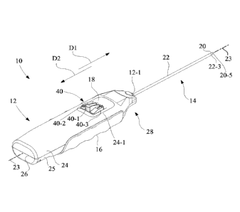
[0053] Referring to Figs. 1-3, biopsy device 10 includes a housing 12, a
biopsy needle
14, a charge handle 16, and an actuator mechanism 18. Biopsy needle 14
includes a stylet
20 and a cannula 22. In the present embodiment, housing 12, charge handle 16
and
actuator mechanism 18 are made of plastic, and stylet 20 and cannula 22 are
made from a
metallic material, such as stainless steel. In describing the orientation of
components and
the operation of biopsy device 10 in more detail below, for convenience,
reference will be
made to a distal direction D1 and a proximal direction D2 with respect to a
longitudinal
axis 23. The proximal direction D2 is a longitudinal direction opposite to
distal direction
Dl.
[0054] As shown in Figs. 1-3, biopsy needle 14 extends away from housing 12
along
longitudinal axis 23 in the distal direction Dl. Each of stylet 20 and cannula
22 of biopsy
needle 14 is positioned to extend on the longitudinal axis 23. In the present
embodiment,
CA 02984601 2017-10-31
WO 2016/178656 PCT/US2015/028902
12
as depicted in Figs. 1-3, cannula 22 is external to stylet 20, such that
cannula 22 is
arranged as the outer tube of the coaxial arrangement of stylet 20 and cannula
22. Stylet
20 and cannula 22 are sized such that stylet 20 is slidably received in
cannula 22 in a close
sliding fit, wherein the inside diameter of cannula 22 is slightly larger than
the outside
diameter of stylet 20 in a tolerance range of 0.01 millimeters (mm) to 1.0 mm.
[0055] In describing the invention, common directional terms such as upper,
lower, up,
down, top, bottom, right, left, vertical, horizontal, etc., may be used with
respect to the
orientation of biopsy device 10 shown in Figs. 1 and 3-5, for convenience, to
aid the
reader in understanding the invention as presented in the figures of the
drawings.
[0056] In the orientation shown in Fig. 1, and referring also to Figs. 4 and
5, housing 12
includes an upper case portion 24 and a lower case portion 25. Upper case
portion 24 is
jointed to lower case portion 25 by removable fasteners 27, such as screws.
Upper case
portion 24 of housing 12 has an opening 24-1, and opposing upper slots 24-2,
24-3.
Opening 24-1 is configured to receive and mount actuator mechanism 18.
Alternatively,
actuator mechanism 18 may be formed integral with upper case portion 24 of
housing 12
at the location of opening 24-1. Lower case portion 25 is configured with
opposing guide
rails 25-1, 25-2 which define a lower slot, which in conjunction with the
opposing upper
slots 24-2, 24-3 of upper case portion 24, slidably mount charge handle 16.
[0057] Upper case portion 24 and lower case portion 25 of housing 12
collectively
define a proximal end wall 26 and a distal end portion 28 spaced from proximal
end wall
26 along a longitudinal axis 23. The distal direction D1 is in a direction
from proximal
end wall 26 toward distal end portion 28, e.g., in a direction of the extent
of biopsy needle
14 away from housing 12 and away from the user. The proximal direction D2
(opposite
distal direction D1) is in a direction from distal end portion 28 toward
proximal end wall
26, e.g., toward the user.
[0058] Housing 12 is configured to define a housing chamber 30 between
proximal end
wall 26 and distal end portion 28. In the present embodiment, lower case
portion 25 of
housing 12 further includes an intermediate wall 32 that is interposed between
proximal
end wall 26 and distal end portion 28. Referring to Fig. 1, distal end portion
28 has a
needle opening 12-1 configured to receive biopsy needle biopsy needle 14, with
longitudinal axis 23 passing through needle opening 12-1 and through the
longitudinal
CA 02984601 2017-10-31
WO 2016/178656 PCT/US2015/028902
13
extent of biopsy needle 14. Proximal end wall 26 faces the user when the user
grasps the
biopsy device 10 in a normal operating fashion.
[0059] Charge handle 16 and actuator mechanism 18 provide the user with fully
accessible control features used to operate biopsy device 10 in an intuitive
manner to
obtain a tissue sample from suspect tissue of a patient via biopsy needle 14.
[0060] Charge handle 16 is used to ready biopsy device 10 for performing a
biopsy
procedure by facilitating the generation of vacuum and preparing biopsy needle
14 for
severing and collecting the tissue sample.
[0061] As shown in Figs. 4-6, charge handle 16 has a U-shaped charge handle
body 34,
a charge handle end wall 36, and a charge handle latch arm 38. Charge handle
latch arm
38 extends in a cantilever manner in the proximal direction D2 from charge
handle end
wall 36. Charge handle end wall 36 laterally intersects U-shaped charge handle
body 34
to define a handle mount opening 36-1 (see Fig. 5) having a half-circle shape
located
below charge handle end wall 36. During assembly, distal end portion 28 of
lower case
portion 25 is manipulated through handle mount opening 36-1, such that charge
handle
end wall 36 rests on the opposing guide rails 25-1, 25-2 of lower case portion
25 to
slidably mount charge handle 16 in the laterally spaced lower case portion
slots defined by
the laterally spaced guide rails 25-1, 25-2 in conjunction with upper slots 24-
2, 24-3 of
upper case portion 24.
[0062] Charge handle latch arm 38 has a free end 38-1 having a laterally
protruding
catch 38-2. Charge handle latch arm 38 is configured to be longitudinally
rigid, and
laterally resilient in a direction substantially perpendicular to the
longitudinal extent of
charge handle latch arm 38. As used herein, the term "substantially
perpendicular" is a
direction having a range of deviation from perpendicular of plus or minus five
degrees.
[0063] Referring to Figs. 4-6, charge handle 16 is biased in distal direction
D1 by a
biasing mechanism formed by at least one biasing spring, and in the present
embodiment,
includes a pair of biasing springs 39-1, 39-2 interposed between housing 12
and charge
handle 16. In the present embodiment, biasing springs 39-1, 39-2 are coil
springs having a
contracted relaxed state. In particular, biasing spring 39-1 is attached at
its ends to spring
attachment loop 25-3 of lower case portion 25 of housing 12 (see Figs. 4 and
5) and to
spring attachment loop 36-2 of charge handle end wall 36 of charge handle 16
(see Fig. 6).
Likewise, biasing spring 39-2 is attached at its ends to spring attachment
loop 25-4 of
CA 02984601 2017-10-31
WO 2016/178656 PCT/US2015/028902
14
lower case portion 25 of housing 12 (see Figs. 4 and 5) and to spring
attachment loop 36-3
of charge handle end wall 36 of charge handle 16 (see Fig. 6). Charge handle
16 is
configured to be grasped by a user's hand and to move between a home (distal)
position,
as depicted in Figs. 1-3, and a retracted (proximal) position.
[0064] Referring again to Figs. 1-3, actuator mechanism 18 is used to
initialize the
sequential operations of firing the biopsy needle in distal direction D1 to
perform a
piercing shot into tissue to be biopsied, to apply vacuum and open a sample
port in biopsy
needle 14, and to operate biopsy needle 14 to sever a tissue sample, as more
fully
described below.
[0065] Referring also to Figs. 7A and 7B, actuator mechanism 18 includes three
control
buttons 40, individually identified as a pierce button 40-1, a cannula retract
button 40-2,
and a sample acquisition button 40-3, each of which is accessible to the user
from the
exterior of biopsy device 10. As shown in Fig. 7B, each of the control buttons
40 includes
a mechanical extension that serves as respective actuator portions 42-1, 42-2,
42-3.
[0066] Also, as shown in Fig. 7B, actuator mechanism 18 further includes a
carriage
latch strike 44. In the present embodiment, carriage latch strike 44 is
configured as an
inverted ramp having a downwardly facing ramp surface 44-1 that diverges in
the
proximal direction D2 to define a proximal end face 44-2.
[0067] Referring again to Figs. 4 and 5, biopsy device 10 further includes a
carriage
assembly 50 that is positioned and slidably contained in housing chamber 30 of
housing
12. Carriage assembly 50 is configured to move longitudinally along
longitudinal axis 23
as a whole relative to housing 12, and is configured to mount biopsy needle
14, as more
fully described below.
[0068] As shown in Figs. 4 and 5, carriage assembly 50 includes a prime pierce
carriage
52, a cannula slide 58, a sampling slide 56, and a vacuum system 54. As used
herein, the
term "prime" is used to mean a function associated with readying, i.e.,
cocking, biopsy
device 10 for performing a tissue piercing function by having both stylet 20
and cannula
22 retracted in preparation for "piercing". The term "pierce" is used to mean
a function
associated with firing stylet 20 and cannula 22 simultaneously in distal
direction D1 such
that biopsy needle 14 punctures the tissue of a patient at the desired
location. The term
"slide" is a mechanical structure having a guide that is configured to move
along a
predefined path defined by another mechanical structure. In the present
embodiment, each
CA 02984601 2017-10-31
WO 2016/178656 PCT/US2015/028902
of prime pierce carriage 52, cannula slide 58, and sampling slide 56 are
configured to
move along a substantially linear path. The term "substantially linear path"
is a path
having a range of deviation from a straight line of plus or minus three
degrees along the
length of the path.
[0069] Referring now also to Fig. 8, prime pierce carriage 52 includes
carriage slide 60
serving as a lower prime pierce carriage portion and a carriage latch cover
member 62
serving as an upper prime pierce carriage portion. Referring also to Fig. 5,
sampling slide
56 and cannula slide 58 are individually, as well as collectively,
longitudinally movable
relative to prime pierce carriage 52 formed by carriage slide 60 and carriage
latch cover
member 62. Each of prime pierce carriage 52, sampling side 56 and cannula
slide 58 is
formed of plastic.
[0070] Carriage slide 60 of prime pierce carriage 52 of carriage assembly 50
has a
carriage base 64 and a stylet mount end wall 66. In the present embodiment,
carriage base
64 and stylet mount end wall 66 are formed as a unitary carriage structure.
[0071] Carriage base 64 of carriage slide 60 is configured to define a U-
shaped wall 64-
1 having a U-shaped exterior surface 64-2 and a U-shaped interior surface 64-
3, thus
having a U-shaped cross-section that extends in the distal direction D1 from
stylet mount
end wall 66 to define a U-shaped distal edge 64-4, a pair of laterally spaced
upper
mounting edges 64-5, 64-6, and a pair of laterally spaced recessed slot edges
64-7, 64-8.
Extending upwardly and proximally from U-shaped interior surface 64-3 is a
longitudinally oriented prime pierce spring mount post 64-9.
[0072] U-shaped exterior surface 64-2 corresponds to the interior shape of
housing
chamber 30, and is in sliding contact with housing chamber 30, with housing
chamber 30
serving as a longitudinal guide for carriage assembly 50. U-shaped distal edge
64-4
defines an open distal end 60-1 of carriage slide 60. The pair of laterally
spaced upper
mounting edges 64-5, 64-6 in conjunction with the pair of laterally spaced
recessed slot
edges 64-7, 64-8 further define an open top 60-2 of carriage slide 60.
[0073] Stylet mount end wall 66 has a stylet hole 66-1, a first pump mounting
opening
66-2 and a second pump mounting opening 66-3, and a prime pierce spring
opening 66-4.
Prime pierce spring opening 66-4 is axially aligned with prime pierce spring
mount post
64-9 of carriage base 64 of carriage slide 60. Stylet hole 66-1 is configured
to fixedly
mount stylet 20, e.g., by a press fit and/or adhesive coupling. First pump
mounting
CA 02984601 2017-10-31
WO 2016/178656
PCT/US2015/028902
16
opening 66-2 and second pump mounting opening 66-3 are configured to mount a
pair of
syringe-type vacuum pumps of vacuum system 54, as will be more fully described
below.
[0074] As best shown in Fig. 8, and with reference to Figs. 1-5, stylet 20 of
biopsy
needle 14 has a side wall 20-1 configured to define a lumen 20-2 and a side
sample port
20-3 that extends through side wall 20-1 to lumen 20-2. Stylet 20 has an open
first end
20-4 and a closed second end 20-5. The closed second end 20-5 defines a distal
piercing
tip. Lumen 20-2 is in fluid communication with the open first end 20-4 and
side sample
port 20-3.
[0075] A proximal portion 20-6 of stylet 20 extends in a proximal direction D2
away
from stylet mount end wall 66 and a distal portion 20-7 of stylet 20 extends
in the distal
direction D1 away from stylet mount end wall 66. The distal portion 20-7 of
stylet 20
extends in the distal direction D1 beyond the distal extent of carriage base
64 and carriage
latch cover member 62, and is received through needle opening 12-1 of housing
12 (see
also Fig. 1).
[0076] Carriage latch cover member 62 is configured to attach, e.g., a snap
fit and/or
adhesive, to the pair of laterally spaced upper mounting edges 64-5, 64-6 of
carriage base
64. Carriage latch cover member 62 includes laterally spaced recessed slot
edges 62-1,
62-2 that are respectively vertically opposed to the laterally spaced recessed
slot edges 64-
7, 64-8 of carriage base 64. Carriage latch cover member 62 covers the open
top 60-2 of
carriage slide 60, and extends over carriage base 64 of carriage slide 60 to
define an
interior region in which cannula slide 58 may move longitudinally relative to
stylet mount
end wall 66. In particular, the U-shaped interior surface 64-3 of U-shaped
wall 64-1 of
carriage slide 60 in conjunction with open distal end 60-1 of carriage slide
60 are
configured to slidably receive and longitudinally guide sampling slide 56 and
cannula
slide 58.
[0077] Carriage latch cover member 62 is configured to facilitate a selective
longitudinal coupling and uncoupling of carriage slide 60 with each of
sampling slide 56
and cannula slide 58. Carriage latch cover member 62 has a latch strike 70, a
latch strike
72, a carriage latch arm 74, and a deflector arm 76. Carriage latch cover
member 62
further includes conduit mounts 62-3, 62-4.
[0078] In the present embodiment, each of latch strike 70 and latch strike 72
is
configured as a latching notch. More particularly, each of latch strike 70 and
latch strike
CA 02984601 2017-10-31
WO 2016/178656 PCT/US2015/028902
17
72 of carriage latch cover member 62 is configured as a rectangular opening
having
proximal and distal end walls oriented to be substantially perpendicular to
longitudinal
axis 23.
[0079] Carriage latch arm 74 is configured as a cantilever arm with a free end
having an
upwardly extending catch 78. Catch 78 is configured as ramp having an upwardly
facing
ramp surface 78-1 that diverges in the distal direction D1 to define a distal
end face 78-2.
Carriage latch arm 74 is configured to be longitudinally rigid, and vertically
resilient in
directions D3, D4 (e.g., up, down) substantially perpendicular to the
longitudinal extent of
carriage latch arm 74. Directions D3 and D4 are opposite directions.
[0080] When carriage assembly 50 is fully retracted in proximal direction D1
by
operation of charge handle 16, as will be more fully described below, carriage
latch arm
74 of carriage latch cover member 62 releasably engages the proximal end face
44-2 of
carriage latch strike 44 of actuator mechanism 18 (see Fig. 7B) so as to
couple, and
prohibit relative movement between, carriage assembly 50 and housing 12, until
carriage
latch arm 74 is released by operation of pierce button 40-1 of actuator
mechanism 18.
[0081] Referring also to Fig. 9, deflector arm 76 of carriage latch cover
member 62 is
configured to be longitudinally rigid, and also is laterally rigid in
directions substantially
perpendicular to the longitudinal extent of deflector arm 76. Deflector arm 76
is
configured as a cantilever arm having a free end having a downwardly facing
deflector
head 80. Deflector head 80 has a downwardly facing ramp surface 80-1 that
diverges in
the distal direction D1 and terminates at a downwardly facing longitudinal
surface 80-2.
[0082] Referring again to Figs. 4 and 5, vacuum system 54 is mounted to, and
carried
by, carriage assembly 50, and is a completely self-contained within housing 12
of biopsy
device 10. More particularly, referring also to Fig. 10, vacuum system 54 is
mounted to
prime pierce carriage 52 of carriage assembly 50.
[0083] Referring to Figs. 5, 10 and 11, vacuum system 54 includes a vacuum
pump 90, a
vacuum pump 92, a manifold 94, a control valve 96, and flexible connection
conduits 98-
1, 98-2, 98-3, and 98-4.
[0084] Referring particularly to Fig. 11, vacuum pump 90 is a syringe-type
vacuum
pump. Vacuum pump 90 includes an elongate cylinder 100 having a first end 100-
1 and a
second end 100-2. Extending from first end 100-1 is a tip portion 100-3 that
defines a first
vacuum port 100-4. Second end 100-2 has a first opening 100-5. A first piston
102 is
CA 02984601 2017-10-31
WO 2016/178656 PCT/US2015/028902
18
slidably received in elongate cylinder 100 through first opening 100-5. A
first plunger 104
is attached to, or integrally formed with, first piston 102. First plunger 104
is configured
to extend from second end 100-2 of elongate cylinder 100. First plunger 104
has a free
end 104-1 having a head 104-2. A first vacuum spring 106 is interposed between
second
end 100-2 of elongate cylinder 100 and head 104-2 of first plunger 104. First
vacuum
spring 106 is configured to store mechanical energy when in a compressed state
and, in the
orientation shown, is configured to bias first piston 102 in the distal
direction D1 to
establish a vacuum at first vacuum port 100-4.
[0085] Vacuum pump 92 also is a syringe-type vacuum pump, and is configured
identical to vacuum pump 90. Vacuum pump 92 includes an elongate cylinder 110
having
a first end 110-1 and a second end 110-2. Extending from first end 110-1 is a
tip portion
110-3 that defines a second vacuum port 110-4. Second end 110-2 defines a
second
opening 110-5. A second piston 112 is slidably received in elongate cylinder
110 through
second opening 110-5. A second plunger 114 is attached to, or formed
integrally with,
second piston 112. Second plunger 114 configured to extend from second end 110-
2 of
elongate cylinder 110. Second plunger 114 has a free end 114-1 having a head
114-2. A
second vacuum spring 116 is interposed between second end 110-2 of elongate
cylinder
110 and head 114-2 of second plunger 114. Second vacuum spring 116 is
configured to
store mechanical energy when in a compressed state and, in the orientation
shown, is
configured to bias second piston 112 in the distal direction D1 to establish a
vacuum at
second vacuum port 110-4.
[0086] Manifold 94 has a first vacuum draw port 94-1, a second vacuum draw
port 94-2,
and a first vacuum application port 94-3. Manifold 94 has a first one-way
valve 94-4 that
is coupled in fluid communication with first vacuum draw port 94-1 and a
second one-way
valve 94-5 that is coupled in fluid communication with second vacuum draw port
94-2.
Each of first one-way valve 94-4 and second one-way valve 94-5 is configured
to release
positive pressure to the atmosphere to facilitate a purge of positive pressure
from vacuum
pumps 90, 92 during the charging of sampling spring 182, and to close upon
establishment
of a vacuum by vacuum pumps 90, 92. In the present embodiment, each of first
one-way
valve 94-4 and second one-way valve 94-5 is a duckbill valve.
[0087] Control valve 96 has a third vacuum draw port 96-1, a second vacuum
application port 96-2, and a button actuator 96-3. Button actuator 96-3
selectively controls
CA 02984601 2017-10-31
WO 2016/178656 PCT/US2015/028902
19
fluid communication between third vacuum draw port 96-1 and second vacuum
application port 96-2. Referring also to Figs. 5, 7A and 7B, control valve 96
is operated
by actuation of cannula retract button 40-2 of actuator mechanism 18, which in
turn
actuates button actuator 96-3 of control valve 96 to apply the vacuum at side
sample port
20-3 of stylet 20 simultaneously with movement of cannula 22 in the proximal
direction
D2 relative to stylet 20 to ready biopsy device 10 for taking a tissue sample.
[0088] First vacuum port 100-4 of vacuum pump 90 is coupled in fluid
communication
with first vacuum draw port 94-1 of manifold 94 via flexible connection
conduit 98-1.
Second vacuum port 110-4 of vacuum pump 92 is coupled in fluid communication
with
second vacuum draw port 94-2 of manifold 94 via flexible connection conduit 98-
2.
Flexible connection conduits 98-1 and 98-2 may be in the form of a rubber
tubular sleeve,
and in the present embodiment, are integral with manifold 94. First vacuum
application
port 94-3 of manifold 94 is coupled in fluid communication with third vacuum
draw port
96-1 of control valve 96 via flexible connection conduit 98-3. Second vacuum
application
port 96-2 of control valve 96 is coupled in fluid communication with lumen 20-
2 at open
first end 20-4 of stylet 20 via flexible connection conduit 98-4. Flexible
connection
conduits 98-3 and 98-4 may be in the form of rubber tubes, and are secured to
carriage
latch cover member 62 via conduit mounts 62-3, 62-4.
[0089] Referring again to Figs. 5, 10 and 11, vacuum pump 90 is received and
mounted,
e.g., by press fit and/or adhesive, in first pump mounting opening 66-2 of
stylet mount end
wall 66 of stylet mounting slider 54, with a proximal cylinder portion 100-6
having first
vacuum port 100-4 configured to extend in the proximal direction D2 from
stylet mount
end wall 66, and a distal cylinder portion 100-7 having second opening 100-5
configured
to extend in the distal direction D1 from stylet mount end wall 66. Likewise,
vacuum
pump 92 is received and mounted, e.g., by press fit and/or adhesive, in second
pump
mounting opening 66-3 of stylet mount end wall 66 of stylet mounting slider
54, with a
proximal cylinder portion 110-6 having second vacuum port 110-4 configured to
extend in
the proximal direction D2 from stylet mount end wall 66, and a distal cylinder
portion
110-7 having second opening 110-5 configured to extend in the distal direction
D1 from
stylet mount end wall 66.
[0090] Referring to Figs. 5 and 11, head 104-2 of plunger 104 of vacuum pump
90 and
head 114-2 of plunger 114 of vacuum pump 92 are positioned to be engaged by
cannula
CA 02984601 2017-10-31
WO 2016/178656 PCT/US2015/028902
slide 58. During a first retraction of charge handle 16 in proximal direction
D2, there is a
corresponding movement of cannula slide 58 in the proximal direction D2, which
in turn
moves the respective piston/plunger combinations 102/104 and 112/114 of vacuum
pumps
90, 92 in the proximal direction D2. As such, each of first vacuum spring 106
of vacuum
pump 90 and second vacuum spring 116 of vacuum pump 92 is charged
(compressed).
The compression of first vacuum spring 106 of vacuum pump 90 and second vacuum
spring 116 of vacuum pump 92 occurs simultaneously.
[0091] The charging of first vacuum spring 106 and second vacuum spring 116 is
accompanied by an evacuation, i.e., purging, of air under positive pressure
from elongate
cylinders 100, 110 of vacuum pumps 90, 92 via one-way valves 94-4, 94-5 as
respective
piston/plunger combinations 102/104 and 112/114 are moved in the proximal
direction
D2. Upon a subsequent movement of cannula slide 58 in the distal direction D1,
first
vacuum spring 106 and second vacuum spring 116 begin to decompress, and thus
bias and
tend to move piston/plunger combinations 102/104 and 112/114 of vacuum pumps
90, 92
in the distal direction Dl. This movement of piston/plunger combinations
102/104 and
112/114 of vacuum pumps 90, 92 in the distal direction D1 establishes a vacuum
at first
vacuum port 100-4 of vacuum pump 90 and second vacuum port 110-4 of vacuum
pump
92, thereby closing via one-way valves 94-4, 94-5 of manifold 94.
[0092] Referring to Figs. 4, 5, 12A and 12B, interposed between carriage slide
60 of
prime pierce carriage 52 and cannula slide 58 is sampling slide 56. Referring
also to Fig.
8, sampling slide 56 of carriage assembly 50 is configured to be slidably
received in the
open distal end 60-1 of prime pierce carriage 52.
[0093] Referring particularly to Figs. 12A and 12B, sampling slide 56 has an
intermediate slide wall 120, a pair of opposed side walls 122, 124, a latch
arm 126, and a
latch arm deflection member 128. Intermediate slide wall 120 has a needle hole
120-1, a
first pump opening 120-2, a second pump opening 120-3, and a distal face 120-
4. A pair
of spring attachment loops 120-5, 120-6 extend distally from distal face 120-
4.
[0094] Needle hole 120-1 is configured to slidably receive stylet 20 of biopsy
needle 14,
with needle hole 120-1 being sized and shaped to serve as a bearing guide
surface against
stylet 20. The inside diameter of needle hole 120-1 is slightly larger than
the outside
diameter of stylet 20 in a tolerance range of 0.01 millimeters (mm) to 1.0 mm.
CA 02984601 2017-10-31
WO 2016/178656 PCT/US2015/028902
21
[0095] First pump opening 120-2 is configured to slidably receive vacuum pump
90 in a
loose fit, and in particular, first pump opening 120-2 is configured to freely
pass elongate
cylinder 100, first plunger 104, and first vacuum spring 106 of vacuum pump
90. Second
pump opening 120-3 is configured to slidably receive vacuum pump 92 in a loose
fit, and
in particular, second pump opening 120-3 is configured to freely pass elongate
cylinder
110, first plunger 114, and first vacuum spring 116.
[0096] The pair of opposed side walls 122, 124 are connected to, or formed
integral
with, an outer perimeter of intermediate slide wall 120. Side wall 122 extends
in both the
distal direction D1 and the proximal direction D2 from intermediate slide wall
120, and
has a curved cross-section that defines a curved exterior surface 122-1.
Likewise, side
wall 124 extends in both the distal direction D1 and the proximal direction D2
from
intermediate slide wall 120, and has a curved cross-section that defines a
curved exterior
surface 124-1. The shape of side walls 122, 124, in combination, correspond to
the shape
of U-shaped interior surface 64-3 of U-shaped wall 64-1 of carriage slide 60
(see also Fig.
8). Thus, the curved exterior surfaces 122-1, 124-1 of the pair of opposed
side walls 122,
124 of intermediate slide wall 120 are in sliding contact with U-shaped
interior surface 64-
3 of U-shaped wall 64-1 of carriage slide 60, which serves as an internal
longitudinal
guide for sampling slide 56.
[0097] Referring again to Figs. 12A and 12B, latch arm 126 is configured as a
cantilever
arm that extends in the distal direction D1 from intermediate slide wall 120,
with a free
end of the cantilever arm having an upwardly facing catch 130. Latch arm 126
is
configured to be longitudinally rigid, and vertically resilient in up and down
directions D3,
D4 that are substantially perpendicular to the longitudinal extent of latch
arm 126.
Referring to Fig. 12B, latch arm 126 has a downwardly facing longitudinally
extending bi-
direction ramp 126-1 having a pair of longitudinally opposed ramp surfaces
that define a
central apex 126-2.
[0098] Referring again to Fig. 12A, catch 130 is configured as ramp having an
upwardly
facing ramp surface 130-1 that diverges in the distal direction D1 to define a
distal end
face 130-2. An extension portion 126-3 of latch arm 126 extends in distal
direction D1
beyond distal end face 130-2 to define a proximally facing L-shaped notch
having a floor
substantially perpendicular to distal end face 130-2.
CA 02984601 2017-10-31
WO 2016/178656 PCT/US2015/028902
22
[0099] Catch 130 of latch arm 126 is configured to releasably engage latch
strike 72 of
carriage latch cover member 62 (see also Figs. 8 and 10) so as to couple, and
prohibit
distal movement of sampling slide 56 with respect to carriage slide 60 of
carriage
assembly 50, until latch arm 126 is released from latch strike 72 of carriage
latch cover
member 62 by actuation of sample acquisition button 40-3.
[00100] As shown in Fig. 12B, latch arm deflection member 128 defines a
retention
channel 128-1 having a downwardly facing ramp surface 128-2 and a ceiling 128-
3.
Ceiling 128-3 extends substantially perpendicularly from intermediate slide
wall 120 in
distal direction Dl. Downwardly facing ramp surface 128-2 diverges in the
proximal
direction D2 from an upper surface 128-4 of latch arm deflection member 128 to
join
ceiling 128-3.
[00101] Referring Figs. 5, 8 and 13, cannula slide 58 of carriage assembly 50
also is
configured to be slidably received in the open distal end 60-1 of prime pierce
carriage 52.
[00102] As best shown in Fig. 13, cannula slide 58 has a cannula mount end
wall 132
and a latch arm 134 that extends in the proximal direction D2 from cannula
mount end
wall 132. Referring to Fig. 5, cannula mount end wall 132 is longitudinally
spaced
distally along longitudinal axis 23 from stylet mount end wall 66 of carriage
slide 60.
Intermediate slide wall 120 is longitudinally interposed between, and spaced
along
longitudinal axis 23 from each of, stylet mount end wall 66 and cannula mount
end wall
132.
[00103] Referring to Figs. 13 and 16-18, cannula mount end wall 132 includes
an
indexing window 132-1, a proximal face 132-2, and a pair of spring attachment
loops 132-
3, 132-4. The pair of spring attachment loops 132-3, 132-4 extend proximally
from
proximal face 132-2.
[00104] Extending in proximal direction D2 from cannula mount end wall 132 is
a
longitudinally extending cannula mount tube 136 having a flared proximal end
136-1, an
annular bearing surface 136-2, and a tubular aperture 136-3. Cannula 22 is
fixedly
mounted in cannula mount tube 136 of cannula mount end wall 132, e.g., by
press fit
and/or adhesive.
[00105] As shown in the breakaway portion of Fig. 13, cannula 22 has a side
wall 22-1
configured to define a lumen 22-2, and has a distal cutting edge 22-3. A
proximal portion
22-4 of cannula 22 extends in proximal direction D2 from cannula mount end
wall 132
CA 02984601 2017-10-31
WO 2016/178656 PCT/US2015/028902
23
and a distal portion 22-5 of cannula 22 extends in a distal direction D1 from
cannula
mount end wall 132. Distal portion 22-5 of cannula 22 is slidably received
through needle
opening 12-1 of housing 12 (see Fig. 1).
[00106] Cannula mount end wall 132 further includes a pair of opposed side
walls 138,
140, having a curved cross-section, and each having a respective curved
exterior surface
138-1, 140-1 that corresponds to the shape of U-shaped interior surface 64-3
of U-shaped
wall 64-1 of carriage slide 60 (see also Figs. Sand 8). The pair of opposed
side walls 138,
140 of cannula mount end wall 132 is in sliding contact with U-shaped interior
surface 64-
3 of U-shaped wall 64-1 of carriage slide 60, which serves as a longitudinal
guide for
cannula slide 58 within prime pierce carriage 52.
[00107] Referring again to Figs. 13 and 16-18, positioned centrally under
cannula
mount tube 136 is indexing window 132-1, which is formed as a rectangular
opening that
extends through cannula mount end wall 132. Indexing window 132-1 is laterally
interposed between two vertically oriented guide channels identified as guide
channel 142
and guide channel 144. Guide channels 142, 144 are vertically oriented to be
parallel to
proximal face 132-2 of cannula mount end wall 132. In the present embodiment,
guide
channel 142 is defined by a vertical structure 142-1 having a side slot 142-2,
and guide
channel 144 is defined by upper guide opening 144-1 and a lower guide opening
144-2.
[00108] Indexing window 132-1 is sized to freely receive charge handle latch
arm 38
(see Fig. 6) without inducing a latching engagement. However, indexing window
132-1
may be selectively intersected, in part, by a portion of an indexing mechanism
150, as
described in more detail below, such that the portion of indexing mechanism
150
intersecting indexing window 132-1 may be engaged by catch 38-2 of charge
handle latch
arm 38 of charge handle 16 on a return stroke of charge handle 16 in distal
direction Dl.
[00109] As shown in Fig. 13, latch arm 134 is configured as a cantilever arm
that
extends in the proximal direction D2 from cannula mount end wall 132, with a
free end of
the cantilever arm having an upwardly facing catch 146. Latch arm 134 is
configured to
be longitudinally rigid, and vertically resilient in up and down directions
D3, D4 that are
substantially perpendicular to the longitudinal extent of latch arm 134. Catch
146 is
configured as ramp having an upwardly facing ramp surface 146-1 that diverges
in the
proximal direction D2 to define a proximal end face 146-2. In the present
embodiment, an
extension portion 134-1 of latch arm 134 extends in proximal direction D2
beyond
CA 02984601 2017-10-31
WO 2016/178656 PCT/US2015/028902
24
proximal end face 146-2 to define a proximally facing L-shaped notch having a
floor
substantially perpendicular to proximal end face 146-2.
[00110] Catch 146 of latch arm 134 of cannula slide 58 is configured to
selectively and
releasably engage latch strike 70 of carriage latch cover member 62, and when
so engaged,
couples cannula slide 58 to carriage slide 60 to prohibit proximal movement of
cannula
slide 58 with respect to carriage slide 60 of carriage assembly 50, until
latch arm 134 is
released from latch strike 70 by actuation of cannula retract button 40-2 (see
Fig. 1).
[00111] Referring to Figs. 13-18, indexing mechanism 150 includes a cannula
slide
indexer 152 and a sampling slide indexer 154 which cooperate to coordinate the
various
stages of operation of biopsy device 10. Cannula slide indexer 152 is
configured to pivot
about cannula mount tube 136 of cannula slide 58, and sampling slide indexer
154 is
configured to move linearly in directions D3 and D4, e.g., up and down, as
guided by
guide channels 142, 144 of cannula slide 58. Here directions D3 and D4 are
opposite
directions, e.g., up and down, and directions D3 and D4 are substantially
perpendicular to
longitudinal axis 23.
[00112] Referring to Fig. 13, cannula slide indexer 152 is selectively
operated by a
deflection of latch arm 134 of cannula slide 58. Referring also to Figs. 14A,
14B, in the
embodiment shown, cannula slide indexer 152 is configured as an integral
structure that
includes an axel 156, a lever arm 158, an actuator arm 160, and a torsion
spring 162.
[00113] Axel 156 of cannula slide indexer 152 has a longitudinal extent 156-1
and a
cylindrical opening 156-2 configured to be received over bearing surface 136-2
of cannula
mount tube 136. The inside diameter of cylindrical opening 156-2 is slightly
larger than
the outside diameter of bearing surface 136-2 in a tolerance range of 0.01
millimeters
(mm) to 1.0 mm so as to permit a pivoting (rotational) motion of cannula slide
indexer 152
about cannula mount tube 136, and in turn, to permit pivoting motion about
longitudinal
axis 23. Axel 156 is retained on bearing surface 136-2 of cannula mount tube
136 by
flared proximal end 136-1.
[00114] Lever arm 158 is radially offset from cylindrical opening 156-2 and in
turn is
radially offset from longitudinal axis 23 by a radial distance Rl. Referring
also to Figs.
16-18, lever arm 158 is positioned and oriented to be selectively engaged by
latch arm 134
of cannula slide 58. As best shown in Figs. 14A and 14B, lever arm 158 has a
tangential
extent 158-1 and a longitudinal extent 158-2. Tangential extent 158-1 of lever
arm 158 is
CA 02984601 2017-10-31
WO 2016/178656 PCT/US2015/028902
oriented to tangentially extend in a cantilever manner in a direction D5 from
axel 156
along a tangent of an imaginary circle corresponding to radial distance Rl.
Since cannula
slide indexer 152 pivots about longitudinal axis 23, direction D5 is relative
and the actual
direction is dependent upon the angular rotational position of cannula slide
indexer 152.
Lever arm 158 is configured to be rigid, and may be defined as a plate 158-3
having
tangential extent 158-1 and longitudinal extent 158-2, and having a free end
158-4 that
defines a longitudinal engagement surface 158-5 having a radius. Longitudinal
engagement surface 158-5 is positioned and oriented to be selectively engaged
by a
bottom surface of latch arm 134 of cannula slide 58 (see also Figs. 13, and 16-
18).
[00115] Actuator arm 160 is radially offset from cylindrical opening 156-2 and
in turn
is radially offset from longitudinal axis 23 by a radial distance R2. Actuator
arm 160 has a
tangential extent 160-1. Tangential extent 160-1 of actuator arm 160 is
oriented to
tangentially extend in a cantilever manner in a direction D6 from axel 156
along a tangent
of an imaginary circle corresponding to radial distance R2. Since cannula
slide indexer
152 pivots about longitudinal axis 23, direction D6 is relative and the actual
direction is
dependent upon the angular rotational position of cannula slide indexer 152.
However,
direction D6 is in a fixed relationship to direction D5, which are in
substantially opposite
directions, wherein the term "substantially opposite" means a range of linear
(180 degrees)
plus or minus 15 degrees. In the present embodiment, the angular range a of
the fixed
relationship of direction D5 relative to direction D6 with respect to
longitudinal axis 23
may be in a range of 165 degrees to 180 degrees (linear). Actuator arm 160 is
configured
to be rigid, and may be defined as a triangular plate 160-2 having tangential
extent 160-1,
and having a free end 160-3 and a planar engagement surface 160-4.
[00116] Torsion spring 162 is radially offset from cylindrical opening 156-2
and in turn
is radially offset from longitudinal axis 23 by a radial distance R3. Torsion
spring 162 has
an outward extent 162-1. Outward extent 162-1 of actuator arm 160 is oriented
to extend
in a cantilever manner from axel 156. In the orientation of components shown
in Figs. 5
and 13-14B, torsion spring 162 is configured to bias cannula slide indexer 152
to pivot in a
clockwise direction about longitudinal axis 23. In particular, torsion spring
162 is
configured to be resilient, and may be defined as curved cantilever arm 162-2
having a
free end 162-3, wherein a contact surface 162-4 of cantilever arm 162-2
engages a fixed
feature of cannula slide indexer 152, such as spring attachment loop 132-3
(see Figs. 16-
CA 02984601 2017-10-31
WO 2016/178656
PCT/US2015/028902
26
18), where cantilever arm 162-2 applies a counterclockwise force to spring
attachment
loop 132-3 to bias cannula slide indexer 152 to pivot in the clockwise
direction.
[00117] Referring to Figs. 13 and 15A-18, sampling slide indexer 154 is
configured
generally as an integral plate-like planar structure, and includes a base 164,
a left slide
166, a right slide 168, a window blocking plate 170, a first cam arm 172, a
second cam
arm 174, and a cantilever spring 176. Left slide 166, right slide 168, window
blocking
plate 170, first cam arm 172, and second cam arm 174 extend vertically in
direction D3
from base 164. Cantilever spring 176 is located below, in direction D4, from
base 164.
[00118]
Referring also to Figs. 16-18, left slide 166 is sized and shaped to be
slidably
received in side slot 142-2 of guide channel 142 of cannula mount end wall 132
of cannula
slide 58. Right slide 168 is sized and shaped to be slidably received in upper
guide
opening 144-1 and lower guide opening 144-2 of guide channel 144 of cannula
mount end
wall 132 of cannula slide 58.
[00119] Window blocking plate 170 may be formed integral with left slide 166.
Window blocking plate 170 is configured as a vertically extending plate
positioned and
oriented to selectively intersect, and partially cover, indexing window 132-1
of cannula
mount end wall 132 of cannula slide 58. Window blocking plate 170 includes a
side
surface 170-1 and a proximal surface 170-2.
[00120] First cam arm 172 is configured to vertically extend from base 164.
First cam
arm 172 is laterally interposed between, and spaced from, window blocking
plate 170 and
second cam arm 174. First cam arm 172 is positioned and oriented to be
selectively
engaged by engagement surface 160-4 of actuator arm 160 of cannula slide
indexer 152.
In particular, first cam arm 172 has a free end 172-1 having a radial
engagement surface
172-2 that may be engaged by the planar engagement surface 160-4 of actuator
arm 160 of
cannula slide indexer 152.
[00121] Second cam arm 174 may be formed integral with right slide 168. Second
cam
arm 174 is configured to vertically extend from base 164. Second cam arm 174
is
positioned and oriented to be selectively engaged by bi-direction ramp 126-1
of latch arm
126 of sampling slide 56. In particular, second cam arm 174 has a free end 174-
1 having
an engagement surface 174-2 that may be engaged by bi-direction ramp 126-1 of
latch arm
126 of sampling slide 56.
CA 02984601 2017-10-31
WO 2016/178656 PCT/US2015/028902
27
[00122] Cantilever spring 176 is configured as a curved cantilever that
extends below a
lower portion of base 164, and in turn is located below, base 164, in
direction D4. In the
orientation of components shown in Figs. 5, 13 and 15A-18, cantilever spring
176 is
configured to bias sampling slide indexer 154 in direction D3 (in an upward
direction). In
particular, cantilever spring 176 is configured to be resilient, and is
defined as curved
cantilever arm 176-1 having a free end 176-2, wherein a contact surface 176-3
of
cantilever arm 176-1 engages a fixed feature of cannula slide indexer 152,
such as
longitudinally extending pin 132-5 of cannula slide 58 (see Figs. 16-18),
where cantilever
arm 176-1 applies a downward force in direction D4 to pin 132-5 to bias
sampling slide
indexer 154 upwardly in direction D3.
[00123] Referring again to Fig. 5, the motive force provided to power the
various
functions of biopsy device 10 is provided by at least one cannula retract
spring (the present
embodiment having a pair of cannula retract springs 180-1, 180-2), a sampling
spring 182,
and a prime pierce spring 184. Those skilled in the art will recognize that
the effect of
each of cannula retract springs 180-1, 180-2, sampling spring 182, and prime
pierce spring
184 may be accomplished by a single spring, or by a plurality of springs.
[00124] Referring to Fig. 19, sampling slide 56 and cannula slide 58 are
assembled as a
subassembly prior to insertion into prime pierce carriage 52 over stylet 20
(see Fig. 8). In
particular, cannula retract spring 180 (two shown in the present embodiment,
as 180-1,
180-2), is interposed between, and connected to each of, intermediate slide
wall 120 of
sampling slide 56 and cannula mount end wall 132 of cannula slide 58. In
particular,
cannula retract spring 180-1, is attached at its ends to spring attachment
loop 120-5 of
intermediate slide wall 120 of sampling slide 56 and spring attachment loop
132-3 of
cannula mount end wall 132 of cannula slide 58. Likewise, cannula retract
spring 180-2,
is attached at its ends to spring attachment loop 120-6 of intermediate slide
wall 120 of
sampling slide 56 and spring attachment loop 132-4 of cannula mount end wall
132 of
cannula slide 58.
[00125] As shown in Fig. 19, latch arm deflection member 128 of sampling slide
is
configured for engagement with latch arm 134 of cannula slide 58, and when so
engaged,
latch arm deflection member 128 deflects, i.e., flexes, latch arm 134
downwardly toward
carriage base 64. More particularly, referring also to Figs. 12B and 13,
extension portion
134-1 at the free end of latch arm 134 is positioned for engagement with
intermediate slide
CA 02984601 2017-10-31
WO 2016/178656 PCT/US2015/028902
28
wall 120 in retention channel 128-1 of intermediate slide wall 120 of sampling
slide 56,
such that when so engaged, a longitudinal movement of cannula slide 58 in the
proximal
direction D2 results in a corresponding longitudinal movement of sampling
slide 56 in the
proximal direction D2.
[00126] In turn, as shown in Fig. 17, a bottom surface of latch arm 134 of
cannula slide
58 engages lever arm 158 to rotate cannula slide indexer 152 counterclockwise
about
longitudinal axis 23, thus lifting actuator arm 160 and permitting sampling
slide indexer
154 to move upward in direction D3 by action of cantilever spring 176, thereby
positioning window blocking plate 170 to block a portion, e.g., the lower left
quadrant, of
indexing window 132-1 of cannula slide 58, such that window blocking plate 170
may be
engaged by catch 38-2 of charge handle latch arm 38 of charge handle 16 (see
also Fig. 6).
[00127] When cannula slide 58 is moved in distal direction D1 away from
sampling
slide 56, latch arm 134 disengages from latch arm deflection member 128 and,
due to its
resiliency, latch arm 134 returns to its non-deflected position, i.e., latch
arm 134 moves
back to its original non-flexed position. In turn, as shown in Fig. 18,
cannula slide indexer
152 rotates counterclockwise about longitudinal axis 23 to its full clockwise
position via
the torsional force exerted by torsion spring 162, thus moving sampling slide
indexer 154
downwardly in direction D4, such that window blocking plate 170 no longer
blocks a
portion of indexing window 132-1 of cannula slide 58.
[00128] As shown in Fig. 19, sampling slide 56 is spaced a minimum allowable
distance from cannula slide 58 and places each of cannula retract springs 180-
1, 180-2 in a
contracted state, but each of cannula retract springs 180-1, 180-2 may be in a
slight state
of extension. The term "slight state of extension" means the storage of a
force that is less
than 10 percent of the total force available from springs 180-1, 180-2 in a
charged
(extended) state. As used herein, the term "charge" or "charged" means the
storage of
mechanical energy by one or more springs. The term "charging" means the act of
storing
mechanical energy in a storage device in the form of one or more springs.
Thus, cannula
retract springs 180-1, 180-2 are charged to a charged state to store
mechanical energy
when cannula retract springs 180 are in an extended state.
[00129] Referring also to Fig. 5, cannula retract springs 180-1, 180-2 are
releasably
held in the charged (extended) state under the condition: (a) catch 130 of
latch arm 126 of
sampling slide 56 is engaged with latch strike 72 of carriage latch cover
member 62 and
CA 02984601 2017-10-31
WO 2016/178656 PCT/US2015/028902
29
(b) catch 146 of latch arm 134 of cannula slide 58 is engaged with latch
strike 70 of
carriage latch cover member 62. When catch 146 of latch arm 134 is released
from latch
strike 70, cannula retract springs 180 are released from the charged
(extended) state and
exert a contraction force to bias and move cannula mount end wall 132 of
cannula slide
58, and in turn cannula 22, in the proximal direction D2 toward sampling slide
56, which
in turn exposes side sample port 20-3 of stylet 20.
[00130] Referring again to Fig. 5, sampling spring 182 is interposed between
stylet
mount end wall 66 of carriage slide 60 of prime pierce carriage 52 and
intermediate slide
wall 120 of sampling slide 56. In the present embodiment, sampling spring 182
is a coil
spring that is charged, via a compression of sampling spring 182, to store
mechanical
energy when held in a charged (compressed) state. Sampling spring 182 is held,
i.e.,
retained, in the charged (compressed) state, when catch 130 of latch arm 126
of sampling
slide 56 is engaged with latch strike 72 of carriage latch cover member 62.
When released
from the charged (compressed) state, sampling spring 182 exerts an expansion
force to
bias and move sampling slide 56, and in turn cannula slide 58 and cannula 22,
in distal
direction Dl.
[00131] Referring to Figs. 4 and 5, prime pierce spring 184 is interposed
between
intermediate wall 32 of housing 12 and prime pierce mount post 64-9 of
carriage base 64
of carriage slide 60 of prime pierce carriage 52. Prime pierce spring 184 is
charged to a
charged state to store mechanical energy when prime pierce spring 184 is in a
compressed
state. Prime pierce spring 184 is releasably held in the charged (compressed)
state when
carriage latch arm 74 of carriage latch cover member 62 is engaged with
carriage latch
strike 44 of actuator mechanism 18 (see also Fig. 7B). At this stage, biopsy
device 10 is
described as being primed.
[00132] When prime pierce spring 184 is released from the charged (compressed)
state,
which enters a "pierce" condition, prime pierce spring 184 exerts an expansion
force to
fire, i.e., to rapidly move, carriage assembly 50 carrying biopsy needle 14,
as a whole, in
the distal direction Dl. In other words, the firing of carriage assembly 50
carrying biopsy
needle 14 simultaneously moves stylet 20 and cannula 22 in distal direction D1
such that
biopsy needle 14 punctures the tissue of a patient at the desired location.
[00133] Referring again to Fig. 5, charge handle 16 and indexing mechanism 150
in
combination form a cocking mechanism, as more fully described below.
CA 02984601 2017-10-31
WO 2016/178656 PCT/US2015/028902
[00134] Referring also to Figs. 1 and 20-26, a user grasps charge handle 16
and
manually effects a proximal stroke, i.e., a retraction, of charge handle 16
relative to
housing 12 by pulling charge handle 16 in the proximal direction D2 from the
home
position to the retracted position, which in turn charges (extends) biasing
springs 39-1, 39-
2. While a plurality of biasing springs are shown in the present embodiment,
those skilled
in the art will recognize that the effect of biasing springs 39-1, 39-2 may be
accomplished
by a single spring or by multiple springs. The user releases charge handle 16,
and biasing
springs 39-1, 39-2 contract to effect movement of charge handle 16 relative to
housing 12
in a return (distal) stroke in the distal direction D1 to return charge handle
16 to the home
position.
[00135] Figs. 20-26 shows biopsy 10 with the upper case portion 24 removed to
expose
carriage assembly 50, and with carriage latch cover member 62 disconnected
from
carriage slide 60 of prime pierce carriage 52 to expose sampling slide 56 and
cannula slide
58, so as to show the various positional relationships of components during
various stages
of preparing biopsy device 10 for use in taking a biopsy sample from a
patient.
[00136] Fig. 20 shows biopsy device 10 when biopsy device 10 is new, and after
biopsy
device has been fully cycled in taking a biopsy. As shown, all of the biasing
springs 39-1,
39-2, vacuum springs 106, 116, cannula retract springs 180-1, 180-2, sampling
spring
182, and prime pierce spring 184 are in a discharged state.
[00137] Referring to Fig. 21, on a first retraction (proximal) stroke of
charge handle 16
in proximal direction D2, sampling slide 56 and cannula slide 58 are moved
collectively in
the proximal direction D2 relative to carriage slide 60 to simultaneously
charge vacuum
springs 106, 116 and sampling spring 182. In particular, at the onset of the
first retraction
(proximal) stroke of charging handle 16, cannula slide indexer 152 and
sampling slide
indexer 154 of indexing mechanism 150 are positioned as shown in Fig. 16, such
that
charge handle latch arm 38 of charge handle 16 is free to pass through
indexing window
132-1 of cannula slide 58, and charge handle end wall 36 of charge handle 16
engages
cannula mount end wall 132 of cannula slide 58. With latch arm 134 of cannula
slide 58
engaged with, i.e., abutting, intermediate slide wall 120 of sampling slide 56
and with
latch arm 134 of cannula slide 58 being deflected downwardly by latch arm
deflection
member 128 of sampling slide 56, sampling slide 56 and cannula slide 58 are
moved
collectively in the proximal direction D2 by the proximal movement of charge
handle 16.
CA 02984601 2017-10-31
WO 2016/178656 PCT/US2015/028902
31
[00138] Thus, the first retraction (proximal) stroke of the charge handle 16
toward to
the retracted position moves the sampling slide 56 and the cannula mount end
wall 132 of
cannula slide 58 that carries the cannula 22 in unison, by virtue of the latch
arm 134 being
engaged with the intermediate slide wall 120 of sampling slide 56, in the
proximal
direction D2 to charge (compress) sampling spring 182. Simultaneously, the
proximal
movement of cannula mount end wall 132 of cannula slide 58 moves the
respective
piston/plunger combinations 102/104 and 112/114 of vacuum pumps 90, 92 in the
proximal direction D2 to charge (compress) first vacuum spring 106 of vacuum
pump 90
and second vacuum spring 116 of vacuum pump 92.
[00139] When charge handle 16 is in the retracted position at the end of the
first
proximal stroke, catch 130 of latch arm 126 of sampling slide 56 pivots
upwardly to
engage latch strike 72 of carriage latch cover member 62 to retain sampling
spring 182 in
the charged (compressed) state. However, even though the upward pivoting latch
arm 126
releases sampling slide indexer 154 for movement upward in direction D3, since
charge
handle latch arm 38 of charge handle 16 is inserted into indexing window 132-1
of
cannula slide 58, charge handle latch arm 38 prevents sampling slide indexer
154 from
extending vertically over a portion of indexing window 132-1.
[00140] At this time as well, vacuum springs 106, 116 are retained in their
charged state
only by the internally generated vacuum in vacuum pumps 90, 92. As the vacuum
is
released, e.g., by operation of button 40-2 of actuator mechanism 18, then
vacuum springs
106, 116 will tend to return to their non-charged state.
[00141] The movement of cannula mount end wall 132 of cannula slide 58 during
the
first proximal stroke causes cannula 22 to be retracted to open side sample
port 20-3 of
stylet 20. Referring to Fig. 22, upon release of charge handle 16, biasing
springs 39-1, 39-
2 discharge to effect a first return (distal) stroke of charge handle 16 to
return charge
handle 16 to the home position, and to sequence the indexing mechanism 150 to
the
cannula retract position as depicted in Fig. 17 by removal of charge handle
latch arm 38
from indexing window 132-1, thereby permitting sampling slide indexer 154 to
move in
upward direction D3 by the force exerted by cantilever spring 176, to in turn
laterally
positioning window blocking plate 170 of sampling slide indexer 154 over a
portion, e.g.,
the lower left quadrant, of indexing window 132-1 of cannula slide 58.
CA 02984601 2017-10-31
WO 2016/178656 PCT/US2015/028902
32
[00142] Referring to Fig. 23, on a second retraction (proximal) stroke of
charge handle
16 in proximal direction D2, charge handle latch arm 38 of charge handle 16
attempts to
pass through indexing window 132-1 of cannula slide 58, however this time the
tapered
nose portion of catch 38-2 of charge handle latch arm 38 slidably engages side
surface
170-1 of window blocking plate 170 of sampling slide indexer 154 (see Fig.
17), and
deflects around window blocking plate 170 to then pass through indexing window
132-1
of cannula slide 58. Thus, with the second retraction of charge handle 16,
charge handle
latch arm 38 of charge handle 16 is releasably coupled to cannula mount end
wall 132 of
cannula slide 58 via indexing mechanism 150.
[00143] Referring to Fig. 24, upon release of charge handle 16, biasing
springs 39-1,
39-2 discharge to effect a second return (distal) stroke of charge handle 16
to return charge
handle to the home position. However, on the second return (distal) stroke,
catch 38-2 of
charge handle latch arm 38 (see Fig. 6) catches proximal surface 170-2 of
window
blocking plate 170 of sampling slide indexer 154 (see Fig. 17), which in turn
pulls cannula
mount end wall 132 of cannula slide 58 in distal direction D1 away from
intermediate
slide wall 120 of sampling slide 56, and cannula 22 is moved by cannula mount
end wall
132 in the distal direction D1 to close side sample port 20-3 of stylet 20.
The movement
of cannula mount end wall 132 of cannula slide 58 in distal direction D1 away
from
intermediate slide wall 120 of sampling slide 56 extends cannula retract
springs 180-1,
180-2 to the charged (extended) state. As such, the force exerted by biasing
springs 39-1,
39-2 and any mechanical advantage associated therewith, if any, must be
greater than the
force exerted by cannula retract springs 180-1, 180-2 and any mechanical
advantage
associated therewith, if any, so as to return charge handle 16 to the home
position while
pulling cannula slide 58 to its latched position.
[00144] Also, the distal movement of cannula mount end wall 132 of cannula
slide 58
away from intermediate slide wall 120 of sampling slide 56 in turn causes
latch arm 134 of
cannula slide 58 to disengage from latch arm deflection member 128 of sampling
slide 56
to thus pivot upwardly, which in turn, referring also to Fig. 18, allows
cannula slide
indexer 152 to pivot clockwise by the force exerted by torsion spring 162
about
longitudinal axis 23, which in turn moves actuator arm 160 downwardly in
direction D4 to
move window blocking plate 170 of sampling slide indexer 154 downwardly in
direction
CA 02984601 2017-10-31
WO 2016/178656 PCT/US2015/028902
33
D4, thus opening indexing window 132-1 of cannula slide 58, thus facilitating
the release
of charge handle 16 from cannula slide 58.
[00145] As charge handle 16 approaches the home position, catch 146 of latch
arm 134
of cannula slide 58 releasably engages latch strike 70 of carriage latch cover
member 62 to
retain cannula retract springs 180 in the charged (extended) state. Also,
catch 130 of latch
arm 126 of sampling slide 56 remains engaged with latch strike 72 of carriage
latch cover
member 62. With catch 146 of latch arm 134 engaged with latch strike 70 of
carriage
latch cover member 62 and with latch arm 126 of sampling slide 56 engaged with
latch
strike 72 of carriage latch cover member 62, sampling slide 56 and cannula
slide 58 are
spaced at a maximum separation distance from one another, and each of sampling
slide 56
and cannula slide 58 are latched to maintain the maximum separation distance,
until catch
146 of latch arm 134 of cannula slide 58 is released by actuation of cannula
retract button
40-2.
[00146] Referring to Fig. 25 in conjunction with Fig 5, with each of catch 130
of
sampling slide 56 and catch 146 of cannula slide 58 latched in latch strike 70
and latch
strike 72, respectively, of carriage latch cover member 62 of prime pierce
carriage 52 of
carriage assembly 50, a third retraction (proximal) stroke of charge handle 16
in proximal
direction D2 moves carriage assembly 50 as a whole in the proximal direction
D2 to
charge (compress) prime pierce spring 184. In particular, at the onset of the
third proximal
stroke of charging handle 16, cannula slide indexer 152 and sampling slide
indexer 154 of
indexing mechanism 150 are positioned as shown in Fig. 18, such that charge
handle latch
arm 38 of charge handle 16 (see Fig. 6) is free to pass through indexing
window 132-1 of
cannula slide 58, and charge handle end wall 36 of charge handle 16 engages
cannula
mount end wall 132 of cannula slide 58, and in turn moves carriage assembly 50
in its
entirety in proximal direction D2. Stated differently, the third retraction
(proximal) stroke
of charge handle 16 toward to the retracted position primes biopsy needle 14
for firing by
simultaneously retracting both sampling slide 56 carrying stylet 20 and
cannula slide 58
carrying cannula 22, in unison, in the proximal direction D2 to charge
(compress) prime
pierce spring 184.
[00147] When charge handle 16 is in the retracted position at the end of the
third
proximal stroke, (referring also to Figs. 7B and 10) catch 78 of carriage
latch arm 74
CA 02984601 2017-10-31
WO 2016/178656 PCT/US2015/028902
34
engages carriage latch strike 44 of actuator mechanism 18 to retain prime
pierce spring
184 in the compressed state.
[00148] Referring to Fig. 26, after release of charge handle 16, biasing
springs 39-1, 39-
2 discharge to effect a third return (distal) stroke of charge handle 16 to
return charge
handle 16 to the home position. Biopsy device 10 is now fully primed, with all
of vacuum
spring 106, 116, cannula retract springs 180-1, 180-2, sampling spring 182,
and prime
pierce spring 184 being in a charged state, and biopsy device is ready for
operation in
performing a vacuum-assisted biopsy procedure with a piercing shot function.
It is to be
understood that in the event that the physician performing the biopsy
procedure does not
choose to perform a piercing shot function, then the third retraction/return
(proximal/distal) strokes of charge handle 16 can be omitted.
[00149] In the piercing shot mode of the state of biopsy device 10 shown in
Fig. 26,
wherein prime pierce spring 184 is charged during the third retraction
(proximal) stroke,
referring to Figs. 1, 7B and 10, an actuation of pierce button 40-1 releases
carriage latch
arm 74 from carriage latch strike 44, so as to release prime pierce spring 184
from the
charged (compressed) state to effect a transition of biopsy device 10 from the
state shown
in Fig. 26 to that shown in Fig. 24. Prime pierce spring 184 in turn exerts an
expansion
force to rapidly move carriage assembly 50 carrying stylet 20 and cannula 22,
in unison, in
the distal direction D1 to pierce tissue and to position biopsy needle 14 at
the biopsy site
within the patient.
[00150] Next, or in the absence of the piercing shot mode, with biopsy device
10 in the
state shown in Fig. 24, and further referring to Figs. 1, 5 and 10, an
actuation of cannula
retract button 40-2 releases latch arm 134 of cannula slide 58 from latch
strike 70 of
carriage latch cover member 62, thereby releasing cannula retract springs 180
from the
charged (extended) state to exert a compressive force to move cannula slide 58
and
cannula 22 in the proximal direction D2 to in turn open side sample port 20-3
of stylet 20
to transition biopsy device 10 from the state shown in Fig. 24 to that shown
in Fig. 22.
Simultaneously with this actuation of cannula retract button 40-2, cannula
retract button
40-2 further actuates button actuator 96-3 of vacuum system 54 to open control
valve 96 to
apply the vacuum generated by vacuum pumps 90, 92 to flexible connection
conduit 98-4
coupled in fluid communication with lumen 20-2 of stylet 20, and in turn to
lumen 20-2 at
CA 02984601 2017-10-31
WO 2016/178656 PCT/US2015/028902
side sample port 20-3 of stylet 20. Tissue in the vicinity of side sample port
20-3 of stylet
20 will be drawn by the vacuum into side sample port 20-3.
[00151] Next, with biopsy device 10 in the state shown in Fig. 22, and further
referring
to Figs. 1, 5 and 10, an actuation of sample acquisition button 40-3 releases
catch 130 of
latch arm 126 from latch strike 72 of carriage latch cover member 62, thereby
releasing
sampling spring 182 from the charged (compressed) state to exert an expansion
force to
rapidly move cannula slide 58 carrying cannula 22 in the distal direction D1
to transition
biopsy device 10 from the state shown in Fig. 22 to that shown in Fig. 20, and
with the aid
of distal cutting edge 22-3 of cannula 22, cannula 22 severs the tissue at
side sample port
20-3 of stylet 20 and covers over, i.e., closes, side sample port 20-3,
thereby retaining any
tissue drawn into side sample port 20-3 of stylet 20 as the tissue sample,
thus completing
the tissue sample collection.
[00152] Following tissue sample collection, biopsy needle 14 is removed from
the
patient, and the first retraction (proximal) stroke is repeated so as to
retract cannula 22 to
open side sample port 20-3 of stylet 20. Since vacuum springs 106, 116 and
sampling
spring 182 are charged during the first retraction stroke of charge handle 16,
then if no
further tissue samples are desired, the vacuum may be purged by actuating
cannula retract
button 40-2, and sampling spring 182 may be discharged by actuating sample
acquisition
button 40-3. However, if a further tissue sample is desired, then the second
retraction and
return strokes are repeated to charge cannula retract springs 180-1, 180-2.
Biopsy device
10 is now ready for a manual insertion into the patient, i.e., no piercing
shot. However, if
the piercing shot mode is desired, then the third retraction stroke of charge
handle 16 is
repeated to charge prime pierce spring 184 and prime biopsy needle 14 to the
fully
retracted position for the simultaneous firing of stylet 20 and cannula 22
into the tissue of
the patient.
[00153] Thus, with respect to the various aspects of biopsy device 10 there is
disclosed:
[00154] 1.1. A biopsy device having a housing, a biopsy needle comprising a
stylet and
a cannula, a carriage assembly comprising a carriage slide, a cannula slide
and a sampling
slide, the carriage slide having a stylet mount end wall and the cannula slide
having a
cannula mount end wall, and a charge handle slidably mounted to the housing,
the charge
handle having a home position and a retracted position, the biopsy device
further
comprising a vacuum system positioned in the housing and carried by the
carriage
CA 02984601 2017-10-31
WO 2016/178656 PCT/US2015/028902
36
assembly, the vacuum system being charged to generate a vacuum when a sampling
spring
is compressed, the vacuum system including a first vacuum pump, a second
vacuum
pump, a manifold and a control valve, the first vacuum pump having a first
vacuum port,
the second vacuum pump having a second vacuum port, the manifold having a
first
vacuum draw port, a second vacuum draw port, and a first vacuum application
port, the
control valve having a third vacuum draw port and a second vacuum application
port, the
first vacuum port of the first vacuum pump being coupled in fluid
communication with the
first vacuum draw port of the manifold, the second vacuum port of the second
vacuum
pump being coupled in fluid communication with the second vacuum draw port of
the
manifold, the first vacuum application port of the manifold being coupled in
fluid
communication with the third vacuum draw port of the valve, the second vacuum
draw
port of the control valve being coupled in fluid communication with a first
lumen of the
stylet, the manifold having a first one-way valve coupled in fluid
communication with the
first vacuum draw port and a second one-way valve coupled in fluid
communication with
the second vacuum draw port, each of the first one-way valve and the second
one-way
valve configured to release positive pressure to the atmosphere and to close
upon
establishment of vacuum; the control valve being operated by actuation of a
cannula
retract button of an actuator mechanism to apply the vacuum to a side sample
port of the
stylet simultaneously with movement of the cannula in a proximal direction by
a force
generated by a cannula retract spring to open the side sample port of the
stylet.
[00155] 1.2. The biopsy device of paragraph 1.1, wherein: the first vacuum
pump
includes: a first cylinder having a first end and a second end, the first end
having the first
vacuum port and the second end defines a first opening; a first piston
received in the first
cylinder through the first opening; a first plunger attached to the first
piston, the first
plunger configured to extend from the second end of the first cylinder, the
first plunger
having a free end having a first head; and a first vacuum spring interposed
between the
second end of the cylinder and the first head of the first plunger, the first
vacuum spring
configured to store mechanical energy when in a compressed state and
configured to bias
the first piston in the distal direction to establish a vacuum at the first
vacuum port; and the
second vacuum pump includes: a second cylinder having a first end and a second
end, the
first end having the second vacuum port and the second end defines a second
opening; a
second piston received in the second cylinder through the second opening; a
second
CA 02984601 2017-10-31
WO 2016/178656 PCT/US2015/028902
37
plunger attached to the second piston, the second plunger configured to extend
from the
second end of the second cylinder, the second plunger having a free end having
a second
head; and a second vacuum spring interposed between the second end of the
second
cylinder and the second head of the second plunger, the second vacuum spring
configured
to store mechanical energy when in a compressed state and configured to bias
the second
piston in the distal direction to establish a vacuum at the second vacuum
port.
[00156] 1.3. The biopsy device of paragraph 1.2, wherein the first head of
the first
vacuum pump and the second head of the second vacuum pump are positioned for
engagement with the cannula mount end wall of the cannula slide, and wherein
each of the
first vacuum spring of the first vacuum pump and the second vacuum spring of
the second
vacuum pump is compressed during a movement of the cannula mount end wall in
the
proximal direction that occurs prior to the cannula retract spring being
charged to the
extended state.
[00157] 1.4. The biopsy device of paragraph 1.3, wherein compression of the
first
vacuum spring of the first vacuum pump and the second vacuum spring of the
second
vacuum pump occurs simultaneously with the compression of a sampling spring.
[00158] 1.5. The biopsy device of any one of paragraphs 1.2 to 1.4, wherein
the stylet
mount end wall includes a first pump mounting hole and a second pump mounting
hole,
the first vacuum pump received in the first pump mounting hole with a first
proximal
cylinder portion having the first vacuum port configured to extend in the
proximal
direction from the stylet mount end wall and a first distal cylinder portion
having the first
opening configured to extend in the distal direction from the stylet mount end
wall, the
first head of the first plunger positioned to engage the cannula mount end
wall so as to
compress the first vacuum spring when the cannula mount end wall is moved in
the
proximal direction during a first retraction of the charge handle; and the
second vacuum
pump received in the second pump mounting hole with a second proximal cylinder
portion
having the second vacuum port configured to extend in the proximal direction
from the
stylet mount end wall and a second distal cylinder portion having the second
opening
configured to extend in the distal direction from the stylet mount end wall,
the second
head of the second plunger positioned to engage the cannula mount end wall so
as to
compress the second vacuum spring, simultaneously with compression of the
first vacuum
CA 02984601 2017-10-31
WO 2016/178656 PCT/US2015/028902
38
spring, when the cannula mount end wall is moved in the proximal direction
during the
first retraction of the charge handle.
[00159] 1.6. The biopsy device of any preceding paragraph, wherein the housing
comprises an actuator mechanism; the carriage assembly is movable relative to
the
housing, the carriage assembly including the stylet mount wall that mounts a
stylet having
a sample port, the cannula slide that mounts a cutting cannula, the sampling
slide movably
interposed between the stylet mount wall and the cannula slide, and a carriage
latch cover
member, the cannula slide being longitudinally spaced from and movable
relative to the
stylet mount wall, the cannula slide having a first latch member, the sampling
slide having
a second latch member, and the carriage latch cover member having a third
latch member;
a sampling spring interposed between the stylet mount wall and the sampling
slide; a
cannula retract spring interposed between, and connected to each of, the
sampling slide
and the cannula slide; a prime pierce spring interposed between the carriage
assembly and
a portion of the housing; the biopsy device configured such that: a first
retraction of the
charge handle moves the cannula slide and the sampling slide in unison in a
proximal
direction to charge the sampling spring, to latch the second latch member of
the sampling
slide with the carriage latch cover member to retain the sampling spring in a
charged state,
and to charge the vacuum system to generate the vacuum; a first return of the
charge
handle returns the charge handle to the home position; a second retraction of
the charge
handle moves the charge handle to the retracted position; a second return of
the charge
handle to the home position moves the cannula slide in a distal direction away
from the
sampling slide to charge the cannula retract spring and to latch the first
latch member of
the cannula slide with the carriage latch cover member to retain the cannula
retract spring
in a charged state; and a third retraction of the charge handle moves the
carriage assembly
as a whole in the proximal direction to charge the prime pierce spring and to
latch the third
latch member of the carriage latch cover member with the actuator mechanism to
retain
the prime pierce spring in a charged state.
[00160] 1.7. The biopsy device of paragraph 1.6, the actuator mechanism
having a
cannula retract button, a sample acquisition button, and a pierce button, the
biopsy device
further configured such that: the pierce button is actuated to unlatch the
third latch
member of the carriage latch cover member from the actuator mechanism to
release the
prime pierce spring from the charged state to propel the carriage assembly in
the distal
CA 02984601 2017-10-31
WO 2016/178656 PCT/US2015/028902
39
direction to facilitate a piercing of tissue with the stylet and the cannula;
the cannula
retract button is actuated to unlatch the first latch member of the cannula
slide from the
carriage latch cover member to release the cannula retract spring from the
charged state
and move the cannula in the proximal direction to open a side sample port of
the stylet,
and to simultaneously apply the vacuum to the side sample port to draw tissue
into the side
sample port; and the sample acquisition button is actuated to unlatch the
second latch
member of the sampling slide from the carriage latch cover member to release
the
sampling spring from the charged state to propel the cannula in the distal
direction to close
the side sample port to sever the tissue drawn by vacuum into the side sample
port.
[00161] 1.8. The biopsy device according to any one of paragraphs 1.1 to
1.6, wherein
the stylet is positioned to extend on a longitudinal axis, the stylet having a
first side wall
configured to define a first lumen and a side sample port that extends through
the first side
wall to the first lumen; the cannula is coaxial with the stylet, the cannula
having a second
side wall configured to define a second lumen, the cannula having a distal
cutting edge,
the housing has a proximal end wall, an intermediate wall, and a distal end
portion spaced
along the longitudinal axis, the distal end portion having a needle opening,
the housing
configured to define a housing chamber between the proximal end wall and the
distal end
portion, the intermediate wall being interposed between the proximal end wall
and the
distal end portion, the stylet and the cannula being received through the
needle opening,
and wherein a proximal direction is from the distal end portion toward to the
proximal end
wall and a distal direction is from the proximal end wall toward the distal
end portion; an
actuator mechanism has a pierce button, a cannula retract button, a sample
acquisition
button, and having a carriage latch strike; the carriage assembly is
positioned in the
housing chamber, the carriage assembly configured to move longitudinally as a
whole
relative to the housing, the carriage assembly including the carriage slide, a
carriage latch
cover member, the cannula slide, and the sampling slide, each of the cannula
slide and the
sampling slide being configured to be movable relative to the carriage slide,
the cannula
slide having a first latch arm that extends in the proximal direction from the
cannula
mount end wall, the sampling slide being movably interposed between the stylet
mount
end wall of the carriage slide and the cannula mount end wall of the cannula
slide, the
sampling slide having a second latch arm that extends in the distal direction;
the carriage
latch cover member having a first latch strike, a second latch strike, and a
carriage latch
CA 02984601 2017-10-31
WO 2016/178656 PCT/US2015/028902
arm, the first latch strike configured to releasably engage the first latch
arm, the second
latch strike configured to releasably engage the second latch arm, and the
carriage latch
arm configured to releasably engage the carriage latch strike of the actuator
mechanism; a
sampling spring interposed between the stylet mount end wall and the sampling
slide, the
sampling spring configured to store mechanical energy when in a compressed
state and
configured to bias the sampling slide in the distal direction, the sampling
spring being held
in the compressed state when the second latch arm is engaged with the second
latch strike;
a cannula retract spring interposed between, and connected to each of, the
sampling slide
and the cannula slide, the cannula retract spring configured to store
mechanical energy in
an extended state to bias the cannula slide in the proximal direction, the
cannula retract
spring being releasably held in the extended state when the first latch arm is
engaged with
the first latch strike and the second latch arm is engaged with the second
latch strike; and
a prime pierce spring interposed between the intermediate wall of the housing
and the
stylet mount end wall, the prime pierce spring configured to store mechanical
energy when
in a compressed state and configured to bias the carriage assembly as a whole
in the distal
direction, the prime pierce spring being held in the compressed state when the
carriage
latch arm is engaged with the carriage latch strike of the actuator mechanism.
[00162] 1.9. The biopsy device of paragraph 1.8, the charge handle
configured for
sequential actuations to sequentially facilitate: a movement of the sampling
slide and the
cannula slide collectively as a unit in the proximal direction relative to the
carriage slide to
charge the sampling spring, a movement of the cannula slide individually in
the distal
direction away from the sampling slide to charge the cannula retract spring,
and a
movement of the carriage assembly as a whole in the proximal direction
relative to the
housing to charge the prime pierce spring.
[00163] 1.10. The biopsy device of any preceding paragraph, further
comprising:
[00164] an indexing mechanism movably coupled to the cannula mount end wall of
the
cannula slide, the cannula mount end wall having an indexing window, the
indexing
mechanism configured to selectively cover a portion of the indexing window;
and
[00165] the charge handle having a charge handle latch arm configured to pass
through
the indexing window when the charge handle is moved to the retracted position,
and when
the indexing mechanism is positioned to cover the portion of the indexing
window, a
subsequent movement of the charge handle in the distal direction toward the
home
CA 02984601 2017-10-31
WO 2016/178656 PCT/US2015/028902
41
position causes the charge handle latch arm to engage the indexing mechanism
to move
the cannula slide in the distal direction away from the sampling slide to
charge the cannula
retract spring.
[00166] 1.11. The biopsy device of any preceding paragraph, wherein the
cannula
retract spring is a pair of laterally spaced springs.
[00167] 1.12. The biopsy device of any preceding paragraph, further
comprising a
biasing spring coupled to the housing and to the charge handle, the biasing
spring
configured to bias the charge handle in the distal direction to the home
position.
[00168] 1.13. The biopsy device of paragraph 1.12, wherein a force exerted
by the
biasing spring is greater than a force exerted by the cannula retract spring.
[00169] 1.14. The biopsy device of paragraph 1.12 or paragraph 1.13,
wherein the
biasing spring is a pair of laterally spaced springs.
[00170] 1.15. The biopsy device of any one of paragraphs 1.1 to 1.9,
comprising: an
indexing mechanism movably coupled to the cannula mount end wall of the
cannula slide,
the charge handle and the indexing mechanism in combination configured to
selectively
facilitate movement of the sampling slide and the cannula slide collectively
as a unit in the
proximal direction relative to the carriage slide to charge the sampling
spring, to facilitate
movement of the cannula slide individually in a distal direction relative to
carriage slide to
charge the cannula retract spring, and to facilitate movement of the carriage
assembly as a
whole in the proximal direction relative to the housing to charge the prime
pierce spring.
[00171] 1.16. The biopsy device of any one of paragraphs 1.1 to 1.9,
comprising: the
cannula mount end wall having an indexing window; the charge handle having a
charge
handle latch arm configured to pass through the indexing window; and an
indexing
mechanism having a cannula slide indexer and a sampling slide indexer, the
cannula slide
indexer being rotatably coupled to the cannula mount end wall of the cannula
slide, the
sampling slide indexer being slidably coupled to the cannula mount end wall of
the
cannula slide and configured to move in a first direction toward the
longitudinal axis, the
cannula slide indexer being operably engaged with the sampling slide indexer,
sampling
slide indexer being biased by a bias spring in the first direction, the
sampling slide indexer
having a window blocking plate configured to cover a portion of the indexing
window
when the sampling slide indexer is moved in the first direction; the cannula
slide indexer
being configured to be operably engaged by the first latch arm of the cannula
slide to
CA 02984601 2017-10-31
WO 2016/178656 PCT/US2015/028902
42
rotate the cannula slide indexer into contact with the sampling slide indexer
to move the
sampling slide indexer in a second direction opposite to the first direction
such that the
window blocking plate of the sampling slide indexer does not cover the portion
of the
indexing window, the sampling slide indexer being configured to be operably
engaged by
the second latch arm of the second latch arm of the sampling slide to move the
sampling
slide indexer in the second direction opposite to the first direction such
that the window
blocking plate of the sampling slide indexer does not cover the portion of the
indexing
window, wherein when the window blocking plate of the sampling slide indexer
is
positioned to cover the portion of the indexing window, the charge handle
configured to be
moved in a proximal stoke such that the charge handle latch arm passes through
the
indexing window and passes the window blocking plate of the sampling slide
indexer, the
charge handle configured such that in a return distal stoke of the charge
handle in the
distal direction toward the home position causes the charge handle latch arm
to engage the
window blocking plate to move the cannula slide in the distal direction away
from the
sampling slide to charge the cannula retract spring.
[00172] 1.17. The biopsy device of paragraph 1.16, the sampling slide
further
including a slider wall and a latch arm deflection member, the second latch
arm configured
to extend in a distal direction from the slider wall, the second latch arm
configured to
releasably engage the second latch strike of the carriage latch cover member,
the latch arm
deflection member configured to engage the first latch arm of the cannula
slide and deflect
the first latch arm to engage with the cannula slide indexer of the indexing
mechanism, the
cannula slide indexer being rotated to allow movement of the sampling slide
indexer in the
first direction.
[00173] 1.18. The biopsy device of any one of paragraphs 1.1 to 1.9,
further
comprising: an indexing mechanism movably coupled to the cannula mount end
wall of
the cannula slide, the cannula mount end wall having an indexing window, the
indexing
mechanism configured to selectively cover a portion of the indexing window;
the charge
handle having a charge handle latch arm that extends in the proximal
direction; and a
biasing spring coupled to the housing and to the charge handle, the biasing
spring
configured to bias the charge handle in the distal direction to the home
position, the charge
handle configured for sequential actuations, wherein: a first retraction of
the charge handle
moves the sampling slide and the cannula mount end wall that carries the
cannula, in
CA 02984601 2017-10-31
WO 2016/178656
PCT/US2015/028902
43
unison, by virtue of the first latch arm being engaged with the sampling
slide, in the
proximal direction to compress the sampling spring and to engage the second
latch arm
with the second latch strike to retain the sampling spring in the compressed
state, the
cannula being retracted to expose the side sample port of the stylet, the
charge handle
configured to return to the home position by force exerted by the biasing
mechanism; a
second retraction of the charge handle passes the charge handle latch arm
through the
indexing window, and during the return of the charge handle to the home
position by force
exerted by the biasing mechanism: the charge handle latch arm engages the
indexing
mechanism and moves the cannula slide in the distal direction away from the
sampling
slide which in turn extends the cannula retract spring to the extended state,
the first latch
arm releasably engages the first latch strike to retain the cannula retract
spring in the
extended state, and the indexing mechanism is sequenced to fully open the
indexing
window of the cannula slide to disengage the indexing mechanism from the
charge handle
latch arm, the cannula being positioned to close the side sample port of the
stylet; and a
third retraction of the charge handle moves the carriage assembly as a whole
in the
proximal direction to compress the prime pierce spring, the carriage latch arm
configured
to engage the carriage latch strike of the actuator mechanism to retain the
prime pierce
spring in the compressed state.
[00174] 1.19.
The biopsy device of paragraph 1.18, configured wherein: an actuation
of the pierce button releases the carriage latch arm from the carriage latch
strike, the prime
pierce spring being released from the compressed state to exert a force to
move the
carriage assembly, including the stylet and the cannula in unison, in the
distal direction; an
actuation of the cannula retract button releases the first latch arm from the
first latch strike,
the cannula retract spring being released from the extended state to exert a
force to move
the cannula in the proximal direction and open the side sample port of the
stylet; and an
actuation of the sample acquisition button releases the second latch arm from
the second
latch strike, the sampling spring being released from the compressed state to
exert a force
to move the cannula in the distal direction to close the side sample port.
[00175] 1.20.
The biopsy device of any of the previous paragraphs, wherein the stylet
has an open first end and a closed second end, the closed second end defining
a distal
piercing tip.
CA 02984601 2017-10-31
WO 2016/178656 PCT/US2015/028902
44
[00176] 1.21. The biopsy device of any of the previous paragraphs, wherein
the
carriage slide further includes a carriage base, the stylet mount end wall and
the carriage
base being formed as a unitary carriage structure, the carriage base
configured to define a
U-shaped wall having a U-shaped cross-section that extends in the distal
direction from
the stylet mount end wall to define an open distal end and an open top, the
carriage latch
cover member configured to attach to the carriage base to cover the open top,
the open
distal end configured to slidably receive and longitudinally guide the
sampling slide and
the cannula slide.
[00177] 1.22. A biopsy device, comprising: a housing having a longitudinal
axis, the
housing configured to define a housing chamber; an actuator mechanism having a
cannula
retract button, a sample acquisition button, and a carriage latch strike; a
carriage assembly
positioned in the housing chamber, the carriage assembly including a carriage
slide having
a carriage base and a stylet mount wall, the carriage assembly further
including a sampling
slide, a cannula slide and a carriage latch cover member, the cannula slide
being
longitudinally spaced from and movable relative to the stylet mount wall, the
cannula slide
having a first latch arm, the carriage latch cover member being fixedly
attached to the
carriage base, the carriage latch cover member having a first latch strike and
a second
latch strike, the first latch arm of the cannula slide configured to
releasably engage the first
latch strike of the carriage latch cover member; a stylet fixedly connected to
stylet mount
wall, the stylet configured to extend along the longitudinal axis, the stylet
having a side
sample port; a vacuum source carried by the carriage assembly, the vacuum
source
configured to selectively apply a vacuum to the side sample port of the
stylet; a cannula
fixedly connected to the cannula slide, the cannula being coaxial with the
stylet, the
cannula having a distal cutting edge; a sampling slide movably interposed
between the
stylet mount wall and the cannula slide, the sampling slide having a second
latch arm and
a latch arm deflection member, the second latch arm configured to releasably
engage the
second latch strike of the carriage latch cover member, the latch arm
deflection member
configured to engage the first latch arm of the cannula slide and deflect the
first latch arm
toward the carriage base; a sampling spring interposed between the stylet
mount wall and
the sampling slide, the sampling spring being held in the compressed state
when the
second latch arm is engaged with the second latch strike; a cannula retract
spring
interposed between, and connected to each of, the sampling slide and the
cannula slide, the
CA 02984601 2017-10-31
WO 2016/178656 PCT/US2015/028902
cannula retract spring being releasably held in an extended state to store
mechanical
energy when the first latch arm is engaged with the first latch strike and the
second latch
arm is engaged with the second latch strike; a cocking mechanism having a
charge handle,
a biasing spring, and an indexing mechanism, the charge handle slidably
mounted to the
housing and biased by the biasing spring in the distal direction to a home
position, the
charge handle configured to move between the home position and a retracted
position, the
indexing mechanism being movably coupled to the cannula slide, wherein: a
first
retraction of the charge handle moves the sampling slide and the cannula slide
in unison in
the proximal direction to compress the sampling spring, to engage the second
latch arm
with the second latch strike to retain the sampling spring in the compressed
state, and to
charge the vacuum source, the charge handle configured to return to the home
position by
force exerted by the biasing spring and to sequence the indexing mechanism to
a next
selection position; and a second retraction of the charge handle moves the
charge handle to
the retracted position, and during a return of the charge handle to the home
position by
force exerted by the biasing spring, the charge handle engages the indexing
mechanism
movably coupled to the cannula slide and the cannula slide is moved in the
distal direction
which in turn extends the cannula retract spring to the extended state and the
first latch
arm releasably engages the first latch strike to retain the cannula retract
spring in the
extended state, the cannula being positioned to close the side sample port of
the stylet; and
the actuator mechanism configured such that: an actuation of the cannula
retract button
releases the first latch arm from the first latch strike to in turn release
the cannula retract
spring to exert a retraction force to move the cannula in the proximal
direction to open the
side sample port of the stylet and to simultaneously apply the vacuum to the
side sample
port; and an actuation of the sample acquisition button releases the second
latch arm from
the second latch strike to release the sampling spring to exert a force to
move the cannula
in the distal direction to close the side sample port.
[00178] 1.23. The biopsy device of paragraph 1.22, the carriage assembly
configured to
move as a whole longitudinally relative to the housing, and further
comprising: the
actuator mechanism having a pierce button and a carriage latch strike; the
carriage latch
cover member having a carriage latch arm, the carriage latch arm configured to
releasably
engage the carriage latch strike; an intermediate wall interposed in the
housing between
the proximal end wall and the distal end portion; a prime pierce spring
interposed between
CA 02984601 2017-10-31
WO 2016/178656 PCT/US2015/028902
46
the intermediate wall and the stylet mount wall, the prime pierce spring
configured to store
mechanical energy when in a compressed state and configured to bias the
carriage
assembly as a whole in the distal direction, the prime pierce spring being
held in the
compressed state when the carriage latch arm is engaged with the carriage
latch strike of
the actuator mechanism, wherein: a third retraction of the charge handle prior
to operation
of the actuator mechanism moves the carriage assembly as a whole in the
proximal
direction to compress the prime pierce spring, the carriage latch arm
configured to engage
the carriage latch strike of the actuator mechanism to retain the prime pierce
spring in the
compressed state; and, prior to actuation of the cannula retract button and
the sample
acquisition button, an actuation of the pierce button releases the carriage
latch arm from
the carriage latch strike, the prime pierce spring being released from the
compressed state
to exert a force to move the carriage assembly, the stylet, and the cannula in
unison in the
distal direction.
[00179] 1.24. A method of operating a biopsy device, comprising: providing
a housing
having an actuator mechanism; providing a carriage assembly movable relative
to the
housing, the carriage assembly including a stylet mount wall that mounts a
stylet, a
cannula slide that mounts a cutting cannula, a sampling slide movably
interposed between
the stylet mount wall and the cannula slide, and a carriage latch cover
member, the
cannula slide being longitudinally spaced from and movable relative to the
stylet mount
wall; providing a charge handle to sequentially move at least one of the
cannula slide, the
sampling slide, and the carriage assembly as a whole, the charge handle having
a home
position and a retracted position; providing a sampling spring interposed
between the
stylet mount wall and the sampling slide; providing a cannula retract spring
interposed
between, and connected to each of, the sampling slide and the cannula slide;
providing a
prime pierce spring interposed between the carriage assembly and a portion of
the
housing; providing a vacuum system to selectively supply a vacuum to a sample
port of
the stylet; retracting the charge handle a first time to move the cannula
slide and the
sampling slide in unison in a proximal direction to charge a sampling spring,
to latch the
sampling slide with the carriage latch cover member to retain the sampling
spring in a
charged state, and to charge the vacuum system with a vacuum; returning the
charge
handle a first time to the home position; retracting the charge handle a
second time to the
retracted position; returning the charge handle a second time to the home
position to move
CA 02984601 2017-10-31
WO 2016/178656 PCT/US2015/028902
47
the cannula slide in a distal direction relative to the sampling slide to
charge the cannula
retract spring and to latch the cannula slide with the carriage latch cover
member to retain
the cannula retract spring in a charged state; and retracting the charge
handle a third time
to move the carriage assembly as a whole in the proximal direction to charge
the prime
pierce spring and to latch the carriage latch cover member with the actuator
mechanism to
retain the prime pierce spring in a charged state.
[00180] 1.25. The method of paragraph 24, the actuator mechanism having a
cannula
retract button, a sample acquisition button, and a pierce button, the method
further
comprising: actuating the pierce button to unlatch the carriage latch cover
member from
the actuator mechanism to release the prime pierce spring from the charged
state to propel
the carriage assembly in the distal direction to facilitate a piercing of
tissue with the stylet
and the cannula; actuating the cannula retract button to unlatch the cannula
slide from the
carriage latch cover member to release the cannula retract spring from the
charged state
and move the cannula in the proximal direction to open a side sample port of
the stylet and
to simultaneously apply the vacuum to the side sample port to draw tissue into
the side
sample port; and actuating the sample acquisition button to unlatch the
sampling slide
from the carriage latch cover member to release the sampling spring from the
charged
state to propel the cannula in the distal direction to close the side sample
port to sever the
tissue drawn by vacuum into the side sample port.
[00181] While this invention has been described with respect to at least one
embodiment, those skilled in the art will recognize that the present invention
may be
further modified within the spirit and scope of this disclosure. This
application is
therefore intended to cover any variations, uses, or adaptations of the
invention using its
general principles. Further, this application is intended to cover such
departures from the
present disclosure as come within known or customary practice in the art to
which this
invention pertains and which fall within the limits of the appended claims.