Note: Descriptions are shown in the official language in which they were submitted.
CA 02984608 2017-10-31
WO 2016/178779
PCT/US2016/026232
METHODS OF MEDIATING CYTOKINE EXPRESSION WITH ANTI CCR4 ANTIBODIES
RELATED APPLICATIONS
[0001] This application claims the benefit of, and priority to, U.S.
Provisional
Application No. 62/155,966, filed on May 1, 2015, U.S. Provisional Application
No.
62/217,419, filed on September 11, 2015, and U.S. Provisional Application No.
62/237,942,
filed on October 6, 2015, the contents of each of which are hereby
incorporated in their
entireties.
FIELD OF THE INVENTION
[0002] The present invention relates generally to modulating T-cell
function.
GOVERNMENT INTEREST
[0003] This invention was made with government support under U01 CA-152990
awarded by the National Institutes of Health. The government has certain
rights in the
invention.
BACKGROUND OF THE INVENTION
[0004] Chemokines are a family of secreted proteins known primarily for
their roles in
leukocyte activation and chemotaxis. Their specific interaction with chemokine
receptors
on target cells trigger signaling cascades that result in inflammatory
mediator release,
changes in cell shape, and cellular migration. The CC chemokine receptor 4
(CCR4) is the
cognate receptor for the CC chemokines CCL17 and CCL22, and is expressed on
functionally distinct subsets of T cells, including T helper type 2 cells
(Th2), and the
majority of regulatory T cells (Tregs). Growing evidence indicates that
CCL17/22 secretion
promotes increased numbers of tumor-infiltrating Tregs by malignant entities
such as
colorectal, ovarian, Hodgkin's lymphoma and glioblastoma Increased levels of
Treg in
tumors hinder efficient antitumor immune responses and are often associated
with poor
clinical outcome and tumor progression.
[0005] Accordingly, one major obstacle of successful cancer therapies might
be caused
by migration of Tregs into tumors and their suppression of antitumor immune
responses in
the tumor microenvironment. In an effort to abrogate Treg suppressive function
and
consequently promote antitumor immunity, monoclonal antibodies (mAbs) as
immunotherapeutics against Tregs have been evaluated in preclinical and
clinical studies in
1
CA 02984608 2017-10-31
WO 2016/178779
PCT/US2016/026232
recent years However, a caveat to systemic Treg depletion with mAb
immunotherapy is its
highly anticipated association with autoimmunity. An alternative strategy to
avoid Treg
induced cancer immune evasion is to develop a tumor-associated Treg targeting
therapy that
directly hinders Treg attraction and accumulation in tumor tissue.
SUMMARY OF THE INVENTION
[0006] The invention provides methods of depleting regulatory T-cells
(Tregs) in a
subject by administering to a subject in need thereof a humanized anti-CCR4
antibody
having a heavy chain with three CDRs having the amino acid sequences GYTFASAW
(SEQ ID NO: 9), INPGNVNT (SEQ ID NO: 11), and STYYRPLDY (SEQ ID NO: 13)
respectively and a light chain with three CDRs having the amino acid sequences
QSILYSSNQKNY (SEQ ID NO: 10), WASTRE (SEQ ID NO: 12), and HQYMSSYT
(SEQ ID NO: 14) respectively. Optionally, the antibody has an IgG1 heavy chain
constant
region.
[0007] In another aspect the inventions provides methods of inhibiting
migration of
regulatory T-cells (Tregs) to a cytokine secreting tumor in a subject by
administering to a
subject having a cytokine secreting tumor a humanized anti-CCR4 antibody
having a heavy
chain with three CDRs having the amino acid sequences GYTFASAW (SEQ ID NO: 9),
INPGNVNT (SEQ ID NO: 11), and STYYRPLDY (SEQ ID NO: 13) respectively and a
light chain with three CDRs having the amino acid sequences QSILYSSNQKNY (SEQ
ID
NO: 10), WASTRE (SEQ ID NO: 12), and HQYMSSYT (SEQ ID NO: 14) respectively.
Optionally, the antibody has an IgG4 heavy chain constant region. In some
aspects the
constant region comprises a 5228P mutation. The cytokine is CC12, CC14, CCL5,
CCL17
or CCL22.
[0008] The effector T-cells are not substantially depleted. The ratio of
effector T cells
to regulatory T-cells is modulated, e.g. increased in the tumor or subject.
The effector T-
cell proliferation is increased or not substantially reduced. The effector T-
cell number is
increased or not substantially reduced.
[0009] In some aspects the regulatory T-cell is a follicular regulatort T-
cell.
[00010] In various aspects cytokine release from an effector T-cell
population is
modulated. The cytokine is for example interferon-gamma. In another aspect, an
effector
polypeptide released from an effector T-cell population is modulated. The
effector
polypeptide is for example granzyme B or a perforin.
2
CA 02984608 2017-10-31
WO 2016/178779
PCT/US2016/026232
[00011] In yet another aspect the invention provides methods of inhibiting
tumor cell
growth in a subject by administering to a subject in need thereof a humanized
anti-CCR4
antibody having a heavy chain with three CDRs comprising the amino acid
sequences
GYTFASAW (SEQ ID NO: 9), INPGNVNT (SEQ ID NO: 11), and STYYRPLDY (SEQ
ID NO: 13) respectively and a light chain with three CDRs comprising the amino
acid
sequences QSILYSSNQKNY (SEQ ID NO: 10), WASTRE (SEQ ID NO: 12), and
HQYMSSYT (SEQ ID NO: 14) respectively. The antibody has an IgG4 heavy chain
consant region or an IgG1 heavy chain constant region.
[00012] The tumor is a solid tumor or a hematologic tumor. The hematologic
tumor is
cutaneous T-cell Lymphoma (CTCL), mycosis fungoides (MF), primary cutaneous
anaplastic large cell Lymphoma (cutaneous ALCL), Sezary syndrome, or adult T
cell
Leukemia/Lymphoma (ATLL).
[00013] The solid tumor is renal cell carcinoma, breast cancer, lung
cancer, ovarian
cancer, skin cancer, prostate cancer, colon cancer, cervical cancer, brain
cancer, liver
cancer, pancreatic cancer, kidney or stomach cancer, Hodgkins disease or
glioblastoma
multiforme (GBM).
[00014] In another aspect, the invention provides a method of vaccinating
for an antigen
by administering to a subject an antigen and an anti-CCR4 antibody. The anti
CCR4
antibody is administered before, at the same time or after administration of
an antigen
[00015] In a further aspect the invention provides methods of inhibiting IL-
2 binding to
CCR4+ Tregs comprising contacting Tregs with a humanized anti-CCR4 antibody
having a
heavy chain with three CDRs comprising the amino acid sequences GYTFASAW (SEQ
ID
NO: 9), INPGNVNT (SEQ ID NO: 11), and STYYRPLDY (SEQ ID NO: 13) respectively
and a light chain with three CDRs comprising the amino acid sequences
QSILYSSNQKNY
(SEQ ID NO: 10), WASTRE (SEQ ID NO: 12), and HQYMSSYT (SEQ ID NO: 14)
respectively.
[00016] In another aspect the invention provides methods of inducing CD25
cleavage
comprising contacting a Treg with a humanized anti-CCR4 antibody having a
heavy chain
with three CDRs comprising the amino acid sequences GYTFASAW (SEQ ID NO: 9),
INPGNVNT (SEQ ID NO: 11), and STYYRPLDY (SEQ ID NO: 13) respectively and a
light chain with three CDRs comprising the amino acid sequences QSILYSSNQKNY
(SEQ
ID NO: 10), WASTRE (SEQ ID NO: 12), and HQYMSSYT (SEQ ID NO: 14) respectively.
3
CA 02984608 2017-10-31
WO 2016/178779
PCT/US2016/026232
[00017] In various aspects the humanized anti-CCR4 antibody is a first
member of a
bispecific antibody. The second member of the bispecific antibody is specific
for a tumor
associated antigen, a T-cell function modulating molecule, a T-cell receptor
polypeptide.
The tumor associated antigen is CA-IX, ErbB2 or HVEM. The T-cell function
modulating
molecule is PD-L1, PD1, CTLA4, GITR, IL21, IL21R, CD160, TIM3,LAG3 or GAL9.
The T-cell receptor polypeptide is CD3.
[00018] Also provided by the invention are pharmaceutical compositions
having a
humanized anti-CCR4 antibody in an amount effective to increase the ratio
effector T-cells
to regulatory T-cells in or associated with a tumor present in a human subject
to whom the
pharmaceutical composition is administered one or more times. The tumor is for
example a
solid tumor.
[00019] Unless otherwise defined, all technical and scientific terms used
herein have the
same meaning as commonly understood by one of ordinary skill in the art to
which this
invention pertains. Although methods and materials similar or equivalent to
those described
herein can be used in the practice of the present invention, suitable methods
and materials
are described below. All publications, patent applications, patents, and other
references
mentioned herein are expressly incorporated by reference in their entirety. In
cases of
conflict, the present specification, including definitions, will control. In
addition, the
materials, methods, and examples described herein are illustrative only and
are not intended
to be limiting.
[00020] Other features and advantages of the invention will be apparent
from and
encompassed by the following detailed description and claims.
BRIEF DESCRIPTION OF THE DRAWINGS
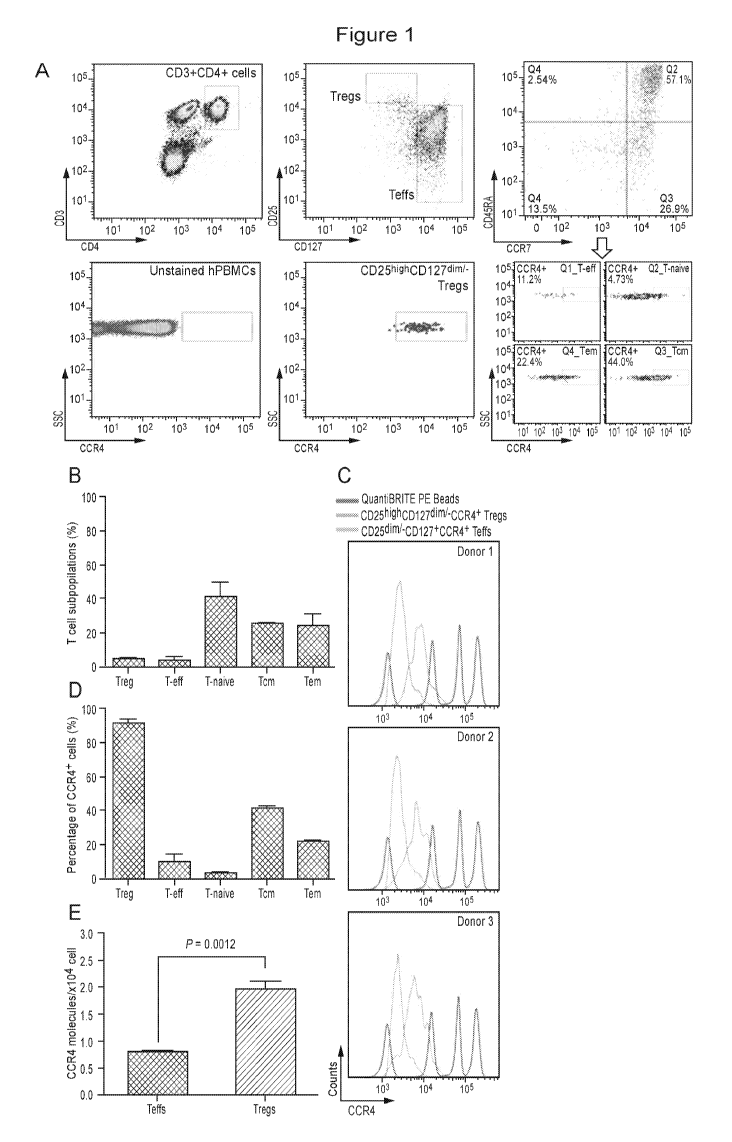
[00021] Figure I. Identification of CCR4 molecules on CD4+ T cell
populations. (A)
Gating strategy for identification of human T cell populations and the CCR4-
expressing T
cell subsets. (B) The percentage of T cell subpopulations. (C) Fluorescence
histograms of
QuantiBRITE PE beads (red line), CD25highCD127dimi-CCR4+ Tregs (blue line),
and
CD25d1mi-CD127+CCR4+ T cells (orange line) were performed by flow cytometry in
three
independent healthy donors. PE beads showed the fluorochrome contained low
level (474
PE molecules/bead), medium low level (5,359 PE molecules/bead), medium high
level
(23,843 PE molecules/bead), and high level (62,336 PE molecules/bead) of PE
molecules.
(D) The percentage of CCR4+ subsets in each T cell subpopulation. (E) The
expression
4
CA 02984608 2017-10-31
WO 2016/178779
PCT/US2016/026232
levels of CCR4 molecule on CD4+CD25-CD127+ Teffs and CD4+CD25highCD127dimi-
Tregs.
All experiments were performed in three independent donors and showed the
means
S.E.M.
[00022] Figure 2. CCR4+ Treg mediated immunosuppression. (A) Expression of
CCR4 on CD4+CD25+FoxP3+ Tregs was assessed by flow cytometry. Representative
fluorescence-activated cell sorter (FACS) plots of CD25 and FoxP3 expression
gated on
CD3 CD4
++ lymphocytes (left two plots) and CCR4 staining gated on CD25+FoxP3+
lymphocytes (right plot) in healthy donor blood sample. (B) CFSE cell
proliferation profiles
of CD4+ effector T cells cultured with or without 20 [tg/m1 PHA and Tregs or
CCR4-Tregs
(at the ratio of Teff:Treg=10:1) were analyzed using flow cytometry by gating
CFSE+ cells.
CCR4-Tregs were separated by using mAb2-3-conjugated beads. Percentages
represent the
proportion of dividing CFSE-labeled CD4+ Teffs after 7 days in culture.
Experiments were
reproduced in three independent donors. (C) In vitro suppression assay of
Teffs in the
coculture of CCR4-depleted Tregs. The suppression assay was measured by the
proliferation of CFSE-labeled Teffs cocultured in the presence or absence of
CD4+ CD25+
CD127dimi- CCR4 Tregs from three independent healthy donors at the Teff/Treg
ratio of
10/1 and stimulated with PHA for 5 days. Percentages of CFSE-diluting Teffs
were
calculated. Shown are mean S.E.M. analyzed by two-way ANOVA. value < 0.05.
[00023] Figure 3. Inhibition of ovarian cancer cells mediated Treg
chemotaxis by
mAb2-3 in vitro and in vivo. (A) Intracellular chemokine CCL22 staining was
performed
with (blue lines) or without (red lines) the addition of brefeldin A (BFA) in
the culture of
ovarian cancer cell lines, IGROV-1, OVCAR-5, and OVCAR-8. (B) In vitro
chemotaxis of
CD4+CD25+ Tregs induced by CCL22-expressing ovarian cancer cell supernatant
was
performed using transwell assay. Treg recruitment was inhibited by mAb2-3 IgG1
and
IgG4, but not by control antibodies. (C) The in vivo bioluminescence images of
ovarian
cancer xenograft mouse model at 18 h post-injection of luciferized CD4+ T
cells and (D)
CD4+CD25+CD127d1mi- Tregs. The intensity of the region of interest (ROT) (red
circle,
xenografted tumor) was further quantified in the left panel. Results were
expressed as
means S.D. "*" and "**" represent student's t-test p value < 0.05 and 0.01,
respectively.
[00024] Figure 4. The activity of tumor-primed T cells on IGROV-1 cells.
(A)
Tumor-primed T cells were stained by anti-CD3, CD4, CD8, CD25 and (B) CCR4
antibodies. CCR4-depleted tumor-primed T cells by mAb2-3-conjugated beads and
T cell
CA 02984608 2017-10-31
WO 2016/178779
PCT/US2016/026232
subsets in tumor-primed T cells were analyzed by flow cytometry. (C) Tumor-
primed T
cells and mAb2-3-depleted tumor-primed T cells were incubated with IGROV-1
cells for 24
and 48 hours and then the supernatant were harvested and detected the
expression level of
IFN-y. The IFN-y in the cocultured supernatant was measured by mesoscale
discovery
(MSD) and showed the folds of the IFN-y concentration in the tumor-primed T
cells
cultured supernatant. (D) Intracellular IFN-y staining of CD4 and CD8 T cells
from
coculture. Cells were harvested at 48 hours post-coculture; incubated for 6 h
in the presence
of brefeldin A; stained for CD3, CD4, and CD8; fixed in paraformaldehyde;
permeabilized;
and stained for intracellular IFN-y. The cells were gated on lymphocytes by
size and CD
markers and analyzed by flow cytometry. (E) The cytotoxic activities of tumor-
primed T
cells and mAb2-3-depleted tumor primed T cells were further detected by LDH
ELISA
assay. All experiments represented triplicates in each time point with the
means S.D. and
performed in two independent experiments. *, **, and *** represents student's
t-test p value
<0.05, 0.01, and 0.005, respectively.
[00025] Figure 5. mAb2-3 mediated the tumor growth inhibition in IGROV-1-
xenografted mice reconstructed with IGROV-1-primed T cells. (A) NSG mice were
inoculated with 2x10⁶ luciferased IGROV-1 tumor cells subcutaneously, injected
4 x10⁶
IGROV-1-primed T cells intravenously, and treated with anti-CCR4 antibodies.
Tumor
growth curves of luciferased IGROV-1 human ovarian carcinoma tumor xenografts
in NSG
mice were measured. Mice were treated with 3 mg/kg of control IgG4 (n=2), mAb2-
3 IgG1
(n=3), and mAb2-3 IgG4 (n=3) and equal volume of PBS (n=2). Antibodies were
administered intravenously twice a week for 5 weeks. Mice were imaged using an
IVIS
imaging system every 10 days. Color scale: luminescent signal intensity: blue,
least intense
signal; red, most intense signal. (B) Luciferase signals of tumor tissues in
each group were
quantified. (C) Tumor size and (D) body weight in mice treated with antibodies
were
measured twice a week. (E) Tumor tissue and (F) tumor weight were harvested
and
measured. Bar scale, 1 cm. *, p < 0.05; ***, p < 0.005; p value was calculated
with two-way
ANOVA. All data were shown the means S.E.M.
[00026] Figure 6. The intermediation of mAb2-3 in interaction between IL-2
and
CD25. (A) In the absence of exogenous IL-2, endogenous IL-2 levels in 1x10⁴
CD4+CD25-
Teffs cultured supernatants incubated with or without mAb2-3 were analyzed by
ELISA.
(B) CD4+CD127dimCD49d- Tregs (3000/reaction) were incubated with 0.25 IU/ml of
6
CA 02984608 2017-10-31
WO 2016/178779
PCT/US2016/026232
exogenous IL-2 in the presence and absence of 20 pg/ml of mAbs and 0.5/1 pg/ml
of plate-
bound anti-CD3/28 antibodies. Bars represent S.D. (C) In the absence of
exogenous IL-2,
endogenous IL-2 concentration was shown from 1x10⁴ Teffs alone or with Tregs
and
treated with 20 pg/ml of mAb2-3. Bars represent S.D. (D) The concentrations
of IL-2 in
supernatants from Teffs and Tregs coculture treated with mAb2-3 in the
presence of 4
IU/ml of exogenously added IL-2. Bars represent S.D. (E) In the presence of
exogenous
IL-2 (20 IU/ml), the IL-2 concentrations of supernatants from 2x10⁵ Mac-1
cells treated
with or without antibodies (mAb2-3 or anti-CD25, including anti-TAC and
control mAbs)
were detected by ELISA. Bars represent S.D. (F) In vitro cell survival assay
was
performed by measuring the viability dye in cultured Tregs treated with the
presence or
absence of 0.5 IU/ml IL-2, 20 pg/ml mAb2-3 IgGl, and 20 pg/ml control IgG1 for
5 days.
The normalized percentage of dead Tregs from different groups among
spontaneous death
Tregs was shown. Each dot indicates an individual donor in each group. Bars
represent
means S.E.M. "**" and "***" represent p value < 0.01 and 0.005, respectively,
by using
student's t-test.
[00027] Figure 7. Representative flow cytometry plots for human peripheral
blood
T cells and phycoerythrin (PE)-conjugated beads. (A) The CD3+CD4+ T cells were
gated
and stained with PE-Cy5 conjugated anti-CD45RA and PerCp-Cy5 conjugated anti-
CCR7
antibodies to distinguish Tcms, Tems, Teffs, and Tnaive (as shown in Figure
1).
CD4+CD25-CD127+CCR4+ T cell subpopulations were gated for further analysis.
Fluorescence histograms of PE beads and T cell subpopulations were shown in
red and blue
lines, respectively. (B) Calibration curve relating PE fluorescence to the
number of PE
molecules per bead. (C) The expression levels of CCR4 molecule on T cell
subpopulations.
All experiments were performed in three independent donors and showed the
means S.D.
[00028] Figure 8.111 vivo distribution of human PBMCs in the presence and
absence
of mAb2-3. (A) 1 x10⁷ human PBMCs and antibodies were injected into mice
intravenously. After 24 hour circulation in vivo, mouse blood were collected
and human
PBMCs were stained with Pacific Blue conjugated anti-CD3, Brilliant Violet
conjugated
anti-CD4, APC conjugated anti-CD25, and PE-Cy7 conjugated anti-CD127, gated to
distinguish Tregs. (B) The percentage of CD25+CD127- Treg was shown the
average from
three individual mice in each group. *, p value < 0.05. (C) Analysis of Tregs
after in vivo
circulation.
7
CA 02984608 2017-10-31
WO 2016/178779
PCT/US2016/026232
[00029] Figure 9.111 vivo response of human PBMCs to mAb2-3 IgG4 in a huPBL-
NSG animal model. (A) 107 freshly isolated human PBMCs were injected
intravenously
via the tail vein into adult NSG immunodeficiency mice. The huPBL-NSG mice
were
received 1 mg/kg antibodies through tail vein twice a week. Peripheral blood
was collected
from huPBL-NSG mice weekly and stained with anti-human specific antibodies for
human
CD45, (B) CD19 with CD45, (C) CD3 with CD45, (D) CD4 with CD3 and CD45, (E)
CD8
with CD3 and CD45, and (F) CD25 with CD3 and CD45, and quantified per 104
PBMCs by
flow cytometry. (G) The percentages of CD3+CD4+CD25+CD127- cells in CD45+
cells from
blood were examined each week in different treatment. (H) The percentage of
CD3+CD4+CD25+CD127- cells in CD45+ cells from blood, spleen, and bone marrow
are
shown at the third week. Two-way ANOVA test was performed. Each data point
represents
the average (n=6 NSG mice, 2 PBMC donors) S.E.M. * and **,p value <0.05 and
0.01,
respectively.
[00030] Figure 10. mAb2-3 inhibited the chemoattration mediated by CCL22.
(A)
The expression of CCR4 on IGROV-1 ovarian cancer cells. (B) mAb2-3 effectively
inhibited chemotaxis of CD4+CD25- and (C) CD4+CD25+ T cells to CCR4 ligand,
CCL22
in a dose-dependent manner.
[00031] Figure 11. Chemoattraction of human lymphocytes by CCL22-secreting
ovarian cancer cells is inhibited by mAb2-3. (A) The images show in vivo
bioluminescence images of ovarian cancer xenograft mouse model at 48 hours
(imaging)
post-injection of luciferized CD4+CD25+CD127d1lli/- T cells. (B) The
quantification of
intensity of ROT (red circle, xenografted tumor) was shown that mAb2-3
inhibits Tregs
recruitment to tumor tissue. (C) Tumor tissues were harvested, digested by
collagenase, and
then stained by anti-CD3, CD4, and CD25 antibodies. CD3+CD4+CD25+ T cells were
analyzed and counted by flow cytometry and shown as the percentage of total
cells from
tumor tissues.
[00032] Figure 12. Development of IGROV-1-pulsed dendritic cells and IGROV-
1-
primed T cells. (A) Monocytic differentiation into dendritic cells (DCs) by
Granulocyte
macrophage colony-stimulating factor (GM-CSF) and Interleukin-4 (IL-4) in
combination.
Monocytes were isolated from human PBMCs and cultured in the presence of GM-
CSF
(100 ng/ml) and IL-4 (100 ng/ml) combination. After culturing for 7 days,
cells were
harvested and analyzed by flow cytometry for surface expression of various
markers for DC
8
CA 02984608 2017-10-31
WO 2016/178779
PCT/US2016/026232
differentiation as indicated. (B) IFN-y secretions were detected from the
cocultured
supernatant of tumor-primed T cells (TP-T cells), untreated DCs, and tumor-
pulsed DCs.
Bars represent S.D. "*" represent student's t-test p <0.05.
[00033] Figure 13. mAb2-3 mediated tumor growth inhibition in large-scale
IGROV-1-xenografted mice bearing IGROV-1-primed T cells. (A) NSG mice were
inoculated with 5x10⁶ IGROV-1 tumor cells subcutaneously, injected 1 x10⁷
IGROV-1-
primed T cells intravenously, and treated with anti-CCR4 antibodies. Tumor
growth curves
of IGROV-1 human ovarian carcinoma tumor xenografts in NSG mice were measured.
Mice were treated with 3 mg/kg of control IgG4 (n=2), mAb2-3 IgG1 (n=2), mAb2-
3 IgG4
(n=2) and equal volume of PBS (n=2). Antibodies were administered
intravenously twice a
week for 4 weeks. Tumor size and (B) body weight in mice treated with
antibodies were
measured once a week. (C) Tumor tissues were harvested. Bar scale, 1 cm. (D)
Mouse
PBMCs were stained by anti-human CD3, CD4, (E) CD8, and (F) CD25 antibodies
and
analyzed using flow cytometry. *, P <0.05; **, P < 0.01; p value were
calculated with two-
way ANOVA; black and grey asterisks indicate respectively mAb2-3 IgG1 and IgG4
compared to control groups. All data were shown as means S.E.M. (G)
Immunohistochemistry staining was performed using anti-CD3 (upper panel) and
anti-
CD25 (lower panel) antibodies in tumor tissues of each group. The dark red
staining
membrane (arrows) represents CD3 or CD25 TP-T cell in tumor. Bar scale, 50
p.m.
[00034] Figure 14. Suppressive cytokine production by Tregs. Effect of
antibody
treatment on suppressive cytokine production by Tregs was evaluated using
ELISA
measurement of the IL-10 and TGF-13 cytokines in the supernatants collected
from cultures
of non-treated, control mAb or mAb2-3 with CD4+CD25- and CD4+CD25+ T cells. T
cells
cultured without mAbs were used as control for any detected background levels
of IL-10
and TGF-13 . Data are presented as mean S.D.
[00035] Figure 15. Flow cytometry-based IL-2 binding and competition
analyses.
(A) Mac-1 cells were washed with cold PBS and then stained sequentially with
20 pg/ml or
(B) three concentrations of competitive antibodies (mAb2-3, anti-TAC or
control mAbs),
100 nM of biotinylated IL-2, and APC-labeled streptavidin. The binding of
biotinylated IL-
2 to Mac-1 cells was detected by flow cytometry.
[00036] Figure 16 Mechanisms of Actions of Ab2-3 on IL-2 binding The
intermediation of mAb2-3 in the interaction between IL-2 and CD25. (A) In the
absence of
9
CA 02984608 2017-10-31
WO 2016/178779
PCT/US2016/026232
exogenous IL-2, endogenous IL-2 levels in 1x10⁴ CD4+CD25- Teffs cultured
supernatants
incubated with or without mAb2-3 were analyzed by ELISA. (B) CD4+CD127d1mCD49d-
Tregs (3000/reaction) were incubated with 0.25 IU/ml of exogenous IL-2 in the
presence
and absence of 20 pg/ml of mAbs and 0.5/1 pg/ml of plate-bound anti-CD3/28
antibodies.
Bars represent S.D. (C) In the absence of exogenous IL-2, endogenous IL-2
concentration
was shown from 1x10⁴ Teffs alone or with Tregs and treated with 20 pg/ml of
mAb2-3.
Bars represent S.D. (D) The concentrations of IL-2 in supernatants from
Teffs and Tregs
coculture treated with mAb2-3 in the presence of 4 IU/ml of exogenously added
IL-2. Bars
represent S.D. (E) In the presence of exogenous IL-2 (20 IU/ml), the IL-2
concentrations
of supernatants from 2x10⁵ Mac-1 cells treated with or without antibodies
(mAb2-3 or anti-
CD25, including anti-TAC and control mAbs) were detected by ELISA. Bars
represent
S.D.
[00037] Figure 17 mAb2-3 and chemokine induced shedding of CD25 from Mac-1
cells (A) Mac-1 cells were incubated with mAb2-3, CCL17, or CCL22 in the
presence or
absence of MMP-9 inhibitor or with negative control for MMP inhibitors. After
24 hours
incubation, culture supernatants were harvested and tested the concentration
of soluble
CD25 by ELISA. Bars represent S.E.M. (B) The concentration of soluble CD25
in 12-
hour, (C) 24-hour, and (D) 48-hour cultured supernatant of Mac-1 cells treated
with mAb2-
3 or control mAbs was investigated with ELISA. The figure shows soluble CD25
concentration expressed as pg/ml in the cultured supernatant. Bars represent
S.E.M. "*"
indicates p value < 0.05 by student's t-test.
[00038] Figure 18. mAb2-3 induced CD25 shedding from Tregs Tregs were
incubated with mAb2-3 and control IgGl. After 48 hours incubation, culture
supernatants
were harvested and tested the concentration of soluble CD25 by ELISA. (A) The
concentration of soluble CD25 (sCD25) in 48-hour cultured supernatant of Tregs
treated
with mAb-2-3 or control IgG1 was investigated with ELISA. The data presented
are the
average from three independent donors. Bars represent S.E.M. "*" and "**"
represent p
value < 0.05 and 0.01, respectively, by using two-way ANOVA. (B) The results
are
presented from in vitro cell survival assays that were performed by measuring
the viability
dye in cultured Tregs treated with the presence or absence of IL-2, mAB2-3
IgGl, and
control IgG1 for 5 days. The normalized percentage of Treg death from the
different groups
CA 02984608 2017-10-31
WO 2016/178779
PCT/US2016/026232
among spontaneous death of Tregs is shown. Bars represent S.E.M. "*" and
"**"
represent p value <0.01 and 0.005, respectively, by using Student's t-Test.
[00039] Figure 19. Representative phenotypic analysis of CCR4 expression in
CD4+CD25 FoxP3+ T cells of Macaque PBMCs Freshly isolated PBMCs were stained
for CD4, CD25, CCR4 and intracellular FoxP3. CCR4 expression was analyzed in
the
CD4+CD25+FoxP3+ population. The average CCR4 expression on Tregs was
calculated in
three independent macaques.
DETAILED DESCRIPTION OF THE INVENTION
[00040] The invention is based in part upon the discovery that an anti-CCR4
antibody,
mAb2-3 can inhibit regulatory T-cell (Treg) chemotactic activity, restore
effector T-cell
(Teff) proliferation and deplete CCR4 + Tregs.
[00041] One important role of human mAbs in cancer immunotherapy lies in
their
capacity to reverse the immune dysregulation caused by tumor cell
commandeering of
surface expression and secretion of proteins that promote immune evasion. The
tumor
microenvironment contains a plethora of mixed immune cell types that play a
paradoxical
role in tumor immunosurveillance by either activating anti-tumor responses or
promoting
tumor progression. Among these tumor infiltrating lymphocytes (TILs) are
CD4+CD25+FoxP3+ Tregs that have been shown in several malignancies to play a
critical
role in suppressing local tumor immunity. The recruitment of Tregs to the
tumor is
mediated through high-level secretion of the CCR4 receptor chemokine CCL22 by
tumor
cells and microenviornmental macrophages. These CCR4+ Tregs create a favorable
environment for dysregulation of local anti-tumor immunity and enhancement of
tumor
growth. Moreover, the tumor-associated chemokines of CCR4 have been detected
in
patients with different types of cancer. Thus, the targeted approach of human
anti-CCR4
mAb immunotherapy described herein offers significant advantages in improving
cancer
immunotherapeutic efficacy while simultaneously reducing its side effects.
[00042] As described in detail in the Examples below, the in vitro and in
vivo activity of
a human anti-CCR4 mAb, mAb2-3, against Tregs was examined. About 85% of
CD4+CD25+FoxP3+ Tregs overexpress CCR4 compared to Teffs (Figures 1 and 7) and
they
are responsible for the majority of suppressor activity. In addition, removal
of CCR4 + Tregs
by mAb2-3 restores Teff proliferation (Figure 2). In vitro studies
demonstrated that mAb2-
11
CA 02984608 2017-10-31
WO 2016/178779
PCT/US2016/026232
3 can inhibit Treg chemotactic activity to CCL22-expressing OvCA cells. In a
humanized
mouse model bearing a OvCA xenograft, mAb2-3 showed therapeutic capability to
modulate human Treg function and enhance anti-tumor activity (Figures 4, 5,
and 13).
[00043] It has been previously demonstrated that mAb2-3 IgG1 exhibits
potent antibody-
dependent cellular cytotoxicity (ADCC) and complement-dependent cytotoxicity
(CDC)
activities in vitro and in vivo against CCR4 expressing tumors (18,42). (See,
WO
2009/086514 and WO 2013/166500 the contents of which are incorporated by
reference in
their entireties). In the Examples provided herein, the biological functions
of both IgG1 and
IgG4 isotypes of mAb2-3 were tested and showed similar capacity to block
CCR4+Treg
migration in vitro (Figure 3B and 10B) but revealed their different mechanisms
of action in
vivo. In particular, mAb2-3 IgG1 induced a profound immunodepletion of Tregs
as
evidenced by in vivo clearance studies (Figure 3D and 8B) and decreased tumor
cell
infiltration (Figure 11). In OvCA xenograft studies, mAb2-3 IgG1 treatment led
to marked
inhibition of tumor cell growth (Figures 5 and 13) and in two animal studies
the mice
showed significant weight loss (Figure 5D and 13B). In contrast, the IgG4
isotype appeared
to work primarily through ligand-receptor blockade (Figure 3, 9, and 11). In
vivo
trafficking studies showed that this isotype caused blockade of Treg
chemotaxis to CCL22
secreting OvCA tumors and a decrease in tumor cell infiltration (Figure 11).
The IgG4
isotype also caused a slower and less complete depletion of Tregs (Figure 9G).
The slower
in vivo clearance observed for IgG4-mediated depletion of Tregs may be through
a different
mechanism of action as a recent report showed that IgG4 isotype has similar
ADCP
capacity to IgG1 (43). In addition, mAb2-3 IgG4 treatment showed lesser anti-
tumor effect
however, the mice had no weight loss (Figure 5D and 5F). These results suggest
that the
two mAb2-3 isotypes may have unique roles at different stages of OvCA disease
with IgG4
treatment having a possibly preferred role at earlier stages when tumor burden
is smaller
and immune dysfunction is more easily reversed.
[00044] Additionally, it was shown that mAb2-3 can inhibit IL-2 binding to
CCR4+IL-
2R+ Mac-1 and Treg cells (Figure 6), but did not did not cross-bind to IL-2R
subunits alone
or in combination using transfected 293T cells. With Macl cells, mAb2-3
treatment led to
enhanced cleavage of sCD25, a property that was shared by the CCR4 ligands
CCL22 and
CCL17 (Figures 18A and Figure 17 B-D). This shared activity suggests that mAb2-
3 has
agonist activity and triggers cell activation which results in CD25 cleavage.
Studies of
12
CA 02984608 2017-10-31
WO 2016/178779
PCT/US2016/026232
CCR4 signaling through CCL22/CCL17 binding have shown evidence of PI(3)
kinase/AKT
activation (25,26). In addition, distinct conformations of CCR4 have been
reported to
respond differently to the two ligands, a property that is supported by our
evidence that
CCL22 and mAb2-3 are more potent activators of sCD25 cleavage than is CCL17
(Figure
17A) (44,45). High level CD25 expression on Tregs leads to formation of the
trimeric high
affinity IL-2 receptor that supports greater IL-2 binding which has been shown
to be
required for survival (46). It is possible that the increased cleavage of CD25
will result in
decreased affinity for IL-2 binding to Tregs and the released sCD25 may be
associated with
blockade of IL-2 uptake by Tregs and their decreased survival (Figures 6A-F)
(47).
[00045] Data presented herein demonstrates that circa 85% of peripheral
blood
CD4+CD25+FoxP3+ Tregs overexpress CCR4 compared to Teffs and they are
responsible
for the majority of suppressor activity. In vitro studies demonstrated that
CCR4+ Treg
depletion by mAb2-3 inhibits Treg chemotactic activity to CCL22-expressing
ovarian
cancer (OvCA) cells and restores Teff proliferation and anti-OvCA immunity. In
a
humanized mouse model bearing an OvCA xenograft, both mAb2-3 IgG1 and IgG4
isotypes showed therapeutic capability to modulate human Treg function and
enhance anti-
tumor activity.
[00046] The data presented here also demonstrate that mAb2-3 treatment also
leads to
blockade of IL-2 uptake by Tregs and inhibition of IL-2-mediated survival
which may play
a role in the in vivo anti-tumor effects seen with non-immunodepleting mAb2-3
IgG4.
[00047] The biological functions of both IgG1 and IgG4 isotypes of mAb2-3
were tested
and showed similar capacity to block CCR4+Treg migration in vitro but revealed
their
different mechanisms of action in vivo. In particular, mAb2-3 IgG1 induced a
profound
immunodepletion of Tregs as evidenced by in vivo clearance studies and
decreased tumor
cell infiltration. In OvCA xenograft studies, mAb2-3 IgG1 treatment led to
marked
inhibition of tumor cell growth and in two animal studies the mice showed
significant
weight loss. In contrast, the IgG4 isotype appeared to work primarily through
ligand-
receptor blockade. In vivo trafficking studies showed that this isotype caused
blockade of
Treg chemotaxis to CCL22 secreting OvCA tumors and a decrease in tumor cell
infiltration.
The IgG4 isotype also caused a slower and less complete depletion of Tregs.
13
CA 02984608 2017-10-31
WO 2016/178779
PCT/US2016/026232
[00048] METHODS OF TREATMENT
[00049] The invention provides for both prophylactic and therapeutic
methods of treating
a subject at risk of (or susceptible to) a cancer, or other diseases or
disorders by
administering an anti-CCR4 antibody. Such diseases or disorders include but
are not
limited to, e.g., those diseases or disorders associated with regulatory T
cell mediated
immunosuppression. By regulatory T cells it is meant to include Treg and/or
follicular
regulatory T cells (TFR). Administration of a prophylactic agent can occur
prior to the
manifestation of disease such that the disease is prevented or, alternatively,
delayed in its
progression. The invention further provides methods of vaccination in which an
CCR4
antibody is include in or administered in conjunction with an antigen. The
CCR4antibody
act as an adjuvant to increase the immune response to the antigen by depleting
regulatory T
cells and/or increasing effector T-cell proliferation.
[00050] For example, the methods are used to deplete regulatory T-cells
(Tregs and or
TFR ) and or inhibit the migration (e.g, chemotaxis) of regulatory T-c ell to
a cytokine
secreting tumor by contacting a cell or administering to a subject a CCR4
antibody. The
cytokine secreting tumor secretes CCL1, CCL4, CC15, CCL17 and/or CCL22. When
the
CCR4 antibody is used to deplete regulatory cells, the antibody preferably has
an IgG1
heavy chain constant region. When the CCR4 antibody is used to inhibit
migration of a
regulatory T-cell, the antibody preferably has an IgG4 heavy chain constant
region.
[00051] In various embodiments the effector T-cells are not substantially
depleted By
"not substantially depleted" it is meant that no more than 1%, 2%, 3%, 4%, 5%,
10%, 15%.
20%, 25% effector T-cells are depleted. In other embodiments effector T-cell
proliferation
and or number is increased or not substantially reduced. By "not substantially
reduced "it
is meant that effector T-cells proliferation and/or is not reduced more than
1%, 2%, 3%,
4%, 5%, 10%, 15%. 20%, 25% compared to untreated cell population.
[00052] In other embodiments the methods are uses to increase effector T-
cell
proliferation. Effector T-cell proliferation is increased 1%, 2%, 3%, 4%, 5%,
10%, 15%.
20%, 25%, 50%, 75%, 80%, 85%, 90%, 95% or more compared to an untreated cell
population.
[00053] In other embodiments the methods modulate (e.g., increase), the
ratio of effector
T cells to regulatory T-cells in the tumor or subject. The ratio is increases
1, 2, 3, 4, 5 or
more fold.
14
CA 02984608 2017-10-31
WO 2016/178779
PCT/US2016/026232
[00054] The methods modulate cytokine (e.g., interferon-gamma) or effector
polypeptide
(e.g., granzyme B or a perforin) release from an effector T-cell population.
By modulate it
is meant an increase or decrease cytokine or effector polypeptide release.
Cytokine or
effector polypeptide release is increased of decrease 1%, 2%, 3%, 4%, 5%, 10%,
15%. 20%,
25%, 50%, 75%, 80%, 85%, 90%, 95% or more compared to an untreated cell
population.
[00055] In another aspect, tumor cell growth is inhibited or slowed by
contacting a cell
or administering to a subject with a CCR4 antibody. For example, the tumor is
a
hematologic cancer such cutaneous T-cell Lymphoma (CTCL), mycosis fungoides
(MF),
primary cutaneous anaplastic large cell Lymphoma (cutaneous ALCL), Sezary
syndrome, or
adult T cell Leukemia/Lymphoma (ATLL).
[00056] Alternatively, the tumor is a solid tumor such as renal cell
carcinoma, breast
cancer, lung cancer, ovarian cancer, prostate cancer, colon cancer, cervical
cancer, brain
cancer, skin cancer (e.g., melanoma), liver cancer, pancreatic cancer Hodgkins
disease,
glioblastoma mutiforme (GBM) or stomach cancer. In particular embodiments, the
cancer
is ovarian cancer or melanoma.
[00057] Inhibiting or slowing tumor growth increase the survival of a
subject having the
tumor. Survival is increased by 1 , 2, 3, 4, 5, or more years.
[00058] In a further aspect, the invention provides methods of inhibiting
IL-2 binding to
CCR4 + regulatory T cells or inducing CD25 cleavage by contacting the
regulatory T cell
with a CCR4 antibody. The loss of IL-2 binding through CD25 results in
metabolic
starvation due to the dependency of IL-2 for regulatory T cells survival
thereby inducing
regulatory T cells death. An increase of regulatory T cells increases the
ratio of effector T-
cells to regulatory T cells.
[00059] In some aspects the CCR4 antibody uses in the above described
methods has an
IgG1 or IgG4 heavy chain constant region. In some embodiments the heavy chain
constant
region has one or more mutations. For example the IgG4 constant region has a
5228P
mutation.
[00060] In particular embodiments, of the invention the subject both a
CCR4antibody
with a IgG1 heavy chain constant region and a CCR4antibody with a IgG4 heavy
chain
constant region. Alternatively, the subject is selected to receive
CCR4antibody with a IgG1
heavy chain constant region or a CCR4antibody with a IgG4 heavy chain constant
region
depending upon the disease stage. For example, it may be advantageous for a
patient in the
CA 02984608 2017-10-31
WO 2016/178779
PCT/US2016/026232
early stage of a disease to receive treatment with a CCR4antibody with a IgG4
heavy chain
constant region as the tumor burden is smaller and the immune dysfunction is
more easily
reversed. In contrast, when a patient has a later stage of a disease, e.g.,
high tumor burden it
may be advantageous for the subject to receive treatment with a CCR4antibody
with a IgG1
heavy chain constant region
[00061] The cell is any cell that expresses CCR4. For example the cell is a
T-cell. T cell
includes regulatory T cell, follicular regulatory T cells, and effector T
cells.
[00062] CCR4 ANTIBODIES
[00063] CCR4 antibodies are known in the art and are suitable for use in
the methods of
the inventions. Exemplary humanized CCR4 antibodies are described in for
example in
WO 2009/086514, WO 2013/166500 and PCT/US2015/054202, the contents of which
are
incorporated by reference in their entireties and are described below. A
preferred CCR4
antibody is mAb2-3. The Exemplary antibodies described herein have
advantageous
features compared to other humanized CCR4 antibodies, such as Mogamulizumab.
For
example, the exemplary antibodies of the invention, in particular mAb2-3
recognize a
conformational epitope that encompasses the N-terminal domain and the
extracellular loop
that mediates biological signaling. In this regard, mAb2-3 treatment of Mac 1
cell and
Tregs led to enhanced sCD25 shedding, a property that is shared with the CCR4
ligands
CCL22 and CCL17. Thus the exemplary antibodies of the invention, in particular
MAb2-3
have agonist activity and triggers cell activation. Additionally, in contrast
to
Mogamulizumab, the exemplary antibodies of the invention, in particular mAb2-3
mediates
complement-dependent cytoxicity (CDC). This may be the direct result of an
optimal
orientation of the Fc region, allowing the angle of attachment to be
permissive for
complement pore formation.
Table 1A. mAb2-3 Variable Region nucleic acid sequences
VH chain of mAb2-3 (SEQ ID NO:1)
CAGGTGCAGCTGGTGCAGAGCGGCGCGGAAGTGAAAAAACCGGGCGCGAGCGTGAAAGT
GAGCTGCAAAGCGAGCGGCTATACCTTTGCGAGCGCGTGGATGCATTGGATGCGCCAGG
CGCCGGGCCAGGGCCTGGAATGGATTGGCTGGATTAACCCGGGCAACGTGAACACCAAA
TATAACGAAAAATTTAAAGGCCGCGCGACCCTGACCGTGGATACCAGCACCAACACCGC
GTATATGGAACTGAGCAGCCTGCGCAGCGAAGATACCGCGGTGTATTATTGCGCGCGCA
GCACCTATTATCGCCCGCTGGATTATTGGGGCCAGGGCACCCTGGTGACCGTGAGCAGC
16
CA 02984608 2017-10-31
WO 2016/178779
PCT/US2016/026232
VL chain of mAb2-3 IgG4 (SEQ ID NO:3)
GATATT GT GAT GACCCAGAGCCCGGATAGCCT GGCGGT GAGCCT GGGCGAACGCGCGAC
CAT TAACT GCAAAAGCAGCCAGAGCAT T CT GTATAGCAGCAACCAGAAAAACTAT C T GG
CGTGGTATCAGCAGAAACCGGGCCAGAGCCCGAAACTGCTGATTTATTGGGCGAGCACC
CGCGAAAGCGGCGTGCCGGATCGCTTTAGCGGCAGCGGCAGCGGCACCGATTTTACCCT
GACCATTAGCAGC CT GCAGGCGGAAGAT GT GGCGGT GTATTATT GCCAT CAGTATAT GA
GCAGCTATACCTTTGGCCAGGGCACCAAACTGGAAATTAAA
Table 1B. mAb2-3 IgG4 Variable Region amino acid sequences
VH chain of mAb2-3 IgG4 (SEQ ID NO: 2)
QVQLVQSGAEVKKPGASVKVSCKASGYT FASAWMHWMRQAPGQGLEWIGWINPGNVNTK
YNEKFKGRATLTVDTSTNTAYMELS SLRSEDTAVYYCARST YYRPLDYWGQGTLVTVS S
VL chain of mAb2-3 IgG4 (SEQ ID NO:4)
DIVMTQS PDS LAVS LGERAT INCKS S QS ILYS SNQKNYLAWYQQKPGQS PKLL I YWAST
RES GVPDRFS GS GS GT DFTLT IS SLQAEDVAVYYCHQYMS S YT FGQGTKLE IK
Table 2A. Antibody 1-44 Variable Region nucleic acid sequences
VH chain of 1-44 (SEQ ID NO:15)
CAGGTGCAGCTGGTGCAGAGCGGAGCCGAGGTGAAGAAGCCTGGAGCTTCCGTCAAGGT
GTCCTGCAAGGCCAGCGGCTACACCTTCGCCAGCCAATGGATGCACTGGATGCGGCAGG
CACCT GGACAGGGCCT CGAAT GGAT CGGCT GGAT CAACCCC GGCAAC GT GAACACCAAG
TACAACGAGAAGTTCAAGGGCAGGGCCACCCTGACCGTGGACACCAGCACCAACACCGC
CTACATGGAACTGAGCAGCCTGCGGAGCGAGGACACCGCCGTGTACTACTGCGCCAGAA
GCAC CT GGTACCGGCCGCT GGAC TACT GGGGC CAGGGCACC CT GGT GACCGT GAGCAGC
VL chain of 1-44 (SEQ ID NO:17)
GACATCGT GAT GACCCAGAGCCCCGACAGCCT GGCCGT GAGCCT GGGCGAGCGGGCCAC
CAT CAACT GCAAGAGCAGCCAGAGCAT C CT GTACAGCAGCAACCAGAAGAACTACC T GG
CCTGGTATCAGCAGAAGCCCGGCCAGAGCCCCAAGCTGCTGATCTACTGGGCCAGCACC
CGGGAGAGCGGCGTGCCCGACCGGTTTAGCGGCAGCGGCTCCGGCACCGACTTCACCCT
GAC CAT CAGCAGC CT GCAGGCCGAGGAC GT GGCCGT GTACTACT GCCACCAGTACAT CA
GCAGCTACACCTTCGGCCAGGGCACAAAGCTGGAAATCAAG
Table 2B. Antibody 1-44 Variable Region amino acid sequences
VH chain of 1-44 (SEQ ID NO: 16)
QVQLVQSGAEVKKPGASVKVSCKASGYT FAS QWMHWMRQAP GQGLEWI GWI N PGNVNT KY
NEKFKGRATLTVDTSTNTAYMELS SLRS EDTAVYYCARSTWYRPLDYWGQGTLVTVS S
VL chain of 1-44 (SEQ ID NO:18)
DIVMTQS PDS LAVS LGERAT INCKS S QS ILYS SNQKNYLAWYQQKPGQS PKLL I YWASTR
17
CA 02984608 2017-10-31
WO 2016/178779
PCT/US2016/026232
ESGVPDRFS GSGS GTDFTLT ISSLQAEDVAVYYCHQY ISSYT FGQGTKLEIK
Table 3A. Antibody 1-49 Variable Region nucleic acid sequences
VH chain of 1-49 (SEQ ID NO:19)
CAGGTGCAGCTGGTGCAGAGCGGAGCCGAGGTGAAGAAGCCTGGAGCTTCCGTCAAGGT
GTCCTGCAAGGCCAGCGGCTACACCTTCGCCAGCAGCTGGATGCACTGGATGCGGCAGG
CACCT GGACAGGGCCT CGAAT GGAT CGGCT GGAT CAACCCC GGCAAC GT GAACACCAAG
TACAACGAGAAGTTCAAGGGCAGGGCCACCCTGACCGTGGACACCAGCACCAACACCGC
CTACATGGAACTGAGCAGCCTGCGGAGCGAGGACACCGCCGTGTACTACTGCGCCAGAA
GCACGTGGTATCGGCCGAATGACTACTGGGGCCAGGGCACCCTGGTGACCGTGAGCAGC
VL chain of 1-49 (SEQ ID NO:21)
GACATCGTGATGACCCAGAGCCCCGACAGCCTGGCCGTGAGCCTGGGCGAGCGGGCCAC
CAT CAACT GCAAGAGCAGCCAGAGCAT C CT GTACAGCAGCAACCAGAAGAACTACC T GG
CCTGGTATCAGCAGAAGCCCGGCCAGAGCCCCAAGCTGCTGATCTACTGGGCCAGCACC
CGGGAGAGCGGCGTGCCCGACCGGTTTAGCGGCAGCGGCTCCGGCACCGACTTCACCCT
GACCAT CAGCAGC CT GCAGGCCGAGGAC GT GGCCGT GTACTACT GCCACCAGTACAAAA
GCAGCTACACCTTCGGCCAGGGCACAAAGCTGGAAATCAAG
Table 3B. Antibody 1-49 Variable Region amino acid sequences
VH chain of 1-49 (SEQ ID NO: 20)
QVQLVQSGAEVKKPGASVKVSCKASGYT FAS S WMHWMRQAP GQGLEWI GWI N PGNVNT
KYNEKFKGRATLTVDTSTNTAYMELSSLRSEDTAVYYCARSTWYRPNDYWGQGTLVTV
SS
VL chain of 1-49 (SEQ ID NO:22)
DIVMTQS PDS LAVS LGERAT INCKS S QS ILYS SNQKNYLAWYQQKPGQS PKLL I YW
ASTRESGVPDRFS GS GS GT DFTLT I S S LQAEDVAVYYCHQYKS S YT FGQGTKLEIK
Table 4A. Antibody 2-1 Variable Region nucleic acid sequences
VH chain of 2-1 (SEQ ID NO:23)
CAGGTGCAGCTGGTGCAGAGCGGAGCCGAGGTGAAGAAGCCTGGAGCTTCCGTCAAG
GTGTCCTGCAAGGCCAGCGGCTACACCTTCGCCAGCAGCTGGATGCACTGGATGCGG
CAGGCACCTGGACAGGGCCTCGAATGGATCGGCTGGATCAACCCCGGCAACGTGAAC
ACCAAGTACAACGAGAAGTTCAAGGGCAGGGCCACCCTGACCGTGGACACCAGCACC
AACACCGCCTACATGGAACTGAGCAGCCTGCGGAGCGAGGACACCGCCGTGTACTAC
TGCGCCAGAACCACCCGTTATCGGCCCCTGGACTACTGGGGCCAGGGCACCCTGGTG
ACCGTGAGCAGC
VL chain of 2-1 (SEQ ID NO:25)
GACATCGTGATGACCCAGAGCCCCGACAGCCTGGCCGTGAGCCTGGGCGAGCGGGCC
18
CA 02984608 2017-10-31
WO 2016/178779
PCT/US2016/026232
ACCATCAACTGCAAGAGCAGCCAGAGCATCCTGTACAGCAGCAACCAGAAGAACTAC
CT GGCCT GGTATCAGCAGAAGCCCGGCCAGAGCCCCAAGCT GCT GAT CTACT GGGCC
AGCACCCGGGAGAGCGGC GT GCC CGACC GGTT TAGCGGCAGCGGCT CCGGCACCGAC
TTCACCCTGACCATCAGCAGCCTGCAGGCCGAGGACGTGGCCGTGTACTACTGCCAC
CAGTACCGTAGCAGCTACACCTTCGGCCAGGGCACAAAGCTGGAAATCAAG
Table 4B. Antibody 2-1 Variable Region amino acid sequences
VH chain of 2-1 (SEQ ID NO: 24)
QVQLVQSGAEVKKPGASVKVSCKASGYT FAS SWMHWMRQAPGQGLEWIGWINPGNVNT
KYNEKFKGRATLTVDTSTNTAYMELS S L RS EDTAVYYCART TRYRPL DYWGQGTLVTV
S S
VL chain of 2-1 (SEQ ID NO:26)
DIVMTQS PDS LAVS LGERAT INCKS S QS ILYS SNQKNYLAWYQQKPGQS PKLL I YWAST
RES GVPDRFS GS GS GT DFTLT IS SLQAEDVAVYYCHQYRS S YT FGQGTKLE IK
Table 5A. Antibody 2-2 Variable Region nucleic acid sequences
VH chain of 2-2 (SEQ ID NO:27)
CAGGTGCAGCTGGTGCAGAGCGGAGCCGAGGTGAAGAAGCCTGGAGCTTCCGTCAA
GGT GT CCT GCAAGGCCAGCGGCTACACCTT CGCCAGC CAATATAT GCACT GGAT GC
GGCAGGCAC CT GGACAGGGCCT C GAAT GGAT C GGCT GGAT CAACCCC GGCAACGT G
AACACCAAGTACAACGAGAAGTTCAAGGGCAGGGCCACCCTGACCGTGGACACCAG
CACCAACAC CGCCTACAT GGAACT GAGCAGCCT GCGGAGCGAGGACACCGC CGT GT
ACTACTGCGCCAGACTGACCTATTATCGGCCGCCGGACTACTGGGGCCAGGGCACC
CT GGT GACCGT GAGCAGC
VL chain of 2-2 (SEQ ID NO:29)
GACATCGT GAT GACCCAGAGCCCCGACAGCCT GGCCGT GAGCCT GGGCGAGCGGGCCA
CCAT CAACT GCAAGAGCAGCCAGAGCAT CCT GTACAGCAGCAACCAGAAGAACTAC CT
GGCCT GGTAT CAGCAGAAGCCCGGCCAGAGCC CCAAGCT GCT GAT CTACT GGGCCAGC
ACCCGGGAGAGCGGCGT GCCCGACCGGT TTAGCGGCAGCGGCTCCGGCACCGACTT CA
CCCTGACCATCAGCAGCCTGCAGGCCGAGGACGTGGCCGTGTACTACTGCCACCAGTA
CTATAGCAGCTACACCTTCGGCCAGGGCACAAAGCTGGAAATCAAG
Table 5B. Antibody 2-2 Variable Region amino acid sequences
VH chain of 2-2 (SEQ ID NO: 28)
QVQLVQSGAEVKKPGASVKVSCKASGYT FAS QYMHWMRQAP GQGLEWI GWI N PGNVNT KY
NEKFKGRATLTVDTSTNTAYMELS SLRS EDTAVYYCARLTYYRPPDYWGQGTLVTVS S
VL chain of 2-2 (SEQ ID NO:30)
DIVMTQS PDS LAVS LGERAT INCKS S QS ILYS SNQKNYLAWYQQKPGQS PKLL I YWAST
19
CA 02984608 2017-10-31
WO 2016/178779
PCT/US2016/026232
RES GVPDRFS GS GS GT DFTLT IS SLQAEDVAVYYCHQYYS S YT FGQGTKLEIK
Table 6A. huCCR4 Variable Region nucleic acid sequences
VH chain of huCCR (SEQ ID NO:43)
CAGGTGCAGCTGGTGCAGAGCGGAGCCGAGGTGAAGAAGCCTGGAGCTTCCGTCAAGGT
GTCCTGCAAGGCCAGCGGCTACACCTTCGCCAGCTACTACATGCACTGGATGCGGCAGG
CACCT GGACAGGGCCT CGAAT GGAT CGGCT GGAT CAACCCC GGCAAC GT GAACACCAAG
TACAACGAGAAGTTCAAGGGCAGGGCCACCCTGACCGTGGACACCAGCACCAACACCGC
CTACATGGAACTGAGCAGCCTGCGGAGCGAGGACACCGCCGTGTACTACTGCGCCAGAA
GCACCTACTACCGGCCCCTGGACTACTGGGGCCAGGGCACCCTGGTGACCGTGAGCAGC
VL chain of huCCR (SEQ ID NO:45)
GACATCGT GAT GACCCAGAGCCCCGACAGCCT GGCCGT GAGCCT GGGCGAGCGGGCCAC
CAT CAACT GCAAGAGCAGCCAGAGCAT C CT GTACAGCAGCAACCAGAAGAACTACC T GG
CCTGGTATCAGCAGAAGCCCGGCCAGAGCCCCAAGCTGCTGATCTACTGGGCCAGCACC
CGGGAGAGCGGCGTGCCCGACCGGTTTAGCGGCAGCGGCTCCGGCACCGACTTCACCCT
GAC CAT CAGCAGC CT GCAGGCCGAGGAC GT GGCCGT GTACTACT GCCACCAGTACCT GA
GCAGCTACACCTTCGGCCAGGGCACAAAGCTGGAAATCAAG
Table 6B. huCCR4 Variable Region, amino acid sequences
VH chain of huCCR (SEQ ID NO:44)
QVQLVQSGAEVKKPGASVKVSCKASGYT FAS Y YMHWMRQAP GQGLEWI GWI N PG
NVNTKYNEKFKGRATLTVDTSTNTAYMELS S L RS EDTAVYYCARST Y YRPL DYWG
QGTLVTVS S
VL chain of huCCR (SEQ ID NO:46)
DIVMTQS PDS LAVS LGERAT INCKS S QS ILYS SNQKNYLAWYQQKPGQS PKLL I YWA
SIRE S GVPDRFS GS GS GT DFTLT IS SLQAEDVAVYYCHQYLS SYT FGQGTKLEIK
Table 7A. IgG4 Isotype Region nucleic acid sequences
IgG4 Isotype Region nucleic acids (SEQ ID NO:5)
CA 02984608 2017-10-31
WO 2016/178779
PCT/US2016/026232
GCGAGCACCAAAGGCCCGAGCGTGTTTCCGCTGGCGCCGTGCAGCCGCAGCACCAGCGAA
AGCACCGCGGCGCTGGGCTGCCTGGTGAAAGATTATTTTCCGGAACCGGTGACCGTGAGC
TGGAACAGCGGCGCGCTGACCAGCGGCGTGCATACCTTTCCGGCGGTGCTGCAGAGCAGC
GGCCTGTATAGCCTGAGCAGCGTGGTGACCGTGCCGAGCAGCAGCCTGGGCACCAAAACC
TATACCT GCAACGT GGAT CATAAACCGAGCAACACCAAAGT GGATAAACGC GT GGAAAGC
AAATATGGCCCGCCGTGCCCGAGCTGCCCGGCGCCGGAATTTCTGGGCGGCCCGAGCGTG
ITT CT GTTT CC GC CGAAACCGAAAGATACCCT GAT GAT TAGCCGCAC CCCGGAAGT GACC
TGCGTGGTGGTGGATGTGAGCCAGGAAGATCCGGAAGTGCAGTTTAACTGGTATGTGGAT
GGCGTGGAAGTGCATAACGCGAAAACCAAACCGCGCGAAGAACAGTTTAACAGCACCTAT
CGCGTGGTGAGCGTGCTGACCGTGCTGCATCAGGATTGGCTGAACGGCAAAGAATATAAA
T GCAAAGT GAGCAACAAAGGC CT GC C GAGCAG CAT T GAAAAAAC CAT TAGCAAAGCGAAA
GGCCAGCCGCGCGAACCGCAGGTGTATACCCTGCCGCCGAGCCCGGAAGAAATGACCAAA
AACCAGGTGAGCCTGACCTGCCTGGTGAAAGGCTTTTATCCGAGCGATATTGCGGTGGAA
TGGGAAAGCAACGGCCAGCCGGAAAACAACTATAAAACCACCCCGCCGGTGCTGGATAGC
GAT GGCAGCTTTT TT CT GTATAGCCGCCT GAC CGT GGATAAAAGCCGCT GGCAGGAAGGC
AACGT GTTTAGCT GCAGC GT GAT GCAT GAAGC GCT GCATAACCATTATACC CAGAAAAGC
CTGAGCCTGAGCCTGGGCAAA
Table 7B. IgG4 Isotype Region amino acid sequences
IgG4 Isotype Region amino acid sequences (SEQ ID NO: 6)
ASTKGPSVFPLAPCSRST SESTAALGCLVKDY FPE PVTVSWNS GALT SGVHT FPAVLQS
S GLY S L S SVVTVP S S S LGTKT YT CNVDHKPSNTKVDKRVES KYGP PC PS C PAPE FL GGP
SVFL FP PKP KDT LMI S RT PEVT CVVVDVS QE D PEVQFNWYVDGVEVHNAKT KPREE QFN
ST YRVVSVLTVLHQDWLNGKEYKCKVSNKGL P S S IEKT I SKAKGQPRE PQVYTL P P S PE
EMTKNQVS LTCLVKGFY P S DIAVEWESNGQPENNYKTT P PVLDS DGS FFLYSRLTVDKS
RWQEGNVFS CSVMHEALHNHYTQKSLSLSLGK
Table 8A. IgG4 with stabilized IgG4 core hinge, nucleic acid sequences
IgG4 Isotype Region nucleic acids (SEQ ID NO:7)
21
CA 02984608 2017-10-31
WO 2016/178779 PCT/US2016/026232
ACCAAAGGC CCGAGCGT GTTT CC GCT GGCGCC GT GCAGCCGCAGCAC CAGC GAAAGCAC
CGCGGCGCTGGGCTGCCTGGTGAAAGATTATTTTCCGGAACCGGTGACCGTGAGCTGGA
ACAGCGGCGCGCTGACCAGCGGCGTGCATACCTTTCCGGCGGTGCTGCAGAGCAGCGGC
CTGTATAGCCTGAGCAGCGTGGTGACCGTGCCGAGCAGCAGCCTGGGCACCAAAACCTA
TACCTGCAACGTGGATCATAAACCGAGCAACACCAAAGTGGATAAACGCGTGGAAAGCA
AATATGGCCCGCCGTGCCCGCCGTGCCCGGCGCCGGAATTTCTGGGCGGCCCGAGCGTG
ITT CT GTTT CC GC CGAAACCGAAAGATACCCT GAT GAT TAGCCGCAC CCCGGAAGT GAC
CTGCGTGGTGGTGGATGTGAGCCAGGAAGATCCGGAAGTGCAGTTTAACTGGTATGTGG
AT GG C GT GGAAGT GCATAAC GC GAAAAC CAAAC C GC G C GAAGAACAGT T TAACAGCAC C
TATCGCGTGGTGAGCGTGCTGACCGTGCTGCATCAGGATTGGCTGAACGGCAAAGAATA
TAAAT GCAAAGT GAGCAACAAAG GC CT GCCGAGCAGCATT GAAAAAAC CAT TAGCAAAG
CGAAAGGCCAGCCGCGCGAACCGCAGGTGTATACCCTGCCGCCGAGCCCGGAAGAAATG
ACCAAAAACCAGGTGAGCCTGACCTGCCTGGTGAAAGGCTTTTATCCGAGCGATATTGC
GGT GGAAT GGGAAAGCAACGGCCAGCCGGAAAACAACTATAAAACCACCCC GCCGGT GC
TGGATAGCGATGGCAGCTTTTTTCTGTATAGCAAACTGACCGTGGATAAAAGCCGCTGG
CAGGAAGGCAACGT GTTTAGCT GCAGCGT GAT GCAT GAAGC GCT GCATAAC CATTATAC
CCAGAAAAGCCTGAGCCTGAGCCTGGGCAAA
Table 8B. IgG4 with stabilized IgG4 core hinge, amino acid sequences
IgG4 Isotype Region amino acid sequences (SEQ ID NO: 8)
TKGP SVFPLAPCS RST S E STAAL GCLVKDY FPE PVTVSWNS GALT S GVHT FPAVLQSSG
LYS L S SVVTVPS S S LGTKT YTCNVDHKP SNTKVDKRVESKYGP PC P PC PAPE FLGG PSV
FL FP PKPKDTLMI S RT PEVTCVVVDVSQEDPEVQFNWYVDGVEVHNAKTKPREEQFNST
YRVVSVLTVLHQDWLNGKEYKCKVSNKGL PS S IEKT I SKAKGQPRE PQVYT L P PS PEEM
TKNQVS LTCLVKG FY PS D IAVEWESNGQPENNYKTT P PVLDSDGS FFLYSKLTVDKSRW
QEGNVFSCSVMHEALHNHYTQKSLSLSLGK
[00064] The amino acid sequences of the heavy and light chain
complementarity
determining regions of selected antibodies are shown in Table 9 below.
[00065] Table 9. Amino Acid Sequences of Heavy and Light Chains.
Antibody Variable CDR1 CDR2 CDR3
region
GYTFASYY INP GNVNT STYYRPLDY
Mouse 1567 VH
(SEQ ID NO: 31) (SEQ ID NO: 11) (SEQ ID NO: 13)
Humanized GYTFASYY INP GNVNT STYYRPLDY
1567 VH(SEQ ID NO: 31) (SEQ ID NO: 11) (SEQ ID NO: 13)
Abl 44 VH
GYTFASQW INP GNVNT STWYRPLDY
-
(SEQ ID NO: 32) (SEQ ID NO: 11) (SEQ ID NO: 34)
Ab GYTF AS SW INP GNVNT STWYRPNDY
l -49 VH
(SEQ ID NO: 33) (SEQ ID NO: 11) (SEQ ID NO: 35)
22
CA 02984608 2017-10-31
WO 2016/178779 PCT/US2016/026232
Ab2 1 VH GYTFASSW INPGNVNT TTRYRPLDY
-
(SEQ ID NO: 33) (SEQ ID NO: 11) (SEQ ID NO: 36)
Ab2 2 VH GYTFASQY INPGNVNT LTYYRPPDY
-
(SEQ ID NO: 33) (SEQ ID NO: 11) (SEQ ID NO: 37)
Ab2 3 VH GYTFASAW INPGNVNT STYYRPLDY
-
(SEQ ID NO: 9) (SEQ ID NO: 11) (SEQ ID NO: 13)
M 1567 VL QSILYSSNQKNY WASTRE HQYLSSYT
ouse
(SEQ ID NO: 10) (SEQ ID NO: 12) (SEQ ID NO: 38)
Humanized VL QSILYSSNQKNY WASTRE HQYLSSYT
1567 (SEQ ID NO: 10) (SEQ ID NO: 12) (SEQ ID NO: 38)
Ab QSILYSSNQKNY WASTRE HQYISSYT
1-44 VL
(SEQ ID NO: 10) (SEQ ID NO: 12) (SEQ ID NO: 39)
Ab1 49 VL QSILYSSNQKNY WASTRE HQYKSSYT
-
(SEQ ID NO: 10) (SEQ ID NO: 12) (SEQ ID NO: 40)
Ab QSILYSSNQKNY WASTRE HQYRSSYT
2-1 VL
(SEQ ID NO: 10) (SEQ ID NO: 12) (SEQ ID NO: 41)
Ab2 2 VL QSILYSSNQKNY WASTRE HQYYSSYT
-
(SEQ ID NO: 10) (SEQ ID NO: 12) (SEQ ID NO: 42)
Ab2 3 VL QSILYSSNQKNY WASTRE HQYMSSYT
-
(SEQ ID NO: 10) (SEQ ID NO: 12) (SEQ ID NO: 14)
[00066] As used herein, the term "antibody" refers to immunoglobulin
molecules and
immunologically active portions of immunoglobulin (Ig) molecules, i.e.,
molecules that
contain an antigen binding site that specifically binds (immunoreacts with) an
antigen. By
"specifically binds" or "immunoreacts with" is meant that the antibody reacts
with one or
more antigenic determinants of the desired antigen and does not react with
other
polypeptides. Antibodies include, but are not limited to, polyclonal,
monoclonal, chimeric,
dAb (domain antibody), single chain, Fab, Fab' and F(ab')2 fragments, scFvs,
and Fab
expression libraries.
[00067] A single chain Fv ("scFv") polypeptide molecule is a covalently
linked VH: :VL
heterodimer, which can be expressed from a gene fusion including VH- and VL-
encoding
genes linked by a peptide-encoding linker. (See Huston et al. (1988) Proc Nat
Acad Sci
USA 85(16):5879-5883). A number of methods have been described to discern
chemical
structures for converting the naturally aggregated, but chemically separated,
light and heavy
polypeptide chains from an antibody V region into an scFv molecule, which will
fold into a
three dimensional structure substantially similar to the structure of an
antigen-binding site.
See, e.g., U.S. Patent Nos. 5,091,513; 5,132,405; and 4,946,778.
23
CA 02984608 2017-10-31
WO 2016/178779
PCT/US2016/026232
[00068] Very large naive human scFy libraries have been and can be created
to offer a
large source of rearranged antibody genes against a plethora of target
molecules. Smaller
libraries can be constructed from individuals with infectious diseases in
order to isolate
disease-specific antibodies. (See Barbas et al., Proc. Natl. Acad. Sci. USA
89:9339-43
(1992); Zebedee et al., Proc. Natl. Acad. Sci. USA 89:3175-79 (1992)).
[00069] In general, antibody molecules obtained from humans relate to any
of the classes
IgG, IgM, IgA, IgE and IgD, which differ from one another by the nature of the
heavy chain
present in the molecule. Certain classes have subclasses as well, such as
IgGi, IgG2, IgG3,
IgG4 and others. Furthermore, in humans, the light chain may be a kappa chain
or a lambda
chain. The term "antigen-binding site," or "binding portion" refers to the
part of the
immunoglobulin molecule that participates in antigen binding. The antigen
binding site is
formed by amino acid residues of the N-terminal variable ("V") regions of the
heavy ("H")
and light ("L") chains. Three highly divergent stretches within the V regions
of the heavy
and light chains, referred to as "hypervariable regions," are interposed
between more
conserved flanking stretches known as "framework regions," or "FRs". Thus, the
term "FR"
refers to amino acid sequences which are naturally found between, and adjacent
to,
hypervariable regions in immunoglobulins. In an antibody molecule, the three
hypervariable
regions of a light chain and the three hypervariable regions of a heavy chain
are disposed
relative to each other in three dimensional space to form an antigen-binding
surface. The
antigen-binding surface is complementary to the three-dimensional surface of a
bound
antigen, and the three hypervariable regions of each of the heavy and light
chains are
referred to as "complementarity-determining regions," or "CDRs."
[00070] As used herein, the term "epitope" includes any protein determinant
capable of
specific binding to an immunoglobulin, a scFv, or a T-cell receptor. Epitopic
determinants
usually consist of chemically active surface groupings of molecules such as
amino acids or
sugar side chains and usually have specific three dimensional structural
characteristics, as
well as specific charge characteristics. For example, antibodies may be raised
against N-
terminal or C-terminal peptides of a polypeptide.
[00071] As used herein, the terms "immunological binding," and
"immunological
binding properties" refer to the non-covalent interactions of the type which
occur between
an immunoglobulin molecule and an antigen for which the immunoglobulin is
specific. The
strength, or affinity of immunological binding interactions can be expressed
in terms of the
24
CA 02984608 2017-10-31
WO 2016/178779
PCT/US2016/026232
dissociation constant (Kd) of the interaction, wherein a smaller Kd represents
a greater
affinity. Immunological binding properties of selected polypeptides can be
quantified using
methods well known in the art. One such method entails measuring the rates of
antigen-
binding site/antigen complex formation and dissociation, wherein those rates
depend on the
concentrations of the complex partners, the affinity of the interaction, and
geometric
parameters that equally influence the rate in both directions. Thus, both the
"on rate
constant" (Koo) and the "off rate constant" (Koff) can be determined by
calculation of the
concentrations and the actual rates of association and dissociation. (See
Nature 361:186-87
(1993)). The ratio of Koff /Km enables the cancellation of all parameters not
related to
affinity, and is equal to the dissociation constant Kd. (See, generally,
Davies et al. (1990)
Annual Rev Biochem 59:439-473). An antibody of the present invention is said
to
specifically bind to a CCR4 epitope when the equilibrium binding constant (Kd)
is
preferably 100 nM, more preferably 10 nM, and most preferably 100 pM to about
1
pM, as measured by assays such as radioligand binding assays or similar assays
known to
those skilled in the art.
[00072] A CCR4 protein, or a derivative, fragment, analog, homolog or
ortholog thereof,
may be utilized as an immunogen in the generation of antibodies that
immunospecifically
bind these protein components.
[00073] Those skilled in the art will recognize that it is possible to
determine, without
undue experimentation, if a human monoclonal antibody has the same specificity
as a
human monoclonal antibody of the invention by ascertaining whether the former
prevents
the latter from binding to CCR4. If the human monoclonal antibody being tested
competes
with the human monoclonal antibody of the invention, as shown by a decrease in
binding by
the human monoclonal antibody of the invention, then it is likely that the two
monoclonal
antibodies bind to the same, or to a closely related, epitope.
[00074] Another way to determine whether a human monoclonal antibody has
the
specificity of a human monoclonal antibody of the invention is to pre-incubate
the human
monoclonal antibody of the invention with the CCR4 protein, with which it is
normally
reactive, and then add the human monoclonal antibody being tested to determine
if the
human monoclonal antibody being tested is inhibited in its ability to bind
CCR4. If the
human monoclonal antibody being tested is inhibited then, in all likelihood,
it has the same,
or functionally equivalent, epitope specificity as the monoclonal antibody of
the invention.
CA 02984608 2017-10-31
WO 2016/178779
PCT/US2016/026232
Screening of human monoclonal antibodies of the invention, can be also carried
out by
utilizing CCR4 and determining whether the test monoclonal antibody is able to
neutralize
CCR4.
[00075] Various procedures known within the art may be used for the
production of
polyclonal or monoclonal antibodies directed against a protein of the
invention, or against
derivatives, fragments, analogs homologs or orthologs thereof (See, for
example,
Antibodies: A Laboratory Manual, Harlow E, and Lane D, 1988, Cold Spring
Harbor
Laboratory Press, Cold Spring Harbor, NY, incorporated herein by reference).
[00076] Antibodies can be purified by well-known techniques, such as
affinity
chromatography using protein A or protein G, which provide primarily the IgG
fraction of
immune serum. Subsequently, or alternatively, the specific antigen which is
the target of
the immunoglobulin sought, or an epitope thereof, may be immobilized on a
column to
purify the immune specific antibody by immunoaffinity chromatography.
Purification of
immunoglobulins is discussed, for example, by D. Wilkinson (The Scientist,
published by
The Scientist, Inc., Philadelphia PA, Vol. 14, No. 8 (April 17, 2000), pp. 25-
28).
[00077] The term "monoclonal antibody" or "mAb" or "monoclonal antibody
composition", as used herein, refers to a population of antibody molecules
that contain only
one molecular species of antibody molecule consisting of a unique light chain
gene product
and a unique heavy chain gene product. In particular, the complementarity
determining
regions (CDRs) of the monoclonal antibody are identical in all the molecules
of the
population. MAbs contain an antigen binding site capable of immunoreacting
with a
particular epitope of the antigen characterized by a unique binding affinity
for it.
[00078] Monoclonal antibodies can be prepared using hybridoma methods, such
as those
described by Kohler and Milstein, Nature, 256:495 (1975). In a hybridoma
method, a
mouse, hamster, or other appropriate host animal, is typically immunized with
an
immunizing agent to elicit lymphocytes that produce or are capable of
producing antibodies
that will specifically bind to the immunizing agent. Alternatively, the
lymphocytes can be
immunized in vitro.
[00079] The immunizing agent will typically include the protein antigen, a
fragment
thereof or a fusion protein thereof Generally, either peripheral blood
lymphocytes are used
if cells of human origin are desired, or spleen cells or lymph node cells are
used if
non-human mammalian sources are desired. The lymphocytes are then fused with
an
26
CA 02984608 2017-10-31
WO 2016/178779
PCT/US2016/026232
immortalized cell line using a suitable fusing agent, such as polyethylene
glycol, to form a
hybridoma cell (Goding, Monoclonal Antibodies: Principles and Practice,
Academic Press,
(1986) pp. 59-103). Immortalized cell lines are usually transformed mammalian
cells,
particularly myeloma cells of rodent, bovine and human origin. Usually, rat or
mouse
myeloma cell lines are employed. The hybridoma cells can be cultured in a
suitable culture
medium that preferably contains one or more substances that inhibit the growth
or survival
of the unfused, immortalized cells. For example, if the parental cells lack
the enzyme
hypoxanthine guanine phosphoribosyl transferase (HGPRT or HPRT), the culture
medium
for the hybridomas typically will include hypoxanthine, aminopterin, and
thymidine ("HAT
medium"), which substances prevent the growth of HGPRT-deficient cells.
[00080] Preferred immortalized cell lines are those that fuse efficiently,
support stable
high level expression of antibody by the selected antibody-producing cells,
and are sensitive
to a medium such as HAT medium. More preferred immortalized cell lines are
murine
myeloma lines, which can be obtained, for instance, from the Salk Institute
Cell Distribution
Center, San Diego, California and the American Type Culture Collection,
Manassas,
Virginia. Human myeloma and mouse-human heteromyeloma cell lines also have
been
described for the production of human monoclonal antibodies. (See Kozbor, J.
Immunol.,
133:3001 (1984); Brodeur et al., Monoclonal Antibody Production Techniques and
Applications, Marcel Dekker, Inc., New York, (1987) pp. 51-63)).
[00081] The culture medium in which the hybridoma cells are cultured can
then be
assayed for the presence of monoclonal antibodies directed against the
antigen. Preferably,
the binding specificity of monoclonal antibodies produced by the hybridoma
cells is
determined by immunoprecipitation or by an in vitro binding assay, such as
radioimmunoassay (RIA) or enzyme-linked immunoabsorbent assay (ELISA). Such
techniques and assays are known in the art. The binding affinity of the
monoclonal
antibody can, for example, be determined by the Scatchard analysis of Munson
and Pollard,
Anal. Biochem., 107:220 (1980). Moreover, in therapeutic applications of
monoclonal
antibodies, it is important to identify antibodies having a high degree of
specificity and a
high binding affinity for the target antigen.
[00082] After the desired hybridoma cells are identified, the clones can be
subcloned by
limiting dilution procedures and grown by standard methods. (See Goding,
Monoclonal
Antibodies: Principles and Practice, Academic Press, (1986) pp. 59-103).
Suitable culture
27
CA 02984608 2017-10-31
WO 2016/178779
PCT/US2016/026232
media for this purpose include, for example, Dulbecco's Modified Eagle's
Medium and
RPMI-1640 medium. Alternatively, the hybridoma cells can be grown in vivo as
ascites in
a mammal.
[00083] The monoclonal antibodies secreted by the subclones can be isolated
or purified
from the culture medium or ascites fluid by conventional immunoglobulin
purification
procedures such as, for example, protein A-Sepharose, hydroxylapatite
chromatography, gel
electrophoresis, dialysis, or affinity chromatography.
[00084] Monoclonal antibodies can also be made by recombinant DNA methods,
such as
those described in U.S. Patent No. 4,816,567. DNA encoding the monoclonal
antibodies of
the invention can be readily isolated and sequenced using conventional
procedures (e.g., by
using oligonucleotide probes that are capable of binding specifically to genes
encoding the
heavy and light chains of murine antibodies). The hybridoma cells of the
invention serve as
a preferred source of such DNA. Once isolated, the DNA can be placed into
expression
vectors, which are then transfected into host cells such as simian COS cells,
Chinese
hamster ovary (CHO) cells, or myeloma cells that do not otherwise produce
immunoglobulin protein, to obtain the synthesis of monoclonal antibodies in
the
recombinant host cells. The DNA also can be modified, for example, by
substituting the
coding sequence for human heavy and light chain constant domains in place of
the
homologous murine sequences (see U.S. Patent No. 4,816,567; Morrison, Nature
368,
812-13 (1994)) or by covalently joining to the immunoglobulin coding sequence
all or part
of the coding sequence for a non-immunoglobulin polypeptide. Such a
non-immunoglobulin polypeptide can be substituted for the constant domains of
an
antibody of the invention, or can be substituted for the variable domains of
one
antigen-combining site of an antibody of the invention to create a chimeric
bivalent
antibody.
[00085] Fully human antibodies are antibody molecules in which the entire
sequence of
both the light chain and the heavy chain, including the CDRs, arise from human
genes.
Such antibodies are termed "humanized antibodies", "human antibodies", or
"fully human
antibodies" herein. Human monoclonal antibodies can be prepared by using
trioma
technique; the human B-cell hybridoma technique (see Kozbor, et al., 1983
Immunol Today
4: 72); and the EBV hybridoma technique to produce human monoclonal antibodies
(see
Cole, et al., 1985 In: MONOCLONAL ANTIBODIES AND CANCER THERAPY, Alan R. Liss,
Inc.,
28
CA 02984608 2017-10-31
WO 2016/178779
PCT/US2016/026232
pp. 77-96). Human monoclonal antibodies may be utilized and may be produced by
using
human hybridomas (see Cote, et al., 1983. Proc Natl Acad Sci USA 80: 2026-
2030) or by
transforming human B-cells with Epstein Barr Virus in vitro (see Cole, et al.,
1985 In:
MONOCLONAL ANTIBODIES AND CANCER THERAPY, Alan R. Liss, Inc., pp. 77-96).
[00086] In addition, human antibodies can also be produced using additional
techniques,
including phage display libraries. (See Hoogenboom and Winter, J. Mol. Biol.,
227:381
(1991); Marks et al., J. Mol. Biol., 222:581 (1991)). Similarly, human
antibodies can be
made by introducing human immunoglobulin loci into transgenic animals, e.g.,
mice in
which the endogenous immunoglobulin genes have been partially or completely
inactivated.
Upon challenge, human antibody production is observed, which closely resembles
that seen
in humans in all respects, including gene rearrangement, assembly, and
antibody repertoire.
This approach is described, for example, in U.S. Patent Nos. 5,545,807;
5,545,806;
5,569,825; 5,625,126; 5,633,425; 5,661,016, and in Marks et al.,
Bio/Technology 10,
779-783 (1992); Lonberg et al., Nature 368 856-859 (1994); Morrison, Nature
368, 812-13
(1994); Fishwild et al, Nature Biotechnology 14, 845-51 (1996); Neuberger,
Nature
Biotechnology 14, 826 (1996); and Lonberg and Huszar, Intern. Rev. Immunol. 13
65-93
(1995).
[00087] Human antibodies may additionally be produced using transgenic
nonhuman
animals which are modified so as to produce fully human antibodies rather than
the
animal's endogenous antibodies in response to challenge by an antigen. (See
PCT
publication W094/02602). The endogenous genes encoding the heavy and light
immunoglobulin chains in the nonhuman host have been incapacitated, and active
loci
encoding human heavy and light chain immunoglobulins are inserted into the
host's
genome. The human genes are incorporated, for example, using yeast artificial
chromosomes containing the requisite human DNA segments. An animal which
provides
all the desired modifications is then obtained as progeny by crossbreeding
intermediate
transgenic animals containing fewer than the full complement of the
modifications. The
preferred embodiment of such a nonhuman animal is a mouse, and is termed the
XenomouseTM as disclosed in PCT publications WO 96/33735 and WO 96/34096. This
animal produces B cells which secrete fully human immunoglobulins. The
antibodies can
be obtained directly from the animal after immunization with an immunogen of
interest, as,
for example, a preparation of a polyclonal antibody, or alternatively from
immortalized B
29
CA 02984608 2017-10-31
WO 2016/178779
PCT/US2016/026232
cells derived from the animal, such as hybridomas producing monoclonal
antibodies.
Additionally, the genes encoding the immunoglobulins with human variable
regions can be
recovered and expressed to obtain the antibodies directly, or can be further
modified to
obtain analogs of antibodies such as, for example, single chain Fv (scFv)
molecules.
[00088] An example of a method of producing a nonhuman host, exemplified as
a
mouse, lacking expression of an endogenous immunoglobulin heavy chain is
disclosed in
U.S. Patent No. 5,939,598. It can be obtained by a method, which includes
deleting the J
segment genes from at least one endogenous heavy chain locus in an embryonic
stem cell to
prevent rearrangement of the locus and to prevent formation of a transcript of
a rearranged
immunoglobulin heavy chain locus, the deletion being effected by a targeting
vector
containing a gene encoding a selectable marker; and producing from the
embryonic stem
cell a transgenic mouse whose somatic and germ cells contain the gene encoding
the
selectable marker.
[00089] One method for producing an antibody of interest, such as a human
antibody, is
disclosed in U.S. Patent No. 5,916,771. This method includes introducing an
expression
vector that contains a nucleotide sequence encoding a heavy chain into one
mammalian host
cell in culture, introducing an expression vector containing a nucleotide
sequence encoding
a light chain into another mammalian host cell, and fusing the two cells to
form a hybrid
cell. The hybrid cell expresses an antibody containing the heavy chain and the
light chain.
[00090] In a further improvement on this procedure, a method for
identifying a clinically
relevant epitope on an immunogen and a correlative method for selecting an
antibody that
binds immunospecifically to the relevant epitope with high affinity, are
disclosed in PCT
publication WO 99/53049.
[00091] The antibody can be expressed by a vector containing a DNA segment
encoding
the single chain antibody described above.
[00092] These can include vectors, liposomes, naked DNA, adjuvant-assisted
DNA, gene
gun, catheters, etc. Vectors include chemical conjugates such as described in
WO 93/64701,
which has targeting moiety (e.g. a ligand to a cellular surface receptor), and
a nucleic acid
binding moiety (e.g. polylysine), viral vector (e.g. a DNA or RNA viral
vector), fusion
proteins such as described in PCT/US 95/02140 (WO 95/22618) which is a fusion
protein
containing a target moiety (e.g. an antibody specific for a target cell) and a
nucleic acid
CA 02984608 2017-10-31
WO 2016/178779
PCT/US2016/026232
binding moiety (e.g. a protamine), plasmids, phage, etc. The vectors can be
chromosomal,
non-chromosomal or synthetic.
[00093] Preferred vectors include viral vectors, fusion proteins and
chemical conjugates.
Retroviral vectors include moloney murine leukemia viruses. DNA viral vectors
are
preferred. These vectors include pox vectors such as orthopox or avipox
vectors,
herpesvirus vectors such as a herpes simplex I virus (HSV) vector (see Geller,
A. I. et al., J.
Neurochem, 64:487 (1995); Lim, F., et al., in DNA Cloning: Mammalian Systems,
D.
Glover, Ed. (Oxford Univ. Press, Oxford England) (1995); Geller, A. I. et al.,
Proc Natl.
Acad. Sci.: U.S.A. 90:7603 (1993); Geller, A. I., et al., Proc Natl. Acad. Sci
USA 87:1149
(1990), Adenovirus Vectors (see LeGal LaSalle et al., Science, 259:988 (1993);
Davidson,
et al., Nat. Genet 3:219 (1993); Yang, et al., J. Virol. 69:2004 (1995) and
Adeno-associated
Virus Vectors (see Kaplitt, M. G. et al., Nat. Genet. 8:148 (1994).
[00094] Pox viral vectors introduce the gene into the cells cytoplasm.
Avipox virus
vectors result in only a short term expression of the nucleic acid. Adenovirus
vectors,
adeno-associated virus vectors and herpes simplex virus (HSV) vectors are
preferred for
introducing the nucleic acid into neural cells. The adenovirus vector results
in a shorter term
expression (about 2 months) than adeno-associated virus (about 4 months),
which in turn is
shorter than HSV vectors. The particular vector chosen will depend upon the
target cell and
the condition being treated. The introduction can be by standard techniques,
e.g. infection,
transfection, transduction or transformation. Examples of modes of gene
transfer include
e.g., naked DNA, CaPO4 precipitation, DEAE dextran, electroporation,
protoplast fusion,
lipofection, cell microinjection, and viral vectors.
[00095] The vector can be employed to target essentially any desired target
cell. For
example, stereotaxic injection can be used to direct the vectors (e.g.
adenovirus, HSV) to a
desired location. Additionally, the particles can be delivered by
intracerebroventricular (icy)
infusion using a minipump infusion system, such as a SynchroMed Infusion
System. A
method based on bulk flow, termed convection, has also proven effective at
delivering large
molecules to extended areas of the brain and may be useful in delivering the
vector to the
target cell. (See Bobo et al., Proc. Natl. Acad. Sci. USA 91:2076-2080 (1994);
Morrison et
al., Am. J. Physiol. 266:292-305 (1994)). Other methods that can be used
include catheters,
intravenous, parenteral, intraperitoneal and subcutaneous injection, and oral
or other known
routes of administration.
31
CA 02984608 2017-10-31
WO 2016/178779
PCT/US2016/026232
[00096] These vectors can be used to express large quantities of antibodies
that can be
used in a variety of ways. For example, to detect the presence of CCR4 in a
sample. The
antibody can also be used to try to bind to and disrupt a CCR4 activity.
[00097] Techniques can be adapted for the production of single-chain
antibodies specific
to an antigenic protein of the invention (see e.g., U.S. Patent No.
4,946,778). In addition,
methods can be adapted for the construction of Fab expression libraries (see
e.g., Huse, et al.,
1989 Science 246: 1275-1281) to allow rapid and effective identification of
monoclonal Fab
fragments with the desired specificity for a protein or derivatives,
fragments, analogs or
homologs thereof Antibody fragments that contain the idiotypes to a protein
antigen may
be produced by techniques known in the art including, but not limited to: (i)
an F(ab')2
fragment produced by pepsin digestion of an antibody molecule; (ii) an Fab
fragment
generated by reducing the disulfide bridges of an F(ab')2 fragment; (iii) an
Fab fragment
generated by the treatment of the antibody molecule with papain and a reducing
agent and
(iv) Fv fragments.
[00098] Heteroconjugate antibodies are also within the scope of the present
invention.
Heteroconjugate antibodies are composed of two covalently joined antibodies.
Such
antibodies have, for example, been proposed to target immune system cells to
unwanted
cells (see U.S. Patent No. 4,676,980), and for treatment of HIV infection (see
WO
91/00360; WO 92/200373; EP 03089). It is contemplated that the antibodies can
be
prepared in vitro using known methods in synthetic protein chemistry,
including those
involving crosslinking agents. For example, immunotoxins can be constructed
using a
disulfide exchange reaction or by forming a thioether bond. Examples of
suitable reagents
for this purpose include iminothiolate and methyl-4-mercaptobutyrimidate and
those
disclosed, for example, in U.S. Patent No. 4,676,980. Heteroconjugate
antibodies may also
refer to bi-specific antibodies, wherein a bi-specific antibody is composed
of, for example,
two covalently joined single chain antibodies, or scFvs, or two covalently
joined variable
heavy chain-variable light chain dimers from two antibodies that recognize
different
antigens.
[00099] It can be desirable to modify the antibody of the invention with
respect to
effector function, so as to enhance, e.g., the effectiveness of the antibody
in treating cancer.
For example, cysteine residue(s) can be introduced into the Fc region, thereby
allowing
interchain disulfide bond formation in this region. The homodimeric antibody
thus
32
CA 02984608 2017-10-31
WO 2016/178779
PCT/US2016/026232
generated can have improved internalization capability and/or increased
complement-mediated cell killing and antibody-dependent cellular cytotoxicity
(ADCC).
(See Caron et al., J. Exp Med., 176: 1191-1195 (1992) and Shopes, J. Immunol.,
148:
2918-2922 (1992)). Alternatively, an antibody can be engineered that has dual
Fc regions
and can thereby have enhanced complement lysis and ADCC capabilities. (See
Stevenson
et al., Anti-Cancer Drug Design, 3: 219-230 (1989)).
[000100] The invention also pertains to immunoconjugates comprising an
antibody
conjugated to a cytotoxic agent such as a toxin (e.g., an enzymatically active
toxin of
bacterial, fungal, plant, or animal origin, or fragments thereof), or a
radioactive isotope (i.e.,
a radio conjugate).
[000101] Enzymatically active toxins and fragments thereof that can be used
include
diphtheria A chain, nonbinding active fragments of diphtheria toxin, exotoxin
A chain (from
Pseudomonas aeruginosa), ricin A chain, abrin A chain, modeccin A chain, alpha-
sarcin,
Aleurites fordii proteins, dianthin proteins, Phytolaca americana proteins
(PAPI, PAPII, and
PAP-S), momordica charantia inhibitor, curcin, crotin, sapaonaria officinalis
inhibitor,
gelonin, mitogellin, restrictocin, phenomycin, enomycin, and the
tricothecenes. A variety
of radionuclides are available for the production of radioconjugated
antibodies. Examples
include 212Bi, 1311, 1311n, 90y, and 186Re.
[000102] Conjugates of the antibody and cytotoxic agent are made using a
variety of
bifunctional protein-coupling agents such as N-succinimidy1-3-(2-
pyridyldithiol) propionate
(SPDP), iminothiolane (IT), bifunctional derivatives of imidoesters (such as
dimethyl
adipimidate HCL), active esters (such as disuccinimidyl suberate), aldehydes
(such as
glutareldehyde), bis-azido compounds (such as bis (p-azidobenzoyl)
hexanediamine),
bis-diazonium derivatives (such as bis-(p-diazoniumbenzoy1)-ethylenediamine),
diisocyanates (such as tolyene 2,6-diisocyanate), and bis-active fluorine
compounds (such
as 1,5-difluoro-2,4-dinitrobenzene). For example, a ricin immunotoxin can be
prepared as
described in Vitetta et al., Science 238: 1098 (1987). Carbon-14-labeled
1-isothiocyanatobenzy1-3-methyldiethylene triaminepentaacetic acid (MX-DTPA)
is an
exemplary chelating agent for conjugation of radionucleotide to the antibody.
(See
W094/11026).
[000103] Those of ordinary skill in the art will recognize that a large
variety of possible
moieties can be coupled to the resultant antibodies or to other molecules of
the invention.
33
CA 02984608 2017-10-31
WO 2016/178779
PCT/US2016/026232
(See, for example, "Conjugate Vaccines", Contributions to Microbiology and
Immunology,
J. M. Cruse and R. E. Lewis, Jr (eds), Carger Press, New York, (1989), the
entire contents
of which are incorporated herein by reference).
[000104] Coupling may be accomplished by any chemical reaction that will bind
the two
molecules so long as the antibody and the other moiety retain their respective
activities.
This linkage can include many chemical mechanisms, for instance covalent
binding, affinity
binding, intercalation, coordinate binding and complexation. The preferred
binding is,
however, covalent binding. Covalent binding can be achieved either by direct
condensation
of existing side chains or by the incorporation of external bridging
molecules. Many
bivalent or polyvalent linking agents are useful in coupling protein
molecules, such as the
antibodies of the present invention, to other molecules. For example,
representative
coupling agents can include organic compounds such as thioesters,
carbodiimides,
succinimide esters, diisocyanates, glutaraldehyde, diazobenzenes and
hexamethylene
diamines. This listing is not intended to be exhaustive of the various classes
of coupling
agents known in the art but, rather, is exemplary of the more common coupling
agents. (See
Killen and Lindstrom, Jour. Immun. 133:1335-2549 (1984); Jansen et al.,
Immunological
Reviews 62:185-216 (1982); and Vitetta et al., Science 238:1098 (1987)).
Preferred linkers
are described in the literature. (See, for example, Ramakrishnan, S. et al.,
Cancer Res.
44:201-208 (1984) describing use of MBS (M-maleimidobenzoyl-N-
hydroxysuccinimide
ester). See also, U.S. Patent No. 5,030,719, describing use of halogenated
acetyl hydrazide
derivative coupled to an antibody by way of an oligopeptide linker.
Particularly preferred
linkers include: (i) EDC (1-ethy1-3-(3-dimethylamino-propyl) carbodiimide
hydrochloride;
(ii) SMPT (4-succinimidyloxycarbonyl-alpha-methyl-alpha-(2-pridyl-dithio)-
toluene
(Pierce Chem. Co., Cat. (21558G); (iii) SPDP (succinimidy1-6 [3-(2-
pyridyldithio)
propionamidolhexanoate (Pierce Chem. Co., Cat #21651G); (iv) Sulfo-LC-SPDP
(sulfosuccinimidyl 6 [3-(2-pyridyldithio)-propianamidel hexanoate (Pierce
Chem. Co. Cat.
#2165-G); and (v) sulfo-NHS (N-hydroxysulfo-succinimide: Pierce Chem. Co.,
Cat.
#24510) conjugated to EDC.
[000105] The linkers described above contain components that have different
attributes,
thus leading to conjugates with differing physio-chemical properties. For
example, sulfo-
NHS esters of alkyl carboxylates are more stable than sulfo-NHS esters of
aromatic
carboxylates. NHS-ester containing linkers are less soluble than sulfo-NHS
esters. Further,
34
CA 02984608 2017-10-31
WO 2016/178779
PCT/US2016/026232
the linker SMPT contains a sterically hindered disulfide bond, and can form
conjugates with
increased stability. Disulfide linkages, are in general, less stable than
other linkages because
the disulfide linkage is cleaved in vitro, resulting in less conjugate
available. Sulfo-NHS, in
particular, can enhance the stability of carbodimide couplings. Carbodimide
couplings (such
as EDC) when used in conjunction with sulfo-NHS, forms esters that are more
resistant to
hydrolysis than the carbodimide coupling reaction alone.
[000106] In some embodiments, mutations are introduced to the constant regions
of the
mAb such that the antibody dependent cell-mediated cytotoxicity (ADCC)
activity of the
mAb is altered. For example, the mutation is an LALA mutation in the CH2
domain,
wherein the leucines at positions 234 and 235 of the Fc region is mutated to
alanine, and
abrogates binding by specific Fc receptors. In one aspect, the mAb contains
mutations on
one scFv molecule of the heterodimeric mAb, which reduces the ADCC activity.
In another
aspect, the mAb contains mutations on both chains of the heterodimeric mAb,
which
completely ablates the ADCC activity. For example, the mutations introduced
one or both
scFv molecules of the mAb are LALA mutations in the CH2 domain. These mAbs
with
variable ADCC activity can be optimized such that the mAbs exhibits maximal
selective
killing towards cells that express one antigen that is recognized by the mAb,
however
exhibits minimal killing towards the second antigen that is recognized by the
mAb.
[000107] The antibodies disclosed herein can also be formulated as
immunoliposomes.
Liposomes containing the antibody are prepared by methods known in the art,
such as
described in Epstein et al., Proc. Natl. Acad. Sci. USA, 82: 3688 (1985);
Hwang et al., Proc.
Natl Acad. Sci. USA, 77: 4030 (1980); and U.S. Pat. Nos. 4,485,045 and
4,544,545.
Liposomes with enhanced circulation time are disclosed in U.S. Patent No.
5,013,556.
[000108] Particularly useful liposomes can be generated by the reverse-phase
evaporation
method with a lipid composition comprising phosphatidylcholine, cholesterol,
and
PEG-derivatized phosphatidylethanolamine (PEG-PE). Liposomes are extruded
through
filters of defined pore size to yield liposomes with the desired diameter.
Fab' fragments of
the antibody of the present invention can be conjugated to the liposomes as
described in
Martin et al., J. Biol. Chem., 257: 286-288 (1982) via a disulfide-interchange
reaction.
[000109] PHARMACEUTICAL COMPOSITIONS
[000110] The CCR4 antibodies (also referred to herein as "active compounds"),
and
derivatives, fragments, analogs and homologs thereof, can be incorporated into
CA 02984608 2017-10-31
WO 2016/178779
PCT/US2016/026232
pharmaceutical compositions suitable for administration. Such compositions
typically
comprise the antibody or agent and a pharmaceutically acceptable carrier. As
used herein,
the term "pharmaceutically acceptable carrier" is intended to include any and
all solvents,
dispersion media, coatings, antibacterial and antifungal agents, isotonic and
absorption
delaying agents, and the like, compatible with pharmaceutical administration.
Suitable
carriers are described in the most recent edition of Remington's
Pharmaceutical Sciences, a
standard reference text in the field, which is incorporated herein by
reference. Preferred
examples of such carriers or diluents include, but are not limited to, water,
saline, ringer's
solutions, dextrose solution, and 5% human serum albumin. Liposomes and non-
aqueous
vehicles such as fixed oils may also be used. The use of such media and agents
for
pharmaceutically active substances is well known in the art. Except insofar as
any
conventional media or agent is incompatible with the active compound, use
thereof in the
compositions is contemplated. Supplementary active compounds can also be
incorporated
into the compositions.
[000111] A pharmaceutical composition of the invention is formulated to be
compatible
with its intended route of administration. Examples of routes of
administration include
parenteral, e.g., intravenous, intradermal, subcutaneous, oral (e.g.,
inhalation), transdermal
(i.e., topical), transmucosal, and rectal administration. Solutions or
suspensions used for
parenteral, intradermal, or subcutaneous application can include the following
components:
a sterile diluent such as water for injection, saline solution, fixed oils,
polyethylene glycols,
glycerine, propylene glycol or other synthetic solvents; antibacterial agents
such as benzyl
alcohol or methyl parabens; antioxidants such as ascorbic acid or sodium
bisulfite; chelating
agents such as ethylenediaminetetraacetic acid (EDTA); buffers such as
acetates, citrates or
phosphates, and agents for the adjustment of tonicity such as sodium chloride
or dextrose.
The pH can be adjusted with acids or bases, such as hydrochloric acid or
sodium hydroxide.
The parenteral preparation can be enclosed in ampoules, disposable syringes or
multiple
dose vials made of glass or plastic.
[000112] Pharmaceutical compositions suitable for injectable use include
sterile aqueous
solutions (where water soluble) or dispersions and sterile powders for the
extemporaneous
preparation of sterile injectable solutions or dispersion. For intravenous
administration,
suitable carriers include physiological saline, bacteriostatic water,
Cremophor ELTM (BASF,
Parsippany, N.J.) or phosphate buffered saline (PBS). In all cases, the
composition must be
36
CA 02984608 2017-10-31
WO 2016/178779
PCT/US2016/026232
sterile and should be fluid to the extent that easy syringeability exists. It
must be stable
under the conditions of manufacture and storage and must be preserved against
the
contaminating action of microorganisms such as bacteria and fungi. The carrier
can be a
solvent or dispersion medium containing, for example, water, ethanol, polyol
(for example,
glycerol, propylene glycol, and liquid polyethylene glycol, and the like), and
suitable
mixtures thereof The proper fluidity can be maintained, for example, by the
use of a
coating such as lecithin, by the maintenance of the required particle size in
the case of
dispersion and by the use of surfactants. Prevention of the action of
microorganisms can be
achieved by various antibacterial and antifungal agents, for example,
parabens,
chlorobutanol, phenol, ascorbic acid, thimerosal, and the like. In many cases,
it will be
preferable to include isotonic agents, for example, sugars, polyalcohols such
as manitol,
sorbitol, sodium chloride in the composition. Prolonged absorption of the
injectable
compositions can be brought about by including in the composition an agent
which delays
absorption, for example, aluminum monostearate and gelatin.
[000113] Sterile injectable solutions can be prepared by incorporating the
active
compound in the required amount in an appropriate solvent with one or a
combination of
ingredients enumerated above, as required, followed by filtered sterilization.
Generally,
dispersions are prepared by incorporating the active compound into a sterile
vehicle that
contains a basic dispersion medium and the required other ingredients from
those
enumerated above. In the case of sterile powders for the preparation of
sterile injectable
solutions, methods of preparation are vacuum drying and freeze-drying that
yields a powder
of the active ingredient plus any additional desired ingredient from a
previously
sterile-filtered solution thereof
[000114] Oral compositions generally include an inert diluent or an edible
carrier. They
can be enclosed in gelatin capsules or compressed into tablets. For the
purpose of oral
therapeutic administration, the active compound can be incorporated with
excipients and
used in the form of tablets, troches, or capsules. Oral compositions can also
be prepared
using a fluid carrier for use as a mouthwash, wherein the compound in the
fluid carrier is
applied orally and swished and expectorated or swallowed. Pharmaceutically
compatible
binding agents, and/or adjuvant materials can be included as part of the
composition. The
tablets, pills, capsules, troches and the like can contain any of the
following ingredients, or
compounds of a similar nature: a binder such as microcrystalline cellulose,
gum tragacanth
37
CA 02984608 2017-10-31
WO 2016/178779
PCT/US2016/026232
or gelatin; an excipient such as starch or lactose, a disintegrating agent
such as alginic acid,
Primogel, or corn starch; a lubricant such as magnesium stearate or Sterotes;
a glidant such
as colloidal silicon dioxide; a sweetening agent such as sucrose or saccharin;
or a flavoring
agent such as peppermint, methyl salicylate, or orange flavoring.
10001151 For administration by inhalation, the compounds are delivered in the
form of an
aerosol spray from pressured container or dispenser which contains a suitable
propellant,
e.g., a gas such as carbon dioxide, or a nebulizer.
[000116] Systemic administration can also be by transmucosal or transdermal
means. For
transmucosal or transdermal administration, penetrants appropriate to the
barrier to be
permeated are used in the formulation. Such penetrants are generally known in
the art, and
include, for example, for transmucosal administration, detergents, bile salts,
and fusidic acid
derivatives. Transmucosal administration can be accomplished through the use
of nasal
sprays or suppositories. For transdermal administration, the active compounds
are
formulated into ointments, salves, gels, or creams as generally known in the
art.
[000117] The compounds can also be prepared in the form of suppositories
(e.g., with
conventional suppository bases such as cocoa butter and other glycerides) or
retention
enemas for rectal delivery.
[000118] In one embodiment, the active compounds are prepared with carriers
that will
protect the compound against rapid elimination from the body, such as a
controlled release
formulation, including implants and microencapsulated delivery systems.
Biodegradable,
biocompatible polymers can be used, such as ethylene vinyl acetate,
polyanhydrides,
polyglycolic acid, collagen, polyorthoesters, and polylactic acid. Methods for
preparation
of such formulations will be apparent to those skilled in the art. The
materials can also be
obtained commercially from Alza Corporation and Nova Pharmaceuticals, Inc.
Liposomal
suspensions (including liposomes targeted to infected cells with monoclonal
antibodies to
viral antigens) can also be used as pharmaceutically acceptable carriers.
These can be
prepared according to methods known to those skilled in the art, for example,
as described
in U.S. Patent No. 4,522,811.
[000119] It is especially advantageous to formulate oral or parenteral
compositions in
dosage unit form for ease of administration and uniformity of dosage. Dosage
unit form as
used herein refers to physically discrete units suited as unitary dosages for
the subject to be
treated; each unit containing a predetermined quantity of active compound
calculated to
38
CA 02984608 2017-10-31
WO 2016/178779
PCT/US2016/026232
produce the desired therapeutic effect in association with the required
pharmaceutical
carrier. The specification for the dosage unit forms of the invention are
dictated by and
directly dependent on the unique characteristics of the active compound and
the particular
therapeutic effect to be achieved, and the limitations inherent in the art of
compounding
such an active compound for the treatment of individuals.
[000120] The pharmaceutical compositions can be included in a container, pack,
or
dispenser together with instructions for administration.
[000121] BI-SPECIFIC ANTIBODIES
[000122] A bi-specific antibody (bsAb) is an antibody comprising two variable
domains
or scFv units such that the resulting antibody recognizes two different
antigens. The present
invention provides for bi-specific antibodies that recognize CCR4 and a second
antigen.
Exemplary second antigens include tumor associated antigens, cytokines and
cell surface
receptors such as a T-cell receptor polypeptide. In some embodiments, the
second antigen
can be CAIX (carbonic anhydrase IX, or G250), ErbB2, PD-L1, CTLA-4, PD1, IL21,
IL21R, HVEM, CD160, CD3, TIM3 or GAL9.
[000123] A bi-specific antibody of the present invention comprises a heavy
chain and a
light chain combination or scFv of the CCR4 antibody.
[000124] Bi-specific antibodies of the present invention can be constructed
using methods
known art. In some embodiments, the bi-specific antibody is a single
polypeptide wherein
two different heavy-light chain heterodimers or two different scFv antibodies,
or fragments
thereof, that each recognize a different antigen are joined by a long linker
polypeptide, of
sufficient length to allow intramolecular association between the two scFv
molecules to
form a bi-specific antibody, with two heavy chains and two light chains. In
one
embodiment, one of the scFv molecules recognizes CCR4, for example, any of the
scFv
antibodies described herein. In other embodiments, the bi-specific antibody
consists of more
than one polypeptide, for example, two separate scFv antibodies, or fragments
thereof,
linked by covalent or non-covalent bonds, wherein one of the scFv antibodies
recognizes
CCR4.
[000125] In one embodiment, the bi-specific antibody is constructed using the
"knob into
hole" method (Ridgway et al., Protein Eng 7:617-621 (1996)). In this method,
the Ig heavy
chains of the two different variable domains are reduced to selectively break
the heavy
chain pairing while retaining the heavy-light chain pairing. The two heavy-
light chain
39
CA 02984608 2017-10-31
WO 2016/178779
PCT/US2016/026232
heterodimers that recognize two different antigens are mixed to promote
heteroligation
pairing, which is mediated through the engineered "knob into holes" of the CH3
domains.
[000126] In another embodiment, the bi-specific antibody can be constructed
through
exchange of heavy-light chain heterodimers from two or more different
antibodies to
generate a hybrid antibody where the first heavy-light chain heterodimer
recognizes CCR4
and the second heavy-light chain heterodimer recognizes a second antigen. The
mechanism
for generating a bi-specific antibody consisting of two heavy-light chain
heterodimers from
two different antibodies is similar to the formation of human IgG4, which also
functions as
a bispecific molecule. Dimerization of IgG heavy chains is driven by
intramolecular force,
such as the pairing the CH3 domain of each heavy chain and disulfide bridges.
Presence of
a specific amino acid in the CH3 domain (R409) has been shown to promote dimer
exchange and construction of the IgG4 molecules. Heavy chain pairing is also
stabilized
further by interheavy chain disulfide bridges in the hinge region of the
antibody.
Specifically, in IgG4, the hinge region contains the amino acid sequence Cys-
Pro-Ser-Cys
(in comparison to the stable IgG1 hinge region which contains the sequence Cys-
Pro-Pro-
Cys) at amino acids 226-230. This sequence difference of Serine at position
229 has been
linked to the tendency of IgG4 to form novel intrachain disulfides in the
hinge region (Van
der Neut Kolfschoten, M. et al., 2007, Science 317:1554-1557 and Labrijn, A.F.
et al, 2011,
Journal of immunol 187:3238-3246).
[000127] In another embodiment, the use of glutathione and glutathione
disulfide can be
used in the production of bi-specific antibodies from two distinct full
antibodies. For
example, the full antibodies, each which recognize different antigens, are
incubated with
reducing glutathione to separate the antibodies into heavy-light chain
heterodimers, or
molecules. The heavy-light chain heterodimers may be mixed with oxidized
glutathione
(GSSG) which allows reassembly and reoxidation to form highly pure bi-specific
antibodies.
[000128] Therefore, bi-specific antibodies of the present invention can be
created through
introduction of the R409 residue in the CH3 domain and the Cys-Pro-Ser-Cys
sequence in
the hinge region of antibodies that recognize CCR4 or a second antigen, so
that the heavy-
light chain dimers exchange to produce an antibody molecule with one heavy-
light chain
dimer recognizing CCR4 and the second heavy-light chain dimer recognizing a
second
antigen, wherein the second antigen is any antigen disclosed herein. Heavy-
light chain
CA 02984608 2017-10-31
WO 2016/178779
PCT/US2016/026232
heterodimer exchange can also be enhanced with addition of a reducing agent,
such as
reduced glutathione, to promote the exchange. Known IgG4 molecules may also be
altered
such that the heavy and light chains recognize CCR4 or a second antigen, as
disclosed
herein. Use of this method for constructing the bi-specific antibodies of the
present
invention may be beneficial due to the intrinsic characteristic of IgG4
molecules wherein
the Fc region differs from other IgG subtypes in that it interacts poorly with
effector
systems of the immune response, such as complement and Fc receptors expressed
by certain
white blood cells. This specific property makes these IgG4-based bi-specific
antibodies
attractive for therapeutic applications, in which the antibody is required to
bind the target(s)
and functionally alter the signaling pathways associated with the target(s),
however not
trigger effector activities.
[000129] In some embodiments, mutations are introduced to the constant regions
of the
bsAb such that the antibody dependent cell-mediated cytotoxicity (ADCC)
activity of the
bsAb is altered. For example, the mutation is an LALA mutation in the CH2
domain,
wherein the leucines at positions 234 and 235 of the Fc region is mutated to
alanine, and
abrogates binding by specific Fc receptors. In one aspect, the bsAb contains
mutations on
one scFv molecule of the heterodimeric bsAb, which reduces the ADCC activity.
In
another aspect, the bsAb contains mutations on both chains of the
heterodimeric bsAb,
which completely ablates the ADCC activity. For example, the mutations
introduced one or
both scFv molecules of the bsAb are LALA mutations in the CH2 domain. These
bsAbs
with variable ADCC activity can be optimized such that the bsAbs exhibits
maximal
selective killing towards cells that express one antigen that is recognized by
the bsAb,
however exhibits minimal killing towards the second antigen that is recognized
by the
bsAb.
[000130] The present invention provides for bi-specific antibodies that
recognize CCR and
a second antigen. In one embodiment, the second antigen is PD-Li. In another
embodiment, the second antigen is CAIX. In other embodiments the second
antigen is CA-
IX, ErbB2, PD-L1,PD-1, CD3, IL21, IL21R, HVEM, CD160, TIM3, GITR, LAG3 or
GAL 9.
[000131] The bi-specific antibodies disclosed herein may be useful in
treatment of
diseases or medical conditions, for example, cancer. The cancer is, for
example, a solid
cancer, such as renal cell carcinoma, breast cancer or prostate cancer. In
other
41
CA 02984608 2017-10-31
WO 2016/178779
PCT/US2016/026232
embodiments, the cancer is a cancer in which CAIX, PD-Li or HVEM is
overexpressed
when compared to tissue or a subject that does not have cancer. The bi-
specific antibodies
of the present invention may be used to treat, prevent, or alleviate a symptom
of the cancer.
[000132] The bi-specific antibodies of the present invention may be used to
increase T cell
proliferation, in which the T cell is a regulatory T cell. The bi-specific
antibodies of the
present invention may be particularly useful for promoting or augmenting a T
cell response,
such as an antigen-specific T cell response. The bi-specific antibodies of the
present
invention can also be useful for reversing regulatory T cell-mediated
suppression of effector
T cell proliferation.
[000133] FUSION PROTEINS
[000134] In some embodiments, the CCR4 antibody , or functional fragment
thereof, is
joined directly to the second protein. In other embodiments, the CCR4
antibody, or
functional fragment thereof, is joined to the second protein via a linker,
such as a flexible
polypeptide chain. The linker can be any suitable linker of any length, but
can be at least 1,
2, 3, 4, 5, 10, 15, 20, 25, or 30 amino acids in length. In one embodiment,
the linker is an
amino acid sequence that is naturally present in immunoglobulin molecules of
the host, such
that the presence of the linker would not result in an immune response against
the linker
sequence by the mammal. Fusion proteins of the present invention that include
more than
one additional protein to the CCR4 antibody may have multiple linker sequences
that join
each additional protein or peptide sequence.
[000135] The fusion proteins of the present invention may be constructed by
recombinant
methods known to the skilled artisan. For example, an expression vector
containing the
nucleic acid sequence encoding a CCR4 antibody of the present invention can be
operably
linked to the nucleic acid sequence encoding the second protein and can be
introduced to an
expression system to translate and produce the fusion protein. Alternatively,
one skilled in
the art could readily utilize de novo protein synthesis techniques to produce
the fusion
proteins described herein.
[000136] COMBINATORY METHODS
[000137] The invention provides method of administering two antibodies that
bind to the
same epitope of the CCR4 protein or, alternatively, two different epitopes of
the CCR4
protein. Also, the cancer is treated by administering a first antibody that
binds to CCR4 and
a second antibody that binds to a protein other than CCR4.
42
CA 02984608 2017-10-31
WO 2016/178779
PCT/US2016/026232
[000138] Additionally, the invention provides administration of an antibody
that binds to
the CCR4 protein and an anti-neoplastic agent, such a small molecule, a growth
factor, a
cytokine or other therapeutics including biomolecules such as peptides,
peptidomimetics,
peptoids, polynucleotides, lipid-derived mediators, small biogenic amines,
hormones,
neuropeptides, and proteases. Small molecules include, but are not limited to,
inorganic
molecules and small organic molecules. Suitable growth factors or cytokines
include an IL-
2, GM-CSF, IL-12, and TNF-alpha. Small molecule libraries are known in the
art. (See,
Lam, Anticancer Drug Des., 12:145, 1997.)
[000139] It should be understood that the present invention is not limited to
the particular
methodologies, protocols and reagents, and examples described herein. The
terminology
and examples used herein is for the purpose of describing particular
embodiments only, for
the intent and purpose of providing guidance to the skilled artisan, and is
not intended to
limit the scope of the present invention.
EXAMPLES
[000140] EXAMPLE 1 GENERAL METHODS
[000141] Determining molecular densities on the surfaces of T cells
[000142] T cells were incubated with PacBlue-anti-CD3, BV570-anti-CD4, APC-
anti-
CD25, PE-Cy7-anti-CD127, PE-Cy5-anti-CD45RA, PerCP-Cy5.5-CCR7 and PE-anti-
CCR4 mAbs at the concentration recommended in the datasheet. Cells were
stained in
100 p1 of FACS buffer (PBS supplemented with 5 mM EDTA and 1% BSA) at 4 C for
30 min. T cells were gated into different T cell subsets according to the CD
markers and
analyzed for PE fluorescent intensity. The fluorescent intensities were
compared to standard
calibration BD QuantiBRITE PE Beads (BD Biosciences, San Jose, CA) to
determine the
total number of molecules per cell/bead, which were divided by the cell/bead
surface area to
obtain site densities.
[000143] Treg suppression assay
[000144] CD4+CD25- T cells were labeled with CFSE (BioLegend, San Diego, CA)
at the
concentration of 5 1.1.1\4 and cultured in 96-well plates at 5x10⁴ cells/well
in the presence and
absence of 20 pg/ml phytohemagglutinin (PHA, Sigma, St. Louis, MO) as positive
and
negative control for T cell proliferation, respectively. CD4+ and CD4+CCR4-
Tregs were
isolated using Treg Enrichment Kit (StemCell, Vancouver, Canada) and mAb2-3-
43
CA 02984608 2017-10-31
WO 2016/178779
PCT/US2016/026232
conjugated Dynabeads M-280 (Life Technologies, Carlsbad, CA). 5x10³ CD4+ and
CD4+CCR4- Tregs were individually incubated with CFSE-labeled CD4+CD25- T
cells at
37 C for 7 days. To measure the proliferation of CFSE-labeled T cells, co-
cultured cells
were stained with Viability Dye eFluor 506 (eBioscience, San Diego, CA) and
then the live
CFSE+ cells were gated and analyzed using flow cytometry.
[000145] For survival assay, CD4+CD127thm/- CD49d Tregs were isolated from
PBMCs
using Treg Cell Enrichment Kit. 1x10⁵ Tregs were cultured with 0.5 IU/ml IL-2,
20 g/ml
mAb2-3 IgGl, and 20 g/ml control IgG1 separately or in combinations, and
incubated at
37 C for five days. Cells were then stained with Viability Dye (eBioscience)
and analyzed
using flow cytometry.
[000146] Cells
[000147] OvCA cell lines, IGROV-1, OVCAR-5, and OVCAR-8, were incubated at 37
C
in a 5% CO2-containing atmosphere. OVCAR-5 and OVCAR-8 were cultured in RPMI-
1640 (Life Technologies) supplemented with 10% fetal bovine serum (FBS) and 1%
penicillin/streptomycin (Life Technologies). IGROV-1 was cultured in 10% FBS
and 1%
penicillin/streptomycin Dulbecco's modification of Eagle medium (DMEM, Life
Technologies). Luciferase-expressed IGROV-1 and T cells were stably transduced
with a
luciferase reporter retrovirus and authenticated by detecting luminescence. No
additional
authentication of these cell lines was conducted by the authors.
[000148] Animals
[000149] 6-8 weeks-old female NOD.Cg-Prkdc'd Il2rgtmlwillSzJ (NSG) mice (The
Jackson Laboratory, Bar Harbor, ME) were used in this study. 2x10⁶ or 5x10⁶
luciferase-
expressing IGROV-1 cells were injected into the dorsolateral flank
subcutaneously (s.c.) in
NSG mice and incubated for one or three days, respectively. Then, mice were
randomly
assigned into different groups and treated with IGROV-1-primed T cells (4x10⁶
or 1x10⁷)
and 3 mg/kg mAb2-3 IgGl, mAb2-3 IgG4 and control mAb (twice a week for 5
weeks) by
iv. injection. Body weight and tumor size were measured using digital calipers
and
Xenogen imaging. Tumor volumes were calculated as lengthx(width)2x0.52. Animal
care
was carried out in accordance with the guidelines of Animal Care and Use
Committee of
Dana-Farber Cancer Institute (Boston, MA).
[000150] Chemotaxis
44
CA 02984608 2017-10-31
WO 2016/178779
PCT/US2016/026232
[000151] T cells (1x10⁶ cells/well) were placed in transwell migration wells
(Corning,
Tewksbury, MA) with or without mAb2-3 for 5 hours at 37 C. Migrated cells were
harvested from the bottom chamber containing OvCA cell-cultured medium or 100
ng/mL
human CCL22 (R&D Systems) and enumerated by FACS analysis. The OvCA cell-
cultured
medium was harvested from the supernatant of IGROV-1-, OVCAR-5-, and OVCAR-8-
cultured medium (1x10⁶ cells/m1). T cell migration was calculated as a
percentage relative
to culture or CCL22-supplemented medium.
[000152] Establishment of tumor-primed T (TP-T) cells
[000153] PBMCs (2x 10⁶/m1) were incubated with autologous IGROV-1-pulsed
dendritic
cells (DCs, 2x 10⁵/m1) in complete medium containing recombinant IL-2 (30
IU/ml) and IL-
7 (5 ng/ml). Cells were incubated in 50-ml tissue culture flasks at 37 C in 5%
CO2
incubator. PBMCs were re-stimulated with lysate-pulsed autologous DCs every 2
weeks,
and the cultures were fed every 5 days with fresh medium containing
recombinant IL-2 and
IL-7. After 3 to 4 cycles of antigen stimulation and selection, TP-T cells
were established,
and cells were expanded in complete medium containing recombinant IL-2 and IL-
7for 2
weeks and were subjected to functional tests.
[000154] Analysis of cytokine production
[000155] TP-T cells (1x10⁵) were incubated with autologous IGROV-1-pulsed DCs
(2x10⁴), unpulsed DCs (2x10⁴), or Dynabeads Human T-Activator CD3/CD28 (Life
Technologies) in the complete medium at 37 C. After 48-hour incubation, the
supernatant
was harvested and IFN-y was detected using Human IFN-y Reagent Kit (Pierce
Biotechnology, Rockford, IL) and Meso Scale Discovery Sector Imager 2400 (MSD,
Rockville, MD). In addition, TP-T cells were incubated with mAb2-3-conjugated
beads to
deplete CCR4+ TP-T cells. TP-T or CCR4- TP-T cells (1x10⁵) were incubated with
IGROV-
1 (1x10⁴) in the presence or absence of 20 [tg/m1 of mAb2-3 IgG1 or IgG4 in
complete
medium. After 24 and 48 hours incubation, IFN-y production by TP-T cells was
assessed by
MSD and intracellular FACS analysis.
[000156] Supernatant cytokines, IL-2, and sCD25 detection
[000157] Cytokines and soluble CD25 were detected in cell culture supernatants
using
ELISA Ready-SET-Go! Kits for IL-2, IL-10, and TGF-13 (eBioscience) and Human
IL-2
sRa ELISA Set (BD Biosciences) according to manufacturer instructions. Samples
were
diluted (when necessary) in RPMI-1640 medium. For IL-10 and TGF-0, autologous
CA 02984608 2017-10-31
WO 2016/178779
PCT/US2016/026232
CD4+CD25- and CD4+CD25+ T cells (1:1) were cultured with 10% FBS RPMI-1640
medium in anti-CD3/28 (1/0.5 ug/m1) coated plates in the presence or absence
of 20 ug/m1
antibodies. For IL-2, Teffs and Tregs alone or co-culture were incubated with
10% FBS
RPMI-1640 medium in the presence or absence of exogenous IL-2 or antibodies.
In the
presence of exogenous IL-2 (20 IU/ml), 2x10⁵ Mac-1 cells were cultured with or
without
antibodies (mAb2-3 or anti-CD25, including anti-TAC and control mAbs). For
competition
assay, Mac-1 cells were stained with biotinylated IL-2 in the presence or
absence of
different concentration of antibodies and then detected by FACS. For sCD25
study, Mac-1
cells were incubated with mAb2-3, CCL17, or CCL22 in the presence and absence
of
MMP-9 inhibitor (CAS 1177749-58-4) or GM6001, negative control from Calbiochem
(EMD Biosciences, San Diego, CA). Cells were incubated for 12, 24, and 48
hours and then
cultured supernatant were harvested for ELISA.
[000158] Statistical analyses
[000159] Data were analyzed using two-sided unpaired Student t-test and two-
way
ANOVA for in vitro and in vivo experiments, respectively. *, **, and ***
indicate P value <
0.05, 0.01, and 0.005, respectively.
[000160] EXAMPLE 2: CCR4 EXPRESSION PROFILES ON HUMAN T CELL POPULATIONS
[000161] Human peripheral blood mononuclear cells (hPBMCs) were isolated from
healthy donors and stained with multiple T cell surface markers to delineate
the T cell
subpopulations (Figure 1A). Cell markers (CD3, CD4, CD25, and CD127) were used
to
identify CD4+CD25highCD127dillii- Tregs and CD4+CD25-CD127+ Teffs. In
addition, anti-
CCR7 and anti-CD45RA antibodies were used to assign Teffs into four subsets,
i.e. naive
(Tnaive), central memory (Tcm), effector memory (Tem), and other Teff
populations
(oTeffs). The percentage of each CD4+ T cell subset from hPBMCs was measured
(Figure
1B). The CCR4 expression profiles of CD4+ T cell subsets were further screened
and
quantified by QuantiBRITE PE beads and PE-labeled anti-CCR4 antibody using
flow
cytometry (Figures 1C, 7A, and S1B). The CCR4 molecules were uniformly
expressed on
Tregs (Figure 1D) with a surface density (19,717 1416, n=3) that was circa
2.5-fold
higher than on Teffs (8063 165, n=3) (Figure 1E). Although CCR4 expression
on Teffs
was variable (4-40%) (Figure 1D), similar numbers of CCR4 molecules were
present on
CCR4 positive cells (Figure 7C).
46
CA 02984608 2017-10-31
WO 2016/178779
PCT/U52016/026232
[000162] EXAMPLE 3: THE IMMUNOSUPPRESSION ABILITY OF CCR4+ TREGS ON TEFF
CELL PROLIFERATION
[000163] To evaluate if the CCR4 + Tregs mediate immunosuppression, CCR4
staining
was performed together with CD25 and FoxP3 co-staining. In Figure 2A, 85% of
cells in
the CD3+, CD4+, CD25+, and FoxP3+ T-cell gate were found to co-express CCR4.
Treg
suppression assay was then performed to determine the biological function of
CCR4 + Tregs.
As shown in Figure 2B and 2C, Teff proliferation was suppressed in the co-
cultures with
total Tregs but not in the co-culture with CCR4- Tregs, suggesting that the
CCR4 + Treg
subset plays an important role in suppressor activity.
[000164] EXAMPLE 4: IN VIVO DEPLETION OF TREGS BY MAB2-3 IGGI IN HUPBL-NSG
MICE
[000165] To investigate whether mAb2-3 treatment could modulate the Treg
population in
vivo, two isotypes of mAb2-3 - IgG1 and IgG4 isotype were used, the latter has
limited in
vivo depletion activity due to its narrow range and low affinities for Fey
receptors (FcyRs)
(20). These antibodies were injected into human peripheral blood lymphocyte
NSG mice
(aka huPBL-NSG mice) and the Treg percentages in mouse blood were examined. As
shown in Figure 8A, at day 1 post treatment the CD4+CD25+CD127diiiii- Treg
population
were markedly decreased in the mAb2-3 IgG1 group but as, expected, not in the
mAb2-3
IgG4 or control mAb treated groups (Figures 8B and 8C). At Day 7, there was
<50%
recovery of Tregs in mAb2-3 treated mice compared to mAb2-3 IgG4 and control
mAb
treated groups. Long-term effects of mAb2-3 IgG4 multiple dose treatments in
hu-PBL-
NSG mice were also investigated. Figure 9 shows that the percentage of
CD3+CD4+CD25+CD127- cells in human CD45+ lymphocytes in mouse blood, spleen,
and
bone marrow were not altered significantly over the three-week study.
Interestingly, the
total numbers of CD3+ T cells and CD8+ T cells increased in the mice treated
with mAb2-3
IgG4 at the last time point (Figure 9 C&E, respectively). These results
indicate that mAb2-
3 IgGl, not IgG4, leads to in vivo depletion of Tregs.
[000166] EXAMPLE 5: INHIBITION OF OVCA-MEDIATED TREG MIGRATION BY MAB2-3
IN VITRO AND IN VIVO
[000167] The CCL22 expression levels in three OvCA cell lines IGROV-1, OVCAR-5
and OVCAR-8 were examined. In agreement with transcriptional profiling studies
(data not
shown), CCL22 expression was highest in IGROV-1, modest in OVCAR-5 and
47
CA 02984608 2017-10-31
WO 2016/178779
PCT/US2016/026232
undetectable in OVCAR-8 cells (Figure 3A). No CCR4 expression was detected on
any of
these cell lines (Figure 10A). We further performed a chemotaxis assay using
either culture
supernatant from these cell lines or recombinant human CCL22. All three
cultured mediums
showed increased Treg migration compared to fresh medium. However, both mAb2-3
IgG1
and IgG4 were capable of inhibiting Treg chemotaxis induced by CCL22
containing
IGROV-1 and OVCAR-5 supernatants but not by OVCAR-8 supernatants. (Figure 3B).
Treg chemotaxis to CCL22 was also inhibited by mAb2-3 in a dose-dependent
manner
(Figures 10B). These results indicate that both mAb2-3 IgG1 and IgG4 were able
to inhibit
the recruitment of Tregs to CCL22-secreting OvCA cells in vitro.
[000168] To evaluate if mAb2-3 could block Treg recruitment in vivo, w
luciferase-
transduced CD4+ or CD4+CD25+ T cells were injected into mice bearing IGROV-1
xenograft tumors followed by treatment with mAb2-3 or control antibody. After
18 hours,
bioluminescent imaging showed that the CCL22-secreting tumor recruited the
CD4+ and
CD4+CD25+ T cells in mice treated with control IgGl, but such recruitment was
reduced by
treatment with mAb2-3 IgG1 (Figures 3C and 3D, respectively). The recruitment
CD4+CD25+CD127d1mi- Tregs after 48 hours of mAb2-3 treatment was investigated
and it
was found that a) Tregs accumulated in tumor tissue in the control IgG1 group,
b) Tregs
were depleted by mAb2-3 IgG1 and c) Tregs were diffusely distributed in the
mice treated
with mAb2-3 IgG4 (Figure 11A). Both mAb2-3 IgG1 and IgG4 treatment resulted in
lower
bioluminescent intensity in the tumor tissue (Figure 11B) and lesser tumor-
infiltrating
Tregs (Figure 11C) than control group. These results indicate that mAb2-3 IgG1
treatment
resulted in Treg depletion while mAb2-3 IgG4 treatment lead to inhibition of
tumor-
infiltrating Treg recruitment in vivo.
[000169] EXAMPLE 6: ENHANCED ANTI-TUMOR IMMUNITY MEDIATED BY MAB2-3 IN
VITRO
[000170] To establish an OvCA xenograft bearing humanized mouse model, IGROV-1-
specific T cells in vitro were created that could be tested subsequently for
in vivo
immunotherapy. Dendritic cells (DCs) were differentiated from monocytes
harvested from
hPBMCs (Figure 12A), pulsed with IGROV-1 cell lysates, and co-cultured with
autologous
hPBMCs to generate tumor-primed T (TP-T) cells. These TP-T cells were able to
respond
to tumor antigens, leading to production of IFN-y in a co-culture with IGROV-1-
pulsed
DCs (Figure 12B). Furthermore, as shown in the representative experiment in
Figure 4A,
48
CA 02984608 2017-10-31
WO 2016/178779
PCT/US2016/026232
TP-T cells consisted of circa 31.6 1.4% (n=3) CD25+ CCR4+ T cells among all
CD4+ T
cells. These CCR4+ TP-T cells could also be removed using mAb2-3-conjugated
magnetic
beads (Figure 4B). As shown in Figure 4C, TP-T cells co-cultured with IGROV-1
cells
exhibited an increased release of IFN-y compared to TP-T cells cultured alone.
Cell staining
studies showed an increase in IFN-y expression for both CD8+ and CD4+ TP-T
cells
reacting to IGROV-1 cells compared to unprimed T cells from the same donor
(Figure 4D).
[000171] Additionally, the co-culture showed enhanced IFN-y activity when
CCR4+ cells
were depleted with mAb2-3 from the TP-T population (Figure 4C). However, there
was no
enhanced effect of soluble mAb2-3 IgG1 or IgG4 treatment compared to control
IgGl. This
lack of mAb2-3 enhancement suggested that Treg depletion may be required in
this in vitro
system to achieve reversal of TP-T suppression possible because of the high
percentage of
Tregs in the co-cultures, their release of suppressive mediators and/or
requirement for cell-
to-cell contact. Moreover, mAb2-3-depleted TP-T cells could induce higher
cellular
cytotoxicity on IGROV-1 cells than non-depleted TP-T cell (Figure 4E). These
data
indicate that the TP-T cells, especially mAb2-3-depleted CCR4+ TP-T cells,
could induce
anti-tumor responses and mediate tumor cell death.
[000172] EXAMPLE 7: EVALUATION OF MAB2-3 ANTI-TUMOR EFFECT IN VIVO
[000173] The functional and thus potential therapeutic relevance of the
findings that
reduced tumor-infiltrating Tregs by mAb2-3-mediated depletion or blockade
could enhance
anti-tumor activity was confirmed. Mice bearing luciferase-expressing IGROV-1
xenografts received 4x10⁶ TP-T cells and were treated with mAb2-3 twice a week
for five
weeks. Bioluminescent images were taken every ten days to quantitate the tumor
size
(Figure 5A). Mice treated with mAb2-3 IgG1 and IgG4 showed lower relative low
luminescence intensity compared to control groups, with mAb2-3 IgG1 treatment
showing
greater anti-tumor effects than mAb2-3 IgG4 (Figure 5B). The same observation
was seen
in tumor size measurement (Figure 5C). Interestingly, the greatest reduction
in mouse body
weight was seen in the group treated with mAb2-3 IgG1 (Figure 5D). The anti-
tumor effect
of mAb2-3 was also observed in tumor tissue (Figure 5E) and tumor weight
(Figure 5F).
These results showed that mAb2-3 mediated TP-T cells against tumor inhibiting
tumor
growth in vivo.
[000174] To confirm the potent therapeutic effect of mAb2-3, we increased by
2.5-fold the
number of TP-T cells injected into mice bearing IGROV-1 xenografts. Under
these
49
CA 02984608 2017-10-31
WO 2016/178779
PCT/US2016/026232
experimental conditions, there was statistically significant inhibition of the
tumor growth
curves in mice treated with both mAb2-3 IgG1 and IgG4 (Figure 13A). The body
weight
and tumor tissues showed similar results as Figure 5 (Figures 13B and 13C). TP-
T cells
(CD3+) were found infiltrating the site of tumor engraftment for all treatment
groups
(Figure 13G, upper panel). In addition, CD25+ TP-T cells were detectable and
accumulated
in xenografted tumor treated with PBS and control mAb, but accumulation of
CD25+ TP-T
cells reduced in tumor treated with mAb2-3 IgG1 and IgG4 (Figure 13G, lower
panel). The
TP-T cells in mouse blood were further investigated by FACS and the results
showed that
there were no difference in CD4+ and CD8+ T cells among each treatment group
but the
Treg population was decreased only in mAb2-3 IgGl-treated group (Figures 13D-
F).
Taken together, these data indicate that Treg depletion by mAb2-3 IgG1 and
tumor-
recruiting Treg blockade by mAb2-3 IgG4 could enhance anti-tumor immunity in
vivo.
[000175] EXAMPLE 8: MECHANISMS OF ACTION OF MAB2-3
[000176] The suppressive mechanisms used by Tregs include releasing inhibitory
cytokines and cytolytic enzymes, as well as mediating metabolic disruption by
CD25/IL-2
and CD39/adenosine (21). Cytokine production by Tregs was studied and it was
found that
mAb2-3 did not alter the levels of suppressive cytokines, i.e. IL-10 and TGF-
13 (Figure 14).
Next it was determined if the binding of mAb2-3 to CCR4 on Teff and Treg could
affect the
interactions between CD25(TAC), the a chain of IL-2 receptor (IL-2R), and IL-
2. Figure
6A shows that endogenous IL-2 secretion from Teffs was not induced by mAb2-3
treatment. In contrast, when exogenous IL-2 was added into the Tregs culture,
marked
increase in IL-2 accumulation was detected in the supernatant with mAb2-3 only
(Figure
6B). In addition, a Teff/Treg co-culture system was set up in which cells were
treated with
mAb2-3 or control mAb and also incubated without (Figure 6C) or with (Figure
6D)
exogenous IL-2. The results showed that mAb2-3 but not control mAb treatment
lead to an
increase in IL-2 levels in both co-culture supernatants.
[000177] Mac-1 cells expressing both CCR4 and IL-2Rs were then used to further
examine if mAb2-3 affected the binding of IL-2 to IL-2R in a competition
assay. The results
showed that like the anti-TAC (IL-2Ra) mAb, mAb2-3 effectively inhibited the
binding of
biotinylated-IL-2 to Mac-1 cells when compared to treatment with the control
anti-CD25
mAb that does not block IL-2 binding (Figure 15A and 15B) (22). In addition,
the Mac-1
CA 02984608 2017-10-31
WO 2016/178779
PCT/US2016/026232
culture supernatants were harvested following treatment with exogenous IL-2
and different
mAbs, and subjected to ELISA assay for soluble IL-2 detection. Treatment with
both anti-
TAC mAb and mAb2-3 lead to increased IL-2 level in the culture supernatant
presumably
due to inhibition of exogenous IL-2 binding to Mac-1 cells, but control mAb-
treated or
untreated groups did not Figure 6E.
[000178] It is known that IL-2Rs consists of three subunits, a, 13, and y
chains, and the a
chain markedly increases the affinity of the receptor to IL-2, from Kd=1 nM
(fry chains) to
Kd= 10 pM (ar3y chains). In control experiments with transiently transfected
293T cell,
inhibition of IL-2 binding was found not to be due to direct binding of mAb2-3
to the
individual a, 13, and y subunit chains or to the complex. CD25 is also
reportedly cleaved to a
soluble form (sCD25) (23) with Kd=30 nM following T cell activation (24).
sCD25 in the
culture supernatant following treatment with mAb2-3 or control mAb was
monitored. The
data demonstrated that level of sCD25 in the supernatant was increased when
cells were
treated with mAb2-3 in a dose dependent manner (Figure 18A) and this effect
was
positively correlated with the time of incubation (Figures 17B-17D). This
result suggested
that mAb2-3 engagement of CCR4 resulted in T cell activation perhaps similarly
to its
ligands CCL17 and CCL22 (25,26). Indeed, both ligands also induced sCD25
shedding
(Figure 17A). To further determine the function of mAb2-3-mediated sCD25
shedding on
Treg survival, Tregs were cultured with IL-2 and/or mAb2-3/control IgGl. IL-2
showed the
capacity to maintain Treg survival, but interestingly, the positive effect of
IL-2 on Treg
survival was inhibited by mAb2-3 (Figure 6F). These results demonstrate that
mAb2-3
engagement of CCR4 on Tregs leads to modulation othe IL-2/IL-2R complex that
can result
in increased Treg death.
[000179] Since mAb2-3 can block CCL17/22 interaction with CCR4, we sought to
understand whether CCL17/22 engagement of CCR4 might induce sCD25 shedding
similarly to mAb2-3. Figure 17A demonstrates that both CCL17 and CCL22 ligands
showed the activity to induce sCD25 shedding which has not been previously
reported.
[000180] Several studies have shown that matrix metalloproteinase 9 (MMP-9)
possesses
the capacity to cleave CD25 (27-29). To investigate the mechanisms of action
of mAb2-3,
CCL17, and CCL22 in inducing sCD25 shedding, the cultures were treated with
MMP-9
inhibitor or control MMP inhibitor (Figure 17A). Interestingly, the MMP-9
inhibitor
reduced shedding of sCD25 in all treatment groups with no effect observed from
the control
Si
CA 02984608 2017-10-31
WO 2016/178779
PCT/US2016/026232
inhibitor (Figure 17A and Figures 18A-B). These results indicate that mAb2-3
treatment
leads to the cleavage of CD25 in a similar manner as CCL22/17, suggesting that
mAb2-3
possesses agonist activities and shares the capacity with the CCR4 ligands to
activate
MMP-9 function and sCD25 cleavage, with the end result being loss of IL-2
binding.
[000181] REFERENCES
1. Reznek RH. Cancer of the ovary. Cambridge, UK ; New York: Cambridge
University Press; 2007. xi, 176 p. p.
2. Agarwal R, Kaye SB. Ovarian cancer: strategies for overcoming resistance
to
chemotherapy. Nature reviews Cancer 2003;3(7):502-16.
3. Disis ML, Rivkin S. Future directions in the management of ovarian
cancer.
Hematology/oncology clinics of North America 2003;17(4):1075-85.
4. Duraiswamy J, Kaluza KM, Freeman GJ, Coukos G. Dual blockade of PD-1 and
CTLA-4 combined with tumor vaccine effectively restores T-cell rejection
function in
tumors. Cancer research 2013;73(12):3591-603.
5. Hodi FS, Mihm MC, Soiffer RJ, Haluska FG, Butler M, Seiden MV, et al.
Biologic
activity of cytotoxic T lymphocyte-associated antigen 4 antibody blockade in
previously
vaccinated metastatic melanoma and ovarian carcinoma patients. Proceedings of
the
National Academy of Sciences of the United States of America 2003;100(8):4712-
7.
6. Soares KC, Rucki AA, Wu AA, Olino K, Xiao Q, Chai Y, et al. PD-1/PD-L1
blockade together with vaccine therapy facilitates effector T-cell
infiltration into pancreatic
tumors. Journal of immunotherapy 2015;38(1):1-11.
7. Anagnostou VK, Brahmer JR. Cancer Immunotherapy: A Future Paradigm Shift
in
the Treatment of Non-Small Cell Lung Cancer. Clinical cancer research: an
official journal
of the American Association for Cancer Research 2015;21(5):976-84.
8. Boon T, Cerottini JC, Van den Eynde B, van der Bruggen P, Van Pel A.
Tumor
antigens recognized by T lymphocytes. Annual review of immunology 1994;12:337-
65.
9. Sakaguchi S, Yamaguchi T, Nomura T, Ono M. Regulatory T cells and immune
tolerance. Cell 2008;133(5):775-87.
10. Wood KJ, Sakaguchi S. Regulatory T cells in transplantation tolerance.
Nature
reviews Immunology 2003;3(3):199-210.
52
CA 02984608 2017-10-31
WO 2016/178779
PCT/US2016/026232
11. Valzasina B, Piconese S, Guiducci C, Colombo MP. Tumor-induced
expansion of
regulatory T cells by conversion of CD4+CD25- lymphocytes is thymus and
proliferation
independent. Cancer research 2006;66(8):4488-95.
12. Woo EY, Chu CS, Goletz TJ, Schlienger K, Yeh H, Coukos G, et al.
Regulatory
CD4(+)CD25(+) T cells in tumors from patients with early-stage non-small cell
lung cancer
and late-stage ovarian cancer. Cancer research 2001;61(12):4766-72.
13. Curiel TJ, Coukos G, Zou L, Alvarez X, Cheng P, Mottram P, et al.
Specific
recruitment of regulatory T cells in ovarian carcinoma fosters immune
privilege and
predicts reduced survival. Nature medicine 2004;10(9):942-9.
14. Baecher-Allan C, Anderson DE. Regulatory cells and human cancer.
Seminars in
cancer biology 2006;16(2):98-105.
15. Wolf D, Wolf AM, Rumpold H, Fiegl H, Zeimet AG, Muller-Holzner E, et
al. The
expression of the regulatory T cell-specific forkhead box transcription factor
FoxP3 is
associated with poor prognosis in ovarian cancer. Clinical cancer research: an
official
journal of the American Association for Cancer Research 2005;11(23):8326-31.
16. Preston CC, Maurer MJ, Oberg AL, Visscher DW, Kalli KR, Hartmann LC, et
al.
The ratios of CD8+ T cells to CD4+CD25+ FOXP3+ and FOXP3- T cells correlate
with
poor clinical outcome in human serous ovarian cancer. PloS one
2013;8(11):e80063.
17. Chang DK, Sui J, Geng S, Muvaffak A, Bai M, Fuhlbrigge RC, et al.
Humanization
of an anti-CCR4 antibody that kills cutaneous T-cell lymphoma cells and
abrogates
suppression by T-regulatory cells. Molecular cancer therapeutics
2012;11(11):2451-61.
18. Fontenot JD, Gavin MA, Rudensky AY. Foxp3 programs the development and
function of CD4+CD25+ regulatory T cells. Nature immunology 2003;4(4):330-6.
19. Bruhns P, Iannascoli B, England P, Mancardi DA, Fernandez N, Jorieux S,
et al.
Specificity and affinity of human Fcgamma receptors and their polymorphic
variants for
human IgG subclasses. Blood 2009;113(16):3716-25.
20. Vignali DA, Collison LW, Workman CJ. How regulatory T cells work. Nat
Rev
Immunol 2008;8(7):523-32.
21. Richardson JH, Sodroski JG, Waldmann TA, Marasco WA. Phenotypic
knockout of
the high-affinity human interleukin 2 receptor by intracellular single-chain
antibodies
against the alpha subunit of the receptor. Proceedings of the National Academy
of Sciences
of the United States of America 1995;92(8):3137-41.
53
CA 02984608 2017-10-31
WO 2016/178779
PCT/US2016/026232
22. Sheu BC, Hsu SM, Ho FIN, Lien HC, Huang SC, Lin RH. A novel role of
metalloproteinase in cancer-mediated immunosuppression. Cancer Res
2001;61(1):237-42.
23. Jacques Y, Le Mauff B, Boeffard F, Godard A, Soulillou JP. A soluble
interleukin 2
receptor produced by a normal alloreactive human T cell clone binds
interleukin 2 with low
affinity. J Immunol 1987;139(7):2308-16.
24. Cronshaw DG, Owen C, Brown Z, Ward SG. Activation of phosphoinositide 3-
kinases by the CCR4 ligand macrophage-derived chemokine is a dispensable
signal for T
lymphocyte chemotaxis. Journal of immunology 2004;172(12):7761-70.
25. Nakagawa M, Schmitz R, Xiao W, Goldman CK, Xu W, Yang Y, et al. Gain-of-
function CCR4 mutations in adult T cell leukemia/lymphoma. The Journal of
experimental
medicine 2014;211(13):2497-505.
26. Brusko TM, Wasserfall CH, Hulme MA, Cabrera R, Schatz D, Atkinson MA.
Influence of membrane CD25 stability on T lymphocyte activity: implications
for
immunoregulation. PloS one 2009;4(11):e7980.
27. De Paiva CS, Yoon KC, Pangelinan SB, Pham S, Puthenparambil LM, Chuang
EY,
et al. Cleavage of functional IL-2 receptor alpha chain (CD25) from murine
corneal and
conjunctival epithelia by MMP-9. Journal of inflammation 2009;6:31.
28. El Houda Agueznay N, Badoual C, Hans S, Gey A, Vingert B, Peyrard S, et
al.
Soluble interleukin-2 receptor and metalloproteinase-9 expression in head and
neck cancer:
prognostic value and analysis of their relationships. Clinical and
experimental immunology
2007;150(1):114-23.
29. Mortier E, Bernard J, Plet A, Jacques Y. Natural, proteolytic release
of a soluble
form of human IL-15 receptor alpha-chain that behaves as a specific, high
affinity IL-15
antagonist. J Immunol 2004;173(3):1681-8.
30. Lanca T, Silva-Santos B. The split nature of tumor-infiltrating
leukocytes:
Implications for cancer surveillance and immunotherapy. Oncoimmunology
2012;1(5):717-
25.
31. Zou W. Immunosuppressive networks in the tumour environment and their
therapeutic relevance. Nature reviews Cancer 2005;5(4):263-74.
32. Liyanage UK, Moore TT, Joo HG, Tanaka Y, Herrmann V, Doherty G, et al.
Prevalence of regulatory T cells is increased in peripheral blood and tumor
54
CA 02984608 2017-10-31
WO 2016/178779
PCT/US2016/026232
microenvironment of patients with pancreas or breast adenocarcinoma. Journal
of
immunology 2002;169(5):2756-61.
33. Kudo-Saito C, Schlom J, Camphausen K, Coleman CN, Hodge JW. The
requirement
of multimodal therapy (vaccine, local tumor radiation, and reduction of
suppressor cells) to
eliminate established tumors. Clinical cancer research: an official journal of
the American
Association for Cancer Research 2005;11(12):4533-44.
34. Mackensen A, Meidenbauer N, Vogl S, Laumer M, Berger J, Andreesen R.
Phase I
study of adoptive T-cell therapy using antigen-specific CD8+ T cells for the
treatment of
patients with metastatic melanoma. Journal of clinical oncology : official
journal of the
American Society of Clinical Oncology 2006;24(31):5060-9.
35. Morse MA, Hobeika AC, Osada T, Serra D, Niedzwiecki D, Lyerly HK, et
al.
Depletion of human regulatory T cells specifically enhances antigen-specific
immune
responses to cancer vaccines. Blood 2008;112(3):610-8.
36. Curtin JF, Candolfi M, Fakhouri TM, Liu C, Alden A, Edwards M, et al.
Treg
depletion inhibits efficacy of cancer immunotherapy: implications for clinical
trials. PloS
one 2008;3(4):e1983.
37. Dilek N, Poirier N, Hulin P, Coulon F, Mary C, Ville S, et al.
Targeting CD28,
CTLA-4 and PD-Li costimulation differentially controls immune synapses and
function of
human regulatory and conventional T-cells. PloS one 2013;8(12):e83139.
38. Simpson TR, Li F, Montalvo-Ortiz W, Sepulveda MA, Bergerhoff K, Arce F,
et al.
Fc-dependent depletion of tumor-infiltrating regulatory T cells co-defines the
efficacy of
anti-CTLA-4 therapy against melanoma. The Journal of experimental medicine
2013;210(9):1695-710.
39. Hodi FS, Butler M, Oble DA, Seiden MV, Haluska FG, Kruse A, et al.
Immunologic
and clinical effects of antibody blockade of cytotoxic T lymphocyte-associated
antigen 4 in
previously vaccinated cancer patients. Proceedings of the National Academy of
Sciences of
the United States of America 2008;105(8):3005-10.
40. Ansell SM, Lesokhin AM, Borrello I, Halwani A, Scott EC, Gutierrez M,
et al. PD-1
blockade with nivolumab in relapsed or refractory Hodgkin's lymphoma. The New
England
journal of medicine 2015;372(4):311-9.
CA 02984608 2017-10-31
WO 2016/178779
PCT/US2016/026232
41. Wolchok JD, Kluger H, Callahan MK, Postow MA, Rizvi NA, Lesokhin AM, et
al.
Nivolumab plus ipilimumab in advanced melanoma. The New England journal of
medicine
2013;369(2):122-33.
42. Swaika A, Hammond WA, Joseph RW. Current state of anti-PD-Li and anti-
PD-1
agents in cancer therapy. Molecular immunology 2015.
43. Han T, Abdel-Motal UM, Chang DK, Sui J, Muvaffak A, Campbell J, et al.
Human
anti-CCR4 minibody gene transfer for the treatment of cutaneous T-cell
lymphoma. PloS
one 2012;7(9):e44455.
44. Nesspor TC, Raju TS, Chin CN, Vafa 0, Brezski RJ. Avidity confers
FcgammaR
binding and immune effector function to aglycosylated immunoglobulin Gl.
Journal of
molecular recognition: JMR 2012;25(3):147-54.
45. Moon EK, Wang LC, Dolfi DV, Wilson CB, Ranganathan R, Sun J, et al.
Multifactorial T-cell hypofunction that is reversible can limit the efficacy
of chimeric
antigen receptor-transduced human T cells in solid tumors. Clinical cancer
research : an
official journal of the American Association for Cancer Research
2014;20(16):4262-73.
46. Lo AS-Y, Xu C, Murakami A, Marasco WA. Regression of established renal
cell
carcinoma in nude mice using lentivirus-transduced human T cells expressing a
human anti-
CAIX chimeric antigen receptor. Molecular Therapy ¨ Oncolytics 2014;1:14003.
47. Viney JM, Andrew DP, Phillips RM, Meiser A, Patel P, Lennartz-Walker M,
et al.
Distinct conformations of the chemokine receptor CCR4 with implications for
its targeting
in allergy. Journal of immunology 2014;192(7):3419-27.
48. Santulli-Marotto S, Boakye K, Lacy E, Wu SJ, Luongo J, Kavalkovich K,
et al.
Engagement of two distinct binding domains on CCL17 is required for signaling
through
CCR4 and establishment of localized inflammatory conditions in the lung. PloS
one
2013;8(12):e81465.
49. Boyman 0, Kovar M, Rubinstein MP, Surh CD, Sprent J. Selective
stimulation of T
cell subsets with antibody-cytokine immune complexes. Science
2006;311(5769):1924-7.
50. Lindqvist CA, Christiansson LH, Simonsson B, Enblad G, Olsson-Stromberg
U,
Loskog AS. T regulatory cells control T-cell proliferation partly by the
release of soluble
CD25 in patients with B-cell malignancies. Immunology 2010;131(3):371-6.
51. Severson JJ, Serracino HS, Mateescu V, Raeburn CD, McIntyre RC, Sams
SB, et al.
PD-1+Tim-3+ CD8+ T Lymphocytes Display Varied Degrees of Functional Exhaustion
in
56
CA 02984608 2017-10-31
WO 2016/178779
PCT/US2016/026232
Patients with Regionally Metastatic Differentiated Thyroid Cancer. Cancer
immunology
research 2015.
52. Choi SY, Lin D, Gout PW, Collins CC, Xu Y, Wang Y. Lessons from patient-
derived xenografts for better in vitro modeling of human cancer. Advanced drug
delivery
reviews 2014;79-80:222-37.
53. Das R, Verma R, Sznol M, Boddupalli CS, Gettinger SN, Kluger H, et al.
Combination therapy with anti-CTLA-4 and anti-PD-1 leads to distinct
immunologic
changes in vivo. Journal of immunology 2015;194(3):950-9.
57
CA 02984608 2017-10-31
WO 2016/178779
PCT/US2016/026232
OTHER EMBODIMENTS
[000182] While the invention has been described in conjunction with the
detailed
description thereof, the foregoing description is intended to illustrate and
not limit the scope
of the invention, which is defined by the scope of the appended claims. Other
aspects,
advantages, and modifications are within the scope of the following claims.
58