Note: Descriptions are shown in the official language in which they were submitted.
CA 02984802 2017-11-01
WO 2016/183446 PCT/US2016/032361
ANATOMICALLY SHAPED AUGMENTS
BACKGROUND
[0001] Embodiments of the present application generally relate to
orthopedic augments.
More particularly, but not exclusively, embodiments of the present application
relate to
anatomically shaped orthopedic augments that are configured to prevent or
minimize unequal
loading conditions and provide enhanced flexibility in placement within the
associated bone
canal.
[0002] Metaphyseal and/or diaphyseal augments typically assist in
preventing loosening
and/or subsidence of an articular implant/component, such as, for example, an
implanted tibia
baseplate. Such augments can help distribute loads exerted on or by the
articular implant
through the bone, with the articular component maintaining fixation, which can
result in a longer
implant life.
[0003] One of the primary forces attributed to early failures of
orthopedic implants,
particularly in the tibia, is torsional stress. Moreover, torsional stresses
can shear the articular
implant-bone interface (cemented or un-cemented) apart, which can facilitate
premature or early
failure of the implant. Other forces, such as shear forces, can also
contribute to similar
premature or early failure of the articular implant-bone interface.
Additionally, compressive
loads, particularly unequal loads to a median plane (i.e. medial loading) of
the articular implant-
bone interface, can also cause subsidence and early failures of the articular
implant.
[0004] Additionally, too much cortical contact with the augment can, as a
consequence of
carrying too much of the load, stress shield the host bone of the bone
interface. Such situations
can result in bone resorption, which can contribute to early failure of the
implant. Additionally,
unequal cortical contact due to lack of conformity or fit can load a
particular region of the bone,
and thereby relieve the articular implant-bone interface in a similar region.
In at least certain
situations, areas subjected to such unequal loads or contact can exhibit
characteristics similar to a
fulcrum, which can facilitate bone-interface failures for both the augment and
the articular
implant.
1
CA 02984802 2017-11-01
WO 2016/183446 PCT/US2016/032361
BRIEF SUMMARY
[0005] An aspect of the present application is an augment for
implantation of an
orthopedic implant device in a bone, the augment having an augment wall that
includes an outer
portion and an inner portion. The inner portion of the augment wall defines an
inner region of
the augment that is sized to receive placement of one or more components of
the orthopedic
implant device. A distal end of the outer portion has a first shape that is
configured to generally
conform to the shape of a metaphyseal-diaphyseal junction of a canal of the
bone. Additionally,
a proximal end of the outer portion has a second shape that is configured to
generally conform to
a shape of the metaphyseal region of the canal of the bone. Further, the first
shape has a
different shape and size than the second shape.
[0006] Another aspect of the present application is an augment for
implantation of an
orthopedic implant device in a bone, the augment having an augment wall that
includes a
posterior curvature portion and an anterior-medial portion. The posterior
curvature portion at a
first end of the augment is shaped to generally conform to a posterior
curvature wall of a canal of
the bone at a metaphyseal-diaphyseal junction of the canal, while the
posterior curvature portion
at a second end of the augment is shaped to generally conform to a posterior
curvature wall of
the canal at a metaphyseal region of the canal. Further, the anterior-medial
portion at the first
end of the augment is shaped to generally conform to an anterior-medial wall
of the canal at the
metaphyseal-diaphyseal junction, while the anterior-medial portion at the
second end of the
augment is shaped to generally conform to the anterior-medial wall at the
metaphyseal region of
the canal. Additionally, the shape of the posterior curvature portion at the
metaphyseal region is
different than the shape of the anterior-medial portion at the metaphyseal
region.
BRIEF DESCRIPTION OF THE DRAWINGS
[0007] The description herein makes reference to the accompanying figures
wherein like
reference numerals refer to like parts throughout the several views.
[0008] Figure 1 illustrates an anterior-posterior view of a tibial
implant device having a
tibial augment according to an embodiment of the present application.
[0009] Figure 2 illustrates a medial-lateral view of the tibial implant
device shown in
Figure 1.
2
CA 02984802 2017-11-01
WO 2016/183446 PCT/US2016/032361
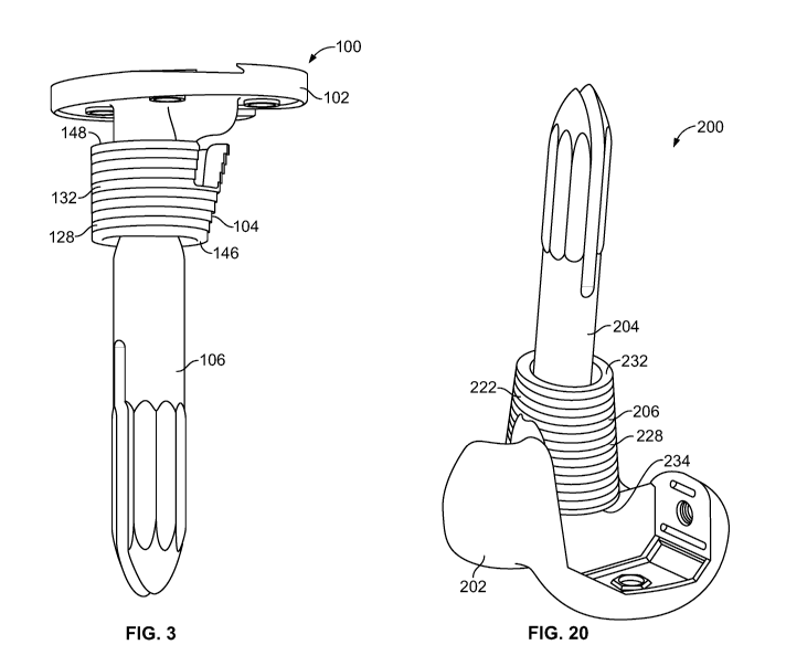
[00010] Figure 3 illustrates an isometric view of the tibial implant
device shown in Figure
1.
[00011] Figure 4 illustrates an isometric view of a tibial implant device
having an
offset/angled coupler.
[00012] Figures 5A and 5C-5J illustrate, respectively, an anterior view of
a proximal tibia
bone and transverse slice views of the proximal tibia bone, as taken from
identified line 5C-5C
through identified line 5J-5J.
[00013] Figure 5B illustrates a medial-lateral view of the proximal tibia
bone illustrated in
Figure 5A.
[00014] Figure 6 illustrates a perspective view of a symmetrical tibial
augment according
to an embodiment of the present application.
[00015] Figure 7 illustrates a front side view of the symmetrical tibial
augment illustrated
in Figure 6.
[00016] Figure 8 illustrates a right side view of the symmetrical tibial
augment illustrated
in Figure 6.
[00017] Figure 9 illustrates a top view of the symmetrical tibial augment
illustrated in
Figure 6.
[00018] Figure 10 illustrates a bottom view of the symmetrical tibial
augment illustrated in
Figure 6.
[00019] Figures 11A-11I illustrate, respectively, the anterior view of the
proximal tibia
bone and slice views from Figures 5A-5J, but with an exemplary symmetrical
tibial augment of
the present application implanted in the intramedullary canal.
[00020] Figure 12A illustrates an anterior view of a proximal tibia bone
with an
exemplary symmetrical tibial augment of the present application implanted in
the intramedullary
canal.
[00021] Figure 12B illustrates a medial-lateral view of the proximal tibia
bone that is
illustrated in Figure 12A.
[00022] Figure 12C illustrates a slice view of the proximal tibia bone and
exemplary
symmetrical tibial augment along line 12C-12C from Figure 12A.
[00023] Figure 12D illustrates a slice view of the proximal tibia bone and
exemplary
symmetrical tibial augment along line 12D-12D from Figure 12A.
3
CA 02984802 2017-11-01
WO 2016/183446 PCT/US2016/032361
[00024] Figure 13 illustrates a top view of an asymmetrical tibial augment
according to an
illustrated embodiment of the present application.
[00025] Figure 14 illustrates a bottom view of the asymmetrical tibial
illustrated in Figure
13.
[00026] Figure 15 illustrates a front view of the asymmetrical tibial
illustrated in Figure
13.
[00027] Figure 16 illustrates a right side view of the asymmetrical tibial
illustrated in
Figure 13.
[00028] Figures 17A and 17C-17J illustrate, respectively, an anterior view
and associated
slice views of a proximal tibia bone with an exemplary asymmetrical tibial
augment of the
present application implanted in the intramedullary canal.
[00029] Figure 17B illustrates a medial-lateral view of the proximal tibia
bone that is
illustrated in Figure 17A.
[00030] Figure 18 illustrates a posterior-anterior view of a femoral
implant device having
a femoral articular component, an intramedullary stem, and a femoral augment
according to an
illustrated embodiment of the present application.
[00031] Figure 19 illustrates a medial-lateral view of the femoral implant
device shown in
Figure 18, which is depicted as further including distal and posterior
augments.
[00032] Figure 20 illustrates an isometric view of the femoral implant
device shown in
Figure 18.
[00033] Figure 21 illustrates an isometric view of the femoral implant
having an
offset/angled coupler.
[00034] Figure 22 illustrates a distal end view of a fully contained
femoral augment
according to an illustrated embodiment of the present application.
[00035] Figure 23 illustrates a proximal end view of the fully contained
femoral augment
according to an illustrated embodiment of the present application.
[00036] Figure 24 illustrates a posterior side view of the fully contained
femoral augment
illustrated in Figures 22 and 23.
[00037] Figure 25 illustrates a medial side view of the fully contained
femoral augment
illustrated in Figures 22 and 23.
4
CA 02984802 2017-11-01
WO 2016/183446 PCT/US2016/032361
[00038]
Figure 26 illustrates an anterior side view of the fully contained femoral
augment
illustrated in Figures 22 and 23.
[00039]
Figure 27 illustrates a distal end view of a femoral augment structured to
accommodate partial containment according to an illustrated embodiment of the
present
application.
[00040]
Figure 28 illustrates a proximal end view of a femoral augment structured to
accommodate partial containment according to an illustrated embodiment of the
present
application.
[00041]
Figure 29 illustrates a posterior side view of the femoral augment illustrated
in
Figures 27 and 28.
[00042]
Figure 30 illustrates a medial side view of the femoral augment illustrated in
Figures 27 and 28.
[00043]
Figure 31 illustrates an anterior side view of the femoral augment illustrated
in
Figures 27 and 28.
[00044]
The foregoing summary, as well as the following detailed description of
certain
embodiments of the present application, will be better understood when read in
conjunction with
the appended drawings in which like reference numbers indicate like features,
components and
method steps. For the purpose of illustrating the invention, there is shown in
the drawings,
certain embodiments. It should be understood, however, that the present
invention is not limited
to the arrangements and instrumentalities shown in the attached drawings.
DESCRIPTION OF THE ILLUSTRATED EMBODIMENTS
[00045]
Certain terminology is used in the foregoing description for convenience and
is
not intended to be limiting. Words such as "upper," "lower," "top," "bottom,"
"first," and
"second" designate directions in the drawings to which reference is made. This
terminology
includes the words specifically noted above, derivatives thereof, and words of
similar import.
Additionally, the words "a" and "one" are defined as including one or more of
the referenced
item unless specifically noted. The phrase "at least one of' followed by a
list of two or more
items, such as "A, B or C," means any individual one of A, B or C, as well as
any combination
thereof
CA 02984802 2017-11-01
WO 2016/183446 PCT/US2016/032361
[00046] Figures 1-4 illustrate anterior-posterior, medial-lateral, and
isometric views,
respectively, of an exemplary tibial implant device 100. In the depicted
embodiment, the tibial
implant device 100 is a tibial articular assembly that includes a tibial
(articular) baseplate 102, a
tibial augment 104, and a stem 106. The stem 106, which can extend along a
central stem axis
108, can be directly or indirectly coupled to the tibial baseplate 102, such
as, for example,
coupled to a tray stem 110. As illustrated in Figure 4, according to certain
embodiments, the
tibial implant device 100 can include an offset/angled coupler 112, which can
offset at least the
central stem axis 108 relative to a central tray stem axis 114 of the tray
stem 110. The tibial
implant device 100 can also include other components, such as, for example,
other
intramedullary stems and other augments that can be assembled to the tibial
implant device 100.
[00047] The depicted tibial implant device 100 is structured to be
cemented into and
through the tibial augment 104 and onto a prepared proximal tibia of a
patient. Further, while
Figures 1-4 illustrate the tibial augment 104 positioned on or about a tibial
implant device 100 in
a non-implanted state or condition, the tibial augment 104 can be implanted in
a bone of the
patient prior to implantation of the remainder of the tibial implant device
100. Thus, an inner
region 144 of the tibial augment 104 can be sized to receive passage and/or
placement of at least
a portion of the stem 106 and/or other components of the tibial implant device
100, including, for
example, the offset/angled coupler 112 and/or the tray stem 110, during
implantation of the tibial
implant device 100 in a patient.
[00048] Figures 5A and 5B illustrate anterior and medial-lateral views,
respectively, of a
proximal tibia bone 116. Figure 5A also includes transverse slice views of the
proximal tibia
bone 116, as taken from identified lines 5C-5C through identified line 5J-5J
(Figures 5C-5J).
The proximal tibia bone 116 is oriented in Figure 5A such that the bone 116
generally tapers
inwardly for each sequential transverse slice view, thereby also reducing the
size of the
intramedullary canal 118. Further, as depicted in each of slices 5E-5E through
5J-5J (Figures
5E-5J) of Figure 5A, the cortical shape of the intramedullary canal 118 can be
generally defined
by an inner wall 120 of the proximal tibia bone 116 that comprises, at least
in part, an anterior-
medial wall 122, a posterior curvature wall 124, and an anterior-lateral wall
126, the anterior-
lateral wall 126 and the posterior curvature wall 124 being separated from
each other, at least in
part, by the anterior-medial wall 122.
6
CA 02984802 2017-11-01
WO 2016/183446 PCT/US2016/032361
[00049] Figures 6-10 illustrate an example of a symmetrical tibial augment
104 according
to an illustrated embodiment of the present application. A variety of
different augments can be
used for the tibial augment 104, including, for example, a cone or sleeve
augment, among other
augments. Further, the tibial augment 104, and more specifically an augment
wall 128, can have
a variety of shapes and sizes, and can have a symmetrical or asymmetrical
configuration. For
example, as illustrated by at least Figures 9 and 10, according to certain
embodiments, the
augment wall 128 of the tibial augment 104 can be generally symmetrical about
a midline 134
that is generally perpendicular to a central longitudinal axis 136 of the
tibial augment 104. Thus,
as shown in Figure 9, according to the illustrate embodiment, the midline 134
can generally
divide the tibial augment 104 into generally symmetrical first and second
sides 138a, 138b. The
augment wall 128 can further include at least one opening 140a, 140b that is
configured to
accommodate placement of a component of the tibial implant device 100. For
example,
according to the embodiment illustrated in Figures 6-10, the tibial augment
104 can include two
opening 140a, 140b that are sized to accommodate at least the passage and/or
placement of at
least a portion of the keel(s) 142a, 142b of the tibial baseplate 102 about or
through the
opening(s) 140a, 140b. Further, while in the illustrated embodiment the
augment wall 128 at the
first and second sides 138a, 138b of the tibial augment 104 are depicted as
having an opening
140a, 140b, other embodiments can be generally symmetrical with the exception
that the
augment wall 128 at one of the first and second sides 138a, 138b can contain
an opening 140a,
140b, while such an opening 140a, 140b is not present at the other of the
first and second sides
138a, 138b.
[00050] The augment wall 128 includes an inner portion 130 and an outer
portion 132.
The inner portion 130 of the augment wall 128 can generally define an inner
region 144 of the
tibial augment 104. At least a portion of the inner region 144 can extend
between a distal end
146 and a proximal end 148 of the tibial augment 104. Additionally, as
discussed above, the
inner region 144 can be sized to receive placement of at least one or more
components of the
tibial augment 104, such as, for example, the stem 106, offset/angled coupler
112, and/or tray
stem 110 of the tibial baseplate 102, among other components. Additionally,
while the surface
of the outer portion 132 of the augment wall 128 in the illustrated embodiment
has a step
appearance or configuration, a variety of other surfaces or surface shapes can
also be employed.
7
CA 02984802 2017-11-01
WO 2016/183446 PCT/US2016/032361
[00051] The outer portion 132 of the augment wall 128 is shaped to
generally fit the
cortical shape of a proximal tibia, and more specifically, a portion of the
intramedullary canal of
the tibia. According to certain embodiments, the outer portion 132 of the
augment wall 128 of
the tibial augment 104 can be configured such that at least the distal end
146, or diaphyseal end,
of the tibial augment 104 conforms to the general shape of the metaphyseal-
diaphyseal junction
of the tibia bone 116, and at least the proximal end 148 of the tibial augment
104 conforms to the
general shape or profile of the metaphyseal region of the tibial bone 116.
According to other
embodiments, the distal end 146 and/or proximal end 148 can be shaped to
provide other cross-
sectional shapes that facilitate the ability of the tibial augment 104 to
conform to the size and/or
shape of at least a portion of the intramedullary canal 118 of the tibia bone
116. Such
conforming may not be limited to the physical shape(s) of each section of the
outer portion 132
of the augment 104 mating or matching the shape of the adjacent portion of the
inner wall 120 of
the intramedullary canal 118, but instead can include being shaped to operably
contact an
adjacent portion of the inner wall 120 of the intramedullary canal 118 while a
central
longitudinal axis 136 of the tibial augment 104 is aligned with, or at a
selected position away
from, a reference axis, including, for example, a longitudinal axis of the
intramedullary canal
118, the central stem axis 108, and/or the central tray stem axis 114, among
other reference axes.
Additionally, according to certain embodiments, the portion of the tibial
augment 104 that is
shaped to generally conform, or fit, to the shape or profile of the
metaphyseal region can be
located at distance away, in the metaphyseal direction, from the portion of
the tibial augment 104
that conforms to the general shape or profile of the metaphyseal-diaphyseal
junction that is about
the same as the distance between the metaphyseal region and metaphyseal-
diaphyseal junction of
the tibia bone 116.
[00052] Shaping the tibial augment 104 to generally conform to, or
accommodate,
changes and/or variances in the shape of the intramedullary canal 118 of the
tibia bone 116, can
prevent or minimize the extent to which the tibial augment 104 is subjected to
unequal loading
conditions. Further, by shaping different portions or areas of the tibial
augment 104, as well as
other augments herein, to generally conform to or otherwise accommodate the
shape of at least
an adjacent inner wall of the associated bone canal or cavity, the generally
anatomically shaped
augments discussed herein, including the tibial augment 104, 104', and the
below-discussed
femoral augment 206, 206', can reduce the impact forces on the corresponding
articular implant-
8
CA 02984802 2017-11-01
WO 2016/183446 PCT/US2016/032361
bone interface by distributing such forces or loads over a relatively larger
surface area. More
specifically, for example, such conforming configurations of the augments 104,
104', 206, 206'
can improve resistance to torsional stress by equally distributing such forces
circumferentially.
[00053] Further, such variations among and/or along at least the augment
wall 128 of the
tibial augment 104 can improve flexibility in the placement of the tibial
augment 104, and thus
reduce or minimize the tibial augment 104 from hindering the ability to
position an associated
articular component relative to a joint line, while also not hindering joint
balance (flexion-
extension balance) and rotation of each component relative to the patella-
femoral joint.
[00054] To generally accommodate the cortical shape(s) of the
intramedullary canal 118
of the tibia bone 116, including, for example, the shape at both the
metaphyseal-diaphyseal
junction and at metaphyseal region of the tibial bone 116, as well as the
shapes therebetween,
different areas or sides of the outer portion 132 of the augment wall 128 can
have different
shapes. Additionally, the shapes along such different areas or sides of the
outer portion 132 of
the augment wall 128 can also vary between the distal and proximal ends 146,
148 of the tibial
augment 104. Such variances or inconsistencies among and/or along the sides or
areas of the
tibial augment 104 can preclude the augment wall 128 of the tibial augment 104
from having a
generally uniform cylindrical or conical shape.
[00055] Referencing Figures 9-10, according to certain embodiments, the
augment wall
128 of the tibial augment 104 can include a first, or posterior curvature,
portion 150 and a
second, or anterior-medial, portion 152 that are separated from each other by
at least a
transversal axis 154 that is at least perpendicular to the midline 134.
Additionally, in the
illustrated embodiment, at least a portion of posterior curvature portion 150
has a shape that is
different than a corresponding portion of the anterior-medial portion 152. For
example, as
shown by the top view of the proximal end 148 of the tibial augment 104 in
Figure 9, the
posterior curvature portion 150 can include a generally flat section 156a that
transitions into
rounded end sections 156b, 156c, while the anterior-medial portion 152 can
include a rounded
section 158a that transitions into generally flat sections 158b, 158c. Such
differences in shapes,
and the resulting dissimilar profiles, are depicted by at least Figure 8 and
10 at least in the region
around the proximal end 148 of the tibial augment 104. As demonstrated by at
least slices 11D-
11D and 11E-11E from Figure 11A, such differences in the shapes of the
posterior curvature and
anterior-medial portions 150, 152 of the tibial augment 104 can facilitate the
ability of the tibial
9
CA 02984802 2017-11-01
WO 2016/183446 PCT/US2016/032361
augment 104 to generally conform to the shape of at least the adjacent
posterior curvature wall
126 and the anterior-medial wall 122, respectively, of the inner wall 120 of
the intramedullary
canal 118. Additionally, as indicated by slice 11D-11D (Figure 11D) from
Figure 11A, a portion
of the anterior-medial portion 152 and/or the posterior curvature portion 150
can contact other
portions of the inner wall 120 of the intramedullary canal 118, including, for
example, the lateral
wall 166.
[00056] The different shapes of the posterior curvature and anterior-
medial portions 150,
152 can alter or vary between the distal and proximal ends 146, 148 along the
augment wall 128
so that the outer position 132 of the augment 104 generally conforms to
changes in shape along
the inner wall 120 of the intramedullary canal 118 of the tibia bone 116, as
depicted each of slice
views 5C-5C through 5J-5J from Figure 5A and 11B-11B through 111-111 (Figures
11B-11I)
from Figure 11A. According to the illustrated embodiment shown in at least
Figures 6-11, such
changes in shape in the inner wall 120 of the intramedullary canal 118, and
corresponding
changes in shape along at least the posterior curvature and anterior-medial
portions 150, 152 of
the augment wall 128, can result in the posterior curvature and anterior-
medial portions 150, 152
generally having similar shapes at the distal end 146 of the tibial augment
104, as shown, for
example, by Figure 10 and slice E-E in Figure 11. Such similarities in the
shape of the posterior
curvature and anterior-medial portions 150, 152 can, for example, provide the
augment wall 128
with a generally circular, semi-circular, or slightly oval shape at the distal
end 146 of the tibial
augment 104. However, again, the particular shape(s) of the augment wall 128
at the distal end
146 of the tibial augment 104 can be configured or selected to generally
conform to the
metaphyseal-diaphyseal junction of the tibia bone 116.
[00057] Figures 12A-12D further illustrate the symmetrical tibial augment
104 that is
shaped to conform to the shape of the cortical shape of the bone 116, and more
specifically, in
the illustrated embodiment, to the adjacent shape of the intramedullary canal
118. For example,
as shown in Figure 12C, the proximal end 148 of the tibial augment 104 is
shaped to generally
conform to the general shape or profile of adjacent portions of the
metaphyseal region of the
tibial bone 116. Further, as shown in Figures 12C and 12D, the region around
the anterior-lateral
wall 126, and moreover, in the region of the tibial tubercle 160, can often be
covered, at least in
part, by the anterior aspect of a transversely resected portion of the
proximal tibia bone 116.
Such coverage can prevent the tibial augment 104 from having direct access
inferior-superior to
CA 02984802 2017-11-01
WO 2016/183446 PCT/US2016/032361
the cancellous area 162 that can be present behind the tibial tubercle 160.
Yet, without direct
access or special instrumentation, preparation, as well as placement of the
tibial augments that
lack the anatomical shapes disclosed herein, at such a location can be
difficult.
[00058]
Referencing Figures 13-16, in at least certain instances, patients can have an
abnormality in the shape and/or size of the intramedullary canal 118 and/or
can require enhanced
support from a tibial augment 104'. In such situations, the tibial augment
104' can have an
asymmetrical configuration about the midline 134, as shown in at least Figures
13 and 14. Such
an asymmetrical configuration can increase a thickness of the augment wall 128
between at least
certain sections of the inner and outer portions 130, 132 of the augment wall
128. For example,
compared to slices 11D-11D, 11E-11E, and 11F-11F (Figures 11D-11F) from Figure
11A, the
asymmetrical tibial augment 104' shown in slice views from 17E-17E, 17F-17F,
and 17G-17G
(Figures 17E-17G) from Figure 17A has and increased thickness in the augment
wall 128 at least
in the vicinity of the lateral wall 166 portion of the inner wall 120 of the
intramedullary canal
118.
However, according to certain embodiments, such increases in the thickness of
the
augment wall 128 and/or increases of the augment wall 128 in certain locations
can be limited
due to the previously discussed limitations associated with the cancellous
area 162 behind the
tibial tubercle 160.
[00059]
Figures 18-20 illustrate posterior-anterior, medial-lateral, and isometric
views,
respectively, of a femoral implant device 200. According to an illustrated
embodiment of the
present application, the femoral implant device 200 includes a femoral
articular component 202,
an intramedullary stem 204, and a femoral augment 206. Additionally, as shown
in Figure 19,
according to certain embodiments, the femoral implant device 200 can also
include a distal
augment 208 and/or a posterior augment 210. The stem 204, which can extend
along a central
stem axis 212, can be directly or indirectly coupled to the femoral articular
component 202, such
as, for example, coupled to a component stem 220 of the femoral articular
component 202, as
shown in Figure 21. As illustrated in Figure 21, according to certain
embodiments, the femoral
implant device 200 can include an offset/angled coupler 216, which can offset
at least the central
stem axis 212 relative to a component stem axis 218 of the component stem 220.
[00060]
The depicted femoral implant device 200 is structured to be cemented into and
through the femoral augment 206 and onto a prepared distal femur of a patient.
Further, while
Figures 18-21 illustrate the femoral augment 206 positioned on or about a
femoral implant
11
CA 02984802 2017-11-01
WO 2016/183446 PCT/US2016/032361
device 200 in a non-implanted state or condition, the femoral augment 206 can
be implanted in a
bone of the patient prior to implantation of the remainder of the femoral
implant device 200.
Thus, an inner region of the femoral augment 206 can be sized to receive
passage and/or
placement of at least a portion of the stem 204 and/or other components of the
femoral implant
device 200, including, for example, the offset/angled coupler 216 and/or the
component stem
220, during implantation of the femoral implant device 200 in a patient.
[00061] Figures 23-26 illustrate an example of a fully contained femoral
augment 206
according to an illustrated embodiment of the present application. A variety
of different
augments can be used for the femoral augment 206, including, for example, a
cone or sleeve
augment, among other augments. Further, the femoral augment 206 can have a
variety of shapes
and sizes. The femoral augment 206 can include an augment wall 222 that
extends about a
central axis 224 of the femoral augment 206. The augment wall 222 has an inner
portion 226
and an outer portion 228. The inner portion 226 of the augment wall 222 can
generally define an
inner region 230 of the femoral augment 206. At least a portion of the inner
region 230 can
extend between a distal end 232 and a proximal end 234 of the femoral augment
206. The inner
region 230 can be sized to receive placement of at least one or more
components of the femoral
augment 206, such as, for example, the stem 204, offset/angled coupler 216,
and/or component
stem 220 of the femoral articular component 202, among other components. For
example,
according to certain embodiments, the inner region 230 is sized to receive
placement of at least
the junction between the stem 204 and the component stem 220.
[00062] The outer portion 228 of the augment wall 222 can be shaped to
generally fit the
cortical shape of a distal femur, and more specifically, of a portion of the
intramedullary canal of
the femur. Thus, according to certain embodiments, a diaphyseal, or distal end
232, of the
femoral augment 206 can be shaped to generally conform to the general shape of
the
metaphyseal-diaphyseal junction. The opposing proximal end 234 of the femoral
augment 206
can be configured to conform to the general shape or profile of the
metaphyseal region of the
femoral bone. According to other embodiments, the distal end 232 and/or
proximal end 234 can
be shaped to provide other cross-sectional shapes that facilitate the ability
of the femoral
augment 206 to conform to the size and/or shape of at least a portion of the
intramedullary canal
of the femur. Such conforming may not be limited to the physical shape(s) of
each section of the
outer portion 228 of the augment mating or matching the shape of the adjacent
portion of the
12
CA 02984802 2017-11-01
WO 2016/183446 PCT/US2016/032361
inner wall of the intramedullary canal of the femoral bone, but instead can
include being shaped
to operably contact an adjacent portion of the inner wall of the
intramedullary canal while a
central axis 224 of the femoral augment 206 is aligned with, or at a selected
position away from,
a reference axis, including, for example, a longitudinal axis of the
intramedullary canal of the
femur, the central stem axis 212, and/or the component stem axis 218, among
other reference
axes. Additionally, the portion of the femoral augment 206 that is shaped to
generally conform
to the shape or profile of the metaphyseal region can be located at distance
away, generally in the
distal direction, from the portion of the femoral augment 206 that conforms to
the general shape
or profile of the metaphyseal-diaphyseal junction that is about the same as
the distance between
the metaphyseal region and metaphyseal-diaphyseal junction of the tibia.
[00063]
Similar to the tibial augment 104, 104', shaping the femoral augment 206 to
generally conform to, or accommodate, changes and/or variances in the shape of
the
intramedullary canal of the femur can prevent or minimize the extent to which
the femoral
augment 206 is subjected to unequal loading conditions. Further, again, by
shaping different
portions or areas of the femoral augment 206, as well as other augments
herein, to generally
conform to or otherwise accommodate the shape of at least an adjacent inner
wall of the
associated bone canal or cavity, the generally anatomically shaped augments
104, 104', 206,
206', discussed herein can reduce the impact forces on the corresponding
articular implant-bone
interface by distributing such forces or loads over a relatively larger
surface area. More
specifically, for example, such conforming configurations of the augments 104,
104', 206, 206'
can improve resistance to torsional stress by equally distributing such forces
circumferentially.
[00064]
To generally accommodate the cortical shape(s) of the medullary canal of the
femur, including, for example, the shape at both the metaphyseal-diaphyseal
junction and at
metaphyseal region of the femur, as well as the shape therebetween, different
areas or sides of
the outer portion 228 of the augment wall 222 can have different shapes.
Additionally, the
shapes along such different areas or sides of the outer portion 228 of the
augment wall 222 can
also vary between the distal and proximal ends 232, 234 of the femoral augment
206. Such
variances or inconsistencies among and/or along the sides or areas of the
femoral augment 206
can preclude the augment wall 222 of the femoral augment 206 from having a
generally uniform
cylindrical or conical shape.
13
CA 02984802 2017-11-01
WO 2016/183446 PCT/US2016/032361
[00065] Referencing Figures 22-26, according to certain embodiments, the
outer portion
228 of the augment wall 222 can include a recess or relief 236. As shown by at
least Figure 26,
according to the illustrated embodiment, the relief 236 can extend along a
portion of the augment
wall 222, such as, for example, extending from the proximal end 234 to a
region generally
adjacent to the distal end 232 of the femoral augment 206. However, according
to other
embodiments, the relief 236 can extend between, and through, the proximal end
234 and/or the
distal end 232 of the femoral augment 206. In the illustrated embodiment, the
relief 236 can
have one or more sidewalls 238 and a base wall 240. For example, as shown in
Figure 23, the
one or more sidewalls 238 can include a first sidewall 238a and a second
sidewall 238b. The
first and second sidewalls 238a, 238b can be angled such that the first and
second sidewalls
238a, 238b converge toward each other from generally opposite directions
and/or angles. For
example, in the illustrated embodiment, the first and second sidewalls 238a,
238b can each
extend from opposing first ends 242a, 242b, and converge toward each other so
as to intersect or
be generally in proximity to each other at second ends 244a, 244b of the first
and second
sidewalls 238a, 238b. Further, in the illustrated embodiment, the second ends
244a, 244b can be
adjacent to, or generally form, an augment flange 246 that projects away from
the first and
second sidewalls 238a, 238b.
[00066] As indicated by Figure 25, according to certain embodiments, the
first and second
sidewalls 238a, 238b can also be angled or tapered, and thus non-parallel to a
longitudinal
central axis 224 of the femoral augment 206. For example, as shown in the
embodiment
depicted in Figure 26, the portion of the first and second sidewalls 238a,
238b at the proximal
end 234 of the augment wall 222 can be separated from the central axis 224 of
the femoral
augment 206 by a distance that is smaller than the distance between the
central axis 224 and the
vicinity of the intersection of the first and second sidewalls 238a, 238b and
the base wall 240.
However, according to other embodiments, the first and second sidewalls 238a,
238b can be
generally parallel to the central axis 224 of the femoral augment 206.
[00067] As shown by at least Figures 19 and 20, according to the
illustrated embodiment,
the relief 236 can be shaped such that, when the femoral augment 206 is
operably positioned on
the femoral implant device 200, the relief 236 is generally parallel to the
bone facing side of the
anterior flange 248 of the femoral implant device 200. Such a shape can at
least assist in
adjustable rotational displacement of the femoral augment 206 relative to the
femoral implant
14
CA 02984802 2017-11-01
WO 2016/183446 PCT/US2016/032361
device 200, and more particularly, of the anterior flange 248 (Figure 19)
relative to the femoral
augment 206. Such rotational adjustment can also be facilitated by the angular
orientation of the
first and/or second sidewalls 238a, 238b of the augment wall 222. For example,
as indicated by
the exemplary femoral augments 206, 206' shown in Figures 23 and Figure 28,
the first sidewall
238a can be oriented to facilitate that ability to adjust the angular position
of the femoral
augment 206 relative to other components of the femoral implant device 200,
such as, for
example, relative to the anterior flange 248, by about 20 degrees, among other
degrees of
rotational freedom. Thus, for example, such rotational displacement can, when
the femoral
implant device 200 is implanted in a patient, allow for selective adjustment
in the distance
between the first end 242a of the first sidewall 238a and the anterior flange
248.
[00068] The rotational freedom provided by incorporation of the relief 236
and the
associated adjustment in the position of the femoral augment 206 relative to
the anterior flange
248 can assist in the femoral augment being adapted to accommodate rotational
variation in the
geometry of the intramedullary canal of the femur. Moreover, the relief 236
can assist in
enhancing the flexibility as to the orientation at which the femoral augment
206 can be implanted
in the intramedullary canal so as to further enhance the ability of the
femoral augment 206 to
conform or otherwise accommodate the particular shape of the intramedullary
canal while also
minimizing or preventing the position of the femoral augment 206 from impeding
the positioning
or operation of other components of the femoral implant device 200. For
example, the rotational
freedom of the femoral augment 206 that is provided by, at least in part, the
inclusion of the
relief 236 can enhance the ability to position the femoral augment 206 to
accommodate for
rotational variation in the shape of the intramedullary canal while also not
preventing the femoral
implant device 200, such a femoral articular component, from being positioned
at a particular
transverse rotational location.
[00069] Figures 27-31 illustrate a femoral augment 206' that is adapted to
accommodate
larger components, or collections of components, in the inner region 230' of
the augment 206'.
For example, the femoral augment 206' depicted in Figures 27-31 can be adapted
to receive in
the inner region one or more of the stem 204, component stem 220, and/or the
offset/angled
coupler 216, among other components. According to such an embodiment, the
relief 236 or
augment flange 246, if any, can include one or more tear lines or relief areas
250 that are adapted
to open, break through, or tear the augment wall 222, or otherwise relieve at
least a portion of the
CA 02984802 2017-11-01
WO 2016/183446 PCT/US2016/032361
augment 206'. Thus, in certain situations, the formation of an opening along
one or more of the
tear lines or relief areas 250 can provide access to additional space so as to
prevent the inner
region 230' from restricting or impeding positioning of components of the
femoral implant
device 200 relative to the femoral augment 206. Thus, unlike the fully
contained inner region
230 of the femoral augment 206 shown in Figures 23-24, the tear lines or
relief areas 250 can
allow for the femoral augment 206' to transition from being fully contained to
partially
contained, which can occur, for example, upon the formation of openings or
breakage along the
tear line or relief areas 250.
[00070] Additionally, in the illustrated embodiments of the femoral
augments 206, 206'
shown in at least Figures 23-31, at least some sides of the of the femoral
augments 206, 206' can
have different shapes and/or configurations. Further, similar to the tibial
augments 104, 104'
discussed above, the shape or configurations of those sides can alter, and can
alter differently,
between the proximal and distal ends 232, 234 of the femoral augments 206,
206'. For example,
referencing the top views of Figures 23 and 28, at the distal ends 232 of the
femoral augments
206, 206', the femoral augments 206, 206' can have be generally egg-shape,
which can assist in
providing different shaped and sized profiles along the augment wall 222 of
the femoral
augments 206, 206', and shown by a comparison of Figures 24 and 26 with Figure
25, and a
similar comparison of Figures 29 and 31 with Figure 30. Further, while the
distal end 234 in the
illustrated embodiments is shown as being generally egg-shaped, as shown in
the Figures 22 and
27, the proximal ends 232 of the femoral augments 206, 206' can be generally
circular. Thus, the
transitions between, and the associated shapes, of the proximal and distal
ends 232, 234 can
preclude the femoral augments 206, 206' from having a generally uniform
cylindrical or conical
shape. Instead, as previously mentioned, such variations in shapes along
different portions of the
femoral augment 206, 206' can be adapted to enhance the ability of the femoral
augment 206,
206' to generally conform to the shape of adjacent portions of the
intramedullary canal of the
femur.
[00071] While the invention has been described in connection with what is
presently
considered to be the most practical and preferred embodiment, it is to be
understood that the
invention is not to be limited to the disclosed embodiment(s), but on the
contrary, is intended to
cover various modifications and equivalent arrangements included within the
spirit and scope of
the appended claims, which scope is to be accorded the broadest interpretation
so as to
16
CA 02984802 2017-11-01
WO 2016/183446 PCT/US2016/032361
encompass all such modifications and equivalent structures as permitted under
the law.
Furthermore it should be understood that while the use of the word preferable,
preferably, or
preferred in the description above indicates that feature so described may be
more desirable, it
nonetheless may not be necessary and any embodiment lacking the same may be
contemplated as
within the scope of the invention, that scope being defined by the claims that
follow. In reading
the claims it is intended that when words such as "a," "an," "at least one"
and "at least a portion"
are used, there is no intention to limit the claim to only one item unless
specifically stated to the
contrary in the claim. Further, when the language "at least a portion" and/or
"a portion" is used
the item may include a portion and/or the entire item unless specifically
stated to the contrary.
17