Note: Descriptions are shown in the official language in which they were submitted.
CA 02985182 2017-11-06
WO 2016/178174 PC171132016/052566
1
METHOD FOR MANUFACTURING BONE IMPLANT AND BONE IMPLANT
Technical Field
The invention regards a method of producing bone implants having a character
of
supporting structure at least partially coated with a synthetic hydroxyapatite
and
bone implants for use in orthopaedic surgery, trauma surgery (traumatology),
regenerative implantology, which facilitate or accelerate the regeneration of
bone
tissue.
Background Art
In medicine, particularly in orthopaedics, dentistry and traumatology, and in
the
treatment of bone defects caused by the removal of the tumour, implants, also
known as scaffolds for bone tissue regeneration, are used in order to induce
or
accelerate regeneration of the bone tissues by the organism; tissue that was
lost
as a result of trauma, surgery of cancer removal, orthopaedic surgery, dental
surgery, tooth extraction, other causes or improvement of aesthetics. For the
production of such implants various kinds of synthetic biomaterials (metals,
ceramics, synthetics - polymers, composites) as well as natural materials are
used. They must meet a number of criteria, including no toxicity to the body,
proper filling of the missing bone volume and appropriate mechanical
properties.
In particular, a very valuable feature of such materials is their
bioresorption.
Presently, there are many treatment methods for small defects in bones, but
still
no solution for large tissue loss (so-called critical loss) is known. To fill
the cavity
and facilitate active bone regeneration an scaffold should be created
(implant),
both filling the space of bone loss and transferring mechanical stresses,
enabling
the bone tissue to gradually fill the empty space. The most preferable
solution is to
have an implant undergoing resorption over time, thus enabling the entire
space to
be filled with new bone tissue. Another criterion is ensuring the flow of
nutrients
CA 02985182 2017-11-06
WO 2016/178174 PCT/1B2016/052566
2
and cells in the bloodstream trough the scaffolds material. Also some
modifications
to the implant surfaces are used to accelerate the regeneration of substantial
loss
of the tissue.
In the effectiveness of the bone implant use the surface layers of an implant
containing calcium phosphates play an important role. The natural bone in up
to
70% (depending on the type of bone tissue) consists of an inorganic matter,
largely composed of hydroxyapatite deposited in a form of crystals.
Hydroxyapatite
Ca10(PO4)5(OH)2 is one of the major minerals in the human body. It is
responsible
for the hardness and strength of bones and teeth. In the human body hydroxyapa-
tite occurs in a form of crystals with lamellar structure, 2 nm thick, 25 nm
wide, and
50 nm long. [M. Sadat-Shojai, M.-T. Khorasani, E. Dinpanah-Khoshdargi, A.
Jamshidi õSynthesis methods for nanosized hydroxyapatite with diverse structu-
res", Acta Biomaterialia, Vol. 9, 2013, pp. 7591-7621). Hydroxyapatite is a
material
widely used in orthopaedics, maxillofacial and implant surgeries, inter alia,
for the
production of layers of implants aimed at bone regeneration.
New potential for facilitation or accelerating the regeneration of bone tissue
lays in
the nanotechnology, the field in which properties of the known materials
change in
a surprising manner along with the change of the material size, ranging from 1
to
100 nm.
Publication W02011/022642 discloses coating of bone implants with a porous
layer composed of hydroxyapatite and zinc dioxide, having a size exceeding
50 nm.
Publication W02013/112743 describes an implant with islands of hydroxyapatite
having an average thickness of 45-70 nm, albeit an information about the size
of
hydroxyapatite nano-particles and the molar ratio of calcium to phosphorus has
been given.
In US6129928 patent specification have been presented layers of calcium
phosphate of undetermined nano-structure and thickness of 2-30 pm, applied to
metallic implants. The disadvantages of the method for covering implants
disclo-
sed there are complex multi-stage manufacturing process and a considerable
thickness of the layers obtained.
CA 02985182 2017-11-06
WO 20161178174 PC171B2016/052566
3
In US5441536 patent specification is disclosed a method for producing hydroxy-
apatite layers on medical implants by hydrothermal treatment in temperatures
higher than 100 C, however the resulting structure of layers obtained is not
specified in this patent. The coating obtained by this method is characterized
by
the average thickness of 50 pm and tendency for delaminating when under
stress.
Publication W02002/078759 describes a bioactive layer consisting of the
various
phases of calcium-phosphate. The composition of this layer consists of
amorphous
and nano-crystalline calcium phosphates forming a porous layer, having a
thickness of 0.1 to 50 pm and a density of pores ranging from 104 to 108/mm2.
Such a layer may be a source of calcium ions needed for the active bone
formation. The ratio of calcium to phosphorus over the entire surface of the
sub-
structure ranges from 0.5 to 2. The disadvantage of the described method is
the
need for forming pores in the layer. In the publication the presence in the
layer of
nano-crystalline hydroxyapatite is mentioned, however, very important data in
terms of nanotechnology, such as the size and distribution of crystallite and
its
structure, are not given. In addition, calcium phosphate and hydroxyapatite
represent only 1 to 40% of the coating. There is also no information on the
coating
of porous scaffolds.
Publication CN101703798 discloses coating facilitated by electrostatic
discharges.
Thus obtained layer consists of a nano-particles of hydroxyapatite in an
amount
ranging from 70 to 90% and the additive of silk fibres in an amount from 10 to
30%
by weight. However, neither layer thickness nor its structure has been
specified.
Publication US2011/0281127 discloses a method of manufacturing a hydroxya-
petite layer having a thickness of 30 to 50 nm. An optimal biocompatibility,
confirmed in cellular assays, has been shown in the layers consisting of
particles
having a diameter of less than 50 nm, preferably 30 nm. However, the actual
effectiveness of such layers has not been specified.
In the review article õCalcium phosphate coatings for bio-implant
applications:
Materials, performance factors, and Methodologies" [Materials Science and Engi-
neering R 66 (2009) 1-70] S.R. Paital and N. Dahore describe various methods
for
production of layers of calcium phosphate on implants for bone regeneration,
CA 02985182 2017-11-06
WO 2016/178174 PCT/11132016/052566
4
especially metal ones, particularly made of titanium alloys. This publication
discloses, among other things, various kinds of deposition from vapour phase
of
PVD family (Physical Vapour Deposition) or CVD family (Chemical Vapour
Deposition), for example, IBAD method (Ion Beam Assisted Deposition) using ion
beam. Described there are also processes of plasma spraying (Plasma Spray
Deposition), PLD laser technologies (Pulsed Laser Deposition), electrophoretic
deposition, electrochemical surface treatment, e.g. MAO (Micro-arc oxidation),
spray deposition of atoms/ions in a magnetic field (Magnetron Sputtering
Deposition), direct laser melting method, a sol-gel method and also the
production
of solutions simulating physiological fluids (Simulated Body Fluid, SBF). A
lot of
these methods can only be used to produce layers on materials resistant to
temperatures above 200 C, thus precluding the use of this methods for most
plastics. Of above mentioned methods only PVD, sol-gel and SBF can be used at
temperatures below 200 C. In the case of the latter two methods further
activation
of the surface materials is required to enable the subsequent process of
nucleation
to run more efficiently, and to strengthen the connection between the
substructure
and hydroxyapatite
An example of implementation of the method of plasma spraying to coat the
implant made of plastic is disclosed in publication W02012/110816. This method
involves the formation of the transition layer, for example titanium one, and
then
deposition of the further layer of polymeric material or ceramic material, for
example hydroxyapatite. The substructure material must be resistant to tempe-
ratures above 200-250 C (e.g. PEEK, PAEK, polyamide). The outer layer of the
hydroxyapatite can be applied by plasma spraying or by electroplating. The
advantage of this method is the good adhesion of the layers to the
substructure,
but its disadvantage is the need for the transition metal layer and the
process
temperature greater than 200 C. No information on the structure and thickness
of
this layer has been specified.
Another method for producing a hydroxyapatite layer is called õlayer by layer
method (LBL). Using this method the composite was made, consisting of layers,
arranged in turns, of chitosan with hydroxyapatite and of polyacrylic acid
(PAA),
CA 02985182 2017-11-06
WO 2016/178174 PCT/IB2016/052566
This method is disclosed in N. Shah, J. Hong et al. õOsteophilic multilayer
coatings
for accelerated bone tissue growth" [Adv Mater. May 15, 2012; 24 (11): pp.1445-
50], wherein the further layer is applied containing growth factors, such as
rhBMP-2. The advantage of this method lays in obtaining a homogeneous layer
containing various types of active substances. The process is carried out at
room
temperature. For application on industrial scale the multiply immersing of the
material in a suspension is necessary. This extends the duration of the
process
and increases the possibility of introducing contamination. In the paper The
future
of biological coatings for orthopaedic implants" [Biomaterials Vol.34, Issue
13 April
2013, pp. 3174-31831 S .Goodman and Z. Yao reveal the problem of insufficient
mechanic resistance of such a layer in terms of its adhesion to the
substructure.
The publication of A.Oyane, C.Choong, J.Triffitt õSimple surface modification
of
poly (e caprolactone) for apatite deposition from simulated body fluid"
[Biorna-
terials, Vol. 26, Issue 15 May 2005, pp. 2407-2413] describes a method of
producing layers of 0-hydroxyapatite by its precipitation in a solution
stimulating
fluids of the human body. The material being coated is a scaffold of
polycaprola-
ctone (PCL), made using three-dimensional printing technique (Fused Deposition
Modeling, FDM). Additionally, the scaffold was immersed in SBF for 14 days,
during which calcium phosphate layer grew. The layer obtained consisted of
hydroxyapatite. A similar technique is described in the paper of T. Kokubo
"Formation of biologically active bone-like apatite on polymers and metals by
a
Biomimetic process" [Thermochimica Acta, Vol. 280-281, July 1996, pp. 479-
490].
The disadvantage of this method is a length of the process, as well as weak
binding between the polymer and the ceramics. On SEM micrographs the cracks
and defects of the layer are shown in a place where the layer detached itself
from
the substructure. Implementation of the same method discloses patent
specification US8075562. The method presented regards obtaining the layers on
the substructure by immersing the polymeric material for the coating in
solution of
simulating physiological fluids (SBF) with the addition of bone growth
factors, for
example the e-BMP-2 and a ready implant in the form of a polymer screw coated
with a layer of hydroxyapatite. The disclosed technology provides a
homogeneous
CA 02985182 2017-11-06
WO 2016/178174 PCT/1132016/052566
6
layer, which contains chemically bound hydroxyapatite and growth factors. The
drawback of this technology is the necessity of immersing the coated material
in a
number of different solutions and a long time needed for layer preparation. No
information about the structure of the nano-particles and layers has been
specified.
Publication EP2251049 discloses a method of producing on a metal substructure
a
hydroxyapatite layer, which consists of collagen, calcium phosphate (hydroxy-
apatite) and optionally some growth factors. According to this method, the
metal
substructure intended for coating is inserted into a liquid containing
collagen or is
coated with such liquid droplets. Followed by removal of excess collagen and
immersing the substructure in a metastable solution containing calcium and
phosphate ions that results in precipitation of calcium phosphates. The
disadvan-
tage of this solution is the long time needed for preparing the coating, at
least 12
hours of soaking in a solution of calcium and phosphate ions, two hours of
freezing
and lyophilisation step taking several hours. Here, too, no information about
the
structure of the layers and particles has been given.
A similar technology discloses patent specification US6280789. It presents the
production of hydroxyapatite coatings on the surface of metallic and ceramic
imp-
lants. The substructure material is dipped in a solution containing calcium,
phosphate and bicarbonate, at a pH in the range of 6.8 to 8. The solution is
heated
to a temperature between 50 and 80 C, resulting in an increase of pH and
precipi-
tation of hydroxyapatite with addition of hydrogencarbonate ions. The
precipitated
crystallites of hydroxyapatite have a length of 10 to 40 nm and a width of 3
to
nm. The advantage of this method is a short duration of the process, however a
relatively high process temperature of this method makes it unsuitable for
polymer
substructures, particularly those with low softening temperature and poor
chemical
resistance.
Technologies for producing layers using energy of ultra-sounds are also known.
Patent specification US7896539 discloses coating with drugs or polymers of
stents
(implants restoring the patency of blood vessels) using an ultrasonic nozzle
for
spraying the coating material. There is, however, no possibility of a uniform
coating
CA 02985182 2017-11-06
WO 2016/178174 PCT/1B2016/052566
7
of porous substructures, and possibility of coating with nano-particles of
hydroxy-
apatite has not been even suggested.
In publication W02007/127193 the preparation of layers on the surface of
medical
implants by electrostatic applying of the spray material is described.
However, this
methods is limited only to coating conductive materials or materials covered
with
pre-added conductive layer. In addition, it is difficult or impossible to
cover the
whole volume of the material with a small size of the pores and a complicated
geometry. Not even a suggestion of the possibility of coating with nano-
particles of
hydroxyapatite has been given.
A lot of the plastic materials used as implants for bone tissue regeneration
are
thermoplastic materials, which can be shaped with the extrusion moulding or
injec-
tion. The temperatures at which such materials can be formed are often in the
region of 100 C or even lower. For example, polycaprolactone softening point
is c.a.
60 C. For this reason, the process of applying the layer of such a material on
an
implant requires temperatures below the softening temperatures to prevent dis-
tortion or damage. Publication US2011/097957 describes a method of ultrasonic
applying of metal oxides (CuO, ZnO, MgO) on fabric, in order to impart
antibacterial
properties, while publication US2011/300767 discloses a method of ultrasonic
adhering to the fabric of the protein microspheres containing some substance,
e.g.
drug, which is then released into the environment. Both publications do not
contain
any teachings regarding covering of the bone implants with hydroxyapatite.
Publication JP2013022234 discloses a method for obtaining a hydroxyapatite
layer
on a substructure of thermoplastic material by applying on the substructure
the
hydroxyapatite particles which blend into the material after heating above the
softening temperature. Effectiveness of the coating is checked by subjecting
the
coating to ultrasound at a frequency of 38 KHz for a period of 10 minutes;
what is
worth noting is that ultrasounds used here are not used in the coating
process. No
information about the nano-structure of hydroxyapatite used has been given.
Publication KR101005499 discloses a method of surface hardening of three
dimensional stents and application of medicinal substances on their surface by
ultrasonic cavitation in a liquid in which the stent is immersed. Also in this
solution,
CA 02985182 2017-11-06
WO 2016/178174 PCT/1B2016/052566
8
there are no guidelines as to the use of ultrasound for obtaining
hydroxyapatite
layers.
The Polish patent application P.396906 discloses a synthetic nano-lamella of
hydroxyapatite with a hexagonal structure and having an average particle size
ranging from 3 to 30 nm. The molar ratio of calcium to phosphorus (Ca/P) of
this
nano-lamella ranges from 1.55 to 1.65. Disclosed nano-powder is intended for
filling undesirable cavities in bone tissue, but in the said application there
are no
guidelines as to the application of such a nano-powder in the production of
implants and implant layers.
The Polish patent application P.399701 discloses a bone implant formed of,
a compacted under high pressure, nano-powder of synthetic hydroxyapatite,
having a hexagonal structure with an average particle size from 3 to 30 nm and
a
specific surface area greater than 200 m2/g. This report does not contain any
guidance on durable coating with the powder of spatially complex bone
implants,
especially ones with high flexibility.
The publication of I. Selma et al., õFirst results of the bone tissue
morphological
evaluation after implantation of new polymer and tricalcium phosphate
scaffolds
coated with resorbable nano hydroxyapatite" [Journal of Tissue Engineering and
Regenerative Medicine 8, 409-410] discloses test results of coating porous
scaffolds with nano hydroxyapatite using ultrasounds. However, this
publication
does not disclose any details of the coating and the obtained coating
properties,
while it is well known that in nanotechnology the properties of the nano-
particles
strongly depend on their size, shape, chemical composition of molecules
attached
to their surfaces and their inner structure.
In their publication "Ultrasonic coating technique of a polymer scaffold for
bone
implant applications" [European Cells and Materials, Vol. 26, Suppl. 2, 2013,
p. 17]
A. Kedzierska et al. describe scaffolds for bone tissue regeneration made by
deposition on the polymer scaffolds the layers of nano-hydroxyapatite, with
lamellar
structure and size from 5 to 30 nm, using ultrasounds. This publication does
not
contain information regarding the chemical composition of the hydroxyapatite
used
and kinds of physical phenomena occurring during the sonication, and the
possible
CA 02985182 2017-11-06
WO 2016/178174 PCT/I132016/052566
9
impact of the structure obtained on the regeneration processes of bone tissue.
It is
very important as these phenomena, as every specialists involved in the
processes
that occur when the size of the particles of matter is less than 100 nm is
aware, are
very unpredictable and strongly depend on the size of nano-structures; as the
same,
from the chemical and physical point of view, material with the particles size
of 70
nm can have fundamentally different properties than a material with the
particles
size of 20 nm.
Disclosure of Invention
The aim of the invention is to obtain an efficient bone implant stimulating
bone
growth and the fast and simple method of manufaturing such implants.
This aim is to be achieved by the method according to the invention consising
on
depositing a synthetic hydroxyapatite on a supporting structure by immersing
the
supporting structure in a liquid being a source of this hydroxyapatite. It
characterized
in that the supporting structure of the implant is first immersed in a
suspension
consisting of a liquid phase, advantageously water, containing a disphersed
phase
of synthetic hydroxyapatite particles having an average particle size not
greater than
100 nm, advantageously not greater than 30 nm. Next, in a portion of the
suspen-
sion being in contact with the supporting structure cavitation is induced.
In one of embodiments of the method according to the invention for the prepara-
tion of the dispersed phase are used the hydroxyapatite particles containing
structural water in an amount from 2 to 6% by weight.
In another embodiment of the method according to the invention molar ratio of
calcium to phosphorus (Ca/P) of the hydroxyapatite particles is greater than
1.55
and less than 1.67.
In another embodiment of the method according to the invention the cavitation
is
induced by means of an object immersed into the suspension near the supporting
structure of the implant and having vibrations induced at a frequency ranging
from
18 to 40 kHz, advantageously at frequency of 20 kHz.
In another embodiment of the method according to the invention the object
immersed in the suspension has a vibrating front surface and wherein during
CA 02985182 2017-11-06
WO 2016/178174 PCT/1132016/052566
inducement of the cavitation state the distance of the front surface of this
object
from the surface of the supporting structureis not greater than 200% of the
front
surface diameter, advantageously about 100% of that diameter.
In another embodiment of the method according to the invention weight ratio of
the
dispersed phase of the suspension (3) is from 0.01% to 2%, advantageously from
0.1% to 0.5%.
In another embodiment of the method according to the invention temperature of
the
suspension does not exceed 100 C, advantageously does not exceed 40 C.
In yet another embodiment of the method according to the invention duration of
the
cavitation state ranges from 1 minute to 30 minutes and advantageously does
not
exceed 15 minutes
Implant accoring to the invention has a supporting structure at least
partially coa-
ted with a synthetic hydroxyapatite. It characterized in that the synthetic
hydroxya-
patite coating the supporting structure is in the form of particles having an
average
particle size grater 100 nm, advantageously not greater than 30 nm, subjected
to
cavitation, advantageously ultrasonic. Thickness of this coating is from 50 nm
to
1000 nm, advantageously from 50 nm to 300 nm.
In one of embodiments of the implant according to the invention the
hydroxyapatite
particles contain structural water in the amount from 2% to 6% by weight.
In another embodiment of the implant according to the invention molar ratio of
cal-
cium to phosphorus (Ca/P) of the hydroxyapatite particles is greater than 1.55
and
less than 1.67.
In another embodiment of the implant according to the invention the coating
covers
at least 50% of the supporting structure.
In another embodiment of the implant according to the invention the supporting
structure is made of a polymeric material or of a ceramic material.
In another embodiment of the implant according to the invention the supporting
structure is made of polymeric material having a porosity raging from 40% to
80%
and it can be made of polymeric fibers.
In yet another embodiment of the implant according to the invention the
supporting
structure is made of ceramic material and is characterized by structural
CA 02985182 2017-11-06
WO 2016/178174 PCT/TB2016/052566
11
microporosity in the range from 25% to 75%, advantageously ammounting 50%.
The ceramic supporting structure of the implant can be made of 13-TCP calcium
phosphate.
Implant according to the invention can be manufactured by the described above
method according to the invention.
The implant according to the invention is coated with a hydroxyapatite which
very effectively stimulates cell proliferation and an bone tissue growth in
the body.
The results from in vivo test with rabbits indicate that at least 25% of the
pores of
such an implant and at least 10% by volume of bone loss is filled with new
bone
tissue three months after implantation. This tissue builds up evenly and is
characterized with a good quality seen in its protein content, its cell
activity and
bone growth factors, indices of tissue distribution and their inhibitors, as
well as
pro- and anti-inflammatory cytokines. The durability of the applied coating of
hydroxyapatite, using the method of the present invention, even to a not
resistant
supporting structure of the implant, facilitates adjustment of the implants
shape, for
example by cutting or bending already during the operation, without losing the
beneficial surface properties.
Manufacturing of an implant by a method according to the invention also allows
one for significant savings due to its short duration (less than 60 minutes)
and the
low process temperature (below 100 C), as well as the possibility of using a
suspension having low concentration of hydroxyapatite. The low process tempera-
ture dramatically extends the range of suitable materials from which
supporting
structure of the implant can be made, in particular materials having low
melting
point, up till now not suitable for such purposes.
Brief Description of Drawings
The exemplary embodiments of the invention are shown on the drawings, in which
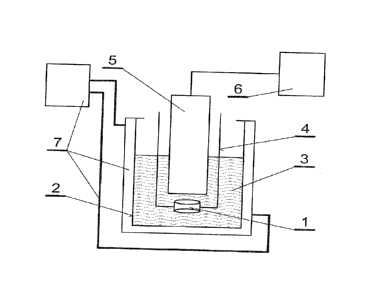
Fig. 1 is a diagram of an exemplary working-stand for covering an implant
supporting structure with hydroxyapatite. Fig. 2 shows a microscope images
(SEM) of the implant from the first embodiment in three different
magnifications.
CA 02985182 2017-11-06
WO 2016/178174 PCT/1B2016/052566
12
Fig. 3 shows a SEM image of the uncoated polymer supporting structure from the
first example in the in vivo tests, while Fig. 4 shows a microscope image of
the
implant with the supporting structure coated with GoHAP in the similar test
using
animal model. Fig. 5 shows microscope images (SEM) of the implant from the
second embodiment in three different magnifications. Fig. 6 shows a
microscopic
image of the ceramic supporting structure of the second embodiment after the
in
vivo test, while Fig. 7 shows a microscope image of such an implant with the
supporting structure coated with GoHAP after similar in vivo test. Fig. 8
shows
microscope images (SEM) of the implant from the third embodiment in three
different magnifications.
Mode for Carrying out Invention
The invention will be described in further detail in the following exemplary
embodiments. In these examples a nano-powder of hydroxyapatite was used,
under the trade name GoHAP, having the following characteristics:
- the nano-powder particles are in a form of platelets having an average
particle
size less than 30 nm, as based on the analysis of the image obtained by the
transmission electron microscope (TEM) using dark-field for at least 200
particles,
wherein the average particle size equals the diameter of the circle drawn
around
the particle shape;
- ratio of calcium to phosphorus (Ca/P) is greater than 1.55, but smaller
than 1.67;
- the nano-powder contains structural water in an amount ranging from 2 to
6% by
weight, wherein the amount of the water is determined by the weight loss of
the
nano-powder during the heating above 200 C;
- solubility, determined by the procedure of ISO 10993-14, ranging from 5 to
35 mg/dm'.
Example 1
Porous Polymer Implant
Supporting structure 1 of the implant was made of biodegradable polymer-poly-
caprolactone (PCL). It has a form of 3-dimensional scaffold, measuring 4 x 6
mm,
CA 02985182 2017-11-06
WO 2016/178174 PCT/D32016/052566
13
made from polymer fibers by printing technology of the spatial "rapid
prototyping"
which is described in W. Swieszkowski et al., "Repair and regeneration of
ostechondral defects in the articular joints" [13iomolecular Engineering, 2007
24
(5): pp. 489-95] The supporting structure 1 is characterized by the porosity
of
approx. 41%. As the coating material of the supporting structure a nano-powder
of
hydroxyapatite GoHAP was used, with a molar ratio (Ca/P) of 1.65, containing
5%
by weight of structural water. This powder (in amount of 0.1% by weight) was
mixed in 50 ml vessel 2 with deionized water to form a homogeneous suspension
3, wherein the external phase is water and the internal phase is
hydroxyapatite,
The supporting structure 1 was rinsed with distilled water, and then fitted to
the
stand 4 for immobilization. The stand 4 was placed in the vessel 2 with the
suspension 3 heated to 30 C. An ultrasound head 5 with the front (emitting)
surface having a diameter of 13 mm, being a source of ultrasounds, was
connected to an power supply device 6 and immersed in the suspension 3. The
distance from the front surface of the head 5 and the supporting structure 1
shall
not be larger than 200% of the diameter of this head's surface, wherein the
optimum is to keep a distance equal to 100% of its diameter. For the following
fifteen minutes the head 5 generated ultrasounds at a frequency of 20 kHz. The
generated ultrasound of this frequency induced a phenomenon of ultrasound
cavitation, i.e. formation and activity of gas bubbles in the liquid exposed
to the
ultrasonic field. Cavitation occurred mainly in the portion of the suspension
3 being
in contact with the surface of the supporting structure 1, including the
suspension 3 filling the pores of the supporting structure 1. The cavitation
was
confirmed by the observation of the liquid and temperature monitoring. In
order to
maintain a stable temperature of the suspension 3 a flow cooling circuit 7 was
used. When the power supply of the head 4 was turned off the coated implant 1
was taken out of the vessel 2, rinsed with distilled water, and then dried in
laminar
flow cabinet of high purity. These steps were repeated dozen times to obtain
the
number of implants sufficient for in vitro and in vivo tests. Based on the SEM
image analysis it was found that GoHAP layer applied on the supporting structu-
re 1 has a morphological features similar to that of the initial GoHAP powder
(size,
CA 02985182 2017-11-06
WO 2016/178174 PCT/1B2016/052566
14
particle shape). The coating was obtained, having a thickness of 200 nm
uniformly
covering more than 85% of the supporting structure surface of implant 1
(Figure 2).
The obtained implants were firstly used in cellular assays in vitro tests. The
cell
line MG-63 (osteosarcoma) and D-MEM culture medium supplemented with 10%
FBS was used. In addition to the cell medium for the above samples penici-
llin/streptomycin was added. The incubation was carried out on 24 well plates
at
37 C and 5% CO2 environment. The cells were separated from the incubation sub-
strate with 0.25% trypsin/EDTA. Implants (scaffold) for testing were rinsed
with
PBS (phosphate buffer saline, pH 7.4). Then cells were planted on the prepared
scaffolds. For each tested scaffold concentration of approx 105 cells in 200
ml
culture medium were used and then scaffolds were placed in the incubator for
one
and a half hours. After this time, the medium was added to the wells in order
to
completely cover the sample. Afterwards, the incubation lasted for five days.
The
results showed that cell proliferation on the polymer scaffold with GoHAP
layer is
higher than on the corresponding polymer scaffold without such a layer.
Analysis
of the number of cells clearly showed that the polymer scaffold with GoHAP
layer
has better features for stimulating cell proliferation. After five days of
culture, the
cell density on the polymer scaffold with such a layer was three times higher
than
on the polymer scaffold without a coating. After five days of culture
duration, on the
inverted microscope it was noted that the confluence of cells in all wells
around the
test material was ?.. 95%.
The implants prepared in this embodiment were also examined in vivo using an
animal model. A ten-month old, male, New Zealand rabbits were given, using
standard procedures, a general anesthetic and in this state in their tibia
bone holes
were made that were filled with implants mentioned. As reference material the
clean polymer scaffolds described above were used for filling the holes in the
hip
bone of individuals from the control group. Upon completion of implantation
periosteum of all animals was sutured and soft tissue was closed layer by
layer
with 5-0 Vicryl sutures. The skin was stitched using interruptible 4-0 Prolene
sutures. The subcutaneous injection of the antibiotic solution Enrobioflox 5%
CA 02985182 2017-11-06
WO 2016/178174 PCT/1B2016/052566
(50 mg/m I solution) were applied once a day for 5 days, containing 5 mg per
kilograms of weight of the active substance Enrofloxacin. After three months,
euthanasia, using standard procedures, of all study subjects was carried out,
after
which the hard and soft tissue samples were collected and examined regarding
their histology and capacity for facilitating bone regeneration. Routine
staining with
hematoxylin and eosin was performed in each case. The extracted polymer
scaffold without a hydroxyapatite layer is shown in section in the Figure 3,
where
there is a space for the red marrow (BM) penetration around the fibers (S) of
the
pure scaffold. Figure 4 shows the new bone (NB) filling the spaces between the
residues of the fibers (S) of the implant according to the invention.
Morphometric
analysis of the image of Figure 4 indicated that the proportion of new tissue
in the
porous space of the implant with GoHAP layer amounted to approx. 33%, of which
35% was constituted by the new bone tissue (NB), whereas for a scaffold (S)
without the such layer bone tissue growth was negligible.
Example 2:
Porous Ceramic Implant
In order to produce a ceramic implant coated with a layer of nano-particles of
hydroxyapatite a supporting structure in a form of porous ceramic pellets was
used. For the production of pellets a method of uniaxial pressing of the 8-TCP
powder was used with a pressure force of 15 kN. For the production of one
sample
3.06 g of powder was taken. In order to achieve structural microporosity a
heat
treatment method was used in which said pallet was subjected to 1200 C for a
period of two hours. The pellets were then examined for signs of porosity
using
methods of computer microtomography and Archimedes method. According to the
p-CT calculations a porosity of 49% was reached, while the result of the
analysis
by the Archimedes method was 52% (+/- 2.6%). The coating processes with
GoHAP of the prepared ceramic implant supporting structure was carried out in
a manner analogous to the first example, wherein a nano-powder with a water
content of 5% by weight was used. Obtained coating had the thickness of 250
nm,
uniformly covering more than 80% of the surface of the supporting structure
(1) of
CA 02985182 2017-11-06
WO 2016/178174 PCT/1132016/052566
16
the implant (Figure 5). The material used for coating, i.e. GoHAP nano-powder
of
hydroxyapatite was characterized by a molar ratio (Ca/P) of 1.66.
The ceramic layer on GoHAP implants was tested in vivo using an animal model
(New Zealand rabbits). The procedure of implanting ceramic implant, the used
comparative material, the collection of samples for testing and hematoxylin
and
eosin staining were performed similarly as in the first example. The extracted
after
three months ceramic scaffolds without GoHAP layer have been substantially
filled
with bone tissue (NB in Figure 6). An morphometric analysis of these
structures
images showed that almost 50% of the implant (scaffold) pores were filled with
the
bone tissue. Morphometric analysis of the image of the ceramic scaffold with a
layer of GoHAP, presented at Figure 7, showed that almost 70% of the scaffold
pores were filled with the bone tissue.
Example 3
Monolithic metallic implant
In order to produce a monolithic metal implant a supporting structure was used
in
a form of a titanium screw having a diameter of 5 mm and a length of 150 mm,
dedicated to arthroscopy. The screw was coated with a layer of GoHAP nano-
particles by ultrasonic effect on the aqueous suspensions described in detail
in the
first embodiment, wherein a powder has a diameter of not more than 6 nm, a
structural water content of 3% by weight, molar ratio of calcium to phosphorus
(Ca/P) of 1.60. Based on the SEM image analysis it was found that the
resulting
coating has morphological properties characteristic for the initial GoHAP
powder.
The titanium screw was coated with a uniform layer of hydroxyapatite having a
thickness of 200 nm, covering 85% of its surface (Figure 8).