Note: Descriptions are shown in the official language in which they were submitted.
CA 02985498 2017-11-08
WO 2017/011522 PCT/US2016/042033
MICROCAVITY-CONTAINING POLYMERIC MEDICAL DEVICES FOR ENHANCED
ULTRASONIC ECHOGENICITY
TECHNICAL FIELD
[0001] The presently disclosed subject matter relates generally to the
design of polymeric
medical devices which contain specially designed microcavities to generate
improved
echogenicity characteristics when visualized within the human body using
ultrasound.
BACKGROUND
[0002] Noninvasive medical methods such as ultrasound imaging offer
tremendous medical
value and point-of-care utility for diagnosis and measurement. It often is
desirable to locate a
medical device which is currently within the human body or to identify a site
where a procedure
or measurement has been previously performed. However, interpretation of
greyscale (B-mode)
ultrasound requires expertise and it may be difficult to use native landmarks
to determine
whether the desired location has been reached. For example, returning to the
site of a prior
procedure can be challenging because of first, the difficulty in locating the
postoperative site and
second, the difficulty in determining the orientation of the image vis-à-vis
earlier images
collected in order to accurately analyze data collected from the location.
[0003] For the visualization of devices placed within the human body, many
methods of
rendering surfaces echogenic have been described. The goal of these
modifications is often to
make an edge (such as the edge of a metal needle) more easily visualized under
ultrasound. Such
methods may include machining small divots into the surface of the edge in
order to reflect sonic
waves in multiple directions. However, such methods are generally applicable
only to metallic
surfaces where the significant impedence difference between metal and human
tissue means that
the majority of ultrasound waves will be reflected back from the tissue-metal
surface towards the
transducer and will not penetrate the metallic material. For materials which
are closer in acoustic
impedence to human tissue, such as most polymers of medical interest, most of
the ultrasound
waves will pass through the polymer, generating detectable signals only at the
entry and exit
point of the waves. Based on our experimentation, any such surface
modification attempts will
fail to significantly increase the echogenicity of polymer devices. Similarly,
attempts to create
1
CA 02985498 2017-11-08
WO 2017/011522 PCT/US2016/042033
divots or indentations either randomly distributed across the device body or
penetrating through
the entire thickness of the device (e.g., from the front to back surface of
the device) fail to
improve echogenicity as desired.
[0004] Ultrasound tissue marker devices do exist within the medical device
landscape for use
in localizing sites within the body but are undesirable for certain
applications because they lack
such an echogenic enhancement method. These markers are often composed of a
large surface
area to volume ratio (e.g., they may be made up of many small pellets which
may be randomly
oriented) because the increased surface area maximizes the return of
ultrasound waves. If the
thickness of the marker body (i.e., the axis perpendicular to the beam
direction) is large relative
to the ultrasound wavelength, only the device edges and not the interior will
be visualized. This
is because substantial changes in either density or compressibility do not
exist throughout the
volume on a microscopic scale. For both the purposes of human visualization as
well as medical
imaging algorithm detection, it would often be desirable if a method were
available to visualize
the entire object under ultrasound rather than just the edges.
[0005] Another significant problem relates to the angle of insonation
dependence with
respect to the ultrasound. Devices which rely on edge-only reflectance (e.g.,
the previously
mentioned existing markers) function as largely specular reflectors which
reflect the ultrasound
beam according to the standard laws of reflection. While this is desirable
when the surface is
perpendicular to the angle of insonation (because the strongest reflections
are back towards the
transducer), as the angle of insonation begins to change towards parallel most
of the ultrasound
energy reflects away from the transducer and is lost, making the surface dark
and causing loss of
the contrast necessary to visualize the object.
[0006] Therefore, what is desired is a method of producing polymer-
comprised medical
devices which are 1) visualized throughout their entire thickness rather than
just their edges as
well as 2) more tolerant of variable insonation angles while still producing
echogenic contrast
compared to surrounding tissue.
2
CA 02985498 2017-11-08
WO 2017/011522 PCT/US2016/042033
SUMMARY
[0007] The presently disclosed subject matter provides an ultrasound-
detectable polymeric
medical device with superior visibility of the body of the device and less
ultrasound angle
dependence. These desirable characteristics are created by introducing
controlled microcavities
within the marker to alter the reflection mechanism of the ultrasound waves as
they pass through
the implant.
[0008] The cavities have two main purposes: (a) creating differences in
density and
compressibility within the marker on a small scale, and (b) creating diffuse
reflection robust to
insonation angle as compared to what is otherwise largely specular reflection.
The small-scale
density changes ensure that acoustic signal reflections occur throughout the
depth of penetration.
The distance over which these changes occur is tuned to be relative to the
wavelength of the
ultrasound, with optimal cavity-polymer transitions occurring at distances
comparable to the
ultrasound wavelength. The proper choice of microcavity ratio and dimension is
essential
because creation of excessive acoustic impedence will cause premature
absorption of all the
ultrasound energy and failure of the object to fully illuminate, while
inadequate impedence will
result in the internal structure being inadequately echogenic.
[0009] However, production of density changes alone simply create greater
reflections in the
body of the object. For example, production of the object using an additive
manufacturing
process such as 3D printing yields objects with a series of layers which may
cause impedence
changes. However, such methods result in impedence changes that continue to be
specular
reflectors and result in objects seen best when perpendicular to the source of
the sound wave.
This renders it impossible to ever fully visualize a 3-dimensional shape where
some surfaces are
not perpendicular to the ultrasound beam (for example, the sides of a sphere
will not show up
well).
[0010] In order to accommodate various orientations of geometric shape that
may be desired
in, for example, an ultrasound marker device, the microcavities and their
essentially random
surface orientation vis-à-vis the ultrasound beam will reflect the signal in a
diffuse manner. Thus
the acoustic signal from the object returns to the probe irrespective of
orientation and causes the
whole cross-section of the object to appear visible on the ultrasound screen.
3
CA 02985498 2017-11-08
WO 2017/011522 PCT/US2016/042033
[0011]
In other aspects, the presently disclosed subject matter provides a method for
inserting and visualizing a medical device containing microcavities, the
method comprising: (a)
inserting a polymeric medical device with microcavities into a patient; (b)
visualizing and
detecting the device using B-mode ultrasound during or after surgery; and (c)
performing this
visualization in multiple near-simultaneous frames, representing different
angles of insonation.
[0012]
This method of detecting the medical device from multiple angles of insonation
is
of particular importance. In many clinical environments, it is desirable to
understand the
orientation of the imaging plane to gather repeatable data longitudinally, but
also to assess a
specific site from a variety of perspectives. Furthermore, it is rare that the
user will approach the
site from the proper angle, so the device must tolerate and accommodate
initial error. Thus, the
user of the ultrasound must be able to detect the device from essentially all
angles of insonation.
BRIEF DESCRIPTION OF THE DRAWINGS
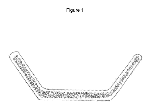
[0013]
Figure 1 displays the cross section of a medical device with internal
microcavities.
The cavities resemble a spherical or semi-spherical shape across a range of
sizes. The device
contains an outer layer free of microcavities.
[0014]
Figure 2 displays the reflectance of the ultrasound beam for a medical device
both
with and without microcavities. Figure 2A, a medical device without
microcavity, exhibits a
specular reflection of the ultrasound beam, which results in little to no
signal returning to the
probe. Figure 2B displays the diffuse reflection that is generated when the
ultrasound beam
contacts the microcavities. Unlike in Figure 2A, a significant portion of the
signal is reflected
back to the probe, irrespective of the originating angle of the emitted
signal.
DETAILED DESCRIPTION
[0015]
In one aspect, the invention provides an ultrasound-detectable medical device
comprising a polymer with microcavities dispersed in some or all of its body
capable of
providing improved visibility throughout some or all of its volume and under
variable angles of
insonation Figures 1 and 2B. In some instances, the microcavities extend
throughout the entire
4
CA 02985498 2017-11-08
WO 2017/011522 PCT/US2016/042033
volume of the medical device. In other instances, the microcavities occupy a
central region of the
medical device. In additional instances, the space containing microcavities is
surrounded by an
outer layer of material without microcavities.
[0016] In another aspect, the invention provides an ultrasound-detectable
device wherein
the diameter (microcavity size) ranges between 0.1 to 950 microns, and
commonly between 50 to
350 microns. In some instances, the microcavity diameter exceeds 1,000
microns. In other
instances, the microcavity diameter ranges from 10 to 500 microns. In
additional instances, the
microcavities exhibit diameters from 10 to 1,500 microns.
[0017] In a further aspect, the invention provides an ultrasound-
detectable device
wherein the ideal volume to volume ratio of cavity space to polymer structures
should be less
than 60%, and is commonly between 12% and 50%. In some instances, the
microcavities
comprise between 30 to 50% of the volume. In other instances, the volume ratio
of microcavities
exceeds 60%.
[0018] The ultrasound-detectable device contains microcavities. In one
aspect of the
device, the microcavities are composed of gas. In one aspect of the invention,
the device is
created via injection molding. In another embodiment, the device is
manufactured by extrusion.
In some aspects of the invention, microcavities are created by introducing gas
into the polymer
material prior to manufacturing, commonly through injection. In other aspects
of the invention,
microcavities are introduced during the manufacturing process, which can be
performed by
injecting gas into a mold either before, while, or after the polymer enters
the mold.
[0019] The microcavities may be composed of a variety of biocompatible
gases. In some
instances, super-critical CO2 is used, and in other instances, N2 is used.
[0020] In another embodiment, the microcavities are created via a
chemical reaction such
that gas is released into the polymer. This may be accomplished with a foaming
agent or other
chemical processes. The gas may be activated by pressure or temperature
changes in the
manufacturing process.
[0021] For a variety of reasons, including mechanical, material
degradation, visibility,
and manufacturing considerations, it is desirable to have the microcavities
consume a region
CA 02985498 2017-11-08
WO 2017/011522 PCT/US2016/042033
within the overall volume, rather than the entire device. In one embodiment,
the region
containing the microcavities is central to the device. In this embodiment, the
region containing
the microcavities is surrounded by a layer of polymeric or non-polymeric
material that does not
contain microcavities. In other embodiments of the device, this external
layer, or "skin", contains
microcavities, though of a reduced density. In further embodiments, the region
containing the
microcavities resides on the top surface of the device (superficial towards
the position of the
ultrasound probe), while in other embodiments, the microcavity region resides
on the bottom
surface of the device.
[0022] In one aspect of the invention, there is an outer layer of the
device which is meant
to maintain the structural integrity of the inner microcavity-containing
region. This outer layer
does not contain microcavities and thus provides a barrier protecting the
inner region, especially
from fluid flow, which could accelerate degradation and also negatively impact
the ultrasonic
visibility. In another aspect of the invention, the outer layer described has
a smooth surface to
minimize irritation and other adverse events to surrounding tissue or vessels
once the device is
implanted.
[0023] Another aspect of the device relates to the visibility of the
device under ultrasonic
imaging. In this aspect, the device is used as an echogenic marker for
ultrasound location in the
human body. Some anatomic structures that can be marked using this device
include: veins,
arteries, soft tissue, urinary tracts, nerves, and ducts. The device enables
location of any of these
structures after implantation. In particular, the device gives the clinician
knowledge of the spatial
relationship between the ultrasound probe and anatomic structure, independent
of the angle of
insonation. The device enables locating the anatomic location repeatedly
across many
examinations after placement of the device. The size of the device ranges from
1 to 60 mm in
length, 1 to 60 mm in width, and 1 to 40 mm in height. Some embodiments of the
device
represent curved, cradle-like structures. Other embodiments of the device are
spheres, rectangles,
cubes, plates, pellets, and discs. Some instances of when this device could be
used are for:
microvascular anastomoses, solid organ transplants, vascular bypass, and
vascular access.
[0024] In one embodiment of the device, it is comprised of one or more
resorbable
polymers selected from the group of: poly(lactic-co-glycolic acid) (PLGA),
polylactide (PLA),
6
CA 02985498 2017-11-08
WO 2017/011522 PCT/US2016/042033
polyglycolide (PGA), polyhydroxyalkanoate (PHA), polycaprolactone (PCL),
polyethylene
glycol (PEG) and copolymers thereof
[0025]
In another embodiment of the device, it is comprised of one or more non-
resorbable polymers selected from the group of:polycarbonate,
polyetheretherketone,
polypropylene, silicone, polyethylene, polyester, polybutylene terephthalate
(PBT), polyvinyl
chloride, polyethylsulfone, polyacryclate, polyetheretherketone, poly-p-
xylylene (parylene),
polytetrafluoroethylene, cyclo olefin, acrylonitrile butadiene styrene,
polyeurethane,
acrylonitrile styrene acrylate, acetals, polyetherimide, ethylene,
chlorotrifluoroethylene, ethylene
tetrafluoroethylene, polyvinyl fluoride,
polyvinylidene difluoride, and polyhydroxybutyrate.
In a further embodiment, the device is comprised of both resorbable and non-
resorbable
materials, which may be in the form of multiple sections with unique
materials, a single blend of
materials, or multiple sections of blended materials.
[0026]
In one aspect of the invention, the device is manufactured via a foaming
process.
Microcavities are introduced into the polymer by introducing a blowing agent.
The blowing
agent created the cellular structure of the microcavities. In one embodiment
of the invention, the
blowing agent is a physical blowing agent. In another embodiment, the blowing
agent is a
chemical blowing agent. An alternative way of generating the foam is using a
solvent such as
acetone. In addition to introducing the foaming agent, this invention
describes injecting the
polymer into a mold. An alternative way of producing the device is via
extrusion.
[0027]
This invention describes a method for using the device where the device is
first
inserted into a patient, it is then detected using B-mode ultrasound during or
after surgery, and
the device is detected in multiple frames, representing different angles of
insonation. The
ultrasound user can leave the patient and return to find the device at a later
time point. This is
important because it is often desired to track anatomical or physiological
features over a time
horizon of multiple days or weeks, and sometimes months or years. This means
that user needs
to walk away from the patient, return to the patient, and easily locate the
device. Another critical
feature of the invention is the ability to detect the device using ultrasound
from any angle of
insonation. This is important because a non-expert is able to locate the
marked site and use the
visual information to achieve a desired angle or set of angles. The invention
enables strong
7
CA 02985498 2017-11-08
WO 2017/011522 PCT/US2016/042033
visibility in angles ranging from 25 degrees to 155 degrees from the skin
surface. The
microcavity feature of the invention provides the ability to visualize the
device across such a
broad range of insonation angles. Due to the geometry and microcavity feature
of the device, the
user is able to understand the angle of insonation. Therefore, the user can
repeatedly match the
same orientation upon each examination, generate the same image of the device,
and thus
compare anatomic or physiologic conditions reliably over time. Alternatively,
the user can
approach the device from a new orientation in each additional examination,
though will have the
geometric information from the device to make proper calculations to adjust
for the new angle of
insonation.
[0028]
The device should not be compromised at 40 degrees Celsius when in a dark and
moist environment, such as human or animal tissue. Compromise includes but is
not limited to
geometric changes, mechanical deformation, degradation, or microcavity change.
The device
must maintain its original integrity for at least 72 hours in such conditions.
The device must yield
contrast when visualized using B-mode ultrasound between 1 cm and 5 cm deep
from the surface
of the skin.
EXAMPLES
[0029]
Example 1. An ultrasound-detectable medical device made by extrusion.
Specifically, a Nano 16 mm extruder was used with a GFA3-10-30 screw element
at 270 mm.
The extruder has four zones, each with individual temperature control, which
ultimately lead to a
die to achieve the desired geometry of the device. The zones were first
preheated to 110, 140,
130 and 100 C respectively. The pressure within the die ranged from 10-70
psi. The feeding rate
of the polymer was 2.5 cc/min, and the screw speed fell between 75-100 rpm.
The torque on the
screw ranged from 1500-3000 Gm. The supercritical CO2 was injected at 200 psi
with a flow rate
of 20 cfh. When the extruded polymer left the die, it was cooled via an air
jacket. In cases when
it was desired to achieve variance along the extrusion axis, the device was
laser cut once it
cooled to room temperature using the air jacket.
[0030]
Example 2. An ultrasound-detectable medical device made by injection molding.
The
polymer was introduced into the mold via injection through the port. While the
material was
8
CA 02985498 2017-11-08
WO 2017/011522 PCT/US2016/042033
being injected into the mold, CO2 gas was simultaneously injected to provide
microbubbles. In
another example, the CO2 was introduced into the material prior to injection
into the mold. Once
the material filled the mold, the mold was released via its pins, the part was
removed, and the
process was repeated.
9