Note: Descriptions are shown in the official language in which they were submitted.
A METHOD AND SYSTEM FOR AUTOMATED MICROBIAL COLONY
COUNTING FROM STREAKED SAMPLE ON PLATED MEDIA
BACKGROUND OF THE INVENTION
[0002] Thcrc is increased focus on digital imagery of culture plates for
detection of
microbial growth. Techniques for imaging plates for detecting microbial growth
are
described in PCT Publication No. W02015/114121.
Using such techniques, laboratory staff is no longer required to read plates
by direct visual inspection but can use high quality digital images for plate
inspection.
Shifting laboratory workflow and decision-making to examination of digital
images of
culture plates can also improve efficiency. Images can be marked by an
operator for further
work-up by either the operator or another person with the appropriate skills.
Additional
images may also be taken and used to guide secondary processes.
[0003] Detection of colonies, colony enumeration, colony population
differentiation and
colony identification define the objectives for a modern microbiology imaging
system.
Having these objectives realized as early as possible achieves the goals of
delivering results
to a patient quickly and providing such results and analysis economically.
Automating
laboratory workflow and decision-making can improve the speed and cost at
which these
goals may be achieved.
[0004] Although significant progress has been made regarding imaging
technologies for
detecting evidence of microbial growth, it is still sought to extend such
imaging technologies
to support an automated workflow. Apparatus and methods for inspecting culture
plates for
indications of microbial growth are difficult to automate, due in part to the
highly visual
nature of plate inspection. In this regard, it is desirable to develop
techniques that may
automatically interpret culture plate images and determine the next steps to
be perfointed
(e.g., identification of colonies, susceptibility testing, etc.) based on the
automated
interpretation.
[0005] For example, counting colonies in a plated culture can be
difficult, especially
when the colonies are of different size and shape and are touching each other.
These
-1-
Date Recue/Date Received 2023-01-04
CA 02985854 2017-10-18
WO 2016/172532 PCT/US2016/028919
problems are exacerbated when growth has already reached confluence in some
regions of
the plate. For these reasons, it is preferable, if possible, to count CFUs
early in the incubation
process. However, time for incubation is still needed to allow for at least
some growth of the
colonies. Thus, on the one hand, the longer that colonies are allowed to grow,
the more they
begin to contrast with their background and each other, and the easier it
becomes to count
them. Yet, on the other hand, if the colonies are allowed to grow too long and
they begin to
fill the plate and/or touch one another, thereby forming confluent regions on
the plate, it
becomes more difficult to contrast them from one another, making counting more
difficult. If
one were able to detect colonies at an incubation time when the colonies were
still small
enough to be isolated from one another despite relatively poor contrast, or if
one were able to
estimate colony count even when the colonies are large enough to form
confluent regions on
the plate, this problem could be resolved.
BRIEF SUMMARY OF THE INVENTION
[0006] An aspect of the present disclosure is directed to an automated
method for
evaluating growth on plated media, comprising: providing a culture media
inoculated with a
biological sample; incubating the inoculated culture media; following
incubation, obtaining a
first image of the inoculated media at a first time (ti); after further
incubation, obtaining a
second image of the inoculated media at a second time (t2); aligning the first
image with the
second image, such that the coordinates of a pixel in the second image are
about the same as
the coordinates of a corresponding pixel in the first image; comparing image
features of the
second image with image features of the first image; classifying image
features of the second
image as colony candidates based on image feature changes from time ti to time
t2; for colony
candidates determined to be from a common microorganism in the biological
sample
inoculated on the culture media, counting said colony candidates; and
determining whether
the number of counted colonies meets or exceeds the threshold count value
stored in memory
and indicative of significant growth.
[0007] In some examples, if the number of counted colonies meets or exceeds
the
threshold count value, the method may further comprise: identifying at least
one of the
colonies using matrix-assisted laser desorption ionization (MALDI); testing
said at least one
colony for antibacterial susceptibility; and outputting a report containing
the MALDI and
antibacterial susceptibility test results. A plurality of threshold count
values may be stored in
the memory, each threshold count value being associated with a different
microorganism.
[0008] In some examples, classifying image features of the second image as
colony
candidates may comprise: determining contrast information of the second image,
the contrast
-2-
CA 02985854 2017-10-18
WO 2016/172532 PCT/US2016/028919
information including at least one of spatial contrast information indicating
differences
between pixels of the second image and temporal contrast information
indicating differences
between pixels of the second image and corresponding pixels of a previous
image;
identifying an object in the second image based on the contrast information;
and obtaining
one or more object features of the identified object from pixel information
associated in the
first and second images, wherein the object is classified as a colony
candidate based on the
object features. The method may further comprise, for each colony candidate,
determining
whether the colony candidate is a colony or an artifact based on pixel
information associated
with the colony candidate, wherein colony candidates that are determined to be
artifacts are
not counted. Determining whether a colony candidate is a colony or an artifact
may further
comprise determining whether the colony candidate is present in each of the
first and second
images and larger in the second image than the first image by a threshold
growth factor,
wherein a colony candidate that is present in both images and is larger in the
second image by
at least the threshold growth factor is classified as a colony. Determining
whether a colony
candidate is a colony or an artifact may further comprise, for a colony
candidate that is
present in the second image and not the first image: obtaining one or more
object features of
the identified object from the pixel information associated with the object in
the second
image; detemtining a probability that the colony candidate is a colony based
on the one or
more object features; and comparing the determined probability to a predefined
threshold
probability value, wherein, if the determined probability is greater than the
predefined
threshold probability value, then the colony candidate is classified as a
colony. The method
may further comprise: classifying (i) colony candidates that are present in
both images and
not larger in the second image, and (ii) colony candidates that are present in
first image and
not in the second image, as definite artifacts; classifying colony candidates
that are present in
each of the first and second images and larger in the second image by the
threshold growth
factor as definite colonies; and calculating an artifact probability value
based on a
combination of the definite artifacts and the definite colonies, wherein the
determined
probability that a colony candidate is a colony is further based on artifact
probability.
[0009] In some examples, the object features may comprise at least one of
object shape,
object size, object edge, object color, color, hue, luminance and chrominance
of the pixels of
the object. The method may further comprise obtaining background feature
information,
wherein background feature information comprises media type and media color,
and wherein
the object is classified as a colony candidate based further on the background
feature
information. In some examples, aligning the first image with the second image
may comprise
-3-
CA 02985854 2017-10-18
WO 2016/172532 PCT/US2016/028919
assigning polar coordinates to pixels of each of the first and second images
such that the polar
coordinates of a pixel in the second image are the same as the polar
coordinates of a
corresponding pixel in the first image.
[00101 Another aspect of the present disclosure is directed to automated
method for
estimating a number of colony foiniing units on plated media that has been
inoculated with a
culture according to a predefined pattern and incubated, comprising: after
incubation of the
culture, obtaining a digital image of the plated media; from the digital
image, identifying
colony candidates in the image; linearizing the digital image according to the
predefined
pattern; plotting the colony candidates according to pixels of the linearized
coordinates of the
digital image; and estimating the number of colony forming units on the plated
media based
on pixels of the colony candidates in the linearized digital image.
[0011] In some examples, the plated media from which the image was obtained
may have
been inoculated using a magnetically controlled bead streaked along a
continuous zig-zag
pattern, wherein the digital image may be linearized according to the zig-zag
streaking
pattern with the zig-zag streaking pattern being a main axis of the linearized
image. The
initial bead load of the magnetically controlled bead may be estimated from
the plot of
colony candidates. Estimating the initial bead load may comprise: selecting a
distance from
origin along the main axis of the linearized image; determining a probability
that a colony
forming unit is released by the bead at the selected distance; and counting
the number of
colony forming units present in the digital image that are farther from origin
along the main
axis than the selected distance, wherein the estimated initial bead load is
equal to the ratio
between said determined probability and said counted number of colony forming
units. The
distance may be selected such that no confluent regions of microbial growth
are present in the
image at a distance farther from an origin of the linearized image than the
selected distance.
The method may further comprise: selecting a plurality of distances along the
main axis of
the linearized image; for each of the selected distances, counting the number
of colony
forming units present in the digital image that are farther from an origin of
the linearized
image along the main axis than the selected distance; and based on the counted
number of
colony forming units for each distance, calculating a probability that a
colony forming unit is
released onto the media by the bead when a point of the bead containing the
colony forming
unit makes contact with the media. Determining a probability that a colony
forming unit is
released by the bead at a given distance may be based on said calculated
probability that a
colony forming unit is released onto the media by the bead when a point of the
bead
containing the colony forming unit makes contact with the media.
-4-
CA 02985854 2017-10-18
WO 2016/172532 PCT/US2016/028919
[0012] In some examples, the method may further comprise: comparing the
digital image
to a plurality of distribution models stored in the memory, each distribution
model showing
an expected distribution of colony forming units across an imaged plate for a
given initial
bead load, and a given probability that a colony forming unit is released onto
the media when
contact is made with the media; and determining the initial bead load based at
least in part on
the compared distribution models.
[0013] In some examples the method may further comprise: selecting a
distance from the
origin along the main axis of the linearized image; determining a fraction of
pixels at the
selected distance that are associated with a colony candidate; and estimating
the initial bead
load based on said determined fraction.
[0014] In some examples the method may further comprise: after incubation
of the
culture, obtaining a plurality of digital images of the plated media, each
digital image
containing one or more colony candidates; identifying one digital image in
which at least
some of the colony candidates form a confluent region; identifying an earlier
digital image in
which said colony candidates that form the confluent region in the digital
image have not
combined to form a confluent region; and estimating the number of colony
forming units in
the confluent region based on the earlier digital image.
[0015] Yet another aspect of the present disclosure is directed to computer-
readable
memory storage medium having program instructions encoded thereon configured
to cause a
processor to perform a method. The method may be any of the above methods for
evaluating
microbial growth on plated media, or for estimating a number of colony forming
units on
plated media.
[0016] Yet a further aspect of the present disclosure is directed to a
system for evaluating
growth in a culture media inoculated with a biological sample. The system
comprises an
image acquisition device for capturing digital images of the culture media,
memory, and one
or more processors operable to execute instructions to perform a method. In
some examples,
the memory may store information regarding predicted amounts of microbial
growth for one
or more different organisms in one or more different culture media, and the
method
performed by the executed instructions may be any one the above described
methods for
evaluating microbial growth on plated media. In other examples, the memory may
store
information regarding a pattern for inoculating the culture media with the
biological sample,
and the method perfoimed by the executed instructions may be any one the above
described
methods for estimating a number of colony forming units on plated media.
BRIEF DESCRIPTION OF THE DRAWINGS
-5-
CA 02985854 2017-10-18
WO 2016/172532 PCT/US2016/028919
[0017] FIG. 1 is a schematic diagram of a system for imaging analyzing and
testing a
culture according to an aspect of the disclosure.
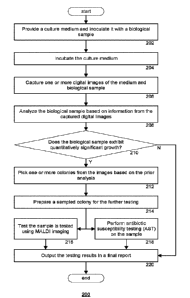
[00181 FIG. 2 is a flow chart illustrating an automated laboratory workflow
routine for
imaging analyzing and testing a culture according to an aspect of the
disclosure.
[0019] FIGS. 3A, 3B and 3C are images showing a visual representation of
colony
morphology as it changes over time according to an aspect of the disclosure.
[0020] FIGS. 3D and 3E are images showing a visual representation of
colonies under
different illumination conditions.
[0021] FIG. 4 is a flow chart of an example routine for counting colonies
according to an
aspect of the disclosure.
[0022] FIG. 5 is a flow chart of an example routine for collecting a global
list of colony
candidates according to an aspect of the disclosure.
[0023] FIG. 6 is a flow chart of an example routine for sorting colony
candidates
according to an aspect of the disclosure.
[0024] FIG. 7 is a flow chart of an example routine for counting colonies
based on
statistical analysis according to an aspect of the disclosure.
[0025] FIG. 8 is an image of a streaking pattern for streaking plated media
with a sample
according to an aspect of the disclosure.
[00261 FIG. 9A is a graphical representation of an image of identified
colony candidates
according to an aspect of the disclosure.
[0027] FIG. 9B is a graphical representation of the streaking pattern shown
in FIG. 8.
[0028] FIGS. 10A and 10B are graphical representations for colony forming
unit (CFU)
distribution models according to an aspect of the disclosure.
[0029] FIG. 11A is a graphical representation of confluence ratio along a
main axis of the
plate shown in FIG. 8 according to an aspect of the disclosure.
[00301 FIG. 11B is a graphical representation of a colony growth simulation
according to
an aspect of the disclosure.
[0031] FIG. 12 is a side-to side depiction of two images of plate media
with colony
growth.
[0032] FIG. 13 is a series of images taken over time according to an aspect
of the
disclosure.
[0033] FIG. 14 is a Vorondi diagram according to an aspect of the
disclosure.
[0034] FIGS. 15A, 15B and 15C are graphical depictions of determinations of
isolation
factor according to an aspect of the disclosure.
-6-
[0035] FIGS. 16A and 16B show a section of an imaged plate, with zoomed
and
reoriented images of sample colonies of the image
[0036] FIG. 16C are polar transformed images of the zoomed in sections of
FIG. 16B,
respectively, according to an aspect of the disclosure.
[0037] FIG. 17 is a flow chart comparing the timeline of the routine of
FIG. 2 to the
timeline of a comparable manually-performed process.
DETAILED DESCRIPTION
[0038] The present disclosure provides apparatus and methods for
identifying and
analyzing microbial growth in on plated media based in at least in part on the
number of
identified colonies counted in one or more digital images of the plated media.
Many of the
methods described herein can be fully or partially automated, such as being
integrated as part
of a fully or partially automated laboratory workflow.
[0039] The systems described herein are capable of being implemented in
optical systems
for imaging microbiology samples for the identification of microbes and the
detection of
microbial growth of such microbes. There are many such commercially available
systems,
which are not described in detail herein. One example is the BD KiestraTM
ReadA Compact
intelligent incubation and imaging system. Other example systems include those
described in
PCT Publication No. W02015/114121 and U.S. Patent Publication 2015/0299639.
Such optical imaging platforms are
well known to those skilled in the art and not described in detail herein.
[0040] FIG. 1 is a schematic of a system 100 having a processing module
110 and image
acquisition device 120 (e.g., camera) for providing high quality imaging of
plated media.
The processing module and image acquisition device may be further connected
to, and
thereby further interact with, other system components, such as an incubation
module (not
shown) for incubating the plated media to allow growth of a culture inoculated
on the plated
media. Such connection may be fully or partially automated using a track
system that
receives specimens for incubation and transports them to the incubator, and
then between the
incubator and image acquisition device.
[0041] The processing module 110 may instruct the other components of the
system 100
to perfoun tasks based on the processing of various types of information. The
processor 110
may be hardware that performs one or more operations. The processor 110 may be
any
standard processor, such as a central processing unit (CPU), or may be a
dedicated processor,
such as an application-specific integrated circuit (ASIC) or a field
programmable gate array
(FPGA). While one processor block is shown, the system 100 may also include
multiple
-7-
Date Recue/Date Received 2023-01-04
CA 02985854 2017-10-18
WO 2016/172532 PCT/US2016/028919
processors which may or may not operate in parallel, or other dedicated logic
and memory for
storing and tracking information related to the sample containers in the
incubator and/or
image acquisition device 120. In this regard, the processing unit may track
and/or store
several types of information regarding a specimen in the system 100, including
but not
limited to the location of the specimen in the system (incubator or image
acquisition device,
locations and/or orientation therein, etc.), the incubation time, pixel
information of captured
images, the type of sample, the type of culture media, precautionary handling
information
(e.g., hazardous specimens), etc. In this regard, the processor may be capable
of fully or
partially automating the various routines described herein. In one embodiment,
instructions
for performing the routines described herein may be stored on a non-transitory
computer-
readable medium (e.g. a software program).
[0042] FIG. 2 is a flow chart showing an example automated laboratory
routine 200 for
imaging, analyzing and, optionally, testing a culture. The routine 200 may be
implemented
by an automated microbiology laboratory system, such as the KiestraTM Total
Lab
Automation or KiestraTM Work Cell Automation, both manufactured by Becton,
Dickenson &
Co. The example systems include interconnected modules, each module configured
to
execute one or more steps of the routine 200.
[0043] At 202, a culture medium is provided and inoculated with a
biological sample.
The culture medium may be an optically transparent container, such that the
biological
sample may be observed in the container while illuminated from various angles.
Inoculation
may follow a predetermined pattern. Streaking patterns and automated methods
for streaking
a sample onto a plate are well known to one skilled in the art. One automated
method uses
magnetically controlled beads to streak sample onto the plate.
[0044] In some examples of the present disclosure, the bead is streaked
across the plate
according to a zig-zag pattern (see, e.g., FIG. 8). The starting point and end
point of the zig-
zag pattern may be located at opposite ends of the plate (e.g., separated by a
distance about
equal to the diameter of the plate). In such examples, the "main axis" of the
plate may be
thought of as a straight line beginning at the starting point and ending at
the ending point of
the zig-zag pattern.
[0045] At 204, the medium is incubated to allow for growth of the
biological sample.
[0046] At 206, one or more digital images of the medium and biological
sample are
captured. As will be described in greater detail below, digital imaging of the
medium may be
performed multiple times during the incubation process (e.g., at the start of
incubation, at a
time in the middle of incubation, at the end of incubation) so that changes in
the medium may
-8-
CA 02985854 2017-10-18
WO 2016/172532 PCT/US2016/028919
be observed and analyzed. Imaging of the medium may involve removing the
medium from
the incubator. Where multiple images are taken of the medium at different
times, the
medium may be returned to the incubator for further incubation between imaging
sessions.
[00471 At 208, the biological sample is analyzed based on information from
the captured
digital images. Analysis of the digital image may involve analysis of pixel
information
contained in the image. In some instances, pixel information may be analyzed
on a pixel by
pixel basis. In other instances, pixel information may be analyzed on a block
by block basis.
In yet further instances, pixels may be analyzed based on entire regions of
pixels, whereby
the pixel information of individual pixels in the region may be derived by
combining
information of the individual pixels, selecting sample pixels, or by using
other statistical
methods such as the statistical histogram operations described in greater
detail below. In the
present disclosure, operations that are described as being applied to "pixels"
are similarly
applicable to blocks or other groupings of pixels, and the term "pixel" is
hereby intended to
include such applications
[0048] The analysis may involve determining whether growth is detected in
the medium.
From an image analysis perspective, growth can be detected in an image by
identifying an
imaged object (based on differences between the object and its adjacent
surroundings) and
then identifying changes in the object over time. As described in greater
detail herein, these
differences and changes are both forms of "contrast." In addition to detecting
growth, the
image analysis at 208 may further involve quantifying the amount of growth
detected,
identifying distinct colonies, identifying sister colonies, etc.
[0049] At 210, it is determined whether the biological sample
(particularly, the identified
sister colonies) exhibits quantitatively significant growth. If no growth, or
an insignificant
amount of growth, is found, then the routine 200 may proceed to 220, in which
a final report
is output. In the case of proceeding from 210 to 220, the final report will
likely indicate the
lack of significant growth, or report the growth of nomial flora.
[0050] If it is determined that the biological sample exhibits
quantitatively significant
growth, then at 212, one or more colonies may be picked from the images based
on the prior
analysis. Picking colonies may be a fully automated process, in which each of
the picked
colonies is sampled and tested. Alternatively, picking colonies may be a
partially automated
process, in which multiple colony candidates are automatically identified and
visually
presented in a digital image to an operator, such that the operator may input
a selection of one
or more candidates for sampling and further testing. The sampling of selected
or picked
colonies may itself be automated by the system.
-9-
CA 02985854 2017-10-18
WO 2016/172532 PCT/US2016/028919
[0051] At 214, a sampled colony is prepared for the further testing, such
as by plating the
sample in an organism suspension. At 216, the sample is tested using matrix-
assisted laser
desorption ionization (MALDI) imaging to identify the type of specimen that
was sampled
from the original medium. At 218, the sample is also, or alternatively,
subjected to antibiotic
susceptibility testing (AST) to identify possible treatments for the
identified specimen.
[0052] At 220, the testing results are output in a final report. The report
may include the
MALDI and AST results. As mentioned above, the report may also indicate a
quantification
of specimen growth. Thus, the automated system is capable of beginning with an
inoculated
culture medium and generating a final report regarding a specimen found in the
culture, with
little or no additional input.
[0053] In routines such as the example routine of FIG. 2, the detected and
identified
colonies are often referred to as Colony Fanning Units (CFUs). CFUs are
microscopic
objects that begin as one or a few bacteria. Quantitative growth may be
measured based on
the number of CFUs that can be counted in the plate. However, as explained
above, the
number of CFUs cannot always be counted directly. For instance, the CFUs may
touch or
blend with one another, thereby forming confluent regions without discrete
units to be
counted. In such situations, the present disclosure provides for ways to
estimate the colony
count based on a combination of known information ¨ such as the streaking
pattern applied to
the plated media, knowledge of how quickly the streaking implement is unloaded
as it is
streaked across the plate, standard size and growth rate for a particular type
of colony being
counted, etc. ¨ and measured information collected from one or more digital
images of the
plate. Such estimations may be automated by the above described systems and
routines.
[0054] Determining whether the estimated growth is significant may be
derived from
comparing the estimated colony count to a predefined threshold value. More
than one
threshold value may be set for a given plate and/or colony. For instance,
colony growth may
be affected by the medium in which the colony is being grown. Therefore, what
constitutes
significant growth in one medium may not constitute significant growth in
another medium,
and different thresholds may be set. Additionally, while testing may not be
warranted for one
type of bacteria until a high threshold is met, testing for particularly
harmful or dangerous
bacteria (e.g., Group B streptococcus in testing of pregnant female) may be
warranted when
even a low threshold value is met, in some cases even as low as one counted
colony.
Therefore, it should be understood that the system is capable of storing
multiple threshold
values and applying each of those various threshold values under the
appropriate
circumstances.
-10-
CA 02985854 2017-10-18
WO 2016/172532 PCT/US2016/028919
[0055] Over time, the bacteria grow to form a colony. The earlier in time
from when the
bacteria are placed in the plate, the less bacteria there is to detect and,
consequently the
smaller the colony and the lower that contrast to the background. Stated
another way, a
smaller colony size yields a smaller signal, and a smaller signal on a
constant background
results in smaller contrast. This is reflected by the following equation:
Signal-background
(1) Contrast =
Signal+background
[005611 Contrast can play an important role in identifying objects, such as
CFUs or other
artifacts, in the images. An object can be detected in an image if it is
significantly different in
brightness, color and/or texture from its surroundings. Once an object has
been detected, the
analysis may also involve identifying the type of object that has been
detected. Such
identifications can also rely on contrast measurements, such as the smoothness
of edges of
the identified object, or the uniformity (or lack of uniformity) of the color
and/or brightness
of the object. This contrast must be great enough to overcome the image noise
(background
signals) in order to be detected by the image sensor.
[0057] The human perception of contrast (governed by Weber's law) is
limited. Under
optimal conditions, human eyes can detect a light level difference of 1%. The
quality and
confidence of image measurements (e.g., brightness, color, contrast) may be
characterized by
a signal-to-noise ratio (SNR) of the measurements, in which an SNR value of
100 (or 40db),
independent from pixel intensities, would match human detection capabilities.
Digital
imaging techniques utilizing high SNR imaging information and known SNR per
pixel
information can allow for detection of colonies even when those colonies are
not yet visible
to human eyes.
[0058] In the present disclosure, contrast may be collected in at least two
ways: spatially
and temporally. Spatial contrast, or local contrast, quantifies the difference
in color or
brightness between a given region (e.g., pixel, group of adjacent pixels) and
its surroundings
in a single image. Temporal contrast, or time contrast, quantifies the
difference in color or
brightness between a given region of one image against that same region in
another image
taken at a different time. The formula governing temporal contrast is similar
to that for
spatial contrast:
ISignal(t1)-Signal(t2)I
(2) Temporal Contrast =
Signal(t1)+Signal(t2)
[005911 In which t2 is a time subsequent to ti. Both spatial and temporal
contrasts of a
given image may be used to identify objects. The identified objects may then
be further
tested to determine their significance (e.g., whether they are CFUs, nonnal
flora, dust, etc.).
-11-
CA 02985854 2017-10-18
WO 2016/172532 PCT/US2016/028919
[0060] FIGS. 3A and 3B provide a visual demonstration of the effect that
temporal
contrast can have on an imaged sample. The images shown in FIG. 3A were
captured at
different points in time (left to right, top row to bottom row) showing the
overall growth in
the sample. While growth in noticeable in FIG. 3A, the growth is even more
noticeable, and
can be noticed even earlier in the sequence, from the corresponding contrast
temporal images
of FIG. 3B. For purposes of clarity, FIG. 3C shows a zoomed section of FIG.
3B. As can be
seen in FIG. 3C, the longer a portion of a colony has been imaged, the
brighter a spot it
makes in the contrast image. In this way, the center of mass of each colony
may be denoted
by the bright center, or peak, of the colony. Thus, image data obtained over
time can reveal
important information about changes in colony morphology.
[0061] To maximize spatial or temporal contrast of an object against its
background, the
system may capture images using different incident lights on different
backgrounds. For
instance, any of top lighting, bottom lighting, or side lighting may be used
on either a black
or white background.
[0062] FIGS. 3D and 3E provide a visual demonstration of the effect that
lighting
conditions can have on an imaged sample. The image in FIG. 3D was captured
using top
lighting, whereas the image in FIG. 3E was captured at approximately the same
time (e.g.,
close enough in time that no noticeable or significant growth has occurred)
using bottom
lighting. As can be seen, each of the images in the samples of FIGS. 3D and 3E
contains
several colonies, but additional information about the colonies (in this case,
hemolysis) can
be seen thanks to the back-lighting or bottom lighting in the image of FIG.
3E, whereas that
same information is difficult to grasp in the image of FIG. 3D.
[0063] At a given point in time, multiple images may be captured under
multiple
illumination conditions. Images may be captured using different light sources
that are
spectrally different due to illumination light level, illumination angle,
and/or filters deployed
between the object and the sensor (e.g. red, green and blue filters). In this
manner, the image
acquisition conditions may be varied in terms of light source position (e.g.,
top, side, bottom),
background (e.g., black, white, any color, any intensity), and light spectrum
(e.g. red channel,
green channel, blue channel). For instance, a first image may be captured
using top
illumination and a black background, a second image captured using side
illumination and a
black background, and a third image captured using bottom illumination and no
background
(i.e. a white background). Furthermore, specific algorithms may be used to
create a set of
varying image acquisition conditions in order to maximize spatial contrast
using. These or
other algorithms can also be useful to maximize temporal contrast by varying
the image
-12-
CA 02985854 2017-10-18
WO 2016/172532 PCT/US2016/028919
acquisition conditions according to a given sequence and/or over a span of
time. Some such
algorithms are described in PCT Publication No. W02015/114121.
[0064] FIG. 4 is a flow chart showing an example routine for analyzing an
imaged plate
based at least in part on contrast. The routine of FIG. 4 may be thought of as
an example
subroutine of the routine 200 of FIG. 2, such that 206 and 208 of FIG. 2 are
carried out at
least in part using the routine of FIG. 4.
[0065] At 402, a first digital image is captured at time ti. Time ti may be
a time after the
incubation process has begun, such that bacteria in the imaged plate have at
least begun to
form some visible colonies, but those colonies have not yet begun to touch or
overlap with
one another.
[0066] At 404, coordinates are assigned to one or more pixels of the first
digital image.
In some instances, the coordinates may be polar coordinates, having a radial
coordinate
extending from a center point of the imaged plate and an angular coordinate
around the center
point. The coordinates may be used in later steps to help align the first
digital image with
other digital images of the plate taken from different angles and/or at
different times. In
some cases, the imaged plate may have a specific landmark (e.g., an off-center
dot or line),
such that coordinates of the pixel(s) covering the landmark in the first image
may be assigned
to the pixel(s) covering the same landmark in the other images. In other
cases, the image
itself can be considered as a feature for future alignment.
[0067] At 406, a second digital image is captured at time t2. Time t2 is a
time after ti at
which the colonies in the imaged plate have had an opportunity to grow even
more.
Additional colonies that were too small to be visible at ti may also be
visible at t2. Also,
there is a possibility that colonies at time t2 have begun to touch or overlap
with one another.
[0068] At 408, the second digital image is aligned with the first digital
image based on
the previously assigned coordinates. Aligning the images may further involve
normalization
and standardization of the images, for instance, using the methods and systems
described in
PCT Publication No. W02015/114121.
[0069] At 410, a global list of colony candidates is collected based on the
first and second
digital images. The global list of colony candidates may identify any objects
in the first and
second digital images that may be a colony for which further testing (as in
the routine of
FIG. 1) may be desired.
[0070] At 412, each of the colony candidates included in the global list is
sorted. Sorting
the colony candidates involves identifying, for each candidate, whether the
candidate is in
fact an artifact or a colony. As explained in greater detail below, in some
cases, it may not be
-13-
CA 02985854 2017-10-18
WO 2016/172532 PCT/US2016/028919
possible to definitively determine whether a given candidate is an artifact or
a colony.
Nonetheless, a probabilistic or fuzzy determination may be made and the
candidate may be
sorted according to said determination.
[00711 At 414, the sorted colony candidates that were identified as
colonies are counted.
As explained in greater detail below, counting colonies is not always
straightforward due to
confluence among individual colonies. Therefore, the present disclosure
provides methods
and techniques for counting based on a statistical analysis of the second
digital image.
[00721 At 416, a final report including an estimated colony count is
outputted. The final
report may optionally include additional infonnation impacting the accuracy of
the estimated
colony count, such as a swarming probability among the counted colonies.
Swarming refers
to the confluence of colonies, thereby resulting in a swarm that the
individual colonies cannot
be separately identified. In some instances, the swarming probability may be
reported only if
it exceeds a preset threshold (e.g., 50%).
[0073] FIG. 5 is a flow chart showing an example routine 500 for collecting
a global list
of colony candidates. The routine of FIG. 5 may be thought of as an example
subroutine of
the routine 400 of FIG. 4, such that 410 of FIG. 4 is carried out at least in
part using the
routine of FIG. 5.
[0074] At 502, contrast infonnation of the second digital image is
determined. The
contrast information may be gathered on a pixel-by-pixel basis. For example,
the pixels of
the second digital image may be compared with the corresponding pixels (at the
same
coordinates) of the first digital image to determine the presence of temporal
contrast.
Additionally, adjacent pixels of the second digital image may be compared with
one another,
or with other pixels known to be background pixels, to determine the presence
of spatial
contrast. Changes in pixel color and/or brightness are indicative of contrast,
and the
magnitude of such changes from one image to the next or from one pixel (or
region of pixels)
to the next, may be measured, calculated, estimated, or otherwise determined.
In cases where
both temporal contrast and spatial contrast is determined for a given image,
an overall
contrast of a given pixel of the image may be determined based on a
combination (e.g.,
average, weighted average) of the spatial and temporal contrasts of that given
pixel.
[0075] At 504, objects in the second digital image are identified based on
the contrast
information computed at 502. Adjacent pixels of the second digital image
having similar
contrast information may be considered to belong to the same object. For
instance, if the
difference in brightness between the adjacent pixels and their background, or
between the
pixels and their brightness in the first digital image, is about the same
(e.g., within a
-14-
CA 02985854 2017-10-18
WO 2016/172532 PCT/US2016/028919
predetermined threshold amount), then the pixels may be considered to belong
to the same
object. As an example, the system could assign a "1" to any pixel having
significant contrast
(e.g., over the threshold amount), and then identify a group of adjacent
pixels all assigned "1"
as an object. The object may be given a specific label or mask, such that
pixels with the same
label share certain characteristics. The label can help to differentiate the
object from other
objects and/or background during later processes.
[0076] Identifying objects in a digital image may involve segmenting or
partitioning the
digital image into multiple regions (e.g., foreground and background). The
goal of
segmentation is to change the image into a representation of multiple
components so that it is
easier to analyze the components. Image segmentation is used to locate objects
of interest in
images. [add cross-ref here]
[0077] At 506, the features of a given object (identified at 504) may be
characterized.
Characterization of an object's features may involve deriving descriptive
statistics of the
object (e.g., area, reflectance, size, optical density, color, plate location,
etc.). The descriptive
statistics may ultimately quantitatively describe certain features of a
collection of information
gathered about the object (e.g., from a SHQI image, from a contrast image).
Such information
may be evaluated as a function of species, concentrations, mixtures, time and
media.
However, in at least some cases, characterizing an object may begin with a
collection of
qualitative information regarding the object's features, whereby the
qualitative information is
subsequently represented quantitatively. Table 1 below provides a list of
example features
that may be qualitatively evaluated and subsequently converted to a
quantitative
representation:
Table 1: Qualitative Attributes of Objects, and
Criteria for Quantitatively Converting the Attributes
Number Feature Score Criteria
1 Growth 0 No growth
1 Growth
2 Expected Time to Visually Observe nia Record time in hours
3 Size (diameter) 1 <1 mm
2 >1-4 mm
3 >4 mm
4 Growth Rate (A diameter / 2hrs) 1 <1 mm
2 >1-2 mm
3 >2 mm
Color 1 grey/white
2 rose-pink
3 colorless
4 red
-15-
CA 02985854 2017-10-18
WO 2016/172532 PCT/US2016/028919
Number Feature Score Criteria
blue
6 blue-green
7 brown
8 pale yellow to_yellow
9 green
6 Hemolysis 0 none
1 small beta( <1mm)
2 large beta( lmm)
3 alpha
7 Shape 1 convex
2 flat
3 spread
4 Concave
8 Surface/Edge 1 smooth
2 rough
3 mucoid
4 feet
[00781 Some features of an object, such as shape or the time until it is
observed visually,
may be measured a single time for the object as a whole. Other features may be
measured
several times (e.g., for each pixel, for every row of pixels having a common y-
coordinate, for
every column of pixels having a common x-coordinate, for every ray of pixels
having a
common angular coordinate, for a circle of pixels having a common radial
coordinate) and
then combined, for instance using a histogram, into a single measurement. For
example,
color may be measured for each pixel, growth rate or size for every row,
column, ray or circle
of pixels, and so on.
[0079] At 508, it is determined whether the object is a colony candidate
based on the
characterized features. The colony candidate determination may involve
inputting the
quantitative features (e.g., the scores shown in Table 1, above), or a subset
thereof, into a
classifier. The classifier may include a confusion matrix for implementing a
supervised
machine learning algorithm, or a matching matrix for implementing an
unsupervised machine
learning algorithm, to evaluate the object. Supervised learning may be
preferred in cases
where an object is to be discriminated from a limited set (e.g., two or three)
of possible
organisms (in which case the algorithm could be trained on a relatively
limited set of training
data). By contrast, unsupervised learning may be preferred in cases where an
object is to be
discriminated from an entire database of possible organisms, in which case it
would be
difficult to provide comprehensive ¨ or even sufficient ¨ training data. In
the case of either
confusion or a matching matrix, differentiation could be measured numerically
on a range.
-16-
For instance, for a given pair of objects, a "0" could mean the two objects
should be
discriminated from each other, whereas a "1" could mean that the objects are
difficult to
differentiate one from the other.
[0080] If at 508 the object is determined to be a colony candidate, then
at 510, it is added
to the global list of colony candidates. Otherwise, routine 500 ends (and may
continue with
412 of routine 400) without adding the object to the global list.
[0081] Additional routines and subroutines for identifying colony
candidates based on
contrast information of digital images is discussed in the commonly owned and
copending
application titled "COLONY CON __ FRAST GATHERING ."
[0082] FIG. 6 is a flow chart showing an example routine 600 for sorting
the colony
candidates. The routine of FIG. 6 may be thought of as an example subroutine
of the routine
400 of FIG. 4, such that 412 of FIG. 4 is carried out at least in part using
the routine of
FIG. 6. As a subroutine of FIG. 4, routine 600 may be applied iteratively to
each of the
colony candidates appearing on the global list.
[0083] At 602, it is determined whether the colony candidate is growing.
Growth may be
indicated by (a) the colony candidate's presence in both the first and second
digital images,
and (b) the colony candidate's size being significantly larger in the second
digital image than
in the first digital image. Whether a change in size is considered significant
may he
determined by comparing the change in size to a predetermined growth
threshold, whereby
changes that meet or exceed the growth threshold are considered significant.
[0084] If the colony candidate is determined to be growing, then the
colony candidate is
validated and identified as a colony. Otherwise, routine 600 continues at 604,
in which it is
determined whether the colony candidate is present in both the first and
second digital
images.
[0085] If the colony candidate is determined to be present in both images
(meaning that
there was no significant growth between the two images), then the colony
candidate is
identified as an artifact. Otherwise, routine 600 continues at 606, in which
it is determined
whether the colony candidate is present in the second digital image.
[0086] If the colony candidate is not present in the second image (meaning
that it was
only present in the first image and then disappeared), then the colony
candidate is identified
as an artifact (e.g., a piece of dust that was likely blown off the plate
between ti and t2).
Otherwise, further analysis is performed to determine whether the colony
candidate is a
-17-
Date Recue/Date Received 2023-01-04
CA 02985854 2017-10-18
WO 2016/172532 PCT/US2016/028919
colony that simply had not grown enough to be visible at time ti, or an
artifact such as a piece
of dust that blew onto the plate between times t1 and t2.
[0087] At 608, given the knowledge that the colony candidate does not
appear in the first
digital image, features of the colony candidate are characterized based solely
on information
from the second digital image. The characterization may rely on static
features, such as
color, size, shape and surface (described above in connection with step 506 of
FIG. 5).
[0088] At 610, an overall probability that the colony candidate is in fact
a colony is
determined based at least in part on the characterization. For instance, the
characterized
features of an object may be compared to features of an expected colony type
(i.e., the colony
type included in the global list and being counted in FIG. 6). In one
embodiment, the
comparison is executed in the same manner as in step 508 of FIG. 5, such that
a "0" would
mean the object is the expected colony type, whereas a "1" would mean that the
object is not
the expected colony type, and a number in between "0" and "1" would indicate a
probability
of the object being the expected colony type, also referred to as a colony
probability.
[0089] In some instances, the colony probability may be the overall
probability of 610.
Alternatively, the determination at 610 may be further based on information
gathered about
artifacts in the image. Such information may include an artifact probability,
which gauges
the likelihood of objects in the image being artifacts. In the example of FIG.
6, objects that
are definitely determined to be artifacts (e.g., no growth between ti and t2,
presence only in ti
and not t2) or colonies (e.g., significant growth between ti and t2) are
provided as an input at
612 in order to determine the artifact probability. The artifact probability
of 612 is then
combined with the colony probability to yield the overall probability. In one
embodiment,
the colony probability and artifact probability are combined according to the
following
equation:
(3) P(overall) = P(colony) X (1-P(artifact))
[0090] At 614, the overall probability is compared to a predetermined
threshold (e.g.,
50%). If the overall probability meets or exceeds the threshold, then the
colony candidate is
identified as a colony. Otherwise, the colony candidate is identified as an
artifact.
[0091] While the routines of FIG. 5 and 6 are useful for classifying
identified colonies,
those routines do not ensure that each colony is an individual colony and not
a confluence of
multiple colonies. Therefore, the colony candidates that are identified as
colonies cannot
necessarily be counted as discrete elements, but rather counted using
estimation techniques.
[0092] FIG. 7 is a flow chart showing an example routine 700 for counting
colonies
based on statistical analysis. The routine of FIG. 7 may be thought of as an
example
-18-
CA 02985854 2017-10-18
WO 2016/172532 PCT/US2016/028919
subroutine of the routine 400 of FIG. 4, such that 414 of FIG. 4 is carried
out at least in part
using the routine of FIG. 7. The routine of FIG. 7 presumed use of a
magnetically controlled
bead to streak colonies onto the imaged plate according to a predetermined
streaking pattern.
Those skilled in the art should understand that the underlying concepts of the
routine of
FIG. 7 may be adapted for various streaking media, techniques and patterns,
other than those
described below.
[0093] At 702, the second digital image is linearized according to a
streaking pattern
along which the imaged plate is streaked by the magnetically controlled bead.
To illustrate
the streaking pattern, FIG. 8 shows an image a sample growing in a plated
media. The image
is digitally overlaid with a zig-zag pattern beginning toward the bottom right
of the image
and ending toward the top left. The zig-zag pattern indicates the streaking
pattern of the
magnetically controlled bead used to streak the media.
[0094] For purposes of clarity, linearizing the digital image may be
thought of as plotting
the pixels of the zig-zag pattern along an x-axis of the linearized image,
such that the zig-zag
pattern is unfolded into a straight line along the x-axis. For each pixel of
the digital image
that does not directly overlay the zig-zag pattern, and the pixel may be
associated with the
closest part of the zig-zag pattern to the pixel (e.g., along a y-axis of the
linearized image).
The linearize image is useful for indicating the density gradient of colonies
deposited onto the
media by the bead as the bead moves along the streaking pattern over time.
[0095] At 704, each colony candidate is plotted along the linearized
coordinates of the
second digital image. In other words, the density gradient of colonies
deposited onto the
media is assessed using the linearized image. FIG. 9A is a graphical
representation of
previously identified colony candidates (e.g., from the routine 500 of FIG.
5). FIG. 9B is a
graphical representation of the zig-zag pattern. By overlaying the
representations of
FIGS. 9A and 9B, the distance of every colony candidate along the zigzag
pattern from the
pattern origin (leftmost end of the image) can be computed.
[0096] At 706, an initial bead load (a concentration, measured in CFUs per
milliliter) is
estimated based on the plotted colonies present in the linearized second
digital image.
Calculations for estimating initial bead load are presented herein for a bead
streaking a path
having a width (W) measured in mm (also referred to as the "contact width")
and having a
surface area (SA) of "SA" measured in mm2.
[0097] As an initial point, it is noted that for a given point on the
surface of the bead, on
average that point will come into contact with the plate once for every "B" mm
that the bead
travels along the zig-zag pattern, where:
-19-
CA 02985854 2017-10-18
WO 2016/172532 PCT/US2016/028919
SA
(4) B= ¨w
[00981 If
it is assumed that the given point is loaded with a colony forming unit (CFU),
then the probability of the CFU being released onto the media (PR) when the
contact between
the given point and the media is made may be characterized as a number between
0 and 1.
[0099] By
the time the bead has progressed a distance x (measured in mm) along the
streaking path, the probability that a CFU at the given point has been
released ( PNR (X)) is
given according to the following relationship:
(5) PNR(x) = (1 ¨ PR)7B (18)
[0100] As
the bead progress and releases CFUs, the CFU load present on the bead
decreases. The total CFU load present on the bead at the given time at which
the bead has
so far travelled distance x along the streaking pattern may be characterized
as K(x). K(x)
can further be expressed as a function of the initial bead load Ko (i.e., the
CFU load of the
bead before the streaking pattern began and, thereby, before any CFUs were
released):
(6) K(x) = K0 x PNR(X) = K0 X (1 ¨ PR)x B (19)
[0101]
K(x) can also be estimated based on the linearized digital image. Specially,
it
may be assumed that all of the CFUs initially loaded onto the bead will be
released onto the
media by completion of the streaking pattern, therefore, the remaining load on
the bead at any
given distance x may be characterized as
CFU, the number of colonies shown in the digital
image past distance x. (In reality the upper limit of the sum should be the
length of the
streaking pattern, not Go, but given the assumption that all CFUs are released
by the end of the
streaking pattern, an upper limit of infinity is equally acceptable.)
[0102]
Using the estimate of K(x), that estimate can be plugged into the above
equations
to solve for initial bead load Ko. In fact, for any given distance x that K(x)
can be estimated
(e.g., xl, x2, x3, etc.), Ko can be also be independently solved for, as shown
by the following
equations:
ETiCFU Y2 CFU rx-'3 CPU
(7) Ko =
¨ (1¨PR) 18 v (1¨PR) - (1 -PR )X3/13
[0103]
While in the above example the release probability PR was assumed, it is
further
noted that the above series of equations can also be used to solve for PR
using two or more of
the estimations of K(x) = Ex' CFU along the streaking pattern. The following
is an example
of a formula for solving for PR using the estimated K(x) values for distances
xi and x2.
xin (42
35.5 CFU)
(8) _________________________ pR = 1 _ e (x2 -xi) opv)
-20-
CA 02985854 2017-10-18
WO 2016/172532 PCT/US2016/028919
[01041 Having solved for PR, Ko may be estimated using the determined
values for
PR and K(x), according to the following equation:
K(x)
(9) Ko = (22)
(1-PR)x/B
[0105] It should be noted that the more values of K(x) that are estimated,
the more
precise the estimates of PR and Ko may become. Therefore, while the above
example uses
only xl and x2 to estimate Ko, other examples may use additional distances
(e.g., x3).
[0106] Alternatively, PR may be a predetermined value based on known
features of the
colony, media, bead, or any combination thereof, in which case K0 may be
estimated based
on the estimated value of K(x) at a single given distance x.
DISTRIBUTION MODEL
[01071 In addition to the above calculations, colonies on the imaged plate
may be
estimated based on a comparison between the digital image and a distribution
model.
FIGS. 10A and 10B illustrate CFU distribution models based on a given bead
release
probability (PR) Each of FIGS. 10A and 10B show distributions of CFUs for
varying initial
bead loads (ranging from a small initial load, e.g., 102, at the leftmost
image to a large initial
bead load, e.g., 105, at the rightmost image). The distribution models may
also be modified
to account for variables such as release probability and colony size. In the
examples of
FIGS. 10A and 10B, the release probability is set to 0.14. In FIG. 10A, the
colony size is set
to 1.66mm in diameter. In FIG. 10B, the colony size is set to a smaller
diameter.
Accordingly, with knowledge of the release probability and colony size for a
given sample,
distribution models may be used to estimate initial bead load for an image
having a similar
appearance in distribution.
CONFLUENCE RATIO
[0108] Confluence ratio can also be utilized in order to improve the colony
count
estimation. Confluence ratio is the fraction of pixels, at a given distance
along the main axis
of the plate, that are associated with a colony candidate. Confluence ratio
may be
characterized according to the following:
xy=R
Ex y=_RCFU candidate pixel
(10) __________________________________ Confa% = x such that ' õ=R = a%
R Media pixel
[0109] FIG. 11A illustrates the confluence ratio of a plate as measured
along the main
axis of the streaking pattern from origin to end. In the example of FIG. 11A,
the expected
CFUs on the plate were 4800. FIG. 11B depicts the result of a simulation with
a 4800 CFU
initial load having a release probability of 0.185 and a colony size of about
2mm in diameter.
-21-
As can be seen in the present example, confluence shown in the plate of FIG.
11A and the
simulation of FIG. 11B are fairly similar to one another.
[0110] Confluence ratio can also be used to identify a tangent line zero
crossing, which is
the point along the main axis at which the confluent region mainly or mostly
ends (e.g., more
discrete colonies than confluent colonies, confluent regions make up less than
50% of the
pixels along a line running through said point and perpendicular to the main
axis, etc.).
[0111] It should be recognized from the above examples that the expected
confluence
ratio along the main axis of a plate depends largely on the initial load
(CFU/ml), the size of
isolated colonies at the given time that the plate is being imaged, and the
given incubation
timc. To highlight these factors, FIG. 12 is a side-to sidc dcpiction of two
plates having
similar confluence ratios but significantly different CFU loads. The top plate
contains a total
of about 39,500 CFUs of staphylococcus aureus, whereas the bottom plate
contains about 305
CFUs of pseudomonas aeruginosa. Notably the confluence ratio of these plates
is about the
same as the plate shown in FIG. 11A (which contains about 4,800 CPUs of
serratia
marcescens on a blood agar media after 18 hours of incubation). 'INVENTORS:
What is a
"tangent line zero crossing" method?'
TIME-SERIES ANALYSIS
[0112] Another way of estimating CFU content within a confluence region is
to
performing time-series analysis by splitting the confluent region into
discrete colonies using
past images. As stated above, colonies that have confluence at a given time
may still be
discrete and individually countable at an earlier time. Therefore, an analysis
may be
conducted using images of the confluent region from earlier incubation times
when
confluence conditions were not yet met for at least some of the colonies
(e.g., running
segmentation routines, building a Voronoi diagram, etc.). This analysis could
then be used to
keep tracks of ongoing changes over time to help maintain identification of
discrete colonies
at subsequent times.
[0113] FIG. 13 illustrates the series of images taken over time. In FIG.
13, each row
contains a digital image (left), a segmentation result (middle) and a contrast
image (right) at a
specified time point during incubation: at 8 hours, at 12 hours, at 16 hours
and at 20 hours
respectively (from bottom to top). Segmentation and contrast images are
described in greater
detail in the commonly owned, copending patent application titled "COLONY
CONTRAST
GATHERING ."
[0114] Those skilled in the art will recognize that the results of the
above colony
estimation techniques, distribution models, confluence ratios and time-series
analysis may be
-22-
Date Recue/Date Received 2023-01-04
CA 02985854 2017-10-18
WO 2016/172532 PCT/US2016/028919
used in combination with one another to provide a more accurate estimates, or
confirm the
accuracy of prior estimates.
OBJECT FEATURES
[01151 As discussed above in connection with FIG. 5, features of an object
on an imaged
plate may be characterized as part of the image analysis performed on the
imaged plate. The
characterized features may include both static features (pertaining to a
single image) and
dynamic image (pertaining to a plurality of images).
[01161 Static features aim at reflecting object attributes and/or
surrounding background at
a given time. Static features include the following:
(i) Center of gravity: this is a static feature that provides a center of
gravity of an
imaged object in a coordinate space (e.g., x-y, polar). The center of gravity
of an
object, like the polar coordinates of the object, provides invariance in the
feature
set under given lighting and background conditions. The center of gravity may
be
obtained by first determining a weighted center of mass for all colonies in
the
image (M being the binary mask of all detected colonies). The weighted center
of
mass may be determined based on an assumption that each pixel of the image is
of
equal value. The center of gravity for a given colony may then be described in
x-
y coordinates by the following equation (in which E = {p Ip E M} (E is the
current colony's binary mask), the range for the x-coordinate is [0, image
width],
the range for the y-coordinate is 110, image height], and each pixel is one
unit):
(11) igv(,,y) (x = EpEE Px Y = -v pEE. X EpEE Py)
z
(ii) Polar coordinates: this is also a static feature, and can be used to
further
characterize locations on the imaged plate, such as a center of gravity.
Generally,
polar coordinates are measured along a radial axis (d) and an angular axis
(0),
with the coordinates of the plate center being [0,0]. Coordinates d and 0 of
igv(x,y) are given (in millimeters for d, and in degrees for 0) by for
following
equations (Where k is a pixel density corresponding pixels to millimeters, and
"barcode" is a landmark feature of the imaged plate to ensure alignment of the
plate with previous and/or future images):
(12) d = k X dist(igv(x,y),0(X,y))
(13) 0 = Angle (barcode, 0(x,y),igv(x,y))
(iii) Image vector: The two-dimensional polar coordinates may in turn be
transformed
into a one-dimensional image vector. The image vector may characterize
intensity
-23-
CA 02985854 2017-10-18
WO 2016/172532 PCT/US2016/028919
of the pixels of an image as a function of the radial axis (generally, with
the center
of the colony having the highest intensity) and/or a function of the angular
axis. In
many cases, the image vector may be more accurate at classifying
similarities/distinctions among imaged objects.
(iv) Morphometric features, which describe the shape and size of a given
object.
(a) Area: This is a morphometric feature, and can be determined based on the
number of pixels in the imaged object (also referred to as a "blob"), not
counting holes in the object. When pixel density is available, area may be
measured in physical size (e.g., mm2). Otherwise, when pixel density is not
available, the total number of pixels may indicate size, and pixel density (k)
is
set to equal one. In one embodiment, area is calculated using the following
equation:
(14) A = k2 x A.,7pEE 1
(b) Perimeter: The perimeter of the object is also a morphometric feature, and
can
be determined by measuring the edges of the objecting and adding together the
total length of the edges (e.g., a single pixel having an area of 1 square
unit
has a perimeter of 4 units). As with area, length may be measured in terms of
pixel units (e.g., when k is not available) or physical lengths (e.g., when k
is
available). In some circumstances, the perimeter may also include the
perimeter of any holes in the object. Additionally, the ladder effect (which
results when diagonal edges are digitized into ladder-like boxes) may be
compensated by counting inside corners as -\/2, rather than 2. In one
embodiment, perimeter may be deteimined using the following equations:
(15) P = k X EpeEq(np)
It
(16) np = 1 p r
1
b
(17) if:
f(tEM,IEM,rEM,beM). 2,(/EM#rEM),(tEM#bEM)1
(p is interior and p is a corner)
then: q(np) = V2
else: q(np) = 4¨ E(t E M, / E M, r E M,b EM)
-24-
CA 02985854 2017-10-18
WO 2016/172532 PCT/US2016/028919
(c) Circularity: The circularity of the object is also a morphometric feature,
and
can be deteimined based on a combination of the area and perimeter. In one
embodiment, circularity is calculated using the following equation:
47ril
(18) C =
p2
(d) Radius Coefficient of Variation (RCV): This is also a morphometric
feature,
and is used to indicate variance in radius of the object by taking a ratio
between the mean radius k of the object in all N directions or angles 0
extending from the center of gravity and standard deviation of the radii o-R .
In
one embodiment, this value can be calculated using the following equations:
v2IT
(19) R u
No
(20) 0-R = P7r= (Re-R)2
Ne-1
(21) RCV =
(v) Contextual features, which describe the neighborhood topographical
relationships
of the object under scrutiny to the other detected objects and plate walls
edges.
For example, in the case of an imaged colony, one contextual feature of the
colony
may be whether the colony is free, has limited free space, or is competing for
access to resources with other surrounding colonies. Such features tend to
help
classify colonies growing in the same perceived environment, and/or
discriminating colonies growing in different environments.
(a) Region of Influence: this is a contextual feature that considers the space
between an object and its neighboring objects and predicts a region that the
object under analysis may expend to occupy (without other, different objects
expending to occupy that same region first). The region of influence can be
expressed in the form of a VoronoI diagram, such as the diagram shown in
FIG. 14, which shows a region of influence (shaded) based on the distance d
between a colony 1401 and its neighboring colonies, e.g., 1405. In one
embodiment, the distance from the edge of the object to the edge of the region
of influence (DNc) may be characterized using the following equation:
(22) DNc = k x Min[dist(p E E, E M-Ã-E)]
-25-
CA 02985854 2017-10-18
WO 2016/172532 PCT/US2016/028919
(b) Distance to Plate Wall: this is a contextual feature that calculates the
distance
of the edge of the object from the nearest plate wall (Dpw). In one
embodiment, this distance may be characterized using the following equation:
(23) Dpw = k X Min[dist(p E E -G-Plate)]
(c) Isolation Factor: this is a contextual feature characterizing the relative
isolation of a given object based on the object's size and distance to the
nearest edge (e.g., of another object, a plate wall). FIGS. 15A-C illustrate
aspects of isolation factor. FIG. 15A illustrates an instance in which the
nearest edge is distance d from the colony to a plate wall. FIGS. 15B and 15C
illustrate an instance in which the nearest edge belongs to another colony. In
such a case, a circle is drawn centered around the colony under analysis and
then expanded (first small, as in FIG. 15B, then larger as in FIG. 15C) until
the circle touches a neighboring colony. In the embodiments of FIGS. 15A-C,
the isolation factor (IF) may be characterized using the following equation:
(24) IF = min(DNc,Dpw)
(d) Neighboring Occupancy Ratio: this is a contextual feature characterizing
the
area fraction of a plate's bounded Voronol region of influence (V) within a
given distance d for a given object. In one embodiment, the neighboring
occupancy ratio (OR) may be characterized using the following equation (in
which for this equation, E ={13 IP E V. dist(p,igv(zy)) < d}):
z
(25) OR(d) = k2 xp,Ei
7(d/2)2
(e) Relative Neighboring Occupancy Ratio: in some instances, the given
distance
d may be derived using the mean radius of the object multiplied by a
predetermined factor (d = x X r?). The result is a relative neighboring
occupancy ratio (RNOR), and may be derived for a given factor x using the
following equations:
(26) RNOR(x) = N 0 R(d)
(vi) Spectral features, which describe the light properties of a given
object. Color (red,
green, and blue light channels; hue, luminance and chrominance, or any other
color space transformation), texture and contrast (over time and/or across
space)
are examples of such features. Spectral features can be derived from images
captured at various time points and/or under various illumination conditions
-26-
CA 02985854 2017-10-18
WO 2016/172532 PCT/US2016/028919
during incubation using colony masks, and can further be associated with a
VoronoI region of influence for a given colony.
(a) Channel Image: this is a spectral feature in which a specific color
channel
(e.g., red (R), green (G), blue (B)) is used to spectrally resolve the image.
(b) Luma: this is also a spectral feature used to characterize brightness of
an
image using RGB channels as an input.
(c) Hue: this is a spectral feature in which an area of the image is
characterized as
appearing to be similar to a perceived color (e.g., red, yellow, green, blue)
or a
combination thereof. Hue (H2) is generally characterized using the following
equations:
(27) H2 = atan2(13, a)
(28) a = R ¨ (G + B)
2
V3
(29)
2
(d) Chroma: this is a spectral feature for characterizing the colorfulness of
an area
of an image relative to its brightness if that area were similarly illuminated
white. Chroma (C2) is generally characterized using the following equation:
(30) C2 = (7C 2 /3 2
Maximum Contrast:
(31) [reserved for max contrast]
(vii) Background features, which describe alterations in the media in the
neighborhood
of the analyzed object. For instance, in the case of an imaged colony, the
changes
could be caused by microbial growth around the colony (e.g., signs of
hemolysis,
changes in PH, or specific enzymatic reactions).
[0117] Dynamic features aim at reflecting a change of object attributes
and/or
surrounding background over time. Time series processing allows static
features to be related
over time. Discrete first and second derivatives of these features provide
instantaneous
"speed" and "acceleration" (or plateauing or deceleration) of the change in
such features to be
characterized over time. Examples of dynamic features include the following:
(i) Time series processing for tracking the above static features over
time. Each
feature measured at a given incubation time may be referenced according to its
relative incubation time to allows for the features to be related ones
measured at
later incubation times. A time series of images can be used to detect objects
such
as CFUs appearing and growing over time, as described above. Time points for
-27-
CA 02985854 2017-10-18
WO 2016/172532
PCT/US2016/028919
imaging may be preset or defined by an automated process based upon ongoing
analysis of previously captured images of the objects. At each time point the
image can be a given acquisition configuration, either for the entire series
of a
single acquisition configuration, or as a whole series of images captured from
multiple acquisition configurations.
(ii) Discrete first and second derivatives of the above features for
providing instant
speed and acceleration (or plateauing or deceleration) of the changes to such
features over time (e.g., tracking growth rate, as discussed above):
(a) Velocity: a first derivative of a feature over time. Velocity (V) of a
feature x
may be characterized in terms of (x units) / hour, with At being a span of
time
expressed in hours, based on the following equations:
(32) dx\ )n
V = ¨dt
(33) V1,0 =
(34) x2-xõ
172,1 =
-ti
(b) Acceleration: a second derivative of the feature over time, also the first
derivative of Velocity. Acceleration (A) may be characterized based on the
following equation:
dV
(35) A = limAt,o¨dt
[01181 Dynamic features may include a change in color acquisition signature
of an object
over the course of incubation. The dynamic change in color allows for further
differentiation
of objects that may express the same color at a given time point, but
different colors at a
different time point. Thus, different temporal signatures of color acquisition
for two objects
would help to conclude that those two objects are different (e.g., different
organisms).
Conversely, if changes in the color of two objects over time are the same
(e.g., follows the
same path in a color space), these two objects may be considered to be the
same (e.g., the
same type or species of organism).
[0119] The above image features are measured from the objects or the
objects' context
and aim at capturing specificities of organisms growing on various media and
incubation
conditions. The listed features are not meant to be exhaustive and any
knowledgeable person
in the field could modify, enlarge or restrict this feature set according to
the variety of known
image processing based features known in the field.
-28-
CA 02985854 2017-10-18
WO 2016/172532 PCT/US2016/028919
[0120] Image features may be collected for each pixel, group of pixels,
object, or group
of objects, in the image. A distribution of the collected features can be
constructed in a
histogram in order to more generally characterize regions of the image, or
even the entire
image. The histogram can itself rely on several statistical features in order
to analyze or
otherwise process the incoming image feature data, such as those features
described in the
commonly owned, copending application titled "COLONY CONTRAST GATHERING."
IMAGE ALIGNMENT
[01211 When multiple images are taken over time, very precise alignment of
images is
needed in order to obtain valid temporal estimations from them. Such alignment
can be
achieved by way of a mechanical alignment device and/or algorithms (e.g.,
image tracking,
image matching). Those knowledgeable in the field are cognizant of these
solutions and
techniques to achieve this goal.
[01221 For instance, in cases where multiple images of an object on the
plate are
collected, the coordinates of an object's location may be determined. Image
data of the
object collected at a subsequent time may then be associated with the previous
image data
based on the coordinates, and then used to determine the change in the object
over time.
[01231 For rapid and valuable usage of images (e.g., when used as input to
classifiers), it
is important to store the images in a spatial reference to maximize their
invariance. As the
basic shape descriptor for a colony is generally circular, a polar coordinate
system can be
used to store colony images. The colony center of mass may be identified as
the center of the
location of the colony when the colony is first detected. That center point
may later serve as
origin center for a polar transform of each subsequent image of the colony.
FIG. 16A shows
a zoomed portion of an imaged plate having a center point "0." Two rays "A"
and "B"
extending from point "0" are shown (for purposes of clarity) overlaid on the
image. Each
ray intersects with a respective colony (circled). The circled colonies of
FIG. 16A shown in
even greater detail in the images 1611 and 1612 FIG. 16B. In FIG. 16B, image
1611 (the
colony intersecting ray "A") is reoriented into image 1613 ("A¨) such that the
radial axis of
image 1613 is aligned with that of image 1612, such that the leftmost part of
the reoriented
image is closest to point "0" of FIG. 16A, and the rightmost part of the
reoriented image is
farthest from point "0." This polar reorientation allows for easier analysis
of the differently
oriented (with respect to such factors as illumination) colonies of an imaged
plate.
[0124] In FIG. 16C, a polar transform is completed for each of the images
1611, 1612
and 1613 of FIG. 16B. In the polar transform images 1621, 1622 and 1623, the
radial axis of
the respective reoriented images 1611, 1612 and 1613 (extending from the
center of each
-29-
CA 02985854 2017-10-18
WO 2016/172532 PCT/US2016/028919
respective imaged colony) are plotted from left to right in the images of FIG.
16C, and the
angular axis (of the respective colonies) is plotted from top to bottom
[0125] For each polar image, summary one-dimensional vector sets can be
generated
using, for example, shape features and/or histogram features (e.g., average
and/or standard
deviation of color or intensity of an object) along the radial and/or angular
axis. Even if
shape and histogram features are mostly invariant when considering rotation,
it is possible
that some texture features will show significant variations when rotated;
thus, invariance is
not guaranteed. Therefore, there is a significant benefit to presenting each
of the colony
images from the same viewpoint or angle illumination-wise, as the objects'
texture
differences can then be used to discriminate among each other. As illumination
conditions
mostly show variations linked to angular position around a plate imaging
center, the ray
going through the colony and plate center (shown as a line in each of images
1611, 1612
and 1613 of FIG. 16) may serve as origin (0) for each image polar transform.
IMPROVEMENT OF SNR
[0126] Under typical illumination conditions, the photon shot noise
(statistical variation
in the arrival rate of incident photons on the sensor) limits the SNR of the
detection system.
Modern sensors have a full well capacity that is about 1,700 to about 1,900
electrons per
active square micron. Thus, when imaging an object on a plate, the primary
concern is not the
number of pixels used to image the object but rather the area covered by the
object in the
sensor space. Increasing the area of the sensor improves the SNR for the
imaged object.
[0127] Image quality may be improved by capturing the image with
illumination
conditions under which photon noise governs the SNR (photon noise = signal)
without
saturating the sensor (maximum number of photons that can be recorded per
pixel per frame).
In order to maximize the SNR, image averaging techniques are commonly used.
These
techniques are used to address images with significant brightness (or color)
differences since
the SNR of dark regions is much lower than the SNR of bright regions, as shown
by the
following formula:
).
(36) (SN R dark = S N R bright
,1-1 bright
'dark
in which I is the average current created by the electron stream at the
sensor. As colors are
perceived due to a difference in absorption/reflection of matter and light
across the
electromagnetic spectrum, confidence on captured colors will depend upon the
system's
-30-
ability to record intensity with a high SNR. Image sensors (e.g. CCD sensors,
CMOS sensors,
etc.) are well known to one skilled in the art and are not described in detail
herein.
[0128] To overcome classical SNR imaging limitations, the imaging system
may conduct
analysis of an imaged plate during the image acquisition and adjust the
illumination
conditions and exposure times in real time based on the analysis. This process
is described in
PCT Publication No. W02015/114121 and
generally referred to
as Supervised High Quality Imaging (SHQI). The system can also customize the
imaging
conditions for the various brightness regions of the plate within the
different color channels.
[0129] For a given pixel x,y of an image, SNR information of the pixel
acquired during a
current frame N may be combincd with SNR information of the samc pixel
acquired during
previous or subsequent acquired frames (e.g., N-1, N+1). By example, the
combined SNR is
dictated by the following formula:
(37) SNR = jSNR;(2,3,,N + SNR2õ,y,N+1
[0130] After updating the image data with a new acquisition, the
acquisition system is
able to predict the best next acquisition time that would maximize SNR
according to
environmental constraints (e.g. minimum required SNR per pixel within a region
of interest).
For example, averaging 5 images captured in non-saturating conditions will
boost the SNR of
a dark region (10% of max intensity) by -N15, when merging the information of
two images
captured in bright and dark conditions optimum illumination will boost the
dark regions SNR
by vii in only two acquisitions.
APPLICATIONS
[0131] The present disclosure is based largely on testing performed in
saline at various
dilutions to simulate typical urine reporting amounts (CFU/ml Bucket groups).
A suspension
for each isolate was adjusted to a 0.5 McFarland Standard and used to prepare
dilutions at
estimated 1 X 10⁶, 1 X 10⁵, 5 X 10⁴, 1 X 10⁴, 1 X 10³, and 1 X 10² CFU/ml
suspension in BD
Urine Vacutainer tubes (Cat. No. 364951). Specimen tubes were processed using
Kiestra
InoquIA (WCA1) with the standard urine streak pattern - #4 Zigzag (0.01 ml
dispense per
plate).
[0132] All acquired images were corrected for lens geometrical and
chromatic
aberrations, spectrally balanced, with known object pixel size, normalized
illumination
conditions and high signal to noise ratio per band per pixel. Suitable cameras
for use in the
methods and systems described herein are well known to one skilled in the art
and not
described in detail herein. As an example, using a 4-megapixel camera to
capture a 90mm
-31-
Date Recue/Date Received 2023-01-04
CA 02985854 2017-10-18
WO 2016/172532 PCT/US2016/028919
plate image should allow enumeration up to 30 colonies/mm2 local densities
(>105
CFU/plate) when colonies are in the range of 1001.1m in diameter with adequate
contrast.
[0133] The magnetic rolling bead used for streaking the sample plates was
5mm in
diameter, 15.7mm in circumference and 78mm2 in surface area. The average
surface of
contact with the media is about 4mm2 which represents a contact disk of
roughly 2.2mm in
diameter.
[0134] The following media were used evaluate the contrast of colonies
grown thereon:
[01351 TSAII 5% Sheep blood (BAP): a non selection media with worldwide
usage for
urine culture.
[0136] BAV: used for colony enumeration and presumptive ID based on colony
morphology and hemolysis.
[01371 MacConkey II Agar (MAC): a selective media for most common Gram
negative
UTI pathogens. MAC is used for differentiation of lactose producing colonies.
MAC also
inhibits Proteus swarming. BAP and MAC are commonly used worldwide for urine
culture.
Some media are not recommended for use for colony counting due to partial
inhibition of
some gram negatives.
[0138] Colistin Nalidixic Acid agar (CNA): a selective media for most
common Gram
positive UTI pathogens. CNA is not as commonly used as MAC for urine culture
but helps to
identify colonies if over-growth of Gram negative colonies occurs.
[0139] CHROMAgar Orientation (CHROMA): a non-selection media used worldwide
for
urine culture. CHROMA is used for colony enumeration and ID based on colony
color and
morphology. E. coli and Enterococcus are identified by the media and do not
require
confirmatory testing. CHROMA is used less than BAP due to cost. For mixed
samples,
CLED media was also used.
[0140] Cystine Lactose Electrolyte-Deficient (CLED) Agar: used for colony
enumeration
and presumptive ID of urinary pathogens based on lactose fermentation.
[0141] The Specimen Processing BD KiestraTM InoqulATM was used to automate
the
processing of bacteriology specimens to enable standardization and ensure
consistent and
high quality streaking. The BD KicstraTM InoqulATM specimen processor uses a
magnetic
rolling bead technology to streak media plates using customizable patterns.
[0142] FIG. 17 shows a pair of flow charts comparing a timeline of an
automated test
process 1700 (e.g., the routine 200 of FIG. 2) to a timeline of a comparable
manually-performed test process 1705. Each process begins with the specimen
for testing
being received at a laboratory 1710, 1715. Each process then proceeds with
incubation 1720,
-32-
CA 02985854 2017-10-18
WO 2016/172532 PCT/US2016/028919
1725, during which the specimen may be imaged several time. In the automated
process, an
automated evaluation 1730 is made after approximately 12 hours of incubation,
after which
time it can be definitively determined whether there is significant growth or
no growth (or
normal growth) in the specimen 1740. As shown from the above disclosure, the
use of
statistical methods to classify and count colonies in the automated process
greatly improves
the ability to determine whether there has been significant growth, even after
only 12 hours.
By contrast, in the manual process, a manual evaluation 1735 cannot be made
until nearly 24
hours into the incubation process. Only after 24 hours can it be definitively
detelinined
whether there is significant growth or no growth (or normal growth) in the
specimen 1745.
[01431 The use of an automated process also allows for faster AST and MALDI
testing.
Such testing 1750 in an automated process can begin soon after the initial
evaluation 1730,
and the results can be obtained 1760 and reported 1775 by the 24 hour mark. By
contrast,
such testing 1755 in a manual process often does not begin until close to the
36 hour mark,
and takes an additional 8 to 12 hours to complete before the data can be
reviewed 1765 and
reported 1775.
[0144] Altogether, the manual test process 1705 is shown to take up to 48
hours, requires
a 18-24 hour incubation period, only after which is the plate evaluated for
growth, and further
has no way to keep track of how long a sample has been in incubation. By
contrast, because
the automated test process 1700 can detect even relatively poor contrast
between colonies
(compared to background and each other), and can conduct imaging and
incubation without a
microbiologist having to keep track of timing, only 12-18 hours of incubation
is necessary
before the specimen can be identified and prepared for further testing (e.g.,
AST, MALDI),
and the entire process can be completed within about 24 hours. Thus, the
automated process
of the present disclosure, aided with the contrast processing described
herein, provides faster
testing of samples without adversely affecting the quality or accuracy of the
test results.
[01451 Although the invention herein has been described with reference to
particular
embodiments, it is to be understood that these embodiments are merely
illustrative of the
principles and applications of the present invention. It is therefore to be
understood that
numerous modifications may be made to the illustrative embodiments and that
other
arrangements may be devised without departing from the spirit and scope of the
present
invention as defined by the appended claims.
-33-