Note: Descriptions are shown in the official language in which they were submitted.
CA 02986367 2017-11-17
1
DESCRIPTION
Combination use of WT1 Antigen Peptide and Immunomodulator
TECHNICAL FIELD
[0001]
The present invention relates to a combination use of
a WT1 antigen peptide and an immunomodulator.
BACKGROUND
[0002]
Cellular immunity, particularly cytotoxic T cells
(referred to as CTLs hereinafter) involved therein play an
important role to remove tumor cells or virus-infected
cells in the living body. CTLs are produced by
differentiation and proliferation of precursor T cells that
recognizes a complex between an antigen peptide on tumor
cells (tumor antigen peptide) and an MHC (Major
Histocompatibility Complex) class I molecule. CTLs attack
cancer cells.
[00033
MHC in human is called human leukocyte-type antigen
(HLA) and HLA subtypes such as HLA-A, B and Cw are known.
Tumor antigen peptides are produced intracellularly from
tumor antigen proteins, which are proteins highly expressed
in tumor, when the tumor antigen proteins produced within
the cells are degraded by proteases. A tumor antigen
peptide thus produced forms a complex with an MHC class I
antigen in the endoplasmic reticulum and the complex is
delivered to and presented on the cell surface. Tumor-
reactive CTLs recognize the tumor antigen peptide (killer
peptide) presented on the cell surface and show anti-tumor
effects through the cytotoxic activity or production of
lymphokines.
CA 02986367 2017-11-17
2
[0004]
Tumor antigen proteins or killer peptides derived
therefrom have been considered as active ingredients for
cancer immunotherapy (such as cancer vaccines) to treat
cancer by potentiating cancer-specific CTLs within the
bodies of cancer patients. For example, WT1 (Wilm's tumor
1)-targeted cancer immunotherapies are under development.
WT1 has been identified as a responsible gene of Wilms
tumor, a kidney cancer in children, and encodes a
transcriptional factor having zinc finger motifs (non-
patent literature 1). The WT1 gene was initially considered
as a tumor-suppressing gene, but later identified as a
cancer gene in hematopoietic tumors or solid cancers by
subsequent research. The WT1 gene is reported to highly
express in various malignant tumors (non-patent literature
2). WT1 is considered as a novel cancer antigen protein in
leukemia or solid cancers (non-patent literature 3). Thus,
cancer vaccine therapies or dendritic cell therapies using
WT1 protein or WT1-derived peptides, TCR-like antibodies
that recognize a complex between a WT1-derived peptide and
an HLA molecule, or chimeric antigen receptor (CAR) T-cell
therapies that use genetically engineered T cells
expressing CAR derived from TCR-like antibodies are under
development.
[0005]
For WT1 protein, MHC class I-binding killer peptides
such as WT1126-134 peptide, WT 1235-243 peptide, WT110-18 peptide,
WT1187-195 peptide, WT1 3U- Ko peptide, and WT137-45 peptide are
reported (patent literature 1, patent literature 2, non-
patent literatures 4 and 5).
[0006]
In addition to CTLs, helper T (Thl) cells are also
play an important role in cancer immunotherapies. Typically,
antigenic proteins are intracellularly degraded in
lysosomes to provide peptide fragments, and a part of the
CA 02986367 2017-11-17
3
peptide fragments having about 13-17 amino acids binds to
MHC class II molecules as antigen peptides (helper
peptides). The complex between the antigen peptide and an
MHC class II molecule is presented to the TCR-0O3 complex
to activate Th1 cells. The activated Thl cells promote
induction and activation of CTLs. Human MHC class II
molecules such as HLA-DR, Q and DP are known and WT1-
derived helper peptides have been identified (non-patent
literatures 6 and 7).
[0007]
The immunoregulatory system has been reported to
involve stimulatory signals that interact with one another
to induce immune suppression or tolerance. T cell
activation by antigen presenting cells uses the
immunoregulatory system, and agents that interact with co-
stimulatory molecules on the surface of antigen presenting
cells or T cells have been reported to regulate co-
stimulatory signals (non-patent literature 8).
[0008]
As an example, tumor shrinkage is not observed in some
cases even when CTLs are present in the tumor. One possible
reason is that tumor-infiltrating CTLs are exhausted at an
early stage and lose the cytotoxic activity against tumor
cells, production ability of various cytokines, and
proliferative activity, and results in cell death. The
exhaustion has been identified to be induced by negative
signals from immune checkpoint molecules expressed on the
cell surface of CTLs.
[0009]
Immune checkpoint molecules such as CTLA-4, PD-1, PD-
L1, PD-L2, LAG-3, KIR, TIM-3, B7-H3, B7-H4, VISTA/PD-1H,
HVEM, BTLA, CD160, GAL9, TIGIT, PVR, BTNL2, BTN1A1, BTN2A2,
BTN3A2, and CD244 have been reported (non-patent
literatures 8 and 9). For example, PD-1 is a member of the
CD28 receptor family expressed on activated lymphocytes (T
CA 02986367 2017-11-17
4
cells, B cells, NKT cells) and myeloid cells, and binds to
a PD-1 ligand (PD-L1 or PD-L2) expressed on antigen
presenting cells to negatively regulate the activated
lymphocytes by delivering inhibitory signals to the
lymphocytes. In addition to antigen presenting cells, PD-L1
has been reported to express in various tumor tissues. Thus,
cancer cells escape from attack by CTLs using PD-L1.
[0010]
Under the circumstances, inhibitory antibodies against
immune checkpoint molecules have recently been developed
(non-patent literature 9 and 10). These antibodies release
the exhaustion of CTLs. For example, anti-PD-1 antibodies
or anit-PD-L1 antibodies inhibit the binding between PD-1
and PD-L1 and restore the cytotoxic activity of CTLs.
Practically, clinical trials of an anti-PD-1 or anit-PD-L1
antibody have been conducted in patients such as those
having non-small cell lung cancer or melanoma and
significant effects have been observed. The PD-1 or PD-L1
antibody therapies, however, are far from satisfactory
because patients showing a market response are 20-30% of
total patients and severe immune related adverse events
have also been observed.
CITATION LIST
PATENT DOCUMENT
[00111
Patent Document 1: W000/06602
Patent Document 2: W000/18795
NON PATENT DOCUMENT
[0012]
Non Patent Document 1: Am J Hum Genet. 1993; 52: 192-
203
Non Patent Document 2: Blood.1997; 89: 1405-1412
Non Patent Document 3: Immunogenetics. 2000; 51: 99-
.
CA 02986367 2017-11-17
107
Non Patent Document 4: Clin Cancer Res. 2005; 11:
8799-807
Non Patent Document 5: Blood. 2008 Oct 1; 112(7):
5 2956-64
Non Patent Document 6: J Immunother. 2007; 30: 282-93
Non Patent Document 7: Cancer Immunol Immunother.
2010; 59: 1467-79
Non Patent Document 8: Nat Rev Cancer. 2012; 12: 252-
64
Non Patent Document 9: Nat Rev Drug Discov. 2015 Aug;
14 (8): 561-8
Non Patent Document 10: Nat Rev Drug Discov. 2013 Feb;
12 (2): 130-46
SUMMARY
[0013]
An object of the present invention is to provide
methods and pharmaceutical compositions for treating or
preventing cancer using a WT1 antigen peptide and an
immunomodulator.
[0014]
One reason why the anti-PD-1 or anit-PD-L1 antibodies
do not show sufficient effects could be that the CTL level
in tumor is low, or that a strong immune suppressive
mechanism other than PD-1/PD-L1 exists. Then, combinations
with cancer vaccines or inhibitors of immune checkpoint
molecules other than PD-1 and PD-L1 have been conceived.
The present inventors have examined combinations between
cancer vaccines that increase tumor-reactive CTLs in tumor
and immune checkpoint inhibitors or other immunomodulators.
Trough intensive studies using mice, the inventors have
demonstrated that the administration of a WT1 antigen
peptide induces expression of immune checkpoint molecules
CA 02986367 2017-11-17
6
in CDW- T cells, in particular WT1-speciifc killer T cells,
and CDC' T cells; and that the activation of the induced
WT1-speciifc killer T cells is enhanced by an
immunomodulator such as an immune checkpoint inhibitor.
Further, trough intensive studies using human peripheral
blood mononuclear cells, the inventors have demonstrated
that WT1-speciifc killer T cells are efficiently induced
from naive T cells when a WT1 antigen peptide is combined
with an immunomodulator; and that the activation of the
WT1-speciifc killer T cells induced with a WT1 antigen
peptide is enhanced by an immunomodulator. The inventors
have further tried to improve the effects of the
combination, and finally demonstrated that using a cancer
vaccine comprising both a WT1 killer peptide and a WT1
helper peptide in the combination induces CTLs that are not
suppressed by cancer cells and thus significantly improves
the effects of the combination with an immunomodulator.
[0015]
Accordingly, following are provided by the present
invention.
[0016]
1. A pharmaceutical composition for treating or
preventing cancer, comprising a WT1 antigen peptide or a
pharmaceutically acceptable salt thereof, wherein the
pharmaceutical composition is used in combination with an
immunomodulator.
2. A pharmaceutical composition for treating or
preventing cancer, comprising an immunomodulator, wherein
the pharmaceutical composition is used in combination with
a WT1 antigen peptide or a pharmaceutically acceptable salt
thereof.
3. A pharmaceutical composition for treating or
preventing cancer, comprising a WT1 antigen peptide or a
CA 02986367 2017-11-17
7
pharmaceutically acceptable salt thereof and an
immunomodulator.
4. The pharmaceutical composition according to any
one of items 1-3, wherein the WT1 antigen peptide is a WT1
killer peptide.
5. The pharmaceutical composition according to item
4, wherein the WT1 killer peptide or a pharmaceutically
acceptable salt thereof is
a peptide consisting of the amino acid sequence selected
from
RMFPNAPYL (SEQ ID NO: 2),
CMTWNQMNL (SEQ ID NO: 3),
CYTWNOMNL (SEQ ID NO: 4),
ALLPAVPSL (SEQ ID NO: 5),
SLGEQQYSV (SEQ ID NO: 6),
RVPGVAPTL (SEQ ID NO: 7),
VLDFAPPGA (SEQ ID NO: 8),
C-CMTWNQMNL (SEQ ID NO: 9) (wherein the bond within C-C is
a disulfide bond),
C-CYTWNQMNL (SEQ ID NO: 10) (wherein the bond within C-C is
a disulfide bond),
RYFPNAPYL (SEQ ID NO: 21), and
YMFPNAPYL (SEQ ID NO: 26);
a peptide comprising an altered amino acid sequence of the
amino acid sequence selected from SEQ ID NOS: 2-10, 21 and
26 that comprises deletion, substitution, and/or addition
of one to several amino acids in the amino acid sequence
and having a CTL induction activity; or
a compound selected from the group consisting of
the compound of formula (1):
CA 02986367 2017-11-17
8
CRMFPNAPYL (1)
1
CSLGEQQYSV
(wherein the bond within C-C is a disulfide bond),
the compound of formula (2):
CALLPAVPSL
1 (2)
CYTWNQMNL
(wherein the bond within C-C is a disulfide bond), and
the compound of formula (3):
CRMFPNAPYL
1 (3)
CYTWNQMNL
(wherein the bond within C-C is a disulfide bond);
or a pharmaceutically acceptable salt thereof.
6. The pharmaceutical composition according to item
5, wherein the WT1 killer peptide or a pharmaceutically
acceptable salt thereof is
a peptide consisting of the amino acid sequence selected
from
RMFPNAPYL (SEQ ID NO: 2),
CMTWNQMNL (SEQ ID NO: 3),
CYTWNQMNL (SEQ ID NO: 4),
ALLPAVPSL (SEQ ID NO: 5),
C-CYTWNQMNL (SEQ ID NO: 10), and
YMFPNAPYL (SEQ ID NO: 26); or
the compound of formula (3):
CRMFPNAPYL
1 (3)
CYTWNQMNL
CA 02986367 2017-11-17
9
(wherein the bond within C-C is a disulfide bond);
or a pharmaceutically acceptable salt thereof.
7. The pharmaceutical composition according to any
one of claims 4-6, wherein the pharmaceutical composition
further comprises a WT1 helper peptide or a
pharmaceutically acceptable salt thereof.
8. The pharmaceutical composition according to any
one of claims 4-6, wherein the pharmaceutical composition
is used in combination with a WT1 helper peptide or a
pharmaceutically acceptable salt thereof.
9. The pharmaceutical composition according to claim
7 or 8, wherein the WT1 helper peptide or a
pharmaceutically acceptable salt thereof is
a peptide consisting of the amino acid sequence selected
from
KRYFKLSHLQMHSRKH (SEQ ID NO: 11),
SGQARMFPNAPYLPSCLES(SEQ ID NO: 12),
RSDELVRHHNMHQRNMTKL (SEQ ID NO: 13),
PGCNKRYFKLSHLQMHSRKHTG (SEQ ID NO: 14),
CNKRYFKLSHLQMHSRK (SEQ ID NO: 15),
CNKRYFKLSHLQMHSRKH (SEQ ID NO: 16),
CNKRYFKLSHLQMHSRKHTG (SEQ ID NO: 17),
WAPVLDFAPPGASAYGSL (SEQ ID NO: 18),
CWAPVLDFAPPGASAYGSL (SEQ ID NO: 19),
WAPVLDFAPPGASAYGSLC (SEQ ID NO: 20), and
SGQAYMFPNAPYLPSCLES (SEQ ID NO: 37); or
a peptide comprising an altered amino acid sequence of the
amino acid sequence selected from SEQ ID NOS: 11-20 that
comprises deletion, substitution, or addition of one to
several amino acids in the amino acid sequence and having a
helper T cell induction activity; or
a pharmaceutically acceptable salt thereof.
CA 02986367 2017-11-17
10. The pharmaceutical composition according to item
9, wherein the WT1 helper peptide or a pharmaceutically
acceptable salt thereof is
5 a peptide consisting of the amino acid sequence selected
from
KRYFKLSHLQMHSRKH (SEQ ID NO: 11),
PGCNKRYFKLSHLQMHSRKHTG (SEQ ID NO: 14),
WAPVLDFAPPGASAYGSL (SEQ ID NO: 18), and
10 SGQAYMFPNAPYLPSCLES (SEQ ID NO: 37); or
a pharmaceutically acceptable salt thereof.
11. The pharmaceutical composition according to item
10, wherein the WT1 killer peptide or a pharmaceutically
acceptable salt thereof is RMFPNAPYL (SEQ ID NO: 2) or a
pharmaceutically acceptable salt thereof, and the WT1
helper peptide or a pharmaceutically acceptable salt
thereof is KRYFKLSHLQMHSRKH (SEQ ID NO: 11) or a
pharmaceutically acceptable salt thereof.
12. The pharmaceutical composition according to item
10, wherein the WT1 killer peptide or a pharmaceutically
acceptable salt thereof is RMFPNAPYL (SEQ ID NO: 2) or a
pharmaceutically acceptable salt thereof, and the WT1
helper peptide or a pharmaceutically acceptable salt
thereof is PGCNKRYFKLSHLQMHSRKHTG (SEQ ID NO: 14) or a
pharmaceutically acceptable salt thereof.
13. The pharmaceutical composition according to item
10, wherein the WT1 killer peptide or a pharmaceutically
acceptable salt thereof is ALLPAVPSL (SEQ ID NO: 5) or a
pharmaceutically acceptable salt thereof, and the WT1
helper peptide or a pharmaceutically acceptable salt
thereof is KRYFKLSHLQMHSRKH (SEQ ID NO: 11) or a
pharmaceutically acceptable salt thereof.
CA 02986367 2017-11-17
11
14. The pharmaceutical composition according to item
10, wherein the WT1 killer peptide or a pharmaceutically
acceptable salt thereof is YMFPNAPYL (SEQ ID NO: 26) or a
pharmaceutically acceptable salt thereof, and the WT1
helper peptide or a pharmaceutically acceptable salt
thereof is SGQAYMFPNAPYLPSCLES (SEQ ID NO: 37) or a
pharmaceutically acceptable salt thereof.
15. The pharmaceutical composition according to item
10, wherein the WT killer peptide or a pharmaceutically
acceptable salt thereof is
the compound of formula (3):
CRMFPNAPYL
1 (3)
CYTWNQMNL
(wherein the bond within C-C is a disulfide bond) or a
pharmaceutically acceptable salt thereof, and the WT1
helper peptide or a pharmaceutically acceptable salt
thereof is WAPVLDFAPPGASAYGSL (SEQ ID NO: 18) or a
pharmaceutically acceptable salt thereof.
16. The pharmaceutical composition according to any
one of items 1-15, wherein the pharmaceutical composition
is used as a cancer vaccine.
17. The pharmaceutical composition according to any
one of items 1-16, wherein the immunomodulator is at least
one agent selected from the group consisting of
(1) an immune checkpoint inhibitor,
(2) a costimulatory molecule agonist,
(3) an immune activating agent, and
(4) a low-molecular inhibitor.
CA 02986367 2017-11-17
12
18. The pharmaceutical composition according to item
17, wherein the immunomodulator is an antibody, a nucleic
acid molecule, a protein, a peptide or a low-molecular
compound.
19. The pharmaceutical composition according to item
17 or 18, wherein the immunomodulator is the immune
checkpoint inhibitor.
20. The pharmaceutical composition according to item
19, wherein the immune checkpoint inhibitor is at least one
agent directed to a molecule selected from the group
consisting of
(1) CTLA-4,
(2) PD-1,
(3) LAG-3,
(4) BTLA,
(5) KIR,
(6) TIM-3,
(7) PD-L1,
(8) PD-L2,
(9) B7-H3,
(10) B7-H4,
(11) HVEM,
(12) GRL9,
(13) CD160,
(14) VISTA,
(15) BTNL2,
(16) TIGIT,
(17) PVR,
(18) BTN1A1,
(19) BTN2A2,
(20) BTN3A2, and
(21) CSF-1R.
CA 02986367 2017-11-17
13
21. The pharmaceutical composition according to item
20, wherein the immune checkpoint inhibitor is at least one
agent directed to a molecule selected from the group
consisting of CTLA-4, PD-1, LAG-3, TIM-3, BTLA, VISTA, HVEM,
TIGIT, PVR, PD-L1 and CD160.
22. The pharmaceutical composition according to item
21, wherein the immune checkpoint inhibitor is an agent
directed to PD-1 or PD-Ll.
23. The pharmaceutical composition according to item
21, wherein the immune checkpoint inhibitor is an agent
directed to CTLA-4.
24. The pharmaceutical composition according to item
21, wherein the immune checkpoint inhibitor is an agent
directed to LAG-3.
25. The pharmaceutical composition according to item
21, wherein the immune checkpoint inhibitor is an agent
directed to TIM-3.
26. The pharmaceutical composition according to item
21, wherein the immune checkpoint inhibitor is an agent
directed to BTLA.
27. The pharmaceutical composition according to item
21, wherein the immune checkpoint inhibitor is an agent
directed to HVEM.
28. The pharmaceutical composition according to item
21, wherein the immune checkpoint inhibitor is an agent
directed to TIGIT.
29. The pharmaceutical composition according to item
CA 02986367 2017-11-17
14
21, wherein the immune checkpoint inhibitor is an agent
directed to PVR.
30. The pharmaceutical composition according to item
21, wherein the immune checkpoint inhibitor is an agent
directed to 00160.
31. The pharmaceutical composition according to item
21, wherein the immune checkpoint inhibitor is an agent
directed to CSF-1R.
32. The pharmaceutical composition according to any
one of items 19-31, wherein the immune checkpoint inhibitor
is an antibody.
33. The pharmaceutical composition according to item
32, wherein the immune checkpoint inhibitor is an antibody
against P0-1 or PD-L1.
33. The pharmaceutical composition according to item
33, wherein the antibody against PD-1 is Nivolumab or
Pembrolizumab.
34. The pharmaceutical composition according to claim
33, wherein the antibody against PD-L1 is Durvalumab,
Atezolizumab (MPDL3280A) or BMS-936559.
35. The pharmaceutical composition according to item
17 or 18, wherein the immunomodulator is the costimulatory
molecule agonist.
36. The pharmaceutical composition according to item
35, wherein the costimulatory molecule agonist is at least
one agent directed to a molecule selected from the group
consisting of
CA 02986367 2017-11-17
(1) 4-1BB,
(2) 4-1BB-L,
(3) OX40,
(4) 0X40-L,
(5) GITR,
(6) CD28,
(3) CD40,
(8) CD40-L,
(9) ICOS,
10 (10) ICOS-L,
(11) LIGHT, and
(12) CD27.
37. The pharmaceutical composition according to item
15 36, wherein the costimulatory molecule agonist is at least
one agent directed to a molecule selected from the group
consisting of 4-1BB, 0X40, GITR, C 40 and ICOS.
38. The pharmaceutical composition according to item
37, wherein the costimulatory molecule agonist is an agent
directed to 4-1BB.
39. The pharmaceutical composition according to item
37, wherein the costimulatory molecule agonist is an agent
directed to 0X40.
40. The pharmaceutical composition according to item
37, wherein the costimulatory molecule agonist is an agent
directed to GITR.
41. The pharmaceutical composition according to item
37, wherein the costimulatory molecule agonist is an agent
directed to CD40.
42. The pharmaceutical composition according to item
CA 02986367 2017-11-17
16
37, wherein the costimulatory molecule agonist is an agent
directed to ICOS.
43. The pharmaceutical composition according to any
one of items 35-42, wherein the costimulatory molecule
agonist is an antibody.
44. The pharmaceutical composition according= to item
17 or 18, wherein the immunomodulator is the immune
activating agent.
45. The pharmaceutical composition according to item
44, wherein the immune activating agent is a Toll-like
receptor (TLR) agonist.
46. The pharmaceutical composition according to item
45, wherein the TLR agonist is at least one agent selected
from the group consisting of
(1) a TLR1/2 agonist,
(2) a TLR2 agonist,
(3) a TLR3 agonist,
(4) a TLR4 agonist,
(5) a TLR5 agonist,
(6) a TLR6/2 agonist,
(7) a TLR7 agonist,
(8) a TLR7/8 agonist,
(9) a TLR7/9 agonist,
(10) a TLR8 agonist,
(11) a TLR9 agonist, and
(12) a TLR11 agonist.
47. The pharmaceutical composition according to item
46, wherein the TLR agonist is at least one agent selected
from the group consisting of a TLR3 agonist, a TLR7 agonist,
a TLR7/8 agonist, and a TLR9 agonist.
CA 02986367 2017-11-17
17
48. The pharmaceutical composition according to item
47, wherein the TLR agonist is a TLR3 agonist.
49. The pharmaceutical composition according to item
47, wherein the TLR agonist is a TLR7 agonist.
50. The pharmaceutical composition according to item
47, wherein the TLR agonist is a TLR7/8 agonist.
51. The pharmaceutical composition according to item
47, wherein the TLR agonist is a TLR9 agonist.
52. The pharmaceutical composition according to any
one of items 45-51, wherein the TLR agonist is a nucleic
acid molecule.
53. The pharmaceutical composition according to item
17 or 18, wherein the immunomodulator is the low-molecular
inhibitor.
54. The pharmaceutical composition according to item
53, wherein the low-molecular inhibitor is at least one
agent selected from the group consisting of a P-catenin
inhibitor, a IDO inhibitor, a COX-2 inhibitor, a CXCR4
inhibitor, a STAT3 inhibitor, and a multikinase inhibitor.
55. The pharmaceutical composition according to item
54, wherein the low-molecular inhibitor is a P-catenin
inhibitor.
56. The pharmaceutical composition according to item
54, wherein the low-molecular inhibitor is a IDO inhibitor.
57. The pharmaceutical composition according to item
CA 02986367 2017-11-17
18
54, wherein the low-molecular inhibitor is a COX-2
inhibitor.
58. The pharmaceutical composition according to item
54, wherein the low-molecular inhibitor is a CXCR4
inhibitor.
59. The pharmaceutical composition according to item
54, wherein the low-molecular inhibitor is a STAT3
inhibitor.
60. The pharmaceutical composition according to item
54, wherein the low-molecular inhibitor is a multikinase
inhibitor.
62. The pharmaceutical composition according to any
one of items 1-60, wherein the cancer is selected from the
group consisting of leukemia, myelodysplastic syndrome,
multiple myeloma, malignant lymphoma, gastric cancer,
colorectal cancer, lung cancer, breast cancer, germ cell
cancer, liver cancer, skin cancer, urinary bladder cancer,
prostate cancer, uterine cancer, cervical cancer, ovarian
cancer, brain tumor, bone cancer, pancreatic cancer, cancer
of the head or neck, cutaneous or intraocular malignant
melanoma, rectal cancer, cancer of the anal region,
testicular cancer, carcinoma of the fallopian tubes,
carcinoma of the endometrium, carcinoma of the cervix,
carcinoma of the vagina, carcinoma of the vulva, Hodgkin's
Disease, non-Hodgkin's lymphoma, cancer of the esophagus,
cancer of the small intestine, cancer of the endocrine
system, cancer of the thyroid gland, cancer of the
parathyroid gland, cancer of the adrenal gland, sarcoma of
soft tissue, cancer of the urethra, cancer of the penis,
chronic or acute leukemia such as acute myeloid leukemia,
chronic myeloid leukemia, acute lymphoblastic leukemia, or
CA 02986367 2017-11-17
19
chronic lymphocytic leukemia, childhood solid tumor,
lymphocytic lymphoma, cancer of the kidney or ureter,
carcinoma of the renal pelvis, central nervous system (CNS)
tumor, primary CNS lymphoma, tumor angiogenesis, spinal
tumor, brainstem glioma, pituitary adenoma, Kaposi's
sarcoma, epidermoid cancer, squamous cell cancer, T-cell
lymphoma, glioblastoma multiforme, malignant melanoma, non-
small cell lung cancer, renal cell carcinoma, and asbestos-
induced cancer
62. The pharmaceutical composition according to any
one of items 1-61, wherein the WT1 antigen peptide and the
immunomodulator are administered simultaneously.
63. The pharmaceutical composition according to any
one of items 1, 2 and 4-61, wherein the WT1 antigen peptide
and the immunomodulator are administered separately.
64. The pharmaceutical composition according to any
one of items 1, 2 and 4-61, wherein the WT1 antigen peptide
is administered before the administration of the
immunomodulator.
65. The pharmaceutical composition according to any
one of items 1, 2 and 4-61, wherein the WT1 antigen peptide
is administered after the administration of the
immunomodulator.
66. The pharmaceutical composition according to any
one of items 1-66, wherein the pharmaceutical composition
is for use in treating cancer.
67. The pharmaceutical composition according to any
one of items 1-66, wherein the pharmaceutical composition
further comprises a pharmaceutically acceptable carrier.
CA 02986367 2017-11-17
68. A method for treating or preventing cancer,
comprising administering the WT1 antigen peptide or a
pharmaceutically acceptable salt thereof and the
5 immunomodulator as defined in any one of items 1-67 to a
mammal.
69. The method according to item 68, wherein the WT1
antigen peptide and the immunomodulator are administered
10 simultaneously or separately.
70. A kit for treating or preventing cancer,
comprising the WT1 antigen peptide or a pharmaceutically
acceptable salt thereof and the immunomodulator as defined
15 in any one of items 1-67.
[0017]
Further, followings are provided by the present
invention:
20 a method for treating or preventing cancer, comprising
administering a WT1 antigen peptide or a pharmaceutically
acceptable salt thereof and an immunomodulator to a mammal;
a WT1 antigen peptide or a pharmaceutically acceptable salt
thereof for use in the treatment or prevention of cancer,
wherein the WT1 antigen peptide or a pharmaceutically
acceptable salt thereof is used in combination with an
immunomodulator;
an immunomodulator for use in the treatment or prevention
of cancer, wherein the immunomodulator is used in
combination with a WT1 antigen peptide or a
pharmaceutically acceptable salt thereof;
use of a WT1 antigen peptide or a pharmaceutically
acceptable salt thereof for the manufacture of a medicament
for the treatment or prevention of cancer, wherein WT1
antigen peptide or a pharmaceutically acceptable salt
CA 02986367 2017-11-17
21
thereof is used in combination with an immunomodulator;
use of an immunomodulator for the manufacture of a
medicament for the treatment or prevention of cancer,
wherein the immunomodulator is used in combination with a
WT1 antigen peptide or a pharmaceutically acceptable salt
thereof; and
use of a WT1 antigen peptide or a pharmaceutically
acceptable salt thereof and an immunomodulator for the
manufacture of a medicament for the treatment or prevention
of cancer.
[0018]
Further, followings are provided by the present
invention:
a method for treating or preventing cancer, comprising
administering a WT1 antigen peptide or a pharmaceutically
acceptable salt thereof and an immune checkpoint inhibitor
to a mammal;
a WT1 antigen peptide or a pharmaceutically acceptable salt
thereof for use in the treatment or prevention of cancer,
wherein the WT1 antigen peptide or a pharmaceutically
acceptable salt thereof is used in combination with an
immune checkpoint inhibitor;
an immune checkpoint inhibitor for use in the treatment or
prevention of cancer, wherein the immune checkpoint
inhibitor is used in combination with a WT1 antigen peptide
or a pharmaceutically acceptable salt thereof;
use of a WT1 antigen peptide or a pharmaceutically
acceptable salt thereof for the manufacture of a medicament
for the treatment or prevention of cancer, wherein WT1
antigen peptide or a pharmaceutically acceptable salt
thereof is used in combination with an immune checkpoint
inhibitor;
use of an immune checkpoint inhibitor for the manufacture
of a medicament for the treatment or prevention of cancer,
wherein the immune checkpoint inhibitor is used in
CA 02986367 2017-11-17
22
combination with a WT1 antigen peptide or a
pharmaceutically acceptable salt thereof; and
use of a WT1 antigen peptide or a pharmaceutically
acceptable salt thereof and an immune checkpoint inhibitor
for the manufacture of a medicament for the treatment or
prevention of cancer.
[0019]
The present invention provides a method, a
pharmaceutical composition, or a kit for treating or
preventing cancer characterized in that a WT1 antigen
peptide or a pharmaceutically acceptable salt thereof and
an immunomodulator is used in combination. Further, the
present invention provides a method, a pharmaceutical
composition, or a kit for treating or preventing cancer
characterized in that a WT1 antigen peptide or a
pharmaceutically acceptable salt thereof and an
immunomodulator are combined.
BRIEF DESCRIPTION OF DRAWINGS
[0020]
Fig. 1 shows the detection of killer peptide B-
specific CTLs in PBMCs treated with an anti-PD-1 antibody.
The horizontal axis shows the staining intensity with an
anti-CD8 antibody, and the vertical axis shows the staining
intensity with an HLA-tetramer. The dots in the dashed-line
box show WT1 antigen peptide-specific CTLs.
[0021]
Fig. 2 shows the detection of killer peptide A-
specific CTLs in PBMCs treated with an anit-PD-L1 antibody.
The horizontal axis shows the staining intensity with an
anti-CD8 antibody, and the vertical axis shows the staining
intensity with an HLA-tetramer. The dots in the dashed-line
box show WT1 antigen peptide-specific CTLs.
[0022]
Fig. 3 shows the detection of killer peptide B-
CA 02986367 2017-11-17
23
specific CTLs in PBMCs treated with an anit-PD-L1 antibody.
The horizontal axis shows the staining intensity with an
anti-CD8 antibody, and the vertical axis shows the staining
intensity with an HLA-tetramer. The dots in the dashed-line
box show WT1 antigen peptide-specific CTLs.
[0023]
Fig. 4 shows the detection of WT1 antigen peptide-
specific CTLs. The vertical axis shows the number of spots
detected in the IFN-y ELISPOT assay.
[0024]
Fig. 5 shows the detection of PD-1 in CD8+ T cells.
The change of PD-1 expression by vaccine administration in
mouse spleen cells is shown. The horizontal axis shows the
staining intensity of PD-1 by flow cytometry analysis. The
dashed dotted line, dashed line, and solid line indicate
the results of analysis of CD8+, tetramer+ fraction of
spleen cells from a vaccinated mouse; CD8+, tetramer-
fraction of spleen cells from a vaccinated mouse; and CD8+,
tetramer- fraction of spleen cells from an unvaccinated
mouse, respectively. The dotted line indicates the result
of staining with an isotype control.
[0025]
Fig. 6 shows the detection of PD-1 in CD4+ T cells.
The change of PD-1 expression by vaccine administration in
mouse spleen cells is shown. The horizontal axis shows the
staining intensity of PD-1 by flow cytometry analysis. The
dashed line and solid line indicate the results of analysis
of CD4+ T cells from spleen cells of a vaccinated mouse and
an unvaccinated mouse, respectively. The dotted line
indicates the result of staining with an isotype control.
[0026]
Fig. 7 shows the detection of PD-1 in CD4-/CD8- T
cells. The change of PD-1 expression by vaccine
administration in mouse spleen cells is shown. The
horizontal axis shows the staining intensity of PD-1 by
CA 02986367 2017-11-17
24
flow cytometry analysis. The dashed line and solid line
indicate the results of analysis of CD4-/CD8- T cells from
spleen cells of a vaccinated mouse and an unvaccinated
mouse, respectively. The dotted line indicates the result
of staining with an isotype control.
[0027]
Fig. 8 shows the detection of PD-L1 in CD81. T cells.
The change of PD-L1 expression by vaccine administration in
mouse spleen cells is shown. The horizontal axis shows the
staining intensity of PD-L1 by flow cytometry analysis. The
dashed dotted line, dashed line, and solid line indicate
the results of analysis of CM+, tetramer' fraction of
spleen cells from a vaccinated mouse; CD8+, tetramer-
fraction of spleen cells from a vaccinated mouse; and ON+,
tetramer- fraction of spleen cells from an unvaccinated
mouse, respectively. The dotted line indicates the result
of staining with an isotype control.
[0028]
Fig. 9 shows the detection of PD-L1 in CD4+ T cells.
The change of PD-L1 expression by vaccine administration in
mouse spleen cells is shown. The horizontal axis shows the
staining intensity of PD-L1 by flow cytometry analysis. The
dashed line and solid line indicate the results of analysis
of CD4+ T cells from spleen cells of a vaccinated mouse and
an unvaccinated mouse, respectively. The dotted line
indicates the result of staining with an isotype control.
[0029]
Fig. 10 shows the detection of PD-L1 in CD4-/CD8- T
cells. The change of PD-L1 expression by vaccine
administration in mouse spleen cells is shown. The
horizontal axis shows the staining intensity of PD-L1 by
flow cytometry analysis. The dashed line and solid line
indicate the results of analysis of CD4-/CD8- T cells from
spleen cells of a vaccinated mouse and an unvaccinated
mouse, respectively. The dotted line indicates the result
CA 02986367 2017-11-17
of staining with an isotype control.
[0030]
Fig. 11 shows the IFN-y production by the treatment
with an anti-PD-1 antibody. WT1 antigen peptide-specific T
5 cells induced in a vaccinated mouse were treated with an
anti-PD-1 antibody, and the IFN-y production from the cells
when cocultured with EL4HHD tumor cells was measured by
ELISA.
[0031]
10 Fig. 12A shows the IFN-y production by the treatment
with an immune checkpoint inhibitor. WT1 antigen peptide-
specific T cells induced in a vaccinated mouse were treated
with an anti-PD-1 or an isotype control antibody, and the
IFN-y production from the cells when cocultured with a WT1
15 killer peptide and LLC-HHD-WT1 tumor cells was measured by
ELISA.
[0032]
Fig. 12B shows the IFN-y production by the treatment
with an immune checkpoint inhibitor. WT1 antigen peptide-
20 specific T cells induced in a vaccinated mouse were treated
with an anti-0D160 or an isotype control antibody, and the
IFN-y production from the cells when cocultured with a WT1
killer peptide and LLC-HHD-WT1 tumor cells was measured by
ELISA.
25 [0033]
Fig. 120 shows the IFN-y production by the treatment
with an immune checkpoint inhibitor. WT1 antigen peptide-
specific T cells induced in a vaccinated mouse were treated
with an anti-BTLA or an isotype control antibody, and the
IFN-y production from the cells when cocultured with a WT1
killer peptide and LLC-HHD-WT1 tumor cells was measured by
ELISA.
[0034]
Fig. 12D shows the IFN-y production by the treatment
with an immune checkpoint inhibitor. WT1 antigen peptide-
CA 02986367 2017-11-17
26
specific T cells induced in a vaccinated mouse were treated
with an anti-TIM-3 or an isotype control antibody, and the
IFN-7 production from the cells when cocultured with a WT1
killer peptide and LLC-HHD-WT1 tumor cells was measured by
ELISA.
[0035]
Fig. 12E shows the IFN-y production by the treatment
with an immune checkpoint inhibitor. WT1 antigen peptide-
specific T cells induced in a vaccinated mouse were treated
with an anti-LAG-3 or an isotype control antibody, and the
IFN-7 production from the cells when cocultured with a WT1
killer peptide and LLC-HHD-WT1 tumor cells was measured by
ELISA.
[0036]
Fig. 12F shows the IFN-7 production by the treatment
with an immune checkpoint inhibitor. WT1 antigen peptide-
specific T cells induced in a vaccinated mouse were treated
with an anti-PD-L1 or an isotype control antibody, and the
IFN-7 production from the cells when cocultured with a WT1
killer peptide and LLC-HHD-WT1 tumor cells was measured by
ELISA.
[0037]
Fig. I2G shows the IFN-7 production by the treatment
with an immune checkpoint inhibitor. WT1 antigen peptide-
specific T cells induced in a vaccinated mouse were treated
with an anti-HVEM or an isotype control antibody, and the
IFN-7 production from the cells when cocultured with a WT1
killer peptide and LLC-HHD-WT1 tumor cells was measured by
ELISA.
[0038]
Fig. 12H shows the IFN-y production by the treatment
with an immune checkpoint inhibitor. WT1 antigen peptide-
specific T cells induced in a vaccinated mouse were treated
with an anti-VISTA or an isotype control antibody, and the
IFN-7 production from the cells when cocultured with a WT1
CA 02986367 2017-11-17
27
killer peptide and LLC-HHD-WT1 tumor cells was measured by
ELISA.
[0039]
Fig. 121 shows the IFN-y production by the treatment
with an immune checkpoint inhibitor. WT1 antigen peptide-
specific T cells induced in a vaccinated mouse were treated
with an anti-PVR or an isotype control antibody, and the
IFN-y production from the cells when cocultured with a WT1
killer peptide and LLC-HHD-WT1 tumor cells was measured by
ELISA.
[0040]
Fig. 13A shows the IFN-y production by the treatment
with a costimulatory molecule agonist antibody. WT1 antigen
peptide-specific T cells induced in a vaccinated mouse were
treated with an anti-4-1BB or an isotype control antibody,
and the IFN-y production from the cells when cocultured
with a WT1 killer peptide and LLC-HHD-WT1 tumor cells was
measured by ELISA.
[0041]
Fig. 13B shows the IFN-7 production by the treatment
with a costimulatory molecule agonist antibody. WT1 antigen
peptide-specific T cells induced in a vaccinated mouse were
treated with an anti-OX-40 or an isotype control antibody,
and the IFN-y production from the cells when cocultured
with a WT1 killer peptide and LLC-HHD-WT1 tumor cells was
measured by ELISA.
[0042]
Fig. 13C shows the IFN-y production by the treatment
with a costimulatory molecule agonist antibody. WT1 antigen
peptide-specific T cells induced in a vaccinated mouse were
treated with an anti-GITR or an isotype control antibody,
and the IFN-y production from the cells when cocultured
with a WT1 killer peptide and LLC-HHD-WT1 tumor cells was
measured by ELISA.
[0043]
CA 02986367 2017-11-17
28
Fig. 13D shows the IFN-y production by the treatment
with a costimulatory molecule agonist antibody. WT1 antigen
peptide-specific T cells induced in a vaccinated mouse were
treated with an anti-CD40 or an isotype control antibody,
and the IFN-y production from the cells when cocultured
with a WT1 killer peptide and LLC-HHD-WT1 tumor cells was
measured by ELISA.
[0044]
Fig. 14A shows the IFN-y production by the treatment
with a Toll-like receptor (TLR) agonist. WT1 antigen
peptide-specific T cells induced in a vaccinated mouse were
treated with PolyI:C, and the IFN-y production from the
cells when cocultured with a WT1 killer peptide and LLC-
HHD-WT1 tumor cells was measured by ELISA.
[0045]
Fig. 14B shows the IFN-y production by the treatment
with a TLR agonist. WT1 antigen peptide-specific T cells
induced in a vaccinated mouse were treated with Imiquimod,
and the IFN-y production from the cells when cocultured
with a WT1 killer peptide and LLC-HHD-WT1 tumor cells was
measured by ELISA.
[0046]
Fig. 14C shows the IFN-y production by the treatment
with a TLR agonist. WT1 antigen peptide-specific T cells
induced in a vaccinated mouse were treated with R848, and
the IFN-y production from the cells when cocultured with a
WT1 killer peptide and LLC-HHD-WT1 tumor cells was measured
by ELISA.
[0047]
Fig. 140 shows the IFN-y production by the treatment
with a TLR agonist. WT1 antigen peptide-specific T cells
induced in a vaccinated mouse were treated with CpG-ODN,
and the IFN-y production from the cells when cocultured
with a WT1 killer peptide and LLC-HHD-WT1 tumor cells was
measured by ELISA.
CA 02986367 2017-11-17
29
[0048]
Fig. 15 shows the IFN-7 production by the treatment
with a P-catenin inhibitor. WT1 antigen peptide-specific T
cells induced in a vaccinated mouse were treated with
XAV939, and the IFN-y production from the cells when
cocultured with a WT1 killer peptide and LLC-HHD-WT1 tumor
cells was measured by ELISA.
[0049]
Fig. 16A shows the IFN-y production by the treatment
with an immune checkpoint inhibitor. WT1 antigen peptide-
specific T cells induced in a vaccinated tumor-bearing
mouse were treated with an anti-PD-1 or an isotype control
antibody and followed by culture, and the IFN-y production
from the cells was measured by ELISA.
Fig. 16B shows the IFN-y production by the treatment
with an immune checkpoint inhibitor. WT1 antigen peptide-
specific T cells induced in a vaccinated tumor-bearing
mouse were treated with an anti-CTLA-4 or an isotype
control antibody and followed by culture, and the IFN-y
production from the cells was measured by ELISA.
Fig. 16C shows the IFN-y production by the treatment
with an immune checkpoint inhibitor. WT1 antigen peptide-
specific T cells induced in a vaccinated tumor-bearing
mouse were treated with an anti-TIGIT or an isotype control
antibody and followed by culture, and the IFN-y production
from the cells was measured by ELISA.
[0050]
Fig. 17 shows the IFN-7 production by the treatment
with a costimulatory molecule agonist antibody. WT1 antigen
peptide-specific T cells induced in a vaccinated tumor-
bearing mouse were treated with an anti-ICOS or an isotype
control antibody and followed by culture, and the IFN-7
production from the cells was measured by ELISA.
[0051]
Fig. 18A shows the IFN-y production by the treatment
CA 02986367 2017-11-17
with an immune checkpoint inhibitor. WT1 antigen peptide-
specific T cells induced in a vaccinated mouse were treated
with an anti-PD-1 or an isotype control antibody, and the
'FN.-7 production from the cells when cocultured with a WT1
5 killer peptide and LLC-HHD-WT1 tumor cells was measured by
ELISA.
[0052]
Fig. 18B shows the IFN-1, production by the treatment
with an immune checkpoint inhibitor. WT1 antigen peptide-
10 specific T cells induced in a vaccinated mouse were treated
with an anti-B7-H4 or an isotype control antibody, and the
IFN-7 production from the cells when cocultured with a WT1
killer peptide and LLC-HHD-WT1 tumor cells was measured by
ELISA.
15 [0053]
Fig. 18C shows the IFN-1, production by the treatment
with an immune checkpoint inhibitor. WT1 antigen peptide-
specific T cells induced in a vaccinated mouse were treated
with an anti-PD-L1 or an isotype control antibody, and the
20 IFN-7 production from the cells when cocultured with a WT1
killer peptide and LLC-HHD-WT1 tumor cells was measured by
ELISA.
[0054]
Fig. 19A shows the IFN-y production by the treatment
25 with a costimulatory molecule agonist antibody. WT1 antigen
peptide-specific T cells induced in a vaccinated mouse were
treated with an anti-4-1BB or an isotype control antibody,
and the IFN-7 production from the cells when cocultured
with a WT1 killer peptide and LLC-HHD-WT1 tumor cells was
30 measured by ELISA.
[0055]
Fig. 19B shows the IFN-1, production by the treatment
with a costimulatory molecule agonist antibody. WT1 antigen
peptide-specific T cells induced in a vaccinated mouse were
treated with an anti-OX-40 or an isotype control antibody,
CA 02986367 2017-11-17
31
and the IFN-y production from the cells when cocultured
with a WT1 killer peptide and LLC-HHD-WT1 tumor cells was
measured by ELISA.
{0056}
Fig. 20A shows the IFN-y production by the treatment
with an immune checkpoint inhibitor. WT1 antigen peptide-
specific T cells induced in a vaccinated mouse were treated
with an anti-PD-1 or an isotype control antibody, and the
IFN-y production from the cells when cocultured with a WT1
killer peptide and LLC-HHD-WT1 tumor cells was measured by
ELISA.
[0057]
Fig. 20B shows the IFN-y production by the treatment
with an immune checkpoint inhibitor. WT1 antigen peptide-
specific T cells induced in a vaccinated mouse were treated
with an anti-B7-H4 or an isotype control antibody, and the
IFN-y production from the cells when cocultured with a WT1
killer peptide and LLC-HHD-WT1 tumor cells was measured by
ELISA.
[0058]
Fig. 200 shows the IFN-y production by the treatment
with an immune checkpoint inhibitor. WT1 antigen peptide-
specific T cells induced in a vaccinated mouse were treated
with an anti-PD-L1 or an isotype control antibody, and the
IFN-y production from the cells when cocultured with a WT1
killer peptide and LLC-HHD-WT1 tumor cells was measured by
ELISA.
[0059]
Fig. 21A shows the 1FN-y production by the treatment
with a costimulatory molecule agonist antibody. WT1 antigen
peptide-specific T cells induced in a vaccinated mouse were
treated with an anti-4-1BB or an isotype control antibody,
and the IFN-y production from the cells when cocultured
with a WT1 killer peptide and LLC-HHD-WT1 tumor cells was
measured by ELISA.
CA 02986367 2017-11-17
32
[0060]
Fig. 21B shows the IFN-y production by the treatment
with a costimulatory molecule agonist antibody. WTI antigen
peptide-specific T cells induced in a vaccinated mouse were
treated with an anti-OX-40 or an isotype control antibody,
and the IFN-y production from the cells when cocultured
with a WT1 killer peptide and LLC-HHD-WT1 tumor cells was
measured by ELISA.
(0061]
Fig. 22A shows the IFN-y production by the treatment
with an immune checkpoint inhibitor. WT1 antigen peptide-
specific T cells induced in a vaccinated mouse were treated
with an anti-PD-1 or an isotype control antibody, and the
IFN-y production from the cells when cocultured with a WT1
killer peptide and LLC-HHD-WT1 tumor cells was measured by
ELISA.
[0062]
Fig. 22B shows the IFN-y production by the treatment
with an immune checkpoint inhibitor. WT1 antigen peptide-
specific T cells induced in a vaccinated mouse were treated
with an anti-PD-L1 or an isotype control antibody, and the
IFN-y production from the cells when cocultured with a WT1
killer peptide and LLC-HHD-WT1 tumor cells was measured by
ELISA.
[0063]
Fig. 23A shows the IFN-y production by the treatment
with an immune checkpoint inhibitor. WT1 antigen peptide-
specific T cells induced in a vaccinated mouse were treated
with an anti-PD-1 or an isotype control antibody, and the
IFN-y production from the cells when cocultured with a WT1
killer peptide and LLC-HHD-WT1 tumor cells was measured by
ELISA.
[0064]
Fig. 23B shows the IFN-y production by the treatment
with an immune checkpoint inhibitor. WT1 antigen peptide-
CA 02986367 2017-11-17
33
specific T cells induced in a vaccinated mouse were treated
with an anti-B7-H4 or an isotype control antibody, and the
IFN-y production from the cells when cocultured with a WT1
killer peptide and LLC-HHD-WT1 tumor cells was measured by
ELISA.
[0065]
Fig. 230 shows the IFN-y production by the treatment
with an immune checkpoint inhibitor. WT1 antigen peptide-
specific T cells induced in a vaccinated mouse were treated
with an anti-PD-L1 or an isotype control antibody, and the
IFN-y production from the cells when cocultured with a WT1
killer peptide and LLC-HHD-WT1 tumor cells was measured by
ELISA.
[0066]
Fig. 24A shows the IFN-y production by the treatment
with a costimulatory molecule agonist antibody. WT1 antigen
peptide-specific T cells induced in a vaccinated mouse were
treated with an anti-4-1BB or an isotype control antibody,
and the IFN-y production from the cells when cocultured
with a WT1 killer peptide and LLC-HHD-WT1 tumor cells was
measured by ELISA.
[0067]
Fig. 24B shows the IFN-y production by the treatment
with a costimulatory molecule agonist antibody. WT1 antigen
peptide-specific T cells induced in a vaccinated mouse were
treated with an anti-OX-40 or an isotype control antibody,
and the IFN-y production from the cells when cocultured
with a WT1 killer peptide and LLC-HHD-WT1 tumor cells was
measured by ELISA.
[0068]
Fig. 240 shows the IFN-y production by the treatment
with a costimulatory molecule agonist antibody. WT1 antigen
peptide-specific T cells induced in a vaccinated mouse were
treated with an anti-GITR or an isotype control antibody,
and the IFN-y production from the cells when cocultured
CA 02986367 2017-11-17
34
with a WT1 killer peptide and LLC-HHD-WT1 tumor cells was
measured by ELISA.
[0069]
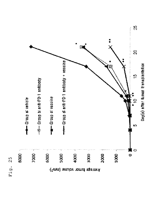
Fig. 25 shows the suppression of tumor proliferation
in vivo by a combination of an anti-PD-1 antibody and a
vaccine. Shown is the average tumor volume in
EL4-A24/Kb-WT1 tumor cells-transplanted mice that received
a vehicle (Group a), an anti-PD-1 antibody (Group b), a
vaccine (Group c), or an anti-PD-1 antibody and a vaccine
(Group d).
[0070)
Fig. 26 shows the suppression of tumor proliferation
in vivo by a combination of an anti-CTLA-4 antibody and a
vaccine. Shown is the average tumor volume in
EL4-A24/Kb-WT1 tumor cells-transplanted mice that received
a vehicle (Group a), an anti-CTLA-4 antibody (Group b), a
vaccine (Group c), or an anti-CTLA-4 antibody and a vaccine
(Group d).
[0071]
Fig. 27 shows the detection of WT1 antigen peptide-
specific CTLs by an HLA tetramer. The vertical axis shows
the ratio of WT1 antigen peptide-specific CTLs included in
spleen cells from a vaccinated mouse.
[0072]
Fig. 28 shows the IFN-y production by the stimulation
with a WT1 antigen peptide. WT1 antigen peptide-specific T
cells induced in a vaccinated mouse were cultured in the
presence of a WT1 killer peptide, and the IFN-y production
from the cells was measured by ELISA.
[0073]
Fig. 29 shows the 1FI-y production by the stimulation
with a WT1 antigen peptide. WT1 antigen peptide-specific T
cells induced in a vaccinated mouse were cultured with LLC-
HHD-WT1 tumor cells in the presence or absence of a WT1
killer peptide, and the IFN-y production from the cells was
CA 02986367 2017-11-17
measured by ELISA.
[0074]
Fig. 30A shows the IFN-y production by the treatment
with an immune checkpoint inhibitor. WT1 antigen peptide-
5 specific T cells induced in a vaccinated mouse were treated
with an anti-PD-1 or an isotype control antibody, and the
IFO-y production from the cells when cocultured with LLC-
HHD-WT1 tumor cells in the presence or absence of a WT1
killer peptide was measured by ELISA.
10 [0075]
Fig. 30B shows the IFN-y production by the treatment
with an immune checkpoint inhibitor. WT1 antigen peptide-
specific T cells induced in a vaccinated mouse were treated
with an anti-BTLA antibody, and the IFN-y production from
15 the cells when cocultured with LLC-HHD-WT1 tumor cells in
the presence of a WT1 killer peptide was measured by ELISA.
[0076]
Fig. 30C shows the IFN-y production by the treatment
with an immune checkpoint inhibitor. WT1 antigen peptide-
20 specific T cells induced in a vaccinated mouse were treated
with an anti-LAG-3 antibody, and the IFN-y production from
the cells when cocultured with LLC-HHD-WT1 tumor cells in
the presence of a WT1 killer peptide was measured by ELISA.
[0077]
25 Fig. 30D shows the IFN-y production by the treatment
with an immune checkpoint inhibitor. WT1 antigen peptide-
specific T cells induced in a vaccinated mouse were treated
with an anti-PD-L1 antibody, and the IFN-y production from
the cells when cocultured with LLC-HHD-WT1 tumor cells in
30 the presence of a WT1 killer peptide was measured by ELISA.
[0078]
Fig. 30E shows the IFN-y production by the treatment
with an immune checkpoint inhibitor. WTI antigen peptide-
specific T cells induced in a vaccinated mouse were treated
35 with an anti-VISTA antibody, and the IFN-y production from
CA 02986367 2017-11-17
36
the cells when cocultured with LLC-HHD-WT1 tumor cells in
the presence of a WT1 killer peptide was measured by ELISA.
[0079]
Fig. 31A shows the IFN-y production by the treatment
with a costimulatory molecule agonist antibody. WT1 antigen
peptide-specific T cells induced in a vaccinated mouse were
treated with an anti-4-1BB or an isotype control antibody,
and the IFN-y production from the cells when cocultured
with LLC-HHD-WT1 tumor cells in the presence or absence of
a WT1 killer peptide was measured by ELISA.
[0080]
Fig. 31B shows the IFN-y production by the treatment
with a costimulatory molecule agonist antibody. WT1 antigen
peptide-specific T cells induced in a vaccinated mouse were
treated with an anti-OX-40 antibody, and the IFN-y
production from the cells when cocultured with LLC-HHD-WT1
tumor cells in the presence of a WT1 killer peptide was
measured by ELISA.
[0081]
Fig. 31C shows the IFN-y production by the treatment
with a costimulatory molecule agonist antibody. WT1 antigen
peptide-specific T cells induced in a vaccinated mouse were
treated with an anti-GITR antibody, and the IFN-y
production from the cells when cocultured with LLC-HHD-WT1
tumor cells in the presence of a WT1 killer peptide was
measured by ELISA.
[0082]
Fig. 32 shows the IFN-y production by the treatment
with a P-catenin inhibitor. WT1 antigen peptide-specific T
cells induced in a vaccinated mouse were treated with
XAV939, and the IFN-y production from the cells when
cocultured with LLC-HHD-WT1 tumor cells in the presence of
a WT1 killer peptide was measured by ELISA.
[0083]
Fig. 33 shows the detection of WT1 antigen peptide-
CA 02986367 2017-11-17
37
specific CTLs by an HLA tetramer. The vertical axis shows
the ratio of WTI antigen peptide-specific CTLs included in
spleen cells from a vaccinated mouse.
[0084]
Fig. 34A shows the IFN-y production by the stimulation
with a WT1 antigen peptide. WT1 antigen peptide-specific T
cells induced in a mouse vaccinated with a killer vaccine
were cultured in the presence of a WT1 killer peptide, and
the IFN-y production from the cells was measured by ELISA.
[0085]
Fig. 34B shows the IFN-y production by the stimulation
with a WT1 antigen peptide. WT1 antigen peptide-specific T
cells induced in a mouse vaccinated with a cocktail vaccine
were cultured in the presence of a WT1 killer peptide, and
= 15 the IFN-y production from the cells was measured by ELISA.
[0086)
Fig. 35A shows the IFN-y production by the treatment
with an anti-PD-1 antibody. WT1 antigen peptide-specific T
cells induced in a mouse vaccinated with a killer vaccine
were treated with an anti-PD-1 antibody, and the IFN-y
production from the cells when cocultured with LLC-HHD-WT1
tumor cells in the presence of a WT1 killer peptide was
measured by ELISA.
[0087]
Fig. 35B shows the IFN-y production by the treatment
with an anti-PD-1 antibody. WTI antigen peptide-specific T
cells induced in a mouse vaccinated with a cocktail vaccine
were treated with an anti-PD-1 antibody, and the IFN-y
production from the cells when cocultured with LLC-HHD-WTI
tumor cells in the presence of a WT1 killer peptide was
measured by ELISA.
[0088]
Fig. 36A shows the IFN-y production by the treatment
with an anti-B7-H4 antibody. WT1 antigen peptide-specific T
cells induced in a mouse vaccinated with a killer vaccine
CA 02986367 2017-11-17
38
were treated with an anti-B7-H4 antibody, and the IFN-y
production from the cells when cocultured with LLC-HHD-WT1
tumor cells in the presence of a WT1 killer peptide was
measured by ELISA.
[0089]
Fig. 36B shows the IFN-y production by the treatment
with an anti-B7-H4 antibody. WT1 antigen peptide-specific T
cells induced in a mouse vaccinated with a cocktail vaccine
were treated with an anti-B7-H4 antibody, and the IFN-y
production from the cells when cocultured with LLC-HHD-WT1
tumor cells in the presence of a WT1 killer peptide was
measured by ELISA.
[0090]
Fig. 37A shows the IFN-y production by the treatment
with an anti-PD-L1 antibody. WT1 antigen peptide-specific T
cells induced in a mouse vaccinated with a killer vaccine
were treated with an anti-PD-L1 antibody, and the IFN-y
production from the cells when cocultured with LLC-HHD-WT1
tumor cells in the presence of a WT1 killer peptide was
measured by ELISA.
[0091]
Fig. 37B shows the IFN-y production by the treatment
with an anti-PD-L1 antibody. WT1 antigen peptide-specific T
cells induced in a mouse vaccinated with a cocktail vaccine
were treated with an anti-PD-L1 antibody, and the IFN-y
production from the cells when cocultured with LLC-HHD-WT1
tumor cells in the presence of a WT1 killer peptide was
measured by ELISA.
[0092]
Fig. 38A shows the IFN-y production by the treatment
with an anti-4-1BB antibody. WT1 antigen peptide-specific T
cells induced in a mouse vaccinated with a killer vaccine
were treated with an anti-4-18B antibody, and the IFN-y
production from the cells when cocultured with LLC-HHD-WT1
tumor cells in the presence of a WT1 killer peptide was
CA 02986367 2017-11-17
39
measured by ELISA.
[0093]
Fig. 38B shows the IFN-y production by the treatment
with an anti-4-1BB antibody. WT1 antigen peptide-specific T
cells induced in a mouse vaccinated with a cocktail vaccine
were treated with an anti-4-1BB antibody, and the IFN-y
production from the cells when cocultured with LLC-HHD-WT1
tumor cells in the presence of a WT1 killer peptide was
measured by ELISA.
[0094]
Fig. 39A shows the IFN-y production by the treatment
with an anti-OX-40 antibody. WT1 antigen peptide-specific T
cells induced in a mouse vaccinated with a killer vaccine
were treated with an anti-OX-40 antibody, and the IFN-y
production from the cells when cocultured with LLC-HHD-WT1
tumor cells in the presence of a WT1 killer peptide was
measured by ELISA.
[0095]
Fig. 39B shows the IFN-y production by the treatment
with an anti-OX-40 antibody. WT1 antigen peptide-specific T
cells induced in a mouse vaccinated with a cocktail vaccine
were treated with an anti-OX-40 antibody, and the IFN-y
production from the cells when cocultured with LLC-HHD-WT1
tumor cells in the presence of a WT1 killer peptide was
measured by ELISA.
DESCRIPTION OF EMBODIMENTS
[0096]
Embodiments of the present invention are explained in
detail hereinafter.
[0097]
The present invention relates to a combination use of
a WT1 antigen peptide and an immunomodulator.
[0098]
As used herein, a "WT1 antigen peptide" refers to a
CA 02986367 2017-11-17
peptide that has an amino acid sequence derived from WT1
protein, and binds to an MHC class I or class II molecule
to be presented on the cell surface as a complex with the
MHC molecule and induces killer T cells or helper T cells.
5 As used herein, a WT1 antigen peptide that binds to an MHC
class I molecule to induce killer T cells is referred as a
WT1 killer peptide, and a WT1 antigen peptide that binds to
an MHC class II molecule to induce helper T cells is
referred to as a "WT1 helper peptide". WT1 protein may be,
10 but not limited to, a mouse or human WT1 protein, and
preferably is a human WT1 protein. The human WT1 protein
has the amino acid sequence of SEQ ID NO: 1. As used herein,
the term "WT1 antigen peptide" include a pharmaceutically
acceptable salt thereof as long as it is appropriate in the
15 context.
[0099]
The WT1 antigen peptide may be modified at a part or
all of the amino acid residues in its amino acid sequence.
Such modified peptides may be prepared by any method known
20 in the art. For example, the modified peptide may be
prepared by modification such as esterification, alkylation,
halogenation, phosphorylation, sulfonation, or amidation of
the functional group of the side chain of the amino acid
residue(s) constituting the peptide. Also, a variety of
25 substances may be bound to the peptide at the N- and/ or C-
terminus. For example, an amino acid, a peptide, or an
analog thereof may be bound to the peptide. When such a
substance is bound to the WT1 antigen peptide, the
substance may be removed by any process, for example by an
30 enzymatic reaction in vivo or by intracellular processing,
such that the WT1 antigen peptide is finally generated. The
substance may be a substance that regulates the solubility
of the peptide; improves the stability of the peptide such
as protease resistance; delivers the peptide to a specific
35 tissue or organ; or increases an uptake of the peptide by
CA 02986367 2017-11-17
41
antigen presenting cells. The substance may be a substance
that increases the CTL inducing activity, such as a killer
or helper peptide different from the WT1 antigen peptide or
a pharmaceutically acceptable salt thereof.
[0100]
The WT1 antigen peptide may comprise a bond other than
a peptide bond such as a carbon-carbon bond, a carbon-
nitrogen bond, or a carbon-sulfur bond. Also, the WT1
antigen peptide may comprise one or more D-amino acids.
[0101]
The modified peptides as described above are
illustrative only and those skilled in the art can easily
conceive, prepare, examine and use other variations of the
peptides.
[0102]
The "amino acid residue" as used herein refers to a
single unit in the amino acids constituting a peptide or
protein molecule. The "amino acid residue" may be a natural
or non-natural a-amino acid residue, P-amino acid residue,
7-amino acid residue or 6-amino acid residue. Specifically,
The "amino acid residue" may be a natural a-amino acid
residue, ornithine residue, homoserine
residue,
homocysteine residue, 0-alanine, y-aminobutanoic acid or 6-
aminopentanoic acid.
[0103]
The "amino acid residue" as used herein may be
represented by the following abbreviations.
Ala or A: alanine residue
Arg or R: arginine residue
Asn or N: asparagine residue
Asp or D: aspartic acid residue
Cys or C: cysteine residue
Gln or Q: glutamine residue
Glu or E: glutamic acid residue
Gly or G: glycine residue
CA 02986367 2017-11-17
42
His or H: histidine residue
Ile or I: isoleucine residue
Leu or L: leucine residue
Lys or K: lysine residue
Met or M: methionine residue
Phe or F: phenylalanine residue
Pro or P: proline residue
Ser or S: serine residue
Thr or T: threonine residue
Trp or W: tryptophan residue
Tyr or Y: tyrosine residue
Val or V: valine residue
Abu: 2-aminobutyric acid residue (also referred to as a-
aminobutyric acid residue)
Orn: ornithine residue
Cit: citrulline residue
[0104]
The amino acid sequence of a peptide disclosed herein
is described according to the conventional method, wherein
the amino acid residue of the N-terminal amino acid is
positioned on the left side, and the amino acid residue of
the C-terminal amino acid is positioned on the right side.
In a peptide, unless otherwise indicated, the amino group
of the N-terminal amino acid residue binds to a hydrogen
atom, and the carbonyl group of the C-terminal amino acid
residue binds to a hydroxyl group. A divalent peptide group
means a group that binds at the amino group of the N-
terminal amino acid residue and the carbonyl group of the
C-terminal amino acid residue. For example, in the peptide
comprised in the compounds of formulae (1) to (3), the
amino group of the N-terminal amino acid residue binds to a
hydrogen atom, and the carbonyl group of the C-terminal
amino acid residue binds to a hydroxyl group.
[01051
MHC in human is called human leukocyte-type antigen
CA 02986367 2017-11-17
43
(HLA). HLA subtypes corresponding to MHO class I-molecules
include HLA-A, B, Cw, F and G. The expression "MHC class I-
restricted" as used herein means the property of inducing
killer T cells by binding to an MHC class I molecule.
Preferred examples of "MHC class I-restricted" peptides
include HLA-A-restricted peptides, HLA-B-restricted
peptides, and HLA-Cw-restricted peptides.
[0106]
Allelic Polymorphism is known for each HLA subtype.
The polymorphism in HLA-A has 27 or more types such as HLA-
Al, HLA-A2, and HLA-A24. The polymorphism in HLA-B has 59
or more types such as HLA-B7, HLA-B40, and HLA-B44. The
polymorphism in HLA-Cw has 10 or more types such as HLA-
Cw0301, HLA-Cw0401, and HLA-Cw0602. Among such polymorphism,
HLA-A0201 and HLA-A24 are preferred.
[0107]
In one embodiment, the WT1 antigen peptide is a killer
peptide, which binds to an MHC class I molecule and induces
killer T cells (cytotoxic T cells, CTLs). The WT1 killer
peptide induces WT1-specific killer T cells when presented
on the cell surface in a form of a complex with an MHC
class I molecule.
[0108]
In one embodiment, the WT1 killer peptide is a peptide
consisting of contiguous 7-30 amino acids in the amino acid
sequence of human WT1 protein of SEQ ID NO: 1 or a variant
thereof. The WT1 killer peptide may be a peptide comprising
the amino acid sequence selected from
=
RMFPNAPYL (SEQ ID NO: 2),
CMTWNQMNL (SEQ ID NO: 3),
CYTWNQMNL (SEQ ID NO: 4),
ALLPAVPSL (SEQ ID NO: 5),
SLGEQQYSV (SEQ ID NO: 6),
RVPGVAPTL (SEQ ID NO: 7),
VLDFAPPGA (SEQ ID NO: 8),
CA 02986367 2017-11-17
44
C-CMTWNQMNL (SEQ ID NO: 9) (wherein the bond within C-C is
a disulfide bond), and
C-CYTWNQMNL (SEQ ID NO: 10) (wherein the bond within C-C is
a disulfide bond),
or a peptide comprising an altered amino acid sequence of
the amino acid sequence selected from SEQ ID NOS: 2-10 that
comprises amino acid alteration in the amino acid sequence
and having the CTL inducing activity. Preferably, the WT1
killer peptide is a peptide consisting of the amino acid
sequence selected from SEQ ID NOS: 2-10, or a peptide
consisting of an altered amino acid sequence of the amino
acid sequence selected from SEQ ID NOS: 2-10 that comprises
amino acid alteration in the amino acid sequence and having
the CTL inducing activity. More preferably, the WT1 killer
peptide is a peptide consisting of the amino acid sequence
selected from SEQ ID NOS: 2-10. Even more preferably, the
WT1 killer peptide is a peptide consisting of the amino
acid sequence selected from SEQ ID NOS: 2-6, 8 and 10.
[0109]
As used herein, a peptide "comprising" a given amino
acid sequence means, as usually understood, a peptide that
may comprise a further amino acid(s) added to the N-
terminal and/or C-terminal amino acid of the given amino
acid sequence.
[0110]
As used herein, the "peptide comprising an altered
amino acid sequence of the amino acid sequence of ... that
comprises amino acid alteration in the amino acid sequence
and having the CTL inducing activity" is also called as an
altered killer peptide. The "altered killer peptide" means
a peptide that consists of an amino acid sequence wherein
one to several, preferably one to three amino acids are
deleted from, substituted in, and/or added to the original
amino acid sequence, and binds to MHC class I to induce
CTLs. The position of the amino acid to be substituted may
CA 02986367 2017-11-17
be position 1 (N-terminal), position 2, position 3 or
position 9 for a peptide consisting of 9 amino acid
residues. The number of amino acids to be added (or
inserted, since "addition" encompasses "insertion") is
5 preferably 1 or 2, more preferably 1. A preferable position
for addition is the C-terminal. The number of amino acids
to be deleted is preferably 1. In the alteration, the amino
acid to be added or substituted may be a non-natural amino
acid other than the 20 genetically encoded amino acids.
10 [0111]
The regularity in the amino acid sequence (binding
motif) has been known in peptides that bind to an HLA
antigen for each type of polymorphism in an HLA subtype.
For example, to make the binding motif for HLA-A24, in a
15 peptide consisting of 8 to 11 amino acid residues, the
amino acid at position 2 is Tyr, Phe, Met or Trp, and the
amino acid at the C-terminus is Phe, Leu, Ile, Trp or Met
(J. Immunol., 152, p3913, 1994; J. Immunol., 155, p4307,
1994; Immunogenetics, 41, p178, 1995). Thus, in a peptide
20 consisting of 9 amino acid residues, the amino acid at
position 2 may be replaced with Tyr, Phe, Met or Trp and/or
the amino acid at position 9 may be replaced with Phe, Leu,
Ile, Trp or Met. A peptide containing such amino acid
alteration is a preferred altered killer peptide. Similarly,
25 to make the binding motif for HLA-A2, in a peptide
consisting of 8 to 11 amino acid residues, the amino acid
at position 2 is Leu or Met, and the amino acid at the C-
terminus is Val or Leu. Thus, in a peptide consisting of 9
amino acid residues, the amino acid at position 2 may be
30 replaced with Leu or Met and the amino acid at the C-
terminus may be replaced with Val or Leu. A peptide
containing such amino acid alteration is a preferred
altered killer peptide
[0112]
35 Examples of altered killer peptides include
CA 02986367 2017-11-17
46
an altered killer peptide of RMFPNAPYL (SEQ ID NO: 2) such
as
RYFPNAPYL (SEQ ID NO: 21) (W003/106682)
FMFPNAPYL (SEQ ID NO: 22),
RLFPNAPYL (SEQ ID NO: 23),
RMMPNAPYL (SEQ ID NO: 24),
RMFPNAPYV (SEQ ID NO: 25) and
YMFPNAPYL (SEQ ID NO: 26) (W02009/072610);
an altered killer peptide of CMTWNQMNL (SEQ ID NO: 3) such
as
CYTWNQMNL (SEQ ID NO: 4) (W002/79253),
Xaa-Met-Thr-Trp-Asn-Gln-Met-Asn-Leu (SEQ ID NO: 27),
(wherein Xaa is Ser or Ala) and
Xaa-Tyr-Thr-Trp-Asn-Gln-Met-Asn-Leu (SEQ ID NO: 28)
(wherein Xaa is Ser, Ala, Abu, Arg, Lys, Orn, Cit, Leu, Phe
or Asn) (W02004/026897);
an altered killer peptide of ALLPAVPSL (SEQ ID NO: 5) such
as
AYLPAVPSL (SEQ ID NO: 29) (W02003/106682);
an altered killer peptide of SLGEQQYSV (SEQ ID NO: 6) such
as
FLGEQQYSV (SEQ ID NO: 30),
SMGEQQYSV (SEQ ID NO: 31) and
SLMEQQYSV (SEQ ID NO: 32) (W02009/072610); and
an altered killer peptide of RVPGVAPTL (SEQ ID NO: 7) such
as
RYPGVAPTL (SEQ ID NO: 33) (W02003/106682).
[0113]
In one embodiment, the WT1 killer peptide is
the compound of formula (1):
CA 02986367 2017-11-17
47
CRMFPNAPYL
(1)
CSLGEQQYSV
(wherein the bond within C-C is a disulfide bond),
the compound of formula (2):
CALLPAVPSL
(2)
CYTWNQMNL
(wherein the bond within C-C is a disulfide bond), or
the compound of formula (3):
CRMFPNAPYL
(3)
CYTWNQMNL
(wherein the bond within C-C is a disulfide bond).
[0114]
In addition to the peptides and compounds as described
above, examples of WT1 antigen peptides (killer or helper
peptides) include the compounds disclosed in W02014/157692.
[0115]
In one embodiment, the WT1 antigen peptide is a helper
peptide, which binds to an MHC class II molecule and
induces helper T cells (CD4+ T cells). The WT1 helper
peptide induces WT1-specific helper T cells when presented
on the cell surface in a form of a complex with an MHC
class II molecule. The WT1-specific helper T cells produce
cytokines such as IL-2, IL-4, IL-5, IL-6 or interferons
(IFNs) and promote proliferation, differentiation, or
maturation of B cells or other subsets of T cells. Thus,
the WT1 helper peptide activates helper T cells to induce
or maintain CTLs or to activate effector cells such as
macrophages, being useful to efficiently treat or prevent
cancer.
CA 02986367 2017-11-17
48
[0116]
HLA subtypes corresponding to the MHC class II-
molecules include HLA-DR, DQ and DP. The expression "MHC
class II-restricted" as used herein means the property of
inducing helper cells by binding to an MHC class II
molecule. Preferred examples of the "MHC class II-
restricted" peptides include HLA-DR-restricted peptides,
HLA-DQ-restricted peptides and HLA-DP-restricted peptides.
[0117]
In one embodiment, the WT1 helper peptide is a peptide
consisting of contiguous 7-30 amino acids, preferably 14-30
amino acids, in the amino acid sequence of human WT1
protein of SEQ ID NO: 1 or a variant thereof. The WT].
helper peptide may be a peptide comprising the amino acid
sequence selected from
KRYFKLSHLQMHSRKH (SEQ ID NO: 11),
SGQARMFPNAPYLPSCLES (SEQ ID NO: 12),
RSDELVRHHNMHQRNMTKL (SEQ ID NO: 13),
PGCNKRYFKLSHLQMHSRKHTG (SEQ ID NO: 14),
CNKRYFKLSHLQMHSRK (SEQ ID NO: 15),
CNKRYFKLSHLQMHSRKH (SEQ ID NO: 16),
CNKRYFKLSHLQMHSRKHTG (SEQ ID NO: 17),=
WAPVLDFAPPGASAYGSL (SEQ ID NO: 18),
CWAPVLDFAPPGASAYGSL (SEQ ID NO: 19),
WAPVLDFAPPGASAYGSLC (SEQ ID NO: 20),
SGQARMFPNAPYLPSC (SEQ ID NO: 34),
SGQAYMFPNAPYLPSC (SEQ ID NO: 35),
SGQARMFPNAPYLPSCLES (SEQ ID NO: 36),
SGQAYMFPNAPYLPSCLES (SEQ ID NO: 37),
PGCNKRYFKLSHLQMHSRK (SEQ ID NO: 38),
PGCNKRYFKLSHLQMHSRKH (SEQ ID NO: 39),
PGCNKRYFKLSHLQMHSRKHTG (SEQ ID NO: 40),
QARMFPNAPYLPSCL (SEQ ID NO: 44),
LKGVAAGSSSSVKWT (SEQ ID NO: 45) and
RYFKLSHLQMHSRKH (SEQ ID NO: 46),
CA 02986367 2017-11-17
49
or a peptide comprising an altered amino acid sequence of
= the amino acid sequence selected from SEQ ID NOS: 11-20,
34-40 and 44-46 that comprises amino acid alteration in the
amino acid sequence and having the helper T cell inducing
activity. Preferably, the WT1 helper peptide is a peptide
consisting of the amino acid sequence selected from SEQ ID
NOS: 11-20 and 34-40 or a peptide consisting of an altered
amino acid sequence of the amino acid sequence selected
from SEQ ID NOS: 11-20 and 34-40 that comprises amino acid
alteration in the amino acid sequence, or a peptide
consisting of an altered amino acid sequence of the amino
acid sequence selected from SEQ ID NOS: 11-20 and 34-40
that comprises amino acid alteration in the amino acid
sequence and having the helper T cell inducing activity.
More preferably, the WT1 helper peptide is a peptide
consisting of the amino acid sequence selected from SEQ ID
NOS: 11-20 and 34-40. Even more preferably, the WT1 helper
peptide is a peptide consisting of the amino acid sequence
selected from SEQ ID NOS: 11-20.
[0118]
As used herein, the "peptide comprising an altered
amino acid sequence of the amino acid sequence of ... that
comprises amino acid alteration in the amino acid sequence
and having the helper T cell inducing activity" is also
called as an altered helper peptide. The "altered helper
peptide" means a peptide that consists of an amino acid
sequence wherein one to several, preferably one to three
amino acids are deleted from, substituted in, and/or added
to the original amino acid sequence, and binds to MHC class
II to induce CTLs. In the alteration, the amino acid to be
added or substituted may be a non-natural amino acid other
than the 20 genetically encoded amino acids.
[0119]
Examples of altered helper peptides include
CA 02986367 2017-11-17
an altered helper peptide of SGQARMFPNAPYLPSCLES (SEQ ID
NO: 36) such as
SGQAYMFPNAPYLPSCLES (SEQ ID NO: 37) (W02007/120673),
SGQARMFPNAPYLPSC (SEQ ID NO: 34), and
5 SGQAYMFPNAPYLPSC (SEQ ID NO: 35); and
an altered helper peptide of PGCNKRYFKLSHLQMHSRKHTG (SEQ ID
NO: 40) such as
PGCNKRYFKLSHLQMHSRK (SEQ ID NO: 38),
10 PGCNKRYFKLSHLQMHSRKH (SEQ ID NO: 39),
KRYFKLSHLQMHSRKH (SEQ ID NO: 11),
CNKRYFKLSHLQMHSRK (SEQ ID NO: 15),
CNKRYFKLSHLQMHSRKH (SEQ ID NO: 16) and
CNKRYFKLSHLQMHSRKHTG (SEQ ID NO: 17).
15 [0120]
The WT1 antigen peptide may be prepared according to
any method generally used in the peptide synthesis.
Examples of such methods include those described in the
references such as Peptide Synthesis, Interscience, New
20 York, 1966; The Proteins, Vol 2, Academic Press Inc., New
York, 1976; peptide synthesis, Maruzen Co., LTD., 1975;
Basics and Experiment of Peptide Synthesis, Maruzen Co.,
LTD., 1985; and Development of Pharmaceutical Product
subsequent vol. 14, Peptide Synthesis, Hirokawa Shoten,
25 1991. For example, the peptide may be prepared by the Fmoc
method or the Boc method using a solid phase synthesizer,
or by sequential condensation of Boc-amino acid or Z-amino
acid in the liquid phase synthesis (wherein Fmoc is a 9-
fluorenylmethoxycarbonyl group, Boc is a t-butoxycarbonyl
30 group, and Z is a benzyloxycarbonyl group).
[0121]
A functional group in an intermediate to prepare the
WT1 antigen peptide such as an amino, carboxy, or mercapto
group may be protected by a suitable protecting group and
35 deprotected as necessary using protection and deprotection
CA 02986367 2017-11-17
51
techniques. Preferred protecting groups, protection methods,
and deprotection methods are described in detail, for
example, in "Protective Groups in Organic Synthesis 2nd
Edition (John Wiley & Sons, Inc.; 1990)". Examples of
mercapto-protecting groups include an acetamidomethyl group
and a trityl group.
[0122]
When the WT1 antigen peptide has a disulfide bond, the
disulfide bond may be formed between two different peptides
each containing a cysteine residue, or between a peptide
containing a cysteine residue and cysteine, according to
any method generally used in peptide chemistry. Examples of
methods to form a disulfide bond include those described in
the references such as Peptide Synthesis, Interscience, New
York, 1966; The Proteins, Vol. 2, Academic Press Inc., New
York, 1976; peptide synthesis, Maruzen Co., LTD., 1975;
Basics and Experiment of peptide synthesis, Maruzen Co.,
LTD., 1985; and Development of Pharmaceutical Product
sequential vol. 14, Peptide Synthesis, Hirokawa Shoten,
1991.
[0123]
Specifically, when the peptide contains one cysteine
residue, a compound having a disulfide bond (also referred
to as "a disulfide compound") may be prepared by removing
all protecting groups including the mercapto-protecting
group on the cysteine side chain and oxidizing the peptide
in an inert solvent. In addition, a disulfide compound may
be prepared by mixing two intermediates each having a
mercapto group in a suitable solvent and oxidizing the
mixture. The method for the oxidation may be selected as
appropriate from known methods for disulfide bond formation
in usual peptide synthesis. For example, the oxidation may
be iodine oxidation; air oxidation under alkali conditions;
or oxidation with an oxidant under alkaline or acidic
conditions to form a disulfide bond. Examples of the
CA 02986367 2017-11-17
52
oxidant include iodine, dimethyl sulfoxide (DMSO), and
potassium ferricyanide. Examples of the solvent include
water, acetic acid, methanol, chloroform, DMF, and DMSO, or
a mixture thereof. The oxidation reaction often provides a
mixture of symmetric and asymmetric disulfide compounds.
The desired asymmetric disulfide compound may be obtained
by purification using techniques such as various types of
chromatography or recrystallization. Alternatively, a
disulfide bond may be selectively formed by mixing an
intermediate having an activated mercapto group and another
intermediate having a mercapto group. Examples of the
intermediate having an activated mercapto group include an
intermediate comprising a mercapto group bonded with an
Npys group (3-nitro-2-pyridinesulphenyl group).
Alternatively, one intermediate is mixed with an agent to
activate the mercapto group, for example, 2,2'-dithiobis(5-
nitropyridine), and then the other intermediate is added
thereto, whereby a disulfide bond may be selectively formed
(Tetrahedron Letters. Vol. 37. No. 9, pp. 1347-1350).
[0124]
The methods as described above also may be used when
the peptide contains two or more cysteine residues. In this
case, isomers having different disulfide bonding patterns
are obtained. Using a specific combination of protecting
groups for the side chains of cysteines provides a desired
dimer having a disulfide bond between specific cysteine
residues. Examples of the combination of protecting groups
include MeBz1 (methylbenzyl) group and Acm
(acetamidomethyl) group, Trt (trityl) group and Acm group,
Npys (3-nitro-2-pyridylthio) group and Acm group, and S-Bu-
t (S-tert-butyl) group and Acm group. For example, when the
combination of MeBz1 group and Acm group is used, MeBz1
groups and protecting groups other than those of cysteine
side chains are removed and then a solution containing the
peptide monomers is subjected to air oxidation reaction to
CA 02986367 2017-11-17
53
form a disulfide bond between the deprotected cysteine
residues. Then, after the deprotection with iodine and
oxidation, a disulfide bond between the cysteine residues
protected with Acm groups is formed.
[0125]
The WT1 antigen peptide may be a compound composed of
a killer peptide and a helper peptide or two different
killer or helper peptides conjugated via a disulfide bond.
Such peptides may be synthesized by a method comprising the
following steps (1) to (3).
[0126]
The step (1) uses Fmoc-C(Mmt)A-SBn and a first antigen
peptide. The step (1) synthesizes a peptide wherein the
carbonyl group of the C-terminal amino acid of C(Mmt)A
binds to the N-terminal amino group of the first antigen
peptide. The "Fmoc" is a 9-fluorenylmethoxycarbony1 group.
The "Mmt" is a monomethoxytrityl group. The "SBn" is a
thiobenzyl group.
[0127]
The step (2) uses the peptide obtained in step (1) and
a second antigen peptide that has a cysteine residue
protected by Npsy group at the N-terminus. The step (2)
synthesizes a peptide wherein the thioether group of the
cysteine residue of the first antigen peptide in the
peptide obtained in step (1) binds to the thioether group
of the cysteine residue added to the N-terminus of the
second antigen peptide. The "Nspy" is a 3-nitro-2-
pyridylthio group.
[0128]
The step (3) uses the peptide obtained in step (2) and
a third antigen peptide that has a cysteine residue
protected by Spy group at the N-terminus. The step (3)
synthesizes a peptide wherein the thioether group of the N-
terminus cysteine residue of the second antigen peptide in
the peptide obtained in step (2) binds to the thioether
CA 02986367 2017-11-17
54
group of the cysteine residue of the third antigen peptide.
The Spy" is a 2-pyridylsulfide group.
[0129]
The WT1 antigen peptide thus obtained may be purified
according to any method known to those of ordinary skill in
the art or generally used for peptide chemistry. For
example, The WT1 antigen peptide may be purified by
techniques such as various types of chromatography (e.g.,
silica gel column chromatography, ion exchange column
chromatography, gel filtration or reversed-phase
chromatography) and recrystallization. For example, the
recrystallization solvent may be an alcohol solvent such as
methanol, ethanol or 2-propanol; ether solvent such as
diethyl ether; ester solvent such as ethyl acetate;
aromatic hydrocarbon solvent such as benzene or toluene;
ketone solvent such as acetone; hydrocarbon solvent such as
hexane; aprotonic solvent such as = dimethylformamide or
acetonitrile; water; or a mixture thereof. Different
purification methods described in Jikken Kagaku Kouza (The
Chemical Society of Japan ed., Maruzen) vol. 1 or other
references also may be used.
[0130]
Purification methods for disulfide compounds are
described in the references such as Peptide Synthesis,
Interscience, New York, 1966; The Proteins, Vol. 2,
Academic Press Inc., New York, 1976; peptide synthesis,
Maruzen Co., LTD., 1975; Basics and Experiment of Peptide
Synthesis, Maruzen Co., LTD., 1985; and Development of
Pharmaceutical Product sequential vol. 14, peptide
synthesis, Hirokawa Shoten, 1991. Among these methods, HPLC
is preferred.
(0131]
When the WT1 antigen peptide has one or more
asymmetric points, the peptide may be prepared according to
a general method using starting materials (amino acids)
CA 02986367 2017-11-17
having the asymmetric point(s). To increase the optical
purity of the WT1 antigen peptide, processes such as
optical resolution may be performed at a suitable stage of
the preparation. Examples of methods for optical resolution
5 include a diastereomer method, which forms a salt of the
WT1 antigen peptide or an intermediate thereof with an
optically active acid (e.g., monocarboxylic acid such as
mandelic acid, N-benzyloxyalanine, or lactic acid,
dicarboxylic acid such as tartaric acid, o-
10 diisopropylidenetartaric acid, or malic acid, or sulfonic
acid such as camphorsulfonic acid or bromocamphorsulfonic
acid) in an inert solvent (e.g., alcohol solvent such as
methanol, ethanol, or 2-propanol, ether solvent such as
diethyl ether, ester solvent such as ethyl acetate,
15 hydrocarbon solvent such as toluene, aprotonic solvent such
as acetonitrile, or a mixture thereof). When the WT1
antigen peptide or intermediate has an acidic functional
group such as carboxy group, optical resolution may also be
performed following the formation of a salt with an
20 optically active amine (e.g., organic amine such as a-
phenethylamine, kinin, quinidine, cinchonidine, cinchonine,
or strychnine).
[0132]
The temperature for forming the salt is selected from
25 the range of room temperature to the boiling point of the
solvent. To improve the optical purity, it is desirable to
once raise the temperature to around the boiling point of
the solvent. When a precipitated salt is collected by
filtration, the yield may be increased as necessary by
30 cooling. The optically active acid or amine may be used in
an amount of about 0.5 - about 2.0 equivalents, preferably
about 1 equivalent, relative to the substrate. Where
necessary, crystals may be recrystallized in an inert
solvent (e.g., alcohol solvent such as methanol, ethanol,
35 or 2-propanol, ether solvent such as diethyl ether, ester
CA 02986367 2017-11-17
56
solvent such as ethyl acetate, hydrocarbon solvent such as
toluene, aprotonic solvent such as acetonitrile, or a
mixture thereof) to provide an optically active salt with
high purity. Where necessary, the optically resolved salt
may be treated with an acid or base by a general method to
give a free form.
[0133]
The "pharmaceutically acceptable salt" as used herein
may be an acid addition salt or a base addition salt. The
acid addition salt may be an inorganic acid salt such as
hydrochloride, hydrobromide, sulfate, hydroiodide, nitrate,
and phosphate, or an organic acid salt such as citrate,
oxalate, acetate, formate, propionate,
benzoate,
trifluoroacetate, maleate, tartrate, methanesulfonate,
benzenesulfonate, or p-toluenesulfonate. The base addition
salt may be a salt with an inorganic base such as sodium
salt, potassium salt, calcium salt, magnesium salt, or
ammonium salt, a salt with an organic base such as
triethylammonium salt, triethanolammonium salt, pyridinium
salt, or diisopropylammonium salt. Also, the salt may be an
amino acid salt with a basic or acidic amino acid such as
arginine, aspartic acid, and glutamic acid.
[0134]
The pharmaceutically acceptable salt of a WT1 killer
peptide may be an acid addition salt or a base addition
salt of a peptide consisting of the amino acid sequence
selected from
RMFPNAPYL (SEQ ID NO: 2),
CMTWNQMNL (SEQ ID NO: 3),
CYTWNQMNL (SEQ ID NO: 4),
ALLPAVPSL (SEQ ID NO: 5),
SLGEQQYSV (SEQ ID NO: 6),
RVPGVAPTL (SEQ ID NO: 7),
VLDFAPPGA, (SEQ ID NO: 8),
C-CMTWNQMNL (SEQ ID NO: 9) (wherein the bond within C-C is
CA 02986367 2017-11-17
57
a disulfide bond), and
C-CYTWNQMNL (SEQ ID NO: 10) (wherein the bond within C-C is
a disulfide bond), or
a peptide comprising an altered amino acid sequence of the
amino acid sequence selected from SEQ ID NOS: 2-10 that
comprises one to several amino acid deletions,
substitutions, or additions in the amino acid sequence and
having the CTL inducing activity; or
a compound represented by any one of the formulae (1) to
(3). The acid addition salt may be an inorganic acid salt
such as hydrochloride, hydrobromide, sulfate, hydroiodide,
nitrate, and phosphate, or an organic acid salt such as
citrate, oxalate, acetate, formate, propionate, benzoate,
trifluoroacetate, maleate, tartrate, methanesulfonate,
benzenesulfonate, or p-toluenesulfonate. The base addition
salt may be a salt with an inorganic base such as sodium
salt, potassium salt, calcium salt, magnesium salt, or
ammonium salt, a salt with an organic base such as
triethylammonium salt, triethanolammonium salt, pyridinium
salt, or diisopropylammonium salt. Also, the salt may be an
amino acid salt with a basic or acidic amino acid such as
arginine, aspartic acid, and glutamic acid.
[0135]
The pharmaceutically acceptable salt of a WT1 helper
peptide may be an acid addition salt or a base addition
salt of a peptide consisting of the amino acid sequence
selected from
KRYFKLSHLQMHSRKH (SEQ ID NO: 11),
SGQARMFPNAPYLPSCLES(SEQ ID NO: 12),
RSDELVRHHNMHQRNMTKL (SEQ ID NO: 13),
PGCNKRYFKLSHLQMHSRKHTG (SEQ ID NO: 14),
CNKRYFKLSHLQMHSRK (SEQ ID NO: 15),
CNKRYFKLSHLQMHSRKH (SEQ ID NO: 16),
CNKRYFKLSHLQMHSRKHTG (SEQ ID NO: 17),
WAPVLDFAPPGASAYGSL (SEQ ID NO: 18),
CA 02986367 2017-11-17
58
CWAPVLDFAPPGASAYGSL (SEQ ID NO: 19) and
WAPVLDFAPPGASAYGSLC (SEQ ID NO: 20); or
a peptide comprising an altered amino acid sequence of the
amino acid sequence selected from SEQ ID NOS: 11-20 that
comprises one to several amino acid deletions,
substitutions, or additions in the amino acid sequence and
having the helper T cell inducing activity. The acid
addition salt may be an inorganic acid salt such as
hydrochloride, hydrobromide, sulfate, hydroiodide, nitrate,
and phosphate, or an organic acid salt such as citrate,
oxalate, acetate, formate, propionate,
benzoate,
trifluoroacetate, maleate, tartrate, methanesulfonate,
benzenesulfonate, or p-toluenesulfonate. The base addition
salt may be a salt with an inorganic base such as sodium
salt, potassium salt, calcium salt, magnesium salt, or
ammonium salt, a salt with an organic base such as
triethylammonium salt, triethanolammonium salt, pyridinium
salt, or diisopropylammonium salt. Also, the salt may be an
amino acid salt with a basic or acidic amino acid such as
arginine, aspartic acid, and glutamic acid.
[0136]
The pharmaceutically acceptable salt of a WT1 killer
peptide may be, but not limited to,
RMFPNAPYL acetate,
CMTWNQMNL acetate,
CYTWNQMNL acetate,
ALLPAVPSL acetate,
SLGEQQYSV acetate,
RVPGVAPTL acetate,
VLDFAPPGA acetate,
C-CMTWNQMNL acetate,
C-CYTWNQMNL acetate,
CA 02986367 2017-11-17
59
CRMFPNAPYL (1)
CSLGEQQYSV
acetate,
CALLPAVPSL
(2)
CYTWNQMNL
acetate,
CRMFPNAPYL
(3)
CYTWNQMNL
acetate,
KRYFKLSHLQMHSRKH acetate,
SGQARMFPNAPYLPSCLES acetate,
RSDELVRHHNMHQRNMTKL acetate,
PGCNKRYFKLSHLQMHSRKHTG acetate,
CNKRYFKLSHLQMHSRK acetate,
CNKRYFKLSHLQMHSRKH acetate,
CNKRYFKLSHLQMHSRKHTG acetate,
WAPVLDFAPPGASAYGSL acetate,
CWAPVLDFAPPGASAYGSL acetate,
WAPVLDFAPPGASAYGSLC acetate,
RMFPNAPYL trifluoroacetate,
CMTWNQMNL trifluoroacetate,
CYTWNQMNL trifluoroacetate,
ALLPAVPSL trifluoroacetate,
SLGEQQYSV trifluoroacetate,
RVPGVAPTL trifluoroacetate,
VLDFAPPGA trifluoroacetate,
C-CMTWNQMNL trifluoroacetate,
C-CYTWNQMNL trifluoroacetate,
CRMFPNAPYL (i)
CSLGEQQYSV
trifluoroacetate,
CA 02986367 2017-11-17
CALLPAVPSL
(2)
CYTWNQMNL
trifluoroacetate,
CRP FPNAPYL
(3)
CYTWNQMNL
trifluoroacetate,
KRYFKLSHLQMHSRKH trifluoroacetate,
SGQARMFPNAPYLPSCLES trifluoroacetate,
5 RSDELVRHHNMHQRNMTKL trifluoroacetate,
PGCNKRYFKLSHLQMHSRKHTG trifluoroacetate,
CNKRYFKLSHLQMHSRK trifluoroacetate,
CNKRYFKLSHLQMHSRKH trifluoroacetate,
CNKRYFKLSHLQMHSRKHTG trifluoroacetate,
10 WAPVLDFAPPGASAYGSL trifluoroacetate,
CWAPVLDFAPPGASAYGSL trifluoroacetate, or
WAPVLDFAPPGASAYGSLC trifluoroacetate.
[0137]
The present invention also encompasses any hydrate and
15 solvate, such as ethanol solvate, of the WT1 antigen
peptide or a pharmaceutically acceptable salt thereof.
Further, the present invention encompasses any possible
stereoisomer, such as diastereomer or enantiomer, or any
crystal form of the WT1 antigen peptide.
20 [0138]
The CTL inducing activity may be confirmed by
measuring the number of CTLs by the HLA tetramer method
(Int. J. Cancer: 100, 565-570 (2002)) or the limiting
dilution method (Nat. Med.: 4, 321-327 (1998)). To
25 determine HLA-A24-restricted CTL inducting activity, the
HLA-A24 model mouse described in WO 02/47474 and Int. J.
Cancer: 100, 565-570 (2002) may be used. The helper T cell
inducing activity may be confirmed by any method known in
the art, such as the method described in Cancer Immunol.
CA 02986367 2017-11-17
61
Immunother. 51:271 (2002).
[0139]
As used herein, the term "immunomodulator" means any
agent that controls transmission of costimulatory signals
during activation of T cells with antigen-presenting cells
by interacting with molecules involved in the transmission
of the costimulatory signals and present on the antigen-
presenting cells and/or T cells, as well as any agent that
directly or indirectly controls function of molecules
involved in establishment of immune
tolerance
(immunosuppression) in the immune system. Examples of
"immunomodulator" include, but are not limited to,
antibodies, nucleic acids, proteins, peptides, and small
compounds. As used in the context of "immunomodulator", the
term "antibody" includes antibody fragments. Examples of
the antibody fragment include heavy and light chain
variable regions of an antibody (VH and VL), F(ab')2, Fab',
Fab, Fv, Fd, sdFv, and scFV. As used in the context of
"immunomodulator", the term "protein" means any protein
other than antibodies. Examples of the term
"immunomodulator" include immune checkpoint inhibitors,
costimulatory molecule agonists, immune activating agents,
and low-molecular inhibitors.
[0140]
The "immune checkpoint inhibitor" inhibits
immunosuppressive effect induced by cancer cells or antigen
presenting cells. Examples of the immune checkpoint
inhibitor include, but are not limited to, agents against a
molecule selected from the group consisting of: (1) CTLA-4
(e.g., ipilimumab and tremelimumab); (2) PD-1 (e.g.,
nivolumab, pembrolizumab, AMP-224, AMP-514 (MEDI0680), and
pidilizumab (CT-011)); (3) LAG-3 (e.g., IMP-321 and BMS-
986016); (4) BTLA; (5) KIR (e.g., IPH2101); (6) TIM-3; (7)
PD-L1 (e.g., durvalumab (MEDI4736), MPDL3280A, BMS-936559,
and avelumab (MSB0010718C)); (8) PD-12; (9) B7-H3 (e.g.,
CA 02986367 2017-11-17
62
MGA-271); (10) B7-H4; (11) HVEM; (12) GAL9; (13) CD160;
(14) VISTA; (15) BTNL2; (16) TIGIT; (17) PVR; (18) BTN1A1;
(19) BTN2A2; (20) BTN3A2 (Nat Rev Drug Discov. 2013; 12:
130-146; Nikkei Medical Cancer Review 2014; 9; Nat Rev
Immunol. 2014; 14: 559-69); and (21) CSF1-R.
[0141]
The "costimulatory molecule agonist" activates T-cells
by transmitting an auxiliary signal via a costimulatory
molecule on the T cells and/or antigen presenting cells to
attenuate the immunosuppressive effect of cancer cells or
antigen presenting cells. Examples of the costimulatory
molecule agonist include, but are not limited to, agents
against a molecule selected from the group consisting of:
(1) 4-1BB; (2) 4-1BB-L; (3) 0X40 (4) 0X40-L; (5) GITR; (6)
CD28; (7) CD40; (8) CD40-L; (9) ICOS; (10) ICOS-L; (11)
LIGHT; and (12) CD27.
[0142]
The "immune activating agent" efficiently stimulates
killer T cells in the lymph nodes by directly or indirectly
activating immune cells such as T cells and dendritic cells.
Examples of the immune activating agent include, but are
not limited to, Toll-like receptor (TLR) agonists,
stimulator of interferon genes (STING) agonists, cytokines,
and agents against heat shock protein (HSP).
[0143]
Examples of the "Toll-like receptor (TLR) agonist"
include, but are not limited to, TLR1/2 agonists, TLR2
agonists, TLR3 agonists (e.g., PolyI:C), TLR4 agonists
(e.g.. S-type lipopolysaccharide, paclitaxel, lipid A, and
monophosphoryl lipid A), TLR5 agonists (e.g., flagellin),
TLR6/2 agonists (e.g., MALP-2), TLR7 agonist, TLR7/8
agonists (e.g., gardiquimod, imiquimod, loxoribine, and
resiquimod (R848)). TLR7/9 agonists (e.g.,
hydroxychloroquine sulfate), TLR8 agonists (e.g., motolimod
(VTX-2337)), TLR9 agonists (e.g., CpG-ODN), and TLR11
CA 02986367 2017-11-17
63
agonists (e.g., profilin).
[0144]
Examples of the "cytokine" include, but are not
limited to, IL-la, IL-1p, IL-2, IL-3, IL-4, IL-5, IL-6, IL-
7, IL-8, IL-9, IL-10, IL-11, IL-12, IL-13, IL-14, IL-15,
IL-16, IL-17, IL-18, interferon (INF)-a, INF-p, INF-y, SCF,
GM-CSF, G-CSF, M-CSF, erythropoietin, thrombopoietin,
macrophage inflammatory protein (MIP), and monocyte
chemoattractant protein (MCP).
[0145]
Examples of the "heat shock protein (HSP)" include,
but are not limited to, HSP70, HSP90, HSP90u, HSP90p,
HSP105, HSP72, and HSP40. Agents against a heat shock
protein include HSP inhibitors. For example, examples of
HSP90 inhibitor include, but are not limited to,
tanespimycin (17-AAG), luminespib (AUY-922, NVP-AUY922),
alvespimycin (17-DMAG) hydrochloride, ganetespib (STA-9090),
BIIB021, onalespib (AT13387), geldanamycin, NVP-BEP800,
SNX-2112 (PF-04928473), PF-4929113 (SNX-5422), KW-2478,
XL888, VER155008, VER-50589, CH5138303, VER-49009, NMS-E973,
PU-H71, HSP990 (NVP-HSP990) and KNK437.
[0146]
Examples of the "low-molecular inhibitor" include, but
are not limited to, histone deacetylase inhibitors, histone
demethylase inhibitors, histone
acetyltransferase
inhibitors, histone methyltransferase inhibitors, DNA
methyltransferase inhibitors, anthracycline antibiotics,
platinum formulations, MAPK inhibitors, p-catenin
inhibitors, STAT3 inhibitors, NF-kB inhibitors, JAK
inhibitors, mTOR inhibitors, IDO inhibitors, COX-2
inhibitors, CXCR4 inhibitors, and arginase inhibitors.
[0147]
Examples of the "histone deacetylase inhibitor"
include, but are not limited to, vorinostat (SAHA, MK0683),
entinostat (MS-275), panobinostat (LBH589), trichostatin A
CA 02986367 2017-11-17
64
(TSA), mocetinostat (MGCD0103), BG45, BRD73954, belinostat
(PXD101), romidepsin (FK228, depsipeptide), 4SC-202, HPOB,
LMK-235, CAY10603, tasquinimod, TMP269, nexturastat A,
rocilinostat (ACY-1215), RGFP966, RG2833
(RGFP109),
scriptaid, tubastatin A, pracinostat (SB939), CUDC-101,
M344, PCI-34051, dacinostat (LAQ624), tubastatin A
hydrochloride, abexinostat (PCI-24781), CUDC-907, AR-42,
sodium phenylbutyrate, resminostat, tubacin, quisinostat
(JNJ-26481585) dihydrochloride, MC1568,
givinostat
(ITF2357), droxinostat, chidamide (C S055, HBI-8000), CHR-
2485, CHR-3996, DAC-060, FRM-0334 (EVP-0334), MGCD-290,
CXD-101 (AZD-9468), CG200745,
arginine butyrate,
sulforaphane, SHP-141, CUDC-907, YM753 (OBP-801), sodium
valproate, apicidin, and CI994 (tacedinaline).
[0148]
Examples of the "histone demethylase inhibitor"
include, but are not limited to, GSK J4 HC1, 0G-L002, JIB-
04, IOX1, SP2509, ORY-1001 (RG-6016), GSK Jl, ML324, and
GSK-LSD1 2HC1.
[0149]
Examples of the "histone acetyltransferase inhibitor"
include, but are not limited to, C646, MG149, remodelin,
and anacardic acid.
[0150]
Examples of the "histone methyltransferase inhibitor"
include, but are not limited to, pinometostat (EPZ5676),
EPZ005678, GSK343, BIX01294, tazemetostat (EPZ6438), 3-
deazaneplanocin A (DZNeP) HC1, UNC1999, MM-102, SGC0946,
entacapone, EPZ015666, UNC0379, E11, MI-2 (menin-MLL
inhibitor), MI-3 (menin-MLL inhibitor), PFI-2, GSK126,
EPZ04777, BRD4770, GSK-2816126, and UNC0631.
[0151]
Examples of the "DNA methyltransferase inhibitor"
include, but are not limited to, decitabine, azatidine,
RG108, thioguanine, zebularine, SGI-110, CC-486, SGI-1027,
CA 02986367 2017-11-17
lomeguatrib, and procainamide hydrochloride.
[0152]
The "anthracycline antibiotic" is intercalated between
DNA strands to inhibit DNA relaxation. Examples of the
5
anthracycline antibiotic include, but are not limited to,
doxorubicin, liposomal doxorubicin,
daunorubicin,
pirarubicin, epirubicin, idarubicin, aclarubicin, amrubicin,
aloin, and mitoxantrone.
[0153]
10 Examples
of the "platinum formulation" include, but
are not limited to, cisplatin, carboplatin, miboplatin,
nedaplatin, satraplatin (JM-126), oxaliplatin (ELOXATIN),
triplatin tetranitrate, and DDS formulations thereof.
[0154]
15 Examples
of the "MA2K inhibitor" include, but are not
limited to, SB203580, doramapimod (BIRB796), SB202190
(FHPI), LY2228820, VX-702, SB239063, pexmetinib (ARRY-614),
2H-797804, VX-745, and TAK-715.
[0155]
20 Examples
of the "P-catenin inhibitor" include, but are
not limited to, XAV-939, ICG-001, IWR-1-endo, Wnt-059 (C59),
LGK-974, KY02111, IWP-2, IWP-L6, WIKI4, and FH535.
[0156]
Examples of the "STAT3 inhibitor" include, but are not
25 limited
to, S3I-201, Stattic, niclosamide, nifuroxazide,
napabucasin (BBI-608), cryptotanshinone, HO-3867, WHI-2154,
FLLL32, STA-21, W21066, and SH-4-54.
[0157]
Examples of the "NF-kB inhibitor" include, but are not
30 limited to, QNZ (EVP4593), sodium 4-aminosalicylate, JSH-23,
phenethyl caffeate, sodium salicylate, andrographolide, and
SC75741.
[0158]
Examples of the "JAK inhibitor" include, but are not
35 limited to, ruxolitinib (INCB018424), tofacitinib (CP-
CA 02986367 2017-11-17
66
690550) citrate, AZD1480, fedratinib (SAR302503, TG101348),
AT9283, tyrphostin B42 (AG-490), momelotinib (CYT387),
tofacitinib (CP-690550, tasocitinib), WP1066, TG101209,
gandotinib (LY2784544), NVP-BSK805 211C1, baricitinib
(LY3009104, INCB02850), AZ960, CEP-33779, pacritinib
(SB1518), WHI-P154, XL019, S-ruxolitinib (INCB018424),
ZM39923 HC1, decernotinib (VX-509), cerdulatinib (PRT062070,
PRT2070), filgotinib (GLPG0634), FLLL32, peficitinib
(ASP015K, JNJ-54781532), GLPG0634 analogue, Go6976, and
Curcumol.
[0159]
Examples of the "mTOR inhibitor" include, but are not
limited to, sirolimus (rapamycin), deforolimus (AP23573,
MK-8669), everolimus (RAD-001), temsirolimus (CCI-779,
NSC683864), zotarolimus (ABT-578), biolimus A9 (umirolimus),
AZD8055, KU-0063794, voxtalisib (XL765, SAR245409), MHY1485,
dactolisib (3EZ235, NVP-BEZ235), PI-103, and torkinib
(PP242).
[0160]
Examples of the "IDO inhibitor" include, but are not
limited to, NLG919, INCB024360 analog, indoximod (NLG-8189),
and epacadostat (INCB024360).
[0161]
Examples of the "COX-2 inhibitor" include, but are not
limited to, valdecoxib, rofecoxib, carprofen, celecoxib,
lumiracoxib, tolfenamic acid, nimesulide, niflumic acid,
asaraldehyde, lornoxicam, sodium meclofenamate, amfenac
sodium hydrate, diclofenac sodium, ketoprofen, ketorolac,
naproxen sodium, indomethacin, = ibuprofen,
aspirin,
mefenamic acid, bromfenac sodium, oxaprozin, zaltoprofen,
and nepafenac.
[0162]
Examples of the "CXCR4 inhibitor" include, but are not
limited to, WZ811, plerixafor (AMD3100), and plerixafor
8HC1 (AMD3100 8HC1).
CA 02986367 2017-11-17
67
[0163]
The WT1 antigen peptide disclosed herein produces
anti-cancer effect when combined with an immunomodulator.
The effect may be further enhanced or the QOL of the
patient may be improved by combining the WT1 antigen
peptide with at least one additional medicament (multidrug
therapy).
[0164]
The WT1 antigen peptide disclosed herein may be used
in combination with one or more drugs selected from the
group consisting of "hormone therapy
agent",
"immunotherapeutic agent", "biopharmaceutical", "cell
growth factor", "cell growth factor inhibitor", "cell
growth factor receptor inhibitor", "radiotherapeutic agent",
"auxiliary agent", and "chemotherapeutic agent". Preferably,
the WT1 antigen peptide disclosed herein may be used in
combination with one to five drugs selected from the group.
More preferably, the WT1 antigen peptide disclosed herein
may be used in combination with one to three drugs selected
from the group. Particularly preferably, the WT1 antigen
peptide disclosed herein may be used in combination with
one drug selected from the group. The drug that may be
combined with the WT1 antigen peptide disclosed herein and
the immunomodulator is hereinafter referred to as
"concomitant drug". The dose of the concomitant drug may be
appropriately determined on the basis of the generally
employed clinical dose.
[0165]
Examples of the "hormone therapy agent" include
adrenal cortical hormone agents (e.g., steroidal anti-
inflammatory agents, estrogen preparations, progesterone
preparations, and androgen preparations), anti-estrogen
agents, estrogen-controlling agents, estrogen synthesis
inhibitors, anti-androgen agents, androgen-controlling
agents, androgen synthesis inhibitors, LH-RH agonist
CA 02986367 2017-11-17
68
preparations, LH-RH antagonist preparations, aromatase
inhibitors, steroid-lactonase inhibitors, contraceptive
pills, retinoids, and agents which delay metabolism of a
retinoid.
[0166]
Examples of the "hormone therapy agent" include
fosfestrol, diethylstilbestrol,
fluoxymesterol,
chlorotrianisene, methyl testosterone, medroxyprogesterone
acetate, megestrol acetate, chlormadinone acetate,
cyproterone acetate, danazol, allylestrenol, gestrinone,
mepartricin, raloxifene, ormeloxifene, levormeloxifene,
tamoxifen citrate, toremifene citrate, iodoxyfene, pill
formulations, mepitiostane,
testololactone,
aminoglutethimide, goserelin acetate,
buserelin,
leuprorelin, leuprolide, droloxifene, epitiostanol,
ethinylestradiol sulfonate, estramustine,
fadrozole
hydrochloride, anastrozole, terorazole, ketoconazole,
letrozole, exemestane, vorozole, formestane, exemestane,
flutamide, bicalutamide, nilutamide,
enzalutamide,
mifepristone, finasteride, dexamethasone, prednisolone,
betamethasone, triamcinolone, abiraterone, liarozole,
bexarotene, and DN101.
[0167]
Examples of the "immunotherapeutic agent" include
picibanil, krestin, sizofiran, lentinan, ubenimex,
interferon (IFN)-a, interferon (IFN)-13, interferon (IFN)-y,
interleukin, macrophage colony stimulating factor,
granulocyte-colony stimulating factor, erythropoietin,
lymphotoxin, BCG vaccine, Corynebacterium parvum,
levamisole, polysaccharide K, procodazole, anti-CTLA4
antibody, anti-PD-1 antibody, and ILR agonists (e.g., TLR7
agonists, TLR8 agonists, TLR9 agonists).
[0168]
Examples of the "biopharmaceutical" include, but are
not limited to, interleukin-2 (aldesleukin), interferon-a,
CA 02986367 2017-11-17
69
interferon-p, interferon-y, erythropoietin (EPO),
granulocyte-colony stimulating factor
(filgrastim),
granulocyte-macrophage-colony stimulating factor
(sargramostim), 1L13-PE38QQR, Bacille Calmette-Guerin,
levamisole, octreotide, CPG7909, Provenge, GVAX, Myvax,
Favld, lenalidomide, trastuzumab, rituximab, gemtuzumab
ozogamicin, alemtuzumab, endostatin, ibritumomab tiuxetan,
tositumomab, cetuximab, zanolimumab, ofatumumab, HGS-ETR1,
pertuzumab, M200, SGN-30, matuzumab, adecatumumab,
denosumab, zalutumumab, MDX-060, nimotuzumab, MORAb-003,
Vitaxin, MDX-101, MDX-010, DPC4 antibodies, NF-1 antibodies,
NF-2 antibodies, Rb antibodies, p53 antibodies, WT1
antibodies, BRCA1 antibodies, BRCA2 antibodies, ganglioside
(GM2), prostate specific antigens (PSA), a-fetoprotein
(AFP), carcinoembryonic antigens (CEA), melanoma-associated
antigens (MART-1, gap100, MAGE 1,3 tyrosine), papilloma
virus E6 and E7 fragments, and DDS formulations thereof.
[01691
Regarding the "cell growth factor", the "cell growth
factor inhibitor", and the "cell growth factor receptor
inhibitor", the cell growth factor is an agent that
promotes cell proliferation. For example, the cell growth
factor may be a peptide that has molecular weight of not
more than 20,000 and can produce the effect at low
concentration through binding to a receptor.
[0170]
Examples of the "cell growth factor" include, but are
not limited to, epidermal growth factor (EGF), insulin-like
growth factor (IGF (e.g., insulin, IGF-1, and IGF-2)),
transforming growth factor (TGF (e.g., TGF-a and TGF-p)),
nerve growth factor (NGF), brain-derived neurotrophic
factor (BDNF), vascular endothelial growth factor (VEGF),
colony stimulating factor (CSF (e.g., granulocyte-colony
stimulating factor (G-CSF)), granulocyte-macrophage-colony
stimulating factor (GM-CSF)), platelet-derived growth
CA 02986367 2017-11-17
factor (PDGF), erythropoietin (EPO), fibroblast growth
factor (FGF (e.g., acidic FGF, basic FGF, keratinocyte
growth factor (KGK), and FGF-10)), hepatocyte growth factor
(HGF), heregulin, and angiopoietin. The term "cell growth
5 factor" is synonymous with the term "growth factor".
(0171]
Examples of the "cell growth factor inhibitor" include,
but are not limited to, epidermal growth factor inhibitors
(EGF inhibitors), insulin-like growth factor inhibitors
10 (IGF inhibitors), nerve growth factor inhibitors (NGF
inhibitors), brain-derived neurotrophic factor inhibitors
(BDNF inhibitors), vascular endothelial cell growth factor
inhibitors (VEGF inhibitors), colony stimulating factor
inhibitors (CSF inhibitors), platelet-derived growth factor
15 inhibitors (PDGF inhibitors), erythropoietin inhibitors
(EPO inhibitors), fibroblast growth factor inhibitors (FGF
inhibitors), hepatocyte growth factor inhibitors (HGF
inhibitors), heregulin inhibitors, and angiopoietin
inhibitors. The term "cell growth factor inhibitor" is
20 synonymous with the term "growth factor inhibitor".
[0172]
Examples of the "cell growth factor receptor
inhibitor" include, but are not limited to, epidermal
growth factor receptor inhibitors (EGFR inhibitors),
25 insulin-like growth factor receptor inhibitors (IGFR
inhibitors), nerve growth factor receptor inhibitors (NGFR
inhibitors), brain-derived neurotrophic factor receptor
inhibitors (BDNFR inhibitors), vascular endothelial cell
growth factor inhibitors (VEGF inhibitors), colony
30 stimulating factor inhibitors (CSF inhibitors), platelet-
derived growth factor receptor inhibitors (PDGFR
inhibitors), erythropoietin receptor inhibitors (EPOR
inhibitors), fibroblast growth factor receptor inhibitors
(FGFR inhibitors), hepatocyte growth factor receptor
35 inhibitors (HGFR inhibitors), heregulin receptor inhibitors,
CA 02986367 2017-11-17
71
and angiopoietin receptor inhibitors. The term "cell growth
factor receptor inhibitor" is synonymous with the term
"growth factor receptor inhibitor".
[0173]
Examples of the "radiotherapeutic agent" include, but
are not limited to, radioactive materials and
radiosensitizers.
[0174]
The "auxiliary agent" is used for suppressing an
adverse effect caused by an anticancer agent such as a side
effect or vomiting. Examples of the auxiliary agent include,
but are not limited to, aprepitant, ondansetron, lorazepam,
dexamethasone, diphenhydramine, ranitidine, cimetidine,
ranitidine, famotidine, cimetidine, Procrit, epoetin alfa,
filgrastim, oprelvekin, leucovorin, and granulocyte-
macrophage-colony stimulating factor (GM-CSF).
[0175]
Examples of the "chemotherapeutic agent" include, but
are not limited to, alkylating agents, platinum
formulations, antimetabolites, topoisomerase inhibitors,
DNA intercalators, antimitotic agents,
antitumor
antibiotics, plant-derived anticancer agents, epigenome
drugs, immunomodulators, molecular targeted therapeutics,
angiogenesis inhibitors, and other chemotherapeutic agents.
Some typical examples of the chemotherapeutic agent are
listed below.
[0176]
Examples of the "alkylating agent" include, but are
not limited to, nitrogen mustard, nitrogen mustard N-oxide
hydrochloride, chlorambucil, cyclophosphamide, ifosfamide,
thiotepa, carboguone, improsulfan tosylate, busulfan,
nimustine hydrochloride, mitobronitol,
melphalan,
dacarbazine, procarbazine, ranimustine, estramustine sodium
phosphate, triethylenemelamine, carmustine, lomustine,
streptozocin, pipobroman, etoglucid, altretamine,
CA 02986367 2017-11-17
72
ambamustine, dibrospidium hydrochloride, fotemustine,
prednimustine, bendamustine, uramustine,
semustine,
pumitepa, ribomustin, temozolomide,
treosulfan,
trofosfamide, zinostatin stimalamer,
adozelesin,
cystemustine, bizelesin, mechlorethamine, uracil mustard,
streptozocin, trabectedin, becaterin,
chlormethine,
mannosulfan, triaziguone, procarbazine, canfosfamide,
nitrosoureas, and DDS formulations thereof.
[0177]
Examples of the "platinum formulation" include, but
are not limited to, cisplatin, carboplatin, miboplatin,
nedaplatin, satraplatin, oxaliplatin,
triplatin
tetranitrate, and DDS formulations thereof.
[0178]
Examples of the "antimetabolite" include, but are not
limited to, antifolates, pyrimidine metabolism inhibitors,
purine metabolism inhibitors, ribonucleotide reductase
inhibitors, and nucleotide analogs.
[0179]
Examples of the "antimetabolite" include, but are not
limited to, mercaptopurine, 6-mercaptopurine riboside,
thioinosine, methotrexate, pemetrexed,
eoshitabin,
enocitabine, cytarabine, cytarabine ocfosfate, ancitabine
hydrochloride, 5-FU agents (e.g., fluorouracil, Carzonal,
Bennan, Lunachol, Lunapon, tegafur, tegafur-uracil,
tegafur-gimeracil-oteracil potassium (TS-1), UFT,
doxifluridine, carmofur, gallocitabine, emitefur, and
capecitabine), aminopterin, nelarabine, leucovorin calcium,
Tabloid, butocine, folinate calcium, levofolinate calcium,
cladribine, emitefur, fludarabine, gemcitabine,
hydroxycarbamide, pentostatin, piritrexim, idoxuridine,
mitoguazone, tiazofurine, ambamustine,
bendamustine,
floxuridine, nelarabine, leucovorin,
hydroxyurea,
thioguanine, asparaginase, bortezomib,
raltitrexed,
clofarabine, enocitabine, sapacitabine,
azacytidine,
CA 02986367 2017-11-17
73
sulfadiazine, sulfamethoxazole, trimethoprim, Liproxstatin-
1, D4476, Xanthohumol,
Epacadostat (INCB024360),
Vidofludimus, P7C3, GMX1778 (CHS828), NCT-501, SW033291,
Ro61-8048, and DDS formulations therof.
[0180]
Examples of the "topoisomerase inhibitor" include, but
are not limited to, doxorubicin, daunorubicin, epirubicin,
idarubicin, anthracenedione, mitoxantrone, mitomycin C,
bleomycin, dactinomycin,
plicatomycin, irinotecan,
camptothecin, rubitecan, belotecan, etoposide, teniposide,
topotecan, amsacrine, and DDS formulations thereof.
[0181]
Examples of the "DNA intercalator" include, but are
not limited to, proflavine, doxorubicin (adriamycin),
daunorubicin, dactinomycin, thalidomide, and DDS
formulations thereof.
[0182]
Examples of the "antimitotic agent" include, but are
not limited to, paclitaxel, paclitaxel derivatives (e.g.,
DHA paclitaxel, paclitaxel polyglutamate, nab-paclitaxel,
micellar paclitaxel, 7a-g1ucosy1oxyacety1pac1itaxe1, and
BMS-275183), docetaxel, vinorelbine,
vincristine,
vinblastine, vindesine, vinzolidine, etoposide, teniposide,
ixabepilone, larotaxel, ortataxel, tesetaxel, ispinesib,
colohicine, vinflunine, and DDS formulations thereof.
[0183]
Examples of the "antitumor antibiotic" include, but
are not limited to, actinomycin D, actinomycin C, mitomycin
C, chromomycin A3, mithramycin A, bleomycin hydrochloride,
bleomycin sulfate, peplomycin sulfate, daunorubicin
hydrochloride, doxorubicin hydrochloride, aclarubicin
hydrochloride, pirarubicin hydrochloride, epirubicin
hydrochloride, amrubicin hydrochloride, neocarzinostatin,
zinostatin stimalamer, mithramycin,
sarkomycin,
carzinophilin, mitotane, zorubicin
hydrochloride,
CA 02986367 2017-11-17
74
mitoxantrone hydrochloride, idarubicin hydrochloride,
liposomal doxorubicin, and DDS formulations thereof.
[0184]
Examples of the "plant-derived anticancer agent"
include, but are not limited to, irinotecan, nogitecan,
etoposide, etoposide phosphate, eribulin, sobuzoxane,
vinblastine sulfate, vincristine sulfate, vindesine sulfate,
teniposide, paclitaxel, paclitaxel injection, docetaxel,
DJ-927, vinorelbine, topotecan, and DDS formulations
thereof.
[0185]
Examples of the "epigenome drug" include, but are not
limited to, DNA methylation inhibitors, histone deacetylase
(HDAC) inhibitors, DNA methyl transferase (DNMT) inhibitors,
histone deacetylase activators, histone demethylase
inhibitors, and methylated nucleotides.
[0186]
Examples of the "epigenome drug" include, but are not
limited to, vorinostat, belinostat, mocetinostat (MGCD0103),
entinostat (SNDX-275), romidepsin, azacytidine, decitabine,
GSK2879552 2H1, SGC707, ORY-1001 (RG-6016), FFI-4, SirRea12,
GSK2801, CPI-360, GSK503, AMI-1, CPI-169, and DDS
formulations thereof.
[0187]
Examples of the "immunomodulator" include, but are not
limited to, thalidomide, lenalidomide, pomalidomide, and
DDS formulations thereof.
[0188]
The "molecular targeted therapeutics" may be a small
compound or an antibody. Examples of the "molecular
targeted therapeutics" include, but are not limited to,
kinase inhibitors, proteasome inhibitors, monoclonal
antibodies, mTOR inhibitors, TNF inhibitors, and T-cell
inhibitors.
[0189]
CA 02986367 2017-11-17
Examples of the "kinase inhibitor" include, but are
not limited to, tyrosine kinase
inhibitors,
serine/threonine kinase inhibitors, Raf kinase inhibitors,
cyclin-dependent kinase (CDK) inhibitors, and mitogen-
5 activated protein kinase (MEK) inhibitors.
[0190]
Specific examples of the "kinase inhibitor" include,
but are not limited to, imatinib, gefitinib, erlotinib,
afatinib, dasatinib, bosutinib, vandetanib, sunitinib,
10 axitinib, pazopanib, lenvatinib, lapatinib, nintedanib,
nilotinib, crizotinib, ceritinib, alectinib, ruxolitinib,
tofacitinib, ibrutinib, sorafenib, vemurafenib, dabrafenib,
palbociclib, trametinib, regorafenib,
cedivanib,
lestaurtinib, bandetinib, vatalanib, seliciclib, tivantinib,
15 canertinib, pelitinib, tesevatinib, cediranib, motesanib,
midostaurin, foretinib, cabozantinib,
selumetinib,
neratinib, volasertib, saracatinib, enzastaurin, tandutinib,
semaxanib, aivocidib, 1CR-62, EE788, PD0325901, PD153035,
TK787, BB1503, E6201, E7050, and DDS formulations thereof.
20 [0191]
Examples of the "proteasome inhibitor" include, but
are not limited to, bortezomib, carfilzomib, and DDS
formulations thereof.
[0192]
25 Examples
of the "monoclonal antibody" include, but are
not limited to, anti-CD22 antibodies, anti-CD20 antibodies,
anti-CD25 antibodies, anti-CD30 antibodies, anti-CD33
antibodies, anti-CD5 antibodies, anti-CD52 antibodies,
anti-epidermal growth factor receptor antibodies (EGFR
30 antibodies), anti-vascular endothelial cell growth factor
antibodies (VEGF antibodies), anti-TNF-a antibodies, anti-
IL-1 receptor antibodies, anti-IL-2 receptor antibodies,
anti-IL-5 receptor antibodies, anti-IL-6 receptor
antibodies, anti-HER2 antibodies, anti-IgE antibodies,
35 anti-IgG antibodies, anti-RS virus antibodies, anti-CCR4
CA 02986367 2017-11-17
76
antibodies, anti-cytotoxic T lymphocyte-associated antigen
4 (CTLA-4, CD152) antibodies, anti-PD-1 antibodies, anti-
receptor activator of nuclear factor KB ligand (RANKL)
antibodies, anti-c-Met antibodies, and anti-CXCR4
antibodies.
[0193]
Specific examples of the "monoclonal antibody" include,
but are not limited to, ibritumomab tiuxetan, rituximab,
cetuximab, infliximab, basiliximab, brentuximab vedotin,
tocilizumab, trastuzumab, bevacizumab, omalizumab,
mepolizumab, gemtuzumab, ozogamicin,
palivizumab,
ranibizumab, certolizumab, ocrelizumab, mogamulizumab,
eculizumab, pertuzumab, alemtuzumab,
inotuzumab,
panitumumab, ofatumumab, golimumab, adalimumab, ramucirumab,
nivolumab, anakinra, denosumab, ipilimumab, pembrolizumab,
matuzumab, farletuzumab, MORAb-004, MORA-b009, and DDS
formulations thereof.
[0194]
Examples of the "mTOR inhibitor" include, but are not
limited to, everolimus (RAD001), rapamycin (sirolimus),
AZD8055, temsirolimus (CCI-779, NSC683864), KU-0063794,
voxtalisib (XL-765, SAR245409), MHY1485, dactolisib (BEZ235),
PI-103, torkinib (PP242), ridaforolimus (deforolimus, MK-
8669), INK-128 (MLN0128), Torinl, omipalisib (GSK2126458,
GSK458), OSI-027, PF-04691502, apitolisib (GDC-0980,
RG7422), GSK1059615, gedatolisib (PF-05212384, PKI-587),
WYE-132, PP121, WYE-354, AZD2014, Torin2, WYE-687,
CH5132799, WAY-600, ETP-46464, GDC-0349, XL388, zotarolimus
(ABT-578), tacrolimus (FK506), BGT226 (NVP-BGT226), Palomid
529 (P529), chrysophanic acid, and DDS formulations thereof.
[0195]
Examples of the "TNF inhibitor" include, but are not
limited to, etanercept, lenalidomide (CC-
5013),
pomalidomide, thalidomide, necrostatin-1, and QNZ (EVP4593).
[0196]
CA 02986367 2017-11-17
77
Examples of the "T-cell inhibitor" include, but are
not limited to, abatacept.
[0197]
Examples of the "angiogenesis inhibitor" include, but
are not limited to, CM101, IFN-a, IL-12, platelet factor-4,
suramin, semaxanib, thrombospondin, VEGFR antagonists,
combinations of an angiostatic steroid and heparin,
cartilage-derived angiogenesis inhibitors, matrix
metalloproteinase inhibitors, batimastat, marimastat,
angiostatin, endostatin, 2-methoxyestradiol, tecogalan,
thrombospondin, aV[33 inhibitors, linomide, ADH-1, E7820,
and DDS formulations thereof.
[0198]
Examples of the "other chemotherapeutic agent" include,
but are not limited to, finasteride, sobuzoxane, obatoclax,
efaproxiral, tipifarnib, and lonafarnib.
[0199]
The pharmaceutical composition of the invention may
comprise a further ingredient such as, but not limited to,
a pharmaceutically acceptable carrier in addition to the
active ingredient(s), the WT1 antigen peptide and/or the
immunomodulator. The WT1 antigen peptide in the
pharmaceutical composition induces WT1-specific CTLs and/or
WT1-specific helper T cells, and thus the pharmaceutical
composition may also comprise or may be administered with a
suitable adjuvant to increase the induction efficiency.
[0200]
The pharmaceutically acceptable carrier is not toxic
to the cell or mammal which receives the carrier at the
amount and concentration applied to the cell or mammal. The
pharmaceutically acceptable carrier may often be a pH
buffered aqueous solution. Examples of pharmaceutically
acceptable carriers include: buffering agents (such as
phosphate, citrate, lactate, tartarate, trifluoroacetate
and other organic acids); antioxidants (such as ascorbic
CA 02986367 2017-11-17
78
acid); low molecular weight polypeptides (less than about
residues); proteins (such as serum albumin, gelatin or
immunoglobulin); hydrophilic polymers (such
as
polyvinylpyrrolidone); amino acids (such as glycine,
5 glutamine,
asparagine, arginine, methionine or lysine );
monosaccharides, disaccharides and other carbohydrates
(such as glucose, mannose or dextrin); chelating agents
(such as EDTA); sugar alcohols (such as mannitol, trehalose
or sorbitol); stabilizers (such as
10 diethylenetriaminepentaacetic acid); salt forming
counterion (such as sodium); solubilizing agents (such as
polysorbate 800, and/or nonionic surfactants (such as
TWEENe, polyethylene glycol (PEG) and PLURONICSe). The
pharmaceutically acceptable carrier may be a macromolecule
that is large and slowly metabolized, such as a protein,
polypeptide, liposome, polysaccharide,
polylactide,
polyglycolic acid, polymeric amino acid, amino acid
copolymer, or inactive virus particle. The WT1 antigen
peptide may be administered in the form of a liposome
preparation, a particulate preparation comprising the
peptide bound to a bead with a diameter of several
micrometers, or a preparation comprising the peptide bound
to a lipid.
[0201]
The pharmaceutical composition may also comprise or
may be administered with a suitable adjuvant to efficiently
establish the cellular immunity. The adjuvant may be an
agent described in Clin. Microbiol. Rev., 7: 277-289, 1994.
Specifically, the adjuvant may be any of fungus-derived
components, GM-CSF, cytokines such as interleukin-2,
interleukin-7, and interleukin-12, plant-derived components,
marine organism-derived components, mineral gels such as
aluminum hydroxide, lysolecithin, surfactants such as
pluronic polyol, polyanion, peptides, and oil emulsions
(emulsion preparation). Examples of the fungus-derived
CA 02986367 2017-11-17
79
components include lipid A, monophosphoryl lipid A, which
is a derivative of lipid A, dead bacteria (Mycobacterium
bacteria such as BCG bacteria), bacterium-derived proteins,
polynucleotides, Freund's incomplete adjuvant, Freund's
complete adjuvant, cell wall skeleton components (e.g.,
BCG-CWS), trehalose dimycolate (TDM).
[0202]
Also, the adjuvant may be a sedimentary adjuvant or an
oil adjuvant. The sedimentary adjuvant is a suspension of
an inorganic substance that absorbs peptides. Examples of
the sedimentary adjuvant include sodium hydroxide, aluminum
hydroxide (Alum), calcium phosphate, aluminum phosphate,
aluminum salts, Pepesu, and carboxyvinyl polymer. The oil
adjuvant emulsifies aqueous solutions comprising peptides
by forming micelles with mineral oil. Examples of the oil
adjuvant include, but not limited to, liquid paraffin,
lanolin, Freund's adjuvant (Freund's complete adjuvant,
Freund's incomplete adjuvant), Montanide, and W/0 emulsion
(see W02006/078059).
[0203]
The pharmaceutical composition of the present
invention may comprise an excipient selected from, but not
limited to, a sugar alcohol, such as mannitol, trehalose,
or lactose; a pH adjusting agent selected from the group
consisting of hydrochloric acid, sulfuric acid, nitric acid,
acetic acid, citric acid, tartaric acid, lactic acid,
maleic acid, =phosphoric acid, sodium hydroxide, potassium
hydroxide, aqueous ammonia, sodium acetate hydrate,
anhydrous sodium acetate, sodium citrate hydrate, sodium
dihydrogen citrate, sodium tartrate, disodium phosphate,
dipotassium phosphate, sodium dihydrogen phosphate,
potassium dihydrogen phosphate, trisodium phosphate, and
any other pH adjusting agents conventionally used in
pharmaceutical compositions; a filler; a buffer; a
suspending agent; a wetting agent; a solubilizer; a
CA 02986367 2017-11-17
dispersant; a preservative; and/or a coloring agent.
[0204]
The pharmaceutical composition may be provided as a
solid or liquid dosage form for oral administration, or a
5 dosage form
for parenteral administration by injection,
topical application, inhalation or transnasal application,
or as a suppository. Examples of the solid dosage form for
oral administration include a tablet, a pill, a capsule
(including a hard capsule and a soft capsule), a powder,
10 and a
granule. The pharmaceutical composition may also be
formulated into such a tablet form as a sublingual tablet,
a buccal tablet, or a rapidly disintegrating oral tablet.
[0205]
The solid oral dosage form may be prepared in
15 accordance
with a conventionally known preparation method,
and may comprise one or more active agents either alone or
in combination with a filler (such as lactose, mannitol,
glucose, microcrystalline cellulose, or starch), a binder
(such as hydroxypropyl cellulose, polyvinyl pyrrolidone, or
20 magnesium aluminometasilicate), a disintegrating agent
(such as calcium cellulose glycolate), a lubricant (such as
magnesium stearate), a stabilizer, a solubilizing aid (such
as glutamic acid, or aspartic acid), or any other
appropriate excipient(s). The solid oral dosage form may
25 optionally
be coated with sucrose, gelatin, hydroxypropyl
cellulose, hydroxypropylmethyl cellulose phthalate, or any
other appropriate coating agent(s). Two or more layers of
coating may be applied on the dosage form. The solid oral
dosage form may be a capsule which is formed of a
30 digestible
substance, such as gelatin. The solid oral
dosage form may additionally comprise a preservative, an
antioxidant, a coloring agent, a sweetener, or any other
appropriate additive(s).
[0206]
35 The
pharmaceutical composition in the form of a
CA 02986367 2017-11-17
81
sublingual tablet may be prepared in accordance with a
conventionally known preparation method. In particular, the
sublingual tablet may be prepared by incorporating one or
more active agents with a filler (such as lactose, mannitol,
glucose, microcrystalline cellulose, colloidal silica, or
starch), a binder (such as hydroxypropyl cellulose,
polyvinyl pyrrolidone, or magnesium aluminometasilicate), a
disintegrating agent (such as starch, L-hydroxypropyl
cellulose, carboxymethyl cellulose, croscarmellose sodium,
or calcium cellulose glycolate), a lubricant (such as
magnesium stearate), a swelling agent (such as
hydroxypropyl cellulose, hydroxypropylmethyl cellulose,
carbopol, carboxymethyl cellulose, polyvinyl alcohol,
xanthan gum, or guar gum), a swelling aid (such as glucose,
fructose, mannitol, xylitol, erythritol, maltose, trehalose,
phosphate, citrate, silicate, glycine, glutamic acid, or
arginine), a stabilizer, a solubilizing aid (such as
polyethylene glycol, propylene glycol, glutamic acid, or
aspartic acid), a flavoring agent (such as an orange,
strawberry, mint, lemon, or vanilla flavor), or any other
appropriate excipient(s). The sublingual tablet may
optionally be coated with sucrose, gelatin, hydroxypropyl
cellulose, hydroxypropylmethyl cellulose phthalate, or any
other appropriate coating agent(s). Two or more layers of
coating may be applied on the tablet. The sublingual tablet
may additionally comprise a preservative, an antioxidant, a
coloring agent, a sweetener, or any other appropriate
additive(s).
[0207j
The pharmaceutical composition in the form of a buccal
tablet may be prepared in accordance with a conventionally
known preparation method. In particular, the buccal tablet
may be prepared by incorporating one or more active agents
with a filler (such as lactose, mannitol, glucose,
microcrystalline cellulose, colloidal silica, or starch), a
CA 02986367 2017-11-17
82
binder (such as hydroxypropyl cellulose, polyvinyl
pyrrolidone, or magnesium aluminometasilicate), a
disintegrating agent (such as starch, L-hydroxypropyl
cellulose, carboxymethyl cellulose, croscarmellose sodium,
or calcium cellulose glycolate), a lubricant (such as
magnesium stearate), an adhesive agent (such as
hydroxypropyl cellulose, hydroxypropylmethyl cellulose,
carbopol, carboxymethyl cellulose, polyvinyl alcohol,
xanthan gum, or guar gum), an adhesive aid (such as glucose,
fructose, mannitol, xylitol, erythritol, maltose, trehalose,
phosphate, citrate, silicate, glycine, glutamic acid, or
arginine), a stabilizer, a solubilizing aid (such as
polyethylene glycol, propylene glycol, glutamic acid, or
aspartic acid), a flavoring agent (such as an orange.
strawberry, mint, lemon, or vanilla flavor), or any other
appropriate excipient(s). The buccal tablet may optionally
be coated with sucrose, gelatin, hydroxypropyl cellulose,
hydroxypropylmethyl cellulose phthalate, or any other
appropriate coating agent(s). Two or more layers of coating
may be applied on the tablet. The buccal tablet may
additionally comprise a preservative, an antioxidant, a
coloring agent, a sweetener, or any other appropriate
additive(s).
[0208]
The pharmaceutical composition in the form of a
rapidly disintegrating oral tablet may be prepared in
accordance with a conventionally known preparation method.
In particular, for preparing the rapidly disintegrating
oral tablet, one or more active agents may be provided in a
powder or granule form, which may optionally be coated with
a coating agent (such as ethyl cellulose, hydroxypropyl
cellulose, hydroxypropylmethyl cellulose, or an acrylic
acid/methacrylic acid copolymer) or a plasticizer (such as
polyethylene glycol, or triethyl citrate). Then, the powder
or granule form of active agents may be incorporated with a
CA 02986367 2017-11-17
83
filler (such as lactose, mannitol,
glucose,
microcrystalline cellulose, colloidal silica, or starch), a
binder (such as hydroxypropyl cellulose, polyvinyl
pyrrolidone, or magnesium aluminometasilicate), a
disintegrating agent (such as starch, L-hydroxypropyl
cellulose, carboxymethyl cellulose, croscarmellose sodium,
or calcium cellulose glycolate), a lubricant (such as
magnesium stearate), a disintegrating aid (such as glucose,
fructose, mannitol, xylitol, erythritol, maltose, trehalose,
phosphate, citrate, silicate, glycine, glutamic acid, or
arginine), a stabilizer, a solubilizing aid (such as
polyethylene glycol, propylene glycol, glutamic acid, or
aspartic acid), a flavoring agent (such as an orange,
strawberry, mint, lemon, or vanilla flavor), or any other
appropriate excipient(s), thereby forming a rapidly
disintegrating oral tablet. The rapidly disintegrating oral
tablet may optionally be coated with sucrose, gelatin,
hydroxypropyl cellulose, hydroxypropylmethyl cellulose
phthalate, or any other appropriate coating agent(s). Two
or more layers of coating may be applied on the tablet. The
rapidly disintegrating oral tablet may additionally
comprise a preservative, an antioxidant, a coloring agent,
a sweetener, or any other appropriate additive(s).
[0209]
The pharmaceutical composition in a liquid dosage form
for oral administration may be in the form of a solution, a
suspension, an emulsion, a syrup, or an elixir, in which
one or more active agents are dissolved, dispersed or
emulsified in a conventionally used vehicle (such as
purified water, ethanol, or a mixture thereof). The liquid
oral dosage form may optionally comprise a wetting agent, a
dispersant, an emulsifier, a sweetener, a flavoring agent,
a preservative, a buffer, or any other appropriate
additive(s).
[0210]
CA 02986367 2017-11-17
84
The pharmaceutical composition in a dosage form for
topical application may be in the form of an ointment, a
gel, a cream, a plaster, a patch, a liniment, a spray, an
inhalant, an aerosol, an eye drop, or a nasal drop, which
may be prepared in accordance with a conventionally known
preparation method, and may comprise one or more active
agents.
[0211]
The ointment may be prepared in accordance with a
conventionally known preparation method. In particular, the
ointment may be prepared by incorporating one or more
active agents with an ointment base by grinding or melting.
For preparing the ointment, any conventionally used
ointment base may be used, which may comprise a higher
13 fatty acid or fatty acid ester (such as adipic acid,
myristic acid, palmitic acid, stearic acid, or oleic acid,
or an ester thereof), a wax (such as beeswax, spermaceti,
or ceresin), a surfactant (such as polyoxyethylene alkyl
ether phosphate), a higher alcohol (such as cetanol,
stearyl alcohol, or cetostearyl alcohol), a silicone oil
(such as dimethylpolysiloxane), a hydrocarbon (such as a
hydrophilic petrolatum, white petrolatum, purified lanolin,
or liquid paraffin), a glycol (such as ethylene glycol,
diethylene glycol, propylene glycol, polyethylene glycol,
or macrogol), a vegetable oil (such as castor oil, olive
oil, sesame oil, or turpentine oil), an animal oil (such as
mink oil, egg-yolk oil, squalane, or squalene), water, an
absorption enhancer, a skin protective agent, or a
combination thereof. The ointment may additionally comprise
a humectant, a preservative, a stabilizer, an antioxidant,
a fragrance, or any other appropriate additive(s).
[02121
The pharmaceutical composition in a gel form may be
prepared in accordance with a conventionally known
preparation method. In particular, the gel may be prepared
CA 02986367 2017-11-17
by incorporating one or more active agents with a gel base
by melting. For preparing the gel, any conventionally used
pharmaceutical gel base may be used, which may comprise a
lower alcohol (such as ethanol, or isopropyl alcohol), a
5 gelling agent (such as carboxymethyl cellulose,
hydroxyethyl cellulose, hydroxypropyl cellulose, or ethyl
cellulose), a neutralizing agent (such as triethanolamine,
or diisopropanolamine), a surfactant (such as
polyoxyethylene glycol monostearate), a gum, water, an
10 absorption enhancer, a skin protective agent, or a
combination thereof. The gel may additionally comprise a
preservative, an antioxidant, a fragrance, or any other
appropriate additive(s). The pharmaceutical composition in
a cream form may be prepared in accordance with a
15
conventionally known preparation method. In particular, the
cream may be prepared by incorporating one or more active
agents with a pharmaceutical cream base by melting or
emulsification. For preparing the cream, any conventionally
used pharmaceutical cream base may be used, which may
20
comprise a higher fatty acid ester, a lower alcohol, a
hydrocarbon, a polyhydric alcohol (such as propylene glycol,
or 1,3-butylene glycol), a higher alcohol (such as 2-
hexyldecanol, or cetanol), an emulsifier (such as a
polyoxyethylene alkyl ether, or a fatty acid ester), water,
25 an
absorption enhancer, a skin protective agent, or a
combination thereof. The cream may additionally comprise a
preservative, an antioxidant, a fragrance, or any other
appropriate additive(s).
[0213]
30 The
pharmaceutical composition in the form of a
plaster may be prepared in accordance with a conventionally
known preparation method. In particular, the plaster may be
prepared by incorporating one or more active agents with a
plaster base by melting, and applying the mixture onto a
35
support. For preparing the plaster, any conventionally used
CA 02986367 2017-11-17
86
pharmaceutical plaster base may be used, which may comprise
a thickening agent (such as polyacrylic acid, polyvinyl
pyrrolidone, gum arabic, starch, gelatin, or methyl
cellulose), a humectant (such as urea, glycerol, or
propylene glycol), a filler (such as kaolin, zinc oxide,
talc, calcium, or magnesium), water, a solubilizing aid, a
tackifier, a skin protective agent, or a combination
thereof. The plaster may additionally comprise a
preservative, an antioxidant, a fragrance, or any other
appropriate additive(s).
[0214]
The pharmaceutical composition in the form of a patch
may be prepared in accordance with a conventionally known
preparation method. In particular, the patch may be
prepared by incorporating one or more active agents with a
patch base by melting, and applying the mixture onto a
support. For preparing the patch, any conventionally used
pharmaceutical patch base may be used, which may comprise a
polymer, an oil or fat, a higher fatty acid, a tackifier, a
skin protective agent, or a combination thereof. The patch
may additionally comprise a preservative, an antioxidant, a
fragrance, or any other appropriate additive(s).
[0215]
The pharmaceutical composition in the form of a
liniment may be prepared in accordance with a
conventionally known preparation method. In particular, the
liniment may be prepared by dissolving, dispersing or
emulsifying one or more active agents in a vehicle which
may comprise water, an alcohol (such as ethanol, or
polyethylene glycol), a higher fatty acid, glycerol, soap,
an emulsifier, a dispersant, or a combination thereof. The
liniment may additionally comprise a preservative, an
antioxidant, a fragrance, or any other appropriate
additive(s).
[0216]
CA 02986367 2017-11-17
87
The pharmaceutical composition in the form of a spray,
or an inhalant may comprise active agent(s), and optionally
a stabilizing agent such as sodium hydrogen sulfite, or a
tonicity agent or buffer, such as sodium chloride, sodium
citrate or citric acid, in a vehicle.
[0217]
The pharmaceutical composition in a dosage form for
injection may be in the form of a solution, a suspension,
or an emulsion, which comprises one or more active agents
dissolved, dispersed or emulsified in a liquid for
injection, or may be provided as a solid formulation
comprising active agent(s) to be dissolved or dispersed in
a liquid for injection at use. The liquid for injection for
preparing an injectable preparation may comprise water for
injection, physiological saline, a vegetable oil, propylene
glycol, polyethylene glycol, an alcohol such as ethanol, or
a combination thereof. The injectable preparation may
additionally comprise a stabilizer, a solubilizing aid
(such as glutamic acid, aspartic acid, or polysorbate 80e),
a dispersant, an emulsifier, an analgesic, a buffer, a
preservative, or any other appropriate additive(s). For
providing the injectable preparationas a sterilized
preparation, it may be subjected to sterilization in the
final step of its production, or produced aseptically
throughout its production. The solid formulation for
injection may be provided as a sterilized solid formulation
or, in particular, lyophilized formulation, which may be
dissolved in sterilized water for injection or any other
appropriate sterilized liquid at use.
[0218]
The pharmaceutical composition in a dosage form for
inhalation may be in the form of an aerosol, an inhalable
powder, or an inhalable liquid, or may be provided as a
liquid concentrate which is to be dissolved or dispersed in
water or any other appropriate vehicle to form an inhalable
CA 02986367 2017-11-17
88
preparation at use. The preparation for inhalation may be
prepared in accordance with a conventionally known
preparation method. The inhalable liquid may optionally
comprise a preservative (such as benzalkonium chloride, or
paraben), a coloring agent, a buffer (such as sodium
phosphate, or sodium acetate), a tonicity agent (such as
sodium chloride, or concentrated glycerin), a thickening
agent (such as a carboxyvinyl polymer), an absorption
enhancer, or any other appropriate additive(s).
[0219]
The inhalable powder may optionally comprise a
lubricant (such as stearic acid, or a salt thereof), a
binder (such as starch, or dextrin), a filler (such as
lactose, or cellulose), a coloring agent, a preservative
(such as benzalkonium chloride, or paraben), an absorption
enhancer, or any other appropriate additive(s).
[0220]
For administrating the inhalable liquid, a
conventionally used spray device (such as an atomizer, or a
nebulizer) may be used. The inhalable powder may be
dispensed from a conventionally used powder inhalation
device.
[0221]
The pharmaceutical composition in the form of a spray
may comprise active agent(s), and optionally a stabilizing
agent (such as sodium hydrogen sulfite), or a tonicity
agent or buffer (such as sodium chloride, sodium citrate,
or citric acid) in a vehicle. The spray may be prepared in
accordance with a preparation method as described, for
example, in US 2,868,691, or US 3,095,355.
[0222]
The pharmaceutical composition may be prepared in any
other parenteral dosage form comprising one or more active
agents by a conventionally known preparation method.
Examples of other parenteral dosage forms include a rectal
CA 02986367 2017-11-17
89
suppository, and a vaginal pessary.
[0223]
In one embodiment, the pharmaceutical composition
comprising a WT1 antigen peptide comprises one or more
pharmaceutically acceptable carriers selected from the
group consisting of trehalose, mannitol, methionine, citric
acid, lactic acid, tartaric acid, acetic acid,
trifluoroacetic acid and a pH adjusting agent.
[0224]
In one embodiment, the pharmaceutical composition
comprising an immunomodulator comprises one or more
pharmaceutically acceptable carriers selected from the
group consisting of mannitol, sodium citrate hydrate,
sodium chloride, diethylenetriamine pentaacetic acid,
polysorbate BOe and a pH adjusting agent.
[0225]
The WT1 antigen peptide and the immunomodulator may be
administered in any route depending on the factors such as
the disease to be treated, the condition of the subject,
and the target site. The administration includes
intravenous, intramuscular, intradermal, intraperitoneal,
subcutaneous or intraspinal administration by injection or
infusion, and other parenteral administration. The term
"parenteral administration" as used herein is a common mode
of administration by injection or infusion other than
enteral administration and topical administration, and may
be, but not limited to, intravenous, intramuscular,
intraarterial, intrathecal, intracapsular, intraorbital,
intracardiac, intradermal, intraperitoneal, intratracheal,
subcutaneous, subepidermal, intraarticular, subcapsular,
subarachnoidal, intraspinal , epidural or infrasternal
injection or infusion. The WT1 antigen peptide may be
administered in the lymphocyte therapy or DC (dendritic
cell) therapy. The immunomodulator may be administered by
transdermal administration or transmucosai administration
CA 02986367 2017-11-17
such as intranasal, buccal, vaginal, rectal, or sublingual
administration.
[0226]
In addition to the immunomodulator, The WT1 antigen
5 peptide
may be combined with a non-drug therapy to treat or
prevent cancer more effectively. The non-drug therapy may
be surgery, radiotherapy, gene therapy, hyperthermia,
cryotherapy, or laser burning therapy, and two or more non-
drug therapies may be combined. For example, the
10
pharmaceutical composition of the present invention, or a
combination of the pharmaceutical composition and a
concomitant drug, may be used before or after a non-drug
therapy such as surgery, or before or after the treatment
with two or three non-drug therapies, to prevent the
15
development of resistance, prolong Disease-Free Survival,
prevent metastasis or recurrence of cancer, or prolong
survival.
[0227]
The dosage amount, dosage form, and frequency of
20
administration may be appropriately selected for a WT1
antigen peptide or an immunomodulator depending on the
factors such as the disease to be treated, the condition of
the subject, and the target site. The dosage amount of a
WT1 antigen peptide per administration is generally 0.0001
25 mg-1000
mg, preferably 0.001 mg-1000 mg, more preferably
0.1 mg-10 mg. The dosage amount of an immunomodulator per
kg body weight is generally 0.0001 mg-1000 mg, preferably
0.001 mg-1000 mg, more preferably 0.1 mg-10 mg.
[0228]
30 The term
"effective amount" as used herein means an
amount of a WT1 antigen peptide or an immunomodulator, or a
combination of two or more WT1 antigen peptides or
immunomodulators, that completely or partially inhibits the
progression of cancer, or that at least partially remits
35 one or
more symptoms of cancer. The effective amount may be
CA 02986367 2017-11-17
91
a therapeutically or prophylactically effective amount. The
effective amount is determined based on the factors such as
the age or sex of the subject, the condition to be treated,
the severity of the condition, and the desired result. An
effective amount for a certain subject may be determined by
any method known to the skilled person.
[0229]
The present invention may be used for the treatment or
prevention (including prevention of recurrence) of cancer
expressing WT1 gene or cancer accompanied by an elevated
expression level of WT1 gene. Thus, in one embodiment, the
cancer may be hematologic cancer such as leukemia,
myelodysplastic syndrome, multiple myeloma, and malignant
lymphoma, and solid tumor such as gastric cancer,
colorectal cancer, lung cancer, breast cancer, germ cell
cancer, liver cancer, skin cancer, urinary bladder cancer,
prostate cancer, uterine cancer, cervical cancer, ovarian
cancer, glioblastoma multiforme, malignant melanoma, non-
small cell lung cancer, renal cell carcinoma or brain tumor.
[0230]
Further, as an immunomodulator is used in combination,
the present invention is expected to increase the T cell
activation threshold and activate responses to tumors
within the host. Thus, in another embodiment, the cancer
may be bone cancer, pancreatic cancer, cancer of the head
or neck, cutaneous or intraocular malignant melanoma,
rectal cancer, cancer of the anal region, testicular cancer,
carcinoma of the fallopian tubes, carcinoma of the
endometrium, carcinoma of the cervix, carcinoma of the
vagina, carcinoma of the vulva, Hodgkin's Disease, non-
Hodgkin's lymphoma, cancer of the esophagus, cancer of the
small intestine, cancer of the endocrine system, cancer of
the thyroid gland, cancer of the parathyroid gland, cancer
of the adrenal gland, sarcoma of soft tissue, cancer of the
urethra, cancer of the penis, chronic or acute leukemia
CA 02986367 2017-11-17
92
such as acute myeloid leukemia, chronic myeloid leukemia,
acute lymphoblastic leukemia, or chronic lymphocytic
leukemia, childhood solid tumor, lymphocytic lymphoma,
cancer of the kidney or ureter, carcinoma of the renal
pelvis, central nervous system (CNS) tumor, primary CNS
lymphoma, tumor angiogenesis, spinal tumor, brainstem
glioma, pituitary adenoma, Kaposi's sarcoma, epidermoid
cancer, squamous cell cancer, T-cell
lymphoma,
environmentally induced cancers including asbestos-induced
cancer, and combinations of the cancers as described above.
The present invention is also expected to be useful for the
treatment of metastatic cancers, particularily metastatic
cancers that express PD-L1 (Iwai et al. (2005) Int. Immunol.
17:133-144).
[0231]
The term "mammal" as used herein encompasses human and
non-human animals. The non-human animal may be, but not
limited to, a mammal such as non-human primate, ovine,
canine, feline, equine, and bovine. Among such mammals, a
human subject, particularly a human subject who needs
potentiation of immune response is preferred. Thus, the
present invention is particularly suitable to treat a human
subject suffering from a disease that is expected to be
treated by promotion of T cell-mediated immune response.
[0232]
The WT1 antigen peptide and the immunomodulator as
disclosed herein may be comprised in separate formulations
or in a single formulation. Thus, the pharmaceutical
composition of the present invention may be a
pharmaceutical composition comprising a WT1 antigen peptide
that is used in combination with an immunomodulator, a
pharmaceutical composition comprising an immunomodulator
that is used in combination with a WT1 antigen peptide, or
a pharmaceutical composition comprising a WT1 antigen
peptide and an immunomodulator (that is, a combination
CA 02986367 2017-11-17
93
preparation). The pharmaceutical composition of the present
invention may be provided in the form of a kit. For example,
the kit may comprise a pharmaceutical composition
comprising a WT1 antigen peptide and a pharmaceutical
composition comprising an immunomodulator. The
pharmaceutical composition and the kit of the present
invention may be provided with a package insert showing the
information such as dosage amount or dosage regimen for the
combined use of a WT1 antigen peptid and an immunomodulator,
package, or instruction manual. In one embodiment, the
pharmaceutical composition and the kit of the present
invention may be provided as a medical product for treating
cancer.
[0233]
The WT1 antigen peptide and the immunomodulator may be
administered simultaneously or separately. Also, the WT1
antigen peptide and the immunomodulator may be administered
simultaneously or separately with a further concomitant
drug. The expression "administered simultaneously" as used
herein mean that the active ingredients are administered in
the same dosage regimen, and the active ingredients may be
comprised in a single formulation or in separate
formulations. When the active ingredients are comprised in
separate formulations, the formulations may be administered
in a single dose, or sequentially. The expression
"administered separately" means that the active ingredients
are administered in different dosage regimens. Thus, the
administration of an active ingredient is followed by the
administration of a different active ingredient after an
interval, and there is no limitation in the order of
administration of the active ingredients and the length of
the interval. The frequency of administration may be the
same or different between the active ingredients. For
example, the frequency of administration of an active
ingredient may be once a day while that of a different
CA 02986367 2017-11-17
94
active ingredient may be two or more times per day.
[0234]
When the active ingredients are comprised in a single
formulation, the ratio of the active ingredients may be
determined appropriately based on the factor(s) such as the
subject, route of administration, disease to be treated,
conditions of the subject, or combination thereof. For
example, when the subject is human, the ratio of a WT1
antigen peptide to an immunomodulator or a concomitant drug
may be 1 parts by weight to 0.01-100 parts by weight.
[0235]
The pharmaceutical composition of the present
invention may be used in combination with a further agent
such as an antiemetic agent, sleep-inducing agent, or
anticonvulsant to reduce side effects
[0236]
WT1 antigen peptides increase tumor-reactive CTLs in
tumor. Thus, when a WT1 antigen peptide is combined with an
immunomodulator, the dosage amount of the immunomodulator,
and thus adverse events, may be reduced. Therefore, the
combination of a WT1 antigen peptide and an immunomodulator
would provide more effective therapies with higher safety.
EXAMPLES
[0237]
The present invention is specifically explained
hereinafter by referring to Examples, but not limited by
the same in any sense.
[0238]
Example 1:
Effect of combination use of immune checkpoint inhibitor on
induction of tumor antigen peptide-specific cytotoxic T
cells from human peripheral blood mononuclear cells
[0239]
Cryopreserved peripheral blood mononuclear cells
CA 02986367 2017-11-17
(PBMCs) of an HLA-A*02:01 positive adult (C.T.L) were
reconstituted, suspended in a culture medium and seeded on
a U-bottom 96 well plate at 1.5 x 10⁵ cells/well. The cells
were cultured with AIM V medium (Life Technologies)
supplemented with 5% human serum (Lonza) and 1% MEM Non-
Essential Amino Acids Solution (100x) (Life Technologies)
at 37 C , 5% 002. To the PBMCs, an anti-human PD-1 antibody
(Group A) or an anti-human PD-L1 antibody (Group B), or
none of the anti-human PD-1 and PD-L1 antibodies (Group C,
10 control group) was added. Twenty four samples (24 wells)
were included in each group. The anti-human PD-1 antibody
was clone EH12.2H7 (BioLegend), and the anti-human PD-Ll
antibody was 29E.243 (BioLegend). Mouse IgGl, K (BioLegend)
and Mouse IgG2b, ic (BioLegend) were used as isotype
15 controls of the anti-human PD-1 and PD-L1 antibodies,
respectively.
[0240]
In Group A, the anti-human PD-1 antibody and the
isotype control Mouse IgG2b, K were added at a final
20 concentration of 10 gg/ml, respectively. In Group B, the
anti-human PD-L1 antibody and the isotype control Mouse
IgGl, K were added at a final concentration of 10 g/ml,
respectively. In Group C, the isotype controls Mouse IgGl,
K and Mouse IgG2b, K were added at a final concentration of
25 10 pg/ml, respectively. At Day 1 of culture (one day after
the start of culture), a WT1 killer peptide (compound of
formula (3)) and a WT1 helper peptide= (SEQ ID NO: 18) were
added to the cultured cells at a final concentration of 20
pg/ml, respectively. At Day 2 of culture, human IL-2
30 (Shionogi & Co., Ltd.) was added at a final concentration
of 50 U/ml. At Day 5 and Day 9 of culture, a half of the
culture medium was replaced with a culture medium
containing 100 U/ml human IL-2. At Day 13 of culture, the
cells of 24 samples in each group were collected and
35 stained with a combination of a PE-labeled HLA tetramer
CA 02986367 2017-11-17
96
(MBL) against killer peptide A (SEQ ID NO: 2) and an FITC-
labeled anti-CD8 antibody (BD), or a combination of a PE-
labeled HLA tetramer (MBL) against killer peptide B (SEQ ID
NO: 8) and an FITC-labeled anti-CD8 antibody (BD), and
analyzed for killer peptide-specific CTLs with a flow
cytometer MACSQuant Analyzer (Miltenyi Biotec).
[0241]
In Group C, to which none of the anti-human PD-1
antibody nor the anti-human PD-L1 antibody were added, CTLs
specific to the killer peptide A or B were not detected in
any of the 24 samples. In Group A, to which the anti-human
PD-1 antibody was added, CTLs specific to the killer
peptide B were detected in one of the 24 samples (Fig. 1).
In Group B, to which the anti-human PD-L1 antibody was
added, CTLs specific to the killer peptide A were detected
in one of the 24 samples (Fig. 2) and CTLs specific to the
killer peptide B were detected in two of the 24 samples
(Fig. 3). The results are shown in Figs .1-3. These results
demonstrate that the immune checkpoint inhibitors, anti-
human PD-1 and anti-human PD-L1 antibodies, enhance the CTL
induction from PBMCs stimulated with a WT1 peptide in vitro.
[0242]
Example 2
Effect of immune checkpoint inhibitor on immune
response to WT1 antigen peptide of WT1 antigen peptide-
specific CTLs
[0243]
Cryopreserved peripheral blood mononuclear cells
(PBMCs) of an HLA-A*02:01 positive adult (C.T.L) were
reconstituted, suspended in a culture medium and seeded on
a U-bottom 96 well plate. The cells were cultured with AIM
V medium (Life Technologies) supplemented with 5% human
serum (Lonza) and 1% MEM Non-Essential Amino Acids Solution
(100x) (Life Technologies) and containing 20 pg/m1 killer
peptide B and 100 U/ml human IL-2 (Shionogi & Co., Ltd.).
CA 02986367 2017-11-17
97
At Day 3, Day 7 and Day 11 of culture, a half of the
culture medium was replaced with a fresh culture medium. At
Day 13 of culture, the cells of each well were collected
and stained with a PE-labeled HLA tetramer (MBL) against
the killer peptide B and an FITC-labeled anti-CD8 antibody
(BD), and analyzed for killer peptide B-specific CTLs with
a flow cytometer MACSQuant Analyzer (Miltenyi Biotec). The
cells collected from the wells in which killer peptide B-
specific CTLs were detected were mixed to provide a WT1
antigen peptide-specific CTL line. The WT1 antigen peptide-
specific CTL line was cryopreserved with a cryopreservation
medium CELLBANKER (Nippon Zenyaku Kogyo Co.,Ltd.).
[0244]
The cryopreserved WT1 antigen peptide-specific CTL
line was reconstituted and cultured overnight with a
culture medium containing 100 U/m1 human IL-2 (Shionogi &
Co., Ltd.). The cells were collected, washed with a culture
medium, and cultured for 2 hours with the anti-human PD-1
antibody (EH12.2H7) (Group A) or the anti-human PD-L1
antibody (29E.243) (Group B), or without the anti-human PD-
1 and anti-human PD-L1 antibodies (Group C, control group).
In Group A, the anti-human PD-1 antibody and the isotype
control Mouse IgG2b, x were added at a final concentration
of 10 pg/ml, respectively. In Group B, the anti-human PD-L1
antibody and the isotype control Mouse IgGl, x were added
at a final concentration of 10 pg/ml, respectively. In
Group C, the isotype controls Mouse IgGl, K and Mouse IgG2b,
K were added at a final concentration of 10 g/ml,
respectively. The antibodies were those used in Example 1.
Peptide-specific cellular response was measured using IFN-y
ELISPOT Set (BD), The cells were placed in ELISPOT plates
and cultured for 18 hours with or without killer peptide B
(SEQ ID NO: 8) at a final concentration of 40 pg/ml. In
each group, two samples were prepared. Then, the plates
were processed according to the manufacture's protocol and
CA 02986367 2017-11-17
98
developed with ACE Substrate Set (BD). The spots were
counted with ELISPOT analyzer (C.T.L). For each of Groups A,
B and C, the response was calculated by subtracting the
average number of spots from the samples without peptide
addition from the average number of spots from the samples
with peptide addition.
[0245]
Compared to the control Group C, Groups A and B, which
were treated with the anti-human PD-1 antibody and the
anti-human PD-L1 antibody, respectively, developed 1.7
times and 1.4 times more spots than the control Group C,
respectively. The results are shown in Fig. 4. These
results demonstrate that the immune checkpoint inhibitors
enhance the response to WT1 antigen peptides of WT1 antigen
peptide-specific CTLs.
[0246]
Example 3
Expression change of immune checkpoint molecules in mouse
spleen cells that received cocktail vaccine of WT1 killer
peptide and WT1 helper peptide
The HLA-A02:01 transgenic mouse (C57BL/6CrHLA-
A2.1DR1) used in this example is deficient in mouse MHC
molecules but expresses a chimeric 1-{LA molecule of human
MHC HLA-A02:01 and mouse MHC H-2Db as well as HLA-
DRB1*01:01 (Eur J Immunol. 2004; 34: 3060-9). In this mouse,
CTLs may be induced with human HLA-A'02:01-binding peptides.
Also, helper T cells may be induced with human HLA-
DRB1*01:01-binding peptides in this mouse and thus
enhancement of CTL induction with helper peptides may be
evaluated.
[0247]
A composition comprising a WT1 killer peptide
(compound of formula (3)) and a WT1 helper peptide (SEQ ID
NO: 18) was mixed with an equal volume of an incomplete
Freund's adjuvant, Montanide ISA51VG, to provide an
CA 02986367 2017-11-17
99
emulsion. The cocktail vaccine thus obtained (referred to
as " vaccine" herein after) was intradermally administered
to the root of tail of the HLA-A*02:01 transgenic mouse two
times with one week interval (1 mg WT1 killer peptide and
0.75 mg WT1 helper peptide per immunization were
administered per mouse). One week after the final
administration, the vaccinated and unvaccinated mice were
euthanized with CO2 gas and spleens were removed from the
mice to prepare spleen cells. The spleen cells were stained
with a FITC-labeled anti-CD8 antibody (BD Pharmingen), PE-
labeled anti-CD4 antibody (BD Pharmingen), PE-labeled HLA
tetramer against a WT1 killer peptide (SEQ ID NO: 8) (MBL),
APC-labeled anti-PD-1 antibody (BD Pharmingen, clone J43),
APC-labeled isotype control antibody (BD Pharmingen,
Hamster IgG2, APC-
labeled anti-PD-L1 antibody
(BioLegends, clone 10F.9G2), and APC-labeled isotype
control antibody (eBioscience, Rat IgG2b, k) and analyzed
with a flow cytometer MACSQuant Analyzer (Miltenyi Biotec).
[0248]
The results are shown in Figs. 5-10. Figs. 5 and 8
show expression of PD-1 (Fig. 5) and PD-L1 (Fig. 8) in CD8+,
tetramer+ fraction of spleen cells from a vaccinated mouse
(dashed dotted line); CD8+, tetramer- fraction of spleen
cells from a vaccinated mouse (dashed line); and CDS+,
tetramer- fraction of spleen cells from an unvaccinated
mouse (solid line). The dotted line indicates the results
of staining with the isotype control. Figs. 6, 7, 9 and 10
show expression of PD-1 (Figs. 6 and 7) and PD-L1 (Figs. 9
and 10) in CD4+ T cells (Figs. 6 and 9, dashed line) and
CD4-, 0D8- cells (Figs. 7 and 10, dashed line) from spleen
cells of a vaccinated mouse; and CDC' T cells (Figs. 6 and
9, solid line) and CD4-, CD8- cells (Figs. 7 and 10, solid
line) from spleen cells of a unvaccinated mouse. The dotted
line indicates the results of staining with the isotype
control. As shown in the figures, the vaccine
CA 02986367 2017-11-17
100
administration induced a high PD-1 expression in CDS+ T
cells, in particular CD8+, WT1 tetramer+ T cells (Figs. 5-
7). PD-L1 was expressed in any type of cells in vaccinated
and unvaccinated mice and upregulated in CDC T cells with
the vaccine administration (Figs. 8-10). These results
demonstrate that the vaccine administration induces
expression of PD-1 and PD-L1 in WT1-specific CD8+ T cells
and CD4+ T cells, respectively.
[0249]
Example 4
Effect of immune checkpoint inhibitor on immune response to
tumor cells of WT1 antigen peptide-specific CTLs
As described in Example 3, the composition comprising
the WT1 killer peptide and the WT1 helper peptide was
emulsified with an equal amount of Montanide and
intradermally administered to the root of tail of the HLA-
A*02:01 transgenic mouse (1 mg WT1 killer peptide and 0.75
mg WT1 helper peptide per immunization were administered
per mouse). One week after the final administration, the
mouse was euthanized with 002 gas and the spleen was
removed from the mouse to prepare a suspension of spleen
cells using Complete T-cell medium (referred to as "CTM"
hereinafter). A WT1 killer peptide (SEQ ID NO: 2) was
dissolved with DMS0 at 40 mg/mL and diluted with CTM to 500
pg/mL. The peptide solution was added to a part of the
spleen cells at a final concentration of 100 pg/mL, and the
cells were stood for about one hour at 37 C, 5% CO2. After
excess peptides were washed out with CTM, the peptide-
pulsed spleen cells and unpulsed spleen cells were mixed at
a ratio of 1:10. To the suspension of spleen cells thus
obtained, an anti-PD-1 antibody (Bio X cel, clone RMP1-14)
or an isotype control antibody (Bio X cell, Rat IgGa, K)
was added at 10 pg/mL, and the cells were cultured at 37 C,
5% CO2 for five days. The cultured spleen cells were washed
with 10%FBS RPMI1640 and used as responder cells. Then, the
CA 02986367 2017-11-17
101
WT1 killer peptide (SEQ ID NO: 2) was dissolved with DMS0
at 40 mg/mL and further diluted with 10%FBS RPMI1640 to 100
pg/mL. EL4HHD cells (J Exp Med.1997; 185: 2043-51) were
suspended with the peptide-containing RPMI1640 medium or an
RPMI1640 medium not containing the peptide and stood for
about one hour at 37 C, 5% CO2. EL4HHD was a cell line
established by stably expressing a chimeric HLA molecule
called HHD, which has a3 domain of human HLA-A*02:01
instead of that of mouse MHC class I H-2Db, in a mouse
lymphoma cell line EL4 S3- Rob (Eur J Immuno1.1990; 20:
171-7). After excess peptides were washed out with 10%FBS
RPMI1640, the cells were used as stimulator cells. The
stimulator EL4HHD cells (1x10⁵ cells/well) and the
responder spleen cells cultured for five days (5x10⁵
cells/well) were mixed on U-bottom 96 well plates and
cultured for 24 hours at 37 C, 5% CO2. The concentration of
mouse IFN-7 in the culture supernatant was measured with an
ELISA kit (R&D Systems).
[0250]
The IFN-7 production was observed only in the
responder cells mixed with the stimulator cells pretreated
with the peptide. Also, the IFN-y production of the
responder cells cultured with the anti-PD-1 antibody was
higher than that of the responder cells cultured with the
isotype control antibody. No IFN-y production was observed
in the responder cells mixed with the stimulator cells
untreated with the peptide. Thus, it was suggested WT1
antigen peptide-specific T cells produced IFN-7. These
results demonstrate that WT1 antigen peptide-specific T
cells induced by the vaccine administration were activated
with the treatment of the anti-PD-1 antibody and thus the
immune response to tumor cells was enhanced.
[0251]
The above Examples demonstrate that WT1-specific T
cells are more efficiently induced when a WT1 antigen
CA 02986367 2017-11-17
102
peptide is combined with an anti-PD-1 or PD-L1 antibody;
the expression of immune checkpoint molecules is
upregulated in T cells induceol by the vaccine
administration; and the activity of peptide-specific T
cells induced by a WT1 antigen peptide is increased with
the treatment of an anti-PD-1 or PD-L1 antibody.
Accordingly, the combination therapy of a WT1 antigen
peptide and an immune checkpoint inhibitor synergistically
increases the therapeutic effects of respective agents and
would contribute the improvement of therapeutic effects on
cancers and the improvement of QOL.
[0252]
Example 5
Effect of combined agent on immune response to WT1 antigen
peptide or tumor cells of WT1 antigen peptide-specific CTLs
A composition comprising a WT1 killer peptide
(compound of formula (3)) and a WT1 helper peptide (SEQ ID
NO: 18) was emulsified with an equal volume of Montanide
and intradermally administered to the root of tail of the
HLA-A"02:01 transgenic mouse (0.5 mg WT1 killer peptide and
0.375 mg WT1 helper peptide were administered per mouse).
One week after the administration, the mouse was euthanized
with CO2 gas and the spleen was removed from the mouse to
prepare a suspension of spleen cells using CT. A WT1
killer peptide (SEQ ID NO: 2) was dissolved with DMSO at 40
mg/mL. The WT1 killer peptide solution was added to a part
of the spleen cells at a final concentration of 100 pg/mL,
and the cells were stood for about one hour at 37 C, 5% CO2.
After excess peptides were washed out with CTM, the
peptide-pulsed spleen cells and unpulsed spleen cells were
mixed at a ratio of 1:10 and seeded on a U-bottom 96 well
plate at 3.85x10⁵ cells/well. LLC-HHD-WT1 tumor cells,
established by stably expressing HHD and the WT1 killer
peptide (SEQ ID NO: 2) in a mouse Lewis lung carcinoma cell
line LLC, were used as tumor cells. X-ray irradiated (50Gy)
CA 02986367 2017-11-17
103
LLC-HHD-WT1 tumor cells were cultured in the presence of a
mouse recombinant IFN-y (100 ng/mL) for about two days and
washed with CTM. The LLC-HHD-WT1 tumor cells were seeded on
the U-bottom 96 well plate containing the spleen cells at
5x10⁴ cells/well or 3.5x10⁴ cells/well and cultured for
about three days at 37 C, 5% CO2 with CTM (medium), an
isotype control antibody, an immune checkpoint inhibitor, a
costimulatory molecule agonist antibody, a TLR agonist, or
a P-catenin inhibitor. The immune checkpoint inhibitor was
an anti-PD-1 (BioLegend, clone 29F.1Al2), anti-TIM-3
(BioLegend, clone RMT3-23), anti-CD160 (eBioscience, clone
eBioCNX46-3), anti-LAG-3 (BioLegend, clone C9B7W), anti-
BTLA (BioLegend, clone 6A6), anit-PD-L1 (eBiosciecne, clone
MIH5), anti-HVEM (BioLegend, clone HMHV-1B18), anti-VISTA
(BioLegend, clone MH5A), or anti-PVR (Hycult Biotech, clone
3F1) antibody at a final concentration of 10 pg/mL. The
costimulatory molecule agonist antibody was an anti-4-1BB
(Bio X Cell, clone LOB12.3), anti-OX-40 (Bio X Cell, clone
OX-86), anti-GITR (BioLegend, clone DTA-1), or anti-CD-40
(BioLegend, clone 1010) antibody at a final concentration
of 30 pg/mL. The isotype control antibody was Rat IgG2ax
(BD Pharmingen) for the anti-PD-1, anti-TIM-3, anti-CD160,
and anti-CD-40 antibodies; Rat IgG2a (Bio X Cell) for the
anit-PD-L1 and anti-PVR antibodies; Armenian Hamster IgG
(eBioscience) for the anti-BTLA, anti-HVEM, and anti-VISTA
antibodies; Rat IgG1K (eBiosciecne) for the anti-LAG-3,
anti-4-1BB and anti-OX-40 antibodies; and Rat IgG2b (Bio X
Cell) for the anti-GITR antibody. The TLR agonist was a
TLR3 agonist, PolyI:C HMW (GE Healthcare, final
concentration of 30 g/mL) or PolyI:C LMW (GeneDesign,
final concentration of 30 pg/mL); a TLR7 agonist, Imiquimod
(final concentration of 10 pg/mL); a TLR7/8 agonist, R848
(final concentration of 1 pmol/L); or a TLR9 agonist, CpG-
ODN-D19 (final concentration of 1 gmol/L), CpG-ODN-1826
(final concentration of 1 pmol/L) or CpG-ODN-0583 (final
CA 02986367 2017-11-17
104
concentration of 1 mol/L). The P-catenin inhibitor was
XAV939 at a final concentration of 5 mol/L. The
concentration of mouse IFN-y in the culture supernatant was
measured with an ELISA kit (R&D Systems).
[0253]
The results are shown in Figs. 12-15. The 1FN-y
production in the culture with the immune checkpoint
inhibitor (Figs. 12A-121) or the costimulatory molecule
agonist antibody (Figs. 13A-13D) was higher than that in
the culture with the corresponding isotype control antibody.
Also, the IFN-y production in the culture with the TLR
agonist (Figs. 14A-14D) or the P-catenin inhibitor (Fig.
15) was higher than that in the culture with the CTM medium.
These results demonstrate that immune checkpoint inhibitors,
costimulatory molecule agonists, TLR agonists, and p-
catenin inhibitors enhance the response to WT1 antigen
peptides or tumor cells of WT1 antigen peptide-specific
CTLs.
[0254]
Example 6
Effect of combined agent on immunoreactivity of WT1 antigen
peptide-specific CTLs from tumor-bearing animal
EL4-A24/Kb-WT1 tumor cells had been established by
stably expressing HLA-A2402/Kb, a chimeric HLA molecule
having a3 domain of mouse MHC class I H-2Kb instead of that
of human HLA-A*24:02, and a WT1 killer peptide (SEQ ID NO:
4) in mouse lymphoma cell line EL4. The EL4-A24/Kb-WT1
tumor cells were suspended in Hanks' Balanced Salt Solution
and intradermally transplanted to the ventral region of the
HLA-A*24:02 transgenic mouse (3x10⁵ cells or 5x10⁵ cells per
mouse). One day after and eight days after the
transplantation, a composition comprising a WT1 killer
peptide (compound of formula (3)) and a WT1 helper peptide
(SEQ ID NO: 18) was emulsified with an equal volume of
Montanide and intradermally administered to the HLA-A924:02
CA 02986367 2017-11-17
105
transgenic mouse in two sites, the roots of forelimb and
hindlimb (1 mg WT1 killer peptide and 0.75 mg WT1 helper
peptide were administered per administration per mouse).
Fifteen days after the transplantation, the mouse was
euthanized with CO2 gas and the spleen was removed from the
mouse to prepare a suspension of spleen cells using CTM.
The spleen cells were seeded on U-bottom 96 well plate at
3.85x10⁵ cells/well and cultured for about three days at
37 C, 5% CO2 with CTM, an isotype control antibody, an
immune checkpoint inhibitor, or a costimulatory molecule
agonist antibody. The immune checkpoint inhibitor was an
anti-PD-1 (BioLegend, clone 29F.1Al2), anti-CTLA-4
(BioLegend, clone UC10-4B9), or anti-TIGIT (Bio X Cell,
clone 1G9) antibody at a final concentration of 30 g/mL.
The costimulatory molecule agonist antibody was an anti-
ICOS antibody (BioLegend, clone C398.4A) at a final
concentration of 30 gg/mL. The isotype control antibody was
Rat IgG2aK (BD Pharmingen) for the anti-PD-1 antibody;
Armenian Hamster IgG (eBioscience) for the anti-CTLA-4 and
anti-ICOS antibodies; and Mouse IgG1 (Bio X Cell) for the
anti-TIGIT antibody. The concentration of mouse IFN-y in
the culture supernatant was measured with an ELISA kit (R&D
Systems).
[0255]
The results are shown in Figs. 16-17. The IFN-y
production in the culture with the immune checkpoint
inhibitor (Figs. 16A-16C) or the costimulatory molecule
agonist antibody (Fig. 17) was higher than that in the
culture with the corresponding isotype control antibody.
These results demonstrate that immune checkpoint inhibitors
and costimulatory molecule agonists enhance the
immunoreactivity of WTI antigen peptide-specific CTLs from
tumor-bearing animals.
[0256]
Example 7
CA 02986367 2017-11-17
106
Effect of combined agent on immune response to WT1 antigen
peptide or tumor cells of WT1 antigen peptide-specific CTLs
A composition comprising a WT1 killer peptide (SEQ ID
NO: 2) and a WT1 helper peptide (SEQ ID NO: 11) was
emulsified with an equal volume of Montanide and
intradermally administered to the root of tail of the HLA-
A*02:01 transgenic mouse (0.3 mg WT1 killer peptide and 0.3
mg WT1 helper peptide were administered per mouse). One
week after the administration, the mouse was euthanized
with CO2 gas and the spleen was removed from the mouse to
prepare a suspension of spleen cells using CTM. A WT1
killer peptide (SEQ ID NO: 2) was dissolved with DMS0 at 40
mg/mL. The WT1 killer peptide solution was added to a part
of the spleen cells at a final concentration of 100 pg/mL,
and the cells were stood for about one hour at 37 C, 5% CO2.
After excess peptides were washed out with CTM, the
peptide-pulsed spleen cells and unpulsed spleen cells were
mixed at a ratio of 1:10 and seeded on a U-bottom 96 well
plate at 3.85x10⁵ cells/well. LLC-HHD-WT1 tumor cells were
used as tumor cells. X-ray irradiated (50Gy) LLC-HHD-WT1
tumor cells were cultured in the presence of a mouse
recombinant IFN-y (100 ng/mL) for about two days and washed
with CTM. The LLC-HHD-WT1 tumor cells were seeded on the U-
bottom 96 well plate containing the spleen cells at 3.5x10⁴
cells/well and cultured for about three days at 37 C, 5%
002 with CTM, an isotype control antibody, an immune
checkpoint inhibitor, or a costimulatory molecule agonist
antibody. The immune checkpoint inhibitor was an anti-PD-1
(BioLegend, clone 29F.1Al2), anti-B7-H4 (BioLegend, clone
HMH4-5G1) or anit-PD-L1 (eBiosciecne, clone MIH5) antibody
at a final concentration of 30 pg/mL. The costimulatory
molecule agonist antibody was an anti-4-1BB (Bio X Cell,
clone LOB12.3) or anti-OX-40 (Bio X Cell, clone 0X-86)
antibody at a final concentration of 30 p.g/mL. The isotype
control antibody was Rat IgG2alc (BD Pharmingen) for the
CA 02986367 2017-11-17
107
anti-PD-1 antibody; Rat IgG2a (Bio X Cell) for the anit-PD-
Ll antibody; Armenian Hamster IgG (eBioscience) for the
anti-B7-H4 antibody; and Rat IgG1K (eBiosciecne) for the
anti-4-1BB and anti-OX-40 antibodies. The concentration of
mouse IFN-y in the culture supernatant was measured with an
ELISA kit (R&D Systems).
[0257]
The results are shown in Figs. 18-19. The IFN-y
production in the culture with the immune checkpoint
inhibitor (Figs. 18A-18C) or the costimulatory molecule
agonist antibody (Figs. 19A-19B) was higher than that in
the culture with the corresponding isotype control antibody.
These results demonstrate that immune checkpoint inhibitors
and costimulatory molecule agonists enhance the response to
WT1 antigen peptides or tumor cells of WT1 antigen peptide-
specific CTLs.
[0258]
Example 8
Effect of combined agent on immune response to WT1 antigen
peptide or tumor cells of WT1 antigen peptide-specific CTLs
A composition comprising a WT1 killer peptide (SEQ ID
NO: 2) and a WTI helper peptide (SEQ ID NO: 14) was
emulsified with an equal volume of Montanide and
intradermally administered to the root of tail of the HLA-
A.02:01 transgenic mouse (0.3 mg WT1 killer peptide and 0.3
mg WT1 helper peptide were administered per mouse). One
week after the administration, the mouse was euthanized
with CO2 gas and the spleen was removed from the mouse to
prepare a suspension of spleen cells using CTM. A WT1
killer peptide (SEQ ID NO: 2) was dissolved with DMSO at 40
mg/mL. The WT1 killer peptide solution was added to a part
of the spleen cells at a final concentration of 100 pg/mL,
and the cells were stood for about one hour at 37 C, 5% 002.
After excess peptides were washed out with CTM, the
peptide-pulsed spleen cells and unpulsed spleen cells were
CA 02986367 2017-11-17
108
mixed at a ratio of 1:10 and seeded on a U-bottom 96 well
plate at 3.85x10⁵ cells/well. LLC-HHD-WT1 tumor cells were
used as tumor cells. X-ray irradiated (50Gy) LLC-HHD-WT1
tumor cells were cultured in the presence of a mouse
recombinant IFN-y (100 ng/mL) for about two days and washed
with CTM. The LLC-HHD-WT1 tumor cells were seeded on the U-
bottom 96 well plate containing the spleen cells at 3.5x10⁴
cells/well and cultured for about three days at 37 C, 5%
CO2 with CTM, an isotype control antibody, an immune
checkpoint inhibitor, or a costimulatory molecule agonist
antibody. The immune checkpoint inhibitor was an anti-PD-1
(BioLegend, clone 29F.1Al2), anti-B7-H4 (BioLegend, clone
HMH4-5G1) or anit-PD-L1 (eBiosciecne, clone MIH5) antibody
at a final concentration of 30 ttg/mL. The costimulatory
molecule agonist antibody was an anti-4-1BB (Bio X Cell,
clone LOB12.3) or anti-OX-40 (Bio X Cell, clone 0X-86)
antibody at a final concentration of 30 ilg/mL. The isotype
control antibody was Rat IgG2aK (BD Pharmingen) for the
anti-PD-1 antibody; Rat IgG2a (Bio X Cell) for the anit-PD-
L1 antibody; Armenian Hamster IgG (eBioscience) for the
anti-B7-H4 antibody; and Rat IgG1K (eBiosciecne) for the
anti-4-1BB and anti-OX-40 antibodies. The concentration of
mouse IFN-y in the culture supernatant was measured with an
ELISA kit (R&D Systems).
[0259]
The results are shown in Figs. 20-21. The IFN-y
production in the culture with the immune checkpoint
inhibitor (Figs. 20A-20C) or the costimulatory molecule
agonist antibody (Figs. 21A-21B) was higher than that in
the culture with the corresponding isotype control antibody.
These results demonstrate that immune checkpoint inhibitors
and costimulatory molecule agonists enhance the response to
WT1 antigen peptides or tumor cells of WT1 antigen peptide-
specific CTLs.
[0260]
CA 02986367 2017-11-17
109
Example 9
Effect of combined agent on immune response to WT1 antigen
peptide or tumor cells of WT1 antigen peptide-specific CTLs
A composition comprising a WT1 killer peptide (SEQ ID
NO: 26) and a WT1 helper peptide (SEQ ID NO: 37) was
emulsified with an equal volume of Montanide and
intradermally administered to the root of tail of the HLA-
A*02:01 transgenic mouse (0.234 mg WT1 killer peptide and
0.234 mg WT1 helper peptide were administered per mouse).
One week after the administration, the mouse was euthanized
with CO2 gas and the spleen was removed from the mouse to
prepare a suspension of spleen cells using CTM. A WT1
killer peptide (SEQ ID NO: 26) was dissolved with DMSO at
40 mg/ml. The WT1 killer peptide solution was added to a
part of the spleen cells at a final concentration of 100
Pg/mL, and the cells were stood for about one hour at 37 C,
5% CO2. After excess peptides were washed out with CTM, the
peptide-pulsed spleen cells and unpulsed spleen cells were
mixed at a ratio of 1:10 and seeded on a U-bottom 96 well
plate at 3.85x10⁵ cells/well. LLC-HHD-WT1 tumor cells were
used as tumor cells. X-ray irradiated (50Gy) LLC-HHD-WT1
tumor cells were cultured in the presence of a mouse
recombinant IFN-y (100 ng/mL) for about two days and washed
with CTM. The LLC-HHD-WT1 tumor cells were seeded on the U-
bottom 96 well plate containing the spleen cells at 3.5x10⁴
cells/well and cultured for about three days at 37 C, 5%
002 with CTM, an isotype control antibody, or an immune
checkpoint inhibitor. The immune checkpoint inhibitor was
an anti-PD-1 (BioLegend, clone 29F.1Al2) or anit-PD-L1
(eBiosciecne, clone MIH5) antibody at a final concentration
of 30 pg/mL. The isotype control antibody was Rat IgG2aK
(BD Pharmingen) for the anti-PD-1 antibody and Rat IgG2a
(Bio X Cell) for the anit-PD-L1 antibody. The concentration
of mouse IFN-y in the culture supernatant was measured with
an ELISA kit (R&D Systems).
CA 02986367 2017-11-17
110
[0261]
The results are shown in Fig. 22. The IFN-y production
in the culture with the immune checkpoint inhibitor was
higher than that in the culture with the corresponding
isotype control antibody. These results demonstrate that
immune checkpoint inhibitors enhance the response to WT1
antigen peptides or tumor cells of WT1 antigen peptide-
specific CTLs.
[0262)
Example 10
Effect of combined agent on immune response to WT1 antigen
peptide or tumor cells of WT1 antigen peptide-specific CTLs
A composition comprising a WT1 killer peptide (SEQ ID
NO: 5) and a WT1 helper peptide (SEQ ID NO: 11) was
emulsified with an equal volume of Montanide and
intradermally administered to the root of tail of the HLA-
A*02:01 transgenic mouse (0.3 mg WT1 killer peptide and 0.3
mg WT1 helper peptide were administered per mouse). One
week after the administration, the mouse was euthanized
with CO2 gas and the spleen was removed from the mouse to
prepare a suspension of spleen cells using CTM. The WT1
killer peptide (SEQ ID NO: 5) was dissolved with DMSO at 40
mg/mL. The WT1 killer peptide solution was added to a part
of the spleen cells at a final concentration of 100 pg/mL,
and the cells were stood for about one hour at 37 C, 5% CO2.
After excess peptides were washed out with CTM, the
peptide-pulsed spleen cells and unpulsed spleen cells were
mixed at a ratio of 1:10 and seeded on a U-bottom 96 well
plate at 3.85x10⁵ cells/well. After excess peptides were
washed out with CTM, the peptide-pulsed spleen cells and
unpulsed spleen cells were mixed at a ratio of 1:10 and
seeded on a U-bottom 96 well plate at 3.85x10⁵ cells/well.
LLC-HHD-WT1 tumor cells were used as tumor cells. X-ray
irradiated (50Gy) LLC-HHD-WT1 tumor cells were cultured in
the presence of a mouse recombinant IFN-y (100 ng/mL) for
CA 02986367 2017-11-17
111
about two days and washed with CTM. The LLC¨HHD¨WT1 tumor
cells were seeded on the 0-bottom 96 well plate containing
the spleen cells at 3.5x10⁴ cells/well and cultured for
about three days at 37 C, 5% 002 with CTM, an isotype
control antibody, an immune checkpoint inhibitor, or a
costimulatory molecule agonist antibody. The immune
checkpoint inhibitor was an anti-PD-1 (BioLegend, clone
29F.1Al2), anti-B7-H4 (BioLegend, clone HMH4-5G1) or anit-
PD-L1 (eBiosciecne, clone MIH5) antibody at a final
concentration of 30 g/mL. The costimulatory molecule
agonist antibody was an anti-4-1BB (Bio X Cell, clone
LOB12.3), anti-OX-40 (Bio X Cell, clone OX-86), or anti-
GITR (BioLegend, clone DTA-1) antibody at a final
concentration of 30 g/mL. The isotype control antibody was
Rat IgG2alc (BD Pharmingen) for the anti-PD-1 antibody; Rat
IgG2a (Bio X Cell) for the anit-PD-L1 antibody; Armenian
Hamster IgG (eBioscience) for the anti-B7-H4 antibody; Rat
(eBiosciecne) for the anti-4-1BB and anti-OX-40
antibodies; and Rat IgG2b (Bio X Cell) for the anti-GITR
antibody. The concentration of mouse IFN-7 in the culture
supernatant was measured with an ELISA kit (R&D Systems).
[0263]
The results are shown in Figs. 23-24. The IFN-y
production in the culture with the immune checkpoint
inhibitor (Figs. 23A-23C) or the costimulatory molecule
agonist antibody (Figs. 24A-24C) was higher than that in
the culture with the corresponding isotype control antibody.
These results demonstrate that immune checkpoint inhibitors
and costimulatory molecule agonists enhance the response to
WT1 antigen peptides or tumor cells of WT1 antigen peptide-
specific CTLs.
[0264]
Example 11
Enhancement with immune checkpoint inhibitor of in vivo
suppression of tumor proliferation by vaccine
CA 02986367 2017-11-17
112
The HLA-A'24:02 transgenic mouse (C57BL/6CrHLA-A24/Kb)
used in this example expresses a chimeric HLA molecule of
human MEC HLA-A24:01 and mouse MHC H-2Kb (Int. J. Cancer
2002; 100: 565-570. In this mouse, CTLs may be induced with
human HLA-A'24:02-binding peptides.
[0265]
EL4-A24/Kb-WT1 tumor cells were suspended in Hanks'
Balanced Salt Solution and intradermally transplanted to
the ventral region of HLA-A*24:02 transgenic mice (3x10⁵
cells per mouse). The mice received a vehicle (Montanide
and phosphate buffered saline) (Group a); Montanide and an
anti-PD-1 antibody (Group b); a vaccine and an isotype
control antibody (Group c); or a vaccine and an anti-PD-1
antibody (Group d). Five mice per group were used. One day
after and eight days after the tumor transplantation, for
the mice of Groups a and b, a composition comprising water
for injection was emulsified with an equal volume of
Montanide and intradermally administered in two sites, the
roots of forelimb and hindlimb (0.1 mL per administration
per mouse). For the mice of Groups c and d, a composition
comprising a WT1 killer peptide (compound of formula (3))
and a WT1 helper peptide (SEQ ID NO: 18) was emulsified
with an equal volume of Montanide and intradermally
administered to the HLA-A*24:02 transgenic mouse in two
sites, the roots of forelimb and hindlimb (0.5 mg WT1
killer peptide and 0.375 mg WT1 helper peptide were
administered per administration per mouse). One, four,
eight and eleven days after the tumor transplantation,
phosphate buffered saline was
intraperitoneally
administered to the mice of Group a (0.1 mL per
administration per mouse). To the mice of Groups b and d,
an anti-PD-1 antibody (Bio X Cell, clone RMP1-14) was
intraperitoneally administered (0.2 mg per administration
per mouse). To the mice of Group c, an isotype control
antibody (Bio X Cell, rat IgG2a) was intraperitoneally
CA 02986367 2017-11-17
113
administered (0.2 mg per administration per mouse). The
tumor size was measured 4, 7, 10, 11, 17 and 21 days after
the tumor transplantation and the tumor volume and the
ratio of animals showing tumor rejection was calculated.
[0266]
The results are shown in Fig. 25. The anti-PD-1
antibody (Group b) or the WT1 vaccine (Group c)
significantly suppressed the tumor proliferation compared
to the vehicle (Group a) (parametric Dunnett's multiple
test, *: p<0.05). When the anti-PD-1 antibody was used in
combination with the WT1 vaccine, the suppression of tumor
proliferation was more significant (Group D, **: p<0.01).
As shown in Table 1, 11 days after the tumor
transplantation, no animal showed tumor rejection in the
group received the anti-PD-1 antibody, but the ratio of
animals showing tumor rejection was 40% when the anti-PD-1
antibody was combined with the WT1 vaccine. These results
demonstrate that combining a WT1 vaccine and an immune
checkpoint inhibitor enhances the suppression of tumor
proliferation and improves the therapeutic response.
Table 1: Ratio of animals showing tumor rejection
Mice showing tumor rejection
(11 days after the tumor transplantation)
Group a: vehicle 0
Group b: anti-PD-1 antibody 0
Group c: vaccine 20
Group d: anti-PD-1 antibody vaccine 40
[0267]
Example 12
Enhancement with immune checkpoint inhibitor of in vivo
suppression of tumor proliferation by vaccine
[0268]
EL4¨A24/Kb¨WT1 tumor cells were suspended in Hanks'
Balanced Salt Solution and intradermally transplanted to
CA 02986367 2017-11-17
114
the ventral region of HLA-A*24:02 transgenic mice (3x10⁵
cells per mouse). The mice received a vehicle (Montanide
and phosphate buffered saline) (Group a); Montanide and an
anti-CTLA-4 antibody (Group b); a vaccine and an isotype
control antibody (Group c); or a vaccine and an anti-CTLA-4
antibody (Group d). Six mice per group were used. Three,
four and ten days after the tumor transplantation, for the
mice of Groups a and b, a composition comprising water for
injection was emulsified with an equal volume of Montanide
and intradermally administered in two sites, the roots of
forelimb and hindlimb (0.1 mL per administration per mouse).
For the mice of Groups c and d, a composition comprising a
WT1 killer peptide (compound of formula (3)) and a WT1
helper peptide (SEQ ID NO: 18) was emulsified with an equal
volume of Montanide and intradermally administered to the
HLA-A*24:02 transgenic mouse in two sites, the roots of
forelimb and hindlimb (0.5 mg WT1 killer peptide and 0.375
mg WT1 helper peptide were administered per administration
per mouse). One, four, seven and ten days after the tumor
transplantation, phosphate buffered saline was
intraperitoneally administered to the mice of Group a (0.1
mL per administration per mouse). To the mice of Groups b
and d, an anti-CTLA-4 antibody (Bio X Cell, clone UC10-
4F10-11) was intraperitoneally administered (0.2 mg per
administration per mouse). To the mice of Group c, an
isotype control antibody (Bio X Cell, Armenian hamster IgG)
was intraperitoneally administered (0.2 mg per
administration per mouse). The tumor size was measured 21
days after the tumor transplantation and the tumor volume
was calculated.
[0269]
The results are shown in Fig. 26. The anti-CTLA-4
antibody (Group b) or the WT1 vaccine (Group c) did not
significantly suppress the tumor proliferation compared to
the vehicle (Group a) (parametric Dunnett's multiple test,
CA 02986367 2017-11-17
115
NS: no significant difference). In contrast, the
combination of the anti-CTLA-4 antibody and the WT1 vaccine
significantly suppressed the tumor proliferation (Group d,
*: p<0.05). These results demonstrate that combining a WT1
vaccine and an immune checkpoint inhibitor enhances the
suppression of tumor proliferation.
[0270]
Example 13
Effect of combined agent on immune response to WT1 antigen
peptide or tumor cells of WT1 antigen peptide-specific CTLs
induced by cocktail vaccine
A composition comprising a WT1 killer peptide
(compound of formula (3)) and a WT1 helper peptide (SEQ ID
NO: 18) was emulsified with an equal volume of Montanide
and intradermally administered to the root of tail of the
HLA-A*02:01 transgenic mouse (0.5 mg WT1 killer peptide and
0.375 mg WT1 helper peptide were administered per mouse).
Alternatively, a composition comprising a WT1 killer
peptide (compound of formula (3)) was emulsified with an
equal volume of Montanide and intradermally administered to
the root of tail of the HLA-A*02:01 transgenic mouse (0.5
mg WT1 killer peptide were administered per mouse). One
week after the administration, the mice were euthanized
with CO2 gas and the spleens were removed from the mice to
prepare spleen cells using CTM. The spleen cells were
stained with a FITC-labeled anti-CD8 antibody (BD
Pharmingen) and PE-labeled HLA tetramer against a WT1
killer peptide (SEQ ID NO: 2) (MBL) and analyzed for killer
peptide-specific CTLs with a flow cytometer.
[0271]
Also, a peptide solution comprising a WT1 killer
peptide (SEQ ID NO: 2) was added to a part of the spleen
cells at a final concentration of 100 pg/mL, and the cells
were stood for about one hour at 37 C, 5% CO2. After excess
peptides were washed out with CTM, the peptide-pulsed
CA 02986367 2017-11-17
116
spleen cells and unpulsed spleen cells were mixed at a
ratio of 1:10, seeded on a U-bottom 96 well plate at
3.85x10⁵ cells/well, and cultured for about three days at
37 C, 5% CO2. The concentration of mouse IFN-1, in the
culture supernatant was measured with an ELISA kit (R&D
Systems).
[0272]
Further, as described in Example 5, X-ray irradiated
(50Gy) LLC¨HHD¨WT1 tumor cells were cultured in the
presence of a mouse recombinant IFN-7 (100 ng/mL) for about
two days and washed with CTM. The LLC¨HHD¨WT1 tumor cells
were seeded on the U-bottom 96 well plate containing the
spleen cells at 3.5x10⁴ cells/well and cultured for about
three days at 37 C, 5% CO2 with CTM, an isotype control
antibody, an immune checkpoint inhibitor, a costimulatory
molecule agonist antibody, or a P-catenin inhibitor. The
immune checkpoint inhibitor was an anti-PD-1 (BioLegend,
clone 29F.1Al2), anti-LAG-3 (BioLegend, clone C9B7W), anti-
BTLA (BioLegend, clone 6A6), anit-PD-L1 (eBiosciecne, clone
MIH5), or anti-VISTA (BioLegend, clone MH5A) antibody at a
final concentration of 30 g/mL. The costimulatory molecule
agonist antibody was an anti-4-1BB (Bio X Cell, clone
LOB12.3), anti-OX-40 (Bio X Cell, clone OX-86), or anti-
GITR (BioLegend, clone DTA-1) antibody at a final
concentration of 30 g/mL. The isotype control antibody was
Rat IgG2aK (BD Pharmingen) for the anti-PD-1, antibody; Rat
IgG2a (Bio X Cell) for the anit-PD-L1 antibody; Armenian
Hamster IgG (eBioscience) for the anti-BTLA and anti-VISTA
antibodies; Rat IgG1K (eBiosciecne) for the anti-LAG-3,
anti-4-1BB and anti-OX-40 antibodies; and Rat IgG2b (Bio X
Cell) for the anti-GITR antibody. The P-catenin inhibitor
was XAV939 at a final concentration of 5 mol/L. The
concentration of mouse IFN-y in the culture supernatant was
measured with an ELISA kit (R&D Systems).
[0273]
CA 02986367 2017-11-17
117
The results are shown in Figs. 27-32. The flow
cytometric analysis showed that the spleen cells from the
mice that received the vaccine comprising the WT1 killer
peptide (compound of formula (3)) and the WT1 helper
peptide (SEQ ID NO: 18) (referred to as "cocktail vaccine
a") contained CTLs specific to the WT1 killer peptide (SEQ
ID NO: 2) 1.9 times as many as those in the spleen cells
from the mice that received the vaccine comprising the WT1
killer peptide (compound of formula (3)) only (referred to
as "killer vaccine a") (Fig. 27). When these spleen cells
were cultured in the presence of the WT1 killer peptide
(SEQ ID NO: 2), the spleen cells from the mice that
received the cocktail vaccine a produced 2 times higher
amount of IFN-y than the spleen cells from the mice that
received the killer vaccine a (Fig. 28). In contrast, when
these spleen cells were first mixed with tumor cells and
then cultured in the presence or absence of the WT1 killer
peptide (SEQ ID NO: 2), the IFN-y production from the
spleen cells was decreased compared to the spleen cells
that were not mixed with tumor cells prior to the culture
with the WT1 killer peptide. Nevertheless, the spleen cells
from the mice that received the cocktail vaccine a produced
12 times and 6.9 times higher amount of IFN-y than the
spleen cells from the mice that received the killer vaccine
a, when cultured in the presence and absence of the WT1
killer peptide, respectively (Fig. 29). There results
demonstrate that CTLs included in the spleen cells from the
mouse that received the killer vaccine a were supressed by
tumor cells more strongly than CTLs included in the spleen
cells from the mouse that received the cocktail vaccine a.
Also, when the spleen cells were cultured with an immune
checkpoint inhibitor (Fig. 30), a costimulatory molecule
agonist antibody (Fig. 31), or a P-catenin inhibitor (Fig.
32) in addition to the WT1 killer peptide in the presence
of tumor cells, the spleen cells from the mouse that
CA 02986367 2017-11-17
118
received the cocktail vaccine a were significantly
activated by the combined agent in the same manner as
Example 5. In contrast, the spleen cells from the mouse
that received the killer vaccine a were not activated.
These results demonstrate that it is important to include
both a killer peptide and a helper peptide in a WTI vaccine
to increase the response of WT1 antigen peptide-specific
CTLs to the WT1 antigen peptide or tumor cells by an immune
checkpoint inhibitor, a costimulatory molecule agonist
antibody or a P-catenin inhibitor.
[0274]
Example 14
Effect of combined agent on immune response to WT1 antigen
peptide or tumor cells of WT1 antigen peptide-specific CTLs
induced by cocktail vaccine
A composition comprising a WT1 killer peptide (SEQ ID
NO: 2) and a WTI helper peptide (SEQ ID NO: 11) was
emulsified with an equal volume of Montanide and
intradermally administered to the root of tail of the HLA-
A*02:01 transgenic mouse (0.3 mg WT1 killer peptide and 0.3
mg WT1 helper peptide were administered per mouse).
Alternatively, a composition comprising the WT1 killer
peptide (SEQ ID NO: 2) was emulsified with an equal volume
of Montanide and intradermally administered to the root of
tail of the HLA-A*02:01 transgenic mouse (0.3 mg WT1 killer
peptide were administered per mouse). One week after the
administration, the mice were euthanized with CO2 gas and
spleens were removed from the mice to prepare spleen cells
using CTM. The spleen cells were stained with a FITC-
labeled anti-CD8 antibody (BD Pharmingen) and PE-labeled
HLA tetramer against a WT1 killer peptide (SEQ ID NO: 2)
(MBL) and analyzed for killer peptide-specific CTLs with a
flow cytometer.
[0275]
Also, a peptide solution comprising a WT1 killer
CA 02986367 2017-11-17
119
peptide (SEQ ID NO: 2) was added to a part of the spleen
cells at a final concentration of 100 pg/mL, and the cells
were stood for about one hour at 37 C, 5% CO2. After excess
peptides were washed out with CTM, the peptide-pulsed
spleen cells and unpulsed spleen cells were mixed at a
ratio of 1:10, seeded on a U-bottom 96 well plate at
3.85x10⁵ cells/well, and cultured for about three days. at
37 C, 5% CO2. The concentration of mouse IFN-y in the
culture supernatant was measured with an ELISA kit (R&D
Systems).
[0276]
Further, as described in Example 7, X-ray irradiated
(50Gy) LLC-HHD-WT1 tumor cells were cultured in the
presence of a mouse recombinant IFN-y (100 ng/mL) for about
two days and washed with CTM. The LLC-HHD-WT1 tumor cells
were seeded on the U-bottom 96 well plate containing the
spleen cells at 3.5x10⁴ cells/well and cultured for about
three days at 37 C, 5% CO2 with an isotype control antibody,
an immune checkpoint inhibitor, or a costimulatory molecule
agonist antibody. The immune checkpoint inhibitor was an
anti-PD-1 (BioLegend, clone 29F.1Al2), anti-
B7-H4
(BioLegend, clone HMH4-5G1), or anit-PD-L1 (eBiosciecne,
clone MIMS) antibody at a final concentration of 30 gg/mL.
The costimulatory molecule agonlst antibody was an anti-4-
1BB (Bio X Cell, clone LOB12.3) or anti-OX-40 (Bio X Cell,
clone OX-86) antibody at a final concentration of 30 gg/mL.
The isotype control antibody was Rat IgG2aK (BD Pharmingen)
for the anti-PD-1 antibody; Rat IgG2a (Bio X Cell) for the
anit-PD-L1 antibody; Armenian Hamster IgG (eBioscience) for
the anti-B7-H4 antobody; and Rat IgG1K (eBiosciecne) for
the anti-4-1BB and anti-OX-40 antibodies. The concentration
of mouse IFN-y in the culture supernatant was measured with
an ELISA kit (R&D Systems).
[0277]
The results are shown in Figs. 33-39. The flow
CA 02986367 2017-11-17
120
cytometric analysis showed that the spleen cells from the
mouse that received the vaccine comprising the WT1 killer
peptide (SEQ ID NO: 2) and the WT1 helper peptide (SEQ ID
NO: 11) (referred to as "cocktail vaccine b") contained
CTLs specific to the WT1 killer peptide (SEQ ID NO: 2) 1.3
times as many as those in the spleen cells from the mice
that received the vaccine comprising the WT1 killer peptide
(SEQ ID NO: 2) only (referred to as "killer vaccine b")
(Fig. 33). When these spleen cells were cultured in the
presence of the WT1 killer peptide (SEQ ID NO: 2), the
spleen cells from the mouse that received the cocktail
vaccine b produced 2.6 times higher amount of IFN-1 than
the spleen cells from the mice that received the killer
vaccine b (Figs. 34A-34B). When these spleen cells were
first mixed with tumor cells and then cultured in the
presence or absence of the WT1 killer peptide (SEQ ID NO:
2), the IFN-y production from the spleen cells was
decreased compared to the spleen cells that were not mixed
with tumor cells prior to the culture with the WT1 killer
peptide. When the spleen cells were cultured with an immune
checkpoint inhibitor (Figs. 35-37) or a costimulatory
molecule agonist antibody (Figs. 38-39) in addition to the
WT1 killer peptide in the presence of tumor cells, the
spleen cells from the mouse that received the cocktail
vaccine b were significantly activated by the combined
agent in the same manner as Example 7. In contrast, the
spleen cells from the mouse that received the killer
vaccine b were not activated. These results demonstrate
that it is important to include both a killer peptide and a
helper peptide in a WT1 vaccine to increase the response of
WT1 antigen peptide-specific CTLs to WT1 antigen peptides
or tumor cells by an immune checkpoint inhibitor or a
costimulatory molecule agonist antibody.
INDUSTRIAL APPLICABILITY
CA 02986367 2017-11-17
121
[0278]
The invention is useful in the pharmaceutical field,
for example in the development or manufacture of
therapeutic and prophylactic compositions for cancer.
SEQUENCE FREE TEXT
[0279]
SEQ ID NO: 2 peptide
SEQ ID NO: 3 peptide
SEQ ID NO: 4 peptide
SEQ ID NO: 5 peptide
SEQ ID NO: 6 peptide
SEQ ID NO: 7 peptide
SEQ ID NO: 8 peptide
SEQ ID NO: 9 peptide
SEQ ID NO: 10 peptide
SEQ ID NO: 11 peptide
SEQ ID NO: 12 peptide
SEQ ID NO: 13 peptide
SEQ ID NO: 14 peptide
SEQ ID NO: 15 peptide
SEQ ID NO: 16 peptide
SEQ ID NO: 17 peptide
SEQ ID NO: 18 peptide
SEQ ID NO: 19 peptide
SEQ ID NO: 20 peptide
SEQ ID NO: 21 peptide
SEQ ID NO: 22 peptide
SEQ ID NO: 23 peptide
SEQ ID NO: 24 peptide
SEQ ID NO: 25 peptide
SEQ ID NO: 26 peptide
SEQ ID NO: 27 peptide
SEQ ID NO: 28 peptide
SEQ ID NO: 29 peptide
CA 02986367 2017-11-17
122
SEQ ID NO: 30 peptide
SEQ ID NO: 31 peptide
SEQ ID NO: 32 peptide
SEQ ID NO: 33 peptide
SEQ ID NO: 34 peptide
SEQ ID NO: 35 peptide
SEQ ID NO: 36 peptide
SEQ ID NO: 37 peptide
SEQ ID NO: 38 peptide
SEQ ID NO: 39 peptide
SEQ ID NO: 40 peptide
SEQ ID NO: 41 compound of formula (1)
SEQ ID NO: 42 compound of formula (2)
SEQ ID NO: 43 compound of formula (3)
SEQ ID NO: 44 peptide
SEQ ID NO: 45 peptide
SEQ ID NO: 46 peptide