Note: Descriptions are shown in the official language in which they were submitted.
CA 02986379 2017-11-17
WO 2016/172226 PCT/US2016/028461
COMPOSITIONS FOR DETECTING CIRCULATING INTEGRIN BETA-3
BIOMARKER AND METHODS FOR DETECTING CANCERS AND ASSESSING
TUMOR PRESENCE OR PROGRESSION, CANCER DRUG RESISTANCE AND
TUMOR STEMNESS
RELATED APPLICATIONS
This application claims the benefit of priority to U.S. Provisional Patent
Application Serial No. (USSN) 62/150,209, filed April 20, 2015, and USSN
62/238,377,
filed October 7, 2015. The aforementioned applications are expressly
incorporated herein
by reference in their entirety and for all purposes.
GOVERNMENT RIGHTS
This invention was made with government support under grant numbers
CA045726, awarded by the National Institutes of Health (NIH). The government
has
certain rights in the invention.
TECHNICAL FIELD
The invention generally relates to cell and molecular biology, diagnostics and
oncology. More specifically, provided are compositions, including kits, and
methods
comprising use of a biomarker (33 integrin (CD61), including the 43 integrin,
for
detecting circulating tumor cells (CTCs), as well as the tumor from which the
CTCs
derive, and to make a patient prognosis, and to assess tumor progression and
drug
resistance (for example, resistance to tyrosine kinase inhibitors), e.g., for
several cancers
including: breast, colon, lung and pancreatic cancers. In alternative
embodiments,
compositions, including kits, and methods as provided are used to detect the
biomarker (33
integrin (CD61), including e.g. the 43 integrin, on or in extracellular
vesicles (EV),
including exosomes and microvesicles, that are released by CTCs or cancer
cells, and this
detection detects and diagnoses the presence of a tumor or cancer, e.g., a
breast, colon,
lung and/or pancreatic cancer. In alternative embodiment, this EV detection
also is used
to determine drug sensitivity vs. resistance. In alternative embodiments, a
patient fluid
sample, e.g., a blood, serum, urine or CSF sample, is taken and used to detect
EV- and/or
CTC-compri sing (33 integrin and/or a 43 integrin or EVs having contained on
or in a (33
integrin and/or a 43 integrin, wherein the CTC can be a cancer stem cell. Also
provided
are compositions, including kits, and methods and uses of the biomarker (33
integrin for
1
CA 02986379 2017-11-17
WO 2016/172226 PCT/US2016/028461
anti-cancer drug design. In alternative embodiments, applications of
compositions,
including kits, and methods and uses as provided herein include conjugation of
an
imaging or therapeutic agent to an antibody targeting integrin (33 for
detection and/or
targeted destruction of integrin (33 expressing cancer cells, cancer stem
cells and/or CTCs,
including circulating cancer stem cells.
BACKGROUND
Growth factor inhibitors have been used to treat many cancers including
pancreatic, breast, lung and colorectal cancers. However, resistance to growth
factor
inhibitors has emerged as a significant clinical problem.
Tumor resistance to targeted therapies occurs due to a combination of
stochastic
and instructional mechanisms. Mutation/amplification in tyrosine kinase
receptors or
their downstream effectors account for the resistance of a broad range of
tumors. In
particular, oncogenic KRAS, the most commonly mutated oncogene in human
cancer, has
been linked to EGFR inhibitor resistance. However, in lung and pancreatic
carcinomas,
recent studies suggest that oncogenic KRAS is not sufficient to account for
EGFR
inhibitor resistance indicating that other factor(s) might control this
process.
SUMMARY
Provided are compositions, including kits, and methods and uses of a biomarker
(33
integrin, including a biomarker as found in the integrin of av(33, for
detecting (33-
expressing circulating tumor cells (CTCs) and the (non-(33-expressing) tumor
from which
these cells derive. Provided are compositions, including kits, and methods and
uses for
detecting a biomarker (33 integrin (CD61), including a biomarker as found in
the integrin
of av(33, in or on an extracellular vesicle (EV), including exosomes and
oncosomes,
released by a cancer cell. In alternative embodiments, compositions, including
kits, and
methods and uses as provided herein, by detecting and/or measuring levels of
(33 integrin-
expressing CTC cells or (33 integrin-comprising EVs, can diagnose the presence
of the
cancer or tumor, or assess tumor progression and drug resistance, for example,
to tyrosine
kinase inhibitors, for several cancers including: breast, colon, lung and
pancreatic
cancers.
In alternative embodiments, compositions, including kits, and methods and uses
as
provided herein by detecting and/or measuring levels of (33 integrin-
expressing CTC cells
2
CA 02986379 2017-11-17
WO 2016/172226
PCT/US2016/028461
and/or (33 integrin-comprising EVs. In alternative embodiments, (33 integrin-
comprising
EVs and/or CTCs are detected for the assessment or determination of a patient
prognosis,
a cancer's metastatic potential, tumor stemness and/or drug resistance, where
(33 integrin-
expression or presence (e.g., as in or on the EV) correlates with the
diagnosis of a cancer,
poor patient prognosis, metastatic potential, tumor stemness and/or drug
resistance.
Inventors have shown that a primary tumor may be (33 negative and CTCs (33
positive, thereby providing an early indication of cancer progression. It is
believed that
CTCs may seed secondary metastatic tumors with increased stemness. Also,
treating a
patient with a growth factor inhibitor may actually drive (not select) tumors
to (33 positive
phenotype and growth factor inhibitor resistance.
In alternative embodiments, provided are compositions, including kits, and
methods for detecting and measuring CTCs and EVs that are (33 positive by
taking and
analyzing a sample or biopsy from an individual, e.g., a liquid-based sample
such as a
blood, serum, urine or CSF sample, or a liquefied tissue sample. When a liquid-
based
sample is used, this exemplary approach is less invasive compared to a tumor
biopsy and
avoids issues of removing and testing tissue samples from only a minor portion
of a
tumor. Exemplary applications of compositions, including kits, and methods and
uses as
provide herein include diagnostics for cancer, tumor progression, metastasis,
and tumor
growth factor resistance.
In alternative embodiments, also provided are methods for screening for new
therapeutics targeting (33 for treating cancer.
In alternative embodiments, provided are compositions, including kits, and
methods for identifying, detecting and/or measuring a CTC population of (33-
positive
cancer cells, or (33-positive EVs, that are enhanced in tumor cells, and
optionally that are
resistant to tyrosine kinase inhibitors. These exemplary aspects are
particularly unique
because traditional mechanisms of drug resistance or tumor progression are
specific for
only certain tumor types. However, as provided herein, (33 integrin presence
can predict
behavior for a variety of tumors. Also, as provided herein, (33 integrin is a
biomarker for
tumor stem cells that have a high degree of metastatic capacity.
In alternative embodiments, provided are compositions, including kits, and
methods and uses for identifying, detecting and/or measuring levels of surface
expression
of (33 integrin in human cancer cells, including CTCs, and/or EVs comprising
(33 integrin,
3
CA 02986379 2017-11-17
WO 2016/172226
PCT/US2016/028461
thereby providing a diagnostic tool for early indication of cancer
progression, assessing
patient prognosis, assessing metastatic potential, assessing tumor stemness
and/or
assessing drug resistance. Any method (for example, Immunoprecipitation, Flow
Cytometry, Functional Assay, Immunohistochemistry, and Immunofluorescence) or
reagent can be used to detect or measure (33 integrin, for example, any
monoclonal
antibody, e.g., LM609 (EMD Millipore, Billerica, MA), to e.g., detect (e.g.,
stain for) (33
integrin-expressing or (33 integrin-comprising human cancer cells or EVs.
In alternative embodiments, provided are compositions, including kits, and
methods and uses for identifying, detecting and/or measuring (33 integrin on
circulating
EVs or cells, e.g., on circulating tumor cells, including (33 integrin-
expressing cancer stem
cells, or EVs from tumor cells; thus, also provided are compositions,
including kits, and
methods and uses for monitoring expression from a tissue or liquid sample,
e.g., a blood,
serum, urine or CSF sample, rather than a tumor biopsy; however, in another
embodiment, liquefied tissue samples are also used for identifying, detecting
and/or
measuring (33 integrin on circulating EVs or cells, e.g., on circulating tumor
cells,
including (33 integrin-expressing cancer stem cells, or EVs from tumor cells.
In
alternative embodiments, a single patient is monitored for (33 expression over
time as a
predictor of tumor progression or drug sensitivity. In alternative
embodiments, "a
circulating cell or EV" includes and cell or EV not associated or located from
a primary
source, e.g., a tumor, and includes cells and EV's found in any body
compartment,
including blood, serum, lymph, urine and CSF.
In alternative embodiments, provided are compositions, including kits, and
methods and uses for eradicating or decreasing the amounts of (33 positive
tumor and
cancer cells, including cancer stem cells, e.g., by targeting (33 positive
tumor cells or
cancer stem cells, e.g., in circulation (including cells found in any body
compartment,
including blood, serum, lymph, urine and CSF), with a (33 specific agent,
e.g., an
antibody specific for (33 integrin (e.g., LM609-drug or ¨toxin conjugates);
thus eradicating
or decreasing the amounts of these cancer cells, including CTCs, and/or cancer
stem cells.
In alternative embodiments, provided are methods for:
- diagnosing or detecting the presence of a (33 integrin (CD61)-expressing
tumor cell, circulating tumor cell (CTC), cancer cell, or cancer stem cell,
- assessing progression of a tumor or a cancer,
4
CA 02986379 2017-11-17
WO 2016/172226 PCT/US2016/028461
- assessing a cancer's metastatic potential,
- assessing the sternness of a tumor or a cancer cell, or
- assessing a drug resistance in a tumor or a cancer cell or the presence
of a
receptor tyrosine kinase inhibitor resistant cell,
comprising
(a) providing a sample from an individual;
(b) (i) detecting the presence of a (33 integrin in the sample, or
(ii) detecting the presence of a cancer cell-derived extracellular vesicles
(EV)
in the sample,
wherein detecting the presence of a (33 integrin in the sample, or detecting
the
presence of a cancer cell-derived or a (33 integrin-expressing extracellular
vesicle (EV) in
the sample:
- diagnoses or detects the presence of a (33 integrin (CD61)-expressing
tumor cell, circulating tumor cell (CTC), cancer cell, or cancer stem cell in
the
sample,
- assesses progression of a tumor or a cancer,
- assesses a cancer's metastatic potential,
- assesses the sternness of a tumor or a cancer cell, or
- assesses a drug resistance in a tumor or a cancer cell or the presence of
a
receptor tyrosine kinase inhibitor resistant cell.
In alternative embodiments of the method provided herein:
- detecting the presence of a (33 integrin in the sample, or detecting the
presence of
a cancer cell-derived extracellular vesicles (EV) in the sample, comprises
detecting the
presence of a (33 integrin polypeptide, an c1,133 polypeptide, or a (33
integrin-expressing
nucleic acid in the sample;
- detecting the presence of a (33 integrin in the sample, or detecting the
presence of
a cancer cell-derived extracellular vesicles (EV) in the sample, comprises use
of an
antibody or antigen binding fragment, or a monoclonal antibody, that
specifically binds to
a (33 integrin polypeptide or an c1,133 polypeptide; or comprises use of:
Immunoprecipitation, Flow Cytometry, Functional Assay, Immunohistochemistry,
and/or
Immunofluorescence;
5
CA 02986379 2017-11-17
WO 2016/172226 PCT/US2016/028461
- the sample comprises a blood sample, a serum sample, a blood-derived
sample, a
urine sample, a CSF sample, or a biopsy sample, or a liquefied tissue sample;
or the
sample comprises a human or an animal sample;
- detecting the presence of a (33 integrin in the sample, or detecting the
presence of
a cancer cell-derived extracellular vesicles (EV) in the sample, comprises
detecting the
presence a (33 integrin polypeptide, an av(33 polypeptide, or a (33 integrin-
expressing
nucleic acid in or on a tumor cell or cancer stem cell, or in or on a
circulating tumor cell
(CTC) or in or on an extracellular vesicle (EV),
wherein optionally the EV comprises a cell-derived vesicle, a fragment of a
plasma membrane, a circulating micro-particle or micro-vesicle, an exosome or
an
oncosome, and optionally the cell is a cancer cell, cancer stem cell, or a
tumor cell,
and optionally the method comprises partially, substantially or completely
isolating the tumor cell, cancer stem cell, CTC or EV before the detecting the
presence of
a (33 integrin in the sample, or the detecting the presence of a cancer cell-
derived
extracellular vesicles (EV) in the sample;
- the tumor or a cancer cell is a cancer stem cell, an epithelial tumor, an
adenocarcinoma cell, a breast cancer cell, a prostate cancer cell, a colon
cancer cell, a
lung cancer cell or a pancreatic cancer cell;
- detecting the presence of a (33 integrin (CD61) in the sample diagnoses
or
detects the presence of a tumor or a cancer in the individual, wherein
optionally the tumor
or a cancer in the individual does not express a (33 integrin (CD61);
- assessing progression of a tumor or a cancer comprises detecting the
presence of
a (33 integrin in the sample, or detecting the presence of a cancer cell-
derived extracellular
vesicle (EV) in the sample, in two samples taken at two different time points,
wherein an
increase in (33 integrin in a later sample is diagnostic of progression of the
tumor or
cancer;
- assessing a cancer's metastatic potential comprises detecting the
presence of a (33
integrin, or a cancer cell-derived extracellular vesicle (EV), in the sample,
optionally in or
on the cancer cell-derived EV, or in or on a CTC;
- assessing the stemness of a tumor or a cancer cell, comprises detecting the
presence of a (33 integrin or a cancer cell-derived extracellular vesicle (EV)
in the sample,
optionally in or on the cancer cell-derived EV, or in or on a CTC; or
6
CA 02986379 2017-11-17
WO 2016/172226 PCT/US2016/028461
- assessing a drug resistance in a tumor or a cancer cell, comprises detecting
the
presence of a (33 integrin or a cancer cell-derived extracellular vesicle
(EV), or circulating
tumor cells (CTCs), in the sample, optionally detecting the presence of a (33
integrin in or
on the cancer cell-derived EV, or in or on a CTC, and optionally assessing a
drug
resistance in a tumor or a cancer cell, comprises detecting the presence of a
(33 integrin in
two samples taken at two different time points, wherein an increase in (33
integrin in a
later sample is diagnostic of development or worsening of a drug resistance.
In
alternative embodiments, the drug resistance is receptor tyrosine kinase
inhibitor
resistance, and by detecting the presence of a (33 integrin-expressing EV or
CTC, the
methods detect the presence of a receptor tyrosine kinase inhibitor resistant
cell, e.g., a
cancer or a cancer stem cell.
In alternative embodiments, provided are methods for treating or ameliorating
a
cancer or a tumor, or removing or decreasing the amount of (33 integrin-
expressing cancer
stem cells in vivo, comprising: removing or decreasing the amount or levels of
cancer
cell-derived extracellular vesicles (EVs) and/or circulating tumor cells
(CTCs), including
circulating cancer stem cells, including (33 integrin-expressing cancer stem
cells, in an
individual in need thereof, which optionally can be by in vivo administration
of a
cytotoxic or cytostatic antibody, or by ex vivo removal of cancer cell-derived
extracellular
vesicles (EVs) and/or circulating tumor cells (CTCs) or (33 integrin-
expressing cancer
stem cells, from the blood or serum or CSF or other body component,
wherein optionally the tumor or cancer is an epithelial tumor, an
adenocarcinoma,
a breast cancer, a colon cancer, a prostate cancer, a lung cancer or a
pancreatic cancer,
and optionally the cancer cell-derived extracellular vesicles (EVs) or CTC is
a (33
integrin-expressing or (33 integrin-comprising EV or CTC
and optionally the EV comprises a cell-derived vesicle, a fragment of a plasma
membrane, a circulating micro-particle or micro-vesicle, an exosome or an
oncosome,
and optionally removing or decreasing the amount or levels of cancer cell-
derived EVs or CTCs, or (33 integrin-expressing cancer stem cells, in the
individual in
need thereof comprises: use of an antibody or antigen binding fragment, or a
monoclonal
antibody, that specifically binds to a (33 integrin polypeptide or an a,133
polypeptide; and
optionally the removing or decreasing the amount or levels of cancer cell-
derived EVs or
CTCs in the individual in need thereof comprises physical removal of the EV or
cancer or
7
CA 02986379 2017-11-17
WO 2016/172226 PCT/US2016/028461
cancer stem cell, e.g., by use of chromatography, centrifugation and/or
filtration; or, a
method a described in US 20140056807 Al, or Morello et al Cell Cycle. 2013 Nov
15;
12(22): 3526-3536. In alternative embodiments, the removing or decreasing the
amount
or levels of cancer cell-derived EVs or CTCs, (33 integrin-expressing cancer
stem cells, in
the individual in need thereof comprises targeted killing or destruction of
the cell, and any
cytotoxic or cytostatic agent can be conjugated to an antibody used, e.g.,
small-molecule
cytotoxic agents such as duocarmycin analogues, maytansinoids, calicheamicin,
and
auristatins (e.g., antimicrotubule agent monomethyl auristatin E, or MMAE),
which can
be conjugating using any linker, e.g., disulfide, hydrazone, lysosomal
protease-substrate
groups, and non-cleavable linkers; or a radionuclide, e.g., Yttrium-90, for
radioimmunotherapy.
In alternative embodiments, provided are kits, compositions or products of
manufacture, for
- diagnosing or detecting the presence of, or isolating, a (33 integrin
(CD61)-expressing circulating tumor or cancer cell (CTC), extracellular
vesicle (EV), or a (33 integrin (CD61)-expressing circulating cancer stem
cell,
- assessing progression of a tumor or a cancer,
- assessing a cancer's metastatic potential,
- assessing the stemness of a tumor or a cancer cell, or
- assessing a drug resistance in a tumor or a cancer cell or the presence of a
receptor tyrosine kinase inhibitor resistant cell,
comprising:
(a) an antibody or antigen binding fragment, or a monoclonal antibody, that
specifically binds to a (33 integrin polypeptide or an av(33 polypeptide;
(b) a chromatographic column or filter for isolating or separating out or
isolating, or specifically binding to, or detecting: a cancer cell-derived
extracellular
vesicle (EV) and/or a circulating tumor cell (CTC), and optionally the EV or
CTC is a (33
integrin-expressing or (33 integrin-comprising EV or CTC, wherein optionally
the
chromatographic column or filter is contained in a syringe; or
(c) a slide (optionally a glass slide) or test strip, a well (optionally a
multi-well
plate), an array (optionally an antibody array), a bead (optionally a latex
bead for an
agglutination assay, or a magnetic bead, or a bead for a colorimetric bead-
binding assay),
8
CA 02986379 2017-11-17
WO 2016/172226
PCT/US2016/028461
an enzyme-linked immunosorbent assay (ELISA), a solid-phase enzyme immunoassay
(ETA), for isolating or separating out, or detecting: a cancer cell-derived
extracellular
vesicle (EV) and/or a circulating tumor cell (CTC), optionally a (33 integrin
(CD61)-
expressing circulating tumor or cancer cell (CTC), extracellular vesicle (EV),
or a (33
integrin (CD61)-expressing circulating cancer stem cell, and optionally the EV
or CTC is
a (33 integrin-expressing or (33 integrin-comprising EV or CTC,
and optionally the kit, composition or product of manufacture of any of (a) to
(c)
further comprises instructions for practicing a method as provided herein,
and optionally the EV comprises a cell-derived vesicle, a fragment of a plasma
membrane, a circulating micro-particle or micro-vesicle, an exosome or an
oncosome.
In alternative embodiments, provided are methods for screening for a compound
for treating or ameliorating a cancer or tumor, or for preventing or
ameliorating a
metastasis, or for decreasing the stemness of a cancer of tumor cell,
comprising:
(a) providing a test compound;
(b) administering the test compound to an individual, or a non-human animal,
having a cancer or a tumor, or administering the test compound in vitro to a
cancer or a
tumor cell or cells;
(c) determining, detecting or measuring the level of cancer cell-derived
extracellular vesicles (EVs), or (33 integrin polypeptide-comprising or ay(33
polypeptide-
comprising EVs, before and after administering the test compound; or
determining, detecting or measuring the amount or level of cancer cell-derived
EVs, or (33 integrin polypeptide-comprising or ay(33 polypeptide-comprising
EVs, by
administering the test compound to a test (with test compound) sample and a
control (no
test compound) sample,
wherein a decrease in the amount or level of cancer cell-derived EVs, or (33
integrin polypeptide-comprising or ay(33 polypeptide-comprising EVs, after
administering
the test compound indicates that the compound is effective for treating or
ameliorating a
cancer or tumor, or for preventing or ameliorating a metastasis, or
wherein a decrease in the amount or level of cancer cell-derived EVs, or (33
integrin polypeptide-comprising or ay(33 polypeptide-comprising EVs, in the
test sample
versus the control sample indicates that the compound is effective for
treating or
ameliorating a cancer or tumor, or for preventing or ameliorating a
metastasis,
9
CA 02986379 2017-11-17
WO 2016/172226 PCT/US2016/028461
and optionally the EV comprises a cell-derived vesicle, a fragment of a plasma
membrane, a circulating micro-particle or micro-vesicle, an exosome or an
oncosome.
In alternative embodiments, applications of compositions, including kits, and
methods and uses as provided herein include use of (33 integrin as a biomarker
for drug
resistance, tumor progression, and for isolating tumor stem cells from patient
peripheral
samples, including blood, serum, urine, CSF and other samples.
In alternative embodiments, applications of compositions, including kits, and
methods and uses as provided herein include conjugation of an imaging or
therapeutic
agent to an antibody targeting integrin (33 for detection and/or targeted
destruction of
integrin (33 expressing cancer stem cells and/or CTCs.
Details of one or more embodiments as provided herein are set forth in the
accompanying drawings and in the description below. Other features, objects,
and
advantages of the invention will be apparent from the description and
drawings, and from
the claims. All publications, patents, patent applications cited herein are
hereby
expressly incorporated by reference for all purposes.
BRIEF DESCRIPTION OF THE DRAWINGS
The drawings set forth herein are illustrative of embodiments of the invention
and
are not meant to limit the scope of the invention as encompassed by the
claims.
Figure 1 illustrates that integrin av(33 expression promotes resistance to
EGFR
TKI: Fig. 1(a) illustrates flow cytometric quantification of cell surface
markers after 3
weeks treatment with erlotinib (pancreatic and colon cancer cells) or
lapatinib (breast
cancer cells); Fig. 1(b) illustrates flow cytometric analysis of av(33
expression in FG and
Miapaca-2 cells following erlotinib; Fig. 1 (c) illustrates: Top,
immunofluorescence
staining of integrin av(33 in tissue specimens obtained from orthotopic
pancreatic tumors
treated with vehicle or erlotinib; Bottom, Integrin av133 expression was
quantified as ratio
of integrin av(33 pixel area over nuclei pixel area using METAMORPHTm; Fig.
1(d)
Right, intensity of (33 expression in mouse orthotopic lung tumors treated
with vehicle or
erlotinib, Left, immunohistochemical staining of (33, Fig. 1(e) illustrates
data showing that
(33 expressing tumor cells were intrinsically more resistant to EGFR blockade
than 03-
negative tumor cell lines, where the cells were first screened for av(33
expression and then
CA 02986379 2017-11-17
WO 2016/172226 PCT/US2016/028461
analyzed for their sensitivity to EGFR inhibitors erlotinib or lapatinib; Fig.
1(f) illustrates
tumor sphere formation assay to establish a dose-response for erlotinib, Fig.
1(g)
illustrates orthotopic FG tumors treated for 10 days with vehicle or
erlotinib, results are
expressed as % tumor weight compared to vehicle control, immunoblot analysis
for tumor
lysates after 10 days of erlotinib confirms suppressed EGFR phosphorylation;
as
discussed in detail in Example 1, below.
Figure 2 illustrates that integrin av(33 cooperates with K-RAS to promote
resistance to EGFR blockade: Fig. 2(a-b) illustrates tumor sphere formation
assay of FG
tumor cells expressing (a) or lacking (b) integrin (33 depleted of KRAS
(shKRAS) or not
(shCTRL) and treated with a dose response of erlotinib; Fig. 2(c) illustrates
confocal
microscopy images of PANC-1 and FG- (33 cells grown in suspension; Fig. 2(d)
illustrates
an immunoblot analysis of RAS activity assay performed in PANC-1 cells using
GST-
Rafl-RBD immunoprecipitation as described below; Fig. 2(e) illustrates an
immunoblot
analysis of Integrin av(33 immunoprecipitates from BxPC-3 (33-positive cells
grown in
suspension and untreated or treated with EGF, and RAS activity was determined
using a
GST-Rafl-RBD immunoprecipitation assay; as discussed in detail in Example 1,
below.
Figure 3 illustrates that RalB is a key modulator of integrin av(33 -mediated
EGFR
TKI resistance: Fig. 3(a) illustrates tumor spheres formation assay of FG-133
treated with
non-silencing (shCTRL) or Ra1B-specific shRNA and exposed to a dose response
of
erlotinib; Fig. 3(b) illustrates effects of depletion of RalB on erlotinib
sensitivity in (33-
positive tumor in a pancreatic orthotopic tumor model; Fig. 3(c) illustrates
tumor spheres
formation assay of FG cells ectopically expressing vector control, WT RalB
FLAG
tagged constructs or a constitutively active RalB G23V FLAG tagged treated
with
erlotinib (0.5 11.M); Fig. 3(d) illustrates RalB activity was determined in
FG, FG-133
expressing non-silencing or KRAS-specific shRNA, by using a GST-Ra1BP1-RBD
immunoprecipitation assay; Fig. 3(e) illustrates: Right, overall active Ral
immunohistochemical staining intensity between (33 negative and (33 positive
human
tumors; as discussed in detail in Example 1, below.
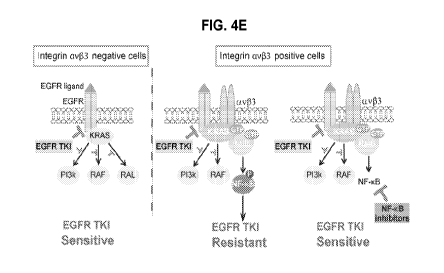
Figure 4 illustrates that integrin av(33/Ra1B complex leads to NF- B
activation
and resistance to EGFR TKI: Fig. 4(a) illustrates an immunoblot analysis of
FG, FG-133
and FG-133 stably expressing non-silencing or Ra1B-specific ShRNA, grown in
suspension and treated with erlotinib (0.5 11.M); Fig. 4(b) illustrates tumor
spheres
11
CA 02986379 2017-11-17
WO 2016/172226
PCT/US2016/028461
formation assay of FG cells ectopically expressing vector control, WT NF-KB
FLAG
tagged or constitutively active S276D NF-KB FLAG tagged constructs treated
with
erlotinib; Fig. 4(c) illustrates tumor spheres formation assay of FG-133
treating with non-
silencing (shCTRL) or NF-KB-specific shRNA and exposed to erlotinib; Fig. 4(d)
illustrates dose response in FG-I33 cells treated with erlotinib (10 nM to 5
lenalidomide (10 nM to 5 l.M) or a combination of erlotinib (10 nM to 5 l.M)
and
lenalidomide (1 ; Fig. 4(e) illustrates Model depicting the integrin
av(33-mediated
EGFR TKI resistance and conquering EGFR TKI resistance pathway and its
downstream
RalB and NF-KB effectors; as discussed in detail in Example 1, below.
Figure 5 (or Supplementary Fig. 1, Example 1) illustrates that prolonged
exposure
to erlotinib induces Integrin av(33 expression in lung tumors; representative
immunohistochemical staining of integrin (33 in mouse tissues obtained from
H441
orthotopic lung tumors long-term treated with either vehicle or erlotinib
(scale bar, 100
p.m); as discussed in detail in Example 1, below.
Figure 6 (or Supplementary Fig. 2, Example 1) illustrates integrin av(33, even
in
its unligated state, promotes resistance to Growth Factor inhibitors but not
to
chemotherapies: Fig. 6(a) illustrates a tumor sphere formation assay comparing
FG
lacking (33 (FG), FG expressing (33 wild type (FG-133) or the (33 D119A (FG-
D119A)
ligand binding domain mutant, treated with a dose response of erlotinib (Error
bars
represent s.d. (n = 3 independent experiments); Fig. 6(b) illustrates tumor
sphere
formation assay of FG and FG- (33 cells untreated or treated with erlotinib
(0.5 OSI-
906 (0.1
gemcitabine (0.01 l.M) or cisplatin (0.1 11.M); Fig. 6(c) illustrates the
effect
of dose response of indicated treatments on tumor sphere formation (Error bars
represent
s.d. (n = 3 independent experiments); as discussed in detail in Example 1,
below.
Figure 7 (or Supplementary Fig. 3, Example 1) illustrates that integrin av(33
does
not colocalize with active HRAS, NRAS and RRAS: Fig. 7(a) illustrates that Ras
activity
was determined in PANC-1 cells grown in suspension by using a GST-Rafl-RBD
immunoprecipitation assay as described in Methods, see Example 1 (data are
representative of two independent experiments); Fig. 7(b) illustrates confocal
microscopy
images of PANC-1 cells grown in suspension and stained for KRAS, RRAS, HRAS,
NRAS (red), integrin av(33 (green) and DNA (TOPRO-3, blue) (Scale bar, 10 p.m.
Data
12
CA 02986379 2017-11-17
WO 2016/172226
PCT/US2016/028461
are representative of two independent experiments); as discussed in detail in
Example 1,
below.
Figure 8 (or Supplementary Fig. 4, Example 1) illustrates that Galectin-3 is
required to promote integrin av(33/KRAS complex formation: Fig. 8(a-b)
illustrates
confocal microscopy images of Panc-1 cells lacking or expressing integrin
av(33 grown in
suspension; Fig. 8(a) illustrates cells stained for KRAS (green), Galectin-3
(red), and
DNA (TOPRO-3, blue); Fig. 8(b) illustrates cells stained for integrin av(33
(green),
Galectin-3 (red) and DNA (TOPRO-3, blue), Scale bar, 10 p.m, data are
representative of
three independent experiments; Fig. 8(c) illustrates an immunoblot analysis of
Galectin-3
immuno-precipitates from PANC-1 cells expressing non-silencing (sh CTRL) or
integrin
03-specific shRNA (sh (33), data are representative of three independent
experiments; Fig.
8(d) illustrates an immunoblot analysis of integrin (33 immunoprecipitates
from PANC-1
cells expressing non-silencing (sh CTRL) or Galectin-3-specific shRNA (sh
Ga13), data
are representative of three independent experiments; as discussed in detail in
Example 1,
below.
Figure 9 (or Supplementary Fig. 5, Example 1) illustrates that ERK, AKT and
RalA are not specifically required to promote integrin av(33-mediated
resistance to EGFR
TKI; Fig. 9A 03-negative cells, and Fig. 9B, 03-positive cells; tumor spheres
formation
assay of FG and FG-03 expressing non-silencing or ERKI/2, AKT I and Ra1A-
specific
shRNA and treated with erlotinib (0.5 error bars represent s.d. (n = 3
independent
experiments); as discussed in detail in Example 1, below.
Figure 10 (or Supplementary Fig. 6, Example 1) illustrates that RalB is
sufficient
to promote resistance to EGFR TKI: Fig. 10(a) (supplementary Figure 6, Example
1)
illustrates a tumor sphere formation assay of FG expressing non-silencing or
RalB
specific shRNA and treated with a dose response of erlotinib. Error bars
represent s.d. (n
= 3 independent experiments); Fig. 10(b) (supplementary Figure 6) illustrates
a tumor
spheres formation assay of PANC-1 stably expressing integrin 03-specific shRNA
and
ectopically expressing vector control, WT RalB FLAG tagged or a constitutively
active
RalB G23V FLAG tagged constructs treated with erlotinib (0.5
error bars represent
s.d. (n = 3 independent experiments); Fig. 10(c) (Supplementary Figure 7,
Example 1)
shows that integrin av[33 colocalizes with RalB in cancer cells: illustrates
confocal
microscopy images of Panc-1 cells grown in suspension. Cells are stained for
integrin
13
CA 02986379 2017-11-17
WO 2016/172226 PCT/US2016/028461
av(33 (green), RalB (red), pFAK (red), and DNA (TOPRO-3, blue), scale bar, 10
p.m, data
are representative of three independent experiments; as discussed in detail in
Example 1,
below.
Figure 11 (or Supplementary Fig. 8, Example 1) illustrates that integrin av(33
colocalizes with RalB in human breast and pancreatic tumor biopsies and
interacts with
RalB in cancer cells: Fig. 11(a) illustrates confocal microscopy images of
integrin av(33
(green), RalB (red) and DNA (TOPRO-3, blue) in tumor biopsies from breast and
pancreatic cancer patients, Scale bar, 20 Ilm; Fig. 11(b) illustrates a Ral
activity assay
performed in PANC-1 cells using GST-Ra1BP1-RBD immunoprecipitation assay,
Immunoblot analysis of RalB and integrin (33, data are representative of three
independent
experiments; as discussed in detail in Example 1, below.
Figure 12 (or Figure 1 in Example 2) illustrates data showing that integrin
(33 is
expressed in EGFR inhibitor resistant tumors and is necessary and sufficient
to drive
EGFR inhibitor resistance: Fig. 12(A) schematically illustrates that the
identification of
the most upregulated tumor progression genes common to erlotinib resistant
carcinomas;
Fig. 12(B) in table form shows Erlotinib IC50 in a panel of human carcinoma
cell lines
treated with erlotinib in 3D culture; Fig. 12(C) graphically illustrates
percentage of
integrin (33 positive cells in parental lines vs. after 3 or 8 weeks treatment
with erlotinib;
Fig. 12(D) graphically illustrates quantification of integrin (33 (ITG133)
gene expression in
human lung cancer biopsies from patients from the BATTLE Study (18) who were
previously treated with an EGFR inhibitor and progressed (n = 27), versus
patients who
were EGFR inhibitor naive (n = 39); Fig. 12(E) illustrates images of paired
human lung
cancer biopsies obtained before and after erlotinib resistance were
immunohistochemically stained for integrin (33, scale bar, 50 p.m; Fig. 12(F)
graphically
illustrates: Right graph shows effect of integrin (33 knockdown on erlotinib
resistance of
(33-positive cells, and Left graph shows effect of integrin (33 ectopic
expression on
erlotinib resistance in FG and H441 cells; Fig. 12(G) graphically illustrates:
Right graph
shows the effect of integrin (33 knockdown on erlotinib resistance in vivo,
A549 shCTRL
and A549 sh integrin (33 (n=8 per treatment group) were treated with erlotinib
(25
mg/kg/day) or vehicle during 16 days, results are expressed as average of
tumor volume
at day 16. *P < 0.05; and Left graph shows orthotopic FG and FG-133 tumors
treated for
14
CA 02986379 2017-11-17
WO 2016/172226 PCT/US2016/028461
30 days with vehicle or erlotinib, results are expressed as % tumor weight
compared to
vehicle control; as further described in Example 2, below.
Figure 13 (or Figure 2 in Example 2) illustrates data showing that integrin
(33 is
required to promote KRAS dependency and KRAS-mediated EGFR inhibitor
resistance:
Fig. 13(A) illustrates confocal microscopy images showing immunostaining for
integrin
(33 (green), K-, N-, H-, R-Ras (red), and DNA (TOPRO-3, blue) for BxPc3 cells
grown in
suspension in media with 10% serum, arrows indicate clusters where integrin
(33 and
KRAS colocalize (yellow); Fig. 13(B-C) illustrates confocal microscopy images
showing
immunostaining for integrin 13 3 (green), KRAs (red) and DNA (Topro-3, blue)
for
PANC-1 (KRAS mutant) and HCC827 (KRAS wild-type) after acquired resistance to
erlotinib (HCC827R) grown in suspension in absence (Vehicle) or in presence of
erlotinib
(0.5 i.tM and 0.111.M respectively), arrows indicate clusters where integrin
(33 and KRAS
colocalize (yellow); Fig. 13(D) graphically illustrates the effect of KRAS
knockdown on
tumorspheres formation in a panel of lung and pancreatic cancer cells
expressing or
lacking integrin (33; Fig. 13(E) graphically illustrates the effect of KRAS
knockdown on
tumorsphere formation in PANC-1 (KRAS mutant) stably expressing non-target
shRNA
control (IA-positive) or specific-integrin (33 shRNA ((33 negative) in FG
(KRAS mutant)
and BxPc3 (KRAS wild-type) stably expressing vector control or integrin 133;
Fig. 13(F)
graphically illustrates the effect of KRAS knockdown on erlotinib resistance
of 133-
negative and 133-positive epithelial cancer cell lines, cells were treated
with a dose
response of erlotinib; Fig. 13(G) illustrates confocal microscopy images
showing
immunostaining for integrin 133 (green), KRAS (red) and DNA (TOPRO-3, blue)
for
PANC-1 cells expressing non-target shRNA control or Galectin 3-specific shRNA
grown
in suspension; Fig. 13(H) illustrates: Top: immunoblot analysis of integrin
133
immunoprecipitates from PANC-1 cells expressing non-target shRNA control
(CTRL) or
Galectin-3-specific shRNA (Gal-3); Bottom: immunoblot analysis of Galectin-3
immunoprecipitates from PANC-1 cells expressing non-target shRNA control
(CTRL) or
integrin 133-specific shRNA (133); Fig. 13(I) graphically illustrates
erlotinib dose response
of FG-(33 cells expressing a non-target shRNA control or a Galectin-3-specific
shRNA (sh
Gal-3); as further described in Example 2, below.
Figure 14 (or Figure 3 in Example 2) illustrates data showing that RalB is a
central player of integrin 133-mediated EGFR inhibitor resistance: Fig. 14(A)
graphically
CA 02986379 2017-11-17
WO 2016/172226 PCT/US2016/028461
illustrates the effect of RalB knockdown on erlotinib resistance of (33-
positive epithelial
cancer cell lines, cells were treated with 0.5 tM of erlotinib: Fig. 14(B)
graphically
illustrates the effect of RalB knockdown on erlotinib resistance of (33-
positive human
pancreatic (FG-133) orthotopic tumor xenografts, established tumors expressing
non-target
shRNA, (shCTRL) or a shRNA targeting RalB (sh RalB) were randomized and
treated for
days with vehicle or erlotinib, results are expressed as % of tumor weight
changes
after erlotinib treatment compared to vehicle; Fig. 14(C) graphically
illustrates the effect
of expression of a constitutively active Ral G23V mutant on erlotinib response
of (33
negative cells, cells were treated with 0.5 tM of erlotinib; Fig. 14(D)
illustrates the effect
10 of expression of integrin (33 on KRAS and RalB membrane localization;
Fig. 14(E)
illustrates Ral activity that was determined in PANC-1 cells grown in
suspension by using
a GST-Ra1BP1-RBD immunoprecipitation assay, immunoblots indicate RalB activity
and
association of active RalB with integrin (33; Fig. 14(F) illustrates confocal
microscopy
images of integrin av(33 (green), RalB (red) and DNA (TOPRO-3, blue) in tumor
biopsies
from pancreatic cancer patients; Fig. 14(G) illustrates the effect of (33
expression and
KRAS expression on RalB activity, measured using a GST-Ra1BP1-RBD
immunoprecipitation assay; Fig. 14(H) illustrates immunoblot analysis of FG
and FG-133
stably expressing non-target shRNA control or Ra1B-specific shRNA, grown in
suspension and treated with erlotinib (0.5 l.M); Fig. 14(I) graphically
illustrates the effect
of a Tank Binding Kinase (TBK1) and p65 NEKB on erlotinib resistance of FG-133
cells,
cells were treated with 0.5 of erlotinib; as further described in Example
2, below.
Figure 15 (or Figure 4 in Example 2) illustrates data showing that reversal of
(33-
mediated EGFR inhibitor resistance in oncogenic KRAS model by pharmacological
inhibition: Fig. 15(A) graphically illustrates the effect of NFkB inhibitors
on erlotinib
response of (33-positive cells (FG-(33, PANC-1 and A549), cells were treated
with vehicle,
erlotinib (0.5 p,M), lenalidomide (1-2 p,M), bortezomib (4 nM) alone or in
combination;
Fig. 15(B) graphically illustrates data from: Lei, mice bearing subcutaneous
(33-positive
tumors (FG-133) were treated with vehicle, erlotinib (25 mg/kg/day),
lenalidomide (25
mg/kg/day) or the combination of erlotinib and lenalidomide, tumor dimensions
are
reported as the fold change relative to size of the same tumor on Day 1;
Right, mice
bearing subcutaneous (33-positive tumors (FG-R) after acquired resistance to
erlotinib
were treated with vehicle, erlotinib (25 mg/kg/day), bortezomib (0.25 mg/kg),
the
16
CA 02986379 2017-11-17
WO 2016/172226 PCT/US2016/028461
combination of erlotinib and bortezomib, tumor dimensions are reported as the
fold
change relative to size of the same tumor on Day 1; Fig. 15(C) schematically
illustrates a
model depicting an integrin av(33-mediated KRAS dependency and EGFR inhibitor
resistance mechanism; as further described in Example 2, below.
Figure 16 (or supplementary Figure Si, in Example 2) illustrates data showing
that illustrates resistance to EGFR inhibitor is associated with integrin (33
expression in
pancreatic and lung human carcinoma cell lines: Fig. 16(A) illustrates
immunoblots
showing integrin (33 expression in human cell lines used in Figure 12; Fig.
16(B)
graphically illustrates data showing the effect of erlotinib on HCC827
xenograft tumors in
immuno - compromised mice relative to vehicle-treated control tumors; Fig.
16(C) left,
graphically illustrates data of Integrin av(33 quantification in orthotopic
lung (upper
panel) and pancreas (lower panel) tumors treated with vehicle or erlotinib
until resistance,
Fig. 16(C) right, illustrates a representative immunofluorescent staining of
integrin av(33
in lung (upper panel) and pancreatic (lower panel) human xenografts treated 4
weeks with
vehicle or erlotinib; as further described in Example 2, below.
Figure 17 (or supplementary Figure S2, in Example 2) illustrates Integrin (33
expression predicts intrinsic resistance to EGFR inhibitors in tumors; Fig.
17A
graphically illustrates a plot of progression-free survival for erlotinib-
treated patients with
low versus (vs.) high protein expression of (33 integrin measured from non-
small cell lung
cancer biopsy material (Fig. 17B illustrates: in right panel (33 integrin high
cells and left
panel (33 integrin low cells) obtained at diagnosis; as further described in
Example 2,
below.
Figure 18 (or supplementary Figure S3, in Example 2) illustrates Integrin (33
confers Receptor Tyrosine Kinase inhibitor resistance: Fig. 18(A) illustrates
immunoblots
showing integrin (33 knockdown efficiency in cells used in Figure 12; Fig.
18(B)
graphically illustrates response of A549 lung carcinoma cells non-target shRNA
control
or shRNA targeting integrin (33 to treatment with either vehicle or erlotinib
(25
mg/kg/day) during 16 days; Fig. 18(C) illustrates immunoblots showing
expression of
indicated proteins of representative tumors; Fig. 18(D) illustrates
representative
photographs of crystal violet-stained tumorspheres of 03-negative and 03-
positive cells
after erlotinib, OSI-906, gemcitabine and cisplatin treatment; Fig. 18(E)
graphically
illustrates the effect of integrin (33 expression on lapatinib and OSI-906
(left panel), and
17
CA 02986379 2017-11-17
WO 2016/172226 PCT/US2016/028461
cisplatin and gemcitabine (right panel); Fig. 18(F) graphically illustrates
data from a
viability assay of FG and FG-133 cells grown in suspension in media with or
without
serum; as further described in Example 2, below.
Figure 19 (or supplementary Figure S4, in Example 2) illustrates integrin (33-
mediated EGFR inhibitor resistance is independent of its ligand binding: Fig.
19A
graphically illustrates the effect of ectopic expression of (33 wild-type (FG-
(33) or the 133
D119A (FG-D119A) ligand binding domain mutant on erlotinib response; Fig. 19B
illustrates an immunoblot showing transfection efficiency of vector control,
integrin 133
wild-type and integrin 133 D119A; as further described in Example 2, below.
Figure 20 (or supplementary Figure S5, in Example 2) illustrates integrin 133
colocalizes and interacts with oncogenic and active wild-type KRAS: Fig. 20(A)
illustrates confocal microscopy images of FG and FG-(33 cells grown in
suspension in
media 10% serum with or without erlotinib (0.5 11M) and stained for KRAS
(red), integrin
av(33 (green) and DNA (TOPRO-3, blue); Fig. 20(B) illustrates Ras activity was
determined in PANC-1 cells grown in suspension by using a GST-Rafl-RBD
immunoprecipitation assay, immunoblots indicate KRAS activity and association
of
active KRAS with integrin 133; Fig. 20(C) illustrates an immunoblot analysis
showing that
Integrin av(33 immunoprecipitates from BxPC-3 cells grown in suspension in
presence or
absence of growth factors; as further described in Example 2, below.
Figure 21 (or supplementary Figure S6, in Example 2) illustrates integrin 133
expression promotes KRAS dependency: Fig. 21(A) illustrates Immunoblots
showing
KRAS knockdown efficiency in cells used in Figure 13; Fig 21(B) illustrates
Representative photographs of crystal violet-stained tumorspheres of FG and
A549
cells expressing non-target shRNA control or specific-KRAS shRNA; Fig. 21(C)
illustrates the effect of an additional KRAS knockdown on tumorspheres
formation in
PANC-1 stably expressing non-target shRNA control (133-positive) or specific-
integrin 133
shRNA ((33 negative); Fig. 21(D) illustrates immunoblots showing KRAS
knockdown
efficiency; as further described in Example 2, below.
Figure 22 (or supplementary Figure S7, in Example 2) illustrates images
showing
that KRAS and Galectin-3 colocalize in integrin 133-positive cells, in
particular, confocal
microscopy images of FG and FG-(33 cells grown in suspension and stained for
KRAS
18
CA 02986379 2017-11-17
WO 2016/172226 PCT/US2016/028461
(green), galectin-3 (red) and DNA (TOPRO-3, blue); as further described in
Example 2,
below.
Figure 23 (or supplementary Figure S8, in Example 2) illustrates Integrin 03-
mediated KRAS dependency and erlotinib resistance is independent of ERK, AKT
and
RalA: Fig. 23(A) graphically illustrates the effect of ERK, AKT, RalA and RalB
knockdown on erlotinib response (erlotinib 0.5 [NI) of (33-negative FG (left
panel) and
03-positive FG-03 cells (right panel); Fig. 23(B) illustrates Immunoblots
showing ERK,
AKT RalA and RalB knockdown efficiency on 03-negative FG (upper panel) and 03-
positive FG-03 cells (lower panel); Fig. 23(C) illustrates Immunoblots showing
RalB
knockdown efficiency in the 03-positive epithelial cancer cells used in Figure
14; as
further described in Example 2, below.
Figure 24 (or supplementary Figure S9, in Example 2) illustrates constitutive
active NFkB is sufficient to promote erlotinib resistance: Fig. 24(A)
illustrates
immunoblots showing a Tank Binding Kinase (TBK1) (upper panel) and NFkB
knockdown efficiency (lower panel) used in Figure 14; Fig. 24(B) graphically
illustrates
the effect of constitutive active S276D p65NFkB on erlotinib response
(erlotinib 0.511M)
of 03-negative cells (FG cells); as further described in Example 2, below.
Figure 25 (or supplementary Figure S10, in Example 2) illustrates NFkB
inhibitors in combination with erlotinib increase cell death in vivo: Fig.
25(A) and Fig. 25
(B) illustrate Immunoblots showing expression of indicated proteins of
representative
tumors from shown in Figure 15B; Fig. 25(C) illustrates Confocal microscopy
images of
cleaved caspase 3 (red) and DNA (TOPRO-3, blue) in tumor biopsies from
xenografts
tumors used in Fig. 15B treated with vehicle, erlotinib, lenalidomide or
lenalidomide and
erlotinib in combo; Fig. 25(D) illustrates Confocal microscopy images of
cleaved caspase
3 (red) and DNA (TOPRO-3, blue) in tumor biopsies from xenografts tumors used
in
Figure 15B treated with vehicle, erlotinib, bortezomib or bortezomib and
erlotinib in
combo); as further described in Example 2, below.
Figures 26, 27, and 28, illustrate supplementary Table 1 from Example 2,
showing
that differentially expressed genes in cells resistant to erlotinib (PANC-1,
H1650, A459)
compared with the average of two sensitive cells (FG, H441) and in HCC827
after acquired
resistance in vivo (HCC8271k) vs. the HCC827 vehicle-treated control; as
further described
in Example 2, below.
19
CA 02986379 2017-11-17
WO 2016/172226
PCT/US2016/028461
Figure 29 illustrates supplementary Table 2, from Example 2, showing KRAS
mutational status in pancreatic and lung cell lines used in the study of
Example 2, below.
Figure 30 illustrates data showing integrin (33 (CD61) is a RTKI (Receptor
Tyrosine Kinase (RTK) Inhibitor) drug resistance biomarker on the surface of
circulating
tumor cells; as discussed in detail in Example 2, below. As schematically
illustrated in
Fig. 30A, CD61 ((33, or beta3) negative human lung cancer cells (HCC827; this
lung
adenocarcinoma has an acquired mutation in the EGFR tyrosine kinase domain
(E746 -
A750 deletion), and they are sensitive to erlotinib and develop acquired
resistance after
6/8 weeks) were injected orthotopically into the lung of mice and treated over
3 months
with erotinib at 25 mg/kg/day. As graphically illustrated in Fig. 30B, Human
lung cancer
cells detected in the circulation were positive for av(33 (or avb3, CD61)
whereas the cells
in the untreated group were essentially negative for this marker. CD45
negative cells
indicates that the detected cells were not leukocytes and pan cytokeratin
positive cells
indicate tumor cells. CD61 (beta3) positive expression correlated with tumor
expression.
Figure 31 illustrates data showing how targeting the NF-KB pathway using
compositions and methods as provided herein can sensitize resistant tumors to
growth
factor inhibitors by showing the effect of NFkB inhibitors on erlotinib
response of 133-
negative (b3-negative) cells (FG) and 133-positive cells (FG- 133, MDA-MB231
(intrinsic
resistance, Fig. 31A) and FG-R (acquired resistance, Fig. 31B), and EGFR TKI
(Tyrosine
Kinase Inhibitor) sensitive cells, Fig. 31C. Cells embedded in agar (anchorage
independent growth) were treated with vehicle, erlotinib (0.5 pM),
Lenalidomide (2 [NI),
PS-1145 (1 pM) alone or in combination for 10 to 15 days. Then, the soft agar
were
stained with crystal violet and the colonies were counted manually. The
results show that
while 133-positive cells (intrinsic Fig. 31A or acquired resistant Fig. 31B
cells ) were
resistant to erlotinib and each NEKB inhibitor alone, the combination of
erlotinib with
either Lenalidomide or PS-1145 decreased tumorsphere formation.
Figure 32 (or Figure 1 of Example 3) illustrates: Integrin 133 expression
increase
tumor-initiating and self-renewal capacities: Fig. 32(a) Limiting dilution in
vivo
determining the frequency of tumor-initiating cells for A549 cells expressing
non-target
shRNA control or integrin 133-specific shRNA and for FG cells expressing
control vector
or integrin 133 (FG-133); Fig. 32(b-c-d) Self-renewal capacity of A549 (Fig.
32b) and
PANC-1 (Fig. 32c) cells expressing non-target shRNA control (CTRL) or integrin
133-
CA 02986379 2017-11-17
WO 2016/172226 PCT/US2016/028461
specific shRNA and of FG expressing control vector or integrin (33 (FG-133)
(Fig. 32d); as
described in detail in Example 3, below.
Figure 33 (or Figure 2, of Example 3) illustrates: Integrin (33 drives
resistance to
EGFR inhibitors: Fig. 33(a) graphically illustrates the Effect of integrin (33
expression
(ectopic expression for FG and integrin 133-specific knockdown for PANC-1)
cells on
drug treatment response; Fig. 33(b) graphically illustrates the Effect of
integrin (33
knockdown on erlotinib response in MDA-MB-231 (MDA231), A549 and H1650; Fig.
33(c) and 33(d) graphically illustrate the effect of integrin (33 knockdown on
erlotinib
resistance in vivo using A549 shCTRL and A549 sh (33 treated with erlotinib or
vehicle,
Fig. 33(c) measuring tumorspheres, and 33(d) measuring tumor volume in A549
shCTRL
(integrin (33+), left panel, and A549 (integrin (33-) (right panel); Fig.
33(e) [[33(d)]]
graphically illustrates Orthotopic FG and FG-(33 tumors (>1000 mm3; n = 5 per
treatment
group) were treated for 30 days with vehicle or erlotinib; Fig. 33(f)
graphically illustrates
Relative mRNA expression of integrin 133 (ITGB3) in HCC827 vehicle-treated
tumors
(n= 5) or erlotinib-treated tumors (n= 7) from (e) after acquired resistance;
Fig. 33(g)
H&E sections and immunohistochemical analysis of integrin 133 expression in
paired
human lung cancer biopsies obtained before and after erlotinib resistance;
Fig. 33(h)
illustrates images of Limiting dilution in vivo determining the frequency of
tumor-
initiating cells for HCC827 vehicle-treated (vehicle) and erlotinib-treated
tumors from
(erlotinib resistant non-sorted) (e); Fig. 33(1) and Fig. 33(j) graphically
illustrate the Self-
renewal capacity of HCC827 vehicle-treated (vehicle), erlotinib-treated
(erlotinib
resistant non-sorted), erlotinib-treated integrin 133- population and
erlotinib-treated
integrin 133+ population; as described in detail in Example 3, below.
Figure 34 (or Figure 3, of Example 3) illustrates: Integrin (33/KRAS complex
is
critical for integrin 133-mediated stemness: Fig. 34(a) Confocal microscopy
images show
immunostaining for Integrin 133 (green), KRAS (red) and DNA (TOPRO-3, blue)
for FG-
133, PANC-1, A549 and HCC827 after acquired resistance to erlotinib (HCC827
ER)
grown in suspension, Arrows indicate clusters where integrin 133 and KRAS
colocalize
(yellow); Fig. 34(b) Ras activity was determined in PANC-1 cells grown in
suspension by
using a GST-Rafl-RBD immunoprecipitation assay, Immunoblots indicate KRAS
activity and association of active KRAS with integrin 133; Fig. 34(c) Effect
of KRAS
knockdown on tumorspheres formation in lung (A549 and H441) and pancreatic (FG
and
21
CA 02986379 2017-11-17
WO 2016/172226 PCT/US2016/028461
PANC-1) cancer cells expressing or lacking integrin (33; Fig. 34(d) Effect of
KRAS
knockdown on erlotinib resistance of (33-negative and (33-positive epithelial
cancer cell
lines, Cells were treated with a dose response of erlotinib; Fig. 34(e) Self-
renewal
capacity of FG-133 cells expressing non-target shRNA control (shCTRL) or KRAS-
specific shRNA measured by quantifying the number of primary and secondary
tumorspheres; Fig. 34(f) Confocal microscopy images show immunostaining for
integrin
(33 (green), KRAS (red) and DNA (TOPRO-3, blue) for PANC-1 cells expressing
non-
target shRNA control or Galectin 3-specific shRNA grown in suspension; Fig.
34(g)
immunoblot analysis of integrin (33 immunoprecipitates from PANC-1 cells
expressing
non-target shRNA control (CTRL) or Galectin-3 -specific shRNA (Gal-3); Fig.
34(h)
Effect of Galectin-3 knockdown on integrin 133-mediated anchorage independent
growth
and erlotinib resistance; Fig. 34(1) Self-renewal capacity of PANC-1 cells
expressing
non-target shRNA control (shCTRL) or Galectin-3-specific shRNA (sh Gal-3)
measured
by quantifying the number of primary and secondary tumorspheres; as described
in detail
in Example 3, below.
Figure 35 (or Figure 4, of Example 3) illustrates: Ra1B/TBK1 signaling is a
key
modulator of integrin 133-mediated stemness: Fig. 35(a) Effect of RalB
knockdown on
anchorage independence; Fig. 35(b) Self-renewal capacity of FG-133 cells
expressing non-
target shRNA control (sh CTRL) or Ra1B-specific shRNA (sh RalB) measured by
quantifying the number of primary and secondary tumorspheres; Fig. 35(c)
Limiting
dilution in vivo determining the frequency of tumor-initiating cells for FG-
133 cells
expressing non-target shRNA control or integrin Ra1B-specific shRNA; Fig.
35(d) Effect
of RalB knockdown on erlotinib resistance of (33-positive epithelial cancer
cell lines; Fig.
35(e) Effect of RalB knockdown on erlotinib resistance of (33-positive human
pancreatic
(FG-133) orthotopic tumor xenografts. Established tumors expressing non-target
shRNA,
(sh CTRL) or a shRNA targeting RalB (sh RalB); Fig. 35(f) Immunoblot analysis
of FG
and FG-(33 stably expressing non-target shRNA control or Ra1B-specific shRNA,
grown
in 3D and treated with erlotinib (0.5 l.M); Fig. 35(g) Effect of TBKI
knockdown on
PANC-1 self-renewal capacity; Fig. 35(h) Effect of TBKI knockdown on erlotinib
resistance of PANC-1 cells. Cells were treated with 0.5 tM of erlotinib; Fig.
35(1) Mice
bearing subcutaneous 133-positive tumors (PANC-1) were treated with vehicle,
erlotinib
22
CA 02986379 2017-11-17
WO 2016/172226 PCT/US2016/028461
(25 mg/kg/day), amlexanox (25 mg/kg/day) or the combination of erlotinib and
amlexanox; as described in detail in Example 3, below.
Figure 36 (or Figure Si, of Example 3) illustrates: Fig. 36(a-b) Limiting
dilution
tables; Fig. 36(c) Immunoblots showing integrin (33 knockdown or ectopic
expression
efficiency in cells used in Figure 1 (of Example 3); Fig. 36(d) Viability
assay (CellTiter-
Glo assay) of FG and FG-133 cells grown in 3D in media with or without serum;
Fig. 36(e)
Immunohistochemical analysis of integrin (33 expression in paired human lung
cancer
biopsies obtained before (upper panel) and after (lower panel) erlotinib
resistance; Fig.
36(f) Limiting dilution table; Fig. 36(g) image of Immunohistochemistry
staining of
CD166 (upper panel) and integrin (33 (lower panel) in human lung tumor
biopsies after
EGFR TKI acquired resistance; as described in detail in Example 3, below.
Figure 37 (or Figure S2, of Example 3) illustrates: Fig. 37(a) Effect of
cilengetide
treatment on erlotinib resistance in FG-133 and PANC-1 cells; Fig. 37(b)
Effect of ectopic
expression of (33 wild-type (FG- 13 3) or the (33 D1 19A (FG-D1 19A) ligand
binding
domain mutant on erlotinib response; Fig. 37(c) Confocal microscopy images of
FG- 13 3
cells grown in 3D and stained for integrin - (33 (green) and RAS family
members (red);
Fig. 37(d) Immunoblots showing KRAS knockdown efficiency in cells used in
Figure 3
(of Example 3); Fig. 37(e) Representative photographs of crystal violet-
stained
tumorspheres of FG and A549 cells expressing non-target shRNA control or
specific-
KRAS; Fig. 37(f) illustrates the Effect of a second KRAS knockdown (shKRAS 2)
on
tumorspheres formation in PANC-1 stably expressing non-target shRNA control (3-
positive) or specific-integrin - 13 3 shRNA (3 negative), left panel
graphically presenting
data and right panel illustrating an immunoblot showing KRAS expression in sh
CTRL,
SH KRAS and sh KRAS 2; as described in detail in Example 3, below.
Figure 38 (or Figure S3, of Example 3) illustrates: Fig. 38(a) graphically
illustrates the Effect of ERK, AKT and RalA knockdown on erlotinib response of
13 3-
negative FG and 3-positive FG-3 cells; Fig. 38(b) Immunoblots showing ERK, AKT
and
RalA knockdown efficiency in cells used in (a); Fig. 38(c) Immunoblots showing
RalB
knockdown efficiency in cells used in Figure 3 (of Example 3); Fig. 38(d)
graphically
illustrates the effect of a second RalB knockdown (shRalB 2) on tumorspheres
formation
in PANC-1 stably expressing non-target shRNA control ((3 3-positive) or
specific-integrin
13 3 shRNA (3 negative); Fig. 38(e) Limiting dilution table; Fig. 38(f)
Confocal
23
CA 02986379 2017-11-17
WO 2016/172226 PCT/US2016/028461
microscopy images of integrin av(33 (green), RalB (red) and DNA (TOPRO-3,
blue) in
tumor biopsies from pancreatic cancer patients; Fig. 38(g) Ral activity was
determined in
PANC-1 cells grown in suspension by using a GST-Ra1BP1-RBD immunoprecipitation
assay. Immunoblots indicate RalA and RalB activities; Fig. 38(h) Effect of (33
expression
and KRAS expression on RalB activity, measured using a GST-Ra1BP1-RBD
immunoprecipitation assay; Fig. 38(1) illustrates the effect of expression of
a
constitutively active Ral G23V mutant on erlotinib resistance of 0 3 positive
and negative
cells, left panel graphically presenting data and right panel illustrating an
immunoblot
showing FLAG, RalB and Hsp90 expression; as described in detail in Example 3,
below.
Figure 39 (or Figure S4, of Example 3) illustrates: Fig. 39(a) Immunoblot
showing TBK1 knockdown efficiency in PANC-1 cells used in Figure 4 (of Example
3);
Fig. 39(b) Effect of theTBK1 inhibitor amlexanox on erlotinib response of PANC-
1 cells;
Fig. 39(c) Effect of the NFkB inhibitor borthezomib on (33-positive cells (FG-
(33 (left
panel), PANC-1 (middle panel) and A549 (right panel)); Fig. 39(d) Mice bearing
subcutaneous (33-positive tumors (FG-133) were treated with vehicle, erlotinib
(25
mg/kg/day), bortezomib (0.25 mg/kg), the combination of erlotinib and
bortezomib; Fig.
39(e) Confocal microscopy images of cleaved caspase 3 (red) and DNA (TOPRO-3,
blue)
in tumor biopsies from xenografts tumors used in (d) treated with vehicle,
erlotinib,
bortezomib or bortezomib and erlotinib in combo; as described in detail in
Example 3,
below.
Figure 40 graphically illustrates data demonstrating that depletion of RalB
overcomes erlotinib resistance in KRAS mutant cells: Fig. 40A graphically
illustrates
number of tumorspheres as a percent of control for FG, FG-beta3, PANC-1, and
A539
expressing cells, with or without erlotinib, in vitro soft agar conditions;
and Fig. 40B
graphically illustrates tumor weight as a percent of control, in in vivo
orthotopic pancreas
xenograft; as discussed in detail in Example 2, below.
Figure 41 graphically illustrates data demonstrating that depletion of TBK1
overcomes erlotinib resistance in KRAS mutant cells: Fig. 41A illustrates data
demonstrating that integrin mediates TBK1 activation through Ralb; Fig. 41B
and Fig.
41C graphically illustrate data demonstrating TBK1 depletion (with siRNA)
overcomes
integrin beta-3-mediated erlotinib resistance, where Fig. 41A shows the number
of
tumorspheres as a percent of non-treated cells with and without siRNA
depletion of
24
CA 02986379 2017-11-17
WO 2016/172226
PCT/US2016/028461
TBK1, and Fig. 41C shows tumor size as a percent of control with erlotinib,
amlexanox
and erlotinib + amlexanox; as discussed in detail in Example 2, below.
Like reference symbols in the various drawings indicate like elements.
Reference will now be made in detail to various exemplary embodiments of the
invention, examples of which are illustrated in the accompanying drawings. The
following detailed description is provided to give the reader a better
understanding of
certain details of aspects and embodiments of the invention, and should not be
interpreted
as a limitation on the scope of the invention.
DETAILED DESCRIPTION
In alternative embodiments, provided are compositions, including kits, and
methods and uses for detecting and/or measuring levels of: (33 integrin-
expressing cells,
including tumor and cancer cells, including Circulating Tumor Cells (CTCs);
and, (33
integrin-comprising extracellular vesicles (EV), e.g., including EVs released
by cancer
cells, including EVs such as exosomes and oncosomes, to assess patient
prognosis,
metastatic potential, tumor stemness and drug resistance, and provide an early
indication
of cancer progression, wherein (33 integrin-expression correlates with poor
patient
prognosis, metastatic potential, tumor stemness and drug resistance.
Inventors have shown that a primary tumor may be (33 negative and CTCs (33
positive, and/or EVs released by cancer cells (33 positive, thereby their
detection provides
an early indication of cancer progression. It is believed that CTCs may seed
secondary
metastatic tumors with increased stemness. Also, treating a patient with a
growth factor
inhibitor may actually drive (not select) tumors to (33 positive phenotype and
growth
factor inhibitor resistance.
In alternative embodiments, provided are compositions, including kits, and
methods for detecting and measuring tumor cells, CTCs, cancer stem cells,
and/or EVs
that are (33 positive by using samples, including tissue, blood-based or other
samples,
including blood, serum urine, CSF and other samples; this exemplary approach
is less
invasive compared to a tumor biopsy and avoids issues of removing and testing
tissue
samples from only a minor portion of a tumor; however, in alternative
embodiments,
liquefied tissue samples are also used. Exemplary applications of
compositions,
CA 02986379 2017-11-17
WO 2016/172226
PCT/US2016/028461
including kits, and methods and uses as provide herein include diagnostics and
treatments
for cancer, tumor progression, metastasis, and tumor growth factor resistance.
In alternative embodiments, also provided are methods for screening for new
therapeutics targeting (33 for treating cancer.
In alternative embodiments, patient monitoring is performed using whole blood
obtained from the patient and placed into sodium-EDTA tubes. A FICOLL gradient
is
run to obtain the buffy coat layer. These cells and/or isolated EVs are
stained for (33 (the
marker of interest), pan-cytokeratin (a marker of epithelial tumor cells),
CD45 (a marker
of lymphoid cells), and a nuclear marker (DAPi). The circulating tumor cell or
EV
fraction is identified as 03-positive, cytokeratin-positive, and CD45-negative
using
confocal microscopy or flow cytometry.
As provided herein, (33 is been identified as a biomarker of cancer stem cells
and
receptor tyrosine kinase inhibitor (RTKI) resistance. We observed a 2-fold
increase in
circulating tumor cells (CD45 cytokeratin + cells) and a 4-fold increase in
(33 integrin
during acquired resistance to RTKI.
Detection of (33 integrin and/or integrin av(33 on extracellular vesicles
(exosomes and
oncosomes) as a diagnostic cancer test and therapeutic target
In alternative embodiments, provided are compositions, including kits, and
methods for detecting and measuring integrin 03-comprising extracellular
vesicles (EVs)
such as exosomes and oncosomes that are released by cancer cells, including
CTCs.
Because EVs can contain cargoes, such as proteins, mRNA, and microRNA, and EVs
can
be taken up into recipient cells to modulate intercellular communication,
promote tumor
progression and modify their microenvironment, compositions and methods
provided
herein are used to detect cancer cell-derived EVs, including circulating EVs
by e.g.,
taking and using an exosome-based liquid biopsy, and for cancer diagnosis.
Described herein is the discovery that human lung cancer-derived exosomes
(from
the HCC827 cell line) are highly enriched with integrin (33 by approximately
100-fold
relative to membranes isolated from the intact cells. In addition, inventors
found that
circulating tumor cells (CTC) isolated from lung cancer patients show 03-
positive
membrane protrusions on their cell surface that appear to be secreted as 03-
positive large
oncosomes. In alternative embodiments, provided are compositions, including
kits, and
methods for detecting and measuring integrin (33 to assess tumor stemness and
drug
26
CA 02986379 2017-11-17
WO 2016/172226 PCT/US2016/028461
resistance; and detecting (33-positive EVs as a new diagnostic biomarker and
therapeutic
target for cancer.
This invention shows that integrin (33 is detectable on EV (exosomes and
oncosomes) released by tumors into the bloodstream of cancer patients, thus
providing
diagnostic and/or prognostic information about the initiation, growth,
progression or drug
resistance of the tumor. Inventors found that integrin (33 is specifically
upregulated on the
surface of genetically and histologically distinct epithelial tumors exposed
to receptor
tyrosine kinase inhibitors (TKI), such as erlotinib. Thus, provided herein are
compositions and methods for detecting (33-positive EVs as biomarkers for not
only
diagnosis but also drug sensitivity vs. resistance. Compared to existing EV
biomarker
studies, the monitoring of tumor-initiation capacity, including drug
resistance, using
exosomes is very unique and helps in translational research.
In alternative embodiments, EV exosomes of between about 50 to 100 nm
diameter and/or EV oncosomes of between about 1 to 10 um diameter are isolated
and/or
detected, and compositions and methods of the invention are used to determine
whether
the EV is derived from a cancer cell and/or the EV comprises an integrin (33.
Exosomes: analysis of the characteristics of integrin (33-positive exosomes in
vitro: We isolated exosomes from HCC827 lung adenocarcinoma cells using
standard
protocols. By Western blot analysis, we determined that the integrin (33 is
enriched in
exosomes relative to the intact cell.
Large oncosomes: We isolated circulating tumor cells from the blood of lung
cancer patients. We detected integrin 133-enriched membrane blebbing and
adjacent
secreted large oncosomes using immunofluorescence analysis.
In alternative embodiments, provided are compositions (e.g., kits) and methods
to
isolate and/or detect EVs, including exosomes and oncosomes, from samples from
an
individual, including tissue, blood or blood derived or other samples,
including blood,
serum, urine, CSF and other samples. The presence of 133+ EV and/or
circulating 133+
cancer stem cells indicates metastasis, disease progression, drug resistance,
and/or
correlate with tumor stage/grade. Once detected, 133+ EC presence indicates a
shift in
tumor phenotype toward a cancer stem-like state that could be treated with a
different
class of drugs than the originating epithelial-like cancer. Therefore,
compositions and
methods as provided herein not only detect a shift in tumor phenotype, but
also can
27
CA 02986379 2017-11-17
WO 2016/172226 PCT/US2016/028461
instruct as to a specific means to halt progression once integrin (33
expression is
present. As delivery of their cargo can have profound impact on the function
and
phenotype of the recipient cells, (33+ extracellular vesicles are both a
detection tool and a
therapeutic target.
In alternative embodiments, provided are compositions (e.g., kits) and methods
for
detecting integrin (33-positive EVs (including exosomes and large oncosomes)
as a
biomarker for aggressive, metastatic, stem-like cancer cell phenotypes, and
also as a
therapeutic target to slow the progression of cancer and metastasis. In
alternative
embodiments, compositions and methods use a liquid biopsy to detect (33-
positive CTCs
and/or EVs to: determine the presence of a cancer; and/or determine or predict
an
aggressive, metastatic, stem-like cancer cell phenotype.
Growth Factor Inhibitor (GFI) resistance
In alternative embodiments, provided are compositions and methods for
overcoming or diminishing or preventing Growth Factor Inhibitor (GFI)
resistance in a
cell, or, a method for increasing the growth-inhibiting effectiveness of a
Growth Factor
inhibitor on a cell, or, a method for re-sensitizing a cell to a Growth Factor
Inhibitor
(GFI). In alternative embodiments, the cell is a tumor cell, a cancer cell or
a
dysfunctional cell. In alternative embodiments, provided are compositions and
methods
for determining: whether an individual or a patient would benefit from or
respond to
administration of a Growth Factor Inhibitor, or, which individuals or patients
would
benefit from a combinatorial approach comprising administration of a
combination of: at
least one growth factor and at least one compound, composition or formulation
used to
practice a method provided herein, such as an NfKb inhibitor.
We found that integrin anb3 is upregulated in cells that become resistant to
Growth Factor inhibitors. Our findings demonstrate that integrin anb3 promotes
de novo
and acquired resistance to Growth factor inhibitors by interacting and
activating RalB.
RalB activation leads to the activation of Src and TBK1 and the downstream
effectors
NFKB and IRF3. We also found that depletion of RalB or its downstream
signaling
(Src/NFKB) in b3-positive cells overcomes resistance to growth factor
inhibitors. This
demonstrates that the integrin anb3/Ra1B signaling complex promotes resistance
to
growth factor inhibitors; and in alternative embodiments, integrin av133
(anb3) and active
RalB are used as biomarkers in patient samples to predict which patients will
respond to
28
CA 02986379 2017-11-17
WO 2016/172226 PCT/US2016/028461
growth factor inhibitors and which patients might rather benefit from
alternative/combinatorial approaches such as a combination of growth factor
inhibitors
and NfKb inhibitors.
Described are compositions and methods for using (33 integrin, integrin av(33
and/or active RalB as a biomarker for tumors that are or have become (e.g., de
novo and
acquired) resistant to growth factors blockade. Accordingly, in alternative
embodiments,
provided are compositions and methods for the depletion of RalB, Src, NFkB and
its
downstream signaling effectors to sensitize av(33-expressing tumors to growth
factor
blockade. These findings reveal a new role for integrin av(33 in mediating
tumor cell
resistance to growth factor inhibition and demonstrate that targeting the
av(33/ RalB/
NfkB/ Src signaling pathway will circumvent growth factor resistance of a wide
range of
cancers.
In alternative embodiments, any NF-kB inhibitor can be used to practice
compositions and methods provided herein, e.g., lenalidomide or (RS)-3 -(4-
amino-l-oxo-
3H-isoindo1-2-yl)piperidine-2,6-dione, which can be REVLIMIDTm (Celgene Corp.,
Summit, NJ), or thalidomide, or any other derivative of thalidomide, or any
composition
having an equivalent activity.
In alternative embodiments, compositions and methods as provided herein are
used to sensitize tumors to drugs, e.g., such as erlotinib and lapatinib
(which are
commonly used to treat a wide range of solid tumors). We have shown that when
tumors
become resistant to these drugs they become very sensitive to NFkB inhibitors.
Thus, in
alternative embodiments, compositions and methods as provided herein are used
to
sensitize tumors using NFkB inhibitors, such as e.g., lenalidomide or (RS)-3-
(4-amino-l-
oxo-3H-isoindo1-2-yl)piperidine-2,6-dione or REVLIMIDTm, or a composition as
listed in
Table 1.
In alternative embodiments, compositions and methods as provided herein are
used to sensitize tumors using an IKK inhibitor, e.g., such as PS1145
(Millennium
Pharmaceuticals, Cambridge, MA) (see e.g., Khanbolooki, et al., Mol Cancer
Ther 2006;
vol. 5:2251-2260; Published online September 19, 2006; Yemelyanov, et al.,
Oncogene
(2006) vol. 25:387-398; published online 19 September 2005), or any IKBa
(nuclear
factor of kappa light polypeptide gene enhancer in B-cells inhibitor, alpha)
29
CA 02986379 2017-11-17
WO 2016/172226 PCT/US2016/028461
phosphorylation and/or degradation inhibitor, e.g., one or more compositions
listed in
Table 3.
In alternative embodiments, compositions and methods as provided herein
comprise use of an NFkB inhibitor and an IKK inhibitor to treat a drug
resistant tumor,
e.g., a solid tumor. In alternative embodiments, compositions and methods as
provided
herein comprise use of an NFkB inhibitor and an IKK inhibitor to treat a drug
resistant
tumor in combination with an anticancer drug, e.g., an NFkB inhibitor and an
IKK
inhibitor are used to sensitize a tumor to drugs such as erlotinib and
lapatinib. In
alternative embodiments, the drug combination used to practice the invention
comprises
lenalidomide (such as a REVLIMIDTm) and the IKK inhibitor PS1145 (Millennium
Pharmaceuticals, Cambridge, MA). For example, lenalidomide (such as a
REVLIMIDTm)
and PS1145 are used to sensitize a tumor that is resistant to a cancer drug,
e.g., an EGFR
inhibitor, such that the tumor is now responsive to the cancer drug.
In alternative embodiments, in practicing the invention, an NFkB inhibitor and
an
IKK inhibitor are used in combination with a tyrosine kinase receptor (also
called
Receptor Tyrosine Kinases, or RTKs) inhibitor, e.g., an 5U14813 (Pfizer, San
Diego,
CA) or as listed in Table 2 or 3, below, to treat a drug resistant tumor. In
alternative
embodiments, compositions and methods as provided herein (e.g., including
lenalidomide
or PS1145; lenalidomide and PS1145; or lenalidomide, PS1145 and an RTK
inhibitor are
administered to patients that have become resistant to a cancer drug, e.g.,
drugs like
erotinib or lapatinib, to produce a strong antitumor effect.
In alternative embodiments, any NF-kB inhibitor can be used to practice this
invention, e.g., an antioxidant can be used to inhibit activation of NF-kB,
e.g., including
the compositions listed in Table 1:
Table 1: Antioxidants that have been shown to inhibit activation of NF-kB
Molecule Reference
a-Lipoic acid Sen et al, 1998; Suzukiet al, 1992
a-tocopherol Islam et al, 1998
Ide & Lau, 2001; Langet al,
Aged garlic extract (allicin) 2004; Hasan et al, 2007
2-Amino-l-methy1-6-phenylimidazo [4,5-
blpyridine (PhIP) Yun et al, 2005
CA 02986379 2017-11-17
WO 2016/172226
PCT/US2016/028461
N-acetyldopamine dimers (from P. cicadae) Xu et al, 2006
Allopurinol Gomez-Cabrera et al, 2006
Anetholdithiolthione Sen et al, 1996
Apocynin Barbieri et al, 2004
Shi & Jiang, 2002; Daviset al,
Apple juice/extracts 2006; Jung et al, 2009
Aretemsia p7F (5,6,3',5'-tetramethoxy 7,4'-
hydroxyflavone) Lee et al, 2004
Astaxanthin Lee et al, 2003
Autumn olive extracts; olive leaf extracts Wang et al, 2007; Wanget al,
2008
Avenanthramides (from oats) Guo et al, 2007; Sur et al, 2008
Bamboo culm extract Lee et al, 2008
Benidipine Matsubara & Hazegawa, 2004
bis-eugenol Murakami et al, 2003
Bruguiera gymnorrhiza compounds Homhual et al, 2006
Israel et al, 1992; Schulze-Osthoffet
Butylated hydroxyanisole (BHA) al, 1993
Okamoto et al, 1994;Tamatani et al,
Cepharanthine 2007
Caffeic Acid Phenethyl Ester (3,4- Natarajan et al, 1996;Nagasaka et al,
dihydroxycinnamic acid, CAPE) 2007
Carnosol Lo et al, 2002; Huang et al, 2005
Bai et al, 2005;Guruvayoorappan&
beta-Carotene Kuttan, 2007
Carvedilol Yang et al, 2003
Suzuki & Packer, 1994;Zheng et al,
Catechol Derivatives 2008
Centaurea L (Asteraceae) extracts Karamenderes et al, 2007
Chalcone Liu et al, 2007
Chlorogenic acid Feng et al, 2005
-chloroacety1-2-amnio-1,3 -se lenazole s Nam et al, 2008
31
CA 02986379 2017-11-17
WO 2016/172226
PCT/US2016/028461
Cholestin Lin et al, 2007
Chroman-2-carboxylic acid N-substituted
phenylamides Kwak et al, 2008
Cocoa polyphenols Lee et al, 2006
Coffee extract (3-methy1-1,2-
cyclopentanedione) Chung et al, 2007
Crataegus pinnatifida polyphenols Kao et al, 2007
Curcumin (Diferulolylmethane); Singh & Aggarwal, 1995;Pae et al,
dimethoxycurcumin; EF24 analog 2008; Kasinskiet al, 2008
Dehydroepiandrosterone (DHEA)
and DHEA-sulfate (DHEAS) Iwasaki et al, 2004; Liuet al, 2005
Dibenzylbutyrolactone lignans Cho et al, 2002
Diethyldithiocarbamate (DDC) Schreck et al, 1992
Sappey et al, 1995;Schreck et al,
Diferoxamine 1992
Dihydroisoeugenol; isoeugenol; Murakami et al, 1995;Park et al,
epoxypseudoisoeugeno1-2-methyl butyrate 2007; Ma et al, 2008
Dihydrolipoic Acid Suzuki et al, 1992, 1995
Dilazep + fenofibric acid Sonoki et al, 2003; Yanget al, 2005
Dimethyldithiocarbamates (DMDTC) Pyatt et al, 1998
Dimethylsulfoxide (DMSO) Kelly et al, 1994
Disulfiram Schreck et al, 1992
Ebselen Schreck et al, 1992
Kokura et al, 2005; Ariiet al,
Edaravone 2007; Yoshida et al, 2007
EPC-Kl (phosphodiester compound of vitamin
E and vitamin C) Hirano et al, 1998
Epigallocatechin-3-gallate (EGCG; green tea Lin & Lin, 1997; Yang et
polyphenols) a1,1998; Hou et al, 2007
Ergothioneine Rahman et al, 2003
Song et al, 2004; Tsunget al,
Ethyl Pyruvate (Glutathione depletion) 2005; Jimenez-Lopezet al, 2008
32
CA 02986379 2017-11-17
WO 2016/172226
PCT/US2016/028461
Ethylene Glycol Tetraacetic Acid (EGTA) Janssen et al, 1999
Eupatilin Lee et al, 2008
Exercise Goto et al, 2007
Fisetin Park et al, 2006; Sunget al, 2007
Flavonoids (Crataegus; Boerhaavia diffusa Zhang et al, 2004; Chenet al,
root; xanthohumol; Eupatorium arnottianum; 2004; Pandey et al, 2005; Albini
et al,
genistein; kaempferol; quercetin, daidzein; 2005;Colgate et al, 2006;Clavin
et al,
flavone; isorhamnetin; naringenin; 2007;Hamalainen et al,
pelargonidin; finestin; Sophora flavescens; 2008;Zheng et al, 2008; Junget
al,
Seabuckthorn fruit berry) 2008; Mishra et al, 2008
Folic acid Au-Yeung et al, 2006
Gamma-glutamylcysteine synthetase (gamma-
GCS) Manna et al, 1999
Ganoderma lucidum polysaccharides Zhang et al, 2003; Ho et al, 2007
Garcinol (from extract of Garcinia indica fruit
rind) Liao et al, 2004
Ginkgo biloba extract Chen et al, 2003
Cho et al, 1998; Schrecket al,
Glutathione 1992; Wang et al, 2007
Guaiacol (2-methoxyphenol) Murakami et al, 2007
Hematein Choi et al, 2003
Hinokitiol Byeon et al, 2008
HMC05 herbal extract Kim et al, 2007
Hydroquinone Pyatt et al, 1998; Yanget al, 2006
23-hydroxyursolic acid Shin et al, 2004
IRFI 042 (Vitamin E-like compound) Altavilla et al, 2001
Iron tetrakis Kang et al, 2001
Isosteviol Xu et al, 2008
Isovitexin Lin et al, 2005
Kumar et al, 2007; Kimet al,
Isoliquiritigenin 2008; Kim et al, 2008
Justicia gendarussa root extract Kumar et al, 2011
33
CA 02986379 2017-11-17
WO 2016/172226
PCT/US2016/028461
Kallistatin Shen et al, 2008
Satoh et al, 2005;Yokozawa et al,
Kangen-karyu extract 2007
L-cysteine Mihm eta!, 1991
Lacidipine Cominacini et al, 1997
Lazaroids Marubayashi et al, 2002
Ligonberries Wang et al, 2005
Lupeol Saleem et al, 2004; Leeet al, 2007
Lutein Kim et al, 2008
Chen et al, 2002; Ou et al,
Magnolol 2006; Kim et al, 2007
Maltol Yang et al, 2006
Manganese superoxide dismutase (Mn-SOD) Manna et al, 1998
Extract of the stem bark of Mangifera indica L. Leiro et al, 2004; Garridoet
al, 2005
Gilad et al, 1998; Mohanet al,
Melatonin 1995; Li eta!, 2005
21 (alpha, beta)-methylmelianodiol Zhou et al, 2007
Mulberry anthocyanins Chen et al, 2006
N-acetyl-L-cysteine (NAC) Schreck et al, 1991
Nacyselyn (NAL) Antonicelli et al, 2002
Brennan & O'Neill, 1998;Israel et
a1,1992; Schulze-Osthoff et al,
Nordihydroguaiaritic acid (NDGA) 1993; Staalet al, 1993
Ochnaflavone Suh et al, 2006
Onion extract (2,3-dihydro-3,5-dihydroxy-6-
methy1-4H-pyranone) Ban et al, 2007; Tang et al, 2008
Orthophenanthroline Schreck et al, 1992
N-(3-oxo-dodecanoyl) homoserine lactone Kravchenko et al, 2008
Paricalcitol Tan et al, 2008
Phenolic antioxidants (Hydroquinone and tert-
butyl hydroquinone) Ma et al, 2003
34
CA 02986379 2017-11-17
WO 2016/172226
PCT/US2016/028461
alkenylphenols from Piper obliquum Valdivia et al, 2008
alpha-phenyl-n-tert-butyl-nitrone (PBN) Kotake et al, 1998;Linet al, 2006
Phenylarsine oxide (PAO, tyrosine phosphatase
inhibitor) Arbault et al, 1998
Chularojmontri et al, 2005; Shen et
Phyllanthus urinaria al, 2007
Phytosteryl ferulates (rice bran) Islam et al, 2008; Junget al, 2008
Piper longum Linn. extract Singh et al, 2007
Tounai et al, 2007; Wang& Kitajima,
Pitavastatin 2007
Prodelphinidin B2 3, 3' di-O-gallate Hou et al, 2007
Pterostilbene Cichocki et al, 2008; Panet al, 2009
Pyrrolinedithiocarbamate (PDTC) Schreck et al, 1992
Musonda & Chipman, 1998; Shih et
al, 2004;Garcia-Mediavillaet al,
2006; Ruiz et al, 2007;Min et al,
Quercetin 2007; Kim et al, 2007
Red orange extract Cimini et al, 2008
Blanco-Colio et al, 2000;Cui & He,
Red wine 2004
Ref-1 (redox factor 1) Ozaki et al, 2002
Rg(3), a ginseng derivative Keum et al, 2003
Rotenone Schulze-Osthoff et al, 1993
Roxithromycin Ueno et al, 2005; Ou et al, 2008
Rutin Kyung et al, 2008
S-allyl-cysteine (SAC, garlic compound) Geng et al, 1997
Salogaviolide (Centaurea ainetensis) Ghantous et al, 2008
Lee et al, 2003; Hwang et al,
Sauchinone 2003
Schisandrin B Giridharan et all, 2011
Silybin Gazak et al, 2007
CA 02986379 2017-11-17
WO 2016/172226
PCT/US2016/028461
Spironolactone Han et al, 2006
Strawberry extracts Wang et al, 2005
Taxifolin Wang et al, 2005
Tempo! Cuzzocrea et al, 2004
Tepoxaline (5-(4-chloropheny1)-N-hydroxy-(4-
methoxyphenyl) -N-methy1-1H-pyrazole-3-
propanamide) Kazmi et al, 1995; Ritchieet al, 1995
Thio avarol derivatives Amigo et al, 2007; Amigoet al, 2008
El Gazzar et al, 2007;1Sethi et al,
Thymoquinone 2008
Tocotrienol (palm oil) Wu et al, 2008
Tomato peel polysaccharide De Stefano et al, 2007
UDN glycoprotein (Ulmus davidiana Nakai) Lee & Lim, 2007
Vaccinium stamineum (deerberry) extract Wang et al, 2007
Vanillin (2-hydroxy-3-methoxybenzaldehyde) Murakami et al, 2007
Vitamin C Staal et al, 1993; Son et al, 2004
Vitamin B6 Yanaka et al, 2005
Suzuki & Packer, 1993;Ekstrand-
Hammarstrom et al, 2007; Glauert,
Vitamin E and derivatives 2007
Staal et al, 1993; Suzuki & Packer,
a-torphryl succinate 1993
a-torphryl acetate Suzuki & Packer, 1993
PMC (2,2,5,7,8-pentamethy1-6-
hydroxychromane) Suzuki & Packer, 1993
Yakuchinone A and B Chun et al, 2002
In alternative embodiments, any proteasome inhibitor and/or protease inhibitor
ran be used to practice the invention, , any proteasome inhibitor and/of
protease
inhibitor that can inhibit Rd l and/or NF-kB can be used to practice this
invention, e.g.,
including the compositions listed in Table 2:
36
CA 02986379 2017-11-17
WO 2016/172226
PCT/US2016/028461
Table 2: Proteasome and proteases inhibitors that inhibit Rel/NF-kB
Molecule References
Proteasome inhibitors
Palombella et al, 1994; Grisham et al,
Peptide Aldehydes: 1999; Jobin et al, 1998
ALLnL
(N-acetyl-leucinyl-leucynil-
norleucynal, MG101)
LLM (N-acetyl-leucinyl-leucynil-
methional)
Z-LLnV
(carbobenzoxyl-leucinyl-leucynil-
norvalinal,MG115)
Z-LLL
(carbobenzoxyl-leucinyl-leucynil-
leucynal, MG132)
Lactacystine, beta-lactone Fenteany & Schreiber, 1998; Grisham et al,
1999
Boronic Acid Peptide Grisham et al, 1999; Iqbal et al, 1995
Dithiocarbamate complexes with
metals Cvek & Dvorak, 2007
CEP-18770 Piva et al, 2007
Ubiquitin Ligase Inhibitors Yaron et al, 1997
PS-341 (Bortezomib) Adams, 2004
Salinosporamide A (1, NPI-0052) Macherla et al, 2005; Ahn et al, 2007
Frantz et al, 1994; Kunz et al,
1995; Marienfeld et al, 1997; McCaffrey et al,
Cyclosporin A 1994;Meyer et al, 1997; Wechsler et al, 1994
FK506 (Tacrolimus) Okamoto et al, 1994; Venkataraman et al, 1995
Deoxyspergualin Tepper et al, 1995
Disulfiram Lovborg et al, 2005
37
CA 02986379 2017-11-17
WO 2016/172226
PCT/US2016/028461
PT-110 Momose et al, 2007
Protease inhibitors
APNE (N-acetyl-DL-phenylalanine-b-
naphthylester) Higuchi et al, 1995
BTEE (N-benzoyl L-tyrosine-
ethylester) Rossi et al, 1998
DCIC (3,4-dichloroisocoumarin) D'Acquisto et al, 1998
DFP (diisopropyl fluorophosphate)
TPCK (N-a-tosyl-L-phenylalanine
chloromethyl ketone)
TLCK (N-a-tosyl-L-lysine
chloromethyl ketone)
In alternative embodiments, any I-KBa (nuclear factor of kappa light
polypeptide
gene enhancer in B-cells inhibitor, alpha) phosphorylation and/or degradation
inhibitor
can be used to practice this invention, e.g., including the compositions
listed in Table 3:
Table 3: IKBa phosphorylation and/or degradation inhibitors
Molecule Point of Inhibition References
Wu et al, 2004;Scadding,
2005;Roumestan et al,
Desloratadine; diphenhydramine Histamine H1 receptor 2008
Kobayashi,
2006;Kanayama et al,
Bikunin LPS receptor agonists 2007
Suppresses TNF
Ron Tyrosine kinase receptor production Lentsch et al, 2007
TLR4 intracellular
TAK-242 domain Kawamoto et al, 2008
38
CA 02986379 2017-11-17
WO 2016/172226 PCT/US2016/028461
Salmeterol, fluticasone propionate beta2 agonists Baouz et al, 2005
Endothelin receptor
CPU0213 antagonist He et al, 2006
alphal-adrenergic
Doxazosin receptor antagonist Hui et al, 2007
Erbin overexpression NOD2 inhibitor McDonald et al, 2005
Protein-bound polysaccharide
from basidiomycetes LPS-CD14 interaction Asai et al, 2005
Anti-CD146 antibody AA98 upstream of IKK Bu et al, 2006
upstream of IKK
Calagualine (fern derivative) (TRAF2-NIK) Manna et al, 2003
NS3/4A (HCV protease) upstream of IKK Karayiannis, 2005
golli BG21 (product of myelin
basic protein) upstream of IKK (PKC) Feng et al, 2004
NPM-ALK oncoprotein Traf2 inhibition Hone et al, 2004
NS5A (Hepatitis C virus) Traf2 inhibition Park et al, 2002
LY29 and LY30 P13 Kinase inhibitors Choi et al, 2004
Shiga toxin (Enterohemorrhagic E
coli) P13 Kinase inhibitor Gobert et al, 2007
Evodiamine (Evodiae Fructus
component) AKT-IKK interaction Takada et al, 2005
up-regulates Raf-1
Rituximab (anti-CD20 antibody) kinase inhibitor Jazirehi et al, 2005
Kinase suppressor of ras (KSR2) MEKK3 inhibitor
Channavajhala et al, 2005
Cholecystokinin ocatpeptide
(CCK-8) p38 kinase Li et al, 2007
Gedey et al,
M2L (Vaccinia virus) ERK2 inhibitor 2006;Hinthong et al, 2008
Pefabloc (serine protease inhibitor) upstream of IKK Tando et al, 2002
39
CA 02986379 2017-11-17
WO 2016/172226 PCT/US2016/028461
Rocaglamides (Aglaia derivatives) upstream of IKK Baumann et al, 2002
Ymer Binds to Ub-RIP Bohgaki et al, 2007
Epoxyquinol B TAK1 crosslinker Kamiyama et al, 2008
Betaine NIK/IKK Go et al, 2004, 2007
TNAP NIK Hu et al, 2005
Selected peptides NEMO binding to Ub Wyler et al, 2007
IKK complex formation
Desflurane with TNF-R1 Li et al, 2008
Geldanamycin IKK complex formation Chen et al, 2002
Mantena & Katiyar,
2006; Sharma et al,
2007; Cheng et al,
Grape seed proanthocyanidins IKKa activity 2007; Xu et al, 2008
Laretia acaulis azorellane
diterpenoids IKKa activity Borquez et al, 2007
MC160 (Molluscum contagiosum
virus) IKKa activity Nichols & Shisler, , 2006
NS5B (Hepatitis C protein) IKKa activity Choi et al, 2006
Afaq et al, 2004;Khan et
Pomegranate fruit extract IKKa activity al, 2006
Ho et al, 2004; Xueet al,
Tetrandine (plant alkaloid) IKKa activity 2008; Lin et al, 2008
BMS-345541 (4(2'-
Aminoethyl)amino-1,8-
dimethylimidazo(1,2-a) Burke et al, 2002;Yang et
quinoxaline) and 4-amino IKKa and IKKb kinase al, 2006;Beaulieu et al,
derivatives activity 2006
1-0-acetylbritannilactone IKKb activity Liu et al, 2007
CA 02986379 2017-11-17
WO 2016/172226 PCT/US2016/028461
2-amino-3-cyano-4-ary1-6-(2-
hydroxy-phenyl)pyridine Murata et al,
derivatives IKKb activity 2003,2004, 2004
IKKb activity/p50 DNA Vallacchi et al,
Acrolein binding 2005; Lambert et al, 2007
Anandamide IKKb activity Sancho et al, 2003
Frelin et al,
2003:Griessinger et al,
A5602868 IKKb activity 2007
IKKb activity/p50 DNA
Cobrotoxin binding Park et al, 2005
Joo et al,
2005;Shrivastava et al,
Core protein (Hepatitis C) IKKb activity 1998
1-[2-cyano-3,12-dioxooleana-
1,9(11)-dien-28-oyl]
imidazole IKKb activity Yore et al, 2006
Dihydroxyphenylethanol IKKb activity Guichard et al, 2006
Iwasaki et al,
1992;Mahon & O'Neill,
Herbimycin A IKKb activity 1995; Ogino et al, 2004
Inhibitor 22 IKKb activity Baxter et al, 2004
Isorhapontigenin IKKb activity Li et al, 2005
Bernier et al,
2005;Frassanito et al,
Manumycin A IKKb activity 2005
6-methy1-2-propolyimino-6,7-
dihydro-5H-
benzo[1,3]oxathio1-4-one IKKb Kim et al, 2008
MLB120 (small molecule) IKKb activity Nagashima et al, 2006
41
CA 02986379 2017-11-17
WO 2016/172226
PCT/US2016/028461
Naphthopyrones (6-
methoxycomaparvin and 6-
methooxycomaparvin 5-methyl
ether) IKKb activity Fulmer et al, 2008
Novel Inhibitor IKKb activity Kamon et al, 2004
vIRF3 (KSHV) IKKb activity Seo et al, 2004
Katsuyama et al,
1998; Matthews et al,
IKKb activity/IkB 1996; Spieker & Liao,
Nitric oxide phosphorylation 1999;Reynaert et al, 2004
SC-514 (small molecule) IKKb activity Kishore et al, 2003
Thienopyridine IKKb activity Morwick et al, 2006
Acetyl-boswellic acids IKK activity Syrovets et al, 2004,2005
Amino-pyrimidine derivative IKK activity Karin et al, 2004
Benzoimidazole derivative IKK activity Karin et al, 2004
BMS-345541 IKK activity Burke et al, 2003
Butein IKKb activity Pandey et al, 2007
Beta-carboline IKK activity Yoon et al, 2005
CYL-19s and CYL-26z, two
synthetic alpha-methylene-
gamma-butyrolactone derivatives IKK activity Huang et al, 2004
ACHP (2-amino-6-[2-
(cyclopropylmethoxy)-6-
hydroxypheny1]-4-piperidin-4-y1 IKKb activity (ATP
nicotinonitrile analog) Sanda et al, 2006
Hu et al, 2007; Yi et al,
Berberine IKKb activity 2008; Pandey et al, 2008
IKKb activity (ATP
Compound A analog) Ziegelbauer et al, 2005
42
CA 02986379 2017-11-17
WO 2016/172226 PCT/US2016/028461
IKK activity and RelA Takada & Aggarwal,
Flavopiridol phosphor. 2003
Cyclopentones IKKb activity Bickley et al, 2004
Dehydroascorbic acid (Vitamin C) IKKb activity Carcamo et al, 2004
Kunnumakkara et al,
Gossypyin or Gossypium extracts IKKb activity 2007; Ji et al, 2008
M protein (SARS-Cornonavirus
protein) IKKb activity Fang et al, 2007
Tanaka et al,
2004,2006; Inayama et al,
IMD-0354 IKKb activity 2006
IKKb activity; DNA
Jesterone dimer binding Liang et al, 2003, 2006
KINK-1 IKKb activity Schon et al, 2008
LCY-2-CHO IKKb activity Ho et al, 2007
Prolyl hydroxylase-1 IKKb activity Cummins et al, 2006
Naphthopyrones (Echinoderm
Comanthus parvicirrus) IKKb activity Folmer et al, 2007
Neuropeptides CGRP, PACAP and
VIP IKKb activity Ding et al, 2007
PS-1145 (MLN1145) IKKb activity Hideshima et al, 2002
2-[(aminocarbonyl)amino]-5-(4-
fluoropheny1)-3- Bonafoux et al,
thiophenecarboxamides (TPCA-1) IKKb activity 2005; Podolin et al, 2005
l'-Acetoxychavicol acetate Ichikawa et al,
(Languas galanga) IKK activity 2005; Ito et al, 2005
17-Acetoxyj olkinolide B IKK activity Yan et al, 2008
Acute alcohol exposure IKK activity Mandrekar et al, 2007
Anacardic acid (6-nonadecyl-
salicylic acid) IKK activity Sung et al, 2008
43
CA 02986379 2017-11-17
WO 2016/172226
PCT/US2016/028461
Shukla & Gupta,
Apigenin (plant flavinoid) IKK activity 2004; Yoon et al, 2006
Asiatic acid IKK activity Yun et al, 2008
Cardamomin IKK activity Lee et al, 2005
CDDO-Me (synthetic triterpenoid) IKK activity Shishodia et al, 2006
CHS 828 (anticancer drug) IKK activity Olsen et al, 2004
CML-1 IKK activity Mo et al, 2006
Compound 5 (Uredio-
thiophenecarboxamide derivative) IKK activity Roshak et al, 2002
CT20126 IKK activity/NIK Lee et al, 2008
Diaylpyridine derivative IKK activity Murata et al, 2003
3,4-dihydroxyb enzalacetone (from
Chaga) IKK activity Sung et al, 2008
Shishodia & Aggarwal,
Diosgenin IKK activity 2005;Liagre et al, 2005
E3-14.7K (Adenovirus) IKK activity Li et al, 1999
Friedman & Horwitz,
E3-10.4K/14.5K (Adenovirus) IKK activity 2002
E7 (human papillomavirus) IKK activity Spitkovsky et al, 2002
Furonaphthoquinone IKK activity Shin et al, 2006
IKKb activity/p65 DNA
3-Formylchromone binding Yadav et al, 2011
Ichikawa & Aggarwal,
2006;Deng, 2007; Lv et
Guggulsterone IKK activity al, 2008; Lee et al, 2008
HB-EGF (Heparin-binding
epidermal growth factor-like
growth factor) IKK activity Mehta & Besner, 2003
Falcarindol IKK activity Shiao et al, 2005
44
CA 02986379 2017-11-17
WO 2016/172226 PCT/US2016/028461
Hammerhead ribozyme to IKKa/b IKK activity Yang et al, 2007
Min et al, 2005;Gong et
Hepatocyte growth factor IKK activity al, 2006
Tse et al, 2005;Munroe et
Honokiol IKK activity al, 2007
Humulone IKK activity Lee et al, 2007
Hypoestoxide IKK activity Ojo-Amaize et al, 2001
Indolecarboxamide derivative IKK activity Karin et al, 2004
Labdane diterpenoids IKK activity Giron et al, 2008
LF15-0195 (analog of 15-
deoxyspergualine) IKK activity Yang et al, 2003
gamma-mangostin (from Garcinia
mangostana) IKK activity Nakatani et al, 2004
Garcinone B IKK activity Yamakuni et al, 2005
(Amino)imidazolylcarboxaldehyde
derivative IKK activity Karin et al, 2004
Imidazolylquinoline-
carboxaldehyde derivative IKK activity Karin et al, 2004
Kahweol IKK activity Kim et al, 2004
Kava (Piper methysticum)
derivatives IKK activity Folmer et al, 2006
Lead IKK activity Xu et al, 2006
Marasmius oreades liquid extract IKK activity Petrova et
al, 2008
Menatetrenone (vitamin K2
analogue) IKK activity Ozaki et al, 2007
Metformin IKK activity Huang et al, 2008
Mild hypothermia IKK activity Han et al, 2003
ML120B IKK activity Catley et al, 2006
CA 02986379 2017-11-17
WO 2016/172226 PCT/US2016/028461
Morin (3,5,7,2',4'-
Pentahydroxyflavone) IKK activity Manna et al, 2007
Morusin IKK activity Lee et al, 2008
MX781 (retinoid antagonist) IKK activity Bayon et al, 2003
N-acetylcysteine IKK activity Oka et al, 2000
Nitrosylcobalamin (vitamin B12
analog) IKK activity Chawla-Sarkar et al, 2003
NSAIDs IKK activity Takada et al, 2004
Hepatits C virus NS5B IKK activity Choi et al, 2006
PAN1 (aka NALP2 or PYPAF2) IKK activity Bruey et al, 2004
Pectin (citrus) IKK activity Chen et al, 2006
Pinitol IKK activity Sethi et al, 2008
PMX464 IKK activity Callister et al, 2008
Pyrazol o [4,3 -c] quinoline
derivative IKK activity Karin et al, 2004
Pyridooxazinone derivative IKK activity Karin et al, 2004
Shishodia et al,
N-(4-hydroxyphenyl) retinamide IKK activity 2005; Kuefer et al, 2007
Scytonemin IKK activity Stevenson et al, 2002
Semecarpus anacardiu extract IKK activity Singh et al, 2006
SPC-839 IKK activity Palanki et al, 2002
Xu et al,
2005;Murakami et al,
Sulforaphane and 2007; Liu et al,
phenylisothiocyanate IKK activity 2008: Hayes et al, 2008
Survanta (Surfactant product) IKK activity Raychaudhuri et al, 2003
Torque Teno virus ORF2 IKK activity Zheng et al, 2007
Piceatannol IKK activity Islam et al, 2004
46
CA 02986379 2017-11-17
WO 2016/172226
PCT/US2016/028461
Plumbagin (5-hydroxy-2-methyl-
1,4-naphthoquinone) IKK activity Sandur et al, 2006
IKKb peptide to NEMO binding
domain IKK-NEMO interaction May et al, 2000
NEMO CC2-LZ peptide NEMO oligomerization Agou et al, 2004
AGRO100 (G-quadraplex
oligodeoxynucleotide) NEMO binding Girvan et al, 2006
PTEN (tumor suppressor) Activation of IKK Gustin et al, 2001
Anej a et al, 2004;Ukil et
Theaflavin (black tea component) Activation of IKK al, 2006;Kalra et al,
2007
Tilianin Activation of IKK Nam et al, 2005
Withanolides Activation of IKK Ichikawa et al, 2006
Zerumbone Activation of IKK Takada et al, 2005
Dhanalakshmi et al,
IKKa activity; nuclear 2002; Singh et al,
Silibinin translocation 2004; Min et al, 2007
IKKa and IKKb kinase Wahl et al,
Sulfasalazine activity 1998:Weber et al, 2000
Sulfasalazine analogs IKK kinase activity Habens et al, 2005
Quercetin IKK activity Peet & Li, 1999
Rosmarinic acid IKK activity Lee et al, 2006
Staurosporine IKK activity Peet & Li, 1999
Shah & Sylvester,
gamma-Tocotrienol IKK activity 2005; Ahn et al, 2006
Wedelolactone IKK activity Kobori et al, 2003
IKKa activity and p65 Takada & Aggarwal,
Betulinic acid phosphorylation 2003; Rabi et al, 2008
47
CA 02986379 2017-11-17
WO 2016/172226
PCT/US2016/028461
Shishodia et al,
IKKa activity and p65 2003; Manu & Kuttan,
Ursolic acid phosphorylation 2008
Keifer et al, 2001;Ge et
Thalidomide (and thalidomide al, 2006;Carcache de-
analogs) IKK activity Blanco et al, 2007
IKK
Salubrinal activity/degradation Huang et al, 2011
Fas-associated factor-1 IKK assembly Park et al, 2007
Reduced IKKa and
Interleukin-10 IKKb expression Tabary et al, 2003
MC160 (molluscum contagiosum Reduced IKKa
virus) expression Nichols & Shisler, 2006
Kim et al,
Monochloramine and glycine 2005;Midwinter et al,
chloramine (NH2C1) Oxidizes IkB 2006
Blocks IkB Nakajima et al,
GS143 ubiquitylation 2008; Hirose et al, 2008
Blocks IkB
Salmonella Secreted Factor L ubiquitylation Le Negrate et al, 2008
Anethole Phosphorylation Chainy et al, 2000
Anti-thrombin III Phosphorylation Oelschlager et al, 2002
Artemisia vestita Phosphorylation Sun et al, 2006
Frantz & O'Neill,
Phosphorylation, 1995; Kopp & Ghosh,
Aspirin, sodium salicylate IKKbeta 1994; Yin et al, 1998
Ghosh et al,
2003;Kurokawa et al,
Azidothymidine (AZT) Phosphorylation 2005
Baoganning Phosphorylation Tan et al, 2005
48
CA 02986379 2017-11-17
WO 2016/172226
PCT/US2016/028461
BAY-11-7082
(E3((4-methylpheny1)-sulfony1)-2-
propenenitrile) Phosphorylation Pierce et al, 1997
BAY-117083
(E3((4-t-butylpheny1)-sulfony1)-2-
propenenitrile) Phosphorylation Pierce et al, 1997
Benzyl isothiocyanate Phosphorylation Srivastava & Singh, 2004
Black raspberry extracts (cyanidin
3-0-glucoside, cyanidin 3-0-
(2(G)-xylosylrutinoside), cyanidin Huang et al,
3-0-rutinoside) Phosphorylation 2002;Hecht et al, 2006
Buddlejasaponin IV Phosphorylation Won et al, 2006
Cacospongionolide B Phosphorylation Posadas et al, 2003
Calagualine Phosphorylation Manna et al, 2003
Carbon monoxide Phosphorylation Sarady et al, 2002
Carboplatin Phosphorylation Singh & Bhat, 2004
Cardamonin Phosphorylation Israf et al, 2006
Chorionic gonadotropin Phosphorylation Manna et al, 2000
Kim et al, 2006;Huang et
Cordycepin Phosphorylation al., 2007
Crassocephalum rabens
galactolipid Phosphorylation Hou et al., 2007
Cycloepoxydon; 1-hydroxy-2-
hydroxymethy1-3-pent-l-
enylbenzene Phosphorylation Gehrt et al, 1998
Cytomegalovirus Phosphorylation Jarvis et al, 2006
Decursin Phosphorylation Kim et al, 2006
Delphinidin Phosphorylation Syed et al, 2008
Dexanabinol Phosphorylation Juttler et al, 2004
49
CA 02986379 2017-11-17
WO 2016/172226
PCT/US2016/028461
Srivastava et al,
2004; Jagielska et al,
Digitoxin Phosphorylation 2009
Dihydrotestosterone Phosphorylation Xu et al, 2011
Diterpenes (synthetic) Phosphorylation Chao et al, 2005
Chen et al, 2005;Zand et
Docosahexaenoic acid Phosphorylation al, 2008
Kammanadiminti &
Entamoeba histolytica Phosphorylation Chadee, 2006
Extensively oxidized low density
lipoprotein (ox-LDL), 4- Brand et al, 1997;Page et
Hydroxynonenal (HNE) Phosphorylation al, 1999
FBD Phosphorylation Lin et al, 2008
FHIT (Fragile histidine triad
protein) Phosphorylation Nakagawa & Akao, 2006
Fructus Ligustrum lucidi Phosphorylation An et al, 2007
Gabexate mesilate Phosphorylation Uchiba et al, 2003
Kim et al, 2005;Aktan et
al, 2006;Ishiguro et al,
[6]-gingerol; casparol Phosphorylation 2007
Gleditsia sinensis thorns extract Phosphorylation Ha et
al, 2008
Gleevec (Imatanib) Phosphorylation Wolf et al, 2005
Wu et al, 2004; Haet al,
Glossogyne tenuifolia Phosphorylation 2006
Shishodia & Aggarwal,
Guggulsterone Phosphorylation 2004
4-hydroxy-3,6,7,8,3 ',4'-
hexamethoxyflavone Phosphorylation Lai et al, 2007
Hydroquinone Phosphorylation Kerzic et al, 2003
Ibuprofen Phosphorylation Palayoor et al, 1998
CA 02986379 2017-11-17
WO 2016/172226
PCT/US2016/028461
Indirubin-3 '-oxime Phosphorylation Mak et al, 2004
Inonotus obliquus ethanol extract Phosphorylation Kim et
al, 2007
Interferon-alpha Phosphorylation Manna et al, 2000
Inhaled isobutyl nitrite Phosphorylation Ponnappan et al, 2004
Garcia-Mediavilla et al,
Kaempferol Phosphorylation 2006; Kim et al, 2007
Kushen flavonoids and kurarinone Phosphorylation Han et al, 2006
Kim et al, 2006:Kwon et
Licorce extracts Phosphorylation al, 2007
Alonso et al,
Melatonin Phosphorylation 2006;Tamura et al, 2009
Marine natural products (several) IKKb/proteasome Folmer
et al, 2009
Majumdar & Aggarwal,
Methotrexate Phosphorylation 2001;Yozai et al, 2005
Monochloramine Phosphorylation Omori et al, 2002
Nafamostat mesilate Phosphorylation Noguchi et al, 2003
Obovatol Phosphorylation Lee et al, 2008
Manna et al,
2000;Sreeivasan et al,
Oleandrin Phosphorylation 2003
Oleanolic acid (Aralia elata) Phosphorylation Suh et al, 2007
Omega 3 fatty acids Phosphorylation Novak et al, 2003
Panduratin A (from Kaempferia
pandurata, Zingiberaceae) Phosphorylation Yun et al, 2003
Petrosaspongiolide M Phosphorylation Posadas et al, 2003
Pinosylvin Phosphorylation Lee et al, 2006
Plagius flosculosus extract
polyacetylene spiroketal Phosphorylation Calzado et al, 2005
51
CA 02986379 2017-11-17
WO 2016/172226 PCT/US2016/028461
Phytic acid (inositol
hexakisphosphate) Phosphorylation Ferry et al, 2002
Pomegranate fruit extract Phosphorylation Ahmed et al, 2005
Prostaglandin Al Phosphorylation/IKK Rossi et al, 1997,2000
Protocatechuic Aldehyde Phosphorylation Xu et al, 2011
20(S)-Protopanaxatriol Oh et al, 2004; Leeet al,
(ginsenoside metabolite) Phosphorylation 2005
Rengyolone Phosphorylation Kim et al, 2006
Kim et al,
Rottlerin Phosphorylation 2005;Torricelli et al, 2008
Phosphorylation; Leung et al, 2005;Dang et
Saikosaponin-d Increased IkB al, 2007
Saline (low Na+ istonic) Phosphorylation Tabary et al, 2003
Salvia miltiorrhizae water-soluble
extract Phosphorylation Kim et al, 2005
Sanguinarine
(pseudochelerythrine, 13-methyl-
[1,3]-benzodioxolo-[5,6-c]-1,3-
dioxolo-4,5 phenanthridinium) Phosphorylation Chaturvedi et al, 1997
Scoparone Phosphorylation Jang et al, 2005
Sesaminol glucosides Phosphorylation Lee et al, 2006
Shikonins Phosphorylation Nam et al, 2008
Manna et al,
Silymarin Phosphorylation 1999;Saliou et al, 1998
Snake venom toxin (Vipera
lebetina turanica) Phosphorylation Son et al, 2007
Kinjyo et al,
2002;Nakagawa et al,
SOCS1 Phosphorylation 2002
Spilanthol Phosphorylation Wu et al, 2008
52
CA 02986379 2017-11-17
WO 2016/172226 PCT/US2016/028461
Hilgendorff et al, 2003;
Han et al,
2004; Planavila et al,
Statins (several) Phosphorylation 2005
Sulindac IKK/Phosphorylation Yamamato et al, 1999
THI 52 (1-naphthylethy1-6,7-
dihydroxy-1,2,3,4-
tetrahydroisoquinoline) Phosphorylation Kang et al, 2003
1,2,4-thiadiazolidine derivatives Phosphorylation Manna et
al, 2004
Tomatidine Phosphorylation Chiu & Lin, 2008
Manna & Aggarwal,
Vesnarinone Phosphorylation 2000; Harada et al, 2005
Xanthoangelol D Phosphorylation Sugii et al, 2005
YC-1 Phosphorylation Huang et al, 2005
Schesser et al,
1998; Zhou et al,
Deubiquintinase for 2005; Mittal et al,
YopJ (encoded by Yersinia IkBa; Acetylation of 2006; Mukherjee & Orth,
pseudotuberculosis) IKKbeta 2008
Osmotic stress IkB ubiquitination Huangfu et al, 2007
Acetaminophen Degradation Mancini et al, 2003
Activated Protein C (APC) Degradation Yuksel et al, 2002
Shimomura-Shimizu et
Alachlor Degradation al, 2005
Allylpyrocatechol Degradation Sarkar et al, 2008
a-melanocyte-stimulating hormone
(a-MSH) Degradation Manna & Aggarwal, 1998
Banerjee et al,
2002;Guruvayoorappan &
Amentoflavone Degradation Kuttan, 2007
53
CA 02986379 2017-11-17
WO 2016/172226
PCT/US2016/028461
Angelica dahurica radix extract Degradation Kang et al,
2006
Apple extracts Degradation/proteasome Yoon & Liu, 2007
Artemisia capillaris Thunb extract Hong et al, 2004;Kim et
(capillarisin) Degradation al, 2007; Leeet al, 2007
Artemisia iwayomogi extract Degradation Kim et al, 2005
L-ascorbic acid Degradation Han et al, 2004
Antrodia camphorata Degradation Hseu et al, 2005
Aucubin Degradation Jeong et al, 2002
Baicalein Degradation Ma et al, 2004
N-(quinolin-8-
yl)benzenesulfonamindes Degradation Xie et al, 2007
beta-lapachone Degradation Manna et al, 1999
Blackberry extract Degradation Pergola et al, 2006
1-Bromopropane Degradation Yoshida et al, 2006
Buchang-tang Degradation Shin et al, 2005
Capsaicin (8-methyl-N-vanilly1-6- Singh et al, 1996;Mori et
nonenamide) Degradation al, 2006;Kang et al, 2007
Catalposide Degradation Kim et al, 2004
Clerodendron trichotomum
Tunberg Leaves Degradation Park & Kim, 2007
Clomipramine/imipramine Degradation Hwang et al, 2008
Coptidis rhizoma extract Degradation Kim et al, 2007
Cyclolinteinone (sponge
sesterterpene) Degradation D'Acquisto et al, 2000
DA-9601 (Artemisia asiatica
extract) Degradation Choi et al, 2006
Toledano & Leonard,
Diamide (tyrosine phosphatase 1991;Singh & Aggarwal,
inhibitor) Degradation 1995
54
CA 02986379 2017-11-17
WO 2016/172226
PCT/US2016/028461
Dihydroarteanniun Degradation Li et al, 2006
Dobutamine Degradation Loop et al, 2004
Docosahexaenoic acid Degradation Weldon et al, 2006
E-73 (cycloheximide analog) Degradation Sugimoto et al, 2000
Ecabet sodium Degradation Kim et al, 2003
Electrical stimulation of vagus
nerve Degradation Guarini et al, 2003
Emodin (3-methyl-1,6,8- Kumar et al,
trihydroxyanthraquinone) Degradation 1998;Huang et al, 2004
Ephedrae herba (Mao) Degradation Aoki et al, 2005
Equol Degradation Kang et al, 2005
Erbstatin (tyrosine kinase
inhibitor) Degradation Nataraj an et al, 1998
Sun et al,
1998;Kalaitzidis &
Degradation/and various Gilmore, 2005; Steffan et
Estrogen (E2) other steps al, 2006
Degradation (and DNA
Ethacrynic acid binding) Han et al, 2004
Fludarabine Degradation Nishioka et al, 2007
Fosfomycin Degradation Yoneshima et al, 2003
Fungal gliotoxin Degradation Pahl et al, 1999
Gabexate mesilate Degradation Yuksel et al, 2003
Gamisanghyulyunbueum Degradation Shin et al, 2005
Nataraj an et al,
Genistein (tyrosine kinase Degradation; caspase 1998; Baxa &
inhibitor) cleavage of IkBa Yoshimura, 2003
Genipin Degradation Koo et al, 2004
Glabridin Degradation Kang et al, 2004
CA 02986379 2017-11-17
WO 2016/172226 PCT/US2016/028461
Ginsenoside Re Degradation Zhang et al, 2007
Glimepiride Degradation Schiekofer et al, 2003
Largo et al, 2003;Rafi et
Glucosamine (sulfate or al, 2007;Rajapakse et al,
carboxybutyrylated) Degradation 2008
gamma-glutamylcysteine
synthetase Degradation Manna et al, 1999
Singleton et al,
2005; Fillmann et al,
Glutamine Degradation 2007; Chen et al, 2008
Glycochenodeoxycholate Degradation Bucher et al, 2006
Guave leaf extract Degradation Choi et al, 2008
Gumiganghwaltang Degradation Kim et al, 2005
Gum mastic Degradation He et al, 2007
Chan et al, 2004;Shi et al,
Heat shock protein-70 Degradation 2006
Herbal mixture (Cinnamomi
ramulus, Anemarrheriae rhizoma,
Officinari rhizoma) Degradation Jeong et al, 2008
Hypochlorite Degradation Mohri et al, 2002
Kiebala & Maggirwar,
Ibudilast Degradation 1998
IL-13 Degradation Manna & Aggarwal, 1998
Incensole acetate Degradation Moussaieff et al, 2007
Intravenous immunoglobulin Degradation Ichiyama et al, 2004
Isomallotochromanol and
isomallotochromene Degradation Ishii et al, 2003
KlL (Vaccinia virus protein) Degradation Shisler & Jin, 2004
56
CA 02986379 2017-11-17
WO 2016/172226 PCT/US2016/028461
Kochia scoparia fruit (methanol
extract) Degradation Shin et al, 2004
Kummerowia striata (Thunb.)
Schindl (ethanol extract) Degradation Tao et al, 2008
Leflunomide metabolite (A77
1726) Degradation Manna & Aggarwal, 1999
Feng et al, 2007;Lahat et
Lidocaine Degradation al, 2008
Lipoxin A4 Degradation Zhang et al, 2007
Degradation/NF-kB Chen et al, 2002;Zhu et
Losartan expression al, 2007
Low level laser therapy Degradation Rizzi et al, 2006
LY294002 (PI3-kinase
inhibitor) [2-(4-morpholiny1)-8-
phenylchromone] Degradation Park et al, 2002
MC159 (Molluscum contagiosum
virus) Degradation of IkBb Murao & Shisler, 2005
Melatonin Degradation Zhang et al, 2004
Meloxicam Degradation Liu et al, 2007
5'-methylthioadenosine Degradation Hevia et al, 2004
Midazolam Degradation Kim et al, 2006
Momordin I Degradation Hwang et al, 2005
Morinda officinalis extract Degradation Kim et al, 2005
Mosla dianthera extract Degradation Lee et al, 2006
Mume fructus extract Degradation Choi et al, 2007
Murrl gene product Degradation Ganesh et al, 2003
Neurofibromatosis-2 (NF-2;
merlin) protein Degradation Kim et al, 2002
57
CA 02986379 2017-11-17
WO 2016/172226
PCT/US2016/028461
Opuntia ficus indica va saboten
extract Degradation Lee et al, 2006
Ozone (aqueous) Degradation Huth et al, 2007
Paeony total glucosides Degradation Chen et al, 2007
Pectenotoxin-2 Degradation Kim et al, 2008
Penetratin Degradation Letoya et al, 2006
Pervanadate (tyrosine phosphatase Singh & Aggarwal,
inhibitor) Degradation 1995; Singh et al, 1996
Mahboubi et al,
Phenylarsine oxide (PAO, tyrosine 1998; Singh & Aggarwal,
phosphatase inhibitor) Degradation 1995
beta-Phenylethyl (PEITC) and 8-
methylsulphinyloctyl
isothiocyanates (MSO)
(watercress) Degradation Rose et al, 2005
Phenytoin Degradation Kato et al, 2005
c-phycocyanin Degradation Cherng et al, 2007
Ahn et al, 2005; Leeet al,
Platycodin saponins Degradation 2008
Polymeric formula Degradation de Jong et al, 2007
Polymyxin B Degradation Jiang et al, 2006
Degradation; Shin et al, 2006;Kim et
Poncirus trifoliata fruit extract phosphorylation of IlcBa al, 2007
Probiotics Degradation Petrof et al, 2004
Pituitary adenylate cyclase-
activating polypeptide (PACAP) Degradation Delgado & Ganea, 2001
Cuzzocrea et al,
Prostaglandin 15-deoxy- 2003; Chatterjee et al,
Delta(12,14)-PGJ(2) Degradation 2004
Prodigiosin (Hahella chejuensis) Degradation Huh et al,
2007
58
CA 02986379 2017-11-17
WO 2016/172226
PCT/US2016/028461
PS-341 Degradation/proteasome Hideshima et al, 2002
Radix asari extract Degradation Song et al, 2007
Radix clematidis extract Degradation Lee et al, 2009
Resiniferatoxin Degradation Singh et al, 1996
Sabaeksan Degradation Choi et al, 2005
SAIF (Saccharomyces boulardii
anti-inflammatory factor) Degradation Sougioultzis et al, 2006
Sanguis Draconis Degradation Choy et al, 2007
San-Huang-Xie-Xin-Tang Degradation Shih et al, 2007
Kang et al, 2006;Guo et
Schisandra fructus extract Degradation al, 2008
Scutellarin Degradation Tan et al, 2007
Sesquiterpenelactones Hehner et al, 1998;Whan
(parthenolide; ergolide; Han et al, 2001; Schorr et
guaianolides; alpha-humulene; al, 2002; Medeiros et al,
trans-caryophyllene) Degradation 2007
Sevoflurane/isoflurane Degradation Boost et al, 2009
Siegeskaurolic acid (from
Siegesbeckia pubescens root) Degradation Park et al, 2007
5T2 (IL-1-like receptor secreted
form) Degradation Takezako et al, 2006
Synadenium carinatum latex lectin Degradation Rogerio et al, 2007
Taiwanofungus camphoratus Degradation Liu et al, 2007
Taurene bromamine Degradation Tokunaga et al, 2007
Thiopental Degradation Loop et al, 2002
Tipifarnib Degradation Xue et al, 2005
Titanium Degradation Yang et al, 2003
TNP-470 (angiogenesis inhibitor) Degradation Mauriz et al, 2003
59
CA 02986379 2017-11-17
WO 2016/172226
PCT/US2016/028461
Stinging nettle (Urtica dioica)
plant extracts Degradation Riehemann et al, 1999
Trichomomas vaginalis infection Degradation Chang et al, 2004
Triglyceride-rich lipoproteins Degradation Kumwenda et al, 2002
Tussilagone (Farfarae fios) Degradation Lim et al, 2008
U0126 (MEK inhibitor) Degradation Takaya et al, 2003
Ursodeoxycholic acid Degradation Joo et al, 2004
Xanthium strumarium L. Kim et al, 2005;Yoon et
(methanol extract) Degradation al, 2008
Yulda-Hanso-Tang Degradation Jeong et al, 2007
Uzzo et al, 2006;Bao et
Zinc Degradation al, 2006
Molluscum contagiosum virus
MC159 protein IkBbeta degradation Murao & Shisler, 2005
Degradation (and CBP- Delgado & Ganea,
Vasoactive intestinal peptide RelA interaction) 2001; Delgado, 2002
TrCP ubiquitin ligase
HIV-1 Vpu protein inhibitor Bour et al, 2001
Epoxyquinone A monomer IKKb/DNA binding Liang et al, 2006
IkB a ubiqutination
Ro106-9920 (small molecule) inhibitor Swinney et al, 2002
Furonaphthoquinone IKK activity Shin et al, 2006
Pharmaceutical compositions
In alternative embodiments, the invention provides pharmaceutical compositions
for practicing the methods of the invention, e.g., pharmaceutical compositions
for
overcoming or diminishing or preventing Growth Factor Inhibitor (GFI)
resistance in a
cell, or, a method for increasing the growth-inhibiting effectiveness of a
Growth Factor
inhibitor on a cell, or, a method for re-sensitizing a cell to a Growth Factor
Inhibitor.
CA 02986379 2017-11-17
WO 2016/172226
PCT/US2016/028461
In alternative embodiments, compositions used to practice the methods of the
invention are formulated with a pharmaceutically acceptable carrier. In
alternative
embodiments, the pharmaceutical compositions used to practice the methods of
the
invention can be administered parenterally, topically, orally or by local
administration,
__ such as by aerosol or transdermally. The pharmaceutical compositions can be
formulated
in any way and can be administered in a variety of unit dosage forms depending
upon the
condition or disease and the degree of illness, the general medical condition
of each
patient, the resulting preferred method of administration and the like.
Details on
techniques for formulation and administration are well described in the
scientific and
__ patent literature, see, e.g., the latest edition of Remington's
Pharmaceutical Sciences,
Maack Publishing Co, Easton PA ("Remington's").
Therapeutic agents used to practice the methods of the invention can be
administered alone or as a component of a pharmaceutical formulation
(composition).
The compounds may be formulated for administration in any convenient way for
use in
__ human or veterinary medicine. Wetting agents, emulsifiers and lubricants,
such as
sodium lauryl sulfate and magnesium stearate, as well as coloring agents,
release agents,
coating agents, sweetening, flavoring and perfuming agents, preservatives and
antioxidants can also be present in the compositions.
Formulations of the compositions used to practice the methods of the invention
__ include those suitable for oral/ nasal, topical, parenteral, rectal, and/or
intravaginal
administration. The formulations may conveniently be presented in unit dosage
form and
may be prepared by any methods well known in the art of pharmacy. The amount
of
active ingredient which can be combined with a carrier material to produce a
single
dosage form will vary depending upon the host being treated, the particular
mode of
__ administration. The amount of active ingredient which can be combined with
a carrier
material to produce a single dosage form will generally be that amount of the
compound
which produces a therapeutic effect.
Pharmaceutical formulations used to practice the methods of the invention can
be
prepared according to any method known to the art for the manufacture of
pharmaceuticals. Such drugs can contain sweetening agents, flavoring agents,
coloring
agents and preserving agents. A formulation can be admixtured with nontoxic
pharmaceutically acceptable excipients which are suitable for manufacture.
Formulations
61
CA 02986379 2017-11-17
WO 2016/172226 PCT/US2016/028461
may comprise one or more diluents, emulsifiers, preservatives, buffers,
excipients, etc.
and may be provided in such forms as liquids, powders, emulsions, lyophilized
powders,
sprays, creams, lotions, controlled release formulations, tablets, pills,
gels, on patches, in
implants, etc.
Pharmaceutical formulations for oral administration can be formulated using
pharmaceutically acceptable carriers well known in the art in appropriate and
suitable
dosages. Such carriers enable the pharmaceuticals to be formulated in unit
dosage forms
as tablets, geltabs, pills, powder, dragees, capsules, liquids, lozenges,
gels, syrups,
slurries, suspensions, etc., suitable for ingestion by the patient.
Pharmaceutical
preparations for oral use can be formulated as a solid excipient, optionally
grinding a
resulting mixture, and processing the mixture of granules, after adding
suitable additional
compounds, if desired, to obtain tablets or dragee cores. Suitable solid
excipients are
carbohydrate or protein fillers include, e.g., sugars, including lactose,
sucrose, mannitol,
or sorbitol; starch from corn, wheat, rice, potato, or other plants; cellulose
such as methyl
cellulose, hydroxypropylmethyl-cellulose, or sodium carboxy-methylcellulose;
and gums
including arabic and tragacanth; and proteins, e.g., gelatin and collagen.
Disintegrating or
solubilizing agents may be added, such as the cross-linked polyvinyl
pyrrolidone, agar,
alginic acid, or a salt thereof, such as sodium alginate.
Dragee cores are provided with suitable coatings such as concentrated sugar
solutions, which may also contain gum arabic, talc, polyvinylpyrrolidone,
carbopol gel,
polyethylene glycol, and/or titanium dioxide, lacquer solutions, and suitable
organic
solvents or solvent mixtures. Dyestuffs or pigments may be added to the
tablets or dragee
coatings for product identification or to characterize the quantity of active
compound (i.e.,
dosage). Pharmaceutical preparations used to practice the methods of the
invention can
also be used orally using, e.g., push-fit capsules made of gelatin, as well as
soft, sealed
capsules made of gelatin and a coating such as glycerol or sorbitol. Push-fit
capsules can
contain active agents mixed with a filler or binders such as lactose or
starches, lubricants
such as talc or magnesium stearate, and, optionally, stabilizers. In soft
capsules, the
active agents can be dissolved or suspended in suitable liquids, such as fatty
oils, liquid
paraffin, or liquid polyethylene glycol with or without stabilizers.
Aqueous suspensions can contain an active agent (e.g., a composition used to
practice the methods of the invention) in admixture with excipients suitable
for the
62
CA 02986379 2017-11-17
WO 2016/172226 PCT/US2016/028461
manufacture of aqueous suspensions. Such excipients include a suspending
agent, such
as sodium carboxymethylcellulose, methylcellulose,
hydroxypropylmethylcellulose,
sodium alginate, polyvinylpyrrolidone, gum tragacanth and gum acacia, and
dispersing or
wetting agents such as a naturally occurring phosphatide (e.g., lecithin), a
condensation
product of an alkylene oxide with a fatty acid (e.g., polyoxyethylene
stearate), a
condensation product of ethylene oxide with a long chain aliphatic alcohol
(e.g.,
heptadecaethylene oxycetanol), a condensation product of ethylene oxide with a
partial
ester derived from a fatty acid and a hexitol (e.g., polyoxyethylene sorbitol
mono-oleate),
or a condensation product of ethylene oxide with a partial ester derived from
fatty acid
and a hexitol anhydride (e.g., polyoxyethylene sorbitan mono-oleate). The
aqueous
suspension can also contain one or more preservatives such as ethyl or n-
propyl p-
hydroxybenzoate, one or more coloring agents, one or more flavoring agents and
one or
more sweetening agents, such as sucrose, aspartame or saccharin. Formulations
can be
adjusted for osmolarity.
Oil-based pharmaceuticals are particularly useful for administration
hydrophobic
active agents used to practice the methods of the invention. Oil-based
suspensions can be
formulated by suspending an active agent in a vegetable oil, such as arachis
oil, olive oil,
sesame oil or coconut oil, or in a mineral oil such as liquid paraffin; or a
mixture of these.
See e.g., U.S. Patent No. 5,716,928 describing using essential oils or
essential oil
components for increasing bioavailability and reducing inter- and intra-
individual
variability of orally administered hydrophobic pharmaceutical compounds (see
also U.S.
Patent No. 5,858,401). The oil suspensions can contain a thickening agent,
such as
beeswax, hard paraffin or cetyl alcohol. Sweetening agents can be added to
provide a
palatable oral preparation, such as glycerol, sorbitol or sucrose. These
formulations can
be preserved by the addition of an antioxidant such as ascorbic acid. As an
example of an
injectable oil vehicle, see Minto (1997) J. Pharmacol. Exp. Ther. 281:93-102.
The
pharmaceutical formulations of the invention can also be in the form of oil-in-
water
emulsions. The oily phase can be a vegetable oil or a mineral oil, described
above, or a
mixture of these. Suitable emulsifying agents include naturally-occurring
gums, such as
gum acacia and gum tragacanth, naturally occurring phosphatides, such as
soybean
lecithin, esters or partial esters derived from fatty acids and hexitol
anhydrides, such as
sorbitan mono-oleate, and condensation products of these partial esters with
ethylene
63
CA 02986379 2017-11-17
WO 2016/172226
PCT/US2016/028461
oxide, such as polyoxyethylene sorbitan mono-oleate. The emulsion can also
contain
sweetening agents and flavoring agents, as in the formulation of syrups and
elixirs. Such
formulations can also contain a demulcent, a preservative, or a coloring
agent.
In practicing this invention, the pharmaceutical compounds can also be
administered by in intranasal, intraocular and intravaginal routes including
suppositories,
insufflation, powders and aerosol formulations (for examples of steroid
inhalants, see
Rohatagi (1995) J. Clin. Pharmacol. 35:1187-1193; Tjwa (1995) Ann. Allergy
Asthma
Immunol. 75:107-111). Suppositories formulations can be prepared by mixing the
drug
with a suitable non-irritating excipient which is solid at ordinary
temperatures but liquid
at body temperatures and will therefore melt in the body to release the drug.
Such
materials are cocoa butter and polyethylene glycols.
In practicing this invention, the pharmaceutical compounds can be delivered by
transdermally, by a topical route, formulated as applicator sticks, solutions,
suspensions,
emulsions, gels, creams, ointments, pastes, jellies, paints, powders, and
aerosols.
In practicing this invention, the pharmaceutical compounds can also be
delivered
as microspheres for slow release in the body. For example, microspheres can be
administered via intradermal injection of drug which slowly release
subcutaneously; see
Rao (1995) J. Biomater Sci. Polym. Ed. 7:623-645; as biodegradable and
injectable gel
formulations, see, e.g., Gao (1995) Pharm. Res. 12:857-863 (1995); or, as
microspheres
for oral administration, see, e.g., Eyles (1997) J. Pharm. Pharmacol. 49:669-
674.
In practicing this invention, the pharmaceutical compounds can be parenterally
administered, such as by intravenous (IV) administration or administration
into a body
cavity or lumen of an organ. These formulations can comprise a solution of
active agent
dissolved in a pharmaceutically acceptable carrier. Acceptable vehicles and
solvents that
can be employed are water and Ringer's solution, an isotonic sodium chloride.
In
addition, sterile fixed oils can be employed as a solvent or suspending
medium. For this
purpose any bland fixed oil can be employed including synthetic mono- or
diglycerides.
In addition, fatty acids such as oleic acid can likewise be used in the
preparation of
injectables. These solutions are sterile and generally free of undesirable
matter. These
formulations may be sterilized by conventional, well known sterilization
techniques. The
formulations may contain pharmaceutically acceptable auxiliary substances as
required to
approximate physiological conditions such as pH adjusting and buffering
agents, toxicity
64
CA 02986379 2017-11-17
WO 2016/172226 PCT/US2016/028461
adjusting agents, e.g., sodium acetate, sodium chloride, potassium chloride,
calcium
chloride, sodium lactate and the like. The concentration of active agent in
these
formulations can vary widely, and will be selected primarily based on fluid
volumes,
viscosities, body weight, and the like, in accordance with the particular mode
of
administration selected and the patient's needs. For IV administration, the
formulation
can be a sterile injectable preparation, such as a sterile injectable aqueous
or oleaginous
suspension. This suspension can be formulated using those suitable dispersing
or wetting
agents and suspending agents. The sterile injectable preparation can also be a
suspension
in a nontoxic parenterally-acceptable diluent or solvent, such as a solution
of 1,3-
butanediol. The administration can be by bolus or continuous infusion (e.g.,
substantially
uninterrupted introduction into a blood vessel for a specified period of
time).
The pharmaceutical compounds and formulations used to practice the methods of
the invention can be lyophilized. The invention provides a stable lyophilized
formulation
comprising a composition of the invention, which can be made by lyophilizing a
solution
comprising a pharmaceutical of the invention and a bulking agent, e.g.,
mannitol,
trehalose, raffinose, and sucrose or mixtures thereof. A process for preparing
a stable
lyophilized formulation can include lyophilizing a solution about 2.5 mg/mL
protein,
about 15 mg/mL sucrose, about 19 mg/mL NaC1, and a sodium citrate buffer
having a pH
greater than 5.5 but less than 6.5. See, e.g., U.S. patent app. no.
20040028670.
The compositions and formulations used to practice the methods of the
invention
can be delivered by the use of liposomes (see also discussion, below). By
using
liposomes, particularly where the liposome surface carries ligands specific
for target cells,
or are otherwise preferentially directed to a specific organ, one can focus
the delivery of
the active agent into target cells in vivo. See, e.g., U.S. Patent Nos.
6,063,400; 6,007,839;
Al-Muhammed (1996) J. Microencapsul. 13:293-306; Chonn (1995) Curr. Opin.
Biotechnol. 6:698-708; Ostro (1989) Am. J. Hosp. Pharm. 46:1576-1587.
The formulations used to practice the methods of the invention can be
administered for prophylactic and/or therapeutic treatments. In therapeutic
applications,
compositions are administered to a subject already suffering from a condition,
infection
or disease in an amount sufficient to cure, alleviate or partially arrest the
clinical
manifestations of the condition, infection or disease and its complications (a
"therapeutically effective amount"). For example, in alternative embodiments,
CA 02986379 2017-11-17
WO 2016/172226 PCT/US2016/028461
pharmaceutical compositions of the invention are administered in an amount
sufficient to
treat, prevent and/or ameliorate normal, dysfunction (e.g., abnormally
proliferating) cell,
e.g., cancer cell, or blood vessel cell, including endothelial and/or
capillary cell growth;
including neovasculature related to (within, providing a blood supply to)
hyperplastic
tissue, a granuloma or a tumor. The amount of pharmaceutical composition
adequate to
accomplish this is defined as a "therapeutically effective dose." The dosage
schedule and
amounts effective for this use, i.e., the "dosing regimen," will depend upon a
variety of
factors, including the stage of the disease or condition, the severity of the
disease or
condition, the general state of the patient's health, the patient's physical
status, age and
the like. In calculating the dosage regimen for a patient, the mode of
administration also
is taken into consideration.
The dosage regimen also takes into consideration pharmacokinetics parameters
well known in the art, i.e., the active agents' rate of absorption,
bioavailability,
metabolism, clearance, and the like (see, e.g., Hidalgo-Aragones (1996) J.
Steroid
Biochem. Mol. Biol. 58:611-617; Groning (1996) Pharmazie 51:337-341; Fotherby
(1996) Contraception 54:59-69; Johnson (1995) J. Pharm. Sci. 84:1144-1146;
Rohatagi
(1995) Pharmazie 50:610-613; Brophy (1983) Eur. J. Clin. Pharmacol. 24:103-
108; the
latest Remington's, supra). The state of the art allows the clinician to
determine the
dosage regimen for each individual patient, active agent and disease or
condition treated.
Guidelines provided for similar compositions used as pharmaceuticals can be
used as
guidance to determine the dosage regiment, i.e., dose schedule and dosage
levels,
administered practicing the methods of the invention are correct and
appropriate.
Single or multiple administrations of formulations can be given depending on
the
dosage and frequency as required and tolerated by the patient. The
formulations should
provide a sufficient quantity of active agent to effectively treat, prevent or
ameliorate a
conditions, diseases or symptoms as described herein. For example, an
exemplary
pharmaceutical formulation for oral administration of compositions used to
practice the
methods of the invention can be in a daily amount of between about 0.1 to 0.5
to about
20, 50, 100 or 1000 or more ug per kilogram of body weight per day. In an
alternative
embodiment, dosages are from about 1 mg to about 4 mg per kg of body weight
per
patient per day are used. Lower dosages can be used, in contrast to
administration orally,
into the blood stream, into a body cavity or into a lumen of an organ.
Substantially higher
66
CA 02986379 2017-11-17
WO 2016/172226
PCT/US2016/028461
dosages can be used in topical or oral administration or administering by
powders, spray
or inhalation. Actual methods for preparing parenterally or non-parenterally
administrable formulations will be known or apparent to those skilled in the
art and are
described in more detail in such publications as Remington's, supra.
The methods of the invention can further comprise co-administration with other
drugs or pharmaceuticals, e.g., compositions for treating cancer, septic
shock, infection,
fever, pain and related symptoms or conditions. For example, the methods
and/or
compositions and formulations of the invention can be co-formulated with
and/or co-
administered with antibiotics (e.g., antibacterial or bacteriostatic peptides
or proteins),
particularly those effective against gram negative bacteria, fluids,
cytokines,
immunoregulatory agents, anti-inflammatory agents, complement activating
agents, such
as peptides or proteins comprising collagen-like domains or fibrinogen-like
domains (e.g.,
a ficolin), carbohydrate-binding domains, and the like and combinations
thereof.
Nanoparticles and Liposomes
The invention also provides nanoparticles and liposomal membranes comprising
compounds used to practice the methods of the invention. In alternative
embodiments,
the invention provides nanoparticles and liposomal membranes targeting
diseased and/or
tumor (cancer) stem cells and dysfunctional stem cells, and angiogenic cells.
In alternative embodiments, the invention provides nanoparticles and liposomal
membranes comprising (in addition to comprising compounds used to practice the
methods of the invention) molecules, e.g., peptides or antibodies, that
selectively target
abnormally growing, diseased, infected, dysfunctional and/or cancer (tumor)
cell
receptors. In alternative embodiments, the invention provides nanoparticles
and
liposomal membranes using IL-11 receptor and/or the GRP78 receptor to targeted
receptors on cells, e.g., on tumor cells, e.g., on prostate or ovarian cancer
cells. See, e.g.,
U.S. patent application publication no. 20060239968.
In one aspect, the compositions used to practice the methods of the invention
are
specifically targeted for inhibiting, ameliorating and/or preventing
endothelial cell
migration and for inhibiting angiogenesis, e.g., tumor-associated or disease-
or infection-
associated neovasculature.
The invention also provides nanocells to allow the sequential delivery of two
different therapeutic agents with different modes of action or different
pharmacokinetics,
67
CA 02986379 2017-11-17
WO 2016/172226 PCT/US2016/028461
at least one of which comprises a composition used to practice the methods of
the
invention. A nanocell is formed by encapsulating a nanocore with a first agent
inside a
lipid vesicle containing a second agent; see, e.g., Sengupta, et al., U.S.
Pat. Pub. No.
20050266067. The agent in the outer lipid compartment is released first and
may exert its
effect before the agent in the nanocore is released. The nanocell delivery
system may be
formulated in any pharmaceutical composition for delivery to patients
suffering from a
diseases or condition as described herein, e.g., such as a retinal age-related
macular
degeneration, a diabetic retinopathy, a cancer or carcinoma, a glioblastoma, a
neuroma, a
neuroblastoma, a colon carcinoma, a hemangioma, an infection and/or a
condition with at
least one inflammatory component, and/or any infectious or inflammatory
disease, such
as a rheumatoid arthritis, a psoriasis, a fibrosis, leprosy, multiple
sclerosis, inflammatory
bowel disease, or ulcerative colitis or Crohn's disease.
In treating cancer, a traditional antineoplastic agent is contained in the
outer lipid
vesicle of the nanocell, and an antiangiogenic agent of this invention is
loaded into the
nanocore. This arrangement allows the antineoplastic agent to be released
first and
delivered to the tumor before the tumor's blood supply is cut off by the
composition of
this invention.
The invention also provides multilayered liposomes comprising compounds used
to practice this invention, e.g., for transdermal absorption, e.g., as
described in Park, et
al., U.S. Pat. Pub. No. 20070082042. The multilayered liposomes can be
prepared using
a mixture of oil-phase components comprising squalane, sterols, ceramides,
neutral lipids
or oils, fatty acids and lecithins, to about 200 to 5000 nm in particle size,
to entrap a
composition of this invention.
A multilayered liposome used to practice the invention may further include an
antiseptic, an antioxidant, a stabilizer, a thickener, and the like to improve
stability.
Synthetic and natural antiseptics can be used, e.g., in an amount of 0.01% to
20%.
Antioxidants can be used, e.g., BHT, erysorbate, tocopherol, astaxanthin,
vegetable
flavonoid, and derivatives thereof, or a plant-derived antioxidizing
substance. A
stabilizer can be used to stabilize liposome structure, e.g., polyols and
sugars. Exemplary
polyols include butylene glycol, polyethylene glycol, propylene glycol,
dipropylene
glycol and ethyl carbitol; examples of sugars are trehalose, sucrose,
mannitol, sorbitol and
chitosan, or a monosaccharides or an oligosaccharides, or a high molecular
weight starch.
68
CA 02986379 2017-11-17
WO 2016/172226 PCT/US2016/028461
A thickener can be used for improving the dispersion stability of constructed
liposomes in
water, e.g., a natural thickener or an acrylamide, or a synthetic polymeric
thickener.
Exemplary thickeners include natural polymers, such as acacia gum, xanthan
gum, gellan
gum, locust bean gum and starch, cellulose derivatives, such as hydroxy
ethylcellulose,
hydroxypropyl cellulose and carboxymethyl cellulose, synthetic polymers, such
as
polyacrylic acid, poly-acrylamide or polyvinylpyrollidone and
polyvinylalcohol, and
copolymers thereof or cross-linked materials.
Liposomes can be made using any method, e.g., as described in Park, et al.,
U.S.
Pat. Pub. No. 20070042031, including method of producing a liposome by
encapsulating
a therapeutic product comprising providing an aqueous solution in a first
reservoir;
providing an organic lipid solution in a second reservoir, wherein one of the
aqueous
solution and the organic lipid solution includes a therapeutic product; mixing
the aqueous
solution with said organic lipid solution in a first mixing region to produce
a liposome
solution, wherein the organic lipid solution mixes with said aqueous solution
so as to
substantially instantaneously produce a liposome encapsulating the therapeutic
product;
and immediately thereafter mixing the liposome solution with a buffer solution
to produce
a diluted liposome solution.
The invention also provides nanoparticles comprising compounds used to
practice
this invention to deliver a composition of the invention as a drug-containing
nanoparticles
(e.g., a secondary nanoparticle), as described, e.g., in U.S. Pat. Pub. No.
20070077286. In
one embodiment, the invention provides nanoparticles comprising a fat-soluble
drug of
this invention or a fat-solubilized water-soluble drug to act with a bivalent
or trivalent
metal salt.
Liposomes
The compositions and formulations used to practice the invention can be
delivered
by the use of liposomes. By using liposomes, particularly where the liposome
surface
carries ligands specific for target cells, or are otherwise preferentially
directed to a
specific organ, one can focus the delivery of the active agent into target
cells in vivo. See,
e.g., U.S. Patent Nos. 6,063,400; 6,007,839; Al-Muhammed (1996) J.
Microencapsul.
13:293-306; Chonn (1995) Curr. Opin. Biotechnol. 6:698-708; Ostro (1989) Am.
J. Hosp.
Pharm. 46:1576-1587. For example, in one embodiment, compositions and
formulations
used to practice the invention are delivered by the use of liposomes having
rigid lipids
69
CA 02986379 2017-11-17
WO 2016/172226 PCT/US2016/028461
having head groups and hydrophobic tails, e.g., as using a polyethyleneglycol-
linked lipid
having a side chain matching at least a portion the lipid, as described e.g.,
in US Pat App
Pub No. 20080089928. In another embodiment, compositions and formulations used
to
practice the invention are delivered by the use of amphoteric liposomes
comprising a
mixture of lipids, e.g., a mixture comprising a cationic amphiphile, an
anionic amphiphile
and/or neutral amphiphiles, as described e.g., in US Pat App Pub No.
20080088046, or
20080031937. In another embodiment, compositions and formulations used to
practice
the invention are delivered by the use of liposomes comprising a polyalkylene
glycol
moiety bonded through a thioether group and an antibody also bonded through a
thioether
group to the liposome, as described e.g., in US Pat App Pub No. 20080014255.
In
another embodiment, compositions and formulations used to practice the
invention are
delivered by the use of liposomes comprising glycerides,
glycerophospholipides,
glycerophosphinolipids, glycerophosphonolipids, sulfolipids, sphingolipids,
phospholipids, isoprenolides, steroids, stearines, sterols and/or carbohydrate
containing
lipids, as described e.g., in US Pat App Pub No. 20070148220.
Antibodies
In alternative embodiments, the invention provides compositions and methods
for
detecting the presence of a (33 integrin in a sample, or detecting the
presence of a cancer
cell-derived extracellular vesicles (EV) in the sample, e.g., a blood or blood
derived,
urine, CSF or other sample, or detecting the presence of a (33 integrin-
expressing cell, e.g.,
a cancer stem cell, in the sample, comprising use of an antibody or antigen
binding
fragment, or a monoclonal antibody, that specifically binds to a (33 integrin
polypeptide or
an 43 polypeptide.
In alternative embodiments, the invention provides compositions and methods
for
imaging or targeting a f33 integrin-expressing cell, e.g., a cancer stem cell
(CSC), or a
cancer cell or CSC resistant to a receptor tyrosine kinase inhibitor,
comprising use of an
antibody or antigen binding fragment, e.g., a monoclonal or polyclonal
antibody, that
specifically binds to a (33 integrin polypeptide or an c1,(33 polypeptide,
wherein the
antibody or antigen binding fragment is conjugated to a targeting moiety or an
agent or
compound that is cytotoxic or cytostatic.
In alternative embodiments, the invention provides compositions and methods
for
isolating a circulating tumor cell from, e.g., a blood or other body fluid
(e.g., urine, CSF)
CA 02986379 2017-11-17
WO 2016/172226 PCT/US2016/028461
or a tissue sample, comprising use of an antibody or antigen binding fragment,
e.g., a
monoclonal or polyclonal antibody, that specifically binds to a f33 integrin
polypeptide or
an avf33 polypeptide. In alternative embodiments, the isolated cell is a
cancer cell or a
CSC resistant to a receptor tyrosine kinase inhibitor, or a cancer stem cell.
Thus, in this
embodiment, provided are methods for assessing the presence of f33
integrin¨expressing
cancer cells resistant to a receptor tyrosine kinase inhibitor, which can also
determine the
stemness, tumor progression and/or level of drug resistance of the circulating
cells.
In alternative embodiments, the invention provides compositions and methods
for
inhibiting or depleting an integrin av133 (anb3), or inhibiting an integrin
av133 (anb3)
protein activity, or inhibiting the formation or activity of an integrin
anb3/Ra1B signaling
complex, or inhibiting the formation or signaling activity of an integrin
av133
(anb3)/Ra1B/NFkB signaling axis; or inhibiting or depleting a RalB protein or
an inhibitor
of RalB protein activation; or inhibiting or depleting a Src or TBK1 protein
or an
inhibitor of Src or TBK1 protein activation. In alternative embodiments, this
is achieved
by administration of inhibitory antibodies.
In alternative embodiments, the invention uses isolated, synthetic or
recombinant
antibodies that specifically bind to and/or inhibit a 133 and/or an integrin
av133 (anb3), or
any protein of an integrin av133 (anb3)/Ra1B/NFkB signaling axis, a RalB
protein, a Src or
TBK1 protein, or an NFkB protein.
In alternative aspects, an antibody for practicing the invention can comprise
a
peptide or polypeptide derived from, modeled after or substantially encoded by
an
immunoglobulin gene or immunoglobulin genes, or fragments thereof, capable of
specifically binding an antigen or epitope, see, e.g. Fundamental Immunology,
Third
Edition, W.E. Paul, ed., Raven Press, N.Y. (1993); Wilson (1994) J. Immunol.
Methods
175:267-273; Yarmush (1992) J. Biochem. Biophys. Methods 25:85-97. In
alternative
aspects, an antibody for practicing the invention includes antigen-binding
portions, i.e.,
"antigen binding sites," (e.g., fragments, subsequences, complementarity
determining
regions (CDRs)) that retain capacity to bind antigen, including (i) a Fab
fragment, a
monovalent fragment consisting of the VL, VH, CL and CH1 domains; (ii) a
F(ab')2
fragment, a bivalent fragment comprising two Fab fragments linked by a
disulfide bridge
at the hinge region; (iii) a Fd fragment consisting of the VH and CH1 domains;
(iv) a Fv
fragment consisting of the VL and VH domains of a single arm of an antibody,
(v) a dAb
71
CA 02986379 2017-11-17
WO 2016/172226 PCT/US2016/028461
fragment (Ward etal., (1989) Nature 341:544-546), which consists of a VH
domain; and
(vi) an isolated complementarity determining region (CDR). Single chain
antibodies are
also included by reference in the term "antibody."
In alternative embodiments, the invention uses "humanized" antibodies,
including forms of non-human (e.g., murine) antibodies that are chimeric
antibodies
comprising minimal sequence (e.g., the antigen binding fragment) derived from
non-
human immunoglobulin. In alternative embodiments, humanized antibodies are
human
immunoglobulins in which residues from a hypervariable region (HVR) of a
recipient
(e.g., a human antibody sequence) are replaced by residues from a
hypervariable region
(HVR) of a non-human species (donor antibody) such as mouse, rat, rabbit or
nonhuman
primate having the desired specificity, affinity, and capacity. In alternative
embodiments,
framework region (FR) residues of the human immunoglobulin are replaced by
corresponding non-human residues to improve antigen binding affinity.
In alternative embodiments, humanized antibodies may comprise residues that
are not found in the recipient antibody or the donor antibody. These
modifications may be
made to improve antibody affinity or functional activity. In alternative
embodiments, the
humanized antibody can comprise substantially all of at least one, and
typically two,
variable domains, in which all or substantially all of the hypervariable
regions correspond
to those of a non-human immunoglobulin and all or substantially all of Ab
framework
regions are those of a human immunoglobulin sequence.
In alternative embodiments, a humanized antibody used to practice this
invention
can comprise at least a portion of an immunoglobulin constant region (Fc),
typically that
of or derived from a human immunoglobulin.
However, in alternative embodiments, completely human antibodies also can be
used to practice this invention, including human antibodies comprising amino
acid
sequence which corresponds to that of an antibody produced by a human. This
definition
of a human antibody specifically excludes a humanized antibody comprising non-
human
antigen binding residues.
In alternative embodiments, antibodies used to practice this invention
comprise
"affinity matured" antibodies, e.g., antibodies comprising with one or more
alterations in
one or more hypervariable regions which result in an improvement in the
affinity of the
antibody for antigen; e.g., a f33 integrin polypeptide or an avf33 polypeptide
(integrin av133
72
CA 02986379 2017-11-17
WO 2016/172226 PCT/US2016/028461
(anb3)), or NFkB, or any protein of an integrin av133 (anb3)/Ra1B/NFkB
signaling axis, a
RalB protein, a Src or TBK1 protein, compared to a parent antibody which does
not
possess those alteration(s).
In alternative embodiments, antibodies used to practice this invention are
matured antibodies having nanomolar or even picomolar affinities for the
target antigen,
e.g., NFkB, a 03 integrin polypeptide or an integrin av133 (anb3), or any
protein of an
integrin av133 (anb3)/Ra1B/NFkB signaling axis, a RalB protein, a Src or TBK1
protein.
Affinity matured antibodies can be produced by procedures known in the art.
In alternative embodiments, any cytotoxic or cytostatic agent can be
conjugated
to an antibody used to practice methods as provided herein, including small-
molecule
cytotoxic agents such as duocarmycin analogues, maytansinoids, calicheamicin,
and
auristatins (e.g., antimicrotubule agent monomethyl auristatin E, or MMAE),
which can
be conjugating using any linker, e.g., disulfide, hydrazone, lysosomal
protease-substrate
groups, and non-cleavable linkers; or a radionuclide, e.g., Yttrium-90, for
radioimmunotherapy.
In alternative embodiments, any identifying marker or moiety can be conjugated
to an antibody used to practice methods as provided herein, including e.g.,
any
fluorophore, e.g., a fluorescent agent such as fluorescein or rhodamine, or
imaging
liposomes, polymers, protein-bound particles, gold nanoparticles (GNPs),
superparamagnetic iron oxides, quantum dots and the like. Near-infrared (NIR)
fluorophores can be used for in vivo imaging, e.g., including Kodak X-SIGHT
Dyes and
Conjugates, Pz 247, DyLight 750 and 800 Fluors, Cy 5.5 and 7 Fluors, Alexa
Fluor 680
and 750 Dyes, IRDye 680 and 800CW Fluors.
Antisense, siRNAs and microRNAs as Pharmaceutical compositions
In alternative embodiments, the invention provides compositions and methods
for
inhibiting or depleting an integrin av133 (anb3), or inhibiting an integrin
av133 (anb3)
protein activity, or inhibiting the formation or activity of an integrin
anb3/Ra1B signaling
complex, or inhibiting the formation or signaling activity of an integrin
av133
(anb3)/Ra1B/NFkB signaling axis; or inhibiting or depleting a RalB protein or
an inhibitor
of RalB protein activation; or inhibiting or depleting a Src or TBK1 protein
or an
inhibitor of Src or TBK1 protein activation. In alternative embodiments, this
is achieved
73
CA 02986379 2017-11-17
WO 2016/172226 PCT/US2016/028461
by administration of inhibitory nucleic acids, e.g., siRNA, antisense nucleic
acids, and/or
inhibitory microRNAs.
In alternative embodiments, compositions used to practice the invention are
formulated with a pharmaceutically acceptable carrier. In alternative
embodiments, the
pharmaceutical compositions used to practice the invention can be administered
parenterally, topically, orally or by local administration, such as by aerosol
or
transdermally. The pharmaceutical compositions can be formulated in any way
and can
be administered in a variety of unit dosage forms depending upon the condition
or disease
and the degree of illness, the general medical condition of each patient, the
resulting
preferred method of administration and the like. Details on techniques for
formulation
and administration are well described in the scientific and patent literature,
see, e.g., the
latest edition of Remington's Pharmaceutical Sciences, Maack Publishing Co,
Easton PA
("Remington' s").
While the invention is not limited by any particular mechanism of action:
microRNAs (miRNAs) are short (20-24 nt) non-coding RNAs that are involved in
post-
transcriptional regulation of gene expression in multicellular organisms by
affecting both
the stability and translation of mRNAs. miRNAs are transcribed by RNA
polymerase II
as part of capped and polyadenylated primary transcripts (pri-miRNAs) that can
be either
protein-coding or non-coding. The primary transcript is cleaved by the Drosha
ribonuclease III enzyme to produce an approximately 70-nt stem-loop precursor
miRNA
(pre-miRNA), which is further cleaved by the cytoplasmic Dicer ribonuclease to
generate
the mature miRNA and antisense miRNA star (miRNA*) products. The mature miRNA
is
incorporated into a RNA-induced silencing complex (RISC), which recognizes
target
mRNAs through imperfect base pairing with the miRNA and most commonly results
in
translational inhibition or destabilization of the target mRNA.
In alternative embodiments pharmaceutical compositions used to practice the
invention are administered in the form of a dosage unit, e.g., a tablet,
capsule, bolus,
spray. In alternative embodiments, pharmaceutical compositions comprise a
compound,
e.g., an antisense nucleic acid, e.g., an siRNA or a microRNA, in a dose:
e.g., 25 mg, 30
mg, 35 mg, 40 mg, 45 mg, 50 mg, 55 mg, 60 mg, 65 mg, 70 mg, 75 mg, 80 mg, 85
mg, 90
mg, 95 mg, 100 mg, 105 mg, 110 mg, 115 mg, 120 mg, 125 mg, 130 mg, 135 mg, 140
mg, 145 mg, 150 mg, 155 mg, 160 mg, 165 mg, 170 mg, 175 mg, 180 mg, 185 mg,
190
74
CA 02986379 2017-11-17
WO 2016/172226
PCT/US2016/028461
mg, 195 mg, 200 mg, 205 mg, 210 mg, 215 mg, 220 mg, 225 mg, 230 mg, 235 mg,
240
mg, 245 mg, 250 mg, 255 mg, 260 mg, 265 mg, 270 mg, 270 mg, 280 mg, 285 mg,
290
mg, 295 mg, 300 mg, 305 mg, 310 mg, 315 mg, 320 mg, 325 mg, 330 mg, 335 mg,
340
mg, 345 mg, 350 mg, 355 mg, 360 mg, 365 mg, 370 mg, 375 mg, 380 mg, 385 mg,
390
mg, 395 mg, 400 mg, 405 mg, 410 mg, 415 mg, 420 mg, 425 mg, 430 mg, 435 mg,
440
mg, 445 mg, 450 mg, 455 mg, 460 mg, 465 mg, 470 mg, 475 mg, 480 mg, 485 mg,
490
mg, 495 mg, 500 mg, 505 mg, 510 mg, 515 mg, 520 mg, 525 mg, 530 mg, 535 mg,
540
mg, 545 mg, 550 mg, 555 mg, 560 mg, 565 mg, 570 mg, 575 mg, 580 mg, 585 mg,
590
mg, 595 mg, 600 mg, 605 mg, 610 mg, 615 mg, 620 mg, 625 mg, 630 mg, 635 mg,
640
mg, 645 mg, 650 mg, 655 mg, 660 mg, 665 mg, 670 mg, 675 mg, 680 mg, 685 mg,
690
mg, 695 mg, 700 mg, 705 mg, 710 mg, 715 mg, 720 mg, 725 mg, 730 mg, 735 mg,
740
mg, 745 mg, 750 mg, 755 mg, 760 mg, 765 mg, 770 mg, 775 mg, 780 mg, 785 mg,
790
mg, 795 mg, or 800 mg or more.
In alternative embodiments, an siRNA or a microRNA used to practice the
invention is administered as a pharmaceutical agent, e.g., a sterile
formulation, e.g., a
lyophilized siRNA or microRNA that is reconstituted with a suitable diluent,
e.g., sterile
water for injection or sterile saline for injection. In alternative
embodiments the
reconstituted product is administered as a subcutaneous injection or as an
intravenous
infusion after dilution into saline. In alternative embodiments the
lyophilized drug
product comprises siRNA or microRNA prepared in water for injection, or in
saline for
injection, adjusted to pH 7.0-9.0 with acid or base during preparation, and
then
lyophilized. In alternative embodiments a lyophilized siRNA or microRNA of the
invention is between about 25 to 800 or more mg, or about 25, 50, 75, 100,
125, 150, 175,
200, 225, 250, 275, 300, 325, 350, 375, 425, 450, 475, 500, 525, 550, 575,
600, 625, 650,
675, 700, 725, 750, 775, and 800 mg of a siRNA or microRNA of the invention.
The
lyophilized siRNA or microRNA of the invention can be packaged in a 2 mL Type
I,
clear glass vial (e.g., ammonium sulfate-treated), e.g., stoppered with a
bromobutyl
rubber closure and sealed with an aluminum overseal.
In alternative embodiments, the invention provides compositions and methods
comprising in vivo delivery of antisense nucleic acids, e.g., siRNA or
microRNAs. In
practicing the invention, the antisense nucleic acids, siRNAs, or microRNAs
can be
modified, e.g., in alternative embodiments, at least one nucleotide of
antisense nucleic
CA 02986379 2017-11-17
WO 2016/172226
PCT/US2016/028461
acid, e.g., siRNA or microRNA, construct is modified, e.g., to improve its
resistance to
nucleases, serum stability, target specificity, blood system circulation,
tissue distribution,
tissue penetration, cellular uptake, potency, and/or cell-permeability of the
polynucleotide. In alternative embodiments, the antisense nucleic acid, siRNA
or
microRNA construct is unmodified. In other embodiments, at least one
nucleotide in the
antisense nucleic acid, siRNA or microRNA construct is modified.
In alternative embodiments, guide strand modifications are made to increase
nuclease stability, and/or lower interferon induction, without significantly
decreasing
antisense nucleic acid, siRNA or microRNA activity (or no decrease in
antisense nucleic
acid, siRNA or microRNA activity at all). In certain embodiments, the modified
antisense nucleic acid, siRNA or microRNA constructs have improved stability
in serum
and/or cerebral spinal fluid compared to an unmodified structure having the
same
sequence.
In alternative embodiments, a modification includes a 2'-H or 2'-modified
ribose
sugar at the second nucleotide from the 5'-end of the guide sequence. In
alternative
embodiments, the guide strand (e.g., at least one of the two single-stranded
polynucleotides) comprises a 2'-0-alkyl or 2'-halo group, such as a 2'-0-
methyl modified
nucleotide, at the second nucleotide on the 5'-end of the guide strand, or, no
other
modified nucleotides. In alternative embodiments, polynucleotide constructs
having such
modification may have enhanced target specificity or reduced off-target
silencing
compared to a similar construct without the 2'-0-methyl modification at the
position.
In alternative embodiments, a second nucleotide is a second nucleotide from
the
5'-end of the single-stranded polynucleotide. In alternative embodiments, a
"2'-modified
ribose sugar" comprises ribose sugars that do not have a 2'-OH group. In
alternative
embodiments, a "2'-modified ribose sugar" does not include 2'-deoxyribose
(found in
unmodified canonical DNA nucleotides), although one or more DNA nucleotides
may be
included in the subject constructs (e.g., a single deoxyribonucleotide, or
more than one
deoxyribonucleotide in a stretch or scattered in several parts of the subject
constructs).
For example, the 2'-modified ribose sugar may be 2'-0-alkyl nucleotides, 2'-
deoxy-2'-
fluoro nucleotides, 2'-deoxy nucleotides, or combination thereof
In alternative embodiments, an antisense nucleic acid, siRNA or microRNA
construct used to practice the invention comprises one or more 5'-end
modifications, e.g.,
76
CA 02986379 2017-11-17
WO 2016/172226 PCT/US2016/028461
as described above, and can exhibit a significantly (e.g., at least about 25%,
300 0, 3500,
40%, 450, 50%, 550, 60%, 65%, 70%, 750, 80%, 85%, 90% or more) less "off-
target"
gene silencing when compared to similar constructs without the specified 5'-
end
modification, thus greatly improving the overall specificity of the antisense
nucleic acid,
siRNA or microRNA construct of the invention.
In alternative embodiments, an antisense nucleic acid, siRNA or microRNA
construct to practice the invention comprises a guide strand modification that
further
increase stability to nucleases, and/or lowers interferon induction, without
significantly
decreasing activity (or no decrease in microRNA activity at all). In
alternative
embodiments, the 5'-stem sequence comprises a 2'-modified ribose sugar, such
as 2'-0-
methyl modified nucleotide, at the second nucleotide on the 5'-end of the
polynucleotide,
or, no other modified nucleotides. In alternative embodiments the hairpin
structure having
such modification has enhanced target specificity or reduced off-target
silencing
compared to a similar construct without the 2'-0-methyl modification at same
position.
In alternative embodiments, the 2'-modified nucleotides are some or all of the
pyrimidine nucleotides (e.g., C/U). Examples of 2'-0-alkyl nucleotides include
a 2'-0-
methyl nucleotide, or a 2'-0-ally1 nucleotide. In alternative embodiments, the
modification comprises a 2'-0-methyl modification at alternative nucleotides,
starting
from either the first or the second nucleotide from the 5'-end. In alternative
embodiments,
the modification comprises a 2'-0-methyl modification of one or more randomly
selected
pyrimidine nucleotides (C or U). In alternative embodiments, the modification
comprises
a 2'-0-methyl modification of one or more nucleotides within the loop.
In alternative embodiments, the modified nucleotides are modified on the sugar
moiety, the base, and/or the phosphodiester linkage. In alternative
embodiments the
modification comprise a phosphate analog, or a phosphorothioate linkage; and
the
phosphorothioate linkage can be limited to one or more nucleotides within the
loop, a 5'-
overhang, and/or a 3'-overhang.
In alternative embodiments, the phosphorothioate linkage may be limited to one
or
more nucleotides within the loop, and 1, 2, 3, 4, 5, or 6 more nucleotide(s)
of the guide
sequence within the double-stranded stem region just 5' to the loop. In
alternative
embodiments, the total number of nucleotides having the phosphorothioate
linkage may
be about 12-14. In alternative embodiments, all nucleotides having the
phosphorothioate
77
CA 02986379 2017-11-17
WO 2016/172226
PCT/US2016/028461
linkage are not contiguous. In alternative embodiments, the modification
comprises a 2'-
0-methyl modification, or, no more than 4 consecutive nucleotides are
modified. In
alternative embodiments, all nucleotides in the 3'-end stem region are
modified. In
alternative embodiments, all nucleotides 3' to the loop are modified.
In alternative embodiments, the 5'- or 3'-stem sequence comprises one or more
universal base-pairing nucleotides. In alternative embodiments universal base-
pairing
nucleotides include extendable nucleotides that can be incorporated into a
polynucleotide
strand (either by chemical synthesis or by a polymerase), and pair with more
than one
pairing type of specific canonical nucleotide. In alternative embodiments, the
universal
nucleotides pair with any specific nucleotide. In alternative embodiments, the
universal
nucleotides pair with four pairings types of specific nucleotides or analogs
thereof In
alternative embodiments, the universal nucleotides pair with three pairings
types of
specific nucleotides or analogs thereof. In alternative embodiments, the
universal
nucleotides pair with two pairings types of specific nucleotides or analogs
thereof
In alternative embodiments, an antisense nucleic acid, siRNA or microRNA used
to practice the invention comprises a modified nucleoside, e.g., a sugar-
modified
nucleoside. In alternative embodiments, the sugar-modified nucleosides can
further
comprise a natural or modified heterocyclic base moiety and/or a natural or
modified
internucleoside linkage; or can comprise modifications independent from the
sugar
modification. In alternative embodiments, a sugar modified nucleoside is a 2'-
modified
nucleoside, wherein the sugar ring is modified at the 2' carbon from natural
ribose or 2'-
deoxy-ribose.
In alternative embodiments, a 2'-modified nucleoside has a bicyclic sugar
moiety.
In certain such embodiments, the bicyclic sugar moiety is a D sugar in the
alpha
configuration. In certain such embodiments, the bicyclic sugar moiety is a D
sugar in the
beta configuration. In certain such embodiments, the bicyclic sugar moiety is
an L sugar
in the alpha configuration. In alternative embodiments, the bicyclic sugar
moiety is an L
sugar in the beta configuration.
In alternative embodiments, the bicyclic sugar moiety comprises a bridge group
between the 2' and the 4'-carbon atoms. In alternative embodiments, the bridge
group
comprises from 1 to 8 linked biradical groups. In alternative embodiments, the
bicyclic
78
CA 02986379 2017-11-17
WO 2016/172226 PCT/US2016/028461
sugar moiety comprises from 1 to 4 linked biradical groups. In alternative
embodiments,
the bicyclic sugar moiety comprises 2 or 3 linked biradical groups.
In alternative embodiments, the bicyclic sugar moiety comprises 2 linked
biradical
groups. In alternative embodiments, a linked biradical group is selected from -
-Om --S--,
--N(R1)--, --C(R1)(R2)--, --C(R1)=C(R1)--, --C(R1)=N--, --C(=NR1)--, --
Si(R1)(R2)--, --
S(=0)2--, --S(=0)--, --C(=0)-- and --C(=S)--; where each R1 and R2 is,
independently, H,
hydroxyl, Cl to C12 alkyl, substituted C1-C12 alkyl, C2-C12 alkenyl,
substituted C2-C12
alkenyl, C2-C12 alkynyl, substituted C2-C12 alkynyl, C2-C20 aryl, substituted
C2-C20
aryl, a heterocycle radical, a substituted heterocycle radical, heteroaryl,
substituted
heteroaryl, C2-C7 alicyclic radical, substituted C2-C7 alicyclic radical,
halogen, substituted
oxy (--0--), amino, substituted amino, azido, carboxyl, substituted carboxyl,
acyl,
substituted acyl, CN, thiol, substituted thiol, sulfonyl (S(=0)2--H),
substituted sulfonyl,
sulfoxyl (S(=0)--H) or substituted sulfoxyl; and each substituent group is,
independently,
halogen, Cl-C12 alkyl, substituted Cl -C12 alkyl, C2-C12 alkenyl, substituted
C2-C12
alkenyl, C2-C12 alkynyl, substituted C2-C12 alkynyl, amino, substituted amino,
acyl,
substituted acyl, Cl -C12 aminoalkyl, Cl-C12 aminoalkoxy, substituted Cl-C12
aminoalkyl,
substituted Cl-C12 aminoalkoxy or a protecting group.
In alternative embodiments, the bicyclic sugar moiety is bridged between the
2'
and 4' carbon atoms with a biradical group selected from --0--(CH2)x--, --0--
CH2--,
CH2CH2--, --0--CH(alkyl)-, --NH--(CH2)P--, --N(alkyl)-(CH2)x--, --0--CH(alkyl)-
, --
(CH(alkyl))-(CH2)x--, --NH--0--(CH2)x--, --N(alkyl)-0--(CH2)x--, or --0--
N(alkyl)-
(CH2)x--, wherein x is 1, 2, 3, 4 or 5 and each alkyl group can be further
substituted. In
certain embodiments, x is 1, 2 or 3.
In alternative embodiments, a 2'-modified nucleoside comprises a 2'-
substituent
group selected from halo, allyl, amino, azido, SH, CN, OCN, CF3, OCF3, S--,
or
N(Rm)-alkyl; S--, or N(Rm)-alkenyl; S-- or N(Rm)-alkynyl; 0-alkyleny1-
0-
alkyl, alkynyl, alkaryl, aralkyl, 0-alkaryl, 0-aralkyl, 0(CH2) 2 SCH3, 0--
(CH2)
N(Rm)(Rn) or 0--CH2--C(=0)--N(Rm)(Rn), where each Rm and Rn is, independently,
H, an amino protecting group or substituted or unsubstituted C1-C10 alkyl.
These 2'-
sub stituent groups can be further substituted with one or more sub stituent
groups
independently selected from hydroxyl, amino, alkoxy, carboxy, benzyl, phenyl,
nitro
(NO2), thiol, thioalkoxy (S-alkyl), halogen, alkyl, aryl, alkenyl and alkynyl.
79
CA 02986379 2017-11-17
WO 2016/172226 PCT/US2016/028461
In alternative embodiments, a 2'-modified nucleoside comprises a 2'-
substituent
group selected from F, 0--CH3, and OCH2CH2OCH3.
In alternative embodiments, a sugar-modified nucleoside is a 4'-thio modified
nucleoside. In alternative embodiments, a sugar-modified nucleoside is a 4'-
thio-2'-
modified nucleoside. In alternative embodiments a 4'-thio modified nucleoside
has a
.beta.-D-ribonucleoside where the 4'-0 replaced with 4'-S. A 4'-thio-2'-
modified
nucleoside is a 4'-thio modified nucleoside having the 2'-OH replaced with a
2'-
substituent group. In alternative embodiments 2'-substituent groups include 2'-
OCH3, 2'-
0--(CH2)2--OCH3, and 2'-F.
In alternative embodiments, a modified oligonucleotide of the present
invention
comprises one or more internucleoside modifications. In alternative
embodiments, each
internucleoside linkage of a modified oligonucleotide is a modified
internucleoside
linkage. In alternative embodiments, a modified internucleoside linkage
comprises a
phosphorus atom.
In alternative embodiments, a modified antisense nucleic acid, siRNA or
microRNA comprises at least one phosphorothioate internucleoside linkage. In
certain
embodiments, each internucleoside linkage of a modified oligonucleotide is a
phosphorothioate internucleoside linkage.
In alternative embodiments, a modified internucleoside linkage does not
comprise
a phosphorus atom. In alternative embodiments, an internucleoside linkage is
formed by
a short chain alkyl internucleoside linkage. In alternative embodiments, an
internucleoside linkage is formed by a cycloalkyl internucleoside linkages. In
alternative
embodiments, an internucleoside linkage is formed by a mixed heteroatom and
alkyl
internucleoside linkage. In alternative embodiments, an internucleoside
linkage is formed
by a mixed heteroatom and cycloalkyl internucleoside linkages. In alternative
embodiments, an internucleoside linkage is formed by one or more short chain
heteroatomic internucleoside linkages. In alternative embodiments, an
internucleoside
linkage is formed by one or more heterocyclic internucleoside linkages. In
alternative
embodiments, an internucleoside linkage has an amide backbone, or an
internucleoside
linkage has mixed N, 0, S and CH2 component parts.
In alternative embodiments, a modified oligonucleotide comprises one or more
modified nucleobases. In certain embodiments, a modified oligonucleotide
comprises
CA 02986379 2017-11-17
WO 2016/172226 PCT/US2016/028461
one or more 5-methylcytosines, or each cytosine of a modified oligonucleotide
comprises
a 5-methylcytosine.
In alternative embodiments, a modified nucleobase comprises a 5-hydroxymethyl
cytosine, 7-deazaguanine or 7-deazaadenine, or a modified nucleobase comprises
a 7-
deaza-adenine, 7-deazaguanosine, 2-aminopyridine or a 2-pyridone, or a
modified
nucleobase comprises a 5-substituted pyrimidines, 6-azapyrimidines and N-2, N-
6 and 0-
6 substituted purines, or a 2 aminopropyladenine, 5-propynyluracil or a 5-
propynylcytosine.
In alternative embodiments, a modified nucleobase comprises a polycyclic
heterocycle, or a tricyclic heterocycle; or, a modified nucleobase comprises a
phenoxazine derivative, or a phenoxazine further modified to form a nucleobase
or G-
clamp.
Therapeutically effective amount and doses
In alternative embodiment, compounds, compositions, pharmaceutical
compositions and formulations used to practice the invention can be
administered for
prophylactic and/or therapeutic treatments; for example, the invention
provides
compositions and methods for overcoming or diminishing or preventing Growth
Factor
Inhibitor (GFI) resistance in a cell, or, a method for increasing the growth-
inhibiting
effectiveness of a Growth Factor inhibitor on a cell, or, a method for re-
sensitizing a cell
to a Growth Factor Inhibitor. In alternative embodiments, the invention
provides
compositions and methods for treating, preventing or ameliorating: a disease
or condition
associated with dysfunctional stem cells or cancer stem cells, a retinal age-
related
macular degeneration, a diabetic retinopathy, a cancer or carcinoma, a
glioblastoma, a
neuroma, a neuroblastoma, a colon carcinoma, a hemangioma, an infection and/or
a
condition with at least one inflammatory component, and/or any infectious or
inflammatory disease, such as a rheumatoid arthritis, a psoriasis, a fibrosis,
leprosy,
multiple sclerosis, inflammatory bowel disease, or ulcerative colitis or
Crohn's disease.
In therapeutic applications, compositions are administered to a subject
already suffering
from a condition, infection or disease in an amount sufficient to cure,
alleviate or partially
arrest the clinical manifestations of the condition, infection or disease
(e.g., disease or
condition associated with dysfunctional stem cells or cancer stem cells) and
its
complications (a "therapeutically effective amount"). In the methods of the
invention, a
81
CA 02986379 2017-11-17
WO 2016/172226 PCT/US2016/028461
pharmaceutical composition is administered in an amount sufficient to treat
(e.g.,
ameliorate) or prevent a disease or condition associated with dysfunctional
stem cells or
cancer stem cells. The amount of pharmaceutical composition adequate to
accomplish
this is defined as a "therapeutically effective dose." The dosage schedule and
amounts
effective for this use, i.e., the "dosing regimen," will depend upon a variety
of factors,
including the stage of the disease or condition, the severity of the disease
or condition, the
general state of the patient's health, the patient's physical status, age and
the like. In
calculating the dosage regimen for a patient, the mode of administration also
is taken into
consideration.
Kits, Compositions and Products of Manufacture and Instructions
Provided are kits, compositions and products of manufacture for practicing the
methods of the invention, including instructions for use thereof.
In alternative embodiment, provided are kits, compositions and products of
manufacture, for: diagnosing or detecting the presence of a (33 integrin
(CD61)-expressing
tumor or cancer cell; assessing progression of a tumor or a cancer; assessing
a cancer's
metastatic potential; assessing the stemness of a tumor or a cancer cell; or,
assessing a
drug resistance in a tumor or a cancer cell, comprising:
- an antibody or antigen binding fragment, or a monoclonal antibody, that
specifically binds to a (33 integrin polypeptide or an av(33 polypeptide;
- a chromatographic column or filter for isolating or separating out, or
specifically binding to, or detecting: a cancer cell-derived extracellular
vesicle (EV)
and/or a circulating tumor cell (CTC), and optionally the EV or CTC is a (33
integrin-
expressing or (33 integrin-comprising EV or CTC, wherein optionally the
chromatographic
column or filter is contained in a syringe; or
- a slide (optionally a glass slide) or test strip, a well (optionally a multi-
well
plate), an array (optionally an antibody array), a bead (optionally a latex
bead for an
agglutination assay, or a magnetic bead, or a bead for a colorimetric bead-
binding assay),
an enzyme-linked immunosorbent assay (ELISA), a solid-phase enzyme immunoassay
(ETA), for isolating or separating out, or detecting: a cancer cell-derived
extracellular
vesicle (EV) and/or a circulating tumor cell (CTC), and optionally the EV or
CTC is a (33
integrin-expressing or (33 integrin-comprising EV or CTC,
In alternative embodiments, the invention provides kits, blister packages,
lidded
82
CA 02986379 2017-11-17
WO 2016/172226 PCT/US2016/028461
blisters or blister cards or packets, clamshells, trays or shrink wraps
comprising a
combination of compounds.
The invention will be further described with reference to the following
examples;
however, it is to be understood that the invention is not limited to such
examples.
EXAMPLES
EXAMPLE 1: Methods of the invention are effective for sensitizing and re-
sensitizing
cancer cells to growth factor inhibitors: CD61 (133 integrin) found to be the
one
marker consistently upregulated on EGFR inhibitor resistant tumor cells
The data presented herein demonstrates the effectiveness of the compositions
and
methods of the invention in sensitizing and re-sensitizing cancer cells, and
cancer stem
cells, to growth factor inhibitors, and validates this invention's therapeutic
approach to
overcome growth factor inhibitor, e.g., EGFR inhibitor, resistance for a wide
range of
cancers. The data presented herein demonstrates that genetic and
pharmacological
inhibition of RalB or NF-KB was able to re-sensitize avf33-expressing tumors
to EGFR
inhibitors.
Resistance to epidermal growth factor receptor (EGFR) inhibitors has emerged
as
a significant clinical problem in oncology owing to various resistance
mechanisms1'2.
Since cancer stem cells have been associated with drug resistance3, we
examined the
expression of stem/progenitor cell markers for breast, pancreas and colon
tumor cells with
acquired resistance to EGFR inhibitors. We found that CD61 (133 integrin) was
the one
marker consistently upregulated on EGFR inhibitor resistant tumor cells.
Moreover,
integrin avf33 expression was markedly enhanced in murine orthotopic lung and
pancreas
tumors following their acquired resistance to systemically delivered EGFR
inhibitors. In
fact, avf33 was both necessary and sufficient to account for the tumor cell
resistance to
EGFR inhibitors and other growth factor receptor inhibitors but not cytotoxic
drugs.
Mechanistically, in drug resistant tumors avf33 forms a complex with KRAS via
the adaptor Galectin-3 resulting in recruitment of RalB and activation of its
effector
TBK1/NF-KB, revealing a previously undescribed integrin-mediated pathway.
Accordingly, genetic or pharmacological inhibition of Galectin-3, RalB or NF-
KB was
able to re-sensitize avf33-expressing tumors to EGFR inhibitors, demonstrating
the
83
CA 02986379 2017-11-17
WO 2016/172226 PCT/US2016/028461
effectiveness of the compositions and methods of the invention and validating
this
invention's therapeutic approach to overcome EGFR inhibitor resistance for a
wide range
of cancers.
Despite some level of clinical success achieved with EGFR Tyrosine Kinase
inhibitors (TKIs), intrinsic and acquired cellular resistance mechanisms limit
their
efficacy1,2,4. A number of resistance mechanisms have been identified,
including KRAS
and EGFR mutations, resulting in constitutive activation of the ERK pathway'.
While
KRAS-mediated ERK signaling is associated with resistance to EGFR inhibition,
KRAS
also induces PI3K and Ral activation leading to tumor cell survival and
proliferation'''.
Nevertheless, it is clear that treatment of tumors with EGFR inhibitors
appears to
select for a cell population that remains insensitive to EGFR blockadel'2.
Prolonged
administration of tumors with EGFR TKIs also selects for cells characterized
by a distinct
array of membrane proteins, including cancer stem/progenitor cell markers
known to be
associated with increased cell survival and metastasis'. While a number of
EGFR-
inhibitor resistance mechanisms have been defined, it is not clear whether a
single
unifying mechanism might drive the resistance of a broad range of cancers.
To investigate this, we exposed pancreatic (FG, Miapaca-2), breast (BT474,
SKBR3 and MDAMB468) and colon (SW480) human tumor cell lines to increasing
concentrations of erlotinib or lapatinib for three weeks, to select cell
subpopulations that
were at least 10-fold more resistant to these targeted therapies than their
parental
counterparts. Parent or resistant cells were then evaluated for a panel of
stem/progenitor
cell markers previously identified to be upregulated in the most aggressive
metastatic
tumor cells'''.
As expected, the expression of some of these markers was significantly
increased
in one or more of these resistant cell populations. Surprisingly, we observed
that CD61
(integrin (33) was the one marker upregulated in all resistant cell lines
tested, Figure la.
The longer cells were exposed to erlotinib the greater the expression level of
av(33 was
observed, Figure lb. These findings were extended in vivo as mice bearing
orthotopic FG
pancreatic tumors with minimal integrin av(33 evaluated following four weeks
of erlotinib
treatment showed a 10-fold increase in av(33 expression, Figure lc. Moreover,
H441
human lung adenocarcinoma orthotopic tumors" exposed to systemic erlotinib
treatment
in vivo for 7-8 weeks developed resistance and a qualitative increase in
integrin av(33
84
CA 02986379 2017-11-17
WO 2016/172226 PCT/US2016/028461
expression compared with vehicle-treated tumors, see Fig. id and Figure 5
(Supplementary Fig. 1). Thus, exposure of histologically distinct tumor cells
in vitro or in
vivo to EGFR inhibitors selects for a tumor cell population expressing high
levels of
av(33.
In addition to being expressed on a subpopulation of stem/progenitor cells
during
mammary development', av(33 is a marker of the most malignant tumor cells in a
wide
range of cancers'''. To determine whether endogenous expression of integrin
av(33
might predict tumor cell resistance to EGFR blockade, various breast, lung and
pancreatic
tumor cells were first screened for av(33 expression and then analyzed for
their sensitivity
to EGFR inhibitors (Supplementary Table 1).
Seguin et al., Supp1ementari Table I
"rtsie 1. KRAS mutation, integrin v33(
expression and EGFR TVd sensitivity of cancer cell fines
I Mutated integrin ov3 I EGFR I-K,
Ce Ine I tarin
I KRAS e*ression I sensitive
P4NC-1 pancreas 1 yes, 1 Yes 1 I I no i
FO pancreas yes no yes
Mapscs-2 (ttP2) pancreas ye.s no yea
CAPA1-1 pancreas yes E1C* yes
XP-1 pancreas no no yes
CFPAC-1 pancreas Yes , yes no
A549 lung yes yes .. I .. IS ..
SKERS breast no no I yes
IS.CAM6231 breast yeS bnd _ i no L
MD.AMB468 (rtgom$38) breast no DA yes
S1474 breast rto no yes
laT20 breast AO [Ad : no ,
:i
T470 _____________________ breast yes no ______ yes
[SW480 I colon i yes no I yes
In all cases, (33 expressing tumor cells were intrinsically more resistant to
EGFR
blockade than 03-negative tumor cell lines (Fig. le). In fact, av(33 was
required for
resistance to EGFR inhibitors, since knockdown of av(33 in PANC-1 cells
resulted in a
10-fold increase in tumor cell sensitivity to erlotinib (Fig. if). Moreover,
integrin av(33
was sufficient to induce erlotinib resistance since ectopic expression of
av(33 in FG cells
lacking this integrin dramatically increased erlotinib resistance both, in
vitro and in
orthotopic pancreatic tumors after systemic treatment in vivo (Fig. if and g).
CA 02986379 2017-11-17
WO 2016/172226 PCT/US2016/028461
Integrin av(33 not only promotes adhesion-dependent signaling via activation
of
focal adhesion kinase FAK16 but it can also activate a FAK-independent
signaling
cascade in the absence of integrin ligation that is associated with increased
survival and
tumor metastasis'. To determine whether av(33 ligation was required for its
causative
role in erlotinib resistance, FG cells transfected with either WT (33 or a
ligation deficient
mutant of the integrin (D 1 1 9 A)17 were treated with erlotinib. The same
degree of
erlotinib resistance was observed in cells expressing either the ligation
competent or
incompetent form of integrin av(33, see Figure 6a (Supplementary Fig. 2a)
indicating that
expression of av(33, even in the unligated state, was sufficient to induce
tumor cell
resistance to erlotinib.
Tumor cells with acquired resistance to one drug can often display resistance
to a
wide range of drugs18"9. Therefore, we examined whether av(33 expression also
promotes
resistance to other growth factor inhibitors and/or cytotoxic agents.
Interestingly, while
av(33 expression accounted for EGFR inhibitor resistance, it also induced
resistance to the
IGFR inhibitor OSI-906, yet failed to protect cells from the antimetabolite
agent
gemcitabine and the chemotherapeutic agent cisplatin, see Figure 6b and Figure
6c
(Supplementary Fig. 2b and c). These results demonstrate that integrin av(33
accounts for
tumor cell resistance to drugs that target growth factor receptor mediated
pathways but
does not promote for a more general resistant phenotype to all drugs,
particularly those
that induce cell cytotoxicity.
In some cases oncogenic KRAS has been associated with EGFR TKIs
resistance20, however, it remains unclear whether oncogenic KRAS is a
prerequisite for
EGFR resistance'. Thus, we examined the KRAS mutational status in various
tumor cell
lines and found that KRAS oncogenic status did not account for resistance to
EGFR
inhibitors (Supplementary Table 1). Nevertheless, knockdown of KRAS in av(33
expressing cells rendered them sensitive to erlotinib while KRAS knockdown in
cells
lacking av(33 had no such effect, see Figure 6a and Figure 6b, indicating that
av(33 and
KRAS function cooperatively to promote tumor cell resistance to erlotinib.
Interestingly,
even in non-adherent cells, av(33 colocalized with oncogenic KRAS in the
plasma
membrane (Figure 2c) and could be co-precipitated in a complex with KRAS, see
Figure
6d. This interaction was specific for KRAS, as av(33 was not found to
associate with N-,
R- or H- RAS isoforms in these cells, see Figure 6d and Figure 7a and Figure
7b
86
CA 02986379 2017-11-17
WO 2016/172226 PCT/US2016/028461
(Supplementary Fig. 3a and b). Furthermore, in BXPC3 human pancreatic tumor
cells
expressing wildtype KRAS, av(33 showed increased association with KRAS only
after
these cells were stimulated with EGF, see Figure 6e. Previous studies have
indicated that
the KRAS interacting protein Galectin-3 can also couple to integrins22'23.
Therefore, we
considered whether Galectin-3 might serve as an adaptor facilitating an
interaction
between av(33 and KRAS in epithelial tumor cells. In PANC-1 cells with
endogenous (33
expression, av(33, KRAS, and Galectin-3 co-localized to membrane clusters, see
Figure
8a and Figure 8b (Supplementary Fig.4a-b). Furthermore, knockdown of either
(33 or
Galectin-3 prevented the localization of KRAS to these membrane clusters or
their co-
immunoprecipitation, see Figure 8 (Supplementary Fig 4).
KRAS promotes multiple effector pathways including those regulated by RAF,
phosphatidylinosito1-3-0H kinases (PI3Ks) and RalGEFs leading to a variety of
cellular
functions24. To investigate whether one or more KRAS effector pathway(s) may
contribute to integrin 03/KRAS-mediated tumor cell resistance to EGFR
inhibitors, we
individually knocked-down or inhibited each downstream RAS effector in cells
expressing or lacking integrin av(33. While suppression of AKT, ERK and RalA
sensitized tumor cells to erlotinib, regardless of the av(33 expression
status, see Figure 9
(Supplementary Fig.5), knockdown of RalB selectively sensitized av(33
expressing tumor
cells to erlotinib, see Figure 7a and Figure 10a (Supplementary Fig. 6a). This
was
relevant to pancreatic tumor growth in vivo since, knockdown of RalB re-
sensitized av(33-
expressing pancreatic orthotopic tumors to erlotinib in mice, see Figure 7b.
In fact,
expression of a constitutively active RalB (G23V) mutant in 03-negative cells
was
sufficient to confer resistance to EGFR inhibition, see Figure 7c and Figure
10b
(Supplementary Fig. 6b). Furthermore, ectopic expression of av(33 enhanced
RalB
activity in tumor cells in a KRAS-dependent manner, see Figure 7d).
Accordingly,
integrin av[33 and RalB were co-localized in tumor cells, see Figure 10c
(Supplementary
Fig. 7) and in human breast and pancreatic cancer biopsies, see Figure 11
(Supplementary
Fig. 8) and a strong correlation was found between av[33 expression and Ral
GTPase
activity in patients biopsies suggesting the av[33/RalB signaling module is
clinically
relevant, see Figure 7e. Together, these findings indicate that integrin av(33
promotes
erlotinib resistance of cancer cells by complexing with KRAS and RalB
resulting in RalB
activation.
87
CA 02986379 2017-11-17
WO 2016/172226 PCT/US2016/028461
RalB, an effector of RAS has been shown to induce TBK1/NF-KB activation
leading to enhanced tumor cell survival25'26. In addition, it has been shown
that NF-KB
signaling is essential for KRAS-driven tumor growth and resistance to EGFR
blockade'
29. This prompted us to ask whether av03 could regulate NF-KB activity through
RalB
activation and thereby promote tumor cell resistance to EGFR targeted therapy.
To test
this, tumor cells expressing or lacking integrin av03 and/or RalB were grown
in the
presence or absence of erlotinib and lysates of these cells were analyzed for
activated
downstream effectors of RalB. We found that erlotinib treatment of av03
negative cells
reduced levels of phosphorylated TBK1 and NF-KB, whereas in 03-positive cells
these
effectors remained activated unless RalB was depleted, see Figure 4a. NF-KB
activity was
sufficient to account for EGFR inhibitor resistance since ectopically
expressed a
constitutively active NF-KB (S276D) in 03-negative FG pancreatic tumor cells"
conferred resistance to EGFR inhibition, see Figure 4b). Accordingly, genetic
or
pharmacological inhibition of NF-KB in 03-positive cells completely restored
erlotinib
sensitivity', see Figure 4c and d). These findings demonstrate that RalB, the
effector of
the av03/KRAS complex, promotes tumor cell resistance to EGFR targeted therapy
via
TBK1/NF-KB activation. Together, our studies describe a role for av03
mediating
resistance to EGFR inhibition via RalB activation and its downstream effector
NF-KB,
opening new avenues to target tumors that are resistant to EGFR targeted
therapy, see
Figure 4e.
Recent studies have shown that, upon prolonged treatment with EGFR inhibitors,
tumor cells develop alternative or compensatory pathways to sustain cell
survival, leading
to drug resistance"2. Here we show that integrin av03 is specifically
upregulated in
histologically distinct tumors where it accounts for resistance to EGFR
inhibition. At
present, it is not clear whether exposure to EGFR inhibitors may promote
increased av03
expression or whether these drugs simply eliminate cells lacking av03 allowing
the
expansion of av03-expressing tumor cells. Given that integrin av03 is a marker
of
mammary stem cells', it is possible that acquired resistance to EGFR
inhibitors selects
for a tumor stem-like cell population3'33. While integrins can promote
adhesion
dependent cell survival and induce tumor progression', here, we show that
integrin av03,
even in the unligated state, can drive tumor cell survival and resistance to
EGFR blockade
by interaction with KRAS. This action leads to the recruitment and activation
of RalB and
88
CA 02986379 2017-11-17
WO 2016/172226 PCT/US2016/028461
its downstream signaling effector NF-KB. In fact, NF-KB inhibition re-
sensitizes av(33-
bearing tumors to EGFR blockade. Taken together, our findings not only
identify av(33
as a tumor cell marker of drug resistance but reveal that inhibitors of EGFR
and NF-KB
should provide synergistic activity against a broad range of cancers.
Figure legends
Figure 1. Integrin av(33 expression promotes resistance to EGFR TKI.
(a) Flow cytometric quantification of cell surface markers after 3 weeks
treatment
with erlotinib (pancreatic and colon cancer cells) or lapatinib (breast cancer
cells). (b)
Flow cytometric analysis of av(33 expression in FG and Miapaca-2 cells
following
erlotinib. Error bars represent s.d. (n = 3 independent experiments). (c) Top,
immunofluorescence staining of integrin av(33 in tissue specimens obtained
from
orthotopic pancreatic tumors treated with vehicle (n = 3) or erlotinib (n =
4). Scale bar, 50
p.m. Bottom, Integrin av133 expression was quantified as ratio of integrin
av(33 pixel area
over nuclei pixel area using Metamorph (*P = 0.049 using Mann-Whitney U test).
(d)
Right, intensity (scale 0 to 3) of (33 expression in mouse orthotopic lung
tumors treated
with vehicle (n = 8) or erlotinib (n = 7). Left, immunohistochemical staining
of (33. Scale
bar, 100 p.m. (**P = 0.0012 using Mann-Whitney U test) (e) IC50 for cells
treated with
erlotinib or lapatinib. (0 Tumor sphere formation assay to establish a dose-
response for
erlotinib. Error bars represent s.d. (n = 3 independent experiments). (g)
Orthotopic FG
tumors (>1000 mm3; n = 10 per treatment group) were treated for 10 days with
vehicle or
erlotinib. Results are expressed as % tumor weight compared to vehicle
control. *P <
0.05. Immunoblot analysis for tumor lysates after 10 days of erlotinib
confirms
suppressed EGFR phosphorylation.
Figure 2. Integrin av(33 cooperates with KRAS to promote resistance to EGFR
blockade.
(a-b) Tumor sphere formation assay of FG expressing (a) or lacking (b)
integrin
(33 depleted of KRAS (shKRAS) or not (shCTRL) and treated with a dose response
of
erlotinib. Error bars represent s.d. (n = 3 independent experiments). (c)
Confocal
microscopy images of PANC-1 and FG- 133 cells grown in suspension. Cells are
stained
for integrin av(33 (green), KRAS (red), and DNA (TOPRO-3, blue). Scale bar, 10
Em.
Data are representative of three independent experiments. (d) RAS activity
assay
performed in PANC-1 cells using GST-Rafl-RBD immunoprecipitation as described
in
89
CA 02986379 2017-11-17
WO 2016/172226 PCT/US2016/028461
Methods. Immunoblot analysis of KRAS, NRAS, HRAS, RRAS, integrin 131 and
integrin
133. Data are representative of three independent experiments. (e) Immunoblot
analysis of
Integrin av133 immunoprecipitates from BxPC-3 133-positive cells grown in
suspension
and untreated or treated with EGF 50 ng / ml for 5 minutes. RAS activity was
determined
using a GST-Rafl-RBD immunoprecipitation assay. Data are representative of
three
independent experiments.
Figure 3. RalB is a key modulator of integrin av133-mediated EGFR TKI
resistance.
(a) Tumor spheres formation assay of FG-133 treated with non-silencing
(shCTRL)
or Ra1B-specific shRNA and exposed to a dose response of erlotinib. Error bars
represent
s.d. (n = 3 independent experiments). Immunoblot analysis showing RalB
knockdown. (b)
Effects of depletion of RalB on erlotinib sensitivity in 133-positive tumor in
a pancreatic
orthotopic tumor model. Established 133-positive tumors expressing non-
silencing
(shCTRL) or Ra1B-specific shRNA (>1000 mm3; n = 13 per treatment group) were
randomized and treated for 10 days with erlotinib. Results are expressed as %
of tumor
weight changes after erlotinib treatment compared to control. *P < 0.05, **P <
0.01.
Tumor images, average weights +/- s.e are shown. (c) Tumor spheres formation
assay of
FG cells ectopically expressing vector control, WT RalB FLAG tagged constructs
or a
constitutively active RalB G23V FLAG tagged treated with erlotinib (0.5
Error bars
represent s.d. (n = 3 independent experiments). *P <0.05, NS = not
significant.
Immunoblot analysis showing RalB WT and RalB G23 FLAG tagged constructs
transfection efficiency. (d) RalB activity was determined in FG, FG-133
expressing non-
silencing or KRAS-specific shRNA, by using a GST-Ra1BP1-RBD
immunoprecipitation
assay as described in Methods. Data are representative of three independent
experiments.
(e) Right, overall active Ral immunohistochemical staining intensity between
133 negative
(n = 15) and 133 positive (n = 70) human tumors. Active Ral staining was
compared
between each group by Fisher's exact test (*P <0.05, P = 0.036, two-sided).
Left,
representative immunohistochemistry images of human tumor tissues stained with
an
integrin 133-specific antibody and an active Ral antibody. Scale bar, 50 p.m.
Figure 4. Integrin av133/Ra1B complex leads to NF- B activation and resistance
to
EGFR TKI.
CA 02986379 2017-11-17
WO 2016/172226 PCT/US2016/028461
Immunoblot analysis of FG, FG-03 and FG-03 stably expressing non-silencing or
Ra1B-specific ShRNA, grown in suspension and treated with erlotinib (0.5 M).
pTBK I
refers to phospho-S172 TBKI, p-p65 NF-KB refers to phospho-p65 NF-KB S276,
pFAK
refers to phospho-FAK Tyr 861. Data are representative of three independent
experiments. (b) Tumor spheres formation assay of FG cells ectopically
expressing
vector control, WT NF-KB FLAG tagged or constitutively active S276D NF-KB FLAG
tagged constructs treated with erlotinib (0.5 M). Error bars represent s.d.
(n = 3
independent experiments). *P < 0.05, **P <0.001, NS = not significant.
Immunoblot
analysis showing NF-KB WT and S276D NF-KB FLAG transfection efficiency. (c)
Tumor
spheres formation assay of FG-03 treating with non-silencing (shCTRL) or NF-KB-
specific shRNA and exposed to erlotinib (0.5 M). Error bars represent s.d. (n
= 3
independent experiments). *P < 0.05, NS = not significant. (d) Dose response
in FG-I33
cells treated with erlotinib (10 nM to 5 M), lenalidomide (10 nM to 5 M) or
a
combination of erlotinib (10 nM to 5 M) and lenalidomide (1 M). Error bars
represent
s.d. (n = 3 independent experiments). *P <0.05, NS = not significant. (e)
Model depicting
the integrin avf33-mediated EGFR TKI resistance and conquering EGFR TKI
resistance
pathway and its downstream RalB and NF-KB effectors.
METHODS
Compounds and cell culture.
Human pancreatic (FG, PANC-1, Miapaca-2 (MP2), CFPAC-1, XPA-1, CAPAN-
I, BxPc3), breast (MDAMB231, MDAMB468 (MDA468), BT20, SKBR3, BT474),
colon (5W480) and lung (A549, H441) cancer cell lines were grown in ATCC
recommended media supplemented with 10% fetal bovine serum, glutamine and non-
essential amino acids. We obtained FG-03, FG-D119A mutant and PANC-sh133 cells
as
previously described'''. Erlotinib, OSI-906, Gemcitabine and Lapatinib were
purchased
from Chemietek. Cisplatin was generated from Sigma-Aldrich. Lenalidomide was
purchased from LC Laboratories. We established acquired EGFR TKI resistant
cells by
adding an increasing concentration of erlotinib (50 nM to 15 M) or lapatinib
(10 nM to
15 M), daily in 3D culture in 0.8% methylcellulose.
Lentiviral studies and Transfection.
Cells were transfected with vector control, WT, G23V Ra1B-FLAG, WT and
5276D NF-KB-FLAG using a lentiviral system. For knock-down experiments, cells
were
91
CA 02986379 2017-11-17
WO 2016/172226 PCT/US2016/028461
transfected with KRAS, RalA, RalB, AKT1, ERK1/2, p65 NF-KB siRNA (Qiagen)
using
the lipofectamine reagent (Invitrogen) following manufacturer's protocol or
transfected
with shRNA (Open Biosystems) using a lentiviral system. Gene silencing was
confirmed
by immunoblots analysis.
Tumor sphere formation.
Tumor spheres formation assays were performed essentially as described
previously 17. Briefly, cells were seeded at 1000 to 2000 cells per well and
grown for 12
days to 3 weeks. Cells were treated with vehicle (DMSO), erlotinib (10 nM to 5
[tM),
lapatinib (10 nM to 5 [tM), gemcitabine (0.001 nM to 5 [tM), OSI-906 (10 nM to
5 [tM),
lenalidomide (10 nM to 5 [tM), or cisplatin (10 nM to 5 [tM), diluted in DMSO.
The
media was replaced with fresh inhibitor every day for erlotinib, lapatinib,
lenalidomide
and 3 times a week for cisplatin and gemcitabine. Colonies were stained with
crystal
violet and scored with an Olympus SZH10 microscope. Survival curves were
generated at
least with five concentration points.
Flow Cytometry.
200,000 cells, after drug or vehicle treatment, were washed with PBS and
incubated for 20 minutes with the Live/Dead reagent (Invitrogen) according to
the
manufacturer's instruction, then, cells were fixed with 4% paraformaldehyde
for 15 min
and blocked for 30 min with 2% BSA in PBS. Cells were stained with fluorescent-
conjugated antibodies to CD61 (LM609), CD44 (eBioscience), CD24 (eBioscience),
CD34 (eBioscience), CD133 (Santa Cruz), CD56 (eBioscience), CD29 (P4C10) and
CD49f (eBioscience). All antibodies were used at 1:100 dilutions, 30 minutes
at 4 C.
After washing several times with PBS, cells were analyzed by FACS.
Immunohistochemical analysis.
Immunostaining was performed according to the manufacturer's
recommendations (Vector Labs) on 5 [tM sections of paraffin-embedded tumors
from the
orthotopic xenograft pancreas and lung cancer mouse models" or from a
metastasis tissue
array purchased from US Biomax (MET961). Antigen retrieval was performed in
citrate
buffer pH 6.0 at 95 C for 20 min. Sections were treated with 0.3% H202 for 30
min,
blocked in normal goat serum, PBS-T for 30 min followed by Avidin-D and then
incubated overnight at 4 C with primary antibodies against integrin (33
(Abcam) and
active Ral (NewEast) diluted 1:100 and 1:200 in blocking solution. Tissue
sections were
92
CA 02986379 2017-11-17
WO 2016/172226
PCT/US2016/028461
washed and then incubated with biotinylated secondary antibody (1:500, Jackson
ImmunoResearch) in blocking solution for lh. Sections were washed and
incubated with
Vectastain ABC (Vector Labs) for 30 min. Staining was developed using a Nickel-
enhanced diamino-benzidine reaction (Vector Labs) and sections were counter-
stained
with hematoxylin. Sections stained with integrin (33 and active Ral were
scored by a H-
score according to the staining intensity (SI) on a scale 0 to 3 within the
whole tissue
section.
Immunoprecipitation and Immunoblot analysis.
Cells were lysed in either RIPA lysis buffer (50 mM Tris pH 7.4, 100 mM NaCL,
2 mM EDTA, 10% DOC, 10% Triton, 0.1% SDS) or Triton lysis buffer (50 mM Tris
pH
7.5, 150 mN NaC1, 1 mM EDTA, 5 mM MgC12, 10% Glycerol, 1% Triton) supplemented
with complete protease and phosphatase inhibitor mixtures (Roche) and
centrifuged at
13,000 g for 10 min at 4 C. Protein concentration was determined by BCA assay.
500 tg
to 1 mg of protein were immunoprecipitated with 3 tg of anti-integrin av13-3
(LM609)
overnight at 4 C following by capture with 25 11.1 of protein A/G (Pierce).
Beads were
washed five times, eluted in Laemmli buffer, resolved on NuPAGE 4-12% Bis-Tris
Gel
(Invitrogen) and immunoblotting was performed with anti-integrin (33 (Santa
Cruz), anti-
RalB (Cell Signaling Technology), anti KRAS (Santa Cruz). For immunoblot
analysis, 25
tg of protein was boiled in Laemmli buffer and resolved on 8% to 15% gel. The
following antibodies were used: KRAS (Santa Cruz), NRAS (Santa Cruz), RRAS
(Santa
Cruz), HRAS (Santa Cruz), phospho-S172 NAK/TBK1 (Epitomics), TBK1 (Cell
Signaling Technology), phospho-p65NF-KB S276 (Cell Signaling Technology),
p65NF-
KB (Cell Signaling Technology), RalB (Cell Signaling Technology), phospho-EGFR
(Cell Signaling Technology), EGFR (Cell Signaling Technology), FLAG (Sigma),
phospho-FAK Tyr 861 (Cell Signaling Technology), FAK (Santa Cruz), Galectin 3
(BioLegend) and Hsp90 (Santa Cruz).
Affinity pull-down assays for Ras and Ral.
RAS and Ral activation assays were performed in accordance with the
manufacturer's (Upstate) instruction. Briefly, cells were cultured in
suspension for 3h,
lysed and protein concentration was determined. 10 of Ral Assay Reagent
(Ral BP1,
agarose) or RAS assay reagent (Raf-1 RBD, agarose) was added to 500 mg to 1 mg
of
total cell protein in MLB buffer (Millipore). After 30 min of rocking at 4 C,
the activated
93
CA 02986379 2017-11-17
WO 2016/172226 PCT/US2016/028461
(GTP) forms of RAS/Ral bound to the agarose beads were collected by
centrifugation,
washed, boiled in Laemmli buffer, and loaded on a 15% SDS-PAGE gel.
Immunofluorescence Microscopy.
Frozen sections from tumors from the orthotopic xenograft pancreas cancer
mouse
model or from patients diagnosed with pancreas or breast cancers (as approved
by the
institutional Review Board at University of California, San Diego) or tumor
cell lines
were fixed in cold acetone or 4% paraformaldehyde for 15 min, permeabilized in
PBS
containing 0.1% Triton for 2 min and blocked for lh at room temperature with
2% BSA
in PBS. Cells were stained with antibodies to integrin avf33 (LM609), RalB
(Cell
Signaling Technology), Galectin 3 (BioLegend), pFAK (Cell Signaling
Technology),
NRAS (Santa Cruz), RRAS (Santa Cruz), HRAS (Santa Cruz) and KRAS (Abgent). All
primary antibodies were used at 1:100 dilutions, overnight at 4 C. Where mouse
antibodies were used on mouse tissues, we used the MOM kit (Vector
Laboratory). After
washing several times with PBS, cells were stained for two hours at 4 C with
secondary
antibodies specific for mouse or rabbit (Invitrogen), as appropriate, diluted
1:200 and co-
incubated with the DNA dye TOPRO-3 (1:500) (Invitrogen). Samples were mounted
in
VECTASHIELD hard-set media (Vector Laboratories) and imaged on a Nikon Eclipse
Cl confocal microscope with 1.4 NA 60x oil-immersion lens, using minimum
pinhole (30
p.m). Images were captured using 3.50 imaging software. Colocalization between
Integrin
avf33 and KRAS was studied using the Zenon Antibody Labeling Kits
(Invitrogen).
Orthotopic pancreas cancer xenograft model.
All mouse experiments were carried out in accordance with approved protocols
from the UCSD animal subjects committee and with the guidelines set forth in
the NIH
Guide for the Care and Use of Laboratory Animals. Tumors were generated by
injection
of FG human pancreatic carcinoma cells (106 tumor cells in 30 [IL of sterile
PBS) into the
tail of the pancreas of 6-8 week old male immune compromised nu/nu mice.
Tumors were
established for 2-3 weeks (tumor sizes were monitored by ultrasound) before
beginning
dosing. Mice were dosed by oral gavage with vehicle (6% Captisol) or 100
mg/kg/day
erlotinib for 10 to 30 days prior to harvest.
Orthotopic lung cancer xenograft model.
Tumors were generated by injection of H441 human lung adenocarcinoma cells
(106 tumor cells per mouse in 50 tL of HBSS containing 50 mg growth factor-
reduced
94
CA 02986379 2017-11-17
WO 2016/172226 PCT/US2016/028461
Matrigel (BD Bioscience) into the left thorax at the lateral dorsal axillary
line and into the
left lung, as previously described" of 8 week old male immune-compromised
nu/nu mice.
3 weeks after tumor cell injection, the mice were treated with vehicle or
erlotinib (100
mg/kg/day) by oral gavage until moribund (approximately 50 and 58 days,
respectively).
Statistical Analyses.
All statistical analyses were performed using Prism software (GraphPad). Two-
tailed Mann Whitney U tests, Fisher's exact tests, or t-tests were used to
calculate
statistical significance. A P value < 0.05 was considered to be significant.
EXAMPLE 2: Methods of the invention are effective for sensitizing and re-
sensitizing
cancer cells to growth factor inhibitors: integrin av(33 as a biomarker of
intrinsic and acquired resistance to erlotinib
The data presented herein demonstrates the effectiveness of the compositions
and
methods of the invention in sensitizing and re-sensitizing cancer cells, and
cancer stem
cells, to growth factor inhibitors, and validates this invention's therapeutic
approach to
overcome growth factor inhibitor resistance for a wide range of cancers. In
particular, the
data presented in this Example demonstrates that (33 integrin induces
erlotinib resistance
in cancer cells by switching tumor dependency from EGFR to KRAS.
In alternative embodiments, the compositions and methods of the invention
overcome tumor drug resistance that limits the long-term success of therapies
targeting
EGFR. Here, we identify integrin av(33 as a biomarker of intrinsic and
acquired
resistance to erlotinib in human pancreatic and lung carcinomas irrespective
of their
KRAS mutational status. Functionally, av(33 is necessary and sufficient for
this resistance
where it acts in the unligated state as a scaffold to recruit active KRAS into
membrane
clusters switching tumor dependency from EGFR to KRAS. The KRAS effector RalB
is
recruited to this complex, where it mediates erlotinib resistance via a TBK-
1/NF-KB
pathway. Disrupting assembly of this complex or inhibition of its downstream
effectors
fully restores tumor sensitivity to EGFR blockade. Our findings uncouple KRAS
mutations from erlotinib resistance, revealing an unexpected requirement for
integrin
av(33 in this process.
We hypothesized that upregulation of specific genes common to multiple tumor
types exposed to erlotinib drives a conserved pathway that governs both
intrinsic and
CA 02986379 2017-11-17
WO 2016/172226 PCT/US2016/028461
acquired resistance. To identify genes associated with erlotinib (N-(3-
ethynylpheny1)-6,7-
bis(2-methoxyethoxy) quinazolin-4-amine) resistance, we analyzed the
expression of a
tumor progression gene array for human cell lines with intrinsic resistance or
murine
xenografts following the acquisition of resistance in vivo. The most
upregulated gene
common to all drug resistant carcinomas tested was the cell surface ITGB3,
integrin (33
(Fig. 1A, and table Si) associated with the integrin av(33 whose expression
has been
linked to tumor progression. av(33 expression completely predicted erlotinib
resistance
for a panel of histologically distinct tumor cell lines (Fig. 1B and fig.
SIB). Moreover,
chronic treatment of the erlotinib sensitive lines resulted in the induction
of (33 expression
concomitantly with drug resistance (Fig.1C and fig. SIB, C). We also detected
increased
(33 expression in lung carcinoma patients who had progressed on erlotinib
therapy (fig.
S2). In addition, we examined both treatment naive and erlotinib resistant
NSCLC
patients from the BATTLE Study (/0) of non-small cell lung cancer (NSCLC) and
found
(33 gene expression was significantly higher in patients who progressed on
erlotinib (Fig.
1D). Finally, we examined serial primary lung tumors biopsies from patients
before
treatment or after erlotinib resistance and found a qualitative increase in
integrin (33
expression concurrent with the loss of erlotinib sensitivity (Fig. 1E). Taken
together, our
findings show that integrin (33 is a marker of acquired and intrinsic
erlotinib resistance for
pancreas and lung cancer.
To assess the functional role of av(33 in erlotinib resistance we used a gain
and
loss-of-function approach and found that integrin (33 was both necessary and
sufficient to
account for erlotinib resistance in vitro and during systemic treatment of
lung and
orthotopic pancreatic tumors in vivo (Fig. 1F, G and fig. 53A-C).
Interestingly, integrin
(33 expression did not impact resistance to chemotherapeutic agents such as
gemcitabine
and cisplatin while conferring resistance to inhibitors targeting EGFR1/EGFR2
or IGFR
(fig. 53C-E), suggesting this integrin plays a specific role in tumor cell
resistance to RTK
inhibitors.
As integrin av(33 is functions as an adhesion receptor, ligand binding
inhibitors
could represent a therapeutic strategy to sensitize tumors to EGFR inhibitors.
However,
av(33 expression induced drug resistance in cells growing in suspension. Also,
neither
function blocking antibodies nor cyclic peptide inhibitors sensitized integrin
av(33-
expressing tumors to EGFR inhibitors (not shown), and tumor cells expressing
wild-type
96
CA 02986379 2017-11-17
WO 2016/172226 PCT/US2016/028461
integrin (33 or the ligation-deficient mutant (33 D119A (//) showed equivalent
drug
resistance (fig. S4). Since the contribution of integrin av(33 to erlotinib
resistance appears
to involve a non-canonical, ligation-independent mechanism that is not
sensitive to
traditional integrin antagonists, understanding the molecular mechanisms
driving this
pathway could provide therapeutic opportunities.
Integrins function in the context of RAS family members. Interestingly, we
found
that av(33 associated with KRAS but not N- , H- or R-RAS (Fig. 2A). While
oncogenic
KRAS has been linked to erlotinib resistance, there are many notable
exceptions (6-9). In
fact, we observed a number of tumor cell lines with oncogenic KRAS to be
sensitive to
erlotinib (FG, H441, and CAPAN1), whereas H1650 cells were erlotinib resistant
despite
their expression of wildtype KRAS and mutant EGFR (table S2). In fact, av(33
expression consistently correlated with erlotinib resistance for all cell
lines tested
(Pearson's correlation coefficient R2=0.87) making a better predictor of
erlotinib
resistance. Interestingly, we observed active KRAS to be distributed within
the cytoplasm
in 03-negative cells (fig. 55A) whereas in cells expressing (33 endogenously
or
ectopically, KRAS was localized to 03-containing membrane clusters, even in
the
presence of erlotinib (Fig. 2B,C and fig.55A) a relationship that was not
observed for 131
integrin (fig. S5B and C). Furthermore, knockdown of KRAS impaired tumorsphere
formation and restored erlotinib sensitivity in 03-positive cells (Fig. 2D-F
and fig. 56A-
C). In contrast, KRAS was dispensable for tumorsphere formation and erlotinib
response
the in cells lacking 133 expression (Fig. 2D-F). Thus, 133 integrin expression
switches
tumor cell dependency from EGFR to KRAS, and that the localization of 133 with
KRAS
at the plasma membrane appears to be a critical determinant of tumor cell
resistance to
erlotinib. Also, our results reveal that tumors expressing oncogenic KRAS
without 133
remain sensitive to EGFR blockade.
Independent studies have shown that galectin-3 can interact with either KRAS
(12) or 03 (/3) so we asked whether this protein might serve as an adaptor to
promote
KRAS/03 complex formation. Under anchorage-independent growth conditions,
integrin
133, KRAS, and Galectin-3 were co-localized in membrane clusters (Fig. 2G and
fig. S7),
and knockdown of either integrin 133 or Galectin-3 prevented complex
formation, KRAS
membrane localization, and importantly sensitized av(33 expressing tumors to
erlotinib
(Fig. 2G-I).
97
CA 02986379 2017-11-17
WO 2016/172226 PCT/US2016/028461
We next evaluated the signaling pathways driven by the integrin 03/KRAS
complex. Erlotinib resistance of 03-positive cells was not affected by
depletion of known
KRAS effectors, including AKT, ERK, or RalA (fig. S8A,B). However, knockdown
of
RalB sensitized (33-expressing cells to erlotinib in vitro (Fig. 3A and fig.
S8A-C) and in
pancreatic orthotopic tumors in vivo (Fig. 3B). Accordingly, expression of
constitutively
active RalB in 03-negative cells conferred erlotinib resistance (Fig. 3C).
Mechanistically,
RalB was recruited to the 03/KRAS membrane clusters (Fig. 3D-F) where it
became
activated in a KRAS-dependent manner (Fig. 3G). Recent studies have reported
that
TBK1 and NF-KB are RalB effectors linked to KRAS dependency (14) and erlotinib
resistance (15). We found that erlotinib decreased the activation of these
effectors only in
the absence of integrin (33 (Fig. 3H). In fact, loss of RalB in 03-expressing
cells restored
erlotinib-mediated inhibition of TBK1 and NF-KB (Fig. 3H). Accordingly,
depletion of
either TBK1 or NF-KB sensitized 03-positive cells to erlotinib (Fig. 31 and
fig. S9A),
while ectopic expression of activated NF-KB was sufficient to promote drug
resistance in
03-negative cells (fig. S9B). To evaluate the therapeutic potential of
targeting this
pathway, we examined whether erlotinib resistance of 03-expressing tumors
could be
reversed with approved drugs known to suppress NF-KB activation, lenalidomide/
REVLIMID (16) and bortezomib/VELCADE (17). While monotherapy with these
drugs failed to impact tumor growth, either drug used combination with
erlotinib
decreased tumorsphere formation in vitro (Fig. 4A) and completely suppressed
tumor
growth in vivo (Fig. 4B, C and fig. S10). These findings support the model
depicted in
Fig. 4D where inhibition of NF-KB restores erlotinib sensitivity in (33
expressing tumors.
These findings support the model depicted in Fig. 4D that av(33 expression in
lung and
pancreatic tumors recruits oncogenic KRAS facilitating NEKB activity leading
to erlotinib
resistance which can be overcome by a combination of currently approved
inhibitors of
NF-KB and EGFR.
See also Figure 40 and Figure 41, graphically illustrating data demonstrating
that
depletion of RalB overcomes erlotinib resistance in KRAS mutant cells, and
depletion of
TBK1 overcomes erlotinib resistance in KRAS mutant cells, respectively. In
Figure 41:
Integin b3 mediates TBK1 activation through RalB and TBK1 depletion overcomes
integrin b3-mediated erlotinib resistance.
98
CA 02986379 2017-11-17
WO 2016/172226 PCT/US2016/028461
Our observations demonstrate that the ability of (33 integrin to recruit KRAS
into a
membrane complex along with Galectin-3 and RalB functions to switch tumor cell
dependency from EGFR to KRAS. In fact, oncogenic KRAS requires this non-
canonical
133-mediated pathway to drive erlotinib resistance. We show that currently
available
approved inhibitors of this pathway can be used to practice the methods of
this invention
to treat patients with solid tumors, rendering them sensitive to EGFR
inhibitors such as
erlotinib.
Material and Methods
Compounds and cell culture. Human pancreatic (FG, PANC-1, CFPAC-1, XPA-1,
HPAFII, CAPAN-1, BxPC3) and lung (A549, H441, HCC827 and H1650) cancer cell
lines were grown in ATCC recommended media supplemented with 10% fetal bovine
serum, glutamine and non-essential amino acids. We obtained FG-133, FG-D119A
mutant and PANC-sh133 cells as previously described (10). Erlotinib, OSI-906,
Gemcitabine, Bortezomib and Lapatinib were purchased from Chemietek. Cisplatin
was
generated from Sigma-Aldrich. Lenalidomide was purchased from LC Laboratories.
Gene expression analysis. The Tumor Metastasis PCR Array (Applied Biosystem),
consisting of 92 genes known to be involved in tumor progression and
metastasis, was
used to profile the common genes upregulated in erlotinib-resistant cells
compared to
erlotinib-sensitive cells according to the manufacturer's instructions.
Briefly, total RNA
was extracted and reverse transcribed into cDNA using the RNeasy kit (Qiagen).
The
cDNA was combined with a SYBR Green qPCR Master Mix (Qiagen), and then added
to each well of the same PCR Array plate that contained the predispensed gene-
specific
primer sets.
Tumor digestion and Flow Cytometry. Fresh tumor tissue from lung cancer cell
lines
was mechanically dissociated and then enzymatically digested in trypsin. The
tissue
was further filtered through a cell strainer to obtain a suspension of single
tumor cells.
Then, cells were washed were washed with PBS and incubated for 20 minutes with
the
Live/Dead reagent (Invitrogen) according to the manufacturer's instruction,
then, cells
were fixed with 4% paraformaldehyde for 15 min and blocked for 30 min with 2%
BSA
in PBS. Cells were stained with fluorescent-conjugated antibodies to integrin
av(33
(LM609, Cheresh Lab), After washing several times with PBS, cells were
analyzed by
FACS.
99
CA 02986379 2017-11-17
WO 2016/172226 PCT/US2016/028461
Tumorsphere assay. Tumorsphere assay was performed as previously described
(10).
Cells were treated with vehicle (DMSO), erlotinib (10 nM to 511M), lapatinib
(10 nM to 5
11M), gemcitabine (0.001 nM to 511M), OSI-906 (10 nM to 511M), lenalidomide
(111M),
cisplatin (10 nM to 511M), or bortezomib (4 nM) diluted in DMSO. The media was
replaced with fresh inhibitor 2/6 times a week. Survival curves were generated
at least
with five concentration points.
Mouse cancer models. All research was conducted under protocol S05018 and
approved
by the University of California¨San Diego Institutional Animal Care and Use
Committee
(IACUC). FG pancreatic carcinoma cells (1 x 10⁶ tumor cells in 30 pi of PBS)
were
injected into the pancreas of 6-to 8-week-old male nude mice as previously
described
(10). Tumors were established for 2-3 weeks (tumor sizes were monitored by
ultrasound)
before beginning dosing. Mice were dosed by oral gavage with vehicle (6%
Captisol) or
10, 25 and 50 mg/kg/day erlotinib for 10 to 30 days prior to harvest. H441
lung
adenocarcinoma cells were generated as previously described (21). 3 weeks
after
tumor cell injection, the mice were treated with vehicle or erlotinib (100
mg/kg/day) by
oral mouse cancer models. All research was conducted under protocol S05018 and
approved by the University of California¨San Diego Institutional Animal Care
and Use
Committee (IACUC). FG pancreatic carcinoma cells (1 x 10⁶ tumor cells in 30 pi
of
PBS) were injected into the pancreas of 6-to 8-week-old male nude mice as
previously
described (10). Tumors were established for 2-3 weeks (tumor sizes were
monitored by
ultrasound) before beginning dosing. Mice were dosed by oral gavage with
vehicle (6%
Captisol) or 10, 25 and 50 mg/kg/day erlotinib for 10 to 30 days prior to
harvest. H441
lung adenocarcinoma cells were generated as previously described (21). 3 weeks
after
tumor cell injection, the mice were treated with vehicle or erlotinib (100
mg/kg/day) by
oral gavage until moribund (approximately 50 and 58 days, respectively). To
generate
subcutaneous tumors, FG-03, FG-R (after erlotinib resistance) and HCC-827
human
carcinoma cells (5 x 10⁶ tumor cells in 200 pi of PBS) were injected
subcutaneously to
the left or right flank of 6-8-week-old female nude mice. Tumors were measured
every
2-3 days with calipers until they were harvested at day 10,16 or after
acquired
resistance.
NSCLC specimens from the BATTLE trial. The BATTLE (Biomarker-integrated
Approaches of Targeted Therapy for Lung Cancer Elimination) trial was a
randomized
100
CA 02986379 2017-11-17
WO 2016/172226
PCT/US2016/028461
phase II, single-center, open-label study in patients with advanced NSCLC
refractory to
prior chemotherapy and included patients with and without prior EGFR inhibitor
treatment (12). Patients underwent a tumor new biopsy prior to initiating
study
treatment. The microarray analysis of mRNA expression on frozen tumor core
biopsies
was conducted using the Affymetrix Human Gene 1. ST' platform as previously
described (22).
Serial biopsies from NSCLC patients. Tumor biopsies from University of
California, San
Diego (UCSD) Medical Center stage IV non-small cell lung cancer patients were
obtained before erlotinib treatment and 3 patients before and after erlotinib
resistance.
All biopsies are from lung or pleural effusion. Patients 1 had a core biopsy
from the
primary lung tumor, and Patient 2 and 3 had a fine needle biopsy from a
pleural
effusion. All patients had an initial partial response, followed by disease
progression
after 920, 92, and 120 days of erlotinib therapy, respectively. This work was
approved
by the UCSD Institutional Review Board (IRB).
Immunofluorescence microscopy. Frozen sections from tumors from orthotopic
pancreatic tumors, from patients diagnosed with pancreas cancers (as approved
by the
institutional Review Board at University of California, San Diego) or tumor
cell lines
were processed as previously described (23). Cells were stained with indicated
primary,
followed by secondary antibodies specific for mouse or rabbit (Invitrogen), as
appropriate. Samples imaged on a Nikon ECLIPSE ClTM confocal microscope with
1.4
NA 60x oil-immersion lens, using minimum pinhole (30 pm). The following
antibodies
were used: anti-integrin (33 (LM609), KRAS (Pierce and Abgent M01), Galectin-
3,
NRAS, RRAS,
Genetic knockdown and expression of mutant constructs. Cells were transfected
with
vector control, WT, G23V Ra1B-FLAG, WT and 5276D NF-KB-FLAG using a lentiviral
system. For knock-down experiments, cells were transfected with a pool of
RalA, RalB,
AKT1, ERK1/2 siRNA (Qiagen) using the lipofectamine reagent (Invitrogen)
following
manufacturer's protocol or transfected with shRNA (integrin (33, KRAS,
Galectin-3,
RalB, TBK1 and p65NF-kB) (Open Biosystems) using a lentiviral system. Gene
silencing was confirmed by immunoblots analysis.
Immunohistochemical analysis. Immunostaining was performed according to the
manufacturer's recommendations (Vector Labs) on 5 pM sections of paraffin-
embedded
101
CA 02986379 2017-11-17
WO 2016/172226 PCT/US2016/028461
tumors from tumor biopsies from lung cancer patients. Tumor sections were
processed
as previously described (23) using integrin (33 (Abcam clone EP2417Y).
Sections stained
with integrin (33 were scored by a H-score according to the staining intensity
(SI)
on a scale 0 to 3 within the whole tissue section.
Immunoprecipitation and immunoblots. Lysates from cell lines and xenograft
tumors
were generated using standard methods and RIPA or Triton buffers.
Immunoprecipitation experiments were performed as previously described (23)
with
anti-integrin av(33 (LM609) or Galectin-3. For immunoblot analysis, 251.tg of
protein was
boiled in Laemmli buffer and resolved on 8% to 15% gel. The following
antibodies were
used: anti-integrin (33, KRAS, NRAS, RRAS, HRAS, Hsp60 and Hsp90 from Santa
Cruz, phospho-S172 NAK/TBK1 from Epitomics, TBK1, phospho-p65NF-KB S276,
p65NF-KB, RalB, phospho-EGFR, EGFR, from Cell Signaling Technology, and
Galectin
3 from BioLegend.
Membrane extracts. Membrane fraction from FG and FG-133 grown in suspension in
media complemented with 0.1% BSA were isolated using the MEM-PER membrane
extraction kit (Fisher) according to the manufacturer's instructions. Affinity
pull-down
assays for Ras and Ral. RAS and Ral activation assays were performed in
accordance
with the manufacturer's (Upstate) instruction. Briefly, cells were cultured in
suspension
for 3h. 101.tg of Ral Assay Reagent (Ral BP1, agarose) or RAS assay reagent
(Raf-1
RBD, agarose) was added to 500 mg to 1 mg of total cell protein in MLB buffer
(Millipore). After 30 min of rocking at 40C, the activated (GTP) forms of
RAS/Ral bound
to the agarose beads were collected by centrifugation, washed, boiled in
Laemmli buffer,
and loaded on a 15% SDS-PAGE gel.
Statistical Analyses. All statistical analyses were performed using Prism
software
(GRAPHPADTm). Two-tailed Mann Whitney U tests, Chi-squared tests, one way
ANOVA tests or t-tests were used to calculate statistical significance. A P
value < 0.05
was considered to be significant.
Figure legends
Figure 1 (Fig. 12/31) illustrates data showing that integrin (33 is expressed
in
EGFR inhibitor resistant tumors and is necessary and sufficient to drive EGFR
inhibitor
resistance.
102
CA 02986379 2017-11-17
WO 2016/172226 PCT/US2016/028461
(A) Identification of the most upregulated tumor progression genes common to
erlotinib resistant carcinomas. (B) Erlotinib IC50 in a panel of human
carcinoma cell lines
treated with erlotinib in 3D culture. n = 3 independent experiments. (C)
Percentage of
integrin (33 positive cells in parental lines vs. after 3 or 8 weeks treatment
with erlotinib.
(D) Quantification of integrin (33 (ITG133) gene expression in human lung
cancer biopsies
from patients from the BATTLE Study (18) who were previously treated with an
EGFR
inhibitor and progressed (n = 27), versus patients who were EGFR inhibitor
naïve (n =
39). (*P = 0.04 using a Student's t test). (E) Paired human lung cancer
biopsies obtained
before and after erlotinib resistance were immunohistochemically stained for
integrin (33.
Scale bar, 50 p.m. (F) Right, effect of integrin (33 knockdown on erlotinib
resistance of
(33-positive cells. Cells were treated with 0.5 tM of erlotinib. Results are
normalized
using non-treated cells as controls. n = 3; mean SEM. *P <0.05, **P < 0.001.
Left,
effect of integrin (33 ectopic expression on erlotinib resistance in FG and
H441 cells. Cells
were treated with 0.5 tM of erlotinib. n = 3; mean SEM. *P < 0.05, **P <
0.001. (G)
Right, effect of integrin (33 knockdown on erlotinib resistance in vivo, A549
shCTRL and
A549 sh integrin (33 (n=8 per treatment group) were treated with erlotinib (25
mg/kg/day)
or vehicle during 16 days. Results are expressed as average of tumor volume at
day 16.
*P < 0.05. Left, orthotopic FG and FG-133 tumors (>1000 mm3; n = 5 per
treatment
group) were treated for 30 days with vehicle or erlotinib. Results are
expressed as %
tumor weight compared to vehicle control. *P < 0.05.
Figure 2 (Fig. 13/31) illustrates data showing that integrin (33 is required
to
promote KRAS dependency and KRAS-mediated EGFR inhibitor resistance.
(A) Confocal microscopy images show immunostaining for integrin (33 (green),
K-, N-, H-, R-Ras (red), and DNA (TOPRO-3, blue) for BxPc3 cells grown in
suspension
in media with 10% serum. Arrows indicate clusters where integrin (33 and KRAS
colocalize (yellow). Scale bar, 10 p.m. Data are representative of three
independent
experiments. Erlotinib IC50 in a panel of human carcinoma cell lines
expressing non-
target shRNA control or KRAS-specific shRNA and treated with erlotinib. n = 3
mean
SEM. *P < 0.05, **P < 0.01. (B-C) Confocal microscopy images show
immunostaining
for integrin 0 3 (green), KRAs (red) and DNA (Topro-3, blue) for PANC-1 (KRAS
mutant) and HCC827 (KRAS wild-type) after acquired resistance to erlotinib
(HCC827R)
grown in suspension in absence (Vehicle) or in presence of erlotinib (0.5 tM
and 0.111M
103
CA 02986379 2017-11-17
WO 2016/172226 PCT/US2016/028461
respectively). Arrows indicate clusters where integrin 133 and KRAS colocalize
(yellow).
Scale bar, 10 p.m. Data are representative of three independent experiments.
(D) Effect of
KRAS knockdown on tumorspheres formation in a panel of lung and pancreatic
cancer
cells expressing or lacking integrin 133. n = 3 mean SEM. *P < 0.05, **P <
0.01. (E)
Effect of KRAS knockdown on tumorsphere formation in PANC-1 (KRAS mutant)
stably
expressing non-target shRNA control (.O-positive) or specific-integrin 133
shRNA ((33
negative) in FG (KRAS mutant) and BxPc3 (KRAS wild-type) stably expressing
vector
control or integrin 133. *n = 3; mean + SEM. *P < 0.05. **P < 0.01. (F) Effect
of KRAS
knockdown on erlotinib resistance of 133-negative and 133-positive epithelial
cancer cell
lines. Cells were treated with a dose response of erlotinib. n = 3; mean
SEM, *P <
0.05, **P < 0.01. (G) Confocal microscopy images show immunostaining for
integrin 133
(green), KRAS (red) and DNA (TOPRO-3, blue) for PANC-1 cells expressing non-
target
shRNA control or Galectin 3-specific shRNA grown in suspension. Scale bar = 10
p.m.
Data are representative of three independent experiments. (H) Top: immunoblot
analysis
of integrin 133 immunoprecipitates from PANC-1 cells expressing non-target
shRNA
control (CTRL) or Galectin-3-specific shRNA (Gal-3). Bottom: immunoblot
analysis of
Galectin-3 immunoprecipitates from PANC-1 cells expressing non-target shRNA
control
(CTRL) or integrin 133-specific shRNA (133). Data are representative of three
independent
experiments. (I) Erlotinib dose response of FG-133 cells expressing a non-
target shRNA
control or a Galectin-3-specific shRNA (sh Gal-3). n = 3; mean SEM.
Figure 3 (Fig. 14/31) illustrates data showing that RalB is a central player
of
integrin 133-mediated EGFR inhibitor resistance.
(A) Effect of RalB knockdown on erlotinib resistance of 133-positive
epithelial
cancer cell lines. Cells were treated with 0.5 [tM of erlotinib. n = 3; mean
SEM, *P <
0.05, **P <0.01. (B) Effect of RalB knockdown on erlotinib resistance of 133-
positive
human pancreatic (FG-133) orthotopic tumor xenografts. Established tumors
expressing
non-target shRNA, (shCTRL) or a shRNA targeting RalB (sh RalB) (>1000 mm3; n =
13
per treatment group) were randomized and treated for 10 days with vehicle or
erlotinib.
Results are expressed as % of tumor weight changes after erlotinib treatment
compared to
vehicle. **P < 0.01. (C) Effect of expression of a constitutively active Ral
G23V mutant
on erlotinib response of 133 negative cells. Cells were treated with 0.5 [tM
of erlotinib. n
= 3; mean SEM. *P <0.05. (D) Effect of expression of integrin 133 on KRAS
and RalB
104
CA 02986379 2017-11-17
WO 2016/172226 PCT/US2016/028461
membrane localization. Data are representative of two independent experiments.
(E) Ral
activity was determined in PANC-1 cells grown in suspension by using a GST-
Ra1BP1-
RBD immunoprecipitation assay. Immunoblots indicate RalB activity and
association of
active RalB with integrin (33. Data are representative of three independent
experiments.
(F) Confocal microscopy images of integrin av(33 (green), RalB (red) and DNA
(TOPRO-
3, blue) in tumor biopsies from pancreatic cancer patients. Scale bar, 20 [tm.
(G) Effect of
(33 expression and KRAS expression on RalB activity, measured using a GST-
Ra1BP1-
RBD immunoprecipitation assay. Data are representative of three independent
experiments. (H) Immunoblot analysis of FG and FG-133 stably expressing non-
target
shRNA control or Ra1B-specific shRNA, grown in suspension and treated with
erlotinib
(0.5 [tM). Data are representative of three independent experiments. (I)
Effect of TBK1
and p65 NEKB on erlotinib resistance of FG-133 cells. Cells were treated with
0.5 [tM of
erlotinib. n = 3; mean SEM. *P < 0.05, **P < 0.01.
Figure 4 (Fig. 15/31) illustrates data showing that reversal of 133-mediated
EGFR
inhibitor resistance in oncogenic KRAS model by pharmacological inhibition.
(A) Effect of NFkB inhibitors on erlotinib response of (33-positive cells (FG-
(33,
PANC-1 and A549). Cells were treated with vehicle, erlotinib (0.5 [tM),
lenalidomide (1-
2 [tM), bortezomib (4 nM) alone or in combination. n = 3; mean SEM. *P <
0.05, **P
<0.01. (B) Left, mice bearing subcutaneous (33-positive tumors (FG-133) were
treated
with vehicle, erlotinib (25 mg/kg/day), lenalidomide (25 mg/kg/day) or the
combination
of erlotinib and lenalidomide. Tumor dimensions are reported as the fold
change relative
to size of the same tumor on Day 1. Mean SEM, (A) *P =0.042 using a one way
ANOVA test. n = 6 mice per group. Right, mice bearing subcutaneous (33-
positive
tumors (FG-R) after acquired resistance to erlotinib were treated with
vehicle, erlotinib
(25 mg/kg/day), bortezomib (0.25 mg/kg), the combination of erlotinib and
bortezomib.
Tumor dimensions are reported as the fold change relative to size of the same
tumor on
Day 1. *P =0.0134 using a one way ANOVA test. n = 8 mice per group. (C) Model
depicting the proposed integrin av(33 -mediated KRAS dependency and EGFR
inhibitor
resistance mechanism.
Supplementary Fig. 51 (Fig. 16/31) illustrates resistance to EGFR inhibitor is
associated with integrin 133 expression in pancreatic and lung human carcinoma
cell lines.
(A) Immunoblots showing integrin 133 expression in human cell lines used in
Figure 1A
105
CA 02986379 2017-11-17
WO 2016/172226 PCT/US2016/028461
and Figure 1B. (B) Effect of erlotinib on HCC827 xenograft tumors in immuno -
compromised mice (n = 5 mice per treatment group) relative to vehicle-treated
control tumors. Representative Integrin (33 cell surface quantification in
HCC827 treated
with vehicle or erlotinib during 64 days. (C) Integrin av(33 quantification in
orthotopic
lung and pancreas tumors treated with vehicle or erlotinib until resistance.
For lung
cancer, integrin (33 expression was scored (scale 0 to 3) and representative
images are
shown. For pancreatic cancer, integrin (33 expression was quantified as ratio
of integrin
av(33 pixel area over nuclei pixel area using METAMORPHTm (**P = 0.0012, *P =
0.049
using Mann-Whitney U test). Representative immunofluorescent staining of
integrin av(33
in pancreatic human xenografts treated 4 weeks with vehicle or erlotinib.
Supplementary Fig. S2 (Fig. 17/31) illustrates Integrin (33 expression
predicts
intrinsic resistance to EGFR inhibitors in tumors. Plot of progression-free
survival for
erlotinib-treated patients with low vs. high protein expression of (33
integrin measured
from non-small cell lung cancer biopsy material obtained at diagnosis
(*P=0.0122, using
Mann-Whitney U test). Representative images showing immunohistochemical
staining
for (33 integrin (brown) are shown.
Supplementary Fig. S3 (Fig. 18/31) illustrates Integrin (33 confers Receptor
Tyrosine Kinase inhibitor resistance.
(A) Immunoblots showing integrin (33 knockdown efficiency in cells used in
Figure 1. (B)
Response of A549 lung carcinoma cells non-target shRNA control or shRNA
targeting
integrin (33 to treatment with either vehicle or erlotinib (25 mg/kg/day)
during 16 days.
Tumor volumes are expressed as mean SEM. n = 8 mice per group. (C)
Immunoblots
showing expression of indicated proteins of representative tumors. (D)
Representative
photographs of crystal violet-stained tumorspheres of 03-negative and 03-
positive cells
after erlotinib, OSI-906, gemcitabine and cisplatin treatment. (E) Effect of
integrin (33
expression on lapatinib, OSI-906, cisplatin and gemcitabine n = 3; mean SEM.
(F)
Viability assay (CellTiter-Glo assay) of FG and FG-03 cells grown in
suspension in
media with or without serum. n = 2; mean + SEM. *P < 0.05. **P <0.01.
Supplementary Fig. S4 (Fig. 19/31) illustrates Integrin 133-mediated EGFR
inhibitor resistance is independent of its ligand binding.
Effect of ectopic expression of 133 wild-type (FG- (33) or the 133 D119A (FG-
D119A)
ligand binding domain mutant on erlotinib response. n = 3; mean SEM.
Immunoblot
106
CA 02986379 2017-11-17
WO 2016/172226
PCT/US2016/028461
showing transfection efficiency of vector control, integrin (33 wild-type and
integrin (33
D119A.
Supplementary Fig. S5 (Fig. 20/3 1) illustrates Integrin (33 colocalizes and
interacts with
oncogenic and active wild-type KRAS.
(A) Confocal microscopy images of FG and FG-133 cells grown in suspension in
media
10% serum with or without erlotinib (0.5 pM) and stained for KRAS (red),
integrin av(33
(green) and DNA (TOPRO-3, blue). Scale bar, 10 tm. Data are representative of
three
independent experiments. (B) Ras activity was determined in PANC-1 cells grown
in
suspension by using a GST-Rafl-RBD immunoprecipitation assay. Immunoblots
indicate
KRAS activity and association of active KRAS with integrin (33. Data are
representative
of three independent experiments. (C) Immunoblot analysis of Integrin av(33
immunoprecipitates from BxPC-3 cells grown in suspension in presence or
absence of
growth factors.
Supplementary Fig. S6 (Fig. 21/3 1) illustrates Integrin (33 expression
promotes
KRAS dependency.
(A) Immunoblots showing KRAS knockdown efficiency in cells used in Figure 2.
(B)
Representative photographs of crystal violet-stained tumorspheres of FG and
A549
cells expressing non-target shRNA control or specific-KRAS shRNA. (C) Effect
of an
additional KRAS knockdown on tumorspheres formation in PANC-1 stably
expressing
non-target shRNA control (03-positive) or specific-integrin (33 shRNA ((33
negative). n =
3; mean +SEM. *P <005. Immunoblots showing KRAS knockdown efficiency.
Supplementary Fig. S7 (Fig. 22/31) illustrates KRAS and Galectin-3 colocalize
in
integrin 133-positive cells.
Confocal microscopy images of FG and FG-133 cells grown in suspension and
stained
for KRAS (green), galectin-3 (red) and DNA (TOPRO-3, blue). Scale bar, 10 jim.
Data
are representative of three independent experiments.
Supplementary Fig. S8 (Fig. 23/31) illustrates Integrin 133-mediated KRAS
dependency
and erlotinib resistance is independent of ERK, AKT and RalA.
(A) Effect of ERK, AKT, RalA and RalB knockdown on erlotinib response
(erlotinib 0.5
pM) of 133-negative FG and 133-positive FG-133 cells. n= triplicate. (B)
Immunoblots
showing ERK, AKT RalA and RalB knockdown efficiency. (C) Immunoblots showing
RalB knockdown efficiency in cells used in Figure 3.
107
CA 02986379 2017-11-17
WO 2016/172226 PCT/US2016/028461
Supplementary Fig. S9 (Fig. 24/31) illustrates Constitutive active NFkB is
sufficient to promote erlotinib resistance.
(A) Immunoblots showing TBK1 and NFkB knockdown efficiency used in Figure 3.
(B)
Effect of constitutive active 5276D p65NFkB on erlotinib response (erlotinib
0.5 1.tM) of
03-negative cells (FG cells). n = 3; mean SEM. *P < 0.05.
Supplementary Fig. S10 (Fig. 25/31) illustrates NFkB inhibitors in combination
with erlotinib increase cell death in vivo.
(A-B) Immunoblots showing expression of indicated proteins of representative
tumors
from shown in Figure 4B (C) Confocal microscopy images of cleaved caspase 3
(red)
and DNA (TOPRO-3, blue) in tumor biopsies from xenografts tumors used in Fig.
4B
treated with vehicle, erlotinib, lenalidomide or lenalidomide and erlotinib in
combo. Scale
bar, 20 p.m. (D) Confocal microscopy images of cleaved caspase 3 (red) and DNA
(TOPRO-3, blue) in tumor biopsies from xenografts tumors used in Figure 4B
treated
with vehicle, erlotinib, bortezomib or bortezomib and erlotinib in combo.
Supplementary Table 1: shows differentially expressed genes in cells resistant
to
erlotinib (PANC-1, H1650, A459) compared with the average of two sensitive
cells (FG,
H441) and in HCC827 after acquired resistance in vivo (HCC827R) vs. the HCC827
vehicle-treated control. The genes upregulated more than 2.5 fold are in red.
Supplementary Table 2: shows KRAS mutational status of the pancreatic and lung
cancer cell lines used in this study.
EXAMPLE 3: A (33 integrin/KRAS complex shift tumor phenotype toward stemness
The data presented herein demonstrates the effectiveness of the compositions
and
methods of the invention in reversing tumor initiation and self-renewal, and
resensitizing
tumors to Receptor Tyrosine Kinase (RTK) inhibition.
Integrin av(33 expression is a marker of tumor progression for a wide range of
histologically distinct cancers', yet the molecular mechanism by which av(33
influences
the growth and malignancy of cancer is poorly understood. Here, we reveal that
integrin
av(33, in the unligated state, is both necessary and sufficient to promote
tumor initiation
and self-renewal through its recruitment of KRAS/RalB to the plasma membrane
leading
to the activation of TBK-1/NFkB. Accordingly, this pathway also drives KRAS-
mediated resistance to receptor tyrosine kinases inhibitors such as erlotinib.
Inhibition of
RalB or its effectors not only reverses tumor initiation and self-renewal but
resensitizes
108
CA 02986379 2017-11-17
WO 2016/172226 PCT/US2016/028461
tumors to Receptor Tyrosine Kinase (RTK) inhibition. These findings provide a
molecular basis to explain how av(33 drives tumor progression and reveals a
therapeutic
strategy to target and destroy these cells.
Tumor-initiating cells (also known as cancer stem cells), EMT, and drug
resistance have recently been linked together as a challenge for cancer
therapy2. Here, we
propose integrin av(33 as a potential lynchpin capable of influencing and
integrating these
three critical determinants of cancer progression. Indeed, expression of (33
integrin has
long been associated with poor outcome and higher incidence of metastasis for
a variety
of epithelial cancers', its expression has been reported on a subpopulation of
breast3'4 and
myeloid leukemia cancer stem cells, and (33 has been implicated in the process
of
epithelial-to-mesenchymal transition, especially in the context of TGF-05'6.
Although the primary influence of integrins is considered to be their
regulation of
cell-matrix adhesion events leading to clustering of focal adhesions to drive
intracellular
signaling cascades, we have recently made the surprising observation that
av(33 integrin is
capable of forming clusters on the surface of non-adherent cells to recruit
signaling
complexes that can drive cell survival in the absence of ligand binding'. This
property is
not shared by other integrins, including (31, suggesting that av(33 expression
may provide
a critical survival signal for cells invading hostile environments. Indeed,
exposing
quiescent endothelial cells to angiogenic growth factors results in the
upregulation of
av(33 expression that is required for their conversion to the
angiogenic/invasive state'.
We propose that expression of av(33 offers tumor cells an equivalent survival
advantage,
and that targeting this pathway could undercut a tumors ability to metastasize
and resist
therapy.
Since we previously reported that integrin av(33 expression was associated
with
increased anchorage-independent growth', we postulated that (33 expression may
play a
role in tumor progression by shifting epithelial tumor cells toward a stem-
like phenotype.
To evaluate a possible effect of (33 expression on tumor stemness in vivo, we
knocked
down integrin (33 in various human carcinoma cells expressing this receptor,
or
ectopically expressed (33 in tumor cells lacking this integrin. Compared with
their
respective 03-negative counterparts, 03-positive cells showed a 50-fold
increased tumor-
initiating capacity, measured as a higher frequency of tumor initiating cells
in a limiting
109
CA 02986379 2017-11-17
WO 2016/172226
PCT/US2016/028461
dilution assay (see Fig. la and Fig. Sla-c (of Example 3), which are Figure
32a and
Figure 36a, 36b and 36c, respectfully).
In vitro, tumor stemness is also associated with an increased capacity to form
tumorspheres and undergo self-renewal. Consequently, we measured the capacity
of (33
expressing tumor cells to form primary and secondary tumorspheres. Notably,
the ratio
of secondary tumorspheres to primary tumorspheres was 2-4 fold higher for
cells
expressing integrin (33 (see Fig. lb-d and Fig. Slc (of Example 3); which are
Figure 32b-
d and Figure 36c, respectively). Together, these findings indicate that (33
expression
enhances the stem-like behavior of these tumors.
Tumor-initiating cells are known to be particularly resistant to cellular
stresses,
such as nutrient deprivation or exposure to anti-cancer drugs9. Indeed, (33-
positive cells
survived to a greater degree when stressed by removal of serum from their
growth media
compared with cells lacking this integrin (Fig. Sld (of Example 3), or Figure
36d).
However, (33 expression did not impact the response to the chemotherapeutic
agent
cisplatin or the anti-metabolite agent gemcitabine for cells growing in 3D
(Fig. 2a, or
Figure 33a). Under these same conditions, (33 expression did strongly
correlate with
reduced sensitivity to Receptor Tyrosine Kinase (RTK) inhibitors, including
the EGFR1
inhibitor erlotinib, the EGFR1/EGFR2 inhibitor lapatinib, and the IGF-1R
inhibitor
linsitinib (0SI906) (Fig. 2b-cõ or Figure 33b-c).
This link between (33 expression and RTK inhibitor resistance was also
observed
in vivo, as knockdown of integrin (33 overcame erlotinib resistance for
subcutaneous
A549 xenografts (Fig. 2d, or Figure 33d), while ectopic expression of integrin
(33
conferred erlotinib resistance to FG tumors growing orthotopically in the
pancreas (Fig.
2eõ or Figure 33e).
In clinic, human non-small cell lung cancer harboring activating mutations in
EGFR often initially respond to erlotinib but invariably develop resistance
through
multiple mechanisms including acquired or selected mutations, gene
amplification and
alternate routes of kinase pathway activation. Recent studies indicate that
multiple
resistance mechanisms may operate within an individual tumor to promote
acquired
resistance to EGFR TKIs in persons with NSCLC and accumulating evidence
supports
the concept that the tumor-initiating cells contribute to EGFR TKI resistance
in lung.
110
CA 02986379 2017-11-17
WO 2016/172226 PCT/US2016/028461
To assess the clinical relevance of our findings, mice with established HCC827
(human NSCLC cells with deletion of exon 19 of EGFR) have been treated with
erlotinib
until development of acquired resistance (Fig. 2fõ or Figure 33f). Integrin
(33 expression
was significantly higher in erlotinib resistant tumors compared to vehicle-
treated tumors
(Fig. 2gõ or Figure 33g).
To validate these findings, we examined biopsies from lung cancer patients
harboring an EGFR mutation before erlotinib treatment and after acquired
resistance and
we found that integrin (33 expression was qualitatively higher after acquired
resistance to
erlotinib (Fig. 2hõ or Figure 33h ; Fig. Sle, or, or Figure 36e). To
investigate the role of
integrin (33 in this context, we sorted erlotinib-resistant HCC827 tumors into
integrin (33+
and Integrin (33- populations and tested them for tumor initiating cell
abilities. As
expected, the integrin (33+ population showed enhanced tumor initiating and
self-renewal
capacities compared to the integrin (33- population (Fig. 2i-jõ or Figure 33i-
j ; Fig. Slfõ
or Figure 36f) suggesting that integrin (33 contribute to the stem-like
phenotype of the
drug resistance tumor. In addition integrin (33 has been found in a
subpopulation of the
CD166+ cells in human adenocarcinoma after acquired resistance to erlotinib
(Fig. Sig,,
or Figure 36g). Together these findings reveal that (33 expression is both
necessary and
sufficient to account for tumor stem-like properties in vitro and in vivo.
Our results suggest that targeting integrin (33 function may represent a
viable
approach to reverse stem-like properties and sensitize tumors to RTK
inhibitors.
However, integrin antagonists that compete for ligand binding sites and
disrupt cell
adhesion are not likely to have an impact on the stemness and drug resistance
properties
that are represented by 3D growth of tumor cells under anchorage-independent
conditions. Accordingly, neither expression of a mutant integrin (33 (D119A)
incapable
of binding ligand nor treating cells with cyclic peptides that compete with
av(33 for ligand
binding impacted the (33-mediated enhancement of 3D colony formation in the
presence
of erlotinib (Fig. S2a-b, or Figure 37a-b). Thus, the contribution of (33
integrin to
stemness and drug resistance appears to involve a non-canonical function for
this integrin,
independent from its traditional role as a mediator of cell adhesion to
specific (33 ligands.
If this is the case, then blocking this pathway will require understanding the
downstream
molecular mechanism(s) that become engaged in the presence of (33.
111
CA 02986379 2017-11-17
WO 2016/172226 PCT/US2016/028461
To study how (33 integrin influences tumor stemness, we considered that
integrins
frequently transmit signals in the context of RAS family members'''. To
examine a
possible link between (33 expression and RAS, tumor cells growing in 3D were
stained for
133 and various RAS family members. Interestingly, in cells growing in
suspension, (33
co-localized in clusters at the plasma membrane with KRAS, but not with NRAS,
RRAS,
or HRAS (Fig. 3a, or Figure 34a, Fig.S2c, or Figure 37c). In fact, KRAS could
be
specifically co-immunoprecipitated with (33 but not 131 integrin (Fig. 3b, or
Figure 34b),
indicating a specific interaction between 133 and KRAS in cells undergoing
anchorage-
independent growth. Finally, we observed that KRAS knockdown abolished the (33-
induced anchorage independence, self-renewal, and erlotinib resistance (Fig.
3c-e, or
Figure 34c-e), indicating that 133 and KRAS cooperate to drive 133-mediated
stem-like
phenotype.
Since there are no known KRAS binding sites on the 133 cytoplasmic tail, it is
likely that this KRAS/(33 interaction occurs through an intermediary. Galectin-
3 is a
carbohydrate-binding lectin linked to tumor progression" that is known to
separately
interact with KRAS12 and integrin av13313. Therefore, we considered whether
Galectin-3
might serve as an adaptor facilitating the (33/KRAS interaction in anchorage-
independent
tumor cells. Indeed, we observed co-localization of 133, KRAS, and Galectin-3
within
membrane clusters for cells grown under anchorage-independent conditions (Fig.
3f, or
Figure 34f). Knockdown of Galectin-3 not only prevented formation of the
KRAS/(33
complex (Fig. 3f-g, or Figure 34f-g), but also reversed the advantage of 133
expression for
anchorage independence erlotinib resistance and self-renewal (Fig. 3h-iõ or
Figure 34h-
i). These findings provide evidence that Galectin-3 facilitates an interaction
between (33
and KRAS that is required for the promotion of stemness.
The activation of KRAS elicits changes in cellular function by signaling
through a
number of downstream effectors, most prominently AKT/PI3K, RAF/MEK/ERK, and
Ral
GTPases14. Depletion of Akt, Erk, or RalA inhibited the 3D growth of 133k
versus (33"
tumor cells equally (Fig. S3a-b, or Figure 38a-b), suggesting these effectors
were not
selectively involved in the ability of 133 to enhance stemness. In contrast,
knockdown of
RalB not only selectively impaired colony formation for 133k cells (Fig. 4a,
or Figure 35a;
Fig. 53c-d), but it also negated the effect of 133 expression and stem-like
phenotype (Fig.
4b-c; Fig.53e, or Figure 38e) and erlotinib resistance (Fig. 4d-e, or Figure
35d-e).
112
CA 02986379 2017-11-17
WO 2016/172226 PCT/US2016/028461
Mechanistically, the association between KRAS and integrin (33 at the plasma
membrane
was able to recruit and activate RalB (Supplementary Information, Fig.S3f-h,
or Figure
38f-h). In fact, the activation of RalB alone is sufficient to drive this
pathway, since
expression of a constitutively active RalB G23V mutant in 03-negative tumor
cells
conferred erlotinib resistance (Fig.S3i, or Figure 38i).
Consistent with recent studies that have linked the RalB effectors TBK1 and
RelA
to RTKI resistance and stemness15, 03+ tumor cells showed activation of these
effectors
even in the presence of erlotinib (Fig. 4f, or Figure 35f). Loss of RalB
restored erlotinib-
mediated inhibition of TBK1 and RelA for 03+ tumor cells (Fig. 4f, or Figure
35f),
suggesting these as therapeutic targets relevant for this pathway. Since
targeting integrin
ligation events cannot perturb this pathway, and RAS inhibitors have
underperformed
expectations in the clinic, interrupting signaling downstream of RalB could
reverse the
stemness potential of 03+ tumor cells. Indeed, genetic or pharmacological
inhibition of
TBK1 or RelA overcame self-renewal and 133-mediated erlotinib resistance (Fig.
4g-iõ or
Figure 35g-i; Fig. 54a-e, or Figure 39a-e). Taken together, our observations
indicate that
integrin (33 expression promotes a cancer stem-like program by cooperating
with KRAS
to regulate the activity of RalB, and that elements of this pathway can be
disrupted to
provide therapeutic benefit in mouse models of lung and pancreatic cancer.
Despite numerous advances in our knowledge of cancer, most advanced cancers
remain incurable. At present, conventional therapies can control tumor growth
initially
but most patients ultimately relapse, highlighting the urgent need for new
approaches to
treat cancerous tumors. One such approach may be to target the tumor-
initiating cells.
An emerging picture is that tumor-initiating cells do not constitute a
homogenous
population of cells explaining the lack of reliability of cancer stem markers.
We
discovered an integrin (33+ subpopulation of tumor-initiating cells that are
specifically
resistant to RTKIs. Several studies have shown that integrin-mediated cellular
adhesion
to extracellular matrix components is an important determinant of therapeutic
response. In
fact, integrin 133 increases adhesion-mediated cell survival, drug resistance
and suppresses
antitumor immunity' suggesting that blocking integrin 133 could offer a
therapeutic
strategy. We and other previously established that besides the adhesion-
dependent
functions, integrins can also be involved in different cellular mechanisms. In
fact, we
recently showed the ability of 03 to drive anchorage-independent growth in 3D
without
113
CA 02986379 2017-11-17
WO 2016/172226
PCT/US2016/028461
providing any growth or survival advantage in 2137. Since there is also
evidence that 3D
cultures mimic drug sensitivity in vivo more accurately than 2D cultures'', we
focused on
the role of (33 in promoting stemness and drug resistance using 3D culture
models in vitro
and tumor growth in vivo.
Although KRAS mutations, present in 95% of pancreatic tumors and 25% of lung
cancers, have been linked to RTK inhibitor resistance, recent studies have
demonstrated
that expression of oncogenic KRAS is an incomplete predictor of erlotinib
resistance in
pancreatic and lung cancer, since a number of individual patients presenting
with KRAS
mutation unexpectedly respond to therapy. In fact, for 3D growth in soft agar
and in vivo
experiments, we found that erlotinib resistance could be predicted by
evaluating integrin
(33 expression in KRAS mutant cancers suggesting that oncogenic KRAS is not
sufficient
to drive erlotinib resistance. It has been demonstrated that its localization
to the plasma
membrane is a critical component to its function and inhibiting its membrane
localization could represent a therapeutic strategy. Here, we revealed an
unexpected role
for integrin b3 that can maintain KRAS in membrane clusters through its
interaction with
Galectin-3 representing a potential therapeutic opportunity. KRAS dependency
had
previously been linked to erlotinib sensitivity for tumor cells growing in
2D". These
results emphasize the contribution of (33 integrin to tumor cell behavior for
cells grown in
3D, and suggest that alternative or even opposing pathways may dominate when
cells are
grown in 2D under adherent conditions.
The invention thus provides methods for determining or predicting the course
of
cancer therapy in terms of personalized medicine. Our results demonstrate that
biopsies
taken at diagnosis can be screened for (33 expression to predict a poor
response to RTK-
targeted therapies. If a biopsy is positive, we would predict that co-
administering an
inhibitor of Ra1B/TBK1/Re1A could improve the response. Since (33+ tumor cells
are
particularly sensitive to KRAS knockdown, such tumors represent a population
of
particularly good candidates for KRAS-directed therapies which have shown only
poor
responses thus far.
Our work demonstrates that a tumor could be sensitized to therapy by reversing
the advantages of (33 expression. We demonstrate this can be achieved by
inhibiting
Ra1B-mediated signaling using genetic knockdown or by treating with a number
of FDA-
approved drugs. We focused our efforts on the role of 133 expression on lung
and
114
CA 02986379 2017-11-17
WO 2016/172226 PCT/US2016/028461
pancreatic cancers in the context of erlotinib therapy, since it is approved
for these
patients. However, we were able to correlate KRAS dependency and (33
expression for a
diverse panel of epithelial cancer cells.
METHODS Example 3
Compounds and cell culture. Human pancreatic (FG, PANC-1), breast
(MDAMB231 (MDA231) and lung (A549 and H1650) cancer cell lines were grown in
ATCC recommended media supplemented with 10% fetal bovine serum, glutamine and
non-essential amino acids. We obtained FG-133, FG-D119A mutant and PANC-sh133
cells
as previously described. Erlotinib, linsitinib, Gemcitabine, Bortezomib and
Lapatinib
were purchased from Chemietek. Cisplatin was generated from Sigma-Aldrich.
Self renewal tumorsphere assay and Soft Agar assay. Tumorsphere assay was
performed as previously described. Soft agar formation assays were performed
essentially
as described previously. Cells were treated with vehicle (DMSO), erlotinib (10
nM to 5
M), lapatinib (10 nM to 5 M), gemcitabine (0.001 nM to 5 M), linsitinib (10
nM to 5
M), cisplatin (10 nM to 5 M), or bortezomib (4 nM) diluted in DMSO. The media
was
replaced with fresh inhibitor 2/5 times a week. Survival curves were generated
at least
with five concentration points.
Limiting dilution. All mouse experiments were carried out in accordance with
approved protocols from the UCSD animal subjects committee and with the
guidelines set
forth in the NIH Guide for the Care and Use of Laboratory Animals. 102,103,
104, 105
and 106 of A549 NS, A549 sh(33, FG, FG- (33 and FG-133 sh RalB cells were
suspended in
a mixture of Basement Membrane Matrix Phenol Red-free (BD Biosciences) and PBS
1:1
and injected in the flanks of 6/8 weeks old female immune compromised nu/nu
mice.
After 30/40 days, palpable tumors were counted and the tumor-initiating cells
frequency
was calculated using the ELDA software.
Orthotopic pancreas cancer xenograft model. Tumors were generated as
previously described (JAY). Tumors were established for 2-3 weeks (tumor sizes
were
monitored by ultrasound) before beginning dosing. Mice were dosed by oral
gavage with
vehicle (6% captisol) or 10, 25 and 50 mg/kg/day erlotinib for 10 to 30 days
prior to
harvest.
Immunofluorescence microscopy. Frozen sections from tumors from patients
diagnosed with pancreas or tumor cell lines were processed as previously
described
115
CA 02986379 2017-11-17
WO 2016/172226
PCT/US2016/028461
(Mielgo). Cells were stained with indicated primary, followed by secondary
antibodies
specific for mouse or rabbit (Invitrogen), as appropriate. Samples imaged on a
Nikon
Eclipse Cl confocal microscope with 1.4 NA 60x oil-immersion lens, using
minimum
pinhole (30 p.m). Colocalization between Integrin (33 and KRAS was studied
using the
Zenon Antibody Labeling Kits (Invitrogen) and the KRAS rabbit antibody.
Biopsies from NSCLC patients. Tumor biopsies from University of California,
San Diego (UCSD) Medical Center breast, pancreas and non-small cell lung
cancer
patients were obtained. This work was approved by the UCSD Institutional
Review
Board (IRB).
Cell viability assay. Cell viability assays were performed as described12.
Briefly
cells were seeded in low adherent plates 7 days in DMEM containing 10% or 0%
serum,
0.1% BSA.
Genetic knockdown and expression of mutant constructs. Cells were transfected
with vector control, WT, G23V Ra1B-FLAG, using a lentiviral system. For knock-
down
experiments, cells were transfected with KRAS, RalA, RalB, AKT1, ERK1/2, TBK1,
siRNA (Qiagen) using the lipofectamine reagent (Invitrogen) following
manufacturer's
protocol or transfected with shRNA (Open Biosystems) using a lentiviral
system. Gene
silencing was confirmed by immunoblots analysis.
Immunohistochemical analysis. Immunostaining was performed according to the
manufacturer's recommendations (Vector Labs) on 5 E M sections of paraffin-
embedded
tumors from tumor biopsies from lung cancer patients. Tumor sections were
processed as
previously described27 using integrin (33 (Abcam) + stem markers, diluted
1:200. Sections
stained with integrin (33 were scored by a H-score according to the staining
intensity (SI)
on a scale 0 to 3 within the whole tissue section.
RNA extraction PCR
Immunoprecipitation and immunoblots. Lysates from cell lines and xenograft
tumors were generated using standard methods and RIPA or Triton buffers.
Immunoprecipitation experiments were performed as previously described59 with
anti-
integrin -3 (LM609) or Galectin-3. For immunoblot analysis, 25 [tg of protein
was boiled
in Laemmli buffer and resolved on 8% to 15% gel. The following antibodies were
used:
anti-integrin (33 (), KRAS, NRAS, RRAS, HRAS, FAX and Hsp90 from Santa Cruz,
phospho-5172 NAK/TBK1 from Epitomics, TBK1, phospho-p65NFKB S276, p65NFKB,
116
CA 02986379 2017-11-17
WO 2016/172226 PCT/US2016/028461
RalB, phospho-EGFR, EGFR, phospho-FAK Tyr 861 from Cell Signaling Technology,
and Galectin 3 from BioLegend.
Affinity pull-down assays for Ras and Rat. RAS and Ral activation assays were
performed in accordance with the manufacturer's (Upstate) instruction.
Briefly, cells were
cultured in suspension for 3h. 10 tg of Ral Assay Reagent (Ral BP1, agarose)
or RAS
assay reagent (Raf-1 RBD, agarose) was added to 500 mg to 1 mg of total cell
protein in
MLB buffer (Millipore). After 30 min of rocking at 4 C, the activated (GTP)
forms of
RAS/Ral bound to the agarose beads were collected by centrifugation, washed,
boiled in
Laemmli buffer, and loaded on a 15% SDS-PAGE gel.
Statistical Analyses. All statistical analyses were performed using Prism
software
(GraphPad). Two-tailed Mann Whitney U tests, Chi-squared tests, Fisher's exact
tests,
one way ANOVA tests or t-tests were used to calculate statistical
significance. A P value
<0.05 was considered to be significant.
FIGURE LEGENDS ¨ Example 3
Figure 1: Integrin (33 expression increase tumor-initiating and self-renewal
capacities:
(a) Limiting dilution in vivo determining the frequency of tumor-initiating
cells
for A549 cells expressing non-target shRNA control or integrin 03-specific
shRNA and
for FG cells expressing control vector or integrin (33 (FG-03). The frequency
of tumor-
initiating cells per 10,000 cells was calculated using the ELDA extreme
limiting dilution
software. (b-c-d) Self-renewal capacity of A549 and PANC-1 cells expressing
non-target
shRNA control (CTRL) or integrin 03-specific shRNA and of FG expressing
control
vector or integrin (33 (FG-03), measured by quantifying the number of primary
and
secondary tumorspheres. Representative images of tumorspheres are shown. n =
3; mean
SEM. *P <005, **P <0.01.
Figure 2: Integrin (33 drives resistance to EGFR inhibitors:
(a) Effect of integrin (33 expression (ectopic expression for FG and integrin
03-
specific knockdown for PANC-1) cells on drug treatment response. Cells were
treated
with a dose response of gemcitabine, cisplatin, erlotinib, lapatinib and
linsitinib. Results
are normalized using non-treated cells as controls. n = 3; mean SEM. *P <
0.05, **P <
0.001. (b) Effect of integrin (33 knockdown on erlotinib response in MDA-MB-
231
(MDA231), A549 and H1650. n = 3; mean SEM. *P < 0.05, **P < 0.001. (c)
Effect of
117
CA 02986379 2017-11-17
WO 2016/172226 PCT/US2016/028461
integrin (33 knockdown on erlotinib resistance in vivo, A549 shCTRL and A549
sh (33
(n=8 per treatment group) were treated with erlotinib (25 mg/kg/day) or
vehicle during 16
days. Tumor volumes are expressed as mean SEM. *P < 0.05. (d) Orthotopic FG
and
FG-133 tumors (>1000 mm3; n = 5 per treatment group) were treated for 30 days
with
vehicle or erlotinib. Results are expressed as % tumor weight compared to
vehicle
control. *P < 0.05. (e) Effect of erlotinib treatment on HCC827 xenograft
tumors (n = 8
tumors per treatment group). HCC827 cells were treated with vehicle control or
erlotinib
(12.5 mg/kg/day) until acquired resistance. (f) Relative mRNA expression of
integrin (33
(ITGB3) in HCC827 vehicle-treated tumors (n= 5) or erlotinib-treated tumors
(n= 7) from
(e) after acquired resistance. Data are mean SE; **P < 0.001. (g) H&E
sections and
immunohistochemical analysis of integrin (33 expression in paired human lung
cancer
biopsies obtained before and after erlotinib resistance. Scale bar, 50 (h)
Limiting
dilution in vivo determining the frequency of tumor-initiating cells for
HCC827 vehicle-
treated (vehicle) and erlotinib-treated tumors from (erlotinib resistant non-
sorted) (e). The
HCC827 erlotinib-treated tumors have been digested and sorted in two groups:
the
integrin (33- and the integrin (33+ population. (i) and (j) Self-renewal
capacity of HCC827
vehicle-treated (vehicle), erlotinib-treated (erlotinib resistant non-sorted),
erlotinib-treated
integrin (33- population and erlotinib-treated integrin (33+ population,
measured by
quantifying the number of primary and secondary tumorspheres. n = 3; mean
SEM. *P
<0.05, **P < 0.01.
Figure 3: Integrin 133/KRAS complex is critical for integrin 133-mediated
stemness:
(a) Confocal microscopy images show immunostaining for Integrin (33 (green),
KRAS (red) and DNA (TOPRO-3, blue) for FG-133, PANC-1, A549 and HCC827 after
acquired resistance to erlotinib (HCC827 ER) grown in suspension. Arrows
indicate
clusters where integrin (33 and KRAS colocalize (yellow). Scale bar = 10 p.m.
Data are
representative of three independent experiments. (b) Ras activity was
determined in
PANC-1 cells grown in suspension by using a GST-Rafl-RBD immunoprecipitation
assay. Immunoblots indicate KRAS activity and association of active KRAS with
integrin
(33. Data are representative of three independent experiments. (c) Effect of
KRAS
knockdown on tumorspheres formation in lung (A549 and H441) and pancreatic (FG
and
PANC-1) cancer cells expressing or lacking integrin 133. n = 3 mean SEM. *P
< 0.05,
**P < 0.01. (d) Effect of KRAS knockdown on erlotinib resistance of 133-
negative and
118
CA 02986379 2017-11-17
WO 2016/172226
PCT/US2016/028461
(33-positive epithelial cancer cell lines. Cells were treated with a dose
response of
erlotinib. n = 3; mean SEM, *P < 0.05, **P < 0.01. (e) Self-renewal capacity
of FG-133
cells expressing non-target shRNA control (shCTRL) or KRAS-specific shRNA
measured by quantifying the number of primary and secondary tumorspheres. n =
3;
mean SEM. *P < 0.05, **P < 0.01. (1) Confocal microscopy images show
immunostaining for integrin (33 (green), KRAS (red) and DNA (TOPRO-3, blue)
for
PANC-1 cells expressing non-target shRNA control or Galectin 3-specific shRNA
grown
in suspension. Scale bar = 10 p.m. Data are representative of three
independent
experiments. (g) immunoblot analysis of integrin (33 immunoprecipitates from
PANC-1
cells expressing non-target shRNA control (CTRL) or Galectin-3-specific shRNA
(Gal-
3). Data are representative of three independent experiments. (h) Effect of
Galectin-3
knockdown on integrin 133-mediated anchorage independent growth and erlotinib
resistance. PANC-1 cells expressing a non-target shRNA control or a Galectin-3-
specific
shRNA (sh Gal-3) were treated with vehicle or erlotinib (0.5 n =
3; mean SEM.
(i) Self-renewal capacity of PANC-1 cells expressing non-target shRNA control
(shCTRL) or Galectin-3-specific shRNA (sh Gal-3) measured by quantifying the
number
of primary and secondary tumorspheres. n = 3; mean SEM. *P < 0.05, **P <
0.01.
Figure 4. Ra1B/TBK1 signaling is a key modulator of integrin 133-mediated
stemness:
(a) Effect of RalB knockdown on anchorage independence. n = 3; mean SEM,
*P < 0.05, **P < 0.01. (b) Self-renewal capacity of FG-133 cells expressing
non-target
shRNA control (sh CTRL) or Ra1B-specific shRNA (sh RalB) measured by
quantifying
the number of primary and secondary tumorspheres. n = 3; mean SEM. *P <
0.05, **P
<0.01. (c) Limiting dilution in vivo determining the frequency of tumor-
initiating cells
for FG-133 cells expressing non-target shRNA control or integrin Ra1B-specific
shRNA.
(d) Effect of RalB knockdown on erlotinib resistance of (33-positive
epithelial cancer cell
lines. Cells were treated with 0.5 i.tM of erlotinib. n = 3; mean SEM, *P <
0.05, **P <
0.01. (e) Effect of RalB knockdown on erlotinib resistance of 133-positive
human
pancreatic (FG-(33) orthotopic tumor xenografts. Established tumors expressing
non-
target shRNA, (sh CTRL) or a shRNA targeting RalB (sh RalB) (>1000 mm3; n = 13
per
treatment group) were randomized and treated for 10 days with vehicle or
erlotinib.
Results are expressed as % of tumor weight changes after erlotinib treatment
compared to
119
CA 02986379 2017-11-17
WO 2016/172226
PCT/US2016/028461
vehicle. *P < 0.05. (f) Immunoblot analysis of FG and FG-133 stably expressing
non-
target shRNA control or Ra1B-specific shRNA, grown in 3D and treated with
erlotinib
(0.5 [tM). Data are representative of three independent experiments. (g)
Effect of TBK I
knockdown on PANC-1 self-renewal capacity. n = 3; mean SEM. *P < 0.05, **P <
0.01. (h) Effect of TBK I knockdown on erlotinib resistance of PANC-1 cells.
Cells were
treated with 0.5 [tM of erlotinib. n = 3; mean SEM. *P < 0.05, **P < 0.01.
(i) Mice
bearing subcutaneous (33-positive tumors (PANC-1) were treated with vehicle,
erlotinib
(25 mg/kg/day), amlexanox (25 mg/kg/day) or the combination of erlotinib and
amlexanox. Tumor dimensions are reported as the fold change relative to size
of the same
tumor on Day 1. Mean SEM, (A) *P =0.042 using a one way ANOVA test. n = 8
mice
per group.
Figure Si ¨ Example 3
(a-b) Limiting dilution tables. (c) Immunoblots showing integrin (33 knockdown
or ectopic expression efficiency in cells used in Figure 1. (d) Viability
assay (CellTiter-
Glo assay) of FG and FG-133 cells grown in 3D in media with or without serum.
n = 3;
mean + SEM. *P <0.05. **P <0.01. (e) Immunohistochemical analysis of integrin
(33
expression in paired human lung cancer biopsies obtained before and after
erlotinib
resistance. Scale bar, 50 p.m. (f) Limiting dilution table. (g)
Immunohistochemistry
staining of CD166 and integrin (33 in human lung tumor biopsies after EGFR TKI
acquired resistance.
Figure S2 ¨ Example 3
(a) Effect of cilengetide treatment on erlotinib resistance in FG-133 and PANC-
1
cells. n = 3; mean + SEM. (b) Effect of ectopic expression of (33 wild-type
(FG- 133) or
the (33 D119A (FG-D119A) ligand binding domain mutant on erlotinib response. n
= 3;
mean SEM. Immunoblot showing transfection efficiency of vector control,
integrin 13 3
wild-type and integrin 3 D119A. (c) Confocal microscopy images of FG- 13 3
cells grown
in 3D and stained for integrin - 133 (green) and RAS family members (red).
Scale bar, 10
p.m. Data are representative of three independent experiments. (d) Immunoblots
showing
KRAS knockdown efficiency in cells used in Figure 3. (e) Representative
photographs of
crystal violet-stained tumorspheres of FG and A549 cells expressing non-target
shRNA
control or specific-KRAS. (f) Effect of a second KRAS knockdown (shKRAS 2) on
120
CA 02986379 2017-11-17
WO 2016/172226
PCT/US2016/028461
tumorspheres formation in PANC-1 stably expressing non-target shRNA control (3-
positive) or specific-integrin - (3 3 shRNA (3 negative). n = 3; mean + SEM.
*P < 0.05.
Figure S3 ¨ Example 3
(a) Effect of ERK, AKT and RalA knockdown on erlotinib response of (3 3-
negative FG and 3-positive FG-3 cells. (b) Immunoblots showing ERK, AKT and
RalA
knockdown efficiency in cells used in (a). (c) Immunoblots showing RalB
knockdown
efficiency in cells used in Figure 3. (d) Effect of a second RalB knockdown
(shRalB 2)
on tumorspheres formation in PANC-1 stably expressing non-target shRNA control
(0 3-
positive) or specific-integrin 13 3 shRNA (0 3 negative). n = 3; mean + SEM.
*P < 0.05.
(e) Limiting dilution table. (f) Confocal microscopy images of integrin av(33
(green),
RalB (red) and DNA (TOPRO-3, blue) in tumor biopsies from pancreatic cancer
patients.
Scale bar, 20 jim. (g) Ral activity was determined in PANC-1 cells grown in
suspension
by using a GST-Ra1BP1-RBD immunoprecipitation assay. Immunoblots indicate RalA
and RalB activities. Data are representative of three independent experiments.
(h) Effect
of 133 expression and KRAS expression on RalB activity, measured using a GST-
RalBP1-
RBD immunoprecipitation assay. Data are representative of three independent
experiments. (i) Effect of expression of a constitutively active Ral G23V
mutant on
erlotinib resistance of 13 3 positive and negative cells. n =3; mean SEM. *P
< 0.05.
Figure S4¨ Example 3
(a) Immunoblot showing TBK1 knockdown efficiency in PANC-1 cells used in
Figure 4. (b) Effect of theTBK1 inhibitor amlexanox on erlotinib response of
PANC-1
cells. Cells were treated with vehicle, erlotinib (0.5[tM), amlexanox alone or
in
combination. (c) Effect of the NFkB inhibitor borthezomib on 133-positive
cells (FG-(33,
PANC-1 and A549). Cells were treated with vehicle, erlotinib (0.5 11M),
bortezomib (4
nM) alone or in combination. n = 3; mean SEM. *P < 0.05, **P < 0.01. (d)
Mice
bearing subcutaneous 133-positive tumors (FG-(33) were treated with vehicle,
erlotinib (25
mg/kg/day), bortezomib (0.25 mg/kg), the combination of erlotinib and
bortezomib.
Tumor dimensions are reported as the fold change relative to size of the same
tumor on
Day 1. *P =x using a one way ANOVA test. n = 8 mice per group. (e) Confocal
microscopy images of cleaved caspase 3 (red) and DNA (TOPRO-3, blue) in tumor
biopsies from xenografts tumors used in (d) treated with vehicle, erlotinib,
bortezomib or
bortezomib and erlotinib in combo. Scale bar, 20
121
CA 02986379 2017-11-17
WO 2016/172226 PCT/US2016/028461
EXAMPLE 4: Detecting (33 integrin-comprising vesicles in urine
The data presented herein demonstrates the detection of (33 integrin-
comprising
vesicles in urine.
Provided herein are compositions and methods for detecting extracellular
vesicles
(EVs), including exosomes and microvesicles, which are released by a variety
of tumor
cells. EVs encapsulate various compositions, such as proteins, mRNA, and
microRNAs,
as novel modulators of intercellular communication in humans. EVs and
biomarkers
present in blood are also found in urine. Cancer cell-derived EVs play crucial
roles in
promoting tumor progression and modifying their microenvironment. Furthermore,
as
provided herein, circulating EV and exosome-based liquid biopsy is an
attractive tool for
cancer diagnosis. Here, we discovered that the urine-derived exosomes in lung
cancer
and prostate cancer patients are highly enriched with integrin av(33. Because
integrin (33
drives tumor stemness and drug resistance, detection of urine-derived EV with
integrin (33
(CD61) or integrin av(33 is a biomarker, as provided herein, can be used for
cancer
diagnosis and tumor stemness phenotype.
Provided herein are kits and methods for taking and using urine sample
analysis as
a non-invasive method for disease diagnosis and follow-up. This invention
shows
that integrin (33 (CD61) or integrin av(33 is non-invasively detectable on EVs
released by
tumors into the urine of cancer patients to obtain diagnostic or prognostic
information
about the initiation, growth, progression or drug resistance of the tumor. In
alternative
embodiments, the detection of av(33 -positive urine-derived EVs indicates the
presence of
cancer; and the test can be used as a routine screen, e.g., at yearly
checkups.
Furthermore, as integrin (33 is specifically upregulated on the surface of
various
tumor cells, e.g., epithelial tumor cells, exposed to receptor tyrosine kinase
inhibitors
(TKI), such as erlotinib, provided herein are methods for detecting integrin
(33 (CD61) or
av(33-positive urine-derived EVs, where this detection can be a biomarker for
not only the
initial diagnosis of cancer, but also as a marker of progression for an
existing cancer, e.g.,
such as a non-invasive indicator of metastatic spread or therapy refraction.
Compared to existing EV biomarker studies, the non-invasive monitoring of
integrin (33 (CD61) or av(33-positive urine-derived EVs for av(33 expression
will have a
positive impact both translational research and provide a new tool for
diagnostic and
prognostic use in the clinic.
122
CA 02986379 2017-11-17
WO 2016/172226
PCT/US2016/028461
In alternative embodiment, other biomarkers also can be detected, including
e.g.,
integrins which have previously been identified in EVs, e.g., exosomes, from
urine and
EVs derived from cancer cell lines, including integrin VLA-4, integrin a3,
integrin aM,
integrin 131 and integrin 132.
Exosomal integrin a3 is increased in urine exosomes of metastatic prostate
cancer
patients; thus, methods and kits as provided herein for detecting circulating
EVs can be
non-invasive diagnostic tools for cancer patients.
We isolated exosomes from urine samples taken from lung cancer or prostate
cancer patients, and we detected the presence of integrin av(33 in these
exosomes using
simple benchtop tests (western blot and flow cytometry analysis). Furthermore,
the
abundance of av(33-positive exosomes correlated with the extent of metastatic
spread
that was measured using standard clinical tests. Therefore, methods and kits
as provided
herein using a urine sample for av(33-positive exosome detection are novel,
non-invasive
tests and methods for clinical use in cancer detection, including lung,
prostate, or other
types of cancer. In alternative embodiments, methods and kits as provided
herein use
av(33-positive urine-derived EVs as a non-invasive biomarker for detecting
cancer
progression, e.g., lung and prostate cancer progression, especially distant
metastasis. For
example, as studies have demonstrated that integrin av(33 binds osteopontin,
av(33-
positive urine-derived EVs can be a unique biomarker to detect the metastatic
spread of
prostate cancer to bone, where osteopontin/av(33 is a functional contributor
to this
process.
Urine has several advantages over blood; for example, urine can be collected
non-invasively and in large quantities. Urine samples are neither infectious
nor
considered biohazardous, making disposal much easier. While blood is generally
obtained from a single time point, multiple urine samples can be collected
over a
period of time, allowing for easier monitoring of time-dependent changes in
biomarker
levels. The liquid biopsy using the urine-derived EVs has the capacity for
predicting
cancer progression or presence of metastasis, especially bone, e.g., when the
test is used
as a prognostic biomarker for patients already diagnosed with cancer.
References
References ¨ Example 1
123
CA 02986379 2017-11-17
WO 2016/172226 PCT/US2016/028461
1. Wheeler, D.L., Dunn, E.F. & Harari, P.M. Understanding resistance to
EGFR
inhibitors-impact on future treatment strategies. Nat Rev Clin Oncol 7, 493-
507
(2010).
2. Dorans, K. Outpacing cancer. Nature Medicine 15, 718-722 (2009).
3. Dean, M., Fojo, T. & Bates, S. Tumour stem cells and drug resistance.
Nature
Reviews 5, 275-284 (2005).
4. Engelman, J.A. & Janne, P.A. Mechanisms of acquired resistance to
epidermal
growth factor receptor tyrosine kinase inhibitors in non-small cell lung
cancer.
Clin Cancer Res 14, 2895-2899 (2008).
5. Montagut, C., et al. Identification of a mutation in the extracellular
domain of the
Epidermal Growth Factor Receptor conferring cetuximab resistance in colorectal
cancer. Nature Medicine 18, 221-223 (2012).
6. Sharma, S.V., Bell, D.W., Settleman, J. & Haber, D.A. Epidermal
growth factor
receptor mutations in lung cancer. Nature Reviews 7, 169-181 (2007).
7. Zoppoli, G., et al. Ras-induced resistance to lapatinib is overcome by
MEK
inhibition. Current Cancer Drug Targets 10, 168-175 (2010).
8. Gupta, S., et al. Binding of ras to phosphoinositide 3-kinase p1 10alpha
is required
for ras-driven tumorigenesis in mice. Cell 129, 957-968 (2007).
9. Lim, K.H., et al. Activation of RalA is critical for Ras-induced
tumorigenesis of
human cells. Cancer Cell 7, 533-545 (2005).
10. Sharma, S.V., et al. A chromatin-mediated reversible drug-tolerant
state in cancer
cell subpopulations. Cell 141, 69-80 (2010).
11. Liu, C., et al. The microRNA miR-34a inhibits prostate cancer stem
cells and
metastasis by directly repressing CD44. Nature Medicine 17, 211-215 (2011).
12. Vaillant, F., et al. The mammary progenitor marker CD61/beta3 integrin
identifies
cancer stem cells in mouse models of mammary tumorigenesis. Cancer Research
68, 7711-7717 (2008).
13. Adhikari, AS., Agarwal, N. & Iwakuma, T. Metastatic potential of
tumor-
initiating cells in solid tumors. Front Biosci 16, 1927-1938 (2011).
14. Cascone, T., et al. Upregulated stromal EGFR and vascular remodeling in
mouse
xenograft models of angiogenesis inhibitor-resistant human lung
adenocarcinoma.
J Clin Invest 121, 1313-1328 (2011).
124
CA 02986379 2017-11-17
WO 2016/172226 PCT/US2016/028461
15. Asselin-Labat, M.L., et al. Gata-3 is an essential regulator of mammary-
gland
morphogenesis and luminal-cell differentiation. Nature Cell Biology 9, 201-209
(2007).
16. Desgrosellier, J.S. & Cheresh, D.A. Integrins in cancer: biological
implications
and therapeutic opportunities. Nature Reviews 10, 9-22 (2010).
17. Desgrosellier, J.S., et al. An integrin alpha(v)beta(3)-c-Src oncogenic
unit
promotes anchorage-independence and tumor progression. Nature Medicine 15,
1163-1169 (2009).
18. Borst, P., Jonkers, J. & Rottenberg, S. What makes tumors multidrug
resistant?
Cell Cycle 6, 2782-2787 (2007).
19. Schmitt, C.A., et al. A senescence program controlled by p53 and
pl6INK4a
contributes to the outcome of cancer therapy. Cell 109, 335-346 (2002).
20. Baselga, J. & Rosen, N. Determinants of RASistance to anti-epidermal
growth
factor receptor agents. J Clin Oncol 26, 1582-1584 (2008).
21. Moore, M.J., et al. Erlotinib plus gemcitabine compared with
gemcitabine alone in
patients with advanced pancreatic cancer: a phase III trial of the National
Cancer
Institute of Canada Clinical Trials Group. J Clin Oncol 25, 1960-1966 (2007).
22. Levy, R., Grafi-Cohen, M., Kraiem, Z. & Kloog, Y. Galectin-3 promotes
chronic
activation of K-Ras and differentiation block in malignant thyroid carcinomas.
Molecular Cancer Therapeutics 9, 2208-2219 (2010).
23. Markowska, AT., Liu, F.T. & Panjwani, N. Galectin-3 is an important
mediator of
VEGF- and bFGF-mediated angiogenic response. The Journal of Experimental
Medicine 207, 1981-1993 (2010).
24. Buday, L. & Downward, J. Many faces of Ras activation. Biochim Biophys
Acta
1786, 178-187 (2008).
25. Barbie, D.A., et al. Systematic RNA interference reveals that oncogenic
KRAS-
driven cancers require TBK1. Nature 462, 108-112 (2009).
26. Chien, Y., et al. RalB GTPase-mediated activation of the IkappaB family
kinase
TBK1 couples innate immune signaling to tumor cell survival. Cell 127, 157-170
(2006).
125
CA 02986379 2017-11-17
WO 2016/172226 PCT/US2016/028461
27. Ling, J., et al. Kras(G12D)-Induced IKK2/beta/NF-kappaB Activation by
IL-
lalpha and p62 Feedforward Loops Is Required for Development of Pancreatic
Ductal Adenocarcinoma. Cancer Cell 21, 105-120 (2012).
28. Bivona, T.G., et al. FAS and NF-kappaB signalling modulate dependence
of lung
cancers on mutant EGFR. Nature 471, 523-526 (2011).
29. Min, J., et al. An oncogene-tumor suppressor cascade drives metastatic
prostate
cancer by coordinately activating Ras and nuclear factor-kappaB. Nature
Medicine 16, 286-294 (2010).
30. Dong, J., Jimi, E., Zeiss, C., Hayden, M.S. & Ghosh, S. Constitutively
active NF-
kappaB triggers systemic TNFalpha-dependent inflammation and localized
TNFalpha-independent inflammatory disease. Genes & Development 24, 1709-
1717 (2011).
31. Braun, T., et al. Targeting NF-kappaB in hematologic malignancies. Cell
Death
Differ 13, 748-758 (2006).
32. Workman, P. & Clarke, P.A. Resisting targeted therapy: fifty ways to
leave your
EGFR. Cancer Cell 19, 437-440 (2011).
33. Lewis, M.T. & Wicha, M.S. Tumor-initiating cells and treatment
resistance: how
goes the war? Journal of Mammary Gland Biology and Neoplasia 14, 1-2 (2009).
References ¨ Example 2
1. R. J. Gillies, D. Verduzco, R. A. Gatenby, Evolutionary dynamics of
carcinogenesis and why targeted therapy does not work. Nature reviews. Cancer
12, 487 (Jul, 2012).
2. S. Zhang et at., Combating trastuzumab resistance by targeting SRC, a
common
node downstream of multiple resistance pathways. Nature medicine 17, 461 (Apr,
2011).
3. J. S. Duncan et at., Dynamic reprogramming of the kinome in response to
targeted
MEK inhibition in triple-negative breast cancer. Ce// 149, 307 (Apr 13, 2012).
4. D. L. Wheeler, E. F. Dunn, P. M. Harari, Understanding resistance to
EGFR
inhibitors-impact on future treatment strategies. Nature reviews 7, 493 (Sep,
2010).
5. F. Ciardiello, G. Tortora, EGFR antagonists in cancer treatment. The New
England journal of medicine 358, 1160 (Mar 13, 2008).
126
CA 02986379 2017-11-17
WO 2016/172226 PCT/US2016/028461
6. C. M. Ardito et at., EGF receptor is required for KRAS-induced
pancreatic
tumorigenesis. Cancer Cell 22, 304 (Sep 11, 2012).
7. C. Navas et at., EGF receptor signaling is essential for k-ras oncogene-
driven
pancreatic ductal adenocarcinoma. Cancer Cell 22, 318 (Sep 11,2012).
8. C. Ferte et al., Durable responses to Erlotinib despite KRAS mutations
in two
patients with metastatic lung adenocarcinoma. Ann Oncol 21, 1385 (Jun, 2010).
9. M. J. Moore et at., Erlotinib plus gemcitabine compared with
gemcitabine alone in
patients with advanced pancreatic cancer: a phase III trial of the National
Cancer
Institute of Canada Clinical Trials Group. J Clin Oncol 25, 1960 (May 20,
2007).
10. E. S. Kim et at., The BATTLE Trial: Personalizing Therapy for Lung
Cancer.
Cancer discovery 1, 44 (Jun, 2012).
11. J. S. Desgrosellier et at., An integrin alpha(v)beta(3)-c-Src
oncogenic unit
promotes anchorage-independence and tumor progression. Nature medicine 15,
1163 (Oct, 2009).
12. A. U. Newlaczyl, L. G. Yu, Galectin-3--a jack-of-all-trades in cancer.
Cancer
letters 313, 123 (Dec 27, 2011).
13. A. I. Markowska, F. T. Liu, N. Panjwani, Galectin-3 is an important
mediator of
VEGF- and bFGF-mediated angiogenic response. The Journal of experimental
medicine 207, 1981 (Aug 30, 2010).
14. D. A. Barbie et al., Systematic RNA interference reveals that oncogenic
KRAS-
driven cancers require TBK1. Nature 462, 108 (Nov 5, 2009).
15. Y. Chien et at., RalB GTPase-mediated activation of the IkappaB
family kinase
TBK1 couples innate immune signaling to tumor cell survival. Cell 127, 157
(Oct
6, 2006).
16. Y. Yang et at., Exploiting Synthetic Lethality for the Therapy of ABC
Diffuse
Large B Cell Lymphoma. Cancer Cell 21, 723 (Jun 12, 2012).
17. M. S. Kumar et at., The GATA2 transcriptional network is requisite for
RAS
oncogene-driven non-small cell lung cancer. Cell 149, 642 (Apr 27, 2012).
18. E. S. Kim et at., The BATTLE Trial: Personalizing Therapy for Lung
Cancer.
Cancer discovery, (April 3, 2011, 2011).
References ¨ Example 3
127
CA 02986379 2017-11-17
WO 2016/172226 PCT/US2016/028461
1. Desgrosellier, J.S. & Cheresh, D.A. Integrins in cancer: biological
implications
and therapeutic opportunities. Nat Rev Cancer 10, 9-22 (2010).
2. Singh, A. & Settleman, J. EMT, cancer stem cells and drug resistance: an
emerging axis of evil in the war on cancer. Oncogene 29, 4741-4751 (2010).
3. Lo, P.K., et al. CD49f and CD61 identify Her2/neu-induced mammary tumor-
initiating cells that are potentially derived from luminal progenitors and
maintained by the integrin-TGFbeta signaling. Oncogene (2011).
4. Vaillant, F., et at. The mammary progenitor marker CD61/beta3 integrin
identifies
cancer stem cells in mouse models of mammary tumorigenesis. Cancer Res 68,
7711-7717 (2008).
5. Galliher, A.J. & Schiemann, W.P. Beta3 integrin and Src facilitate
transforming
growth factor-beta mediated induction of epithelial-mesenchymal transition in
mammary epithelial cells. Breast cancer research : BCR 8, R42 (2006).
6. Mamuya, F.A. & Duncan, M.K. aV integrins and TGF-beta-induced EMT: a
circle of regulation. Journal of cellular and molecular medicine 16, 445-455
(2012).
7. Desgrosellier, J.S., et at. An integrin alpha(v)beta(3)-c-Src oncogenic
unit
promotes anchorage-independence and tumor progression. Nat Med 15, 1163-
1169 (2009).
8. Boudreau, N., et at. Induction of the angiogenic phenotype by Hox D3. J
Cell Blot
139, 257-264 (1997).
9. Dean, M., Fojo, T. & Bates, S. Tumour stem cells and drug resistance.
Nature
Reviews Cancer 5, 275-284 (2005).
10. Martin, K.H., et at. Integrin Connections Map: To Infinity and Beyond.
Science
296, 1652-1653 (2002).
11. Newlaczyl, A.U. & Yu, L.G. Galectin-3--a jack-of-all-trades in cancer.
Cancer
letters 313, 123-128 (2011).
12. Shalom-Feuerstein, R., et at. K-ras nanoclustering is subverted by
overexpression
of the scaffold protein galectin-3. Cancer research 68, 6608-6616 (2008).
13. Markowska, AT., Liu, F.T. & Panjwani, N. Galectin-3 is an important
mediator of
VEGF- and bFGF-mediated angiogenic response. J Exp Med 207, 1981-1993
(2010).
128
CA 02986379 2017-11-17
WO 2016/172226 PCT/US2016/028461
14. Pylayeva-Gupta, Y., Grabocka, E. & Bar-Sagi, D. RAS oncogenes: weaving
a
tumorigenic web. Nat Rev Cancer 11, 761-774 (2011).
15. Delhase, M., et al. TANK-binding kinase 1 (TBK1) controls cell survival
through
PAI-2/serpinB2 and transglutaminase 2. Proceedings of the National Academy of
Sciences of the United States of America 109, E177-186 (2012).
16. Jinushi, M., et al. ATM-mediated DNA damage signals mediate immune
escape
through integrin-alphavbeta3-dependent mechanisms. Cancer Res 72, 56-65
(2012).
17. Schmeichel, K.L. & Bissell, M.J. Modeling tissue-specific signaling and
organ
function in three dimensions. Journal of cell science 116, 2377-2388 (2003).
18. Singh, A., et al. A gene expression signature associated with "K-Ras
addiction"
reveals regulators of EMT and tumor cell survival. Cancer Cell 15, 489-500
(2009).
References Example 4
1) Fedele et al, J Biol Chem. 2015. Feb 20, Vol. 290(8):4545-51. Published
online
2015 Jan 8.
2) Amrita Singh, et al, Abstract 357: Exosome-mediated transfer of alphaV
integrins
promotes prostate cancer cell-cell communication. AACR 106th Annual Meeting
2015;
April 18-22, 2015; Philadelphia, PA. Cancer Res August 1,2015 75; 357.
3) Irene V. Bijnsdorp, Journal of Extracellular 'Vesicles 2013, 2: 22097.
A number of embodiments of the invention have been described. Nevertheless, it
will be understood that various modifications may be made without departing
from the
spirit and scope of the invention. Accordingly, other embodiments are within
the scope of
the following claims.
129