Note: Descriptions are shown in the official language in which they were submitted.
CA 02986462 2017-11-17
WO 2016/187456
PCT/US2016/033335
METHODS AND APPARATUS FOR GUIDING MEDICAL CARE, RASED ON
SENSOR DATA FROM THE GASTROINTESTINAL TRACT
CROSS,REFERENCE TO -RELATED APPLICATIONS
10001.1 This application laims the benefit of priority to U.S. Provisional
Application No,
62/164,488 filed May 2, 201.5, US. Provisional Application No, 621258,329
filed
November 20th, 2015, US, Provisional Application No. 62/185,697 filed June 28,
2015,
U.S. Provisional Application No. 62/312,257 filed March 23'1, 2016, each of
which is
incorporated herein by reference in its entirety,
.10
FIELD OF THE .INVENTION
100021 The present invention relates to the measuring, of gastrit.volume,.
gaStriC erriptyintg,.
re x, and feed Um tube placement/monitoring.
INCORPORATION BY REFERENCE
[00031 All publications and patent applications mentioned in this
specification are herein
incorporated by reference to the same extent as if each such individual
publication or patent
application were specifically and individually indicated to be so incorporated
by reference,
BACKGROUND OF THE INVENTION
[00041 The provision of adequate nutrition is widely recotwized as important
for recovery.
.froin acute illnesses. Nutritional support is often required for intensive
care unit KU)
patients, hospital ward patients, and nursing home patients. Results from 14
randomized
trials demonstrated 41% lower mortality and 27% fewer infectious complications
in ICU.
patients randomized to early (vs. delayed) enteral nutrition according to
Heyland et al,
"Review of ICU :Early vs. Delayed. Feeding Randomized Trials." iPEN,
200327:355-73,
hereafter "Heyland" which is hereby- incorporated in its entirety herein by
reference.
Patients receiving early enteral nutrition had a shorter ICU length of stay
(4.7 vs 8.5 days),
shorter time on the ventilator (3.0 vs CO days), and reduced mortality (5.5%
vs 38.)%)
according to Woo et al, "Early vs delayed enteral nutrition in critically ill
medical
patients." Nutr Chi) Pract. 2010 Apr;25(2):205-11, hereafter "Woo" which is
hereby
incorporated in its entirety herein by reference. Early nutrition was
associated with a 20%
CA 02986462 2017-11-17
WO 2016/187456
PCT/US2016/033335
clecrease in KM/. .mortality arid 25% decrease in 'hospital-mortality, but
lind.an.attendant
increase .in the incidence of pneumonia. according to Artiniatftt al, 'Effects
of early
enteral feeding on the outcome of critically ill mechanically ventilated
medical ,patients."
Chest., 2006 Apr 1294):9)-'. hereafter "Artinian" which is hereby incorporated
in its
entirety herein by reference, The Society of Critical Care IN,ledicine tSCCM)
and the
American Society of -Parenteral and Enteral Nutrition (ASPEN) guidelines
recommend
early nutrition,
[0005J While guidelines recommend -feeding patients.:eatly, many acute care:
paticiaits<rnay:
not be ready for nutrition, as evidenced by a study .where up to 6.2,8% of ICU
patients.
exhibited signs of .feeding intolerance, and where feeding intolerance was
subsequently
associated with higher mortality (31% vs 16%, p<0.001) according to JC
Monteio, -Enteral
nutrition-related gastrointestinal complications in critically ill pat:lents:
a multicenter
study," Crit Care Med.. 1999 Ang;27(8):1447-53, hereafter "IVIontejo" which is
hereby
incorporated in its entirety herein by reference.,
10061 Clinicians are fearful of .feeding patients too early since they may be
at risk for
aspirating gastric contents into .the lungs, Pulmonary aspiration is very
COMI.110.11 .in acute
care patients. For example, 62% of hospitalized elderly patients aspirate
according to
Murry et al., -The significance of accumulated oropharynge.al secretions .and
swallowing
frequency in predicting aspiration." Dysphagia. 1996 Spring;11(2):99-103,
hereafter
"Murry" -which is hereby incorporate.d in its entirety herein by reference.
Similarly, it has
been found that 38% a stroke -patients aspirate; in more .than 2/3 of these
patients,
aspiratiori goes unrecognized by the clinicians caring for them according to
Daniels et al,
"Aspiration in patients with acute stroke." Arch Phys MediRehabil. 1998
Jan;79(1):14-9,
hereafter "DallialS" Willa) is hereby incorporated iri its entirety -herein by
'reference.
A.nother study .found that 50% of patients with stroke or brain injury
aspirate, according to
Veis et al, "Swallowing disorders in persons with cerehrovaseular accident."
Arch Phys
Med Rehabit, 1985 Jun;66(6):372-5, hereafter "Veis" which is hereby
incorporated in its
entirety herein by reference.
100071
Aspirating these gastric contents into the lungs can result in .devastating
consequences, such as themical pneurnonitis or pneumonia, or even death due to
asphyxiation. For example, aspiration pneumonia occurred in 19% of elderly
hospitalized
patients and 44% of MITSing home patients according to Langmore et al,
"Predictors of
aspiration pneumonia in nursing home residents," Dysphagia, 2002
Fall:17(4):298-307,
CA 02986462 2017-11-17
WO 2016/187456
PCT/US2016/033335
hemfter. 4.Langrocre' which is hereby cop rat in its entirety herein by
referehee.
Stroke patients who 'aspirate had a 6.95 times greater"rislc of developing
pneumonia than
patients who did. not aspirate according to Holas et al, "Aspiration and
relative risk of
medical complications .1bIlowing stroke." .Arch Neurol. 1.994.
Oct;51(1(i):1)51-3, hereafter.
".1-lolas" which is hereby incorporated in its entirety- herein by.
reference.. Pneurnani(is
sterile inflammation) is often misdiagnosed as pneumonia, since pneumonia and
pneumonitis (triggered by the presence of gastric juice and particulate. -
matter) can be
associated with .similar
findings., Furthermore, sterile pneumonitis can lead to the
development of pneumonia. Thus, 40% of cases of suspected pneumonia in nursing
home
residents were classified as pneumonitis based on definite or suspected
aspiration .event
according to Mylotte et al, -Pneumonia versus aspiration pneumonitis in
nursing home
residents: prospective application of a clinical algrithn.i.' i Am Geriatr
Soc. 2005
'May;53(5):755-.61õ hereafter "Mylotte" which is hereby incorporated in its
entirety herein
by reference. Overall, .high .mortality rates (--50%) are associated 'with
aspiration
pneumonia and pneumonitis according to DeLegge e al, "Aspiration ,pneumonia
incidence, mortality, and at-risk populations." WEN J Parenter Enteral Nutt,
2002 Nov-
Doc;2((6 Suppl.):S19-24 discus.sion S24-5, hereafter "Delegge" which is hereby
ineorpornted in its entirety herein by reference.
1.0008j Many acute care patients are at greater risk fori.deyoloping
compliptiqns: fivm.
aspiration because of trouble swallowing, also called dyspnagiaõ impaired.
eag.retie7( andlor-:
a compromised. immune system. These include patient populations such as the
.elderly,
patients who are heavily sedated, patients suffering -ctom stroke, traumatic
'brain Minty,
brain tumor, or head and neck cancer. The following are risk factors for
aspiration:
gastroesophageal reflux., increased age -with physiologic insult, cerebral
.vascular accident,
decreased consciousness, gastroparesis, tracheal imuhation, .nasoioral enteral
intubation,
enteral feeding, anesthesia, supine position, seizure according -to DeLegge.
[0009I Among patients, who are receiving enteral nutrition via (continuous or
in(ermittent)
administration of a tube feeding Ibrintila into the stomach through an
orogastric OT (more
commonly) a nasogastric feeding tube, the risk of .aspiration of gastric
contents is increased
when gastric emptying into the small intestine is impaired. The most common
cause of
impaired or delayed gastric emptying is gastric dens (i.e., gastric
dyamotility). When
patients are receiving enteral nutrition via continuous administration of a
tube -feeding
formula through a feeding tube, clinicians (nurses and -physicians) commonly
seek to
CA 02986462 2017-11-17
WO 2016/187456
PCT/US2016/033335
prevent :aspiration. by !periOdic: measurements:: of. .'".aStric:: residual.
or: :CAV,.
Typically, GRV.is estitnated by periodicaliy connectingalarge.Syringe to the
orogastric or
nasogastric feeding tube, and applying suction to remove the gastric contents
and then
measuring the -volume of contents thereby removed.. Often, enteral feeding is
temporarily
discontinued if the measured GRV is greater than some (arbitrarily determined)
threshold
value (e.g., 200 or 300 mt..).
[001.01 This fear of aspiration often results in clinicians underfeeding
patients. For
example, -up to 45% of ICU patients do not receive nutrition during the -first
3-5 days after
admission to the unit., according to Nguyen et al, "The impact of delaying
enteral .feeding
1.0 on gastric emptying, plasma cholecystokinin, and. peptide YY
concentrations in critically ill
patients." Crit Care Ivied. 2008 May;36(5):1469-74, hereafter "Nguyen" which
is hereby
incorporated in its entirety herein by reference. Surgical ICU patients on
average started
enteral nutrition after 57.8 hours according to .Drover et al, "Nutrition
Therapy for the
Critically Ill Surgical 'Patient : We Need To Do Better!" PEN ,1Parenter
*Enteral Nati: .2010
34: 644, hereafter "Drover" Which is hereby .incorporated in its entirety
herein by reference,
[001:11 Gastric contents -typically are acidic. If acidic gastric contents
enter the esophagus,
the -result can be the symptoms of acid reflex, such as heartburn, acid
indigestion, and
burning pain. If the acid reflux -progresses further up the esophaRus, it can
possibly enter
the trachea and lungs and result in pulmonaty aspiration
l001.21 Thus, while clinicians want to feed patients early, they are concerned
that .patients
might be at risk for reflux. Llnfortunately, there are no reliable signs to
help clinicians
determine whether patients are experiencing reflux or may be at risk. The
patient
population appears to be quite variable with respect to which patients are
exhibiting reflux
and how -much reflux these patients are experiencing. In one study, 22 of 24
(9l%) of
ventilated pediatric ICU patients exhibite reflux according to Abdel-Gawad et
al,
"Gastroesophatteal reflux in :mechanically ventilated pediatric patients and
its relation to
ventilator-associated pneumonia," Crit Care. 2009; I 3(5 ):R 64, hereafter "A
hdel-Gawad"
which is hereby incorporated in i.ts entirety herein by reference. In -this
same study by
A.bdel-Gawad, pneumonia patients had. more episodes (6.5) and longer total
reflux time (50
min) compared to non-pneumonia 'patients (I episode, 3 min), in another study,
6 of I I
(55%) of adult mechanically ventilated ICU patients experienced 25 reflux
events, as
measured by impedance monitoring according to Nind et al, "Mechanisms of
gastroesophageal reflux. -in critically ill mechanically -ventilated
patients." Gastroenterology_
CA 02986462 2017-11-17
WO 2016/187456
PCT/US2016/033335
200.5 .N.tar;1280:600-i.6s. hereafter Nind'twhich is hemby incorporated n its
.entirety:
herein by teferente. .Nind thither described hOw there was significant inter-
patient
variability Avith one patient havín i 3 reflux events and five patients having
no reflux
events. The incidence in the overall ICU population is likely :higher since
the Nind study
consisted only of a. "healthier" population already tolerating enteral
nutrition, plus the. data
were only recorded for one hour prior to feeding and five hours during NG
feeding. In
another study, 30 of 36 (83%) mechanically ventilated ICU patients aged month
to 7
years of age experienced 338 episodes of reflux, with a mean of 9.3 episodes
per patient
(SD 16, median 2, range 1-79) according to Solana et al, "Multichannel
iniraluminal
impedance to study gastroesophageal reflux in meelumically ventilated children
in the .first
48 h after PICU admission," Nutrition. 2013 u1-Au 2Ç78:972-, hereafter
"Solana"
which is hereby incorporated in its entirety herein by reference. Solana also
describes how
.16 of the 338 episodes were found to reach the superior channels in impedance
measurement. 'The incidence is likely higher since no feeding was done during
the
I 5 mea.suremen t timeframe.
[0013J 1\rlost commonly, feeding tubes are small bore (5 French to 12 French
outside
diameter) plastic tubes. Very small bore tubes are intrinsically very
flexible, and therefOre
are difficult to pass. .Accordingly, very small tubes often are provided with
a thin wire
stiffener, or stylet, located in the lumen. The stiffening wire, which
facilitates insertion of
the tube, is removed after the tube has been correctly positioned in the
stomach, 'Most
commonly, feeding tubes are inserted into one of the flares. The tube is
advanced
sequentially through the nasopbarynx, oropharynx, hypophaiynx, and esophagus,
ultimately leading to placement of the distal tip of the tube in the lumen of
the stomach.
Occasionally, as the tube is being advanced through the hypopharynx, the tube
goes
through the larynx and. enters the trachea, rather than passing into the
esophagas. If the
feeding tube has a wire stylet and is advanced all the way down the
tracheohronchial tree
into a distal subsegmental bronchiole, then the lung parenchyma can be
perforated, leading
to pneumothorax or even formation of a bronchopleural fistula, .Even -if the
tube is not
stiffened with. a stylet or is not advanced into the distal tracheobronchial
tree, the
introduction of tube feeding formula into the airways (nachea, mainstern
bronchi, or
Segriletrtai bronchi), can have catastrophic consequences, including
pneumonitis,
pneumonia or even death. Because clinicians are aware of the risk of
inadvertently
introducing tube feeding fomntla into the airways, most institutions mandate
radiographic
CA 02986462 2017-11-17
WO 2016/187456
PCT/US2016/033335
confirmation that the-4.0f the tbedinia .tube is propetly Located in'the
stoinacitfor thesmall
hues ine). pri or to initi Linn g tube feeding
[00141 Enteral feeding through. a .feeding tube allows: 'patients. to 'receive
nutrition Mien.
he/she cannot mceive nutrition through the mouth, cannot. AVaitaaV Safely or
to prbvide:
supplemental nutrition. Current .standard of care requires periodic monitoring
of the gastric
residual volume (GRV) after feeding. GR.V is the volume of residual gastric
contents that
remain in the stomach afler a certain period of time has elapsed after feeding
via a 'feeding
tube. The concern is that high GRV values may indicate pulmonary: aspiration,
a critical
issue that could lead to pneumonia with serious consequences. Usually these
GRV
1.0
measurements occur every 4 ¨ 6 hours., and particularly during the fiTSE few
days of enteral
.feeding -to allow acceptance of the feeding .tube.
[00151 The current standard .method of determining GRV is via aspiration from
a
nasogastric tube. There are several issues with the current methods of
determining GRV
includinea
[001(1 I) Aspiration of contents to measure GRV is a burden on -inirsing
staff. Even -With
expertise in the .procedure, the process takes 5 minutes. With this repeated
every
hours for every patient requiring GRV monitoring.
[00=17] 2) The process of aspirating gastric contents through manual
mechanical means
may increase the incidence of pulmonary aspiration
[00181 3) Lack of standardization of means to .manually measure GRV, whether
through
aspiration by syringe., low-wall suction, gravity drainage or other niethod,
introduce errors
in measurement.
[00191 A solution is needed which addresses these and other issues .with
.measurina, GRV
in patients.
SUMMARA.' OF THE INVENTION
[00201 The .present invention is a. GRV measuring device and methods which
determine the
volume of gastric content by introduction of at least one additive component
(a GRV
indicator) that is dispersed and then changes a physical (chemical,
electrical, thermal,
mechanical, optical, etc.) characteristic within the stomach contents by a
measureable
degree. The degree of change of this physical characteristic, and/or the rate
of realm to the
previous state., may be used to determine the GRV of a. patient, lf the GRV is
small, the
6
CA 02986462 2017-11-17
WO 2016/187456
PCT/US2016/033335
:magnitude- of change.: will likely be greater; :und the. rate of change .of
this physical:.
characteristic back to baseline. will be slowen I f the. CAW ìs large, the
magni tude ehange .
µvill likely be smaller, and the rate of return to baseline will be fasterõ
The determined GRV
can also be used to automatically or semi-automatically control the -patient's
feeding rate
andlor volume and/or frequency to adequately nourish the patient but avoid
complications.
The physical characteristic(s) may also be used to detect that the feeding
catheter or tube is
in the correct location (i.e. stomach vs lung or esophagus). Note that the
term "OW" may
refer to Ciastric Residual 'Volume or Gastric Emptyine or Gastric residual
feed. Gastric
emptying in all illdiCatOT of gastric volume, or rate of gastric emptying,
either of which can
indicate when a patient needs to be fed. The GRV measuring device embodiments
disclosed here may measure gastric residual volume, or gastric emptying, or
gastric residual
feed, Specificallyõ the GRV measuring device embodiments disclosed her may -
measure
gastric food percentage (food vs gastric fluids), gastric residual volume,
and/or gastric
residual food.
[00211 One variation of an apparatus for detennining a gastric residual volume
may
generally comprise an elongate tube defining at least one hanen therethroughõ
a inedium
having one or more GRV indicators in fluid communication with the at least one
lumen,
one or more sensors positioned at .or near a distal tip of the elongate tube,
wherein the one
or more sensors are configured to measure a change in a .parameter of the .GRV
indicators,
2) and a
controller in communication with the one or 11:10re sensors, wherein the
controller is
configured to determine a G-R.V based on the change in the parameter of the
GRV
indicators,
10022] In use generally, such an apparatus may be used to determine the GRV by
positioning the elongate tube defining, at least one lumen therethrough into
the 'body lumen,
introducing the medium having one or more GRV indicators through the at least
.one lumen
and into the body lumen, and sensing the one or more GRV indicators via one or
more
sensors positioned at or near a distal tip of the elongate tube.. The one o.r
MOM GRV
indicators may be monitored for a change in a parameter of the GRV indicators
and. the
GRV of the stomach may he determined 'based on the change in the parameter of
the GM,
indicators.
7
CA 02986462 2017-11-17
WO 2016/187456
PCT/US2016/033335
BRIEF DESOUPTION OF TETE DRAWING.$
100231 'Various exemplary embodiments ate described in detail with reference
to the
following figures, wherein:
100241 Fins. 1A-1C are schematics of an apparatus for pladng a feeding. tube
in accordance
with an exemplary embodiment;
100251 Fig. 2 is a flowchart of an exemplary method and apparatus for placing
a feeding
rube;
100261 Fig, 3 is a schematic of a use interface screen of the exemplaty
monitOtOfFW.:1;
[00271 Fig. 4 is schematic of user interface screen of the exemplary monitor
of Fig. 1;
[00281 Fig. 5 is schematic fuser interface screen of the exemplary monitor of
Fig. 1;
100291 Fig. 6 .is t schematic of an apparatus for placing a feeding tube in
accordance with
another exemplary embodiment;
[00301 Fin, 7 is a schematic of an apparatus fOripiKing a feeding tube in
accordance: with
another exemplary embodiment;
.15 [0031.1 Fig. 8 is a schematic of an apparatus for assessing gastric
motility in accordance
with an exemplary embodiment;
100321 Fig. 9 is a schematic of an apparatus for measuring gastric residual
volume in
accordance with an exemplary embodiment;
100331 Fig. 10 is a flowchart of an exemplary method and apparatus for
measuring gastric
residualvcme
[00341 Fig. 11 is a schematic of an apparatus for monitoring reflux in
accordance with an
exemplary embodiment;
[0035] Fig. 12 is a flowchart of an exemplary method and apparatus for
monitoring reflux;
100361 Fig, 13 is a schematic of an exemplary conductive ink approach Ibr
measuring
impedance;
100371 Figs, 14A-14B are schematics of an exemplary tube connecrOt
100381 Fig. 15 is schematic of a user interface screen of the exemplary
monitor of Fig. 11;
100391 Fig, 16 is schematic of a user interface screen of the exemplary
monitor of Fig. 11;
100401 Fig. 17 is a flowchart of an exemplary method and apparatus tbr
algorithms to
determine presences of reflux;
100411 Fig. 18 is a. chart of impedance data indicating a liquid reflux event;
8
CA 02986462 2017-11-17
WO 2016/187456
PCT/US2016/033335
100421 Fig, 19 tsa.ehart:citimpedance data indicating:a..gas:::refing event;
[00431Fig. 2 is a c hart of impedance data indicatin a. swallow event;
100441 Fig. 21 is a chart of impedance data indicatinl?, a swallow and liquid
reflux event;
[00451 Figs, 22A-22E. are schematics of an apparatus for measuring impedance
and
conductivity in accordance with exemplary etribodiments;
[00461 Fig. 23 is a flowchart of an exemplary method and apparatus for
measuring GRV
[0047] Fig. 24 is a flowchart of an exemplary method and apparatus fbr
determining the
location a feeding tube via impedance measurements
100481 Fig, 25 is a flowchart of an exemplary method and apparatus for
determining the
location a feeding tube via local conductivity measurements
100491 Fig. 26 shows an embodiment of the GRV measuring device in a human
stomach.
[00501 Fig. 27 shows a stomach into which a. substance containing, a
concentration of a.
GRV indicator is introduced,
[00511 Fig. 28 shows a graph of -the temperature of the stomach contents over
time as
sensed by sensor(s) after a bolus of cold substance is introduced into the
stontach
100521 Fig, 29 shows a graph of the concentration or pH of a GRV indicator
over time after
in trod tic ti on i n to the sioniach.
[00531 Fig. 30 shows a graph of the temperature .4-11 the: stomach cootents
Qei tiine.as
sensed by sensor(s) after .multiple boluses of cold .substance are introduced
into the
stomach,
[00541 Fig. 31 shows an embodiment of the GM' easutingAtvia 'Where sehaors:
are
outside of the :stomach,
[00551 Fig. 32 shows an embodiment of the GRV measuring device where sensors
are
located along the length of the catheter or tube.
100561 Figs. 33 and 34 ShOW embodiments of the (JRV measuring device where
sensor(s)
are at different location.
[00571 Fig. 35 shows an embodiment of the GRV measuring device \which is
separate from
a. feeding tube.
[00581 Fig. 36 shows a GM/ measuring device where the GRV measuring device is
inserted through a feeding tube,
9
CA 02986462 2017-11-17
WO 2016/187456
PCT/US2016/033335
100591 Figs, 37 and 38 Illustrate how the seilsor(s) of the GRV measuring iloy-
ice may be :
located: at various places relative to- the feeding tube:
[0060) Fig. 38 Shows sensor(s) of the GRV measuring device in the pylorus
1.00611 Figs. 39-41 show embodiments of the invention in which there is at
least one.
transmitter and/or receiver to track location of the device within the stomach
andfor
stomach contents.
[00621 Figs. 42 and 43 show embodiments of the GRV measuring device for use
percutaneously.
1063] Fig. 44 shows an embodiment of the GRV measuring device for use with a
jeilltIOStOity tube.
)09641 Figs. 4549 show embodiments date GRV 010aSuring device.
[00651 Fig. 50 shows an embodiment of the device where GRV and cloy:in the
stomach
based on a continuously or intermittently monitored physical characteristic.
[00661 Fig. 51 shows an embodiment of the device.
[00671 Fig. 52 is a block diagram of a data processing system, which may be
used with any
embodiments of the invention,
[00681 Figs, 53 and 54 show other embodiments of the GRV measuring device.
100691 Fig. 55 shows the conductivity of various media -when 'NI gastric acid
is increased.
[00701 Fig. 56 shows pH and conductivity in various anatomical locations in a
pig.
[00711 Figs. 57 and 58 show conductivity and oscillations of conductivity in
various
locations in the anatomy before and after feeding.
[00721 Figs. 59 and 60 show Oti and oscillations of pH itt various locations
in the anatomy-.
[0073 l Fig. 61 Shows conductivity and pH before and after feeding,.
[00741 Fig. 62 shows an embodiment of the GRV measuring device with retention
balloon.
109751 Fig. 63 Shows a GRV measuring device with a pH. sensor.
[00761 Fig, 64 shows a GRV measuring device with a pH sensor.
[00771 Fig. 65 shows a cross section of a feeding tube of the GRV measuring
device.
100781 Fig. 66 shows an embodiment of the GRV measurement device.
479) Fig. 67 shows an embodiment of the GRV measurement device_
[0801 Fig. 68 shows an embodimeot of the GRV measurement device.
CA 02986462 2017-11-17
WO 2016/187456
PCT/US2016/033335
DETAILED DESVRIPTION.OF, THE INVENTION
[0081.1 For COBVCIIi011C0 of explanation, exemplary embodiments are described
below with
reference to the figures in the COITLOXI of placing feeding tubes, assessing
gastric motility,
3 and monitoring reflux in acute care patients.
[00821 A table of contents of some embodiment's speeitically disclosed in the
Detailed
Description is provided 'below.
[00831 I. Feeding Tube Location Systein and Apparatus
A. Determine Tube Location Using Acoustic Sensor
13. Determine Tube Location Using Magnetic Field Sensor
[00841 11. Motility Measurement System and Apparatus
A. Determine Motility Using Acou,stic Sensor
B. Determine Gastric Residual Volume Using Temperature Sensor
I 5 C Determine Gastric Residual Volume Using Bioelectrical Impedance
D. Determine Motility Using 1.-mpedance Sensors
[00851 III, Reflux Nleasurement System and Apparatus
A. Reflux Measurement System
B. Feeding Tube Design
C. 'Monitor Cable Design
D. Suction and Feeding Pump connector Design
E. Monitor Design
[00861 IV. Impedance Based Algorithms
A, Data Collection for Algorithms
B. Algorithms for Detecting Liquid. Reflux
C. AlgOrithMS for Detecting Gas :Reflux or :Belching
D. Algorithms for Detecting Swallows
E. Algorithms for Detecting Nfixed Conditions
F. Algorithms for Smart Alarms
[00871 V. Aspiration Prevention :Interventions
11
CA 02986462 2017-11-17
WO 2016/187456
PCT/US2016/033335
A. AspitationPreyeation Via. Sutton o f Gast.tio Contents:
B. Aspiration Prevention Via Adjustment of Feeding Pump
C. A spi ration Prevention Vi a Esophageal Ohs true non
[00881 VT Impedance Based Local. GRV Measurement
[0089J 'VII, Tithe Localization Through linpedance Measurepiet.nt.i
[00901 VIE Tube Localization Through Local Conductiyity. Measurements
[00911 The present diselositte:inONt*ill be deScribed moreIuIh he= reinafter
*ith reference:
to the accompanying drawings, in which various embodiments are ShOW11. The
invention
1.0 may., however, be embodied in many different forms and should not be
construed as limited
to the example einhodiments set forth herein. These ex-ample embodiments are
just that: ¨
examples ¨ and many implementations and variations are possible that do not
require the
details provided herein. it should also be emphasized that the disclosure
provides details of
alternative examples., but such listing of alternatives is not exhaustive.
Furthermore,. any
1 5 consistency of detail between various examples should not be
interpreted as requiring such
detail ---- it is impracticable to list every possible variation for every
feature described
herein. The language of the claims should be referenced in determining the
requirements
of the invention,
[00921 In the drawings, the size and relative sizes of layers and regions may
be
20 exaggerated for clarity. 'Like numbers refer to like elements
throughout.
[00931 The terminology -used herein is for the purpose of describing
particular
embodiments only and is not intended to be limiting of the invention. As used
herein, the
singular forms "a", "an" and. the are intended to include the plural forms as
well, unless
the context clearly indicates otherwise. As used herein, the term "andlor"
includes any and
25 all combinations of one or more of the associated listed items and may-
be abbreviated as
1.00941 It will be understood that, althou.gli the terms itr,si,=.$0,00(1,
third. etc.. May be used
herein to describe various elements, components, regions, layers and/or
sections, these
eleinents, components, regions, layers and/or sections should not be limited
by these terms..
30 Unless the context indicates otherwise, these temis are only used to
distinguish one
element, component, region, layer or section from another element, component,
region,
layer or section, for example as a naming. convention. Thusõ a first ele
meta, component,
12
CA 02986462 2017-11-17
WO 2016/187456
PCT/US2016/033335
=-= =
reg. . layer .0r:we:lion:. discussed below in one seeLiouof gie:-.speci fi cat
ion can be termed a
second element,. component, -regiOn, Iayetor..Sectionin another sectioribf the
specificatiou.
or in the claims without departingõ from the teachings of the pre-sent
invention.. In addition,
in certain cases, even if a term is not described -using "first," "second,"
etc., in the
specification, it may st.i1.1 be .referred to as "first" or "second" in a
claim in order to
distinguish different claimed elements from each other,
[00951 It wiII be Maher understood. that the terms "comprises" and/or
"comprising: or
"includes" andlor "including" when -used in this specification, are open-
end.ed and specify
the presence of stated features, regions, integers, steps, operations.,
elements, andlor
1.0 components, but do not preclude the presence or addition of one or more
other features,
regions, integers, steps, operations, elements, components, and/or groups
thereof.
[00961 it will be understood that when an element is referred to as being
"connected' or
"coupled" to or "on" another element, it cim be directly- connected or coupled
to or on the
other element or intervening elements may- be present. In contrast, when an
element is
referred to as being "directly connected" or "directly coupled" to another
element, there are
no intervening elements present. Other .words used to describe the
relationship 'between
elements should be. interpreted in a like fashion (e,g,, "between" .versus
"directly between.,"
"adjacent" -versus "directly adjacent," etc.). However, the term "contact," as
used herein
re.-rs to direct contact (Le, touching) unless the context indicates
otherwise.
[00971 Spatially relative terms, such as "beneath,' "below," "lower," "above,"
"upper" and
the like., may be used herein for ease of description to describe one
element's or feature's
relationship to another element(s) or feature(s) as illustrated in the
figures. It will be
understood that the spatially relative terms are intended to encompass
different orientations
of the device in use or operation in addition to the orientation depicted in
the. figures. For
ex.ample, if the device in .the figures is turned over, ele-ments described as
"Mow" or
"beneath" other elements or features would then be oriented "above" the other
elements or
.features. Thus., the term 'below" as used in a relative .sense may encompass
both an
orientation of above. and below in the real world. 'The device .may be
otherwise oriented.
(e.g., rotated 90 degrees or at other orientations) and. not affect the
relationships described
by the spatially relative descriptors.
10098] Unless otherwise defined, all terms .(including technical and
scientific terms) used
herein have the same meaning, as commonly understood by one of ordinary- skill
in the art
to which this disclosure belongs. It wili be further understood that terms,
such as those
13
CA 02986462 2017-11-17
WO 2016/187456
PCT/US2016/033335
defined in cornatorlY used diefiCinalies,....should belnterpreted
as.:.hayingia meaning that 45.-
consistent with their meaning in the context of = the relevant art andiOr the
present
application, and. will not be interpreted in an idealized or overly formal
sense unless
expresSly so defined herein.
l009.91 I. Feeding- Tube Location System and Apparatus
[01001 .Determining the feeding tube location is important in a number of
clinical settings.
For all patients who receive a feeding tube, it is critical for the tube to
.be located in the
stomach and. not in the lungs. 'Feeding nibes inadvertently inserted into he
tra.che.a or lung
airways occurs in 0,3% to 154 of all insertions according to Thomas et al,
"Confirmation
of nasogastric tube placement by- colorimetric indicator detection of carbon
dioxide: a
preliminary report." J. Am Coll Nur. 1998 Apr;17(2):195-7, hereafter "Thomas"
which is
hereby incorporated in its entirety herein by reference. Inserting a feeding
tube- into the
lungs can cause a number of severe complications, such as -lung tissue
perforation and
pneumonia. Described. embodiments are designed to ensure that the feeding tube
is
appropriately placed in the stomach and not in the trachea, bronchi or lungs.
[01011 Proposed embodiments can be used with all types of feeding tubes,
including the
many different sizes (e.g., in a range of 6 Fr through .18 Fr) and feeding
tube forms, which
can include, but is not limited to, Levin feeding tubes, Salem. Sump style
feeding tubes,
DobhotT feeding tubes, Keofeed feeding tubes, small bore feeding tubes,
pediatric le.edintz
tubes, and nasojejunal feeding,. tubes.
[01021 .A. Determine Tube Location Using Acoustic Sensor
1.01031 An exemplary embodiment for detemnaing tube location is to use a
sensor to
measure, acoustic signals to determine where the tube is positioned in the
body. The
acoustic sensor can measure different frequency ranges and types of vibrations
:including,
but not limited to, vibrations associated with the frequency range of audible
sounds (20-
20,000 Hz). In an exemplary embodiment, a piezoelectric sensor is used to
measure the
acoustic signals. A nuniber of other exemplary sensors can be used to measure
:acoustic
signals, including but not limited to an electret, condenser, piezoelectric
crystal,
piezoelectric ceramic, piezoelectric film, fiber optic microphone, or contact
accelerometers.
Figs. 1A-IC shows an exemplary apparatus for a feeding. tube µvith an acoustic
sensor. In
this exemplary embodiment, the patient: utilizes a feeding tube 102 to receive
enteral
nutrition IMO the stomach .103. The. enterai nutrition is administered by a
1.7e.eding pump
116, which is conveyed via a. feeding pump tube 114 and a tube connector 112.
This
14
CA 02986462 2017-11-17
WO 2016/187456
PCT/US2016/033335
feeding tribe 10 .enritantS
.acoustie-senspr. 1;,04. In an ..exemplaty embodiment, this.
aconstiesetisorl04is 'located on the distakip of the tube. The acoustic sensor
1,04..may.be
designed to detect certain vibrations, such as audible sounds, non-audible
sounds or 'both
audible and non-audible sounds. In an exemplary embodiment, the acoustic
sensor 104 is
connected via a wire that is located in a second lumen that runs the length of
the feeding
tube .102. In .some examples., the wire is embedded in the wall of the feeding
tube 102. In
some examples, the acoustic sensor 104 and the wire are connected into a
single and
separate component that is .placed inside the main lumen of the feeding tube
102. This
separate component is then removed aller the tube insertion is completed by
pulling the
proximal end of the component. A hydrophilic coating can be applied in the
interior of the
tube to reduce the friction in the interior .and thu.s make it easier to
remove the component.
A code or other unique identifier can be integrated. into the component andior
feeding tube
102 such that .the 'unique identifier is received by the monitor 110 and
validated to ensure
the same component andfor .feeding tube 102 are not used for multiple
patients. Use on
.multiple patients may not be safe or hygienic. In an exemplary embodiment,
the code can
be a series of printed alphanumeric characters or machine readable code
(numeric andior
text represented by a bar code or in near field communication device) ascribed
to the
component and/or feedinv, tube that is entered into the monitor, or
controller, 110 for a
validation step. In an alternative embodiment, part of the component andlor
feeding tube
.102 can be disabled upon removal of the component andlor electrical connector
106,
making it. infeasible to reuse the component andlor feeding tube in multiple
patients. In all
of the .previously described embodiments, a wire can be connected to the
electrical
connector 106, which is subsequently connected to the monitor 110 via the
cable 108. In
an alternative embodiment, the sensor can connect to the monitor via a
wireless interface,
such as Binetooth, cellular, or .any other advantageous wireless network.
It should
be noted that use of the noun "monitor" herein refers to a computer, unless
the context
indicates otherwise. Such a computer can be configured to track a patient's
condition,
other data. of a patient, medical .in.stniments or equipment used to assist a
patient, etc_ The
.monitor can preferabIy but optionally include a display (e.g., monitor
screen) or other
indicator (e.g" audible .alarin) for a clinician, Although the disclosed
embodiments refer to
a. "monitor," this usage should not be used to limit the invention
101041 This exemplary apparatus includes a sound emitter 118. The sound
emitter 1.18 is
used to generate sounds that are then captured by the acoustic sensor 104. The
sound
CA 02986462 2017-11-17
WO 2016/187456
PCT/US2016/033335
emitter 118 t aft utiliZe-a piezoelectric transrliicer or other advantageous
mechanisms. to
genetate the desired:: so wick 'µ,Sourid" att:iised hereiì . refers to anyncom-
lie. wave 'and í :not
limited to an audible sound. Thus, the emitted sounds of the sound emitter 118
may be
audible or non-audible (or 'both). This sound emitter 1.18 is connected by the
wire 124 to
the electrical connector 106, The sound emitter 118 can be designed such that
a standard
ECG pad can be p.laced on the end of the sound emitter. Alternatively, .the
sound emitter
118 can be built into an ECG pad, and thus can be connected to. the. \vire 124
with the
.modified ECG clip, The sound. emitter 118 may be placed close to the stomach.
In an
exemplary embodiment the sound. emitter 1.18 may be placed on the abdomen just
caudal to
the left costal margin..
101051 In art exemplary embodiment, this apparatus also includes two electrode
sensors. in
this exemplary embodiment, the two electrode sensors are used to capture ECG
data that
can help the process of determining the -location of the feeding tube. In this
exemplary
embodiment, electrode sensors 12:0-122 are connected to electrical connector
.106 by wires
126-128, The electrode sensors are used to record heart patterns .and
interpret respiratory
patterns.. To determine heart patterns, the electrodes detect the .electrical
activity of the
heart. \\Then the electrical activity of the heart is displayed on an
oscilloscope, a display of
the :monitor or -paper chart, it is called an electrocardiogram (EKG or ECG),
The
respiratory .pattern can be estimated from the ECG signal, hereafter called
the 'ECG
respiratory pimem, by detecting the respiratory sinus arrhythmia (RSA), that
is the
modulation of the .R-R interval (le., the time between consecutive ECG R-
waves) during
the respiratory cycle, as described by de Gets et al, "Ambulatory measurement
of
respiratory sinus arrhythmia and respiration rate." Biological Psychology, VOL
41., no, 3,
pp. 205-227, 1995, hereafter "de Gets' which is hereby incorporated in its
entirety herein
by reference. In an exemplaty embodiment where another electrode is added .to
the
apparatus, another option for estimating respiratory 'patterns from ECG
signals is enabled.
Specifically, .by examining the change of cardiac axis during breathing that
manifests itself
as a change in QR.S amplitude, according to Moody et al, "Derivation of
respiratory signals
from multi-lead ECGs," Computers in Cardiology, vol, 12, pp, 113-116, 1985,
hereafter.
"Moody" which is hereby incorporated in its entirety herein by reference. ln
another.
exemplary embodiment, the respiratory pattern am -be determined through
impedance
pneumography. In this case, an .impedance between two electrodes is measured.
The
impedance increases with inspiration and decreases with expiration. The
electrode sensors
I 6
CA 02986462 2017-11-17
WO 2016/187456
PCT/US2016/033335
120-122 .canlae: attached to the chest .arms, .or. othea eatoenient or
advantageous. tocations...
iri ari exemplary embodiment, the sound emitter. I18: can also serve the
function Of an
electrode sensor, I'or example, the same stand.ard ECG pad may have both the
sound.
emitter and 'ECG conductive electrode mounted thereon. Elements of the sound
emitter
and ECG conductive electrode may be shared. For example, the ECG conductive
electrode
may also function as a housing of the sound emitter, This can obviate the need
.for
second standalone electrode sensor in certain circumstances. For example,
utilizing the
sound emitter 118 as an electrode sensor c:an avoid the need -for electrode
sensor 122.
[01061 An exemplary process for utilizing the apparatus described in Figs. I A-
1C is
1.0 provided
in Fig. 2. In the first step of this exemplary process 2(1, the clinician
enters
patient data into the .inonitor (also referred to herein as a controller) as
shown in an
exemplary depiction of a monitor screen shown in Fig. 3. Patient data, such as
name 302.
ID number 304, sex 306, height 308 and weight 310, can be entered into the
monitor
manually or through an electronic data interchange. Optionally, the clinician
can measure
he distance between the nose, ear, and the unibilicus and enter the same into
the 111011i0.1",
in step 202, the monitor, based on this patient data, calculates and displays
a target distance
312 for inserting the .ttibe. This: target distance indicates the length the
tube should be
inserted into the patient (e.g.õ a distance measured from the patient's front
teeth, lips or
nose) arid can be visually verified by reference to measurement markings on
the tube. The
2) algorithm
I-Or calculating the -target measurement can be based on nomogram data as
described by Cirgin Ellett et al, "Predicting the Insertion 'Distance for
Placing, Gastric
Tubes," OM Mes Res 2005 Feb;14(1):11-27, hereafter "Cirgin Ellett" which is
hereby
incorporated in its entirety herein by reference. The nomogram c:an be used to
identify an
appropriate target depth for tube insertion that ìs determined to correspond
with :correct
placement of the end of the tube in the stomach of the patient.
[01.071 An insertion message 314 is -presented .instructing the clinician to
pause the
insertion once the tube has been inserted into the patient an intermediate
distance (e.g.., to a
point vhere a specific distance marking on the tube is about to pa.ss into the
.nose or
mouth). In example, this intermediate distance has been calculated as 25 cm.
This
intermediate distance can be determined by the computer as a function of the
identified
target depth for tube insertion (e.g., a ratio of the identified target depth
or by referencing a:
.look-up table pairing a range of tube insertion depths to corresponding
intermediate
distances). This distance marking signifies the point -where the clinician
should pause the
17
CA 02986462 2017-11-17
WO 2016/187456
PCT/US2016/033335
insertion to: nionitor for orte. or more signals .consisteot with the tube
being correctly
inserted or consistent with the tube being. incorrectly. inserted. The signals
can be.
generated by the patient, such as the sound of a heartbeat or the sound of
breathing. In this
example, the clinician can check the monitor to make sure that the tube- is
not in an
inappropriate part of the airway, such as in the lower respiratory tract
ti.e., past the glottis
opening, or past the vocal folds, in the larynx, in the trachea or in the
bronchi) andlor -that
auscultated heart pattern is consistent with correct -position. D.ata that
indicate that the tube
is correctly positioned in the esophagus might include one or more of: 1)
failure to detect
the characteristic sounds of air moving in the lower respiratory tract, such
as in the larynx,
trachea. andlor bronchi ("auscultated lower respiratory tract pattern"); 2)
detection of the
sounds made by the beating heart ("auscultated heart pattern") within a range.
of
appropriate intensities; 3) calculation of an appropriate distance 'between
the: acoustic
sensor and sound emitter.
[01.08I in step 2. the cìínician attaches the two .electrocles .113., 120 to
the bocly, s,.vith 113
15.: being ideally: placed .on ihe:abdomen just .caudal to the. left
.:costai.rnartio nod 120 beitg
placed in an accessible location such as the shoulder. After attaching the two
.electrodes,
the monitor will show a message 316, if heart and respiration .signals are
being correctly
received and processed. Next, the clinician can proceed to step three and
press a button
318 at the start of tube placement, thus initiating the capturing, and
analyzing, acoustic
signals by the monitor. In this example, the monitor continually analyzes
these acoustic
signals to detect three separate patterns, specifically the auscultated lower
respiratory tract
pattern that is associated with being located in the larynx, trachea and/or
'bronchi, the
auscultated heart .pattern, and the sound pattern from the sound emitter,
[0109] in step 204, the clinician begins to inseri the feeding -tube 102 into
the patient. As
the feeding tube 102 is :inserted, it. is possible for the tube to enter the
trachea. 107, as shown
in Fig. la, As the tube is being inserted, the acoustic sensor 104 is
capturing acoustic
signals and sending this data back to the monitor 110.
101101 In step 205, the monitor l .10 analyzes these acoustic signals to
.determine if the
signals indicate an auscultated. lower respiratory tract pattern that is
typically recorded.
when the acoustic sensor is located. in the larynx, trachea 107 or a 'bronchus
10.9. The
frequency of normaHung sounds auscultated from outside of the body are in the
range of
50 to 2500 Hz according to Reichert et. al, "Analysis of Respiratory Sounds:
State of the
Art." Clin 'Med Circ :Respirat Puha Med, 2008 May l62:45-58, hereafter
".Reichert" which
18
CA 02986462 2017-11-17
WO 2016/187456
PCT/US2016/033335
ìs hereby : :i1lcaparaled in its :omirety herein: byire*renee. Reichert also
describes how
tracheal soundsnlan reach up to 4000-.Hz.. There are number= i.l.radvantageous
algorithms
and analysis techniques to determine if the acoustic signals indicate
auscultated lower
respiratory tract patterns, including but not limited to Fourier transform,
wave-formõ
wavelet, and neural networks according to Earls et al, "Current methods used
for:
computerized respiratory sound analysis." Eur Respir Rev: 2000; '10: 77,586-
590, hereafter
"Earis" which is hereby incorporated in its entirety herein by reference, A
Fourier
transform approach to detecting and analyzing respiratory sounds is described
in
Charbonneau et al, "Basic techniques for respiratory sound analysis." Eur
Respir Rev 2000;
'10: 77, 625-635, .hereafter "Charbonne.att" which is hereby incorporated in
its entirety
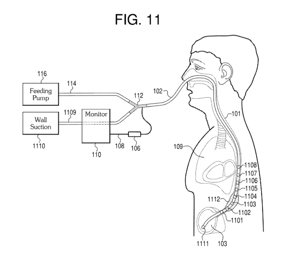
herein by reference. In step 25, the monitor also analyzes the ECG sin:m.1s to
determine
an ECG respiratory pattern, which can be accomplished via the previou,sly
described
exemplary approaches of detecting the RS.A or changes in. QRS amplitude.
ln an exemplary embodiment :the monitor .may perform an analysis to match the
initially detected auscultated respiratory pattern with the ECG respiratory
pattern that is
derived from :the electrodes, as .shown in step 206. In an .exemplary
embodiment, this
comparison. can provide additional information that can help ensure that an
auscultated
respiratory pattern has been detected in the sound detected by the acoustic
sensor. There
are a number of exemplary approaches to determine if there is a match 'between
the
auscultated and ECG respiratory pattems. In an exemplary embodiment, the
monitor
performs an analysis to inatch the key or identifiable points in the
auscultated respiration
pattern with the key or identifiable points in the ECG respiration. pattern.
These key or
identifiable points can be derived from specific aspects of inhalation and
exhalation related
to respiration and the timing of such key or :identifiable points. For
example, a key or
identifiable point can be :the timing of the peak: of the auscultated lower
respiratory tract
pattem and the timing of the peak of the ECG respiration -pattern. Similarly,
the timing of
the trough of :the auscultated respiration pattern and the tinting of the
trough of the ECG
respiration :pattern can be used as a key or :identifiable point. In an
exemplary embodiffient,
one respiration pattern can be used as a suminn point to determine if the
other respiration
pattern is a match. For example, by continually- measuring the ECG respiration
pattern, it.
is possible to generate a baseline of key or identifiable points of the
respiration pattern and
then analyze the auscultated.re.spiration pattern to determine the
corresponding existence of
these key or identifiable points, or to analyze just these key or identifiable
points to
19
CA 02986462 2017-11-17
WO 2016/187456
PCT/US2016/033335
deterniMeilthemis a inoteli of :correlation of:snificientimagnitiade,:
ThiS.patternmatching.õ
ca
include one or more ofdata smoothing, time series: unalysis, :ernsscorrelation
analysis,
convolution analysis, :regression analysis, anti neural networks. Matching the
two patterns
can be ad.vantageous, since the auscultated respiration pattern is more likely
to be a Ntatid
rewiration pattern indicative of being 'located in the larynx, trachea or
bronchi when the
auscultated respiration pattern matches the continually measured .ECG
respiration pattern
derived from the ECG signal. If the two patterns (i.e., the ECG respiratory
pattern and the
auscultated respiratory pattern) match or correlate to a sufficient degree,.
then it is highly
likely that the acoustic sensor is located in the lower respiratory tract,
such as the larynx,
trachea or a bronchus.
101121 The results of this :respiratory pattern analysis are presented on the
monitor as
shown in step 207. :If the results are negative, the monitor displays the
imessage that the
feeding tube is not. located in the trachea or a bronchus. If an auscultated
lower respiratory
tract pattern is detected (e.g., a match or significant correlation with the
ECG respiratory
pattern is detected), the monitor can indicate a visual alarm., antkor an
auditory alarm, to
warn the clinician .that the tube may be located within the lower' respiratory
tract, such as
the larynx, .trachea or a bronchus.. The clinician can then stop tube
insertion and withdraw
the tube.
[Off 3J in step 208, the acoustic sensor deteett the auscultated heart
patterit.as the ifeeding
tube is being inserted. The auscultated heart Pattern.. can be detected inn
mediatoly aptrn
tribe insertion in the body since the heart can emit a strong signal, i,e., a
signal that is loud
and can travel significant distances within. the body. The spectrum -for
capturing the
auscultated heart 'pattern is generally defined as between 20 and 100 Hz
according to
Reichart. 'There are a number of advantageous means to analyze heart sounds to
determine
the auscuhated. heart pattern .including but not limited to Fourier transform
and wavelet
transform according to Debbal et al, "Computerized 17teit.rt sounds analysis,"
Comput Biol
Med. 2008 Feb;38(2):263-80. Epub 207 Nov 26, :hereafter "Debbal" which is
hereby
incorporated in its entirety herein by reference.
[01.14) If the monitor detects an auscultated heart pattern, in .an exemplary
embodiment the
monitor will then .peribrin an analysis to match the auscultated heart
'pattern with the ECG
heart pattern that is derived from the electrodes, as shown in step 209. in
this exemplary
embodiment, this comparison can provide additional information that can help
ensure that
an auscultated heart pattern has been detected.. A nuniber of processing steps
can be
CA 02986462 2017-11-17
WO 2016/187456
PCT/US2016/033335
required:to performthis.pattern matehing,:including but notlimited to
data:.:snxiothing, tia
series ana 1 ysik tross,corre I ati on analysis, cortvoluti oi . analYais
regression analysis, and.
neural networks. .M.atching, the two patterns is advantageous, since the
detected heart
sound pattern is mire likely to be an actual heart sound pattern when the
pattern matches
the continually measured and well-known ECG-based heart pattern derived from
the
electrodes. -Matching these two patterns therefore increases the confidence
that the
acoustic sensor is correctly detecting the heart pattern,
[Oil 5J As shown in Fit. b, as the clinician inserts the feeding. tube 102
down the
esophagus 101, the acoustic sensor 104 moves within close proximity of the
heart 1 1 I.
The monitor 110 is continually measuring the intensity of the heart sound as
the feeding
tube 102 transits .the -length of the .esophagus 101 as it moves toward the
stomach 103. As
the feeding tube 102 gets closer -to the heart 1 1 1 during this transit, the
heart sound should
increase in intensity, or amplitude. Conversely, as the feeding tube 102 moves
past the
heart I and gets close to entering the stomach 103, the heart sound should
decrease in
intensity, or amplitude. This increasing and decreasing intensity in the
measured
auscultated heart pattern can be analyzed to determine an approximate location
of the
.feeding tube, e.g., a location of the feeding tube's relative to the heart of
the patient. The
measurement of the auscultated..heart pattern will be analyzed over time to
determine if it
.matches a similar increasing and decreasing pattern of intensity, as shown in
step 210, A
number of processing steps can be required to perform this pattern matching,
including but
not limited to data smoothing, time .series analysis, cross<orrelation
analysis, convolution
analysis, regression analysis, and neural networks_ For example, the _maximum
amplitude
of the auscultated heart pattern can be identified and 'plotted versus rime.
These points Can
then be analyzed to confirm a substantially continuous rise of the amplitudes
to the
MaXiMUM amplitude and. or confirm a substantially continuous decline from the
maximum amplitude, In step 211, the results of this analysis are shown on the
monitor,
such as a. message describing the status of the analysis and a chart .showing
the intensity of
the auscultated heart pattern over time.
101161.11n. step 212, the monitor .analyzes the acoustic signals coming .from
the sound
emitter 118 and captured by the acoustic sensor 104, and calculates the
distance between
the sound emitter and acoustic sensor. The acoustic signal can take many
exemplary
forms, including but not limited to a sound pulse, a continuous vari.able tone
and can he
emitted at different audible, Ultrasound, or other advantageous frequencies.
By knowing.
21
CA 02986462 2017-11-17
WO 2016/187456
PCT/US2016/033335
the paveiSe titning-a. initiating ttie.acotiatie signal livin the sound
emitter arid thelithing cif
receiving the signal by-= the :acoustic -sensor, the -monitor <can perform
calculations: to
determine the distance between the sound emitter and the .aeoustic sensor._
For example,
Skilen a pulse is emitted at time tl by the sound emitter 118 and received at
time t2 by the
-- acoustic sensor 104, in an exemplary entbodiment, the distance x can be
calculated as x
(t1.-t2) x v, where v----- the speed of sound throagh the patient (V can be
determined through
calibration, i.e., tested on the patient from known distances, or determined
from empirical
data. This process therefore provides the distance 'between the acoustic
sensor 104 and the
sound. emitter 1 .18. in step 213, the monitor displays the status of the
distance calculation
-- and a chart showing these distance calculations over time. In some
embodiments, the
monitor may display a distance that is derived fronr the distance between the
acoustic
sensor 104 and the sound e-mitter 118 (e.g., a distance remaining to complete
filSertiOil of
the feeding tube).
[0117] 'In an exemplary embodiment, the clinician pauses: the insertion to
aheek the.
-- monitor after inserting the tube approximately halfway. into the patient -
suelt:AS '41 Ft. rt).
The purpose of checking, the monitor is to determine if any of the summary
data -would
indicate the tube is located in the -trachea or bronchi, or if the tube
appears to be located
correctly in the esophagus, as shown step 214. 'In this example., the
clinic:ian pa.uses the
insertion based on tube inarkings close to the point of entry, such a.s the
nose or mouth,
21) -- indicating the tube had been inserted 25 cm. Other ex.emplary markings
and insertion
distances may apply, such as those having a dependence on or calculated
'based. -upon the
identified target depth or otherwise calculated as a function of the data of
the patient (such
as size, age, sex, etc.). The clinician can check the monitor to see if there
is any indication
the tube tip is located in the lalynxõ trachea or bronchi:. The clinician can
also check the
-- monitor to see if the auscultated heart pattern has increased in intensity,
which would be an
indication that tube has progressed down the esophagus and is near the heart.
The
combination of no indicated auscultated lower respiratory tract pattern along
with an
increase in intensity- of an auscultated ..heart pattern indicates that the
tube is progressing
correctly down the esophagus towards the stomach. The clinician can also check
the
-- monitor to see if -the calculated distance between the acoustic .sensor and
the sound emitter
has decreased during the period of insertion. This decreasing distance
indicates that tube
has progressed down the esophagus, and conversely has not bCCOMO coiled. in
the mouth,
nasopharynx or hypopharynx. The combination of no auscultated lower
respiratory tract
22
CA 02986462 2017-11-17
WO 2016/187456
PCT/US2016/033335
pattOM an increase. :in intensity of
auscultated heart pattern; and .40er:easing distance<
between the acoustic sensOr and tbe'sound emitter provide a. strong indication
that the tube
is progressi n correctly down the esophagus towards the stomach,. Any
combination of an
indication of an ausculta.ted lower respiratory tract pattern, an indication
that the
auscultated heart pattern intensity has not increased with insertion of the
tube, and no
evidence of decreasing distance between the acoustic sensor and the sound
emitter, may
indicate the tube is not progressing correctly down the esophagus towards the
stomach and
.may be in the: trachea or bronchi or has become coiled, After reviewing these
data, the
clinician can then decide whether to proceed with the tube insertion or take
other action,
to such as
removing and reinserting the feeding tube or taking an X-ray to confirm the
placement of the tube.
[01181 in Fin. lc, the acoustic sensor 104 is showniin the sto.mach 103: and
located more
closely to the sound emitter 1.18. .After the clinician has inserted th.e
tube. 102 to the
recommended insertion distance, the clinician can check the monitor to see the
distance
between the sound emitter 1.1.8 and the acoustic sensor 104. The clinician can
then
compare this calculated distance with a -visual identification of seeing
.where the sound
emitter 1.18 is physically placed on the patient and. making an assessment as
to whether the
distance corresponds with the feeding tube 1.02 being correctly placed in the
stomach 1 03
and conversely not in the trachea or bronchi 109. As the feeding tube 102
progresses
toward and into the stomach 103, the calculated distance between the sound.
emitter 118
and acoustics sensor 104 should decrease.
yoi 91 in step 215, the clinician reviews the summary information and inputs
the .distance..
marking on the -tube into the monitor.. This distance marking corresponds with
the furthest
point the tube has been inserted into the patient. The monitor then compares
the inputted.
tube distance with the recommend insertion distance calculated in step 202, if
the
difference between the two insertion distances is above a defined threshold,
the monitor
will display a message that the .tube insertion distance .may not .be
sufficient for proper
location in the stomach. If the difference between the two insertion distances
is below a
defined threshold, the monitor will display a message that the tube insertion
distance i.s
sufficient, In an exemplary embodiment, the threshold for the difference in
tube distance is
5 cm; in another embodiment, the threshold -for the difference is 1.0% of the
identified.
target depth.,
23
CA 02986462 2017-11-17
WO 2016/187456
PCT/US2016/033335
101201 Fig, 4 depicts araexampleinoaltorseteen her all
assessment:a:have:indicated that
the iiibeis.placed.correctly. in the stomach. The .most visibe indication on
the acreerria the
status indicator 401, which displays the color green to signify the tube is
placed correctly.
if the monitor alaorittun calculations result in an indication that the tube
is not placed
correctly, or if .there is .not enough information to determine if the tube is
placed correctly,
the status indicator 401 displays the color yellow. If the monitor algorithm
calculations
result in an indication that the tube is placed in the trachea or a bronchus,
the status
indicator 401 displays the color red. In addition, a text status indic,ater
402 conveys the
status of the tube placement, In the scenario thr Fig, 4, the text status
indicator indicates
the "Tithe Placed Correctly". The heart and. respiratory signal indicator 403
displays a
message. "Heart and Respiratory Connected", signaling that the heart and
respiratory
signals from the electrodes are 'being correctly. c.aptured. The trachea and
lung sounds
signal indicator 404, displays a message, "NO Trachealning Sounds Detected.",
signaling
that the acoustic sensor and monitor are currently not detecting any trachea
or lung sounds,
and thus indicating the feecliug tube tip is not in the trachea or bronchus.
The heart. transit
indicator 405, displays a message. "Tip Passed Heart in Esophagus", signaling
that the
acoustic sensor and monitor detected the pattern of the tube tip passing by
the heart during
transit down the esophagus. The indication of the tube tip passing by the
heart during
traasil down the esophagus is further confirmation that the tube tip is not
located in the
2.0 trachea
or bronchus. The sound emitter distance indicator 406, displa.ys a .message,
"Tip 3
Cm .froin Sound. 'Emitter", signaling the acoustic sensor is located. 3 cm
from the sound
emitter. The indication that the acoustic sensor is located a short distance
from the sound
emitter, which should be located caudal to the left costal margin,. is further
confirmation
that the tube tip is located in the .stomachõ and conversely is not located in
the trachea or
bronchus. The .atbe insertion .indicator 407 displays a message, "Tube
Insertion of 45 cm Is
Sufficient, signaling that the inputted tube insertion distance is sufficient
for the tube to be
placed correctly in the stomach, If the clinician is satisfied that the data
presented on the
monitor is sufficient to confirm the tube is correctly placed in the stomach,
the clinician can
then submit the electronic medical record lay pressing button 409.
[0121 l The clinician also has the option to review .more detailed d.ata on
the monitor. By
pressing the Data. Chart View button 408, the clinician can view more detailed
information
in chart form. Fig. 5 shows this optional view to review the data in chart
form. For.
example, the clinician can review the Heart Sound Intensity chart 501 to see a
time-based
24
CA 02986462 2017-11-17
WO 2016/187456
PCT/US2016/033335
Niow of the heart sound intensity data. Iî the. feeding tube transited the
esophagus=.
comedy, this :would be indicated 'by- artincrease in. =se uhated heart -
pattern intenaity as the
tube tip gets closer to the heart and a decrease in intensity as it passes the
heart on the way
towards the stomach. The clinician can also view feeding tube tip distance.
502 over time
(ex.., uraphically), which should generally indicate a decrease in tube tip
distance from the
sound emitter as the tube is being inserted. Alternatively-, the clinician may
review the
heart sound. intensity with respect to the measured feeding tube tip distance.
In this
example, the measured feeding tube tip distance may be plotted along the x-
axis and the
heart SOtilld intensity may be plotted along the y-axis. The clinician may
repetitively
sample the heart sound. intensity at the same distances by inserting and
retracting the
feeding tube.
[01221 Alter the tube has been .inserted, the elinitian can: All' refer to the
luonitOt to set A.n.
update of the distance between the acoustic.. sensor and the sound emitter.
This may .be
valuable to determine if the tube has moved during treatment and if the tube
insertion may
need to be adjusted,
[01231 In an exemplary embodiment, the clinician can utilize the same .monitor
for
multiple patients, tti this embodiment, .the monitor associates a. unique ID
with each
feeding tube. if the feeding tube was disconnected from the monitor and
subsequently
reconnected, the monitor can utilize the unique 113 of the feeding tube to
associate ail
previously entered and measured data. -from that feeding. Therefore, a
clinician can .utilize
one monitor -to insert feeding tubes into .inultiple patients, and as
necessary reconnect the
monitor to a tube to assess the location of the tube without having to reenter
any patient
data. .A history of patient data is stored such that any previously entered
patient data can
also be accessed and associated -with any new feeding tubes.
[01241 Fig. 6 shows an alternative embodiment of the apparatus to determine
"'Cedilla tube
to-cation. In this exemplary embodiment, two sound emitters 601 and 602 are
used to
determine the distance .from the acoustic- sensor 104 to the sound etnitters.
The sound
emitters 601 and 602 are connected to a rigid member 603. The rigid member 603
is
connected via Wire 604 to electrical connector 106. The two sound emitters
601. and 602
also serve as electrodes to capture heart and respiratory signals. The
distance between the
acoustic sensor 104 and the sound emitters 601 and 602 that Call be calculated
in the same
fashion as described elsewhere herein. Therefore, in each instance in timeõ
the distance
betweem acoustic sensor 104 and sound emitter 601 is known and the distance
between
CA 02986462 2017-11-17
WO 2016/187456
PCT/US2016/033335
acoustic sensor 104.. aad sound etnitter-602. is known, Additionally, the
distance :between:
sound eminer-60.1 and sound. emitter .602 -is known given their fitted
location on the rigid
member 603, 'These three distances -form a triangle, so knowing the lengths of
each side of
the triangle .makes it possible to calculate. the angles of this triangle and
thus determine the
location of the acoustic sensor. With one sound emitter (e.g., just one of 601
and 602), you
can determine the location of the acoustic sensor as being a certain distance
away from the
one acoustic sensor (e.g., determined to be on a point of the surface of an
imaginary three-
dimensional .sphere having, the location of the. one acoustic sensor as its
center)õ In this
exemplary embodiment with two sound emitters, you can determine the location
of the
acoustic sensor as being on a point of the circumference of an imaginary two-
dimensional
circle. 605. This level of accuracy in determining the location of the
acoustic sensor 104
may be advantageous. For example, when the two sound emitters 601 and 602 are
arranged verticallyõ the imaginary circle on which the acoustic sensor 104 is
.determined. io
lie will be horizontal. Thus, a vertical location of the acoustic sensor 104
can be accurately-
determined even if its horizontal location has 'been determined via this
calculation to be on
the horizontal imaginary circle. Using three sound emitters (not shown .in
FIG. 6) that are
arranged in a triangle and. spaced apart known distances (i.e,õ not linearly
arranged) allows
for further precision in determining the location of the acoustic sensor.
Triangulation can
be .used to calculate a point in space relative to the location of the three
sound emitters. For
2.0 e.xample,
for each of the three sound emitters, a sphere with the corresponding sound
emitter as its center can be determined, W th the radius of the sphere
representing. the
determined distance between the sound emitter and the acoustic sensor and the
surface of
the sphere representing a possible location of the acoustic sensor_ The
intersection of these
three determined spheres can be determined as the location of th.e acoustic
sensor. Another
approach is to use each pair of the three sound emitters to calculate possible
locations along
a corresponding imaginary circle. The intersection of these three imaginary
circles win
correspond to a determined location of the acoustic sensor I 04.
[01.251 Fig, 7 shows an alternative embodiment of the apparatus to determine -
feeding tube
location. In this exemplary embodiment, the apparatus utilizes a mobile device
'701 (shown
as 701a, 701b and 701 c, at respective dine-rent locations). This mobile
device 701 can take
the form of a mobile phone, tablet, or other advantageous mobile device. In
this
embodiment, the mobile device 701 -uses a wireless connection to communicate
with the
.monitor 110, A tip com.ponent 707 is located in the tip of the feeding tube
and, in this
26
CA 02986462 2017-11-17
WO 2016/187456
PCT/US2016/033335
embodimenti is used .to seise :the acoustie Sipes fnr,nn the: mobile device
701: In an
exemplary embodiment, the mobile =deviee.701..continuously emits a. acoustic
signal, lri
this .embodiment, the tip component 707 is the acoustic sensor 104, 'The
signal received by
the tip component 707 is used by the monitor 110 to calculate the distance
'between the tip
component 707 and the mobile device 701,
[01261 An exemplary process is to move the Mobile device 7Tì While the
tip..cOmponent
707 receives the sound signals. In a first position., InObile device 701a, is
.:a calculated first
distance from tip component 707 where the distance can be visualized as the
radius of a
first sphere 702 (with mobile device 70.1a at the center) and the location of
the tip
1.0 component 707 is somewhere on the surface of that lust sphere 702. In a
second position,
mobile device 701b is a calculated second distance aim tip component 707 Where
the
distance can be visualized as the radius of a second sphere 703 (with:imbue
device 70Ib at
the center) and .the location of the tip component 707 is someWhere on the
surface of that
second sphere 703. In a third position, mobile device 701c is a calculated
third distance
from tip component 707 where the distance can be visualized as the radius of a
third sphere
704 (with .mobile device 'Mc at the center) and the location of the tip
coniponent 707 is
somewhere on the surface of that third sphere 704. If you aSSUIlle the
acoustic sensor is not
moving between the three .positions then the intersection point 705 where each
of the three
spheres intersects is the location of the tip component 707 in three-
dimensional space
Determining location via this process is called trilateration, as described by
Wikipedia,
"Tri latera don". Wikipedia, 'Web. 3 March 2015, Web.
<http://en.wikipedia.orghvirrilateration>. To determine this -intersection
point 705, you
need to .first determine the location of the mobile device 701 in each
position relative to the
(Ayer posi tions
[01271 in an exemplary embodiment, the location .Ot tad: posOnt rOlotiVe= to =
the ther
positions can be determined hy utilizing an .acce*oitwter and gyroscope
contained: in the,
mobile device 701. The accelerometer and gyroscope can .enable calculating the
position.,
orientation and velocity of .the mobile device and thus enable determining a
relative
location at each time interval. In an exemplary embodiment, the mobile device
emits
sound signals at 20 Hz, or 20 times per second, and therefore continually
calculates relative
location 20 tittles per second, Other exemplary frequenciestbr emitting a
sound signal arid
calculating relative location can be used.
27
CA 02986462 2017-11-17
WO 2016/187456
PCT/US2016/033335
101281 in .an exemplary embocliments the accuracy of detenniiiing
the.,relatiye location of:
the mobile device 701 cian .be enhanced by using: an. additional reference.
point. In an.
exemplary embodiment, a known visual reference 706 can be utilized. In this
:embodiment,
a camera contained in the .mobile device 701 can be utilized to vim, the
visual reference
706. An exemplary "bon for this visual reference 706 can be a tw-o dimensional
rectangle
with a length of 2 cm on one .side and 4cm on. the other side attached -to the
-body of the
patient. Another exemplary fornn for this visual reference 706 can be a high
contrast, two
dimensional marker of known dimensions which lacks reflection .symmetry andfor
rotation
symmetry attached to the body of the patient. Other forms and dimensions can
be used.
Since the form and dimensions are know), algorithms in the sound emitter 701.
can analyze
data from the camera to calculate the distance from the visual reference 706
to the mobile
device 701 based on the characteristics of the visual reference 706, such as
calculated
diameter of the disk compared to the known diameter and the shape of the disk
compared
to the knOWD. shape.
[01291 'In an exemplary embodiment, the relative position of tip component 707
can be
communicated to the clinician -via an augmented 'reality interface on a screen
containe.d iu
the mobile device 701. Augmented. reality .is defined as viewing data in what
appears to be
a camera field of view. In this embodiments the clinician can look at the
screen of the
.mobile device 701 and see v1/4.hat appears to be a real-time video feed .of
the .patient. The
2t)
cliniciaar can first wave the mobile device 701 forward, then backward., and
from side to
side over the patient to calculate a relative loc.ation of the tip component
707. The clinician
can -then see on the screen of the mobile deice 701 a representation of the
tip component
707 on the :screen relative to its position within the patient's body. The
visual
representation of the patient's body on the screen can exhibit a transparency
effect to create
the illusion that you can see the tip component 707 within the patient's body.
.An
exemplary embodiment of this transparency effect can be -within the sight line
of the tip
component 707, the 'patient's body has an algorithm generated surface
appearance that
makes it look as if you're looking beneath the surface of the patient's body.
'The clinician
should there-fine be able to see the location of the tip component 707
relative to the
patient's anatomy and determine if the 'tip component 707 is correctly placed
in the
stomac.h., and conversely is not. placed in the trachea or bronchus. In an
exemplary
emboditnent, an algorithm-generated appearance can also simulate a. visual.
representation
28
CA 02986462 2017-11-17
WO 2016/187456
PCT/US2016/033335
of organs:..suCh:-as. Ole stomach, hins:.and heart to-
assis¶he..Clirticiwiuld:de.termining the
1 o cation 'of the aeons& .settscir.
101.301 B. Determine Tube Location Using Magnetic Field Sensor
[01311 In an exemplary embodimenteart aiternaiielor additional) apparatus can
be used in
4 similar process previously described for the mobile device 701 and the tip
component
707. This alternative (or additional) apparatus comprises a magnetic fielci
sensor, or
magnetometer, contained within the mobile device 701. The magnetometer can
measure
the strength of the magnetic field that is around the mobile device 701. In an
.exemplary
embodiment, the tip component 707 can contain magnetic material with a known
magnetic
.10 moment. In all alternative embodiment, .the tip component 707 consists
of .coiled wiring
that upon application o.f a known electric signal creates a known .magnetic ìr
oment. in an
exemplary embodiment, the clinician can also wave the mobile device 701
forward, then
backward, and from side to side over .the patient, Instead of .measuring
distance between
the mobile device 701 and the .tip component 707 based on the elapsed time for
the sound
signal to travel between them, the distance is instead calculated based on the
strength of the
magnetic .field measured from the magnetic material in the tip component 707.
The
measurement of distance (measured in meters) from the mobile device 701 in
.the tip
component 707 can be calculated 1.141.14 the following exemplary formula.:
20101321
f .
a ee. --
On- 8
[01331 .ht this formula po represents the permeability constant (47tx 104 T
miA), p is the
magnetic moment of the magnetic material in the tip component 707, and is the
magnetic
field (measured in tesla). The clinician can similarly- be able to view an
augmented reality
visual representation of the tip component 707 relative to the patient's body
on the screen
of the .mobile device 701. In an exemplary embodiment, the accuracy of
determining the
relative location of the mobile device 701 can also be enhanced by using a
known visual
rerence 706, as previously described.
[01341 II, Motility Measurement .System and Apparatus
SO 101351 Determining if the patient is tolerating enteral nutrition is
critically important in
delivering eft'ective care a:nd avoiding devastating complications.
Determining whether the
patient has satisfactory gastric .motility can indicate .if enteral nutrition
will be tolerated.
29
CA 02986462 2017-11-17
WO 2016/187456
PCT/US2016/033335
NOtanal gastrimotility an 1e defined.asperiodic or occasional
peristaltieamovements of:
the stomach that propel the.mitentsof the stomach into the:small intestine
Figa8 shows.
an example of this peristaltic movement. In 'Fig. 8, a peristaltic wave 801 is
shown moving
through the stomach 103. These peristaltic waves 801 move the .food through
the stomach
103 and out through the pyloric sphincter 803 into the duodenum 804 according
to Elnlein
et al, "Ciastrointe.stinal Motility." Class Tutorial, Technische Universitat
Munchen, .2014,
hereafter "Ehrlein" which is hereby incorporated in its entirety herein by
reference. During
the movement of a peristaltic wave 801., .intiscles in the stomach 103
contract to push food
802 from the .proximal stomach 103 towards the pylorus 803 at the distal end
of the
stomach. In addition to peristaltic waves 801, the .stomach 103 .moves food
802 towards
the pylorus 803 by generally contractin.g and thus shrinking the size of the
overall stomach
103; this phenomenon is called tonic contraction. The peristaltic waves 801
and. .tonic
contraction encourage the food 802 to be further broken down and thus
digested. Food. that
is broken down to a point Nkhere it is .inore of a liquid is referred to as
gastric chyme 80.5.
The .peristaltic waves 801 more easily move Ibis gastric chyme 805 past the
pylorus 803
and into the duodenum 804, .and conversely larger undigested pieces of food
802 are.
pushed back into the stomach 103 to be further digested in an effect called
retropulsion.
Understanding gastric motility can be helpful to the, clinician in many -ways.
First, if the
patient appears to have good motility, there is less risk. the patient .may
reflux gastric
contents into their lungs. Second, good motility may indicate the clinician
can accelerate
the level of nutrition for .the patient, which would potentially assist with
recovery. Certain
embodiments provide, advantageous means for measuring inotiW.
[0136) A. Determine Motility Us:ilia Acoustic Sensor
[01.37] in an exemplary embodiment, an acoustic sensor is used to measure
gastric motility.
This acoustic sensor 104 can be located on the distal tip of the feeding tube
102, as shown
in Fig. 8. This acoustic. sensor 104 can measure different frequency ranges
and types of
vibrations including, but not limited to, .vibrations associated with
frequency ranne of
audible sounds (2.0-20,000 Hz). In an exemplary embodiment, a piezoelectric
sensor is
used to measure the acoustic signals. A number of other exemplary sensors can
be used to
3t/ measure
acoustic signals, including but not limited to an electret, condenser,
piezoelectric
crystal, piezoelecnic ceramic, piezoelectric film, or contact accelerometers.
The vibrations
measured by .the acoustic sensor 104 can originate from a number of sources,
including but
not limited to peristalsis, gas bubbles, flatutenceõ and sounds .from nearby
organs, such as
CA 02986462 2017-11-17
WO 2016/187456
PCT/US2016/033335
the Ittnm iheart, or small. bowel. The .study of phonooterography revealed
that bowel:.
sounds increased. íi intensity during- a ineal and high intensity :persisted
for more than two
hours after the meal according to Watson et al., "Phonoenterography: the
recording and.
analysis of bowel sounds," Gut. 197 Feb, 8(1): 88-94, hereafter "Watson" which
is
hereby- incorporated in its entirety herein by reference, Bowel
sounds are complex with a
mixture of tones and often in a sequence of closely connected sounds in. a
frequency range
of 150 to 5,000 Hz,
[0138J A number of sources of vibration may lx dotected. iri .a.:study by
Ca.mpbeiteta
"Surface vibration analysis (SVA): a new. non-liwasive. õmonitor of
gastrointestinal
1.0 activity." Gut, 1989 Jan; 30(1)39-45, (hereafter "Campbell" which is
hereby inewporated
in its entirety herein by reference) Campbell attempted to quantify these
bowel sounds and
measured vibrations in the 40-10,000 Hz frequency range with a piezoelectric
sensor.
Specific patterns of bowel sounds can be associated with the fasting state and
the
postprandial state associated with the food being digested and thus monitored
to determine
he same.
[01391 In one embodiment, an acoustic sensor detects the .vibrations
associated with the
movement of a peristaltic wave. in this exemplary embodiment, the acoustic
sensor 104
can be a piezoelectric-based accelerometer. In addition (or in the
alternative), the
accelerometer is capable of .ineasuring movement (e.gõ physical movement of
the
accelerometer itself), such as the peristaltic wave 8)1 that moves down the
stomach 103..
For exam.ple, the peristaltic wave 801 can be detected by detecting a pattern
having a
frequency of one to three contractions per minute, as shown ia Fie, 8.
[0101 A positive indication of nark! motility (e.g., between 1 and 3
contractions / waves
per minute) should con-elate with the patient tolerating enteral nutrition,
and therefore have
sufficient sensitivity. In contrast. a negative indication a motility (e,g.,
less than 1
contractions / waves per minute) can help identify patients that are not
tolerating nutrition.,
enabling the clinician to adjust care and monitor the patient more closely.
1p1411 To ensure that the acoustic sensor 104 is functioning correctly, the
.monitor can
routinely monitor other body sounds as a means to cheek it the apparatus is
functioning
correctly, In an exemplary embodiment, the monitor routinely captures an
auscultated
heart pattern. If the auscultated heart pattern is routinely captured
correctly, this indicates
the acoustic sensor is functioning correctly, and thus it can be inferred it
is also correctly
capturing g,astric motility vibration .measurements. in an .alternative
embodiment, the.
31
CA 02986462 2017-11-17
WO 2016/187456
PCT/US2016/033335
monitor can routinely capture acoustic signals from the sound emitter 118 to
confirm the
acoustic sensor 104 is functioning correctly.
101421 .in an exemplary embodiment, an acoustic sensor measures the vibrations
associated
with oscillating gas bubbles in the small intestine. In this exemplary
embodiment, the
acoustic sensor 104 can be a piezoelectric-based accelerometer. There is
evidence these
gas bubbles are continually present in the small intestine according to Liu et
al.
"Oscillating Gas Bubbles as the Origin of Bowel Sounds: A Combined Acoustic
and.
Imaging Study." Chin I Physiol. 2010 Aug 31; 53(4):245-53 'hereafter "Liu"
which is
hereby incorporated in its entirety herein by reference. Bubbles Call be
identified by their
oscillating frequencies. Further, the size of the bubbles can also be
determined by the
measured frequ.ency. The size of the bubbles can change as they move within
the different
sized structures of the small intestine. In one embodiment, the pattern and
frequency of the
bubbles is arialyze.d to assess gastrointestinal motility and other gastric
functions. The
analysis of gas bubbles in the small intestine can be combined with the
analysis of
peristaltic waves in the gastric environment, or other sensor data., to assess
overall
gastrointestinal motility,
101431 In an exemplary embodiment, the various gastric motility measurements
can
additionally be used to determine the effectiveness of the type and amount of
enteral
nutrition. In an exemplary einbodiment, the patient condition can also be a
factor in
determining the effectiveness of the type and amount of enteral nutrition. By
collecting
data on the patient condition, the type and amount of enteral nutritiori
and/or the resulting
measurement of gastric motility, it may be possible to guide the type and
amount of enteral
nutrition and/or obtain the optimal gastric motility for each patient
condition. Conversely,
analyzing these data can provide insights ort which enteral nutrition types
and amounts
should not be used for specific patient conditions.
101441 B. Determine Gastric Residual Volume Using Temperature Sensok
101451 It is standard of care in hospitals to measure the volume of the
gastric contents of
patients receiving enteral nutrition. This procedure is referred to as
measuring Gastric
Residual Volume (GRV). Gastric residual volume is typically measured by
attaching a
large syringe to the proximal opening in the feeding tube and suctioning all
of the gastric
contents into the syringe. The syringe has measurement tnarkings denoting the
volume in.
milliliters, which is then recorded in the patient's medical rixord.. A low
volume of gastric
contents may indicate the patient is tolerating enteral nutrition and has
sufficient gastric
32
CA 02986462 2017-11-17
WO 2016/187456
PCT/US2016/033335
.mottlity. A bighvoitimeHof gastric ..contents may indicate the patient < is
not tolerating.
enteral nutrition and does :not have sufficient gastric 'motility. A high-
volume c,f gasttit.
contents is defined as 250 mt., according to =McCIave et al, "Guidelines for
the provision
and assessment of nutrition support therapy in the adult critically ill.
patients" JPEN
Parenter Enteral Nut% 2009 May-June;33(3):277-31.6, hereafter "McClave" which
is
hereby incorporated iri its entirety herein by .reference. A clinician can
adjust the care of
the patient based on receiving a high gastric residual volume m_easurement,
$UCh as
reducing, the rate of tube feeding, prescribing pm-kinetic agents or
raisirn:.,!. the head of bed.
However, a. high gastric residual volume may not always correspond to a
gastric motility
problem. In some eases, a gastric resid.ual volume of over 250 mi., may be
required to
trigger gastric emptying by the stomach in.to the small intestine., and ii10S
a gastric .residual
VOiume of over 250 nit¨may be part of normal digestion. In addition., these
gastric .residual
measurement calculations are often performed every fur hours in acute care
patients
receiving enteral nutrition. This proced.ure therefore takes ante away from
the clinicians
that .otherwise can be used in treating patients. The procedure is also often -
unsanitary due
to the .removal and reinsertion of gastric contents.
[01461 In an exempliny embodiment, an indicator dilution technique is used to
measure
(IRV_ Indicator dilution techniques are well known and are widely used in
medicine.,
scientific work and .industry for measuring fluid (gas or liquid) volumes or
flow rates. As
2) described
by Schoeller "Indicator Dilution Methods.:' Quality of the Body Cell. Mass.
Serono Symposia USA 2000, pp SS-67, hereafter "Schoeller" which is hereby
incorporated.
in its entirety herein by reference, indicator dilution _methods for
'measuring volume (or
.flow rate) are based on the principle of conservation of mass. The addition
of a known
quantity of tracer (indicator) to the pooi of tracee yields, after
equilibration, a solution
wherein the final concentration ofiu dic.ator ìs equal to the amount of
indicator added
divided_ by the pool volume. If volume is the desired read-outõ then the
equation is re*
arranged: -volume (pool size) dose of .tracer/concentration of tracerõ
l01471 The validity of indicator dilution approach depends on four
a.ssumptions. the
indicator mixes rapi.dly and. thoroughly with the fluid in the -pool. Second.,
after mixing is
complete, the concentration of the indicator is homogeneous in the pool
volume. Third, the
indicator is distributed only to the pool volume of intere.st. Fourth, the
tracer is stable (over
the time period required for the .ineasuremems).
33
CA 02986462 2017-11-17
WO 2016/187456
PCT/US2016/033335
101481 Although .these :assumpiions..are:: o4en: valid in: vitro, when
indicator dilution
techniques are applied in vivo4Stich as, liar medicai applications):Some or
even all of the
assumptions typically are not satisfied per&ctly. Thus, for medical (or other
in vivo
biological) applications, indicator dilution methods are rarely perfectly
accurate.
Neyertheless, these approaches are often accurate enough to be clinically
mail.
-Moreover, as will be discussed below, it may be possible to partially adjust
for systematic
errors in measurement by incorporating empirically derived coefficients into
the final
equations that are employed. for converting the measured signal into
clinically useful
information,
1.( 491
Indicators often are chemicals that can be easily detected, using .appropriate
analytical ITICallS. Ex.amples include: radio-labelled compounds (e.g..:
tritium-la.belled
water), stable isotope-labelled compounds (e.g., 13C-1abelled glucose), dyes
indocyanine green dye),. or easily detected chemical entities (e.g, 1i cation,
polyethylene
gyro0.
191501 GRV has been measured, using an indicator dilution approach In the
laboratory, a
model stomach showed that dilution of phenol red dye can be employed to
determine
"unstric" vohtme according to Hurwitz, "Measuring gastric volume by dy:e
dilution," Gut
1981 Feb;22(2):85-93., hereafter "Hurwitz" which is hereby incorporated in.
its entirety
herein by reference. Subsequently. GRV measurement in patients was
demonstrated using
polyethylene ,f_412,,,col (PEG) as the tracer and turbidometry as the means
for measuring. PEG
concentrations according to Hardy et al, ".Determining gastric contents during
general
anaesthesia: evaluation of two methods." Can J Anaesth. 1987 Sep;34(3):474-7,
hereafter
"Hardy" which is hereby incorporated in its entirety herein by reference.
These :authors
showed that GRV measured using the indicator dilution technique was not
significantly
different from the GR-V measured by aspiration (via a Levin uasogastric tube)
of die gastric
contents.
f0II] 'Temperature also cam be a useful. tracer. For example, cardiac output
the flow
rate of blood through the heart) is commonly measured. in clinical practice by
injecting a:
known quantity of cold normal saline solution into the venous side of the
circulation and.
detecting the resulting change in blood temperature in the pulmonary artery.
hi. an
exemplary embodiment, gastric residual volume is determined by measuring the
temperature of gastric contents before and after a control. In this
enibodinient,
temperature sensor is located on the distal tip of the feeding tube, -which
can be connected
34
CA 02986462 2017-11-17
WO 2016/187456
PCT/US2016/033335
'ìa a. wire :to the monitor. The :monitor 110 can<take:continuou.s.
:temperature measurements:=.
of the =gagEric contents:: The apparattiS ii4=.Sirritiar to that sho'w ì 8
but instead .:of an
acoustic sensor 1.04 located the distal tip, a temperature sensor t.Yr
thermistor is used. Of
course, a combination of multiple sensors can be -used, such as those senson
disclosed
herein and other sensors, and such combinations should be considered within
the scope of
this invention. The monitor can then control a known. amount of fluid to be
introduced
through the feeding tube and into the stomach, which in an exemplary
embodiment can be
50 rrit a distilled water cooled to a temperature of I C. Utilizing water as a
control can be
advantageous.: since acute care patients require hydration and water is a safe
substance.
The temperature sensor can -then continually measure the change in temperature
of the
gastric contents after introducing this control of cooled water. in an
exemplaty
embodiment, the OW can be calculated using the following equation:
101521 GRY=1.1:x
1-01.53j In this equation, Vinj,,,õ,, is the volume of cold water injected in-
to the stomach via
the feeding -tube. Tivrom is the temperature of the gastric contents prior to
injecting the
aliquot of cold water. Tiõj,..,:tate is the temperature of the cold injectate.
TA, is the
temperature of the gastric contents after the injection of the indicator. ki
is a dimensionless
constant that depends on the specific heat content of water., the density of
water, the
specific heat content of gastric contents and the density of gastric contents.
ki is a
dimensionless constant that accounts for the dead space volume of the feeding
tube and
perhaps other unmeasured sources of systematic error. In an exemplary
embodiment, the
rate in which the temperature change occurs throughout the iiastric contents
can be used to
calculate CiR V. Calculating, the rate of change of the temperature can add
information to
improve the calculation of GRV or provide other insights that may prove
helpful to the.
clinician. The nature of the stomach environment and the gastric contents are
such .that
significant mixing occurs as part of .digestion and the subsequent measured
temperature
change will be reflective of the entirety of die gastric contents. Other
factors that can be
used to calculate (IRV include the rate of enteral nutrition, the rate of
gastric emptying, and
medicines that may be introduced into the gastric environment. Since there may
be
unaccounted-kyr factors that may influence the accuracy of the gastric
residual volume
calculation, the algorithm can also present a probability measurement
associated with the
CA 02986462 2017-11-17
WO 2016/187456
PCT/US2016/033335
volume calculation to help the clinician understand the relative certainty in
the GRV.
calculation.
[01541 C. Determine Gastr.ic Residual Volume Using Bioelectrical Impedance
101.551 In an alternative embodiment, Gastric Residual Volume can be
determined in
certain circumstances by measuring changes in the impedance of an alternating
current
applied in the abdominal region by electrodes according to Soulsby et al:
"Measurements
of gastric emptying during continuous nasogastric infusion of liquid feed:
electric
impedance tomography versus gamma scintigraphy." Clin Num 2006 Aug;25(4):671-
80,
hereafter "Soulsby" which is hereby incorporated in its entirety herein by
reference. In the
approach described by Soulsby, 16 electrodes (ECG pads) are applied in a
circumferential
pattern around the abdomen. Impedance changes are measured by passing a
sinusoidal
alternating current (5(i-100 kHz; 1 to 10 mA) between one pair of electrodes
on the
abdomen and measuring the resulting voltage drop between another pair of
electrodes. In
the approach described. by Soulsby, impedance is measured from all the
possible
combinations of pairs of electrodes, and an impedance versus time curve is
generated by a
proprietary algorithm. The system is "calibrated" by introducing a 100 mL
bolus of tube
feeding into the stomach. The conductivity of the calibrating bolus of tube
feeding .formula.
is increased by dissolving 17 g/100 niL of table salt (NaC1) in the formula
prior to
introducing it into the stomach.
10156] A similar approach to Soulsby., is described by McClelland et al,
"Epigastric
impedance: a non-invasive method for the assessment of gastric emptying and
motility."
Gut. 1985 Jun;26(6):607-14, hereafter "McClelland." which is hereby
incoiporated in its
entirety herein by reference. There is also a. similar approach described by
Sutton et al,
"Measurement of gastric emptying rates by radioactive isotope scanning and
epigastric
impedance." Lancet, 1985 Apr 20;1(8434);898-900, hereafter "Sutton" which is
hereby
incorporated in its entirety herein by reference. The approach described by
"McClelland
and Sutton is similar to the one described above, except that only four
electrodes are
employed (two on the anterior epigastrium and two on corresponding 'locations
on the
back). The system described by McClelland and Sutton uses
"standard" impedance cardiography equipment for signal generation and
detection, but
employs appropriate low-pass filtering to exclude interference from cardiac
signals. In the
approach described by McClelland. and Sutton, gastric volume is not measured;
rather,
the impedance based system is designed only to measure fractional changes in
gastric
36
CA 02986462 2017-11-17
WO 2016/187456
PCT/US2016/033335
VQ11.1100...after a:test:mea s a &fiction of iine. Thus,. the ,pritnaty .read-
out is the half-time'
far gastric. emptying..
101571 In one embodiment, a CiRV monitoring device can ern the
the. electrode placement
strategy described by McClelland and Sutton , as Miawn in Fig.9. Tn ait
exernplary
embodiment, a plurality- of electrodes are placed an the abdomen. Here, faur
electrodes
118, 12.0, 902 and 9t.I4 are placed on the abdomen. An alternating electric
cutrent is driven
through the patient's body between electrodes 118 and 902. The voltage between
electrodes 120 and 904 .may also be measured. The voltage measurement is
filtered to only
reflect the changes within the frequency range of the alternating current
between electrodes
1.0 .1.18 and
9(12. The voltage measurement comprises an impedance .measurement whose
amplitude depend.s in part on the GRV. Electrodes c.an have plural functions.
For
example, electrode 118 can record ECG= signals during tube placement and serve
as an
anterior electrode that inputs alternating current for .subsequent impedance
.measurement.
Electrode 118 can be placed in the angle between the lel costal margin (of the
rib cage)
and-.Nyphoid process of the sternum. (breast bone). Similarly, -the electrode
120 can record
ECG signals and also serve as an anterior electrode for measuring voltage tbr
impedance
measurement. Electrode 120 can be placed just candid to the left costal margin
at location
that is about 4 finger-breadtbs (about 4-5 cm) to left of the midline. In this
.embodiment,
electrode 902 can be, placed on the .posterior of the ,patient and input
alternating, current for
subsequent impedance measurement. Electrode 904 can also be placed on the
posterior of
the patient and pleasures voltage along with electrode 120 for impedance
measurement.
another embodiment, the same electrode placement strategy can be employed,
except
instead of placing a pair of electrodes on the posterior, these electrodes
instead can be
located on the distal end of the feeding tube and connected by wires (or
conductive ink) to
an appropriate fitting OP the proximal end of the tube. .111 this exemplary
embodiment,
electrode 120 on the anterior abdominal. -wall and electrode 910 on the
feeding tube can
input, or "inject", an alternating. current. (50-100 kIlz.; 1. toll) mA) into
the patient. Each of
the second electrod.e 912 on the .distal end of the feeding tube and electrode
118 on the
anterior abdominal wall can receive an alternating current from each of
electrodes 120 and
910. Detecting the voltage and current between two of the electrodes, the
impedance of the
gastric contents plus other relevant tissues (e.g., fat, muscle, and skin)
between the two
electrodes can be determined. In an exemplary embodiment, the patient is
electrically.
isolated fronc the external environment during this 'procedure.
37
CA 02986462 2017-11-17
WO 2016/187456
PCT/US2016/033335
[01581 An exemplary proms for-utizng .the. apparatus described ittFg. JO
10.:Ineashre-:
GRV Yia impedance is provided in Fit .1Ø In the first scep of this exemplary
process.
1.001, the clinician installs electrodes OP the .ahdomen and inserts the -
feeding tube. In step
1002, the nutrition type and infusion rate is entered into the .monitor 110,
In step 1003, the
clinician confirms that the patient is prescribed acid .suppression medication
via the -monitor
.1.10. In step 1004, the system is calibrated by first emptying the stomach
completely by
aspiration of contents via the feeding tube). Impedance is then measured with
GRV
O. Then, a known quantity (e,g., 50 mL), of the prescribed tube feeding
formula is injected
into the stomach via the feeding tube and impedance measured again (e.g., with
GRV 50
ID For .more
accuracy, three-point calibration can be achieved by injecting a. second
bolus of tithe feeding formula. (el., another 5( .m11, aliquot) and again
recording. impedance. In step 1005, impedance measurement is initiated via the
-monitor
.1.10. in step .1006, ..nutrition ìs initiated via the feeding pump 116. GRV
measurement is
then displayed on the monitor HO per step 1007. In step 1008, in audible
and/or visual
alarm is initiated if the GRV exceeds a defined -threshold. This defined
threshold of GRV
measurement can he customized via a setup configuration of the monitor 110,
and may
include a default configuration based on patient sex, height, weight, feed
type, fluid
restriction, propofol rate, etc. In step 1009, the monitor 110 can display
whether peristalsis
has been detected or not. Knowing if peristalsis has been detected can help
the clinician
determine if nutrition is being tolerated. Peristalsis can be detected through
described
emboditnents or other advantageous means. ln step 1010, a probability is
calculated to
determine if the GRV measurement is being captured and .analyzed correctly.
This
probability will be based on a number of factors., including but not limited
to the GRV
measurement value, the trending of GRV measurement values, nutrition type and
infusion.
rate, prescription of acid suppression -medicmion, calibration results, and
peristalsis
detection. The results of this probability calculation can be displayed on
monitor Il(). In
step 1011, the clinician may decide to perform an optional additional
calibration. In this
optional calibration, the impedance measurement is noted via the monitor HO.
The
stomach is then emptied completely by aspiration of contents via the feeding
tube.. The
clinician then manually measures the volume of the aspirated gastric,
contents. The
clinician then enters the manually ineasured volume of the gastric contents
into -the monitor
LW, 'The monitor 110 then utilizes the manually measured volume to calibrate
the GRV
measurements going forward. In step 1012, the GRV measurement results are
entered into
the EMR. system,
38
CA 02986462 2017-11-17
WO 2016/187456
PCT/US2016/033335
101591Ii an exentillinyeaitiodiment, a conductivity:sensor canbe.:located at
the distal end
of the :feeding tubea in an exemplary embodiment, the conductivity sensor can
take the
form of electrode 910. 'The conductivity sensor can be utilized to determine
the
conductivity (inverse of resistivity) of the gastric contents. This
conductance measurement
can be helpful for estimation of (RV, since the estimation of (IRV using
measurements of
epigasnic impedance (see Figure 1.0) depends on conductivity of the gastric
contents and
the other structures (e.g.., skin, muscle tissue, .adipose tissue) and their
volumes in the.
epiaastric region being interrogated .by the impedance measurement electrodes.
The
stomach contains relatively conductive material and .when GRV increases, the
measured
impedance decreases. An increase in the conductivity of the gastric contents
will also
cause a drop in the, measured impedance., By measuring the conductivity, this
confounding
variable can be factored out from the estimation of GRV. 113y extension., the
measurement
GRV via impedance depends on a .difference in :impedance of the gastric
contents and
these, other structures (e.g., skin, muscle tissue, adipose tissue). If the
ionic strength of the
tube feeding formula. is too low, then the difference in impedance between the
gastric
contents and the other structures in the epigastric region of interest will be
insufficient to
provide a reliable signal for estimating GRV. In the research laboratory, this
problem can
be solved i.a a simpie. way by adding a large quantity (e.g, ) 154
mall) of sodium
chloride (NaCI; table salt) to the standard hibe feeding formula to .ensure
that the ionic
strength of the tube feeding fortnula is sufficient to provide a good
impedance signal for.
estimation of GR.V. In the clinical sett.ii7q?.., however, it would be ill-
advised to add large
quantities of .sodium chloride to the utile feeding tbmiulas that are
administered to patients,
since many patients cannot tolerate large loads of either sodium ion (,Na--)
or chloride ion:
(Cla). Some commercial tube feeding formulas contain high concentrations of
Nit+ and
potassium ions (K ), and therefore have sufficient ionic strength to permit
reli.able
estimates of GRVõ using the epigastric impedance methodology. One such formula
is
Osmolite 1.2, which. cmitains 58 mEgil.: of Na+ and 46 inEq/11, of K+. Other
commercial
tube .feeding formulas contain relatively low concentrations of Na+ and. K+,
and therelbre
.may not have enough ionic strength to permit reliable estimates of GM', using
the
epiaastric impedance methodology. An example of this type of tube feeding
formula is
Nutrihep, which contains 7 mEq/L., of .Na-1- and 33 mEg.1., of :K.+. The ionic
strength (and,
hence, the conductivity) of the gastric. contents is detennined not only by
the ionic
composition of the tube feeding formula, but also by the secretion of ions
(H+, K+, Cl-) by
the gastric mucosa into the lumen of the stomach. Thus, in order to determine
'whether the
39
CA 02986462 2017-11-17
WO 2016/187456
PCT/US2016/033335
cconents tat' stomaeh: at -any giyenapoint in time have aaeompositiOnathat s
suitable for
determination of GRV"would be desirable :to continnouslYmonitor -the
conductivitybithe
gastric contents. .Moreoverõ since calibration of the epigastric impedance
monitoring
system (by injecting into the stomach a known volume of tube feeding formula)
will be
done onla, intermittently and the rate and composition of gastric secretion of
iOT1S can
change on a minute to -minute basis, it would be useful to adjust the GRV
calibration
settings continuously by taking into consideration measured changes (relative
to the value
measured at the ODIC of calibration) of the conductivity of the gastric
contents.
101601 D. Determine Motility Usinia Impedance Sensors
[01611 in an exemplary embodiment, im.pedance sensors like those described in
this
disclosure to measure reflux in the esophagus, Can be used in the stomach to
measure the
patient's motility. In one embodiment, this measurement can function the same
way as
impedance sensors in the esophaaus. While iri the
.stotnach, food and gastric secretions that
span two sensors typically have a lower impedance measurement than if no
.foodisecretions
were spanning the sensors. Measuring the pattern of impedance that results
from food
coining .into the stomach and draining into the small intestine can create
data that can be
interpreted as a measurement of motility. The timing, duration, sequencing,
and other
measurements .from the impedance sensors can be interpreted and correlated
with either
normal or potentially abnormal .motility. In an exemplary embodiment. the
measurements
from the impedance sensors can be correlated .with the peristalsis waves
within the stomach
that are a normal function of digestion.. This information can be used by the
clinician to
modify the nutrition delivery or potentially- trigger other types of care, fit
the case .where an
algorithm determine.s there is abnormal inutility, the level of nutrition can
be automatically
reduced to prevent the risk of aspiration_ in another example, abnormal
motility can triager
suction of gastric contents.
10101 This impedance sensor data in the stomach can also be combined with
sensor data
in the esophagus, so .the combination of motility data and reflux data, can
then
automatically reduce the feeding level, trigger an alarm, trigger suction, -
trigger a balloon,
or actuate other -features. Conversely, the sensor data can trigger an
increase of the feeding
level if proven algorithms determine that the -patient has good motility, no
reflux., and is
tolerating the nutrition well.
[01631 111.
CA 02986462 2017-11-17
WO 2016/187456
PCT/US2016/033335
101641 Some ertibodimentsfare directed to methods And apparatus for addressing
a .reflux
event in an esophagus, N4ore particularly., &e embodiments ate directed to
determining
initiation of a reflux event, and taking remedial measures.
101651 A. 'Reflux Measurement System
[0166J An exemplary apparatus forin011itOrillg reflux and .providing some
protection from
aspiration is shown in Fig.11. In this exemplary cm/Win-rent, the patient
utilizes a feeding
tube, 102 to receive enteral nutrition into the stomach. 103, The enteral
.nutrition is
administered by a feeding .pump I. which is conveyed via a feeding pump tube
114 and a
tube connector 112. This feedin.g tube 102 contains impedance sensors 1101-
1108 that are
.10 positioned on the outside of the tube 102 and along the tube .section
that is located in the
esophagus 101. ln an exemplary embodiraent, there are eight impedance sensors
1101-
[108 that comprise a conductive electrode in order to measure the impedance
between two
different sensors. -When reflux material spans two sensors, the electrical
impedance
between the two sensors is reduced. This difference in measured impedance is
captured by.
the monitor 110 via a cable 108 and electrical connector 106 that connects the
impedance
sensors 1101.4108 on the .feeding tube 102 to the monitor 1.10. in specific
circumstances,
it can be determined that .the patient is at risk for numediate aspiration, so
in an exemplary
embodiment the gastric contents are .suctioned to prevent the gastric contents
from being
aspirated. ln this embodiment, suctioning is accomplished via a suction tube
1109 that is
connected to the .feeding tube 102 via a tube connector 112. The suction tube
111.09 is
connected to wall auction 1110, which passes through monitor 110. Monitor 110
contains
a. valve that controls the level of .suction from wail suction 1110 that is
applied to suction
tube 1109.
[01671 .An exemplary process for 'utilizing the apparatus described in Fig. 11
is provided in
Fig. 12. The first step of this exemplary process 1201 is to insert the
feeding tube 1.02 into
the patient. This can be accomplished via the standard methods for inserting
feeding tubes.
In step 1202 the .feeding nibe 102 is connected to the monitor via cable 108,
to wall suction
1.11.0 via suction tube 1'109 and tube connector 112, and to the feeding pump
116 via the
.feedina pump .utbe 114. 11.1 step 1203, .the feeding pump 116 is initiated
and enteral.
nutrition starts to flow through the feeding .pump tube 114 into the feeding
tube 102 via the
tube connector 112, in step 1.204, the monitor 110 continually measures
impedance .data
from the impedance sensors .1.10.1-1108 on the feeding tube 102. hi step 1205,
the monitor
HO continually analyzes the changes in the impedance data to determine if die-
changes
41
CA 02986462 2017-11-17
WO 2016/187456
PCT/US2016/033335
create to patterns that trtay todiato.: reflux or .swallows: .th step 12.06e.
suction oan..be<
initiated if a pattern .is :recognized for Mimi .This suction is initiated by
monitor 110
opening a valve that enables wall suction .1110 through suction tube 1109 and.
'feeding tube
l02 via the tube connector 112.
[01681 B. Feeding Tube Design
[01691 An exemplary embodiment for the .deOce: that Captures. data :th the
.refltix.
monitoring system is a feeding tube. In this anbodiment, the feeding tube can
be any
-
relevant feeding tube used in acute care or delivering enteral nutrition to
patients. In one
exemplary embodiment, the feeding tube is of size 14 Fr., which has an outer
diameter
4.7m.m. Other exemplary embodiments ean include other 'feeding tube sizes,
tvhich can.
include, but is not limited to 10 Fr., 16 Fr., and 18 Fr. In an exemplary
embodiment,. the
tube is in the form of a Levin Fee-ding Tube. This is a non-sterile standard
Levin type
feeding tube tbr nasogastric or orogastrie insertion. Other exemplary
.embodiments can
include other 'feeding tube forms, Which can include, but is not limited to,
Salem Sump
style feeding tubes. Dobhoff feeding ttibes, 1(eo.feed feeding tubes, small
bore feeding,
tubes, pediatric feeding tubes, and nasojejunal feeding tubes.. In
one .exemplary
embodiment, the feeding tube is 48 inches (122 crn) long. Other exemplary
embodiments
can include other .feeding tube lengths.
[0170] In an. exemplaiN embodiment, the feeding tub:el:Slim& dpOlyarethane. In
another
exemplary embodiment, the feeding tube is ntade 'of PVC. another exemplary
enibodiment, the feeding tube. is 'made of silicon. Other exemplary
embodiments can.
include other feeding tube materials,
[01711 In an exemplary embodiment, the feeding tube has holes in the distal
tip of the tube
to allow enteral nutrition to enter the gastric environinent, or enable
suction to remove
gastric contents. These holes Call be of a different number, sizes and shapes
and can
additionally include a hole in the tip of the feeding tube.
[01721 In an exemplary embodiment, the tube includes radio opaque material to
confirm
placement in the stomach .103 and esophagus 101 with an X-ray. In an
.exemplary
em.bodimem, radiopaque markings can be placed at 30 cm, 40 cm, 50 cm and 60 cm
'from
distal end. Other exemplary embodiments can include other placements of
radiopaque
marking.s or radiopaque material can be included in the tube material. In an
exemplary
embodiment, radiopaque material can be placed along the length of the feeding
tube, and
potentially be integrated into the feeding tube via the tube extrusion
process, in another
42
CA 02986462 2017-11-17
WO 2016/187456
PCT/US2016/033335
exemplary embodiment, the itupedanee .sensorsHcan potentially serve as the
radiopaque
markings: Other 'radiopaque materials, and placements can be used. These
radiopaque
markings ran be used in combination with other techniques to determine tube
placement.
For example, if the nomogram reconmends a depth of 50 cm from the nose, and
the tube is
inserted to this distance, the likelihood is higher that the plain film .X-ray
Will show the
feeding tube to be in the correct position. It is desirable for die impedance
sensors to be
radio-opaque enough to be visible on a. plain. X,-ray film, e.g. regular chest
X-ray, In
etneral, correct position is defined as .all impedance sensors being located
in the thafileie
region of the esophagus, i.e. the sensor closest to the feet is above
(supefior to) the
I 0 stomach.
101731 In an exemplary embodiment, it may be desirable fbr half the sensors to
be above
the carina (a clear 'landmark on. chest X-ray) and the other half of the
sensors to be below
(inferior to) the carina but still above the diaphragm_ .A potentially
accurate method of
measuring the extent of reflux :is measuring based on the location of the
mina. This
apparatus and .method allows .for reporting of the height of reflux events
relative to a fixed
part of the body, e.g. the carina, rather than relative to the feeding tube..
In this exemplary
embodiment, the feeding tube has radio-opaque markets that are numbered. or
othetwise
disting.uisha.ble from one another. For example, the :most distal sensor
(closest to the feet)
can be 1 bar. The next sensor can be 2 bars, so on and so forth, until the
most proximal
(the e sensor) can be 6 hars. After insertion to the recommended depth (using
an acoustic
sensor, nomogram, or any other method) a plain X.-ray may be taken..
Description of the
acoustic sensor and its use described elsewhere herein may be implemented as
all or part of
sensor 1111. The inonitor then asks the user to input the number of bars for
the impedance
sensor that: is closest to and below -the carina. The cat-Ma may be a
.preferred reference
point since it is both easily identified by most clinicians and it is located
approximately
the midpoint of the thoracic, so it represents the point approximately half -
way up the
esophagus. Once the sensor information is inputted as above) the .monitor can
then report
whether reflux is occurring "below" OF "above" the Carifla. Therefore, instead
of (or ì.n
addition to) reporting heiht. of reflux. relative .to the feeding tube, e.g,
10 cm, the reporting
of height is -relative to .the approximate midpoint of the esophagus, with
higher eflux, i.e..
superior to the eari n a, being more extensive.
[0174J An alternative embodiment for confirming the location of a feeding
.tube is to
measure, the tube insertion relative to the location of the Lower Esophageal
Sphincter
43
CA 02986462 2017-11-17
WO 2016/187456
PCT/US2016/033335
hlõart.exemplatN, :embodiment, a: $mall 'balloon is. integrated :into. the.
=.foedinglube,
This is similar .to anendotratheal tithe itt concept¨ After inserting .:The
feeding tube into the
stomach, one inflates a high volume (e.g. 100 ni.1) low-pressure balloon at or
near the distal
end of the tube. After inflation with 100 niL of air -using a standard
syringe, one gently
pulls .the tube out until there is pressure resulting from the balloon hitting
the LES. :It may
be desirable for the balloon to be 10 or 15 cm .from the tip of the feeding
tube such .thai
when resistance is met, the tube is taped in place, and one knows that the tip
of the feeding
tube, is in the, stomach to allow for administration of tub e feeds and
medications into the
stomach, Using this approach, it would be desirable for the most distal
(closest to tip)
impedance sensors to be located 5-10 cm from the area of the balloon that is
closest to the
head. At the, conclusion of .placement of the feeding tube, die balloon is
deflated (i.e. air
removed) and the feeding .tube is taped, bridled, or otherwise affixed to the
nose in the
example of nasal insertion. This method and apparatus may accurately locate
the 1 S, and
more particularly, a length of the passage from the point of tube insertion
(e.g., mouth or
nose) to the LS. The approach .may 'alleviate the need to confirm tube
placement with an
X-ray since the feedback of knowing .the tube is located proximal to the LES
may also
confirm it is not in the lungs or curled up in the stomach given the known
'length of the tube
proximal to the balloon,
1017Si In an exemplary em.bodiment, the 04x ...includes b.ib0. length markings
:or other
indicia to indicate, a length of the tube from its..digtal tip and thusie
used. by the clinitian. to.:
determine a length of a tube inserted into the patient For example, the tube
length
markings may start at 25 cm from the distal tip to g.5 cm in 5 em increments.
These tube
length markings will allow the clinician to determine how much of the feeding
tube has
been inserted into the patient_ Other exemplary embodiments can include other
tube length
mark ing s.
[01761 'Exemplary :feeding tubes may utilize sensors to pleasure potential
reflux in -patients.
in one ex.emplary embodiment, impedance sensors may be utilized, which
neurally
contain sensor material that is conductive in order to measure the impedance
between to
different sensors. ln one example, a bolus 1112 of stomach contents moves up
the
esophagus lOI in a retrograde movement, as shown in in Figure 11, and
simultaneously
collies into contact with multiple impedance sensors 1102,1103 and 1104. The
impedance
between eac.h adjacent pair of impedance sensors may be continually measured.
In an
exemplary embodiment, this continuous measurement is at a frequency of 50 Hz.
Other
44
CA 02986462 2017-11-17
WO 2016/187456
PCT/US2016/033335
advantageous frequencies.: can be used n the successful operation of the
produce AVhen.
bolus .11:12 spans twoeserisom .1:102 and .1103., .the electrical impedance
between the two..
sensors, 1102 and 1103, :is red.u.ced. This measurement of the impedance
between two
sensors may be referred to as measuring the impedance of. a Channel.
Additionally in this
example, the bolus 1.1.12 spans sensors 1103 and 1104, reducing the impedance
in this
additional channel. in an exemplary embodiment, the impedance sensors 1101-
1108 are
spaced 2 cm apart. Other exemplary- embodiments can include other sensor space
locations. In an exemplary embodiment, the feeding tube 102 will contain six
channels and
thus eight impedance sensors located approximately 25 cm to 50 CM from the
distal tipõ
such as being, located at 30 cm for sensor 1101, 32 cm for .sensor 1102, 34 cm
for sensor
1103, 36 cm for sensor 1104, 41 cm for sensor 1105, 43 cm for sensor 1106, 45
cm for
sensor 1107 and 4.7 CM 'for sensor 1108, from the distal. tip. These locations
assume the
gastro-esophageal Junction, also the location of the LES, is located
approximately 25 cm
.from the distal tip. Other exemplary embodiments can. include other total
number of
impedance sensors and other locations of sensors.
[0177 hi one exemplary embodiment, sensor 1111 rnay be implemented as a pH
sensor
11.1.1 that is located on or adjacent the distal tip of the feeding tube at 0
cm location, as
shown in Fig.. 1 i, or at a. location 0 cm to 5cm. or 0 cm to 1..5 cm. The pH
sensor III]. may
be used to pleasure the pH of the gastric contents. In an exemplary
embodiment, this pH
sensor 1111 is used in conjunction with a drug to determine if a patient may
tolerate enteral
nutrition. In this embodiment, the feeding tube may measure the pH change
before and
after administration of the drug.
J0178] The
sensor Jill call be placed in other locations almw the feeding tube. One
exemplary embodiment is to place a pH sensor 1111 at approximately 25 .cm from
the
distal tip and to .use the sensor to .measure pH and help determine if there
is reflux present.
This pH information can augment the impedance information OT be -used
independently. In
an exemplary embodiment, if pH drops at the same time the impedance sensors
measure a
drop in impedance, the pH: drop can be considered additional con:tinning
evidence there is
reflux Additionally, the pH sensor I. I. I I can -provide the pH of the
potential reflux helping
to determine the -relative acidity of the reflux. The level of acidity can be -
used in
determining the appropriate level of care.. For example, reflux at a low pH
can be more
damaging to the esophagus and lungs, so specific care can be initiated, such
as prescribing
acid suppressants, prescribing pro-kinetio agents, or further raising, the
head-of-bed,
45.
CA 02986462 2017-11-17
WO 2016/187456
PCT/US2016/033335
[01791 In an .exemplary einhodimentõ the .nff :sensor I I l willi-conform to
the owg
specific-at-ions, 'The pH- sensorrnaterial cmsists: of antimony.. The pH :-
setisorill may.
measure arid display .0H to at least one decimal place, i.e. x.x. The sensor
aecumcy (offset)
of the initial pH measurement may have a margin of error of +/-- 0.3,
Subsequent PH
measurements may have precision of +I- 0.1. The pisi sensor 1M can have an
internal
reference for ease of use. The pH sensor 1111 may have a useful life of 3.
days.
[01801 In one exemplary embodiment, the impedance sensors can take the form of
metal
rings that are to be applied to the feeding tube to measure impedance between
the metal
rings. An exemplary material for these sensors can be stainless steel., hut
other exemplary
1.0 metal materials can be used that satisfy the impedance measurement
requirements..
Impedance sensors -utilizing metal rings, and specifically stainless steel, my
have a similar
design, for example, as those provided with the Sandhill Scientific ZepHr
catheters that are
used to nieasure reflux to assist in the diagnosis of Gastro-Esophageal Reflux
Disease
(GERD) (see
http://www.sandhillsci_comlindex..php?activePageenrellux&page=zprobes)
but have a size to wrap around al,eeding tube that is sized 10-18 Fr.
f0181.1 In an exemplary embodiment, metal rings are integrated into -the
feeding tube 102 to
measure impedance in patients -that are receiving enteral nutrition. An
exemplary
embodintent for integrating metal rings can be to create the feeding tube with
two lumens.
The main lumen is -utilized to deliver food. to the -stomach and to apply
suction to remove
stomach contents. A second lumen is utilized to route wires that will connect
to the metal
rings to help complete an -impedance measurement circuit. In this embodiment,
the metal
rings are attached by bending around the outside of the feeding tube, staying
in -place by
either friction, bonding via an .adhesive material, or some exemplary
combination. These
metal rings can substantially cover the circumference of the feeding tube,
partially cover
the circumference or cover the enti.re circumference. In this embodiment, each
wire that is
drawn through the lumen is drawn through a hole in the feeding. tube at each
location of a
metal -ring and soldered to the metal ring. Exemplary embodiments can -include
various
sizes., shapes, and different locations in the lumen to sufficiently connect
the metal rings.
[01821 In an exemplary embodiment, the proximal end of the feeding tube has
all electrical
connector that connects the wires in. the second lumen to a cable that will
enable
connectivity to a controller Of a monitor. The electrical COMICCIOT can
connect these wires
in litany different ways,.
46
CA 02986462 2017-11-17
WO 2016/187456
PCT/US2016/033335
101831 in an.exemplaty entbodintent, an...electrical eourieelor = 106 will
connect the wires..
attached to the Sensors 110.i-1108.R): a cable '1.08 that will connect' with a
11101140I ir.
an exemplary embodiment, this cable 108 is 2 m in length, although other
exemplary
lengths can 'be used. In an exemplary embodiment, this cable 108 has an R:1-
4.9 connector
at the end that Vill connect to the monitor 10, although other exemplary
connectors can be
used.
[0.1841 In an exemplary embodiment, conductive ink is utilized as the sensors
1 10.1-1108
instead of metallic rings, as shown in Fat, 13, In an exemplary enibodiment,
the wires
1301-1308 that connect to the sensors 1101-1108 may instead be formed of a.
conductive
ink. Many different types of conductive ink can enable effectively collecting
this data. In
an exemplary embodiment, the conductive ink used is A.Cia, - 675 Silver/Silver
Chloride
Ink provided by a company called Conductive Compounds. In these ethbodiments,
the
conductive ink is printed or otherwise applied directly to the surface of the
feeding tube. In.
one exemplary embodiment, the conductive ink wires 130.1-.1308 and sensors
.1.101-1108
are applied by a printing process to thin films 1310-1311, The conductive ink
can be
applied in other exemplary processes such as a pad, pen, inkjet, laser, screen
print, nano-
based processes and other processes that may prove advantageous. An exemplary
pattern
for printing the conductive ink wires 1301-130S and sensors 110.1-.1.108 is in
a design that
does not allow the wires or the sensors to overlap on the ,printed thin films
1310-1311
suffice. An exemplary d.esign can be made of different layers of ink and.
dielectric.. One
exemplary embodiment is to print these different layers as part of a printing
process. An
exemplary process for printing is to first print a dielectric material in a
pattern that matches
the wiring 'pattern. In this exemplary 'process, no dielectric is printed
where the design
specifies sensors 1.1.01.-1108. In this exemplary process, the conductive ink
'wires 1301-
.1308 are printed on top of the dielectric pattern. In this exemplary process,
the sensors
1101-1108 are then printed on the thin .filins 1310-1311 in such a way as to
be in contact
with the conductive ink wires 130.1-1308. In this exemplary process, an
adhesive 'material
is then printed on top of the wires 1301-.1308 and sensors 1 101 -1108.
101851 .tn an exemplary process, the conductive ink wires 1301-1308 and
sensors 1101-
1108 are applied to the feeding tube 102 via a combination of pressure and
heat. In this
exemplary process, specific tooling is created for the size of tube 102 where
the conductive
ink is applied. The conductive ink film .1310-1311 and tube 102 are placed in
the tooling
and then heat is applied to activate the adhesive material. The tooling also
applies pressure
47
CA 02986462 2017-11-17
WO 2016/187456
PCT/US2016/033335
to press theconductiVe-ink onto :fire tube: .102.õ After this process,:
the:film-1310-1311
removed from the tube 1.02,..leavitigonly the printed conductive :ink and
dielectric: material
reinaining on the tube 102. The resulting tube 102 includes a plurality of
sensors 1101.-
1108 adhered to the tube and exposed to the external through openings in the
dielectric
material to the environment. Each of the plurality of sensors 1101-.1.108 are
connected to a
corresponding one of the conductive ink wires .1301-1308, also adhered to the
outer surface
of the tube but insulated from the external environment from the dielectric
material,
[01861 The :SiZe and :shape of the :conductive .ink.sensors..can..take 'many
.exemplaty formsõ
In one e.xemplary embodiment,. the sensors 1101,1.108 are in the form of .a
rectangle that.
covers a portion of the circumference. of the tube 102, as shown in Fig. 13.
Other
ex.eniplary shapes can be used, such as a square, oval, circle, etc. Per Fig.
13, aiì exemplary
approach to connecting the sensors 1101-1108 to the monitor .1.10 is via
conductive ink
wires. 130.1-1308 that transverse the tube -from the sensors 1101-1.1.08 to an
electrical
connector 1.06. An exemplary pattern for the conductive ink wires 1301-1308 is
to :run
parallel to the length of the film 1310-1311 and connect to the sensors 1 1 0
1- 1 J08 via a
right angle. There may be other exemplary patterns for efficiently printing
the conductive
ink. wires 1301-1.308 to each sensor .1.101-1108. An exemplary thickness of
the conductive
ink wires is about 0.020 inche.s, but other thicknesses can be used,
[01871 The size and Shape of the films 1310-1311 the conductive ink wires 1301-
1308 and
sensors 1101-1108 are printed on and the number of sensors 110.1-1108 printed
can virry
depending on the size of the tube .102, the location of the sensors 1101-1108
to .capture
sensor data, and the number of sensors 1101-1108 required to capture the
.dataõ.An
exemplary size and shape is shown in Fig. 13, 'µvhere two conductive ink
'films 13:10-1311
can be utilized to capture the required sensor data. In this exemplary
embodinientõ the tube
102 to 'which 'the conductive films 1310-1311 are applied is a 14 Fr, Levin
tube that is 4,67
min (3/1( inch) outer diameter and 122 cm (48 inches) long. In this exempitily
embodiment, each of the .films 1310-1311 can cover an arc of 12.0 degrees when
placed on
the .14 Fr. tube 102, equating to a film. 'width. of 4,92 .mm (0.193 inches).
The length of the
first film 1310 can be 50 cm (19.66 :inches), and the length of the second
.film 1311 can be
62 em (24.41 inches). Each film can contain 4 impedance sensors, .which
enables three
charmels of sensor data collection since two adjacent sensors can form a
channel.
J 881 The positioning of each film. 1310-131 .1 depends on tlictors such as
tube length and
the area to be monitored. In this exemplary embodiment, -the proximal end of
both films
48
CA 02986462 2017-11-17
WO 2016/187456
PCT/US2016/033335
010-131.1 .callfbe=.:placed...startiog.at -90viax. from the .distattip:oftlie
feed in this-:.
exemplary embodiment, each film -1.3111-13.1..l. ,can be poSitioned. 180.
degrees: with respect
to the other, so they effectively cover opposite sides of the tube. This
embodiment Should.
enable effectively capturing the data and positioning the films 1310-131 i
optimally for
ease of production.
[01891 In this embodiment, the first sensor 1101 :can- be positioned
approximately:- 30:
.from the distal tip. This position .should be advantageous= since the lower
esophageal
sphincter is located approximately 25 cm from the distal tip. Therefore,
reflux is measured
starting at 5 cm above the lower esophageal sphincter. As a comparison, the
clinical
1.0 diagnosis of GERD is reflux reaching 5 cm above the -lower esophageal
sphincter. 'In this
embodiment, each impedance sensor is located approximately 2 cm from the
other.
[01.901 At the proximal end of the tube 102 near -where the thin films 1310-
1311 are
positioned at 90 CM, conductive ink can be designed and applied to facilitate
connecting to
electrical connector 106 and cable .108.. The conductive sensors 1101-1108
and. wires
1301-1308 need to be connected to an elect-heal connector 11)6 that is -part
of cable 108 that
then connects to the monitor 110. A specially designed conductive ink pattern
will
facilitate connecting the conductive ink wires -1.301-1308 to this electrical
connector 106.
In an exemplary embodiment, the electrical connector 106 has conductive
.connection
points lining the inner diameter that when placed over the outer diameter of
the tube 102
makes electrical contact with the conductive ink pattern and completing an
electrical
circuit.
101911 C. Monitor Cable Design.
10192j In an exemplary embodiment, a cable 108 is used to connect a monitor
1I0 with the
.feeding tube 102. In this exemplary embodiment, this cable 108 is
approximately- 2
long. At one end of the cable 108, a. female R..1-45 connector can connect
with a male RS-
415 from .the electrical connector 1.06 At the other end of the cable 108, a
male. R.1-45 can
connect directly into the monitor 110. The cable 108 can have protective
material that is
appropriate for a clinical setting, such as resisting bodily fluids. The cable
108 is meant to
he reusable,
[01931 D. Suction and Feeding Pump ConneCter Resign
101941 A tube connector 112 connects the feeding tube 102 to the ttthes
leading to the
feeding .pump .116 and the wall suction 1110. In an exemplary embodiment, the
1.7e.eding
tube 102 is used both to deliver the .enteral feed and to enable suction of
stomach contents.
49
CA 02986462 2017-11-17
WO 2016/187456
PCT/US2016/033335
knattexemplary.embodiment as shown in Fig, l4a, the..connector
catnhave.a.''Y'' patterrn
Thehottom portion 1402 of the connector 111 can connect to the. feeding tube
102. The
right side 1404 of the connector 112 connects to the feeding pump tube 114
lea.ding to the
.feeding pump 116. The left side 1410 of the connector 112 connects to the
suction tube
1109 leading to the wall suction 1110. Enteral nutrition can follow the
general path tiara
the feeding pump 1.16, down the feeding pump tube 114 into the tube connector
112, and
then into the feeding tube 102. The specific path of enteral nutrition within
the connector
112 is the enteral nutrition enters the connector 112 a.t point 1412 and as
the nutrition
passes corner 1406 some tube feeds may go toward the left side 1410 of the
connector, but
the nutrition will be 'blocked by valve 1408 that is closed. Having been
blocked by valve
1408, the enteral nutrition vilì pass point 1416 and proceed to back to bottom
portion
1402, The valve 1408 can take many exemplary forms with the key operating
criteria that
when suction is applied the valve pulls open, but when fluid tries to push
into the valve, it
remains closed given the large pressure differences between suction and the
feeding ,pump,
Many exemplary valves .may meet these criteria such as the umbrella type valve
shown in
Figs. 14.A-1413, which may include, but is not limited to, butterfly valves,
belleville Valves,
and duckbill valves. In some examples, a deformable suction may be created on
the
VaCUUM pump side which collapses because of the pressure difference between
the suction
and atmospheric pressure. This deformable region may permit the Check valve to
open .in
the deformed state andlor lower its stiffness.
[0195] In an ex.em.plary embodiment as shown in Fig. I4A, suction can be
applied to
remove the gastric contents. The wall suction 1110 is controlled at the
monitor 110 to a
setting of being 011 Or off After the suction is turned on, the valve 1408
opens and enables
the suctioning and. removal of all gastric contents. These gastric contents
are sucked up
into the lumen of feeding tube 102 in a retrograde motion and into connector
112. Once in
connector 112, they first pass point 1418, and then are drawn to the left side
1410 and pass
point 1420. The nastric contents then continue around valve 1408, past point
1422, and
onward to a collection trap at the wall suction 1.1.10 or monitor 110
location. In this
illustration, valve 1408 is in an open position due to the suction force. When
suction is
turned on, any tube feeds that may be coming from the feeding pump may also be
suctioned into the suction tube .1109_ Specifically, the tube feeds will enter
on the right
side 1404 and pass point 1412. The suction force then draws all or a portion
of the tube
feed around corner 1406 and past point 1414, where it continues around valve
1408 and
CA 02986462 2017-11-17
WO 2016/187456
PCT/US2016/033335
pat punt 1424: This suctioningolanyttibe feeds iSnot detrimental to the
patient since it
is intermittent and clinicians:: can decide whether to discontinue or change
the rate: cif.
administration of tube feeds. This connector .112 is designed to come with the
feeding tube
102 and thus be disposable along with the tube.
-- [01961 E. Monitor Design
101971 The monitor 110 may comprise a computer.(ega, controller) and a
display. The
computer may be programmed (e.g., have access tn-a Software prograin). to
monitOt patient
conditions by receiving sensor data as described hemin, and to initiate and
control actions
of apparatuses as described herein (e.g., provide commands or other signals to
servos,
-- pumps, voltage supplies, etc.). The monitor 110 may- include a user
interface to allow input
of data and commands to the monitor 110, such as a touch screen display., a
mouse, a track
pad, a keyboard, a microphone and -voice recognition software, batons, etc.
[01981 A "computer" refers to one or more apparatus andSor one o.r more
systems that are
capable of accepting a. structured input, pincessing the structured input
according to
5 -- prescribed rules, and producing results of the processing as output.
Examples of a
computer may include; a stationary and/or portable computer; a computer having
a single
processor, multiple processors, or multi-core processors, which may operate in
parallel
andlor not in parallel; a general purpose computer; a superc.omputer; a
mainframe; a
workstation; a micro-computer; a controller; a server; a client; an
interactive television; a
-- wth appliance; a telecommunications device with interact access; a hybrid
combination of
a computer and an interactive teleViSi011; a portable computer; a tablet
personal computer
(PC); a personal digital assistant (PDA); a portable telephone; applic.ation-
specific
hardware to emulate a computer and/or software, such as, for example, a
digital signal
processor (i)S1), a field-programmable gate array (FPGA), an application
specific
-- integrated circuit (ASIC), an application specific instruction-set
processor (ASH)), a chip,
chips, or a chip set; a system on a chip (SoC), or a -multiprocessor system-on-
chip
(MPSoC); etc.
101991 "Software" refers to prescribed rules to operatea Coniputer that .ma.y
be stored in*
computer-readable medium. Examples of software may include: code segments;
-- instructions; applets; pre-compiled code; compiled code; interpreted code;
computer
programs; and programmed logic.
[02001 .A "computer-readable medium" refers to any storagedevice used. for
storing data
accessible by a computer. Examples of a computer-readable medium ma.y include:
a
51
CA 02986462 2017-11-17
WO 2016/187456
PCT/US2016/033335
magnetic hard disk; a floppy disk; an optical disk, such as a CD-ROM and a
DVD; a
magnetic tape; a flash removable memory; a memory chip; andior other types of
media that
can store machine-readable instructions thereon.
102011 A "computer system" refers to a system having one or more computers,
where each
computer may include a computer-readable medium embodying software to operate
the
computer. Exfunples of a computer syste,m may include: a distributed computer
system for
processing information via computer systems linked by a network; two or more
computer
systems connected together via a network for transmitting andlor receiving
information
between the computer systems; and one or more apparatuses and/or one or more
systems
that may accept data, may process data in accordance with one or more stored
software
programs, may generate results, and typically may include input, output,
storage,
arithmetic, logic, and control units.
[02021 A "network" refers to a number of computers and associated devices that
may be
connected by communication facifities. A network may involve permanent
connections
such as cables or temporary connections such as those made through telephone
or other
communication links, .A network may further include hard-wired connections
(e.g., coaxial
cable, twisted pair, optical fiber, waveguides, etc.) andior wireless
connections (e.g., radio
frequency waveforms, free-space optical waveforms, acoustic waveforms, etc.).
Examples
of a network may include: an internet, such as the Internet.: an intranet; a.
local area network
(LAN); a wide area network (WAN); and a combination of networks, such as an
internet
and an intranet. 'Exemplary networks may operate with any of a number of
protocols, such
as Internet protocol (IP), asynchronous transfer mode (ATM.), and/or
synchronous optical
network (SONET), user datagram protocol (IMP), IEEE 802.x, etc.
[02031 Monitor 110 may serve several functions. For example, the monitor 110
may
-function may analyze the reflux data, present a summary to the clinician in a
simple and
intuitive trimmer, analyze the reflux data in real-t.ime, initiate auto-
suction if warranted,
etc..
[0204] The specific design of the monitor and display may differ from the
existing GERD
related products that may also use impedance sensors. The physical design.
needs may
include a display and a pole mounting connector to be conducive to an ICU' or
acute care
setting. The data display may include both a summary of the reflux and suction
events and
the ability to see the raw data captured by the sensors. For the summary of
reflux events,
the data may be presented in text and/or graphical form as presented in the
exemplary
52
CA 02986462 2017-11-17
WO 2016/187456
PCT/US2016/033335
MI:boat:neatsii in Fi.15' In this exemplary<embodimentõ. viewirm the specific
time:
period for when the impedance data was collectedcau be. set hyeinput 1502 to
ri period of
the last 3 hot, (i hours, 12 'hours, or 24 hours. In this exemplary
embodiment, the text can
be presented as a summary of the number of reflux and suction events over the
past '24
hours and the range of height of the reflux events. For example, the display
1504 shows
the number of times suction was triggered. The total number of reflux events
in the last 24
hours is shown in display 1506, as well as a summary film many reflux events
achieved
a. specific hei,dit. The total number of swallow events during the 2.4-hour
period is shown
in display 1508. and the number of belching events is shown in display 1510,
An option
for .providing, more information on the patient history is to also have an
arrow next to the
number of events to denote whether the incidence of reflux is -increasing (up
arrow) or
decreasing (down arrow).
[02051 In graphical form., time may be represented on. the x-axis, as shown in
display 1512,
and reflux. height may he represented. on the y-axis, as shown in display
1514. A summary
of the impedance data that is determined to be a reflux event is represented
as arrows, such
as in display J 516, that are -positioned at the time of the -reflux event on
the x-axis and with
the height of the reflux represented as the length of the arrow along the y-
axis. Suction
events can also be noted on the y-axis, such as in display 1518. A suction
event or high
level of reflux may also be -indicated by a visual alarm condition, such as
the red circle in
display 1520, The area for display 1.520 can also be used for conveying a non-
alarm status,
such as a green colored circle signifying that all measurements represent a
.no-mtal OT safe
condition. Similarly-, a low level of reflux or other combination of
measurements .can be
represented by a yellow colored circle sienifyini.-f caution. Swallow events
can be shown,
such as display 1522 and belching events can be .shown, such as display-1524.
'The monitor
may enable scrolling, between time periods and zooming into specific time
periods
(expansion of selected time periods) to see more detail. The monitor can .also
enable input
of other -relevant: data, .such as When bolus .feeding occurred, when there
was a .relevant
event, such as spit-up/vomit, or when certain gastrointestinal-related
medicines were taken
These additional data. may help put the impedance data in better context and
thus help the
clinician understand how the patient is doing. In addition to the impedance
data, the
display can show a summaty view of current and historicai pH data, both for
esophageal
and stomach pH sensors.
53
CA 02986462 2017-11-17
WO 2016/187456
PCT/US2016/033335
12061Iflatl..eX-0.tnplary.embodiment,.. the monitor HO can 'provide acce*ss:
to. .an interface:
that enables setting preferences for the: .monitor I10 and :feeding tube 102,
as: shown in Fig:
16_ For example, setting preferences can include actions such as determining
Al:tether to
monitor SWallOWS or not, such as via input 1602. Settings can also include-
determining.
whether an alarm,. as -well as which 'type of alarm, should be initiated, such
as via input
604. Other alarm settings include the threshold upon which alarm is initiated,
such as via
input 1606, as well as the 'volume of the alarm., such as via input 1610.
Settings -may also
include configuring the auto suction threshold. WO as via. input 1608, For
example, the
threshold can be configured to automatic, which is a combination of factors,
or specifically
to factors such as measured reflux greater than 1.0 cm, greater than 15 cm, or
greater than 5
etri for over 20 minutes.
102071 In an exemplary embodiment, the detailed impedance and pH data are
recotded: to a
storage medium for later analysis. This storage medium can be a -flash card,
dr something
else that is simple to access and transfer to a PC for further analysis. Via
an offline PC, the
clinician can vim the detailed impedance data collected since the measurements
began.
While viewing the data. offline on a. PC, a clinician may be able to 'input
into the 'monitor
1 10 to add a reflux event. and have it saved along with the other noted
reflux events.
[02081 In an exemplary- embodiment, the form factor of the :monitor may be a
small LCD
or similar display adequate for tvadout. The monitor can have a size at least
3.5 inches
diagonal and a minimum resolution of 3.2)x 480 pixels in color. The monitor
can have a
flat bottom, and thus the ability to rest on a shelf. A clamp accessory can
enable
connecting, to a rolling IV type poll.
[02091 The monitor 110 can 'run on wall current and have battery backup
containing
replaceable batteries. In another exemplary e.mbodiment, the monitor 110 can
have a
battery backup containing an internal rechargeable battery.
[021.01 In an exemplary embodiment, hardware buttons are designed to
facilitate both
capturing and reviewing information, These buttons can facilitate: capturing
events, such as
the feeding, reflux, vomiting, 'feeding intolerance, and .medication
administration events.
These buttons can also have standard inputs for navigating and selecting items
within the
.monitor Ui. The buttons and monitor user interface can allow input of the
patient's name
and M.edical Record. Number.
54
CA 02986462 2017-11-17
WO 2016/187456
PCT/US2016/033335
[0211.1 itt an :exemplary .'enibodianiant The, screen :is touch enahle&..
whic*. can allov.
interacting with the software userinterface via tonchgestures directly on the
screen. .The:
monitor can also have a combination of a touthscreen and hardware buttons for
interaction.
102121 in an exemplary embodiment, the electronics of the monitor can be
integrated into
an alternative device, such as the electrical connector 106., tube connector t
12, or a cloud-
based computing device. In an exemplary embodiment, the electrical connector
106 can
have all the electronics integrated into its packaging. 'This can include all
processing,
memory and data connectivity. In this scenario, the processing of the
impedance data cm
occur at the electrical connector 106. The output of this processing can then
be managed in
multiple ways. For example, if during the processing of impedance data the
algorithms
determined auto-suction should be initiated, a solenoid valve can be
integrated directly into
the electrical connectorl 06 to initiate stiklioll In this embodiment,. the
electrical connector
106 is no longer a disposable part of the tube, but instead can be reusable.
Another way the
data can be managed is to enable viewing the summary data on an alternate
device. This
'15 alternate
device can be a. PC, a mobile phone, a tablet feeding pump, modular patient
monitor or any other device capable of viewing the summary data. All set up
and
management flinctions for the feeding tube 102 that were previously described
as occurring
on the monitor 110 can instead be accomplished via an alternative device.
[02131 In this embodiment, power can be supplied to the electrical connector
106 to power
the electronics. One option is to have a power cord that connects the
electrical connector
106 to a wall outlet, This power cord can be advantageously attached to the
suction tube
1109. Alternatively, in combination with low voltage processing and memory
technology
it cau be possible to harvest the power from a number of potentially
advantageous
mechanisms, such as piezoelectric, thermoelectric, solar, and battery
technologies. In one
exemplary embodiment, this battery can be a standard battery situated saithin
the electrical
connector 106,
[02141 IV. Impedance Based :Algorithms
102151 .A. Data Collection for Algorithms
[0216] In an exemplary apparatus, eight impedance sensors are utilized,
consisting of two
thin films where ea.ch thin film has four impedance sensors, as shown in Fig.
13. These
impedance sensors are located approximately 2 cm apart. Two impedance sensors
form
one channel liar data collection Therefore, among the four impedance sensors
there are
three channels, Impedance is constantly measured between the two sensors in
each
CA 02986462 2017-11-17
WO 2016/187456
PCT/US2016/033335
channel, An exemplatyprocess for :collecting dam flinn these impedance
.sensnrsis:.Showir
[02171 The first step 1702 in the process is.. to. capture data from all of:
the impedance
channels. The data collected from the impedance. sensors is :the meaSuremein.
Of ohms
between two sensors. The sensors do not necessarily have to be adjacent along
the feeding
tube. In the exemplaiy apparatus, the eight. impedance MUM will therefore
collect data as
six channels. This data. will naturally have small variations in a normal
state within the
esophagus, given factors such as minor changes in the esophageal environments
These
small changes c.an be considered noise from an interpretation perspective, so
the raw data.
1.0 needs to
be smoothed in step 1704 in order to be processed in algorithms. Smoothing.,
whic.h is a fom of filtering, is the process of removing these minor
variations in the data so
the data can be more easily processed in algorithms to determine the patterns
associated
with specific conditions such as liquid reflux, gas reflux (belching), n
swallow, or any
com.bination. There are many exemplary .techniques for data smoothing that can
be utilized.
in thjs. embodiment, including but not :limited to moving average, least
squares, exponential
smoothing, and LOESS/ LOWESS regression. The output of this smoothing step is
to
create .an impedance measurement that can be used in the algorithms.
[02.1.8) In an exemplary embodiment, the impedance data smoothing is
calculated using an.
exponential moving average, as depicted by the formula below:
B,.= a x 4- - )x i
[02201 The coefficient a (alpha) .represents the degree of weighting d=
ecrease. The
co-efficient alpha is a constant s.moothing factor, where a higher alpha value
discounts olde.r
impedance measurements faster. The variable It is the impedance measurement at
any time
period. t. The variable 13, is the resultintõ, impedance measurement after
data smoothing at
any time period t.
[02211 'The impedance measurement can then be analyzed in step 1706 to provide
information about the status of the patient. It'i an exemplary embodiment, the
derivative of
the above impedance function can be calculated to provide an indication .of
the rate of
change in the impedance measurements. One exemplary way of calculating the
derivative
of the: .function Bt is via .the following formula 'using Leibuiz's notation:
56
CA 02986462 2017-11-17
WO 2016/187456
PCT/US2016/033335
102221 d(FitIldt * F,
[0223] The derivatiw of impedance may provide additional infomiation for
helping to
understand how quickly the impedance tneasurements are dropping, and therefore
that a
liquid reflux event tnay be occurring. it can also provide an indication if
the impedance
has reached a Mill/MUM OT a maximum.
[0224] In another exemplary embodiment, the second. derivative of the above
131 function
can be calculated to provide an indication of when the impedance has reached a
minimum
as opposed to a maximum. A shorter period to reach the lowest impedance
measure.ment
can better indicate a reflux event, or potentially signify a stronger or
faster reflux event that
may put the patient at risk of aspiration.
[0225] One ex.emplary way of calculating the second derivative of-the function
B is via the
-following formula using Leibniz's notation:
[0226] d2(E)/de *13,5_1+
[0227] In an exemplary embodiment, the percent change in the impedance
measurement is
calculated to determine if there is a trend of impedance increasing or
decreasing.. One
exemplary way of calculating the percent change of impedance is the following:
[0228] .J)4
[02291 In this exemplary embodiment, factor n, denoting the beginning time
period for
calculating the summation, and the factor in, denoting the ending time period
for
calculating the summation, can be determined by- a number of advantageous
means.. In one
exemplary embodiment, the time period for n or in can be calculated based on
when the
sign Changes in the percentage change calculation. In another exemplary
embodiment, the
factor m can be calculated based On the second derivative of the impedance
measurement
57
CA 02986462 2017-11-17
WO 2016/187456
PCT/US2016/033335
sineetbe second derleafive.:signifies. When the impedancenneasurement
has...reached a Weal:.
mmum. In another exemplary embodiment, the fader nõ=.cati be, alixed
difference with
factor m, µvbere values of the .impedance measure.ment at time ;2 and in are
compared. if a
predefined threshold 'based on the difference of the impedance measurement al
time ri and
m is exceeded, n is set as the starlinn period for the .summation. For
example, the
difference between time n and m can be set: as a fixed number such as (m - 5)
seconds. If
the ratio of the resulting impedance measurement of time m over time n is
0.75, then time n
and is set as the starting point for the summation,
[02301 After the change in impedance is calculated for each channel, this
change data is
1.0 then
conipared between all eight channels in step I. For multiple, the .rime
interval for
any changes :in baseline impedance in one channel is compared to the time
interval of
baseline impedance changes itt the other channels.
ip.2,311 'rhe change in the baseline impedance measurement for each channel is
processed
continually .by algorithms to determine if the impedance change satisfies the
definition for.
a condition such as liquid reflux, as reflux (belching), a swallow, or any
combination.. if
the algorithm determines the definition of a condition has been inet, this
information. is then
acted on a number of wa.ys in step 010, such as displayed on a monitor,
signified as an
alert, or .processed in an algorithm to determine any further action,
[02321 B. Algorithms for Detectinn Liquid Reflux
[0233) The use of impedance sensors to measure reflux has been well
established by
products used to diagnose Gastroesophageal Reflux Disease (GERD). The
.measurement
principle for impedance sensors is when reflux passes over adjacent sensors
the impedance
measured between these two sensors decreases.. Impedance data collected from
two
sensors is defined as a channel. Liquid reflux has been generally defined as a
.retrograde
50% drop ie the measured impedance in at: least two adjacent channels, as
described by
Zerbib, Frank et A, "Normal Values of Pharyngeal and Esophageal Twenty-four-
Hour pH
impeclance in individuals on a.nd off Therapy and interobserver
Reproducibility." Clin
Gastroenterol fiepatat. 2013 Apn11.(4):366-72, hereafter "Zerbib," whic.h is
hereby
incorporated in its entirety herein by reference. This definition of reflux is
based on
relatively healthy patients being diagnosed for GERD. Zerbib also describes
how only
impedance drops lasting more than 3 seconds are counted.
1p2341 .An example liquid reflux episode is :shown in Fig.:. It. .:111
represents time and the y-axis represents the impedance measurements fOrneach
Channel
58
CA 02986462 2017-11-17
WO 2016/187456
PCT/US2016/033335
1801-1806, In.thiS::exemplaty.embodiment, the six channels represent .the
.eight impedance.
sensors.11-01.4108õ Specifleally, channet.l. 1:81Threpreserits the impedance
measurement of'
impedance sensors 1101 and 1102. Similarly, channel 2 1802 represents the
impedance
measurement for sensors 1102 and 1103, channel 3 1803 represents sensors 1103
and 11.04,
channel 4 1804 represents sensors 1105 and 1106, channel 5 1805 represents
sensors 1106
and 1107, and channel 6 1806 represents sensors 1107 and .1.108. As the
impedance is
measured in channel l 1801 at time one 1810 the impedance me.asurement starts
to drop.
The: channel. 1 .1801 impedance measurement reaches a local minimum at -time
two 1812.
If the difference itt impedance between time one 1810 and time two 1812 is
greater than
50% that can signify that reflux has been detected in channel 1 .1801. A
similar pattern of a.
drop in impedance measurement is seen in channels 2 1802, 3 1803 and 4 1804,
Since the
time of the impedance drop is delayed in each successive channel, it. can
signify that reflux
is moving retrograde 'up the esophagus over time and is cutTently present in
the location of
channels 1 through 4, 180i -1804. 1.n channel 1 1801 at time three 1814 the
impedance
mea.surement starts to increase. This increase continues to time four 1816,
where the
impedance measurement then levels. off. This increase and leveling off of the
impedance
measurement can signify the reflux is no longer present in the location of
Channel I 1801.
This increase in impedance measurement first started in channel 4 1.804, and
then
successively in channels 3, 2 and 1 1803-1801. This signifies the reflux
reached a
maximum height in the location of Channel 4 1804 and then began to drop :down
in an
antegrade motion through the locations of channels 3, 2 and 1 1803-1801.
Fig.... 18
illustrates a typical .reflux event, where reflux is seen _moving retrograde
up the esophagus
and then coming back down antegrade into the stomach.
[02351 While the same impedance measurement principles can be applied to
measuring
reflux in acute care patients, the a.b.orithrr may need to he adjusted to
account for the
unique parameters and condition of acute care patients.
102361 .For example, in an acute care setting the emphasis is on pmtenting the
patient from
the immediate threat of reflux potentially leading to aspirating gastric
eontents. This
immediate threat is in contrast to the existing use of impedance. measurement
and
algorithms to diagnose GERD, which is a non-real time analysis performed hours
or days
after collecting all the impedance data from the patient. Therefore, the
measurements that
trigger ttn alarm or suction event in an acute care setting may be a lower
threshold to ensure
the patient is safe. For example, in an exemplary embodiment, liquid reflux
may be
59
CA 02986462 2017-11-17
WO 2016/187456
PCT/US2016/033335
defined as.: a. retrograde..3" drop. in-ttwineasured impedance -front ..a
winbination: of tytio
distal sensors compared to at lea.st. the next:one oftwo=proximal sensors.
This .definition.
may improve how potential reflux events are captured and processed in the
algorithm...
192371 This definition can also account for the act that many acute care -
patients are taking
acid suppression medication, such as proton pump inhibitors and 112 Mockers,
This acid
suppression medication would result in a higher pI value overall in acute
.care .patietits.
This higher pH liquid :is typically not as conductive as the lower pH liquid,
potentially
resulting in a smaller imped.ance Chan.ge. The level of impedance may also
change over
time given the changing material in the gastric environment. Different
medicines, .foods,
1.0 and the
changing condition of the acute care patient .may also affect the impedance
measurements,
[02381 A key difference in how existing, impedance measurement syStents Work
in
identifying GERD is the data is analyzed. off line -usually hours or days
after the reflux
events have already occurred, and thus not processed in real. time. Therefore,
.the current
algorithms designed .1br GERD diagnosis are defined to identify the entire
reflux episode,
frOM .the initiation of reflux until termination, The emphasis in a GERD
diagnosis is to
make sure the data is accurate in diagnosing a GERD condition, -which will
then drive
decisions about medication usage, such. as acid suppressants, and diet. In
contrast, in the
acute care setting the data needs to be processed in real time and decisions
.on whether to
initiate an alarm or suction need to be made quickly in order to -reduce the -
risk of
aspiration. In the acute care setting, therefore, the emphasis is on
determining if reflux has
potentially- been initiated. in each channel, an.d not waiting to determine if
it has terminated.,
These are key distinctions between modem impedance measuring systems and the
proposed embodiMelli.
102391 Given the importance of determining &ran% hais. been initiated, in an
exernplarr
embodiment, it =may be beneficial to use probability analysis- 'to determine
the relative
probability -that reflux has been initiated. For example, -impedance
measurements that
indicate a larger impedance drop over -time may be assigned a higher
probability. 'This may
be implemented on a graduated scale, such that the larger the impedance
measurement
drop, the hitd:ter the assiwled probability. In contrast, a smaller impedance
drop can be
assigned a lower probability. This probability analysis can also account for
the
measurements of multiple sensors. For example, as shown in -Fig. 18, if there
is a
progression of lower impedance measurements starting from the distal channels.
and
CA 02986462 2017-11-17
WO 2016/187456
PCT/US2016/033335
eontinuing to. the more pmgcnnal channels; the measurements:from .each channel
canybe..
assigned...a:higher probability: since data. from multiple channels reinforces
the d.efinition
that a reflux event is occurring. .In contrast, if only a proximaLimpedance
channel were to
suddenly show lower impedance measurements with no other neighboring impedance
channels showing reflux, the liwasurement can be assigned a lower probability-
.
[02401 C. Algorithms for Detecting Gas Reflux or Belching
102411 .fhe same apparatus and methods of use can also be used to detect
reflux of a gas,
often referred to as belching or burping. The proposed embodiment can
discriminate air, or
any belched gas, since it is a very poor conductor and thus typically causes
ail increase in
impedance (e.g. from. 4000 to 5500) versus the decrease in impedance caused by
liquid
(e.g. from 4000 to 2000) which is much more conductive. Traditionally, gas
reflux is
defined as a rapid (3Kohints) increase in impedance > 5Ko1m, occurring
simultaneously in
at least two impedance channels, as described in Zerbib. An exainple of gas
reflux is
showing in Fig. 1).
[02421 In this figure, the 8-axis represents time and the y-axis represents
the impedance
measurements for each charm! 18014806. In this exemplary embodiment, the six
channels represent the eight impedance sensors 1101-1408. As the impedance is
measured
in channel 1 1801 at titne one 1910 the impedance measurement increases very
quickly. At
time two 1912 the impedance measurement subsequently drops very quickly. The
impedance measurement lbr channels 2 through 6 Int2-'1806 also increase and
decrease at
time one 1910 and time two 1912 respectively. This pattern can signify a belch
event,
since the impedance rises very quickly and simultaneously across each channel,
representing gas from a bekh movieg retrograde quickly up the esophagus.
102431 Detecting gas reflux in patients may be beneficial by allOWinkt
detection of those
with gas forming bacteria in their stomach or small bowel. This can be an
indicator of
overgrowth with non-resident bacteria, which also causes other symptoms (e,g,
bloating)
and signs (e.g. diarrhea), Belching is a sign of feeding intolerance.
Therefore, a patient
that exhibits excessive belching may require different treatment by the
clinician.
Treatment can generally follow treatment for feeding intolerance, such as
reducing tube
feeds, raising the head of bed, administration of prokinetic agents, etc.
[0244) D. Algorithms thr Detecting Swallows
[02451 The same apparatus for measuring reflux can also detect, record and
report
swallowing events. A swallow is an antegrade movement of a conductive
material, e.g.
61
CA 02986462 2017-11-17
WO 2016/187456
PCT/US2016/033335
saìva fdod,. dririk :down the: esophagus. impedance measwment data of a ypicat
swal tow,:is Shown
102461 In this figure, the x-axis represents time ant. the<y4xis.represents
the impedance
.measurements for each channel 1801-1806. ln this 'exeMplary embodiment ..the
sìx
channels represent the eight impedance sensors 1101-1108, The impedance
measurement
in channel 6 i806 begins to drop at time one 2010. Since the first change in
impedance
measurement comes from the .proximal channel 6, it signifies bolus material
such as food.
or liquid is moving, ante:grade from the oropharynx and down the esophagus.
The channel
6 .1806 impedance measurement reaches a local at
time two 2012, If the
difference in impedance between time one 2010 and time two 2012 is greater
than 50% that:
can signify that bolus material has been detected. in channel .6 80(i. .A
similar pattern of a
drop in impedance measurement is seen in Channels five 1805, 4 1804, 3 .1803,
2 1802 and
1 .1801. Since: .the time of the: impedance drop is delayed in each
.successive channel, it can.
signify that 'bolus material is moving antegrade down the esophagus over time
and is
successively present in all six channels 1801-1806, In channel 6 1806 at time
three 2014
the impedance measurement starts to increase.. This increase continues to nine
four 2016,
where the impedance measurement then levels off. This increase and leveling
off of the
impedance measurement can signify the bolus Inaterial is no longer present in
the location
of Channel 6 1806, This increase in impedance measurement first started in
channel 6
1806, and then successively in channels 5, 4, 3, 2 and 1 '1.8(35-1801. This
signifies the
bolus .material. progressed .from Channel 6 1806 and dropped down in an
antegrade and.
motion through the locations of channels 5, 4, 3, 2 and 1 1805-1801. Fig, 20
illustrates a
typical bolus material swallow event, 'where bolus material is seen moving
antegrade down
the esophagus and imo the stomach..
[02471 Understanding the frequency and pattern:0f Swallowing.can: te helpfd
For example, it may be a sign that they are: bet,oniing. more .consciousõ
his cari be
imp-ottant for patients who are sedated in art ICU where their level of
sedation and
consciousness needs to be monitored closely: in order to titrate the
administration of
sedative drugs. If a patient requires paralysis, e.g, for life threatening
hypoxemia., the
frequency of swallows can also be used to titrate the administration of
paralytic cìrugs, esav
cisatricuriumõ .rocuronium, .with initiation of swallowing revealing a wearing
off of the
desired. paralysis. In another example, a patient after a traumatic brain
injury or after a
severe stroke may be in a coma and also have impaired swallowing,. Seeing a
resumption
62
CA 02986462 2017-11-17
WO 2016/187456
PCT/US2016/033335
of swallowing riiicreawin the frequency can be a itnpo t che
to..chniCiansi:lhat the
patient is .recovering ie.ro1ogìc fuiction. In contrast, failure to resume
such swallOwing
can be a potential sign that the brain injury is not improving.
102481 E. Algorithms for Detecting Mixed Conditions
[0249J The algorithms need to account for scenarios where there are mixed
conditions. For
ex.ample, a patient that is swallowing may also -be simultaneously refluxing
gastric
contents. An example of impedance measurements showing -both swallowing and
reflux. is
Shown in Fig. 21.
[0250J in this figure, the x-axis represents time and. the .y-axis represents
the impedance
1.0
measurements for each channel 18014806. In this exemplary em.bodim.ent, the
six
channels represent the eight impedance sensors 1.101-1108. The impedance
.measurement
in channel 6 1806 begins to drop at time one 2110 and begins to level off at
time three
2114. Since the first chance in impedance measurement comes front the proximal
channel
6, it signifies a swallow, i.e. bolus material is Moving antegrade from the
oropharynx and.
down the esophagus. At time two 2112, the impedance measured in channel 1 1801
starts
to drop and .then levels off at time four 2116. The change in Channel 1 1801
signifies that
reflux is present. Channels 5 1 805 and 4 1804 also indicate a swallow event.
Channels 2
1802 and 3 1803 indicate reflux is present. Fig. 21 illustrates a combination
event where
the swallow and reflux event occur at .roughly the .same time.
[02S1l A swallow measurement can potentially mask a reflux event. Therefore.,
it is
important to ensure that reflux is accurately assessed in these .mixed
conditions since the
patient ma.y be at risk for aspiration..
[0252) Another example of a mixed condition is a com.bination of liquid and
gas reflux, or
mixed reflux. This mixed reflux may be measured as gas reflux occurring
immediately
before or during a liquid reflux .measurement. in an exemplary embodiment.,
additional
algorithms can be created to correlate gas reflux. with liquid reflux.. For
example, if there is
a pattern where gas reflux occurs before liquid reflux, interventions can
potentially. be
initiated before any liquid reflux is measured. Another exemplary embodiment
is
correlating swallows with liquid reflux, A lack of swallows measurements can
indicate the
patient is at a higher risk for aspiration. Therefore., interventions may be
initiated earlier or
inore quickly, and based on alternative interpretations of potential reflux.
[02531 F. Ahrorithins for Smart Alarms
63
CA 02986462 2017-11-17
WO 2016/187456
PCT/US2016/033335
102541 Alarms may.be beneficial for 14,arning: of potentinlispiretion based on
reflux and
other measurements. The devite. tan have lin option 176f:turning on one or
more alarms.
An alarm can be triggered, for example, for any reflux event. This. however,
may lead to
"alarm fatigue" in a patient: with multiple small episodes of reflux.
Therefore, it is
desirable to allow customization of alarms based on the clinician's
preferences with respect
to a particular patient. For example, it may be desirable to set the threshold
for triggering
the alarm to be if any of the ibllowing conditions are met: 1) any reflux
event of at least 10
cm in excursion/height, 2) the presence of at least 5 episodes a reflux less
than 10 cm in
height over a 6 }
oour period, 3) the continuous presence (at least 15 minutes) of a
conductive material (likely liquid) spanning the sensors closest to the head,
4) a rapid rate
of detecting an impedance drop in multiple charmels,
102551 There can be an option for a visual alarm only., an audio alarm only,
or a
combination of a. visual and audio alarm. The visual alami can consist of a
blinking red
light that is part of the user interface, as shown in Fig_ 15. There can be an
option to allow
customization of the sound level of the audio alarm. addition, there can be
an option to
for different levels of alarm based on the nature of the reflux. For example,
it may- be
desirable to have a visual alarm only for reflux events that may be of less
concern, for
example, less than 5 cm in height. Holy-ever, if a reflux event between 10-15
CM in height
is obsemed this can trn.f.ger the addition of the audible alarm. The system
can also allow
tor escalation of the alann volume, for example, if the "alarm silence" button
is wt enabled
to show acknowledgement of the alarm state, the alarm volume can escalate over
5 IllinUteS
to the highest volume possible. The alarm state can also be transmitted, for
example,
wirelessly to a nurse's station, mobile phone. PC, feeding pump, modular
patient monitor,
or other device. In additio,n to use of reflux frequency and height, the alarm
triggering
thres.hold can a.lso take into account data such as pH data, level a feeding,
level of
motility, plus specific data on the patient, i.e. medical condition, age,
weight, etc.
102561 V. Aspiration Prevention Interventions
102571 There are several approaches to preventing aspiration in enterally fed
patients,
íncl udinE, "passive" and "active" systems,
102581 A 'passive system may simply providOnfOrmation,:to.tbe clinician and
solely relies..
on the clinician's response to this information to change the manauemenTof the
patient. trt
one example, the feeding tube continuously records the presence of a
conductive material
(e.g. gastric contents) in the esophagus, and a pattern of retrograde bolus
movement
64
CA 02986462 2017-11-17
WO 2016/187456
PCT/US2016/033335
coasistent with reflux .of .gastrio.eontetnts.:iS.: Observed; These data are
preseuted on the
monitor. If the clinician itcconcemed about these .data. they :tan change the.
mimagemem of
the patient. SOMe common responses include, but are not limited to, one or
more of the
following: 1) temporarily stop or reduce the rate/volume of tube feeds; 2)
initiate
administration of a prokinetic agent, .e.a. erythromycin or metoclopramide, to
enhance
gastric emptying; 3) increase the elevation of the head of bed, e.g. front 30
to 45 degrees, in
order .to use gravity to minimize superior excursion of refluxed gastric
contents 4) switch
feeding tube from gastric tube (naSo- or oro-gastric) to a post-pyloric tube,
ideally distal to
the Ligament of Treitz; 5.) manually suction out gastric contents.
1.0 lp2591
.After one or more of these interventions, the clinicians can determine if
they have
achieved a reduction in the .frequency and/or superior excursion of re.fluxed
gastric tube
.feeds. They can also determine if there is reduced bolus presence in the
esophagus.
[02,601 While there are many benefits .to a passive, system, in many clinical
contexts there
can be substantial incremental benefit from .an active system. In addition to
sharing the
data .collection features of the passive system, an active system has at least
one automated
intervention. This automated intervention has the benefit of being executed
immediately
once specific criteria have been met. :In an exemplary embodiment, impedance
data is
captured and then analyzed via algorithms to determine if the specific
criteria have 'been
met, in one exemplary embodiment, the criterion is reflux detected at the
highest
impedance channel, signifying a higher probability of a .poteinial aspiration
event. in
another exemplary embodiment,. the criterion is the occurrence of reflux
events at lower
channels over a specific peri.od, also potentially signifying a higher
probability of a
potential aspiration event. A number of exemplary data and criteria can be
.used to initiate
an active process of intervention.
1#2611 In one exemplary active system, once Abe .criteria ..have. been met the
computer 110
.inay initiate .processes (e.g., control apprOpriate puiups) so that gastric
contents are
automatically suctioned out of the stomach, which may assist in preventing
gastric contents
from being aspirated. In another exemplary active system, once the criteria
have been .met
the feeding pump is automatically turn ed. off by the computer so no
additional feeds are
introduced into the .stomachõ which may reduce any gastric contents from
potentially being
aspirated. In another exemplary active system, the computer may cause an
obstruction to
be automatically created in the esophagus, which may assist in preventing
gastric contents
from being aspirated.
CA 02986462 2017-11-17
WO 2016/187456
PCT/US2016/033335
[02621 A. .Asplration Prevention.Via$uction of Gastric t out ents
E02631 ;Applying suction to the feeding tube removes gastric contents and thus
can
potentially- prevent or minimize subsequent aspinition since less material is
present in the
stomach and esophagus that can potentially be aspirated into the luaus. The
.general
concept is an apparatus and system as shown in Fig.] .1 that monitors .reflux,
When a
certain frequency or superior excursion of reflux is measured or when there is
Et certain
level of bolus presence in the esophagus, suction is transiently (e.g. 3
minutes) applied to
the feeding tube to remove inost or aJi of the gastric contents, in an
exemplary
embodiment, it may be desirable to set the threshold for triggering automatic
suction to be
1.0 if any of -the following conditions are met 1) any reflux event of at
least 10 cm in
excursionfheightõ 2) the .presence of at least 5 episodes of reflux less than
10 cm in height
over a 6 hour -period, 3) the continuous presence (at least 15 minutes) of a
conductive
material (likely liquid) spanning the sensors ClOSOSI to the head, 4) a rapid
rate of detecting
an impedance drop in multiple channels.
192641 In an exemplary em.bodiment, the auto-suction product is comprised of a
reflux
detecting feeding tube, a connector for attaching the enteral feeding tube and
suction tube,
a monitor with auto-suction capability or control and containing a .sollware
component with
customizable options to manage the process, as Shown in. Fig. 16,
12651 In one exemplary embodiment, the .feeding tubefimpedance catheter has a
dedicated
2t./ suction lumen. For the apparatus containing a dedicated suction lumen,
one lumen can be
accessed for delivery of enteral feeds as well as medications, and the other
lumen is
connected to the monitor's Suction device.
102661 In an exemplary embodiment, there is a shared lumen such that no space
within the
tube is wasted on a lumen for .suction that may never be needed. :In a feeding
tube with the
shared lumen, an exernplary connector is attached to the proximal side of the
feeding rube
and has 2 connections or ports as shown in Fig. 14. One port connects to the
enteral
-feeding tube where feeds are introduced:, either bolus or via an infusion
pump. The other
port connects to a tube that is connected to a device which provides suction
which can be
provided by the monitor with a built-in suction capability, or it can. be
:provided by standard
hospital wall suction where the monitor exercises control of the suction.
[02671 .During suction, one can envision automation of the -feeding infusion
pump to
decrease or transiently stop infusion of tube feeds, .however, this is not
necessary-, as a
transient vacuum is more than adequate to reduce the stom.ach contents.
Infusion rates of
66
CA 02986462 2017-11-17
WO 2016/187456
PCT/US2016/033335
..tboitare :generally no more. than 1.5. rniimiti,..themlam ..even if feeding
is .:not ,:stopped there:
is minimal additional gastric contents achieved over the=Short run. In
addition, there may.
be a benefit to not stopping tube feeds in patients who are at increased risk
for
hypoglycemia, e.g. .patients on insulin who may become hypoglycemic if feeding
is
completely stopped.
[02681 'The monitor's auto-suction system has several embodimentS. In one
embodiment:,
the monitor contains its own vacuum source: e.g. internal to the monitor.
.This must be able
to generate 50 to 150 min HQ negative presstu'eõ In this embodiment, Plastic
tubing is
connected from the "patient side" connector 011 the monitor to the suction
port of the
feeding tube connector. For ease of use. this tubing can be connected to .the
feeding; tube's
electronic connecting apparatus and .tubinn, used. for infusing feeding, so
only one apparatus
contains up to 3 functional groups, i,e, wiring, feeding tubing, suction
tubing. If the
monitor detects specific triggering criteriaõ the vacuum system in turned on
.for a transient
period., e.g, 3 minutes, in order to remove all or most of the gastric
contents. An alarm can
be triggered to notify the clinicians that such an intervention occurred. The
system can
automatically reset such that if it detects additional triggering criteria
additional suction is
app.li.ed. A lockout can he. set such that if desired the apparatus niust wait
at least 15
minutes in between suctioning events, to minimize potential damage to the
stomach's
.mucosa (lin ing).
[0269J in a preferred embodiment, the system relies on externally provided
suction, e.g. the
hospital's wall suction., as shown itt -Fig. 11. hì this embodiment, the
monitor controls a 2
way normally closed solenoid valve. In this embodiment, plastic tubing is
connected from
the hospital's routine wall suction regulator (reduces maximum pressure to
approximately
-150 mm lig and allows for manual adjustment of suction pressure), passes
through the
monitor, and then connects to the suction port of the lèeding tube connector,
as shown in
Fig. 14, For ease of use this tubing can be connected to the tube's electronic
connecting
apparatus and the tubing used for infusing feeding, so only one apparatus
contains up to 3
.functional groups, i.e. wiring. feeding tubing, suction tubing.
[02701 IT .the monitor detects specified triggering criteria, the .solenoid
valve is energized.
for a transient time (e.g. 3 minutes) resulting in it being opened such that
the external
VaCUUM source is connected with the tubing connected to the patients feeding
tube, in order
to remove all or most of the gastric. contents. .An alarm can be ttiggered to
notify the
clinicians that such an intervention was -made. The system can .automaticaily
reset such
67
CA 02986462 2017-11-17
WO 2016/187456
PCT/US2016/033335
that if it detects additional triggering criteria an additional suction is
applied by
reenert,vizing the solenoid valve. A. lockout can be set such that if desired
the apparatus
must wait at least 15 minutes in between suctioning events.
102711 Regardless of whether suction is internally or externally acquired,
there is a need
fbr a disposable suction trap as routinely employed during suction events. For
example, the
d.isposable suction trap can be located between the feeding tube connector
1.12 and the
monitor 110.
10272j The valve mechanism can be located in multiple places. For example, the
valve for
opening and closing suction can be located at the actual tnonitor. Another
embodiment is
to locate the valve at the bead of the feeding tube. This can allow the
feeding pump tube
and the suction tube to be attached as-is to the feeding tube. In one
exemplary
embodiment, the valve can be connected via the wires that run back to the
monitor and thus
controlled via the monitor. In another exemplary embodiment, the control of
the valve can
be integrated directly into the valve assembly. In this embodiment, there may
not be a
physically separate monitor. The monitor functions are essentially integrated
into the
connector component. This includes functions such as monitoring the impedance
.measurements, controlling the valve for suction, initiating any alarms and
managing
settings.
102731 There are a mtmber of criteria for triggering suction. In one exemplary
embodiment, a higher frequency of reflux events triggers suction regardless of
the height of
this reflux event, e.g. 1 event 5 cm in height may not trigger suction,
however, 5 of these
events over 30 minutes may trigger suction. In another exemplary embodiment,
superior
excursion of reflux, e.g. reaching 10 or 15 cm, triggers suction, even if
there is only 1 such
event. Alternatively, a single event of reflux rising to above the carina can
trigger the
suction. The rate of detecting reflux in each channel can also trigger
suction. For example,
suction can be initiated when reflux is detected in each successive channel at
a fast rate,
such as one channel per second (2 cm/s) starting in the distal channels. This
fast rate of
detected reflux can signify aspiration may occur imminently and thus the
patient is at a
higher risk. The size of the reflux, or bolus, can also be a trigger for
suction. For example,
a bolus spanning two channels, therefore 4 cnt, can trigger suction if
measured in the more
proximal channels that are closer to the tracheal opening 107. This may also
be customized
based on patient factors. For example, in another exemplary embodiment, in a
patient who
is 3 days postoperatively front a lung transplant it may be desirable to
suction tube 'feeds if
68
CA 02986462 2017-11-17
WO 2016/187456
PCT/US2016/033335
any:reflux::is<deteoted,.: e:geotily one 5 :orn gh event. Sin anotherexemplary
enibodimentõ
the monitor may compute itrisk factorr based many inputs such as the number of
reauxes
within a time period., the extent of those refluxes, the raw of the retrograde
motion of the
reflux, the number of SWallOWS -within a time period, the number of belches in
a time
period, etc. For example, each reflux may be assigned a .numerical severity
based on As
extent, rate of retrograde motion, and volume of the reflux. The risk factor
may be
computed from these severities with a sliding w-eight function.. This weight
function may
place the most weight on the severity of the most recent refluxes with a
decreasing, weight
given to the severity influxes which happened earlier. The risk factor may be
computed
as the SUM of the weighted severities of each of the relluxes. The suction may
be initiated.
if the. risk factor was above a certain threshold. The weighing, and severity
functions may
be designed to produce a risk factor above a default threshold for a rapid and
extensive
reflux happening in real tirtie., Other events such as swallows or even
suction may be
assigned a. negative severity and consequently reduce the risk factor. The
clinician .may be
able to view this risk factor displayed on the monitor.. The embodiments of
the display risr.
factor include 'but are not litnited to text, a colored indicator, the risk
factor plotted with
respect to time, and a bar graph,
102741 Suction is routinely applied to gastric tubes, typically either as
continuous low
suction (e.g., 3(-50 nun fif-4) or intermittent high suction (e.g. 150 min
Hg). There is
generally a desire to, minimize continuous suctioning -with high suction in
order to avoid
damage to the gastric mucosa. (i.e. stomach lining). 'Therefore, a default
setting, may call
for 3 minutes of suctioning, however, this can be Changed based on .clinician
desire, in
addition,. the suctioning duration can escalate based on recorded data. For
example, if
reflux is still very active after 3 minutes of suctioning, the monitor (in
addition to alarming
and. alerting the caregiver) can apply constant suction. (up to a maximum of
30 minutes)
until the reflux Abates. In addition, it may be possible for the 'monitor to -
vary the suction
strength. For example, for the monitor with the Internal vacuum Source., it IS
possible to
customize the system such that upon less concerning reflux (e.g. 3 events x 5
cm in height
over 1 hour) 3 minutes of low vacuum pressure (e.g. 5) mm. Hg) is applied,
however if
there is very concerning reflux (e.g, 2 events x 1.5 crn 'in beig,ht) 'then
full strength suction
(e.g. 150 mm Hg) is applied. Similarly, in the case %Own (he monitor is
controlling
suction, an exemplary valve can open tip part way to initiate low vacuum
pressure (e.g. 50
rnnr lig), but be open -hilly to initiate full strength vacuum pressure (e.g.
150 min HO. ln
69
CA 02986462 2017-11-17
WO 2016/187456
PCT/US2016/033335
othetexernplarytnibodithents.thewcan be variations of thisAranSitiou from IOW
VACUUM
pressure to full strength vacuum pressure. .1-or erainple far the first
ininute the monitor can
initiate fidi strength vacuum pressure but then lower it to low vacuum
pressure for the
remaining two minutes, :in another exemplary enibodiment, the monitor can
initiate fun
strength vacuum pressure until the level of reflux has declined to a
sufficient point.
[02751 To help the clinician understand the suction events, there can also be
the option for
a report that tells the clinician how malty suctioning interventions vere
triggered. in a time
period, e.eõ 6 or 24 hours. This report can contain detailed information about
the
measurements bethreõ during, and after the suction event.
[0276] in such a setting where reflux of gastric contents is monitored. and
either passive or
active interventions can be triggered there may be no need for routine, manual
measurements of Gastric Residual Volume (RV). This can reduce the workload of
nurses
since measurement of GRAT is a time consuming task. It is done to try to
identify patients
at hig.her risk for reflux and aspiration since currently there is no way of
detecting if
patients are exhibiting reflux,
[0271l In an exemplary embodiment, the algorithm may be adjusted to reflect
how the
head of bed angle affects the measurement of reflux and the potential risk of
aspiration. .A
lower .head of bed angle is common in acute care patients: The guidelines
recommend a
head of bed angle of at least 30 for acute care patients. Often these patients
slide down in
their beds for an effective angle of less than 30. Many patients are not
monitored that
closely, so the effective angle of the patient is quite often less than 30*.
The specific sensor
location of any measured maim and the length of time the patient is refluxing
can affect
when to initiate suction. In an exemplary embodiment, a lower proximal
location of reflux
and a shorter length of time refluxing may be 'utilized to initiate auto
suction given patients
are often at a lower effective head of bed angle and may more easily reflux
and as-pirate
gastric contents. For example, reflux measured at a sensor location of 5 ern,
which is
approximately 5 cm above the LES, is Allay be specified to initiate suction,
in another
example, reflux measured for a period of fiye minutes at a location of 5 em
above the LES
may be specified to initiate suction,
[0278] The ability to detect reflux and suction....gagiit-Content$ :tan-even
:enable
head of bed than is DOW routinely prescribed dve irttiwabsence these:us:0bl
tooK This
lower head of bed may enable a number of benefits, In some patients with head
injuries, it
is beneficial to have a lower head of bed angle to assist in the recovery of
the patient. It
CA 02986462 2017-11-17
WO 2016/187456
PCT/US2016/033335
.May..41:$o be feaSible that a lower 'head abed angle-can help prevent
.subglottiosec.retions:=.
that contain. oropharyngeal bacteria from eoloniaing the trachea ur and.
raising the risk of
aspiration pneumonia. In each case, the suction capability of the product can
reduce the
patient's rigk of aspirating gastric contents and potentially developing
aspiration
pneumonia,
102791 Acute care patients with a feeding tube May:ben-tore -stisceptible to
reflux. since the:
feeding tube passes through the :LES and therefore can -sometimes compromise
the
effectiveness of the LS in preventing: gastric contents from entering the
esophagus.
Therefore,. reflux my be detected in the distal channels, such as 5 CM above
the LES, more
frequently-, In an exemplary embodiment., suction may- only be initiated if
reflux reaches
the higher proximal channels, since reflux may be more common at the lower
distal
h
[02801 B. Aspiration Prevention Via Adjustment. of Feeding Pump
10281.1 Data provided by the impedance: sensors may be used to adjust feeding
levels. For
example, if the: patient is experiencitag reflux past a predetermined level,
the feeding pump
may be instructed to decrease the level of feeding, e.g.. frOM 80 nillhr to 40
ml/hr. This
decrease in feeding may allow the patient to further digest the nutrition, and
may prevent a
inore severe episode(s) of reflux .from occurring that would increase the risk
for aspiration.
In this example, the smart feeding tube is connected to a feeding pwnp,
enabling a direct
24,1 connection to capture and. process impedance data and then make changes
to the delivery of
nutrition. An alarm may also be added to the device to communicate to
.clinicians of any
reflux events and when the feeding level has been reduced.
102821 Many clinicians initiate nutrition at a vety low rate given the fear of
reflux and
aspiration of vast:de contents. Such a system as described above can enable
clinicians to
initiate -feeding at a higher rate and with more confidence, knowing .that if
any clinically
significant reflux is detected, the .feeding rate can be automatically
decreased andfor the.
gastric contents automatically removed .via suction. This can enable acute
care .patients
receive more nutrition, and therefore recover more quickly and effectively
while also being
protected from .potential aspiration of gastric contents.
[02831 C. Aspiration Prevention Via Esophageal Obstruction
to2841 Another exemplary embodiment prevents aspiration by automatically
initiating an
obstruction in the esophagus in response to the impedance sensor data that can
prevent
gastric contents from traveliNg up the esophagus to the trachea's opening 107.
A balloon
71
CA 02986462 2017-11-17
WO 2016/187456
PCT/US2016/033335
closicetnay be located ..soniewhere between: the LES. and trachea ..107,
automatically inflate
based on the sensor data. .1f the testi .0f. rellakineasured by tlic .qtnAors
1101-110S is .high
enough to indicate that the patient is at risk: of aspiration, the monitor may
control a pump
to pump air into the balloon and inflate the balloon. The inflated balloon
creates a barrier
in the esophagus 101 that prevents the reflux from getting to the trachea 1.07
and
potentially- entering the lungs. This same approach may also be used to
prevent vomiting
upon detection of a risk of vomiting by the monitor. The balloon mechanism
should inflate
quickly enough to block any reflux.. The algorithm for determining .when to
inflate can
correspond with the speed. of inflation. The materials of the balloon and
level of inflation
are designed to accomplis.h the goal yet not injure the esophagus 101, After
inflating, the
balloon can .ideally be used again. The monitor can show information about he
balloon
being deployed. An alannamay also be triggered. to wam clinicians when such a
severe
reflux or vomit event occurs so they can potentially attend to the patient. As
the balloon is
inflated, the gastric contents may automatically be removed via SUCtiOn as
discussed
elsewhere herein,
[028Si It should be emphasized that the embodiments disclosed :herein are not
mutually
exclusive but are useable with one another, in whole or in part - it is
impracticable to set
forth a separate description to for each possible combination of features of
the
embodiments described herein, and thus a particular combination of features
according to
the invention may be described in connection with separate embodiments in this
.disclosure.
Some embodiments utilize sensors .to assist the clinician place the feeding
tube correctly in
the patient. Such enabodiments may- re-duce time and expense to confirm the
placement
(e.g., via an Xaray). These embodiments may allow clinicians to insert feeding
tubes and
provide timely feedback to indicate -that the insertion is correctly placed:,
or progressing
correctly or that the feeding tube is not in a correct location, such as the
lumen of the
trachea or a bronchus. Some embodiments provide feedback to indicate that
enteral
nutrition is being tolerated, or. as appropriate, \van-ling the clinician that
enteral nutrition is
DOI being tolerated Gastric residual volume 'intty- be automatically measured
via a sensor,
which may ease the burden on diniCianS in connection µvith manual measurements
or other
labor intensive measurements. Some embodiments assess gastric motility, a
clinical read-
out that can help clinicians determine if patients are tolerating enteral
nutrition. According
to other embodiments, reflux in the esophagus may be monitored, providing both
a warning
to clinicians and an automatic suction capability to remove the gastric
contents and the risk
72
CA 02986462 2017-11-17
WO 2016/187456
PCT/US2016/033335
of aspiration... While example einhodiments have been particularly .shown and
described, it
will be understood by ..one ofaMinary 'skill in he art that variations in
.form and detail may
be made therein without departing from the spirit and scope of the .attached
192861 VI :impedance 'Based Local GRV Estimation
[0287J ln an exemplary embodiment, a sensor located at the distal end of the
feeding; tube
is used to measure GRV. The GRV sensor may be composed of two or .M0Te
electrodes.
'When these electrodes are placed in the patient's stomach, they measure the
impedance of
nearby tissue and. fluids. This impedance measure will depend on the
conductivity and
distribution of the tissues and fluids sun-mauling the electrodes. Since the
gastric chyme
typically has a high conductivity relative to the stomach or other surrounding
'tissue, as the
volume: of the stomach increases due to an increase in. gastric chyme, the
impedance
measured by these electrodes win decrease. This decrease in impedance may
occur due to
the creation of a larger path width between the electrodes created by the
gastric chyme. If
the G.R.V sensor is composed of Just two electrodes, this will. be referred to
as a bipolar
impedance sensor/measurement, With two electrodes, impedance may be measured
by
injecting a current between the two electrodes and simultaneously measuring
the resulting
voltage on the same two electrodes These two electrodes simultaneously work as
botr the
am-cut source electrodes and the voltage sensing, electrodes. lf the GRV
sensor is
composed of three electrodes, this will be referred to as a tripolar impedance
sensor/measurement. With three electrodes, impedance may be measured by
injecting a
current between a common electrode and a source electrode. The .voltage is
then measured
between a sensing electrode and the common. electrode. The common electrode
finictions
as both a source electrode and a sensing electrode simultaneously. If the GRV
sensor is
composed of four electrodes., -this will be referred to as a .tetrapolar
'impedance
sensor/measurement. With four electrodes, impedance may be me.asured by
injecting a
current between two source electrodes and the voltage .may be :measured
between two
sensing electrodes, In some embodiments, impedance may also be measured 'hy
applying a
voltage to the source electrodes and measuring, the resulting current: an the
sensing
electrodes in bipolar, tripolar, and tetrapolar impedance measurement
configurations_ Two
or more electrodes may be ganged together to -Conn a single electrode. For
example, tWO or
more electrodes on a conductivity sensor may .function as a single electrode
for the GRV
sensor by electrically connecting these electrodes, such as outside the
patient, such as
within the monitor.
73
CA 02986462 2017-11-17
WO 2016/187456
PCT/US2016/033335
l.2881 In :ait :exemplary embotlimetut,:14. conductivitY sensor may also :ha
:/ocated. at the
distal end of the.: feeding tube .along.urith the GRV sensor.. The
conductiVity 'sensor :can be
utilized to determine the conductivity (inverse of resistivity) of the gastric
contents. The
conductivity sensor functions in the much the same Nvay as an impedance
sensor, but it is
intended to only measure .the intrinsic property of electrical conduction of a
relatively well-
known volume of tissue Or liquid., By contrast, the impedance measurement of
the GR.V
sensor is a reading of the extrinsic property of electric...al conduction of a
volume of liquids
and tissues whose conductivity and distribution is unknown. Similar to the OW
sensor, the
conductivity sensor may be composed of two, three, or four electrodes. If the
conductivity
sensor is composed of just two electrodes, this will be referred to as a
bipolar conductivity
sensor/measurement With two electrodes, conductivity may be measured by
injecting
current between the two electrodes and simultaneously measuring the resulting
voltage on
the same MO electrodes. These two electrodes work as both the current source
electrodes
and the voltage sensing electrodes, if the conductivity sensor is composed of
three
electrodes, this will be referred to as a tripolar conductivity
sensor/measurement. 'With
three electrodes, conductivity may be measured by injecting a current between
a COMMOD
electrode and a source electrode,. The..voltage is then measured between a
sensing electrode
and the common electrode. The common electrode functions as both a source
electrode and
a sensing electrode :simultaneously. If the conductivity sensor is composed of
four
electrodes, this Will be referred to as a tetrapolar conductivity
:sensor/measurement -With
four electrodes, conductivity may be measured by injecting a current between
two source
electrodes and the voltage may be .measured between two sensing electrodes. In
some
embodiments, conductivity may be also be measured by applying a voltage to the
source
electrodes arid measuring the resulting current on the sensing electrodes in
bipolar, tripolar,
and tetrapolar conductivity measurement configurations.
102891 Fig. 22A shows an exemplary embodiment of a tetrapolar conductivity
sensor at the
distal end of the feeding .turbe 102, It is composed of source electrodes,
2201 and 2202, and
two sensing electrodes, 2203 and. 2204. Electrodes 2201 and 2202 may also be
usei in
bipolar conductivity measurement configurations to simultaneously inject
current and sense
voltage as shown in Fig. 22E. This conductivity measurement can be helpful for
estimation
of (IRV, since the estimation of (ZìRV using measurements of epigastric
impedance (see
Figure 10) depends on conductivity of the gastiic contents and the other
structures (e.g..
Skin, muscle tissue, adipose tissue) and their volumes in the epigastric
region being
74
CA 02986462 2017-11-17
WO 2016/187456
PCT/US2016/033335
interrogated by the (IRV ,soisor'S impedance measurement electrode& The
.stotnach
containslielatively .conductive material arid:when GR\!. increase5 the
measured imped.ance
decreases. An increase in .the conductivity of the gastric contents will also
cause a. drop in
he measured impedance. By measuring the conductivity, this contbunding
impedance
variable can be factored out from the estimation of GRV. By extension, the
.measurement
of GRV via impedance depends on a difference in impedance of the gastric
contents and
these other structures. If the ionic strength of the tube feeding tbmiula is
t,00 low, then the.
difference in impedance between the gastric contents and -the other structures
in the
epigastric region of interest wi.11 be insufficient to provide a reliable
signal .for estimating
GR.. In the research 'laboratory, this problem can be solved in a simple way
by adding a
large quantity (e.g.., 9 gIL; 154 inEWL) of sodium chloride (NaCl; table salt)
to the standard.
tube .feeding formula .to ensure that the jOille. strength of the tube feeding
formula is
sufficient to provide a good impedance signal for estimation of GRV. In the
.clinical
setting, however, it 'would be ill-advised .to .add large quantities of sodium
chloride .to the
tube, feeding tbrmulas that are administered to patients, since many patients
cannot tolerate
large loads of either .sodium ion (Nat) or chloride ion (07). Some commercial
tube
feeding formulas contain high concentrations of Na+ and potassium ions (I(+),
and.
theretbre have sufficient ionic strength to permit reliable estimates of GRV,
using the
epigastric impedance methodology. One such formula is Osmolite 1.2, which
contains 58
mEgiL. of Na+ and 46 of K+. Other commercial tube feeding formulas .contain
relatively low concentrations of Na+ and K+, and therefore .may not have
enough ionic
strength to permit reliable estimates of GRVõ using the *gastric impedance
methodology.
An example. of this type of tube feeding formula is Nutribep, which contains 7
mlAIL of
Na+ and 33 maft of K+. The ionic strength tand, hence, -the conductivity) of
the gastric
contents is deterinined not only- by the .ionic composition of the tube
feeding formula., but
also by the secretion of ions (114-, K.-t- C1-) by the gastric .mucosa into
the lumen of the
stomach. Thus, in order to determine whether the contents of stomach at any
given point in
time have a composition that is suitable for determination of GRA% íi may be
desirable to
continuously monitor the conductivity of the gastric contents. Moreover, since
calibration
of the epigastric impedance monitoring system (by injecting into the stomach a
known
volume of tube feeding forrnula) will be done only intertnittently and the
rate and
composition of gastric secretion of ions can change on. a .minute to minute
basis, it may be
useful to adjust the GRAY calibration settings continuously .by taking into
consideration
75.
CA 02986462 2017-11-17
WO 2016/187456
PCT/US2016/033335
measured changes (relative :to ;the value measured at the time of calibration)
of the
conductivity:of the: gastrit contents.
1.0290) In an exemplary embodiment, the calculation of impedance is bused on
the
following exemplary equation according to Jaakko MallniVuO: & Robert Pionsey,
"Bioeleclromagnetism - Principles and Applications of Bioelectric and
Biomagnetic
:Fields", Oxford University Press, .Nev York, 1995, 25.3.3, hereafter
"Mairnivuo" which is
incorporated in its entirety herein by reference_
Z Z
7
sv,
,ht zz
[02911
[02921 in the above exemplary equation, there is an:assumption that the tissue
outside the
stomach and the stomach are essentially parallel resistors. in an exemplary
embodiment,
there is an assumption that the impedance of the surrounding tissue (4) is
constant.
[02931
'15 [02941 In this exemplaty embodiment, the impedance: of the Stomach:
(.4) aties 05 0:
function of its conductivity (al length (1) and cross-sectional area (A,),
which is
perpendicular to the flow of current. in this exemplary embodiment, there can
be an
assumption that the length is the distance between our electrodes, which is
known. In this
exemplary embodiment, an exemplary equation for the impedance of the
storriach, Z..8, can
be the following:
47.
[02951
102961 Combihitta these equatiOns: resta$: it :the foNwitig: oxetoplAiy
equation for
measuring impedance:
s7,
¨
a ¨
A al: =+. -1?
=
[02971
76
CA 02986462 2017-11-17
WO 2016/187456
PCT/US2016/033335
E02981 in this exemplary equation, the measured impedance varies 'with the
area, length,
and conductivity t:/f the stornach. in this exemplary embodiment, there is an
.assumption
that the impedance of the tissue is constant and the length is known. By
accounting ibr the
changes in the measured impedance that are from the changes in conductivity,
we can use
this exemplary rnodel to calculate changes in the stomach's area,
102991 Fig 22A shows an exemplary embodiment cif a CiRV sensor composed of two
electrodes, 2208 and 2209, in a bipolar impedance measurement configuration.
The
electrodes of the conductivity sensor may also be ganged together form a
single electrode
for -the GRV Sensor. In this: case, Fig 22A shows a GRV .sensor composed of
three
electrodes, 2201-2204, 2208, and 2209, in a tripolar impedance measurement
configuration. Fig 22B shows an exemplary embodiment of a GRV sensor composed
of
two electrodes in a bipolar impedance measurement configuration. The
electrodes of the
conductivity sensor be gauged together form a. single electrode for the GRV
sensor.... Fig
22C shows an exemplary embodiment of a GRV sensor composed of two source
electrodes, 22o2 and 2205, and two sense electrodes, 2206 and 2201, in a
tetrapolar
impedance measurement. configuration. Fig 220 shows an exemplary embodiment of
a:
tetrapolar impedance measurement configuration. The four electrodes of the
conductivity
sensor, 2201-2204, are ganged together to form one source electrode for the
:GRV sensor.
The other source electrode is 2205. The sense electrodes in. Fig .22D are 2206
and 2207.
Fig 22E shows an embodiment of a GRV sensor composed of four electrodes in a
tetrapolar impedance measurement configuration. The
conductivity sensor in this
embodiment is composed al just two electrodes, 2201 and 2202, to
simultaneously line:ct
current: and sense voltage forming a bipolar conductivity :measurement
configuration For
measuring GR.V, the source electrodes are 2205 and 2202 and the sense
electrodes are 2206
and 2201. These embodiments are only examples and are not intended to liiniì
the scope of
this invention. Many other embodiments of electro:des forming a GRV sensor and
a
conductivity sensor are possible with or without shared electrodes and in
bipolar, :tripolar,
or tetrapolar modes.
. .
[03001 An exemplary process for utiIiing the 'apparatus
deseribedinFig.22is:.0soribedirt
Fig. 23. In the first step of this exemplary process:2301, the clinician
connects-lhe feeding
tube 102 to the: monitor 110. In step 2302, the clinician :inserts the
"'seeding: tube. In step
2303, the clinician locates the feeding tube such that the GRV sensor is
located in the
T7
CA 02986462 2017-11-17
WO 2016/187456
PCT/US2016/033335
patient's stomach. in .step 230k .:the iniatiitOr.perfOnisltlest
to:enstirg.fhat tho:e1ectrodes of:
the CiRV arid COnductivity sensor are in :good. contact' with the patient,
either by touching
tissue, being immersed in conductive fluid like gastric chyme, or a
combination of both. In
step 2305, the monitor begins taking GRV measurements to establish a baseline
measurement. in step 2306, this baseline measurement can be colimared to
models to
estimate the patient's GRV. The 'model may accept the patient's information
such as sex,
age, weight, height, etc, to help make the impedance measurement front the GRV
sensor
.more accurate in step 2307, the monitor takes a conductivity measurement
Nvith the
conductivity sensor at the distal tip of the feeding tube. Since the distal
tip of 'feeding tube
should be immersed in gastric chyme, the .eanductivity measurement should
reflect just. the
intrinsic electrical conductivity of the chyme. In step 2308, the clinician
measures 'GRA.' by
aspiration and enters this into the monitor. The clinician may have measured
the GRV by
aspiration prior to inserting this instrmented feeding tube. In this case, the
clinician may
enter the time and date that the aspirated GRV measurement was taken.
Ahernatively, the
.monitor may be able to get this data .by data exchange with electronic
medical records. In
step 2309, the MOTAitar estimates the' GRV .frmn model data, the conductivity-
readings, and
previous GRV measurement (aspirated GRV measurements or previous reading made
by
the .monitaa Finally, in step 2310, the monitor displays the live GRV
measurement to
clinician. In addition. 'to the live GRV estimate, the monitor may also
display the 'monitor's
confidence in the live GRV measurement and a trend of the previous GRV
readings.
[0301 J .111 at exernplai)., embodiment, conductivity can be a useful
õindicator for estimating
GRV. In this embodiment, gastric residual volume is determined by me-asuring
the
conductivity of gastric contents before and after an injection of a control
bolus. In this
embodiment, a conductivity sensor is located on the distal tip of the
"'seeding' tube. Such a
conductivity sensor nìa be composed of two or more electrodes that can be
connected via
wires to the monitor. The monitor 110 can take continuous conductivity
measurements of
the gastric contents. .In this embodiment, a combination of multiple sensors
can be used,
such as those sensors disclosed herein and .other sensors, and such
combinations should be
considered within the scope of the various embodiments described herein, The
monitor can
then control a known amount of fluid to be introduced through the .1-eding
tube and into the
stomac.h., which in an exemplary embodiment can be 50 ral, of distilled water,
'Utilizing,
water as a control can be advantageous, since acute care patients require
hydration and
78
CA 02986462 2017-11-17
WO 2016/187456
PCT/US2016/033335
water is a safe stibstance. The conductivity sensor can then continually
measure the change
.in conductivity of the gastric contents after introducing this control bolus
of distilled water.
103021 VII. Tube Localization Through Impedance N4easurements
103031 An exemplary embodiment for determining feeding, tube location is to
measure the
impedance between one or more impedance sensors on the feeding hit* and a
reference
sensor. The impedance sensors on the feeding tube l 02 may be electrodes. The
reference
SenSOT may be an electrode patch adhered to the skin of the patient about 4
finger-breadths
(about 4-5 cm) to left of the umbilicus. In this exemplary embodiment, there
are two or
more sotffce patches attached to the skin of the patient between which an
alternating
current is driven. For example, one some patch may be placed on the right side
of the
patient's neck and the other can be placed 1 cm to the left of the reference
sensor. These
source patches may be electrodes adhered to the skin of the patient. The
impedance
sensors on the feeding tube, the reference sensor, and the source patches are
connected to
the monitor. As the feeding tube is inserted down the patient's esophagus, the
distance
between impedance sensors on the feeding tube and the reference sensor
decreases which
causes the measured impedance to also decrease. The impedance between each
impedance
sensor on the feeding tube and the reference sensor is measured in Ohms by the
monitor.
Each of these .measured impedances may be referred to as a location channel..
103041 The distances between all the impedance sensors on the feeding tube is
known.
Differences between the each of these location channels can be related to the
known.
distances between the impedance sensors. For example, a calibration factor can
be
computed for each location within the body and between each pair of two
different
impedance sensors by dividing the measured difference between two location
channels in
Ohms by the distance in centimeter between the impedance sensors associated
with the two
location channels. The calibration factor may change because of the anatomical
stnictures
around it. When two impedance sensors are in an area with relatively low
resistivity, they
will have a low difference in their 'location channels. Similarly, -when two
impedance
sensors are in an area with relatively high resistivity, they will have a
higher difference in
their location channels. As an example, there may be highly resistive gas in
the patient's
stomach. When two electrodes pass -into this gas the calibration factor
between these two
electrodes will increase.
103051 The impedance between the impedance sensors on the feeding tube and the
reference sensor may change because of respiration, the patient's heart,
beating, or other
79
CA 02986462 2017-11-17
WO 2016/187456
PCT/US2016/033335
physiologiOal processes.. liathis.:exemplary embodiment, theiocntion channels
'Are filtered
and contain only frequencies 'below the heartand -breathing tate.Of the
patient. The amount
that the respiratory and cardiac rhythms impact the location channels may also
be used to
indicate, location. While the impedance sensors on the teeding tdbe are near
the heart and
lungs, the amplitude of the changes in the location channels which result from
the heart and
itings will be high. Near the diaphragm or within the stomach, the changes in
the location
channels which result from the heart and lungs will be lower. As the clinician
inserts the
tubes from nose, into esophagus, and into the stomach they will see the
amplitudes of
cardiac and respiratory artifacts rise and fall. If they fail to see this
characteristic pattern, it
may indicate the feeding tube is not in the esophagus but rather in the lower
respiratory
tract.
[03061 The location of the impedance sensor in the .patient's body. may N.
monitored.
consistently by monitor 110. This -location information Play be used to
.detect if the feeding
tube is moving throughout time. The feeding tube may move up into the
esophagus or
move too low in the stomach or past the pyloric sphincter. An alarm may sound
to detect if
the, feeding tube has moved outside of a preset range. This would .indicate to
a clinician that.
the position of the feeding tube may need to be adjusted to return it to an
appropriate
lo-cation.
1103071 An exemplary process for utilizing the apparattts doSeribed is
provided in Fig,: 24:.
in step 2401, patient data, including but not limited. toname, ip
number,.$4,height and
weight can be entered into the monitor inannally or through an electronic data
interchange..
in step 2402, the target distance for tube insertion can be calculated. For
example, the
nomogram proposed by eirgin Ellett may be utilized to calculate the target
distance .from
the patient data. entered. step
2403,. the source patches and 'reference sensors are
attached to the patient's skin. 'The feeding tube 102 is inserted partially
into the patient in
step 2404. In step 2405, the monitor analyzes the location channel from. each
of the
impedance sensors on the feedim tube and computes a calibration factor. This
calibration
.factor is used to convert. the location channels into units of distance, such
as centimeters.
In step 2406, the monitor uses the conversion factors and respiratory
artifacts to determine
its location relative the lungs, heart, LES, stomach, etc. In step 2407, the
monitor
calculates a probability that, the feeding tube is in the lower respiratory
track. or the stomach
from the location channel data, conversion factors, respiratory artifacts.,
and cardiac
artifacts., hi. step 2408, the current status of the feeding tithe is
displayed to the clinician.
CA 02986462 2017-11-17
WO 2016/187456
PCT/US2016/033335
Step..24W.is:reached when .the clniiat has ..inserted the fee. ding tube 4
certain distance.
This distance may he computed as: from thepatient data input in step.2401, it
maybe based.
on the already computed target distance from .step 2402, or it may be a fixed
distance.. In
one exemplary process, this distance is 25 CM. In step 2409, the 'monitor
presents a report
which contains summaries or charts of the tube insertion progress. The
information in the
report may include each a numerical read out of the location channels, the
calibration
factors, the respiratory artifacts, and the cardiac. artifacts. Each of these
variables may- be
plotted against time and displayed in a chart. Likewise, the monitor may
compute an
estimated location. .from all these variables. The calibration factors, the
respiratory
artifacts, and the cardiac artifacts may further be plotted (-)-n. a chart
with respect to the
estimated distance. Step 2410 is reached once the targeted insertion is
reached. Either
automatically or at .the .prompt of .the clinician, the monitor will submit an
EMR, :In step
2411, the feeding tube is in the sto.mach and at the targeted distance which
signals to a feed
pump to start delivering nutrition.
[03081 VIII. Tube Localization Through Local Conductivity- Measurements
[03091 The conductivity measurement from the conductivity sensor (2201-2204)
at the
distal .tip of the feeding tube 102 in Figs. 22A-22E may also be used to
determine feeding
tube placement The conductivity sensor can identify the tissues that it is in
contact with
because each tissue has different conductivity. The conductivio,,, at two or
more
frequencies .may be measured to further help determine the identity of the
tissue. The
lower respinttory tract is filled with airõ lined with mucus, and propped open
'by connective
tissue, namely tracheal cartilage, Air and connective tissue has low
conductivity..
Additionally, since the lower respiratory tract is propped open by connective
tissue., the
conductivity sensor may move away from the walls of the trachea or bronchi and
only be
touching air. This effect can be emphasized by placing the conductivity sensor
iu a
depression on the feeding tube. This ensures that the conductivity sensor
reads the low
conductivity of air if it is ever in .the lower respiratory tract. The
esophagus is a virtual
space lined with smooth muscle. While the feeding tube is in the esophagus,
the walls will
close in around the tube and make ele.ctrical contact with the conductivity
sensor. If the
conductivity measurement is within the range of smooth muscle tissue, this
indicates that
the feeding tube is likely in the esophagus. While the feeding tube in is in
the stomach, the
conductivity sensor wili measure .the high conductivity of gastric chyme. This
high
conductivity indicates that the conductivity sensor is placed in the stomach,
The ,pattem
81
CA 02986462 2017-11-17
WO 2016/187456
PCT/US2016/033335
that the.conductiVitychortoesihrough tie. nay also be indicative of tho.tissue
touthing the
conductivity meter If the feeding tabe"wasin the lower respiratory:tract, the
feeding lithe
may go in and out of electrical contact with the Avails of the tract. This may
produce a
characteristic pattern of conductivity indicative of being in the lungs.
Likewise, in the
esophagus, .the muscles may expand and contract. This will vary the
conductivity of the
muscles and change the pressure on .the conduc,tivity sensor. This may result
in a
conductivity pattern in time that is indicative of being in the esophagus.
[0310J Art exemplary process for using- the conductivity sensor to assist with
tube
placement is show. in Fig. 25. In step 250.1, the clinician inserts the
feeding tube with a
1.0
conductivity sensor into the patient. In step 2502, the monitor 110 measures
the
conductivity at one or .more frequencies. In step 2503, the monitor analyzes
the
conductivity and pattern of conductivity changes. In step 2504, the monitor
correlates
these conductivity values and patterns to the expected conductivity and
conductivity
patterns of different tissues a feeding tube may- COMC into coritact during,
regular or
erroneous use. In step 250,5, the monitor calculates that probability that the
conductivity
sensor is touching a particular tissue. Finally, in .step 2506, the .monitor
displays the
conductivity information and the detected tissue to the clinician. If the
monitor detects that
the conductivity sensor is in the lower respiratory tract with a probability
above a certain
threshold, it will alert the clinician to remove the feeding tube and try to
insert it again,
[0311l Fig. 26 shows an embodimen.t of the GRV measuring device in. a human
stomach.
ORV measuring device 2608 in this embodiment is a. catheter. Of tube
containing at least
one lumen. The GRV measuring device also includes sensor or sensors 2610, in
this
embodiment, at or near the distal tip of the GRV measuring device. The lumen
may be used
for feeding the patient, and/or introducing a GR.V indicator into stomach
2602. Stomach
contents 2604 include gastric. secretions, .nutrients which were previously
present in the
stomach, nutrients that have been added to the stomach via the GRV measuring
device or
otherwise, as -well as any GRV indicators used to determine the GRV of the
stomach.
103121 GR V indicators may include a substance at a. higher or lower
te.mperature than the
stomach contents, a substance at a higher or lower pH. than the stomach
contents, a
substance at a higher or lower 02 concentration than the stomach contents, a
substance at a
higher or lower CO2 concentration than the stomach contents, a substance at a
higher or.
lower ion (such as 3.\:lagnesitim) concentration than the .stomach contents, a
substance .at a
higher or tower glucose concentration than the stomach contents, a substance
at a higher or
82
CA 02986462 2017-11-17
WO 2016/187456
PCT/US2016/033335
over :viscosity than thegorpaeh.:.coutents,: etc. Additional GRV andlor:
stomach entry
indicators: include electrical properties:conductance, resistance, current -
generation based
OD the acid level, impedance, etc.) that will increase or decrease depending
on the ratio of
stomach acid to tube feed in the stomach. Other Gin. indicators are also
possible and some
are described in other embodiments herein,
[03131 GRV indicators may be introduced through the lumen of OW
measuring:4000e.
2608 into stomach contents 2604. Sensor or Sensors. 261.0 then can measure the
change-in
properties of the stomach contents to determine the Gastric Residual 'Volume,
or CAW, of
the stomach,
[031+1 For example,. if a substance is introduced into the stomach which is at
a higher or
lower temperature than the stomach contents, the sensor(s) can measure the
magnitude of
change, andlor the rate of change of temperature of the stomach contents to
determine the
GRV. Both the rate of initial change, and the rate of change back to die pre-
introduction
state cart .be meastuvd, as well as the magnitude of change. in general, the
change from the
maximum change, back to the pre-introduction level, is a slower change and
easier to
measure, but either change can be .measured, After the GRV -indicator is
introduced, and
the maximum level of the GRV indicator has -been .measured, th.e rate of
change of the
indicator, or slope of the temperature vs. time curve, can be inea.sured. A
relatively steep
slope indicates a higher GRV, where a relatively .sballow slope indicates a
lower GRV. The
same can be done ,x.ith concentration and other GRV indicator types. For
example, if the
GRV indicator is glucose, the .sensor(s) would measure the concentration of
glucose within
the stomach contents and the change in concentration over time.
[03151 Alternatively, a bolus of a substance at a fixed temperature (or
concentration, etc,
depending on .the GRV indicator) can be introduced into the stomach and the
temperature
(or concentration:, etc, ) .the .stornach contents Call be measured as soon
as the contents
have had a chance to mix. The relatively immediate magnitude of change in
temperature or
concentration m.ay also be an indicator of the GRV of the .stornach. The lower
the GRVõ the
greater the impact the introduction of the GRA,' .indicator will have on the
stomach contents.
The higher the GM', the lower the impact will be, resulting in a lower
magnitude of
measured change of the OW .inclicator.
10316] Another enibodiment of the OW measuring device includes a temperature
changing, meciumism as part. of the device. In this embodiment, the
temperature of the
stomach contents may be altered by either a heating or cooling element, For
.example, GRV
83
CA 02986462 2017-11-17
WO 2016/187456
PCT/US2016/033335
meastning device. 260 luny include a heating: element (not.. shown). A.I.ihich
heats lhe.:
contents of the stomach. The change of temperature is measured. ovettime and
the rate
andlor magnitude of the .temperature change as the stomach contents heat
andior cool. can
be used to determine the GRV of the stomach.
[03171 Another variation of this enthodiment of the GM/ illeasuring device
measured
instead of temperature. .A substance of a certain pH (higher or lower than
that of the
stomach contents) can be introduced into the stomach, and the change in pH
measured over
time to determine the GRV of the stomach,
[03181 A controller (not shown) rimy be used as part of the .GRV measuring
device to
record and/or interpret the various levels a GRV indicatorfs) measured. by
sensors withi.n.
the stomach. The controller may also use the GRV info to control feeding
volumefratelfrequencylcontents,
103191 Fig, 27 shows a .stotnach into which a substance containing a
concentration of a
GRV indicator is introduced and the concentration .measured over time Within
the stomach
contents., in this embodiment, the seasor(s) measim concentration instead of
temperature.
For example, GRV indicator 2702 in this embodiment .may be ijucose, or
magnesium, of
any other suitable substance, .the concentration of which can be measured.
[0320j 'Fig, 28 shows a graph of the temperature of the stomach contents over
time as
sensed by the sensor(s) and recorded andlor interpreted by the controller
after a bolus of
cold liquid is introduced into the stomach. The magnitude of the temperature
drop and the
slope of the gradual temperature rise baci . to normal can be used either
together, or
separately, to determine the GRV of the stomach.
[0321J fiq. 29 shows a similar graph for the introduction of a GM" indicator
for which the
concentration or pH is -measured. After introduction of the GRV indicator into
the stomach,:
the concentration or pH rises, and then gradually returns to normal over
time.. Again, the
mag.nirtide of the change and the slope of the return to nonnai of the
concentration or pH of
the OW indicator within the stomach can be used torlether, or separately, to
.detertnine the
Ci-R.V of the stomach.
1.03221 Fig. 30 Shows a grap.h of the temperature of the stomach contents over
time as
sensed by the sertsor(s) anci recorded andlor interpreted by the controller
after niulLìple
boluses of cold liquid are introduced into the stomach. Note that in this
example, the
.magnitude andlor slope of the graph after each bolus .may be utilized by the
.controller, in.
84
CA 02986462 2017-11-17
WO 2016/187456
PCT/US2016/033335
additiOnto 'the overall Magnitude 'and slope:of theeboluses combined.
.Multiple houses ay
bensed -with other CiR V indicators as well,.
Jp3231 F. 31 :shows ::.an. embodiment:a:the GRV ieasn.rínit dV= ViCe. where
sensors 3102
ate onside of the StOmaek ac.pteferabiy .otriside the patient's body,...This-
etnbodiment ìs
limited .to GRV indicators which can travel through tissue such as
temperature, radiation,
sound waves, magnetic substances, etc.
103241 Fig. 32 shows an embodiment of the GRV measuring device where sensors
3202
are located along, the length of the catheter or tube. In this embodiment, the
GRV indicator
can be measured at different locations within the stomach, providing more
information
.10 regarding the GR.V. For example, assuming the patient is upright and
the stomach contents
are at the bottom of the stomach, the GRV indic.ator readings at the more
proximal end of
he GRV measuring device would be much lower, or even null, where the
measurements at
the distal end of the device -would change over time as the GRV indicator is
introduced and
diluted .by the stomach contents. Depending on the different GRV indicator
measurements
at different locations alone the GRV measuring device, -more information can
be obtained
about the volume of the contents in the stomach. For example the device .may
be able to
determine that the stomach is a.pproximately half 11111 etc..
103251 Figs. 33 and 34 show embodiments of the GRV measuring device where the
sensor(s) are at different location. Fig. 33 .shows sensor(s) 3302 on the
outside of
tube/catheter. Note that in any of the embodiments herein the sensor(s) may
run radially
around the tube/catheter or be on one or more sides of the catheterStube.
'This, and other
embodiments, also allows for a separate feeding tube to be inserted through
the GRV
measuring device (not shown), This may be desirable where a standard feeding
tube is
being used. Also, it is possible to insert the GRV measuring device into the
patient ove.r a
-feeding tube that is already in place. This would be advantal.wous when it is
not known at
the time: of placement of the feeding tube that the GRV measuring, :device
will be used.
[0326J Fig. 34 :shows .sensor(s) 3402 embedded in the wall of the GRV
measuring device.
This embodiment offers the advantage of a smooth transition on both the
outside and the
inside of the GM/. measuring device.. Note that Me sensor(s) in any of the
embodiments
.may be at any location along the length of the GRV measuring device.
[03271 .Fig. 35 shows an embodiment of the GRV measuring device .which is
separate from
the feeding tube. In this embodiment, feeding tube 3506 may be inserted into
the patient
separately from GRV measuring device 350.2. GRV measuring device in this
embodiment
CA 02986462 2017-11-17
WO 2016/187456
PCT/US2016/033335
ainyto..may.not.have a lumen..:Since.theleedingof the patient:occtusthrbuglAa
separate
tube, thesize.of the CAV tneasurilig deviCe can be much smaller And be
inserted alongside
of the feeding tube. In fact, GRV measuring device in this embodiment may be
similar
dimensions to a guide wire (down to 0.5 TIIM Of less, or 1.0 mm or less, or
2.0 MITI or less)
with sensor(s) 3504 at its distal end or along its length. In this embodiment
the (RV
indicator may be introduced through the separate, feeding tube.
[03281 Fig, 36 shows a (RV measuring device similar to that of Fig, 35,
however in this
figure, the OW measuring device is inserted through the feeding tube. This
configuration
has die advantage of easily being inserted after the feeding tube i.s already
in place. In this
1.0 and any of the embodiments the GRV measuring device may be introduced
only
periodically before or after the GRV indicator is introduced into the stomach.
In this way,
the extra. bulk of the GRV measuring device does not significantly interfere
,with the
feeding process through the feeding tube. Alternatively, the GRV measuring
device may be
small enough to riot adversely impact the flow of nutrients or other
substances through the
feeding tube.
[03291 Figs. 37 and 38 illustrate how the sensor(s) of the GRV measuring
device may be
placed at. various places relative to the 'feeding tube. This may help obtain
cleaner
measurements after introduction of the GRV. indicator. For example, if a
heated substance
is introduced through the feeding tube, it may be advantageous to have the
sensors of the
GRV measuring device some distance away from the exit of the feeding tube,
both to
protect the sensors from extreme heat,õ but also to get a cleaner te.mperature
reading. More
mixing of stomach contents will have occurred the further from the source of
the GR.V
indicator introduction the sensor(s) a:re.
103301 Alternatively the sensors may be placed within the feeding tube when
the GRV
indicator is introduced through the feeding tube to obtain a baseline. reading
:of the
temperaturelconcentrationlpH etc. of the GRV indicator. The sensors may then
he moved
into the stoinach contents to obtain the changing readings which will be used
to determine
GRV. Alternatively, the GRV measuring device may have SeliSOTS along its
length to
achieve the same thing. There may be other advantages to moving tbe GRV
measuring
device during the ineasurement process. :Measuring the GRV indicator at
different places
within the stomach and/or stomach contents will provide 1110Ee information
about the
stomach contents.
86
CA 02986462 2017-11-17
WO 2016/187456
PCT/US2016/033335
[033 ti Fig, 3:8.Shows.senso1'(s) 3802.:01tbe:ORV.tneasuting:.devieeitt<the
py1orus...3804, la.
this embodiment,. the..storriach conterayohnne 4,e:estimated through
direct.mensurement of
the input volume (enteral feeding material.) and output volume (pylorus
transit). The
ameunt of material entering and passim through the pylorus 'may be measured
With a
volumetric flow "meter, or .Doppler ultrasound., or optics, or any other
suitable technology.
In one embodiment, after magnetic materials are introduced into the stomach,
the
movement of the materials induces a current as it passes the pylorus transit
which can be
measured either within the pylorus, or outside of the patient,
103321 Note that in any of the embodiments herein, the GR.V pleasuring device
may- be
1.0 outside of, inside of, incorporated into or completely separate from
the feeding tube.
[(1333] Other embodiments of the invention are Shown in Figs. 39-41. In these
embodiments., the GRV measurement device is also -used to locate the device,
or a feeding.
tube, Within. the stomach to ensure proper feeding and (IRV measurements, =In
these
embodiments transtnitters 3902 give off signal 3904 which is detected by
location receivers
3906. The transmitters may be separate .from the sensor(s) shown in other
embodiments,
however both may be present OD the (IRV measuring device (note that the
sensor(s) are not
shown in Figs. 39-41). The location receivers truly exist outside the body as
shown in Figs.
39 5.md 40, or they may be part of the GRV measuring, device, as shown. in
Fig. 41. The
transmitted signal may be a sound signal, an ultrasound signal, a pressure
signal, or any
other suitable signal. Alternatively-, or in addition, pH, temperature, or any
of the GR.V
indicator signals may be used. The location receivers receive the signal
either through the
tissue, as shown in Figs. 39 and. 40, or after reflected signal 4102 has
bounced off of the
walls of the stomach and possibly the stomach contents, as shown in Fig. 41.
The
enibodiment of the GRV measuring d.evice in Fig. 41 inclutks both the
transmitters and the
location receivers OD the device within the stomach.
103341 Figs. 39 and 40 show the transmitter in the empty part of the stomach
and in the
stomach contents, respectively. The signal received when the transmitter is in
these two
different locations will be very different, and. will aid in locating the tip
of th.e feeding tube.
The transmitters may be at the tip of the feeding tube, andlor may be
elsewhere relative to
the tip of the feeding, tube.
1033t9 Figs. 42 and 43 show embodiments of the GRV measuring device being used
per-cutaneously. For example, the GRV measuring device can be used as, or in
.conjunction
with, a Percutaneous Endoseopic Gastrostomy, or 'PEG, tube. In this situation
the feeding
87
CA 02986462 2017-11-17
WO 2016/187456
PCT/US2016/033335
tube goes through.thetibtlotneri.of the patient, directly .irito the .stomach,
to teed the patient.
Shown ..hem..iS..PEG -uibe:.4202:.goittg ihrotigh skin 4208, fat .4.206 and.
muscle 4204 and.
through the stomach wall so .that the .tip of the 1..3EG tube is in the
stomach, The GRV
measuring device .may be incorporated into the PEG tube, or may be separate as
shown
here. GRV measuring device 4212 is shown here beino used through the inside of
a PEG
tube. In this and other embodiments the G.RV measuring device is connected to
a controller
to record and/or interpret the measurements sensed by the sensors.
[036J -In this and other embodiments., .GRV measuring device may be in the
stomach
throughout feedin.g, or it may be introduced periodically when measurements
are desired..
1.0 -Restrictor 4210 may be used to control the flow of .nutrients into the
stomac.h. The restrictor
may be controlled the
controller in a feed.b.ack loop so that nutrients are only introduced
when the GRV is at OT below a certain level. Nutrients may also be
automatically limited
when the GRV is at or above a certain. level.. These levels may be preset, or
may be set by
the controller and can be adjusted as necessary This type of feedback control
also a.11ows
for bolus feeding vs_ continuous feeding which is more physiologically
representative,
[0337] Fig. 43 is similar to Fin.. 42 except that the entrance point for the
GRV MeaStailW
device is between the patient and the resistor. This allows the resistor to be
.more easily
used when the GRV- measuring device is in place.
it338] Note that the embodiments in Figs. 42. and 43 can he used with a
standard PEG
tube. Alternatively, the GRV measuring..device may be incorporated into a PEG
tube.
1p33i Fig. 44 shows an embodiment of the GRV measuring device for use with a
jejtmostomy tube. In this embodiment the feeding tube enters the intestines
rather than the
stomach. Similar to other ethbodiments herein, the CRY measuring device .may
be used
with a standard jejunostomy tube, or may. be Incorporated 41(0 ajejunostomy
tube.
[03401 .Figs. 45-49 show detailed embodiments of the GRV measuring device.
Fig. 45
shows an embodiment of the GRV measuring device which is incorporated into a
feeding
tube. Device .shaft 4.502 includes sensor or sensors 4504, measurement
communication line
4506, which .may be a metal wire, as well as feeding luinen 4508. Sensor(s)
4504 measure
the temperature, pH., concentration etc. a the GRV indicator in the stomach
after the
indicator is introduced through the device or created by the device. For
example, a fluid
below body temperature .may be introduced into the .stoinach through lumen
4508. The
magnitude of the change of temperature -within the stomach is .measured by
sensor(s) 4504,
as well as the rate of return to nomial temperature, This information is
transferred along
88
CA 02986462 2017-11-17
WO 2016/187456
PCT/US2016/033335
communication tire 45t), along shaft 4502 back lo the controller. The
eontrolior :may
control the feed supply either Avith user input, or autornatically,, depending
on the gastric.
volume: analysis of the controller.
103411 Note: that sensor(s) 4504 may be placed anywhere along the length of
the device.
Also note that sensor(s) imay be placed on either the inside of the device
(within lumen
4508) or on .the outside, or both. .Having separate sensors on both the inside
and outside of
the device may allow measurements of the untainted GRV indicator as it is
entering. the
stomach (inside sensors) as well as measurements of the change in he. GRV
indicator over
time (outside sensors). These sensors may be the same sensor, where it meas-
ares both
1.0 inside
the device:, and outside the device. Also note that there may- be a barrier or
insulator
between the sensor and. either the inside of the device, or the outside of the
device. This
would allow the sensor to measure the GRV. indicator on either the inside of
the device o.r
the outside of the device without being tainted,
1034-21 Alternatively the GRV measuring device may cause a cooling of .the
stomach
conteats Nvith a cooling element (not s.hown) on the device, and measure the
resulting
magnitude and rate of temperature change .to determine gastric .volurne.
[03431 In another example, the p.H. of .the stomach contents may be measured
to determine
gastric volume. A substance of a known pH (which may be only. the .feeding,
substance
itself) is introduced into the stomach, and the sensor(s) measure the change
in pH and the
rale of return to normal pH, send the information back to the controller, and
the controller
can -then determine gastric volume.
1_03441 In another embodiment, the GRV measUring de*.e may- use mat than one
ORV
indicator. For example, both temperature and pH may be used. in this example,
measurement of one GM' indicator may be used to confirm the measurement of the
other
GRA/ indicator for a. more accurate result. In addition, the .measurements may
be taken at
different locations to assure stomach content mixing and/or to improve
accuracy, Other
GRV indicators .may be combined ID a similar .manner..
[03451 Fig. 46 shows another embodiment of the GRV .measuring device. This
embodiment is designed. to be used. with a feeding tube, either alongside it
or through the
lumen of a ifeeding tube. This embodiment may be of a. relatively small dia-
meter (down to
0.5 mm or less, or 1.0 mm o.r less, or 2.0 nun or less) so that it does not
substantially
impede the flow of nutrients to the patient through the .feeding tube, or is
not difficult to
insert into the patient alongside a. feeding tube. Shafi 46o2 is preferably
relatively stiff,
89
CA 02986462 2017-11-17
WO 2016/187456
PCT/US2016/033335
siutar to- a .gnidewire.;. and incorporates the :signaVcommunication from:
sersor(s) 4604.
Shalt :U.02 may be made out =of metatsiach- as stainiess.steel. or other
appropriate .material.
In this embodiment, the GRV indicator may be introduced. through the separate -
feeding
tube. Note that this and other embodimen=ts may be placed into the stomach
before or after
the feeding tube is placed in the stomach.
[03461 Figs. 47-49 .show another embodiment of the GR V measuring
deNice.which.:.can be =
used in conjunction. with a feeding tube after the feeding tube is already
inserted. This
embodiment is designed to go on the outside of a feeding: tube and includes
relatively stiff
shaft 4708, sheath 4704, sensor(s) 4702 and slit 4706, Shaft 4708 may be made
out of
1.0 similar materials to shaft 4602 in. Fig. 46. Sheath 4704 -is preferably
thin enough so that it
can easily be slid over a feeding tube, yet rigid enough so that it does not
collapse. Various
polymers and other materials may be used. To introduce this embodiment after a
feeding
tube is already in place, sheath 4704 is placed over the outside of the
proximal end of the
feeding tube using slit 4706. The GRV measuring device is then slid down the
outside of
the feeding tube into the stomach oldie patient using the relatively rigid
shaft 4708.
[03471 Fig. 48 shows this embodiment of the GRV. measuring device after it is
placed over
feeding tube 4802.
103481 Fig. 49 is a cross sectionai view of this embodiment of the GRV
measuring device.
[0349] Fig, 50 shows an embodiment of the device where GRA/ and entry in the
stomach. is
based on a continuously or intermittently monitored .physical characteristic.
in this
embodiment, the ORV indicator may be inherent in the feed or meal itself, with
no
additives required., In this embodiment,. the GRV indicator may be a physical
characteristic
such as PH or electrical resistance, =impedance or conductance, in this
embodiment, the
physical characteristic may be monitored over =time and the changes Mat occur
as the meal
empties from the stomach may be recorded. .As the meal leaves the stomach arid
the
relative concentration of gastric =fluid increases the -physical
characteristic is altered in a
measure=able way. In one embodiment the physical characteristic is pH wherein
the pH
decreases as the rneal leaves the .stomach and the gastric secretions
represent more o-f what
is left in the stomach. Once the pH reaches a. =sufficiently /ow level the
device may ale.rt the
user that the =meal has left the stomach and the patient is ready for another
bolus. in
another embodiment the physical characteristic is resistance or impedance. The
meal
delivered to the patient may- be formulated to have high resistance or
impedance so that
CA 02986462 2017-11-17
WO 2016/187456
PCT/US2016/033335
subsequent. decreases wiil indicate increasing concentration of gastric
seemtions. The
opposite istrue of. condactance, which may increase as the meal leaves the
stomach.
ipmal In yet another embodiment, -the sensor'..tnay elattaia.of a: circuit
that is ..powered by
acid. For example, two leads may be introduced into the stOniaeh:cOnSiStiht of
,ditTeretit.
metals (in the preferred embodiment these are copper wid inagilesium) hi the
presence of
acid, these metals act like the terminals of a battery and create a current.
'This current can
be continuously or intermittently recorded and report the emptying of the
stomach based on
the increased concentration of acid. The same electrodes may also be 'used to
sense the
electrical parameters (impedance, conductance, resistance. Etc.) of the
stomach to provide
1.0 finther
inkYlMati011 to help .increase the sensitivity and specificity of .the
measurement..
Each of these measurements of the physical characteristics of the stomach .may
be used,
alone or in combination., to report that the sensor (and therefore the tube or
catheter tip) is
in the stomach and not in the lung. Ideally two or more parameters are
measured. (pH,
current due to acid, impedance/conductance, etc.) to improve the accuracy of
the
measurement This is important as the incidence of tube placement in the lung
is as high as
20% and starting tube feeds with the .tip in the lung can be fatal. In this
embodiment, the
sensors may be incorporated into the catheter/tube itself or may be a separate
component
that is threaded down the inside of an existing feeding catheterltube to
provide a spot
reading as to the location of the tip of the tube. In the ideal embodiment,
the sensor(s) is/are
integrated into the catheter/tube to first provide an indication that the
catheteritube is in the
stomach (and not the lung) and then provide a signal to indicate the CiR.V to
help optimize
feeding. in the ideal embodiment, as well, the feeding may be .accomplished
via a closed
loop system that will automatically detect the GRV and deliver tube feed \'hen
appropriate
based on the programmed nutritional goals for each patient. In this
embodiment, target
volumes of tube feed may be set per period. of time and maximum volumes may be
pro,,,,?õ-ratinned.
[0351 l Fig. 50 s.hows a patient with OW measuring device 50)6 placed in
stomach 5.002.
Note the proximity of lungs 5004 and why it is important to be able to confirm
placement
of the GR.V .measuring device in the stomach, rather than the -RIMS, The CiRV
is measured
as discussed herein. Controller 5008 may intermittently or continuously track
the .GRV via
connector 5010 and using this information, control the feeding of the patient
via valve or
restrictor 5014 -using connector 5012. Note that the connectors may be wired,
as shown
here, or wireless. Feed. .supply 5016 is connected to the feeding tube and the
volume,. rate,
91
CA 02986462 2017-11-17
WO 2016/187456
PCT/US2016/033335
ftequemy, an content of the feed iscoatrol led by -controller 50081eGIM
indicators.: maybe'
inherent. in .the kedõ added to the feed:, or added -independently or the
feed. The controller
may collect measurements of .the GRV indicator inside the feeding tube, just
before the
feed is released into the stomach, as vell as within the stomach contents over
time. This
provides the controller with a reading of the GRV indicator just before mixing
begins, to
provide an accurate OW.
[03521 =Fig. 51 shows an embodiment of the OW Ineasu ........ device which
uses the gastric
acid in the stomach to create a sort of battery which creates a measurable
current which is
measured and analyzed by a controller. The measured current is indicative of
the GRV in
the stomach_ CIRV measuring device shaft 5102 contains wires 5108 and 51.10
which
connect to two different e.lectrodes, 5106 and 5104. The electrodes in this
embodiment are
made from dissimilar 'metals, such as Aluminum and Copper, but other
dissimilar metals
may be used. The curreni between the two electrodes is sensed by current sense
resister
5112, In this embodiment, current is generated by the fluid in the stomach,
and measured.
and analyzed by the controller to determine (IRV.
[0353] In an alternative embodiment, the impedance of the .stomach .fluid is=
measured
instead of current. The impedance is indicative of the ratio of gastric acid
to feed, providing
an estimate of G.R.V. This embodiment would. look .similar to the embodiment
shown in
Fig, 51 except that the electrodes would preferably be of the same metal
rather than
dissimilar metals. The controller would generate a voltage and measure the
resulting
current to determine the impedance of the .fluid in the stomach. In this
embodiment, voltage
is generated by the controller, and current is passed through the fluid in the
stomach and
the resistance is measured and analyzed by the controller to determine CiRV,
[0354I 1.Jsing the different el.ectr.ical properties of the gastric acid in
the stomach and the
feed, GRAT can he estimated by condu.ctivity, current, impe.dancee
capacitance, electrical
resistance etc. AC and/'or DC signals can be used -to make these measurements.
Several
possible embodiments are envisioned. For example:
[03551 in one embodiment, an additive liquid element (Such as water, saline or
similar) is
introduced 'by the source that is significantly lower or significantly.
greater in temperature
then the nominal content temperature. Measurement of the temperature may be
recorded by
sensors in one or more locations in the content mixture. In
eillbOdillleric the rate of
change in temperature over a period of time .indicates the gastric volume. In
one
embodiment, the resulting temperature from the mixture after a set period of
time intlicateS
92
CA 02986462 2017-11-17
WO 2016/187456
PCT/US2016/033335
the .gastric volume. In one ernbodimeht physicni therinal-element
introduces::4 sudden
temperature change. 'This element quicklytould heat or chill the gastric
contents: in contact
with the element.
193561 In one embodiment, an additive element is introduced that changes the
viscosity of
the contents. The resulting change in viscosity indicates the gastric volume.
In one.
ethbodiment, the 'additive component glucose is introduced. The resulting
change in
concentration of glucose indicates the gastric volume. in one embodiment,
coloring
elements such as methylene 'blue is introduced and the resulting concentration
is used to
indicate gastric volume. In one embodiment, an additive component is
introduced that
1.0 changes the pH value of the gastric. contents, The rate of change or
resulting pH value
indicates gastric volume. In one .e.mbodiment, an additive element is
introduced that
chsing.es the conductivity of the contents. in one embodiment, an additive
element is
introduced that changes the refractive index opacity, absorptivity, luminosity
or color of
the contents... hi one embodiment, an additive element is introduced that
changes the
specific gravity of the contents.
[03571 In one embodiment., an additive component is introduced that causes the
contents to
change and is measure through a method of titration. In one embodiment, the
additive.
component causes contents to solidify. In one embodiment, the .additive
component .causes
contents to change conductivity,. In one embodiment, the additive component
causes
contents to change optical opacity or color.
[0358] in one embodiment:, pressure is introduced by introducing additional
material into
the gastric space. This material may be air, saline, water, or other. In one
embodiment,
pressure may be introduced by inflation of a balloon. In one embodiment,
pressure
response. is :measured internally, In one embodiment, pressure is measured
externally with
pressure gauges around the abdomen. This pressure difference before and after
introduction
-will indicate volume.
103591 In one enibodiment, an acoustic- source is used to produce standing
waves in the
gastric space. The resulting pattern of pressure indicates the dimensions of
the media, in
this ease the gastric contents. In one embodiment, the acoustic source is
external and an
acoustic or pressure sensors are used internally. In one embodiment, both the
source and
sensors are internal. In one embodiment, the SC/111U is internal and the
pressure or .acoustic
signature can be measured externally. In one embodiment, both the source and
the sensor
are external. The acoustic source may he a point source or an array of
transducers that
93
CA 02986462 2017-11-17
WO 2016/187456
PCT/US2016/033335
prodace:.a.:range..of iteciaencies and amplitudes The acoustic or
prosstav:seusor axay be a
sing.le pointof measurement. or am:array ofsensors:.
[03601 In one embodiment, the flow .rate of material.: is measured dimtlyin
the .pylorus
transit, The stomach content -volume is estimated throughadittanatastiternent
of the input
(enteral feeding material) and output (pylorus transit). In one einbodiment,
the amount of
materiai entering and passing through the pylorus is measured with a Vo
lumetri c flow
meter. In one embodiment, Doppler ultrasound is used. to measure fluid
movement rate. In
one embodiment, eller magnetic materials are introduced into -the stoma.ch,
the movement
of the materials induces a. current as it passes the pylorus transit. In one
embodiment, optics
1.0 are used to measure flow rate.
[(13611 'In one embodiment, an autonomous device travels within the gastric
space to ensure
all of the gastric contents are aspirated.
Example of Data Process*, Sy:gem
103621 Pia 52 is a block &wain of a data. processing .system, which. may- be
used with
any embodiment of the invention. For example, the system 5200 may be used as
part of a
conuoller/monitor disclosed herein. Note that while FIG. 52 illustrates
various components
of a computer system, it is not intended to represent- any particular
architecture or manner
of interconnecting the components; as such details are not germane to the
present
invention, It will also be appreciated that network computers, handheld
computers, nuabile
de-cites, tablets, cell phones and other .data processing systems which have
fewer
components or perhaps more components m.ay also be .used with the present
invention.
103631 As shown in fia 52, the compitter system 5200, which is a fortn of a
data
processing system, includes a bus or interconnect 5202 which is coupled to one
or .more
microprocessors 5203 and a ROM 5207, a volatile RAM 5205, and a non-volatile
memory
5206. The microprocessor 5203 is coupled to cache memory 5204, The bus 5202
interconnects these various components together and also interconnects these
components
5203, 5207, 5205, and 5206 to a display controller and. display device 5208,
as well. as to
input/output (I/0) devices 5210, which may be mice, keyboards, modenis,
network
interfaces, printers, and other devices which are well-known in the art,
[03641 Typically,. the Input/output devices 5210 are coupled to the system
through
inputloutput controllers 5209. The volatile RAM 5205 is typically .implemented
as .0y/tank
RAM (DRAM.) which requires power continuously in order to refresh or maintain
the. data
94
CA 02986462 2017-11-17
WO 2016/187456
PCT/US2016/033335
the memory. The non-vOlatile-meniory -520( ì. typically a magnetic hard
Chive,: a.
magnetic,: .optical drivtean optical drive, ora LAI) RAM or other type of
memory-system
which maintains data even after power is removed from the system. Typically,
the non-
volatile nientoiy will also be a random access .memety, although this is not
required.
[03651 While FIG. 52 shows that the non-volatile memory is a local device
coupled
directly to the rest of the components in the data processing system, the
present invention
:may utilize a non-volatile memory which is remote from the system; such as, a
network
storage device which is coupled to the data processing system through a
network interface
such as a modem or Ethernet interface. The bus 5202 may include one or more
buses
1.0 connected
to each other through various bridges, controllers, andier adapters, as is
well-
known in the art. lr One embodiment, the I/0 controller 5209 includes a USB
(Universal
Serial Bus) adapter for controlling IJSB peripherals, Alternatively, 1/0
controller 520) inay
include an IEEE-1394 'adapter, also .known as FireWire adapter, for control-
lint?, FireWire
devices.
193661 Some portions of the preceding detailed descriptions have been
presented in terms
of algorithms and symbolic representations of operations on data bits within a
computer
memory. These 'algorithmic descriptions and representations are the ways used
by those
skilled in the data processing arts to most effectively convey the substance
of their work to
others skilled in the art. An algorithm is here, and generally, conceived- to
be. a self-
consistent sequence of operations leading. to a desired result. The operations
are those
requiring physical -MallipillatiOTIS of physical quantities.
1.03671 It should be borne in mind., however, that all of these and similar
tenns are to be
associated with the appropriate physical quantities and are merely convenient
labels
applied to these quantities. Unless specifically stated otherwise as apparent:
-from the above
discussion, it is appreciated that throughout the description, discussions
utilizin2 temis
such as those set forth in the claims below, refer to the action and processes
of a computer
system, or similar electronic computing- device, that manipulates and
transforms data
represented as physical (electronic) quantities within the computer system's
registers and
memories into other data similarly represented as physical quantities within
the computer
system memories or registers or other suel information storage, transmission
or display
devices.
[0368] The techniques shown in the figures can be. implemented USing code and
data stored
and executed on one or more electronic devices. Such electronic devices store
and
CA 02986462 2017-11-17
WO 2016/187456
PCT/US2016/033335
eommunicate(internally.andior NvithotheeeIectronic devices .over
anetwork)..code and data
using.compaterereadable media, Such as tion4ransitory computer-readable
storage media
(e.g., magnetic disks; optical disks; random access memory; read only memory;
.flash
memory devices; phase-change memory) and transitory computer-readable
transmission
media electrical, optical, acoustical or other .form of propagated
signals¨such as
carrier waves, infrared signal.s, digital signals).
[03(91 The processes or methods depicted in the preceding figures may be
performed by
processing logic that comprises hardware (e.g. circuitry, dedicated logic,
etc), firmware,
software (e.g., enibodied on a non-transitory computer readable .inedium), or
a combination
1.0 of both. .Although the processes or methods are described above in
terms of some
sequential operations, it should be appreciated that some of the operations
described may
be performed in a different order. M.oreover, some operations may be perfomied
in parallel
rather than sequentially.
In370:1 .Figs. 53 and 54 show embodiments of the QR.V: measuring .device
which.otsum:
accurate gastric tube placement and measure .41a report gastric. residtial.
volume :17.11y.
measuring the electrical conducty of the respiratory and gastric systems.
Placement in
the respiratory system indicates improper placement and signals the user to
replace the
device.
[03711 Various tissues and fluids have different. cOnductivitiek
hugely- based en
the concentration of ions. Acidic solutions. Stich as gastric ackt have
alarge.COncentration.
of ions, and thus are very conductive. Common nutrition that is delivered
through gastric
tubes, such as milk or 'Ensure, are .only sliehtly acidic, and thus are only
slightly
conductive.
[0372] Conductivity may be measured by passing an alternating cumin (AC)
through the
material and measuring voltage. Using Ohm's Law, the cond.uctivity may then be
calculated.. The frequency of the AC current may be varied depending on the
conductivity
of the solution. Higher frequencies may be used for high conductivity
measurements in
order to minimize polarization effects (ions migrate towards the poles and
block the
measurement). Lower .frequencies may be used for low conductivity measurements
iri
order to .minimize the effects of cable capacitance (power loss in the cable).
[0373) .Electrical Conductivity. Meters, such as HM Digital .AP-2 AquaPro
'Water Quality
Electrical Conductivity Tester or EIM Digital EC-3 Electrical Conductivity
Tester, may be
used to measure condu.ctivity.
96
CA 02986462 2017-11-17
WO 2016/187456
PCT/US2016/033335
[03741 Fig, 53 shows. an ,amboditnent of ft. GRAT measuring device wInelt
measures:.
conductante¨.Cathetee 5302 aliso 'serves as a...feeding tube., with openings.
53.08: through
which the feed. andlor GRV indicator may flow. Only the distal end of the
catheter/feeding
tUbe is shown here. Distal electrodes 5304 measure conductance. Proximal
electrodes 5312
also measure conductance. One .set, two sets, or more sets of conductance
electrodes may
be present along the catheter. Electrical connections, such as wires, pass
from the
electrodes, through the length of the catheter, to the proximal end (not
Shown) and
ultimately to a conductivity meter, pH sensor or sensors, 5310 may also be
present on the
catheter, to aid in the placement of the catheter and the measurement of GM/.
1.0 103751
Fig. 54 shows an embodiment of a GRA/ measuring devic.e .which measures
conductance_ Catheter/wire/elongated member 5402 is designed to be used
through, or
alongside, a keding -Ube. Only the distai end of the member- -is shown here.
Distal
electrodes 5.404 measure. conductance. Proximal electrodes 54.12 also measure
conductance. One set, two sets, or more sets of conductance electrodes may be
present
along the. member. Electrical connections, such as wires, pass from the
electrodes, through
the length of the member, to the proximal end (not shown) and ultimately to a
conductivity
meter. p.H. sensor or sensors, 5410 may also be present on the .member, to aid
in the
placement of the .member and/or 'feeding tube .and the measurement of GRV.
[03761 The pH sensors, or electrodes, ma.y consist of an internal reference
and an external
facing electrode. The conductance electrodes preferably exist in pairs, since
one is the
outgoing signal and -the other is the inco.ming signal. Several pairs may-
exist on a GRV
measuring device. Two "pairs" of electrodes may consist of only 3 electrodes,
where one
electrode serves each "pair". For example, electrodes I and 2 could be used
for the first
pair, .and 2 and 3 can be used tbr the second pair. 'Examples of types of pH
electrod.es
which may be used include antimony, glass, andfor ISFET .tion-sensitive field-
effect
transistor) pH electrodes.
103771 The length of the GRV mash-rim. device .may be from about 25 inches to
about 40
inches. The outer diameter of the G.R.\/ measuring device .may range from
about 0.02
inches to about 0.20 inches The. GRV .ineasuring device may be made out of any
suitable
material, including polmers, silicone etc. The GRA," measuring .device may
contain one,
two or more lumens, which may be concentric. Or side by side, to accommodate
the various
lumens and electronics required 1.n some embodiments of the GRV measuring
device the
97
CA 02986462 2017-11-17
WO 2016/187456
PCT/US2016/033335
electrode pairs are located 6:ineties: from each other, however tlie
eleetrodes may be closer,
or antler, from each other.
1.078) Below is a table which shows the results of measuriog the conductance
01w:1:ter,
simulated gastric acid and Ensure Plus, a coitinam feed: It :Can be seen that
the conductivity
of gastric acid is well above the conductivity of water and Ensure Plus.
1 Water Sint Gastric Acid
Ensure Plus
1
Conductivity 55 us 8000 is 589 is
103791 Fig. 55 shows that conductivity increases as the percentage of gastric
acid :increases
in various media.
tO [03801 Similar results have been found in the digestive system and
respiratory system of a
pig. See table below. This thows that conductivity can be used to determine
placement of
the (ikini measuring device andlor a. feeding tube.
Stomach
inse il i on Length Sqn040hii.ty (0)
27n 4699
=="
23n 2994
'19 in 2975
16.5 in 2845
Lungs
Insertion Length Conductivity (uS)=
27n 3385
:19 in 3798
16.5 in 2867
103811 Fig, 56 shows pH and conductivity in various anatomical locations in a
pig. This
graph shows that usimi, the combination of conductivity and pH increases the
ability to
identify the location of the sensors. For example, the pH of the esophagus and
the .pli of the
lungs are similar here. However the 1314 of the esophagus or the lungs is very
different than
the pH of the stomach, The conductivity of the esophagus is different than
that of the lungs.
By using both the pH and the conductivity measurements in any given
location, andfor the
change of the pH and the conductivity measurements in any given location, the
location of
the sensors on the GRV measuring device can be determined. This allows the
user w
98
CA 02986462 2017-11-17
WO 2016/187456
PCT/US2016/033335
ìierrtifNvileWtho GSRV .nieastiting::.devke iS in the lungs by aecidente.in:
the esophagus., or
irithestOrnach.
J0382:1 .Figs: 57. and 5:8:. show. 'conductivity anci. oscillations of
conductivity iri various
locatitinsinthanatomy:Of:almilat before and atter feeding;
[0383J The human subject was initially in a fasting state. The GRA measuring
device was
inserted into the esophagus, and then into the stomach. The subject .was fed
almond milk
three times over two hours, and conductivity :measurements were taken
throughout the
experiment. The results are shown in Fig. 57
[0384J The conductivity in air .is 0 .is. and as the GRM measuring device was
inserted into
1.0 the esophagus, the c.rinductivity .increased significantly. Once the
device entered the
stomach, the conductivity increased further. Fig. 57 shows that the
conductivity drops after
every feedingõ and then rises again as the liquid is digested.
[038,51 .Fig. 58 shows a portion of the graph shown in Fig.. 57 which has been
.magnified.
The data was recorded at: a sample rate of 1 Hz (1 reading per second). 'The
increased
resolution in this graph reveals twenty second oscillations that may be
related to the
peristaltic motion of the esophagus, stoma.ch, and intestines. The .amplitude
of the
oscillations is plotted at the bottom of the graph in 'Figure 57. When the
stoniach is filled,
the magnitude of the oscillations is diminished. Once the meal is diluted with
gastric acid
and the conductivity increases, the magnitude of the oscillations increases.
Then as the
meal is digested, the magnitude of the oscillations again decreases. Fig, 57
shows this
trend, since the oscillation range increases befbre feedings, and then
decreases .after.
feeding. The location of the CiRV measuring device and GM/ may be :measured by
measuring conductivity., andlor the magnitude of conductivity oscillations.
[0386I Figs, 59 and 60 show pH and oscillations of pH while the GM' measuring
device is
being. moved .from the esophagus to the stomach of El fasting human.
[03871 The GMT measuring device was inserted from the esophagus into the
stomach. The
measured pH decreases when the stomach is entered,
[03881 Fig. 60 shows a portion of the graph shown in Fig. 59 which has been
magnified.
The pH measurements are shown to oscillate .similar to those of -the
conductivity
me a s uremert ts,
[03891 Fig. 61 shows both conductivity and pH measurements from the GRV
measuring:
device during feeding. .At teeding, the pH rises. The conductivity
simultaneously drops and
99
CA 02986462 2017-11-17
WO 2016/187456
PCT/US2016/033335
variability among sensors decreases. Although not visible in this graph, as
digestion occurs-
and the stomach empties, pH decreases and conductivity rises. Variability- in
measurements
is lower when the stomach is full due to the homogenous contents of the
stomach. As food.
is digested, variability increases as peristaltic motions increase, and as the
sensors move
between areas of liquid and pockets of air.
[0390i By measuring both pH and conductivity, an algorithm that accounts for
the mean
and variability of pH and conductivity can be used to detect the various
stages of digestion.
103911 For example, food percentage during feeding can be calculated by
applying
empirically measured ftinctions of ph and conductivity -vs food % (foodi(food
+ gastric
.1( fluid)).
With a known volume of a 'known type of food delivered at any given time, the
contents
befbre and after teding can be calculated:
VolBefore * FoodPercentBefore FeedVol = VolAfter * Food.PercentA fler
Immediately before and after .feeding, the following must be true:
VolAfter = VolBefore FeedVol
Therefore, the initial equation can be written:
VolBefore * FoodPercentBelbre FeedVol (VolBefore FeedVol)* FoodPercentAfter
And this equation can be written as an equation in which we have all the
information
needed to calculate the volume prior to feeding:
VolBefore = FeedVol * ((FoodPercentAfter )1 (FoodPercentBefore ---
FoodPercentAfler))
Now that we know the total volume before and after feeding, and the food
percentage
before and after feeding, we can also calculate the food volume before and
after feeding:
FoodVolBefore = VolBefore * FoodPercentBefore
FoodVolAller VolAfter * FoodPercentAtler
103921 hi an exemplary enibodiment, the measurement technique of using
conductivity, as
well as conductivity combined with pH, can be combined with embodiments
measuring the
impedance of local tissue and fluids. In this exemplary embodiment, GRV is
calculated
100
CA 02986462 2017-11-17
WO 2016/187456
PCT/US2016/033335
using the impedance appmaeh.. GRY is also calculated using the conductivity:
approach,
described and illustrated. in Fig 53-61.. The.:resnIting calculations -from
the. impedance
approach and -the conductivity approach are then. compared to each other via
an algorithm.
An exemplary algorithm will calculate the .differences in the point
calculations as well as
the differences in calculations over time to detennine an output sfying if the
calculations are. within a determined tolerance of being in agreement or an
indication they
are not in agreement or not enough infomation to determine if they are in
agreement. The.
calculations may also be combined andlor .smoothed by an algorithm using
exemplary
techniques. that can be utilized in this embodiment., including, but not
limited to moving
average, least squares, exponential smoothing, and LOESS LOWESS regression. If
the
calculations are in agreement, this m.ay be indicated in the controller,
potentially signifying
a higher confidence in. the CAIN andfor gastric: emptying calculation.
103931 In an exemplary apparatus, a common set of electrodes .may be used to
measure
both impedance and conductivity. Fig 22E shows an embodiment of a GRV sensor
composed of :four electrodes tit a tetrapolar impedance measurement
configurations For
measuring OW, the source electrodes are 2205 and 2202 and the sense electrodes
are 2206
and 2201. The conductivity sensor in this embodiment ca.n be composed of just
.t.wo
electrodes, 2201 and 2202, to simultaneously inject, or introduce, current and
sense voltage
forming a bipolar conductivity measurement configuration. In an exemplary
embodiment,
the conductivity sensor can be composed of tbur electrodes. Electrodes 2.201
and .2.202
measure conductivity at the distal tip while electrodes 2205 and 2206 measure
conductivity
at the more proximal location on the tube. This configuration is similar to
Fig 53, Where
electrodes 5304 are equivalent to electrodes 2201 and 2202, and electrodes
5312 are
equivalent to electrodes .2205 and 2206. Each electrode contbination may
simultaneously
inject current and sense voltage forming a bipolar conductivity measure.ment
configuration,
[03941 .1.11 an exemplary embodiment, the electrodes in Fig. 22e and Fig 53
can be applied
to a tube via conductive. ink. Many different types of conductive .itik can
enable effectively
collecting this data. In an exemplary embodiment, the conductive ink used is
AGO.: - 675
Silver/Silver Chloride Ink provided hv a. company called Conductive Compounds.
In these
embodiments, the conductive ink is printed or otherwise applied directly to
the surface of
the feeding tube.
103951 In an exemplary embodiment, pH. is also calculated and used by an
exemplary
algorithm along with the impedance based calculation .and conductivity based
calculation
101
CA 02986462 2017-11-17
WO 2016/187456
PCT/US2016/033335
of (1:-RVõ. The algorithm' would. :combine :1.11e pH ineasurement.."with the
impedance and
conductivity GRV 'measurements to determine a Combined measurement or
indication of
GR.V.
103961 Some etnbodiments a the GRV measuring:device may include -feeding tube
kink"
detection mechanisms, to detect iftwhen the .teeding tube kinks, or doubles
back on itself
during placement, and also to detect. illwhen the tube beco.me kinked after
placement
103971 Some embodiments of -the GRV measuring device .may include a retention
.mechanismõ such as a .retention balloon to keep the feeding tube in place
after it is placed.
The retention mechanism may be moveable: with respect to the feeding openings
of the
feeding tube e to adjust to different size stomachs etc. The retention
m.echanism, such as a
balloon, may be. long, andfor only on one side of feeding tube so that some
segment of the
balloon is in the esophagus. In cases where the sealing mechanism is a
balloon, the balloon
tray be compliant or non-compliant. The balloon rnay be a low pressute
balloon. Fig. 62
shows an embodiment of the GRV .measuring device with retention 'balloon 6202.
[03981 Some embodiments of the GRV measuring device play include the ability
to test
whether .the .feeding tube is 'bent or kinked. .1.n one embodiment, the
controller may
introduce pressurized fluid (gas or liquid) into a lumen of the feeding tube
and .measure the
pressure required for the .fluid. .to flow through the Lupien. A b.aseline
pressure .inay be
detected on a non-bent feeding tube to determine the unkinked pressure range.
illwhen the
tube is bent or kinked, the pressure required will increase. 'The controller
can measure and
track this .pressure over time and. can determine the status of the feeding
tube based on the
absolute pressure, the relative pressure, the change of pressure or the slope
of change of
pressure over time.
103991 Bending or kinking. of .the feeding tube may also be measured
electronically, tbr
example by measuring the proximity of the electrodes to each other. If the
electrodes are
closer to each other thaui their spacing along the -feeding tube, then a. kink
or tight bend is
likely present in the tube. This can be done by pleasuring impedance andlor
conductance
between electrodes. The pairing of electrodes can be changed by the controller
to determine
GRV vs electrode proximity. .Alternatively, the same electrode pairing may be
used.
[04001 For exampleõ see Figs. 63 and 64. Fig. 63 Shows a GRV measuring device -
with pH
sensor 6302, openings (tbr feed) 6304, electrodes 6306 which include
electrodes I., 2, 3, 4,
5, 6, 7, and S. Electrode pairs 1 and 2, 3 and 4, 5 and 6, and 7 and 8 are -
used as pairs &rine
feeding and placement of the feeding tube to determine conductance/impedance
at the
1 02
CA 02986462 2017-11-17
WO 2016/187456
PCT/US2016/033335
electrode pair. However,. .different. electrode pairs. ,inay: :also be used.
For exatttple;..
electrodes ad 6 may be used as a pair. The distance between electrode 1 and
6.can:be
determined via conductancelimpedance. 'When the device is relatively straight,
the distance
between electrodes 1 and 6 is Z. If the distance becomes shorter, as in Z'
shown in Fig. 64,
the controller can either sound an alarm/alert, or automatically attempt an
unkinking
procedure to attempt to -unkink the -tube. .Note that the detection of a kink
may involve any
electrode pair and the pair's relative distances from each other, For
.example, the.
conductancelimpedanee between original electrode pairs may not change in the
presence of
a kink, hut the conductance/impedance between electrode pairs which are
further apart inlay
change,. The combination .may indicate a bend/kink situation,
10401.1 .ECG signal may also be detected by the electrodes on the GRV
measuring device to
determine the location of the tip, or other area, of the .feeding tube
relative to the heart ¨
this may be used to help µvith placement o.f the feeding tube and also to
detect kinks..
1p4O-21 The electrodes may also be used to detect the electrical state of
stomach muscle. A
myoeleetric signals for example, may be detected to determine tube place-ment
and tube
kinks,
[04031 .Anatomical pressure may also be measured .using the CiRV measuring
devicm.
Anatomical pressure may be .measured through a feeding tube lumen, and/or via
balloons
or pressure sensors on or in the feeding tube to determine placement within
the anatomy.
[04041 The GRV measuring device may be straightened, or stiffened, during or
after
placement, by blocking the feeding hole(s) with a balloon, balloons, valves)
or other
mechanism. 'The inner lumen of the feeding tube may then be pressurized to
make it more
rigid. Alternatively, a balloon .may .run inside the feeding lumen, or within
another lumen,
or on the outside of the feeding tube to stifle:a/straighten the tube,
[04051 .A variable durometer .feeding tube inay also be used to prevent kinks
and help with
placement. For example, the feeding rale of the GRV measuring device may be
softer
toward the stomach end.
[0406J A guide sheath may- be used over the feeding tube portion of the GRV
measuring
device to control the durometer of the .feeding tube during; and after
placement or to unkind
the tube. The guide sheath may be slidable over the feeding tube to change the
relative
riichìy of different port-ions along the tube's length.
[0407J .A guide sty:let may be used inside a lumoi of the .feeding -tube
portion of the GRV
measuring device to control the durometer of the feeding tube during- and
after placement
I 03
CA 02986462 2017-11-17
WO 2016/187456
PCT/US2016/033335
or to unkind the Who: The gqide'.stylet :may be satiable within. a. 'feeding
tithe lumen to
change the relativerigiity.rdilkrent portions along the tube's length,
[04081 The GRV measuring device inay include a .sheath or himen vhic.h houses
a camera
or an esophageal scope to view tube placement
[0409J It is desi.rable also to .prevent bìofiint andfor bacteria buildup on
and in at least the
feeding tube portion of the GRV measuring (levice. This can be a.chi.eved -by
using anti-
bacterial coatings andlor impregnating the device materials with atiti-
bacterial materials.
For example; the feeding tube portion may he made from a material impregnated
with
Silver, or may be coated. with Silver or any other antibacterial material, The
.10 coatinglinaterial may be only present on the inside of the feeding -
lumen,
104101 In an exemplary embodiment, the electrodes and wires in Fig 13, Fig.
22e and Fig
53 can be applied to a tube via conductive ink containing silver, such as AGCL
- 675
Silver/Silver Chloride 'Ink 'provided by a company called Conductive
Compounds. In this
einbodiment, the conductive ink 'impregnated with Silver may sufficiently
provide an anti-
-15 bacterial effect.
10411j UV, or other wavelength, light may be used inside the lumen of the
feeding: tube to
disinfect the lumen. For example, a light fiber .may be inserted within the
feeding tube
lumen and UV light shined inside the lumen between -feedings. This .may be
done
manually, or automatically. The light fiber may be inserted and removed
between feedings
20 or remain in place during: 'feedings.
[0412] The feeding tube lumen. may be -aus.hed -between .thedings with
saline., ao.
antibacterial flush or other suitable 'flush. 'Tile flushing .fluid may enter
the stomach or my:
be .circulated (introduced into the lumen and removed from the lumen) so that
none, or very
little, enters 'the stomach. The introduction and removal of the flushing
fluid may -both be
25 done through a single lumen or through more than one lumen. For example,
the flushing
fluid may be introduced and. then sectioned. out, or forced out with another
.fluid (liquid or
gas). Alternatively the -flushing fluid ma.y be introduced through one lumen,
and retrieved
through another lumen ...For example, the feed openings may be sealed or
blocked from. 'the
flushing fluid with a balloon, SCalinf4 mechanism, valve etc. Fig, 65 shows a
cross section
30 of a feeding tube of the GRNI measuring device With iTUSii introduction
lumen 6502 and
flush retrieval lumen 6504. During feeding both lumens may be used to supply
feed..
-Between .feedings, the feed openings may be blocked and 'flushing 'fluid may
be introduced
through lumen 6502. Lumen 6502 is in fluid communic.ation 'with lumen 6504
above the
104
CA 02986462 2017-11-17
WO 2016/187456
PCT/US2016/033335
blockitg. of the feed openings ::SO that the flushingfudVAR then be retrieved
via lumen
6504. This flushing ptocess may be done rritarilall.V or antomaticalty by- the
controller
between feedings.
104131 Simple manufacturing and cost reduction of the GM/ 'measurement device
is also
desirable. The electrical connections for the electrodes may be via wires, or
electronicav
conductive ink which is printed onto the tube shaft. The connections may be on
the outside
of the shaft, embedded in the wall of the shaft, or in a lumen of the shall,
lu one
embodiment, electric-al wires are between concentric tubes of the feeding.
tube Shaft, in this
configuration, they may be fixed in. place, Or allowed to float 'freely in the
space between
1.0 the tubes.
[04.1.41 Fig. 66 ShOWS an CrilbOdirrterit of the GRV measurement device with
outer tube
6602, inner tube 6604 and electrical wires 6606. The electrical -wires are
between the inner.
and outer .inbeõ and connect to the cm:miler on one end, and electrodes andfor
pH sensors
on the other end.
[04151 'Fig. 67 shows an embodiment of the GRV -measurement device with wire
(or Nvires)
6702 within the feeding .lumen 6704, In this etribodiment the wires or other
electrical
connwion is inside the feeding lumen and may also be used. for .sterilization
of the feeding,
umen,
[04161 Fig. 68 shows an embodiment of the. OW measurement: device:. with
telescoping
tubing, each with one or more electrodes/sensors_ hi this embodiment, the
electrical
connection, i.e. wire, may pass between .imbings similar to the embodiment
shown in. .Fig.
66. Electrodes 6802 may be placed at. or near the distal tip of each tube
making them easier
to manufacture.
[04171 .Electrical connections in.ay also be made with wires, or braids
etnbedded or
coextruded in the feeding tube tubing, Conductive polymer may be used for
electrodes for
easier,, less costly :manufacturing.
[04181 The pH measuring device may be a separate device,-stieh as aguide
wiretit:styltts
with a pH sensor at the tip, which is Used only tube placement and -then
optionally -removed
for feeding so that wiring for the pH sensor does not need to be incorporated.
into the
feeding tube,
[04191 'PH measurements may be obtained by 'using 2 electrode rings of
dissimilar metals
by measuring electron flow between the 2 electrodes.
105
CA 02986462 2017-11-17
WO 2016/187456
PCT/US2016/033335
[04201 Balloons.or Qthex spams..may be: incorpomteid into-the =ORV measurement
device'
at, or nearõ the electrodes :to prevent the electrodes from contacting the
stomach wall.
104211 .Neurotnodulation may be used to induce contractions where the mobility
of the
stomach is slow (gastroparesis). The electrodes, or other electrodes may be
used for this, by
energizing them in sequence to stimulate the stoinachlintestinelesophagus walk
The
optimal frequency, or near frequency, would be that of natural peristaltic
waves.
104221 All embodiments disclosed herein may incorporate features from other
embodiments disclosed herein. An. automatic _feedback loop may be used to
automatically
provide the right amount of feed to a patient, based on GRV measurements, The
GRV
measurement system may include an audible, or other type of, alarm when the
volume of
food within the stomach is unacceptable. The feeding tube .may incorporate a
pump, flow
system, pressure system OT other system to help clear the feeding tube of
clogs if they.
OCCIIT. A clog detector may be incorporated into the system.
106