Note: Descriptions are shown in the official language in which they were submitted.
MODIFIED DEMINERALIZED CORTICAL BONE FIBERS
Field of the Invention
The disclosed invention relates to the field of surgical grafts for the repair
of bone defects, more particularly, surgical grafts that include demineralized
bone
particles.
Background of the Invention
Cohesive masses of demineralized cortical bone fibers have been used as
bone void fillers or implants for use in general orthopaedic applications,
trauma
applications, and spinal applications, as well as for repair of craniomaxial
defects, dental
defects, and other bony defects. Such bone void fillers and implants absorb
liquids,
such as saline, blood, or bone marrow aspirate, but are slow to wet upon
initial contact
with a liquid. Further, the hydrated mass of fibers in such implants tends to
lack
structural strength such that it breaks apart when manipulated or irrigated.
1
Date Recue/Date Received 2021-05-28
CA 02986702 2017-11-21
WO 2016/187413 PCT/1JS2016/033246
Summary of the Invention
Demineralized cortical bone fibers may be modified to improve certain
properties of cohesive masses of such fibers that affect their usefulness as
surgical
grafts for bone repair. Such properties include wettability (i.e., surface
tension or
hydrophilicity), structural stability after compression, reduced swelling upon
hydration,
resistance to wash-out of fibers during irrigation, and ease of molding the
fiber masses
in their hydrated form. In a process according to an embodiment of the present
invention, the wettability of the demineralized cortical bone fibers is
increased by
treating them with a biocompatible polar molecule. In an embodiment, the polar
molecule comprises one or more of an alcohol, a polyol (e.g., a glycol or a
glycerol), a
sugar, a ketone, an aldehyde, an organic acid, or another biocompatible polar
organic
compound. In a process according to an embodiment of the present invention,
the
wettability of the demineralized cortical bone fibers is increased by treating
them with a
salt solution, such as saline solution or phosphate buffer. In a process
according to an
embodiment of the present invention, the wettability of the demineralized
cortical bone
fibers and/or masses of cortical bone fibers are modified by exposing them to
an
energetic source such as ultraviolet (UV) radiation. Embodiments of the
present
invention also include demineralized cortical bone fibers prepared by the
aforementioned processes, masses of such demineralized cortical bone fibers,
and
surgical grafts and implants that include such demineralized cortical bone
fibers.
Other embodiments of the present invention include chemical cross-linking
of the demineralized cortical bone fibers. Still other embodiments include
modifying the
2
CA 02986702 2017-11-21
WO 2016/187413 PCT/US2016/033246
surface tension of the fibers by increasing their surface roughness or by
drying at least
one surface of the implant in contact with an appropriate solid or mesh
material.
In embodiments of the present invention, any of the aforesaid methods
may be used to treat other forms of demineralized bone matrix, such as
demineralized
cancellous bone pieces, demineralized cortical bone pieces, or fragments of
demineralized bone. The aforesaid methods may also be used to increase the
wettability of fibers or other graft materials that include tissue types
derived from
suitable organs or other tissue sources, or the wettability and/or mechanical
properties
of masses of such tissue particles.
Embodiments of the present invention include UV containment chambers
which enable optimal exposure of the implant to UV radiation, while protecting
an
operator from exposure to potentially harmful UV radiation. Such
containment
chambers are specially designed for specific embodiments of the energetic
cross-linking
process.
Brief Description of the Figures
For a more complete understanding of the present invention, reference is
made to the following detailed description of exemplary embodiments considered
in
conjunction with the accompanying figures, in which:
FIG. 1 is a block diagram of a process for modifying demineralized bone
particles by a chemical treatment according to an embodiment of the present
invention;
3
CA 02986702 2017-11-21
WO 2016/187413 PCT/US2016/033246
FIG. 2 is a block diagram of a process for modifying demineralized bone
particles by exposure to ultraviolet radiation according to an embodiment of
the present
invention;
FIGS. 3A-3C are schematic partial cross-sectional views of a rectangular
syringe mold as used in one exemplary embodiment of a process for making
implants
from demineralized cortical bone fiber;
FIG. 4 is an image showing demineralized cortical bone fiber implants with
varying degrees of radio-opacity imparted by the addition of mineralized
cortical bone
FIG. 5 is a block diagram of a process for modifying demineralized bone
particles by a chemical treatment and a curing step according to another
embodiment of
the present invention; and
FIGS. 6A-6C are schematic partial cross-sectional views of a rectangular
syringe mold having a plunger with perforation which is used in one exemplary
embodiment of a process for making implants from demineralized cortical bone
fiber;.
Detailed Description of the Invention
Embodiments of the present invention include methods of treating
demineralized bone particles to increase the wettability (i.e., surface
tension or
hydrophilicity) of the particles and modify the wettability and structural
properties of
implants including such particles. Although the exemplary embodiments
presented
herein describe the treatment of demineralized cortical bone fibers, the
methods may be
4
extended to the treatment of other demineralized bone matrix particles, such
as
demineralized cortical bone pieces, demineralized cancellous bone pieces, or
corticocancellous bone pieces. The methods discussed herein may also be used
to
treat particles and implants derived from other tissue types. It is noted that
the
demineralized bone matrix particles and/or other tissue types may be used to
make
autografts, allografts or xenografts. All such options are within the
contemplation of the
methods and articles described hereinafter.
Dem ineralized Bone Matrix Particles and Implants Comprising Such Particles
"Demineralized bone matrix" (DBM) refers to a bone-derived material that
has osteoconductive and osteoinductive activity. DBM may be prepared by acid
extraction of allograft bone, resulting in loss of most of the mineralized
component but
retention of collagen and noncollagenous proteins, including growth factors.
Calcium
can also be extracted from bone using such compounds as guanidine,
ethylenediaminetetraacetatic acid (EDTA), urea, or other compounds that can
form
soluble complexes with calcium. DBM can be prepared in batch processes (e.g.,
in a
flask, beaker, or other container), by a static or agitated soak, or in a flow-
through
apparatus whereby the bone is maintained in the apparatus while the
demineralizing
solution flows through. In agitated soaks, the bone is agitated in the
demineralizing
solution using methods that employ shaking, stirring, vibration, or ultrasonic
techniques.
Methods for preparing demineralized bone matrix from bone are known in the
art, as
disclosed, for example, in U.S. Patent Nos. 5,073,373; 5,484,601; and
5,284,655.
DBM may be prepared from autologous bone,
Date Recue/Date Received 2021-05-28
CA 02986702 2017-11-21
WO 2016/187413 PCT/US2016/033246
allogeneic (or "allograft") bone, or xenogeneic bone. DBM may be prepared from
cancellous bone, cortical bone, corticocancellous bone, or combinations of
cancellous,
cortical and corticocancellous bone.
"Demineralized cortical bone fibers" ("DCBF") refers to elongated particles
of DBM derived from cortical bone, which have a length that is at least twice
as great as
the thickness and width of the fiber. Elongated particles of other tissue
types discussed
in this disclosure are also "fibers" for the purpose of this disclosure when
they have
respective lengths that are at least twice as great as their respective
thicknesses and
widths.
DCBF, according to embodiments of the present invention, may be
derived from the cortical component of the long bones of the femur, tibia,
humerus,
radius, ulna, and fibula, or other suitable long bones of a mammal. Suitable
mammal
sources for DCBF include, without limitation, human, bovine, ovine, caprine,
and
porcine sources. The cortical bone is first stripped of all soft tissue
elements and then
cleaned using detergents/surfactants to remove residual blood and lipids from
the bone
surface. The cleaned cortical bone is then processed into elongated particles
using a
milling process that results in fibers that range in size from about 10 pm to
about 1000
pm in thickness, about 20 pm to about 20 cm in length and about 5 urn to about
1 cm in
width. The cortical fibers are demineralized in dilute acid resulting in a
residual calcium
content ranging from less than 15% w/w for partially demineralized fibers,
less than 8%
w/w for demineralized fibers, and less than 1% w/w for substantially or fully
demineralized fibers. The calcium content of the fully demineralized fibers
may be
6
CA 02986702 2017-11-21
WO 2016/187413 PCT/US2016/033246
negligibly small, such that the fibers consist essentially of collagen, non-
collagen
protein, including glycoproteins, growth factors, and other non-mineral
substances
found in the original bone, although not necessarily in their original
quantities. In other
embodiments of the present invention, blocks of cortical bone are
demineralized, and
the fibers are subsequently produced by crushing or shredding the
demineralized
blocks.
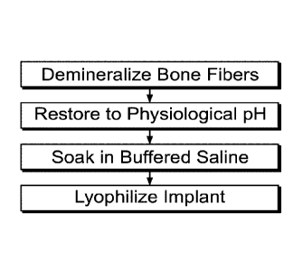
The demineralization process is carefully controlled via the concentration
of acid and duration of soak time in order to enhance the mechanical
properties of the
fibers while retaining the osteoinductive components that are exposed by the
dilute acid
reagents. Following demineralization, the tissue goes through a pH restoration
process
where the residual acid is neutralized by buffering reagents thereby returning
the tissue
to near physiological pH of between 6-8 pH. Subsequently, the demineralized
cortical
bone fibers may be stored in a wet state or dried using lyophilization or
other drying
techniques. The DCBF may be stored at various temperatures including but not
limited
to ambient room temperature (e.g., at about 23 C), refrigerated (e.g., at
about 4 C),
frozen (e.g., at about -20 C), or cryogenically preserved (e.g., at about -
196 C) using
controlled rate or uncontrolled rate freezing.
The DCBF may be placed into an implant forming container, such as a jar
or a mold, and formed into a variety of shapes including, but not limited to,
thin sheets,
cubes, discs and strips. More intricate geometries may also be formed
including, but not
limited to, curves, cutouts, compartments and patterning which can be
determined by
the shape of the implant forming container. DCBF stored in a wet state may be
placed,
7
CA 02986702 2017-11-21
WO 2016/187413 PCT/US2016/033246
for example, into molds directly, whereas dried DCBF will need to be
rehydrated prior to
being placed in molds. For example, when dried DCBF are used they are first
disbursed
into a liquid carrier to form a solution and then agitated to ensure even
distribution of the
DCBF in the solution in the mold. As also discussed hereinbelow, the liquid
carrier used
to form the solution may be, for example without limitation, water, aqueous
saline
solution, Sorensen's buffer, or phosphate buffered saline solution. In some
embodiments, excess liquid from the wet or rehydrated tissue may be separated
from
the DCBF, drained and removed from the mold. In some embodiments, additional
liquids (e.g. water, buffer, or saline) may be added to the tissue before and
during the
molding process. The liquids added to tissue before and during the molding
process
could optionally contain therapeutic factors, cytokines, growth factors,
pharmaceuticals,
antibiotics, free-radical scavengers, sugars, vitamins including, but not
limited to,
riboflavin and ascorbic acid, surfactants, DMEM medium, human or animal serum,
or
other additives. The addition or removal of liquid from the tissue also allows
the density
of the final implant to be controlled and allows for production of an implant
of uniform
density. It is understood that such methods may be contemplated to produce
implants of
variable density, when desirable. The mold may be composed of a single or
multiple
types of materials, including but not limited to metals, glass, plastics,
silicone, Teflon ,
and ceramics. In an embodiment, the vessel or package in which the
demineralized
cortical bone fibers are stored serves as the mold.
In an embodiment, the mold is micro-porous or meshed with pore sizes
ranging up to 5 mm. In an embodiment, the mold includes a non-uniform
material. In
8
CA 02986702 2017-11-21
WO 2016/187413 PCT/US2016/033246
an embodiment, the mold has varying pore sizes or mesh sizes, with the pores
or
meshes having different sizes at different locations in the mold. In an
embodiment, the
mold may include a layer of material placed on the top of the DCBF, the layer
being of
the same material used elsewhere in the mold or of a different material. In
embodiments, the layer is solid, porous, or meshed, or has another geometry
appropriate to the intended use of the mold and implant to be produced
therefrom.
In an embodiment, DCBF are in the form of a mass of DCBF, which are
then used to prepare implants that may be used as bone void filler or bone
graft
extender in bony voids and gaps which have been surgically created or caused
by
traumatic injury to the bone. Implants and grafts, as used herein, refer to
tissues,
organs or components thereof that are transplanted from a donor to a recipient
and
include those transplanted between individuals of the same species
("allograft"), those
donated and transplanted into the same individual ("autograft"), and those
transplanted
between individuals of different species ("xenograft"). Such implants may be
used as a
standalone treatment device or be applied in combination with one or more of a
variety
of bioactive osteogenic materials or cells that facilitate the reconstruction
and healing of
bone. Such implants may include particles of cortical, cancellous, or
corticocancellous
bone. Such
particles may be partially demineralized, demineralized, fully
demineralized, or may have most or all of their original mineral or calcium
content.
In an embodiment, the DCBF are pre-hydrated in an aqueous buffer, or
combined with a carrier, such as, but not necessarily limited to, the
following: an isotonic
solution; a sodium chloride solution at a concentration of about 0.1% to about
1%, more
9
CA 02986702 2017-11-21
WO 2016/187413 PCT/US2016/033246
particularly, about 0.9%; a lactated Ringer's solution, with or without D5LR,
phosphate
buffered saline ("PBS"); platelet rich plasma (PRP); glycerin; lecithin;
alginate;
hyaluronic acid (HA); a derivative of HA; or sodium hyaluronate; or other
suitable
carriers known in the art. The term "carrier" as used herein refers to a
pharmaceutically
acceptable inert agent or vehicle for delivering one or more active agents to
a subject,
and often is referred to as "excipient." The carrier must be of sufficiently
high purity and
of sufficiently low toxicity to render it suitable for administration to the
subject being
treated. The carrier may also comprise "biological components" added to the
carrier,
such as, but not limited to, DNA, RNA, short hairpin RNA (shRNA), small
interfering
RNA (siRNA), micro RNA (mRNA), polysaccharides, peptides, matrix proteins,
glycosaminoglycans (e.g, hyaluronic acid), viral vectors, and liposomes. The
carrier
further should maintain the stability and bioavailability of an active agent
added to the
carrier.
In an embodiment, a mass of DCBF fibers (e.g., an implant) are provided
to a surgeon, who can then add one or more of a carrier, bone marrow, blood,
non-
demineralized bone chips, etc., and then mold or reshape the mass into a
preferred
configuration according to anatomical or surgical needs in the operating room.
The final
form should be cohesive, moldable, and provide some resistance to irrigation
when in
the defect site, and leave minimal residue on the gloves of those handling it.
When the
mass is thus prepared, the surgeon can place it in a bone defect site, a site
with two
adjacent bone defects, or any non-bony defect where it is desired to form new
bone or
repair bone.
CA 02986702 2017-11-21
WO 2016/187413 PCT/US2016/033246
Chemical and Surface Treatment of Demineralized Cortical Bone Fibers
In an embodiment of the present invention, DCBF are prepared as
described in Section I, above, and subjected to treatment with one or more
chemical
solutions to improve the wettability of the individual fibers and of the
fibrous mass. The
increased wettability can be obtained by changing the surface charge of the
DCBF or
changing the surface morphology and/or micro-geometry of the DCBF. The fibers
or
fibrous mass may be treated with such chemical solutions immediately before
the pH
restoration step, after the pH restoration step, or before the fibers or
fibrous mass are
dried. In an embodiment, the fibers or fibrous mass may be dried, then
rehydrated prior
to treatment with the chemical solution. Furthermore, the DCBF may be treated
with
such chemical solutions after formation of the implant and before any final
drying or
lyophilizing step, where applicable. Simplified flow charts of representative
chemical
treatment processes are shown in FIGS. 1 and 5.
The chemical treatment is performed by contacting the DCBF with one or
more chemical solutions selected to improve the wettability of dried or
lyophilized
DCBF. In an embodiment, the DCBF are soaked in the chemical solution for a
period of
time from about 6 hours to about 48 hours, for example from about 12 hours to
about 36
hours, for example from about 20 hours to about 28 hours. In an embodiment,
the soak
is a static soak. In an embodiment, the DCBF are agitated during the soak.
In an embodiment, the chemical solution is isotonic with blood. In an
embodiment, the chemical solution includes a dissolved salt. In an embodiment,
the
11
CA 02986702 2017-11-21
WO 2016/187413 PCT/US2016/033246
chemical solution is a physiologically-balanced solution that includes a salt.
In an
embodiment, the chemical solution is a saline solution. In an embodiment, the
solute in
the chemical solution consists of sodium chloride (e.g., a 1M NaCI solution).
In an
embodiment, the chemical solution is Ringer's solution. In an
embodiment, the
chemical solution is a buffer solution containing a buffering salt. In an
embodiment, the
chemical solution includes a phosphate salt. In an embodiment, the buffer
solution is a
standard buffering solution containing a buffering salt. In an embodiment, the
buffer
solution is a standard phosphate buffered solution (e.g., PBS). In an
embodiment, the
chemical solution is Sorenson's Buffer. In an embodiment, the chemical
solution is
Hanks Buffered Salt Solution. In an embodiment, the chemical solution is a
HEPES-
buffered solution.
In an embodiment, the chemical solution includes a biologically-
compatible polar organic compound. In an embodiment, the chemical solution
includes
an alcohol. In an embodiment, the chemical solution includes ethanol. In
an
embodiment, the chemical solution includes a polyol. In an embodiment, the
chemical
solution includes a glycol. In an embodiment, the chemical solution includes
glycerol.
In an embodiment, the chemical solution includes polyethylene glycol. In an
embodiment, the chemical solution includes a sugar. In an embodiment, the
chemical
solution includes dextrose. In an
embodiment, the chemical solution includes
mannitol-D. In an embodiment, the chemical solution includes sodium ascorbate.
In an
embodiment, the chemical solution includes one or more of a ketone, an
aldehyde, an
organic acid, or another biocompatible polar organic compound. In an
embodiment, the
12
CA 02986702 2017-11-21
WO 2016/187413 PCT/US2016/033246
chemical solution includes an additive to inhibit proteolytic activity of
proteinases (e.g.,
matrix metalloproteinases, "MMP"). In an embodiment, the additive is
chlorhexidine
gluconate. In an embodiment, the additive is galardin. In an embodiment, the
chemical
solution includes a combination of one or more biologically-compatible polar
organic
compounds and one or more dissolved salts. In an embodiment, the chemical
solution
is a non-aqueous solution. In an embodiment, a polar organic liquid is used in
place of
the chemical solution.
In an embodiment, the chemical solution includes a biologically-
compatible polar organic compound and/or a dissolved salt, and an additive. In
an
embodiment, the additive is a therapeutic agent for administration to a
mammal. In an
embodiment, the additive is a cytokine. In an
embodiment, the additive is a
pharmaceutical. In an embodiment, the additive is an antibiotic. In an
embodiment, the
additive is a nutrient. In an embodiment, the additive is a trace element. In
an
embodiment, the additive is a free-radical scavenger. In an embodiment, the
additive is
a growth factor. In an embodiment, the additive is a biologically-active
compound.
In an embodiment, the ratio of DCBF to the chemical solution is in a range
of about 1:10 g/ml to about 1:1 g/ml. In an embodiment, the ratio of the DCBF
to the
chemical solution are selected to provide a desired fiber density and
fractional void
volume in the dried implant. In such an embodiment, lower ratios of DCBF to
chemical
solution result in less dense implants with higher void volumes.
13
CA 02986702 2017-11-21
WO 2016/187413 PCT/US2016/033246
In some embodiments, the implants produced by the methods described
and contemplated herein have uniform density. An implant may be tested for
uniform
density by various methods. One suitable method, for example without
limitation, for
determining whether an implant has uniform density is to measure the overall
density of
the implant, then divide or cut the implant into at least three portions and
measure the
density of each portion, to produce at least four measured density values for
that single
implant. The average density for that implant is calculated by dividing the
sum of all
densities (whole implant and all pieces) by the total number of pieces plus 1
(for the
whole implant density). Next, the percent relative standard deviation (%RSD)
for that
implant is determined as a percentage by first determining the standard
deviation of all
the measured density values using conventional statistical analysis methods,
and then
dividing that standard deviation by the average density and multiplying by
100. As the
term "uniform density" is used herein, an implant is considered to have
uniform density
when the (YORSD is less than about 30%, such as less than about 25%, or less
than
about 20%, or less than about 15%, or less than 10%. Example 26 provides an
example of such calculations.
Following treatment with the chemical solution, the treated DCBF are
dried. In an embodiment, the treated DCBF are dried by air drying. In an
embodiment,
the treated DCBF are dried by vacuum filtration. In an embodiment, the treated
DCBF
are dried by heat-drying. In an embodiment, the treated DCBF are dried by
solvent-
drying. In an embodiment, the treated DCBF have a residual moisture content of
less
14
CA 02986702 2017-11-21
WO 2016/187413 PCT/US2016/033246
than 80% after drying. In an embodiment, the treated DCBF have a residual
moisture
content in a range of about 60% to about 80% after drying.
In an embodiment, the treated DCBF are dried by lyophilization. In an
embodiment, the treated DCBF are frozen before being lyophilized. In an
embodiment,
the treated DCBF are refrigerated before being lyophilized. In an embodiment,
the
treated DCBF are staged at room temperature before being lyophilized. In an
embodiment, the treated DCBF are dried to a residual moisture content of less
than
80% before being lyophilized. In an embodiment, a quantity of a chemical
solution is
added to the dried DCBF, and the solvent is removed from the DCBF fibers by
lyophilization. In an embodiment, the ratio of treated DCBF to chemical
solution is in a
range of about 1:0.8 (g/ml) to about 1:10 (g/ml) before lyophilization. In an
embodiment,
the treated DCBF have a residual moisture content of less than 6% after
lyophilization.
In an embodiment, wet-treated DCBF (i.e., DCBF treated with a chemical
solution) are placed in an implant forming container, such as a mold, prior to
lyophilization, such that the lyophilized DCBF mass takes the shape of the
mold. In an
embodiment, wet treated DCBF are placed in a jar or other container, then
lyophilized.
The final tissue form, or implant comprising treated dried DCBF, may then be
provided
to medical personnel for use as discussed in Section I, above.
As shown FIG. 5, in another embodiment, following treatment with a
chemical solution and prior to drying or lyophilizing, the treated DCBF may be
subjected
to a curing step which involves warming the treated DCBF for a period of time.
For
CA 02986702 2017-11-21
WO 2016/187413 PCT/US2016/033246
example, curing may be accomplished, without limitation, by warming the
treated DCBF
using ambient air, warm air, radiant heat, or energy such as UV light or
microwaves. In
such an embodiment, the treated DCBF may be warmed to a temperature of from
about
20 C to about 50 C, such as from about 25 C to about 45 C, or from about
30 C to
about 45 C, or from about 35 C to about 45 C. In such an embodiment, the
treated
DCBF may be warmed for a period of time of from about 30 minutes to about 24
hours,
such as from about 4 hours to about 20 hours, or from about 4 hours to about
16 hours,
or from about 6 hours to about 12 hours. Without intending to be limited by
theory, it is
believed that performing a warming step as described above produces an implant
comprising treated DCBF that retains its shape after rehydration prior to use.
In an embodiment of the present invention, lyophilized DCBF treated using
the methods described above are rehydrated prior to use. In an embodiment,
lyophilized DCBR treated according to the methods described above are
rehydrated
prior to being packaged. In embodiments of such rehydration, the lyophilized
DCBF are
mixed with PBS, with or without other of the substances described above with
respect to
chemical solutions. In an embodiment, ratio of DCBF/PBS is selected to
generate a
cohesive, moldable composition that includes completely hydrated DCBF. In an
embodiment, the mixture is in a range of about 20:80 DCBF/PBS (g/m1) to about
34/66
DCBF/PBS (g/ml).
In an embodiment of the present invention, the surface roughness of the
DCBF is modified using a surface modification technique known in the art or to
be
discovered. Known suitable techniques include, without limitation,
overcoating, surface
16
CA 02986702 2017-11-21
WO 2016/187413 PCT/US2016/033246
gradient modification, surface-active bulk additives, surface chemical
reactions, etching,
roughening, conversion coatings, ion beam implantation, Langmuir-Blodgett
deposition,
laser roughening, parylene coatings, photografting, radiation grafting,
radiofrequency
glow discharge plasma deposition, radiofrequency glow discharge treatment,
self-
assembled monolayers, silanization, surface-modifying additives, and other
means of
modifying surfaces of fibers. In an embodiment, one or more of the aforesaid
techniques creates surface features on the micron scale, sub-micron scale,
nano-scale,
or other scales.
In an embodiment of the present invention, the wettability of an implant
comprising modified or non-modified DCBF can be measured using standard
methods
for assessing surface tension, including but not limited to static and dynamic
contact
angle measurement techniques. Suitable contact angle measurement techniques
include, but are not limited to, optical tensiometry, force tensiometry,
Wilhelmy plate
methods, sessile drop methods, captive air bubble methods, capillary air
methods, the
du Nouy ring method, or other measurement techniques for determining contact
angles
of liquid substances. In certain embodiments, DCBF implants prepared according
to
methods of the present invention may have at least one surface where the
contact
angle is less than 90 degrees, or less than 60 degrees, or less than 45
degrees.
Another suitable method for measuring the wettability of an implant is, for
example
without limitation, by observing the rate at which a DCBF implant absorbs an
amount of
liquid. In an embodiment, the amount of liquid is a measured volume deposited
on a
surface of the implant and the measured value is known as wettability time.
Implants
17
CA 02986702 2017-11-21
WO 2016/187413 PCT/US2016/033246
produced according to the methods described and contemplated herein have a
wettability time of less than about 5 minutes, such as less than about 4
minutes, or less
than about 3 minutes, or less than about 2 minutes, or less than about 1
minute. Still
another suitable method for measuring the wettability of an implant is, for
example
without limitation, submerging the implant in an excess amount of liquid and
measuring
the time required for the implant to absorb enough of the liquid to completely
submerge
the implant and the measured value is known as complete rehydration time.
Implants
produced according to the methods described and contemplated herein have a
complete rehydration time of less than about 30 minutes, such as less than
about 20
minutes, or less than about 15 minutes, or less than about 10 minutes, or less
than
about 5 minutes.
III. Energetic, Physical, and Chemical Cross-linking of Demineralized
Cortical Bone
Fibers
In an embodiment, the present invention includes an implant that is
comprised of DCBF that have been either fully or partially cross-linked using
energetic
sources. Suitable energetic sources include ultraviolet (UV) radiation, ozone,
plasma,
(e.g., RF plasma), coronal discharge, or other means that provide the energy
needed to
form cross-links between proteins. Suitable plasma media include, but are not
limited
to, air plasma, oxygen plasma, and ammonium plasma. In an embodiment,
energetic
cross-linking binds proteins such as albumin or other blood adsorption
proteins to the
DCBF, otherwise affects the adsorption of the proteins to the DCBF, before
lyophilization to increase the wettability of the DCBF implant. The
wettability of the
18
CA 02986702 2017-11-21
WO 2016/187413 PCT/US2016/033246
energetically cross-linked DCBF implants is measured using the same techniques
described in Section II with regard to the chemical and surface treatment of
DCBF.
Cross-linking imparts a variety of unique properties to the DCBF implant
that a non-cross-linked implant would otherwise not possess. Such properties
include
increased wettability, shape retention under compression, and resistance to
fiber
washout. A simplified flow chart of a representative energetic cross-linking
treatment
process is shown in FIG. 2.
In an embodiment of the present invention, DCBF are cross-linked by
exposing a mass of DCBF to UV radiation. In an embodiment, wet DCBF are placed
into a mold and formed into one of a variety of possibly desirable shapes.
Such shapes
include, but are not limited to, thin sheets, cubes, discs and strips. More
intricate
geometries may also be formed including, but not limited to, curves, cutouts,
compartments and patterned shapes. In an embodiment, the mass is shaped to
approximate a surface of an intact or damaged bone, such as to line a hip
socket or the
interior of a bone void.
In embodiments of the present invention, suitable molds may be
composed of single or multiple types of material or combinations of materials.
Such
materials include, but are not limited to metals, glasses, plastics and
ceramics. Suitable
materials may either block UV radiation completely, partially transmit, or
fully transmit
UV radiation, allowing all or selected portions of the implant to be exposed
to UV
radiation. While most materials exhibit poor transmission of UV radiation,
certain
19
CA 02986702 2017-11-21
WO 2016/187413 PCT/US2016/033246
materials such as fused quartz or silica glass and plastics including, but not
limited to,
optical grade polystyrene and specialized PMMA acrylic (Plexiglas G-UVT,
Solarcryl
SUVT, Acrylite OP-4) allow for near full transmission of certain wavelengths
of UV
radiation. After molding the sample into its final shape, the implant may be
left in the
mold or removed from the mold before undergoing UV cross-linking. The implant
may
be lyophilized within or without the mold before undergoing UV cross-linking,
or
lyophilized and rehydrated again prior to UV cross-linking. The implant may
also be
further masked using materials that completely block or are partially
transmissible to UV
radiation to further control cross-linking in certain regions of the implant.
In embodiments of the present invention, the mold is a composite of
various materials selected to provide variations in the degree of cross-
linking across the
implant. In an exemplary embodiment, the implant is formed with a cavity to
receive an
osteoinductive substances or other therapeutic material. In such an
embodiment, it may
be desirable that the bottom of the implant, opposite the cavity, may be more
densely
cross-linked to provide increased structural stability to the implant. In
other
embodiments, variations in cross-linking density may be used to allow certain
sections
of the implant to be remodeled at different rates than other sections during
the bone
remodeling process.
In an embodiment of the present invention, UV surface cross-linking is
performed by placing the implant in a UV containment chamber and exposing the
implant to UV radiation. The UV radiation alters the collagen molecules within
the
implant, resulting in additional bonds being formed between adjacent collagen
CA 02986702 2017-11-21
WO 2016/187413 PCT/US2016/033246
molecules. This process of photopolymerization of collagen is believed to
occur due to
the generation of free radicals via photooxidation of sensitive amino acid
residues by
UV radiation. The free radicals generated allow the formation of covalent
cross-links
between the collagen polypeptides, resulting in stronger and stiffer collagen
fibers.
Aromatic amino acid residues are the predominant sites of free radical
formation. Other
amino acid residues may be the site of free radical generation under more
energetic
conditions. Further, the rate at which cross-linkages are formed may be
increased by
adding biologically-compatible free radical initiators to the DCBF mass.
Riboflavin is an
example of such an initiator. Other initiators may include other compounds
with
aromatic structures, or may include sugars.
The amount of liquid in the implant affects the rate and degree of cross-
linking. Without being bound by theory, it is believed that the presence of
liquid
provides a medium for transport of free radicals between collagen fibers.
While it is
possible to cross-link dried or lyophilized fibers, the embodiments of cross-
linking
methods according to the present invention are most effective when used with
rehydrated fibers. However, excess water may be added to the DCBF implant
before
cross-linking to swell the implant, thus increasing its porosity, and the
exposure time
increased, if necessary to achieve the desired amount of cross-linkage.
The rate and depth at which cross-linkages are formed may be controlled
by altering the power of the UV radiation source, changing the distance of the
implant
from the UV radiation source, shifting the wavelength of the UV radiation,
varying the
exposure time, and by fully or partially blocking UV radiation transmission to
certain
21
CA 02986702 2017-11-21
WO 2016/187413 PCT/US2016/033246
areas of the implant. Multiple UV radiation sources may be used with a
combined
power rating ranging from a few watts to a few kilowatts. In high power or
energy dense
cross-linking implementations of the present invention, the UV containment
chamber
and implant may be cooled to temperatures ranging from physiological (e.g.,
about 37
C) to freezing (e.g., about -80 C) during the cross-linking process using any
of a
variety of cooling techniques to prevent heat-related degradation of the
implant.
Suitable cooling techniques include but are not limited to refrigerant-based
cooling,
active air cooling, thermoelectric devices, evaporative cooling, and phase-
change
cooling (e.g., the use of dry ice). The implant may also be placed under UV
radiation for
multiple short exposures instead of a single long exposure to reduce the
amount of heat
generated in the tissue. In some embodiments that involve UV cross-linking, it
may be
beneficial to heat the implant to a temperature that is higher than
physiological
temperatures (e.g., the implant may be heated to a temperature in a range of
from
about 37 C to about 70 C). Heat may be applied to the implant by the UV
bulbs or an
additional heating element. The implant may be placed on a heating platform
and/or
heated by UV bulbs placed around the implant. The addition of heat greater
than about
37 C but less than about 70 C for lengths of time of from about 10 minutes
to about 24
hours increases the cohesiveness of the implant and helps prevent dispersion
of the
implant when rehydrated or submerged in a rehydrating liquid (e.g., water,
saline,
blood). In some embodiments, the use of heat to improve the cohesiveness of
the
implant may be used without the addition of UV exposure.
22
CA 02986702 2017-11-21
WO 2016/187413 PCT/US2016/033246
In embodiments of a method according to the present invention, the
intensity or irradiance of the UV radiation at the surface of the implant may
be varied by
the power of the radiation source and/or the distance between the implant and
the UV
radiation source. Suitable energy densities for use in a method according to
an
embodiment of the present invention range from about 100 pW/cm2 to about 5,000
mW/cm2 at the surface of the implant. The wavelength of the UV radiation can
be
shifted between various regions of the UV spectrum including but not limited
to
longwave UVA (e.g., about 400 to about 315 nm), midrange UVB (e.g., about 315
to
about 280 nm), and shortwave UVC (e.g., about 280 to about 100 nm). Shifting
the
wavelength changes the penetration properties of UV radiation into the
implant, with
longer wavelengths allowing increased UV penetration and greater depth of
cross-
linking. For example, in an embodiment of the present invention, exposure to
UVA
radiation is used to create cross-linking to a depth of about 1 mm, which
creates a stiff
shell at the surface of the implant. Shifting wavelengths also changes the
character of
the cross-links, which affects the degree to which properties such as
mechanical
strength, shape memory retention, and hydrophobicity are modified. Concurrent
exposure to UV radiation at differing wavelengths may be used to vary the
changes in
properties across the implant. Wavelengths in the UVC spectrum also have the
added
benefit of being germicidal, and thus can be used to sterilize the surfaces of
the implant
while it undergoes cross-linking.
The length of time that the implant is exposed to the UV radiation source
also affects the degree and effectiveness of cross-link formation. In cross-
linking
23
CA 02986702 2017-11-21
WO 2016/187413 PCT/US2016/033246
methods according to embodiments of the present invention, suitable exposure
times
are in a range of a few seconds to a few hours depending on the desired
properties of
the implant. In some embodiments, exposure times of up to 720 minutes may be
used,
although typical exposure times of about 10 minutes or less may be used (e.g.,
for
commercial production of implants). In some embodiments, even shorter exposure
times (e.g., exposure times of about 10 seconds to about 300 seconds) may be
used
where only a small degree of cross-linking is desired, or where the UV
radiation is
particularly intense. For many embodiments, the practical exposure times would
be in a
range of about 10 minutes to about 60 minutes.
After the cross-linking process is completed, the implant may be stored in
a wet state or dried using lyophilization, air drying, or other drying
methods. The
implant may be stored at various temperatures including but not limited to
ambient room
temperature (e.g., at about 23 C, or up to about 30 C), refrigerated (e.g.,
at about 4
C), frozen (e.g., at about -20 C), or at cryogenic temperatures (e.g., at
about -196 C)
where frozen or cryogenic freezing is achieved using controlled rate and/or
uncontrolled
rate freezing. By changing the variables discussed above before and during the
cross-
linking process, a broad range of implants with varying properties may be
produced.
In an embodiment of the present invention, the cross-linking process is
performed in a containment chamber that allows optimal UV irradiation while
shielding
an operator from potentially harmful UV irradiation. During the cross-linking
process,
the implant may be placed on a flat surface, an uneven surface with ridges and
peaks,
or elevated on a platform or by other means that would allow UV radiation to
reflect onto
24
CA 02986702 2017-11-21
WO 2016/187413 PCT/US2016/033246
all sides of the implant, including its underside. The surface or platform
that the implant
rests on could also be made of multiple types of materials that block UV
radiation
completely, partially transmit UV radiation, or fully transmit UV radiation.
The walls of
the UV containment chamber may be lined or coated with a reflective material
to allow
the radiation to scatter within the UV containment chamber, allowing all
surfaces of the
implant to be exposed to UV radiation. UV radiation sources may also be
mounted on
multiple walls of the UV containment chamber to allow for better coverage of
the implant
during the cross-linking process. The orientation of the implant may also be
changed
during the UV cross-linking process either manually or automated by the UV
containment chamber for a more uniform exposure of all surfaces.
Embodiments of the UV cross-linking method of the present invention
include the aforesaid containment chambers, which may be specially designed to
meet
the needs of specific embodiments of the UV cross-linking method. Containment
chambers according to embodiments of the present invention may also be
designed for
use with energetic sources other than UV radiation sources, such as ozone,
plasma,
(e.g., RF plasma), coronal discharge, or other means that provide the energy
needed to
form cross-links between proteins. In an embodiment, the containment chamber
includes means for positioning and/or moving the implant. In an embodiment,
the
containment chamber includes one or more sources of UV radiation.
In an embodiment of the present invention, the distance of an implant from
a UV radiation source may be changed during the irradiation process using
manually or
automatically operated device to provide optimal UV irradiation for different
types of
CA 02986702 2017-11-21
WO 2016/187413 PCT/US2016/033246
implants. In an embodiment, the device includes a manual or automated moving
platform upon which the implants rest. Such platforms can move along x-, y-,
and z-
axes. In an embodiment, the device includes single or multiple UV radiation
sources
that can move along x-, y-, and z-axes. In an embodiment, the UV radiation
source is
one or more UV lamps in a movable lamp fixture. In an embodiment, the device
includes a rotating drum. In an embodiment, the device includes a rotating
platform. In
an embodiment, the device includes an orbiting platform.
The effectiveness of the irradiation process may be affected by the
temperature of the implant and/or UV radiation source. In an embodiment of the
present invention, the containment chamber includes a temperature control
system for
regulating the temperature of the implant during irradiation by heating or
cooling the
implant. In an embodiment of the present invention, the containment chamber
includes
a temperature control system for heating or cooling the radiation source. In
an
embodiment, the interior of the UV containment chamber is ventilated and/or
cooled
using one or more input and output ports to control heating of the implant
during the UV
irradiation process. In an embodiment, such ventilation and/or cooling is
controlled by a
controller that is operated manually or automatically in response to
temperature
measurements made at the implant or elsewhere in the interior of the
containment
chamber.
In an embodiment of the present invention, the UV radiation source
includes one or more of a fluorescent lamp, a gas discharge lamp, a high-
intensity
discharge lamp, an electroluminescent lamp, a light-emitting diode, a laser,
an
26
CA 02986702 2017-11-21
WO 2016/187413 PCT/US2016/033246
incandescent lamp, an electron-stimulated lamp, and other devices that emit UV
radiation at intensities suitable for cross-linking DCBF.
In an embodiment, a UV radiation controller is integrated in the
containment chamber. The UV radiation controller includes one or more of means
for
opening and/or closing a shutter, means for turning one or more UV radiation
sources
on and/or off, means for controlling the brightness of the UV radiation
source, and other
means for controlling the intensity and/or duration of the irradiation of the
implant. In an
embodiment, a controller is provided, the controller having circuitry for
controlling one or
more of the aforesaid means. In an embodiment, the controller includes a
computer. In
an embodiment, the computer is programmable by an operator.
In an embodiment, the containment chamber includes one or more
sensors to sense the intensity of UV radiation emitted by the UV radiation
sources
and/or the intensity of UV radiation at the surface of the implant. In an
embodiment, a
controller is provided, the controller having circuitry for controlling the
intensity of the UV
radiation source. In an embodiment, the controller controls the intensity of
the UV
radiation source in response to output from the one or more sensors. In an
embodiment, the controller includes a computer. In an embodiment, the computer
is
programmable by an operator such that the UV radiation source provides UV
radiation
of a specified intensity and/or range or wavelengths. In an embodiment, the
computer is
programmable by an operator such that the UV radiation source provides a total
irradiation energy to the implant.
27
CA 02986702 2017-11-21
WO 2016/187413 PCT/US2016/033246
In an embodiment, the UV containment chamber is designed to be used in
one or both of a sterile and a non-sterile environment. In an embodiment where
the
environment is non-sterile, the implant is contained in a sterile interior of
a separate UV-
transmissive chamber that is placed in the UV containment chamber such that
radiation
from the UV radiation source is transmitted through the UV-transmissive
chamber to the
implant. In an embodiment, the interior of a UV containment chamber is
maintained as
a sterile environment by sealing the UV radiation source and controller
circuitry in a
separate compartment. In such an embodiment, the sealed compartment is UV
transmissive such that UV radiation from the UV radiation source is
transmitted from the
sealed compartment into the interior of the UV containment chamber.
UV cross-linking of DCBF provides an implant with properties that an
otherwise non-cross-linked implant would not possess. The current lyophilized
formulations of demineralized cortical fibers have a few shortcomings that can
be
address by UV cross-linking. One such shortcoming is the initial resistance
to
rehydration of a lyophilized DCBF implant. When the implant has been
lyophilized, the
residual moisture level is typically no more than 6% w/w and this lack of
moisture
causes the implant to exhibit hydrophobic characteristics. When a liquid such
as water,
saline, or blood is applied to the surface of the implant, the liquid sits on
the surface and
is not immediately absorbed. Once the initial amount of liquid becomes
absorbed into
the implant, the rehydrated surface exhibits hydrophilic characteristics and
any
additional liquid added is immediately absorbed into the implant. Another
shortcoming is
the lack of mechanical strength and structural rigidity of a lyophilized DCBF
implant after
28
CA 02986702 2017-11-21
WO 2016/187413 PCT/US2016/033246
rehydration. In the lyophilized state, the implant holds its shape and is
rather stiff,
however, after the implant has been rehydrated, the implant becomes soft, the
DCBF
start to swell, and the implant cannot be handled without permanently losing
its shape.
In certain situations, it is preferable for the implant to retain its shape
while also being
compliant and flexible even after being saturated with liquid.
UV cross-linking allows the hydrophilicity and mechanical properties of a
DCBF implant to be modified quickly and efficiently compared to other methods
known
in the art. However, an embodiment of the present invention include physical
cross-
linking by techniques such as those including dehydrothermal treatment (DHT).
An
embodiment of the present invention includes chemical cross-linking of DCBF by
one or
more known methods, or by a chemical cross-linking method yet to be
discovered.
Known chemical cross-linking techniques include, but are not limited to,
the use of glutaraldehyde, carbodiimide (e.g., 1-ethyl-3-(3-
dimethylaminopropyl)
carbodiimide, also known as [DC), EDC with NHS (i.e., N-hydroxysuccinamide),
genipin, catechin, succinic acid, and tannic acid. While some chemical cross-
linkers
have been used in the past on various types of materials, including allograft
tissue,
chemical cross-linking can be a complicated and lengthy process, and is
potentially
hazardous to the patient if the residual chemicals are not completely removed.
Natural
chemical cross-linkers, such as genipin and catechin, are less cytotoxic than
synthetic
cross-linkers, but may also have disadvantages in some applications. In the
case of
genepin, the tissue is stained a dark blue as a result of the cross-linking
process, and
the stain is difficult to remove. Chemical cross-linking is also difficult to
control and is
29
CA 02986702 2017-11-21
WO 2016/187413 PCT/US2016/033246
more easily applied to the entire bulk of the implant rather than to specific
areas or
surfaces. Too much cross-linking of the implant may also impart properties
that are
unfavorable. One of the advantages of implants made from DCBF is that they are
moldable and cohesive after rehydration. This property is diminished as the
DCBF
become more cross-linked, resulting in an implant that cannot be molded into a
different
shape or put together once it has been taken apart. Despite the aforesaid
difficulties
posed by chemical cross-linking techniques, their use in forming cross-linked
implants,
as well as the implants themselves, are useful embodiments of the present
invention.
In contrast to the chemical cross-linking methods discussed above, cross-
linking by UV radiation is easily controlled and can be implemented to prepare
DCBF
implants that have the advantages of both non-cross-linked and cross-linked
DCBF,
while eliminating the disadvantages of excessive stiffness and resistance to
recombination of pieces of the implant. By using UV radiation to cross-link
certain
surfaces of the implant while leaving other areas uncross-linked, an implant
is prepared
that retains its shape after rehydration due to the increased stiffness of the
cross-linked
regions, while also retaining the moldable and cohesive properties of the
uncross-linked
regions. UV cross-linking also reduces the initial hydrophobicity encountered
by the
lyophilized demineralized cortical fibers allowing the implant to be
rehydrated nearly
instantaneously. Furthermore, UV cross-linking imparts some shape memory
retention
to the rehydrated implant. When an external force is applied, the cross-linked
implant is
temporarily deformed and some liquid is displaced. However, as soon as the
force is
removed, the cross-linked implant will return to its original shape and resorb
the
CA 02986702 2017-11-21
WO 2016/187413 PCT/US2016/033246
previously displaced liquid. Only when a sufficient amount of force is applied
does the
implant permanently deform and become moldable. Additionally, the increased
rigidity
of the cross-linked surfaces of the implant prevents the implant from breaking
apart
when an excess of liquid is applied, when the implant is irrigated, or when
the implant is
completely submerged in a liquid.
Embodiments of the cross-linking methods of the present invention can be
used to produce hydrophilic and mechanically stable DCBF implants from fully
demineralized, demineralized, or partially demineralized DCBF, but is most
effective for
cross-linking DCBF with calcium contents of less than 1% w/w. The UV cross-
linking
method of the present invention may be used with DCBF having thicknesses in a
range
of about 80 pm to about 150 pm, or at other thicknesses where the DCBF form a
cohesive mass in the absence of cross-linkages. Further, embodiments of the
energetic
method of the present invention can be used to prepare DCBF implants in the
presence
of additives. Additives such as particles of non-demineralized cortical,
cancellous, or
corticocancellous bone, demineralized cortical, cancellous, or
corticocancellous bone
may be used as long as the implant contains sufficient DCBF to form a cohesive
mass.
Additives such as therapeutic factors, cytokines, growth factors,
pharmaceuticals,
antibiotics, free-radical scavengers, sugars, or other chemical or bioactive
compounds
will retain their effectiveness after exposure, since the energetic exposure,
and thus
cross-linking, occurs at and/or near the surfaces of the implant, and does not
significantly affect the interior of the implant.
31
CA 02986702 2017-11-21
WO 2016/187413 PCT/US2016/033246
Although the exemplary embodiments of the energetic cross-linking
process described herein discuss the use of UV radiation, one having ordinary
skill in
the art and possession of the present disclosure will recognize that other
sources of
energy may be used to cross-link protein-rich fibers. Besides UV radiation,
suitable
energetic sources include, but are not limited to, ozone, plasma, (e.g., RF
plasma),
coronal discharge, or other means that provide the energy needed to form cross-
links
between proteins. Suitable plasma media include, but are not limited to, air
plasma,
oxygen plasma, and ammonium plasma.
IV. Treatment of Tissue Types Other Than Dem ineralized Bone
Without being bound by theory, it is believed that the increased wettability
and other effects observed in DCBF and masses of DCBF that have been treated
as
discussed herein result from interactions with the collagen and/or
glycoproteins present
in cortical demineralized bone matrix. Thus, one having ordinary skill in the
art and
possession of the present disclosure would reasonably expect that similar
beneficial
results may be obtained by applying such treatments to demineralized bone
matrix from
cancellous or corticocancellous bone. One having ordinary skill in the art
and
possession of the present disclosure would also reasonably expect that similar
beneficial results may be obtained by applying such treatments to fibers or
other
particles of tissue types other than demineralized bone matrix. Such other
tissue types
may be derived from any suitable organ or other tissue source, whether
autologous,
allogeneic, or xenogeneic. Examples of suitable xenogeneic sources of tissues
include,
but are not necessarily limited to, warm-blooded vertebrates, including
mammals, such
32
CA 02986702 2017-11-21
WO 2016/187413 PCT/US2016/033246
mammalian sources including human, bovine, ovine, caprine, and porcine
sources.
Suitable tissue types may include, but are not necessarily limited to an
adipose tissue,
an amnion tissue, an artery tissue, a bone tissue, a cartilage tissue, a
chorion tissue, a
colon tissue, a dental tissue, a dermal tissue, a duodenal tissue, an
endothelial tissue,
an epithelial tissue, a fascial tissue, a gastrointestinal tissue, a growth
plate tissue, an
intervertebral disc tissue, an intestinal mucosal tissue, an intestinal
serosal tissue, a
ligament tissue, a liver tissue, a lung tissue, a mammary tissue, a meniscal
tissue, a
muscle tissue, a nerve tissue, an ovarian tissue, a parenchymal organ tissue,
a
pericardial tissue, a periosteal tissue, a peritoneal tissue, a placental
tissue, a skin
tissue, a spleen tissue, a stomach tissue, a synovial tissue, a tendon tissue,
a testes
tissue, an umbilical cord tissue, a urological tissue, a vascular tissue, a
vein tissue, and
a combination thereof. Other suitable tissue types may include, but are not
necessarily
limited to, submucosa, renal capsule membrane, dermal collagen, dura mater,
serosa,
or basement membrane layers, including liver basement membrane. Suitable
submucosa materials for these purposes include, for instance, intestinal
submucosa,
including small intestinal submucosa, stomach submucosa, urinary bladder
submucosa,
and uterine submucosa. Source tissue (i.e., tissue incorporated into a final
processed
product, such as an implant) of the types disclosed above may be separated
from other
tissue types adjacent or connected to the source tissue, or the adjacent or
connected
tissue may remain with the source tissue and become incorporated in the
implant. One
or more source tissues may be included in the final processed product.
V. EXAMPLES
33
CA 02986702 2017-11-21
WO 2016/187413 PCT/US2016/033246
The following examples are set forth so as to provide those of ordinary
skill in the art with an exemplary disclosure and description of how to make
and use the
described invention, and are not intended to limit the scope of what the
inventors regard
as their invention nor are they intended to represent that the experiments
below are all
or the only experiments performed. Efforts have been made to ensure accuracy
with
respect to numbers used (e.g., amounts, temperatures, etc.) but some
experimental
errors and deviations should be accounted for.
EXAMPLE 1: Fabrication of Demineralized Cortical Bone Fibers
Human long bone is recovered aseptically from a deceased donor and
stored at 4 C until ready for processing. The bone is debrided to remove soft
tissue
elements and the shaft of the bone is cut into cross-sections. The cortical
bone is then
cleaned using detergents/surfactants to remove residual blood and lipids from
the bone
surface.
To create DCBF, the bone sections are first shaved across the shaft of the
bone using a controlled advancement rate of a lathe bit having a width
approximately
equal to the desired length of the bone fibers. The shaft segment is secured
in a vice
with a sufficient portion of the shaft protruding such that the protruding
portion may be
shaved. On a milling machine, a straight flute end-mill is set up such that
its axis is
parallel with the axis of the shaft. Utilizing the required length of the of
the broad edge
of the lathe bit, fibers are shaved off of the shaft by running the end-mill
back and forth
along the shaft until substantially all of the bone has been shaved from the
shaft. The
resulting bone fibers are collected for demineralization.
34
CA 02986702 2017-11-21
WO 2016/187413 PCT/US2016/033246
The bone fibers are dem ineralized by agitating them in 0.6 N HCI for a
sufficient period of time to remove the endogenous calcium minerals to a
desired
residual calcium content, after which the fibers are successively rinsed with
water,
soaked in water, soaked in a sodium phosphate dibasic buffer to achieve a
physiological pH, rinsed in water, and soaked in water. The soaked fibers may
then be
dried, lyophilized, or left in a wet state for further processing.
EXAMPLE 2: Treatment of DCBF with PBS
DCBF are prepared as described in Example 1. After completion of the
second water soak, the DCBF are decanted into a vessel, and PBS is added at a
ratio
in a range of about 1:3 DCBF/PBS (g/ml) to about 1:15 DCBF/PBS (g/ml). After 5
to 15
minutes of a static soak, the DCBF are decanted from the PBS, and air-dried.
Additional PBS is added to the DCBF at a ratio in a range of about 1:1
DCBF/PBS
(g/ml) to about 1:5 DCBF/PBS (g/ml) in a plastic jar, and the wet DCBF are
lyophilized.
EXAMPLE 3: Preparation of Low-density Pre-formed Fiber Shapes Using
DCBF and Saline
Low-density pre-formed fiber shapes are lyophilized DCBF which are
suspended in liquid prior to lyophilization to provide a fluffy texture and a
high void
volume. They are hydrated by a surgeon in the operating room to form a putty-
like
substance for use as a bone void filler.
Low-density pre-formed fiber shapes were prepared using water or
different ratios of 0.9% sodium chloride in water ("saline", in particular
0.25X saline,
0.5X saline, 0.75X saline, and 1X saline) to examine the effect of salt
concentration on
CA 02986702 2017-11-21
WO 2016/187413 PCT/US2016/033246
hydration time and handling properties of the implants. The samples prepared
with
water were used as control samples; the samples prepared with saline solutions
were
examined as test samples.
Samples of air-dried DCBF prepared according to Example 1 were soaked
in water or saline at selected concentrations at a ratio in a range of about
1:3
DCBF/liquid (g/m1) to about 1:15 DCBF/liquid (g/ml) for 5 to 15 minutes, after
which they
were air-dried on a vacuum sieve. The samples were then lyophilized. Some
samples
were lyophilized in open jars; others were lyophilized with a vented lid, the
ventilation
holes having been covered by a porous liner having a pore size of greater than
10 pm.
The lyophilized samples were then tested for hydration time and handling
properties.
Test samples prepared with PBS and lyophilized with a lid and porous
liner hydrated more rapidly than the control samples prepared with water.
There was no
significant difference in the handling of any of the test samples in
comparison to the
control samples.
EXAMPLE 4: Preparation of Low-density Pre-formed Fiber Shapes Using
DCBF and PBS
Samples of air-dried DCBF prepared according to Example 1 were soaked
in PBS at a ratio in a range of about 1:1 DCBF/PBS (g/ml) to about 1:5
DCBF/PBS
(g/m1) for 5 to 15 minutes. Sets of samples were prepared using PBS at
concentrations
of 0.5X, and 0.25X of a standard PBS, using water as the diluent. After the
soak, the
samples were air-dried on a vacuum sieve, then lyophilized in open jars. The
samples
36
CA 02986702 2017-11-21
WO 2016/187413 PCT/US2016/033246
were then hydrated with just enough saline to provide good handling
properties, and
tested for appearance and hydration.
Drops of saline deposited onto the top surface of a low-density pre-formed
fiber shape prepared with 0.25X PBS were absorbed in less than one minute.
Drops of
saline deposited onto the top surface of a low-density pre-formed fiber shape
prepared
with 0.5X PBS were absorbed more quickly.
EXAMPLE 5: Preparation of Low-density Pre-formed Fiber Shapes Using
Various Ratios of DCBF and PBS
Samples of wet DCBF were prepared according to Example 1 without the
final drying step. Samples of various sizes were soaked in a standard PBS at a
ratio in
a range of about 1:2 DCBF/PBS (g/m1) to about 1:5 DCBF/PBS (g/ml) for 5 to 15
minutes. After the soak, the samples were air-dried on a vacuum sieve,
deposited in
open jars, frozen, then lyophilized. The lyophilized samples were then tested
for
appearance and hydration. All of the samples had a fluffy appearance.
Low-density pre-formed fiber shapes prepared as described above were
hydrated with sheep's blood, and the rates of absorption were compared with
those of
fiber shapes that had been prepared at a lower DCBF/PBS ratio in a range of
about 1:2
(g/ml) and 1:5 (g/ml). Fiber shapes prepared at the higher ratio absorbed the
sheep's
blood at much faster rates than had been observed for the fiber shapes
prepared at the
lower ratio. The absorption rate was fastest for fiber shapes prepared at the
highest
ratio.
37
CA 02986702 2017-11-21
WO 2016/187413 PCT/US2016/033246
EXAMPLE 6: Comparison of Fiber Shapes Lyophilized with Water and
Fiber Shapes Lyophilized with PBS.
Samples of wet DCBF were prepared according to Example 1 without the
final drying step. Two portions of wet DCBF were subjected to a static soak in
standard
PBS at a ratio in a range of about 1:3 DCBF/PBS (g/ml) to about 1:15 DCBF/PBS
(g/ml), and air-dried. PBS diluted to 0.5X was added to a first portion at a
ratio in a
range of about 1:1 DCBF/PBS (g/ml) to about 1:5 DCBF/PBS (g/ml), and the DCBF
was
lyophilized in a plastic jar. Water was added to the second portion at a ratio
in a range
of about 1:1 DCBF/PBS (g/ml) to about 1:5 DCBF/PBS (g/ml), and the DCBF was
lyophilized in a plastic jar.
Equal amounts of sheep's blood were dropped onto the lyophilized first
(PBS) and second (water) portions of DCBF. The blood was entirely absorbed by
the
first portion within less than one minute, at which time only about one-third
(1/3) of the
blood was absorbed by the second portion.
EXAMPLE 7: Preparation of a DCBF Implant Containinq Mineralized
Granules of Cortical or Cancellous Bone
Samples of wet DCBF are prepared according to Example 1 without the
final drying step. Mineralized granules or chips of cortical or cancellous
bone having
sizes in a range of about 200 pm to about 5 mm are prepared by milling or
cutting of
bone tissue which has been cleaned of any soft tissue adhering to the bone and
treated
with detergents/surfactants to remove blood and lipids. Following separate air-
drying
steps on individual vacuum sieves, mineralized cortical or cancellous
granules/chips
and DCBF are mixed in standard PBS at a ratio in a range of about 1:3 DCBF/PBS
38
CA 02986702 2017-11-21
WO 2016/187413 PCT/US2016/033246
(g/m1) to about 1:15 DCBF/PBS (g/ml). The
ratio of cortical or cancellous
granules/chips to DCBF is in a range of about 1:0.1 to about 0.1:1 (g/g, based
on air-
dried weight), depending on the properties desired for the implant.
After mixing to obtain an approximately homogenous mixture, the resulting
tissue mixture is air-dried on a vacuum sieve and deposited in jars which are
subsequently filled with a volume of 0.5X PBS to re-suspend the tissue in
liquid. The
jars are sealed using lids with openings covered by porous liners, then frozen
and
lyophilized. Alternatively, after mixing and air-drying, the semi-wet tissue
is placed into
molds and lyophilized.
The lyophilized tissue is readily rehydrated with blood or saline and yields
a moldable mass of bone tissue in which the cortical fibers provide
cohesiveness and
depending on their density within the tissue mass, the cortical/cancellous
granules
provide the implant with properties of radiopacity and/or resistance to
compression.
EXAMPLE 8: Preparation of a DCBF Implant Havinq a Stiff Shell
Wet DCBF prepared as in Example 1 was placed into a rectangular mold
and shaped into an implant having dimensions of approximately 10cm x 2.5cm x
7mm.
The fiber implant was removed from the mold and placed in a UV containment
chamber
where it was exposed to 315-400 nm UVA radiation for a period of about 30
minutes at
an intensity in a range of about 4,000 pwatts/cm2 to 20,000 pwatts/cm2. The
orientation
of the implant was changed within the UV chamber during the irradiation
process to
expose all surfaces of the implant evenly to UV radiation, creating a stiff
shell on all
39
CA 02986702 2017-11-21
WO 2016/187413 PCT/US2016/033246
surfaces of the implant. The implant was then lyophilized for storage, and
rehydrated
prior to implantation.
EXAMPLE 9: Preparation of a DCBF Implant Having Compartments
Wet DCBF prepared as in Example 1 is placed into a rectangular mold
having silicone inserts to form two large compartments on one surface of the
implant.
The resulting implant has dimensions of approximately 10cm x 2.5cm x 1.2mm.
The
implant is removed from the mold with the silicone inserts in place. The
implant is
placed in a UV containment chamber where the exposed surfaces are exposed to
both
100-280 nm UVC radiation and 315-400 nm UVA radiation. The longer UVA
wavelength penetrates deeper into the surfaces of the implant, which imparts
additional
stiffness allowing the implant to retain its shape when rehydrated and loaded
with
additional materials in the compartments whereas the shorter UVC wavelength
sterilizes
the surfaces of the implant. The silicone inserts block the UVC and UVA
radiation from
reaching the interior of the cavities so that cross-linking does not occur at
those
surfaces. The resulting "boat" configuration implant has two open compartments
that
allow the user to add other materials such as bone marrow aspirate and
cancellous
chips, or other additives such as those discussed in Section III of the
present disclosure.
The interior surfaces of the compartments are not cross-linked, so that a user
can mix
the additives (e.g., the bone marrow aspirate and cancellous chips) into the
non-cross-
linked DCBF. After mixing, the user can pick up the implant in a single piece
and fold it
so as to close the compartments such that the additives are enclosed within
the implant.
CA 02986702 2017-11-21
WO 2016/187413 PCT/US2016/033246
EXAMPLE 10: Preparation of a Thin DCBF Implant Havinq a Cross-linked
Interior
Wet DCBF prepared according to Example 1 are placed in a shallow
rectangular mold to produce a thin strip-like implant with dimensions of
approximately
cm x 2.5 cm x 2mm. The implant is removed from the mold, and placed in a UV
containment chamber where the exposed surfaces are exposed to 315-400 nm UVA
radiation. The implant is thin enough that penetration of the UV radiation
cross-links the
majority of the DCBF in the interior of the implant. The resulting cross-
linked implant is
a porous and flexible strip that is also strong enough to be placed in areas
of the body
that are subject to mechanical loads that would disrupt implants having only a
cross-
linked shell.
EXAMPLE 11: Preparation of a Low-density DCBF Implant
Wet DCBF prepared according to Example 1 are placed in a square mold
to produce cube-shaped implants having dimensions ranging from about 5 mm to
about
mm. Excess water is added to the mold to produce implants that are highly
porous.
The implants are then lyophilized and rehydrated carefully as to not disturb
the porous
structure. The implants are then placed into a UV containment chamber where
the
exterior surfaces are exposed to 315-400 nm UVA radiation. The implant is then
lyophilized again. The resulting low density implant is highly porous yet is
able to absorb
liquids without swelling or deforming permanently.
41
CA 02986702 2017-11-21
WO 2016/187413 PCT/US2016/033246
EXAMPLE 12: Comparison of a Cross-linked Implant with Non-
crossl inked Implant
Lyophilized DCBF prepared as in Example 1 was rehydrated and
separated into portions. Each portion was placed into a customized cylindrical
mold to
produce puck-shaped implants. A first group of implants were irradiated with
UVA
radiation at an intensity in a range of about 4,000 pwatts/cm2to 20,000
pwatts/cm2. The
top and bottom of the first implant were irradiated for 15 minutes each, for a
total
exposure time of 30 minutes, for a total energy exposure in a range of about
180 Joules
to about 900 Joules. The second group of implants were not irradiated, and
served as
comparison samples. The implants were then lyophilized. The implants had final
dimensions of about 13 mm height and 29 mm diameter.
The implants were compared as follows:
1. Implants from the irradiated and non-irradiated groups were
immersed in water or in saline solution. The implants from both groups
remained intact.
The implants from the irradiated group did not swell by any significant
amount, but the
implants from the non-irradiated group swelled by a considerable amount.
2. Implants from the irradiated and non-irradiated groups were
compressed to a fraction of their initial size. Implants from the irradiated
group showed
better shape memory retention than implants from the non-irradiated group.
3. Rehydration of the lyophilized implants from both groups showed
that the irradiated implants absorbed liquids more rapidly than the non-
irradiated
42
CA 02986702 2017-11-21
WO 2016/187413 PCT/US2016/033246
implants, and retained their shape better after rehydration even after being
compressed
significantly.
4. A moldability test showed that the irradiated implants became
permanently deformed when sufficient pressure was applied to break the outer
shell
formed by cross-linking. The irradiated implants could then be molded into a
variety of
shapes.
EXAMPLE 13: Preparation of an Implant from Cartilage Fibers
Lyophilized fibers of articular cartilage, obtained by grating larger
cartilage
pieces, are rehydrated and placed in a cylindrical mold to produce plug-shaped
implants
having diameters in a range of about 5 mm to about 40 mm and lengths in a
range of
about 5 mm-to about 20 mm. The implants are removed from the molds, and placed
in a
UV containment chamber where the surfaces of the plugs are exposed to 315-400
nm
UVA. The implant is then lyophilized and rehydrated. The resulting implant is
compressible, but retains its shape when subjected to cyclical loading. The
implant also
stays in one piece and does not disperse when subjected to a load, during
irrigation, or
when placed in an aqueous environment.
EXAMPLE 14: Preparation of an Implant from Soft Tissue
Lyophilized tissues (e.g., fibers, flakes or powder) derived from placental
(amnion, chorion, umbilical cord) or dermal tissue were rehydrated and
compressed into
thin sheets. The sheets were then trimmed or otherwise formed into a variety
of shapes
of varying sizes. The sheets were placed in a UV containment chamber where the
43
CA 02986702 2017-11-21
WO 2016/187413 PCT/US2016/033246
surfaces of the sheets were exposed to 315-400 nm UVA radiation for about 20
minutes
at a radiation intensity of about 20,000 pwatts/cm2. The implant was then
lyophilized
and rehydrated prior to implantation. The resulting implant was a very thin
sheet that
retained its shape when flexed, perforated, irrigated, and placed in an
aqueous
environment.
EXAMPLE 15: Preparation of a DCBF Implant by Chemical Cross-linkinci
Wet DCBF prepared as in Example 1 are placed into a rectangular mold
and shaped into an implant having dimensions of approximately 10cm x 2.5cm x
7mm.
The fiber implant is removed from the mold and placed in a sealed vapor
chamber. A
solution of glutaraldehyde is heated within the chamber to generate
glutaraldehyde
vapors which penetrate and cross-link DCBF throughout the entire implant. The
implant
is exposed to the vapors for a set amount of time in a range of about 5
minutes to about
24 hours). After cross-linking, the residual unreacted glutaraldehyde and any
unbound
cross-linking byproducts are rinsed out of the implant using water, solutions
of
neutralization salts, and/or buffer solutions. The implant is then lyophilized
for storage,
and rehydrated prior to implantation.
EXAMPLE 16: Preparation of a DCBF Implant by Natural Cross-linkinci
Wet DCBF prepared as in Example 1 are placed into a rectangular mold
and shaped into an implant having dimensions of approximately 10cm x 2.5cm x
7mm.
A mesh is placed over the open surface of the mold to allow liquids access to
the tissue
while preventing the tissue from escaping the mold. The mold is submerged in a
44
CA 02986702 2017-11-21
WO 2016/187413 PCT/US2016/033246
solution of genipin for a set amount of time in a range of about 5 minutes to
about 24
hours. The genipin solution may remain static or can be stirred to increase
the rate of
cross-linking throughout the implant. After cross-linking, the residual
genipin is rinsed
out of the implant using water. Other rinses including detergents, salts,
and/or buffers
may be used to reduce residual staining of the tissue that occurs during the
genipin
cross-linking process. The implant is then lyophilized for storage, and
rehydrated prior
to implantation.
EXAMPLE 17: Preparation of a Hydrated DCBF Implant
Three grams of lyophilized DCBF was weighed out in a jar and between
about 5 to about 7.5 ml of PBS was added to the DCBF. The two components were
mixed, capped and let stand for more than 15 minutes at room temperature to
ensure
full homogenous hydration. The equilibrated mixture was packed into a plastic
syringe,
which was then capped and sealed in a foil pouch to prevent moisture loss
during long-
term storage. The mixture was extruded from the syringe into a pan and
examined.
The mixture was observed to have a slight off-white color, and to have a
smooth
consistency that held together when manually manipulated.
EXAMPLE 18: Preparation of a Shaped DCBF Implant with Enhanced
Cohesiveness
Wet DCBF prepared as in Example 1 are rolled into a mass and kneaded
to loosen any clumps of fibers and create more fiber entanglement throughout
the
mass. Once thoroughly kneaded, the whole mass is placed into the mold and the
tissue
CA 02986702 2017-11-21
WO 2016/187413 PCT/US2016/033246
is redistributed into the mold space by pressing with fingers or a spatula.
The tissue is
lyophilized in the mold, creating a shaped implant.
Molding DCBF in this manner results in an implant with enhanced
cohesiveness when rehydrated, compared to an implant where tissue is placed
into the
mold in small chunks.
EXAMPLE 19: Preparation of a Shaped DCBF Implant with Enhanced
Cohesiveness using Syringe Mold
Use of a syringe mold to shape the DCBF is shown schematically in FIGS.
3A-30 and will now be described. As shown in FIG. 3A, wet DCBF (comprising
DCBF
fibers 12) prepared as in Example 1 are placed into a custom rectangular
syringe mold
10, with an excess amount of liquid L (e.g., saline) so that the DCBF fibers
12 are
homogenously suspended in solution 14. More particularly, the custom syringe
mold 10
includes a housing 16, the walls of which form a chamber 18, within which is a
reciprocatingly movable plunger 20 that defines the bottom of the chamber 18.
The wet
DCBF and liquid are placed into the chamber 18 of the mold 10. When the
plunger 20
is in its undepressed position (see FIG. 3A), the inner dimensions of the
chamber 18
may be, for example without limitation, approximately 10cm(L) x 2.5cm(W) x
127mm(H).
In some embodiments, the cross-section of the chamber 18 is in the shape of a
rectangle with rounded edges. In such embodiments, the plunger 20 has
dimensions of
approximately 10cm(L) x 2.5cm(W) x 20mm(H) and is also in the shape of a
rectangle
with rounded edges to match the cross-sectional opening of the chamber 18. An
o-ring
(not shown per se) seals the interface between the plunger 20 and the chamber
18.
46
CA 02986702 2017-11-21
WO 2016/187413 PCT/US2016/033246
As shown in FIG. 3B, a detachable filter 22, which is liquid permeable, fits
across the top of the housing 16 to enclose the top of the chamber 18. After
the DCBF
fibers 12 and liquid L are placed into the chamber 18, the detachable filter
22 is placed
over the open surface of the chamber such that when the plunger 20 is
depressed (in
the direction of arrow A), excess liquid is able to escape from the chamber 18
through
the filter 22, while the DCBF fibers 12 are retained within. With reference to
FIG. 30,
the filter 22 may then be detached and the implant 24 comprising the DCBF
fibers (not
shown in the densely packed implant of FIG. 3C) is pushed out of the chamber.
The
implant 24 is then lyophilized for storage and rehydrated prior to
implantation. The
density of the implant 24 may be altered by changing the distance that the
plunger 20 is
pushed toward the filter 22. Implants molded in this manner have enhanced
cohesiveness and better shape retention due to the more homogenous
distribution and
enhanced entanglement of DCBF fibers in the implants.
EXAMPLE 20: Preparation of a radio-opaque shaped DCBF Implant with
mineralized bone
Wet DCBF prepared as in Example 1 is combined with mineralized cortical
or cancellous granules/powder, placed into a rectangular mold, and shaped into
an
implant having dimensions of approximately 10cm(L) x 2.5cm(VV) x 7mm(H). The
implant is removed from the mold and placed in a UV containment chamber where
it is
exposed to 315-400 nm UVA radiation for a period of about 30 minutes at an
intensity in
a range of from about 4,000 pwatts/cm2 to about 20,000 pwatts/cm2 before being
removed and lyophilized. The inclusion of mineralized cortical or cancellous
in the
47
CA 02986702 2017-11-21
WO 2016/187413 PCT/US2016/033246
implant imparts additional radio-opacity due to the added mineral content.
This allows
for improved visualization of the graft by certain imaging methodologies (e.g.
x-ray)
during or after implantation of the graft material.
EXAMPLE 21: Preparation of a radio-opaque shaped DCBF Implant with
Selective Remineralization
Wet DCBF prepared as in Example 1 are placed into a custom cylindrical
syringe mold yielding an implant with final dimensions of approximately 13 mm
in height
and 29 mm in diameter. The implant is removed from the mold and placed in a UV
containment chamber where it was exposed to 315-400 nm UVA radiation for a
period
of about 30 minutes at an intensity in a range of from about 4,000 pwatts/cm2
to about
20,000 pwatts/cm2. The UV irradiation cross-links the outer surface of the
implant
allowing the implant to retain its shape when submerged in a liquid solution.
To remineralize the implant, two solutions are prepared as follows. The
first solution is composed of 0.55M calcium chloride in DI water and the
second solution
is composed of 0.5M sodium phosphate in DI water. The irradiated implant is
fully
submerged in an aliquot of the first solution for 30 minutes under gentle
agitation. After
30 minutes, the first solution is removed and the implant is submerged in an
aliquot of
the second solution for 30 minutes under gentle agitation. This process of
alternating
solutions is repeated until a hard mineralized shell develops on the surface
of the
implant that imparts radio-opacity that is comparable to normal mineralized
human bone
and increased mechanical strength of the implant through the remineralization
of DCBF.
The bulk/interior of the implant may also be mineralized in a similar fashion
of
48
CA 02986702 2017-11-21
WO 2016/187413 PCT/US2016/033246
alternating soaks of calcium chloride and sodium phosphate by the use of
increased
agitation and forcing the solutions through the bulk of the implant using
positive or
negative pressure. The implant is then lyophilized for storage and rehydrated
prior to
implantation.
EXAMPLE 22: Preparation of a shaped DCBF Implant with Enhanced
Cohesiveness and Biological Properties
Wet DCBF prepared as in Example 1 are combined with minced,
powdered, or fibrous periosteum, placed into a rectangular mold, and shaped
into an
implant haying dimensions of approximately 10cm(L) x 2.5cm(VV) x 7mm(H). The
implant is removed from the mold and placed in a UV containment chamber where
it
was exposed to 315-400 nm UVA radiation for a period of about 30 minutes at an
intensity in a range of from about 4,000 pwatts/cm2 to about 20,000 pwatts/cm2
before
being removed and lyophilized. The inclusion of periosteum in the implant
imparts
enhanced cohesiveness and irrigation resistance due to the putty-like nature
of
periosteum and enhanced biological properties due to the addition of growth
factors
endogenous to the periosteum membrane.
EXAMPLE 23: Preparation of a shaped DCBF Implant with Enhanced
Cohesiveness by Heating
Wet DCBF prepared and molded into a shaped implant as in Examples 18
and 19 are placed in a heated chamber (e.g., lyophilizer, incubator, gravity
oven), with
or without UV exposure, at temperatures of from about 24 C to about 70 C, and
allowed
to incubate for a period of time of from about 10 minutes to about 24 hours.
The heating
process improves the cohesiveness of the implant and prevents the implant from
49
CA 02986702 2017-11-21
WO 2016/187413 PCT/US2016/033246
dispersing when placed in a rehydrating solution (e.g., water, saline, blood).
After
heating, the implant is lyophilized for storage and rehydrated prior to
implantation.
EXAMPLE 24: Preparation of a Shaped DCBF Implant with Enhanced
Cohesiveness Using Syringe Mold Having a Perforated Plunger
A different syringe mold than that used for Example 19 was used to shape
DCBF fiber and is shown schematically in FIGS. 6A-6C. As shown in FIG. 6A, wet
DCBF (comprising DCBF fibers 12) prepared as in Example 1 are placed into a
custom
rectangular syringe mold 110, with an excess amount of liquid L (e.g., saline)
so that the
DCBF fibers 112 are homogenously suspended in solution 114. More particularly,
the
custom syringe mold 110 includes a housing 116, the walls and bottom of which
form a
chamber 118 and within which is a reciprocatingly movable plunger 120 which is
operated from the top of the mold and fits within the open cross section of
the chamber
118. The wet DCBF 112 and liquid L are placed into the chamber 118 of the mold
110.
Before the plunger 120 is depressed into the chamber 118 (see FIG. 6B), the
inner
dimensions of the chamber 118 may be, for example without limitation,
approximately
10cm(L) x 2.5cm(VV) x 127mm(H). In some embodiments, the cross-section of the
chamber 118 is in the shape of a rectangle with rounded edges. In such
embodiments,
as shown in FIG. 6B, the plunger 120 has dimensions of approximately 10cm(L) x
2.5cm(1N) x 20mm(H) and also has a cross sectional shape of a rectangle with
rounded
edges to match the cross-sectional opening of the chamber 118. An o-ring (not
shown
per se) seals the interface between the plunger 120 and the chamber 118. (The
steps
of the foregoing method are shown schematically in FIG. 5.)
CA 02986702 2017-11-21
WO 2016/187413 PCT/US2016/033246
As shown in FIG. 6B, the plunger has a plurality of perforations 121 for the
movement of liquid L through the plunger, such that when the plunger is
depressed
within the chamber 118 in the direction shown by the arrow B, the plunger fits
across
the top of the housing 116, enclosing the chamber 118 and compressing the DCBF
fibers 112. More particularly, after the DCBF fibers 112 and liquid L are
placed into the
chamber 118, the plunger 120 is depressed (in the direction of arrow B),
excess liquid L'
is able to escape from the chamber 118 through the perforations 121, while the
DCBF
fibers 112 are retained and compressed within to form an implant. Each of the
perforations 121 may be any suitable size and shape that will allow excess
liquid to
escape from the chamber 118, while retaining the DCBF fibers 112. For example,
in
some embodiments, without limitation, each perforation may have a circular
shape with
a diameter of about 1 millimeter, or about 1.5 millimeters, or even 2
millimeters. With
reference to FIG. 6C, after the DCBF fibers have been compressed to the
desired
degree within the chamber 118, the excess liquid is poured out of the mold 110
and the
plunger 120 removed. The height and density of the implant 124 may be altered
by
changing the distance that the plunger 120 is depressed in the chamber 118. In
an
embodiment, the implant 124 (still in the mold 110) is then frozen in a
freezer at from
about -80 C to about 0 C for up to 48 hours. The implant 124 (still in the
mold 110) is
then cured by warming using a warming plate or warm air at about 40 C for
about 8
hours. The implant 124 comprising the compressed treated DCBF fibers 112
(still in the
mold 110) is lyophilized, after which the implant is easily removed from the
chamber
118. After lyophilizing, the implant 124 may be stored and is rehydrated prior
to
51
CA 02986702 2017-11-21
WO 2016/187413 PCT/US2016/033246
implantation. Implants produced in this manner have enhanced cohesiveness and
retain
their shape better upon rehydration prior to use.
EXAMPLE 25: Determining the Wettability of the Shaped DCBF Implant
Produced Using Syringe Molds Having a Perforated Plunger
Various implants produced by according to Example 24 were tested for
wettability as follows and the results are provided below in Table 1.
Differently shaped
implants were tested where "bricks" were implants having a generally
rectangular cross
section, and "half pipes" were implants having a generally "C" shaped cross
section.
1. Wettability Evaluation: A .drop of 0.9% saline solution (roughly 0.3cc)
is added to
the top or bottom surface of the implant and the length of time it takes for
each droplet
to absorb into the implant is recorded.
2. Rehydration Evaluation: The implant is placed into a basin containing
excess
0.9% saline solution to completely submerge the implant The length of time it
takes for
the implant to absorb enough liquid to sink to the bottom of the basin is
recorded.
3. Wettability/rehydration times utilizing the droplet test range from a
few seconds
(0:07) to a few minutes (4:00) whereas complete rehydration of the implant
varies from
24 seconds to 20 minutes.
4. Preferred/suitable wettability/rehydration times for DCBF implants may
be
tailored to the end user/application and could range anywhere from near
instantaneous
wettability/rehydration (1-2 seconds) to much longer rehydration times of
upwards of 20-
30min (preferably within 5 min).
52
CA 02986702 2017-11-21
WO 2016/187413 PCT/US2016/033246
Table 1
Implant Type Wettability time ¨ Wettability time-
Complete rehydration ¨
top surface (mm:ss) bottom surface(mm:ss) submerged (mm:ss)
Brick ¨ A 1:15 1:02 2:50
Brick ¨ B 0:15 0:15 0:24
Brick ¨ C 1:18 1:16 2:40
Brick ¨ D 0:45 0:07 1:15
Brick ¨ E 2:00 3:00 20:00
Brick ¨ F 0:38 0:31 2:15
Brick ¨ G 0:24 1:03 3:20
Brick ¨ H 0:29 1:05 13:00
Half Pipe ¨A 1:45 4:00 4:00
Half Pipe ¨B 0:45 1:09 2:50
EXAMPLE 26: Testing Shaped DCBF Implant Produced Using Syringe
Molds Having a Perforated Plunger for Uniform Density
Various implants produced by according to Example 24 were tested for
uniform densitys follows and the results are provided below in Table 2.
Differently
shaped implants were tested where a "disc" was an implant having a generally
circular
cross section, and "strips" were implants having a generally rectangular
shaped cross
section.
1. The weight of a DCBF implant was measured using an analytical balance.
2. Calipers were used to measure the dimensions of the implant, and the
volume of
the implant was calculated.
3. The density of the entire implant was calculated by dividing the weight
recorded
in step 1 by the volume calculated in step 2.
53
CA 02986702 2017-11-21
WO 2016/187413 PCT/US2016/033246
4. The implant was then cut into four equally sized pieces (quarters Ql,
02, Q3 and
04) using a scalpel. Steps 1-3 were repeated for each piece of the implant.
5. The relative standard deviation of the densities was calculated by
dividing the
standard deviation of the densities by the average. An implant can be said to
have
uniform density if the relative standard deviation of the measured densities
is less than
about 30%.
Table 2
Shape Weight Length Width Height Volume Density
(g) (mm) (mm) (mm) (cc) (g/cc)
5cc disc 1.317 44.81 9.18 14.470 0.091
Q1 0.349 3.617 0.096
Q2 0.319 3.617 0.088
Q3 0.308 3.617 0.085
Q4 0.292 3.617 0.081
Average 0.088
Std Dev 0.005
%RSD 6.028
Sh Weight Length Width Height Volume Density
ape
(g) (mm) (mm) (mm) (cc) (g/cc)
20cm strip 2.823 192 9.16 7.25 12.751 0.221
Q1 0.749 47.53 9.05 7.32 3.149 0.238
Q2 0.582 47.15 9.17 6.29 2.720 0.214
Q3 0.753 50.3 9.16 6.89 3.175 0.237
Q4 0.754 48.94 9.18 7.05 3.167 0.238
Average 0.230
Std Dev 0.010
%RSD 4.389
54
CA 02986702 2017-11-21
WO 2016/187413 PCT/US2016/033246
Shape Weight Length Width Height Volume Density
(g) (mm) (mm) (mm) (cc) (g/cc)
10cm strip 1.63 95.32 9.01 7.54 6.476 0.252
Q1 0.386 24.05 9.03 7.65 1.661 0.232
Q2 0.432 23.84 8.98 8.84 1.892 0.228
Q3 0.386 25.06 8.92 6.77 1.513 0.255
Q4 0.416 23.72 9.12 7.44 1.609 0.258
Average 0.245
Std Dev 0.012
/oRSD 5.055
While the disclosed invention has been described with reference to the
specific embodiments thereof, it should be understood by those skilled in the
art that
various changes may be made and equivalents may be substituted without
departing
from the true spirit and scope of the invention. In addition, many
modifications may be
made to adapt a particular situation, material, composition of matter,
process, process
step or steps, to the objective, spirit and scope of the described invention.