Note: Descriptions are shown in the official language in which they were submitted.
3
CA 02986747 2017-11-21
WO 2016/191375
PCT/US2016/033792
SYSTEMS AND METHODS FOR SANITIZING SURFACES
CROSS REFERENCE TO RELATED APPLICATIONS
[0001] The present application claims priority to U.S. Provisional Patent
Application No.
62/166,007, entitled "Apparatus and Method for Sanitizing Skin" filed May 24,
2015, and
U.S. Provisional Patent Application No. 62/197,067, entitled "Apparatus and
Method for
Sanitizing Skin" filed July 25, 2015, which are hereby incorporated herein by
reference in
their entireties.
FIELD
[0002] The present disclosure relates to systems and methods for sanitizing
surfaces.
BACKGROUND
[0003] Human disease is frequently caused by pathogenic microorganisms
representing the
major categories of bacteria, viruses and fungi. The movement of an infectious
particle from
a host or infected individual to a susceptible new victim can occur by various
mechanisms,
including breathing of aerosolized fluids from the host, contact with surfaces
contaminated
by the host and host bodily fluids, or by transfer on the hands of the victim
or third party from
the host or from contaminated surfaces to the victim. The particular transfer
mechanism
depends on the organism as well as the particular setting. In hospitals and
other clinical
environments transfer on the hands of caregivers is considered a potentially
important
mechanism for organisms such as Enterococcus faecium, Staphylococcus aureus,
Klebsiella
pneumoniae, Acinetobacter baumannii, Pseudomonas aeruginosa, and Enterobacter
species
(collectively known as ESKAPE pathogens) and Clostridium difficile.
Additionally, multi-
drug resistant organisms (MDR0s), defined as microorganisms, predominantly
bacteria, that
are resistant to one or more classes of antimicrobial agents, have special
clinical significance
because of their acquired resistance. MDROs include but are not limited to
Methicillin
Resistant S. aureus (MRSA), Carbapenem Resistant Enterobacteriaceae (CRE),
Multidrug-
resistant A. baumannii (MDR-Ab), and Vancomycin-Resistant Enterococcus (VRE).
The
number of viable organisms and the site of contact required to start an
infection in a new host
depend on the infectivity of the organisms as well as the immune capacity of
the new
1
CA 02986747 2017-11-21
WO 2016/191375
PCT/US2016/033792
prospective host. Individuals with compromised or weak immune function, such
as hospital
patients, are typically more likely to become hosts for new infections.
Hospital-acquired
infections have become a significant problem for the health-care industry. The
severity of
this problem is likely to continue to increase as additional pathogenic
organisms with
antibiotic resistance arise.
[0004] Some microorganisms, such as norovirus, an intestinal pathogen, are a
significant
concern in the cruise ship industry and in assisted care/nursing home
environments, where
propagation can be rapid within a close-knit community. The illnesses caused
can be life-
threatening. The food preparation industry, for example, large-scale poultry
packaging
facilities, are periodically linked to outbreaks of antibiotic-resistant
Salmonella enterica,
causing numerous deaths. The role of hand contact in the spreading and
transmission of the
norovirus and salmonella organisms in these settings is likely to be
significant.
[00051 The importance of good hand hygiene in clinical and food-preparation
environments
is well established, typically promoted in terms of hand washing or use of
topical alcohol-
containing gels. The conventional approaches, however, have certain
limitations. Hand
washing can remove contaminating superficial organisms without causing
significant harm to
the indigenous organisms found in the skin of healthy individuals. To be
effective, hand-
washing should take on the order of 30 seconds. However, this amount of time
is prohibitive
in fast-paced, high-stress critical care settings, and does not allow
additional time for hand
drying. Availability of sinks can also limit the use of this approach.
Although the
dispensing, application, and drying of an alcohol gel on the hands can be
accomplished
significantly faster than hand washing and drying, these steps also require a
relatively long
time - approximately 10-15 seconds.
[0006] Accordingly, there is a need for improved techniques and devices for
sanitizing
surfaces and hands in health care, home, and other settings.
SUMMARY
[00071 In some aspects, a system for killing or inactivating a pathogen is
provided that can
include a housing having an active agent receptacle in fluid communication
with at least one
nozzle, an air pump in fluid communication with the at least one nozzle; and a
control
module configured to control the delivery of an active agent as an aerosol
through the at least
one nozzle in a delivery dose. The system is configured to deliver the
delivery dose to a
2
CA 02986747 2017-11-21
WO 2016/191375
PCT/US2016/033792
target surface as a thin, uniform, dried coating in a time period that is less
than or equal to 5
seconds. The surface can be any suitable surface. For example, it can be a
surface of one or
both hands.
[0008] The system can vary in any number of ways. For example, the system can
further
include an air tank configured to provide air to the at least one nozzle. The
system can also
include a pressure regulator configured to control pressure at the at least
one nozzle. As
another example, the system can further include a display communicatively
coupled to the
control module and configured to display information related to operation of
the system. The
display can be any suitable display and, in some embodiments, can be an
interactive display
configured to receive instructions related to operation of the system.
[0009] The system can include at least one sensor configured to detect
presence of the target
surface in proximity to the at least one nozzle. The at least one sensor can
be an optical
motion sensor or any other sensor.
[0010] In some embodiments, the system includes a drying component configured
to dry the
delivery dose delivered to the target surface. In some embodiments, the system
can include a
pressure-based fluid pump.
[0011] In some embodiments, the active agent receptacle houses a removable and
refillable
reagent-containing cartridge. In other embodiments, the active agent
receptacle is configured
as a reservoir that receives a supply of the active agent.
[0012] The active agent can include any one or more ingredients. For example,
it can be
selected from the group consisting of an aqueous solution of hydrogen
peroxide, an aqueous
solution of hypochlorous acid, an aqueous solution of isopropyl alcohol, an
aqueous solution
of ethanol, an aqueous solution of peracetic acid, an aqueous solution of
acetic acid, an
aqueous solution of sodium hypochlorite, an aqueous solution of ozone, and any
combination
thereof. In some embodiments, the active agent can include an aqueous mixture
of peracetic
acid and hydrogen peroxide.
[0013] The active agent can have any suitable concentration of one or more
ingredients. For
example, in some embodiments, the aqueous solution of hydrogen peroxide can
have from
about 0.3% to about 15% of hydrogen peroxide. In other embodiments, the
aqueous solution
of hydrogen peroxide can have about 0.33%, 1%, 3%, 6%, 9%, or 12% of hydrogen
peroxide.
3
3
CA 02986747 2017-11-21
WO 2016/191375
PCTIUS2016/033792
[0014] The aqueous solution of hypochlorous acid can have about 0.046% of
hypochlorous
acid. The aqueous solution of isopropyl alcohol can have at least about 70% of
isopropyl
alcohol.
[0015] The at least one nozzle can vary in many different ways. For example,
the at least one
nozzle can be a single stationary nozzle. In other embodiments, the at least
one nozzle can be
two or more stationary nozzles, or two or more moveable nozzles. In some
embodiments, the
at least one nozzle can be an ultrasonic nozzle. In other embodiments, the at
least one nozzle
can be an airflow-based atomizing nozzle.
[0016] In some embodiments, the system can include at least one actuator
configured to
receive user input to activate the at least one nozzles. The at least one
nozzle can be
configured to deliver a uniform layer of the active agent to the target
surface, the uniform
layer having a thickness from about 1 gm to about 50 pm. In some embodiments,
the
uniform layer has a thickness from about 5 gm to about 20 gm.
[0017] In some aspects, a method for killing or inactivating pathogens on a
surface is
provided. The method can include spraying an aerosolized layer of an active
agent onto the
surface, the layer being a thin and substantially uniform coating. The
spraying can occur
over a first time period and the aerosolized layer is effective to dry over a
second time period
while being effective to kill or inactivate the pathogen on the surface, and
wherein a duration
of the first and second time periods is less than 5 seconds.
[0018] The method can vary in many different ways. For example, the pathogens
can include
bacteria, viruses, fungi, spores thereof or any combination thereof. The
bacteria can include
Enterococcus faecium, Staphylococcus aureus, Klebsiella pneumoniae,
Acinetobacter,
Pseudomonas aeruginosa, and Enterobacter ("ESKAPE"). As another example, the
bacteria
can include at least one of Escherichia coli, Salmonella enterica, and
Listeria
monocytogenes. The viruses can be nonenveloped viruses, which can include
norovirus,
rhinovirus, coxsackievirus, rotavinis or any combination thereof. The viruses
can also
include enveloped virus, which can include influenza virus. The spore can
include spores of
Clostridium difficile.
[0019] The duration of the first and second time periods can vary. For
example, the duration
of the first and second time periods can be less than 3 seconds. In some
cases, the first time
4
CA 02986747 2017-11-21
WO 2016/191375
PCT/US2016/033792
period is about 1 second or less. In some cases, the second time period is
about 2 seconds or
less.
[0020] The layer of the active agent can be from about 1 pm to about 50 pin in
thickness.
[0021] In one aspect, the described techniques provide a method including,
when a hand or
hands placed adjacent to a nozzle is detected, delivering a thin, uniform
layer of pathogen
inactivation fluid or germicidal fluid onto the surfaces of the hand or hands,
followed by
allowing the fluid to dry. This process is completed within a short time,
preferably less than
seconds.
[0022] In another aspect, the described techniques provide a low-volume (and
consequently a
low-dose) yet efficacious application of pathogen inactivation or germicidal
fluid to the skin.
The low-dose of an active agent provides minimal irritation or toxicity to the
skin. The use of
the low-dose of the active agent expands a set of safe, non-irritating and non-
toxic fluids
beyond antiseptic fluids to include disinfectant fluids that are normally used
for inactivating
or killing pathogens on inanimate surfaces.
[0023] In another aspect, a method is provided that includes providing the
delivered layer of
pathogen inactivation fluid or germicidal fluid that is thin enough to dry
adequately via
evaporation in less than 5 seconds.
[0024] In another aspect, a method is provided where drying of the pathogen
inactivation
fluid or germicidal fluid is assisted by drawing of air across the hands or by
exposure of the
hands to infrared radiation.
[0025] In another aspect, control of the drying process and the time over
which the hands are
wet is used to control the duration over which pathogen inactivation fluid or
germicidal fluid
is efficacious.
[0026] In another aspect, control of the drying process and the time over
which the hands are
wet is used to minimize potential skin irritation and toxicity effects of the
pathogen
inactivation fluid or germicidal fluid by stopping its activity via drying of
the fluid.
[0027] In another aspect, control of the drying process and the time over
which the hands are
wet is used to minimize harm to the resident microflora on the skin.
5
CA 02986747 2017-11-21
WO 2016/191375
PCT/US2016/033792
[0028] In one aspect, the described method is efficacious at inactivating or
killing a variety of
types of pathogens, including bacteria, fungi, viruses or spores. In another
aspect, this
method includes selectively inactivating or killing pathogens on the surface
of the hands
while not substantially inactivating or killing the resident microflora of the
hands.
[0029] In another aspect, the described techniques are efficacious at
inactivating or killing a
variety of strains of bacterial pathogens such as, for example, the ESKAPE
pathogens,
Escherichia coli, Salmonella enterica, and Listeria monocytogenes.
[0030] In some aspects, the described techniques are efficacious at
inactivating or killing
nonenveloped viruses such as norovirus, rhinovirus, coxsacicievirus and
rotavirus. In other
aspects, the described techniques are efficacious at inactivating or killing
enveloped viruses
such as influenza virus. In yet other aspects, the described techniques are
efficacious at
inactivating or killing spores of Clostridium difficile.
[0031] In some aspects, the active agent includes a pathogen inactivation
fluid or germicidal
fluid that is an aqueous solution of hydrogen peroxide.
10032] In one embodiment, the pathogen inactivation fluid or germicidal fluid
is an aqueous
solution of hypochlorous acid.
[0033] In another embodiment, the pathogen inactivation fluid or germicidal
fluid is an
aqueous solution of isopropyl alcohol.
[0034] In another embodiment, the pathogen inactivation fluid or germicidal
fluid is an
aqueous solution of ethanol.
[0035] In another embodiment, the pathogen inactivation fluid or germicidal
fluid is an
aqueous solution of peracetic acid.
[0036] In another embodiment, the pathogen inactivation fluid or germicidal
fluid is an
aqueous solution of acetic acid.
[0037] In another embodiment, the pathogen inactivation fluid or germicidal
fluid is an
aqueous solution of sodium hypochlorite.
[0038] In another embodiment, the pathogen inactivation fluid or germicidal
fluid is an
aqueous solution of ozone (or zonated water).
6
CA 02986747 2017-11-21
WO 2016/191375
PCT/US2016/033792
100391 In another embodiment, the pathogen inactivation fluid or germicidal
fluid is a
mixture of ozonated water and aqueous hydrogen peroxide.
[0040] In another embodiment, the pathogen inactivation fluid or germicidal
fluid is an
aqueous mixture of peracetic acid and hydrogen peroxide.
[0041] In some aspects, an airflow-based atomizing spray system is provided
that can deliver
a thin, uniform layer of an active agent including a pathogen inactivation
fluid or germicidal
fluid to a hand surface.
[0042] In other aspects, a pressure-based atomizing spray system is provided
that can deliver
a thin, uniform layer of pathogen inactivation fluid or germicidal fluid to a
hand surface.
[0043] In other aspects, an ultrasonic spray system is provided that can
deliver a thin,
uniform layer of pathogen inactivation fluid or germicidal fluid to a hand
surface.
[0044] In some embodiments, the described system incorporates a blower to push
or pull air
across the hands in order to speed up the drying of pathogen inactivation
fluid or germicidal
fluid.
[0045] In some embodiments, the described system incorporates a combination
heater and
blower to push heated air across the hands in order to speed up the drying of
pathogen
inactivation fluid or germicidal fluid.
[0046] In some embodiments, the described system delivers infrared heat to the
hands in
order to hasten the drying of pathogen inactivation fluid or germicidal fluid.
[0047] In some embodiments, an air-atomizing spray system is provided that can
deliver a
layer of coating of pathogen inactivation fluid or germicidal fluid having a
thickness from
about 4 pm to about 10 gm to a hand surface. The coating, which can be dried
within 5
seconds, is efficacious against one or more strains of Escherichia co/i.
[0048] It should be appreciated that while the techniques provided herein are
described as
being used to sanitize one or both hands as a target surface, the techniques
can be applied to
any other target surface, including any inanimate surface.
BRIEF DESCRIPTION OF THE DRAWINGS
7
CA 02986747 2017-11-21
WO 2016/191375
PCT/US2016/033792
[0049] The embodiments described above will be more fully understood from the
following
detailed description taken in conjunction with the accompanying drawings. The
drawings are
not intended to be drawn to scale. For purposes of clarity, not every
component may be
labeled in every drawing. In the drawings:
[0050] FIG. 1 is a schematic diagram of a system in which the described
techniques can be
implemented;
[0051] FIG. 2A is another schematic diagram of a system in which the described
techniques
can be implemented;
[0052] FIG. 2B is another schematic diagram of a system in which the described
techniques
can be implemented;
[0053] FIG. 2C is another schematic diagram of a system in which the described
techniques
can be implemented;
[0054] FIG. 3 is a schematic illustration of a system having a stationary
nozzle that can
dispense an active agent to a surface such as a hand;
[0055] FIG. 4 is a schematic illustration of a system having an array of
nozzles that can
dispense an active agent to a surface such as a pair of hands;
[0056] FIGS. 5A-5C are schematic illustrations of a moveable array of nozzles
that can be
used with a sanitization system to dispense an active agent to a surface such
as a pair of
hands;
[0057] FIG. 6 is a flowchart of a method of sanitizing a surface in accordance
with the
described techniques;
[0058] FIG. 7 is a flowchart of a method of sanitizing a surface in accordance
with the
described techniques;
[0059] FIG. 8 is an image of an agar plate showing results of an experiment
demonstrating
the efficacy of an application of an active agent onto fingers coated with
bacteria;
8
1 CA 02986747 2017-11-21
WO 2016/191375
PCT/US2016/033792
[0060] FIG. 9 is another image of an agar plate showing results of another
experiment
demonstrating the efficacy of an application of an active agent onto fingers
coated with
bacteria;
[0061] FIG. 10 is an image of an agar plate showing results of an experiment
demonstrating
substantial bacterial growth on membranes exposed to a 10,000-fold diluted
bacterial
solution;
[0062] FIG. 11 is another image of an agar plate showing results of another
experiment
demonstrating substantial bacterial growth on membranes exposed to a 10,000-
fold diluted
bacterial solution;
10063] FIG. 12 is an image of an agar plate showing results of another
experiment
demonstrating moderate bacterial growth on membranes exposed to a 100,000-fold
diluted
bacterial solution;
[0064] FIG. 13 is an image of an agar plate showing results of another
experiment
demonstrating limited bacterial growth on membranes exposed to a 1,000,000-
fold diluted
bacterial solution;
[0065] FIG. 14 is an image of an agar plate showing results of an experiment
demonstrating
no bacterial growth on membranes exposed to a 10,000-fold diluted bacterial
solution when a
3% aqueous solution of hydrogen peroxide was used to treat the membranes;
[0066] FIG. 15 is an image of an agar plate showing results of an experiment
demonstrating
no bacterial growth on membranes exposed to a 100,000-fold diluted bacterial
solution when
a 3% aqueous solution of hydrogen peroxide was used to treat the membranes;
[0067] FIG. 16 is an image of an agar plate showing results of an experiment
demonstrating
no bacterial growth on membranes exposed to a 1,000,000-fold diluted bacterial
solution
when a 3% aqueous solution of hydrogen peroxide was used to treat the
membranes;
[0068] FIG. 17 is an image of an agar plate showing results of an experiment
demonstrating
no bacterial growth on membranes exposed to a 10,000-fold diluted bacterial
solution when a
1% aqueous solution of hydrogen peroxide was used to treat the membranes;
9
CA 02986747 2017-11-21
WO 2016/191375
PCT/US2016/033792
[0069] FIG. 18 is an image of an agar plate showing results of an experiment
demonstrating
limited bacterial growth on membranes exposed to a 10,000-fold diluted
bacterial solution
when a 0.33% aqueous solution of hydrogen peroxide was used to treat the
membranes;
[0070] FIG. 19 is an image of an agar plate showing results of an experiment
demonstrating
no bacterial growth on membranes exposed to a 10,000-fold diluted bacterial
solution when a
dilute aqueous solution of hypochlorous acid was used to treat the membranes;
[0071] FIG. 20 is an image of an agar plate showing results of an experiment
demonstrating
limited bacterial growth on membranes exposed to a 100,000-fold diluted
bacterial solution
when a dilute aqueous solution of hypochlorous acid was used to treat the
membranes;
[0072] FIG. 21 is an image of an agar plate showing results of an experiment
demonstrating
no bacterial growth on membranes exposed to a 1,000,000-fold diluted bacterial
solution
when a dilute aqueous solution of hypochlorous acid was used to treat the
membranes;
[0073] FIG. 22 is an image of an agar plate showing results of an experiment
demonstrating
no bacterial growth on membranes exposed to a 10,000-fold diluted bacterial
solution when a
70% aqueous solution of isopropyl alcohol was used to treat the membranes;
[0074] FIG. 23 is an image of an agar plate showing results of an experiment
demonstrating
no bacterial growth on membranes exposed to a 100,000-fold diluted bacterial
solution when
a 70% aqueous solution of isopropyl alcohol was used to treat the membranes;
[0075] FIG. 24 is an image of an agar plate showing results of an experiment
demonstrating
no bacterial growth on membranes exposed to a 1,000,000-fold diluted bacterial
solution
when a 70% aqueous solution of isopropyl alcohol was used to treat the
membranes; and
FIGS. 25A-25E show images of membranes, where each membrane is pre-deposited
with
approximately 30,000 Bacillus subtilis spores and treated with (A) aqueous
hydrogen
peroxide solution having 12% hydrogen peroxide concentrations, (B) aqueous
hydrogen
peroxide solution having 9% hydrogen peroxide concentrations, (C) aqueous
hydrogen
peroxide solution having 6% hydrogen peroxide concentrations, (D) aqueous
hydrogen
peroxide solution having 3% hydrogen peroxide concentrations, and (E)
distilled water.
1 CA 02986747 2017-11-21
WO 2016/191375
PCT/US2016/033792
DETAILED DESCRIPTION
[0076] Certain exemplary embodiments will now be described to provide an
overall
understanding of the principles of the systems and methods disclosed herein.
One or more
examples of these embodiments are illustrated in the accompanying drawings.
Those skilled
in the art will understand that the systems and methods specifically described
herein and
illustrated in the accompanying drawings are non-limiting exemplary
embodiments and that
the scope of the embodiments is defined solely by the claims. Further, the
features illustrated
or described in connection with one exemplary embodiment may be combined with
the
features of other embodiments. Such modifications and variations are intended
to be
included within the scope of the described embodiments.
[0077] The embodiments described herein generally relate to systems and
methods for
sanitizing surfaces, including body surfaces such as, for example, hands, in
various
environments. The described techniques involve delivering a uniform, thin
layer of an active
agent to a target surface being treated in a manner that allows inactivating
or killing
superficial, or transient, microorganisms. The active agent is delivered to a
target surface
quickly and in a controlled manner, and it rapidly dries on the treated
surface as well.
Specifically, in some examples, the agent is delivered onto the surface in
less than one or two
seconds or less than a half-a-second, and it can be dried on the surface
within a few seconds
or less than a second. For example, in some embodiments, the entire sanitizing
process
involving delivery of an active agent to a target surface and drying the
active agent can take
less than ten seconds. In other embodiments, the sanitizing process can take
less than five
seconds. In yet other embodiments, the sanitizing process can take less than
three seconds.
Thus, the target surface can be reliably sanitized in a matter of seconds.
[0078] The active agent, as used herein, is a single ingredient or a mixture
of two or more
ingredients such as antiseptic or disinfectant agents that inactivate or kill
a variety of types of
transient pathogens, including bacteria, fungi, viruses or spores. In some
aspects, the active
agent that can be applied to sanitize hands selectively inactivates or kills
the transient
pathogens on the hands' surface while not substantially affecting viability of
resident
microflora of the hands.
[0079] The systems and methods described herein have a number of advantages.
In
particular, as mentioned above, the process of covering a target surface with
an active agent
11
CA 02986747 2017-11-21
WO 2016/191375
PCT/US2016/033792
can be completed in less than ten, or even less than three to five seconds.
Such an improved
timing of the process of sanitizing the surface, and, more particularly, hand
sanitizing can be
especially advantageous in a healthcare or other setting where timely and
frequent hand
sanitizing is essential. Further, the active agent can be delivered to a
surface being treated as
a low dose without compromising the efficacy of the agent's sanitizing action.
This can be
particularly beneficial when the active agent is delivered to hands.
Specifically, the low dose
provides less irritation or toxicity to the skin and thus allows repeated
application of the agent
to maintain the proper sanitary condition of person's hands. For example, a
health worker
can sanitize his/her hands multiple times during the day without inconvenience
or becoming
uncomfortable. This can also improve compliance of health professionals with
hand
sanitizing standards, which can substantially reduce hospital infections and
thus save lives.
In addition, because of the way in which active agents can be delivered using
the described
techniques, in some settings, harsher active agents can be used than those
that would typically
be used to avoid excessive skin irritation. At the same time, as mentioned
above, the
described sanitizing process can be gentler on the natural (resident)
microflora of the hand.
[0080] The described techniques can be used in conjunction with a variety of
surfaces,
including inanimate surfaces and surfaces of human body parts, such as, for
example, hands
(either with or without gloves), and in a variety of different environments.
[0081] The system that can implement the described surface sanitization
techniques can have
various components and it can atomize the active agent using a number of
different
approaches. Regardless of its specific configuration, and type and number of
components,
the system operates to deposit an active agent onto a target surface in a form
of an aerosol
spray. A variety of technologies can be used in the system to produce the
aerosol spray.
[0082] Before describing examples of the techniques presented herein, non-
limiting
definitions of certain terms as used herein are provided. Thus, the term
"resident microflora"
refers to the community of resident microorganisms that are considered to be
permanent
inhabitants of the skin. These resident microorganisms are found on or within
the epidermal
layer of the skin.
[0083] The term "pathogens" refers to bacteria, fungi, viruses or spores that
are capable of
causing disease. The term "transient pathogens" refers to pathogens found on
the outer layer
12
CA 02986747 2017-11-21
WO 2016/191375
PCT/US2016/033792
of the skin, where they do not normally reside. Transient pathogens are
typically deposited
on the skin through direct contact with a contaminated surface.
[0084] FIG. 1 shows generally one embodiment of a system 100 for sanitizing
surfaces in
which the described techniques can be implemented. The system 100 has a
housing 102
including a controller 104, an active agent receptacle 106, an active agent
dispenser 108, a
sensor 110, a drying component 112, and an optional overspray collector 115.
It should be
appreciated that the housing 102 can include other components that are not
shown in FIG. 1
for the sake of simplicity. Thus, the system 100 includes one or more
aerosolizing, or
atomizing, components configured to transform an active agent present in the
active agent
dispenser 108 into an aerosol. The system can be an airflow-based atomizing
spray system, a
pressure-based atomizing spray system, an ultrasound spray system, or other
type of an
atomizing system. Also, not all communicative connections that exist between
the
components shown in FIG. 1 and other components are shown in FIG. 1.
[0085] The system 100 can be stationary ¨ for example, it can be configured to
be attached to
a wall or other surface. In some cases, the system 100 can be moveable. Also,
the system
100 can be part of another system that includes other components. As an
example, the
system 100 can be part of a moveable cart that can have, in addition to the
system 100, a
glove storage compartment, a supply of an active agent, and any other features
related to
sanitizing hands.
[0086] In this example, the system 100 includes the sensor 110 that can be
associated with
the housing 102 in various ways and that can be used to determine that the
system 100 should
be activated to sanitize a target surface. In some embodiments, the sensor 110
can be a
proximity sensor that detects that the target surface is in proximity to the
active agent
dispenser 108. It should be appreciated, however, that the sensor 110 is shown
by way of
example only. Thus, in some embodiments, other trigger mechanism can be used
additionally or alternatively to activate the system 100 to perform a target
surface sanitization
process. For example, the system 100 can be associated with a footswitch, one
or more
buttons, or one or more other suitable mechanism(s) that can receive a command
(e.g., user
input) to initiate the system 100. Furthermore, the system 100 can be
configured such that it
can be activated in response to a voice command, an instruction received via a
touch-screen
display or a sensor, or in any other way.
13
CA 02986747 2017-11-21
WO 2016/191375
PCT/US2016/033792
[0087] The target surface can be any suitable surface. In the examples
illustrated herein, the
target surface is one hand or both hands of a person. The hand(s) can be
gloved or the target
surface can be the skin surface. It should be appreciated that any other
surface can be
sanitized using the system 100. The target surface can be brought in proximity
to the active
agent dispenser 108. For example, one or both hands can be placed into a
suitable location in
proximity to the active agent dispenser 108. Furthermore, in implementations
in which the
system 100 or a similar system in accordance with the described techniques is
portable, the
system 100 can be brought to a location of the surface being sanitized.
[0088] The active agent receptacle 106 can be configured as a reservoir that
can receive and
store the active agent. The active agent can be created in situ and delivered
to the reservoir.
In some embodiments, the active agent receptacle 106 can house a removable and
refillable
reagent-containing cartridge 107. However, in some cases, the cartridge can be
disposable
and not refillable. The cartridge 107 can be configured to removably fit into
the active agent
receptacle 106 such that the active agent from the cartridge 107 can be
accessed by the
system and provided to the nozzles as required.
[0089] The dispenser component 108 includes one or more spray nozzles 114
configured to
dispense the active agent in the form of an aerosol once the surface to be
treated is detected
by the sensor 110 or when the system 200 is activated in any other suitable
way. The nozzles
114 can be disposed so as to deliver the active agent in a desired manner onto
a target
surface. Operation of the dispenser 108 is controlled by the controller 104.
The spray
nozzles 114 can be stationary or moveable, as discussed in more detail below.
Regardless of
their specific arrangement, configuration, and number, the spray nozzles 114
are controlled
by the controller 104 to deliver a certain amount of the active agent as an
aerosol dosage.
[0090] The drying component 112 of the housing 102 can be activated by the
controller 104
in response to detection of the target surface in proximity to the housing
102. The drying
component 112 can have a variety of different configurations. For example, it
can be
configured as a blower/dryer that can provide an airstream directed such that
the target
surface sprayed by the active agent dispensed from the nozzles 114 is dried by
the airstream.
The drying component 112 can have any other suitable configuration.
[0091] The overspray collector 115 can have a number of different
configurations.
Regardless of its specific configuration and shape, the overspray collector
115 within the
14
CA 02986747 2017-11-21
WO 2016/191375
PCT/1JS2016/033792
housing is configured to collect any excess spray. Following a sanitizing
cycle, airflow can
be directed across the surface of the overspray collector 115 to cause
evaporation. In some
embodiments, additionally or alternatively, excess amount of spray may be
collected to a
drain or other receptacle for removal from the apparatus and disposal.
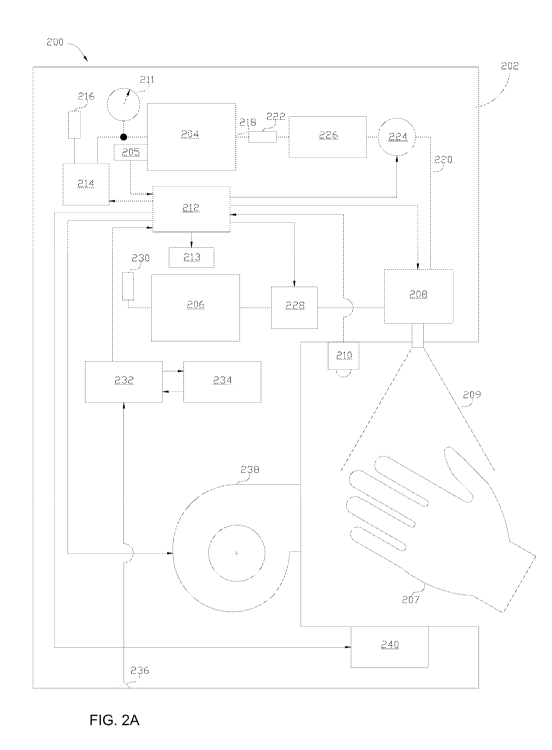
[00921 Systems 200, 200', 200" in FIG. 2A, FIG. 2B, and FIG. 2C, respectively,
illustrate
more detailed examples of the system 100 shown in FIG. 1. As shown in FIG. 2A,
the
system 100 includes a housing 202 which can be similar to the housing 102 of
FIG. 1. As
shown, the housing 202 includes, among other components, an air tank 204, an
air pump 214,
an active agent receptacle 206, a fluid pump 228, a nozzle component 208
having one or
more nozzles, a sensor module 210 having one or more sensors configured to
detect a target
surface in proximity to the nozzle component 208, an optional drying component
238, an
optional overspray collector 240, and a controller 212 operatively coupled to
a display 213.
In this example, the target surface is shown in the form of a hand 207 being
sprayed with an
active agent 209, though it should be appreciated that any other surface can
be sanitized using
the system 200. The air tank 204 and the active agent receptacle 206 are used
to deliver air
and an active agent, respectively, to the nozzle component 208 such that the
active agent is
delivered to a target surface as an aerosol that is deposited onto the surface
as a thin layer.
The aerosol can be generated in many suitable ways. The system 200 can be an
airflow-
based atomizing spray system, an ultrasound spray system, or other type of an
atomizing
system (e.g., a pressure-based atomizing spray system, etc.).
[0093] In the described system, the air pump 214, the air tank 204, the active
agent receptacle
206, the nozzle module 208, as well as other components of the housing 202,
are controlled
via the controller 212. The display 213 is communicatively coupled to the
controller 212 and
is configured to display information related to operation of the system in any
suitable form.
The display 213 can be an interactive display configured to receive
instructions related to
operation of the system. The controller 212 can be implemented in hardware,
software, or
combination thereof.
[0094] The air tank 204 has a pressure sensor 205 associated therewith that is
configured to
monitor pressure in the tank 204. As shown in FIG. 2A, the air tank 204 is
coupled to the air
pump 214 that is controlled to draw ambient air and deliver it to the air tank
204. A
communication line between the air pump 214 and the air tank 204 can be
equipped with a
pressure gauge 211 as shown in FIG. 2A. The air drawn by the air pump 214 can
be passed
CA 02986747 2017-11-21
WO 2016/191375
PCT/US2016/033792
via an air filter 216. The air is provided from an outlet 218 of the air tank
204 to the nozzle
component 208 via a conduit 220. As shown, the air can be passed through a
filter 222 and
its delivery to the nozzle component 208 is controlled via a control valve
224. A pressure
regulator component 226 controls pressure of the air passed through the
conduit 220.
Operation of the air pump 214 maintains pressure in the air tank 204, under
control by the
controller 212.
[0095] The active agent receptacle 206 is in fluid communication with the
fluid pump 228
that delivers a dosage of the active agent from the receptacle 206 to the
nozzle component
208. The controller 212 controls the volume and delivery time of a dose. The
dosage can be
preset such that one or more nozzles of the nozzle component 208 deliver a
predetermined
amount of the active agent each time the nozzles are activated. In some
embodiments,
however, the dosage can be determined by the controller 212 dynamically, based
on size and
other properties of a target object to be sanitized. The properties of the
object can be
determined using the sensor component 210 or in other ways. For example, the
display 213
or other component of the system can be interactive, and can be used to
receive user input
regarding the surface being sanitized, including an input to activate the
system 200. For
example, in some embodiments, two or more options can be provided such that
the user can
select (e.g., by pressing a button or hovering a hand over the button) whether
one hand, both
hands, or any other surface can be sanitized. Furthermore, similar to system
100 in FIG. 1,
the system 200 can receive instructions via a suitable mechanism such as a
button,
touchscreen, footswitch, or other control mechanism configured to activate the
system. The
control mechanism can be coupled to the housing 102 (e.g., it can be attached
to the housing
or coupled thereto via a wired connection) or it can be a remote device
wirelessly
communicating with components of the housing.
[00961 As shown in FIG. 2A, the active agent receptacle 206 can have a filter
230 associated
therewith that filters out dirt and other impurities from vent air that
displaces the active agent
and the agent is withdrawn from the receptacle 206. The filter can be
removable and
replaceable.
[00971 The housing 202 can include a power supply module 232 that can draw
power from a
battery element 234 or from an AC power supply through an AC inlet 236. The
battery
element can be removable and replaceable. In some implementations, the system
200 can be
portable.
16
CA 02986747 2017-11-21
WO 2016/191375
PCT/US2016/033792
[0098] The one or more nozzles of the nozzle component 208 can have a variety
of different
configurations, and they can be stationary or moveable. In some
implementations, the system
can have both stationary and moveable nozzles such that one or more of the
nozzles are
stationary, while one or more of the nozzles are moveable. The nozzles can be
arranged in
various ways so as to deliver an active agent in a desired manner. For
example, the nozzles
can be disposed at certain locations on a housing of the system in a manner
that requires
moving a hand with respect to the nozzles to ensure complete coverage of the
hand with the
active agent. In some examples, however, the nozzles can be disposed such that
a hand can
simply be positioned in proximity thereof and no additional movement of the
hand is required
to adequately cover the hand with the active agent provided by the nozzles. In
such
examples, at least a portion of the housing can be shaped such that one or
more hands can be
positioned to be treated with an active agent and no additional movement of
the hands can be
required for the treatment. This helps to ensure compliance. For example, the
housing can
have a cavity or other opening having nozzles openings on its inner walls. The
cavity can
have any suitable shape and size. As an example, the cavity can be shaped so
as to conform
to the shape of the hand or in other way to allow coverage of the hand without
additional
actions from the user after the hand has been placed into the cavity. However,
it should be
appreciated that the cavity can be oval, rectangular, or it can have any other
shape. The
cavity's size can allow it to receive one or two hands. Furthermore, in some
implementations, more than one person can use the system to sanitize their
hands
simultaneously. The nozzles can have various sizes and shapes in order to
deliver an active
agent aerosol in a desired manner.
[0099] FIGS. 3, 4, and 5A-5C illustrate examples of different types of nozzles
that can be
used in conjunction with the system 200 or other system implementing the
described
techniques, e.g., system 200' (FIG. 2B) and the system 200" (FIG. 2C)
described in more
detail below. FIG. 3 shows an example of a portion of a system 300
implementing the
described techniques. As shown, the system 300 includes a housing 302 having a
single
stationary nozzle 304 configured to dispense an active agent to a surface such
as, in this
example, a hand 306. It should be appreciated that although one user's hand
306 is shown,
depending on size and configuration of the stationary nozzle 304, the nozzle
304 can deliver
an active agent to sanitize both hands of the user at the same time.
17
CA 02986747 2017-11-21
WO 2016/191375
PCT/US2016/033792
[00100] FIG. 3 shows the user's hand 306 placed adjacent to the stationary
nozzle 304 such
that the palm-side of the hand faces the nozzle 304. In this configuration,
the active agent is
dispensed from the nozzle 304 and delivered to the palm-side of the hand. In
order to receive
the active agent on the top of the hand, the user needs to rotate his or her
hand by 180 degrees
such that the top of the hand faces the nozzle 304. In this configuration, the
cone angle of the
sprayed active agent can be designed to allow delivery to the sides of the
hand and to the
sides of the fingers. To ensure that these regions of the hand are not blocked
(e.g., because
the user has closed or bent the fingers, clenched the hands, or the hands are
touching each
other), the system 300 may provide an indication to the user informing the
user of the
requirement to keep his/her hand in an appropriate manner. For example, an
indication can
be provided to the user in an audio, visual, or a combination form reminding
the user to
position his/her hand such that the fingers are spread, the hands are not in
contact with each
other or other objects, etc. In addition, because a person is more likely to
have his/her
fingers spread if the palm of his/her hand is facing up or down (rather then
sideways, as
during a "handshake" position), the system can be configured such that it can
receive a hand
only if it is disposed with the palm facing up or down.
[00101] FIG. 4 shows a portion of a system 400 implementing the described
techniques with
nozzles having another configuration. The system 400 can have the same or
similar
components as those described in connection with systems 100 (FIG. 1), 200
(FIG. 2A), 200'
(FIG. 2B), and 200" (FIG. 2C). In this example, the system 400 includes a
housing two
portions of which are shown as upper and lower housing portions 402a, 402b. As
shown, the
upper and lower housing portions 402a, 402b have arrays of nozzles 404a, 404b
associated
therewith, respectively. It should be appreciated that, even though in the
system 400 each of
the nozzle arrays 404a, 404b has three nozzles, the nozzle arrays can have any
suitable
number of nozzles (e.g., two or more than three), including a different number
of nozzles
among the arrays.
[00102] In the example of FIG. 4, as shown, the nozzle arrays 404a, 404b can
dispense an
active agent to both sides of a target surface, such as the tops and bottoms
of a pair of hands
406. A person skilled in the art will appreciate that any other surface having
appropriate
shape and size can also be sanitized using the system 400.
[00103] The nozzle arrays 404a, 404b can have a variety of different
configurations. In FIG.
4, each of the arrays is a linear array having the nozzles arranged along the
same line. It
18
CA 02986747 2017-11-21
WO 2016/191375
PCT/US2016/033792
=
should be appreciated, however, that in one or both of the arrays the nozzles
can form
rectangular, circular, oval, elliptical, or other patterns.
[00104] In one embodiment, the nozzle array can be a linear strip with a micro-
orifice formed
as a slot along the length of the strip. The linear strip can be patterned as
a serpentine layout
to allow uniform delivery of micro-droplets of an active agent across an area
under (or above)
the serpentine layout, to the hands.
[00105] Furthermore, in some embodiments, the nozzles can be disposed and
directed
towards a location where a target surface is to be placed so as to form
various patterns that
may not necessarily be referred to as "arrays." For example, as discussed
above, a housing
can have a cavity or other structure having a contour conforming to a shape of
a hand, and
multiple nozzles can be arranged such that their orifices are disposed along
inner walls of
such cavity. The cavity can have an opening for a hand to be inserted therein.
In such a
configuration, a hand disposed within the cavity will not need to be turned or
otherwise
moved to be adequately covered with an active agent emitted from the nozzles
which is then
dried. The cavity can be shaped such that a hand can be inserted therein with
the palm facing
up or down, in a position which would be appropriate for a handshake, or in
other manner.
The cavity can also be designed such that both hands of a user can be
sanitized at the same
time. The cavity is positioned such that it can receive hand(s) in a
convenient for a user
manner. Regardless of the configuration and position of the cavity and the
nozzles, the
system can be configured so as to adequately sanitize at least a gripping
surface of the
hand(s).
[00106] FIGS. 5A-5C illustrate schematically a moveable array of nozzles 502
(used with a
sanitization system, not shown) that are configured to dispense an active
agent to a pair of
hands 506 to perform hand sanitization. Unlike a stationary nozzle
arrangement, the
moveable array moves over a surface of the hands (or any other object) during
the
sanitization process. In the illustrated example, the array of nozzles 502 is
shown as a linear
strip, though, as a person skilled in the art will appreciate, other
configurations can be used
alternatively. FIG. 5A shows a position of the array of nozzles 502 at a first
time period, at a
beginning of the hand sanitization process when the array 502 is disposed
around wrists of
the hands. FIG. 5B illustrates a position of the array of nozzles 502 at a
second, intermediate
time period of the sanitization process when the array of nozzles 502 has
moved half-way
with respect to the user's hands 506. Finally, FIG. 5C illustrates a position
of the array of
19
CA 02986747 2017-11-21
WO 2016/191375
PCT/US2016/033792
nozzles 502 at a third, later time period where the array of nozzles 502 has
passed over the
user's hands 506. In the example shown in FIGS. 5A-5C, the hands can be placed
above or
below the array 502 such that the array can scan above and below the pair of
hands, thereby
delivering an active agent to the tops and bottoms of the hands, where the
method of
delivering the active agent and drying the hands can take less than 5 seconds.
[00107] In FIGS. 3, 4, and 5A-5C, the nozzles of the respective systems are
disposed such
that the hand or hands are oriented with palm-sides facing "up" or "down," and
where the
normals to the palms are aligned with the direction of gravity. In other
embodiments, the
system may be configured such that the hands can be oriented with palms
rotated by 90
degrees or facing the side as is conventionally achieved when shaking hands
with another
person
[00108] One or more nozzles of a system implementing the described techniques
can operate
in various different ways. Thus, regardless of their particular configuration
and arrangement,
the nozzles can be driven using airflow-based, pressure-based, ultrasonic, or
other techniques.
The nozzles can have orifices of different sizes and configurations that can
allow expelling an
active agent as a spray having desired distribution patterns. For example, in
some cases, the
nozzles can provide a circular distribution of the sprayed agent on a target
surface. In other
cases, additionally or alternatively, the nozzles can produce a fan-shaped
spray pattern
relative to a stationary target object. The nozzles can be equipped with
various components
(e.g., air caps) that allow generating a spray having desired characteristics.
[00109] The nozzles can have various operating parameters. Thus, the nozzles
can operate at
a certain air pressure to provide an active agent flow rate appropriate to
create a thin, uniform
layer of an active agent on a target surface in a relatively short time period
(e.g., less than five
seconds, less than three seconds, or less than one second). The uniform thin
layer is then
dried on the target surface such that the total time required to sanitize the
hand is less than
five seconds, less than three seconds, or less than one second. For example,
in one
embodiment, an ultrasonic nozzle can operate at an air pressure that ranges
from about 0.2 psi
to about 5 psi. The active agent can be expelled from such nozzle at a flow
rate of about 15
mL/min to achieve a coating thickness of about 10.0 p.m in 1 second on a
surface area
equivalent to one side of one hand, when supplied with air at about 2 psi air
pressure. In
another embodiment, an air-atomizing nozzle can have an active agent flow rate
of about 35
mL/min, operating at 12 psi for the atomizing air and 10 psi for the active
agent. A surface of
CA 02986747 2017-11-21
WO 2016/191375
PCT/US2016/033792
one side of one hand can be coated using such nozzle at a thickness of about
10.0 pm in less
than 0.5 seconds. Air-atomizing nozzles of this type can be operated over an
air pressure
ranging from about 5 psi to about 100 psi, with liquid supplied over a range
from about 10 psi
to about 50 psi.
[00110] Referring back to FIG. 2A, as mentioned above, the housing 202 can
also include an
optional drying component 238 configured to dry the target surface 207 after
the active agent
209 in the form-of an aerosol is dispensed on the target surface 207. In this
example, the
drying component 238 can be an air dryer that can provide an air stream to dry
a target
surface after it has been treated with the active agent. The drying component
238 can have a
variety of configurations. The drying component 238 can use a flow of an
ambient air having
room temperature or the air can be heated. Alternatively, the drying component
can be an
infrared heater that can provide infrared radiation to the target surface to
dry it. The drying
component can have any other configurations, as the described techniques are
not limited in
this respect. Regardless of its specific configuration, the drying component,
if present, is
configured to dry the thin film of an active agent deposited on a target
surface.
(001111 As described above, the drying component can be controlled to dry a
target surface
for a predetermined time period. The time period can be preset such that the
drying process
continues for a predetermined duration of time, e.g., five seconds, three
seconds, or other
duration. As another option, a required level of dryness can depend on user's
actions. In
such scenarios, the drying can proceed until the system determines that the
user's hand (or
other surface) being sanitized is no longer in proximity to the sensor. In
some embodiments,
the sensor 210 or one or more other sensors that can alternatively or
additionally be
associated with the system can detect a dryness level of the target surface.
[00112] The sensor module 210 can also have a variety of different
configurations, and it can
have one or more sensors of any suitable type. The sensor of the sensor module
210 can be
an optical proximity sensor that is able to detect the target object shown by
way of example
only as the hand 207 in FIG. 2A. The optical proximity sensor can also be able
to detect a
position and motion of the hand 207 or other target object. For example, the
sensor can
detect that the hand 207 has been disposed in proximity to the nozzle(s) 208,
and it can also
detect a way in which the hand 207 is positioned with respect to the nozzle(s)
208. Other
events can also be detected by one or more sensors of the sensor module 210,
as described
below.
21
CA 02986747 2017-11-21
WO 2016/191375
PCT/US2016/033792
[00113] The active agent receptacle 206 of the system 200 can have various
configurations
and it can receive and store the active agent in a variety of ways. Thus, the
active agent
receptacle 206 can be configured as a refillable reservoir that is configured
to receive a
supply of the active agent. When the amount of the active agent is below a
certain amount,
an appropriate indication can be provided. In some embodiments, the active
agent receptacle
206 houses a removable and refillable active agent-containing cartridge. The
cartridge can be
replaceable such that it is pre-filled with an active agent.
[00114] It should be appreciated that the system 200 in FIG. 2A is described
by way of
example only, as systems having other configurations can implement the
described
techniques. Thus, another example of a system in which the described
techniques can be
implemented is shown in FIG. 2B where a system 200' is configured to deliver
an active
agent 209' in the form of an aerosol spray to a surface 207' (e.g.,
unprotected or gloved
hand). As shown, the system 200' includes a housing 202' having a controller
212'
associated with a display 213', an active agent receptacle 206', a fluid pump
228', a nozzle
component 208' having one or more nozzles, a sensor module 210' (other ways to
activate
the system 200' can be used additionally or alternatively), a drying component
238', a power
supply module 232' that can draw power from a battery element 234' or from an
AC power
supply through an AC inlet 236', and an optional overspray collector 240'.
These
components can be similar to the corresponding components of the system 200
(FIG. 2A) and
are therefore not described in more detail. The system 200' may not include
air delivery
components. However, in some implementations, one or more air delivery
components can
be present. In this example, the atomized active agent can be moved from the
nozzle(s) of
the nozzle component 208' to the target surface 207' by at least one
(optional) fan 219'. The
system 200' can be an ultrasonic atomizing system. Furthermore, the
configuration of the
system 200' can also be representative of a pressure-based atomizing spray
system.
[00115] Yet another example of a system in which the described techniques can
be
implemented is shown in FIG. 2C where a system 200" is configured to deliver
an active
agent 209" in the form of an aerosol spray to a surface 207" (e.g.,
unprotected or gloved
hand). As shown, the system 200" includes a housing 202" having a filter 216",
a pump
214", a sensor 205", a gauge 211 ", an air tank 204"with an outlet 218", a
filter 222", a
pressure regulator component 226", a control valve 224", a controller 212"
associated with
a display 213", a filter 230", an active agent receptacle 206", a nozzle
component 208"
22
CA 02986747 2017-11-21
WO 2016/191375
PCT/US2016/033792
having one or more nozzles, a sensor module 210" (other ways to activate the
system 200"
can be used additionally or alternatively), a drying component 238", a power
supply module
232" that can draw power from a battery element 234" or from an AC power
supply through
an AC inlet 236", and an optional overspray collector 240". Air is provided
from the outlet
218" of the air tank 204" to the nozzle component 208" via a conduit 220".
These
components can be similar to the corresponding components of the system 200
(FIG. 2A) and
are therefore not described in more detail in connection with FIG. 2C. In this
example, the
active agent is displaced from the active agent receptacle 206" by air from
the air tank 204".
A control valve 229" under control of the controller 212" admits pressurizing
air to the
receptacle 206". Dispensing of fluid from the receptacle 206" to the nozzle
component
208" occurs when a control valve 231" is actuated by the controller 212".
[00116] It should be appreciated that the systems 100 (FIG. 1), 200 (FIG. 2A),
200' (FIG.
2B), 200" (FIG. 2C) are exemplary only, and that the systems 100, 200, 200',
200" can
include other components that are not shown herein.
[00117] Regardless of the specific configuration of the system implementing
the described
techniques, the techniques provide a method of killing or inactivating
transient pathogens on
a surface of the skin or any other surface. FIG. 6 illustrates a process 600
of killing or
inactivating transient pathogens on a surface using an active agent including
one or more
antiseptic or disinfectant reagents, in accordance with the described
techniques. The process
600 is described herein in connection with the system 200 (FIG. 2A) as an
exemplary system
which can perform this process. However, it should be appreciated that the
process 600 can
be performed by system 100 (FIG. 1), system 200' (FIG. 2B), system 200" (FIG.
2C), or by
any other suitable system.
[00118] The process 600 can start at any suitable time. For example, it can
start when a
system performing it (e.g., system 100 or 200) is activated. The process 600
can be
controlled by a control module of a system, such as, e.g., controller 212 in
FIG. 2.
[00119] At block 602, the system can monitor for presence of a target surface
in proximity to
system's nozzle(s) (e.g., nozzle module 208 in FIG. 2A). The target surface is
considered to
be "in proximity" to the nozzles when it is adjacent to the nozzles such that
an active agent
that can be dispensed from the nozzles can reach the surface to adequately
cover it with a
layer of the active agent. The target surface can be one or both of user's
hands, or another
23
CA 02986747 2017-11-21
WO 2016/191375
PCT/US2016/033792
object. The user's hand or hands may be bare or gloved. As discussed above, a
proximity
sensor, a motion sensor, or other type of a sensor can operate to determine
whether a surface
is detected in proximity thereof.
[00120] Also, in some embodiments, the system can be activated to perform the
surface
sanitizing process in other ways, which can be different from the processing
at blocks 602
and 604 in FIG. 6, which are shown by way of example only. Thus, additionally
or
alternatively, the system can receive an instruction from a user via a
suitable mechanism such
as a footswitch, button, touchscreen, sensor, or any other control mechanism
configured to
activate the system. The control mechanism can be coupled to a system's
housing (e.g., it can
be attached to the housing or coupled thereto via a wired connection) or it
can be a remote
device wirelessly communicating with components of the housing. Thus, in some
embodiments, the target surface may not be detected but rather the system is
activated to
perform the described techniques in response to other suitable trigger.
[00121] Regardless of the specific way in which it is determined that the
target surface is in
proximity to the nozzles and/or the way in which an instruction to activate
the system is
received, in response to determining that the target surface is adjacent to
the nozzle, the
process 600 continues to block 606 where the active agent is dispensed onto
the target
surface. The active agent can include one ingredient that can kill or
inactivate a transient
pathogen on the surface, or a mixture of two or more of such ingredients
examples of which
are discussed below. The active agent is dispensed from one or more nozzles,
such as the
nozzle(s) of the nozzle module 208 shown in FIG. 2. The active agent is
dispensed onto the
surface in the form of an aerosol spray, and forms an aerosolized layer that
is a thin and
substantially uniform coating on the target surface.
[00122] The layer of the active agent can be from about 1 gm to about 50 gm in
thickness.
Furthermore, in some embodiments, the active agent layer can be from about 5
gm to about
20 gm in thickness. In yet other embodiments, the thickness of the active
agent layer can be
from about 4 gm to about 10 lam.
[00123] The system can be configured to deliver the active agent such that
there are no
uncoated areas of the surface (or such that the uncoated areas do not affect
the result of the
sanitization process). The uniform coating can be achieved due to aerosol
properties of the
active agent. In particular, the active agent in the form of an aerosol
includes small fluid
24
CA 02986747 2017-11-21
WO 2016/191375
PCT/US2016/033792
droplets having a size (or a size distribution) that allows a thin uniform
layer of the active
agent to be formed on a target surface. In at least some embodiments, the
active agent
droplets can be from about 18 gm to about 56 gm in diameter. The droplets'
size distribution
can vary in different ways and an average diameter of the droplets can be
about 33 gm in
diameter. In at least some embodiments, the droplet sizes can be from about 36
gm to about
107 gm with, an average diameter of about 57 gm. In other embodiments, the
droplet sizes
can range from about 28 gm to about 116 gm, with an average diameter of about
59 gm. It
should be appreciated that active agent droplets of other sizes can be formed
additionally or
alternatively. The active agent delivery process at block 606 can be
controlled to ensure
adequate treatment of the target surface. For example, suitable one or more
sensors (e.g.,
sensor 210 and/or any other sensor) can monitor the agent delivery process to
ensure
adequate surface coverage. The sensors can determine whether any fluid is
present on the
target surface, as a way to control proper delivery of the active agent onto
the surface. The
sensor(s) can also monitor the degree of uniformity of the active agent
deposition over the
target surface.
[00124] If it is determined, at decision block 604, that the target surface
has not been
detected, the process 600 can return to block 602 to continue monitoring for
the presence of
the target surface, as shown in FIG. 6.
[00125] After the active agent has been dispensed onto the surface, the active
agent is dried
on the surface, at block 608. The drying can be performed by a suitable drying
component
such as, e.g., the drying component 238 (FIG. 2A). The drying can be performed
by a stream
of air (which can be cool or warm air), by an infrared dryer, or using any
other approach. As
mentioned above, the drying component is optional, and the layer of the active
agent
dispensed on the target surface can be dried by ambient air. For example, the
user can simply
wait for a few seconds for his/her hands treated with an active agent to dry.
[00126] The processing steps at blocks 606 and 608 can both be completed
quickly, e.g., in
less than five seconds. The dispensing of the active agent can take less than
three seconds,
and the drying of the agent disposed as a layer on the treated surface can
take less than two
seconds. However, the agent dispensing and drying steps can be performed over
other
periods of time. Moreover, in some embodiments, the entire process of treating
a target
surface can take less than three seconds. In this way, the user can have
his/her hands to be
sanitized in a convenient and timely manner.
I I
CA 02986747 2017-11-21
WO 2016/191375 PCT/US2016/033792
[00127] Regardless of how the drying is performed, at block 610 of FIG. 6, the
system can
determine whether the drying is completed and provide an indication of
completion of the
drying step. A suitable optical sensor can monitor the target surface being
treated and it can
determine when the surface is considered to be sufficiently dry. For example,
the sensor can
be used to automatically determine whether there are any moist areas present
on the surface
and to determine a dryness level of the surface. The indication can be
provided to the user in
audio, visual, or other forms. For example, an audio signal can be generated
to indicate to the
user that his/her hands have been sanitized. As another option, additionally
or alternatively, a
visual indication (e.g., a light indicator, a textual message, or other
indication) can be
presented to a user on a display, such as, e.g., display 213 in FIG. 2A. In
addition, a suitable
indication can be provided to a user during the active agent application
and/or drying. For
example, an indicator of one color can be provided while the process is in
progress, and the
color can change once the process is completed. Also, in some embodiments, no
indication
of completion of drying is provided and the user can perceive that his/her
hands are dry.
[00128] Regardless of the way in which the drying is performed, the active
agent on the
hands' surface is effective to kill or inactivate transient pathogens on that
surface. Moreover,
because of the way in which the active agent is dispensed onto the hands, the
active agent can
sanitize the skin without significantly affecting resident microflora of the
skin. In addition,
even if the active agent belongs to a class of strong disinfectants that would
otherwise be
considered harsh to the skin (but that are nevertheless desired for use in
certain settings), the
quick application of a thin layer of the active agent using the described
techniques allows to
reduce the negative affects of such strong disinfectants.
[00129] After the indication of completion of drying is provided, the process
600 can end. It
should be appreciated, however, that the process 600 can be continuous. In
this way, after
one surface has been treated, the system monitors for the presence of another
surface in
proximity to the nozzles, and/or waits for a trigger to initiate dispensing of
the active agent.
For example, in a hospital setting, an apparatus performing the process 600
can, in a rapid
sequence, sanitize the hands of multiple personnel.
[00130 FIG. 7 illustrates another example of a process of killing or
inactivating transient
pathogens on a surface in accordance with the described techniques. The
process 700 shown
in FIG. 7 is similar to process 600 of FIG. 6 and can similarly be performed
by a system such
as system 100 (FIG. 1), system 200 (FIG. 2A), system 200' (FIG. 2B), system
200" (FIG.
26
CA 02986747 2017-11-21
WO 2016/191375
PCT/US2016/033792
2C), or by other suitable system. The process 700 is described here by way of
example only
as being performed by system 200 of FIG. 2A. Also, steps of the process 700
that are similar
to corresponding steps of process 600 are not described in detail in
connection with FIG. 7.
[00131] As shown in FIG. 7, after the process 700 starts at a suitable time,
presence of a
target surface can be detected at block 702. As discussed above, the target
surface can be
detected by one or more suitable sensor(s), or the system performing the
current process can
respond to a suitable trigger, such as an instruction to activate the nozzle.
In some
implementations, sensors used may be able to determine one or more properties
of the
detected surface, such as its size and contours.
[00132] When the target surface is detected, at block 704, airflow with
regulated pressure is
provided to the nozzle. For example, as shown in the example of FIG. 2A, air
from the air
tank 204 can be caused, by the air pump 214, to be provided to the nozzle
component 208.
This process is controlled by a control module (e.g., controller 212 in FIG.
2A). Air can be
delivered to the nozzle module at a desired airflow pressure. The pressure of
the airflow is
selected such that it is sufficiently high to quickly cover a surface being
treated, but at the
same time not excessively high, since high pressure can generate an aerosol
stream that is
deposited as a layer which is thicker than desired. In embodiments in which an
ultrasound
atomizer is used to generate the aerosol, a lower pressure can be used, e.g.,
from about 0.5 psi
to about 5 psi. In other embodiments in which an airflow-based atomizing spray
system is
used, a higher pressure can be used, e.g., greater than about 5 psi.
[00133] At block 706, as shown in FIG. 7, at least one nozzle of the system
performing the
process is activated. A dosage of the active agent can be provided to the
activated nozzle at
block 708. Referring again by way of example only to system 200 in FIG. 2A,
the active
agent can be provided, by the fluid pump 228, from the active agent receptacle
206, to the
nozzle component 208.
[00134] The dosage can be selected based on expected surface of the target
surface to be
sanitized, which can be done in advance (e.g., if the nozzles are used to
spray surfaces of
similar sizes) or the dosage can be selected dynamically, based on properties
(e.g., size,
contours, etc.) of the specific target surface being treated. For example, in
embodiments
where the system is configured to disinfect hands, the dosage can be selected
based on the
size of the area of one or both hands. In the case of an individual hand,
where the surface
27
CA 02986747 2017-11-21
WO 2016/191375
PCT/US2016/033792
area is about 500 cm2, a dispensed dose of 0.5 mL of an active agent uniformly
distributed
across this hand surface would yield a coating about 10 gm thick. An active
agent dose of 3
mL dispensed uniformly across the same surface area would yield a coating
about 60 gm
thick. Allowing for a 30% overspray (excess active agent that spreads beyond
the target
surface being sprayed), the 3 mL dosage can be increased to about 4 mL to
yield a 60 gm
thick coating. Uniform coating of two hands with a 60 gm thick coating and a
30%
overspray would require a dosage of about 8 mL. In cases where only the palms
and adjacent
finger surfaces of a pair of hands are treated, a 30% overspray is expected
and the target
coating thickness is about 10 gm thick, the dosage of the active agent can be
about 1.5 mL.
[00135] At block 710, the dosage of the active agent is dispensed from the
nozzle so as to
form a thin uniform layer on the target surface. As discussed above, the
active agent is
dispensed in the form of an aerosol spray. The aerosol spray can be created by
an air
atomizing nozzle that uses air pressure to create the spray,'as well as to
deliver the droplets of
the spray to the target surface. In other embodiments, the system can be an
ultrasonic nozzle
system. In yet other embodiments, the system can use hydraulic nozzles. A
positive-
displacement pump, a suction- or pressure-based fluid delivery approach, or
any other
suitable approach can be used to deliver fluid to the nozzle or nozzles. As an
example, in the
system 200 of FIG. 2A, the aerosol is created by aerosolizing the active agent
received from
the active agent receptacle 206 in the airstream delivered to the nozzle via
the conduit 220.
[00136] After the target surface is sprayed with the desired dosage of the
aerosolized active
agent, a drying component is activated to dry the active agent on the surface,
at block 712.
An indication of completion of the drying process, and therefore completion of
the surface
sanitization, is provided at block 714. The process 700 can then end, though
it can be
performed continuously, to sanitize another target surface.
[00137] The active agent can include one or more ingredients that can be used
to inactivate or
kill transient pathogens on a target surface such as a surface of the hand.
Any one or more
pathogen inactivation fluids or germicidal fluids can be used in the active
agent. The active
agent is selected such that it can be delivered onto a target surface as a
thin, uniform, quick-
drying coating capable of quickly killing or inactivating transient pathogens
on the target
surface.
28
CA 02986747 2017-11-21
WO 2016/191375
PCT/US2016/033792
[00138] As an example, the active agent can be an aqueous solution of
hypochlorous acid.
Any suitable source of hypochlorous can be used. For example, Excelyte
(Integrated
Environmental Technologies, LTD., Little River, SC) or another suitable source
of
hypochlorous acid can be used. Excelyte can be given as a possible aqueous
hypochlorous
acid composition, since it is reported (according to the package label) to be
effective at killing
Clostridium dlfficile, Escherichia coli, MRSA, Salmonella, Pseudomonas,
Listeria
monocytogenes, Enterococcus faecalis (VRE), Klebsiella pneumonia (NDM-1) and
Staphylococcus aureus. The aqueous solution of hypochlorous acid can have any
suitable
concentration of hypochlorous acid. In some embodiments, the aqueous solution
of
hypochlorous acid includes at least about 0.046% of hypochlorous acid. As
another example,
the aqueous solution of hypochlorous acid can include from about 0.005% to
about 1% of
hypochlorous acid. A suitable commercial product or a solution thereof can be
used as an
active agent.
[00139] As another example, the active agent can be an aqueous solution of
hydrogen
peroxide used as a pathogen inactivation or germicidal fluid, since hydrogen
peroxide is a
broad spectrum antimicrobial capable of inactivating or killing bacteria,
viruses, fungi and
spores. The aqueous solution of hydrogen peroxide can include about 0.3%,
about 1%, about
3%, about 6%, about 9% or about 12% of hydrogen peroxide. In some embodiments,
the
aqueous solution of hydrogen peroxide can be used with hydrogen peroxide
concentration in
the range from about 3% to about 6%.
[00140] As another example, the active agent can be accelerated hydrogen
peroxide or AHP.
AHP is a proprietary blend of hydrogen peroxide, surface active agents,
wetting agents,
chelating agents and water designed for improved germicidal potency and
cleaning
performance (Virox, Oakville, ON, Canada). AHP is used as a pathogen
inactivation or
germicidal fluid since it is a broad spectrum antimicrobial capable of
inactivating or killing
bacteria, viruses, fungi and spores and is expected to readily wet the
surfaces of the hands,
allowing for the development of a thin, uniform coating from a controlled
spray of micro-
droplets of AHP onto the hands. When the active agent is an aqueous solution
of hydrogen
peroxide, the aqueous solution of hydrogen peroxide can be used with hydrogen
peroxide
concentration in the range from about 3.0% to about 12.0%.
[00141] As another example, the active agent can be an aqueous solution that
is a mixture of
peracetic acid and hydrogen peroxide.
29
CA 02986747 2017-11-21
WO 2016/191375
PCT/US2016/033792
[00142] As yet another example, the active agent can be an aqueous solution of
acetic acid.
The aqueous solution of can have acetic acid concentration in the range from
about 1% to
about 10.0%. In other embodiments, the aqueous solution of acetic acid used
can have acetic
acid concentration in the range from about 3.0% to about 6.0%. An aqueous
solution of
acetic acid is used as a pathogen inactivation or germicidal fluid when broad
spectrum
bacterial inactivation or killing is warranted.
[00143] In some embodiments, the active agent can include an aqueous solution
of isopropyl
alcohol. The aqueous solution of isopropyl alcohol can have isopropyl alcohol
concentration
from about 60% to about 90%.
[00144] In some embodiments, the active agent can include an aqueous solution
of ethanol.
The aqueous solution of ethanol can have ethanol concentration from about 60%
to about
90%.
[00145] In another embodiment, the active agent can include an aqueous
solution of peracetic
acid. The aqueous solution of peracetic acid can have peracetic acid
concentration from
about 0.1% to about 1.0%.
[00146] In other embodiment, the active agent can include an aqueous solution
of sodium
hypochlorite. The aqueous solution of sodium hypochlorite can have sodium
hypochlotite
concentration from about 0.1% to about 1.0%.
[00147] Regardless of the specific ingredient or a mixture of ingredients
included in the
active agent, the process of spraying a small volume of the active agent,
followed by rapid
drying of the hands, has the effect of reducing the numbers of viable
transient bacteria on the
surfaces of the hands. This thin, uniform coating of fluid is quickly dried by
exposure to the
ambient environment or using "active" techniques such as, e.g., a stream of
forced air, forced
heated air, or infrared radiation. The act of drying stops or substantially
reduces the pathogen
inactivation or killing process thereby limiting the microbial inactivation or
killing to the
transient pathogens on the outermost surface of the skin. The sanitization
process has the
effect of inactivating or killing transient pathogens on the surfaces of the
hands, which can be
done without substantially altering the resident microflora population or
causing irritation or
toxic effects on the skin.
CA 02986747 2017-11-21
WO 2016/191375
PCT/US2016/033792
[00148] In at least some embodiments, the active agent can also be an aqueous
solution of
ozone having ozone concentration in the range from about 1 parts per million
(ppm) to about
40 ppm. In at least some embodiments, the ozonated water can have dissolved
ozone
concentration from about 0.2% to about 2.0%. Further, in at least some
embodiments, the
active agent can also be an aqueous solution of ozone having dissolved ozone
concentration
in the range from about 0.1 mg/L to about 10 mg/L. An aqueous solution of
ozone can be
used as a pathogen inactivation or germicidal fluid since ozone is a broad
spectrum
antimicrobrial capable of inactivating or killing bacteria, viruses, fungi and
spores.
[00149] The active agent can also be a mixture of aqueous solutions of ozone
(i.e., ozonated
water) and hydrogen peroxide. In some embodiments, the ozonated water and
aqueous
hydrogen peroxide can be delivered to hands as a target surface from separate
nozzles. These
nozzles and the spraying protocols can be designed to provide mixing of the
ozonated water
and aqueous hydrogen peroxide within the delivered spray or upon impingement
onto the
skin surface. In other embodiments, the ozonated water and aqueous hydrogen
peroxide can
be mixed within the delivery apparatus prior to spraying through the same
nozzle or array of
nozzles. Regardless of whether the ozonated water and aqueous hydrogen
peroxide are
delivered together or separately to the target surface, a thin, uniform
coating of the active
agent delivered onto the surface of the hands is quickly dried by exposure to
the ambient
environment or using "active" techniques such as, e.g., a stream of forced
air, forced heated
air, or infrared radiation.
[00150] The following non-limiting examples describe experiments that were
conducted to
assess efficacy of the described techniques.
[00151] The following examples are put forth so as to provide those of
ordinary skill in the
art with examples of how the systems, compositions, devices and/or methods
described
herein can be made and evaluated, and are intended to be purely exemplary of
the present
disclosure and are not intended to limit the scope of the disclosure. Thus,
the examples
below are merely illustrative of techniques for sanitizing hands or other
surfaces in various
settings.
EXAMPLES
Example 1
31
CA 02986747 2017-11-21
WO 2016/191375
PCT/US2016/033792
[00152] This example describes inactivation of bacteria on human hands from a
brief spray of
pathogen inactivation fluid followed by rapid drying.
[00153] An original solution of the K-12 strain of Escherichia coli was
obtained (Carolina
Biological Supply Company, Burlington, NC) and diluted 10,000-fold in nutrient
broth
(Carolina Biological Supply Company, Burlington, NC) to generate a dilute
bacterial
solution. In this example, 25 1_, of the dilute solution was pipetted onto
the pads of the
index, middle and ring fingers of a human hand. After pipetting, the solutions
were spread
with a pipette tip and allowed to dry under a small blower (Delta model
BFB0712HH-A,
Digi-Key, Thief River Falls, MN) for a few minutes. It is estimated that this
25 pL solution
contained between 70 and 100 bacteria, based on bacterial plate counts from
this same batch
of solutions, described in Example 4 below. Just after drying, an aqueous
solution of
hydrogen peroxide (3.0 % concentration) was sprayed for 1 second onto the
middle and ring
finger pads of the previously Escherichia coli treated index, middle and ring
fingers. The
spraying apparatus included an air brush (Patriot Model 105, Badger, Franldin
Park, IL) that
is capable of spraying thin coatings of a wide variety of fluids with
viscosities similar to
water. The index finger also previously treated with the Escherichia coli
dilute solution was
not sprayed with the aqueous solution of hydrogen peroxide. After spraying the
hydrogen
peroxide solution, the middle and ring finger pads were dried under the same
small blower
for 5 seconds. All three finger pads were then depressed onto a pre-cast Luria
broth (LB)
agar plate (Carolina Biological Supply Company, Burlington, NC). FIG. 8 shows
this agar
plate after an overnight incubation at 37 C. This plate shows at least 5
bacterial colonies that
have grown on the region (803) of the agar that was contacted with the non-
sprayed index
finger pad. No bacterial colonies are seen to have grown on the regions of the
agar plate that
were contacted with the middle (801) and ring (802) finger pads that had been
quickly
sprayed with a pathogen inactivation or killing fluid and dried with a small
volume of forced
air.
Example 2
[00154] This example describes inactivation of bacteria on human hands from a
brief spray of
pathogen inactivation fluid followed by rapid drying.
[001551 A solution of the K-12 strain of Escherichia coli was obtained and
diluted 10,000-
fold in nutrient broth to generate a dilute bacterial solution. All biological
supplies were
32
1
CA 02986747 2017-11-21
WO 2016/191375 PCT/US2016/033792
sourced from Carolina Biological Supply Company (Burlington, NC). In this
example, 5 iL
of this dilute solution was pipetted onto the pads of the index, middle and
ring fingers of a
human hand. After pipetting, the solutions were spread with a pipette tip and
allowed to dry
over a few minutes under the small blower as described in Example 1. It is
estimated that
this 5 [EL solution contained between 15 and 20 bacteria, based on bacterial
plate counts
from this same batch of solutions, described in Example 4. Just after drying,
an aqueous
solution of hydrogen peroxide (3.0 % concentration) was sprayed for 1 second
onto the
middle and ring finger pads of the previously Escherichia coli treated index,
middle and ring
fingers. The spraying apparatus was the air brush described in Example 1. The
index finger
also previously treated with the Escherichia coli dilute solution was not
sprayed. After
spraying the hydrogen peroxide solution, the middle and ring finger pads were
fully dried
under the same small blower for 5 seconds. All three finger pads were then
depressed onto a
pre-cast Luria broth (LB) agar plate. FIG. 9 shows this agar plate after an
overnight
incubation at 37 C. This plate shows at least 6 bacterial colonies that have
grown on the
region (901) of the agar that was contacted with the non-sprayed index finger
pad. No
bacterial colonies are seen to have grown on the regions of the agar plate
that were contacted
with the middle (902) and ring (903) finger pads that had been quickly sprayed
with an
aqueous solution of hydrogen peroxide and dried with a small volume of forced
air.
Example 3
[001561 This example describes tests to determine the amount of water
deposited with an
airflow-based atomizing spray system and dried with a small blower.
[00157] The quantity of water deposited from an airbrush and the amount of
drying taking
place in 5 seconds of exposure to airflow from a small blower were assessed on
a 25 mm
diameter track-etched polycarbonate membrane with 0.4 urn diameter pores
(Whatman
Cyclopore Model 7060-2504, Sigma-Aldrich, St. Louis, MO). The airbrush and the
small
blower used in this example are described in Example 1. The initial weight of
the membrane
was measured using a precision balance (Sartorius Model CPA64, Bohemia, NY).
Following
the initial weighing, the membrane was placed on a polycarbonate support block
and retained
by the weight of a polycarbonate plate, containing three bored-through 16.3 mm
diameter
holes. The plate was placed so that one of the through-holes was concentric
with the
33
I
CA 02986747 2017-11-21
WO 2016/191375 PCT/US2016/033792
membrane. The membrane was then thinly coated with water through the hole in
the
retaining plate, using the Badger spray brush for approximately 1 second. The
plate was
immediately removed to allow the membrane to be placed on the balance for
weighing. After
the reading was obtained, the membrane was placed in a different location on
the support
block and held down again with the retaining plate, but aligned with a
different through-hole.
This was done to prevent any water remaining on the retaining plate or support
block from
wicking onto the test disk. The small blower was then held a few inches above
the membrane
with the airflow approximately 15.3 Cubic Feet per Minute (CFM) (according to
the
manufacturer's specifications) impinging on the disk for 5 seconds, after
which the weight of
the membrane was measured again.
The results are listed in Table 1:
Initial Weight after Weight of water Weight after Weight of
weight (mg) spraying (mg) deposited (mg) drying (mg) water removed
(mg)
8.5 9.3 0.8 8.5 0.8
8.5 10.3 1.8 8.7 1.6
8.6 10.4 1.8 8.9 1.5
8.6 10.0 1.4 8.4 1.4
8.6 9.5 0.9 8.6 0.9
8.6 9.6 1.0 8.4 1.0
8.6 9.9 1.3 8.6 1.3
Table 1. Weight measurements to assess amount of water deposited and amount of
water
removed after 5 seconds of active drying. Weight after drying was assumed to
be at least
equal to the value of the initial weight, for calculation of weight of water
removed.
34
CA 02986747 2017-11-21
WO 2016/191375
PCT/US2016/033792
[00158] Based on the spray area and the amount of water deposited on this
area, the
calculated thickness of the water deposited ranges from about 3.8 micrometers
to about 8.6
micrometers. The majority of the deposited water could be removed in 5 seconds
by drying
in a flow of air on the top surface of the membrane.
Example 4
[00159] This example describes the development of controls for subsequent
spraying and
drying studies.
[00160] A solution of the K-12 strain of Escherichia coli was obtained and
diluted 10,000-
fold, 100,000-fold and 1,000,000-fold in nutrient broth to produce three
dilute bacterial
solutions. All biological supplies were sourced from Carolina Biological
Supply Company
(Burlington, NC). Three track-etched polycarbonate membranes (Whatman
Nucleopore,
Sigma-Aldrich, St. Louis, MO), each 25 mm in diameter with 0.4 i.tm pores,
were placed on a
vacuum manifold (Millipore, Bedford, MA). With the vacuum draw operating, 150
'IL of
one of the dilute bacterial solutions was pipetted onto the center of the
exposed (matte)
surface of each of the polycarbonate membranes. The solution was quickly
pulled through
the track-etched membrane, leaving the deposited bacteria on the exposed
(matte) surface.
After 5 minutes of drying on the vacuum manifold, the membranes were prepared
for the next
step in this experiment.
[00161] A first set of controls used the 10,000-fold dilution of the as-
received (original)
solution of Escherichia coli for the deposition step. For this set of
controls, the membranes
were placed matte side up, onto a pre-cast Luria broth (LB) agar plate,
directly after the
bacterial deposition and drying step on the vacuum manifold. The pores of the
track-etched
membrane allow transport of nutrient from the agar to the bacteria deposited
on the matte
side of the membrane. FIG. 10 shows these three membranes on the agar plate
after
overnight incubation at 37 C. FIG. 10 also shows full circular lawns of
bacterial colonies in
the center of each membrane where the 150 j.iL of dilute bacterial solution
had previously
been deposited. This experiment establishes that bacteria can be deposited
onto
polycarbonate membranes, dried and allowed to grow on agar via overnight
incubation.
[00162] The following experiments involved spraying water onto the bacteria-
deposited
membranes followed by drying with a small blower. For these experiments,
spraying was
performed for approximately 1 second using an airbrush and drying was
accomplished by
CA 02986747 2017-11-21
WO 2016/191375
PCT1US2016/033792
holding the membrane near the output of the small blower for approximately 5
seconds. The
airbrush and the small blower used for this example are described in Example
1. The spray
action is estimated to have delivered a uniform coating, based on the observed
reflective
sheen on top of each membrane after each spray. After 5 seconds of drying with
the small
blower, the membranes appeared to be cleared of all fluid and fully dried.
[00163] A second set of controls used the 10,000-fold dilution of the as-
received solution of
Escherichia coil for the deposition step, but instead of directly placing the
membranes onto
an agar plate, the membranes remained on the vacuum manifold and were then
sprayed with
water, were removed, and dried for 5 seconds with forced air from the small
blower, before
being placed onto the agar plate. FIG. 11 shows these three membranes on the
agar plate
after overnight incubation at 37 C. FIG. 11 also shows full circular lawns of
bacterial
colonies in the center of each membrane where the 150 lit of dilute bacterial
solution had
previously been deposited.
[001641A third set of controls used the 100,000-fold dilution of the as-
received solution of
Escherichia coli for the deposition step. After bacterial deposition and
drying, the
membranes remained on the vacuum manifold and were then sprayed with water,
were
removed, and dried for 5 seconds with the forced air from the small blower,
before being
placed onto the agar plate. FIG. 12 shows these three membranes on the agar
plate after
overnight incubation at 370 C. FIG. 12 also shows approximately 50, 65 and 40
bacterial
colonies in the centers of the three membranes, where the 150 tL of dilute
bacterial solution
had previously been deposited.
[00165] A fourth set of controls used the 1,000,000-fold dilution of the as-
received solution
of Escherichia coil for the deposition step. After bacterial deposition and
drying, the
membranes remained on the vacuum manifold and were then sprayed with water,
were
removed, and dried for 5 seconds with the forced air from the small blower,
before being
placed onto the agar plate. FIG. 13 shows these three membranes on the agar
plate after
overnight incubation at 37 C. FIG. 13 also shows 1, 2 and 11 bacterial
colonies in the
centers of the three membranes, where the 150 L of dilute bacterial solution
had previously
been deposited.
[00166] These experiments establish that a dilute solution of bacteria can be
deposited onto
polycarbonate membranes, dried, sprayed with water, air-dried and allowed to
grow on agar
36
CA 02986747 2017-11-21
WO 2016/191375
PCT/US2016/033792
via overnight incubation. The average bacterial colony counts on membranes
from the fourth
(1,000,000-fold dilution) set of controls is 5. The average bacterial counts
on membranes
from the third (100,000-fold dilution) set of controls is 51. Based on these
counts, it is
expected that approximately 500 bacteria are deposited with 150 p.L of the
10,000-fold
dilution of the as-received Escherichia coli solution.
Example 5
[00167] This example describes the inactivation of bacteria on polycarbonate
membranes
from a brief spray of a dilute aqueous solution of hydrogen peroxide (3%),
followed by rapid
drying. For these experiments, the deposition of bacteria onto polycarbonate
membranes
followed by spraying the hydrogen peroxide solution and drying the membrane
with forced
air, was performed using the materials, equipment and methods described in
Example 4.
[00168] A first set of membranes were prepared by depositing 150 of the
10,000-fold
dilution of the as-received solution of Escherichia co/i. Using the guidance
from Example 4,
it is estimated that 500 bacteria were deposited onto each of these membranes.
[00169] An aqueous solution of hydrogen peroxide with hydrogen peroxide
concentration at
3% (w/v) was obtained (Walgreens, Allston, MA) and sprayed onto these bacteria-
deposited
membranes, followed by drying with a small blower and placing the membranes
matte side
up onto an agar plate. FIG. 14 shows these three membranes on the agar plate
after overnight
incubation at 37 C. No bacterial colonies are seen on these membranes
indicating that all
deposited bacteria were inactivated or killed.
[00170] A second set of membranes were prepared by depositing 150 1.t1_, of
the 100,000-fold
dilution of the as-received solution of Escherichia co/i. Using the guidance
from Example 4,
it is estimated that 50 bacteria were deposited onto each of these membranes.
[00171] An aqueous solution of hydrogen peroxide with hydrogen peroxide
concentration at
3% (w/v) was obtained (Walgreens, Allston, MA) and sprayed onto these bacteria-
deposited
membranes, followed by drying with a small blower and placing the membranes
matte side
up onto an agar plate. FIG. 15 shows these three membranes on the agar plate
after overnight
incubation at 37 C. No bacterial colonies are seen on these membranes
indicating that all
deposited bacteria were inactivated or killed.
37
CA 02986747 2017-11-21
WO 2016/191375
PCT/US2016/033792
[00172] A third set of membranes were prepared by depositing 150 L of the
1,000,000-fold
dilution of the as-received solution of Escherichia co/i. Using the guidance
from Example 4,
it is estimated that 5 bacteria were deposited onto each of these membranes.
[00173] An aqueous solution of hydrogen peroxide with hydrogen peroxide
concentration at
3% (w/v) was obtained (Walgreens, Allston, MA) and sprayed onto these bacteria-
deposited
membranes, followed by drying with a small blower and placing the membranes
matte side
up onto an agar plate. FIG. 16 shows these three membranes on the agar plate
after overnight
incubation at 37 C. No bacterial colonies are seen on these membranes
indicating that all
deposited bacteria were inactivated or killed.
[00174] Results from the first set of membranes in this example indicate the
inactivation of
approximately 500 bacteria on the membranes from this first set. This finding
shows that a
brief spraying with a dilute aqueous solution of hydrogen peroxide (3%),
followed by rapid
drying, can produce at least a 2.7 log reduction in the bacterial population
on the treated
surface.
Example 6
[00175] This example describes the inactivation of bacteria on polycarbonate
membranes
from a brief spray of a dilute aqueous solution of hydrogen peroxide (1% w/v
and 0.33%
w/v), followed by rapid drying. For these experiments, the deposition of
bacteria onto
polycarbonate membranes followed by spraying the hydrogen peroxide solution
and drying
the membrane with forced air, was performed using the materials, equipment and
methods
described in Example 4.
[00176] Membranes were prepared by depositing 150 1_, of the 10,000-fold
dilution of the
as-received solution of Escherichia co/i. Using the guidance from Example 4,
it is estimated
that 500 bacteria were deposited onto each of these membranes.
[00177] An aqueous solution of hydrogen peroxide with hydrogen peroxide
concentration at
3% (w/v) was obtained (Walgreens, Allston, MA), diluted 3-fold and 3-fold
again to give 1%
(w/v) and 0.33% (w/v) aqueous solutions of hydrogen peroxide.
[00178] The 1% (w/v) aqueous solution of hydrogen peroxide was sprayed onto a
first set of
bacteria-deposited membranes, followed by drying with a small blower and
placing the
38
CA 02986747 2017-11-21
WO 2016/191375 PCT/US2016/033792
membranes matte side up onto an agar plate. FIG. 17 shows these three
membranes on the
agar plate after overnight incubation at 37 C. No bacterial colonies are seen
on these
membranes indicating that all deposited bacteria were inactivated or killed.
[00179] The 0.33% (w/v) aqueous solution of hydrogen peroxide was sprayed onto
a second
set of bacteria-deposited membranes, followed by drying with a small blower
and placing the
membranes matte side up onto an agar plate. FIG. 18 shows these three
membranes on the
agar plate after overnight incubation at 37 C. Three bacterial colonies are
seen on one of the
membranes while the remaining membranes each contain only one bacterial
colony.
[00180] It can be noted that, while the reduction of hydrogen peroxide
concentration from 3%
(w/v) to 1% (w/v) to 0.33% (w/v) appears to reduce the efficacy of the
pathogen inactivation
or killing fluid, the 0.33% (w/v) hydrogen peroxide solution still retains
substantial efficacy.
Example 7
[00181] This example describes the inactivation of bacteria on polycarbonate
membranes
from a brief spray of a dilute solution of hypochlorous acid, followed by
rapid drying. For
these experiments, the deposition of bacteria onto polycarbonate membranes
followed by
spraying the hypochlorous acid solution and drying the membrane with forced
air, was
performed using the materials, equipment and methods described in Example 4.
[00182] A first set of membranes were prepared by depositing 150 [IL of the
10,000-fold
dilution of the as-received solution of Escherichia co/i. Using the guidance
from Example 4,
it is estimated that 500 bacteria were deposited onto each of these membranes.
[001831 An aqueous solution of hypochlorous acid with hypochlorous acid
concentration of
0.046% was obtained (Excelyte, Integrated Environmental Technologies, LTD.,
Little River,
SC) and sprayed onto these bacteria-deposited membranes, followed by drying
with a small
blower and placing the membranes matte side up onto an agar plate. FIG. 19
shows these
three membranes on the agar plate after overnight incubation at 37 C. No
bacterial colonies
are seen on these membranes indicating that all deposited bacteria were
inactivated or killed.
[001841A second set of membranes were prepared by depositing 150 of the
100,000-fold
dilution of the as-received solution of Escherichia co/i. Using the guidance
from Example 4,
it is estimated that 50 bacteria were deposited onto each of these membranes.
39
CA 02986747 2017-11-21
WO 2016/191375
PCT/US2016/033792
[00185] An aqueous solution of hypochlorous acid with hypochlorous acid
concentration of
0.046% was obtained (Excelyte, Integrated Environmental Technologies, LTD.,
Little River,
SC) and sprayed onto these bacteria-deposited membranes, followed by drying
with a small
blower and placing the membranes matte side up onto an agar plate. FIG. 20
shows these
three membranes on the agar plate after overnight incubation at 37 C. No
bacterial colonies
are seen on the membrane at the top of the figure but there may be a single
bacterial colony
on the other two membranes shown in the figure.
[00186] A third set of membranes were prepared by depositing 150 'IL of the
1,000,000-fold
dilution of the as-received solution of Escherichia coli. Using the guidance
from Example 4,
it is estimated that 5 bacteria were deposited onto each of these membranes.
[00187] An aqueous solution of hypochlorous acid with hypochlorous acid
concentration of
0.046% was obtained (Excelyte, Integrated Environmental Technologies, LTD.,
Little River,
SC) and sprayed onto these bacteria-deposited membranes, followed by drying
with a small
blower and placing the membranes matte side up onto an agar plate. FIG. 21
shows these
three membranes on the agar plate after overnight incubation at 37 C. No
bacterial colonies
are seen on these membranes indicating that all deposited bacteria were
inactivated.
[00188] Results from the first set of membranes in this example indicate the
inactivation of
approximately 500 bacteria on the membranes from this first set. This finding
shows that a
brief spray of a dilute aqueous solution of hypochlorous acid, followed by
rapid drying, can
produce at least a 2.7 log reduction in the bacterial population on the
treated surface.
Example 8
[00189] This example describes the inactivation of bacteria on polycarbonate
membranes
from a brief spray of an aqueous solution of isopropyl alcohol, followed by
rapid drying. For
these experiments, the deposition of bacteria onto polycarbonate membranes
followed by
spraying the hypochlorous acid solution and drying the membrane with forced
air, was
performed using the materials, equipment and methods described in Example 4.
[00190] A first set of membranes were prepared by depositing 150 1_, of the
10,000-fold
dilution of the as-received solution of Escherichia coli. Using the guidance
from Example 4,
it is estimated that 500 bacteria were deposited onto each of these membranes.
CA 02986747 2017-11-21
WO 2016/191375
PCT/US2016/033792
[00191] An aqueous solution of isopropyl alcohol with isopropyl alcohol
concentration of
70% was obtained (CVS, Belmont, MA) and sprayed onto these bacteria-deposited
membranes, followed by drying with a small blower and placing the membranes
matte side
up onto an agar plate. FIG. 22 shows these three membranes on the agar plate
after overnight
incubation at 37 C. No bacterial colonies are seen on these membranes
indicating that all
deposited bacteria were inactivated or killed.
[00192] A second set of membranes were prepared by depositing 150 1, of the
100,000-fold
dilution of the as-received solution of Escherichia co/i. Using the guidance
from Example 4,
it is estimated that 50 bacteria were deposited onto each of these membranes.
[00193] An aqueous solution of isopropyl alcohol with isopropyl alcohol
concentration of
70% was obtained (CVS, Belmont, MA) and sprayed onto these bacteria-deposited
membranes, followed by drying with a small blower and placing the membranes
matte side
up onto an agar plate. FIG. 23 shows these three membranes on the agar plate
after overnight
incubation at 37 C. No bacterial colonies are seen on these membranes
indicating that all
deposited bacteria were inactivated or killed.
[00194] A third set of membranes were prepared by depositing 150 L of the
1,000,000-fold
dilution of the as-received solution of Escherichia coil. Using the guidance
from Example 4,
it is estimated that 5 bacteria were deposited onto each of these membranes.
[00195] An aqueous solution of isopropyl alcohol with isopropyl alcohol
concentration of
70% was obtained (CVS, Belmont, MA) and sprayed onto these bacteria-deposited
membranes, followed by drying with a small blower and placing the membranes
matte side
up onto an agar plate. FIG. 24 shows these three membranes on the agar plate
after overnight
incubation at 37 C. No bacterial colonies are seen on these membranes
indicating that all
deposited bacteria were inactivated or killed.
[00196] Results from the first set of membranes in this example indicate the
inactivation of
approximately 500 bacteria on the membranes from this first set. This finding
shows that a
brief spray of an aqueous solution of isopropyl alcohol, followed by rapid
drying, can
produce at least a 2.7 log reduction in the bacterial population on the
treated surface.
Example 9
41
CA 02986747 2017-11-21
WO 2016/191375
PCT/US2016/033792
100197] This example describes inactivation of bacterial spores on
polycarbonate membranes
from a brief spray of an aqueous solution of hydrogen peroxide, followed by
rapid drying.
The aqueous solutions of hydrogen peroxide used in this example were diluted
using distilled
water or used as-received from an aqueous (12%) hydrogen peroxide solution (0-
W &
Company, Fort Collins, Colorado). The airbrush used for the spraying of fluid
is described in
Example 1, along with the blower used for drying the membranes.
[00198] A solution of the 6633 cell line of Bacillus subtilis spores was
obtained (NAMSA,
Northwood, OH) and diluted 100-fold with distilled water to make a dilute
spore solution
containing approximately 190,000 spores per ml. Three track-etched
polycarbonate
membranes (Whatman Nucleopore, Sigma-Aldrich, St. Louis, MO), each 25 mm in
diameter
with 0.4 pm pores, were placed on a vacuum manifold (Millipore, Bedford, MA).
With the
vacuum draw operating, 150 p L of the diluted spore solution was pipetted onto
the center of
the exposed (matte) surface of each of the polycarbonate membranes. The
solution was
quickly pulled through the track-etched membrane, leaving approximately 30,000
spore on
the exposed (matte) surface of each of the membranes. After 3 minutes of
drying on the
vacuum manifold, the membranes were further dried by holding each of the
membranes
under the airflow from a small blower.
[00199] These spore-deposited membranes were then placed on the surface of pre-
cleaned
laboratory bench and, using an airbrush, were sprayed with either distilled
water, or an
aqueous solution of 3%, 6%, 9% or 12% hydrogen peroxide for approximately 1
second and
then dried for approximately 5 seconds by holding the membrane near the output
of the small
blower. It is estimated that the spray action has delivered a uniform coating,
based on the
observed reflective sheen on top of each membrane after each spray. After 5
seconds of
drying from the small blower, the membranes appeared to be cleared of all
fluid and fully
dried and they were then placed onto an agar plate and allowed to incubate
overnight at 37
C. The agar plates used for this example were pre-cast Luria broth (LB) agar
plates
(Carolina Biological Supply Company, Burlington, NC).
[00200] FIGS. 25A-25E show images of various spore-deposited and fluid-sprayed
membranes on agar, after overnight incubation at 37 C. FIGS. 25A-25E are
described in the
reverse order herein. As shown in a panel 2501 in FIG. 25E, the images show
substantial
bacterial growth on the spore-deposited membranes that had received a brief
spray of distilled
water. Here, evidence for substantial bacterial growth is the (approximately)
circular, dark
42
CA 02986747 2017-11-21
WO 2016/191375
PCT/US2016/033792
patch at the center of each of the circular membranes. In the panel 2501, the
dark patches
include complete lawns of bacteria, grown up where the spore solution had been
deposited
onto the membranes and incubated overnight at 37 C, after water spraying and
drying.
[00201] FIG. 25D (panel 2502) shows images demonstrating substantial bacterial
growth on
the spore-deposited membranes that had received a brief spray of an aqueous
solution of 3%
hydrogen peroxide. Here, evidence for substantial bacterial growth is the
(approximately)
circular, dark patch at the center of each of the circular membranes. In FIG.
25D, the dark
patches include (nearly) complete lawns of bacteria grown up after overnight
incubation at 37
C where the spore solution had been deposited. Some evidence for the ability
of an aqueous
3% hydrogen peroxide solution to quickly inactivate Bacillus subtilis spores
may be seen
through the appearance of small yet distinct dark patches within the larger
dark patch at the
center of each circular membrane. It is likely that these small, distinct
patches correspond to
isolated colonies that have grown up from spores that were not inactivated by
the brief spray
of an aqueous solution of 3% (w/v) hydrogen peroxide. The evidence for some
degree of
inactivation is seen from the appearance of regions that are clear or free of
bacteria between
these small yet distinct dark patches.
[00202] FIG. 25C (panel 2503) shows no bacterial growth on spore-deposited
membranes
that had received a brief spray of an aqueous solution of 6% (w/v) hydrogen
peroxide. FIG.
25B (panel 2504) shows no bacterial growth on spore-deposited membranes that
received a
brief spray of an aqueous solution of 9% (w/v) hydrogen peroxide. Finally,
FIG. 25A (panel
2505) shows no bacterial growth on spore-deposited membranes that had received
a brief
spray of an aqueous solution of 12% (w/v) hydrogen peroxide. In panels 2503
(FIG. 25C),
2504 (FIG. 25B) and 2505 (FIG. 25A), evidence for no bacterial growth, and
therefore
complete inactivation of the bacterial spores, is shown through the absence of
dark patches,
either small and distinct or large and complete, at the centers of each of the
membranes where
the spore solution had been deposited, prior to the spray of aqueous hydrogen
peroxide
solution, drying and subsequent overnight incubation at 37 C. In FIGS. 25A-
25E, all of the
membranes were imaged while situated on top of the agar media. This
configuration allows
for spores that have not been inactivated to germinate and proliferate and
draw nutrients from
the agar via the submicron diameter through-holes or pores within each
membrane.
[00203] Notably, bacterial growth is not seen on the spore-deposited membranes
that had
been sprayed with aqueous solutions of 6%, 9% or 12% (w/v) hydrogen peroxide.
The
43
!
CA 02986747 2017-11-21
WO 2016/191375 PCT/US2016/033792
experiments, results of which are shown in FIGS. 25A-25E, establish that
bacterial spores on
surfaces can be inactivated with a brief spray of an aqueous solution of
hydrogen peroxide,
followed by rapid drying.
[00204] While the present disclosure has been described in conjunction with
various
embodiments and examples, it is not intended that the described techniques be
limited to such
embodiments or examples. On the contrary, the described techniques encompass
various
alternatives, modifications, and equivalents, as will be appreciated by those
of skill in the art.
Accordingly, the foregoing description and drawings are by way of example
only.
What is claimed is:
44