Note: Descriptions are shown in the official language in which they were submitted.
CA 02987323 2017-11-24
WO 2016/191738 PCT/US2016/034815
METHODS FOR MASS SPECTROMETRIC QUANTITATION OF ANALYTES
EXTRACTED FROM A MICROSAMPLING DEVICE
CROSS REFERENCE TO RELATED PATENT APPLICATIONS
[0001] This application claims priority to U.S. Application Serial No.
62/167,164, filed May
27, 2015, each of which is incorporated herein by reference in its entirety.
BACKGROUND OF THE INVENTION
[0002] Mass spectrometric quantitation of analytes from patients requires
collection of fluid
samples in relatively large quantities. Such samples require refrigeration in
dry ice or freezing
for transport, which is expensive and burdensome on personnel handling the
samples. Also the
fluid samples may be considered a biohazard that requires a special transport
method.
[0003] Collection of patient samples using dried blood spot cards requires
less volume than the
fluid collection described above. Dried blood spot specimens are collected by
applying a few
drops of blood obtained from pricking the heel or finger and blotted onto
filter paper, which then
is hole punched for extraction and analysis. However, dried blood spot cards
present issues of
inconsistency and variability in quantitation due to inherent separation of
the red blood cells and
serum that occurs after placement of blood on filter paper. Also, variability
of the location of
the hole punch area to be extracted and analyzed could significantly affect
the quantitation
results.
[0004] A reliable and accurate method for mass spectrometric quantitation of
analytes is
needed.
SUMMARY OF THE INVENTION
[0005] In one aspect, provided herein are methods for mass spectrometric
quantitation of
analytes collected and extracted from a microsampling device.
[0006] In certain embodiments, the methods provided herein are directed to
quantitating the
amount of an analyte in a sample comprising (a) extracting an analyte from a
sample collected
by a microsampling device; (b) ionizing the analyte to generate one or more
ions detectable by
mass spectrometry; and (c) determining the amount of the one or more ions by
mass
spectrometry. In some embodiments, the amount of the one or more ions
determined is used to
determine the amount of analyte in the sample. In some embodiments, the amount
of analyte in
the sample is related to the amount of analyte in the patient.
1
CA 02987323 2017-11-24
WO 2016/191738 PCT/US2016/034815
[0007] In certain embodiments, the methods provided herein are directed to
quantitating the
amount of an analyte in a capillary blood sample comprising (a) extracting an
analyte from a
capillary blood sample collected by a microsampling device; (b) ionizing the
analyte to generate
one or more ions detectable by mass spectrometry; and (c) determining the
amount of the one or
more ions by mass spectrometry. In some embodiments, the amount of the one or
more ions
determined is used to determine the amount of analyte in the sample. In some
embodiments, the
amount of analyte in the sample is related to the amount of analyte in the
patient.
[0008] In some embodiments, the capillary blood is collected by microsampling
device. In
some embodiments, the capillary blood is not collected by a dried blood spot.
[0009] In some embodiments, the methods provided herein comprise purifying the
samples
prior to mass spectrometry. In some embodiments, the methods comprise
purifying the samples
using liquid chromatography. In some embodiments, liquid chromatrography
comprise high
performance liquid chromatography (HPLC) or high turbulence liquid
chromatograph (HTLC).
In some embodiments, the methods comprise subjecting a sample to solid phase
extraction
(SPE). In some embodiments, the methods comprise subjecting a sample to
reverse phase
analytical column.
[0010] In some embodiments, the methods provided herein are directed to
quantitating the
amount of an analyte in a sample comprising (a) extracting an analyte from a
sample collected
by a microsampling device, (b) purifying the sample by liquid chromatography,
(c) ionizing the
analyte to generate one or more ions detectable by mass spectrometry; and (d)
determining the
amount of the one or more ions by mass spectrometry. In some embodiments, the
amount of the
one or more ions determined is used to determine the amount of analyte in the
sample. In some
embodiments, the amount of analyte in the sample is related to the amount of
analyte in the
patient.
[0011] In some embodiments, mass spectrometry comprises tandem mass
spectrometry. In
some embodiments, mass spectrometry is high resolution mass spectrometry. In
some
embodiments, mass spectrometry is high resolution/high accuracy mass
spectrometry.
[0012] In some embodiments, ionization is by atmospheric pressure chemical
ionization
(APCI). In some embodiments, ionization is by electrospray ionization (ESI).
In some
embodiments, said ionization is in positive ion mode. In some embodiments,
said ionization is
in negative ion mode.
[0013] In some embodiments, the microsampling device containing the sample is
placed in a
96-well plate. In some embodiments, the microsampling device containing the
sample is placed
2
CA 02987323 2017-11-24
WO 2016/191738 PCT/US2016/034815
in a 96-rack. In some embodiments, automation places the 96-rack into a 96-
well plate. In
some embodiments, the automation is HAMILTON automation.
[0014] In some embodiments, the methods provided herein comprise adding
internal standards
to the sample. In some embodiments, the internal standard is labeled. In some
embodiments,
the internal standard is deuterated or isotopically labeled. In some
embodiments, the internal
standard is added with extraction buffer. In some embodiments, the
microsampling device is
pre-soaked with internal standard and dried.
[0015] In some embodiments, the extracting step comprises adding an extraction
buffer to the
sample collected by a microsampling device. In some embodiments, the
extracting step
comprises placing the microsampling device containing the sample into a 96-
well plate
containing an extraction solvent. In some embodiments, the extraction step is
automated. In
some embodiments, 96-well plate is vortexed and then the absorbent tips of the
microsampling
device are removed. In some embodiments, the extracting step comprises drying
down under
nitrogen. In some embodiments, the extracting step comprises reconstituting
the sample into
solution. In some embodiments, the reconstitution comprises adding aqueous
acid or organic
solution or both to the sample. In some embodiments, the reconstituted
solution is filtered.
[0016] In some embodiments, the methods provided herein comprise high-
throughput
automation of extraction and mass spectrometric analysis of multiple samples
at the same time.
In some embodiments, the methods provided herein comprise using an apparatus
that enables
automation of extraction and mass spectrometric analysis of multiple samples
at the same time.
In some embodiments, an apparatus that enables automation comprise a
microsampling device.
In some embodiments, the microsampling device is configured in a high-
throughput apparatus.
[0017] In some embodiments, the extracted sample is injected into a mass
spectrometric
system. In some embodiments, the extracted sample is injected into liquid
chromatography. In
some embodiments, the extraction and mass spectrometry steps are performed in
an on-line
fashion to allow for automated sample analysis. In some embodiments, the
extraction,
purification, and mass spectrometry steps are performed in an on-line fashion
to allow for
automated sample analysis.
[0018] In some embodiments, the analyte is underivatized.
[0019] In some embodiments, the sample collected by the microsampling device
does not
require sample processing.
[0020] In some embodiments, the sample collected by the microsampling device
is whole
blood. In some embodiments, the sample collected by the microsampling device
is urine. In
3
CA 02987323 2017-11-24
WO 2016/191738 PCT/US2016/034815
some embodiments, the sample collected by the microsampling device is saliva.
In some
embodiments, the sample collected by the microsampling device is serum or
plasma.
[0021] In some embodiments, the microsampling device comprises an absorbent
tip that
collects the sample. In some embodiments, the sample collected by the
microsampling device
absorbs a fixed volume of patient fluids. In some embodiments the patient
fluid is capillary
blood. In some embodiments, the sample collected by the microsampling device
has a volume
of less than or equal to 150 L. In some embodiments, the sample collected by
the
microsampling device has a volume of less than or equal to 100 L. In some
embodiments, the
sample collected by the microsampling device has a volume of less than or
equal to 50 L. In
some embodiments, the sample collected by the microsampling device has a
volume of between
L and 150 L, inclusive. In some embodiments, the sample collected by the
microsampling
device has a volume of between 10 [tL and 100 [tL, inclusive. In some
embodiments, the
sample collected by the microsampling device has a volume of about 10 L. In
some
embodiments, the sample collected by the microsampling device has a volume of
about 15 L.
In some embodiments, the sample collected by the microsampling device has a
volume of about
20 L. In some embodiments, the sample collected by the microsampling device
has a volume
of about 30 L. In some embodiments, the sample collected by the microsampling
device has a
volume of about 50 L. In some embodiments, the sample collected by the
microsampling
device has a volume of about 100 L. In some embodiments, the sample collected
by the
microsampling device absorbs a fixed volume of blood, regardless of the amount
of hematocrit.
[0022] In certain embodiments, the methods provided herein are directed to
quantitating the
amount of an analyte in a low volume of sample. In some embodiments, the
methods provided
herein are directed to quantitating the amount of an analyte in a sample
comprising (a) extracting
an analyte from a sample of less than or equal to 100 [tL; (b) ionizing the
analyte to generate one
or more ions detectable by mass spectrometry; and (c) determining the amount
of the one or
more ions by mass spectrometry. In some embodiments, the amount of the one or
more ions
determined is used to determine the amount of analyte in the sample. In some
embodiments, the
amount of analyte in the sample is related to the amount of analyte in the
patient.
[0023] In some embodiments, the sample is capillary blood sample. In some
embodiments, the
sample is not venous blood sample.
[0024] In some embodiments, the methods provided herein are directed to
quantitating the
amount of an analyte in a low volume of capillary blood sample. In some
embodiments, the
methods provided herein are directed to quantitating the amount of an analyte
in a sample
comprising (a) extracting an analyte from capillary blood sample of less than
or equal to 100 [tL;
4
CA 02987323 2017-11-24
WO 2016/191738 PCT/US2016/034815
(b) purifying the sample by liquid chromatography; (c) ionizing the analyte to
generate one or
more ions detectable by mass spectrometry; and (d) determining the amount of
the one or more
ions by mass spectrometry. In some embodiments, the amount of the one or more
ions
determined is used to determine the amount of analyte in the capillary blood
sample. In some
embodiments, the amount of analyte in the sample is related to the amount of
analyte in the
patient.
[0025] In some embodiments, the methods comprise extracting an analyte from a
sample of
less than or equal to 50 L. In some embodiments, the methods comprise
extracting an analyte
from a sample of less than or equal to 30 L. In some embodiments, the methods
comprise
extracting an analyte from a sample of less than or equal to 20 L. In some
embodiments, the
methods comprise extracting an analyte from a sample of less than or equal to
15 L. In some
embodiments, the methods comprise extracting an analyte from a sample of less
than or equal to
L.
[0026] In some embodiments, the sample collected by the microsampling device
can be
transported without refrigeration or freezing. In some embodiments, the sample
collected by the
microsampling device can be transported without dry ice. In some embodiments,
the sample
collected by the microsampling device can be transported at room temperature.
In some
embodiments, the sample collected by the microsampling device can be
transported without
biohazard concerns.
[0027] In some embodiments, the sample collected by the microsampling device
requires little
training for collection. In some embodiments, the sample collected by the
microsampling device
can be collected anywhere. In some embodiments, the sample collected by the
microsampling
device can be dried at ambient temperature for shipping.
[0028] In some embodiments, the microsampling device comprises apparatus that
enables
automation of extraction and mass spectrometric analysis. In some embodiments,
the
microsampling device comprises apparatus that enables high-throughput
automation of
extraction and mass spectrometric analysis of multiple samples at the same
time. In some
embodiments, the microsampling device is a MITRA tip. In some embodiments,
the
microsampling device is encased in a cartridge designed for automation of
extraction and mass
spectrometric analysis.
[0029] In some embodiments, the methods further comprise collecting the sample
with a
microsampling device. In some embodiments, the collecting step comprises
performing a finger
prick and applying an absorbent tip of the microsampling device to the blood.
In some
5
CA 02987323 2017-11-24
WO 2016/191738 PCT/US2016/034815
embodiments, the collecting step comprises applying an absorbent tip in the
urine or saliva of
the patient. In some embodiments, the sample collected in the microsampling
device is air
dried. In some embodiments, the sample collected in the microsampling device
is air dried for 1
to 2 hours prior to transport.
[0030] In some embodiments, the analyte is a steroid. In some embodiments, the
steroid is
cortisol, cortisone, progesterone, 17-hydroxyprogesterone, androstenedione,
testosterone,
dehydroepiandrosterone, corticosterone, deoxycorticosterone, 11-deoxycortisol,
pregnenolone,
17-hydroxypregnenolone, 18-hydroxycorticosterone, or 21-deoxycortisol. In some
embodiments, the analyte is a steroid in a steroid panel for diagnosing
congenital adrenal
hyperplasia (CAH). In some embodiments, the steroid is selected from the group
consisting of
cortisol, cortisone, progesterone, 17-hydroxyprogesterone, androstenedione,
testosterone,
dehydroepiandrosterone, corticosterone, deoxycorticosterone, 11-deoxycortisol,
pregnenolone,
17-hydroxypregnenolone, 18-hydroxycorticosterone, and 21-deoxycortisol. In
some
embodiments, the steroid is 25-hydroxyvitamin D2 or 25-hydroxyvitamin D3.
[0031] In some embodiments, one or more ions comprise a cortisone precursor
ion with a mass
to charge ratio (m/z) of 361.4 0.5. In some embodiments, one or more ions
comprise one or
more fragment ions with a mass to charge ratio (m/z) of 121.2 0.5 or 163.2
0.5. In some
embodiments, one or more ions comprise a cortisol precursor ion with a mass to
charge ratio
(m/z) of 363.4 0.5. In some embodiments, one or more ions comprise one or
more fragment
ions with a mass to charge ratio (m/z) of 121.1 0.5 or 267.2 0.5. In some
embodiments, one
or more ions comprise a 21-deoxycortisol precursor ion with a mass to charge
ratio (m/z) of
347.3 0.5. In some embodiments, one or more ions comprise one or more
fragment ions with
a mass to charge ratio (m/z) of 121.1 0.5 or 269.2 0.5. In some embodiments,
one or more
ions comprise a coticosterone precursor ion with a mass to charge ratio (m/z)
of 347.4 0.5. In
some embodiments, one or more ions comprise one or more fragment ions with a
mass to charge
ratio (m/z) of 121.1 0.5 or 311.3 0.5. In some embodiments, one or more ions
comprise a 11-
deoxycortisol precursor ion with a mass to charge ratio (m/z) of 347.4 0.5.
In some
embodiments, one or more ions comprise one or more fragment ions with a mass
to charge ratio
(m/z) of 97.1 0.5 or 109.1 0.5. In some embodiments, one or more ions
comprise an
androstenedione precursor ion with a mass to charge ratio (m/z) of 287.4
0.5. In some
embodiments, one or more ions comprise one or more fragment ions with a mass
to charge ratio
(m/z) of 97.1 0.5 or 109.1 0.5. In some embodiments, one or more ions
comprise a 11-
deoxycorticosterone precursor ion with a mass to charge ratio (m/z) of 331.4
0.5. In some
embodiments, one or more ions comprise one or more fragment ions with a mass
to charge ratio
6
CA 02987323 2017-11-24
WO 2016/191738 PCT/US2016/034815
(m/z) of 97.1 0.5 or 109.1 0.5. In some embodiments, one or more ions
comprise a
testosterone precursor ion with a mass to charge ratio (m/z) of 289.4 0.5.
In some
embodiments, one or more ions comprise one or more fragment ions with a mass
to charge ratio
(m/z) of 97.1 0.5 or 109.1 0.5. In some embodiments, one or more ions
comprise a 17-
hydroxyprogesterone precursor ion with a mass to charge ratio (m/z) of 331.4
0.5. In some
embodiments, one or more ions comprise one or more fragment ions with a mass
to charge ratio
(m/z) of 97.1 0.5 or 109.1 0.5. In some embodiments, one or more ions
comprise a
progesterone precursor ion with a mass to charge ratio (m/z) of 315.3 0.5.
In some
embodiments, one or more ions comprise one or more fragment ions with a mass
to charge ratio
(m/z) of 97.1 0.5 or 109.1 0.5. In some embodiments, one or more ions
comprise a cortisone-
d7 precursor ion with a mass to charge ratio (m/z) of 369.4 0.5. In some
embodiments, one or
more ions comprise one or more fragment ions with a mass to charge ratio (m/z)
of 169.2 0.5.
In some embodiments, one or more ions comprise a cortisol-d4 precursor ion
with a mass to
charge ratio (m/z) of 367.4 0.5. In some embodiments, one or more ions
comprise one or
more fragment ions with a mass to charge ratio (m/z) of 121.0 0.5. In some
embodiments, one
or more ions comprise a corticosterone-d4 precursor ion with a mass to charge
ratio (m/z) of
351.1 0.5. In some embodiments, one or more ions comprise one or more
fragment ions with
a mass to charge ratio (m/z) of 121.1 0.5. In some embodiments, one or more
ions comprise a
11-deoxycortisol-13C3 precursor ion with a mass to charge ratio (m/z) of 350.4
0.5. In some
embodiments, one or more ions comprise one or more fragment ions with a mass
to charge ratio
(m/z) of 100.1 0.5. In some embodiments, one or more ions comprise an
androstendione-13C3
precursor ion with a mass to charge ratio (m/z) of 290.4 0.5. In some
embodiments, one or
more ions comprise one or more fragment ions with a mass to charge ratio (m/z)
of 100.1 0.5.
In some embodiments, one or more ions comprise a testosterone-13C3 precursor
ion with a mass
to charge ratio (m/z) of 292.4 0.5. In some embodiments, one or more ions
comprise one or
more fragment ions with a mass to charge ratio (m/z) of 112.1 0.5. In some
embodiments, one
or more ions comprise a 17-hydroxyprogesterone-13C3 precursor ion with a mass
to charge ratio
(m/z) of 334.3 0.5. In some embodiments, one or more ions comprise one or
more fragment
ions with a mass to charge ratio (m/z) of 100.0 0.5. In some embodiments, one
or more ions
comprise a progesterone-13C3 precursor ion with a mass to charge ratio (m/z)
of 318.5 0.5. In
some embodiments, one or more ions comprise one or more fragment ions with a
mass to charge
ratio (m/z) of 100.1 0.5.
[0032] In some embodiments, the analyte is an opiate. In some embodiments, the
opiate is cis-
tramadol, 0-desmethyl tramadol, tapentadol, N-desmethyltapentadol, codeine,
morphine,
oxymorphone, norhydrocodone, oxycodone, noroxycodone, hydromorphone,
hydrocodone,
7
CA 02987323 2017-11-24
WO 2016/191738 PCT/US2016/034815
buprenorphine, norbuprenorphine, fentanyl, norfentanyl, 6-monoacetylmorphine
(6-MAM),
methadone, dihydrocodeine, naloxone, naltrexone, 60-naltrexol, nalorphine,
nalbuphine, or 2-
ethylidene-1,5-dimethy1-3,3-diphenylpyrrolidine (EDDP). In some embodiments,
the opiate is
selected from the group consisting of cis-tramadol, 0-desmethyl tramadol,
tapentadol, N-
desmethyltapentadol, codeine, morphine, oxymorphone, norhydrocodone,
oxycodone,
noroxycodone, hydromorphone, hydrocodone, buprenorphine, norbuprenorphine,
fentanyl,
norfentanyl, 6-monoacetylmorphine (6-MAM), methadone, dihydrocodeine,
naloxone,
naltrexone, 60-naltrexol, nalorphine, nalbuphine, and 2-ethylidene-1,5-
dimethy1-3,3-
diphenylpyrrolidine (EDDP). In some embodiments, the opiate is extracted from
a whole blood,
salive, or urine sample.
[0033] In some embodiments, the analyte is a benzodiazepine. In some
embodiments, the
benzodiazepine is oxazepam, temazepam, lorazepam, nordiazepam, diazepam,
chlordiazepoxide,
triazolam, midazolam, alprazolam, clonazepam, bromazepam, clobazam,
nitrazepam,
phenazepam, prazepam, medazepam, flunitrazepam, or flurazepam. In some
embodiments, the
benzodiazepine is selected from the group consisting of oxazepam, temazepam,
lorazepam,
nordiazepam, diazepam, chlordiazepoxide, triazolam, midazolam, alprazolam,
clonazepam,
bromazepam, clobazam, nitrazepam, phenazepam, prazepam, medazepam,
flunitrazepam, and
flurazepam. In some embodiments, the benzodiazepine is extracted from a whole
blood or urine
sample.
[0034] In some embodiments, one or more ions comprise a bromazepam precursor
ion with a
mass to charge ratio (m/z) of 316 0.5. In some embodiments, one or more ions
comprise one
or more fragment ions with a mass to charge ratio (m/z) of 214 0.5 or 270
0.5. In some
embodiments, one or more ions comprise an oxazepam precursor ion with a mass
to charge ratio
(m/z) of 287 0.5. In some embodiments, one or more ions comprise one or more
fragment
ions with a mass to charge ratio (m/z) of 104 0.5 or 241 0.5. In some
embodiments, one or
more ions comprise an clobazam precursor ion with a mass to charge ratio (m/z)
of 300 0.5.
In some embodiments, one or more ions comprise one or more fragment ions with
a mass to
charge ratio (m/z) of 224 0.5 or 259 0.5. In some embodiments, one or more
ions comprise
a nitrazepam precursor ion with a mass to charge ratio (m/z) of 282 0.5. In
some
embodiments, one or more ions comprise one or more fragment ions with a mass
to charge ratio
(m/z) of 180 0.5 or 236 0.5. In some embodiments, one or more ions
comprise an
alprazolam precursor ion with a mass to charge ratio (m/z) of 309.1 0.5. In
some
embodiments, one or more ions comprise one or more fragment ions with a mass
to charge ratio
(m/z) of 165 0.5 or 280.9 0.5. In some embodiments, one or more ions
comprise an
8
CA 02987323 2017-11-24
WO 2016/191738 PCT/US2016/034815
triazolam precursor ion with a mass to charge ratio (m/z) of 343 0.5. In
some embodiments,
one or more ions comprise one or more fragment ions with a mass to charge
ratio (m/z) of 206
0.5 or 308 0.5. In some embodiments, one or more ions comprise a clonazepam
precursor ion
with a mass to charge ratio (m/z) of 316 0.5. In some embodiments, one or
more ions
comprise one or more fragment ions with a mass to charge ratio (m/z) of 214
0.5 or 270 0.5.
In some embodiments, one or more ions comprise a flurazepam precursor ion with
a mass to
charge ratio (m/z) of 388 0.5. In some embodiments, one or more ions
comprise one or more
fragment ions with a mass to charge ratio (m/z) of 287.9 0.5 or 315 0.5.
In some
embodiments, one or more ions comprise a lorazepam precursor ion with a mass
to charge ratio
(m/z) of 321 0.5. In some embodiments, one or more ions comprise one or more
fragment
ions with a mass to charge ratio (m/z) of 229.1 0.5 or 331 0.5. In some
embodiments, one or
more ions comprise a flunitrazepam precursor ion with a mass to charge ratio
(m/z) of 314 0.5.
In some embodiments, one or more ions comprise one or more fragment ions with
a mass to
charge ratio (m/z) of 211 0.5 or 268 0.5. In some embodiments, one or more
ions comprise
a temazepam precursor ion with a mass to charge ratio (m/z) of 301.1 0.5. In
some
embodiments, one or more ions comprise one or more fragment ions with a mass
to charge ratio
(m/z) of 177 0.5 or 255 0.5. In some embodiments, one or more ions
comprise a midazolam
precursor ion with a mass to charge ratio (m/z) of 326 0.5. In some
embodiments, one or more
ions comprise one or more fragment ions with a mass to charge ratio (m/z) of
129 0.5 or 244
0.5. In some embodiments, one or more ions comprise an nordiazepam precursor
ion with a
mass to charge ratio (m/z) of 271 0.5. In some embodiments, one or more ions
comprise one
or more fragment ions with a mass to charge ratio (m/z) of 139.8 0.5 or 165
0.5. In some
embodiments, one or more ions comprise an phenazepam precursor ion with a mass
to charge
ratio (m/z) of 351 0.5. In some embodiments, one or more ions comprise one
or more
fragment ions with a mass to charge ratio (m/z) of 185.9 0.5 or 206 0.5.
In some
embodiments, one or more ions comprise a chlordiazepam precursor ion with a
mass to charge
ratio (m/z) of 301 0.5. In some embodiments, one or more ions comprise one
or more
fragment ions with a mass to charge ratio (m/z) of 259 0.5 or 224 0.5. In
some
embodiments, one or more ions comprise a diazepam precursor ion with a mass to
charge ratio
(m/z) of 285 0.5. In some embodiments, one or more ions comprise one or more
fragment
ions with a mass to charge ratio (m/z) of 154 0.5 or 193 0.5. In some
embodiments, one or
more ions comprise a prazepam precursor ion with a mass to charge ratio (m/z)
of 325 0.5. In
some embodiments, one or more ions comprise one or more fragment ions with a
mass to charge
ratio (m/z) of 165 0.5 or 271 0.5. In some embodiments, one or more ions
comprise a
medazepam precursor ion with a mass to charge ratio (m/z) of 271 0.5. In
some embodiments,
9
CA 02987323 2017-11-24
WO 2016/191738 PCT/US2016/034815
one or more ions comprise one or more fragment ions with a mass to charge
ratio (m/z) of 180
0.5 or 207.1 0.5.
[0035] In some embodiments, the analyte is an anti-epileptic drug. In some
embodiments, the
anti-epileptic drug is valproic acid, tiagabine, topiramate, levitiracetum,
lamotrigine, lacosamide,
ethosuximide, carbamazepine, eslicarbamazepine, 10,11-carbamazepine,
phenobarbital,
rufinamide, primidone, phenytoin, zonisamide, felbamate, gabapentin, or
pregablin. In some
embodiments, the anti-epileptic drug is selected from the group consisting of
valproic acid,
tiagabine, topiramate, levitiracetum, lamotrigine, lacosamide, ethosuximide,
carbamazepine,
eslicarbamazepine, 10,11-carbamazepine, phenobarbital, rufinamide, primidone,
phenytoin,
zonisamide, felbamate, gabapentin, and pregablin. In some embodiments, the
anti-epileptic drug
is extracted from a whole blood sample.
[0036] In some embodiments, one or more ions comprise a felbamate precursor
ion with a mass
to charge ratio (m/z) of 339 0.5. In some embodiments, one or more ions
comprise one or
more fragment ions with a mass to charge ratio (m/z) of 117.3 0.5 or 261
0.5. In some
embodiments, one or more ions comprise a felbamate precursor ion with a mass
to charge ratio
(m/z) of 117 0.5. In some embodiments, one or more ions comprise one or more
fragment
ions with a mass to charge ratio (m/z) of 115 0.5 or 91 0.5. In some
embodiments, one or
more ions comprise an ethosuximide precursor ion with a mass to charge ratio
(m/z) of 142
0.5. In some embodiments, one or more ions comprise one or more fragment ions
with a mass
to charge ratio (m/z) of 44.3 0.5 or 39.3 0.5. In some embodiments, one or
more ions
comprise a lacosamide precursor ion with a mass to charge ratio (m/z) of 251
0.5. In some
embodiments, one or more ions comprise one or more fragment ions with a mass
to charge ratio
(m/z) of 91.2 0.5 or 65.2 0.5. In some embodiments, one or more ions
comprise a
lamotrigine precursor ion with a mass to charge ratio (m/z) of 256 0.5. In
some embodiments,
one or more ions comprise one or more fragment ions with a mass to charge
ratio (m/z) of 211
0.5 or 145 0.5. In some embodiments, one or more ions comprise a topiramate
precursor ion
with a mass to charge ratio (m/z) of 338.2 0.5. In some embodiments, one or
more ions
comprise one or more fragment ions with a mass to charge ratio (m/z) of 78.2
0.5 or 96.2
0.5. In some embodiments, one or more ions comprise a gabapentin precursor ion
with a mass
to charge ratio (m/z) of 172.3 0.5. In some embodiments, one or more ions
comprise one or
more fragment ions with a mass to charge ratio (m/z) of 91.2 0.5 or 67.2
0.5. In some
embodiments, one or more ions comprise an eslicarbazepine precursor ion with a
mass to charge
ratio (m/z) of 297.1 0.5. In some embodiments, one or more ions comprise one
or more
fragment ions with a mass to charge ratio (m/z) of 194 0.5 or 179 0.5. In
some
CA 02987323 2017-11-24
WO 2016/191738 PCT/US2016/034815
embodiments, one or more ions comprise a primidone precursor ion with a mass
to charge ratio
(m/z) of 219.8 0.5. In some embodiments, one or more ions comprise one or
more fragment
ions with a mass to charge ratio (m/z) of 79 0.5 or 135.2 0.5. In some
embodiments, one or
more ions comprise a pregabalin precursor ion with a mass to charge ratio
(m/z) of 160.1 0.5.
In some embodiments, one or more ions comprise one or more fragment ions with
a mass to
charge ratio (m/z) of 55.2 0.5 or 77.2 0.5. In some embodiments, one or
more ions comprise
a carbamazepine precursor ion with a mass to charge ratio (m/z) of 237 0.5.
In some
embodiments, one or more ions comprise one or more fragment ions with a mass
to charge ratio
(m/z) of 194.1 0.5 or 179 0.5. In some embodiments, one or more ions
comprise a
phenobarbital precursor ion with a mass to charge ratio (m/z) of 231 0.5. In
some
embodiments, one or more ions comprise one or more fragment ions with a mass
to charge ratio
(m/z) of 44.2 0.5 or 188.1 0.5. In some embodiments, one or more ions
comprise an epoxide
precursor ion with a mass to charge ratio (m/z) of 236.2 0.5. In some
embodiments, one or
more ions comprise one or more fragment ions with a mass to charge ratio (m/z)
of 141.2 0.5
or 112.2 0.5. In some embodiments, one or more ions comprise a zonisamide
precursor ion
with a mass to charge ratio (m/z) of 213.2 0.5. In some embodiments, one or
more ions
comprise one or more fragment ions with a mass to charge ratio (m/z) of 77.2
0.5 or 102.1
0.5. In some embodiments, one or more ions comprise a tiagabine precursor ion
with a mass to
charge ratio (m/z) of 376.2 0.5. In some embodiments, one or more ions
comprise one or
more fragment ions with a mass to charge ratio (m/z) of 111.1 0.5 or 149.1
0.5. In some
embodiments, one or more ions comprise a phenytoin precursor ion with a mass
to charge ratio
(m/z) of 253.1 0.5. In some embodiments, one or more ions comprise one or
more fragment
ions with a mass to charge ratio (m/z) of 104.2 0.5 or 182.2 0.5. In some
embodiments, one
or more ions comprise a levetiracetam precursor ion with a mass to charge
ratio (m/z) of 171.2
0.5. In some embodiments, one or more ions comprise one or more fragment ions
with a mass
to charge ratio (m/z) of 126.2 0.5 or 69.2 0.5. In some embodiments, one or
more ions
comprise a valproic acid precursor ion with a mass to charge ratio (m/z) of
143 0.5. In some
embodiments, one or more ions comprise one or more fragment ions with a mass
to charge ratio
(m/z) of 143 0.5. In some embodiments, one or more ions comprise a
rufinamide precursor
ion with a mass to charge ratio (m/z) of 239 0.5. In some embodiments, one
or more ions
comprise one or more fragment ions with a mass to charge ratio (m/z) of 127.2
0.5 or 261
0.5. In some embodiments, one or more ions comprise a primdone precursor ion
with a mass to
charge ratio (m/z) of 219 0.5. In some embodiments, one or more ions
comprise one or more
fragment ions with a mass to charge ratio (m/z) of 126 0.5 or 141 0.5. In
some
embodiments, one or more ions comprise a topiramate D12 precursor ion with a
mass to charge
11
CA 02987323 2017-11-24
WO 2016/191738 PCT/US2016/034815
ratio (m/z) of 350 0.5. In some embodiments, one or more ions comprise one
or more
fragment ions with a mass to charge ratio (m/z) of 78.2 0.5. In some
embodiments, one or
more ions comprise an epoxide D3 precursor ion with a mass to charge ratio
(m/z) of 256 0.5.
In some embodiments, one or more ions comprise one or more fragment ions with
a mass to
charge ratio (m/z) of 77 0.5. In some embodiments, one or more ions comprise
a lamotrigine
13C3 precursor ion with a mass to charge ratio (m/z) of 259 0.5. In some
embodiments, one or
more ions comprise one or more fragment ions with a mass to charge ratio (m/z)
of 214 0.5.
In some embodiments, one or more ions comprise a levetiracetam D6 precursor
ion with a mass
to charge ratio (m/z) of 177.2 0.5. In some embodiments, one or more ions
comprise one or
more fragment ions with a mass to charge ratio (m/z) of 132.2 0.5.
[0037] In some embodiments, the analyte is an immunosuppressant. In some
embodiments, the
immunosuppressant is cyclosporine A, sirolimus, tacrolimus, or everolimus. In
some
embodiments, the immunosuppressant is selected from the group consisting of
cyclosporine A,
sirolimus, tacrolimus, and everolimus. In some embodiments, the
immunosuppressant is
extracted from a whole blood sample.
[0038] In some embodiments, the analyte is a barbiturate. In some embodiments,
the
barbiturate is phenobarbitol, amobarbitol, butalbital, pentobarbitol,
secobarbitol, or thiopental.
In some embodiments, the barbiturate is selected from the group consisting of
phenobarbitol,
amobarbitol, butalbital, pentobarbitol, secobarbitol, and thiopental. In some
embodiments, the
barbiturate is extracted from a whole blood sample.
[0039] In some embodiments, one or more ions comprise a secobarbital precursor
ion with a
mass to charge ratio (m/z) of 237.0 0.5. In some embodiments, one or more
ions comprise one
or more fragment ions with a mass to charge ratio (m/z) of 42.0 0.5. In some
embodiments,
one or more ions comprise an ammobarbital precursor ion with a mass to charge
ratio (m/z) of
225.0 0.5. In some embodiments, one or more ions comprise one or more
fragment ions with
a mass to charge ratio (m/z) of 182.0 0.5. In some embodiments, one or more
ions comprise a
pentobarbital precursor ion with a mass to charge ratio (m/z) of 225.6 0.5.
In some
embodiments, one or more ions comprise one or more fragment ions with a mass
to charge ratio
(m/z) of 42.0 0.5. In some embodiments, one or more ions comprise a
thiopental precursor ion
with a mass to charge ratio (m/z) of 241.0 0.5. In some embodiments, one or
more ions
comprise one or more fragment ions with a mass to charge ratio (m/z) of 57.9
0.5. In some
embodiments, one or more ions comprise a phenobarbital precursor ion with a
mass to charge
ratio (m/z) of 231.0 0.5. In some embodiments, one or more ions comprise one
or more
fragment ions with a mass to charge ratio (m/z) of 42.0 0.5. In some
embodiments, one or
12
CA 02987323 2017-11-24
WO 2016/191738 PCT/US2016/034815
more ions comprise a butalbital precursor ion with a mass to charge ratio
(m/z) of 223.1 0.5.
In some embodiments, one or more ions comprise one or more fragment ions with
a mass to
charge ratio (m/z) of 42.1 0.5.
[0040] In some embodiments, the analyte is tamoxifen. In some embodiments, the
analyte is a
metabolite of tamoxifen. In some embodiments, said metabolite is norendoxifen.
In some
embodiments, said metabolite is endoxifen or N-Desmethy1-4-Hydroxy Tamoxifen.
In some
embodiments, said metabolite is 4'-Hydroxy Tamoxifen. In some embodiments,
said metabolite
is 4-Hydroxy Tamoxifen. In some embodiments, said metabolite is N-Desmethy1-4'-
Hydroxy
Tamoxifen. In some embodiments, said metabolite is N-Desmethyl Tamoxifen. In
some
embodiments, said metabolite is selected from the group consisting of
norendoxifen, endoxifen,
4'-Hydroxy Tamoxifen, 4-Hydroxy Tamoxifen, N-Desmethy1-4'-Hydroxy Tamoxifen,
and N-
Desmethy1-4'-Hydroxy Tamoxifen. In some embodiments, tamoxifen or its
metabolite is
extracted from a whole blood sample.
[0041] In some embodiments, one or more ions comprise a tamoxifen precursor
ion with a
mass to charge ratio (m/z) of 372.2 0.5. In some embodiments, one or more
ions comprise one
or more fragment ions with a mass to charge ratio (m/z) of 72.14 0.5. In
some embodiments,
one or more ions comprise an endoxifen precursor ion with a mass to charge
ratio (m/z) of 374.2
0.5. In some embodiments, one or more ions comprise one or more fragment ions
with a mass
to charge ratio (m/z) of 58.1 0.5. In some embodiments, one or more ions
comprise a 4-
hydroxy tamoxifen precursor ion with a mass to charge ratio (m/z) of 388.2
0.5. In some
embodiments, one or more ions comprise one or more fragment ions with a mass
to charge ratio
(m/z) of 72.1 0.5. In some embodiments, one or more ions comprise an N-
desmethy1-4'-
hydroxy tamoxifen precursor ion with a mass to charge ratio (m/z) of 374.2
0.5. In some
embodiments, one or more ions comprise one or more fragment ions with a mass
to charge ratio
(m/z) of 58.1 0.5. In some embodiments, one or more ions comprise a 4'-
hydroxy tamoxifen
precursor ion with a mass to charge ratio (m/z) of 388.2 0.5. In some
embodiments, one or
more ions comprise one or more fragment ions with a mass to charge ratio (m/z)
of 72.1 0.5.
In some embodiments, one or more ions comprise an N-desmethy1-4'-hydroxy
tamoxifen
precursor ion with a mass to charge ratio (m/z) of 358.2 0.5. In some
embodiments, one or
more ions comprise one or more fragment ions with a mass to charge ratio (m/z)
of 58.1 0.5.
[0042] In some embodiments, the analyte is an oncology drug. In some
embodiments, the
analyte is anastrozole. In some embodiments, the analyte is letrozole. In some
embodiments,
the analyte is exemestane. In some embodiments, the analyte is selected from
the group
13
CA 02987323 2017-11-24
WO 2016/191738 PCT/US2016/034815
consisting of anastrozole, letrozole, and exemestane. In some embodiments, the
oncology drug
is extracted from a whole blood sample.
[0043] In some embodiments, the analyte is tetrahydrocannabinol (THC) or its
metabolite. In
some embodiments, THC is extracted from a urine sample.
[0044] In some embodiments, the extracted analyte is hydrolyzed. In some
embodiments, the
analyte is hydrolyzed prior to extraction.
[0045] In some embodiments, the collision energy is within the range of about
5 to 60 V. In
some embodiments, the collision energy is within the range of about 5 to 60 V.
[0046] In another aspect, provided herein are methods for diagnosis of
congenital adrenal
hyperplasia in patients. In some embodiments, the methods of quantitation of
endogenous
steroids provided herein are used for diagnosing congenital adrenal
hyperplasia.
[0047] In another aspect, provided herein are methods for detection or
monitoring of THC use
in an individual. In another aspect, provided herein are methods for detection
or monitoring of
barbiturate use in an individual. In another aspect, provided herein are
methods for detection or
monitoring of opiate use in an individual. In another aspect, provided herein
are methods for
detection or monitoring of benzodiazepine use in an individual.
[0048] In another aspect, provided herein are methods for detection or
monitoring of anti-
epileptic drug use in an individual. In another aspect, provided herein are
methods for
monitoring the anti-epileptic drug efficacy in an individual.
[0049] In another aspect, provided herein are methods for detection or
monitoring of tamoxifen
use in an individual. In another aspect, provided herein are methods for
monitoring the
tamoxifen efficacy in an individual.
[0050] In another aspect, certain methods presented herein utilize high
resolution / high
accuracy mass spectrometry to determine the amount of analyte in a sample. In
some
embodiments utilizing high accuracy / high resolution mass spectrometry, the
methods include:
(a) subjecting analyte from a sample to an ionization source under conditions
suitable to
generate ions, wherein the ions are detectable by mass spectrometry; and (b)
determining the
amount of one or more ions by high resolution / high accuracy mass
spectrometry. In these
embodiments, the amount of one or more ions determined in step (b) is related
to the amount of
analyte in the sample. In some embodiments, high resolution / high accuracy
mass spectrometry
is conducted at a FWHM of 10,000 and a mass accuracy of 50 ppm. In some
embodiments, high
resolution / high accuracy mass spectrometry is conducted with a high
resolution / high accuracy
time-of-flight (TOF) mass spectrometer. In some embodiments, the ionization
conditions
14
CA 02987323 2017-11-24
WO 2016/191738 PCT/US2016/034815
comprise ionization of analyte under acidic conditions. In some related
embodiments, the acidic
conditions comprise treatment of said sample with formic acid prior to
ionization.
[0051] In any of the methods described herein, the sample may comprise a
biological sample.
In some embodiments, the biological sample may comprise a biological fluid
such as urine,
plasma, or serum. In some embodiments, the biological sample may comprise a
sample from a
human; such as from an adult male or female, or juvenile male or female,
wherein the juvenile is
under age 18, under age 15, under age 12, or under age 10. The human sample
may be analyzed
to diagnose or monitor a disease state or condition, or to monitor therapeutic
efficacy of
treatment of a disease state or condition. In some related embodiments, the
methods described
herein may be used to determine the amount of analyte in a biological sample
when taken from a
human.
[0052] In embodiments utilizing tandem mass spectrometry, tandem mass
spectrometry may be
conducted by any method known in the art, including for example, multiple
reaction monitoring,
precursor ion scanning, or product ion scanning.
[0053] In some embodiments, tandem mass spectrometry comprises fragmenting a
precursor
ion into one or more fragment ions. In embodiments where the amounts of two or
more
fragment ions are determined, the amounts may be subject to any mathematical
manipulation
known in the art in order to relate the measured ion amounts to the amount of
analyte in the
sample. For example, the amounts of two or more fragment ions may be summed as
part of
determining the amount of analyte in the sample.
[0054] In some embodiments, the high resolution / high accuracy mass
spectrometry is
conducted at a resolving power (FWHM) of greater than or equal to about
10,000, such as
greater than or equal to about 15,000, such as greater than or equal to about
20,000, such as
greater than or equal to about 25,000. In some embodiments, the high
resolution / high accuracy
mass spectrometry is conducted at an accuracy of less than or equal to about
50 ppm, such as
less than or equal to about 20 ppm, such as less than or equal to about 10
ppm, such as less than
or equal to about 5 ppm; such as less than or equal to about 3 ppm. In some
embodiments, high
resolution / high accuracy mass spectrometry is conducted at a resolving power
(FWHM) of
greater than or equal to about 10,000 and an accuracy of less than or equal to
about 50 ppm. In
some embodiments, the resolving power is greater than about 15,000 and the
accuracy is less
than or equal to about 20 ppm. In some embodiments, the resolving power is
greater than or
equal to about 20,000 and the accuracy is less than or equal to about 10 ppm;
preferably
resolving power is greater than or equal to about 20,000 and accuracy is less
than or equal to
about 5 ppm, such as less than or equal to about 3 ppm.
CA 02987323 2017-11-24
WO 2016/191738 PCT/US2016/034815
[0055] In some embodiments, the high resolution / high accuracy mass
spectrometry may be
conducted with an orbitrap mass spectrometer, a time of flight (TOF) mass
spectrometer, or a
Fourier transform ion cyclotron resonance mass spectrometer (sometimes known
as a Fourier
transform mass spectrometer).
[0056] Mass spectrometry (either tandem or high resolution / high accuracy)
may be performed
in positive ion mode. Alternatively, mass spectrometry may be performed in
negative ion mode.
Various ionization sources, including for example atmospheric pressure
chemical ionization
(APCI) or electrospray ionization (ESI), may be used to ionize the analyte.
[0057] In any method presented herein, a separately detectable internal
standard may be
provided in the sample, the amount of which is also determined in the sample.
In embodiments
utilizing a separately detectable internal standard, all or a portion of both
the analyte of interest
and the internal standard present in the sample is ionized to produce a
plurality of ions
detectable in a mass spectrometer, and one or more ions produced from each are
detected by
mass spectrometry. In these embodiments, the presence or amount of ions
generated from the
analyte of interest may be related to the presence of amount of analyte of
interest in the sample
by comparison to the amount of internal standard ions detected.
[0058] Alternatively, the amount of analyte in a sample may be determined by
comparison to
one or more external reference standards. Exemplary external reference
standards include blank
plasma or serum spiked with human or non-human analyte, a synthetic analyte
analogue, or an
isotopically labeled variant thereof.
[0059] The summary of the invention described above is non-limiting and other
features and
advantages of the invention will be apparent from the following detailed
description of the
invention, and from the claims.
BRIEF DESCRIPTION OF THE DRAWINGS
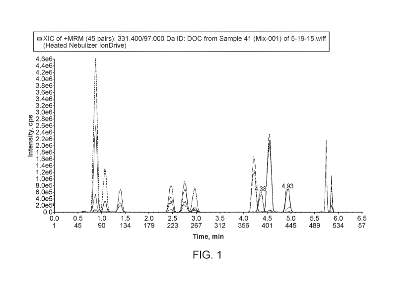
[0060] Figure 1 shows chromatogram of 14 steroids analyzed by mass
spectrometry.
[0061] Figures 2-5 show normal levels of cortisol (Figure 2), cortisone
(Figure 3), testosterone
(Figure 4), and androstenedione (Figure 5) in a normal adult male, quantitated
by the present
assay.
[0062] Figures 6-10 show normal levels of progesterone (Figure 6), cortisol
(Figure 7),
cortisone (Figure 8), androstenedione (Figure 9), 17-0H progesterone (Figure
10) in a normal
adult female, quantitated by the present assay.
16
CA 02987323 2017-11-24
WO 2016/191738 PCT/US2016/034815
[0063] Figures 11-17 show levels of cortisol (Figure 11), cortisone (Figure
12), progesterone
(Figure 13), androstenedione (Figure 14), testosterone (Figure 15), 21-
deoxycortisol (Figure 16),
and 17-0H progesterone (Figure 17) in a child, quantitated by the present
assay.
[0064] Figures 18 shows standard linearity of testosterone between 50-10,000
ng/dL.
[0065] Figure 19 shows chromatogram of tamoxifen and its metabolites.
[0066] Figure 20 shows chromatogram of letrozole, exemestane, and anastrozole.
[0067] Figure 21 shows exemplary chromatograms of opiates (oxymorphone,
hydromorphone,
and codeine) and corresponding internal standards.
[0068] Figure 22 shows exemplary chromatograms of opiates (noroxycodone,
oxycodone, and
norhydrocodone) and corresponding internal standards.
[0069] Figure 23 shows exemplary chromatograms of opiates (morphine,
hydrocodone, and
norfentanyl) and corresponding internal standards.
[0070] Figure 24 shows exemplary chromatogram of opiate (fentanyl) and
corresponding
internal standard.
[0071] Figures 25 to 28 show morphine, codeine, hydromorphone, and oxycodone
(respectively) data obtained from patient urine using 20 uL MITRA tip with
glucuronidase
hydrolysis.
[0072] Figure 29 shows oxycodone data obtained from patient saliva using 50 uL
MITRA tip.
[0073] Figures 30 and 31 show the results of hematocrit study of buprenorphine
and
norfentanyl, respectively.
[0074] Figures 32 and 33 show the results of negative urine spiked with
barbiturates
(secobarbital, ammobarbital, pentobarbital, and thiopental).
[0075] Figures 34 to 38 show the results of various patient samples
quantitated for
phenobarbital and butalbital.
[0076] Figure 39 shows the results of THC carboxy metabolite analysis in
patient sample using
20 uL tip and glucuronidase hydrolysis.
[0077] Figure 40 shows the results of hematocrit study of gabapentin and
rufinamide.
[0078] Figure 41 shows the chromatogram of the 25-hydroxyvitamin D analysis.
[0079] Figure 42 shows the calibration curve of 25-hydroxyvitamin D2 analysis.
[0080] Figure 43 shows the calibration curve of 25-hydroxyvitamin D3 analysis.
17
CA 02987323 2017-11-24
WO 2016/191738 PCT/US2016/034815
DETAILED DESCRIPTION OF THE INVENTION
[0081] As used herein, unless otherwise stated, the singular forms "a," "an,"
and "the" include
plural reference. Thus, for example, a reference to "a protein" includes a
plurality of protein
molecules.
[0082] As used herein, the terms "purification", "purifying", and "enriching"
do not refer to
removing all materials from the sample other than the analyte(s) of interest.
Instead, these terms
refer to a procedure that enriches the amount of one or more analytes of
interest relative to other
components in the sample that may interfere with detection of the analyte of
interest.
Purification of the sample by various means may allow relative reduction of
one or more
interfering substances, e.g., one or more substances that may or may not
interfere with the
detection of selected parent or daughter ions by mass spectrometry. Relative
reduction as this
term is used does not require that any substance, present with the analyte of
interest in the
material to be purified, is entirely removed by purification.
[0083] As used herein, the term "immunopurification" or "immunopurify" refers
to a
purification procedure that utilizes antibodies, including polyclonal or
monoclonal antibodies, to
enrich the one or more analytes of interest. Immunopurification can be
performed using any of
the immunopurification methods well known in the art. Often the
immunopurification
procedure utilizes antibodies bound, conjugated or otherwise attached to a
solid support, for
example a column, well, tube, gel, capsule, particle or the like.
Immunopurification as used
herein includes without limitation procedures often referred to in the art as
immunoprecipitation,
as well as procedures often referred to in the art as affinity chromatography
or immunoaffinity
chromatography.
[0084] As used herein, the term "immunoparticle" refers to a capsule, bead,
gel particle or the
like that has antibodies bound, conjugated or otherwise attached to its
surface (either on and/or
in the particle). In certain preferred embodiments, immunoparticles are
sepharose or agarose
beads. In alternative preferred embodiments, immunoparticles comprise glass,
plastic or silica
beads, or silica gel.
[0085] As used herein, the term "sample" refers to any sample that may contain
an analyte of
interest. As used herein, the term "body fluid" means any fluid that can be
isolated from the
body of an individual. For example, "body fluid" may include blood, plasma,
serum, bile,
saliva, urine, tears, perspiration, and the like. In preferred embodiments,
the sample comprises a
body fluid sample from human; preferably plasma or serum.
18
CA 02987323 2017-11-24
WO 2016/191738 PCT/US2016/034815
[0086] As used herein, the term "solid phase extraction" or "SPE" refers to a
process in which
a chemical mixture is separated into components as a result of the affinity of
components
dissolved or suspended in a solution (i.e., mobile phase) for a solid through
or around which the
solution is passed (i.e., solid phase). In some instances, as the mobile phase
passes through or
around the solid phase, undesired components of the mobile phase may be
retained by the solid
phase resulting in a purification of the analyte in the mobile phase. In other
instances, the
analyte may be retained by the solid phase, allowing undesired components of
the mobile phase
to pass through or around the solid phase. In these instances, a second mobile
phase is then used
to elute the retained analyte off of the solid phase for further processing or
analysis. SPE,
including TFLC, may operate via a unitary or mixed mode mechanism. Mixed mode
mechanisms utilize ion exchange and hydrophobic retention in the same column;
for example,
the solid phase of a mixed-mode SPE column may exhibit strong anion exchange
and
hydrophobic retention; or may exhibit strong cation exchange and hydrophobic
retention.
[0087] Generally, the affinity of a SPE column packing material for an analyte
may be due to
any of a variety of mechanisms, such as one or more chemical interactions or
an immunoaffinity
interaction. In some embodiments, SPE of analyte is conducted without the use
of an
immunoaffinity column packing material. That is, in some embodiments, analyte
is purified
from a sample by a SPE column that is not an immunoaffinity column.
[0088] As used herein, the term "chromatography" refers to a process in which
a chemical
mixture carried by a liquid or gas is separated into components as a result of
differential
distribution of the chemical entities as they flow around or over a stationary
liquid or solid
phase.
[0089] As used herein, the term "liquid chromatography" or "LC" means a
process of selective
retardation of one or more components of a fluid solution as the fluid
uniformly percolates
through a column of a finely divided substance, or through capillary
passageways. The
retardation results from the distribution of the components of the mixture
between one or more
stationary phases and the bulk fluid, (i.e., mobile phase), as this fluid
moves relative to the
stationary phase(s). Examples of "liquid chromatography" include reverse phase
liquid
chromatography (RPLC), high performance liquid chromatography (HPLC), and
turbulent flow
liquid chromatography (TFLC) (sometimes known as high turbulence liquid
chromatography
(HTLC) or high throughput liquid chromatography).
[0090] As used herein, the term "high performance liquid chromatography" or
"HPLC"
(sometimes known as "high pressure liquid chromatography") refers to liquid
chromatography
19
CA 02987323 2017-11-24
WO 2016/191738 PCT/US2016/034815
in which the degree of separation is increased by forcing the mobile phase
under pressure
through a stationary phase, typically a densely packed column.
[0091] As used herein, the term "turbulent flow liquid chromatography" or
"TFLC"
(sometimes known as high turbulence liquid chromatography or high throughput
liquid
chromatography) refers to a form of chromatography that utilizes turbulent
flow of the material
being assayed through the column packing as the basis for performing the
separation. TFLC has
been applied in the preparation of samples containing two unnamed drugs prior
to analysis by
mass spectrometry. See, e.g., Zimmer et al., J Chromatogr A 854: 23-35 (1999);
see also, U.S.
Patents No. 5,968,367, 5,919,368, 5,795,469, and 5,772,874, which further
explain TFLC.
Persons of ordinary skill in the art understand "turbulent flow". When fluid
flows slowly and
smoothly, the flow is called "laminar flow". For example, fluid moving through
an HPLC
column at low flow rates is laminar. In laminar flow the motion of the
particles of fluid is
orderly with particles moving generally in substantially straight lines. At
faster velocities, the
inertia of the water overcomes fluid frictional forces and turbulent flow
results. Fluid not in
contact with the irregular boundary "outruns" that which is slowed by friction
or deflected by an
uneven surface. When a fluid is flowing turbulently, it flows in eddies and
whirls (or vortices),
with more "drag" than when the flow is laminar. Many references are available
for assisting in
determining when fluid flow is laminar or turbulent (e.g., Turbulent Flow
Analysis:
Measurement and Prediction, P.S. Bernard & J.M. Wallace, John Wiley & Sons,
Inc., (2000);
An Introduction to Turbulent Flow, Jean Mathieu & Julian Scott, Cambridge
University Press
(2001)).
[0092] As used herein, the term "gas chromatography" or "GC" refers to
chromatography in
which the sample mixture is vaporized and injected into a stream of carrier
gas (as nitrogen or
helium) moving through a column containing a stationary phase composed of a
liquid or a
particulate solid and is separated into its component compounds according to
the affinity of the
compounds for the stationary phase.
[0093] As used herein, the term "large particle column" or "extraction column"
refers to a
chromatography column containing an average particle diameter greater than
about 50 [tm. As
used in this context, the term "about" means 10%.
[0094] As used herein, the term "analytical column" refers to a chromatography
column having
sufficient chromatographic plates to effect a separation of materials in a
sample that elute from
the column sufficient to allow a determination of the presence or amount of an
analyte. Such
columns are often distinguished from "extraction columns", which have the
general purpose of
separating or extracting retained material from non-retained materials in
order to obtain a
CA 02987323 2017-11-24
WO 2016/191738 PCT/US2016/034815
purified sample for further analysis. As used in this context, the term
"about" means 10%. In
a preferred embodiment the analytical column contains particles of about 5 [tm
in diameter.
[0095] As used herein, the terms "on-line" and "inline", for example as used
in "on-line
automated fashion" or "on-line extraction", refers to a procedure performed
without the need for
operator intervention. In contrast, the term "off-line" as used herein refers
to a procedure
requiring manual intervention of an operator. Thus, if samples are subjected
to precipitation and
the supernatants are then manually loaded into an autosampler, the
precipitation and loading
steps are off-line from the subsequent steps. In various embodiments of the
methods, one or
more steps may be performed in an on-line automated fashion.
[0096] As used herein, the term "mass spectrometry" or "MS" refers to an
analytical technique
to identify compounds by their mass. MS refers to methods of filtering,
detecting, and
measuring ions based on their mass-to-charge ratio, or "m/z". MS technology
generally includes
(1) ionizing the compounds to form charged compounds; and (2) detecting the
molecular weight
of the charged compounds and calculating a mass-to-charge ratio. The compounds
may be
ionized and detected by any suitable means. A "mass spectrometer" generally
includes an
ionizer, a mass analyzer, and an ion detector. In general, one or more
molecules of interest are
ionized, and the ions are subsequently introduced into a mass spectrometric
instrument where,
due to a combination of magnetic and electric fields, the ions follow a path
in space that is
dependent upon mass ("m") and charge ("z"). See, e.g., U.S. Patent Nos.
6,204,500, entitled
"Mass Spectrometry From Surfaces;" 6,107,623, entitled "Methods and Apparatus
for Tandem
Mass Spectrometry;" 6,268,144, entitled "DNA Diagnostics Based On Mass
Spectrometry;"
6,124,137, entitled "Surface-Enhanced Photolabile Attachment And Release For
Desorption
And Detection Of Analytes;" Wright et at., Prostate Cancer and Prostatic
Diseases 1999, 2:
264-76; and Merchant and Weinberger, Electrophoresis 2000, 21: 1164-67.
[0097] As used herein, "high resolution / high accuracy mass spectrometry"
refers to mass
spectrometry conducted with a mass analyzer capable of measuring the mass to
charge ratio of a
charged species with sufficient precision and accuracy to confirm a unique
chemical ion.
Confirmation of a unique chemical ion is possible for an ion when individual
isotopic peaks
from that ion are readily discernable. The particular resolving power and mass
accuracy
necessary to confirm a unique chemical ion varies with the mass and charge
state of the ion.
[0098] As used herein, the term "resolving power" or "resolving power (FWHM)"
(also known
in the art as "m/Am500/0") refers to an observed mass to charge ratio divided
by the width of the
mass peak at 50% maximum height (Full Width Half Maximum, "FWHM"). The effect
of
differences in resolving power is illustrated in Figures 1A-C, which show
theoretical mass
21
CA 02987323 2017-11-24
WO 2016/191738 PCT/US2016/034815
spectra of an ion with a m/z of about 1093. Figure 1A shows a theoretical mass
spectrum from a
mass analyzer with resolving power of about 3000 (a typical operating
condition for a
conventional quadrupole mass analyzer). As seen in Figure 1A, no individual
isotopic peaks are
discernable. By comparison, Figure 1B shows a theoretical mass spectrum from a
mass analyzer
with resolving power of about 10,000, with clearly discernable individual
isotopic peaks. Figure
1C shows a theoretical mass spectrum from a mass analyzer with resolving power
of about
12,000. At this highest resolving power, the individual isotopic peaks contain
less than 1%
contribution from baseline.
[0099] As used herein a "unique chemical ion" with respect to mass
spectrometry refers a
single ion with a single atomic makeup. The single ion may be singly or
multiply charged.
[00100] As used herein, the term "accuracy" (or "mass accuracy") with respect
to mass
spectrometry refers to potential deviation of the instrument response from the
true m/z of the ion
investigated. Accuracy is typically expressed in parts per million (ppm). The
effect of
differences in mass accuracy is illustrated in Figures 2A-D, which show the
boundaries of
potential differences between a detected m/z and the actual m/z for a
theoretical peak at m/z of
1093.52094. Figure 2A shows the potential range of detected m/z at an accuracy
of 120 ppm.
By contrast, Figure 2B shows the potential range of detected m/z at an
accuracy of 50 ppm.
Figures 2C and 2D show the even narrower potential ranges of detected m/z at
accuracies of 20
ppm and 10 ppm.
[00101] High resolution / high accuracy mass spectrometry methods of the
present invention
may be conducted on instruments capable of performing mass analysis with FWHM
of greater
than 10,000, 15,000, 20,000, 25,000, 50,000, 100,000, or even more. Likewise,
methods of the
present invention may be conducted on instruments capable of performing mass
analysis with
accuracy of less than 50 ppm, 20 ppm, 15 ppm, 10 ppm, 5 ppm, 3 ppm, or even
less.
Instruments capable of these performance characteristics may incorporate
certain orbitrap mass
analyzers, time-of-flight ("TOF") mass analyzers, or Fourier-transform ion
cyclotron resonance
mass analyzers. In preferred embodiments, the methods are carried out with an
instrument
which includes an orbitrap mass analyzer or a TOF mass analyzer.
[00102] The term "orbitrap" describes an ion trap consisting of an outer
barrel-like electrode and
a coaxial inner electrode. Ions are injected tangentially into the electric
field between the
electrodes and trapped because electrostatic interactions between the ions and
electrodes are
balanced by centrifugal forces as the ions orbit the coaxial inner electrode.
As an ion orbits the
coaxial inner electrode, the orbital path of a trapped ion oscillates along
the axis of the central
electrode at a harmonic frequency relative to the mass to charge ratio of the
ion. Detection of
22
CA 02987323 2017-11-24
WO 2016/191738 PCT/US2016/034815
the orbital oscillation frequency allows the orbitrap to be used as a mass
analyzer with high
accuracy (as low as 1 ¨ 2 ppm) and high resolving power (FWHM) (up to about
200,000). A
mass analyzer based on an orbitrap is described in detail in U.S. Pat. No.
6,995,364,
incorporated by reference herein in its entirety. Use of orbitrap analyzers
has been reported for
qualitative and quantitative analyses of various analytes. See, e.g., U.S.
Patent Application Pub.
No. 2008/0118932 (filed Nov. 9,2007); Bredehoft, et al., Rapid Commun. Mass
Spectrom.,
2008, 22:477-485; Le Breton, et al., Rapid Commun. Mass Spectrom., 2008,
22:3130-36;
Thevis, et al., Mass Spectrom. Reviews, 2008, 27:35-50; Thomas, et al., J.
Mass Spectrom.,
2008, 43:908-15; Schenk, et al., BMC Medical Genomics, 2008, 1:41; and Olsen,
et al., Nature
Methods, 2007, 4:709-12.
[00103] As used herein, the term "operating in negative ion mode" refers to
those mass
spectrometry methods where negative ions are generated and detected. The term
"operating in
positive ion mode" as used herein, refers to those mass spectrometry methods
where positive
ions are generated and detected. In preferred embodiments, mass spectrometry
is conducted in
positive ion mode.
[00104] As used herein, the term "ionization" or "ionizing" refers to the
process of generating an
analyte ion having a net electrical charge equal to one or more electron
units. Negative ions are
those having a net negative charge of one or more electron units, while
positive ions are those
having a net positive charge of one or more electron units.
[00105] As used herein, the term "electron ionization" or "El" refers to
methods in which an
analyte of interest in a gaseous or vapor phase interacts with a flow of
electrons. Impact of the
electrons with the analyte produces analyte ions, which may then be subjected
to a mass
spectrometry technique.
[00106] As used herein, the term "chemical ionization" or "CI" refers to
methods in which a
reagent gas (e.g. ammonia) is subjected to electron impact, and analyte ions
are formed by the
interaction of reagent gas ions and analyte molecules.
[00107] As used herein, the term "fast atom bombardment" or "FAB" refers to
methods in
which a beam of high energy atoms (often Xe or Ar) impacts a non-volatile
sample, desorbing
and ionizing molecules contained in the sample. Test samples are dissolved in
a viscous liquid
matrix such as glycerol, thioglycerol, m-nitrobenzyl alcohol, 18-crown-6 crown
ether, 2-
nitrophenyloctyl ether, sulfolane, diethanolamine, and triethanolamine. The
choice of an
appropriate matrix for a compound or sample is an empirical process.
23
CA 02987323 2017-11-24
WO 2016/191738 PCT/US2016/034815
[00108] As used herein, the term "matrix-assisted laser desorption ionization"
or "MALDI"
refers to methods in which a non-volatile sample is exposed to laser
irradiation, which desorbs
and ionizes analytes in the sample by various ionization pathways, including
photo-ionization,
protonation, deprotonation, and cluster decay. For MALDI, the sample is mixed
with an energy-
absorbing matrix, which facilitates desorption of analyte molecules.
[00109] As used herein, the term "surface enhanced laser desorption
ionization" or "SELDI"
refers to another method in which a non-volatile sample is exposed to laser
irradiation, which
desorbs and ionizes analytes in the sample by various ionization pathways,
including photo-
ionization, protonation, deprotonation, and cluster decay. For SELDI, the
sample is typically
bound to a surface that preferentially retains one or more analytes of
interest. As in MALDI,
this process may also employ an energy-absorbing material to facilitate
ionization.
[00110] As used herein, the term "electrospray ionization" or "ESI," refers to
methods in which
a solution is passed along a short length of capillary tube, to the end of
which is applied a high
positive or negative electric potential. Solution reaching the end of the tube
is vaporized
(nebulized) into a jet or spray of very small droplets of solution in solvent
vapor. This mist of
droplets flows through an evaporation chamber. As the droplets get smaller the
electrical
surface charge density increases until such time that the natural repulsion
between like charges
causes ions as well as neutral molecules to be released.
[00111] As used herein, the term "atmospheric pressure chemical ionization" or
"APCI," refers
to mass spectrometry methods that are similar to ESI; however, APCI produces
ions by ion-
molecule reactions that occur within a plasma at atmospheric pressure. The
plasma is
maintained by an electric discharge between the spray capillary and a counter
electrode. Then
ions are typically extracted into the mass analyzer by use of a set of
differentially pumped
skimmer stages. A counterflow of dry and preheated N2 gas may be used to
improve removal of
solvent. The gas-phase ionization in APCI can be more effective than ESI for
analyzing less-
polar species.
[00112] The term "atmospheric pressure photoionization" or "APPI" as used
herein refers to the
form of mass spectrometry where the mechanism for the ionization of molecule M
is photon
absorption and electron ejection to form the molecular ion M+. Because the
photon energy
typically is just above the ionization potential, the molecular ion is less
susceptible to
dissociation. In many cases it may be possible to analyze samples without the
need for
chromatography, thus saving significant time and expense. In the presence of
water vapor or
protic solvents, the molecular ion can extract H to form MH+. This tends to
occur if M has a
high proton affinity. This does not affect quantitation accuracy because the
sum of M+ and
24
CA 02987323 2017-11-24
WO 2016/191738 PCT/US2016/034815
MH+ is constant. Drug compounds in protic solvents are usually observed as
MH+, whereas
nonpolar compounds such as naphthalene or testosterone usually form M+. See,
e.g., Robb et
al., Anal. Chem. 2000, 72(15): 3653-3659.
[00113] As used herein, the term "inductively coupled plasma" or "ICP" refers
to methods in
which a sample interacts with a partially ionized gas at a sufficiently high
temperature such that
most elements are atomized and ionized.
[00114] As used herein, the term "field desorption" refers to methods in which
a non-volatile
test sample is placed on an ionization surface, and an intense electric field
is used to generate
analyte ions.
[00115] As used herein, the term "desorption" refers to the removal of an
analyte from a surface
and/or the entry of an analyte into a gaseous phase. Laser desorption thermal
desorption is a
technique wherein a sample containing the analyte is thermally desorbed into
the gas phase by a
laser pulse. The laser hits the back of a specially made 96-well plate with a
metal base. The laser
pulse heats the base and the heat causes the sample to transfer into the gas
phase. The gas phase
sample is then drawn into the mass spectrometer.
[00116] As used herein, the term "selective ion monitoring" is a detection
mode for a mass
spectrometric instrument in which only ions within a relatively narrow mass
range, typically
about one mass unit, are detected.
[00117] As used herein, "multiple reaction mode," sometimes known as "selected
reaction
monitoring," is a detection mode for a mass spectrometric instrument in which
a precursor ion
and one or more fragment ions are selectively detected.
[00118] As used herein, the term "lower limit of quantification", "lower limit
of quantitation" or
"LLOQ" refers to the point where measurements become quantitatively
meaningful. The
analyte response at this LOQ is identifiable, discrete and reproducible with a
relative standard
deviation (RSD %) of less than 20% and an accuracy of 85% to 115%.
[00119] As used herein, the term "limit of detection" or "LOD" is the point at
which the
measured value is larger than the uncertainty associated with it. The LOD is
the point at which a
value is beyond the uncertainty associated with its measurement and is defined
as three times the
RSD of the mean at the zero concentration.
[00120] As used herein, an "amount" of an analyte in a body fluid sample
refers generally to an
absolute value reflecting the mass of the analyte detectable in volume of
sample. However, an
amount also contemplates a relative amount in comparison to another analyte
amount. For
CA 02987323 2017-11-24
WO 2016/191738 PCT/US2016/034815
example, an amount of an analyte in a sample can be an amount which is greater
than a control
or normal level of the analyte normally present in the sample.
[00121] The term "about" as used herein in reference to quantitative
measurements not including
the measurement of the mass of an ion, refers to the indicated value plus or
minus 10%. Mass
spectrometry instruments can vary slightly in determining the mass of a given
analyte. The term
"about" in the context of the mass of an ion or the mass/charge ratio of an
ion refers to +/- 0.50
atomic mass unit.
[00122] Collection of venous blood from newborn can be problematic. Although
the minimum
serum volume needed for the comprehensive steroid panel (or CAH panel) is
minimal, at least 1-
2 mL of whole blood is acquired through venipuncture. Using a microsampling
device (Mitra
tip) requires only 20 uL of capillary blood and makes it easier and less
invasive, especially for
neonates, which eliminates the need to do venipuncture.
[00123] In one aspect, provided herein are methods for mass spectrometric
quantitation of
analytes collected and extracted from a microsampling device.
[00124] In certain embodiments, the methods provided herein are directed to
quantitating the
amount of an analyte in a sample comprising (a) extracting an analyte from a
sample collected
by a microsampling device; (b) ionizing the analyte to generate one or more
ions detectable by
mass spectrometry; and (c) determining the amount of the one or more ions by
mass
spectrometry. In some embodiments, the amount of the one or more ions
determined is used to
determine the amount of analyte in the sample. In some embodiments, the amount
of analyte in
the sample is related to the amount of analyte in the patient.
[00125] In some embodiments, the methods provided herein comprise purifying
the samples
prior to mass spectrometry. In some embodiments, the methods comprise
purifying the samples
using liquid chromatography. In some embodiments, liquid chromatrography
comprise high
performance liquid chromatography (HPLC) or high turbulence liquid
chromatograph (HTLC).
In some embodiments, the methods comprise subjecting a sample to solid phase
extraction
(SPE). In some embodiments, the methods comprise subjecting a sample to
reverse phase
analytical column.
[00126] In some embodiments, the methods provided herein are directed to
quantitating the
amount of an analyte in a sample comprising (a) extracting an analyte from a
sample collected
by a microsampling device, (b) purifying the sample by liquid chromatography,
(c) ionizing the
analyte to generate one or more ions detectable by mass spectrometry; and (d)
determining the
amount of the one or more ions by mass spectrometry. In some embodiments, the
amount of the
26
CA 02987323 2017-11-24
WO 2016/191738 PCT/US2016/034815
one or more ions determined is used to determine the amount of analyte in the
sample. In some
embodiments, the amount of analyte in the sample is related to the amount of
analyte in the
patient.
[00127] In some embodiments, mass spectrometry comprises tandem mass
spectrometry. In
some embodiments, mass spectrometry is high resolution mass spectrometry. In
some
embodiments, mass spectrometry is high resolution/high accuracy mass
spectrometry.
[00128] In some embodiments, ionization is by atmospheric pressure chemical
ionization
(APCI). In some embodiments, ionization is by electrospray ionization (ESI).
In some
embodiments, said ionization is in positive ion mode. In some embodiments,
said ionization is
in negative ion mode.
[00129] In some embodiments, the microsampling device containing the sample is
placed in a
96-well plate. In some embodiments, the microsampling device containing the
sample is placed
in a 96-rack. In some embodiments, automation places the 96-rack into a 96-
well plate. In
some embodiments, the automation is HAMILTON automation.
[00130] In some embodiments, the methods provided herein comprise adding
internal standards
to the sample. In some embodiments, the internal standard is labeled. In some
embodiments,
the internal standard is deuterated or isotopically labeled. In some
embodiments, the internal
standard is added with extraction buffer. In some embodiments, the
microsampling device is
pre-soaked with internal standard and dried.
[00131] In some embodiments, the extracting step comprises adding an
extraction buffer to the
sample collected by a microsampling device. In some embodiments, the
extracting step
comprises placing the microsampling device containing the sample into a 96-
well plate
containing an extraction solvent. In some embodiments, the extraction step is
automated. In
some embodiments, 96-well plate is vortexed and then the absorbent tips of the
microsampling
device are removed. In some embodiments, the extracting step comprises drying
down under
nitrogen. In some embodiments, the extracting step comprises reconstituting
the sample into
solution. In some embodiments, the reconstitution comprises adding aqueous
acid or organic
solution or both to the sample. In some embodiments, the reconstituted
solution is filtered.
[00132] In some embodiments, the extracted sample is injected into a mass
spectrometric
system. In some embodiments, the extracted sample is injected into liquid
chromatography. In
some embodiments, the extraction and mass spectrometry steps are performed in
an on-line
fashion to allow for automated sample analysis. In some embodiments, the
extraction,
27
CA 02987323 2017-11-24
WO 2016/191738 PCT/US2016/034815
purification, and mass spectrometry steps are performed in an on-line fashion
to allow for
automated sample analysis.
[00133] In some embodiments, the analyte is underivatized.
[00134] In some embodiments, the sample collected by the microsampling device
does not
require sample processing.
[00135] In some embodiments, the sample collected by the microsampling device
is whole
blood. In some embodiments, the sample collected by the microsampling device
is urine. In
some embodiments, the sample collected by the microsampling device is saliva.
In some
embodiments, the sample collected by the microsampling device is serum or
plasma.
[00136] In some embodiments, the microsampling device comprises an absorbent
tip that
collects the sample. In some embodiments, the sample collected by the
microsampling device
absorbs a fixed volume of patient fluids. In some embodiments, the sample
collected by the
microsampling device has a volume of less than or equal to 150 L. In some
embodiments, the
sample collected by the microsampling device has a volume of less than or
equal to 100 L. In
some embodiments, the sample collected by the microsampling device has a
volume of less than
or equal to 50 L. In some embodiments, the sample collected by the
microsampling device has
a volume of between 5 [tL and 150 [tL, inclusive. In some embodiments, the
sample collected
by the microsampling device has a volume of between 10 [tL and 100 [tL,
inclusive. In some
embodiments, the sample collected by the microsampling device has a volume of
about 10 L.
In some embodiments, the sample collected by the microsampling device has a
volume of about
15 L. In some embodiments, the sample collected by the microsampling device
has a volume
of about 20 L. In some embodiments, the sample collected by the microsampling
device has a
volume of about 30 L. In some embodiments, the sample collected by the
microsampling
device has a volume of about 50 L. In some embodiments, the sample collected
by the
microsampling device has a volume of about 100 L. In some embodiments, the
sample
collected by the microsampling device absorbs a fixed volume of blood,
regardless of the
amount of hematocrit.
[00137] In some embodiments, the methods provided herein are directed to
quantitating the
amount of an analyte in a sample comprising (a) extracting an analyte from a
sample of less than
or equal to 100 L; (b) ionizing the analyte to generate one or more ions
detectable by mass
spectrometry; and (c) determining the amount of the one or more ions by mass
spectrometry. In
some embodiments, the amount of the one or more ions determined is used to
determine the
28
CA 02987323 2017-11-24
WO 2016/191738 PCT/US2016/034815
amount of analyte in the sample. In some embodiments, the amount of analyte in
the sample is
related to the amount of analyte in the patient.
[00138] In some embodiments, the methods provided herein are directed to
quantitating the
amount of an analyte in a sample comprising (a) extracting an analyte from a
sample of less than
or equal to 100 [tL; (b) purifying the sample by liquid chromatography; (c)
ionizing the analyte
to generate one or more ions detectable by mass spectrometry; and (d)
determining the amount
of the one or more ions by mass spectrometry. In some embodiments, the amount
of the one or
more ions determined is used to determine the amount of analyte in the sample.
In some
embodiments, the amount of analyte in the sample is related to the amount of
analyte in the
patient.
[00139] In some embodiments, the methods comprise extracting an analyte from a
sample of
less than or equal to 50 L. In some embodiments, the methods comprise
extracting an analyte
from a sample of less than or equal to 30 L. In some embodiments, the methods
comprise
extracting an analyte from a sample of less than or equal to 20 L. In some
embodiments, the
methods comprise extracting an analyte from a sample of less than or equal to
15 L. In some
embodiments, the methods comprise extracting an analyte from a sample of less
than or equal to
L.
[00140] In some embodiments, the sample collected by the microsampling device
can be
transported without refrigeration or freezing. In some embodiments, the sample
collected by the
microsampling device can be transported without dry ice. In some embodiments,
the sample
collected by the microsampling device can be transported at room temperature.
In some
embodiments, the sample collected by the microsampling device can be
transported without
biohazard concerns.
[00141] In some embodiments, the sample collected by the microsampling device
requires little
training for collection. In some embodiments, the sample collected by the
microsampling device
can be collected anywhere. In some embodiments, the sample collected by the
microsampling
device can be dried at ambient temperature for shipping.
[00142] In some embodiments, the microsampling device is a MITRA tip. In some
embodiments, the microsampling device is encased in a cartridge designed for
automation of
extraction and mass spectrometric analysis.
[00143] In some embodiments, the methods further comprise collecting the
sample with a
microsampling device. In some embodiments, the collecting step comprises
performing a finger
prick and applying an absorbent tip of the microsampling device to the blood.
In some
29
CA 02987323 2017-11-24
WO 2016/191738 PCT/US2016/034815
embodiments, the collecting step comprises applying an absorbent tip in the
urine or saliva of
the patient. In some embodiments, the sample collected in the microsampling
device is air
dried. In some embodiments, the sample collected in the microsampling device
is air dried for 1
to 2 hours prior to transport.
[00144] In some embodiments, the analyte is a steroid. In some embodiments,
the steroid is
cortisol, cortisone, progesterone, 17-hydroxyprogesterone, androstenedione,
testosterone,
dehydroepiandrosterone, corticosterone, deoxycorticosterone, 11-deoxycortisol,
pregnenolone,
17-hydroxypregnenolone, 18-hydroxycorticosterone, or 21-deoxycortisol. In some
embodiments, the analyte is a steroid in a steroid panel for diagnosing
congenital adrenal
hyperplasia (CAH). In some embodiments, the steroid is selected from the group
consisting of
cortisol, cortisone, progesterone, 17-hydroxyprogesterone, androstenedione,
testosterone,
dehydroepiandrosterone, corticosterone, deoxycorticosterone, 11-deoxycortisol,
pregnenolone,
17-hydroxypregnenolone, 18-hydroxycorticosterone, and 21-deoxycortisol. In
some
embodiments, the steroid is 25-hydroxyvitamin D2 or 25-hydroxyvitamin D3.
[00145] In some embodiments, the analyte is an opiate. In some embodiments,
the opiate is cis-
tramadol, 0-desmethyl tramadol, tapentadol, N-desmethyltapentadol, codeine,
morphine,
oxymorphone, norhydrocodone, oxycodone, noroxycodone, hydromorphone,
hydrocodone,
buprenorphine, norbuprenorphine, fentanyl, norfentanyl, 6-monoacetylmorphine
(6-MAM),
methadone, dihydrocodeine, naloxone, naltrexone, 60-naltrexol, nalorphine,
nalbuphine, or 2-
ethylidene-1,5-dimethy1-3,3-diphenylpyrrolidine (EDDP). In some embodiments,
the opiate is
selected from the group consisting of cis-tramadol, 0-desmethyl tramadol,
tapentadol, N-
desmethyltapentadol, codeine, morphine, oxymorphone, norhydrocodone,
oxycodone,
noroxycodone, hydromorphone, hydrocodone, buprenorphine, norbuprenorphine,
fentanyl,
norfentanyl, 6-monoacetylmorphine (6-MAM), methadone, dihydrocodeine,
naloxone,
naltrexone, 60-naltrexol, nalorphine, nalbuphine, and 2-ethylidene-1,5-
dimethy1-3,3-
diphenylpyrrolidine (EDDP). In some embodiments, the opiate is extracted from
a whole blood,
salive, or urine sample.
[00146] In some embodiments, the analyte is a benzodiazepine. In some
embodiments, the
benzodiazepine is oxazepam, temazepam, lorazepam, nordiazepam, diazepam,
chlordiazepoxide,
triazolam, midazolam, alprazolam, clonazepam, bromazepam, clobazam,
nitrazepam,
phenazepam, prazepam, medazepam, flunitrazepam, or flurazepam. In some
embodiments, the
benzodiazepine is selected from the group consisting of oxazepam, temazepam,
lorazepam,
nordiazepam, diazepam, chlordiazepoxide, triazolam, midazolam, alprazolam,
clonazepam,
bromazepam, clobazam, nitrazepam, phenazepam, prazepam, medazepam,
flunitrazepam, and
CA 02987323 2017-11-24
WO 2016/191738 PCT/US2016/034815
flurazepam. In some embodiments, the benzodiazepine is extracted from a whole
blood or urine
sample.
[00147] In some embodiments, one or more ions comprise a bromazepam precursor
ion with a
mass to charge ratio (m/z) of 316 0.5. In some embodiments, one or more ions
comprise one
or more fragment ions with a mass to charge ratio (m/z) of 214 0.5 or 270
0.5. In some
embodiments, one or more ions comprise an oxazepam precursor ion with a mass
to charge ratio
(m/z) of 287 0.5. In some embodiments, one or more ions comprise one or more
fragment
ions with a mass to charge ratio (m/z) of 104 0.5 or 241 0.5. In some
embodiments, one or
more ions comprise an clobazam precursor ion with a mass to charge ratio (m/z)
of 300 0.5.
In some embodiments, one or more ions comprise one or more fragment ions with
a mass to
charge ratio (m/z) of 224 0.5 or 259 0.5. In some embodiments, one or more
ions comprise
a nitrazepam precursor ion with a mass to charge ratio (m/z) of 282 0.5. In
some
embodiments, one or more ions comprise one or more fragment ions with a mass
to charge ratio
(m/z) of 180 0.5 or 236 0.5. In some embodiments, one or more ions
comprise an
alprazolam precursor ion with a mass to charge ratio (m/z) of 309.1 0.5. In
some
embodiments, one or more ions comprise one or more fragment ions with a mass
to charge ratio
(m/z) of 165 0.5 or 280.9 0.5. In some embodiments, one or more ions
comprise an
triazolam precursor ion with a mass to charge ratio (m/z) of 343 0.5. In
some embodiments,
one or more ions comprise one or more fragment ions with a mass to charge
ratio (m/z) of 206
0.5 or 308 0.5. In some embodiments, one or more ions comprise a clonazepam
precursor ion
with a mass to charge ratio (m/z) of 316 0.5. In some embodiments, one or
more ions
comprise one or more fragment ions with a mass to charge ratio (m/z) of 214
0.5 or 270 0.5.
In some embodiments, one or more ions comprise a flurazepam precursor ion with
a mass to
charge ratio (m/z) of 388 0.5. In some embodiments, one or more ions
comprise one or more
fragment ions with a mass to charge ratio (m/z) of 287.9 0.5 or 315 0.5.
In some
embodiments, one or more ions comprise a lorazepam precursor ion with a mass
to charge ratio
(m/z) of 321 0.5. In some embodiments, one or more ions comprise one or more
fragment
ions with a mass to charge ratio (m/z) of 229.1 0.5 or 331 0.5. In some
embodiments, one or
more ions comprise a flunitrazepam precursor ion with a mass to charge ratio
(m/z) of 314 0.5.
In some embodiments, one or more ions comprise one or more fragment ions with
a mass to
charge ratio (m/z) of 211 0.5 or 268 0.5. In some embodiments, one or more
ions comprise
a temazepam precursor ion with a mass to charge ratio (m/z) of 301.1 0.5. In
some
embodiments, one or more ions comprise one or more fragment ions with a mass
to charge ratio
(m/z) of 177 0.5 or 255 0.5. In some embodiments, one or more ions
comprise a midazolam
precursor ion with a mass to charge ratio (m/z) of 326 0.5. In some
embodiments, one or more
31
CA 02987323 2017-11-24
WO 2016/191738 PCT/US2016/034815
ions comprise one or more fragment ions with a mass to charge ratio (m/z) of
129 0.5 or 244
0.5. In some embodiments, one or more ions comprise an nordiazepam precursor
ion with a
mass to charge ratio (m/z) of 271 0.5. In some embodiments, one or more ions
comprise one
or more fragment ions with a mass to charge ratio (m/z) of 139.8 0.5 or 165
0.5. In some
embodiments, one or more ions comprise an phenazepam precursor ion with a mass
to charge
ratio (m/z) of 351 0.5. In some embodiments, one or more ions comprise one
or more
fragment ions with a mass to charge ratio (m/z) of 185.9 0.5 or 206 0.5.
In some
embodiments, one or more ions comprise a chlordiazepam precursor ion with a
mass to charge
ratio (m/z) of 301 0.5. In some embodiments, one or more ions comprise one
or more
fragment ions with a mass to charge ratio (m/z) of 259 0.5 or 224 0.5. In
some
embodiments, one or more ions comprise a diazepam precursor ion with a mass to
charge ratio
(m/z) of 285 0.5. In some embodiments, one or more ions comprise one or more
fragment
ions with a mass to charge ratio (m/z) of 154 0.5 or 193 0.5. In some
embodiments, one or
more ions comprise a prazepam precursor ion with a mass to charge ratio (m/z)
of 325 0.5. In
some embodiments, one or more ions comprise one or more fragment ions with a
mass to charge
ratio (m/z) of 165 0.5 or 271 0.5. In some embodiments, one or more ions
comprise a
medazepam precursor ion with a mass to charge ratio (m/z) of 271 0.5. In
some embodiments,
one or more ions comprise one or more fragment ions with a mass to charge
ratio (m/z) of 180
0.5 or 207.1 0.5.
[00148] In some embodiments, the analyte is an anti-epileptic drug. In some
embodiments, the
anti-epileptic drug is valproic acid, tiagabine, topiramate, levitiracetum,
lamotrigine, lacosamide,
ethosuximide, carbamazepine, eslicarbamazepine, 10,11-carbamazepine,
phenobarbital,
rufinamide, primidone, phenytoin, zonisamide, felbamate, gabapentin, or
pregablin. In some
embodiments, the anti-epileptic drug is selected from the group consisting of
valproic acid,
tiagabine, topiramate, levitiracetum, lamotrigine, lacosamide, ethosuximide,
carbamazepine,
eslicarbamazepine, 10,11-carbamazepine, phenobarbital, rufinamide, primidone,
phenytoin,
zonisamide, felbamate, gabapentin, and pregablin. In some embodiments, the
anti-epileptic drug
is extracted from a whole blood sample.
[00149] In some embodiments, one or more ions comprise a felbamate precursor
ion with a mass
to charge ratio (m/z) of 339 0.5. In some embodiments, one or more ions
comprise one or
more fragment ions with a mass to charge ratio (m/z) of 117.3 0.5 or 261
0.5. In some
embodiments, one or more ions comprise an ethosuximide precursor ion with a
mass to charge
ratio (m/z) of 142 0.5. In some embodiments, one or more ions comprise one
or more
fragment ions with a mass to charge ratio (m/z) of 44.3 0.5 or 39.3 0.5.
In some
32
CA 02987323 2017-11-24
WO 2016/191738 PCT/US2016/034815
embodiments, one or more ions comprise a lacosamide precursor ion with a mass
to charge ratio
(m/z) of 251 0.5. In some embodiments, one or more ions comprise one or more
fragment
ions with a mass to charge ratio (m/z) of 91.2 0.5 or 65.2 0.5. In some
embodiments, one or
more ions comprise a lamotrigine precursor ion with a mass to charge ratio
(m/z) of 256 0.5.
In some embodiments, one or more ions comprise one or more fragment ions with
a mass to
charge ratio (m/z) of 211 0.5 or 145 0.5. In some embodiments, one or more
ions comprise
a topiramate precursor ion with a mass to charge ratio (m/z) of 338.2 0.5.
In some
embodiments, one or more ions comprise one or more fragment ions with a mass
to charge ratio
(m/z) of 78.2 0.5 or 96.2 0.5. In some embodiments, one or more ions
comprise a
gabapentin precursor ion with a mass to charge ratio (m/z) of 172.3 0.5. In
some
embodiments, one or more ions comprise one or more fragment ions with a mass
to charge ratio
(m/z) of 91.2 0.5 or 67.2 0.5. In some embodiments, one or more ions
comprise an
eslicarbazepine precursor ion with a mass to charge ratio (m/z) of 297.1
0.5. In some
embodiments, one or more ions comprise one or more fragment ions with a mass
to charge ratio
(m/z) of 194 0.5 or 179 0.5. In some embodiments, one or more ions
comprise a primidone
precursor ion with a mass to charge ratio (m/z) of 219.8 0.5. In some
embodiments, one or
more ions comprise one or more fragment ions with a mass to charge ratio (m/z)
of 79 0.5 or
135.2 0.5. In some embodiments, one or more ions comprise a pregabalin
precursor ion with a
mass to charge ratio (m/z) of 160.1 0.5. In some embodiments, one or more
ions comprise one
or more fragment ions with a mass to charge ratio (m/z) of 55.2 0.5 or 77.2
0.5. In some
embodiments, one or more ions comprise a carbamazepine precursor ion with a
mass to charge
ratio (m/z) of 237 0.5. In some embodiments, one or more ions comprise one
or more
fragment ions with a mass to charge ratio (m/z) of 194.1 0.5 or 179 0.5.
In some
embodiments, one or more ions comprise a phenobarbital precursor ion with a
mass to charge
ratio (m/z) of 231 0.5. In some embodiments, one or more ions comprise one
or more
fragment ions with a mass to charge ratio (m/z) of 44.2 0.5 or 188.1 0.5.
In some
embodiments, one or more ions comprise an epoxide precursor ion with a mass to
charge ratio
(m/z) of 236.2 0.5. In some embodiments, one or more ions comprise one or
more fragment
ions with a mass to charge ratio (m/z) of 141.2 0.5 or 112.2 0.5. In some
embodiments, one
or more ions comprise a zonisamide precursor ion with a mass to charge ratio
(m/z) of 213.2
0.5. In some embodiments, one or more ions comprise one or more fragment ions
with a mass
to charge ratio (m/z) of 77.2 0.5 or 102.1 0.5. In some embodiments, one
or more ions
comprise a tiagabine precursor ion with a mass to charge ratio (m/z) of 376.2
0.5. In some
embodiments, one or more ions comprise one or more fragment ions with a mass
to charge ratio
(m/z) of 111.1 0.5 or 149.1 0.5. In some embodiments, one or more ions
comprise a
33
CA 02987323 2017-11-24
WO 2016/191738 PCT/US2016/034815
phenytoin precursor ion with a mass to charge ratio (m/z) of 253.1 0.5. In
some embodiments,
one or more ions comprise one or more fragment ions with a mass to charge
ratio (m/z) of 104.2
0.5 or 182.2 0.5. In some embodiments, one or more ions comprise a
levetiracetam
precursor ion with a mass to charge ratio (m/z) of 171.2 0.5. In some
embodiments, one or
more ions comprise one or more fragment ions with a mass to charge ratio (m/z)
of 126.2 0.5
or 69.2 0.5. In some embodiments, one or more ions comprise a valproic acid
precursor ion
with a mass to charge ratio (m/z) of 143 0.5. In some embodiments, one or
more ions
comprise one or more fragment ions with a mass to charge ratio (m/z) of 143
0.5. In some
embodiments, one or more ions comprise a rufinamide precursor ion with a mass
to charge ratio
(m/z) of 239 0.5. In some embodiments, one or more ions comprise one or more
fragment
ions with a mass to charge ratio (m/z) of 127.2 0.5 or 261 0.5. In some
embodiments, one or
more ions comprise a primdone precursor ion with a mass to charge ratio (m/z)
of 219 0.5. In
some embodiments, one or more ions comprise one or more fragment ions with a
mass to charge
ratio (m/z) of 126 0.5 or 141 0.5. In some embodiments, one or more ions
comprise a
topiramate D12 precursor ion with a mass to charge ratio (m/z) of 350 0.5.
In some
embodiments, one or more ions comprise one or more fragment ions with a mass
to charge ratio
(m/z) of 78.2 0.5. In some embodiments, one or more ions comprise an epoxide
D3 precursor
ion with a mass to charge ratio (m/z) of 256 0.5. In some embodiments, one
or more ions
comprise one or more fragment ions with a mass to charge ratio (m/z) of 77
0.5. In some
embodiments, one or more ions comprise a lamotrigine 13C3 precursor ion with a
mass to charge
ratio (m/z) of 259 0.5. In some embodiments, one or more ions comprise one
or more
fragment ions with a mass to charge ratio (m/z) of 214 0.5. In some
embodiments, one or
more ions comprise a levetiracetam D6 precursor ion with a mass to charge
ratio (m/z) of 177.2
0.5. In some embodiments, one or more ions comprise one or more fragment ions
with a mass
to charge ratio (m/z) of 132.2 0.5.
[00150] In some embodiments, the analyte is an immunosuppressant. In some
embodiments, the
immunosuppressant is cyclosporine A, sirolimus, tacrolimus, or everolimus. In
some
embodiments, the immunosuppressant is selected from the group consisting of
cyclosporine A,
sirolimus, tacrolimus, and everolimus. In some embodiments, the
immunosuppressant is
extracted from a whole blood sample.
[00151] In some embodiments, the analyte is a barbiturate. In some
embodiments, the
barbiturate is phenobarbitol, amobarbitol, butalbital, pentobarbitol,
secobarbitol, or thiopental.
In some embodiments, the barbiturate is selected from the group consisting of
phenobarbitol,
34
CA 02987323 2017-11-24
WO 2016/191738 PCT/US2016/034815
amobarbitol, butalbital, pentobarbitol, secobarbitol, and thiopental. In some
embodiments, the
barbiturate is extracted from a whole blood sample.
[00152] In some embodiments, one or more ions comprise a secobarbital
precursor ion with a
mass to charge ratio (m/z) of 237.0 0.5. In some embodiments, one or more
ions comprise one
or more fragment ions with a mass to charge ratio (m/z) of 42.0 0.5. In some
embodiments,
one or more ions comprise an ammobarbital precursor ion with a mass to charge
ratio (m/z) of
225.0 0.5. In some embodiments, one or more ions comprise one or more
fragment ions with
a mass to charge ratio (m/z) of 182.0 0.5. In some embodiments, one or more
ions comprise a
pentobarbital precursor ion with a mass to charge ratio (m/z) of 225.6 0.5.
In some
embodiments, one or more ions comprise one or more fragment ions with a mass
to charge ratio
(m/z) of 42.0 0.5. In some embodiments, one or more ions comprise a
thiopental precursor ion
with a mass to charge ratio (m/z) of 241.0 0.5. In some embodiments, one or
more ions
comprise one or more fragment ions with a mass to charge ratio (m/z) of 57.9
0.5. In some
embodiments, one or more ions comprise a phenobarbital precursor ion with a
mass to charge
ratio (m/z) of 231.0 0.5. In some embodiments, one or more ions comprise one
or more
fragment ions with a mass to charge ratio (m/z) of 42.0 0.5. In some
embodiments, one or
more ions comprise a butalbital precursor ion with a mass to charge ratio
(m/z) of 223.1 0.5.
In some embodiments, one or more ions comprise one or more fragment ions with
a mass to
charge ratio (m/z) of 42.1 0.5.
[00153] In some embodiments, the analyte is tamoxifen. In some embodiments,
the analyte is a
metabolite of tamoxifen. In some embodiments, said metabolite is norendoxifen.
In some
embodiments, said metabolite is endoxifen or N-Desmethy1-4-Hydroxy Tamoxifen.
In some
embodiments, said metabolite is 4'-Hydroxy Tamoxifen. In some embodiments,
said metabolite
is 4-Hydroxy Tamoxifen. In some embodiments, said metabolite is N-Desmethy1-4'-
Hydroxy
Tamoxifen. In some embodiments, said metabolite is N-Desmethyl Tamoxifen. In
some
embodiments, said metabolite is selected from the group consisting of
norendoxifen, endoxifen,
4'-Hydroxy Tamoxifen, 4-Hydroxy Tamoxifen, N-Desmethy1-4'-Hydroxy Tamoxifen,
and N-
Desmethy1-4'-Hydroxy Tamoxifen. In some embodiments, tamoxifen or its
metabolite is
extracted from a whole blood sample.
[00154] In some embodiments, one or more ions comprise a tamoxifen precursor
ion with a
mass to charge ratio (m/z) of 372.2 0.5. In some embodiments, one or more
ions comprise one
or more fragment ions with a mass to charge ratio (m/z) of 72.14 0.5. In
some embodiments,
one or more ions comprise an endoxifen precursor ion with a mass to charge
ratio (m/z) of 374.2
0.5. In some embodiments, one or more ions comprise one or more fragment ions
with a mass
CA 02987323 2017-11-24
WO 2016/191738 PCT/US2016/034815
to charge ratio (m/z) of 58.1 0.5. In some embodiments, one or more ions
comprise a 4-
hydroxy tamoxifen precursor ion with a mass to charge ratio (m/z) of 388.2
0.5. In some
embodiments, one or more ions comprise one or more fragment ions with a mass
to charge ratio
(m/z) of 72.1 0.5. In some embodiments, one or more ions comprise an N-
desmethy1-4'-
hydroxy tamoxifen precursor ion with a mass to charge ratio (m/z) of 374.2
0.5. In some
embodiments, one or more ions comprise one or more fragment ions with a mass
to charge ratio
(m/z) of 58.1 0.5. In some embodiments, one or more ions comprise a 4'-
hydroxy tamoxifen
precursor ion with a mass to charge ratio (m/z) of 388.2 0.5. In some
embodiments, one or
more ions comprise one or more fragment ions with a mass to charge ratio (m/z)
of 72.1 0.5.
In some embodiments, one or more ions comprise an N-desmethy1-4'-hydroxy
tamoxifen
precursor ion with a mass to charge ratio (m/z) of 358.2 0.5. In some
embodiments, one or
more ions comprise one or more fragment ions with a mass to charge ratio (m/z)
of 58.1 0.5.
[00155] In some embodiments, the analyte is an oncology drug. In some
embodiments, the
analyte is anastrozole. In some embodiments, the analyte is letrozole. In some
embodiments,
the analyte is exemestane. In some embodiments, the analyte is selected from
the group
consisting of anastrozole, letrozole, and exemestane. In some embodiments, the
oncology drug
is extracted from a whole blood sample.
[00156] In some embodiments, the analyte is tetrahydrocannabinol (THC) or its
metabolite. In
some embodiments, THC is extracted from a urine sample.
[00157] In some embodiments, the extracted analyte is hydrolyzed. In some
embodiments, the
analyte is hydrolyzed prior to extraction.
[00158] In some embodiments, the collision energy is within the range of about
5 to 60 V. In
some embodiments, the collision energy is within the range of about 5 to 60 V.
[00159] In another aspect, provided herein are methods for diagnosis of
congenital adrenal
hyperplasia in patients. In some embodiments, the methods of quantitation of
endogenous
steroids provided herein are used for diagnosing congenital adrenal
hyperplasia.
[00160] In another aspect, provided herein are methods for detection or
monitoring of THC use
in an individual. In another aspect, provided herein are methods for detection
or monitoring of
barbiturate use in an individual. In another aspect, provided herein are
methods for detection or
monitoring of opiate use in an individual. In another aspect, provided herein
are methods for
detection or monitoring of benzodiazepine use in an individual.
36
CA 02987323 2017-11-24
WO 2016/191738 PCT/US2016/034815
[00161] In another aspect, provided herein are methods for detection or
monitoring of anti-
epileptic drug use in an individual. In another aspect, provided herein are
methods for
monitoring the anti-epileptic drug efficacy in an individual.
[00162] In another aspect, provided herein are methods for detection or
monitoring of tamoxifen
use in an individual. In another aspect, provided herein are methods for
monitoring the
tamoxifen efficacy in an individual.
[00163] In another aspect, certain methods presented herein utilize high
resolution / high
accuracy mass spectrometry to determine the amount of analyte in a sample. In
some
embodiments utilizing high accuracy / high resolution mass spectrometry, the
methods include:
(a) subjecting analyte from a sample to an ionization source under conditions
suitable to
generate ions, wherein the ions are detectable by mass spectrometry; and (b)
determining the
amount of one or more ions by high resolution / high accuracy mass
spectrometry. In these
embodiments, the amount of one or more ions determined in step (b) is related
to the amount of
analyte in the sample. In some embodiments, high resolution / high accuracy
mass spectrometry
is conducted at a FWHM of 10,000 and a mass accuracy of 50 ppm. In some
embodiments, high
resolution / high accuracy mass spectrometry is conducted with a high
resolution / high accuracy
time-of-flight (TOF) mass spectrometer. In some embodiments, the ionization
conditions
comprise ionization of analyte under acidic conditions. In some related
embodiments, the acidic
conditions comprise treatment of said sample with formic acid prior to
ionization.
[00164] In any of the methods described herein, the sample may comprise a
biological sample.
In some embodiments, the biological sample may comprise a biological fluid
such as urine,
plasma, or serum. In some embodiments, the biological sample may comprise a
sample from a
human; such as from an adult male or female, or juvenile male or female,
wherein the juvenile is
under age 18, under age 15, under age 12, or under age 10. The human sample
may be analyzed
to diagnose or monitor a disease state or condition, or to monitor therapeutic
efficacy of
treatment of a disease state or condition. In some related embodiments, the
methods described
herein may be used to determine the amount of analyte in a biological sample
when taken from a
human.
[00165] In embodiments utilizing tandem mass spectrometry, tandem mass
spectrometry may be
conducted by any method known in the art, including for example, multiple
reaction monitoring,
precursor ion scanning, or product ion scanning.
[00166] In some embodiments, tandem mass spectrometry comprises fragmenting a
precursor
ion into one or more fragment ions. In embodiments where the amounts of two or
more
37
CA 02987323 2017-11-24
WO 2016/191738 PCT/US2016/034815
fragment ions are determined, the amounts may be subject to any mathematical
manipulation
known in the art in order to relate the measured ion amounts to the amount of
analyte in the
sample. For example, the amounts of two or more fragment ions may be summed as
part of
determining the amount of analyte in the sample.
[00167] In some embodiments, the high resolution / high accuracy mass
spectrometry is
conducted at a resolving power (FWHM) of greater than or equal to about
10,000, such as
greater than or equal to about 15,000, such as greater than or equal to about
20,000, such as
greater than or equal to about 25,000. In some embodiments, the high
resolution / high accuracy
mass spectrometry is conducted at an accuracy of less than or equal to about
50 ppm, such as
less than or equal to about 20 ppm, such as less than or equal to about 10
ppm, such as less than
or equal to about 5 ppm; such as less than or equal to about 3 ppm. In some
embodiments, high
resolution / high accuracy mass spectrometry is conducted at a resolving power
(FWHM) of
greater than or equal to about 10,000 and an accuracy of less than or equal to
about 50 ppm. In
some embodiments, the resolving power is greater than about 15,000 and the
accuracy is less
than or equal to about 20 ppm. In some embodiments, the resolving power is
greater than or
equal to about 20,000 and the accuracy is less than or equal to about 10 ppm;
preferably
resolving power is greater than or equal to about 20,000 and accuracy is less
than or equal to
about 5 ppm, such as less than or equal to about 3 ppm.
[00168] In some embodiments, the high resolution / high accuracy mass
spectrometry may be
conducted with an orbitrap mass spectrometer, a time of flight (TOF) mass
spectrometer, or a
Fourier transform ion cyclotron resonance mass spectrometer (sometimes known
as a Fourier
transform mass spectrometer).
[00169] Mass spectrometry (either tandem or high resolution / high accuracy)
may be performed
in positive ion mode. Alternatively, mass spectrometry may be performed in
negative ion mode.
Various ionization sources, including for example atmospheric pressure
chemical ionization
(APCI) or electrospray ionization (ESI), may be used to ionize the analyte.
[00170] In any method presented herein, a separately detectable internal
standard may be
provided in the sample, the amount of which is also determined in the sample.
In embodiments
utilizing a separately detectable internal standard, all or a portion of both
the analyte of interest
and the internal standard present in the sample is ionized to produce a
plurality of ions
detectable in a mass spectrometer, and one or more ions produced from each are
detected by
mass spectrometry. In these embodiments, the presence or amount of ions
generated from the
analyte of interest may be related to the presence of amount of analyte of
interest in the sample
by comparison to the amount of internal standard ions detected.
38
CA 02987323 2017-11-24
WO 2016/191738 PCT/US2016/034815
[00171] Alternatively, the amount of analyte in a sample may be determined by
comparison to
one or more external reference standards. Exemplary external reference
standards include blank
plasma or serum spiked with human or non-human analyte, a synthetic analyte
analogue, or an
isotopically labeled variant thereof.
Sample Preparation for Mass Spectrometric Analysis
[00172] One method of sample purification that may be used prior to mass
spectrometry is
applying a sample to a solid-phase extraction (SPE) column under conditions
where the analyte
of interest is reversibly retained by the column packing material, while one
or more other
materials are not retained. In this technique, a first mobile phase condition
can be employed
where the analyte of interest is retained by the column, and a second mobile
phase condition can
subsequently be employed to remove retained material from the column, once the
non-retained
materials are washed through.
[00173] In some embodiments, analyte in a sample may be reversibly retained on
a SPE column
with a packing material comprising an alkyl bonded surface. For example, in
some
embodiments, a C-8 on-line SPE column (such as an Oasis HLB on-line SPE
column/cartridge
(2.1 mm x 20 mm) from Phenomenex, Inc. or equivalent) may be used to enrich
analyte prior to
mass spectrometric analysis. In some embodiments, use of an SPE column is
conducted with
HPLC Grade 0.2% aqueous formic acid as a wash solution, and use of 0.2% formic
acid in
acetonitrile as an elution solution.
[00174] Another method of sample purification that may be used prior to mass
spectrometry is
liquid chromatography (LC). In liquid chromatography techniques, an analyte
may be purified
by applying a sample to a chromatographic analytical column under mobile phase
conditions
where the analyte of interest elutes at a differential rate in comparison to
one or more other
materials. Such procedures may enrich the amount of one or more analytes of
interest relative to
one or more other components of the sample.
[00175] Certain methods of liquid chromatography, including HPLC, rely on
relatively slow,
laminar flow technology. Traditional HPLC analysis relies on column packing in
which laminar
flow of the sample through the column is the basis for separation of the
analyte of interest from
the sample. The skilled artisan will understand that separation in such
columns is a partition
process and may select LC, including HPLC, instruments and columns that are
suitable for use
with C peptide. The chromatographic analytical column typically includes a
medium (i.e., a
packing material) to facilitate separation of chemical moieties (i.e.,
fractionation). The medium
may include minute particles. The particles typically include a bonded surface
that interacts
39
CA 02987323 2017-11-24
WO 2016/191738 PCT/US2016/034815
with the various chemical moieties to facilitate separation of the chemical
moieties. One
suitable bonded surface is a hydrophobic bonded surface such as an alkyl
bonded or a cyano
bonded surface. Alkyl bonded surfaces may include C-4, C-8, C-12, or C-18
bonded alkyl
groups. In some embodiments, the chromatographic analytical column is a
monolithic C-18
column. The chromatographic analytical column includes an inlet port for
receiving a sample
and an outlet port for discharging an effluent that includes the fractionated
sample. The sample
may be supplied to the inlet port directly, or from a SPE column, such as an
on-line SPE column
or a TFLC column. In some embodiments, an on-line filter may be used ahead of
the SPE
column and or HPLC column to remove particulates and phospholipids in the
samples prior to
the samples reaching the SPE and/or TFLC and/or HPLC columns.
[00176] In one embodiment, the sample may be applied to the LC column at the
inlet port,
eluted with a solvent or solvent mixture, and discharged at the outlet port.
Different solvent
modes may be selected for eluting the analyte(s) of interest. For example,
liquid
chromatography may be performed using a gradient mode, an isocratic mode, or a
polytypic (i.e.
mixed) mode. During chromatography, the separation of materials is effected by
variables such
as choice of eluent (also known as a "mobile phase"), elution mode, gradient
conditions,
temperature, etc.
[00177] In some embodiments, analyte in a sample is enriched with HPLC. This
HPLC may be
conducted with a monolithic C-18 column chromatographic system, for example,
an Onyx
Monolithic C-18 column from Phenomenex Inc. (50 x 2.0 mm), or equivalent. In
certain
embodiments, HPLC is performed using HPLC Grade 0.2% aqueous formic acid as
solvent A,
and 0.2% formic acid in acetonitrile as solvent B.
[00178] By careful selection of valves and connector plumbing, two or more
chromatography
columns may be connected as needed such that material is passed from one to
the next without
the need for any manual steps. In preferred embodiments, the selection of
valves and plumbing
is controlled by a computer pre-programmed to perform the necessary steps.
Most preferably,
the chromatography system is also connected in such an on-line fashion to the
detector system,
e.g., an MS system. Thus, an operator may place a tray of samples in an
autosampler, and the
remaining operations are performed under computer control, resulting in
purification and
analysis of all samples selected.
[00179] In some embodiments, TFLC may be used for purification of analyte
prior to mass
spectrometry. In such embodiments, samples may be extracted using a TFLC
column which
captures the analyte. The analyte is then eluted and transferred on-line to an
analytical HPLC
column. For example, sample extraction may be accomplished with a TFLC
extraction cartridge
CA 02987323 2017-11-24
WO 2016/191738 PCT/US2016/034815
with a large particle size (50 um) packing. Sample eluted off of this column
may then be
transferred on-line to an HPLC analytical column for further purification
prior to mass
spectrometry. Because the steps involved in these chromatography procedures
may be linked in
an automated fashion, the requirement for operator involvement during the
purification of the
analyte can be minimized. This feature may result in savings of time and
costs, and eliminate
the opportunity for operator error.
[00180] In some embodiments, one or more of the above purification techniques
may be used in
parallel for purification of analyte to allow for simultaneous processing of
multiple samples.
Detection and Quantitation of Analyte by Mass Spectrometry
[00181] Mass spectrometry is performed using a mass spectrometer, which
includes an ion
source for ionizing the fractionated sample and creating charged molecules for
further analysis.
In various embodiments, analyte may be ionized by any method known to the
skilled artisan.
For example, ionization of analyte may be performed by electron ionization,
chemical
ionization, electrospray ionization (ESI), photon ionization, atmospheric
pressure chemical
ionization (APCI), photoionization, atmospheric pressure photoionization
(APPI), Laser diode
thermal desorption (LDTD), fast atom bombardment (FAB), liquid secondary
ionization (LSI),
matrix assisted laser desorption ionization (MALDI), field ionization, field
desorption,
thermospray/plasmaspray ionization, surface enhanced laser desorption
ionization (SELDI),
inductively coupled plasma (ICP) and particle beam ionization. The skilled
artisan will
understand that the choice of ionization method may be determined based on the
analyte to be
measured, type of sample, the type of detector, the choice of positive versus
negative mode, etc.
analyte may be ionized in positive or negative mode. In preferred embodiments,
analyte is
ionized by ESI in positive ion mode.
[00182] In mass spectrometry techniques generally, after the sample has been
ionized, the
positively or negatively charged ions thereby created may be analyzed to
determine a mass to
charge ratio (m/z). Various analyzers for determining m/z include quadrupole
analyzers, ion
traps analyzers, time-of-flight analyzers, Fourier transform ion cyclotron
resonance mass
analyzers, and orbitrap analyzers. Some exemplary ion trap methods are
described in
Bartolucci, et al., Rapid Commun. Mass Spectrom. 2000, 14:967-73.
[00183] The ions may be detected using several detection modes. For example,
selected ions
may be detected, i.e. using a selective ion monitoring mode (SIM), or
alternatively, mass
transitions resulting from collision induced dissociation or neutral loss may
be monitored, e.g.,
multiple reaction monitoring (MRM) or selected reaction monitoring (SRM). In
some
41
CA 02987323 2017-11-24
WO 2016/191738 PCT/US2016/034815
embodiments, the mass-to-charge ratio is determined using a quadrupole
analyzer. In a
"quadrupole" or "quadrupole ion trap" instrument, ions in an oscillating radio
frequency field
experience a force proportional to the DC potential applied between
electrodes, the amplitude of
the RF signal, and the mass/charge ratio. The voltage and amplitude may be
selected so that
only ions having a particular mass/charge ratio travel the length of the
quadrupole, while all
other ions are deflected. Thus, quadrupole instruments may act as both a "mass
filter" and as a
"mass detector" for the ions injected into the instrument.
[00184] As ions collide with the detector they produce a pulse of electrons
that are converted to
a digital signal. The acquired data is relayed to a computer, which plots
counts of the ions
collected versus time. The resulting mass chromatograms are similar to
chromatograms
generated in traditional HPLC-MS methods. The areas under the peaks
corresponding to
particular ions, or the amplitude of such peaks, may be measured and
correlated to the amount of
the analyte of interest. In certain embodiments, the area under the curves, or
amplitude of the
peaks, for fragment ion(s) and/or precursor ions are measured to determine the
amount of
analyte. The relative abundance of a given ion may be converted into an
absolute amount of the
original analyte using calibration standard curves based on peaks of one or
more ions of an
internal or external molecular standard.
[00185] One may enhance the resolution of MS techniques employing certain mass
spectrometric analyzers through "tandem mass spectrometry," or "MS/MS". In
this technique, a
precursor ion (also called a parent ion) generated from a molecule of interest
can be filtered in
an MS instrument, and the precursor ion subsequently fragmented to yield one
or more fragment
ions (also called daughter ions or product ions) that are then analyzed in a
second MS procedure.
By careful selection of precursor ions, only ions produced by certain analytes
are passed to the
fragmentation chamber, where collisions with atoms of an inert gas produce the
fragment ions.
Because both the precursor and fragment ions are produced in a reproducible
fashion under a
given set of ionization/fragmentation conditions, the MS/MS technique may
provide an
extremely powerful analytical tool. For example, the combination of
filtration/fragmentation
may be used to eliminate interfering substances, and may be particularly
useful in complex
samples, such as biological samples. In certain embodiments, a mass
spectrometric instrument
with multiple quadrupole analyzers (such as a triple quadrupole instrument) is
employed to
conduct tandem mass spectrometric analysis.
[00186] In certain embodiments using a MS/MS technique, precursor ions are
isolated for
further fragmentation, and collision activated dissociation (CAD) is used to
generate fragment
ions from the precursor ions for further detection. In CAD, precursor ions
gain energy through
42
CA 02987323 2017-11-24
WO 2016/191738 PCT/US2016/034815
collisions with an inert gas, and subsequently fragment by a process referred
to as "unimolecular
decomposition." Sufficient energy must be deposited in the precursor ion so
that certain bonds
within the ion can be broken due to increased vibrational energy.
[00187] In some embodiments, analyte in a sample is detected and/or quantified
using MS/MS
as follows. Analyte is enriched in a sample by first subjecting the sample to
SPE, then to liquid
chromatography, preferably HPLC; the flow of liquid solvent from a
chromatographic analytical
column enters the heated nebulizer interface of an MS/MS analyzer; and the
solvent/analyte
mixture is converted to vapor in the heated charged tubing of the interface.
During these
processes, the analyte is ionized. The ions, e.g. precursor ions, pass through
the orifice of the
instrument and enter the first quadrupole. Quadrupoles 1 and 3 (Q1 and Q3) are
mass filters,
allowing selection of ions (i.e., selection of "precursor" and "fragment" ions
in Q1 and Q3,
respectively) based on their mass to charge ratio (m/z). Quadrupole 2 (Q2) is
the collision cell,
where ions are fragmented. The first quadrupole of the mass spectrometer (Q1)
selects for
molecules with the m/z of an analyte ion. Precursor ions with the correct m/z
are allowed to
pass into the collision chamber (Q2), while unwanted ions with any other m/z
collide with the
sides of the quadrupole and are eliminated. Precursor ions entering Q2 collide
with neutral gas
molecules (such as Argon molecules) and fragment. The fragment ions generated
are passed
into quadrupole 3 (Q3), where the fragment ions are selected for detection.
[00188] Alternate modes of operating a tandem mass spectrometric instrument
that may be used
in certain embodiments include product ion scanning and precursor ion
scanning. For a
description of these modes of operation, see, e.g., E. Michael Thurman, et
al., Chromatographic-
Mass Spectrometric Food Analysis for Trace Determination of Pesticide
Residues, Chapter 8
(Amadeo R. Fernandez-Alba, ed., Elsevier 2005) (387).
[00189] In other embodiments, a high resolution / high accuracy mass analyzer
may be used for
quantitative analysis of analyte according to methods of the present
invention. To achieve
acceptable precision for quantitative results, the mass spectrometer must be
capable of
exhibiting a resolving power (FWHM) of 10,000 or more, with accuracy of about
50 ppm or less
for the ions of interest; preferably the mass spectrometer exhibits a
resolving power (FWHM) of
18,000 or better, with accuracy of about 5 ppm or less; such as a resolving
power (FWHM) of
20,000 or better and accuracy of about 3 ppm or less; such as a resolving
power (FWHM) of
25,000 or better and accuracy of about 3 ppm or less. Three exemplary
analyzers capable of
exhibiting the requisite level of performance for analyte ions are orbitrap
mass analyzers, certain
TOF mass analyzers, and Fourier transform ion cyclotron resonance mass
analyzers.
43
CA 02987323 2017-11-24
WO 2016/191738 PCT/US2016/034815
[00190] Elements found in biological active molecules, such as carbon, oxygen,
and nitrogen,
naturally exist in a number of different isotopic forms. For example, most
carbon is present as
u but approximately 1% of all naturally occurring carbon is present as 13C.
Thus, some
fraction of naturally occurring molecules containing at least one carbon atom
will contain at
least one l'C atom. Inclusion of naturally occurring elemental isotopes in
molecules gives rise
to multiple molecular isotopic forms. The difference in masses of molecular
isotopic forms is at
least 1 atomic mass unit (amu). This is because elemental isotopes differ by
at least one neutron
(mass of one neutron 1 amu). When molecular isotopic forms are ionized to
multiply charged
states, the mass distinction between the isotopic forms can become difficult
to discern because
mass spectrometric detection is based on the mass to charge ratio (m/z). For
example, two
isotopic forms differing in mass by 1 amu that are both ionized to a 5+ state
will exhibit
differences in their m/z of only 0.2. High resolution / high accuracy mass
spectrometers are
capable of discerning between isotopic forms of highly multiply charged ions
(such as ions with
charges of 2, 3, 4, 5, or higher).
[00191] Due to naturally occurring elemental isotopes, multiple isotopic forms
typically exist for
every molecular ion (each of which may give rise to a separately detectable
spectrometric peak
if analyzed with a sensitive enough mass spectrometric instrument). The m/z
ratios and relative
abundances of multiple isotopic forms collectively comprise an isotopic
signature for a
molecular ion. In some embodiments, the m/z ratios and relative abundances for
two or more
molecular isotopic forms may be utilized to confirm the identity of a
molecular ion under
investigation. In some embodiments, the mass spectrometric peak from one or
more isotopic
forms is used to quantitate a molecular ion. In some related embodiments, a
single mass
spectrometric peak from one isotopic form is used to quantitate a molecular
ion. In other related
embodiments, a plurality of isotopic peaks are used to quantitate a molecular
ion. In these later
embodiments, the plurality of isotopic peaks may be subject to any appropriate
mathematical
treatment. Several mathematical treatments are known in the art and include,
but are not limited
to summing the area under multiple peaks, or averaging the response from
multiple peaks.
However, that the precise masses observed for isotopic variants of any ion may
vary slightly
because of instrumental variance.
[00192] In some embodiments, the relative abundance of one or more ion is
measured with a
high resolution / high accuracy mass spectrometer in order to qualitatively
assess the amount of
analyte in the sample. Use of high resolution orbitrap analyzers has been
reported for qualitative
and quantitative analyses of various analytes. See, e.g., U.S. Patent
Application Pub. No.
2008/0118932 (filed Nov. 9,2007); Bredehoft, et al., Rapid Commun. Mass
Spectrom., 2008,
44
CA 02987323 2017-11-24
WO 2016/191738 PCT/US2016/034815
22:477-485; Le Breton, et al., Rapid Commun. Mass Spectrom., 2008, 22:3130-36;
Thevis, et
at., Mass Spectrom. Reviews, 2008, 27:35-50; Thomas, et al.,J J. Mass
Spectrom., 2008, 43:908-
15; Schenk, et al., BMC Medical Genomics, 2008, 1:41; and Olsen, et at.,
Nature Methods,
2007, 4:709-12.
[00193] The results of an analyte assay may be related to the amount of the
analyte in the
original sample by numerous methods known in the art. For example, given that
sampling and
analysis parameters are carefully controlled, the relative abundance of a
given ion may be
compared to a table that converts that relative abundance to an absolute
amount of the original
molecule. Alternatively, external standards may be run with the samples, and a
standard curve
constructed based on ions generated from those standards. Using such a
standard curve, the
relative abundance of a given ion may be converted into an absolute amount of
the original
molecule. In certain preferred embodiments, an internal standard is used to
generate a standard
curve for calculating the quantity of analyte. Methods of generating and using
such standard
curves are well known in the art and one of ordinary skill is capable of
selecting an appropriate
internal standard. For example, in preferred embodiments one or more forms of
isotopically
labeled analyte may be used as internal standards. Numerous other methods for
relating the
amount of an ion to the amount of the original molecule will be well known to
those of ordinary
skill in the art.
[00194] As used herein, an "isotopic label" produces a mass shift in the
labeled molecule
relative to the unlabeled molecule when analyzed by mass spectrometric
techniques. Examples
of suitable labels include deuterium (2H), '3C, and '5N. One or more isotopic
labels can be
incorporated at one or more positions in the molecule and one or more kinds of
isotopic labels
can be used on the same isotopically labeled molecule.
[00195] One or more steps of any of the above described methods may be
performed using
automated machines. In certain embodiments, one or more purification steps are
performed on-
line, and more preferably all of the purification and mass spectrometry steps
may be performed
in an on-line fashion.
[00196] The following Examples serve to illustrate the invention. These
Examples are in no
way intended to limit the scope of the methods.
EXAMPLES
Example 1: Mass spectrometric assay of steroids
[00197] Patient samples were extracted directly from the 20uL MITRA tips. The
tips were
directly placed on the NUNC 96-deep well plate. 500 uL extraction solvent (1M
NH4OH in
CA 02987323 2017-11-24
WO 2016/191738 PCT/US2016/034815
50/50 Methanol/Ethyl Acetate) and 50 uL of internal standard (containing the
stable isotope) and
the extraction buffer were then added to the each well. The plate was mixed at
room
temperature for one hour prior to drying down under nitrogen. After the dry
down step, the
samples were brought back into solution by adding aqueous acid and organic
solution (200 uL
0.1% FA in 50/50 Water/Methanol) to each well. The plate is mixed and then
filtered. 100 uL of
the filtrate was injected into the LC-MS/MS system with APCI (atmospheric
pressure chemical
ionization) source in positive ion mode. The following reagents were used:
Mobile Phase A -
0.1% Formic Acid in Water; Mobile Phase B - 80/20 Methanol/Acetonitrile;
Extraction Solvent:
1M Ammonium Hydroxide in 50/50 Methanol/Ethyl acetate.
[00198] ARIA TX-4 System from Thermo Scientific was used for liquid
chromatography and
separation was accomplished by a reverse phase analytical column (KINETEX
C18) HPLC
column. The detector used was QTRAP 6500 from AB Sciex.
[00199] The following steroids were detected and quantitated underivatized
using one 20uL
MITRA tip or two 6mm punch from DBS: cortisol, cortisone, progesterone, 17-
hydroxyprogesterone, androstenedione, testosterone, dehydroepiandrosterone,
corticosterone,
deoxycorticosterone, 11-deoxycortisol, pregnenolone, 17-hydroxypregnenolone &
21-
deoxycortisol. FIGURE 1.
[00200] The following mass transitions were used to analyze by mass
spectrometry.
Compound Parent (m/z) Product(s) (m/z)
Retention Time (min)
Cortisone 361.4 121.2, 163.2 0.94
Cortisol 363.4 121.1, 267.2 1.28
21-Deoxycortisol 347.3 121.1, 269.2 2.32
Corticosterone 347.4 121.1, 311.3 2.65
11-Deoxycortisol 347.4 97.1, 109.1 2.91
Androstenedione 287.4 97.1, 109.1 4.17
11-Deoxycorticosterone 331.4 97.1, 109.1 2.91
Testosterone 289.4 97.1, 109.1 4.58
17-Hydroxyprogesterone 331.4 97.1, 109.1 5.04
Progesterone 315.3 97.1, 109.1 5.87
Cortisone-d7 369.4 169.2 0.94
Cortisol-d4 367.4 121.0 1.28
Corticosterone-d4 351.1 121.1 2.65
11-Deoxycortisol-13C3 350.4 100.1 2.91
Androstenedione-13C3 290.4 100.1 4.17
Testosterone-13C3 292.4 112.1 4.58
17-Hydroxyprogesterone-13C3 334.3 100.0 5.04
Progesterone-13C3 318.5 100.1 5.87
46
CA 02987323 2017-11-24
WO 2016/191738 PCT/US2016/034815
[00201] Figures 2-17 show levels of various steroids in adult male, adult
female, and child.
[00202] Table 1 shows distinguishing characteristics of the congenital adrenal
hyperplasia
enzyme deficiencies:
Tablet. Distinguinhing awnCeriaties3 eli the: Congenke.1 Acimnal hlyprerpgasga
Enzyme
Llefitlent.ini='' ........:===::=,:..,....,....,s:=:::
11a-rilielnxylase 17n-HytIsniyiase ni-Hydroxyatemid
21a--Hydrczykinegz$5.Sic.'1.1 {p4 5.0c I. i m450071 Detoromemse
Classic Ni3i3d88,Ai8 Classic C.:inank. CUSSiC
Ggfie CYP21.4 2 CYP21.42 (:?'Pl.ci:11 01.P.','-lA H8L-Kiii2
imidert.,:,,eti 1-10.130i.1-2i.1,13t1Ci 1100 1: ltitif)a Rtit.., Rare
Eievated .17-a-iP 17.-i_li4P DOi:..' DOC 1:11-4E.10.
Simian Prngyasterene EaaggeralnEl 11.1.-Detay- Gattice.stilmni:
17-OH p.ragnene-.
Aneresienethonn entaaetkfic., Mfrf SZA P: og es teros rd inn e
OHEP, aime,DHEA., Precynereten
arel 174.-.11-IP
fee.k=sr....crae
to ACTH
Def...,rnasee Aidenitntin Menr: (Arta ni Cattiscii f:dailliini
&tutees Corticesternne Onnizasterene Ainnateinne Aitiosiererie
(aalkeaaring) A:a:este:via
Ocitireal isitnnie
viriltring3
Age al infancy Cinietheedi Nennami tn Puheity
iilacii,'mramy
Olagnasis puberty aduit (9.E.,V6r0
F0St PL*83ty'i-8180.?
Gefiitaii8
Femains Atiezett fitile Miie.iE=eyere Ne pitr:e.riy
kiii<FAriin.atir:n
V.K3 Viiiii'Llir:021 wilimaien
Males NCf.P1,31 4: a: Natillai Ainhignaus ArriWgiintin
..
=
AriareQens ;r ! i: in males
in Inalaias
Esengens ,i. .:. in agnalee
Ne i a! aait-ntaaling ND:Thiii:
o
1-f* Harm& i.
= ,
Binnii Marniai P
= =
. .
Pre ae,ine
[00203] Analytical Sensitivity: The limit of quantitation (LOQ) is the point
where measurements
become quantitatively meaningful. The acceptability criteria for the LOQ is
defined as the
lowest, reproducible concentration at which the coefficient of variation (CV)
is <20%. To
determine the preliminary LOQ, several replicates of samples varying in
concentration were run
over several days. Preliminary analytical measurable range was determined in a
linear range
study. Table 2:
Analyte Linear
Range Units LOQ Units
Cortisone 0.5 ¨ 10 ug/dL 0.2 ug/dL
Cortisol 1 ¨ 50 ug/dL 0.25 ug/dL
21-Deoxycortisol 100 ¨ 10,000 ng/dL 75 ng/dL
Corticosterone 200 ¨ 10,000 ng/dL 100 ng/dL
11-Deoxycortisol 100 ¨ 10,000 ng/dL 50 ng/dL
Androstenedione 50 ¨ 10,000 ng/dL 40 ng/dL
11-Deoxycorticosterone 100 ¨ 10,000 ng/dL 50 ng/dL
Testosterone 50 ¨ 10,000 ng/dL 40 ng/dL
47
CA 02987323 2017-11-24
WO 2016/191738 PCT/US2016/034815
17-Hydroxyprogesterone 50 ¨ 10,000 ng/dL 40 ng/dL
Progesterone 1.5 ¨ 100 ng/mL 1 ng/mL
[00204] Table 3 shows differential diagnosis of enzymatic deficiencies causing
classic
congenital adrenal hyperplasia:
-fable 3. Differential Diagnosis of Ern/yrnati4-: Deficiencies Claming Classic
Congenitai
Atirenai
3-nythexynterniel
21-Hydronlase 1111-Ryclronviase 1744ydronlase
nErdir Deaytirwmase
Deficiency Deficiency Deficiency
DafiGina. Gy
AnekKtelenedione
=
=
=
=
I-DeeyycAlsol
CHEA
I7-i-lydrokyvegoenainne
17-i-lidanyis-westercne
Progesterone
Test sterofts Ootal}
PfPTC<E13i.1 fatici >15'1P1ZA
[00205] Figure 18 shows standard linearity of testosterone between 50-10,000
ng/dL.
Example 2: Oncology drugs
[00206] In this assay, 20 uL MITRA tips were used to collect patient samples.
The tips were
pre-soaked in internal standard and dried for 2-24 hours.
[00207] The tips were soaked in calibration standards. The samples were eluted
in 500 uL of
elution buffer and dried down. The samples were then resuspended in 200 uL of
loading buffer.
90uL of samples were injected into the LC-MS/MS for quantitation.
[00208] Figure 19 shows chromatogram of tamoxifen and its metabolites.
[00209] Figure 20 shows chromatogram of letrozole, exemestane, and
anastrozole.
Example 3: Opiates
[00210] In this assay, whole blood was centrifuged and spiked with opiate
standards at different
concentration levels to serve as assay calibrators.
[00211] 10 uL and 15 uL MITRA tips were dipped into the whole blood
calibrators until fully
saturated. Tips saturated with whole blood calibrators were left to dry at
room temperature for
at least 2.5 hours.
[00212] 400 uL of extraction buffer (deuterated opiate internal standards in
65% ethyl acetate:
0.1% formic acid in methanol) was used to extract opiates from tips on a
vortex for 40 minutes.
Alternatively 500 uL of 60:40 ethyl acetate and methanol with 1% formic acid
was used to
48
CA 02987323 2017-11-24
WO 2016/191738 PCT/US2016/034815
extract opiates from tips on a vortex for 1 hour at 850 rpm. The tips were
then discarded and the
extracted samples were dried down completely under 60 C nitrogen gas by the
Porva ir. The
samples were then resuspended in 30%MeOH:0.1%FA in water, vortexed.
Alternatively,
samples were resuspended in 230 uL of 50:50 methanol and. water with 0.1%
formic acid.
Samples were then injected into the LC-MS/MS for quantrtation on ESI positive
mode on a
Thermo Ultra triple quadropole mass spectrometer.. For mobile phase A, 0.1%
formic acid in
water was used. For mobile phase B, 100% acetomtnle was used. Agilent phenyl
hexyl
3x100mm column was used. The run time was 9 minutes.
[00213] Table 4 shows the linear range of each opiate
te in 10 uL tip vs. 15 uL tip.
lOuL Tip 15uL Tip
ng/mL range range
**4000 ORE 11111999iiiii5ii1i99,9:11
Hydrocodone 5-1000 .5..:1..000
Morphine 104000
Hydromorphone 10-1000 10-1000
Norhydrocodone 5-1000 5-1000
Oxymorphone 5-1000 5-1000
6MAM 10-1000 10-1000
110119#0.011911111
[00214] Figures 21 to 24 show exemplary chromatogram of opiates and
corresponding internal
standard.
[00215] Figures 25 to 28 show morphine, .codeine,inhydromorphone, and
oxycodone
(respectively) data obtained from patient urine usg 20 uL MITRA tip with
glucuromdase
hydrolysis.
[00216] Figure 29 shows oxycodone data obtained from patient saliva using 50
uL MITRA tip.
[00217] Figures 30 and 31 show the results of hematocrit study of
buprenorphine and
norfentanyl, respectively.
49
CA 02987323 2017-11-24
WO 2016/191738 PCT/US2016/034815
Example 4: Benzodiazepines
[00218] In this assay, whole blood was centrifuged and spiked with
benzodiazepine standards at
different concentration levels to serve as assay calibrators.
[00219] 10 uL and 20 uL MITRA tips were dipped into the whole blood
calibrators until fully
saturated. Tips saturated with whole blood calibrators were left to dry at
room temperature for
at least 2.5 hours.
[00220] 400 uL of extraction buffer (deuterated benzodiazepine internal
standards in 65% ethyl
acetate: 0.1% formic acid in methanol) was used to extract opiates from tips
on a vortex for 40
minutes. Alternatively 500 uL of 60:40 ethyl acetate and methanol with 1%
formic acid (or
alternatively, 0.1% formic acid) was used to extract benzodiazepines from tips
on a vortex for 1
hour at 850 rpm. The tips were then discarded and the extracted samples were
dried down
completely under 60 C nitrogen gas by the Porvair. The samples were then
resuspended in
30%MeOH:0.1% formic acid in water, vortexed. Alternatively, samples were
resuspended in
230 uL of 50:50 methanol and water with 0.1% formic acid. Alternatively,
samples were
resuspended in 200 uL of 0.1% formic acid in 10% methanol and 90% water.
Samples were
vortexed at 1200 rpm for 5 to 30 minutes. Samples were then injected into the
LC-MS/MS for
quantitation on ESI positive mode on a Thermo Ultra triple quadropole mass
spectrometer. For
mobile phase A, 0.1% formic acid in water was used. Alternatively, 20 mM
ammonium acetate
at pH 5.2 was used. For mobile phase B, 100% acetonitrile was used. Agilent
phenyl hexyl
3x100mm column was used. Alternatively, BDS Hypersil C18, 100x3mm, 31t column
was used.
The run time was 6 minutes.
[00221] The flow rate of 0.7 mL/minute was obtained: 0-60 sec-90% A: 10% B; 60-
210 sec-
ramp to 30% B; 210-360 sec-ramp to 65% B; 360-420 sec-ramp to 100% B; 420-480
sec-step
100% B; 480-600 sec-step 90% A: 10% B.
[00222] Table 5 shows benzodiazepines analyzed on 20 uL tips.
CA 02987323 2017-11-24
WO 2016/191738 PCT/US2016/034815
Parent Products Tube Lens Collison Energy
Retention lime LOQ on Mitra Tip-ng/mL
Bromazepam 316 214 101 28 3.23 5
316 270 101 35
Oxazepam 287 104 102 35 3.68 2.5
287 241 102 22
Clobazam 300 224 102 21 3.75 1
300 259 102 21
Nitrazepam 282 180 100 35 3.74 1
282 236 100 26
Alprazolam 309.1 165 124 30 3.74 5
309.1 280.9 124 26
Triazolam 343 206 107 19 3.8 1
343 308 107 26
Clonazepam 316 214 101 28 3.82 2.5
316 270 101 25
Flurazepam 388 287.9 127 24 3.82 0.5
388 315 127 24
Lorazepam 321 229.1 104 24 3.79 1
321 331 104 23
Flunitrazepam 314 211 102 35 4.02 0.5
314 268 102 26
Temazepam 301.1 177 92 25 4.08 0.5
301.1 255 92 19
Midazolam 326 129 116 29 4.2 10
326 244 116 25
Nordiazepam 271 139.8 101 27 4.1 5
271 165 101 28
Phenazepam 351 185.9 127 38 4.23 5
351 206 127 34
Chlordiazepam 301 259 107 23 4.1 0.5
301 224 107 19
Diazepam 285 154 99 25 4.5 1
285 193 99 32
Prazepam 325 165 107 38 5.03 0.5
325 271 107 21
Medazepam 271 180 98 25 5.15 1
271 207.1 98 29
[00223] Table 6 shows the linear range of each opiate in 10 uL tip vs. 20 uL
tip.
lOuL Tip 20uL Tip Therapeutic
Range Range Range
(ng/mL) (ng/mL) Mass RT (ng/mL)
Bromazepam 10-1000 5-1000 316 3.23 10-250
Oxazepam 5-1000 2.5-1000 287 3.68 200-
1400
Clobazam 2.5-1000 1-1000 300 3.75
Nitrazepam 2.5-1000 1-1000 282 3.74
Alprazolam 10-1000 5-1000 308 3.74 10-50
Triazolam 2.5-1000 1-1000 344 3.8
Clonazepam 5-1000 2.5-1000 316 3.82 10-100
Flurazepam 1-1000 0.5 -1000 388 3.82
Lorazepam 2.5-1000 1-1000 321 3.79 5-100
Flunitrazepam 1-1000 0.5 -1000 314 4.02 5 -50
Temazepam 1-1000 0.5 -1000 300 4.08
Midolazam 25- 1000 10- 1000 326 4.2
Nordiazepam 10 -1000 5-1000 271 4.1
200-1000
Phenazepam 10 -1000 5-1000 349 4.23
51
CA 02987323 2017-11-24
WO 2016/191738 PCT/US2016/034815
Chlorodiazepam 1-1000 0.5 -1000 301 4.1 10-100
Diazepam 2.5-1000 1-1000 285 4.5 100-800
Prazepam 1-1000 0.5 -1000 325 5.03
Medazepam 2.5-1000 1-1000 271 5.15
Example 5: Barbiturates
[00224] In this assay, urine samples negative for barbiturates were spiked
with barbiturate
standards at different concentration levels to serve as assay calibrators.
[00225] 20 uL MITRA tips were dipped into the urine calibrators until fully
saturated. Tips
saturated with urine calibrators were left to dry at room temperature.
[00226] Samples were extracted in methanol for 1 hour. Extracted samples were
hydrolyzed for
1 hour at 60 C on thermomixer. Samples were then centrifuged and supernatant
was injected
into the LC-MS/MS for quantitation. Liquid chromatography run time was 5.75
minutes.
Acquisition window was 2.5 minutes. The assay allowed for 2 plex, data every
2.75 minutes.
0.03% NH40H was used for mobile phase A. 90% CAN and 10% MP A was used for
mobile
phase B.
[00227] Figures 32 and 33 show the results of negative urine spiked with
barbiturates
(secobarbital, ammobarbital, pentobarbital, and thiopental).
[00228] Figures 34 to 38 show the results of various patient samples
quantitated for
phenobarbital and butalbital.
Example 6: THC
[00229] In this assay, patient urine samples were analyzed.
[00230] 20 uL MITRA tips were dipped into the urine samples until fully
saturated. Tips
saturated with urine samples were left to dry at room temperature.
[00231] Samples were extracted in 100% methanol by vortexing at 900 rpm for 1
hour. Samples
were dried down with nitrogen air at 60 C until completely dry. Samples were
resuspended in
200 uL of 20 mM sodium citrate buffer at pH 4.5. Glucuronidase was added to
the sample and
incubated on thermomixer for 40 minutes at 60 C. Samples were centrifuged at
5500 rpm for 3
minutes and supernatant was injected into the LC-MS/MS (ABI5500) for
quantitation.
[00232] Figure 39 shows the results of THC carboxy metabolite analysis in
patient sample using
20 uL tip and glucuronidase hydrolysis.
52
CA 02987323 2017-11-24
WO 2016/191738 PCT/US2016/034815
Example 7: Anti-epileptic drugs
[00233] In this assay, patient whole blood samples were analyzed.
[00234] 20 uL MITRA tips were dipped into the whole blood samples until fully
saturated.
Tips saturated with whole blood samples were left to dry at room temperature.
[00235] Samples were extracted in 90% methanol and 10% water for 1 hour.
Samples were
dried down with nitrogen air at 60 C until completely dry. Samples were
resuspended in 0.1%
formic acid in water and was injected into the LC-MS/MS for quantitation. 5uL
was injected
into the Thermo Fisher Quantiva. Thermo Fisher Beta-Basic C18, 100x3mm
analytical column
was used. Mobile Phase A: 0.1%FA; Mobile Phase B: Methanol.
[00236] Table 7 shows mass transitions used in the mass spectrometric
analysis.
53
CA 02987323 2017-11-24
WO 2016/191738 PCT/US2016/034815
Compound Start Time (min) End Time (min) Polarity Precursor (m/z) Product
(m/z) Collision Energy (V)
Felbamate 0 6.5 Positive 117 91 24
Felbamate Q 0 6.5 Positive 117 115 20
ethosuximide Q 0 6.5 Positive 142.2 39.3 39
ethosuximide 0 6.5 Positive 142.2 44.3 32
Pregabalin 0 6.5 Positive 160.1 55.25 23
Pregabalin Q 0 6.5 Positive 160.1 77.2 35
Pregabalin D6 0 6.5 Positive 166.2 148 9
Levetiracetam Q 0 6.5 Positive 171.2 69.2 29
Levetiracetam 0 6.5 Positive 171.2 126.2 16
Gabapentin Q 0 6.5 Positive 172.3 67.2 30
Gabapentin 0 6.5 Positive 172.3 91.2 26
Levetiracetam D6 0 6.5 Positive 177 132 16
Gabapentin D10 0 6.5 Positive 182.2 147 26
Gabapentin D10 0 6.5 Positive 182.2 164 26
Zonisamide 0 6.5 Positive 213.2 77.2 32
Zonisamide Q 0 6.5 Positive 213.2 102.1 30
Zonisamide 13C6 0 6.5 Positive 219 82 31
Zonisamide 13C6 0 6.5 Positive 219 108 30
Carbamazepine Q 0 6.5 Positive 237 179 36
Carbamazepine 0 6.5 Positive 237 194.1 20
Rufinamide 0 6.5 Positive 239 127.2 28
Rufinamide Q 0 6.5 Positive 239 261 10
Carbamazepine D10 0 6.5 Positive 247 204 30
lacosamide Q 0 6.5 Positive 251 65.2 58
lacosamide 0 6.5 Positive 251 91.2 23
1012 Epoxide Q 0 6.5 Positive 253 167.2 39
1011 Epoxide 0 6.5 Positive 253 180.1 28
Phenytoin 0 6.5 Positive 253.1 104.2 34
Phenytoin Q 0 6.5 Positive 253.1 182.2 19
Lacosamide 13C D3 0 6.5 Positive 255.1 91.1 23
lamotrigine Q 0 6.5 Positive 256 145 39
lamotrigine 0 6.5 Positive 256 211 27
1011 Epoxide 13C6 0 6.5 Positive 259.2 186.2 30
Lamotrigine 13C 15N4 0 6.5 Positive 261 213
27
Phenytoin D10 0 6.5 Positive 263 192 19
Eslicarbazepine Q 0 6.5 Positive 297.1 179 44
Eslicarbazepine 0 6.5 Positive 297.1 194 58
Felbamate Q 0 6.5 Positive 339 117.3 21
Felbamate 0 6.5 Positive 339 261 9
Tiagabine 0 6.5 Positive 376.2 111.1 33
Tiagabine Q 0 6.5 Positive 376.2 149.1 27
Tiagabine D6 0 6.5 Positive 382 253.1 25
[00237] Table 8 shows the calibration standards used in the analysis.
54
CA 02987323 2017-11-24
WO 2016/191738 PCT/US2016/034815
Calibration Standards (ug/mL)
Standard Ethosuximide Gabapentin Levetiracetam
Pregabalain Zonisamide Lamotrigine Lacosamdie
1 5 1.25 2.5 0.5 2.5 1.25 1
2 10 2.5 5 1 5 2.5 2.5
3 25 6.25 12.5 2.5 12.5 6.25 5
4 40 10 20 4 20 10 8
50 12.5 25 5 25 12.5 10
6 75 18.75 37.5 7.5 37.5 18.75 15
7 100 25 50 10 50 25 20
,
Standard Rufinamide Felbamate 10,11 carbamazepine epoxide Phenytoin
Carbamazepine Eslicarbazepine Tiagabine
1 2.5 2.5 1.25 1 1 2.5 0.01
2 5 5 2.5 2.5 2.5 5 0.02
3 12.5 12.5 6.25 5 5 12.5 0.05
4 20 20 10 8 8 20 0.08
5 25 25 12.5 10 10 25 0.1
6 37.5 37.5 18.75 15 15 37.5 0.15
7 50 50 25 20 20 50 0.2
[00238] Within run precision: Acceptability criteria: The %CV should be less
than allowable <
TEa/2. The Tea for this assay is determined to be 30%. Ten replicates of each
quality control
were analyzed within a single assay in the following order; low, medium and
high.
[00239] Table 9 shows the within run precision of Ethosuximide. The %CV for
Ethosuximide
ranged from 5.16% to 2.23% across all three quality control levels.
ETHOSUXIMIDE
Low QC 15 ug/mL Medium QC 30 ug/mL High QC 60ug/mL
Run 1 17.65 Run 1 33.14 Run 1 63.12
Run 2 15.46 Run 2 31.03 Run 2 60.12
Run 3 15.34 Run 3 32.09 Run 3 59.87
Run 4 15.16 Run 4 30.85 Run 4 60.45
Run 5 15.85 Run 5 29.94 Run 5 61.36
Run 6 15.60 Run 6 35.30 Run 6 61.76
Run 7 16.75 Run 7 30.92 Run 7 62.81
Run 8 15.66 Run 8 30.68 Run 8 61.79
Run 9 16.87 Run 9 30.32 Run 9 59.23
Run 10 15.41 Run 10 31.70 Run 10 62.84
MEAN 15.97 MEAN 31.60 MEAN 61.34
STDEV 0.82 STDEV 1.60 STDEV 1.37
%CV 5.16% %CV 5.05% %CV 2.23%
%Accuracy 106.50% %Accuracy 105.32% %Accuracy 102.23%
[00240] Table 10 shows the within run precision of Gabapentin. The %CV for
Gabapentin
ranged from 7.01% to 3.61% across all three quality control levels.
GABAPENTIN
Low QC 3.75 ug/mL Medium QC 7.5 ug/mL High QC 15ug/mL
Run 1 3.84 Run 1 8.35 Run 1 15.98
Run 2 4.08 Run 2 7.44 Run 2 16.47
Run 3 3.63 Run 3 8.68 Run 3 15.84
Run 4 4.05 Run 4 7.44 Run 4 15.93
Run 5 3.96 Run 5 8.07 Run 5 17.19
Run 6 3.88 Run 6 7.22 Run 6 17.11
Run 7 3.66 Run 7 8.04 Run 7 15.77
Run 8 3.85 Run 8 8.8 Run 8 15.45
Run 9 3.76 Run 9 7.68 Run 9 16.46
Run 10 3.63 Run 10 7.48 Run 10 15.84
MEAN 3.83 MEAN 7.92 MEAN 16.20
STDEV 0.16 STDEV 0.56 STDEV 0.58
%CV 4.30% %CV 7.01% %CV 3.61%
%Accuracy 102.24% %Accuracy 105.60% %Accuracy
108.03%
CA 02987323 2017-11-24
WO 2016/191738 PCT/US2016/034815
[00241] Table 11 shows the within run precision of Levetiracetam. The %CV for
Levetiracetam
ranged from 8.46% to 4.17% across all three quality control levels.
LEVETIRACETAM
Low QC 7.5 ug/mL Medium QC 15 ug/mL High QC 3Oug/mL
Run 1 7.47 Run 1 15.9 Run 1 29.79
Run 2 7.46 Run 2 16.32 Run 2 32.75
Run 3 7.58 Run 3 16.94 Run 3 32.91
Run 4 7.18 Run 4 15.92 Run 4 33.13
Run 5 7.49 Run 5 15.21 Run 5 30.65
Run 6 7.32 Run 6 16.19 Run 6 31.43
Run 7 7.04 Run 7 14.57 Run 7 31.07
Run 8 7.67 Run 8 14.73 Run 8 30.61
Run 9 7.21 Run 9 12.31 Run 9 33.6
Run 10 7.29 Run 10 15.79 Run 10 30.66
MEAN 7.37 MEAN 15.39 MEAN 31.66
STDEV 0.20 STDEV 1.30 STDEV 1.32
%CV 2.66% %CV 8.46% %CV 4.17%
%Accuracy 98.28% %Accuracy 102.59% %Accuracy 105.53%
[00242] Table 12 shows the within run precision of Pregabalin. The %CV for
Pregabalin ranged
from 6.10% to 4.08% across all three quality control levels.
PREGABALIN
Low QC 1.5ug/m1 Medium QC 3 ug/mL High QC
6ug/mL
Run 1 1.57 Run 1 3.34 Run 1 6.1
Run 2 1.52 Run 2 3.67 Run 2 6.27
Run 3 1.55 Run 3 3.23 Run 3 6.45
Run 4 1.49 Run 4 3.07 Run 4 6.89
Run 5 1.53 Run 5 3.15 Run 5 6.68
Run 6 1.52 Run 6 3.14 Run 6 6.97
Run 7 1.66 Run 7 3.34 Run 7 6.21
Run 8 1.53 Run 8 3.37 Run 8 6.87
Run 9 1.41 Run 9 3.5 Run 9 6.3
Run 10 1.54 Run 10 3.03 Run 10 6.02
MEAN 1.53 MEAN 3.28 MEAN 6.48
STDEV 0.06 STDEV 0.20 STDEV 0.35
%CV 4.08% %CV 6.10% %CV 5.41%
%Accuracy 102.13% %Accuracy 109.47% %Accuracy
107.93%
[00243] Table 13 shows the within run precision of Zonisamide. The %CV for
Zonisamide
ranged from 6.35% to 4.87% across all three quality control levels.
ZONISAMIDE
Low QC 7.5ug/mL Medium QC 15ug/mL High QC
3Oug/mL
Run 1 7.54 Run 1 16.78 Run 1 30.06
Run 2 7.69 Run 2 17.18 Run 2 34.48
Run 3 7.26 Run 3 17.85 Run 3 31.08
Run 4 7.68 Run 4 15.63 Run 4 35.21
Run 5 7.82 Run 5 16.12 Run 5 34.49
Run 6 8.4 Run 6 16.31 Run 6 33.71
Run 7 8.54 Run 7 16.97 Run 7 35.16
Run 8 8.44 Run 8 15.52 Run 8 32.5
Run 9 7.62 Run 9 15.38 Run 9 30.49
Run 10 7.37 Run 10 16.16 Run 10 30.53
MEAN 7.84 MEAN 16.39 MEAN 32.77
STDEV 0.46 STDEV 0.80 STDEV 2.08
%CV 5.88% %CV 4.87% %CV 6.35%
%Accuracy 104.48% %Accuracy 109.27% %Accuracy
109.24%
[00244] Table 14 shows the within run precision of Lamotrigine. The %CV for
Lamotrigine
ranged from 6.77% to 6.10% across all three quality control levels.
56
CA 02987323 2017-11-24
WO 2016/191738 PCT/US2016/034815
LAMOTRIGINE
Low QC 3.75ug/mL Medium QC 7.5ug/mL High QC 15
ug/mL
Run 1 3.67 Run 1 8.24 Run 1 15.45
Run 2 3.93 Run 2 8.95 Run 2 15.46
Run 3 3.75 Run 3 9.87 Run 3 16.63
Run 4 3.97 Run 4 8.79 Run 4 16.63
Run 5 4.33 Run 5 8.55 Run 5 16.98
Run 6 3.52 Run 6 8.55 Run 6 15.00
Run 7 3.52 Run 7 9.56 Run 7 14.5
Run 8 3.85 Run 8 9.2 Run 8 14.69
Run 9 3.51 Run 9 8.22 Run 9 17.05
Run 10 3.87 Run 10 8.57 Run 10 15.18
MEAN 3.79 MEAN 8.85 MEAN 15.76
STDEV 0.26 STDEV 0.55 STDEV 0.97
%CV 6.77% %CV 6.20% %CV 6.17%
%Accuracy 101.12% %Accuracy 118.00% %Accuracy 105.05%
[00245] Table 15 shows the within run precision of Lacosamide. The %CV for
Lacosamide
ranged from 5.78% to 3.26% across all three quality control levels.
LACOSAMIDE
Low QC 3 ug/mL Medium QC 6ug/mL High QC 12 ug/mL
Run 1 2.82 Run 1 6.44 Run 1 13.42
Run 2 3.18 Run 2 6.79 Run 2 13.36
Run 3 3.01 Run 3 6.33 Run 3 13.24
Run 4 3.08 Run 4 6.5 Run 4 14.12
Run 5 3.01 Run 5 6.51 Run 5 14.07
Run 6 3.06 Run 6 6.43 Run 6 14.23
Run 7 2.78 Run 7 6.88 Run 7 14.13
Run 8 3.10 Run 8 6.54 Run 8 12.04
Run 9 3.06 Run 9 6.51 Run 9 13.84
Run 10 3.06 Run 10 6.98 Run 10 12.28
MEAN 3.02 MEAN 6.59 MEAN 13.47
STDEV 0.12 STDEV 0.21 STDEV 0.78
%CV 4.11% %CV 3.26% %CV 5.78%
%Accuracy 100.53% %Accuracy 109.85% %Accuracy 112.28%
[00246] Table 16 shows the within run precision of Rufinamide. The %CV for
Rufinamide
ranged from 9.12% to 5.78% across all three quality control levels.
RUFINAM IDE
Low QC 7.5ug/mL Medium QC 15 ug/mL High QC 30 ug/mL
Run 1 7.91 Run 1 15.09 Run 1 30.61
Run 2 8.18 Run 2 17.66 Run 2 29.56
Run 3 6.45 Run 3 15.29 Run 3 31.00
Run 4 7.22 Run 4 15.27 Run 4 30.69
Run 5 7.63 Run 5 14.49 Run 5 30.80
Run 6 6.46 Run 6 14.80 Run 6 31.57
Run 7 6.65 Run 7 15.09 Run 7 31.87
Run 8 7.09 Run 8 15.13 Run 8 27.30
Run 9 6.75 Run 9 15.94 Run 9 26.02
Run 10 8.01 Run 10 14.93 Run 10 29.30
MEAN 7.24 MEAN 15.37 MEAN 29.87
STDEV 0.66 STDEV 0.89 STDEV 1.89
%CV 9.12% %CV 5.78% %CV 6.32%
%Accuracy 96.47% %Accuracy 102.46% %Accuracy 99.57%
[00247] Table 17 shows the within run precision of Felbamate. The %CV for
Felbamate ranged
from 8.63% to 5.89% across all three quality control levels.
57
CA 02987323 2017-11-24
WO 2016/191738 PCT/US2016/034815
FELBAMATE
Low QC 7.5ug/mL Medium QC 15 ug/mL High QC 30
ug/mL
Run 1 7.2 Run 1 15.09 Run 1 28.06
Run 2 7.82 Run 2 16.29 Run 2 29.69
Run 3 6.65 Run 3 14.05 Run 3 29.82
Run 4 7.48 Run 4 15.41 Run 4 32.43
Run 5 6.78 Run 5 15.02 Run 5 36.25
Run 6 6.53 Run 6 15.35 Run 6 35.02
Run 7 6.96 Run 7 14.65 Run 7 31.45
Run 8 7.38 Run 8 13.20 Run 8 28.42
Run 9 7.39 Run 9 13.78 Run 9 29.92
Run 10 7.51 Run 10 14.70 Run 10 30.81
MEAN 7.17 MEAN 14.75 MEAN 31.19
STDEV 0.42 STDEV 0.89 STDEV 2.69
%CV 5.89% %CV 6.06% %CV 8.63%
%Accuracy 95.60% %Accuracy 98.36% %Accuracy
103.96%
[00248] Table 18 shows the within run precision of 10,11 Carbamazepine
Epoxide. The %CV
for 10,11 Carbamazepine Epoxide ranged from 8.46% to 5.89% across all three
quality control
levels.
10,11 CARBAMAZEPINE EPDXIDE
Low QC 3.75ug/mL Medium QC 7.5 ug/mL High QC 15
ug/mL
Run 1 4.16 Run 1 8.45 Run 1 15.13
Run 2 3.72 Run 2 7.96 Run 2 14.48
Run 3 4.07 Run 3 8.08 Run 3 15.96
Run 4 3.76 Run 4 8.44 Run 4 18.71
Run 5 3.77 Run 5 8.57 Run 5 17.84
Run 6 4.04 Run 6 8.14 Run 6 16.01
Run 7 3.94 Run 7 all Run 7 18.59
Run 8 4.05 Run 8 8.55 Run 8 16.28
Run 9 3.47 Run 9 9.71 Run 9 17.41
Run 10 3.80 Run 10 8.29 Run 10 16.72
MEAN 3.88 MEAN 8.43 MEAN 16.71
STDEV 0.21 STDEV 0.50 STDEV 1.41
%CV 5.43% %CV 5.89% %CV 8.46%
%Accuracy 103.41% %Accuracy 112.40% %Accuracy
111.42%
[00249] Table 19 shows the within run precision of Phenytoin. The %CV for
Phenytoin ranged
from 8.40% to 7.26% across all three quality control levels.
PHENYTOIN
Low QC 3ug/mL Medium QC 6ug/mL High QC 12 ug/mL
Run 1 3.07 Run 1 6.67 Run 1 13.85
Run 2 3.25 Run 2 7.33 Run 2 12.18
Run 3 3.03 Run 3 6.02 Run 3 12.09
Run 4 3.03 Run 4 5.48 Run 4 12.84
Run 5 3.34 Run 5 6.19 Run 5 12.26
Run 6 3.46 Run 6 6.65 Run 6 12.63
Run 7 3.48 Run 7 6.28 Run 7 14.23
Run 8 2.81 Run 8 5.96 Run 8 13.62
Run 9 3.00 Run 9 5.95 Run 9 15.21
Run 10 3.42 Run 10 6.14 Run 10 11.90
MEAN 3.19 MEAN 6.27 MEAN 13.08
STDEV 0.23 STDEV 0.51 STDEV 1.10
%CV 7.26% %CV 8.13% %CV 8.40%
%Accuracy 106.30% %Accuracy 104.45% %Accuracy
109.01%
[00250] Table 20 shows the within run precision of Carbamazepine. The %CV for
Carbamazepine ranged from 9.45% to 4.93% across all three quality control
levels.
58
CA 02987323 2017-11-24
WO 2016/191738 PCT/US2016/034815
CARBAMAZEPINE
Low QC 3ug/mL Medium QC 6ug/mL High QC 12
ug/mL
Run 1 3.04 Run 1 7.75 Run 1 12.06
Run 2 3.40 Run 2 7.55 Run 2 13.01
Run 3 3.50 Run 3 6.85 Run 3 14.25
Run 4 3.38 Run 4 7.62 Run 4 13.24
Run 5 3.38 Run 5 6.46 Run 5 12.99
Run 6 3.15 Run 6 6.82 Run 6 15.38
Run 7 3.57 Run 7 7.22 Run 7 16.19
Run 8 3.25 Run 8 6.71 Run 8 13.27
Run 9 3.43 Run 9 7.48 Run 9 13.28
Run 10 3.49 Run 10 6.56 Run 10 12.52
MEAN 3.36 MEAN 7.10 MEAN 13.62
STDEV 0.17 STDEV 0.48 STDEV 1.29
%CV 4.93% %CV 6.72% %CV 9.45%
%Accuracy 111.97% %Accuracy 118.37% %Accuracy
113.49%
[00251] Table 21 shows the within run precision of Eslicarbamazepine. The %CV
for
Eslicarbamazepine ranged from 10.65% to 3.74% across all three quality control
levels.
ESLICARBAMAZEPINE
Low QC 7.5ug/mL Medium QC 15ug/mL High QC 30 ug/mL
Run 1 7.36 Run 1 16.12 Run 1 31.41
Run 2 7.72 Run 2 15.59 Run 2 27.55
Run 3 7.17 Run 3 15.63 Run 3 34.19
Run 4 6.99 Run 4 15.19 Run 4 36.35
Run 5 7.83 Run 5 15.28 Run 5 30.75
Run 6 7.32 Run 6 16.54 Run 6 27.83
Run 7 7.18 Run 7 16.10 Run 7 33.70
Run 8 7.35 Run 8 15.80 Run 8 38.61
Run 9 7.07 Run 9 14.06 Run 9 31.75
Run 10 7.56 Run 10 13.08 Run 10 32.54
MEAN 7.36 MEAN 15.34 MEAN 32.47
STDEV 0.28 STDEV 1.04 STDEV 3.46
%CV 3.74% %CV 6.79% %CV 10.65%
%Accuracy 98.07% %Accuracy 102.26% %Accuracy
108.23%
[00252] Table 22 shows the within run precision of Tiagabine. The %CV for
Tiagabine ranged
from 13.18% to 7.05% across all three quality control levels.
TIAGABINE
Low QC 0.03ug/mL Medium QC 0.06 ug/mL High QC
0.12 ug/mL
Run 1 0.04 Run 1 0.07 Run 1 0.16
Run 2 0.03 Run 2 0.07 Run 2 0.13
Run 3 0.03 Run 3 0.07 Run 3 0.15
Run 4 0.03 Run 4 0.06 Run 4 0.14
Run 5 0.03 Run 5 0.06 Run 5 0.15
Run 6 0.03 Run 6 0.06 Run 6 0.13
Run 7 0.03 Run 7 0.07 Run 7 0.14
Run 8 0.03 Run 8 0.08 Run 8 0.14
Run 9 0.03 Run 9 0.06 Run 9 0.14
Run 10 0.04 Run 10 0.06 Run 10 0.13
MEAN 0.03 MEAN 0.07 MEAN 0.14
STDEV 0.00 STDEV 0.01 STDEV 0.01
%CV 13.18% %CV 10.59% %CV 7.05%
%Accuracy 0.43% %Accuracy 0.44% %Accuracy
0.47%
[00253] Total run precision: Acceptability criteria: unacceptable if Total SD
> 1/2TEa or Total
SD must be less than a defined maximum SD or CV. The %CV should be less than
allowable <
TEa/2. The Tea for this assay is determined to be 30%.
[00254] The %CV for Ethosuximide ranged from 12.84% to 1.11% across all three
quality
control levels.
59
CA 02987323 2017-11-24
WO 2016/191738 PCT/US2016/034815
[00255] The %CV for Gabapentin ranged from 10.43% to 3.05% across all three
quality control
levels.
[00256] The %CV for Levetiracetam ranged from 8.48% to 2.28% across all three
quality
control levels.
[00257] The %CV for Pregabalin ranged from 10.21% to 2.43% across all three
quality control
levels.
[00258] The %CV for Zonisamide ranged from 12.44% to 1.44% across all three
quality control
levels.
[00259] The %CV for Lamotrigine ranged from 12.17% to 3.80% across all three
quality control
levels.
[00260] The %CV for Lacosamide ranged from 12.17% to 3.80% across all three
quality control
levels.
[00261] The %CV for Rufinamide ranged from 12.01% to 2.50% across all three
quality control
levels.
[00262] The %CV for Felbamate ranged from 7.92% to 2.03% across all three
quality control
levels.
[00263] The %CV for 10,11 Carbamazepine Epoxide ranged from 12.44% to 1.76%
across all
three quality control levels.
[00264] The %CV for Phenytoin ranged from 10.92% to 2.55% across all three
quality control
levels.
[00265] The %CV for Carbamazepine ranged from 12.64% to 2.05% across all three
quality
control levels.
[00266] The %CV for Eslicarbamazepine ranged from 13.49% to 3.60% across all
three quality
control levels.
[00267] The %CV for Tiagabine ranged from 16.11% to 0% across all three
quality control
levels.
[00268] Analytical sensitivity: Limit of Detection (LOD) - Calculation: LOD=
mean of blank +
4SD. The following are LODs: Ethosuximide-3.24 ng/ml; Levetiracetam-0.22
ng/ml;
Pregabalin-0.29 ng/ml; Lamotrigine-0.17 ng/ml; Lacosamide-0.47 ng/ml.
[00269] Accuracy: Recovery of known standard-- Acceptability criteria: the
error due to lack of
perfect recovery (amount recovered MINUS amount added) should be < 2SD or
15%CV when
CA 02987323 2017-11-24
WO 2016/191738 PCT/US2016/034815
TEa is 30%. Three whole blood samples were spiked at the following
concentrations: 10, 30 and
60 ug/mL, each spike level was assayed in triplicate. There is no dilution
analysis due to the
way that whole blood is collected and dried on the Mitra microsampling device.
[00270] Table 23 shows accuracy of ethosuximide.
ETHOSUXIM IDE
Patient 1
Replicate 1 Replicate 2 Replicate 3 MEAN STDEV
Target ug/mL
Hematocrit 30% 35.56 32.90 32.41 33.62 1.70 30.00
Hematocrit 40% 31.86 31.37 32.59 31.94 0.61 30.00
Hematocrit 50% 28.17 32.26 28.10 29.51 2.38 30.00
Hematocrit 60% 34.70 32.98 31.30 32.99 1.70 30.00
Total Mean 32.02
Total RSD 1.60
%CV 4.99%
%Accuracy 106.72%
,
ETHOSUXIM IDE
Patient 2
Replicate 1 Replicate 2 Replicate 3 MEAN STDEV
Target ug/mL
Hematocrit 30% 51.76 63.8 57.13 57.56 6.03 60.00
Hematocrit 40% 55 53.66 52.46 53.71 1.27 60.00
Hematocrit 50% 58.01 57.74 48.06 54.60 5.67 60.00
Hematocrit 60% 49.23 54.06 53.21 52.17 2.58 60.00
Total Mean 54.51
Total RSD 3.89
%CV 7.13%
%Accuracy 90.85%
ETHOSUXIM IDE
Patient 3
Replicate 1 Replicate 2 Replicate 3 MEAN STDEV
Target ug/mL
Hematocrit 30% 88.09 82.54 93.77 88.13 5.62 90.00
Hematocrit 40% 81.63 99.19 93.18 91.33 8.92 90.00
Hematocrit 50% 101.8 89.08 110.35 100.41 10.70 90.00
Hematocrit 60% 116.73 122.61 99.18 112.84 12.19 90.00
Total Mean 98.18
Total RSD 9.36
%CV 9.53%
%Accuracy 109.09%
[00271] Table 24 shows accuracy of levetiracetam.
61
CA 02987323 2017-11-24
WO 2016/191738
PCT/US2016/034815
LEVETIRACETAM
Patient 1
Replicate 1 Replicate 2 Replicate 3 MEAN
STDEV Target ug/mL
Hematocrit 30% 7.97 7.07 7.16 7.40 0.50 7.50
Hematocrit 40% 7.14 7.72 7.12 7.33 0.34 7.50
Hematocrit 50% 6.83 7.21 6.71 6.92 0.26 7.50
Hematocrit 60% 6.55 6.99 6.48 6.67 0.28 7.50
Total Mean 7.08
Total RSD 0.34
%CV 4.85%
%Accuracy 94.39%
LEVETIRACETAM
Patient 2
Replicate 1 Replicate 2 Replicate 3 MEAN
STDEV Target ug/mL
Hematocrit 30% 10.3 11.8 11.93 11.34 0.91 12.00
Hematocrit 40% 11.09 10.93 10.38 10.80 0.37 12.00
Hematocrit 50% 9.99 11.45 10.82 10.75 0.73 12.00
Hematocrit 60% 8.82 11.9 11.29 10.67 1.63 12.00
Total Mean 10.89
Total RSD 0.91
%CV 8.36%
%Accuracy 90.76%
LEVETIRACETAM
Patient 3
Replicate 1 Replicate 2 Replicate 3 MEAN
STDEV Target ug/mL
Hematocrit 30% 16.17 16.86 15.45 16.16 0.71 15.00
Hematocrit 40% 17.56 17.09 14.62 16.42 1.58 15.00
Hematocrit 50% 15.55 15.32 17.38 16.08 1.13 15.00
Hematocrit 60% 18.39 17.76 15.56 17.24 1.49 15.00
Total Mean 16.48
Total RSD 1.22
%CV 7.43%
%Accuracy 109.84%
[00272] Table 25 shows accuracy of pregabalin.
62
CA 02987323 2017-11-24
WO 2016/191738
PCT/US2016/034815
PREGABALIN
,
,
Patient 1
Replicate 1 Replicate 2 Replicate 3 MEAN STDEV Target
ug/mL
Hematocrit 30% 2.72 2.35 2.52 2.53 0.19 2.50
Hematocrit 40% 2.59 2.42 2.35 2.45 0.12 2.50
Hematocrit 50% 2.82 2.58 2.43 2.61 0.20 2.50
Hematocrit 60% 2.21 2.56 2.26 2.34 0.19 2.50
Total Mean 2.48
Total RSD 0.17
%CV 6.99%
%Accuracy 99.37%
PREGABALIN
Patient 2
Replicate 1 Replicate 2 Replicate 3 MEAN STDEV Target
ug/mL
Hematocrit 30% 3.75 4.64 4.59 4.33 0.50 4.50
Hematocrit 40% 4.05 4.33 4.13 4.17 0.14 4.50
Hematocrit 50% 3.47 4.42 3.54 3.81 0.53 4.50
Hematocrit 60% 3.21 4.13 3.87 3.74 0.47 4.50
Total Mean 4.01
Total RSD 0.41
%CV 10.27%
%Accuracy 89.13%
PREGABALIN
Patient 3
Replicate 1 Replicate 2 Replicate 3 MEAN STDEV Target
ug/mL
Hematocrit 30% 7.66 6.95 6.94 7.18 0.41 7.50
Hematocrit 40% 8.14 8.95 7.41 8.17 0.77 7.50
Hematocrit 50% 6.04 8.37 7.47 7.29 1.18 7.50
Hematocrit 60% 7.93 5.92 6.42 6.76 1.05 7.50
Total Mean 7.35
Total RSD 0.85
%CV 11.58%
%Accuracy 98.00%
[00273] Table 26 shows accuracy of zonisamide.
63
CA 02987323 2017-11-24
WO 2016/191738 PCT/US2016/034815
ZONISAMIDE
Patient 1
Replicate 1 Replicate 2 Replicate 3 MEAN
STDEV Target ug/mL
Hematocrit 30% 10.21 8.90 9.30 9.47 0.67 10.00
Hematocrit 40% 9.76 9.60 9.35 9.57 0.21 10.00
Hematocrit 50% 9.19 9.68 9.01 9.29 0.35 10.00
Hematocrit 60% 9.36 9.39 8.93 9.23 0.26 10.00
Total Mean 9.39
Total RSD 0.37
%CV 3.95%
%Accuracy 93.90%
ZONISAMIDE
Patient 2
Replicate 1 Replicate 2 Replicate 3 MEAN
STDEV Target ug/mL
Hematocrit 30% 14.46 16.25 15.9 15.54 0.95 15.00
Hematocrit 40% 14.99 13.79 15.02 14.60 0.70 15.00
Hematocrit 50% 12.83 14.96 13.94 13.91 1.07 15.00
Hematocrit 60% 14.09 16.29 15.77 15.38 1.15 15.00
Total Mean 14.86
Total RSD 0.97
%CV 6.50%
%Accuracy 99.05%
ZONISAMIDE
Patient 3
Replicate 1 Replicate 2 Replicate 3 MEAN
STDEV Target ug/mL
Hematocrit 30% 26.94 24.4 23.66 25.00 1.72 25.00
Hematocrit 40% 25.1 28.27 24.56 25.98 2.00 25.00
Hematocrit 50% 27.46 22.94 24.3 24.90 2.32 25.00
Hematocrit 60% 24.31 28.83 24.94 26.03 2.45 25.00
Total Mean 25.48
Total RSD 2.12
%CV 8.33%
%Accuracy 101.90%
[00274] Table 27 shows accuracy of lamotrigine.
64
CA 02987323 2017-11-24
WO 2016/191738 PCT/US2016/034815
LAMOTRIGINE
Patient 1
Replicate 1 Replicate 2 Replicate 3 MEAN
STDEV Target ug/mL
Hematocrit 30% 2.70 2.37 2.34 2.47 0.20 2.50
Hematocrit 40% 2.54 2.36 2.16 2.35 0.19 2.50
Hematocrit 50% 2.56 2.59 2.21 2.45 0.21 2.50
Hematocrit 60% 2.18 2.37 2.34 2.30 0.10 2.50
Total Mean 2.39
Total RSD 0.18
%CV 7.35%
%Accuracy 95.73%
LAMOTRIGINE
Patient 2
Replicate 1 Replicate 2 Replicate 3 MEAN
STDEV Target ug/mL
Hematocrit 30% 3.68 4.13 3.91 3.91 0.23 3.75
Hematocrit 40% 3.65 3.62 3.62 3.63 0.02 3.75
Hematocrit 50% 3.36 3.98 3.46 3.60 0.33 3.75
Hematocrit 60% 3.86 4.25 3.97 4.03 0.20 3.75
Total Mean 3.79
Total RSD 0.19
%CV 5.12%
%Accuracy 101.09%
LAMOTRIGINE
Patient 3
Replicate 1 Replicate 2 Replicate 3 MEAN
STDEV Target ug/mL
Hematocrit 30% 6.62 6.23 6.13 6.33 0.26 6.25
Hematocrit 40% 6.89 6.81 6.1 6.60 0.43 6.25
Hematocrit 50% 6.47 5.5 6.88 6.28 0.71 6.25
Hematocrit 60% 6.33 6.63 6.85 6.60 0.26 6.25
Total Mean 6.45
Total RSD 0.42
%CV 6.44%
%Accuracy 103.25%
[00275] Table 28 shows accuracy of lacosamide.
CA 02987323 2017-11-24
WO 2016/191738
PCT/US2016/034815
LACOSAMIDE
Patient 1 MEAN STDEV Target
ug/mL
Replicate 1 Replicate 2 Replicate 3 2.59 0.14 2.50
Hematocrit 30% 2.74 2.47 2.56 3.04 0.89 2.50
Hematocrit 40% 2.53 2.53 4.07 2.53 0.18 2.50
Hematocrit 50% 2.64 2.63 2.32 2.45 0.07 2.50
Hematocrit 60% 2.51 2.48 2.37 Total Mean 2.65
Total RSD 0.32
%CV 12.08%
%Accuracy 106.17%
LACOSAMIDE
Patient 2 MEAN STDEV Target
ug/mL
Replicate 1 Replicate 2 Replicate 3 4.03 0.18 4.00
Hematocrit 30% 3.86 4.21 4.02 3.41 0.87 4.00
Hematocrit 40% 3.92 3.9 2.4 3.90 0.38 4.00
Hematocrit 50% 3.63 4.34 3.73 3.95 0.09 4.00
Hematocrit 60% 3.85 4.03 3.97 Total Mean 3.82
Total RSD 0.38
%CV 9.96%
%Accuracy 95.54%
' _________________________________________________________________________
LACOSAMIDE
Patient 3 MEAN STDEV Target
ug/mL
Replicate 1 Replicate 2 Replicate 3 6.62 0.31 6.25
Hematocrit 30% 6.77 6.26 6.82 6.82 0.48 6.25
Hematocrit 40% 7.32 6.77 6.37 6.30 0.50 6.25
Hematocrit 50% 6.5 5.73 6.68 6.31 0.36 6.25
Hematocrit 60% 6.16 6.72 6.04 Total Mean 6.51
Total RSD 0.41
%CV 6.35%
%Accuracy 104.19%
[00276] Table 29 shows accuracy of rufinamide.
66
CA 02987323 2017-11-24
WO 2016/191738 PCT/US2016/034815
RUFINAMIDE Patient 1
Replicate 1 Replicate 2 Replicate 3 MEAN
STDEV Target ug/mL
Hematocrit 30% 5.12 4.59 4.61 4.77 0.21 5.00
Hematocrit 40% 4.93 4.84 4.53 4.75 0.75 5.00
Hematocrit 50% 3.89 5.06 5.29 4.94 0.26 5.00
Hematocrit 60% 4.84 5.23 4.74 4.94 0.26 5.00
... Total Mean 4.85
Total RSD 0.37
%CV 7.63%
%Accuracy 96.93%
RUFINAMIDE Patient 2
Replicate 1 Replicate 2 Replicate 3 MEAN
STDEV Target ug/mL
Hematocrit 30% 8.52 7.26 6.51 8.35 0.26 7.50
Hematocrit 40% 8.61 8.36 8.09 7.76 1.15 7.50
Hematocrit 50% 6.52 8.80 7.97 9.08 0.65 7.50
Hematocrit 60% 8.34 9.35 9.56 9.08 0.65 7.50
... Total Mean 8.57
Total RSD 0.68
%CV 7.93%
%Accuracy 114.28%
RUFINAMIDE Patient 1
Replicate 1 Replicate 2 Replicate 3 MEAN
STDEV Target ug/mL
Hematocrit 30% 11.55 10.77 11.31 11.20 0.58 10.00
Hematocrit 40% 11.36 10.56 11.69 9.88 0.53 10.00
Hematocrit 50% 9.67 9.49 10.48 10.59 0.89 10.00
Hematocrit 60% 10.32 11.58 9.87 10.59 0.89 10.00
Total Mean 10.57
Total RSD 0.72
%CV 6.82%
%Accuracy 105.66%
[00277] Table 30 shows accuracy of felbamate.
67
CA 02987323 2017-11-24
WO 2016/191738 PCT/US2016/034815
FELBAMATE
Patient 1
Replicate 1 Replicate 2 Replicate 3 MEAN
STDEV Target ug/mL
Hematocrit 30% 6.30 6.01 5.84 6.05 0.23 5.00
Hematocrit 40% 6.00 6.14 5.28 5.81 0.46 5.00
Hematocrit 50% 4.58 5.75 4.46 4.93 0.71 5.00
Hematocrit 60% 5.55 4.35 5.09 5.00 0.61 5.00
... Total Mean 5.45
Total RSD 0.50
%CV 9.24%
%Accuracy 108.92%
FELBAMATE
Patient 2
Replicate 1 Replicate 2 Replicate 3 MEAN
STDEV Target ug/mL
Hematocrit 30% 8.81 8.44 6.65 7.97 1.16 7.50
Hematocrit 40% 8.14 7.84 8.22 8.07 0.20 7.50
Hematocrit 50% 8.38 8.75 6.78 7.97 1.05 7.50
Hematocrit 60% 7.93 8.99 7.94 8.29 0.61 7.50
Total Mean 8.07
Total RSD 0.75
%CV 9.33%
%Accuracy 107.63%
FELBAMATE
Patient 3
Replicate 1 Replicate 2 Replicate 3 MEAN
STDEV Target ug/mL
Hematocrit 30% 10.57 10.05 11.8 10.81 0.90 10.00
Hematocrit 40% 11.7 10.99 10.56 11.08 0.58 10.00
Hematocrit 50% 11.71 11.42 12.15 11.76 0.37 10.00
Hematocrit 60% 11.51 11.63 11.02 11.39 0.32 10.00
... Total Mean 11.26
Total RSD 0.54
%CV 4.81%
%Accuracy 112.59%
[00278] Table 31 shows accuracy of carbamzepine.
68
CA 02987323 2017-11-24
WO 2016/191738 PCT/US2016/034815
1011 CARBAMZEPINE EPDXIDE
Patient 1
Replicate 1 Replicate 2 Replicate 3
MEAN STDEV Target ug/mL
Hematocrit 30% 2.73 2.07 2.49 2.43 0.33 2.50
Hematocrit 40% 2.57 2.06 2.27 2.30 0.25 2.50
Hematocrit 50% 2.59 2.29 2.30 2.39 0.17 2.50
Hematocrit 6130/0 2.21 2.07 2.56 2.28 0.25 2.50
Total Mean 2.35
Total RSD 0.25
%CV 10.71%
%Accuracy 93.97%
1011 CARBAMZEPINE EPDXIDE
Patient 2
Replicate 1 Replicate 2 Replicate 3
MEAN STDEV Target ug/mL
Hematocrit 30% 3.76 4.21 3.99 3.99 0.23 3.75
Hematocrit 40% 3.75 3.72 3.72 3.73 0.02 3.75
Hematocrit 50% 3.76 4.15 3.87 3.93 0.20 3.75
Hematocrit 6130/0 3.41 4.03 3.51 3.65 0.33 3.75
.......................................................... Total Mean 3.82
Total RSD 0.19
%CV 5.08%
%Accuracy 101.96%
1011 CARBAMZEPINE EPDXIDE
Patient 3
Replicate 1 Replicate 2 Replicate 3
MEAN STDEV Target ug/mL
Hematocrit 30% 6.76 7.1 6.88 6.91 0.17 6.25
Hematocrit 40% 6.75 6.84 6.84 6.81 0.05 6.25
Hematocrit 50% 6.76 6.04 5.76 6.19 0.52 6.25
Hematocrit 6130/0 6.41 6.26 5.74 6.14 0.35 6.25
.......................................................... Total Mean 6.51
Total RSD 0.27
%CV 4.19%
............................................... %Accuracy 104.19%
[00279] Table 32 shows accuracy of phenytoin.
69
CA 02987323 2017-11-24
WO 2016/191738 PCT/US2016/034815
PHENYTOIN
Patient 1
Replicate 1 Replicate 2 Replicate 3 MEAN
STDEV Target ug/mL
Hematocrit 30% 8.14 8.11 7.65 7.97 0.27 7.50
Hematocrit 40% 7.06 7.19 7.12 7.12 0.07 7.50
Hematocrit 50% 6.51 7.95 6.45 6.97 0.85 7.50
Hematocrit 60% 6.90 6.98 6.31 6.73 0.37 7.50
Total Mean 7.20
Total RSD 0.39
%CV 5.40%
.................................................. %Accuracy 95.97%
PHENYTOIN
Patient 2
Replicate 1 Replicate 2 Replicate 3 MEAN
STDEV Target ug/mL
Hematocrit 30% 12.42 14.45 12.7 13.19 1.10 13.00
Hematocrit 40% 12.21 12.59 11.13 11.98 0.76 13.00
Hematocrit 50% 10.78 12.68 9.86 11.11 1.44 13.00
Hematocrit 60% 11.63 12.48 14.62 12.91 1.54 13.00
Total Mean 12.30
Total RSD 1.21
%CV 9.83%
%Accuracy 94.58%
PHENYTOIN
Patient 3
Replicate 1 Replicate 2 Replicate 3 MEAN
STDEV Target ug/mL
Hematocrit 30% 18.79 18.45 16.7 17.98 1.12 18.00
Hematocrit 40% 18.37 16.43 18.38 17.73 1.12 18.00
Hematocrit 50% 15.51 19.03 19.54 18.03 2.19 18.00
Hematocrit 60% 17.21 15.81 19.74 17.59 1.99 18.00
Total Mean 17.83
Total RSD 1.61
%CV 9.02%
.................................................. %Accuracy 99.06%
[00280] Table 33 shows accuracy of carbamazepine.
CA 02987323 2017-11-24
WO 2016/191738 PCT/US2016/034815
CARBAMAZEPINE
Patient 1
Replicate 1 Replicate 2 Replicate 3 MEAN STDEV
Target ug/mL
Hematocrit 30% 3.03 2.95 3.18 3.05 0.12 2.75
Hematocrit 40% 2.87 2.90 2.68 2.82 0.12 2.75
Hematocrit 50% 3.04 3.06 2.32 2.81 0.42 2.75
Hematocrit 60% 2.40 2.89 2.73 2.67 0.25 2.75
Total Mean 2.84
Total RSD 0.23
%CV 8.00%
%Accuracy 103.18%
CARBAMAZEPINE
Patient 2
Replicate 1 Replicate 2 Replicate 3 MEAN STDEV
Target ug/mL
Hematocrit 30% 4.13 4.99 4.59 4.57 0.43 4.50
Hematocrit 40% 4.23 4.31 4.02 4.19 0.15 4.50
Hematocrit 50% 4.10 4.34 3.99 4.14 0.18 4.50
Hematocrit 60% 4.74 4.91 4.50 4.72 0.21 4.50
Total Mean 4.40
Total RSD 0.24
%CV 5.48%
%Accuracy 97.87%
,
CARBAMAZEPINE
Patient 3
Replicate 1 Replicate 2 Replicate 3 MEAN STDEV
Target ug/mL
Hematocrit 30% 6.85 7.1 6.81 6.92 0.16 6.85
Hematocrit 40% 7.37 7.03 6.65 7.02 0.36 6.85
Hematocrit 50% 7.06 6.67 8.26 7.33 0.83 6.85
Hematocrit 60% 7.34 7.56 7.34 7.41 0.13 6.85
Total Mean 7.17
Total RSD 0.37
%CV 5.14%
%Accuracy 104.67%
[00281] Table 34 shows accuracy of eslicarbamazepine.
71
CA 02987323 2017-11-24
WO 2016/191738
PCT/US2016/034815
ESLICARBAMAZEPINE ,
,
,
,
Patient 1
Replicate 1 Replicate 2 Replicate 3 MEAN STDEV
Target ug/mL
Hematocrit 30% 8.02 5.70 6.72 6.81 1.16 6.50
Hematocrit 40% 6.81 6.90 6.44 6.72 0.24 6.50
Hematocrit 50% 6.35 6.60 5.90 6.28 0.35 6.50
Hematocrit 60% 6.89 6.87 6.78 6.85 0.06 6.50
Total Mean 6.67
Total RSD 0.45
%CV 6.83%
%Accuracy 102.54%
,
ESLICARBAMAZEPINE
Patient 2
Replicate 1 Replicate 2 Replicate 3 MEAN STDEV
Target ug/mL
Hematocrit 30% 10.99 12.27 10.81 11.36 0.80 12.00
Hematocrit 40% 10.79 10.91 10.75 10.82 0.08 12.00
Hematocrit 50% 9.31 12.51 9.62 10.48 1.76 12.00
Hematocrit 60% 10.14 10.8 10.69 10.54 0.35 12.00
Total Mean 10.80
Total RSD 0.75
%CV 6.94%
%Accuracy 89.99%
ESLICARBAMAZEPINE
Patient 3
Replicate 1 Replicate 2 Replicate 3 MEAN STDEV
Target ug/mL
Hematocrit 30% 18.21 17.23 16.58 17.34 0.82 18.00
Hematocrit 40% 17.59 19.69 16.23 17.84 1.74 18.00
Hematocrit 50% 17.81 16.75 19.4 17.99 1.33 18.00
Hematocrit 60% 16.58 17.8 17.11 17.16 0.61 18.00
Total Mean 17.58
Total RSD 1.13
%CV 6.41%
%Accuracy 97.68%
[00282] Table 35 shows accuracy of tiagabine.
72
CA 02987323 2017-11-24
WO 2016/191738 PCT/US2016/034815
TIAGABINE
Patient 1
Replicate 1 Replicate 2 Replicate 3 MEAN STDEV
Target ug/mL
Hematocrit 30% 0.11 0.10 0.10 0.10 0.01 0.10
Hematocrit 40% 0.10 0.10 0.10 0.10 0.00 0.10
Hematocrit 50% 0.10 0.11 0.09 0.10 0.01 0.10
Hematocrit 60% 0.08 0.10 0.09 0.09 0.01 0.10
Total Mean 0.10
Total RSD 0.01
%CV 6.55%
%Accuracy 98.33%
TIAGABINE
Patient 2
Replicate 1 Replicate 2 Replicate 3 MEAN STDEV
Target ug/mL
Hematocrit 30% 0.17 0.16 0.19 0.17 0.02 0.15
Hematocrit 40% 0.16 0.18 0.16 0.17 0.01 0.15
Hematocrit 50% 0.15 0.21 0.14 0.17 0.04 0.15
Hematocrit 60% 0.15 0.18 0.15 0.16 0.02 0.15
Total Mean 0.17
Total RSD 0.02
%CV 12.30%
%Accuracy 98.04%
TIAGABINE ,
Patient 3
Replicate 1 Replicate 2 Replicate 3 MEAN STDEV
Target ug/mL
Hematocrit 30% 0.36 0.35 0.31 0.34 0.03 0.20
Hematocrit 40% 0.32 0.32 0.29 0.31 0.02 0.20
Hematocrit 50% 0.28 0.27 0.32 0.29 0.03 0.20
Hematocrit 60% 0.29 0.31 0.29 0.30 0.01 0.20
Total Mean 0.31
Total RSD 0.02
%CV 6.61%
%Accuracy 0.34%
[00283] Figure 40 shows the results of hematocrit study of gabapentin and
rufinamide.
Example 8: 250H Hydroxy Vitamin D
[00284] In this assay, patient whole blood samples were analyzed.
[00285] Vitamin D in human blood was extracted from a 20uL Mitra microsampling
device by
adding 10 uL of internal standard and 500uL of the extraction solvent (1M
ammonium
hydroxide solution in 50:50 ethyl acetate and methanol) into a clean 96-well
plate. The Mitra
tips were dropped into the wells with the IS/extraction solvent mixture. The
plate was mixed in
a heated plate mixer/vortexer at 800rpm for one hour at 45 C (Eppendorf
mixmate). The
extraction solvent in the mixed sample plate was dried down under heated
nitrogen @ 60 C for
- 15 minutes to concentrate the sample. When dry down was complete, 100uL of
0.1 ng/mL of
the derivatization reagent (PTAD) in acetonitrile was added to the sample
wells and incubated at
room temperature for one hour. 100uL of HPLC grade water was added to the
wells to quench
the reaction. The samples were then transferred to a 96-well filter plate
(Captiva ND) with a
clean 96-well collection plate secured underneath it. Positive pressure was
applied to the filter
73
CA 02987323 2017-11-24
WO 2016/191738 PCT/US2016/034815
plate to allow the filtrate to go through. 25uL of the sample ws injected into
the LC-MSMS
system.
[00286] Separation was achieved by using a reverse-phase C18 column and mobile
phase which
consists of 0.1 % aqueous formic acid (mobile phase A) and 50:50 methanol and
acetonitrile
(mobile phase B). The Aria LX-system equipped with Agilent SL pumps were
coupled to a
TSQ Quantum Ultra triple quadrupole mass spectrometer as a detector, with a
heated
electrospray (HEST) source. 25-hydroxyvitamin D2 and D3 were detected and
quantitated on
positive ionization mode MRM/SRM scan. The following parameters were used:
Ionization
Voltage 5000 V; Vaporizer Temperature 450 C; Sheath Gas 20 Arb; Aux 20 Arb;
Collision
Pressure 1.0 mTorr; Collision Energy 16¨ 18 V.
[00287] The following mass transitions are monitored:
Analyte Parent Mass Fragment Mass
Retention Time (mins)
250HD2 570.3 298.1 1.11
250HD2-d3 573.3 301.1 1.11
250HD3 558.3 298.2 1.07
250HD3-d3 561.3 301.2 1.07
[00288] Figure 41 shows the chromatogram of the 25-hydroxyvitamin D analysis.
Figure 42
shows the calibration curve of 25-hydroxyvitamin D2 analysis. Figure 43 shows
the calibration
curve of 25-hydroxyvitamin D3 analysis.
[00289] The linear range of analysis was 5-100 ng/ml. The limit of
quantitation (LOQ) was
4 ng/ml.
[00290] The contents of the articles, patents, and patent applications, and
all other documents
and electronically available information mentioned or cited herein, are hereby
incorporated by
reference in their entirety to the same extent as if each individual
publication was specifically
and individually indicated to be incorporated by reference. Applicants reserve
the right to
physically incorporate into this application any and all materials and
information from any such
articles, patents, patent applications, or other physical and electronic
documents.
[00291] The methods illustratively described herein may suitably be practiced
in the absence of
any element or elements, limitation or limitations, not specifically disclosed
herein. Thus, for
example, the terms "comprising", "including," containing", etc. shall be read
expansively and
without limitation. Additionally, the terms and expressions employed herein
have been used as
terms of description and not of limitation, and there is no intention in the
use of such terms and
expressions of excluding any equivalents of the features shown and described
or portions
74
CA 02987323 2017-11-24
WO 2016/191738 PCT/US2016/034815
thereof. It is recognized that various modifications are possible within the
scope of the
invention claimed. Thus, it should be understood that although the present
invention has been
specifically disclosed by preferred embodiments and optional features,
modification and
variation of the invention embodied therein herein disclosed may be resorted
to by those skilled
in the art, and that such modifications and variations are considered to be
within the scope of
this invention.
[00292] The invention has been described broadly and generically herein. Each
of the narrower
species and subgeneric groupings falling within the generic disclosure also
form part of the
methods. This includes the generic description of the methods with a proviso
or negative
limitation removing any subject matter from the genus, regardless of whether
or not the excised
material is specifically recited herein.
[00293] Other embodiments are within the following claims. In addition, where
features or
aspects of the methods are described in terms of Markush groups, those skilled
in the art will
recognize that the invention is also thereby described in terms of any
individual member or
subgroup of members of the Markush group.