Note: Descriptions are shown in the official language in which they were submitted.
CA 02987577 2017-11-28
WO 2016/191278
PCT/US2016/033523
1
QUANTITATIVE STRUCTURAL ASSAY OF A NERVE GRAFT
BACKGROUND
Peripheral nerves are often damaged or severed when a person suffers a
traumatic
injury. Direct nerve repair can be used for small gaps, but larger gaps are
sometimes repaired
6 using nerve grafts. While the axonal segment proximal to the site of the
injury can regenerate
new axonal sprouts, nonfunctional distal axon segments and their myelin
sheaths are believed
to have growth-inhibitory effects that curtail nerve regeneration. Substantial
evidence
indicates that the clearance of non-functional nerve elements improves axonal
growth in the
distal nerve segment.
One technique for improving the effectiveness of nerve grafts includes
clearing the
12 nerve graft of nonfunctional nerve elements before surgically installing
the graft into the
repair site. Nerve grafts, for example, acellular grafts, having a structure
and composition
similar to a nerve fascicle, can assist in axonal regeneration by providing a
scaffold through
which new axon segments can grow. An acellular nerve graft, sometimes called a
processed
nerve graft, supports and directs the growing axon segments with supporting
structures, while
providing a pathway clear of axonal and myelin debris.
18
BRIEF SUMMARY
The subject invention provides materials and methods for determining the
quality of a
nerve graft by assessing quantitative structural characteristics of the nerve
graft. In certain
embodiments, the methods involve obtaining an image identifying laminin-
containing tissue
in the nerve graft; creating a transformed image using a transformation
function of an image
24 processing application on the image; using an analysis function of the
image processing
application, analyzing the transformed image to identify one or more
structures in accordance
with one or more recognition criteria; and determining one or more structural
characteristics
of the nerve graft derived from a measurement of the one or more structures.
In some embodiments, the structural characteristics are derived from
measurements of
the endoneurial tubes present in the fascicles of the nerve graft. In certain
embodiments,
30 structural characteristics include: the number of endoneurial tubes per
area, the percent of
endoneurial tube lumen per area, the total perimeter of endoneurial tube
lumens per area, or
any combination thereof
In some embodiments, the techniques may further comprise comparing the
structural
characteristics to a qualitative assessment score; one or more reference
ranges indicating an
CA 02987577 2017-11-28
WO 2016/191278
PCT/US2016/033523
2
acceptable structural characteristic of the nerve graft; a bioassay result of
the nerve graft; or
any combination thereof.
This Summary is provided to introduce a selection of concepts in a simplified
form
that are further described below in the Detailed Description. This Summary is
not intended to
identify key features or essential features of the claimed subject matter, nor
is it intended to
6 be used to limit the scope of the claimed subject matter.
BRIEF DESCRIPTION OF THE DRAWINGS
The patent or application file contains at least one drawing executed in
color. Copies
of this patent or patent application publication with color drawings will be
provided by the
Patent Office upon request and payment of the necessary fee.
I 2 Figure 1 shows an image of a slide having a cross-section of a
peripheral nerve fiber
with anti-laminin staining to highlight the endoneurial tubes and other nearby
structures.
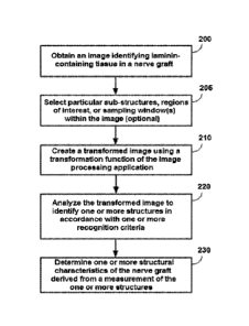
Figure 2 shows an example procedural flow that may be used in some embodiments
of the techniques.
Figure 3A shows the effect of thresholding on an image of a laminin-stained
endoneurial tube cross section.
18 Figure 3B shows an example of the effect of particle analysis on a
thresholded image
of a laminin-stained endoneurial tube cross section.
Figure 4 shows an example embodiment comparing images of a nerve graft as
various described techniques are performed.
Figures 5A-5C show scatter plots comparing various structural characteristics
to the
historical qualitative histology score.
24
DETAILED DESCRIPTION
The degree to which a nerve graft is effective in promoting axon growth is
believed to
be related to the structural characteristics of the nerve graft; however,
effective reproducible
mechanisms of assessing the structural characteristics of a nerve graft have
been lacking. The
subject invention provides techniques are described for determining the
quality of a nerve
30 graft by assessing quantitative structural characteristics of the nerve
graft.
In some embodiments, the structural characteristics are derived from
measurements of
the endoneurial tubes present in the fascicles of the nerve graft.
The outermost layer of the nerve cable is the epineurium, which is the layer
most
often interacted with in peripheral nerve repair. In larger nerve cables, the
cable is subdivided
CA 02987577 2017-11-28
WO 2016/191278
PCT/US2016/033523
3
into multiple fascicles, which are defined by another connective tissue layer,
the perineurium.
"Endoneurial tubes" are the smallest, thinnest and innermost connective tissue
layer in
peripheral nerve cables and may also be called the endoneurium, endoneurial
channel,
endoneurial sheath, or Henle's sheath. They are secreted by and around Schwann
cells, which
are ensheathing axons. The course of the endoneurial tubes is generally
longitudinal along
6
the course of the nerve cable except where fibers leave (or enter, in the case
of
communication branches between different nerve cables) the nerve cable. The
endoneurial
tube is a thin basement membrane principally consisting of a layer of Collagen
IV with a
layer of laminin on the interior surface.
Figure 1 shows an image of a slide having a cross-section of a peripheral
nerve fiber
with anti-laminin immunostaining highlighting the endoneurial tubes.
12
Important aspects of the potency or bioactivity of a nerve graft are the
graft's
structural integrity and structural characteristics. The greater the quantity
and accessibility of
the bioactive scaffold (laminin-coated endoneurial tube geometry) present in
the graft, the
greater the bioactivity of the graft. The reason is that more bioactive
scaffold provides more
growth structures for axons and Schwann cells to extend onto.
Immunohistochemical staining (e.g., anti-laminin staining) can verify the
presence of
18
laminin in the endoneurium. In embodiments of the techniques of the subject
invention,
tissue from the processed nerve graft is stained using an anti-laminin
antibody. The
antibody may be, for example, a polyclonal antibody. Scanned images of the
tissue undergo
image processing to determine the structural characteristics of laminin-
stained structures,
such as endoneurial tubes, present in the two dimensional histology section.
Image processing in some embodiments can include selection of sub-structures
or
24
regions of interest (e.g., fascicles) to further refine those areas of the
image where relevant
structures are to be found. Selection can be manually performed by a human
operator, for
example, by using a selection tool to outline the outer border of the
structure or region.
Selection can also be automated by the image processing application and in
some cases
verified by a human. In some embodiments, a "sampling window" can be used to
define a
subset of the image. In some embodiments, the whole image may be utilized.
30 In
some embodiments, image processing includes manipulating the image to make
structures of interest more visible for analysis. Types of image processing
used in some
embodiments include thresholding the image in accordance with various
parameters.
CA 02987577 2017-11-28
WO 2016/191278
PCT/US2016/033523
4
In some embodiments, the identification of structures (e.g., endoneurial
tubes) used to
determine structural characteristics are in accordance with one or more
recognition criteria,
such as the size and circularity of the structures.
In various embodiments, the structural characteristics can include
measurements of
(1) the number of endoneurial tubes in an area, (2) the percent of endoneurial
lumen in an
6 area, and/or (3) the total perimeter of endoneurial tube lumens in an
area. Better structural
characteristics result in higher determined values. These methods provide
quantitative
evidence of laminin presence and configuration in the endoneurium of the nerve
grafts.
Structural characteristics may be calculated over areas comprised of selected
regions of
interest and/or substructures, over a sampling window, or over fixed areas.
In some embodiments, quantitative assessments of structural quality may be
12 correlated to qualitative assessments. The quantitative metrics can be
correlated to other
metrics such as historically obtained qualitative scores from the same grafts.
One method of
qualitatively assessing the structural integrity of a processed nerve
allograft includes anti-
laminin staining of the tissue and scoring the visual appearance on a
qualitative ranking scale
(e.g., a 1 to 5 scale divided into 0.5 increments) in comparison to a positive
control
containing unprocessed peripheral nerve tissue. However, these methods are
operator-
18 dependent and are unable to precisely assess the quantity and
availability of bioactive
scaffold using a reproducible methodology.
In some embodiments, the determined structural characteristics for a given
sample can
be compared to a reference range for those structural characteristics that
indicate acceptable
nerve graft quality. A nerve graft having values for structural
characteristics that fall outside
the range may be deemed to be of unacceptable quality.
24 In some embodiments, determined structural characteristics may be
compared or
correlated with results from a bioassay of the nerve graft. A bioassay may,
for example,
determine the bioactivity of a graft by measuring the extent of neurite growth
in a cultured
graft. In some cases, results from a bioassay may be correlated with the
results from the
structural characteristics to derive reference ranges for acceptable quality
grafts.
Figure 2 shows an example procedural flow that may be used in some embodiments
30 of the techniques.
Some procedures may be performed using functions or features of an image
processing application, which is a computer program for manipulating the
characteristics of
digital images. An example of an image processing application that may be used
in examples
CA 02987577 2017-11-28
WO 2016/191278
PCT/US2016/033523
herein is Fiji (also known as ImageJ). Furthermore, some procedures described
in Fig. 2 may
be optional in some embodiments.
An image identifying laminin-containing tissue in a nerve graft is obtained
(200).
Generally, these nerve graft cross-sections (or, "sections") are obtained by
histological
preparation of a sample of a nerve graft, e.g., sectioning, fixing, staining,
and mounting a
6
sample on a slide, which is then imaged using slide scanning hardware and
software. Such
images can be a by-product or outcome of, for example, a production,
processing, or quality
control stage of readying the graft for surgical implantation. In some cases,
the images may
have been derived during one phase of production/processing, stored, and then
may be
assessed using the described techniques at a different time.
In some embodiments, the nerve graft is a processed nerve allograft (human)
intended
12
for the surgical repair of peripheral nerve discontinuities to support
regeneration across the
defect. An example of a processed nerve allograft is the Avancee Nerve Graft
from AxoGen.
Nerve allografts provide surgeons with a readily available nerve graft to
repair peripheral
nerves damaged by, for example, traumatic injury or removed during a surgical
procedure. A
processed human nerve allograft is decellularized and processed, resulting in
a surgical
implant with the natural structural pathways to guide axon regeneration. Such
nerve grafts are
18
available in a range of lengths and diameters, and work similarly to an
autograft nerve
without the comorbidities associated with secondary surgical site. Processing
and
decellularization of the nerve allograft clears much of the axonal and myelin
debris so that
nerves may have an unimpeded pathway in which to regrow. Processing also
removes
material and molecules that may potentially elicit a deleterious immune
response in the
recipient.
24 In
some embodiments, the sections of nerve graft undergo immunohistochemical
staining to identify relevant structures in the image. For example, anti-
laminin staining of a
section of a nerve graft can result in high-contrast images showing the
endoneurial tubes and
other laminin- containing structures. In some cases, for example, staining can
be performed
with an immunoperoxidase stain using a polyclonal rabbit anti-laminin (Dako
Z0097) with a
polymer-based secondary system (Dako Envision and Rabbit HRP) and DAB (3,3'-
30
diaminobenzidine) as the developing agent. However, other kinds of staining
(such as a
monoclonal antibody stain) or other structural demarcation techniques that
identify an
endoneurial tube or other key structural components sufficiently in an image
can be used.
Referring again to Figure 1, anti-laminin staining of a nerve graft cross
section is
depicted. In this Figure, laminin-containing structures are shown in brown.
Laminin-
CA 02987577 2017-11-28
WO 2016/191278
PCT/US2016/033523
6
containing structures that are important to determining structural
characteristics include the
endoneurial tubes and perineurium (which defines the fascicle).
In some cases, the quality of staining is reviewed for its adequacy as a
foundation for
analysis of structural characteristics of the graft. Such a review may be
conducted by a human
operator or quality control personnel. Characteristics of quality anti-laminin
staining include:
6
the section is largely free of artifacts and/or technical problems such as
lifting; the staining
color is brown (not blue, black, or other colors); the staining is localized
to extracellular
matrix structures expected to contain laminin (endoneurial tubes and
perineurial layers
principally, but also the basal lamina surrounding fat droplets); and staining
is not present or
is minimal in the interior (lumens) of the endoneurial tubes and in the
epineurium.
In some embodiments, techniques include selecting particular sub-structures,
regions
12 of
interest, or sampling window(s) within the image before further transformation
and
assessment of the structures (205). For instance, in some cases, particular
substructures (e.g.,
the nerve fascicles) are selected to normalize the data to the area that would
be expected to
have the structural characteristics of interest. Selecting sub-structures or
regions of interest in
this way can also allow structural characteristics to be expressed in terms
such as "per
fascicle," or as a ratio of fascicle area. In some cases, selection of
substructures can eliminate
18
areas that may skew structural characteristics or measurements therefrom
(e.g., fat droplets
are usually outside a fascicle).
Selection of regions of interest or substructures (e.g., fascicles) can be
performed
manually or can be automated. In manual selection of fascicles, for instance,
a human
operator might trace the outline of fascicles using a region of interest
selection tool in the
image processing application (for example, to select a region of interest in
Fiji/ImageJ, the
24
"freehand selection tool" can be used to delineate an area of interest which
is then added to
the region of interest list using the manager tool). An automated selection of
fascicles can use
an automated feature identification function, for example, to identify
structures having certain
anti-laminin staining characteristics such as a brown color or a thickness
indicating the
perineurium. Automated selection tasks may also be reviewed in a quality
control step by a
human operator and may be called "computer-assisted selection."
30 In
some cases, a sampling window can be used to select a subset of the image. For
example, a predetermined square area of the image (e.g., a 100,000 pixel area
in the center of
the image) might be used. Use of a fixed size sampling window can obviate the
need for
manual or automated substructure selection steps, allowing the structural
characteristics to be
determined in relation to a fixed area.
CA 02987577 2017-11-28
WO 2016/191278
PCT/US2016/033523
7
Whether an arbitrary selection of areas of interest in the image, a sampling
window,
or the entire image is used, creating a transformed image using a
transformation function of
the image processing application (210) can assist in the identification of
relevant structures.
In some embodiments, transformation may include "thresholding," in which an
image is
converted to binary and image pixels meeting threshold conditions are
selected.
6 Figure 3A shows the effect of thresholding on an image of a laminin-
stained
endoneurial tube cross section. In Figure 3A, a laminin-stained area 300 of an
image is
shown. Thresholding the image 300 produces a binary (e.g., black and white)
image 310.
Thresholding the image 300 in Figure 3A may be performed in image processing
applications such as Fiji/ImageJ. Various settings may be applied to perform
the thresholding,
such as a threshold method, threshold color, color space, and background. The
threshold
12 operation which results in image 310 uses the "default" threshold
method, "black & white"
threshold color, "HSB" color space, and modifies the background color from
white to black.
Thresholding may not need to be adjusted from the default settings in many
cases.
Sometimes, however, additional adjustments (e.g., a manual adjustment of a
"brightness"
control by the human operator) may be performed to obtain quality
thresholding. Some
characteristics of quality thresholding include: primarily the areas staining
dark brown (e.g.,
18 the endoneurial tubes) are thresholded; and areas with light staining or
with Hematoxylin
counterstaining have only occasional pixels thresholded.
In some embodiments, the image may be converted to a different representation
such as an
8-bit image. In some cases, transfolination of the image may include
converting the image to a
different file folmat, such as the TIFF format. Naturally, such
transformations are dependent on the
image processing application chosen in a given embodiment and are intended to
be exemplary
24 rather than limiting.
Using the image processing application, the transformed image is analyzed to
identify
one or more structure in accordance with one or more recognition criteria
(220). Structures
(and measurements of structures) that may be of interest in determining
structural
characteristics include, for example, the endoneurial tubes, the lumens of
endoneurial tubes
(i.e., the enclosed area of space inside space formed by the outer tubular
structure of the
30 endoneurium), the perimeter of the endoneurial tube or its lumen, and
the area of the
endoneurial tube lumen.
In some embodiments, the analysis of the transformed image can include the use
of,
for example, a "particle analysis" feature of an image processing application
(particle
analysis is the term used in Fiji/ImageJ, but it should be appreciated by
practitioners in the art
CA 02987577 2017-11-28
WO 2016/191278
PCT/US2016/033523
8
that different image processing applications can have equivalent features and
functions with
different names). A particle analysis feature can be used to identify
structures having certain
characteristics and to derive measurements from those identified structures.
A recognition criterion is a requirement that a condition or property of the
structure be
satisfied in order for the structure to be recognized as an entity of interest
for identification.
6
For example, when using a "particle analysis" function to identify structures,
the recognition
criterion might require the structure to have certain characteristics to be
recognized as a
particle. These recognition criteria can be introduced by using features of
the image
processing application to set constraints on the identification function or to
eliminate non-
conforming structures from the analysis.
Endoneurial tubes are roughly circular by nature (i.e., they conform to the
12
ensheathing Schwann cells), but clue to the biological nature of the source
material and the
fact that the observations are being made after histological preparation and
sectioning, the
endoneurial tubes may not be completely circular as observed on the slide.
Instead, the
tubes may appear flattened or elongated in cross-section.
In some embodiments, a recognition criterion can include a requirement for a
"circularity" of the structure. Circularity is a measure of the similarity of
the geometry of a
18 structure to that of a circle (mathematically, circularity can be defined
as
4*n*(area/perimeter^2)). In principle, the circularity of a structure ranges
from 0 to 1. In
preferred embodiments, a recognition condition for circularity ranges from 0.5
to 1Ø
A recognition criterion for "size" can be used to filter out structures that
are not of
interest because they are larger or smaller than the structures being
identified. In
embodiments where endoneurial tubes are the structures being identified,
setting a size
24
criterion can eliminate non-endoneurial structures that also have laminin. For
example, the
basal laminae of fat droplets and the perineurium of the fascicles themselves
may in some
cases be filtered out of the analysis due to their size. In preferred
embodiments, the size
criterion for the structures may range from about 4.8 microns to about 16
microns in
diameter. Example 1, below, outlines a procedure by which different
recognition criteria may
be tested for their usefulness in identifying structures.
30
Figure 3B shows an example, in Fiji, of the effect of particle analysis on a
thresholded
image of a laminin-stained endoneurial tube cross section. In Figure 3B, a
thresholded image
350 is shown. Particle analysis on the image 350 produces an image 360 where
relevant
structures have been identified (in the image, the relevant structures are
colored in cyan, as
the background is black).
CA 02987577 2017-11-28
WO 2016/191278
PCT/US2016/033523
9
Returning to Figure 2, one or more structural characteristics of the nerve
graft derived
from a measurement of the one or more structures is determined (230). Once
structures of
interest have been identified, measurements can be performed on the identified
structures
(e.g., their area, perimeter, number, etc., as noted above) and calculations
can be made from
the measurements to determine the structural characteristics of the nerve
graft.
6
Generally, the structural characteristics of relevance are those that indicate
the amount
and accessibility of bioactive scaffold in the graft. These structural
characteristics may be
derived from measurement and computation of the structures that were
identified from the
transformed image. For instance, the structural characteristics can include
measurements of
(1) the number of endoneurial tubes per area, (2) the percent of endoneurial
lumen per area,
and/or (3) the total perimeter of endoneurial tube lumens per area.
12
Some structural characteristics may be determined in reference to an area. An
area
may contain a fixed number of absolute or relative units. Such an area may be
measured, for
example, in relative units (such as an area of pixels, or pixel2 for clarity
in cases where a
length in pixels is also used, which might have a varying true size depending
on
characteristics of the image scanner, image format, or display technology) or
in absolute
units, like microns2. For instance, a sampling window of a fixed number of
units (e.g., 10,000
18
pixe12) might be taken from an image and the structural characteristics
determined in
reference to the sampling window. In another aspect, the area can denote one
or more
regions of interest within a larger area, like a preselected set of fascicles
having certain sizes
or visual characteristics. If fascicles were preselected (either manually or
computer-assisted)
in step 205, the area used to compute structural characteristics might be, for
example, per
each fascicle or per total area of fascicles in a sample.
24 One
example of a structural characteristic, the number of endoneurial tubes per
area,
can be calculated by counting the number of tubes and dividing by the area. As
noted, this
characteristic can be calculated with the area being, e.g., an area of a fixed
number of units of
absolute or relative size, per-fascicle, and/or a total fascicle area.
Another example of a structural characteristic, the percent of endoneurial
lumen per
area, can be calculated by obtaining the area of each of the identified
structures (i.e.,
30
endoneurial tube lumens), summing the lumen areas, and dividing by the area.
As noted, this
characteristic can be calculated with the area being, e.g., an area of a fixed
number of units of
absolute or relative size, the area of a fascicle, and/or a total fascicle
area for a sample.
Another example of a structural characteristic, the total perimeter of
endoneurial tube
lumens per area, can be calculated by obtaining the perimeter of each of the
identified
CA 02987577 2017-11-28
WO 2016/191278
PCT/US2016/033523
structures (i.e., endoneurial tube lumens), summing the perimeters, and
dividing by the area.
As the identified structures (e.g., the particles) are the lumens of the
endoneurial tubes, a
measurement of their perimeter corresponds to measurement of the perimeter of
the laminin-
containing inner surface of the endoneurial tube. As noted, this
characteristic can be
calculated with the area being, for example, an area of a fixed number of
units of absolute or
6 relative size, the area of a fascicle, and/or a total fascicle area for a
sample.
In some embodiments, the structural characteristics are weighted by fascicle
size.
Weighting, in reference to handling the determination of a test statistic from
multiple
fascicles of different sizes, refers to increasing the importance of larger
fascicles for the
determination of the test statistic for the entire section to account for
their larger size (i.e. the
test statistic of the section is the average of the test statistic multiplied
by the relative fascicle
12 area for weighted vs. the average of the test statistic only for
unweighted). Weighting a per
fascicle result average in this manner may be equivalent to converting a per
fascicle result
average into a per total fascicle area average.
Figure 4 shows an example embodiment comparing images of a nerve graft as
various
described techniques are performed. The nerve graft in this example is an
Avance nerve
graft from AxoGen, Inc. In Figure 4, one column of images shows "acceptable
structure" and
18 a second column shows "unacceptable structure." The column labeled
acceptable structure
shows the original staining, thresholded, and analyzed images from a nerve
graft that
originally passed a qualitative assessment by a human operator. The column
labeled
unacceptable structure shows the original staining, thresholded, and analyzed
images for a
nerve graft that did not pass a qualitative assessment. A view of each
original stained image
and the images resulting from the transformation and particle analysis steps
are shown. After
24 analysis, a determination of structural characteristics showed that the
acceptable graft had
endoneurial tube lumens comprising 30.4% of the fascicle area and that the
unacceptable
graft had endoneurial tube lumens comprising only 6.7% of the fascicle area.
Experiments and Examples:
Following are examples illustrating procedures for practicing the techniques
disclosed
30 herein. Advantages of the techniques may be illustrated from results
obtained from one or
more of these examples. Examples may also depict experimental conditions to
refine the
characteristics of certain method parameters. These examples and experiments
should not be
construed as limiting.
CA 02987577 2017-11-28
WO 2016/191278
PCT/US2016/033523
11
EXAMPLE 1
An embodiment of the invention was constructed to experimentally derive
certain
ranges and parameters. As noted in the described method flow, images of
samples containing
a cross-section of a nerve graft were obtained. Experimental conditions
included alternative
options for several parameters, which were then compared for closeness of fit
to a qualitative
6 histology score determined from the same sample images.
The laminin histology images of eleven (11) nerve graft lots comprising Avance
nerve grafts from AxoGen, Inc. were assessed. The lots included 33 large
diameter (3-5 mm)
and 33 small diameter (1-3 mm) samples. Images were derived from slides
scanned into
ImageScope from Aperio. In this case, the images were examined by an operator
for the
quality of the anti-laminin staining.
12 In
this example embodiment, the fascicles were selected using an image processing
application. Here, the image processing application is Fiji (also known as
ImageJ). The
fascicles were selected using two methods that are evaluated as parameters:
manual selection
using a freehand selection tool in Fiji, and computer-assisted selection using
a Fiji macro
followed by a quality review and correction by a human operator. Results of
the two
techniques are compared below.
18 In
this example, transformation of the image using the image processing
application
(here, Fiji) includes applying thresholding settings to the image.
Thresholding may enhance
or reduce certain characteristics of the image so that the image processing
application can
better analyze the structures (e.g., the endoneurial tubes) depicted in the
image. Initial
thresholding settings include using the image processing application's
"default" method;
setting the threshold color to "black and white"; setting the color space to
"HSB"; and setting
24
the background to dark. The brightness of the transformed image may also be
adjusted. Here,
transformation of the image also includes converting the image to an 8-bit
representation.
Structures (e.g., the endoneurial tubes in the fascicles) were identified in
this example
using the "particle analysis" capability of the image processing application
(here, Fiji). The
particle analysis feature identifies structures by virtue of its ability to
recognize discrete
objects in the image because those objects were highlighted by
immunohistochemical
30
staining and, in some cases, because image transformation settings make the
staining more
discernible to the image processing application. Furthermore, when
substructures, regions of
interest, or sampling windows are selected, the analysis may be carried out
only within those
regions.
CA 02987577 2017-11-28
WO 2016/191278
PCT/US2016/033523
12
In the example, settings for particle analysis function included recognition
criteria for
size and circularity and settings to "include holes" and "exclude on edges."
Measurement
settings include "area," "perimeter," and "integrated density."
Example 1 utilizes two recognition criteria for determining a structure: size
and
circularity. A total of 32 different combinations of size and circularity are
shown in Table 1
6 below. The size indicates an area, in pixels, of the structure. In this
case, an image pixel
equals 0.495 microns in accordance with the Aperio slide scanner settings.
Table 1. Criteria for Endoneurial tube recognition
Criteria set Size (pixe1s^2) Circularity
1 20-820 0.3-1.0
2 75-820 0.3-1.0
3 20-1050 0.3-1.0
4 75-1050 0.3-1.0
20-1300 0.3-1.0
6 75-1300 0.3-1.0
7 20-2000 0.3-1.0
8 75-2000 0.3-1.0
9 20-820 0.4-1.0
75-820 0.4-1.0
11 20-1050 0.4-1.0
12 75-1050 0.4-1.0
13 20-1300 0.4-1.0
14 75-1300 0.4-1.0
20-2000 0.4-1.0
16 75-2000 0.4-1.0
17 20-820 0.5-1.0
18 75-820 0.5-1.0
19 20-1050 0.5-1.0
75-1050 0.5-1.0
21 20-1300 0.5-1.0
22 75-1300 0.5-1.0
23 20-2000 0.5-1.0
24 75-2000 0.5-1.0
20-820 0.6-1.0
26 75-820 0.6-1.0
27 20-1050 0.6-1.0
28 75-1050 0.6-1.0
29 20-1300 0.6-1.0
75-1300 0.6-1.0
31 20-2000 0.6-1.0
32 75-2000 0.6-1.0
In Example 1, three structural characteristics were determined from the
recognized
endoneurial tubes: the number of endoneurial tubes in a 100,000 pixel area,
the percent of
12 endoneurial tube lumen in an area, and the total perimeter of
endoneurial tube lumens in a
100,000 pixel area.
In this example, weighting was applied as an experimental parameter. As noted,
weighting may convert a per fascicle test statistic into a per total fascicle
area test statistic.
CA 02987577 2017-11-28
WO 2016/191278
PCT/US2016/033523
13
As an objective of Example 1 was to assess varying parameters of the
techniques,
effects of the varied parameters are discussed. Outcomes of alternative
parameter choices
were evaluated by comparing the values of their "goodness of fit" with
historical qualitative
histology scores (e.g., R2 values). In this example, the qualitative histology
score is a rating
by a human evaluator on a 1 to 5 scale divided into 0.5 increments that
compares the
6 appearance of laminin in a test sample of nerve graft tissue against a
positive control
containing unprocessed peripheral nerve tissue. A higher score indicates a
closer fit, i.e., the
appearance of more bioactive scaffold.
"R2" (or R^2) is the Coefficient of Determination, a measure of the "goodness
of fit"
of an experimental vs. theoretical/modeled data set. Mathematically, R2 = 1-
[sum((yi-
fi)^2)/sum((yi-avgy)^2)] where "y" is the experimental data, "f' is the
modeled data, "i" is
12 the counter for the dataset (i.e. "i" goes from 1 to the number of
datapoints), and "avgy" is
the average of "y" over the full data set.
Little effect of weighting for fascicle/selection area was noted. However,
weighting
the results, as described, by total fascicle/selection area did result in
slightly better
correlation to the historical histological scoring.
Assisted area selection was equivalent to the completely manual method as
evidenced
18 by the similarity of R2 values. The assisted area selection method has
the analyst review
every selection and correct it if necessary. Though the assisted method tended
not to include
some of the smallest fascicles, it is mostly equivalent to the completely
manual method. This
would be expected from the great similarity of the areas selected by both
methods (R2=0.995
comparing total area per section).
The data collected on the number of endoneurial tubes indicates that using a
lower
24 particle limit of 20 pixels leads to selecting features that are not
associated with historical
histological scoring (i.e. lowers the correlation). Thus, a preferred lower
limit for the "size"
recognition criterion is 75 pixels (-5 micron diameter).
The data collected on % area (and perimeter) show that use of an upper
particle limit
of 1,300 and above leads to selecting features that are not associated with
historical
histological scoring (i.e. lowers the correlation). Thus, a preferred upper
limit for the "size"
30 recognition criterion is below 1,300 pixels (e.g., 820 or 1050 pixels;
¨16 or ¨18 microns in
diameter).
The circularities were roughly similar for # of tubes and % area, but the 0.3-
1.0 and
0.4-1.0 circularity ranges were less stable. Thus, a preferred circularity
range is 0.5-1Ø
CA 02987577 2017-11-28
WO 2016/191278
PCT/US2016/033523
14
All three structural characteristics in this example (# of tubes, % area, and
perimeter of
tubes) gave broadly similar results, with some differences depending on the
particle analysis
method.
EXAMPLE 2
6 An
embodiment of the invention was developed to experimentally assess the
closeness of fit of certain described techniques to the qualitative historical
histology score
determined from the same sample images. To summarize, Example 2 used specific
ranges for
the size and circularity recognition conditions, and assisted selection, and
compared three
structural characteristics against a historical qualitative score for goodness
of fit.
As noted in the described method flow, an image of a sample containing a cross-
12
section of a nerve graft was obtained. In Example 2, thirty-two lots of Avance
Nerve Graft
from AxoGen, Inc. were assessed; the lots included four lots that did not pass
the qualitative
historical histology structural acceptance criteria. Result analysis examined
the correlation
between historical scoring data and three quantifiable structural
characteristics. The data was
assessed by comparing the historical score for a given sample (e.g., from a
single graft, also
known as a "section") to each of the three structural characteristics. In
addition, the data was
18
assessed by comparing the historical score average for a lot (average of
scores for 6 samples
with each sample from a separate graft) versus the three structural
characteristics for the
same lot. In summary, the results found that, for individual samples,
comparison to the
perimeter of endoneurial tubes provided the best fit (R2 = 0.622) and for the
lot average, the
percent endoneurial lumen area provided the best fit (R2 = 0.581).
In this embodiment of the described techniques, the following parameters and
24
conditions were used: The areas of all fascicles in a section were outlined in
Fiji using an
initial computer selection followed by a manual inspection and correction,
when necessary
(i.e., computer-assisted).
Transformation of the image using Fiji included applying thresholding settings
to the
image. Initial thresholding settings include using the image processing
application's "default"
method; setting the threshold color to "black and white"; setting the color
space to "HSB";
30 and
setting the background to dark. The brightness of the transformed image may
also be
adjusted. Transformation of the image also includes converting the image to an
8-bit
representation.
Endoneurial tubes in the fascicles were identified using the "particle
analysis"
capability of Fiji. Recognition criteria for performing the particle analysis
included size
CA 02987577 2017-11-28
WO 2016/191278
PCT/US2016/033523
ranges and circularity ranges. The size criterion was set to identify
structures from 75 to 820
pixel in area. The circularity criterion was set to identify structures having
a 0.5-1.0
circularity range. In this example, settings for particle analysis function
included settings to
"include holes" and "exclude on edges." Measurement settings include "area,"
"perimeter,"
and "integrated density."
6
Three structural characteristics were determined from the recognized
endoneurial
tubes: the number of endoneurial tubes in a 100,000 pixel area, the percent of
endoneurial
tube lumen in an area, and the total perimeter of endoneurial tube lumens in a
100,000 pixel
area.
Note that a 100,000 pixel area is equal to 24,502.5 square microns (or ¨0.025
square
millimeters). The units of the test statistic are linear pixels (i.e. length
of pixels) with one
12 pixel being 0.495 microns in length for Aperio ImageScope.
Weighting based on the size of the fascicle was applied in the calculation of
structural characteristics.
As noted, experimental data was assessed by comparing the historical score for
a
given sample or set of samples to each of the three structural characteristics
for the
sample/set. The mathematical "goodness of fit" (or R2) between the
experimental and
18
modeled data set was calculated as part of the assessment. Results are
described below and
in the Figures 5A-5C.
Figure 5A shows scatter plots comparing the number of endoneurial tubes
structural
characteristic to the historical histology score for all sections (individual
samples) and all lots
examined, respectively. The R2 value for the section data set is 0.551, and
the R2 value for the
lot data set is 0.5118.
24
Figure 5B shows scatter plots comparing the percent endoneurial tube lumen
structural characteristic to the historical histology score for all sections
(individual samples)
and all lots examined, respectively. The R2 value for the section data set is
0.6121, and the R2
value for the lot data set is 0.5814.
Figure 5C shows scatter plots comparing the perimeter of endoneurial tubes
structural
characteristic to the historical histology score for all sections (individual
samples) and all lots
30
examined, respectively. The R2 value for the section data set is 0.622, and
the R2 value for the
lot data set is 0.5722.
Table 2 shows the Pearson Correlation Coefficients for historical histology
scores in
comparison to the structural characteristics for the samples. Note: these are
correlation
coefficients ("R") not coefficients of determination ("R2") as shown in the
plots.
CA 02987577 2017-11-28
WO 2016/191278
PCT/US2016/033523
16
Table 2
Historical Histology Number of tubes Percent tube lumen Perimeter of tubes
Historical Histology 1 NA NA NA
Number of tubes 0.742 1 NA NA
Percent tube lumen 0.782 0.899 1 NA
Perimeter 0.789 0.972 0.975 1
To summarize, for experimental results derived from this embodiment, the
perimeter
of endoneurial tubes structural characteristic is a slightly better match to a
historical
6
qualitative analysis of graft structural quality. Two reasons are posited for
this result. First,
the perimeter structural characteristic does not change if the circular
structure collapses
during histological processing. Second, the perimeter of the outside of the
lumen is a direct
measurement of the interior surface of the endoneurial tube, which is coated
with laminin,
and presumably the quantity of accessible laminin is a key bioactive substance
for fostering
neurite regeneration in a graft.
12 It
should be understood that the examples and embodiments described herein are
for
illustrative purposes only and that various modifications or changes in light
thereof will be
suggested to persons skilled in the art and are to be included within the
spirit and purview of
this application.
Although the subject matter has been described in language specific to
structural
features and/or acts, it is to be understood that the subject matter defined
in the appended
18
claims is not necessarily limited to the specific features or acts described
above. Rather, the
specific features and acts described above are disclosed as examples of
implementing the
claims and other equivalent features and acts are intended to be within the
scope of the
claims.