Note: Descriptions are shown in the official language in which they were submitted.
CA 02987636 2017-11-28
WO 2016/210376 PCT/US2016/039431
1
THERAPEUTIC PEPTIDES AND METHODS OF USE THEREOF
CROSS-REFERENCE TO RELATED APPLICATIONS
[0001] This application is related to U.S. provisional patent application
number 62/185,529, filed
June 26, 2015; U.S. provisional patent application number 62/185,527, filed
June 26, 2015; U.S.
provisional patent application number 62/239,743, filed October 9, 2015; U.S.
provisional patent
application number 62/239,739, filed October 9, 2015; U.S. provisional patent
application
number 62/322,724, filed April 14, 2016; and U.S. provisional patent
application number
62/354,642, filed June 24, 2016, each of which are herein incorporated by
reference in their
entirety.
BACKGROUND
[0002] For many types of cancers, patient prognosis is directly influenced by
the efficacy of drug
therapies and surgical access to the tumor. In particular, the precision of
tumor resection is
dependent on intra-operative imaging to detect tumor margins or small foci of
cancer cells.
However, current methods of intra-operative imaging cancerous tissues are
imprecise.
[0003] Of these types of cancers, brain disorders are particularly difficult
to treat. The blood-
brain barrier (BBB) can exclude over 97% of small molecules from entering the
brain, and larger
molecules such as antibodies are excluded almost universally. Usually, most
molecules that enter
the brain are small, lipophilic, and lack target specificity. Few drugs aimed
at treating brain
disorders have proved therapeutically viable with lack of access to target
tissue being a primary
reason for failure. In addition, many drugs that could gain access to the
brain are ill-suited for
treating brain conditions. The lack of access to the target tissue and lack of
specificity also lead to
administration of doses that are higher than would be necessary if a drug
could home, target, or
be directed to, a target region, tissue, structure or cell in the brain.
[0004] Similarly, other types of cancers, particularly solid tumors of several
types, are difficult to
treat as it is difficult to achieve a high enough level of effective drug into
such tumors while
managing side effects of the drugs in normal tissues. Consequently, there is a
need for targeting
drugs to solid tumors specifically to achieve a higher effective dose of drug
in tumor while
minimizing the level of side effects in other tissues. Moreover, there is also
a need for targeting
drugs specifically to any cancerous cell, whether from solid tumors or
otherwise. Typical cancer
drug regimens are often limited by dose-limiting toxicities, and although some
antibody-drug
conjugates are used to target drugs to specific tumors in order to limit off-
site toxicity, such
CA 02987636 2017-11-28
WO 2016/210376 PCT/US2016/039431
2
specific therapies are not available for many tumor types. Herein, we provide
new peptides that
target to tumors.
SUMMARY
[0005] The present disclosure relates to compositions and methods for
treatment of tumors.
Described herein are peptides that home, distribute to, target, are directed
to, accumulate in,
migrate to, and/or bind to cancerous cells following administration to a
subject. In some
embodiments, the compositions and methods herein utilize peptides that home,
distribute to,
target, are directed to, accumulate in, migrate to, and/or bind to cancerous
or diseased cells in the
brain following administration to a subject. In some embodiments, the homing
peptides of the
present disclosure are used to deliver an active agent to a tissue or cell
thereof.
[0006] In various aspects, the present disclosure provides a peptide
comprising a sequence of any
one of SEQ ID NO: 198¨ SEQ ID NO: 209 or SEQ ID NO: 407¨ SEQ ID NO: 418 or a
fragment thereof.
[0007] In various aspects, the present disclosure provides a peptide
comprising a sequence that
has at least 80% sequence identity with any one of SEQ ID NO: 1 ¨ SEQ ID NO:
192 or SEQ ID
NO: 210 ¨ SEQ ID NO: 401, or a fragment thereof. In some aspects, the peptide
comprises the
sequence that has at least 85%, at least 90%, or at least 95% sequence
identity with any one of
SEQ ID NO: 1 ¨ SEQ ID NO: 192 or SEQ ID NO: 210¨ SEQ ID NO: 401, or a fragment
thereof. In other aspects, the peptide comprises a sequence that is any one of
SEQ ID NO: 1 ¨
SEQ ID NO: 192 or SEQ ID NO: 210 ¨ SEQ ID NO: 401, or fragment thereof.
[0008] In various aspects, the present disclosure provides a peptide
comprising a sequence of any
one of SEQ ID NO: 198 ¨ SEQ ID NO: 209, or a fragment thereof.
[0009] In various aspects, the present disclosure provides a peptide
comprising a sequence that
has at least 80% sequence identity with any one of SEQ ID NO: 1 ¨ SEQ ID NO:
192, or a
fragment thereof. In some aspects, the peptide comprises the sequence that has
at least 85%, at
least 90%, or at least 95% sequence identity with any one of SEQ ID NO: 1 ¨
SEQ ID NO: 192,
or a fragment thereof. In other aspects, the peptide comprises a sequence that
is any one of SEQ
ID NO: 1 ¨ SEQ ID NO: 192, or fragment thereof.
[0010] In various aspects, the present disclosure provides a peptide
comprising a sequence of any
one of SEQ ID NO: 407¨ SEQ ID NO: 418, or a fragment thereof.
[0011] In various aspects, the present disclosure provides a peptide
comprising a sequence that
has at least 80% sequence identity with any one of SEQ ID NO: 210 ¨ SEQ ID NO:
401, or a
CA 02987636 2017-11-28
WO 2016/210376 PCT/US2016/039431
3
fragment thereof. In some aspects, the peptide comprises the sequence that has
at least 85%, at
least 90%, or at least 95% sequence identity with any one of SEQ ID NO: 210 -
SEQ ID NO:
401, or a fragment thereof. In other aspects, the peptide comprises the
sequence that is any one of
SEQ ID NO: 210 - SEQ ID NO: 401, or a fragment thereof.
[0012] In some aspects, any peptide of the present disclosure is a knotted
peptide. In other
aspects, the peptide comprises at least 6, at least 8, at least 10, at least
12, at least 14, or at least
16 cysteine residues. In some aspects, the peptide comprises a plurality of
disulfide bridges
formed between cysteine residues. In further aspects, at least 5% or more of
the residues are
cysteines forming intramolecular disulfide bonds. In some aspects, the peptide
comprises a
disulfide through disulfide knot.
[0013] In some aspects, at least one amino acid residue of the peptide is in
an L configuration, or
wherein at least one amino acid residue of the peptide is in a D
configuration. In some aspects,
the sequence is at least 11, at least 12, at least 13, at least 14, at least
15, at least 16, at least 17, at
least 18, at least 19, at least 20, at least 21, at least 22, at least 23, at
least 24, at least 25, at least
26, at least 27, at least 28, at least 29, at least 30, at least 31, at least
32, at least 33, at least 34, at
least 35, at least 36, at least 37, at least 38, at least 39, at least 40, at
least 41, at least 42, at least
43, at least 44, at least 45, at least 46, at least 47, at least 48, at least
49, at least 50, at least 51, at
least 52, at least 53, at least 54, at least 55, at least 56, at least 57, at
least 58 residues, at least 59,
at least 60, at least 61, at least 62, at least 63, at least 64, at least 65,
at least 66, at least 67, at
least 68, at least 69, at least 70, at least 71, at least 72, at least 73, at
least 74, at least 75, at least
76, at least 77, at least 78, at least 79, at least 80, or at least 81
residues long.
[0014] In some aspects, the peptide is arranged in a multimeric structure with
at least one other
peptide.
[0015] In some aspects, the peptide has a positive net charge greater than
+0.5 at physiological
pH. In other aspects, the peptide has a negative net charge lower than -0.5 at
physiological pH.
[0016] In some aspects, upon administration to a subject, the peptide homes,
targets,
accumulates in, migrates to, or is directed to a specific region, tissue,
structure, or cell of the
subject.
[0017] In some aspects, at least one residue of the peptide comprises a
chemical modification. In
some aspects, the chemical modification is blocking the N-terminus of the
peptide. In some
aspects, the modification is methylation, acetylation, or acylation. In other
aspects, the chemical
modification is: methylation of one or more lysine residues or analogue
thereof; methylation of
CA 02987636 2017-11-28
WO 2016/210376 PCT/US2016/039431
4
an N-terminus; or methylation of one or more lysine residue or analogue
thereof and methylation
of the N-terminus. In some aspects, the peptide is linked to an acyl adduct.
[0018] In some aspects, the peptide is linked to an active agent. In further
aspects, the active
agent is fused with the peptide at an N-terminus or a C-terminus of the
peptide. In some aspects,
the active agent is a neurotensin peptide. In further aspects, the neurotensin
peptide has a
sequence of SEQ ID NO: 420. In still further aspects, the peptide fused to
neurotensin peptide
comprises a contiguous sequence. In some aspects, 1, 2, 3, 4, 5, 6, 7, 8, 9,
or 10 active agents are
linked to the peptide.
[0019] In some aspects, the peptide is linked to the active agent via a
cleavable linker. In other
aspects, the peptide is linked to the active agent at an N-terminus, at the
epsilon amine of an
internal lysine residue, at the carboxylic acid of an asparagine or glutamine
residue, or a C-
terminus of the peptide by a linker. In further aspects, the internal lysine
residue is located at a
position corresponding to amino acid residue 17 of SEQ ID NO: 37, amino acid
residue 25 of
SEQ ID NO: 37, or amino acid residue 29 of SEQ ID NO: 37. In other aspects,
the internal lysine
residue is located at a position corresponding to amino acid residue 15 of SEQ
ID NO: 246,
amino acid residue 23 of SEQ ID NO: 246, or amino acid residue 27 of SEQ ID
NO: 246.
[0020] In other aspects, the peptide further comprises a non-natural amino
acid, wherein the non-
natural amino acid is an insertion, appendage, or substitution for another
amino acid.
[0021] In some aspects, the peptide is linked to the active agent at the non-
natural amino acid by
a linker. In other aspects, the linker comprises an amide bond, an ester bond,
a carbamate bond, a
carbonate bond, a hydrazone bond, an oxime bond, a disulfide bond, a thioester
bond, or a
carbon-nitrogen bond. In further aspects, the cleavable linker comprises a
cleavage site for matrix
metalloproteinases, thrombin, cathepsins, or beta-glucuronidase. In some
aspects, the peptide is
linked to the active agent via a noncleavable linker.
[0022] In some aspects, the active agent is selected from the group consisting
of: a peptide, a
polypeptide, a polynucleotide, an antibody, a single chain variable fragment
(scFv), an antibody
fragment, a cytokine, a hormone, a growth factor, a checkpoint inhibitor, an
immune modulator,
a neurotransmitter, a chemical agent, a cytotoxic molecule, a toxin, a radio
sensitizer, a
radioprotectant, a therapeutic small molecule, a nanoparticle, a liposome, a
polymer, a dendrimer,
a fatty acid, peptidomimetic, a complement fixing peptide or protein,
polyethylene glycol, a lipid,
or an Fc region. In other aspects, the active agent is a
polydeoxyribonucleotide or a
polyribonucleotide sequence. In additional aspects, the active agent is an
anti-inflammatory
agent, an antifungal agent, an antiviral agent, or an anti-infective agent. In
some aspects, the
CA 02987636 2017-11-28
WO 2016/210376 PCT/US2016/039431
active agent is a chemotherapeutic agent. In other aspects, the active agent
is a knotted peptide.
In still other aspects, the active agent is a radiosensitizer or
photosensitizer. In some aspects, the
cytotoxic molecule is an auristatin, MMAE, a maytansinoid, DM1, DM4,
doxorubicin, a
calicheamicin, a platinum compound, cisplatin, a taxane, paclitaxel, SN-38, a
BACE inhibitor, a
Bc1-xL inhibitor, WEHI-539, venetoclax, ABT-199, navitoclax, AT-101,
obatoclax, a
pyrrolobenzodiazepine or pyrrolobenzodiazepine dimer, or dolastatin.
[0023] In other aspects, the peptide is linked to a detectable agent. In
further aspects, the
detectable agent is fused with the peptide at an N-terminus or a C-terminus of
the peptide. In still
further, aspects, 1, 2, 3, 4, 5, 6, 7, 8, 9, or 10 detectable agents are
linked to the peptide.
[0024] In some aspects, the peptide is linked to the detectable agent via a
cleavable linker. In
other aspects, the peptide is linked to the detectable agent at an N-terminus,
at the epsilon amine
of an internal lysine residue, or a C-terminus of the peptide by a linker. In
further aspects, the
internal lysine is located at a position corresponding to amino acid residue
17 of SEQ ID NO: 37,
amino acid residue 25 of SEQ ID NO: 37, or amino acid residue 29 of SEQ ID NO:
37. In other
aspects, the internal lysine residue is located at a position corresponding to
amino acid residue 15
of SEQ ID NO: 246, amino acid residue 23 of SEQ ID NO: 246, or amino acid
residue 27 of
SEQ ID NO: 246.
[0025] In some aspects, the peptide further comprises a non-natural amino
acid, wherein the non-
natural amino acid is an insertion, appendage, or substitution for another
amino acid.
[0026] In some aspects, the peptide is linked to the active agent at the non-
natural amino acid by
a linker. In other aspects, the linker comprises an amide bond, an ester bond,
a carbamate bond, a
hydrazone bond, an oxime bond, or a carbon-nitrogen bond. In further aspects,
the cleavable
linker comprises a cleavage site for matrix metalloproteinases, thrombin,
cathepsins, or beta-
glucuronidase. In some aspects, the peptide is linked to the detectable agent
via a noncleavable
linker.
[0027] In other aspects, the detectable agent is a fluorophore, a near-
infrared dye, a contrast
agent, a nanoparticle, a metal-containing nanoparticle, a metal chelate, an X-
ray contrast agent, a
PET agent, a radioisotope, or a radionuclide chelator. In some aspects, the
detectable agent is a
fluorescent dye.
[0028] In some aspects, the peptide homes, targets, is directed to,
accumulates in, or migrates to
a tumor or cancerous cell. In some aspects, the tumor is a solid tumor. In
other aspects, the tumor
is a hematologic malignancy. In further aspects, the peptide penetrates the
solid tumor. In still
further aspects, the peptide is internalized into or penetrates a cancerous
cell. In some aspects, the
CA 02987636 2017-11-28
WO 2016/210376 PCT/US2016/039431
6
tumor or cancerous cell is from a brain cancer, a glioblastoma, a colon
cancer, a triple-negative
breast cancer, metastatic cancer, or a sarcoma.
[0029] In some aspects, the peptide crosses a blood brain barrier to access
the tumor. In other
aspects, the peptide crosses a blood cerebral spinal fluid barrier to access
the tumor.
[0030] In some aspects, the peptide crosses a blood brain barrier or a blood
cerebral spinal fluid
barrier of a subject. In other aspects. In other aspects, the peptide crosses
a blood cerebrospinal
fluid barrier of a subject.
[0031] In some aspects, the peptide homes, targets, is directed to,
accumulates in, or migrates to
a tumor or diseased region, tissue, structure, or cell of the subject after
crossing the blood brain
barrier.
[0032] In other aspects, upon administration to a subject the peptide homes,
targets, is directed
to, accumulates in, or migrates to a specific brain region of the subject. In
further aspects, the
specific region of the brain comprises the ventricles, the cerebrospinal
fluid, the hippocampus,
the meninges, the rostral migratory system, the dentate gyrus, the
subventricular zone, or any
combination thereof.
[0033] In some aspects, the peptide affects neurological disorders, lysosomal
storage diseases,
epilepsy, meningitis, infections in the brain, stroke, and multiple sclerosis.
In some aspects, the
peptide affects aggregation of a protein associated with a neurodegenerative
disease. In other
aspects, the peptide inhibits a pathway associated with brain cancer. In still
other aspects, the
peptide inhibits or activates ion channels. In some aspects, the peptide
exhibits protease inhibitor
activity. In other aspects, the peptide has antibacterial, antifungal, or
antiviral activity.
[0034] In various aspects, the present disclosure provides a pharmaceutical
composition
comprising a peptide of this disclosure or a salt thereof, and a
pharmaceutically acceptable
carrier. In some aspects, the pharmaceutical composition is formulated for
administration to a
subject. In further aspects, the pharmaceutical composition is formulated for
inhalation,
intranasal administration, oral administration, topical administration,
intravenous administration,
subcutaneous administration, intra-articular administration, intramuscular
administration,
intrathecal, intraperitoneal administration, or a combination thereof.
[0035] In various aspects, the present disclosure provides a method of
treating a condition in a
subject in need thereof, the method comprising administering to the subject a
peptide or a
pharmaceutical composition of this disclosure. In some aspects, the peptide or
pharmaceutical
composition is administered by inhalation, intranasally, orally, topically,
intravenously,
CA 02987636 2017-11-28
WO 2016/210376 PCT/US2016/039431
7
subcutaneously, intra-articularly, intramuscularly administration,
intraperitoneally, or a
combination thereof.
[0036] In some aspects, the peptide or pharmaceutical composition of the
method. In some
aspects, the condition is a tumor or cancer. In further aspects, the condition
is a solid tumor. In
some aspects, the tumor is a hematologic malignancy. In other aspects, the
condition is a brain
tumor, triple-negative breast cancer, colon cancer metastases, metastatic
cancer or sarcoma. In
further aspects, the brain tumor is inoperable.
[0037] In some aspects, the peptide of the method crosses a blood brain
barrier to home, target,
migrate to, accumulate in, or get directed to the tumor in the brain. In some
aspects, the peptide
crosses a blood cerebrospinal fluid barrier to home, target, migrate to,
accumulate in, or get
directed to the tumor in the brain.
[0038] In some aspects, the method is combined with other treatments. In
further aspects, the
other treatments comprise chemotherapy, radiation therapy, or immunomodulatory
therapy.
[0039] In some aspects, the peptide of the method crosses the blood brain
barrier of the subject
following administration. In other aspects, the peptide crosses the blood
cerebrospinal fluid
barrier of the subject following administration.
[0040] In some aspects, the peptide of the method homes, targets, is directed
to, accumulates in,
or migrates to the ventricles, cerebrospinal fluid, meninges, rostral
migratory system, or
hippocampus of the subject following administration. In some aspects, the
condition is a brain
condition. In other aspects, the condition is associated with a function of
the ventricles,
cerebrospinal fluid, or hippocampus. In further aspects, the brain condition
is associated with a
function of the brain.
[0041] In some aspects, the peptide of the method diagnoses, prevents, or
treats the brain
condition. In further aspects, the brain condition is a brain tumor or brain
cancer. In other aspects,
the brain condition is memory loss or memory function, Alzheimer's disease,
Parkinson's
disease, multiple system atrophy (MSA), schizophrenia, epilepsy, progressive
multifocal
leukoencephalopathy, fungal infection, depression, bipolar disorder, post-
traumatic stress
disorder, stroke, traumatic brain injury, infection, or multiple sclerosis.
[0042] In various aspects, the present disclosure provides a method of imaging
an organ or body
region of a subject, the method comprising administering to the subject a
peptide or
pharmaceutical composition of this disclosure and imaging the organ or body
region of the
subject.
CA 02987636 2017-11-28
WO 2016/210376 PCT/US2016/039431
8
[0043] In some aspects, the method comprises detecting a cancer or diseased
region, tissue,
structure or cell of the subject. In further aspects, the method comprises
performing surgery on
the subject. In still further aspects, the method comprises treating the
cancer.
[0044] In some aspects, the surgery of the method comprises removing the
cancer or the diseased
region, tissue, structure or cell of the subject. In further aspects, the
method comprises imaging
the cancer or diseased region, tissue, structure, or cell of the subject after
surgical removal.
INCORPORATION BY REFERENCE
[0045] All publications, patents, and patent applications mentioned, disclosed
or referenced in
this specification are herein incorporated by reference in their entirety and
to the same extent as if
each individual publication, patent, or patent application was specifically
and individually
indicated to be incorporated by reference.
BRIEF DESCRIPTION OF THE FIGURES
[0046] The novel features of the invention are set forth with particularity in
the appended claims.
A better understanding of the features and advantages of the present invention
will be obtained
by reference to the following detailed description that sets forth
illustrative embodiments, in
which the principles of the invention are utilized, and the accompanying
drawings of which:
[0047] FIG. 1 illustrates a peptide that was radiolabeled by methylating the
lysines. FIG. 1A
illustrates a native lysine and FIG. 1B illustrates a dimethylated lysine.
[0048] FIG. 2 illustrates 14C signal in the brain and other tissues for the
fluoxetine (top) and
inulin (bottom) control groups.
[0049] FIG. 3 illustrates 14C signal in the brain and other tissues for
radiolabeled peptides of
SEQ ID NO: 1.
[0050] FIG. 4 illustrates 14C signal in the brain and other tissues for
radiolabeled peptides of
SEQ ID NO: 3.
[0051] FIG. 5 illustrates the HPLC profile of a peptide of SEQ ID NO: 1.
[0052] FIG. 6 illustrates an overlay of the HPLC profiles for both a
nonreduced and a reduced
sample of a peptide of SEQ ID NO: 2.
[0053] FIG. 7 illustrates an overlay of the HPLC profiles for both a
nonreduced and a reduced
sample of a peptide of SEQ ID NO: 3.
[0054] FIG. 8 illustrates the HPLC profile of a peptide of SEQ ID NO: 4.
[0055] FIG. 9 illustrates an exemplary architecture of constructs expressing
SEQ ID NO: 1
through SEQ ID: NO. 4.
CA 02987636 2017-11-28
WO 2016/210376 PCT/US2016/039431
9
[0056] FIG. 10 illustrates a schematic of a method of manufacturing of a
peptide of the
disclosure.
[0057] FIG. 11 illustrates quality control data from small scale expression
runs of peptides of
SEQ ID NO: 4 (FIG. 11A), SEQ ID NO: 6 (FIG. 11B), SEQ ID NO: 17 (FIG. 11C),
SEQ ID
NO: 25 (FIG. 11D), and SEQ ID NO: 32 (FIG. 11E).
[0058] FIG. 12 illustrates HPLC data and non-reduced compared to reduced bands
on SDS-
PAGE gels of SEQ ID NO: 39 peptide, and MALDI mass spectrometry graphs of SEQ
ID NO:
25 peptide.
[0059] FIG. 12A illustrates an HPLC profile of SEQ ID NO: 39.
[0060] FIG. 12B illustrates the nonreduced and reduced bands of SEQ ID NO: 39
on an SDS-
PAGE gel.
[0061] FIG. 12C shows the full spectra of a MALDI mass spectrometry graph of
SEQ ID NO:
25.
[0062] FIG. 12D shows a zoomed-in portion of the full spectra of a MALDI mass
spectrometry
graph of SEQ ID NO: 25.
[0063] FIG. 13 illustrates murine white light and corresponding
autoradiographic images three
hours after administration of 9 nmol of the radiolabeled peptide of SEQ ID NO:
5 conjugated to
Alexa 647 fluorescent dye (SEQ ID NO: 5-RA peptide conjugate).
[0064] FIG. 13A illustrates a white light image of a frozen section of a mouse
three hours after
administration of 9 nmol of radiolabeled peptide of SEQ ID NO: 5 conjugated to
Alexa 647
fluorescent dye (SEQ ID NO: 5-RA peptide conjugate).
[0065] FIG. 13B illustrates an autoradiographic image corresponding to FIG.
13A in which the
14C signal identifies the peptide distribution in the tissues, including the
RH-28 tumor of a
mouse, three hours after administration of 9 nmol of the radiolabeled peptide
of SEQ ID NO: 5
conjugated to Alexa 647 fluorescent dye (SEQ ID NO: 5-RA peptide conjugate).
[0066] FIG. 13C illustrates a white light image of a different frozen section
of the same mouse
as in FIG. 13A and FIG. 13B three hours after administration of 9 nmol of the
radiolabeled
peptide of SEQ ID NO: 5 conjugated to Alexa 647 fluorescent dye (SEQ ID NO: 5-
RA peptide
conjugate).
[0067] FIG. 13D illustrates an autoradiographic image corresponding to FIG.
13C in which the
14C signal identifies the peptide distribution in the tissues, including RH-28
tumor, three hours
after administration of 9 nmol of the radiolabeled peptide of SEQ ID NO: 5
conjugated to Alexa
647 fluorescent dye (SEQ ID NO: 5-RA peptide conjugate).
CA 02987636 2017-11-28
WO 2016/210376 PCT/US2016/039431
[0068] FIG. 13E illustrates a white light image of a frozen section of a
different mouse than
shown in FIG. 13A through FIG. 13D three hours after administration of 9 nmol
of the
radiolabeled peptide of SEQ ID NO: 5 conjugated to Alexa 647 fluorescent dye
(SEQ ID NO: 5-
RA peptide conjugate).
[0069] FIG. 13F illustrates an autoradiographic image corresponding to FIG.
13E in which the
14C signal identifies the peptide distribution in the tissues, including the
RH-28 tumor, of a
mouse three hours after administration of 9 nmol of the radiolabeled peptide
of SEQ ID NO: 5
conjugated to Alexa 647 fluorescent dye (SEQ ID NO: 5-RA peptide conjugate).
[0070] FIG. 13G illustrates a white light image of a different frozen section
of the same mouse
as in FIG. 13E and FIG. 13F three hours after administration of 9 nmol of the
radiolabeled
peptide of SEQ ID NO: 5 conjugated to Alexa 647 fluorescent dye (SEQ ID NO: 5-
RA peptide
conjugate).
[0071] FIG. 13H illustrates an autoradiographic image corresponding to FIG.
13G in which the
14C signal identifies the peptide distribution in the tissues, including the
RH-28 tumor, three
hours after administration of 9 nmol of the radiolabeled peptide of SEQ ID NO:
5 conjugated to
Alexa 647 fluorescent dye (SEQ ID NO: 5-RA peptide conjugate).
[0072] FIG. 14 illustrates murine white light and corresponding
autoradiographic images
twenty-four hours after administration of 9 nmol of the radiolabeled peptide
of SEQ ID NO: 5
conjugated to Alexa 647 fluorescent dye (SEQ ID NO: 5-RA peptide conjugate).
[0073] FIG. 14A illustrates a white light image of a frozen section of a mouse
twenty-four hours
after administration of 9 nmol of the radiolabeled peptide of SEQ ID NO: 5
conjugated to Alexa
647 fluorescent dye (SEQ ID NO: 5-RA peptide conjugate).
[0074] FIG. 14B illustrates an autoradiographic image corresponding to FIG.
14A in which the
14C signal identifies the peptide distribution in the tissues, including the
RH-28 tumor, of a
mouse twenty-four hours after administration of 9 nmol of the radiolabeled
peptide of SEQ ID
NO: 5 conjugated to Alexa 647 fluorescent dye (SEQ ID NO: 5-RA peptide
conjugate).
[0075] FIG. 14C illustrates a white light image of a different frozen section
of the same mouse
as in FIG. 14A and FIG. 14B twenty-four hours after administration of 9 nmol
of the
radiolabeled peptide of SEQ ID NO: 5 conjugated to Alexa 647 fluorescent dye
(SEQ ID NO: 5-
RA peptide conjugate).
[0076] FIG. 14D illustrates an autoradiographic image corresponding to FIG.
14C in which the
14C signal identifies the peptide distribution in the tissues, including the
RH-28 tumor, twenty-
CA 02987636 2017-11-28
WO 2016/210376 PCT/US2016/039431
11
four hours after administration of 9 nmol of the radiolabeled peptide of SEQ
ID NO: 5
conjugated to Alexa 647 fluorescent dye (SEQ ID NO: 5-RA peptide conjugate).
[0077] FIG. 14E illustrates a white light image of a frozen section of a
different mouse than
shown in FIG. 14A through FIG. 14D twenty-four hours after administration of 9
nmol of the
radiolabeled peptide of SEQ ID NO: 5 conjugated to Alexa 647 fluorescent dye
(SEQ ID NO: 5-
RA peptide conjugate).
[0078] FIG. 14F illustrates an autoradiographic image corresponding to FIG.
14E in which the
14C signal identifies the peptide distribution in the tissues, including the
RH-28 tumor, of a
mouse twenty-four hours after administration of 9 nmol of the radiolabeled
peptide of SEQ ID
NO: 5 conjugated to Alexa 647 fluorescent dye (SEQ ID NO: 5-RA peptide
conjugate).
[0079] FIG. 14G illustrates a white light image of a different frozen section
of the same mouse
as in FIG. 14E and FIG. 14F twenty-four hours after administration of 9 nmol
of the
radiolabeled peptide of SEQ ID NO: 5 conjugated to Alexa 647 fluorescent dye
(SEQ ID NO: 5-
RA peptide conjugate).
[0080] FIG. 14H illustrates an autoradiographic image corresponding to FIG.
14G in which the
14C signal identifies the peptide distribution in the tissues, including RH-28
tumor, twenty-four
hours after administration of 9 nmol of the radiolabeled peptide of SEQ ID NO:
5 conjugated to
Alexa 647 fluorescent dye (SEQ ID NO: 5-RA peptide conjugate).
[0081] FIG. 15 illustrates murine white light and corresponding
autoradiographic images three
hours after administration of 11 nmol of the radiolabeled peptide of SEQ ID
NO: 5 conjugated to
MMAE via a valine-citrulline linker (SEQ ID NO: 5-RZ peptide conjugate).
[0082] FIG. 15A illustrates a white light image of a frozen section of a mouse
three hours after
administration of 11 nmol of the radiolabeled peptide of SEQ ID NO: 5
conjugated to MMAE
with a valine-citrulline linker (SEQ ID NO: 5-RZ peptide conjugate)
[0083] FIG. 15B illustrates an autoradiographic image corresponding to FIG.
15A in which the
14C signal identifies the peptide distribution in the tissues, including the
RH-28 tumor, of a
mouse three hours after administration of 11 nmol of the radiolabeled peptide
of SEQ ID NO: 5
conjugated to MMAE via a valine-citrulline linker (SEQ ID NO: 5-RZ peptide
conjugate).
[0084] FIG. 15C illustrates a white light image of a different frozen section
of the same mouse
as in FIG. 15A and FIG. 15B three hours after administration of 11 nmol of the
radiolabeled
peptide of SEQ ID NO: 5 conjugated to MMAE via a valine-citrulline linker (SEQ
ID NO: 5-RZ
peptide conjugate).
CA 02987636 2017-11-28
WO 2016/210376 PCT/US2016/039431
12
[0085] FIG. 15D illustrates an autoradiographic image corresponding to FIG.
15C in which the
14C signal identifies the peptide distribution in the tissues, including the
RH-28 tumor, three
hours after administration of 11 nmol of the radiolabeled peptide of SEQ ID
NO: 5 conjugated to
MMAE via a valine-citrulline linker (SEQ ID NO: 5-RZ peptide conjugate).
[0086] FIG. 15E illustrates a white light image of a frozen section of a
different mouse than
shown in FIG. 15A through FIG. 15D three hours after administration of 11 nmol
of the
radiolabeled peptide of SEQ ID NO: 5 conjugated to MMAE via a valine-
citrulline linker (SEQ
ID NO: 5-RZ peptide conjugate).
[0087] FIG. 15F illustrates an autoradiographic image corresponding to FIG.
15E in which the
14C signal identifies the peptide distribution in the tissues, including the
RH-28 tumor, of a
mouse three hours after administration of 11 nmol of the radiolabeled peptide
of SEQ ID NO: 5
conjugated to MMAE via a valine-citrulline linker (SEQ ID NO: 5-RZ peptide
conjugate).
[0088] FIG. 15G illustrates a white light image of a different frozen section
of the same mouse
as in FIG. 15E and FIG. 15F three hours after administration of 11 nmol of the
radiolabeled
peptide of SEQ ID NO: 5 conjugated to MMAE via a valine-citrulline linker (SEQ
ID NO: 5-RZ
peptide conjugate).
[0089] FIG. 15H illustrates an autoradiographic image corresponding to FIG.
15G in which the
14C signal identifies the peptide distribution in the tissue, including the RH-
28 tumor, three hours
after administration of 11 nmol of the radiolabeled peptide of SEQ ID NO: 5
conjugated to
MMAE via a valine-citrulline linker (SEQ ID NO: 5-RZ peptide conjugate).
[0090] FIG. 16 illustrates murine white light and corresponding
autoradiographic images
twenty-four hours after administration of 11 nmol of the radiolabeled peptide
of SEQ ID NO: 5
conjugated to MMAE via a valine-citrulline linker (SEQ ID NO: 5-RZ peptide
conjugate).
[0091] FIG. 16A illustrates a white light image of a frozen section of a mouse
twenty-four hours
after administration of 11 nmol of the radiolabeled peptide of SEQ ID NO: 5
conjugated to
MMAE via a valine-citrulline linker (SEQ ID NO: 5-RZ peptide conjugate).
[0092] FIG. 16B illustrates an autoradiographic image corresponding to FIG.
16A in which the
14C signal identifies the peptide distribution in the tissue, including RH-28
tumor, of a mouse
twenty-four hours after administration of 11 nmol of the radiolabeled peptide
of SEQ ID NO: 5
conjugated to MMAE via a valine-citrulline linker (SEQ ID NO: 5-RZ peptide
conjugate).
[0093] FIG. 16C illustrates a white light image of a different frozen section
of the same mouse
as in FIG. 16A and FIG. 16B twenty-four hours after administration of 11 nmol
of the
CA 02987636 2017-11-28
WO 2016/210376 PCT/US2016/039431
13
radiolabeled peptide of SEQ ID NO: 5 conjugated to MMAE via a valine-
citrulline linker (SEQ
ID NO: 5-Z peptide conjugate).
[0094] FIG. 16D illustrates an autoradiographic image corresponding to FIG.
16C in which the
14C signal identifies the peptide distribution in the tissues, including RH-28
tumor, twenty-four
hours after administration of 11 nmol of the radiolabeled peptide of SEQ ID
NO: 5 conjugated to
MMAE via a valine-citrulline linker (SEQ ID NO: 5-RZ peptide conjugate).
[0095] FIG. 16E illustrates a white light image of a frozen section of a
different mouse than
shown in FIG. 16A through FIG. 16D twenty-four hours after administration of
11 nmol of the
radiolabeled peptide of SEQ ID NO: 5 conjugated to MMAE via a valine-
citrulline linker (SEQ
ID NO: 5-RZ peptide conjugate).
[0096] FIG. 16F illustrates an autoradiographic image corresponding to FIG.
16E in which the
14C signal identifies the peptide distribution in the tissues, including the
RH-28 tumor, of a
mouse twenty-four hours after administration of 11 nmol of the radiolabeled
peptide of SEQ ID
NO: 5 conjugated to MMAE via a valine-citrulline linker (SEQ ID NO: 5-RZ
peptide conjugate).
[0097] FIG. 16G illustrates a white light image of a different frozen section
of the same mouse
as in FIG. 16E and FIG. 16F twenty-four hours after administration of 11 nmol
of the
radiolabeled peptide of SEQ ID NO: 5 conjugated to MMAE via a valine-
citrulline linker (SEQ
ID NO: 5-RZ peptide conjugate).
[0098] FIG. 16H illustrates an autoradiographic image corresponding to FIG.
16G in which the
14C signal identifies the peptide distribution in the tissues, including the
RH-28 tumor, twenty-
four hours after administration of 11 nmol of the radiolabeled peptide of SEQ
ID NO: 5
conjugated to MMAE via a valine-citrulline linker (SEQ ID NO: 5-RZ peptide
conjugate).
[0099] FIG. 17 illustrates murine white light and corresponding
autoradiographic images three
hours after administration of 12.8 nmol of the radiolabeled peptide of SEQ ID
NO: 5 peptide
(SEQ ID NO: 5-R peptide).
[0100] FIG. 17A illustrates a white light image of a frozen section of a mouse
three hours after
administration of 12.8 nmol of the radiolabeled peptide of SEQ ID NO: 5
peptide (SEQ ID NO:
5-R peptide).
[0101] FIG. 17B illustrates an autoradiographic image corresponding to FIG.
17A in which the
14C signal identifies the peptide distribution in the tissues, including the
RH-28 tumor, of a
mouse three hours after administration of 12.8 nmol of the radiolabeled
peptide of SEQ ID NO: 5
(SEQ ID NO: 5-R peptide).
CA 02987636 2017-11-28
WO 2016/210376 PCT/US2016/039431
14
[0102] FIG. 17C illustrates a white light image of a different frozen section
of the same mouse
as in FIG. 17A and FIG. 17B three hours after administration of 12.8 nmol of
the radiolabeled
peptide of SEQ ID NO: 5 (SEQ ID NO: 5-R peptide).
[0103] FIG. 17D illustrates an autoradiographic image corresponding to FIG.
17C in which the
14C signal identifies the peptide distribution in the tissues, including the
RH-28 tumor, three
hours after administration of 12.8 nmol of the radiolabeled peptide of SEQ ID
NO: 5 (SEQ ID
NO: 5-R peptide).
[0104] FIG. 17E illustrates a white light image of a frozen section of a
different mouse than
shown in FIG. 17A through FIG. 17D three hours after administration of 12.8
nmol of the
radiolabeled peptide of SEQ ID NO: 5 (SEQ ID NO: 5-R peptide).
[0105] FIG. 17F illustrates an autoradiographic image corresponding to FIG.
17E in which the
14C signal identifies the peptide distribution in the tissues, including the
RH-28 tumor, of a
mouse three hours after administration of 12.8 nmol of the radiolabeled
peptide of SEQ ID NO: 5
(SEQ ID NO: 5-R peptide).
[0106] FIG. 17G illustrates a white light image of a different frozen section
of the same mouse
as in FIG. 17E and FIG. 17F three hours after administration of 12.8 nmol of
the radiolabeled
peptide of SEQ ID NO: 5 (SEQ ID NO: 5-R peptide).
[0107] FIG. 17H illustrates an autoradiographic image corresponding to FIG.
17G in which the
14C signal identifies the peptide distribution in the tissues, including the
RH-28 tumor, three
hours after administration of 12.8 nmol of the radiolabeled peptide of SEQ ID
NO: 5 (SEQ ID
NO: 5-R peptide).
[0108] FIG. 18 illustrates murine white light and corresponding
autoradiographic images three
hours after administration of 14 nmol of the radiolabeled peptide of SEQ ID
NO: 5 conjugated to
DM-1 via a non-cleavable linker (SEQ ID NO: 5-RY peptide conjugate).
[0109] FIG. 18A illustrates a white light image of a frozen section of a mouse
three hours after
administration of 14 nmol of the radiolabeled peptide of SEQ ID NO: 5
conjugated to DM-1 via
a non-cleavable linker (SEQ ID NO: 5-RY peptide conjugate).
[0110] FIG. 18B illustrates an autoradiographic image corresponding to FIG.
18A in which the
14C signal identifies the peptide distribution in the tissue, including the RH-
28 tumor, of a mouse
three hours after administration of 14 nmol of the radiolabeled peptide of SEQ
ID NO: 5
conjugated to DM-1 via a non-cleavable linker (SEQ ID NO: 5-RY peptide
conjugate).
[0111] FIG. 18C illustrates a white light image of a different frozen section
of the same mouse
as in FIG. 18A and FIG. 18B three hours after administration of 14 nmol of the
radiolabeled
CA 02987636 2017-11-28
WO 2016/210376 PCT/US2016/039431
peptide of SEQ ID NO: 5 conjugated to DM-1 via a non-cleavable linker (SEQ ID
NO: 5-RY
peptide conjugate).
[0112] FIG. 18D illustrates an autoradiographic image corresponding to FIG.
18C in which the
14C signal identifies the peptide distribution in the tissues, including the
RH-28 tumor, three
hours after administration of 14 nmol of the radiolabeled peptide of SEQ ID
NO: 5 conjugated to
DM-1 via a non-cleavable linker (SEQ ID NO: 5-RY peptide conjugate).
[0113] FIG. 18E illustrates a white light image of a frozen section of a
different mouse than
shown in FIG. 18A through FIG. 18D three hours after administration of 14 nmol
of the
radiolabeled peptide of SEQ ID NO: 5 conjugated to DM-1 via a non-cleavable
linker (SEQ ID
NO: 5-RY peptide conjugate).
[0114] FIG. 18F illustrates an autoradiographic image corresponding to FIG.
18E in which the
14C signal identifies the peptide distribution in the tissues, including the
RH-28 tumor, of a
mouse three hours after administration of 14 nmol of the radiolabeled peptide
of SEQ ID NO: 5
conjugated to DM-1 via a non-cleavable linker (SEQ ID NO: 5-RY peptide
conjugate).
[0115] FIG. 18G illustrates a white light image of a different frozen section
of the same mouse
as in FIG. 18E and FIG. 18F three hours after administration of 14 nmol of the
radiolabeled
peptide of SEQ ID NO: 5 conjugated to DM-1 via a non-cleavable linker (SEQ ID
NO: 5-RY
peptide conjugate).
[0116] FIG. 18H illustrates an autoradiographic image corresponding to FIG.
18G in which the
14C signal identifies the peptide distribution in the tissues, including the
RH-28 tumor, three
hours after administration of 14 nmol of the radiolabeled peptide of SEQ ID
NO: 5 conjugated to
DM-1 via a non-cleavable linker (SEQ ID NO: 5-RY peptide conjugate).
[0117] FIG. 19 illustrates murine white light and corresponding
autoradiographic images
twenty-four hours after administration of 14 nmol of the radiolabeled peptide
of SEQ ID NO: 5
conjugated to DM-1 via a non-cleavable linker (SEQ ID NO: 5-RY peptide
conjugate).
[0118] FIG. 19A illustrates a white light image of a frozen section of a mouse
twenty-four hours
after administration of 14 nmol of the radiolabeled peptide of SEQ ID NO: 5
conjugated to DM-1
via a non-cleavable linker (SEQ ID NO: 5-RY peptide conjugate).
[0119] FIG. 19B illustrates an autoradiographic image corresponding to FIG.
19A in which the
14C signal identifies the peptide distribution in the tissues, including the
RH-28 tumor, of a
mouse twenty-four hours after administration of 14 nmol of the radiolabeled
peptide of SEQ ID
NO: 5 conjugated to DM-1 via a non-cleavable linker (SEQ ID NO: 5-RY peptide
conjugate).
CA 02987636 2017-11-28
WO 2016/210376 PCT/US2016/039431
16
[0120] FIG. 19C illustrates a white light image of a different frozen section
of the same mouse
as in FIG. 19A and FIG. 19B twenty-four hours after administration of 14 nmol
of the
radiolabeled peptide of SEQ ID NO: 5 conjugated to DM-1 via a non-cleavable
linker (SEQ ID
NO: 5-RY peptide conjugate).
[0121] FIG. 19D illustrates an autoradiographic image corresponding to FIG.
19C in which the
14C signal identifies the peptide distribution in the tissues, including the
RH-28 tumor, twenty-
four hours after administration of 14 nmol of the radiolabeled peptide of SEQ
ID NO: 5
conjugated to DM-1 via a non-cleavable linker (SEQ ID NO: 5-RY peptide
conjugate).
[0122] FIG. 19E illustrates a white light image of a frozen section of a
different mouse than
shown in FIG. 19A through FIG. 19D twenty-four hours after administration of
14 nmol of the
radiolabeled peptide of SEQ ID NO: 5 conjugated to DM-1 via a non-cleavable
linker (SEQ ID
NO: 5-RY peptide conjugate).
[0123] FIG. 19F illustrates an autoradiographic image corresponding to FIG.
19E in which the
14C signal identifies the peptide distribution in the tissues, including the
RH-28 tumor, of a
mouse twenty-four hours after administration of 14 nmol of the radiolabeled
peptide of SEQ ID
NO: 5 conjugated to DM-1 via a non-cleavable linker (SEQ ID NO: 5-RY peptide
conjugate).
[0124] FIG. 19G illustrates a white light image of a different frozen section
of the same mouse
as in FIG. 19E and FIG. 19F twenty-four hours after administration of 14 nmol
of the
radiolabeled peptide of SEQ ID NO: 5 conjugated to DM-1 via a non-cleavable
linker (SEQ ID
NO: 5-RY peptide conjugate).
[0125] FIG. 19H illustrates an autoradiographic image corresponding to FIG.
19G in which the
14C signal identifies the peptide distribution in the tissues, including the
RH-28 tumor, twenty-
four hours after administration of 14 nmol of the radiolabeled peptide of SEQ
ID NO: 5
conjugated to DM-1 via a non-cleavable linker (SEQ ID NO: 5-RY peptide
conjugate).
[0126] FIG. 20 illustrates white light and corresponding autoradiographic
images from mice
with intact kidneys 3 hours after administration of 100 nmol of the
radiolabeled SEQ ID NO: 37
peptide.
[0127] FIG. 20A illustrates a white light image of a frozen section of a mouse
with intact
kidneys 3 hours after administration of 100 nmol of the radiolabeled SEQ ID
NO: 37 peptide.
[0128] FIG. 20B illustrates an autoradiographic image corresponding to FIG.
20A in which the
14C signal identifies the peptide distribution in the tissues of a mouse with
intact kidneys 3 hours
after administration of 100 nmol of the radiolabeled SEQ ID NO: 37 peptide.
CA 02987636 2017-11-28
WO 2016/210376 PCT/US2016/039431
17
[0129] FIG. 20C illustrates a white light image of a different frozen section
of the same mouse
as in FIG. 20A and FIG. 20B 3 hours after administration of 100 nmol of the
radiolabeled SEQ
ID NO: 37 peptide.
[0130] FIG. 20D illustrates an autoradiographic image corresponding to FIG.
20C in which the
14C signal identifies the peptide distribution in the tissues of the mouse 3
hours after
administration of 100 nmol of the radiolabeled SEQ ID NO: 37 peptide.
[0131] FIG. 20E illustrates a white light image of a different frozen section
of the same mouse
as in FIG. 20A through FIG. 20D 3 hours after administration of 100 nmol of
the radiolabeled
SEQ ID NO: 37 peptide.
[0132] FIG. 20F illustrates an autoradiographic image corresponding to FIG.
20E in which the
14C signal identifies the peptide distribution in tissues of the mouse 3 hours
after administration
of 100 nmol of the radiolabeled SEQ ID NO: 37 peptide.
[0133] FIG. 20G illustrates a white light image of a frozen section of a
different mouse with
intact kidneys than shown in FIG. 20A through FIG. 20F 3 hours after
administration of 100
nmol of the radiolabeled SEQ ID NO: 37 peptide.
[0134] FIG. 20H illustrates an autoradiographic image corresponding to FIG.
20G in which the
14C signal identifies the peptide distribution in the tissues of a mouse 3
hours after administration
of 100 nmol of the radiolabeled SEQ ID NO: 37 peptide.
[0135] FIG. 201 illustrates a white light image of a different frozen section
of the same mouse as
in FIG. 20G and FIG. 20H 3 hours after administration of 100 nmol of the
radiolabeled SEQ ID
NO: 37 peptide.
[0136] FIG. 20J illustrates an autoradiographic image corresponding to FIG.
201 in which the
14C signal identifies the peptide distribution in the tissues of the mouse 3
hours after
administration of 100 nmol of the radiolabeled SEQ ID NO: 37 peptide.
[0137] FIG. 20K illustrates a white light image of a different frozen section
of the same mouse
as in FIG. 20G through FIG. 20J 3 hours after administration of 100 nmol of
the radiolabeled
SEQ ID NO: 37 peptide.
[0138] FIG. 20L illustrates an autoradiographic image corresponding to FIG.
20K in which the
14C signal identifies the peptide distribution in the mouse 3 hours after
administration of 100
nmol of the radiolabeled SEQ ID NO: 37 peptide.
[0139] FIG. 21 illustrates white light and corresponding autoradiographic
images from mice
with ligated kidneys 3 hours after administration of 100 nmol of the
radiolabeled SEQ ID NO: 37
peptide.
CA 02987636 2017-11-28
WO 2016/210376 PCT/US2016/039431
18
[0140] FIG. 21A illustrates a white light image of a frozen section of a mouse
with ligated
kidneys 3 hours after administration of 100 nmol of the radiolabeled SEQ ID
NO: 37 peptide.
[0141] FIG. 21B illustrates an autoradiographic image corresponding to FIG.
21A in which the
14C signal identifies the peptide distribution in the tissues of the mouse
with ligated kidneys 3
hours after administration of 100 nmol of the radiolabeled SEQ ID NO: 37
peptide.
[0142] FIG. 21C illustrates a white light image of a different frozen section
of the same mouse
as in FIG. 21A and FIG. 21B 3 hours after administration of 100 nmol of the
radiolabeled SEQ
ID NO: 37 peptide.
[0143] FIG. 21D illustrates an autoradiographic image corresponding to FIG.
21C in which the
14C signal identifies the peptide distribution in the tissues of the mouse 3
hours after
administration of 100 nmol of the radiolabeled SEQ ID NO: 37 peptide.
[0144] FIG. 21E illustrates a white light image of a frozen section of a
different mouse with
ligated kidneys than shown in FIG. 21A through FIG. 21D 3 hours after
administration of 100
nmol of the radiolabeled SEQ ID NO: 37 peptide.
[0145] FIG. 21F illustrates an autoradiographic image corresponding to FIG.
21E in which the
14C signal identifies the peptide distribution in the tissues of the mouse 3
hours after
administration of 100 nmol of the radiolabeled SEQ ID NO: 37 peptide.
[0146] FIG. 21G illustrates a white light image of a different frozen section
of the same mouse
as in FIG. 21E and FIG. 21F 3 hours after administration of 100 nmol of the
radiolabeled SEQ
ID NO: 37 peptide.
[0147] FIG. 21H illustrates an autoradiographic image corresponding to FIG.
21G in which the
14C signal identifies the peptide distribution in the tissues of the mouse 3
hours after
administration of 100 nmol of the radiolabeled SEQ ID NO: 37 peptide.
[0148] FIG. 22 illustrates white light and corresponding autoradiographic
images from mice
with intact kidneys 3 hours after administration of 100 nmol of the
radiolabeled SEQ ID NO: 35
peptide.
[0149] FIG. 22A illustrates a white light image of a frozen section of a mouse
with intact
kidneys 3 hours after administration of 100 nmol of the radiolabeled SEQ ID
NO: 35 peptide.
[0150] FIG. 22B illustrates an autoradiographic image corresponding to FIG.
22A in which the
14C signal identifies the peptide distribution in the tissues of the mouse
with intact kidneys 3
hours after administration of 100 nmol of the radiolabeled SEQ ID NO: 35
peptide.
CA 02987636 2017-11-28
WO 2016/210376 PCT/US2016/039431
19
[0151] FIG. 22C illustrates a white light image of a different frozen section
of the same mouse
as in FIG. 22A and FIG. 22B 3 hours after administration of 100 nmol of the
radiolabeled SEQ
ID NO: 35 peptide.
[0152] FIG. 22D illustrates an autoradiographic image corresponding to FIG.
22C in which the
14C signal identifies the peptide distribution in the tissues of the mouse 3
hours after
administration of 100 nmol of the radiolabeled SEQ ID NO: 35 peptide.
[0153] FIG. 22E illustrates a white light image of a frozen section of a
different mouse with
intact kidneys than shown in FIG. 22A through FIG. 22D 3 hours after
administration of 100
nmol of the radiolabeled SEQ ID NO: 35 peptide.
[0154] FIG. 22F illustrates an autoradiographic image corresponding to FIG.
22E in which the
14C signal identifies the peptide distribution in the tissues of the mouse 3
hours after
administration of 100 nmol of the radiolabeled SEQ ID NO: 35 peptide.
[0155] FIG. 22G illustrates a white light image of a different frozen section
of the same mouse
as in FIG. 22E and FIG. 22F 3 hours after administration of 100 nmol of the
radiolabeled SEQ
ID NO: 35 peptide.
[0156] FIG. 22H illustrates an autoradiographic image corresponding to FIG.
22G in which the
14C signal identifies the peptide distribution in the tissues of the mouse 3
hours after
administration of 100 nmol of the radiolabeled SEQ ID NO: 35 peptide.
[0157] FIG. 23 illustrates white light and corresponding autoradiographic
images from mice
with ligated kidneys 3 hours after administration of 100 nmol of the
radiolabeled SEQ ID NO: 35
peptide.
[0158] FIG. 23A illustrates a white light image of a frozen section of a mouse
with ligated
kidneys 3 hours after administration of 100 nmol of the radiolabeled SEQ ID
NO: 35 peptide.
[0159] FIG. 23B illustrates an autoradiographic image corresponding to FIG.
23A in which the
14C signal identifies the peptide distribution in the tissues of the mouse
with ligated kidneys 3
hours after administration of 100 nmol of the radiolabeled SEQ ID NO: 35
peptide.
[0160] FIG. 23C illustrates a white light image of a different frozen section
of the same mouse
as in FIG. 23A and FIG. 23B 3 hours after administration of 100 nmol of the
radiolabeled SEQ
ID NO: 35 peptide.
[0161] FIG. 23D illustrates an autoradiographic image corresponding to FIG.
23C in which the
14C signal identifies the peptide distribution in the tissues of the mouse 3
hours after
administration of 100 nmol of the radiolabeled SEQ ID NO: 35 peptide.
CA 02987636 2017-11-28
WO 2016/210376 PCT/US2016/039431
[0162] FIG. 23E illustrates a white light image of a frozen section of a
different mouse with
ligated kidneys than shown in FIG. 23A through FIG. 23D 3 hours after
administration of 100
nmol of the radiolabeled SEQ ID NO: 35 peptide.
[0163] FIG. 23F illustrates an autoradiographic image corresponding to FIG.
23E in which the
14C signal identifies the peptide distribution in the tissues of themouse 3
hours after
administration of 100 nmol of the SEQ ID NO: 35 peptide.
[0164] FIG. 23G illustrates a white light image of a different frozen section
of the same mouse
as in FIG. 23E and FIG. 23F 3 hours after administration of 100 nmol of the
radiolabeled SEQ
ID NO: 35 peptide.
[0165] FIG. 23H illustrates an autoradiographic image corresponding to FIG.
23G in which the
14C signal identifies the peptide distribution in the tissues of the mouse 3
hours after
administration of 100 nmol of the radiolabeled SEQ ID NO: 35 peptide.
[0166] FIG. 24 shows a graph of the half-life of the SEQ ID NO: 5 peptide
after administration.
[0167] FIG. 25 shows a comparison of near-infrared fluorescent images of
Ewing's Sarcoma
tumors excised either from mice 4 hours after administration of 10 nmol of SEQ
ID NO: 4
peptide conjugated to AF647 fluorescent dye (SEQ ID NO: 4-A peptide
conjugate), 10 nmol of
Imperatoxin conjugated to AF647 fluorescent dye (Imperatoxin-A conjugate), 10
nmol of
Conotoxin CVIC conjugated to AF647 fluorescent dye (Conotoxin-A conjugate), or
10 nmol of
SEQ ID NO: 54 peptide conjugated to AF647 fluorescent dye (SEQ ID NO: 54-A
peptide
conjugate), or from mice that did not receive any peptide.
[0168] FIG. 25A shows a near-infrared fluorescence image of Ewing's Sarcoma
tumor excised
from a mouse 4 hours after administration of 10 nmol of SEQ ID NO: 4 peptide
conjugated to
AF647 fluorescent dye (SEQ ID NO: 4-A peptide conjugate).
[0169] FIG. 25B shows a near-infrared fluorescence image of Ewing's Sarcoma
tumor excised
from a different mouse than in FIG. 25A 4 hours after administration of 10
nmol of SEQ ID NO:
4 peptide conjugated to AF647 fluorescent dye (SEQ ID NO: 4-A peptide
conjugate).
[0170] FIG. 25C shows a near-infrared fluorescence image of Ewing's Sarcoma
tumor excised
from a mouse 4 hours after administration of 10 nmol of Imperatoxin conjugated
to AF647
fluorescent dye (Imperatoxin-A conjugate).
[0171] FIG. 25D shows a near-infrared fluorescence image of Ewing's Sarcoma
tumor excised
from a different mouse than in FIG. 25C 4 hours after administration of 10
nmol of Imperatoxin
conjugated to AF647 fluorescent dye (Imperatoxin-A conjugate).
CA 02987636 2017-11-28
WO 2016/210376 PCT/US2016/039431
21
[0172] FIG. 25E shows a near-infrared fluorescence image of Ewing's Sarcoma
tumor excised
from a mouse 4 hours after administration of 10 nmol of Conotoxin CVIC
conjugated to AF647
fluorescent dye (Conotoxin-A conjugate).
[0173] FIG. 25F shows a near-infrared fluorescence image of Ewing's Sarcoma
tumor excised
from a mouse 4 hours after administration of 10 nmol of SEQ ID NO: 54 peptide
conjugated to
AF647 fluorescent dye (SEQ ID NO: 54-A peptide conjugate).
[0174] FIG. 25G shows a near-infrared fluorescence image of Ewing's Sarcoma
tumor excised
from a mouse that did not receive any peptide as a negative control.
[0175] FIG. 26 shows a comparison of near-infrared fluorescent images of
Ewing's Sarcoma
tumors excised either from mice 4 hours after administration of 10 nmol of SEQ
ID NO: 4
peptide conjugated to AF647 fluorescent dye (SEQ ID NO: 4-A peptide
conjugate), 10 nmol of
Imperatoxin conjugated to AF647 fluorescent dye (Imperatoxin-A conjugate), 10
nmol of
Conotoxin CVIC conjugated to AF647 fluorescent dye (Conotoxin-A conjugate), or
10 nmol of
SEQ ID NO: 54 peptide conjugated to AF647 fluorescent dye (SEQ ID NO: 54-A
peptide
conjugate), or from a mouse that did not receive any peptide.
[0176] FIG. 26A shows a near-infrared fluorescence image of the kidneys
excised from a mouse
4 hours after administration of 10 nmol SEQ ID NO: 4 peptide conjugated to
AF647 fluorescent
dye (SEQ ID NO: 4-A peptide conjugate).
[0177] FIG. 26B shows a near-infrared fluorescence image of the kidneys
excised from a
different mouse than in FIG. 26A 4 hours after administration of 10 nmol of
SEQ ID NO: 4
peptide conjugated to AF647 fluorescent dye (SEQ ID NO: 4-A peptide
conjugate).
[0178] FIG. 26C shows a near-infrared fluorescence image of the kidneys
excised from a mouse
4 hours after administration of 10 nmol of Imperatoxin conjugated to AF647
fluorescent dye
(Imperatoxin-A conjugate).
[0179] FIG. 26D shows a near-infrared fluorescence image of the kidneys
excised from a
different mouse than in FIG. 26C 4 hours after administration of 10 nmol of
Imperatoxin
conjugated to AF647 fluorescent dye (Imperatoxin-A conjugate).
[0180] FIG. 26E shows a near-infrared fluorescence image of the kidneys
excised from a mouse
4 hours after administration of 10 nmol Conotoxin CVIC conjugated to AF647
fluorescent dye
(Conotoxin-A conjugate).
[0181] FIG. 26F shows a near-infrared fluorescence image of the kidneys
excised from a mouse
4 hours after administration of 10 nmol of SEQ ID NO: 54 peptide conjugated to
AF647
fluorescent dye (SEQ ID NO: 54-A peptide conjugate).
CA 02987636 2017-11-28
WO 2016/210376 PCT/US2016/039431
22
[0182] FIG. 26G shows a near-infrared fluorescence image of the kidneys
excised from a mouse
that did not receive any peptide as a negative control.
[0183] FIG. 27 shows a near-infrared fluorescence image of livers excised
excised either from
mice 4 hours after administration of 10 nmol of SEQ ID NO: 4 peptide
conjugated to AF647
fluorescent dye (SEQ ID NO: 4-A peptide conjugate), 10 nmol of Imperatoxin
conjugated to
AF647 fluorescent dye (Imperatoxin-A conjugate), 10 nmol of Conotoxin CVIC
conjugated to
AF647 fluorescent dye (Conotoxin-A conjugate), or 10 nmol of SEQ ID NO: 54
peptide
conjugated to AF647 fluorescent dye (SEQ ID NO: 54-A peptide conjugate), or
from a mouse
that did not receive any peptide.
[0184] FIG. 27A shows a near-infrared fluorescence image of the liver excised
from a mouse 4
hours after administration of 10 nmol of SEQ ID NO: 4 peptide conjugated to
AF647 fluorescent
dye (SEQ ID NO: 4-A peptide conjugate).
[0185] FIG. 27B shows a near-infrared fluorescence image of the liver excised
from a different
mouse than in FIG. 27A 4 hours after administration of 10 nmol of SEQ ID NO: 4
peptide
conjugated to AF647 fluorescent dye (SEQ ID NO: 4-A peptide conjugate).
[0186] FIG. 27C shows a near-infrared fluorescence image of the liver excised
from a mouse 4
hours after administration of 10 nmol of Imperatoxin conjugated to AF647
fluorescent dye
(Imperatoxin-A conjugate).
[0187] FIG. 27D shows a near-infrared fluorescence image of the liver excised
from a different
mouse than in FIG. 27C 4 hours after administration of 10 nmol of Imperatoxin
conjugated to
AF647 fluorescent dye (Imperatoxin-A peptide conjugate).
[0188] FIG. 27E shows a near-infrared fluorescence image of the liver excised
from a mouse 4
hours after administration of 10 nmol of Conotoxin CVIC conjugated to AF647
fluorescent dye
(Conotoxin-A conjugate).
[0189] FIG. 27F shows a near-infrared fluorescence image of the liver excised
from a mouse 4
hours after administration of 10 nmol of SEQ ID NO: 54 peptide conjugated to
AF647
fluorescent dye (SEQ ID NO: 54-A peptide conjugate).
[0190] FIG. 27G shows a near-infrared fluorescence image of the liver excised
from a mouse
that did not receive any peptide as a negative control.
[0191] FIG. 28 shows a near-infrared fluorescence image of different tissues
that were excised
from a mouse that did not receive any peptide or from a mouse 4 hours after
the administration of
nmol of SEQ ID NO: 54 peptide conjugated to AF647 fluorescent dye (SEQ ID NO:
54-A
peptide conjugate).
CA 02987636 2017-11-28
WO 2016/210376 PCT/US2016/039431
23
[0192] FIG. 28A shows a near-infrared fluorescence image of different tissues
that were excised
4 hours after the administration of 10 nmol of SEQ ID NO: 54 peptide
conjugated to AF647
fluorescent dye (SEQ ID NO: 54-A peptide conjugate). The tissues on the top
row from left to
right are tumor, kidneys, liver, heart, and the draining lymph node. The
tissues on the bottom row
from left to right are brain, spleen, skeletal muscle, lung, and the lateral
lymph node. Tissue
fluorescence indicates the presence of the peptide-conjugate.
[0193] FIG. 28B shows the near-infrared fluorescence image of FIG. 28A of
different tissues
that were excised 4 hours after the administration of 10 nmol of SEQ ID NO: 54
peptide
conjugated to AF647 fluorescent dye (SEQ ID NO: 54-A peptide conjugate), but
the image was
taken without the kidneys. The tissues on the top row from left to right are
tumor, liver, heart,
and the draining lymph node. The tissues on the bottom row from left to right
are brain, spleen,
skeletal muscle, lung, and the lateral lymph node. Tissue fluorescence
indicates the presence of
the peptide-conjugate.
[0194] FIG. 28C shows a near-infrared fluorescence image of different tissues
that were excised
from a mouse that did not receive any peptide as a negative control. The
tissues on the top row
from left to right are tumor, kidneys, liver, and heart. The tissues on the
bottom row from left to
right are brain, spleen, skeletal muscle, and lung. Tissue fluorescence
indicates autofluorescence.
[0195] FIG. 29 shows an ex vivo near-infrared fluorescence image of the
internal body cavity of
a mouse either with or without the kidneys removed, wherein that the mouse was
euthanized 4
hours after administration of lOnmol of SEQ ID NO: 54 peptide conjugated to
AF647 fluorescent
dye (SEQ ID NO: 54-A peptide conjugate).
[0196] FIG. 29A shows an ex vivo near-infrared fluorescence image of the
internal body cavity
of a mouse that was euthanized 4 hours after administration of lOnmol of SEQ
ID NO: 54
peptide conjugated to AF647 fluorescent dye (SEQ ID NO: 54-A peptide
conjugate). Lv
indicates the location of the liver. Tm indicates the location of the tumor.
Kd indicates the
location of the kidneys. B1 indicates the location of the bladder.
[0197] FIG. 29B shows an ex vivo near-infrared fluorescence image of the
internal body cavity
of a mouse that was euthanized 4 hours after administration of 10 nmol of SEQ
ID NO: 54
peptide conjugated to AF647 fluorescent dye (SEQ ID NO: 54-A peptide
conjugate) as shown in
FIG. 29A, but with the kidneys removed. Lv indicates the location of the
liver. Tm indicates the
location of the tumor. B1 indicates the location of the bladder. Ht indicates
the location of the
heart. Lg indicates the location of the lung.
[0198] FIG. 30 illustrates 14C signal in the brain for peptides of SEQ ID NO:
55.
CA 02987636 2017-11-28
WO 2016/210376 PCT/US2016/039431
24
[0199] FIG. 31 illustrates the HPLC profile of a peptide of SEQ ID NO: 55 with
reduced and
non-reduced chromatograms overlaid.
[0200] FIG. 32 illustrates HPLC radiograms of a It-labeled peptide of SEQ ID
NO: 55 in
whole brain homogenates.
[0201] FIG. 32A shows the peptides spiked into a crude brain homogenate and
run on a
scintillation detector-equipped HPLC on a hydrophobic column using an
acetonitrile gradient and
0.1% TFA.
[0202] FIG. 32B shows a scintillation HPLC trace of three mouse brains
following systemic
administration of the radiolabeled peptide. The arrow indicates the peak
corresponding to the
intact It-labeled peptide of SEQ ID NO: 55 at the same retention time as the
spike control
shown in FIG. 32A.
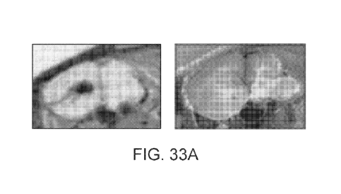
[0203] FIG. 33 illustrates sagittal (FIG. 33A) and coronal (FIG. 33B) brain
sections indicating
localization of a peptide of SEQ ID NO: 55 to specific structures in the
brain, such as ventricles
and CSF. In both FIG. 33A and FIG. 33B, the radioactivity scan is shown on the
left, with dark
areas having higher activity. Images of the tissue in normal light are shown
on the right.
[0204] FIG. 34 illustrates a white light image and a corresponding
autoradiographic image of a
mouse with ligated kidneys 3 hours after administration of 100 nmol of the
radiolabeled SEQ ID
NO: 39 peptide.
[0205] FIG. 34A illustrates a white light image of a frozen section of a mouse
with ligated
kidneys 3 hours after administration of 100 nmol of the radiolabeled SEQ ID
NO: 39 peptide.
[0206] FIG. 34B illustrates an autoradiographic image corresponding to FIG.
34A in which the
14C signal identifies the peptide distribution in the tissues of the mouse
with ligated kidneys 3
hours after administration of 100 nmol of the radiolabeled SEQ ID NO: 39
peptide.
[0207] FIG. 35 illustrates a white light image and the corresponding
autoradiographic image of a
mouse with ligated kidneys 3 hours after administration of 100 nmol of the
radiolabeled SEQ ID
NO: 36 peptide.
[0208] FIG. 35A illustrates a white light image of a frozen section of a mouse
with ligated
kidneys 3 hours after administration of 100 nmol of the radiolabeled SEQ ID
NO: 36 peptide.
[0209] FIG. 35B illustrates an autoradiographic image corresponding to FIG.
35A in which the
14C signal identifies the peptide distribution in the tissues of the mouse
with ligated kidneys 3
hours after administration of 100 nmol of the radiolabeled SEQ ID NO: 36
peptide.
[0210] FIG. 36 illustrates white light and autoradiographic images of murine
coronal brain
sections, identifying peptide distribution 3 hours after administration of 100
nmol of the
CA 02987636 2017-11-28
WO 2016/210376 PCT/US2016/039431
radiolabeled first purified fraction (first HPLC peptide peak) of a peptide of
SEQ ID NO: 55 or
the radiolabeled second purified fraction (second HPLC peptide peak) of a
peptide of SEQ ID
NO: 55 from the same HPLC.
[0211] FIG. 36A illustrates white light images of coronal brain sections of a
mouse on the right
and autoradiographic images that correspond to the white light images on the
left. The 14C signal
in the autographic images identifies the peptide distribution, indicating
localization of the
radiolabeled first purified fraction (first HPLC peptide peak) of a peptide of
SEQ ID NO: 55, to
specific structures in the brain, such as ventricles and CSF 3 hours after
administration of 100
nmol of the peptide.
[0212] FIG. 36B illustrates white light images of coronal brain sections of a
mouse on the right
and autoradiographic images corresponding to the white light images on the
left. The 14C signal
in the autographic images identifies the peptide distribution, indicating
localization of the second
purified fraction (second HPLC peptide peak from the HPLC in FIG. 36A) of a
peptide of SEQ
ID NO: 55, to specific structures in the brain, such as ventricles and CSF 3
hours after
administration of 100 nmol of the peptide.
[0213] FIG. 37 illustrates a white light image and the corresponding
autoradiographic image of a
mouse with ligated kidneys identifying peptide distribution 3 hours after
administration of 100
nmol of the radiolabeled first purified fraction (first HPLC peptide peak) of
a peptide of SEQ ID
NO: 55.
[0214] FIG. 37A illustrates a white light image of a frozen section of a mouse
with ligated
kidneys 3 hours after administration of 100 nmol of the radiolabeled first
purified fraction (first
HPLC peptide peak) of a peptide of SEQ ID NO: 55.
[0215] FIG. 37B illustrates an autoradiographic image corresponding to FIG.
37A in which the
14C signal identifies the peptide distribution in the tissues of a mouse with
ligated kidneys 3 hours
after administration of 100 nmol of the radiolabeled first fraction (first
HPLC peptide peak) of a
peptide of SEQ ID NO: 55.
[0216] FIG. 38 illustrates a white light image and the corresponding
autoradiographic image of a
mouse with ligated kidneys identifying peptide distribution 3 hours after
administration of 100
nmol of the radiolabeled second purified fraction (second HPLC peptide peak of
the HPLC from
FIG. 37) of a peptide of SEQ ID NO: 55.
[0217] FIG. 38A illustrates a white light image of a frozen section of a mouse
with ligated
kidneys 3 hours after administration of 100 nmol of the radiolabeled second
purified fraction
(second HPLC peptide peak of the HPLC from FIG. 37) of a peptide of SEQ ID NO:
55.
CA 02987636 2017-11-28
WO 2016/210376 PCT/US2016/039431
26
[0218] FIG. 38B illustrates an autoradiographic image corresponding to FIG.
38A in which the
14C signal identifies the peptide distribution in the tissues of the mouse
with ligated kidneys 3
hours after administration of 100 nmol of the radiolabeld second purified
fraction (second HPLC
peptide peak of the HPLC from FIG. 37) of a peptide of SEQ ID NO: 55.
[0219] FIG. 39 illustrates a white light and the corresponding
autoradiographic image of a
mouse with ligated kidneys identifying peptide distribution 3 hours after
administration of 100
nmol of the radiolabeled SEQ ID NO: 83 peptide.
[0220] FIG. 39A illustrates a white light image of a frozen section of a mouse
with ligated
kidneys 3 hours after administration of 100 nmol of the radiolabeled SEQ ID
NO: 83 peptide
[0221] FIG. 39B illustrates an autoradiographic image corresponding to FIG.
39A in which the
14C signal identifies the peptide distribution in the tissues of the mouse
with ligated kidneys 3
hours after administration of 100 nmol of the radiolabeled SEQ ID NO: 83
peptide.
[0222] FIG. 40 illustrates white light images of coronal brain sections on the
right and
autoradiographic images that correspond to the white light images on the left.
The 14C signal in
the autographic images identifies the peptide distribution 3 hours after
administration of the
radiolabeled SEQ ID NO: 34 and indicates the localization of the peptide to
specific structures in
the brain, such as ventricles and CSF.
[0223] FIG. 41 illustrates white light images of coronal brain sections on the
right and
autoradiographic images that correspond to the white light images on the left.
The 14C signal in
the autographic images identifies the peptide distribution 3 hours after
administration of the
radiolabeled SEQ ID NO: 83 and indicates the localization of the peptide to
specific structures in
the brain, such as ventricles and CSF.
[0224] FIG. 42 shows a near-infrared fluorescence image of Co10205 tumor (top
left), colon (top
middle), liver (top right), brain (middle left), spleen (middle right), muscle
(bottom left), skin
(bottom middle), and kidney (bottom right) that were excised 24 hours after
administration of 10
nmol of a peptide of SEQ ID NO: 37 conjugated to Cy5.5 to Co10205 tumor-
bearing Female
Harlan athymic mice.
[0225] FIG. 43 shows a near-infrared fluorescence image of MDA-MB-231 tumor
(top left),
colon (top middle), liver (top right), brain (middle left), spleen (middle
right), muscle (bottom
left), skin (bottom middle), and kidney (bottom right) that were excised 24
hours after
administration of 10 nmol of a peptide of SEQ ID NO: 37 conjugated to Cy5.5 to
MDA-MB-231
tumor-bearing Female Harlan athymic mice.
CA 02987636 2017-11-28
WO 2016/210376 PCT/US2016/039431
27
[0226] FIG. 44 shows a near-infrared fluorescence image of U87 tumor (top
left), colon (top
middle), liver (top right), brain (middle left), spleen (middle right), muscle
(bottom left), skin
(bottom middle), and kidney (bottom right) that were excised 24 hours after
administration of 10
nmol of a peptide of SEQ D NO: 37 conjugated to Cy5.5 to U87 tumor-bearing
Female Harlan
athymic mice.
[0227] FIG. 45 shows sequences of SEQ ID NO: 2 aligned with SEQ ID NO: 3 with
annotation
of the location of loops, and their corresponding 3D structures, with the SEQ
ID NO: 2 structure
on the left and the SEQ ID NO: 3 structure on the right.
[0228] FIG. 46 shows the sequence alignment of SEQ ID NO: 1 and SEQ ID NO: 4
with the
location of the loops annotated.
DETAILED DESCRIPTION
[0229] The present disclosure relates to compositions and methods for
treatment of tumors.
Furthermore, it relates to compositions that can cross the blood brain
barrier, enabling treatment
of brain tumors and other brain disorders and diseases. In some embodiments
the compositions
and methods herein utilize peptides that home, distribute to, target, are
directed to, accumulate in,
migrate to, and/or bind to cancerous cells following administration to a
subject. In further
embodiments the compositions and methods herein utilize peptides that home,
distribute to,
target, are directed to, accumulate in, migrate to, and/or bind to cancerous
or diseased cells in the
brain following administration to a subject. In other embodiments, peptides
described herein
cross the blood brain barrier into the neuronal parenchyma to deliver
therapeutically active
molecules to targets of neurological diseases including brain cancers. In some
embodiments, the
homing peptides of the present disclosure are used to deliver an active agent
to a tissue or cell
thereof. The active agent can exert a therapeutic effect on the targeted
tissue or cell thereof. For
example, in certain embodiments, the peptide allows for localized delivery of
a chemotherapeutic
agent to a cancerous tissue or cell thereof. As another example, in certain
embodiments, the
peptide allows for localized delivery of a therapeutic drug to a diseased
tissue or cell of the brain.
In certain embodiments, the homing peptides of the present disclosure are used
to image the
targeted tissue or cell. For example, the peptide allows for imaging using a
fluorophore. In
certain embodiments, the peptide itself possesses or induces therapeutic
responses.
[0230] Many types of tumors are difficult to treat. Often, the prognosis of
the patient is directly
influenced by the ability drug therapies to effectively kill the cancerous
cells and on the precision
with which the cancer cells can be surgically resected. For example, one
challenge in treating
CA 02987636 2017-11-28
WO 2016/210376 PCT/US2016/039431
28
tumors is that many drugs treatments are systemic, and therefore, the efficacy
of their use is
constrained by the toxicity of systemic use. Another challenge is that the
current methods of
intra-operative imaging of cancerous tissues fail to precisely depict tumor
margins or small foci
of cancerous cells. Instead, resection is dependent upon the surgeon's ability
to visually
recognize tumor or physically locate it by touch in a surgical setting, which
can be an imprecise
method to identify the tumor margins or foci.
[0231] Treatment of brain tumors, as well as other brain disorders and
disease, are also
challenging and complicated to treat. One challenge is that many drugs
administered into the
circulatory system of patients fail to cross the blood-brain barrier (BBB) or
the blood CSF
barrier, which are selective barriers that separates the circulating blood
from the brain
extracellular fluid and the central nervous system tissue. Another challenge
is that many drugs
lack sufficient specificity to one or more target regions, tissues, structures
or cells in the brain.
Thus, treatment of brain conditions often requires the use of high
concentrations of non-specific
drugs, leading to suboptimal efficacy and systemic side effects. One other way
to deliver drugs to
the brain is to apply them directly in conjunction with surgical procedures.
Another way is to
identify specific drugs that cross the blood brain barrier. Specific and
potent drugs that are
capable of crossing the BBB can counteract the non-specificity of many
treatments by selectively
targeting and delivering compounds to specific tissues, cells, structures and
regions. Such drugs
can also be useful to modulate ion channels, protein-protein interactions,
extracellular matrix
remodeling (i.e., protease inhibition), intracellular signaling pathways,
neurotransmitter
signaling, infections, and the like. Such targeted therapy can allow for lower
dosing, reduced side
effects, and improvement in therapeutic outcomes, which would be advantageous
not only in
acute disease of the brain, but in chronic conditions as well.
[0232] The present disclosure describes a class of peptides derived from
knottins that can home,
distribute to, target, be directed to, accumulate in, migrate to, and/or bind
to cancerous or
diseased cells, and be used either directly or as carriers of active drugs,
peptides or molecules to
treat the cancerous or diseased cells. A peptide that homes, distributes to,
targets, migrates to, or
accumulates in one or more specific cancerous or diseased regions, tissues,
structures or cells can
have fewer off-target and potentially negative effects.
[0233] The present disclosure also provides a new kind of carrier that can
deliver an active agent
or detectable agent to a specific region, tissue, structure or cell that can
be used for either or both
therapeutic and imaging purposes. As described herein, an active agent or
detectable agent can be
linked to a peptide of the disclosure.
CA 02987636 2017-11-28
WO 2016/210376 PCT/US2016/039431
29
[0234] Furthermore, the present disclosure describes a class of peptides
derived from knottins
that can effectively cross the BBB or blood CSF barrier and be used either
directly or as carriers
of active drugs, peptides or molecules to treat a brain condition. For
instance, Alzheimer's
disease is a brain condition that is associated with the aggregation of
amyloid beta peptide
fragment. The accumulation of the amyloid beta peptide fragment is a result of
proteolytic
cleavage of the amyloid precursor protein (APP) by an enzyme known as beta-
secretase. A
therapeutic peptide that could cross the BBB to interact with and inhibit the
beta-secretase
protease could be used in the treatment and prevention of Alzheimer's disease
by reducing
aggregation of the amyloid beta fragment through, for example, binding or
inhibiting the
protease, antagonizing APP cleavage, regulating the amyloid beta fragment
pathway, or other
mechanism. Furthermore, acetylcholinesterase inhibitors such as rivastigmine
have been used to
treat Alzheimer's disease. However these are systemically dosed and often
cause symptoms such
as bradycardia and bronchoconstriction in the periphery. The opportunity to
deliver more of the
acetylcholineseterase across the BBB as a conjugate may allow for lower doses
and side effects
in the periphery. The peptides of the disclosure can be used to treat the
symptoms of various
conditions.
[0235] Also described herein are peptides that selectively home, distribute
to, target, are directed
to, migrate to, or accumulate in specific regions, tissues, structures or
cells of the brain. In some
cases, the peptides accumulate in one or more of: the hippocampus, the center
of memory and
learning and spatial navigation; the cerebrospinal fluid (CSF), which is found
in the brain and
spine; the ventricular system, the site of CSF production and circulation; the
rostral migratory
stream; the dentate gyrus; neural stem cells; or neuronal precursors. The
dentate gyrus of the
hippocampus and the subventricular zone are two locations of neurogenesis in
the adult brain,
and the rostral migratory stream is one mechanism for migration of new
neurons. Thus, targeting
those regions could allow for modulation of various aspects of neurogenesis,
including repair or
regeneration. A peptide that homes, distributes to, targets, migrates to, or
accumulates in one or
more specific regions, tissues, structures or cells of the brain can have
fewer off-target and
potentially negative effects, for example, side effects that often limit use
and efficacy of drugs for
neurological conditions. In addition, such peptides can increase the efficacy
of existing drugs by
directly targeting them to a specific region, tissue, structure or cell of the
brain and helping the
drug cross the blood brain barrier.
[0236] The present disclosure also provides a new kind of drug carrier that
can deliver an active
agent or detectable agent to the brain that can be used for either or both
therapeutic and imaging
CA 02987636 2017-11-28
WO 2016/210376 PCT/US2016/039431
purposes. The blood¨brain barrier is formed by special tight junctions between
the endothelial
cells that surround the brain tissue, as well as a basement membrane and
astrocyte protrusions.
Similarly the blood CSF barrier is formed by tight junctions between choroidal
epithelial cells, a
basement membrane, and endothelial cells. One of the functions of the BBB and
the blood CSF
barrier is to protect the brain and keep it isolated from harmful toxins that
may be in the blood
stream. As described herein, an active agent or a detectable agent can be
linked to a peptide of
the disclosure and the linked peptide-active agent or linked peptide-
detectable agent compound
can cross the blood brain barrier or blood CSF barrier.
[0237] The disclosure also provides a method for treating a condition of a
subject, wherein the
method comprises administrating to the subject a peptide that homes, targets,
migrates to, is
directed to a region, tissue, structure or cell in the brain of the subject,
for example within the
hippocampus, the CSF, the ventricular system, the meninges, the rostral
migratory stream, or
other specific region of the brain, for example, substantia nigra (which can
be associated with
Parkinsons disease). In some cases, the administered peptide can cross the
blood brain barrier or
blood CSF barrier of the subject. Additional aspects and advantages of the
present disclosure will
become apparent to those skilled in this art from the following detailed
description, wherein
illustrative embodiments of the present disclosure are shown and described. As
will be realized,
the present disclosure is capable of other and different embodiments, and its
several details are
capable of modifications in various respects, all without departing from the
disclosure.
Accordingly, the drawings and description are to be regarded as illustrative
in nature, and not as
restrictive.
[0238] Additional aspects and advantages of the present disclosure will become
apparent to those
skilled in this art from the following detailed description, wherein
illustrative embodiments of the
present disclosure are shown and described. As will be realized, the present
disclosure is capable
of other and different embodiments, and its several details are capable of
modifications in various
respects, all without departing from the disclosure. Accordingly, the drawings
and description are
to be regarded as illustrative in nature, and not as restrictive.
[0239] As used herein, the abbreviations for the natural L-enantiomeric amino
acids are
conventional and are as follows: alanine (A, Ala); arginine (R, Arg);
asparagine (N, Asn);
aspartic acid (D, Asp); cysteine (C, Cys); glutamic acid (E, Glu); glutamine
(Q, Gln); glycine (G,
Gly); histidine (H, His); isoleucine (I, Ile); leucine (L, Leu); lysine (K,
Lys); methionine (M,
Met); phenylalanine (F, Phe); proline (P, Pro); serine (S, Ser); threonine (T,
Thr); tryptophan (W,
Trp); tyrosine (Y, Tyr); valine (V, Val). Typically, Xaa can indicate any
amino acid. In some
CA 02987636 2017-11-28
WO 2016/210376 PCT/US2016/039431
31
embodiments, X can be asparagine (N), glutamine (Q), histidine (H), lysine
(K), or arginine (R).
[0240] Some embodiments of the disclosure contemplate D-amino acid residues of
any standard
or non-standard amino acid or analogue thereof. When an amino acid sequence is
represented as
a series of three-letter or one-letter amino acid abbreviations, the left-hand
direction is the amino
terminal direction and the right-hand direction is the carboxy terminal
direction, in accordance
with standard usage and convention.
Peptides
[0241] Knottins are a class of peptides, usually ranging from about 11 to
about 81 amino acids in
length that are often folded into a compact structure. Knottins are typically
assembled into a
complex tertiary structure that is characterized by a number of intramolecular
disulfide crosslinks
and may contain beta strands and other secondary structures. The presence of
the disulfide bonds
gives knottins remarkable environmental stability, allowing them to withstand
extremes of
temperature and pH and to resist the proteolytic enzymes of the blood stream.
The rigidity of
knottins also allows them to bind to targets without paying the "entropic
penalty" that a floppy
peptide accrues upon binding a target. For example, binding is adversely
affected by the loss of
entropy that occurs when a peptide binds a target to form a complex.
Therefore, "entropic
penalty" is the adverse effect on binding, and the greater the entropic loss
that occurs upon this
binding, the greater the "entropic penalty." Furthermore, unbound molecules
that are flexible lose
more entropy when forming a complex than molecules that are rigidly
structured, because of the
loss of flexibility when bound up in a complex. However, rigidity in the
unbound molecule also
generally increases specificity by limiting the number of complexes that
molecule can form. The
knotted peptides can bind targets with antibody-like affinity. A wider
examination of the
sequence structure and sequence identity or homology of knottins reveals that
they have arisen by
convergent evolution in all kinds of animals and plants. In animals, they are
typically found in
venoms, for example, the venoms of spiders and scorpions and have been
implicated in the
modulation of ion channels. The knottin proteins of plants can inhibit the
proteolytic enzymes of
animals or have antimicrobial activity, suggesting that knottins can function
in the native defense
of plants.
[0242] The present disclosure provides peptides that comprise or are derived
from these knotted
peptides (or knottins). As used herein, the term "knotted peptide" is
considered to be
interchangeable with the terms "knottin" and "optide."
CA 02987636 2017-11-28
WO 2016/210376 PCT/US2016/039431
32
[0243] The peptides of the present disclosure can comprise cysteine amino acid
residues. In some
cases, the peptide has at least 6 cysteine amino acid residues. In some cases,
the peptide has at
least 8 cysteine amino acid residues. In other cases, the peptide has at least
10 cysteine amino
acid residues, at least 12 cysteine amino acid residues, at least 14 cysteine
amino acid residues or
at least 16 cysteine amino acid residues.
[0244] A knotted peptide can comprise disulfide bridges. A knotted peptide can
be a peptide
wherein 5% or more of the residues are cysteines forming intramolecular
disulfide bonds. A
disulfide-linked peptide can be a drug scaffold. In some embodiments, the
disulfide bridges form
a knot. A disulfide bridge can be formed between cysteine residues, for
example, between
cysteines 1 and 4, 2 and 5, or, 3 and 6. In some cases, one disulfide bridge
passes through a loop
formed by the other two disulfide bridges, for example, to form the knot. In
other cases, the
disulfide bridges can be formed between any two cysteine residues.
[0245] The present disclosure further includes peptide scaffolds that, e.g.,
can be used as a
starting point for generating additional peptides. In some embodiments, these
scaffolds can be
derived from a variety of knotted peptides (or knottins). In certain
embodiments, knotted peptides
are assembled into a complex tertiary structure that is characterized by a
number of
intramolecular disulfide crosslinks, and optionally contain beta strands and
other secondary
structures such as an alpha helix. For example, knotted peptides include, in
some embodiments,
small disulfide-rich proteins characterized by a disulfide through disulfide
knot. This knot can be,
e.g., obtained when one disulfide bridge crosses the macrocycle formed by two
other disulfides
and the interconnecting backbone. In some embodiments, the knotted peptides
can include
growth factor cysteine knots or inhibitor cysteine knots. Other possible
peptide structures include
peptide having two parallel helices linked by two disulfide bridges without 0-
sheets (e.g.,
hefutoxin).
[0246] A knotted peptide can comprise at least one amino acid residue in an L
configuration. A
knotted peptide can comprise at least one amino acid residue in a D
configuration. In some
embodiments, a knotted peptide is 15-40 amino acid residues long. In other
embodiments, a
knotted peptide is 11-57 amino acid residues long. In still other embodiments,
a knotted peptide
is 11-81 amino acid residues long. In further embodiments, a knotted peptide
is at least 20 amino
acid residues long.
[0247] These kinds of peptides can be derived from a class of proteins known
to be present or
associated with toxins or venoms. In some cases, the peptide can be derived
from toxins or
venoms associated with scorpions or spiders. The peptide can be derived from
venoms and toxins
CA 02987636 2017-11-28
WO 2016/210376 PCT/US2016/039431
33
of spiders and scorpions of various genus and species. For example, the
peptide can be derived
from a venom or toxin of the Leiurus quinquestriatus hebraeus, Buthus
occitanus tunetanus,
Hottentotta judaicus, Mesobuthus eupeus, Buthus occitanus israelis, Hadrurus
gertschi,
Androctonus australis, Centruroides noxius, Heterometrus laoticus,
Opistophthalmus carinatus,
Haplopelma schmidti, Isometrus maculatus, Haplopelma huwenum, Haplopelma
hainanum,
Haplopelma schmidti, Agelenopsis aperta, Haydronyche versuta, Selenocosmia
huwena,
Heteropoda venatoria, Grammostola rosea, Ornithoctonus huwena, Hadronyche
versuta, Atrax
robustus, Angelenopsis aperta, Psalmopoeus cambridgei, Hadronyche infensa,
Paracoelotes
luctosus, and Chilobrachys jingzhaoor another suitable genus or species of
scorpion or spider. In
some cases, a peptide can be derived from a Buthus martensii Karsh (scorpion)
toxin. In some
embodiments, a peptide can be derived from a member of the pfam005453: Toxin
_6 class.
[0248] TABLE 1 lists exemplary peptides derived from venoms or toxins of
scorpions or spiders
and for use with the present disclosure.
TABLE 1. Exemplary peptides according to the present disclosure.
SEQ ID NO: Peptide Sequence
SEQ ID NO: 1 GSMCIPCFTTNPNMAAKCNACCGSRRGSCRGPQCIC
SEQ ID NO: 2 GSGCLEFWWKCNPNDDKCCRPKLKCSKLFKLCNFSFG
SEQ ID NO: 3 GSECRYWLGTCSKTGDCCSHLSCSPKHGWCVWDWTFRK
SEQ ID NO: 4 GSMCMPCFTTDHQMARRCDDCCGGRGRGRCYGPQCLCR
SEQ ID NO: 5 GSMCMPCFTTHHRMAENCDICCGGDGRGKCYGPQCLCR
SEQ ID NO: 6 GSMCMPCFTTDHRMAENCDICCGGDGRGKCYGPQCLCR
SEQ ID NO: 7 GSMCMPCFTTHHQMAENCDICCGGDGRGKCYGPQCLCR
SEQ ID NO: 8 GSMCMPCFTTHHRMARNCDICCGGDGRGKCYGPQCLCR
SEQ ID NO: 9 GSMCMPCFTTHHRMAERCDICCGGDGRGKCYGPQCLCR
SEQ ID NO: 10 GSMCMPCFTTHHRMAENCDDCCGGDGRGKCYGPQCLCR
SEQ ID NO: 11 GSRCMPCFTTDHQMARRCDDCCGGRGRGKCYGPQCLCR
SEQ ID NO: 12 GSICIPCFTTDHQIARRCDDCCGGRGRGKCYGPQCLCR
SEQ ID NO: 13 GSMCLPCFTTDHQLARRCDDCCGGRGRGKCYGPQCLCR
SEQ ID NO: 14 GSMCMPCFTTEHQMARRCEECCGGRGRGKCYGPQCLCR
SEQ ID NO: 15 GSMCIPCFTTDHQMARRCEECCGGRGRGKCYGPQCLCR
SEQ ID NO: 16 GSICIPCFTTDHQMARRCDDCCGGRGDGKCYGPQCLCR
SEQ ID NO: 17 GSICIPCFTTDHQIARRCDDCCGGRGRGKCYGPQCICR
SEQ ID NO: 18 GSRCMPCFTTDHFMARFCDFCCGGRGRGKCYGPQCLCR
SEQ ID NO: 19 GSRCMPCFTTDHYMARYCDYCCGGRGRGKCYGPQCLCR
SEQ ID NO: 20 GSRCMPCFTTDHRMARRCDRCCGGRGRGKCYGPQCLCR
SEQ ID NO: 21 GSRCMPCFTTDHEMARECDECCGGRGRGKCYGPQCLCR
SEQ ID NO: 22 GSRCMPCFTTDHHMARHCDHCCGGRGRGKCYGPQCLCR
SEQ ID NO: 23 GSLCLPCFTTHHRLADQCDICCGGDGRGKCYGPQCLCR
CA 02987636 2017-11-28
WO 2016/210376
PCT/US2016/039431
34
SEQ ID NO: Peptide Sequence
SEQ ID NO: 24 GSICIPCFTTEHQIARRCEECCGGRGRGKCYGPQCLCR
SEQ ID NO: 25 GSMCMPCFTTDTQMQERCDRCCGGGGRGKCWGPQCLCI
SEQ ID NO: 26 GSMCMPCFTTIYRMAHECDECCGGRGRGKCYGPQCLCR
SEQ ID NO: 27 GSMCMPCFTTGYRMAEYCDICCGGRGRGKCYGPQCLCR
SEQ ID NO: 28 GSMCMPCFTTHRRMANTCDACCGGRSRGKCYGPQCLCR
SEQ ID NO: 29 GSHCMPCFTTDHQMIRRCDDCCGGGSYGKCDGPQCLCF
SEQ ID NO: 30 GSDCMPCFTTDHRMADHCDICCGGDDRGKCYGPQCLCR
SEQ ID NO: 31 GSMCMPCFTTDHEMERRCDDCCGIGGGGKCHGPQCLCG
SEQ ID NO: 32 GSMCMPCFTTEQRMAIICDDCCGGFGRGKCYGPQCLCR
SEQ ID NO: 33 GSMCMPCFTTSEQMFRRCDDCCGGWGDGKCNGPHCLCR
SEQ ID NO: 34 GSGVPINVKCRGSRDCLDPCKKAGMRFGKCINSKCHCTP
SEQ ID NO: 35 GSMCMPCFTTEQRMAIICDDCCGGFGRGRCYGPQCLCR
SEQ ID NO: 36 GSICIPCFTTDHQIARRCDDCCGGRGRGRCYGPQCICR
SEQ ID NO: 37 GSMCMPCFTTDTQMQERCDRCCGGGGRGRCWGPQCLCI
SEQ ID NO: 38 GSMCMPCFTTDTQMQERCDRCCGGGGRGRCWGPQCLC
SEQ ID NO: 39 GSMCMPCFTTDHRMAENCDICCGGDGRGRCYGPQCLCR
SEQ ID NO: 40 GSMCMPCFTTEQRMAIICDDCCGGFGRGKCYGPQCLCI
SEQ ID NO: 41 GSMCMPCFTTEQRMAIICDDCCGGFGRGRCYGPQCLCI
SEQ ID NO: 42 GSICIPCFTTDHQIARRCDDCCGGRGRGKCYGPQCICI
SEQ ID NO: 43 GSICIPCFTTDHQIARRCDDCCGGRGRGRCYGPQCICI
SEQ ID NO: 44 GSMCMPCFTTDTQMQEKCDRCCGGGGRGRCWGPQCLCI
SEQ ID NO: 45 GSMCMPCFTTEQRMAIKCDDCCGGFGRGRCYGPQCLCR
SEQ ID NO: 46 GSICIPCFTTDHQIARKCDDCCGGRGRGRCYGPQCICR
SEQ ID NO: 47 GSMCMPCFTTDHRMAEKCDICCGGDGRGRCYGPQCLCR
SEQ ID NO: 48 GSMCMPCFTTDTQMQERCDRCCGGKGRGRCWGPQCLCI
SEQ ID NO: 49 GSMCMPCFTTEQRMAIICDDCCGGKGRGRCYGPQCLCR
SEQ ID NO: 50 GSICIPCFTTDHQIARRCDDCCGGKGRGRCYGPQCICR
SEQ ID NO: 51 GSMCMPCFTTDHRMAENCDICCGGKGRGRCYGPQCLCR
SEQ ID NO: 52 GSMCMPCFTTDHRMAENCDICCGGDGRGKCYGPQCLCI
SEQ ID NO: 53 GSMCMPCFTTDHRMAENCDICCGGDGRGRCYGPQCLCI
SEQ ID NO: 54 GSMCMPCFTTHHRMAENCDICCGGDGRGRCYGPQCLCR
SEQ ID NO: 55 GSVGCEECPMHCKGKNANPTCDDGVCNCNV
SEQ ID NO: 56 GSVGCEECPMHCKGKNAKPTCDDGVCNCNV
SEQ ID NO: 57 GSVGCEECPMHCKGKNAKPTCDNGVCNCNV
SEQ ID NO: 58 GSVGCEECPMHCKGKHAVPTCDDGVCNCNV
SEQ ID NO: 59 GSVGCEECPAHCKGKNAKPTCDDGVCNCNV
SEQ ID NO: 60 GSVGCEECPAHCKGKNAIPTCDDGVCNCNV
SEQ ID NO: 61 GSVGCEECPMHCKGKMAKPTCDDGVCNCNV
SEQ ID NO: 62 GSVGCEECPMHCKGKNAVPTCDNGVCNCNA
SEQ ID NO: 63 GSVGCEECPMHCKGKMAKPTCYDGVCNCNV
SEQ ID NO: 64 GSVGCEECPMYCKGKNAVPTCDGGVCNCNA
SEQ ID NO: 65 GSVGCEECPKYCKGKNAVPTCDGGVCNCNA
SEQ ID NO: 66 GSVGCEECPVYCKGKKALPTCDGGVCNCNA
CA 02987636 2017-11-28
WO 2016/210376 PCT/US2016/039431
SEQ ID NO: Peptide Sequence
SEQ ID NO: 67 GSVGCEEDPMHCKGKQAKPTCCNGVCNCNV
SEQ ID NO: 68 GSVGCAECPMHCKGKMAKPTCENEVCKCNIGKKD
SEQ ID NO: 69 GSVGCEECPMHCKGKKALPTCDYGCECND
SEQ ID NO: 70 GSIVCKVCKIICGMQGKKVNICKAPIKCKCKKG
SEQ ID NO: 71 GSVSCEDCPDHCSTQKARAKCDNDKCVCEPK
SEQ ID NO: 72 GSVSCEDCPEHCSTQKARAKCDNDKCVCESV
SEQ ID NO: 73 GSVSCEDCPEHCSTQKAQAKCDNDKCVCEPI
SEQ ID NO: 74 GSATCEDCPEHCATQNARAKCDNDKCVCEPK
SEQ ID NO: 75 GSVSCEDCPEHCATKDQRAKCDNDKCVCEPK
SEQ ID NO: 76 GSVGCEDCPEHCSQQNARAKCENDKCVCEPK
SEQ ID NO: 77 GSVSCEDCPEHCATKDQRAKCDNDRCVCEPK
SEQ ID NO: 78 GSVSCEDCPPHCATKDQRAKCENDKCVCEPK
SEQ ID NO: 79 GSVSCEDCPEHCSTQKARAKCDNDKCVCEAI
SEQ ID NO: 80 GSMCMPCFTTEQRMAIICDDCCGGFGRGRCYGPQCLC
SEQ ID NO: 81 GSICIPCFTTDHQIARRCDDCCGGRGRGRCYGPQCIC
SEQ ID NO: 82 GSMCMPCFTTDHRMAENCDICCGGDGRGRCYGPQCLC
SEQ ID NO: 83 GSVGCEECPMHCRGRNANPTCDDGVCNCNV
SEQ ID NO: 84 GSVGCEECPMHCRGRNANPTCDDGVCNC
SEQ ID NO: 85 GSCGPCFTTDHQMEQKCAECCGGIGKCYGPQCLCNR
SEQ ID NO: 86 GSRCGPCFTTDPQTQAKCSECCGRKGGVCKGPQCICGIQY
SEQ ID NO: 87 GSMCMPCFTTDPNMAKKCRDCCGGNGKCFGPQCLCNR
SEQ ID NO: 88 GSMCMPCFTTDHNMAKKCNDCCGGYGKCFGPQCLCR
SEQ ID NO: 89 GSRCPPCFTTNPNMEADCRKCCGGRGYCASYQCICPGG
SEQ ID NO: 90 GSMCMPCFTTDPNMANKCRDCCGGGKKCFGPQCLCNR
GSMKFLYGVILIALFLTVMTATLSEARCGPCFTTDPQTQAKCSECCGRKGG
SEQ ID NO: 91 VCKGPQCICGIQY
SEQ ID NO: 92 GSMCMPCFTTRPDMAQQCRACCKGRGKCFGPQCLCGYD
GSMKFLYGIVFIALFLTVMTATLSDAMCMPCFTTDHNMAKKCRDCCGGN
SEQ ID NO: 93 GKCFGPQCLCNRG
SEQ ID NO: 94 GSMCMPCFTTDHNMAKKCRDCCGGNGKCFGPQCLCNR
GSMKFLYGIVFITLFLTVMIATHTEAMCMPCFTTRPNMAQQCRDCCRGRG
SEQ ID NO: 95 KCFGPQCLCGYD
GSMKFLYGIVFIALFLTVMIATHTEAMCMPCFTTRPNMAQQCRDCCRGRG
SEQ ID NO: 96 KCFGPQCLCGYD
SEQ ID NO: 97 GSRCKPCFTTDPQMSKKCADCCGGKGKGKCYGPQCLC
GSMKFLYGIVFITLFLTVMIATHTEAAMCMPCFTTNLNMEQECRDCCGGT
SEQ ID NO: 98 GRCFGPQCLCGYD
SEQ ID NO: 99 GSRCSPCFTTDQQMTKKCYDCCGGKGKGKCYGPQCICAPY
SEQ ID NO: 100 GSCGPCFTTDPYTESKCATCCGGRGKCVGPQCLCNRI
SEQ ID NO: 101 GSTEAMCMPCFTTDHNMAKKCRDCCGGNGKCFGYQCLCNRG
GSMKFLYGIVFIALFLTVMFATQTDGCGPCFTTDANMARKCRECCGGIGK
SEQ ID NO: 102 CFGPQCLCNRI
CA 02987636 2017-11-28
WO 2016/210376 PCT/US2016/039431
36
SEQ ID NO: Peptide Sequence
GSMKFLYGIVFIALFLTVMFATQTDGCGPCFTTDANMARKCRECCGGNGK
SEQ ID NO: 103 CFGPQCLCNRE
GSMKFLYGTILIAFFLTVMIATHSEARCPPCFTTNPNMEADCRKCCGGRGY
SEQ ID NO: 104 CASYQCICPGG
SEQ ID NO: 105 GSTEAMCMPCFTTRPDMAQQCRDCCGGNGKCFGYQCLCNRG
GSMKFLYGIVFIALFLTVMIATLTEAMCMPCFTTRPDMAQQCRDCCGGNG
SEQ ID NO: 106 KCFGYQCLCNRG
GSMKFLYGIVFIALFLTVMIATHTEAMCMPCFTTRPDMAQQCRDCCGGNG
SEQ ID NO: 107 KCFGYQCLCNRG
GSMKFLYGIILIALFLTVMIATHSEARCPNCFTTNPNAEADCKKCCGNRWG
SEQ ID NO: 108 KCAGYQCVCPMK
GSMKFLYGIVFIALFLTGMIATHTEAMCMPCFTTRPDMAQQCRDCCGGNG
SEQ ID NO: 109 KCFGYQCLCNRGRIVIMYT
SEQ ID NO: 110 GSMCMPCFTTRPGMAQQCRDCCGGNGKCFGYQCLCNR
SEQ ID NO: 111 GSMCIPCFTTNPNMAAKCNACCGSRRGSCRGPQCICR
SEQ ID NO: 112 GSMCIPCFTTNPNMAAKCNACCGSRRGSCRGPQCICN
SEQ ID NO: 113 GSMCIPCFTTNPNMAAKCNACCGGNGSCRGPQCICN
SEQ ID NO: 114 GSMCIPCFTTNPNMAAKCNACCGSRGRGSCRGPQCICN
SEQ ID NO: 115 GSMCIPCFTTNPNMAAKCNACCGSRGRGKCRGPQCICN
SEQ ID NO: 116 GSMCIPCFTTDHQMAAKCNACCGSRRGSCRGPQCICN
SEQ ID NO: 117 GSMCIPCFTTNHQMAAKCNACCGSRRGSCRGPQCICN
SEQ ID NO: 118 GSMCIPCFTTNPNMARKCNACCGSRGRGSCRGPQCICN
SEQ ID NO: 119 GSMCIPCFTTNPNMAAKCNACCGGKGRGSCRGPQCICN
SEQ ID NO: 120 GSMCIPCFTTNPNMAAKCNACCGSRRGSCFGPQCICN
SEQ ID NO: 121 GSMCIPCFTTNPNMAAKCNACCGSRGRGKCFGPQCICN
SEQ ID NO: 122 GSMCIPCFTTNPNMAAKCNACCGSRGRGSCFGPQCICN
SEQ ID NO: 123 GSMCIPCFTTNPNMAAKCNACCGSRGRGSCYGPQCICN
SEQ ID NO: 124 GSMCIPCFTTNPNMAAKCDACCGSRRGSCRGPQCICN
SEQ ID NO: 125 GSMCIPCFTTNHQMAAKCDACCGSRRGSCRGPQCICN
SEQ ID NO: 126 GSMCIPCFTTNHNMAAKCDACCGGRGRGSCRGPQCICN
SEQ ID NO: 127 GSMCIPCFTTNPNMAAKCDACCGSRGRGSCRGPQCICN
SEQ ID NO: 128 GSMCIPCFTTNPNMAAKCDACCGGKGRGSCRGPQCICN
SEQ ID NO: 129 GSMCIPCFTTNHNMAAKCDACCGSRGRGSCRGPQCICN
SEQ ID NO: 130 GSMCIPCFTTNPNMAAKCRDCCGGRGSCRGPQCICN
SEQ ID NO: 131 GSMCMPCFTTNPNMAAKCDDCCGSRGRGSCRGPQCICN
SEQ ID NO: 132 GSMCIPCFTTNPNMAARCNACCGSRRGSCRGPQCIC
SEQ ID NO: 133 GSMCIPCFTTNPNMAAKCNACCGSRRGSCRGPQCICI
SEQ ID NO: 134 GSGCLQFMWKCNPDNDKCCRPNLKCNTYHKWCEFVTGK
SEQ ID NO: 135 GSDCLGFLWKCNPSNDKCCRPNLVCSRKDKWCKYQI
SEQ ID NO: 136 GSDCLGFMRKCIPDNDKCCRPNLVCSRTHKWCKYVFGK
SEQ ID NO: 137 GSECLEIFKACNPSNDQCCKSSKLVCSRKTRACKYQI
SEQ ID NO: 138 GSECGGFWWKCGSGKPACCPKYVCSPKWGLCNFPMP
SEQ ID NO: 139 GSGCLERWWKCNPNDDKCCRPKLKCSKLFKLCNRSRG
CA 02987636 2017-11-28
WO 2016/210376
PCT/US2016/039431
37
SEQ ID NO: Peptide Sequence
SEQ ID NO: 140 GSGCLEEWWKCNPNDDKCCRPKLKCSKLFKLCNESEG
SEQ ID NO: 141 GSGCLEIWWKCNPNDDKCCRPKLKCSKLFKLCNYSIG
SEQ ID NO: 142 GSGCLEFWWKCNPNDDKCCRPKLKCSKLGKLCNFSFG
SEQ ID NO: 143 GSGCLEFWWKCNPNDDKCCRPKLKCSPLGKLCNFSFG
SEQ ID NO: 144 GSGCLEFWWKCNPNDDKCCRPKLKCSPNGKLCNFSFG
SEQ ID NO: 145 GSGCLEFWWKCNPNDDKCCRPKLKCSRKTKLCNFSFG
SEQ ID NO: 146 GSGCLEFWWKCNPNDDKCCRPKLKCGSNFKLCNFSFG
SEQ ID NO: 147 GSGCLEFWWKCNPNDDKCCRPKLKCSTKHKLCNFSFG
SEQ ID NO: 148 GSGCLEFWWKCNPNDDKCCRPKLKCSNDGKLCNFSFG
SEQ ID NO: 149 GSGCLEFWWKCNPNDDKCCRPKLKCSKKTKLCNFSFG
SEQ ID NO: 150 GSGCLEFWWKCNPNDDKCCRPKLKCHSNFKLCNFSFG
SEQ ID NO: 151 GSGCLEFWWKCNPNDDKCCRPKLKCSKKFTACNFSFG
SEQ ID NO: 152 GSGCLEIFKACNPNDDKCCRPKLKCSKLFKLCNFSFG
SEQ ID NO: 153 GSGCLKFGWKCNPNDDKCCRPKLKCSKLFKLCNFSFG
SEQ ID NO: 154 GSGCLEFWWKCNPNDDKCCKSSKLKCSKLFKLCNFSFG
SEQ ID NO: 155 GSGCLEFWWKCNPNDDKCCRPKLKCNKLFKLCNISIG
SEQ ID NO: 156 GSGCLEFWWKCNPNDDCCRKLKCSKLFKLCNFSFG
SEQ ID NO: 157 GSGCLEFWWKCNPSNDQCCRPKLKCSKLFKLCNFSFG
SEQ ID NO: 158 GSGCLEFWWKCNPNDDKCCRPSKLVCSKLFKLCNFSFG
SEQ ID NO: 159 GSGCLEFLGECNPNDDKCCRPKLKCSKLFKLCNFSFG
SEQ ID NO: 160 GSGCLWYLWKCNPNDDKCCRPKLKCSKLFKLCNFSFG
SEQ ID NO: 161 GSGCLEFWWRCNPNDDRCCRPRLRCSRLFRLCNFSFG
SEQ ID NO: 162 GSGCLEFWWRCNPNDDRCCRPRLRCSRLFRLC
SEQ ID NO: 163 GSCRYFLGECKKTSECCEHLACHDKHKWCAWDWTIGK
SEQ ID NO: 164 GSECRYWLGGCSAGQTCCKHLVCSRRHGWCVWDGTF
SEQ ID NO: 165 GSECRWYLGECSQDGDCCKHLQCHSNYEWCIWDGTFSK
SEQ ID NO: 166 GSECRWYLGGCSQDGDCCKHLQCHSNYEWCVWDGTFSK
SEQ ID NO: 167 GSDCRKFLGACTQTSDCCKHLACHNKHKWCAWDWTI
SEQ ID NO: 168 GSECRYLMGGCSKDGDCCEHLVCRTKWPYHCVWDWTFGK
SEQ ID NO: 169 GSECRYRLGTCSKTGDCCSHLSCSPKHGWCVRDRTFRK
SEQ ID NO: 170 GSECRYELGTCSKTGDCCSHLSCSPKHGWCVEDETFRK
SEQ ID NO: 171 GSECRYILGTCSKTGDCCSHLSCSPKHGWCVYDITFRK
SEQ ID NO: 172 GSECRYWLGTCSKTGDCCSHLSCSPKGGWCVWDWTFRK
SEQ ID NO: 173 GSECRYWLGTCSKTGDCCSHLSCSPNHGWCVWDWTFRK
SEQ ID NO: 174 GSECRYWLGTCSKTGDCCSHLSCSRKTGWCVWDWTFRK
SEQ ID NO: 175 GSECRYWLGTCSKTGDCCSHLSCGSNHGWCVWDWTFRK
SEQ ID NO: 176 GSECRYWLGTCSKTGDCCSHLSCSTKHGWCVWDWTFRK
SEQ ID NO: 177 GSECRYWLGTCSKTGDCCSHLSCSSKHGWCVWDWTFRK
SEQ ID NO: 178 GSECRYWLGTCSKTGDCCSHLSCSNDGGWCVWDWTFRK
SEQ ID NO: 179 GSECRYWLGTCSKTGDCCSHLSCSPKTRACVWDWTFRK
SEQ ID NO: 180 GSECRYWLGTCSKTGDCCSHLSCHSNHGWCVWDWTFRK
SEQ ID NO: 181 GSECRYWLGTCSKTGDCCSHLSCSRKHRACVWDWTFRK
SEQ ID NO: 182 GSECRYWLGTCSKTGDQCCKSSHLSCSPKHGWCVWDWTFRK
CA 02987636 2017-11-28
WO 2016/210376 PCT/US2016/039431
38
SEQ ID NO: Peptide Sequence
SEQ ID NO: 183 GSECRYWLGTCSAGQDCCSHLSCSPKHGWCVWDWTFRK
SEQ ID NO: 184 GSECRYWLGGCSATGDCCSHLSCSPKHGWCVWDWTFRK
SEQ ID NO: 185 GSECRYWLGTCSKTGDCCKSSHLVCSPKHGWCVWDWTFRK
SEQ ID NO: 186 GSECLEILGTCSKTGDCCSHLSCSPKHGWCVWDWTFRK
SEQ ID NO: 187 GSECRYWFKACSKTGDCCSHLSCSPKHGWCVWDWTFRK
SEQ ID NO: 188 GSECRYWLGTCSKTGDCCSHLSCSDGHGWCVWDWTFRK
SEQ ID NO: 189 GSECRYWLGTCSKTGDCCSHLSCSKLHGWCVWDWTFRK
SEQ ID NO: 190 GSECRYWLGTCSKTGDCCSHLQCHSKHGWCVWDWTFRK
SEQ ID NO: 191 GSECRYWLGTCSRTGDCCSHLSCSPRHGWCVWDWTFRR
SEQ ID NO: 192 GSECRYWLGTCSRTGDCCSHLSCSPRHGWC
SEQ ID NO: 193 GSRCLPPGRPCYGATQRIPCCGVCSHNNCTGSSELYENKPRRPYIL
SEQ ID NO: 194 GSVGCEECPMHCRGRNANPTCDDGVCNCNVGSSELYENKPRRPYIL
GSMCMPCFTTDTQMQERCDRCCGGGGRGRCWGPQCLCIGSSELYENKPR
SEQ ID NO: 195 RPYIL
GSMCMPCFTTDPNMAKKCRDCCGGNGKCFGPQCLCNRGSSELYENKPRR
SEQ ID NO: 196 PYIL
GSSEKDCIKHLQRCRENKDCCSKKCSRRGTNPEKRCRGSSELYENKPRRPY
SEQ ID NO: 197 IL
SEQ ID NO: 210 MCIPCFTTNPNMAAKCNACCGSRRGSCRGPQCIC
SEQ ID NO: 211 GCLEFWWKCNPNDDKCCRPKLKCSKLFKLCNFSFG
SEQ ID NO: 212 ECRYWLGTCSKTGDCCSHLSCSPKHGWCVWDWTFRK
SEQ ID NO: 213 MCMPCFTTDHQMARRCDDCCGGRGRGRCYGPQCLCR
SEQ ID NO: 214 MCMPCFTTHHRMAENCDICCGGDGRGKCYGPQCLCR
SEQ ID NO: 215 MCMPCFTTDHRMAENCDICCGGDGRGKCYGPQCLCR
SEQ ID NO: 216 MCMPCFTTHHQMAENCDICCGGDGRGKCYGPQCLCR
SEQ ID NO: 217 MCMPCFTTHHRMARNCDICCGGDGRGKCYGPQCLCR
SEQ ID NO: 218 MCMPCFTTHHRMAERCDICCGGDGRGKCYGPQCLCR
SEQ ID NO: 219 MCMPCFTTHHRMAENCDDCCGGDGRGKCYGPQCLCR
SEQ ID NO: 220 RCMPCFTTDHQMARRCDDCCGGRGRGKCYGPQCLCR
SEQ ID NO: 221 ICIPCFTTDHQIARRCDDCCGGRGRGKCYGPQCLCR
SEQ ID NO: 222 MCLPCFTTDHQLARRCDDCCGGRGRGKCYGPQCLCR
SEQ ID NO: 223 MCMPCFTTEHQMARRCEECCGGRGRGKCYGPQCLCR
SEQ ID NO: 224 MCIPCFTTDHQMARRCEECCGGRGRGKCYGPQCLCR
SEQ ID NO: 225 ICIPCFTTDHQMARRCDDCCGGRGDGKCYGPQCLCR
SEQ ID NO: 226 ICIPCFTTDHQIARRCDDCCGGRGRGKCYGPQCICR
SEQ ID NO: 227 RCMPCFTTDHFMARFCDFCCGGRGRGKCYGPQCLCR
SEQ ID NO: 228 RCMPCFTTDHYMARYCDYCCGGRGRGKCYGPQCLCR
SEQ ID NO: 229 RCMPCFTTDHRMARRCDRCCGGRGRGKCYGPQCLCR
SEQ ID NO: 230 RCMPCFTTDHEMARECDECCGGRGRGKCYGPQCLCR
SEQ ID NO: 231 RCMPCFTTDHHMARHCDHCCGGRGRGKCYGPQCLCR
SEQ ID NO: 232 LCLPCFTTHHRLADQCDICCGGDGRGKCYGPQCLCR
SEQ ID NO: 233 ICIPCFTTEHQIARRCEECCGGRGRGKCYGPQCLCR
SEQ ID NO: 234 MCMPCFTTDTQMQERCDRCCGGGGRGKCWGPQCLCI
SEQ ID NO: 235 MCMPCFTTIYRMAHECDECCGGRGRGKCYGPQCLCR
CA 02987636 2017-11-28
WO 2016/210376
PCT/US2016/039431
39
SEQ ID NO: Peptide Sequence
SEQ ID NO: 236 MCMPCFTTGYRMAEYCDICCGGRGRGKCYGPQCLCR
SEQ ID NO: 237 MCMPCFTTHRRMANTCDACCGGRSRGKCYGPQCLCR
SEQ ID NO: 238 HCMPCFTTDHQMIRRCDDCCGGGSYGKCDGPQCLCF
SEQ ID NO: 239 DCMPCFTTDHRMADHCDICCGGDDRGKCYGPQCLCR
SEQ ID NO: 240 MCMPCFTTDHEMERRCDDCCGIGGGGKCHGPQCLCG
SEQ ID NO: 241 MCMPCFTTEQRMAIICDDCCGGFGRGKCYGPQCLCR
SEQ ID NO: 242 MCMPCFTTSEQMFRRCDDCCGGWGDGKCNGPHCLCR
SEQ ID NO: 243 GVPINVKCRGSRDCLDPCKKAGMRFGKCINSKCHCTP
SEQ ID NO: 244 MCMPCFTTEQRMAIICDDCCGGFGRGRCYGPQCLCR
SEQ ID NO: 245 ICIPCFTTDHQIARRCDDCCGGRGRGRCYGPQCICR
SEQ ID NO: 246 MCMPCFTTDTQMQERCDRCCGGGGRGRCWGPQCLCI
SEQ ID NO: 247 MCMPCFTTDTQMQERCDRCCGGGGRGRCWGPQCLC
SEQ ID NO: 248 MCMPCFTTDHRMAENCDICCGGDGRGRCYGPQCLCR
SEQ ID NO: 249 MCMPCFTTEQRMAIICDDCCGGFGRGKCYGPQCLCI
SEQ ID NO: 250 MCMPCFTTEQRMAIICDDCCGGFGRGRCYGPQCLCI
SEQ ID NO: 251 ICIPCFTTDHQIARRCDDCCGGRGRGKCYGPQCICI
SEQ ID NO: 252 ICIPCFTTDHQIARRCDDCCGGRGRGRCYGPQCICI
SEQ ID NO: 253 MCMPCFTTDTQMQEKCDRCCGGGGRGRCWGPQCLCI
SEQ ID NO: 254 MCMPCFTTEQRMAIKCDDCCGGFGRGRCYGPQCLCR
SEQ ID NO: 255 ICIPCFTTDHQIARKCDDCCGGRGRGRCYGPQCICR
SEQ ID NO: 256 MCMPCFTTDHRMAEKCDICCGGDGRGRCYGPQCLCR
SEQ ID NO: 257 MCMPCFTTDTQMQERCDRCCGGKGRGRCWGPQCLCI
SEQ ID NO: 258 MCMPCFTTEQRMAIICDDCCGGKGRGRCYGPQCLCR
SEQ ID NO: 259 ICIPCFTTDHQIARRCDDCCGGKGRGRCYGPQCICR
SEQ ID NO: 260 MCMPCFTTDHRMAENCDICCGGKGRGRCYGPQCLCR
SEQ ID NO: 261 MCMPCFTTDHRMAENCDICCGGDGRGKCYGPQCLCI
SEQ ID NO: 262 MCMPCFTTDHRMAENCDICCGGDGRGRCYGPQCLCI
SEQ ID NO: 263 MCMPCFTTHHRMAENCDICCGGDGRGRCYGPQCLCR
SEQ ID NO: 264 VGCEECPMHCKGKNANPTCDDGVCNCNV
SEQ ID NO: 265 VGCEECPMHCKGKNAKPTCDDGVCNCNV
SEQ ID NO: 266 VGCEECPMHCKGKNAKPTCDNGVCNCNV
SEQ ID NO: 267 VGCEECPMHCKGKHAVPTCDDGVCNCNV
SEQ ID NO: 268 VGCEECPAHCKGKNAKPTCDDGVCNCNV
SEQ ID NO: 269 VGCEECPAHCKGKNAIPTCDDGVCNCNV
SEQ ID NO: 270 VGCEECPMHCKGKMAKPTCDDGVCNCNV
SEQ ID NO: 271 VGCEECPMHCKGKNAVPTCDNGVCNCNA
SEQ ID NO: 272 VGCEECPMHCKGKMAKPTCYDGVCNCNV
SEQ ID NO: 273 VGCEECPMYCKGKNAVPTCDGGVCNCNA
SEQ ID NO: 274 VGCEECPKYCKGKNAVPTCDGGVCNCNA
SEQ ID NO: 275 VGCEECPVYCKGKKALPTCDGGVCNCNA
SEQ ID NO: 276 VGCEEDPMHCKGKQAKPTCCNGVCNCNV
SEQ ID NO: 277 VGCAECPMHCKGKMAKPTCENEVCKCNIGKKD
SEQ ID NO: 278 VGCEECPMHCKGKKALPTCDYGCECND
CA 02987636 2017-11-28
WO 2016/210376 PCT/US2016/039431
SEQ ID NO: Peptide Sequence
SEQ ID NO: 279 IVCKVCKIICGMQGKKVNICKAPIKCKCKKG
SEQ ID NO: 280 VSCEDCPDHCSTQKARAKCDNDKCVCEPK
SEQ ID NO: 281 VSCEDCPEHCSTQKARAKCDNDKCVCESV
SEQ ID NO: 282 VSCEDCPEHCSTQKAQAKCDNDKCVCEPI
SEQ ID NO: 283 ATCEDCPEHCATQNARAKCDNDKCVCEPK
SEQ ID NO: 284 VSCEDCPEHCATKDQRAKCDNDKCVCEPK
SEQ ID NO: 285 VGCEDCPEHCSQQNARAKCENDKCVCEPK
SEQ ID NO: 286 VSCEDCPEHCATKDQRAKCDNDRCVCEPK
SEQ ID NO: 287 VSCEDCPPHCATKDQRAKCENDKCVCEPK
SEQ ID NO: 288 VSCEDCPEHCSTQKARAKCDNDKCVCEAI
SEQ ID NO: 289 MCMPCFTTEQRMAIICDDCCGGFGRGRCYGPQCLC
SEQ ID NO: 290 ICIPCFTTDHQIARRCDDCCGGRGRGRCYGPQCIC
SEQ ID NO: 291 MCMPCFTTDHRMAENCDICCGGDGRGRCYGPQCLC
SEQ ID NO: 292 VGCEECPMHCRGRNANPTCDDGVCNCNV
SEQ ID NO: 293 VGCEECPMHCRGRNANPTCDDGVCNC
SEQ ID NO: 294 CGPCFTTDHQMEQKCAECCGGIGKCYGPQCLCNR
SEQ ID NO: 295 RCGPCFTTDPQTQAKCSECCGRKGGVCKGPQCICGIQY
SEQ ID NO: 296 MCMPCFTTDPNMAKKCRDCCGGNGKCFGPQCLCNR
SEQ ID NO: 297 MCMPCFTTDHNMAKKCNDCCGGYGKCFGPQCLCR
SEQ ID NO: 298 RCPPCFTTNPNMEADCRKCCGGRGYCASYQCICPGG
SEQ ID NO: 299 MCMPCFTTDPNMANKCRDCCGGGKKCFGPQCLCNR
MKFLYGVILIALFLTVMTATLSEARCGPCFTTDPQTQAKCSECCGRKGGVC
SEQ ID NO: 300 KGPQCICGIQY
SEQ ID NO: 301 MCMPCFTTRPDMAQQCRACCKGRGKCFGPQCLCGYD
MKFLYGIVFIALFLTVMTATLSDAMCMPCFTTDHNMAKKCRDCCGGNGK
SEQ ID NO: 302 CFGPQCLCNRG
SEQ ID NO: 303 MCMPCFTTDHNMAKKCRDCCGGNGKCFGPQCLCNR
MKFLYGIVFITLFLTVMIATHTEAMCMPCFTTRPNMAQQCRDCCRGRGKC
SEQ ID NO: 304 FGPQCLCGYD
MKFLYGIVFIALFLTVMIATHTEAMCMPCFTTRPNMAQQCRDCCRGRGKC
SEQ ID NO: 305 FGPQCLCGYD
SEQ ID NO: 306 RCKPCFTTDPQMSKKCADCCGGKGKGKCYGPQCLC
MKFLYGIVFITLFLTVMIATHTEAAMCMPCFTTNLNMEQECRDCCGGTGR
SEQ ID NO: 307 CFGPQCLCGYD
SEQ ID NO: 308 RCSPCFTTDQQMTKKCYDCCGGKGKGKCYGPQCICAPY
SEQ ID NO: 309 CGPCFTTDPYTESKCATCCGGRGKCVGPQCLCNRI
SEQ ID NO: 310 TEAMCMPCFTTDHNMAKKCRDCCGGNGKCFGYQCLCNRG
MKFLYGIVFIALFLTVMFATQTDGCGPCFTTDANMARKCRECCGGIGKCF
SEQ ID NO: 311 GPQCLCNRI
MKFLYGIVFIALFLTVMFATQTDGCGPCFTTDANMARKCRECCGGNGKCF
SEQ ID NO: 312 GPQCLCNRE
MKFLYGTILIAFFLTVMIATHSEARCPPCFTTNPNMEADCRKCCGGRGYCA
SEQ ID NO: 313 SYQCICPGG
CA 02987636 2017-11-28
WO 2016/210376 PCT/US2016/039431
41
SEQ ID NO: Peptide Sequence
SEQ ID NO: 314 TEAMCMPCFTTRPDMAQQCRDCCGGNGKCFGYQCLCNRG
MKFLYGIVFIALFLTVMIATLTEAMCMPCFTTRPDMAQQCRDCCGGNGKC
SEQ ID NO: 315 FGYQCLCNRG
MKFLYGIVFIALFLTVMIATHTEAMCMPCFTTRPDMAQQCRDCCGGNGKC
SEQ ID NO: 316 FGYQCLCNRG
MKFLYGIILIALFLTVMIATHSEARCPNCFTTNPNAEADCKKCCGNRWGKC
SEQ ID NO: 317 AGYQCVCPMK
MKFLYGIVFIALFLTGMIATHTEAMCMPCFTTRPDMAQQCRDCCGGNGKC
SEQ ID NO: 318 FGYQCLCNRGRIVIMYT
SEQ ID NO: 319 MCMPCFTTRPGMAQQCRDCCGGNGKCFGYQCLCNR
SEQ ID NO: 320 MCIPCFTTNPNMAAKCNACCGSRRGSCRGPQCICR
SEQ ID NO: 321 MCIPCFTTNPNMAAKCNACCGSRRGSCRGPQCICN
SEQ ID NO: 322 MCIPCFTTNPNMAAKCNACCGGNGSCRGPQCICN
SEQ ID NO: 323 MCIPCFTTNPNMAAKCNACCGSRGRGSCRGPQCICN
SEQ ID NO: 324 MCIPCFTTNPNMAAKCNACCGSRGRGKCRGPQCICN
SEQ ID NO: 325 MCIPCFTTDHQMAAKCNACCGSRRGSCRGPQCICN
SEQ ID NO: 326 MCIPCFTTNHQMAAKCNACCGSRRGSCRGPQCICN
SEQ ID NO: 327 MCIPCFTTNPNMARKCNACCGSRGRGSCRGPQCICN
SEQ ID NO: 328 MCIPCFTTNPNMAAKCNACCGGKGRGSCRGPQCICN
SEQ ID NO: 329 MCIPCFTTNPNMAAKCNACCGSRRGSCFGPQCICN
SEQ ID NO: 330 MCIPCFTTNPNMAAKCNACCGSRGRGKCFGPQCICN
SEQ ID NO: 331 MCIPCFTTNPNMAAKCNACCGSRGRGSCFGPQCICN
SEQ ID NO: 332 MCIPCFTTNPNMAAKCNACCGSRGRGSCYGPQCICN
SEQ ID NO: 333 MCIPCFTTNPNMAAKCDACCGSRRGSCRGPQCICN
SEQ ID NO: 334 MCIPCFTTNHQMAAKCDACCGSRRGSCRGPQCICN
SEQ ID NO: 335 MCIPCFTTNHNMAAKCDACCGGRGRGSCRGPQCICN
SEQ ID NO: 336 MCIPCFTTNPNMAAKCDACCGSRGRGSCRGPQCICN
SEQ ID NO: 337 MCIPCFTTNPNMAAKCDACCGGKGRGSCRGPQCICN
SEQ ID NO: 338 MCIPCFTTNHNMAAKCDACCGSRGRGSCRGPQCICN
SEQ ID NO: 339 MCIPCFTTNPNMAAKCRDCCGGRGSCRGPQCICN
SEQ ID NO: 340 MCMPCFTTNPNMAAKCDDCCGSRGRGSCRGPQCICN
SEQ ID NO: 341 MCIPCFTTNPNMAARCNACCGSRRGSCRGPQCIC
SEQ ID NO: 342 MCIPCFTTNPNMAAKCNACCGSRRGSCRGPQCICI
SEQ ID NO: 343 GCLQFMWKCNPDNDKCCRPNLKCNTYHKWCEFVTGK
SEQ ID NO: 344 DCLGFLWKCNPSNDKCCRPNLVCSRKDKWCKYQI
SEQ ID NO: 345 DCLGFMRKCIPDNDKCCRPNLVCSRTHKWCKYVFGK
SEQ ID NO: 346 ECLEIFKACNPSNDQCCKSSKLVCSRKTRACKYQI
SEQ ID NO: 347 ECGGFWWKCGSGKPACCPKYVCSPKWGLCNFPMP
SEQ ID NO: 348 GCLERWWKCNPNDDKCCRPKLKCSKLFKLCNRSRG
SEQ ID NO: 349 GCLEEWWKCNPNDDKCCRPKLKCSKLFKLCNESEG
SEQ ID NO: 350 GCLEIWWKCNPNDDKCCRPKLKCSKLFKLCNYSIG
SEQ ID NO: 351 GCLEFWWKCNPNDDKCCRPKLKCSKLGKLCNFSFG
SEQ ID NO: 352 GCLEFWWKCNPNDDKCCRPKLKCSPLGKLCNFSFG
CA 02987636 2017-11-28
WO 2016/210376
PCT/US2016/039431
42
SEQ ID NO: Peptide Sequence
SEQ ID NO: 353 GCLEFWWKCNPNDDKCCRPKLKCSPNGKLCNFSFG
SEQ ID NO: 354 GCLEFWWKCNPNDDKCCRPKLKCSRKTKLCNFSFG
SEQ ID NO: 355 GCLEFWWKCNPNDDKCCRPKLKCGSNFKLCNFSFG
SEQ ID NO: 356 GCLEFWWKCNPNDDKCCRPKLKCSTKHKLCNFSFG
SEQ ID NO: 357 GCLEFWWKCNPNDDKCCRPKLKCSNDGKLCNFSFG
SEQ ID NO: 358 GCLEFWWKCNPNDDKCCRPKLKCSKKTKLCNFSFG
SEQ ID NO: 359 GCLEFWWKCNPNDDKCCRPKLKCHSNFKLCNFSFG
SEQ ID NO: 360 GCLEFWWKCNPNDDKCCRPKLKCSKKFTACNFSFG
SEQ ID NO: 361 GCLEIFKACNPNDDKCCRPKLKCSKLFKLCNFSFG
SEQ ID NO: 362 GCLKFGWKCNPNDDKCCRPKLKCSKLFKLCNFSFG
SEQ ID NO: 363 GCLEFWWKCNPNDDKCCKSSKLKCSKLFKLCNFSFG
SEQ ID NO: 364 GCLEFWWKCNPNDDKCCRPKLKCNKLFKLCNISIG
SEQ ID NO: 365 GCLEFWWKCNPNDDCCRKLKCSKLFKLCNFSFG
SEQ ID NO: 366 GCLEFWWKCNPSNDQCCRPKLKCSKLFKLCNFSFG
SEQ ID NO: 367 GCLEFWWKCNPNDDKCCRPSKLVCSKLFKLCNFSFG
SEQ ID NO: 368 GCLEFLGECNPNDDKCCRPKLKCSKLFKLCNFSFG
SEQ ID NO: 369 GCLWYLWKCNPNDDKCCRPKLKCSKLFKLCNFSFG
SEQ ID NO: 370 GCLEFWWRCNPNDDRCCRPRLRCSRLFRLCNFSFG
SEQ ID NO: 371 GCLEFWWRCNPNDDRCCRPRLRCSRLFRLC
SEQ ID NO: 372 CRYFLGECKKTSECCEHLACHDKHKWCAWDWTIGK
SEQ ID NO: 373 ECRYWLGGCSAGQTCCKHLVCSRRHGWCVWDGTF
SEQ ID NO: 374 ECRWYLGECSQDGDCCKHLQCHSNYEWCIWDGTFSK
SEQ ID NO: 375 ECRWYLGGCSQDGDCCKHLQCHSNYEWCVWDGTFSK
SEQ ID NO: 376 DCRKFLGACTQTSDCCKHLACHNKHKWCAWDWTI
SEQ ID NO: 377 ECRYLMGGCSKDGDCCEHLVCRTKWPYHCVWDWTFGK
SEQ ID NO: 378 ECRYRLGTCSKTGDCCSHLSCSPKHGWCVRDRTFRK
SEQ ID NO: 379 ECRYELGTCSKTGDCCSHLSCSPKHGWCVEDETFRK
SEQ ID NO: 380 ECRYILGTCSKTGDCCSHLSCSPKHGWCVYDITFRK
SEQ ID NO: 381 ECRYWLGTCSKTGDCCSHLSCSPKGGWCVWDWTFRK
SEQ ID NO: 382 ECRYWLGTCSKTGDCCSHLSCSPNHGWCVWDWTFRK
SEQ ID NO: 383 ECRYWLGTCSKTGDCCSHLSCSRKTGWCVWDWTFRK
SEQ ID NO: 384 ECRYWLGTCSKTGDCCSHLSCGSNHGWCVWDWTFRK
SEQ ID NO: 385 ECRYWLGTCSKTGDCCSHLSCSTKHGWCVWDWTFRK
SEQ ID NO: 386 ECRYWLGTCSKTGDCCSHLSCSSKHGWCVWDWTFRK
SEQ ID NO: 387 ECRYWLGTCSKTGDCCSHLSCSNDGGWCVWDWTFRK
SEQ ID NO: 388 ECRYWLGTCSKTGDCCSHLSCSPKTRACVWDWTFRK
SEQ ID NO: 389 ECRYWLGTCSKTGDCCSHLSCHSNHGWCVWDWTFRK
SEQ ID NO: 390 ECRYWLGTCSKTGDCCSHLSCSRKHRACVWDWTFRK
SEQ ID NO: 391 ECRYWLGTCSKTGDQCCKSSHLSCSPKHGWCVWDWTFRK
SEQ ID NO: 392 ECRYWLGTCSAGQDCCSHLSCSPKHGWCVWDWTFRK
SEQ ID NO: 393 ECRYWLGGCSATGDCCSHLSCSPKHGWCVWDWTFRK
SEQ ID NO: 394 ECRYWLGTCSKTGDCCKSSHLVCSPKHGWCVWDWTFRK
SEQ ID NO: 395 ECLEILGTCSKTGDCCSHLSCSPKHGWCVWDWTFRK
CA 02987636 2017-11-28
WO 2016/210376 PCT/US2016/039431
43
SEQ ID NO: Peptide Sequence
SEQ ID NO: 396 ECRYWFKACSKTGDCCSHLSCSPKHGWCVWDWTFRK
SEQ ID NO: 397 ECRYWLGTCSKTGDCCSHLSCSDGHGWCVWDWTFRK
SEQ ID NO: 398 ECRYWLGTCSKTGDCCSHLSCSKLHGWCVWDWTFRK
SEQ ID NO: 399 ECRYWLGTCSKTGDCCSHLQCHSKHGWCVWDWTFRK
SEQ ID NO: 400 ECRYWLGTCSRTGDCCSHLSCSPRHGWCVWDWTFRR
SEQ ID NO: 401 ECRYWLGTCSRTGDCCSHLSCSPRHGWC
SEQ ID NO: 402 RCLPPGRPCYGATQRIPCCGVCSHNNCTELYENKPRRPYIL
SEQ ID NO: 403 VGCEECPMHCRGRNANPTCDDGVCNCNVELYENKPRRPYIL
SEQ ID NO: 404 MCMPCFTTDTQMQERCDRCCGGGGRGRCWGPQCLCIELYENKPRRPYIL
SEQ ID NO: 405 MCMPCFTTDPNMAKKCRDCCGGNGKCFGPQCLCNRELYENKPRRPYIL
SEQ ID NO: 406 EKDCIKHLQRCRENKDCCSKKCSRRGTNPEKRCRELYENKPRRPYIL
[0249] In some instances, a peptide of the disclosure can comprise the
sequence
GSX1CX2PCFTTX3x4x5x6x7x8x9c xl0x1lccGx12x13x14x15Gx160(17Gpx18cx19cx20
(SEQ ID NO: 198) or a fragment thereof, where Xl, x2, x3, x4, xs, x6, x7, xs,
x9, xlo, xi 1, x12,
x13, x14, x15, x16, x17, x18, x19, and X2
are each individually any amino acid or amino acid
analogue.
[0250] In some instances, a peptide of the disclosure can comprise the
sequence
XiCX2PCFTTX3x4x5x6x7x8x9c xl0x1lccGx12x13x14x15Gx160(17Gpx18cx19cx20 (SEQ
ID NO: 407) or a fragment thereof, where Xl, x2, x3, x4, xs, x6, x7, xs, x9,
xlo, x11, x12, x13,
x14, x15, x16, x17, x18, x19, and X2
are each individually any amino acid or amino acid analogue.
[0251] In some instances, the peptides of the disclosure can comprise the
sequence
GSX1CX2PCFTTX3x4x5x6x7x8x9c xl0x1lccGx12x13x14x15Gx160(17Gpx18cx19cx20
(SEQ ID NO: 199) or a fragment thereof, where: X1 is selected from M, R, I, D,
H, or L; X2 is
selected from M, I or L; X3 is selected from D, H, E, S, G, or I; X4 is
selected from H, E, Q, R, Y,
or T; X5 is selected from Q, R, H, E, Y, or F; X6 is selected from M, I, or L;
X7 is selected from
A, F, E, I, or Q; X8 is selected from R, E, I, D, N, or H; X9 is selected from
R, N, H, E, Y, F, I, T,
or Q; X1 is selected from D or E; Xil is selected from D, I H, E, R, Y, F, or
A; X12 is selected
from G or I; X13 is selected from R, D, W, F, or G; X14 is selected from G, D,
or S; X15 is
selected from R, D, G, or Y; X16 is selected from K or R; X17 is selected from
Y, N, H, D, or W;
X18 is selected from Q or H; X19 is selected from L or I; and X2 is selected
from R, G, F, or I.
[0252] In some instances, the peptides of the disclosure can comprise the
sequence
XiCX2PCFTTX3x4x5x6x7x8x9c xl0x1lccGx12x13x14x15Gx160(17Gpx18cx19cx20 (SEQ
ID NO: 408) or a fragment thereof, where: X1 is selected from M, R, I, D, H,
or L; X2 is selected
from M, I or L; X3 is selected from D, H, E, S, G, or I; X4 is selected from
H, E, Q, R, Y, or T;
CA 02987636 2017-11-28
WO 2016/210376 PCT/US2016/039431
44
X5 is selected from Q, R, H, E, Y, or F; X6 is selected from M, I, or L; X7 is
selected from A, F,
E, I, or Q; X8 is selected from R, E, I, D, N, or H; X9 is selected from R, N,
H, E, Y, F, I, T, or Q;
X1 is selected from D or E; Xil is selected from D, I H, E, R, Y, F, or A;
X12 is selected from G
or I; X13 is selected from R, D, W, F, or G; X14 is selected from G, D, or S;
X15 is selected from
R, D, G, or Y; X16 is selected from K or R; X17 is selected from Y, N, H, D,
or W; X18 is selected
from Q or H; X19 is selected from L or I; and X2 is selected from R, G, F, or
I.
[0253] In some instances, a peptide of the disclosure can comprise the
sequence
GSVGCEECPX1HCX2GX3X4AX5PTCDX6GVCNCNV (SEQ ID NO: 201) or a fragment
thereof, wherein X1, X2, X3, X4, X5, and X6 are each individually any amino
acid or amino acid
analogue.
[0254] In some instances, a peptide of the disclosure can comprise the
sequence
VGCEECPX1HCX2GX3X4AX5PTCDX6GVCNCNV (SEQ ID NO: 410) or a fragment thereof,
wherein X1, X2, X3, X4, X5, and X6 are each individually any amino acid or
amino acid analogue.
[0255] In other cases, a peptides can comprise the sequence
GSVGCEECPX1HCX2GX3X4AX5PTCDX6GVCNCNV (SEQ ID NO: 202) or a fragment
thereof, where X1 is selected from M, A, V, I, or L, wherein X2 is selected
from K or R, wherein
X3 is selected from K or R, wherein X4 is selected from N, H, M, K, or Q,
wherein X5 is selected
from N, K, V, I, L, R or Q, and wherein X6 is selected from D, N, G, Y, or E.
[0256] In other cases, a peptides can comprise the sequence
VGCEECPX1HCX2GX3X4AX5PTCDX6GVCNCNV (SEQ ID NO: 411) or a fragment thereof,
where X1 is selected from M, A, V, I, or L, wherein X2 is selected from K or
R, wherein X3 is
selected from K or R, wherein X4 is selected from N, H, M, K, or Q, wherein X5
is selected from
N, K, V, I, L, R or Q, and wherein X6 is selected from D, N, G, Y, or E.
[0257] In some instances, a peptide of the disclosure can comprise the
sequence
GSVGCX1EX2PX3X4CKGKX5AX6PTCX7X8X9XlOcx11cNX12 (SEQ ID NO: 203) or a
fragment thereof, where X1, )(2, )(3, )(4, )(5, )(6, )(7, xs, )(9, xlo, n ¨ii,
and X12 are each
individually any amino acid or amino acid analogue.
[0258] In some instances, a peptide of the disclosure can comprise the
sequence
VGCX1EX2PX3X4CKGKX5AX6PTCX7X8X9XlOcx11cNAX'2 (SEQ ID NO: 412) or a fragment
thereof, where X1, )(2, )(3, )(4, )(5, )(6, )(7, xs, )(9, xlo, n¨ii,
and X12 are each individually any
amino acid or amino acid analogue.
[0259] In some instances, a peptide of the disclosure can comprise the
sequence
GSVGCX1EX2PX3X4CKGKX5AX6PTCX7X8X9XlOcx11cNX12 (SEQ ID NO: 204) or a
CA 02987636 2017-11-28
WO 2016/210376 PCT/US2016/039431
fragment thereof, where: X1 is selected from A or E; X2 is selected from C or
D; X3 is selected
from M, A, K, or V; X4 is selected from H or Y; X5 is selected from N, H, M,
K, or Q; X6 is
selected from N, K V, I, or L; X7 is selected from D, Y, C, or E; X8 is
selected from D, N, G, or
Y; X9 is selected from G or E; X1 is selected from V or is absent; Xil is
selected from N, K, or
E; and X12 is selected from V, A, I, or D.
[0260] In some instances, a peptide of the disclosure can comprise the
sequence
VGCX1EX2PX3X4CKGKX5Ax6pTcx7x8x9xiocxlicNõAi2
(SEQ ID NO: 413) or a fragment
thereof, where: X1 is selected from A or E; X2 is selected from C or D; X3 is
selected from M, A,
K, or V; X4 is selected from H or Y; X5 is selected from N, H, M, K, or Q; X6
is selected from N,
K V, I, or L; X7 is selected from D, Y, C, or E; X8 is selected from D, N, G,
or Y; X9 is selected
from G or E; X1 is selected from V or is absent; Xil is selected from N, K,
or E; and X12 is
selected from V, A, I, or D.
[0261] In some instances, a peptide of the disclosure can comprise the
sequence
GSX1X2CEDCPX3Hcx4x5x6
X7X8-X9AKCXMNDX11CVCEX12X13 (SEQ ID NO: 205) or a
fragment thereof, where Xl, x2, x3, x4, x5, x6, x7, x8, x9, x10, x11, x12, and
A-13
are each
individually any amino acid or amino acid analogue.
[0262] In some instances, a peptide of the disclosure can comprise the
sequence
X1X2CEDCPX3Hcx4x5x6
A''-'7XsX9AKCXMNDX11CVCEX12X13 (SEQ ID NO: 414) or a
fragment thereof, where Xl, x2, x3, x4, x5, x6, x7, x8, x9, x10, x11, x12, and
A-13
are each
individually any amino acid or amino acid analogue.
[0263] In some instances, a peptide of the disclosure can comprise the
sequence
GSX1X2CEDCPX3Hcx4x5x6
X7X8-X9AKCXMNDX11CVCEX12X13 (SEQ ID NO: 206) or a
fragment thereof, where: X1 is selected from V or A; X2 is selected from S, T,
or G; X3 is
selected from D or E; X4 is selected from S or A; X5 is selected from T or Q;
X6 is selected from
Q or K; X7 is selected from K, N, or D; X8 is selected from A or Q; X9 is
selected from R or Q;
X1 is selected from D or E; X11 is selected from K or R; X12 is selected from
P, S, or A; and X13
is selected from K, V, or I.
[0264] In some instances, a peptide of the disclosure can comprise the
sequence
X1X2CEDCPX3Hcx4x5x6
A'7-7XsX9AKCXMNDX11CVCEX12X13 (SEQ ID NO: 415) or a
fragment thereof, where: X1 is selected from V or A; X2 is selected from S, T,
or G; X3 is
selected from D or E; X4 is selected from S or A; X5 is selected from T or Q;
X6 is selected from
Q or K; X7 is selected from K, N, or D; X8 is selected from A or Q; X9 is
selected from R or Q;
CA 02987636 2017-11-28
WO 2016/210376 PCT/US2016/039431
46
Xl is selected from D or E; Xil is selected from K or R; X12 is selected from
P, S, or A; and X13
is selected from K, V, or I.
[0265] In some instances, a peptide of the disclosure can comprise the
sequence
GSX1CX2PCFTTDHQX2ARRCDDCCGGRGRGX3CYGPQCX2CX4 (SEQ ID NO: 207) or a
fragment thereof, where: X1 is any amino acid or amino acid analogue except P
or C; X2 is
independently selected from A, L, V, I, or M; X3 is selected from K or R; and
X4 is any amino
acid or amino acid analogue except C.
[0266] In some instances, a peptide of the disclosure can comprise the
sequence
XiCX2PCFTTDHQX2ARRCDDCCGGRGRGX3CYGPQCX2CX4 (SEQ ID NO: 416) or a
fragment thereof, where: X1 is any amino acid or amino acid analogue except P
or C; X2 is
independently selected from A, L, V, I, or M; X3 is selected from K or R; and
X4 is any amino
acid or amino acid analogue except C.
[0267] In some instances, a peptide of the disclosure can comprise the
sequence
GSMCMPCFTTDHRMAENCDICCGGDGRGXCYGPQCLCR (SEQ ID NO: 208) or a
fragment thereof, where X is R or K.
[0268] In some instances, a peptide of the disclosure can comprise the
sequence
MCMPCFTTDHRMAENCDICCGGDGRGXCYGPQCLCR (SEQ ID NO: 417) or a fragment
thereof, where X is R or K.
[0269] In some instances, a peptide of the disclosure can comprise the
sequence
GSXCMPCFTTXXXMXXXCDXCCGXXXXGXCXGPXCLCX (SEQ ID NO: 209) or a
fragment thereof, where X can independently be any amino acid or amino acid
analogue.
[0270] In some instances, a peptide of the disclosure can comprise the
sequence
XCMPCFTTXXXMXXXCDXCCGXXXXGXCXGPXCLCX (SEQ ID NO: 418) or a fragment
thereof, where X can independently be any amino acid or amino acid analogue.
[0271] In some embodiments, a peptide of the present disclosure comprise a
sequence having
cysteine residues at one or more of positions 4, 5, 7, 8, 12, 18, 21, 22, 26,
28, 30, 35, or 37. For
example, in certain embodiments, a peptide comprises a sequence having a
cysteine residue at
position 4. In certain embodiments, a peptide comprises a sequence having a
cysteine residue at
position 5. In certain embodiments, a peptide comprises a sequence having a
cysteine residue at
position 7. In certain embodiments, a peptide comprises a sequence having a
cysteine residue at
position 8. In certain embodiments, a peptide comprises a sequence having a
cysteine residue at
position 12. In certain embodiments, a peptide comprises a sequence having a
cysteine residue at
position 18. In certain embodiments, a peptide comprises a sequence having a
cysteine residue at
CA 02987636 2017-11-28
WO 2016/210376 PCT/US2016/039431
47
position 21. In certain embodiments, a peptide comprises a sequence having a
cysteine residue at
position 22. In certain embodiments, a peptide comprises a sequence having a
cysteine residue at
position 26. In certain embodiments, a peptide comprises a sequence having a
cysteine residue at
position 28. In certain embodiments, a peptide comprises a sequence having a
cysteine residue at
position 30. In certain embodiments, a peptide comprises a sequence having a
cysteine residue at
position 35. In certain embodiments, a peptide comprises a sequence having a
cysteine residue at
position 37. In some embodiments, the first cysteine residue in the sequence
is disulfide bonded
with the 4th cysteine residue in the sequence, the 2nd cysteine residue in the
sequence is disulfide
bonded to the 5th cysteine residue in the sequence, and the 3rd cysteine
residue in the sequence is
disulfide bonded to the 6th cysteine residue in the sequence. In some
embodiments, the 1st
cysteine residue in the sequence is disulfide bonded to the 4th cysteine
residue in the sequence,
the second cysteine residue in the sequence is disulfide bonded to the 6th
cysteine residue in the
sequence, the 3rd cysteine residue in the sequence is disulfide bonded to the
7th cysteine residue in
the sequence, and the 5th cysteine residue in the sequence is disulfide bonded
to the 8th cysteine
residue in the sequence. Optionally, a peptide can comprise one disulfide
bridge that passes
through a ring formed by two other disulfide bridges, also known as a "two-and-
through"
structure system.
[0272] In some embodiments, a peptide of the present disclosure can comprise
the sequence
GSCXXCXXXXXXXXXXCXXCCXXXXXXXCXXXXCXC (SEQ ID NO: 200), where at
least some or all of the cysteine residues form intramolecular disulfide
bridges and X is any
amino acid or amino acid analogue.
[0273] In some embodiments, a peptide of the present disclosure can comprise
the sequence
CXXCXXXXXXXXXXCXXCCXXXXXXXCXXXXCXC (SEQ ID NO: 409), where at least
some or all of the cysteine residues form intramolecular disulfide bridges and
X is any amino
acid or amino acid analogue.
[0274] In some instances, the peptide can contain only one lysine residue, or
no lysine residues.
In some instances, some or all of the lysine residues in the peptide are
replaced with arginine
residues. In some instances, some or all of the methionine residues in the
peptide are replaced by
leucine or isoleucine. In some instances, some or all of the tryptophan
residues in the peptide are
replaced by phenylalanine or tyrosine. In some instances, some or all of the
asparagine residues
in the peptide are replaced by glutamine. In some cases, the N-terminus of the
peptide is blocked,
such as by an acetyl group. Alternatively or in combination, in some
instances, the C-terminus of
the peptide is blocked, such as by an amide group. In some embodiments, the
peptide is modified
CA 02987636 2017-11-28
WO 2016/210376 PCT/US2016/039431
48
by methylation on free amines. For example, full methylation may be
accomplished through the
use of reductive methylation with formaldehyde and sodium cyanoborohydride.
[0275] In some cases, the first two N-terminal amino acids shown (GS) in SEQ
ID NO: 1 ¨ SEQ
ID NO: 209, or such N-terminal amino acids (GS) can be absent, or substituted
by any other one
or two amino acids, as shown in SEQ ID NO: 210 ¨ SEQ ID NO: 418.
[0276] In some cases, the C-terminal Arg residues of a peptide is modified to
another residue
such as Ala, Asn, Asp, Gln, Glu, Gly, His, Leu, Lys, Met, Phe, Pro, Ser, Thr,
Trp, Tyr, or Val.
For example, the C-terminal Arg residue of a peptide can be modified to Ile.
Alternatively, the C-
terminal Arg residue of a peptide can be modified to any non-natural amino
acid. This
modification can prevent clipping of the C-terminal residue during expression,
synthesis,
processing, storage, in vitro, or in vivo including during treatment, while
still allowing
maintenance of a key hydrogen bond. A key hydrogen bond can be the hydrogen
bond formed
during the initial folding nucleation and is critical for forming the initial
hairpin.
[0277] Generally, the NMR solution structures of related structural homologs
can be used to
inform mutational strategies that may improve the folding, stability,
manufacturability, while
maintaining a particular biological function. They can be used to predict the
3D pharmacophore
of a group of structurally homologous scaffolds, as wells as to predict
possible graft regions of
related proteins to create chimeras with improved properties. For example, we
have used this
strategy to identify critical amino acid positions and loops that may be used
to design drugs with
improved properties or to correct deleterious mutations that complicate
folding and
manufacturability for the peptides of SEQ ID NO: 5, SEQ ID NO: 1, SEQ ID NO:
2, and SEQ ID
NO: 3. TABLE 2 summarizes key amino acid positions and loops that have been
used with some
success as learned from SEQ ID NO: 5. In some aspects, the amino acids listed
in the table below
may be retained while other residues in the peptide sequences may be mutated
to improve,
change, remove, or otherwise modify function, homing, and activity of the
peptide.
TABLE 2. Exemplary key amino acid positions and loops according to the present
disclosure.
Amino Acid Position Interacting Residues
T10 H11, H12
D19 C22, G23, G24, G26, R27
R38 R27
[0278] With respect to the above residues in TABLE 2, it is understood that
the positions and
interacting residues above describe different but corresponding positions
within any peptide
sequence described herein. For example, the first two N-terminal amino acids
shown (GS) in
CA 02987636 2017-11-28
WO 2016/210376 PCT/US2016/039431
49
SEQ ID NO: 1 ¨ SEQ ID NO: 209 can be absent, or substituted by any other one
or two amino
acids, as shown in SEQ ID NO: 210 ¨ SEQ ID NO: 418, and in such peptides where
the N-
terminal amino acids (GS) are absent, amino acid position T10 would correspond
to T8 with the
interacting residues H11, H12 corresponding to H9, H10; amino acid position
D19 would
correspond to D17 with interacting residues C22, G23, G24, G26, and R27
corresponding to
C20, G21, G22, G24, and R25, and amino acid position R38 would correspond to
R36 with
interacting residue R27 corresponding to R25. Additionally, the interacting
residue at position 11
can be substituted with aspartic acid. Similarly, any variants of the peptides
described herein for
SEQ ID NO: 1 ¨ SEQ ID NO: 196 would have similarly corresponding residues.
[0279] Additionally, the comparison of the primary sequences and the tertiary
sequences of two
or more peptides can be used to reveal sequence and 3D folding patterns that
can be leveraged to
improve the peptides and parse out biological activity of these peptides. For
example, comparing
two different peptide scaffolds that cross the BBB or enter the CSF can lead
to the identification
of conserved pharmacophores that can guide engineering strategies, such as
designing variants
with improved folding properties. Important pharmacores, for example, can
comprise aromatic
residues, which can be important for protein-protein binding interactions.
[0280] In some instances, the peptide is any one of SEQ ID NO: 1 ¨ SEQ ID NO:
192 or a
functional fragment thereof. In other embodiments, the peptide of the
disclosure further
comprises a peptide with 99%, 95%, 90%, 85%, or 80% sequence identity or
homology to any
one of SEQ ID NO: 1 ¨ SEQ ID NO: 192, or fragment thereof.
[0281] In other instances, the peptide can be a peptide that is homologous to
any one of SEQ ID
NO: 1 ¨ SEQ ID NO: 192, or a functional fragment thereof. The term
"homologous" is used
herein to denote peptides having at least 70%, at least 80%, at least 90%, at
least 95%, or greater
than 95% sequence identity or homology to a sequence of any one of SEQ ID NO:
1 ¨ SEQ ID
NO: 192 or SEQ ID NO: 210 ¨ SEQ ID NO: 410, or a functional fragment thereof.
[0282] In still other instances, the variant nucleic acid molecules of a
peptide of any one of SEQ
ID NO: 1 ¨ SEQ ID NO: 192 or SEQ ID NO: 210 ¨ SEQ ID NO: 410 can be identified
by either
a determination of the sequence identity or homology of the encoded peptide
amino acid
sequence with the amino acid sequence of any one of SEQ ID NO: 1 ¨ SEQ ID NO:
192 or SEQ
ID NO: 210 ¨ SEQ ID NO: 410, or by a nucleic acid hybridization assay. Such
peptide variants
can include nucleic acid molecules (1) that remain hybridized with a nucleic
acid molecule
having the nucleotide sequence of any one of SEQ ID NO: 1 ¨ SEQ ID NO: 192 or
SEQ ID NO:
210 ¨ SEQ ID NO: 410 (or any complement of the previous sequences) under
stringent washing
CA 02987636 2017-11-28
WO 2016/210376 PCT/US2016/039431
conditions, in which the wash stringency is equivalent to 0.5x-2xSSC with 0.1%
SDS at 55-65
C, and (2) that encode a peptide having at least 70%, at least 80%, at least
90%, at least 95% or
greater than 95% sequence identity or homology to the amino acid sequence of
any one of SEQ
ID NO: 1 ¨ SEQ ID NO: 192 or SEQ ID NO: 210 ¨ SEQ ID NO: 410. Alternatively,
peptide
variants of any one of SEQ ID NO: 1 ¨ SEQ ID NO: 192 or SEQ ID NO: 210 ¨ SEQ
ID NO: 410
can be characterized as nucleic acid molecules (1) that remain hybridized with
a nucleic acid
molecule having the nucleotide sequence of any one of SEQ ID NO: 1 ¨ SEQ ID
NO: 192 or
SEQ ID NO: 210¨ SEQ ID NO: 410 (or any complement of the previous sequences)
under
highly stringent washing conditions, in which the wash stringency is
equivalent to 0.1x-0.2xSSC
with 0.1% SDS at 50-65 C., and (2) that encode a peptide having at least 70%,
at least 80%, at
least 90%, at least 95% or greater than 95% sequence identity or homology to
the amino acid
sequence of any one of SEQ ID NO: 1 ¨ SEQ ID NO: 192 or SEQ ID NO: 210 ¨ SEQ
ID NO:
410.
[0283] Percent sequence identity or homology is determined by conventional
methods. See, for
example, Altschul et al., Bull. Math. Bio. 48:603 (1986), and Henikoff and
Henikoff, Proc. Natl.
Acad. Sci. USA 89:10915 (1992). Briefly, two amino acid sequences are aligned
to optimize the
alignment scores using a gap opening penalty of 10, a gap extension penalty of
1, and the
"BLOSUM62" scoring matrix of Henikoff and Henikoff (Id.). The sequence
identity or
homology is then calculated as: ([Total number of identical matches]/[length
of the longer
sequence plus the number of gaps introduced into the longer sequence in order
to align the two
sequences])(100).
[0284] Additionally, there are many established algorithms available to align
two amino acid
sequences. For example, the "FASTA" similarity search algorithm of Pearson and
Lipman is a
suitable protein alignment method for examining the level of sequence identity
or homology
shared by an amino acid sequence of a peptide disclosed herein and the amino
acid sequence of a
peptide variant. The FASTA algorithm is described by Pearson and Lipman, Proc.
Nat'l Acad.
Sci. USA 85:2444 (1988), and by Pearson, Meth. Enzymol. 183:63 (1990).
Briefly, FASTA first
characterizes sequence similarity by identifying regions shared by the query
sequence (e.g., SEQ
ID NO: 1) and a test sequence that has either the highest density of
identities (if the ktup variable
is 1) or pairs of identities (if ktup=2), without considering conservative
amino acid substitutions,
insertions, or deletions. The ten regions with the highest density of
identities are then rescored by
comparing the similarity of all paired amino acids using an amino acid
substitution matrix, and
the ends of the regions are "trimmed" to include only those residues that
contribute to the highest
CA 02987636 2017-11-28
WO 2016/210376 PCT/US2016/039431
51
score. If there are several regions with scores greater than the "cutoff'
value (calculated by a
predetermined formula based upon the length of the sequence and the ktup
value), then the
trimmed initial regions are examined to determine whether the regions can be
joined to form an
approximate alignment with gaps. Finally, the highest scoring regions of the
two amino acid
sequences are aligned using a modification of the Needleman-Wunsch-Sellers
algorithm
(Needleman and Wunsch, J. MoL Biol. 48:444 (1970); Sellers, Siam J. Appl.
Math. 26:787
(1974)), which allows for amino acid insertions and deletions. Illustrative
parameters for FASTA
analysis are: ktup=1, gap opening penalty=10, gap extension penalty=1, and
substitution
matrix=BLOSUM62. These parameters can be introduced into a FASTA program by
modifying
the scoring matrix file ("SMATRIX"), as explained in Appendix 2 of Pearson,
Meth.
Enzymol.183:63 (1990).
[0285] FASTA can also be used to determine the sequence identity or homology
of nucleic acid
molecules using a ratio as disclosed above. For nucleotide sequence
comparisons, the ktup value
can range between one to six, preferably from three to six, most preferably
three, with other
parameters set as described above.
[0286] Some examples of common amino acids that are a "conservative amino acid
substitution"
are illustrated by a substitution among amino acids within each of the
following groups: (1)
glycine, alanine, valine, leucine, and isoleucine, (2) phenylalanine,
tyrosine, and tryptophan, (3)
serine and threonine, (4) aspartate and glutamate, (5) glutamine and
asparagine, and (6) lysine,
arginine and histidine. The BLOSUM62 table is an amino acid substitution
matrix derived from
about 2,000 local multiple alignments of protein sequence segments,
representing highly
conserved regions of more than 500 groups of related proteins (Henikoff and
Henikoff, Proc.
Nat'l Acad. Sci. USA 89:10915 (1992)). Accordingly, the BLOSUM62 substitution
frequencies
can be used to define conservative amino acid substitutions that may be
introduced into the
amino acid sequences of the present invention. Although it is possible to
design amino acid
substitutions based solely upon chemical properties (as discussed above), the
language
"conservative amino acid substitution" preferably refers to a substitution
represented by a
BLOSUM62 value of greater than ¨1. For example, an amino acid substitution is
conservative if
the substitution is characterized by a BLOSUM62 value of 0, 1, 2, or 3.
According to this system,
preferred conservative amino acid substitutions are characterized by a
BLOSUM62 value of at
least 1 (e.g., 1, 2 or 3), while more preferred conservative amino acid
substitutions are
characterized by a BLOSUM62 value of at least 2 (e.g., 2 or 3).
CA 02987636 2017-11-28
WO 2016/210376 PCT/US2016/039431
52
[0287] Determination of amino acid residues that are within regions or domains
that are critical
to maintaining structural integrity can be determined. Within these regions
one can determine
specific residues that can be more or less tolerant of change and maintain the
overall tertiary
structure of the molecule. Methods for analyzing sequence structure include,
but are not limited
to, alignment of multiple sequences with high amino acid or nucleotide
identity or homology and
computer analysis using available software (e.g., the Insight II® viewer
and homology
modeling tools; MSI, San Diego, Calif.), secondary structure propensities,
binary patterns,
complementary packing and buried polar interactions (Barton, G.J., Current
Opin. StrucL Biol.
5:372-6 (1995) and Cordes, M.H. et al., Current Opin. StrucL Biol. 6:3-10
(1996)). In general,
when designing modifications to molecules or identifying specific fragments
determination of
structure can typically be accompanied by evaluating activity of modified
molecules.
[0288] In further embodiments, the peptide fragment comprises a contiguous
fragment of any
one of SEQ ID NO: 1 ¨ SEQ ID NO: 196 that is at least 17, at least 18, at
least 19, at least 20, at
least 21, at least 22, at least 23, at least 24, at least 25, at least 26, at
least 27, at least 28, at least
29, at least 30, at least 31, at least 32, at least 33, at least 34, at least
35, at least 36, at least 37, at
least 38, at least 39, at least 40, at least 41, at least 42, at least 43, at
least 44, at least 45, at least
46 residues long, wherein the peptide fragment is selected from any portion of
the peptide.
[0289] The peptides of the present disclosure can further comprise positively
charged amino acid
residues. In some cases, the peptide has at least 1 positively charged
residue. In some cases, the
peptide has at least 2 positively charged residues. In some cases, the peptide
has at least 3
positively charged residues. In other cases, the peptide has at least 4
positively charged residues,
at least 5 positively charged residues, at least 6 positively charged
residues, at least 7 positively
charged residues, at least 8 positively charged residues or at least 9
positively charged residues.
While the positively charged residues can be selected from any positively
charged amino acid
residues, in some embodiments, the positively charged residues are either K,
or R, or a
combination of K and R.
[0290] The peptides of the present disclosure can further comprise neutral
amino acid residues.
In some cases, the peptide has 35 or fewer neutral amino acid residues. In
other cases, the peptide
has 81 or fewer neutral amino acid residues, 70 or fewer neutral amino acid
residues, 60 or fewer
neutral amino acid residues, 50 or fewer neutral amino acid residues, 40 or
fewer neutral amino
acid residues, 36 or fewer neutral amino acid residues, 33 or fewer neutral
amino acid residues,
30 or fewer neutral amino acid residues, 25 or fewer neutral amino acid
residues, or 10 or fewer
neutral amino acid residues.
CA 02987636 2017-11-28
WO 2016/210376 PCT/US2016/039431
53
[0291] The peptides of the present disclosure can further comprise negative
amino acid residues.
In some cases the peptide has 6 or fewer negative amino acid residues, 5 or
fewer negative amino
acid residues, 4 or fewer negative amino acid residues, 3 or fewer negative
amino acid residues, 2
or fewer negative amino acid residues, or 1 or fewer negative amino acid
residues. While
negative amino acid residues can be selected from any neutral charged amino
acid residues, in
some embodiments, the negative amino acid residues are either E, or D, or a
combination of both
E and D.
[0292] At physiological pH, peptides can have a net charge, for example, of -
5, -4, -3, -2, -1, 0,
+1, +2, +3, +4, or +5. When the net charge is zero, the peptide can be
uncharged or zwitterionic.
In some embodiments, the peptide contains one or more disulfide bonds and has
a positive net
charge at physiological pH where the net charge can be +0.5 or less than +0.5,
+1 or less than +1,
+1.5 or less than +1.5, +2 or less than +2, +2.5 or less than +2.5, +3 or less
than +3, +3.5 or less
than +3.5, +4 or less than +4, +4.5 or less than +4.5, +5 or less than +5,
+5.5 or less than +5.5, +6
or less than +6, +6.5 or less than +6.5, +7 or less than +7, +7.5 or less than
+7.5, +8 or less than
+8, +8.5 or less than +8.5, +9 or less than +9.5, +10 or less than +10. In
some embodiments, the
peptide has a negative net charge at physiological pH where the net charge can
be -0.5 or less
than -0.5, -1 or less than -1, -1.5 or less than -1.5, -2 or less than -2, -
2.5 or less than -2.5, -3 or
less than -3, -3.5 or less than -3.5, -4 or less than -4, -4.5 or less than -
4.5, -5 or less than -5, -5.5
or less than -5.5, -6 or less than -6, -6.5 or less than -6.5, -7 or less than
-7, -7.5 or less than -7.5, -
8 or less than -8, -8.5 or less than -8.5, -9 or less than -9.5, -10 or less
than -10. In some cases, the
engineering of one or more mutations within a peptide yields a peptide with an
altered isoelectric
point, charge, surface charge, or rheology at physiological pH. Such
engineering of a mutation to
a peptide derived from a scorpion or spider can change the net charge of the
complex, for
example, by decreasing the net charge by 1, 2, 3, 4, or 5, or by increasing
the net charge by 1, 2,
3, 4, or 5. In such cases, the engineered mutation may facilitate the ability
of the peptide to cross
the blood brain barrier. Suitable amino acid modifications for improving the
rheology and
potency of a peptide can include conservative or non-conservative mutations. A
peptide can
comprises at most 1 amino acid mutation, at most 2 amino acid mutations, at
most 3 amino acid
mutations, at most 4 amino acid mutations, at most 5 amino acid mutations, at
most 6 amino acid
mutations, at most 7 amino acid mutations, at most 8 amino acid mutations, at
most 9 amino acid
mutations, at most 10 amino acid mutations, or another suitable number as
compared to the
sequence of the venom or toxin component that the peptide is derived from. In
other cases, a
peptide, or a functional fragment thereof, comprises at least 1 amino acid
mutation, at least 2
CA 02987636 2017-11-28
WO 2016/210376 PCT/US2016/039431
54
amino acid mutations, at least 3 amino acid mutations, at least 4 amino acid
mutations, at least 5
amino acid mutations, at least 6 amino acid mutations, at least 7 amino acid
mutations, at least 8
amino acid mutations, at least 9 amino acid mutations, at least 10 amino acid
mutations, or
another suitable number as compared to the sequence of the venom or toxin
component that the
peptide is derived from. In some embodiments, mutations can be engineered
within a peptide to
provide a peptide that has a desired charge or stability at physiological pH.
[0293] The present disclosure also encompasses multimers of the various
peptides described
herein. Examples of multimers include dimers, trimers, tetramers, pentamers,
hexamers,
heptamers, and so on. A multimer may be a homomer formed from a plurality of
identical
subunits or a heteromer formed from a plurality of different subunits. In some
embodiments, a
peptide of the present disclosure is arranged in a multimeric structure with
at least one other
peptide,or two, three, four, five, six, seven, eight, nine, ten, or more other
peptides. In certain
embodiments, the peptides of a multimeric structure each have the same
sequence. In alternative
embodiments, some or all of the peptides of a multimeric structure have
different sequences.
[0294] The present disclosure further includes peptide scaffolds that, e.g.,
can be used as a
starting point for generating additional peptides. In some embodiments, these
scaffolds can be
derived from a variety of knotted peptides or knottins. Some suitable peptides
for scaffolds can
include, but are not limited to, chlorotoxin, brazzein, circulin, stecrisp,
hanatoxin, midkine,
hefutoxin, potato carboxypeptidase inhibitor, bubble protein, attractin, a-GI,
a-GID, -PIIIA, co-
MVIIA, co-CVID, x-MrIA, p-TIA, conantokin G, contulakin G, GsMTx4, margatoxin,
shK, toxin
K, chymotrypsin inhibitor (CTI), and EGF epiregulin core.
[0295] In some cases the peptide comprises the sequence of any one of SEQ ID
NO: 1 ¨ SEQ ID
NO: 192 or SEQ ID NO: 210 ¨ SEQ ID NO: 410. In some embodiments, the peptide
sequence is
flanked by additional amino acids. One or more additional amino acids can, for
example, confer a
desired in vivo charge, isoelectric point, chemical conjugation site,
stability, or physiologic
property to a peptide.
[0296] Two or more peptides can share a degree of sequence identity or
homology and share
similar properties in vivo. For instance, a peptide can share a degree of
sequence identity or
homology with any one of the peptides of SEQ ID NO: 1 ¨ SEQ ID NO: 192. In
some cases, one
or more peptides of the disclosure can have up to about 20% pairwise sequence
identity or
homology, up to about 25% pairwise sequence identity or homology, up to about
30% pairwise
sequence identity or homology, up to about 35% pairwise sequence identity or
homology, up to
about 40% pairwise sequence identity or homology, up to about 45% pairwise
sequence identity
CA 02987636 2017-11-28
WO 2016/210376 PCT/US2016/039431
or homology, up to about 50% pairwise sequence identity or homology, up to
about 55%
pairwise sequence identity or homology, up to about 60% pairwise sequence
identity or
homology, up to about 65% pairwise sequence identity or homology, up to about
70% pairwise
sequence identity or homology, up to about 75% pairwise sequence identity or
homology, up to
about 80% pairwise sequence identity or homology, up to about 85% pairwise
sequence identity
or homology, up to about 90% pairwise sequence identity or homology, up to
about 95%
pairwise sequence identity or homology, up to about 96% pairwise sequence
identity or
homology, up to about 97% pairwise sequence identity or homology, up to about
98% pairwise
sequence identity or homology, up to about 99% pairwise sequence identity or
homology, up to
about 99.5% pairwise sequence identity or homology, or up to about 99.9%
pairwise sequence
identity or homology. In some cases, one or more peptides of the disclosure
can have at least
about 20% pairwise sequence identity or homology, at least about 25% pairwise
sequence
identity or homology, at least about 30% pairwise sequence identity or
homology, at least about
35% pairwise sequence identity or homology, at least about 40% pairwise
sequence identity or
homology, at least about 45% pairwise sequence identity or homology, at least
about 50%
pairwise sequence identity or homology, at least about 55% pairwise sequence
identity or
homology, at least about 60% pairwise sequence identity or homology, at least
about 65%
pairwise sequence identity or homology, at least about 70% pairwise sequence
identity or
homology, at least about 75% pairwise sequence identity or homology, at least
about 80%
pairwise sequence identity or homology, at least about 85% pairwise sequence
identity or
homology, at least about 90% pairwise sequence identity or homology, at least
about 95%
pairwise sequence identity or homology, at least about 96% pairwise sequence
identity or
homology, at least about 97% pairwise sequence identity or homology, at least
about 98%
pairwise sequence identity or homology, at least about 99% pairwise sequence
identity or
homology, at least about 99.5% pairwise sequence identity or homology, at
least about 99.9%
pairwise sequence identity or homology with a second peptide. Various methods
and software
programs can be used to determine the homology between two or more peptides,
such as NCBI
BLAST, Clustal W, MAFFT, Clustal Omega, AlignMe, Praline, or another suitable
method or
algorithm.
[0297] Pairwise sequence alignment is used to identify regions of similarity
that may indicate
functional, structural and/or evolutionary relationships between two
biological sequences (protein
or nucleic acid). By contrast, multiple sequence alignment (MSA) is the
alignment of three or
more biological sequences. From the output of MSA applications, homology can
be inferred and
CA 02987636 2017-11-28
WO 2016/210376 PCT/US2016/039431
56
the evolutionary relationship between the sequences assessed. One of skill in
the art would
recognize as used herein, "sequence homology" and "sequence identity" and
"percent (%)
sequence identity" and "percent (%) sequence homology" have been used
interchangeably to
mean the sequence relatedness or variation, as appropriate, to a reference
polynucie,otide or
amino acid sequence.
Chemical Modifications
[0298] A peptide can be chemically modified one or more of a variety of ways.
In some
embodiments, the peptide can be mutated to add function, delete function, or
modify the in vivo
behavior. One or more loops between the disulfide linkages can be modified or
replaced to
include active elements from other peptides (such as described in Moore and
Cochran, Methods
in Enzymology, 503, p. 223-251, 2012). Amino acids can also be mutated, such
as to increase
half-life, modify, add or delete binding behavior in vivo, add new targeting
function, modify
surface charge and hydrophobicity, or allow conjugation sites. N-methylation
is one example of
methylation that can occur in a peptide of the disclosure. In some
embodiments, the peptide is
modified by methylation on free amines. For example, full methylation may be
accomplished
through the use of reductive methylation with formaldehyde and sodium
cyanoborohydride. FIG.
1 illustrates a model of a peptide of SEQ ID NO: 1 with and without
methylation.
[0299] A chemical modification can, for instance, extend the half-life of a
peptide or change the
biodistribution or pharmacokinetic profile. A chemical modification can
comprise a polymer, a
polyether, polyethylene glycol, a biopolymer, a polyamino acid, a fatty acid,
a dendrimer, an Fc
region, a simple saturated carbon chain such as palmitate or myristolate, or
albumin. A
polyamino acid can include, for example, a poly amino acid sequence with
repeated single amino
acids (e.g., poly glycine), and a poly amino acid sequence with mixed poly
amino acid sequences
(e.g, gly-ala-gly-ala) that may or may not follow a pattern, or any
combination of the foregoing.
[0300] In some embodiments, the peptides of the present disclosure may be
modified such that
the modification increases the stability and/or the half-life of the peptides.
In some embodiments,
the attachment of a hydrophobic moiety, such as to the N-terminus, the C-
terminus, or an internal
amino acid, can be used to extend half-life of a peptide of the present
disclosure. In other
embodiments, the peptide of the present disclosure can include post-
translational modifications
(e.g., methylation and/or amidation), which can affect, e.g., serum half-life.
In some
embodiments, simple carbon chains (e.g., by myristoylation and/or
palmitylation) can be
conjugated to the fusion proteins or peptides. In some embodiments, the simple
carbon chains
CA 02987636 2017-11-28
WO 2016/210376 PCT/US2016/039431
57
may render the fusion proteins or peptides easily separable from the
unconjugated material. For
example, methods that may be used to separate the fusion proteins or peptides
from the
unconjugated material include, but are not limited to, solvent extraction and
reverse phase
chromatography. The lipophilic moieties can extend half-life through
reversible binding to serum
albumin. The conjugated moieties can, e.g., be lipophilic moieties that extend
half-life of the
peptides through reversible binding to serum albumin. In some embodiments, the
lipophilic
moiety can be cholesterol or a cholesterol derivative including cholestenes,
cholestanes,
cholestadienes and oxysterols. In some embodiments, the peptides can be
conjugated to myristic
acid (tetradecanoic acid) or a derivative thereof. In other embodiments, the
peptides of the
present disclosure are coupled (e.g., conjugated) to a half-life modifying
agent. Examples of half-
life modifying agents include but are not limited to: a polymer, a
polyethylene glycol (PEG), a
hydroxyethyl starch, polyvinyl alcohol, a water soluble polymer, a
zwitterionic water soluble
polymer, a water soluble poly(amino acid), a water soluble polymer of proline,
alanine and
serine, a water soluble polymer containing glycine, glutamic acid, and serine,
an Fc region, a
fatty acid, palmitic acid, or a molecule that binds to albumin.
[0301] In some embodiments, the first two N-terminal amino acids (GS) of SEQ
ID NO: 1 ¨
SEQ ID NO: 196 serve as a spacer or linker in order to facilitate conjugation
or fusion to another
molecule, as well as to facilitate cleavage of the peptide from such
conjugated or fused
molecules. In some embodiments, the fusion proteins or peptides of the present
disclosure can be
conjugated to other moieties that, e.g., can modify or effect changes to the
properties of the
peptides.
Active Agent Conjugates
[0302] Peptides according to the present disclosure can be conjugated or fused
to an agent for
use in the treatment of tumors, cancers, and brain diseases and disorders. For
example, in certain
embodiments, the peptides described herein are fused to another molecule, such
as an active
agent that provides a functional capability. A peptide can be fused with an
active agent through
expression of a vector containing the sequence of the peptide with the
sequence of the active
agent. In various embodiments, the sequence of the peptide and the sequence of
the active agent
are expressed from the same Open Reading Frame (ORF). In various embodiments,
the sequence
of the peptide and the sequence of the active agent can comprise a contiguous
sequence. The
peptide and the active agent can each retain similar functional capabilities
in the fusion peptide
compared with their functional capabilities when expressed separately. In
certain embodiments,
CA 02987636 2017-11-28
WO 2016/210376 PCT/US2016/039431
58
examples of active agents include other peptides such as neurotensin peptide.
Neurotensin is a 13
amino acid neuropeptide that can be involved in the regulation of luteinizing
hormone and
prolactin release, and can interact with the dopaminergic system, but does not
cross the blood
brain barrier. Therefore, the fusion of neurotensin peptide and one of the
peptides described
herein that can cross the blood brain barrier can produce a fusion peptide
capable of crossing the
blood barrier which can retain the functional capabilities of neurotensin
peptide. For example, the
DNA sequence of a peptide of the present disclosure is inserted into the gene
of neurotensin to
manufacture peptide-neurotensin fusions.
[0303] Furthermore, for example, in certain embodiments, the peptides
described herein are
attached to another molecule, such as an active agent that provides a
functional capability. In
some embodiments, 1, 2, 3, 4, 5, 6, 7, 8, 9, or 10 active agents can be linked
to a peptide.
Multiple active agents can be attached by methods such as conjugating to
multiple lysine residues
and/or the N-terminus, or by linking the multiple active agents to a scaffold,
such as a polymer or
dendrimer and then attaching that agent-scaffold to the peptide (such as
described in
Yurkovetskiy, A. V., Cancer Res 75(16): 3365-72 (2015). Examples of active
agents include but
are not limited to: a peptide, an oligopeptide, a polypeptide, a
peptidomimetic, a polynucleotide,
a polyribonucleotide, a DNA, a cDNA, a ssDNA, a RNA, a dsRNA, a micro RNA, an
oligonucleotide, an antibody, a single chain variable fragment (scFv), an
antibody fragment, an
aptamer, a cytokine, an interferon, a hormone, an enzyme, a growth factor, a
checkpoint
inhibitor, a PD-1 inhibitor, a PD-Li inhibitor, a CTLA4 inhibitor, a CD
antigen, a chemokine, a
neurotransmitter, an ion channel inhibitor, an ion channel activator, a G-
protein coupled receptor
inhibitor, a G-protein coupled receptor activator, a chemical agent, a
radiosensitizer, a
radioprotectant, a radionuclide, a therapeutic small molecule, a steroid, a
corticosteroid, an anti-
inflammatory agent, an immune modulator, a complement fixing peptide or
protein, a tumor
necrosis factor inhibitor, a tumor necrosis factor activator, a tumor necrosis
factor receptor family
agonist, a tumor necrosis receptor antagonist, a Tim-3 inhibitor, a protease
inhibitor, an amino
sugar, a chemotherapeutic, a cytotoxic chemical, a toxin, a tyrosine kinase
inhibitor, an anti-
infective agent, an antibiotic, an anti-viral agent, an anti-fungal agent, an
aminoglycoside, a
nonsteroidal anti-inflammatory drug (NSAID), a statin, a nanoparticle, a
liposome, a polymer, a
biopolymer, a polysaccharide, a proteoglycan, a glycosaminoglycan,
polyethylene glycol, a lipid,
a dendrimer, a fatty acid, or an Fc region, or an active fragment or a
modification thereof. In
some embodiments, the peptide is covalently or non-covalently linked to an
active agent, e.g.,
directly or via a linker. For example, cytotoxic molecules that can be
usedinclude auristatins,
CA 02987636 2017-11-28
WO 2016/210376 PCT/US2016/039431
59
MMAE, MMAF, dolostatin, auristatin F, monomethylaurstatin D, DM1, DM4,
maytansinoids,
maytansine, calicheamicins, N-acetyl-y-calicheamicin, pyrrolobenzodiazepines,
PBD dimers,
doxorubicin, vinca alkaloids (4-deacetylvinblastine), duocarmycins, cyclic
octapeptide analogs of
mushroom amatoxins, epothilones, and anthracylines, CC-1065, taxanes,
paclitaxel, cabazitaxel,
docetaxel, SN-38, irinotecan, vincristine, vinblastine, platinum compounds,
cisplatin,
methotrexate, and BACE inhibitors. Additional examples of active agents are
described in
McCombs, J. R., AAPS J, 17(2): 339-51 (2015), Ducry, L., Antibody Drug
Conjugates (2013),
and Singh, S. K., Pharm Res. 32(11): 3541-3571 (2015). The peptide disclosed
herein can be
used to home, distribute to, target, directed to, accumulate in, migrate to,
and/or bind to
cancerous cells, and thus also be used for localizing the attached or fused
active agent.
Furthermore, knotted chlorotoxin peptide can be internalized in cells
(Wiranowska, M., Cancer
Cell Int., 11: 27 (2011)). Therefore, cellular internalization, subcellular
localization, and
intracellular trafficking after internalization of the active agent peptide
conjugate or fusion
peptide can be important factors in the efficacy of an active agent conjugate
or fusion. (Ducry, L.,
Antibody Drug Conjugates (2013); and Singh, S. K., Pharm Res. 32(11): 3541-
3571 (2015)).
Exemplary linkers suitable for use with the embodiments herein are discussed
in further detail
below.
[0304] As compared to antibody-drug conjugates (e.g., Adcetris, Kadcyla,
Mylotarg), in some
aspects the peptide conjugated to an active agent as described herein may
exhibit better
penetration of solid tumors due to its smaller size. In other aspects, the
peptide conjugated to an
active agent as described herein may also be able better reach to brain tumors
due to its ability to
penetrate the BBB as compared to antibody-drug conjugates. In certain aspects,
the peptide
conjugated to an active agent as described herein may be able to carry
different or higher doses
of active agents as compared to antibody-drug conjugates. In still other
aspects, the peptide
conjugated to an active agent as described herein may have better site
specific delivery of defined
drug ratio as compared to antibody-drug conjugates. In other aspects, the
peptide may be
amenable to solvation in organic solvents (in addition to water), which may
allow more synthetic
routes for solvation and conjugation of a drug (which often has low aqueous
solubility) and
higher conjugation yields, higher ratios of drug conjugated to peptide (versus
an antibody),
and/or reduce aggregate/high molecular weight species formation during
conjugation.
Additionally, a unique amino acid residue(s) may be introduced into the
peptide via a residue that
is not otherwise present in the short sequence or via inclusion of a non-
natural amino acid,
allowing site specific conjugation to the peptide.
CA 02987636 2017-11-28
WO 2016/210376 PCT/US2016/039431
[0305] The peptides or fusion peptides of the present disclosure can also be
conjugated to other
moieties that can serve other roles, such as providing an affinity handle
(e.g., biotin) for retrieval
of the peptides from tissues or fluids. For example, peptides or fusion
peptides of the present
disclosure can also be conjugated to biotin. In addition to extension of half-
life, biotin could also
act as an affinity handle for retrieval of peptides or fusion peptides from
tissues or other
locations. In some embodiments, fluorescent biotin conjugates that can act
both as a detectable
label and an affinity handle can be used. Non limiting examples of
commercially available
fluorescent biotin conjugates include Atto 425-Biotin, Atto 488-Biotin, Atto
520-Biotin, Atto-
550 Biotin, Atto 565-Biotin, Atto 590-Biotin, Atto 610-Biotin, Atto 620-
Biotin, Atto 655-Biotin,
Atto 680-Biotin, Atto 700-Biotin, Atto 725-Biotin, Atto 740-Biotin,
fluorescein biotin, biotin-4-
fluorescein, biotin-(5-fluorescein) conjugate, and biotin-B-phycoerythrin,
Alexa fluor 488
biocytin, Alexa flour 546, Alexa Fluor 549, lucifer yellow cadaverine biotin-
X, Lucifer yellow
biocytin, Oregon green 488 biocytin, biotin-rhodamine and tetramethylrhodamine
biocytin. In
some other examples, the conjugates could include chemiluminescent compounds,
colloidal
metals, luminescent compounds, enzymes, radioisotopes, and paramagnetic
labels. In some
embodiments, the peptide described herein can also be attached to another
molecule. For
example, the peptide sequence also can be attached to another active agent
(e.g., small molecule,
peptide, polypeptide, polynucleotide, antibody, aptamer, cytokine, growth
factor,
neurotransmitter, an active fragment or modification of any of the preceding,
fluorophore,
radioisotope, radionuclide chelator, acyl adduct, chemical linker, or sugar,
etc.). In some
embodiments, the peptide can be fused with, or covalently or non-covalently
linked to an active
agent.
[0306] Additionally, more than one peptide sequence derived from a toxin or
venom can be
present on or fused with a particular peptide. A peptide can be incorporated
into a biomolecule
by various techniques. A peptide can be incorporated by a chemical
transformation, such as the
formation of a covalent bond, such as an amide bond. A peptide can be
incorporated, for
example, by solid phase or solution phase peptide synthesis. A peptide can be
incorporated by
preparing a nucleic acid sequence encoding the biomolecule, wherein the
nucleic acid sequence
includes a subsequence that encodes the peptide. The subsequence can be in
addition to the
sequence that encodes the biomolecule, or can substitute for a subsequence of
the sequence that
encodes the biomolecule.
CA 02987636 2017-11-28
WO 2016/210376 PCT/US2016/039431
61
Detectable Agent Conjugates
[0307] A peptide can be conjugated to an agent used in imaging, research,
therapeutics,
theranostics, pharmaceuticals, chemotherapy, chelation therapy, targeted drug
delivery, and
radiotherapy. In some embodiments, a peptide is conjugated to or fused with
detectable agents,
such as a fluorophore, a near-infrared dye, a contrast agent, a nanoparticle,
a metal-containing
nanoparticle, a metal chelate, an X-ray contrast agent, a PET agent, a metal,
a radioisotope, a
dye, radionuclide chelator, or another suitable material that can be used in
imaging. In some
embodiments, 1, 2, 3, 4, 5, 6, 7, 8, 9, or 10 detectable agents can be linked
to a peptide. Non-
limiting examples of radioisotopes include alpha emitters, beta emitters,
positron emitters, and
gamma emitters. In some embodiments, the metal or radioisotope is selected
from the group
consisting of actinium, americium, bismuth, cadmium, cesium, cobalt, europium,
gadolinium,
iridium, lead, lutetium, manganese, palladium, polonium, radium, ruthenium,
samarium,
strontium, technetium, thallium, and yttrium. In some embodiments, the metal
is actinium,
bismuth, lead, radium, strontium, samarium, or yttrium. In some embodiments,
the radioisotope
is actinium-225 or lead-212. In some embodiments, the near-infrared dyes are
not easily
quenched by biological tissues and fluids. In some embodiments, the
fluorophore is a fluorescent
agent emitting electromagnetic radiation at a wavelength between 650 nm and
4000 nm, such
emissions being used to detect such agent. Non-limiting examples of
fluorescent dyes that could
be used as a conjugating molecule in the present disclosure include DyLight-
680, DyLight-750,
VivoTag-750, DyLight-800, IRDye-800, VivoTag-680, Cy5.5, or indocyanine green
(ICG). In
some embodiments, near infrared dyes often include cyanine dyes (e.g., Cy7,
Cy5.5, and Cy5).
Additional non-limiting examples of fluorescent dyes for use as a conjugating
molecule in the
present disclosure include acradine orange or yellow, Alexa Fluors (e.g.,
Alexa Fluor 790, 750,
700, 680, 660, and 647) and any derivative thereof, 7-actinomycin D, 8-
anilinonaphthalene-1-
sulfonic acid, ATTO dye and any derivative thereof, auramine-rhodamine stain
and any
derivative thereof, bensantrhone, bimane, 9-10-bis(phenylethynyl)anthracene,
5,12 ¨
bis(phenylethynyl)naththacene, bisbenzimide, brainbow, calcein,
carbodyfluorescein and any
derivative thereof, 1-chloro-9,10-bis(phenylethynyl)anthracene and any
derivative thereof, DAPI,
Di0C6, DyLight Fluors and any derivative thereof, epicocconone, ethidium
bromide, FlAsH-
EDT2, Fluo dye and any derivative thereof, FluoProbe and any derivative
thereof, Fluorescein
and any derivative thereof, Fura and any derivative thereof, GelGreen and any
derivative thereof,
GelRed and any derivative thereof, fluorescent proteins and any derivative
thereof, m isoform
proteins and any derivative thereof such as for example mCherry, hetamethine
dye and any
CA 02987636 2017-11-28
WO 2016/210376 PCT/US2016/039431
62
derivative thereof, hoeschst stain, iminocoumarin, indian yellow, indo-1 and
any derivative
thereof, laurdan, lucifer yellow and any derivative thereof, luciferin and any
derivative thereof,
luciferase and any derivative thereof, mercocyanine and any derivative
thereof, nile dyes and any
derivative thereof, perylene, phloxine, phyco dye and any derivative thereof,
propium iodide,
pyranine, rhodamine and any derivative thereof, ribogreen, RoGFP, rubrene,
stilbene and any
derivative thereof, sulforhodamine and any derivative thereof, SYBR and any
derivative thereof,
synapto-pHluorin, tetraphenyl butadiene, tetrasodium tris, Texas Red, Titan
Yellow, TSQ,
umbelliferone, violanthrone, yellow fluroescent protein and YOYO-1. Other
Suitable fluorescent
dyes include, but are not limited to, fluorescein and fluorescein dyes (e.g.,
fluorescein
isothiocyanine or FITC, naphthofluorescein, 4', 5'-dichloro-2',7' -
dimethoxyfluorescein, 6-
carboxyfluorescein or FAM, etc.), carbocyanine, merocyanine, styryl dyes,
oxonol dyes,
phycoerythrin, erythro sin, eosin, rhodamine dyes (e.g., carboxytetramethyl-
rhodamine or
TAMRA, carboxyrhodamine 6G, carboxy-X-rhodamine (ROX), lissamine rhodamine B,
rhodamine 6G, rhodamine Green, rhodamine Red, tetramethylrhodamine (TMR),
etc.), coumarin
and coumarin dyes (e.g., methoxycoumarin, dialkylaminocoumarin,
hydroxycoumarin,
aminomethylcoumarin (AMCA), etc.), Oregon Green Dyes (e.g., Oregon Green 488,
Oregon
Green 500, Oregon Green 514., etc.), Texas Red, Texas Red-X, SPECTRUM RED,
SPECTRUM
GREEN, cyanine dyes (e.g., CY-3, Cy-5, CY-3.5, CY-5.5, etc.), ALEXA FLUOR dyes
(e.g.,
ALEXA FLUOR 350, ALEXA FLUOR 488, ALEXA FLUOR 532, ALEXA FLUOR 546,
ALEXA FLUOR 568, ALEXA FLUOR 594, ALEXA FLUOR 633, ALEXA FLUOR 660,
ALEXA FLUOR 680, etc.), BODIPY dyes (e.g., BODIPY FL, BODIPY R6G, BODIPY TMR,
BODIPY TR, BODIPY 530/550, BODIPY 558/568, BODIPY 564/570, BODIPY 576/589,
BODIPY 581/591, BODIPY 630/650, BODIPY 650/665, etc.), IRDyes (e.g., IRD40,
IRD 700,
IRD 800, etc.), and the like. Additional suitable detectable agents are
described in
PCT/U514/56177. Non-limiting examples of radioisotopes include alpha emitters,
beta emitters,
positron emitters, and gamma emitters. In some embodiments, the metal or
radioisotope is
selected from the group consisting of actinium, americium, bismuth, cadmium,
cesium, cobalt,
europium, gadolinium, iridium, lead, lutetium, manganese, palladium, polonium,
radium,
ruthenium, samarium, strontium, technetium, thallium, and yttrium. In some
embodiments, the
metal is actinium, bismuth, lead, radium, strontium, samarium, or yttrium. In
some embodiments,
the radioisotope is actinium-225 or lead-212.
[0308] Other embodiments of the present disclosure provide peptides conjugated
to a
radio sensitizer or photo sensitizer. Examples of radio sensitizers include
but are not limited to:
CA 02987636 2017-11-28
WO 2016/210376 PCT/US2016/039431
63
ABT-263, ABT-199, WEHI-539, paclitaxel, carboplatin, cisplatin, oxaliplatin,
gemcitabine,
etanidazole, misonidazole, tirapazamine, and nucleic acid base derivatives
(e.g., halogenated
purines or pyrimidines, such as 5-fluorodeoxyuridine). Examples of
photosensitizers include but
are not limited to: fluorescent molecules or beads that generate heat when
illuminated,
nanoparticles, porphyrins and porphyrin derivatives (e.g., chlorins,
bacteriochlorins,
isobacteriochlorins, phthalocyanines, and naphthalocyanines),
metalloporphyrins,
metallophthalocyanines, angelicins, chalcogenapyrrillium dyes, chlorophylls,
coumarins, flavins
and related compounds such as alloxazine and riboflavin, fullerenes,
pheophorbides,
pyropheophorbides, cyanines (e.g., merocyanine 540), pheophytins, sapphyrins,
texaphyrins,
purpurins, porphycenes, phenothiaziniums, methylene blue derivatives,
naphthalimides, nile blue
derivatives, quinones, perylenequinones (e.g., hypericins, hypocrellins, and
cercosporins),
psoralens, quinones, retinoids, rhodamines, thiophenes, verdins, xanthene dyes
(e.g., eosins,
erythrosins, rose bengals), dimeric and oligomeric forms of porphyrins, and
prodrugs such as 5-
aminolevulinic acid. Advantageously, this approach allows for highly specific
targeting of
diseased cells (e.g., cancer cells) using both a therapeutic agent (e.g.,
drug) and electromagnetic
energy (e.g., radiation or light) concurrently. In some embodiments, the
peptide is fused with, or
covalently or non-covalently linked to the agent, e.g., directly or via a
linker. Exemplary linkers
suitable for use with the embodiments herein are discussed in further detail
below
Linkers
[0309] Peptides according to the present disclosure that home, distribute to,
target, migrate to,
accumulate in, or are directed to cancerous or diseased cells or a specific
brain region (e.g., the
hippocampus, ventricular system, CSF) can be attached to another moiety (e.g.,
an active agent
or an detectable agent), such as a small molecule, a second peptide, a
protein, an antibody, an
antibody fragment, an aptamer, polypeptide, polynucleotide, a fluorophore, a
radioisotope, a
radionuclide chelator, a polymer, a biopolymer, a fatty acid, an acyl adduct,
a chemical linker, or
sugar or other active agent or detectable agent described herein through a
linker, or directly in the
absence of a linker. A peptide that crosses the blood-brain barrier or blood
CSF can be attached
to another molecule, such as a small molecule, a second peptide, a protein, an
antibody, an
antibody fragment, an aptamer, a polypeptide, a fluorophore, a radioisotope, a
radionuclide
chelator, a polymer, a biopolymer, a fatty acid, an acyl adduct, a chemical
linker, or sugar or
other active agent or detectable agent described herein through a linker or
directly, in the absence
of a linker. In the absence of a linker, for example, an active agent or an
detectable agent can be
CA 02987636 2017-11-28
WO 2016/210376 PCT/US2016/039431
64
fused to the N-terminus or the C-terminus of a peptide to create an active
agent or detectable
agent fusion peptide. In other embodiments, the link can be made by a peptidic
fusion via
reductive alkylation.
[0310] Direct attachment is possible by covalent attachment of a peptide to a
region of the other
molecule. For example, an active agent or an detectable agent can be fused to
the N-terminus or
the C-terminus of a peptide to create an active agent or detectable agent
fusion peptide. As
another example, the peptide can be attached at the N-terminus, an internal
lysine residue, or the
C-terminus to a terminus of the amino acid sequence of the other molecule by a
linker. If the
attachment is at an internal lysine residue, the other molecule can be linked
to the peptide at the
epsilon amine of the internal lysine residue. For example, the internal lysine
residues can be
located at a position corresponding to amino acid residue 17 of SEQ ID NO: 37,
amino acid
residue 25 of SEQ ID NO: 37, or amino acid residue 29 of SEQ ID NO: 37 or
similar residues of
the disclosed peptide(s), such as any of the corresponding lysine residues in
any one of SEQ ID
NO: 1 ¨ SEQ ID NO: 196. As another example, the internal lysine residues can
be located at a
position corresponding to amino acid residue 15 of SEQ ID NO: 246, amino acid
residue 23 of
SEQ ID NO: 246, or amino acid residue 27 of SEQ ID NO: 246 or similar residues
of the
disclosed peptide(s), such as any of the corresponding lysine residues in any
one of SEQ ID NO:
210 ¨ SEQ ID NO: 405. In some further examples, the peptide can be attached to
the other
molecule by a side chain, such as the side chain of a lysine, serine,
threonine, cysteine, tyrosine,
aspartic acid, a non-natural amino acid residue, or glutamic acid residue. A
linker can be an
amide bond, an ester bond, an ether bond, a carbamate bond, a carbonate bond,
a carbon-nitrogen
bond, a triazole, a macrocycle, an oxime bond, a hydrazone bond, a carbon-
carbon single,
double, or triple bond, a disulfide bond, a two carbon bridge between two
cysteines, a three
carbon bridge between two cysteines, or a thioether bond. In still other
embodiments, the peptide
comprises a non-natural amino acid, wherein the non-natural amino acid is an
insertion,
appendage, or substitution for another amino acid, and the peptide is linked
to the active agent at
the non-natural amino acid by a linker. In some embodiments, similar regions
of the disclosed
peptide(s) itself (such as a terminus of the amino acid sequence, an amino
acid side chain, such
as the side chain of a lysine, serine, threonine, cysteine, tyrosine, aspartic
acid, a non-natural
amino acid residue, or glutamic acid residue, via an amide bond, an ester
bond, an ether bond, a
carbamate bond, a carbon-nitrogen bond, a triazole, a macrocycle, an oxime
bond, a hydrazone
bond, a carbon-carbon single, double, or triple bond, a disulfide bond, a
thioether bond, or other
linker as described herein) may be used to link other molecules.
CA 02987636 2017-11-28
WO 2016/210376 PCT/US2016/039431
[0311] Attachment via a linker involves incorporation of a linker moiety
between the other
molecule and the peptide. The peptide and the other molecule can both be
covalently attached to
the linker. The linker can be cleavable, non-cleavable, self-immolating,
hydrophilic, or
hydrophobic. The linker has at least two functional groups, one bonded to the
other molecule,
and one bonded to the peptide, and a linking portion between the two
functional groups. Some
example linkers are described in Jain, N., Pharm Res. 32(11): 3526-40 (2015),
Doronina, S.O.,
Bioconj Chem. 19(10): 1960-3 (2008), Pillow, T.H., J Med Chem. 57(19): 7890-9
(2014),
Dorywalksa, M., Bioconj Chem. 26(4): 650-9 (2015), Kellogg, B.A., Bioconj
Chem. 22(4): 717-
27 (2011), and Zhao, R.Y., J Med Chem. 54(10): 3606-23 (2011).
[0312] Non-limiting examples of the functional groups for attachment include
functional groups
capable of forming, for example, an amide bond, an ester bond, an ether bond,
a carbonate bond,
a carbamate bond, a carbon-nitrogen bond, a triazole, a macrocycle, an oxime
bond, a hydrazone
bond, a carbon-carbon single, double, or triple bond, a disulfide bond or a
thioether bond. Non-
limiting examples of functional groups capable of forming such bonds include
amino groups;
carboxyl groups; aldehyde groups; azide groups; alkyne and alkene groups;
ketones; hydrazides;
hydrazines; acid halides such as acid fluorides, chlorides, bromides, and
iodides; acid anhydrides,
including symmetrical, mixed, and cyclic anhydrides; carbonates; carbonyl
functionalities
bonded to leaving groups such as cyano, succinimidyl, and N-
hydroxysuccinimidyl; maleimides;
linkers containing maleimide groups that are designed to hydrolyze;
maleimidocaproyl; MCC
([N-maleimidomethyl]cyclohexane-l-carboxylate); N-ethylmaleimide; maleimide
alkane; mc-vc-
PABC; DUBA (DuocarmycinhydroxyBenzamide-Azaindole linker); SMCC Succinimidy1-4-
(N-
maleimidomethyl) cyclohexane- 1-carboxylate; SPDP (N-succinimidy1-3-(2-
pyridyldithio)
propionate); SPDB N-succinimidy1-4-(2-pyridyldithio) butanoate; sulfo-SPDB N-
succinimidy1-
4-(2-pyridyldithio) -2-sulfo butanoate; SPP N-succinimidyl 4-(2-
pyridyldithio)pentanoate; a
dithiopyridylmaleimide (DTM); a hydroxylamine, a vinyl-halo group;
haloacetamido groups;
bromoacetamido; hydroxyl groups; sulfhydryl groups; and molecules possessing,
for example,
alkyl, alkenyl, alkynyl, allylic, or benzylic leaving groups, such as halides,
mesylates, tosylates,
triflates, epoxides, phosphate esters, sulfate esters, and besylates.
[0313] Non-limiting examples of the linking portion include alkylene,
alkenylene, alkynylene,
polyether, such as polyethylene glycol (PEG), polyester, polyamide, polyamino
acids,
polypeptides, cleavable peptides, Val-Cit, Phe-Lys, Val-Lys, Val-Ala, other
peptide linkers as
given in Doronina et al., 2008, linkers cleavable by beta glucuronidase,
linkers cleavable by a
cathepsin or by cathep sin B, D, E, H, L, S, C, K, 0, F, V, X, or W, Val-Cit-p-
CA 02987636 2017-11-28
WO 2016/210376 PCT/US2016/039431
66
aminobenzyloxycarbonyl, glucuronide-MABC, aminobenzylcarbamates, D-amino
acids, and
polyamine, any of which being unsubstituted or substituted with any number of
substituents, such
as halogens, hydroxyl groups, sulfhydryl groups, amino groups, nitro groups,
nitroso groups,
cyano groups, azido groups, sulfoxide groups, sulfone groups, sulfonamide
groups, carboxyl
groups, carboxaldehyde groups, imine groups, alkyl groups, halo-alkyl groups,
alkenyl groups,
halo-alkenyl groups, alkynyl groups, halo-alkynyl groups, alkoxy groups, aryl
groups, aryloxy
groups, aralkyl groups, arylalkoxy groups, heterocyclyl groups, acyl groups,
acyloxy groups,
carbamate groups, amide groups, urethane groups, epoxides, charged groups,
zwitterionic groups,
and ester groups. Other non-limiting examples of reactions to link molecules
together include
click chemistry, copper-free click chemistry, HIPS ligation, Staudinger
ligation, and hydrazine-
iso-Pictet-Spengler.
[0314] Non-limiting examples of linkers include:
O 0 0 0
31.1õ,..."?... -3.71,,,..."1õ....rØ...ss.e, 311.,...õ...."...õ/õ..y.y...
0
H
N 0 0 0 S
-.321)( >5.5 3 (=) ..S.S. 3 ..S.S.
H H
-17.(ONsssi lotenSssisS , --zi<SN),s,5
n . . n .
, ,
0 0
H H
3-SSS
O 0 0 0
( ..-V............./. -.37-C..........t ) ( .y........25,..
n n n n ;and
,
CA 02987636 2017-11-28
WO 2016/210376 PCT/US2016/039431
67
0 0
___________ (CH2CH20)m ( rsSCS
n n , wherein each n is independently 0 to
about 1,000;
1 to about 1,000; 0 to about 500; 1 to about 500; 0 to about 250; 1 to about
250; 0 to about 200; 1
to about 200; 0 to about 150; 1 to about 150; 0 to about 100; 1 to about 100;
0 to about 50; 1 to
about 50; 0 to about 40; 1 to about 40; 0 to about 30; 1 to about 30; 0 to
about 25; 1 to about 25;
0 to about 20; 1 to about 20; 0 to about 15; 1 to about 15; 0 to about 10; 1
to about 10; 0 to about
5; or 1 to about 5. In some embodiments, each n is independently 0, about 1,
about 2, about 3,
about 4, about 5, about 6, about 7, about 8, about 9, about 10, about 11,
about 12, about 13, about
14, about 15, about 16, about 17, about 18, about 19, about 20, about 21,
about 22, about 23,
about 24, about 25, about 26, about 27, about 28, about 29, about 30, about
31, about 32, about
33, about 34, about 35, about 36, about 37, about 38, about 39, about 40,
about 41, about 42,
about 43, about 44, about 45, about 46, about 47, about 48, about 49, or about
50. In some
embodiments, m is 1 to about 1,000; 1 to about 500; 1 to about 250; 1 to about
200; 1 to about
150; 1 to about 100; 1 to about 50; 1 to about 40; 1 to about 30; 1 to about
25; 1 to about 20; 1 to
about 15; 1 to about 10; or 1 to about 5. In some embodiments, m is 0, about
1, about 2, about 3,
about 4, about 5, about 6, about 7, about 8, about 9, about 10, about 11,
about 12, about 13, about
14, about 15, about 16, about 17, about 18, about 19, about 20, about 21,
about 22, about 23,
about 24, about 25, about 26, about 27, about 28, about 29, about 30, about
31, about 32, about
33, about 34, about 35, about 36, about 37, about 38, about 39, about 40,
about 41, about 42,
about 43, about 44, about 45, about 46, about 47, about 48, about 49, or about
50. , or any linker
as disclosed in Jain, N., Pharm Res. 32(11): 3526-40 (2015) or Ducry, L.,
Antibody Drug
Conjugates (2013).
[0315] In some cases a linker can be a succinic linker, and a drug can be
attached to a peptide via
an ester bond or an amide bond with two methylene carbons in between. . In
other cases, a linker
can be any linker with both a hydroxyl group and a carboxylic acid, such as
hydroxy hexanoic
acid or lactic acid.
[0316] In some embodiments, the linker can release the active agent in an
unmodified form. In
other embodiments, the active agent can be released with chemical
modification. In still other
embodiments, catabolism can release the active agent still linked to parts of
the linker and/or
peptide.
CA 02987636 2017-11-28
WO 2016/210376 PCT/US2016/039431
68
[0317] The linker may be a noncleavable linker or a cleavable linker. In some
embodiments, the
noncleavable linker can slowly release the conjugated moiety by an exchange of
the conjugated
moiety onto the free thiols on serum albumin. In some embodiments, the use of
a cleavable linker
can permit release of the conjugated moiety (e.g., a therapeutic agent) from
the peptide, e.g., after
targeting to the tumor or cancerous cell. In other embodiments, the use of a
cleavable linker can
permit the release of the conjugated therapeutic from the peptide after
crossing the BBB and
optionally after targeting to the specific brain region. In some cases the
linker is enzyme
cleavable, e.g., a valine-citrulline linker. In some embodiments, the linker
contains a self-
immolating portion. In other embodiments, the linker includes one or more
cleavage sites for a
specific protease, such as a cleavage site for matrix metalloproteases (MMPs),
thrombin,
cathepsins, or beta-glucuronidase. Alternatively or in combination, the linker
is cleavable by
other mechanisms, such as via pH, reduction, or hydrolysis.
[0318] The rate of hydrolysis or reduction of the linker can be fine-tuned or
modified depending
on an application. For example, the rate of hydrolysis of linkers with
unhindered esters is faster
compared to the hydrolysis of linkers with bulky groups next an ester
carbonyl. A bulky group
can be a methyl group, an ethyl group, a phenyl group, a ring, or an isopropyl
group, or any
group that provides steric bulk. In some cases, the steric bulk can be
provided by the drug itself,
such as by ketorolac when conjugated via its carboxylic acid. The rate of
hydrolysis of the linker
can be tuned according to the residency time of the conjugate in the target
location. For example,
when a peptide is cleared from a tumor, or the brain, relatively quickly, the
linker can be tuned to
rapidly hydrolyze. When a peptide has a longer residence time in the target
location, a slower
hydrolysis rate would allow for extended delivery of an active agent.
"Programmed hydrolysis in
designing paclitaxel prodrug for nanocarrier assembly" Sci Rep 2015, 5, 12023
Fu et al.,
provides an example of modified hydrolysis rates.
Methods of Manufacture
[0319] Various expression vector/host systems can be utilized for the
recombinant expression of
peptides described herein. Non-limiting examples of such systems include
microorganisms such
as bacteria transformed with recombinant bacteriophage DNA, plasmid DNA or
cosmid DNA
expression vectors containing a nucleic acid sequence encoding peptides or
peptide fusion
proteins/chimeric proteins described herein, yeast transformed with
recombinant yeast expression
vectors containing the aforementioned nucleic acid sequence, insect cell
systems infected with
recombinant virus expression vectors (e.g., baculovirus) containing the
aforementioned nucleic
CA 02987636 2017-11-28
WO 2016/210376 PCT/US2016/039431
69
acid sequence, plant cell systems infected with recombinant virus expression
vectors (e.g.,
cauliflower mosaic virus (CaMV), tobacco mosaic virus (TMV) or transformed
with recombinant
plasmid expression vectors (e.g., Ti plasmid) containing the aforementioned
nucleic acid
sequence, or animal cell systems infected with recombinant virus expression
vectors (e.g.,
adenovirus, vaccinia virus) including cell lines engineered to contain
multiple copies of the
aforementioned nucleic acid sequence, either stably amplified (e.g., CHO/dhfr,
CHO/glutamine
synthetase) or unstably amplified in double-minute chromosomes (e.g., murine
cell lines).
Disulfide bond formation and folding of the peptide could occur during
expression or after
expression or both.
[0320] A host cell can be adapted to express one or more peptides described
herein. The host
cells can be prokaryotic, eukaryotic, or insect cells. In some cases, host
cells are capable of
modulating the expression of the inserted sequences, or modifying and
processing the gene or
protein product in the specific fashion desired. For example, expression from
certain promoters
can be elevated in the presence of certain inducers (e.g., zinc and cadmium
ions for
metallothionine promoters). In some cases, modifications (e.g.,
phosphorylation) and processing
(e.g., cleavage) of peptide products can be important for the function of the
peptide. Host cells
can have characteristic and specific mechanisms for the post-translational
processing and
modification of a peptide. In some cases, the host cells used to express the
peptides secrete
minimal amounts of proteolytic enzymes.
[0321] In the case of cell- or viral-based samples, organisms can be treated
prior to purification
to preserve and/or release a target polypeptide. In some embodiments, the
cells are fixed using a
fixing agent. In some embodiments, the cells are lysed. The cellular material
can be treated in a
manner that does not disrupt a significant proportion of cells, but which
removes proteins from
the surface of the cellular material, and/or from the interstices between
cells. For example,
cellular material can be soaked in a liquid buffer, or, in the case of plant
material, can be
subjected to a vacuum, in order to remove proteins located in the
intercellular spaces and/or in
the plant cell wall. If the cellular material is a microorganism, proteins can
be extracted from the
microorganism culture medium. Alternatively, the peptides can be packed in
inclusion bodies.
The inclusion bodies can further be separated from the cellular components in
the medium. In
some embodiments, the cells are not disrupted. A cellular or viral peptide
that is presented by a
cell or virus can be used for the attachment and/or purification of intact
cells or viral particles. In
addition to recombinant systems, peptides can also be synthesized in a cell-
free system prior to
extraction using a variety of known techniques employed in protein and peptide
synthesis.
CA 02987636 2017-11-28
WO 2016/210376 PCT/US2016/039431
[0322] In some cases, a host cell produces a peptide that has an attachment
point for a drug. An
attachment point could comprise a lysine residue, an N-terminus, a cysteine
residue, a cysteine
disulfide bond, or a non-natural amino acid. The peptide could also be
produced synthetically,
such as by solid-phase peptide synthesis, or solution-phase peptide synthesis.
Peptide synthesis
can be performed by fluorenylmethyloxycarbonyl (Fmoc) chemistry or by
butyloxycarbonyl
(Boc) chemistry. The peptide could be folded (formation of disulfide bonds)
during synthesis or
after synthesis or both. Peptide fragments could be produced synthetically or
recombinantly.
Peptide fragments can be then be joined together enzymatically or
synthetically.
[0323] FIG. 10 illustrates a schematic of a method of manufacturing a
construct that expresses a
peptide of the disclosure, such as the constructs illustrated in FIG. 9 and as
described throughout
the disclosure and in SEQ ID NO: 1 ¨ SEQ ID NO: 196 provided herein.
[0324] In other aspects, the peptides of the present disclosure can be
prepared by conventional
solid phase chemical synthesis techniques, for example according to the Fmoc
solid phase
peptide synthesis method ("Fmoc solid phase peptide synthesis, a practical
approach," edited by
W. C. Chan and P. D. White, Oxford University Press, 2000).
Peptide Pharmaceutical Compositions
[0325] A pharmaceutical composition of the disclosure can be a combination of
any peptide
described herein with other chemical components, such as carriers,
stabilizers, diluents,
dispersing agents, suspending agents, thickening agents, antioxidants,
solubilizers, buffers,
osmolytes, salts, surfactants, amino acids, encapsulating agents, bulking
agents, cryoprotectants,
and/or excipients. The pharmaceutical composition facilitates administration
of a peptide
described herein to an organism. Pharmaceutical compositions can be
administered in
therapeutically-effective amounts as pharmaceutical compositions by various
forms and routes
including, for example, intravenous, subcutaneous, intramuscular, rectal,
aerosol, parenteral,
ophthalmic, pulmonary, transdermal, vaginal, optic, nasal, oral, sublingual,
inhalation, dermal,
intrathecal, intranasal, and topical administration. A pharmaceutical
composition can be
administered in a local or systemic manner, for example, via injection of the
peptide described
herein directly into an organ, optionally in a depot.
[0326] Parenteral injections can be formulated for bolus injection or
continuous infusion. The
pharmaceutical compositions can be in a form suitable for parenteral injection
as a sterile
suspension, solution or emulsion in oily or aqueous vehicles, and can contain
formulatory agents
such as suspending, stabilizing and/or dispersing agents. Pharmaceutical
formulations for
CA 02987636 2017-11-28
WO 2016/210376 PCT/US2016/039431
71
parenteral administration include aqueous solutions of a peptide described
herein in
water-soluble form. Suspensions of peptides described herein can be prepared
as oily injection
suspensions. Suitable lipophilic solvents or vehicles include fatty oils such
as sesame oil, or
synthetic fatty acid esters, such as ethyl oleate or triglycerides, or
liposomes. Aqueous injection
suspensions can contain substances which increase the viscosity of the
suspension, such as
sodium carboxymethyl cellulose, sorbitol, or dextran. The suspension can also
contain suitable
stabilizers or agents which increase the solubility and/or reduces the
aggregation of such peptides
described herein to allow for the preparation of highly concentrated
solutions. Alternatively, the
peptides described herein can be lyophilized or in powder form for re-
constitution with a suitable
vehicle, e.g., sterile pyrogen-free water, before use. In some embodiments, a
purified peptide is
administered intravenously. A peptide described herein can be administered to
a subject, home,
target, migrate to, or be directed to an organ, e.g., the hippocampus and
cross the blood brain
barrier of a subject.
[0327] A peptide of the disclosure can be applied directly to an organ, or an
organ tissue or cells,
such as brain or brain tissue or cells, during a surgical procedure. The
recombinant peptides
described herein can be administered topically and can be formulated into a
variety of topically
administrable compositions, such as solutions, suspensions, lotions, gels,
pastes, medicated
sticks, balms, creams, and ointments. Such pharmaceutical compositions can
contain solubilizers,
stabilizers, tonicity enhancing agents, buffers and preservatives.
[0328] In practicing the methods of treatment or use provided herein,
therapeutically-effective
amounts of the peptide described herein described herein can be administered
in pharmaceutical
compositions to a subject suffering from a condition that affects the immune
system. In some
embodiments, the subject is a mammal such as a human. A therapeutically-
effective amount can
vary widely depending on the severity of the disease, the age and relative
health of the subject,
the potency of the compounds used, and other factors.
[0329] Pharmaceutical compositions can be formulated using one or more
physiologically-
acceptable carriers comprising excipients and auxiliaries, which facilitate
processing of the active
compounds into preparations that can be used pharmaceutically. Formulation can
be modified
depending upon the route of administration chosen. Pharmaceutical compositions
comprising a
peptide described herein can be manufactured, for example, by expressing the
peptide in a
recombinant system, purifying the peptide, lyophilizing the peptide, mixing,
dissolving,
granulating, dragee-making, levigating, emulsifying, encapsulating,
entrapping, or compression
processes. The pharmaceutical compositions can include at least one
pharmaceutically acceptable
CA 02987636 2017-11-28
WO 2016/210376 PCT/US2016/039431
72
carrier, diluent, or excipient and compounds described herein as free-base or
pharmaceutically-
acceptable salt form.
[0330] Methods for the preparation of peptides described herein comprising the
compounds
described herein include formulating the peptide described herein with one or
more inert,
pharmaceutically-acceptable excipients or carriers to form a solid, semi-
solid, or liquid
composition. Solid compositions include, for example, powders, tablets,
dispersible granules,
capsules, cachets, and suppositories. These compositions can also contain
minor amounts of
nontoxic, auxiliary substances, such as wetting or emulsifying agents, pH
buffering agents, and
other pharmaceutically-acceptable additives.
[0331] Non-limiting examples of pharmaceutically-acceptable excipients can be
found, for
example, in Remington: The Science and Practice of Pharmacy, Nineteenth Ed
(Easton, Pa.:
Mack Publishing Company, 1995); Hoover, John E., Remington's Pharmaceutical
Sciences,
Mack Publishing Co., Easton, Pennsylvania 1975; Liberman, H.A. and Lachman,
L., Eds.,
Pharmaceutical Dosage Forms, Marcel Decker, New York, N.Y., 1980; and
Pharmaceutical
Dosage Forms and Drug Delivery Systems, Seventh Ed. (Lippincott Williams &
Wilkins1999),
each of which is incorporated by reference in its entirety.
Use of Peptides in Imaging and Surgical Methods
[0332] The present disclosure relates to peptides that home, distribute to,
target, migrate to,
accumulate in, or are directed to cancerous or diseased cells. The present
disclosure relates to
peptides that home, target, migrate to, accumulate in, or are directed to
specific regions, tissues,
structures, or cells within the body and methods of using such peptides. These
peptides have the
ability to bind to cross the blood brain barrier or blood CSF barrier, which
makes them useful for
a variety of applications. These abilities make them useful for a variety of
applications. In
particular, the peptides have applications in site-specific modulation of
biomolecules to which
the peptides are directed. End uses of such peptides include, for example,
imaging, research,
therapeutics, theranostics, pharmaceuticals, chemotherapy, chelation therapy,
targeted drug
delivery, and radiotherapy. Some uses can include targeted drug delivery and
imaging.
[0333] In some embodiments, a peptide of the disclosure delivers a metal, a
radioisotope, a dye,
fluorophore, or another suitable material that can be used in imaging. Non-
limiting examples of
radioisotopes include alpha emitters, beta emitters, positron emitters, and
gamma emitters. In
some embodiments, the metal or radioisotope is selected from the group
consisting of actinium,
americium, bismuth, cadmium, cesium, cobalt, europium, gadolinium, iridium,
lead, lutetium,
CA 02987636 2017-11-28
WO 2016/210376 PCT/US2016/039431
73
manganese, palladium, polonium, radium, ruthenium, samarium, strontium,
technetium, thallium,
and yttrium. In some embodiments, the metal is actinium, bismuth, lead,
radium, strontium,
samarium, or yttrium. In some embodiments, the radioisotope is actinium-225 or
lead-212.
[0334] In some embodiments, the fluorophore is a fluorescent agent emitting
electromagnetic
radiation at a wavelength between 650 nm and 4000 nm, such emissions being
used to detect
such agent. Non-limiting examples of fluorescent dyes that could be used as a
conjugating
molecule in the present disclosure include DyLight-680, DyLight-750, VivoTag-
750, DyLight-
800, IRDye-800, VivoTag-680, Cy5.5, ZW800, ZQ800, or indocyanine green (ICG).
In some
embodiments, near infrared dyes often include cyanine dyes (e.g., Cy7, Cy5.5,
and Cy5).
Additional non-limiting examples of fluorescent dyes for use as a conjugating
molecule in the
present disclosure include acradine orange or yellow, Alexa Fluors (e.g.,
Alexa Fluor 790, 750,
700, 680, 660, and 647) and any derivative thereof, 7-actinomycin D, 8-
anilinonaphthalene-1-
sulfonic acid, ATTO dye and any derivative thereof, auramine-rhodamine stain
and any
derivative thereof, bensantrhone, bimane, 9-10-bis(phenylethynyl)anthracene,
5,12 ¨
bis(phenylethynyl)naththacene, bisbenzimide, brainbow, calcein,
carbodyfluorescein and any
derivative thereof, 1-chloro-9,10-bis(phenylethynyl)anthracene and any
derivative thereof, DAPI,
Di0C6, DyLight Fluors and any derivative thereof, epicocconone, ethidium
bromide, FlAsH-
EDT2, Fluo dye and any derivative thereof, FluoProbe and any derivative
thereof, Fluorescein
and any derivative thereof, Fura and any derivative thereof, GelGreen and any
derivative thereof,
GelRed and any derivative thereof, fluorescent proteins and any derivative
thereof, m isoform
proteins and any derivative thereof such as for example mCherry, hetamethine
dye and any
derivative thereof, hoeschst stain, iminocoumarin, indian yellow, indo-1 and
any derivative
thereof, laurdan, lucifer yellow and any derivative thereof, luciferin and any
derivative thereof,
luciferase and any derivative thereof, mercocyanine and any derivative
thereof, nile dyes and any
derivative thereof, perylene, phloxine, phyco dye and any derivative thereof,
propium iodide,
pyranine, rhodamine and any derivative thereof, ribogreen, RoGFP, rubrene,
stilbene and any
derivative thereof, sulforhodamine and any derivative thereof, SYBR and any
derivative thereof,
synapto-pHluorin, tetraphenyl butadiene, tetrasodium tris, Texas Red, Titan
Yellow, TSQ,
umbelliferone, violanthrone, yellow fluroescent protein and YOYO-1. Other
suitable fluorescent
dyes include, but are not limited to, fluorescein and fluorescein dyes (e.g.,
fluorescein
isothiocyanine or FITC, naphthofluorescein, 41,5'-dichloro-2',7'-
dimethoxyfluorescein, 6-
carboxyfluorescein or FAM, etc.), carbocyanine, merocyanine, styryl dyes,
oxonol dyes,
phycoerythrin, erythro sin, eosin, rhodamine dyes (e.g., carboxytetramethyl-
rhodamine or
CA 02987636 2017-11-28
WO 2016/210376 PCT/US2016/039431
74
TAMRA, carboxyrhodamine 6G, carboxy-X-rhodamine (ROX), lissamine rhodamine B,
rhodamine 6G, rhodamine Green, rhodamine Red, tetramethylrhodamine (TMR),
etc.), coumarin
and coumarin dyes (e.g., methoxycoumarin, dialkylaminocoumarin,
hydroxycoumarin,
aminomethylcoumarin (AMCA), etc.), Oregon Green Dyes (e.g., Oregon Green 488,
Oregon
Green 500, Oregon Green 514., etc.), Texas Red, Texas Red-X, SPECTRUM RED,
SPECTRUM
GREEN, cyanine dyes (e.g., CY-3, Cy-5, CY-3.5, CY-5.5, etc.), ALEXA FLUOR dyes
(e.g.,
ALEXA FLUOR 350, ALEXA FLUOR 488, ALEXA FLUOR 532, ALEXA FLUOR 546,
ALEXA FLUOR 568, ALEXA FLUOR 594, ALEXA FLUOR 633, ALEXA FLUOR 660,
ALEXA FLUOR 680, etc.), BODIPY dyes (e.g., BODIPY FL, BODIPY R6G, BODIPY TMR,
BODIPY TR, BODIPY 530/550, BODIPY 558/568, BODIPY 564/570, BODIPY 576/589,
BODIPY 581/591, BODIPY 630/650, BODIPY 650/665, etc.), IRDyes (e.g., IRD40,
IRD 700,
IRD 800, etc.), and the like. Additional suitable detectable agents are
described in
PCT/US14/56177 or another suitable material that can be used in imaging.
[0335] The present invention provides methods for intraoperative imaging and
resection of a
cancer, cancerous tissue, tumor tissue, cancerous cells, or diseased tissue
using a peptide of the
present disclosure conjugated with a detectable agent. In some aspects, the
cancer, cancerous
tissue, tumor tissue, or diseased tissue or cells of the foregoing is
detectable by fluorescence
imaging that allows for intraoperative visualization of the cancer, cancerous
tissue, tumor tissue,
cancerous cells, or diseased tissue using a peptide of the present disclosure.
In some aspects, the
peptide of the present disclosure is conjugated to one or more detectable
agents. In a further
embodiment, the detectable agent comprises a fluorescent moiety coupled to the
peptide. In
another embodiment, the detectable agent comprises a radionuclide. In some
aspects, imaging is
pre-operative imaging. In other aspects, imaging is achieved during open
surgery. In further
aspects, imaging is accomplished while using endoscopy or other non-invasive
surgical
techniques. In yet further aspects, imaging is performed after surgical
removal of the cancer,
cancerous tissue, tumor tissue, or diseased tissue or cells of the foregoing.
[0336] In some aspects, the present disclosure provides a method for detecting
a cancer,
cancerous tissue, tumor tissue or diseased tissue or cells of the foregoing,
the method comprising
the steps of contacting a tissue of interest with a peptide of the present
disclosure, wherein the
peptide is conjugated to a detectable agent and measuring the level of binding
of the peptide,
wherein an elevated level of binding is indicated by an increased detection of
the detectable agent
relative to normal tissue, which is indicative that the tissue is a cancer,
cancerous tissue, tumor
tissue or diseased tissue or cells of the foregoing.
CA 02987636 2017-11-28
WO 2016/210376 PCT/US2016/039431
Treatment of Cancer
[0337] In one embodiment, the method includes administering an effective
amount of a peptide
of the present disclosure to a subject in need thereof.
[0338] The term "effective amount," as used herein, refers to a sufficient
amount of an agent or a
compound being administered which will relieve to some extent one or more of
the symptoms of
the disease or condition being treated. The result can be reduction and/or
alleviation of the signs,
symptoms, or causes of a disease, or any other desired alteration of a
biological system.
Compositions containing such agents or compounds can be administered for
prophylactic,
enhancing, and/or therapeutic treatments. An appropriate "effective" amount in
any individual
case may be determined using techniques, such as a dose escalation study.
[0339] The methods, compositions, and kits of this disclosure may comprise a
method to prevent,
treat, arrest, reverse, or ameliorate the symptoms of a condition. The
treatment may comprise
treating a subject (e.g., an individual, a domestic animal, a wild animal, or
a lab animal afflicted
with a disease or condition) with a peptide of the disclosure. The disease may
be a cancer or
tumor. In treating the disease, the peptide may contact the tumor or cancerous
cells. The subject
may be a human. Subjects can be humans; non-human primates such as
chimpanzees, and other
apes and monkey species; farm animals such as cattle, horses, sheep, goats,
swine; domestic
animals such as rabbits, dogs, and cats; laboratory animals including rodents,
such as rats, mice
and guinea pigs, and the like. A subject can be of any age. Subjects can be,
for example, elderly
adults, adults, adolescents, pre-adolescents, children, toddlers, infants, and
fetuses in utero.
[0340] Treatment may be provided to the subject before clinical onset of
disease. Treatment may
be provided to the subject after clinical onset of disease. Treatment may be
provided to the
subject after 1 day, 1 week, 6 months, 12 months, or 2 years or more after
clinical onset of the
disease. Treatment may be provided to the subject for more than 1 day, 1 week,
1 month, 6
months, 12 months, 2 years or more after clinical onset of disease. Treatment
may be provided to
the subject for less than 1 day, 1 week, 1 month, 6 months, 12 months, or 2
years after clinical
onset of the disease. Treatment may also include treating a human in a
clinical trial. A treatment
can comprise administering to a subject a pharmaceutical composition, such as
one or more of
the pharmaceutical compositions described throughout the disclosure. A
treatment can comprise
delivering a peptide of the disclosure to a subject, either intravenously,
subcutaneously,
intramuscularly, by inhalation, dermally, topically, orally, sublingually,
intrathecally,
transdermally, intranasally, via a peritoneal route, or directly into the
brain, e.g., via and
intracerebral ventrical route. A treatment can comprise administering a
peptide-active agent
CA 02987636 2017-11-28
WO 2016/210376 PCT/US2016/039431
76
complex to a subject, either intravenously, subcutaneously, intramuscularly,
by inhalation,
dermally, topically, orally, intrathecally, transdermally, intransally,
parenterally, orally, via a
peritoneal route, nasally, sublingually, or directly into the brain.
[0341] In some embodiments, the present disclosure provides a method for
treating a cancer or
tumor, the method comprising administering to a subject in need thereof an
effective amount of a
peptide of the present disclosure. One example of cancers or conditions that
can be treated with a
peptide of the disclosure is solid tumors. Further examples of cancers or
conditions that can be
treated with a peptide of the disclosure include triple negative breast
cancer, breast cancer, breast
cancer metastases, metastases of any cancers described herein, colon cancer,
colon cancer
metastases, sarcomas, acute lymphoblastic leukemia, acute myeloid leukemia,
adrenocortical
carcinoma, AIDS-related cancers such as Kaposi sarcoma, AIDS-related lymphoma,
primary
CNS lymphoma, anal cancer, appendix cancer, childhood astrocytomas,
astrocytomas, childhood
atypical teratoid/rhabdiod tumor, CNS atypical teratoid/rhabdiod tumor,
atypical
teratoid/rhabdiod tumor, basal cell carcinoma, skin cancer, bile duct cancer,
bladder cancer, bone
cancer, Ewing sarcoma family of tumors, osteosarcoma, chondroma,
chondrosarcoma, primary
and metastatic bone cancer, malignant fibrous histiocytoma, childhood brain
stem glioma, brain
stem glioma, brain tumor, brain and spinal cord tumors, central nervous system
embryonal
tumors, childhood central nervous system embryonal tumors, central nervous
system germ cell
tumors, childhood central nervous system germ cell tumors, craniopharyngioma,
childhood
craniopharyngioma, ependymoma, childhood ependymoma, breast cancer, bronchial
tumors,
childhood bronchial tumors, burkitt lymphoma, carcinoid tumor,
gastrointestinal cancer,
carcinoma of unknown primary, cardiac tumors, childhood cardiac tumors,
primary lymphoma,
cervical cancer, cholangiocarcinoma, chordoma, childhood chordoma, chronic
lymphocytic
leukemia, chronic myelogenous leukemia, chronic myeloproliferative neoplasms,
colon cancer,
colorectal cancer, cutaneous T cell lymphoma, ductal carcinoma in situ,
endometrial cancer,
esophageal cancer, esthesioneuroblastoma, childhood esthesioneuroblastoma,
ewing sarcoma,
extracranial germ cell tumor, childhood extracranial germ cell tumor,
extragonadal germ cell
tumor, eye cancer, intraocular melanoma, retinoblastoma, fallopian tube
cancer, fibrous
histiocytoma of bone, gallbladder cancer, gastric cancer, gastrointestinal
carcinoid tumor,
gastrointestinal stromal tumors, ovarian cancer, testicular cancer,
gestational trophoblastic
disease, glioma, hairy cell leukemia, head and neck cancer, hepatocellular
cancer, histiocytosis,
Langerhans cell histiocytosis, hodgkin lymphoma, hypopharyngeal cancer,
intraocular
melanoma, melanoma, melanoma metastases, islet cell tumors, pancreatic
neuroendocrine
CA 02987636 2017-11-28
WO 2016/210376 PCT/US2016/039431
77
tumors, kidney cancer, renal cell tumors, Wilms tumor, childhood kidney
tumors, lip and oral
cavity cancer, liver cancer, lung cancer, nonhodgkin lymphoma,
macroglodulinemia,
Waldenstrom macroglodulinemia, male breast cancer, merkel cell carcinoma,
metastatic
squamous neck cancer with occult primary, midline tract carcinoma involving
NUT gene, mouth
cancer, multiple endocrine neoplasia syndromes, childhood multiple endocrine
neoplasia
syndromes, multiple myeloma/plasma cell neoplasm, mycosis fungoides,
myelodysplastic
syndromes, myelodysplastic/myeloproliferative neoplasms, multiple myeloma,
myloproliferative
neoplasms, chronic myeloproliferative neoplasms, nasal cavity and paranasal
sinus cancer,
nasopharyngeal cancer, neuorblastoma, non-small cell lung cancer,
oropharyngeal cancer, low
malignant potential tumor, pancreatic cancer, pancreatic neuroendocrine
tumors, papillomatosis,
childhood papillomatosis, paraganglioma, paranasal sinus and nasal cavity
cancer, parathyroid
cancer, penile cancer, pheochromocytoma, pharyngeal cancer, pituitary tumor,
pleuropulmonary
blastoma, childhood pleuropulmonary blastoma, primary peritoneal cancer,
prostate cancer,
rectal cancer, pregnancy-related cancer, rhabdomyo sarcoma, childhood
rhabdomyo sarcoma,
salivary gland cancer, Sezary syndrome, small cell lung cancer, small
intestine caner, soft tissue
sarcoma, squamous cell carcinoma, testicular cancer, throat cancer, thymoma,
thymic carcinoma,
thyroid cancer, transitional cell cancer of the renal, pelvis, and ureter,
uterine cancer, urethral
cancer, endometrial cancer, uterine sarcoma, vaginal cancer, vascular tumors,
and vulvar cancers.
[0342] In some embodiments, the peptide binds to potassium channels. In some
embodiments the
peptide binds to sodium channels. In some embodiments, the peptide blocks
potassium channels
and/or sodium channels, in some embodiments the peptide activates of potassium
channels
and/or sodium channels. In some embodiments, the peptide interacts with ion
channels or
chloride channels or calcium channels. In some embodiments the peptide
interacts with nicotinic
acetyl choline receptors, transient receptor potential channels, NMDA
receptors, serotonin
receptors, MR channels, GABA channels, glycine receptors, glutamate receptors,
acid sensing
ion channels, K2P channels, Nav1.7, or purinergic receptors. In some
embodiments, the peptide
interacts with matrix metalloproteinase, inhibits cancer cell migration or
metastases, or has
antitumor activity. In some embodiments, the peptide interacts with calcium
activated potassium
channels. In some embodiments, the peptide has antibacterial, antifungal, or
antiviral activity. In
some embodiments, the peptide inhibits proteases. In some embodiments, the
peptide interacts
with channels that influence pain. In some embodiments, the peptide has other
therapeutic effects
on the tissue of an effected organ or structures thereof.
CA 02987636 2017-11-28
WO 2016/210376 PCT/US2016/039431
78
[0343] In some embodiments, the peptides of the present disclosure exhibit
protease inhibitor
activity. In certain embodiments, peptides are used to inhibit proteases of
interest, such as
coagulation-associated proteases (e.g., thrombin, factor 10a), metabolism-
associated proteases
(e.g., DPP-IV), cancer-associated proteases (e.g., matrix metalloproteinases,
cathepsins), viral
infection-associated proteases (e.g., HIV protease), and inflammation-
associated proteases (e.g.,
tryptase, kallikrein).
[0344] In some embodiments, the peptides of the present disclosure can be
modified to be anti-
inflammatory, such as by incorporating properties of Immune Selective Anti-
Inflammatory
Derivatives (ImSAIDs). In certain embodiments, ImSAIDs are incorporated into
or added onto
peptides capable of targeting cancerous cells as described herein. FEG is an
example of a key
sequence that confers anti-inflammatory properties. Alternatively or in
combination, peptides of
the present disclosure can be conjugated to immune regulatory molecules to
reverse, reduce, or
limit inflammation.
[0345] In some embodiments, the peptides of the present disclosure are used to
treat cancers. For
example, in certain embodiments, the peptides provided herein are used to
directly inhibit critical
cancer-associated pathways such as RAS, MYC, PHF5A, BubR1, PKMYT1, or BuGZ.
[0346] In some aspects, the peptides of the present disclosure are conjugated
to one or more
therapeutic agents. In certain aspects, the therapeutic agent is a
chemotherapeutic, anti-cancer
drug, or anti-cancer agent selected from, but are not limited to:
radioisotopes, toxins, enzymes,
sensitizing drugs, nucleic acids, including interfering RNAs, antibodies, anti-
angiogenic agents,
cisplatin, platinum compounds, anti-metabolites, mitotic inhibitors, growth
factor inhibitors,
taxanes, paclitaxel, cabazitaxel, temozolomide, topotecan, fluorouracil,
vincristine, vinblastine,
4-deacetylvinblastine, procarbazine, decarbazine, altretamine, methotrexate,
mercaptopurine,
thioguanine, fludarabine phosphate, cladribine, pentostatin, cytarabine,
azacitidine, etopo side,
teniposide, irinotecan, docetaxel, doxorubicin, daunorubicin, dactinomycin,
idarubicin,
plicamycin, mitomycin, bleomycin, tamoxifen, flutamide, leuprolide, goserelin,
aminogluthimide, anastrozole, amsacrine, asparaginase, mitoxantrone, mitotane
and amifostine,
vinca alkaloids, cyclic octapeptide analogs of mushroom amatoxins,
epothilones, and
anthracylines, CC-1065, SN-38, and BACE inhibitors, and their equivalents, as
well as photo-
ablation agents. For example, in certain embodiments, a peptide of the present
disclosure is
conjugated to palbociclib, a CDK 4/6 inhibitor with limited ability to cross
the blood brain
barrier. As another example, in certain embodiments, a peptide of the present
disclosure is
conjugated to monomethyl auristatine E (MMAE), MMAF, auristatin, dolostatin,
auristatin F,
CA 02987636 2017-11-28
WO 2016/210376 PCT/US2016/039431
79
monomethylauristatin D, maytansinoid (e.g., DM-1, DM4, maytansine),
pyrrolobenzodiazapine
dimer, calicheamicin, N-acetyl-y-calicheamicin, duocarmycin, anthracycline, a
microtubule
inhibitor, or a DNA damaging agent.
[0347] Optionally, certain embodiments of the present disclosure provide
peptides conjugated to
a radio sensitizer or photo sensitizer. Examples of radio sensitizers include
but are not limited to:
ABT-263, ABT-199, WEHI-539, paclitaxel, carboplatin, cisplatin, oxaliplatin,
gemcitabine,
etanidazole, misonidazole, tirapazamine, and nucleic acid base derivatives
(e.g., halogenated
purines or pyrimidines, such as 5-fluorodeoxyuridine). Examples of
photosensitizers include but
are not limited to: fluorescent molecules or beads that generate heat when
illuminated, porphyrins
and porphyrin derivatives (e.g., chlorins, bacteriochlorins,
isobacteriochlorins, phthalocyanines,
and naphthalocyanines), metalloporphyrins, metallophthalocyanines, angelicins,
chalcogenapyrrillium dyes, chlorophylls, coumarins, flavins and related
compounds such as
alloxazine and riboflavin, fullerenes, pheophorbides, pyropheophorbides,
cyanines (e.g.,
merocyanine 540), pheophytins, sapphyrins, texaphyrins, purpurins,
porphycenes,
phenothiaziniums, methylene blue derivatives, naphthalimides, nile blue
derivatives, quinones,
perylenequinones (e.g., hypericins, hypocrellins, and cercosporins),
psoralens, quinones,
retinoids, rhodamines, thiophenes, verdins, xanthene dyes (e.g., eosins,
erythrosins, rose
bengals), dimeric and oligomeric forms of porphyrins, and prodrugs such as 5-
aminolevulinic
acid. Advantageously, this approach allows for highly specific targeting of
cancer cells using
both a therapeutic agent (e.g., drug) and electromagnetic energy (e.g.,
radiation or light)
concurrently.
[0348] In certain embodiments, the peptide of the disclosure is mutated to
home, distribute to,
target, migrate to, accumulate in, or is directed to certain tissues but not
to others, to change the
strength or specificity of its function, or to gain or lose function, such as
agonizing an ion
channel or inhibiting a protease.
[0349] The present disclosure also encompasses the use of "tandem" peptides in
which two or
more peptides are conjugated or fused together. In certain embodiments, a
tandem peptide
comprises two or more knotted peptides conjugated or fused together, where at
least one knotted
peptide is capable of targeting to a specific region, while at least one other
knotted peptide
provides a specific therapeutic activity, such as a BIM analogue, as discussed
above and herein.
[0350] In some embodiments, the present disclosure provides a method for
treating a cancer, the
method comprising administering to a subject in need thereof an effective
amount of a peptide of
the present disclosure.
CA 02987636 2017-11-28
WO 2016/210376 PCT/US2016/039431
[0351] In some embodiments, the present disclosure provides a method for
treating a cancer, the
method comprising administering to a patient in need thereof an effective
amount of a
pharmaceutical composition comprising a peptide of the present disclosure and
a
pharmaceutically acceptable carrier.
[0352] In some embodiments, the present disclosure provides a method for
inhibiting invasive
activity of cells, the method comprising administering an effective amount of
a peptide of the
present disclosure to a subject.
[0353] A peptide comprising the sequence of any of SEQ ID NO: 1 ¨ SEQ ID NO:
192 or SEQ
ID NO: 210 ¨ SEQ ID NO: 401, and any peptide derivative or peptide-active
agent as described
herein, can be used to target upper GI disease and cancers (e.g., throat,
oral, esophageal cancer,
salivary glandc, tonsils, pharynx, adenos,arcomas, oral malignant melanoma
head and neck
cancer). A peptide comprising the sequence of any of SEQ ID NO: 1 ¨ SEQ ID NO:
192 or SEQ
ID NO: 210 ¨ SEQ ID NO: 401, and any peptide derivative or peptide-active
agent as described
herein, can be used to additionally target gall bladder disease and cancers.
[0354] Venom or toxin derived peptide(s), peptides, modified peptides, labeled
peptides, peptide-
active agent conjugates and pharmaceutical compositions described herein can
be administered
for prophylactic and/or therapeutic treatments. In therapeutic applications,
the compositions can
be administered to a subject already suffering from a disease or condition, in
an amount sufficient
to cure or at least partially arrest the symptoms of the disease or condition,
or to cure, heal,
improve, or ameliorate the condition. Such peptides described herein can also
be administered to
prevent (either in whole or in part), lessen a likelihood of developing,
contracting, or worsening a
condition. Amounts effective for this use can vary based on the severity and
course of the disease
or condition, previous therapy, the subject's health status, weight, and
response to the drugs, and
the calculations of the treating physician.
[0355] In some embodiments, the present disclosure provides a method of
treating a tumor or
cancerous cells of a subject, the method comprising administering to the
subject a peptide
comprising the sequence of any of SEQ ID NO: 1 ¨ SEQ ID NO: 196 or SEQ ID NO:
210 ¨ SEQ
ID NO: 405, or a functional fragment thereof.
[0356] Multiple peptides described herein can be administered in any order or
simultaneously. In
some cases, multiple functional fragments of peptides derived from toxins or
venom can be
administered in any order or simultaneously. If simultaneously, the multiple
peptides described
herein can be provided in a single, unified form, such as an intravenous
injection, or in multiple
forms, such as subsequent intravenous dosages.
CA 02987636 2017-11-28
WO 2016/210376 PCT/US2016/039431
81
[0357] Peptides can be packaged as a kit. In some embodiments, a kit includes
written
instructions on the use or administration of the peptides.
Treatment of Brain Tumors, and Other Brain Diseases and Disorders
[0358] In one embodiment, the method includes administering an effective
amount of a peptide
of the present disclosure to a subject in need thereof.
[0359] The term "effective amount," as used herein, refers to a sufficient
amount of an agent or a
compound being administered which will relieve to some extent one or more of
the symptoms of
the disease or condition being treated. The result can be reduction and/or
alleviation of the signs,
symptoms, or causes of a disease, or any other desired alteration of a
biological system.
Compositions containing such agents or compounds can be administered for
prophylactic,
enhancing, and/or therapeutic treatments. An appropriate "effective" amount in
any individual
case may be determined using techniques, such as a dose escalation study.
[0360] The methods, compositions, and kits of this disclosure may comprise a
method to prevent,
treat, arrest, reverse, or ameliorate the symptoms of a condition. The
treatment may comprise
treating a subject (e.g., an individual, a domestic animal, a wild animal, or
a lab animal afflicted
with a disease or condition) with a peptide of the disclosure. The disease may
be a brain or spinal
cord disease. In treating the disease, the peptide may cross the blood brain
barrier or blood
cerebrospinal fluid barrier of a subject. The subject may be a human. Subjects
can be humans;
non-human primates such as chimpanzees, and other apes and monkey species;
farm animals
such as cattle, horses, sheep, goats, swine; domestic animals such as rabbits,
dogs, and cats;
laboratory animals including rodents, such as rats, mice and guinea pigs, and
the like. A subject
can be of any age. Subjects can be, for example, elderly adults, adults,
adolescents, pre-
adolescents, children, toddlers, infants, and fetuses in utero.
[0361] Treatment may be provided to the subject before clinical onset of
disease. Treatment may
be provided to the subject after clinical onset of disease. Treatment may be
provided to the
subject after 1 day, 1 week, 6 months, 12 months, or 2 years or more after
clinical onset of the
disease. Treatment may be provided to the subject for more than 1 day, 1 week,
1 month, 6
months, 12 months, 2 years or more after clinical onset of disease. Treatment
may be provided to
the subject for less than 1 day, 1 week, 1 month, 6 months, 12 months, or 2
years after clinical
onset of the disease. Treatment may also include treating a human in a
clinical trial. A treatment
can comprise administering to a subject a pharmaceutical composition, such as
one or more of
the pharmaceutical compositions described throughout the disclosure. A
treatment can comprise
CA 02987636 2017-11-28
WO 2016/210376 PCT/US2016/039431
82
delivering a peptide of the disclosure to a subject, either intravenously,
subcutaneously,
intramuscularly, by inhalation, dermally, topically, orally, sublingually,
intrathecally,
transdermally, intranasally, via a peritoneal route, or directly into the
brain, e.g., via and
intracerebral ventrical route. A treatment can comprise administering a
peptide-active agent
complex to a subject, either intravenously, subcutaneously, intramuscularly,
by inhalation,
dermally, topically, orally, intrathecally, transdermally, intransally,
parenterally, orally, via a
peritoneal route, nasally, sublingually, or directly into the brain.
[0362] The activity of a plurality of brain regions, tissues, structures or
cells can be modulated by
a peptide of the disclosure. Some of the brain regions, tissues, structures
include: a) the
cerebrum, including cerebral cortex, basal ganglia (striatum), and olfactory
bulb; b) the
cerebellum, including dentate nucleus, interposed nucleus, fastigial nucleus,
and vestibular
nuclei; c) diencephalon, including thalamus, hypothalamus, and the posterior
portion of the
pituitary grand; and d) the brain-stem, including pons, substantia nigra,
medulla oblongata; e) the
temporal lobe, including the hippocampus and the dentate gyrus (including the
subgranular
zone); f) the ventricular system, including the lateral ventricles (right and
left ventricles), third
ventricle, fourth ventricle, intraventricular foramina, cerebral aqueduct,
median aperture, right
and left lateral apertures, choroid plexus, and the subventricular zone; g)
the CSF and associated
tissues, including the subarachnoid space, cisterns, sulci; h) the meninges,
including the dura
mater, arachnoid mater, and pia mater; i) the rostral migratory stream; j)
neural stem cells, neural
progenitor cells, and new neural cells; and k) any cells or cell types in (a)
¨ (j) above. In some
embodiments, the peptides of the present disclosure are capable of crossing
the BBB or blood
CSF barrier and accumulating in one or more specific brain regions, tissue,
structures, or cells.
For example, in certain embodiments, the peptides described herein home,
target, are directed to,
migrate to, or accumulate in the hippocampus, the CSF, the ventricular system,
the meninges, or
the rostral migratory stream, or combinations thereof.
[0363] In some embodiments, the present disclosure provides a method for
treating a brain
disease or condition, the method comprising administering to a subject in need
thereof an
effective amount of a peptide of the present disclosure. A brain disease or
condition can be any
neurodegenerative disease or lysosomal storage disease. A neurodegenerative
disease can be any
disease, state, or condition relating to the loss of structure or function of
the central nervous
system, including any disease, state or condition relating to the loss of
structure or function of the
central nervous system, including without limitation Alzheimer's disease,
Parkinson's disease,
Huntington's disease, Amyotrophic Lateral Sclerosis, Frontotemporal Dementia,
Progressive
CA 02987636 2017-11-28
WO 2016/210376 PCT/US2016/039431
83
Supranuclear Palsy and Corticobasal Degeneration. A lysosomal storage disease
can be any
disease, state, or condition relating to defects in lysosomal function,
including, without
limitation, Krabbe disease, Gaucher disease, Tay-Sachs disease, Niemann-Pick
disease, Pompe
disease, Hurler syndrome, and Hunter syndrome. Further examples of brain
diseases or
conditions that can be treated with a peptide of the disclosure include
Acoustic Neuroma
(Vestibular Schwannoma), Acute Subdural Hematomas, Addictions (e.g.,
alcoholism, drug
addiction, nicotine or tobacco, etc.), Alzheimer's disease, Amyotrophic
Lateral Sclerosis (ALS,
or Lou Gehrig's Disease), Anaplastic Astrocytoma (AA), Anxiety and related
disorders,
Anorexia, Antisocial Personality Disorder, Aqueductal Stenosis, Arachnoid
Cysts, Arnold Chiari
Malformation, Arteriovenous Malformation (AVM), Astrocytoma, Autism, Ballism,
bipolar
disorders, Brain Aneurysm, Brain Attack, Brain Metastases, Brainstem Glioma,
Bulimia, Carotid
Stenosis, Catastrophic Epilepsy in Children, Cavernous Angioma, Cerebral
Aneurysms, Cerebral
Contusion and Intracerebral Hematoma, Cerebral Hemorrhage, Chiari
Malformation,
Chordomas, Chorea, Choroid Plexus Cyst, Chronic Subdural Hematomas, Colloid
Cyst, Coma,
Concussion, Cranial Gun Shot Wounds, Corticobasal Degeneration,
Craniopharyngioma,
Craniosynostosis, Cushing's Disease, Cyst (Epidermoid Tumor), Dementia,
Depression and
related disorders, eating disorders, weight loss and satiety, Diabetes, Dravet
Syndrome,
Ependymoma, Epilepsy, Epidural Hematomas Epilepsy, Essential Tremor,
Extratemporal Lobe
Epilepsies, Facet Joint Syndrome, Frontotemporal Dementia, Ganglioglioma,
Gaucher disease,
Germinoma, Glioblastoma Multiforme (GBM), Glioma, Glomus Jugulare Tumor,
Glossopharyngeal Neuralgia, Hemangioblastomas, Hemi-Facial Spasm,
Hydrocephalus,
Huntington's disease, immune system disorders, Intracerebral Hemorrhage,
Hurler syndrome,
Hunter syndrome, Intracranial Hypotension, JPA (Juvenile Pilocytic
Astrocytoma), Krabbe
disease, Lennox-Gestaut Syndrome, Lipomyelomeningocele, Low-Grade Astocytoma
(LGA),
Lymphocytic Hypophysitis, Lymphoma, Medulloblastoma, Meningioma, Meningitis,
Mesial
Temporal Lobe Epilepsy, Metastatic Brain Tumors, Migraine, Mitochondrial
Disease,
Moyamoya Disease, multiple sclerosis, Multiple system atrophy (MSA), Niemann-
Pick disease,
Nelson's Syndrome, Neurocysticercosis, Neurodegenerative Disorders,
Neurofibroma,
neuropathic pain, Nonfunctional Pituitary Adenoma, Normal Pressure
Hydrocephalus, obsessive-
compulsive disorders, Oligodendroglioma, Optic Nerve Glioma, Osteomyelitis,
Parkinson's
disease, Paranoia and related disorders, Pediatric Hydrocephalus, Phantom Limb
Pain, Pilocytic
Astrocytoma, Pineal Tumor, Pineoblastoma, Pineocytoma, Pituitary Adenoma
(Tumor), Pituitary
Apoplexy, Pituitary Failure, Pompe disease, Postherpetic Neuralgia, Post-
Traumatic Seizures,
CA 02987636 2017-11-28
WO 2016/210376 PCT/US2016/039431
84
Post-Traumatic Stress Disorder, Primary CNS Lymphoma, Prolactinoma,
Pseudotumor Cerebri,
Progressive Supranuclear Palsy, Rathke's Cleft Cyst, Recurrent Adenomas,
Rheumatoid
Arthritis, Schizophrenia, Schwannomas, Scoliosis, Skull Fracture, Slit
Ventricle Syndrome,
Spasticity, Spontaneous Intracranial Hypotension, Stroke (Brain Attack, TIA),
Subarachnoid
Hemorrhage, Syrinx, Tay-Sachs disease, Thyrotroph (TSH) Secreting Adenomas,
Torticollis,
Transient Ischemic Attacks (TIA), Traumatic Brain Injury, Traumatic Hematomas,
Trigeminal
Neuralgia, Ventriculitis, Vestibular Schwannoma, depression, mood disorders,
lysosomal storage
diseases, memory disorders, learning disorders, disorders of spatial memory or
navigation, stress-
related disorders, post-traumatic stress disorder, pain, aging, hippocampal
atrophy, brain
infections including fungal infections and progressive Multifocal
Leukoencepalopathy, or
another brain disease or condition. In other cases, a peptide of the
disclosure can be used to treat
alcoholism, cigarette addiction, drug addiction, or anxiety.
[0364] In some embodiments, the peptide binds to potassium channels in the
brain. In some
embodiments the peptide binds to sodium channels in the brain. In some
embodiments, the
peptide blocks potassium channels and/or sodium channels, in some embodiments
the peptide
activates of potassium channels and/or sodium channels. In some embodiments,
the peptide
interacts with ion channels or chloride channels or calcium channels. In some
embodiments the
peptide interacts with nicotinic acetyl choline receptors, transient receptor
potential channels,
NMDA receptors, serotonin receptors, MR channels, GABA channels, glycine
receptors,
glutamate receptors, acid sensing ion channels, K2P channels, Nav1.7, or
purinergic receptors. In
some embodiments, the peptide interacts with matrix metalloproteinase,
inhibits cancer cell
migration or metastases, or has antitumor activity. In some embodiments, the
peptide interacts
with calcium activated potassium channels. In some embodiments, the peptide
has antibacterial,
antifungal, or antiviral activity. In some embodiments, the peptide inhibits
proteases. In some
embodiments, the peptide interacts with channels that influence pain. In some
embodiments, the
peptide has other therapeutic effects on the brain or structures thereof.
[0365] In some embodiments, the peptides of the present disclosure are used to
diagnose or treat
a disease or condition associated with the hippocampus. The hippocampus is a
critical brain
structure involved in learning, memory, mood, and cognition. Changes in the
hippocampus,
including reduced volume and cellularity, reduced neuronal density, and
defects in
neurotransmitter function, are associated with initiation, persistence, and/or
progression of
disorders including late-life depression (Taylor); major depression and
bipolar disorder
(Drevets); post-traumatic stress disorder (PTSD) (Schmidt); Alzheimer disease
(Nava-Mesa); and
CA 02987636 2017-11-28
WO 2016/210376 PCT/US2016/039431
schizophrenia (Perez). Peptides of the current invention that target the
hippocampus can be used
to treat these diseases or to target therapeutically-active substances to
treat these diseases
amongst others. In some embodiments, the peptides are used to treat these
diseases by acting on
receptors such as GABA, NMDA, AMPA, dopamine, or serotonin receptors. The
dentate gyrus
in the hippocampus can also be a site of neurogenesis.
[0366] In some embodiments, the peptides of the present disclosure are used to
diagnose or treat
a disease or condition associated with the CSF or ventricular system. The CSF
is a fluid that
surrounds and circulates in the brain and spine that provides mechanical
protection for the brain
and plays a role in the homeostasis and metabolism of the central nervous
system. CSF is
produced by and circulated within the ventricular system. Diseases and
conditions that are
associated with the CSF or ventricular system include but are not limited to:
antisocial
personality disorder, cerebral hemorrhage, choroid plexus cyst, dementia,
ependymoma,
hydrocephalus, meningitis, multiple system atrophy (MSA), neurodegenerative
disease (such as
amyotrophic lateral sclerosis, Parkinson's disease, Alzheimer's disease,
Huntington's disease)
post-traumatic stress disorder, schizophrenia, subarachnoid hemorrhage,
traumatic brain injury,
and ventriculitis.
[0367] Peptides of the current disclosure that target the CSF or ventricular
system can be used to
treat these diseases or to target therapeutically-active substances to treat
these diseases, amongst
others. For example, in certain embodiments, the peptides of the present
disclosure are used to
modulate targets associated with a disease, such as mitochondrial
deubiquitinase USP30 (e.g., for
the treatment of Parkinsons' disease) or dual leucine zipper kinase (e.g., for
the treatment of
neurodegeneration). As another example, in certain embodiments, the peptides
are conjugated to
a therapeutic agent used to treat a neurodegenerative disease, such as
Alzheimer's disease. Such
drugs could also include galantamine, donzepil, tacrine, or even neurotoxins
generally thought to
be too toxic, such as sari. Examples of therapeutic agents useful for treating
neurodegenerative
disease include but are not limited to: acetylcholinesterase inhibitors (e.g.,
rivastigimine),
galantamine, donzepil, tacrine, and neurotoxins (e.g., sari). This approach
allows for treatment
with lower dosages and reduced side effects in the periphery, compared to
prior methods which
utilize untargeted systemic delivery. In yet another example, in certain
embodiments, peptides
that home, distribute to, target, migrate to, accumulate in, or are directed
to the ventricular space
are used as radioprotectant (e.g., alone or as a conjugate to a
radioprotective compound such as
amifostine) during treatment of brain metastases with radiation.
CA 02987636 2017-11-28
WO 2016/210376 PCT/US2016/039431
86
[0368] In some embodiments, the peptides of the present disclosure are used to
inhibit small-
conductance, calcium-activated potassium channels (SK channels). Peptides that
inhibit SK
channels include members of the Toxin _6 class, for example. Optionally, such
peptides may
exhibit homing to specific brain regions, such as the ventricles. In certain
embodiments, the
peptides of the present disclosure have specificity for one or more SK channel
subtypes, such as
one or more of the SK1, SK2, SK3, or SK4 channel subtypes. In certain
embodiments, inhibition
of the SK3 subtype increases the frequency of firing in dopaminergic neurons,
thus increasing
levels of dopamine, which may ameliorate the physical symptoms of Parkinson's
disease.
[0369] In some embodiments, the peptides of the present disclosure are used to
affect (e.g.,
reduce, slow, or inhibit) the aggregation of proteins associated with
neurodegenerative disease,
such as tau, prion protein, amyloid beta, alpha synuclein, parkinin, or
huntingtin.
[0370] In some embodiments, the peptides of the present disclosure are used to
inhibit or activate
one or more specific ion channels, and the inhibition or activation of the ion
channels alleviates
the symptoms of a range of diseases. TABLE 3 illustrates exemplary ion
channels and associated
diseases that may be treated in accordance with the compositions and methods
presented herein.
TABLE 3. Exemplary ion channels and associated diseases according to the
present disclosure.
Ion Channel Channel Gain (G) or Loss Disease
Family (L) of Function
Kir Kir1.1 L Bartter's syndrome
K112.1 L Andersen's syndrome
Kir6.2 L congenital hyperinsulinism
neonatal diabetes
SUR2 L dilated cardiomyopathy
Kv Kv1.1 L episodic ataxia type 1
KCNQ1 L long QT syndrome
short QT syndrom
KCNQ2 L benign neonatal febrile
convulsions
KCNQ4 L nonsyndromic deafness
hERG L long QT syndrome
short QT syndrome
TRP TRPP2 polycystic kidney disease
TRPA1 G familial episodic pain
syndrome
TRPC6 G focal segmental
glomerulosclerosis
CNG CNGA1 L retinitis pigmentosa
Ka BK G epilepsy
NA v NAv1.1 G epilepsy
severe myoclonic epilepsy
NAv1.5 G long QT syndrome
NAv1.6 L cerebellar ataxia
NAv1.7 G erythromelalgia, paroxysmal
extreme
CA 02987636 2017-11-28
WO 2016/210376
PCT/US2016/039431
87
Ion Channel Channel Gain (G) or Loss Disease
Family (L) of Function
pain disorder
L congenital indifference to
pain
NAv2.1 G benign familial neonatal
seizures
Ca v Cav1,2 G timothy syndrome
Cav2.1 L episodic ataxia type 2
glycine GLRA1 L stiff baby syndrome
receptors
GABA GAB AA L
juvenile myoclonic epilepsy
AChR CHRNA4 L autosomal dominant nocturnal
frontal
lobe epilepsy
[0371] In some embodiments, the peptides of the present disclosure exhibit
protease inhibitor
activity. In certain embodiments, peptides capable of crossing the BBB are
used to inhibit
Alzheimer's associated proteases such as beta and gamma secretase. In
alternative embodiments,
peptides that may or may not be capable of crossing the BBB are used to
inhibit other proteases
of interest, such as coagulation-associated proteases (e.g., thrombin, factor
10a), metabolism-
associated proteases (e.g., DPP-IV), cancer-associated proteases (e.g., matrix
metalloproteinases,
cathepsins), viral infection-associated proteases (e.g., HIV protease), and
inflammation-
associated proteases (e.g., tryptase, kallikrein).
[0372] In some embodiments, the peptides of the present disclosure can be
modified to be anti-
inflammatory, such as by incorporating properties of Immune Selective Anti-
Inflammatory
Derivatives (ImSAIDs). In certain embodiments, ImSAIDs are incorporated into
or added onto
peptides capable of targeting specific brain regions as described herein. FEG
is an example of a
key sequence that confers anti-inflammatory properties. Alternatively or in
combination, peptides
of the present disclosure can be conjugated to immune regulatory molecules to
reverse, reduce, or
limit inflammation.
[0373] In some aspects, the peptides of the present disclosure are conjugated
to one or more
therapeutic agents. In certain embodiments, the peptides described herein are
used as conjugates
to deliver therapeutic agents across the BBB or blood CSF barrier and
optionally into specific
regions, tissues, structures, or cells in the brain. Examples of such
therapeutic agents include anti-
inflammatory molecules (e.g., dexamethasone, prednisone, prednisolone, methyl
prednisolone, or
traimcinolone), antifungal agents (e.g., fluconazole, amphotericin B,
ketoconazole, or abafungin),
antiviral agents (e.g., acyclovir, cidofovir), growth factors (e.g., NGF or
EGF), or anti-infective
agents (e.g., ciprofloxacin, tetracycline, erythromycin, or streptomycin). For
instance, in certain
embodiments, a peptide of the present disclosure is conjugated to an
antifungal agent in order to
CA 02987636 2017-11-28
WO 2016/210376 PCT/US2016/039431
88
treat a fungal infection of the brain, which is otherwise highly difficult to
treat using prior
methods and compositions. As another example, in certain embodiments, a BBB-
penetrating
peptide of the present disclosure is conjugated to cidofovir in order to treat
progressive multifocal
leucoencephalopathy (PML) caused by the JC virus, which otherwise has no
reliable treatment.
[0374] In some embodiments, the peptides of the present disclosure are used to
treat brain
cancer. For example, in certain embodiments, the peptides provided herein are
used to directly
inhibit critical cancer-associated pathways such as RAS, MYC, PHF5A, BubR1,
PKMYT1, or
BuGZ. Alternatively or in combination, the peptides of the present disclosure
are used to carry a
conjugated therapeutic agent across the BBB in order to treat brain cancer.
[0375] In further aspects, the therapeutic agent is a chemotherapeutic agent,
anti-cancer drug, or
anti-cancer agent selected from, but are not limited to: radioisotopes,
toxins, enzymes, sensitizing
drugs, nucleic acids, including interfering RNAs, antibodies, anti-angiogenic
agents, cisplatin,
platinum compounds, anti-metabolites, mitotic inhibitors, growth factor
inhibitors, taxanes,
paclitaxel, cabazitaxel, temozolomide, topotecan, fluorouracil, vincristine,
vinblastine, 4-
deacetylvinblastine, procarbazine, decarbazine, altretamine, methotrexate,
mercaptopurine,
thioguanine, fludarabine phosphate, cladribine, pentostatin, cytarabine,
azacitidine, etopo side,
teniposide, irinotecan, docetaxel, doxorubicin, daunorubicin, dactinomycin,
idarubicin,
plicamycin, mitomycin, bleomycin, tamoxifen, flutamide, leuprolide, goserelin,
aminogluthimide, anastrozole, amsacrine, asparaginase, mitoxantrone, mitotane
and amifostine,
vinca alkaloids, cyclic octapeptide analogs of mushroom amatoxins,
epothilones, and
anthracylines, CC-1065, SN-38, and BACE inhibitors, and their equivalents, as
well as photo-
ablation agents. For example, in certain embodiments, a peptide of the present
disclosure is
conjugated to palbociclib, a CDK 4/6 inhibitor with limited ability to cross
the BBB. As another
example, in certain embodiments, a peptide of the present disclosure is
conjugated to
monomethyl auristatine E (MMAE), MMAF, an auristatin, dolostatin, auristatin
F,
monomethylauristatin D, a maytansinoid (e.g., DM-1, DM4, maytansine), a
pyrrolobenzodiazapine dimer, N-acetyl-y-calicheamicin, a calicheamicin, a
duocarmycin, an
anthracycline, a microtubule inhibitor, or a DNA damaging agent.
[0376] Optionally, certain embodiments of the present disclosure provide
peptides conjugated to
a radio sensitizer or photo sensitizer. Examples of radio sensitizers include
but are not limited to:
ABT-263, ABT-199, WEHI-539, paclitaxel, carboplatin, cisplatin, oxaliplatin,
gemcitabine,
etanidazole, misonidazole, tirapazamine, and nucleic acid base derivatives
(e.g., halogenated
purines or pyrimidines, such as 5-fluorodeoxyuridine). Examples of
photosensitizers include but
CA 02987636 2017-11-28
WO 2016/210376 PCT/US2016/039431
89
are not limited to: fluorescent molecules or beads that generate heat when
illuminated, porphyrins
and porphyrin derivatives (e.g., chlorins, bacteriochlorins,
isobacteriochlorins, phthalocyanines,
and naphthalocyanines), metalloporphyrins, metallophthalocyanines, angelicins,
chalcogenapyrrillium dyes, chlorophylls, coumarins, flavins and related
compounds such as
alloxazine and riboflavin, fullerenes, pheophorbides, pyropheophorbides,
cyanines (e.g.,
merocyanine 540), pheophytins, sapphyrins, texaphyrins, purpurins,
porphycenes,
phenothiaziniums, methylene blue derivatives, naphthalimides, nile blue
derivatives, quinones,
perylenequinones (e.g., hypericins, hypocrellins, and cercosporins),
psoralens, quinones,
retinoids, rhodamines, thiophenes, verdins, xanthene dyes (e.g., eosins,
erythrosins, rose
bengals), dimeric and oligomeric forms of porphyrins, and prodrugs such as 5-
aminolevulinic
acid. Advantageously, this approach allows for highly specific targeting of
cancer cells using
both a therapeutic agent (e.g., drug) and electromagnetic energy (e.g.,
radiation or light)
concurrently.
[0377] In certain embodiments, the peptide of the disclosure is mutated to
retain ability to cross
the BBB or the blood CSF barrier and home, distribute to, target, migrate to,
accumulate in, or
are directed to certain tissues, but to gain or lose function, such as
agonizing an ion channel or
inhibiting a protease. In other embodiments, the peptide of the disclosure is
mutated to home,
distribute to, target, migrate to, accumulate in, or is directed to certain
tissues but not others, to
change the strength or specificity of its function, or to gain or lose
function.
[0378] The present disclosure also encompasses the use of "tandem" peptides in
which two or
more peptides are conjugated or fused together. In certain embodiments, a
tandem peptide
comprises two or more knotted peptides conjugated or fused together, where at
least one knotted
peptide is capable of crossing the BBB and optionally targeting to a specific
brain region, while
at least one other knotted peptide provides a specific therapeutic activity,
such as a BIM
analogue, as discussed above and herein.
[0379] In some embodiments, the present disclosure provides a method for
treating a cancer, the
method comprising administering to a subject in need thereof an effective
amount of a peptide of
the present disclosure.
[0380] In some embodiments, the present disclosure provides a method for
treating a cancer, the
method comprising administering to a patient in need thereof an effective
amount of a
pharmaceutical composition comprising a peptide of the present disclosure and
a
pharmaceutically acceptable carrier.
CA 02987636 2017-11-28
WO 2016/210376 PCT/US2016/039431
[0381] In some embodiments, the present disclosure provides a method for
inhibiting invasive
activity of cells, the method comprising administering an effective amount of
a peptide of the
present disclosure to a subject.
[0382] In some aspects, the present disclosure provides a method for detecting
a cancer,
cancerous tissue, or tumor tissue, the method comprising the steps of
contacting a tissue of
interest with a peptide of the present disclosure, wherein the peptide is
conjugated to a detectable
agent and measuring the level of binding of the peptide, wherein an elevated
level of binding,
relative to normal tissue, is indicative that the tissue is a cancer,
cancerous tissue or tumor tissue.
[0383] The present invention provides methods for intraoperative imaging and
resection of a
cancer, cancerous tissue, or tumor tissue using a peptide of the present
disclosure conjugated with
a detectable agent. In some aspects, the cancer, cancerous tissue, or tumor
tissue is detectable by
fluorescence imaging that allows for intraoperative visualization of the
cancer, cancerous tissue,
or tumor tissue using a peptide of the present disclosure. In some aspects,
the peptide of the
present disclosure is conjugated to one or more detectable agents. In a
further embodiment, the
detectable agent comprises a fluorescent moiety coupled to the peptide. In
another embodiment,
the detectable agent comprises a radionuclide. In some aspects, imaging is
achieved using open
surgery. In further aspects, imaging is accomplished using endoscopy or other
non-invasive
surgical techniques.
[0384] In some cases, the peptide or peptide-active agent can be used to
target cancer in the brain
by crossing the BBB or blood CSF barrier and then having antitumor function,
targeted toxicity,
inhibiting metastases, etc. In other cases, the peptide or peptide-active
agent can be used to label,
detect, or image such brain lesions, including tumors and metastases amongst
other lesions,
which may be removed through various surgical techniques.
[0385] In addition, certain peptides of the disclosure can have additional
applicability in diseases
and conditions outside the brain. A peptide comprising the sequence of any of
SEQ ID NO: 1 ¨
SEQ ID NO: 192 or SEQ ID NO: 210 ¨ SEQ ID NO: 401, and any peptide derivative
or peptide-
active agent as described herein, can be used to additionally target upper GI
disease and cancers
(e.g., throat, oral, esophageal cancer, salivary gancls. tonsils, pharynx,
adenosarcomas, oral
malignant melanoma, head and neck cancer). A peptide comprising the sequence
of any of SEQ
ID NO: 1 ¨ SEQ ID NO: 192 or SEQ ID NO: 210 ¨ SEQ ID NO: 401, and any peptide
derivative
or peptide-active agent as described herein, can be used to additionally
target gall bladder disease
and cancers.
CA 02987636 2017-11-28
WO 2016/210376 PCT/US2016/039431
91
[0386] Venom or toxin derived peptide(s), peptides, modified peptides, labeled
peptides, peptide-
active agent conjugates and pharmaceutical compositions described herein can
be administered
for prophylactic and/or therapeutic treatments. In therapeutic applications,
the compositions can
be administered to a subject already suffering from a disease or condition, in
an amount sufficient
to cure or at least partially arrest the symptoms of the disease or condition,
or to cure, heal,
improve, or ameliorate the condition. Such peptides described herein can also
be administered to
prevent (either in whole or in part), lessen a likelihood of developing,
contracting, or worsening a
condition. Amounts effective for this use can vary based on the severity and
course of the disease
or condition, previous therapy, the subject's health status, weight, and
response to the drugs, and
the calculations of the treating physician.
[0387] In some embodiments, the present disclosure provides a method of
treating a brain
condition of a subject, the method comprising administering to the subject a
peptide comprising
the sequence of any of SEQ ID NO: 1 ¨ SEQ ID NO: 196, or a functional fragment
thereof.
[0388] Multiple peptides described herein can be administered in any order or
simultaneously. In
some cases, multiple functional fragments of peptides derived from toxins or
venom can be
administered in any order or simultaneously. If simultaneously, the multiple
peptides described
herein can be provided in a single, unified form, such as an intravenous
injection, or in multiple
forms, such as subsequent intravenous dosages.
[0389] Peptides can be packaged as a kit. In some embodiments, a kit includes
written
instructions on the use or administration of the peptides.
EXAMPLES
[0390] The following examples are included to further describe some aspects of
the present
disclosure, and should not be used to limit the scope of the invention.
EXAMPLE 1
Manufacture of Peptides
[0391] This example describes the manufacture of the peptides described
herein. Peptides
derived from knottin proteins of scorpions and spiders were generated in
mammalian cell culture
using a published methodology. (A.D. Bandaranayke, C. Correnti, B.Y. Ryu, M.
Brault, R.K.
Strong, D. Rawlings. 2011. Daedalus: a robust, turnkey platform for rapid
production of
decigram quantities of active recombinant proteins in human cell lines using
novel lentiviral
vectors. Nucleic Acids Research. (39)21, e143).
CA 02987636 2017-11-28
WO 2016/210376 PCT/US2016/039431
92
[0392] The peptide sequence was reverse-translated into DNA, synthesized, and
cloned in-frame
with siderocalin using standard molecular biology techniques. (M.R. Green,
Joseph Sambrook.
Molecular Cloning. 2012 Cold Spring Harbor Press.). The resulting construct
was packaged into
a lentivirus, transfected into HEK293 cells, expanded, isolated by immobilized
metal affinity
chromatography (IMAC), cleaved with tobacco etch virus protease, and purified
to homogeneity
by reverse-phase chromatography. Following purification, each peptide was
lyophilized and
stored frozen.
EXAMPLE 2
Peptide Radiolabeling
[0393] This example describes the radiolabeling of peptides. Several knottins
were radiolabeled
by reductive methylation with 14C formaldehyde and sodium cyanoborohydride
with standard
techniques. The sequences were engineered to have the amino acids, "G" and "S"
at the N
terminus. See Methods in Enzymology V91:1983 p.570 and JBC 254(11):1979 p.
4359. An
excess of formaldehyde was used to ensure complete methylation (dimethylation
of every free
amine). The labeled peptides were isolated via solid-phase extraction on
Strata-X columns
(Phenomenex 8B-S100-AAK), rinsed with water with 5% methanol, and recovered in
methanol
with 2% formic acid. Solvent was subsequently removed in a blowdown evaporator
with gentle
heat and a stream of nitrogen gas.
EXAMPLE 3
Peptide Dosing
[0394] This example illustrates the dosing of peptide. Different dosages of
the peptides were
administered to Female Harlan athymic nude mice, weighing 20g ¨ 25g, via tail
vein injection (n
= 2 mice per knottin). The experiment was done in duplicates. The kidneys were
ligated to
prevent renal filtration of the peptides. The peptides of SEQ ID NO: 1 ¨ SEQ
ID NO: 4, SEQ ID
NO: 34, SEQ ID NO: 35, SEQ ID NO: 55, SEQ ID NO: 5, SEQ ID NO: 36¨ SEQ ID NO:
37,
and SEQ ID NO: 39 were radiolabeled by methylating lysines and the N-terminus
so the actual
binding agent may contain methyl or dimethyl lysine(s) and a methylated or
dimethylated amino
terminus (FIG. 1).
[0395] A target dosage of 20 nmol of each peptide was administered to separate
flank sarcoma
A204 tumor bearing Female Harlan athymic nude mice while anesthetized. The
dosage was
adjusted for whole body autoradiography. Each peptide (target dose of 20 nmol)
was allowed to
freely circulate within the animal for a target of three hours before the
animals were euthanized
CA 02987636 2017-11-28
WO 2016/210376 PCT/US2016/039431
93
and sectioned. Some animals died after receiving the peptides, possibly due to
complications of
the anesthesia and surgical procedure used to ligate the kidneys of each mice,
or due to toxic
effects of some peptides. The circulation time of each peptide varied from
animal-to-animal.
Fluoxetine (Prozac) was used as a positive control that crosses the BBB, in an
animal that did not
undergo kidney ligation. Inulin was used as a negative control, as this
polysaccharide is known to
not cross the BBB.
EXAMPLE 4
Peptide Crossing the Blood Brain Barrier and Homing to the Brain
[0396] This example shows the peptide crossing the blood brain barrier (BBB)
and/or the blood
cerebral spinal fluid (CSF) barrier, and in some cases homing to specific
locations within the
brain. At the end of the dosing period, mice were frozen in a hexane/dry ice
bath and then frozen
in a block of carboxymethylcellulose. Thin, frozen sections of whole animal
sagittal slices that
include the brain, tumor, liver, kidney, lung, heart, spleen, pancreas,
muscle, adipose, gall
bladder, upper gastrointestinal track, lower gastrointestinal track, bone,
bone marrow,
reproductive track, eye, cartilage, stomach, skin, spinal cord, bladder,
salivary gland, and other
types of tissues were obtained with a microtome, allowed to dessicate in a
freezer, and exposed to
phosphoimager plates for about ten days.
[0397] These plates were developed, and the signal (densitometry) from each
organ was
normalized to the signal found in the heart blood of each animal. A signal in
tissue darker than
the signal expected from blood in that tissue indicates accumulation in a
region, tissue, structure
or cell. Fluoxetine can cross the blood brain barrier, and is a positive
control. Inulin cannot cross
the blood brain barrier, and is a negative control. FIG. 2 illustrates 14C
signal in the brain and
other tissues for the fluoxetine (top) and inulin (bottom) control groups.
FIG. 3 illustrates 14C
signal in the brain and other tissues for radiolabeled peptides of SEQ ID NO:
1. FIG. 4 illustrates
14C signal in the brain and other tissues for radiolabeled peptides of SEQ ID
NO: 3. FIG. 30
illustrates an autoradiographic image showing the 14C signal in the brain of a
mouse treated with
a peptide of SEQ ID NO: 55.
[0398] Furthermore, the brain contains approximately 3% blood. Thus when
comparing the
radioactivity signal per area in blood to that in the brain, a signal in the
brain that is much higher
than 3% of the signal in blood can be attributed to accumulation of the
material in the brain
through the BBB. A ratio of at least 10% diffuse signal in brain versus blood
was chosen as a
reference level for high penetration. The densitometric signal can also
indicate high
CA 02987636 2017-11-28
WO 2016/210376 PCT/US2016/039431
94
concentration within specification locations in the brain, which can indicate
crossing the BBB
and/or the blood CSF barrier. FIG. 34A shows a white light image of a frozen
section of a mouse
with ligated kidneys 3 hours after administration of 100 nmol of the
radiolabeled SEQ ID NO: 39
peptide. FIG. 34B shows an autoradiographic image corresponding to FIG. 34A in
which the 14C
signal identifies the peptide distribution in the tissues of the mouse with
ligated kidneys 3 hours
after administration of 100 nmol of the radiolabeled SEQ ID NO: 39 peptide and
the average
brain/blood ratio of the radiolabeled SEQ ID NO: 39 peptide was determined to
be 6.01%. FIG.
35A shows a white light image of a frozen section of a mouse with ligated
kidneys 3 hours after
administration of 100 nmol of the radiolabeled SEQ ID NO: 36 peptide. FIG. 35B
shows an
autoradiographic image corresponding to FIG. 35A in which the 14C signal
identifies the peptide
distribution in the tissues of the mouse with ligated kidneys 3 hours after
administration of 100
nmol of the radiolabeled SEQ ID NO: 36 peptide and the average brain/blood
ratio of the
radiolabeled SEQ ID NO: 36 was determined to be 9.64%.
[0399] TABLE 4 lists a summary of the migration of each peptide to the brain.
Numbers for
blood indicate the densitometric signal in the heart blood. Numbers for the
brain indicate the
percentage of signal in that tissue compared to the signal detected in the
heart blood.
TABLE 4. Summary of peptide migration in the blood or to the brain.
Peptide Blood Brain
SEQ ID NO: 1 2203247460 14.53
SEQ ID NO: 2 1327759044 11.27
SEQ ID NO: 3 2858983560 10.76
SEQ ID NO: 4 964585318.5 10.83
SEQ ID NO: 34 6552245640 9.30
SEQ ID NO: 5 7.1
SEQ ID NO: 35 4523239.203 6.95
SEQ ID NO: 37 9232585.298 10.36
SEQ ID NO: 55 1838144857.46255 6.86
SEQ ID NO: 36 13350663.35 9.64
SEQ ID NO: 39 46425721.51 6.01
Inulin 900918901.9 3.00
GS-Hainantoxin 1768833565 3.65
Potassium Channel
Peptide 364827742 3.74
[0400] The peptides of SEQ ID NO: 1, SEQ ID NO: 2, SEQ ID NO: 3, SEQ ID NO: 4,
SEQ ID
NO: 34, SEQ ID NO: 36, and SEQ ID NO: 37 highly penetrate the blood brain
barrier in
comparison with the negative control peptides Inulin, GS-Hainantoxin
CA 02987636 2017-11-28
WO 2016/210376 PCT/US2016/039431
GSKCLPPGKPCYGATQKIPCCGVCSHNNCT (SEQ ID NO: 419), and a potassium channel
peptide. The peptides of SEQ ID NO: 5, SEQ ID NO: 35, SEQ ID NO: 39, and SEQ
ID NO: 55
moderately penetrate the blood brain barrier in comparison with the negative
control peptides
Inulin, GS-Hainantoxin, and a potassium channel peptide. Peptides of SEQ ID
NO: 35 ¨ SEQ ID
NO: 39 were designed variants of the peptide of SEQ ID NO: 5, thus
additionally illustrating that
variants can be designed to be either highly or not highly penetrating of the
blood brain barrier.
Furthermore, the peptide of SEQ ID NO: 55 was found to migrate specifically to
a region, tissue,
structure or cells in the brain, potentially the hippocampus, the CSF, the
ventricles, the meninges,
and/or the rostral migratory stream (e.g., see Example 11). FIG. 5 through
FIG. 8 and FIG. 31
illustrate the HPLC profiles of a peptide of SEQ ID NO: 1; SEQ ID NO: 2; SEQ
ID NO: 3; SEQ
ID NO: 4; and SEQ ID NO: 55 respectively.
EXAMPLE 5
Peptide Administration and Homing with Therapeutic Agents
[0401] This example describes peptide administration and homing with
therapeutic agents. A
peptide of the disclosure is expressed recombinantly or chemically synthesized
and then is
conjugated to a drug. Alternatively, a peptide of the disclosure is fused
during recombinant
expression to a drug. A drug, such as cytotoxic chemotherapeutics (e.g.,
taxane, alkylating
agents, or microtubule inhibitors), anti-sense (siRNA, dsRNA), anti-
depressants, anti-psychotics,
ion channel blockers, protease inhibitors, neurotransmitters, antivirals,
antibiotics, antifungals,
nerve growth factors, monoclonal antibodies, cytokines, or other drugs that
may affect the brain
such as tianeptine, phenytoin, fluoxetine, lithium, tricyclic antidepressants,
antipsychotics,
sodium valproate, mifepristone, antiseizure, vitamin A, antioxidant,
neurogenesis promoters,
selective serotonin reuptake inhibitors, serotonin/noradrenaline reuptake
inhibitors, paroxetine,
phenytoin, neurotrophic factors, neurturin, hormones, or testosterone is used
in the drug-peptide
conjugate or fusion. The drug-peptide conjugate is made using the technique
described in
Bioconjugate Techniques by Greg Hermanson. One or more drug-peptide conjugates
or fusions
are administered to a human or animal.
EXAMPLE 6
Treatment of a Brain Infection with a Peptide-Conjugate of the Disclosure
[0402] This example describes the treatment of a brain infection with a
peptide-conjugate. A
peptide of the disclosure is expressed recombinantly or chemically synthesized
and then is
conjugated to an antifungal or antibacterial compound, such as Fluconazole,
Rifampicin,
CA 02987636 2017-11-28
WO 2016/210376 PCT/US2016/039431
96
Ciprofloxacin, or Azithromycin. Coupling of the drugs to any one of the
peptides of SEQ ID NO:
1 ¨ SEQ ID NO: 196 or SEQ ID NO: 210 ¨ SEQ ID NO: 405 targets the antifungal
or
antibacterial compound into the brain. One or more antifungal or antibacterial
peptide conjugates
are administered to a human or animal.
EXAMPLE 7
Targeting of a Peptide-Drug Conjugate To a Region of the Brain
[0403] A peptide of the disclosure is expressed is expressed recombinantly or
chemically
synthesized and then is conjugated to a drug, such as: memantine, tacrine,
ravastigmine, or
donzepil using the technique described in Bioconjugate Techniques by Greg
Hermanson. The
drugs mentioned above typically cross the blood brain barrier, nevertheless
coupling of the drugs
to a peptide of any one of SEQ ID NO: 55 ¨ SEQ ID NO: 79, SEQ ID NO: 127, SEQ
ID NO:
130, SEQ ID NO: 152, SEQ ID NO: 158, SEQ ID NO: 160, or SEQ ID NO: 190 targets
the drug
to a hippocampus CSF, ventricular system, meninges, rostral migratory stream,
or dentate gyrus
of the subject. One or more drug-peptide conjugates are administered to a
human or animal.
EXAMPLE 8
Treatment of a Virus with a Peptide-Conjugate of the Disclosure
[0404] This example describes the treatment of a virus with a peptide-
conjugate. Progressive
multifocal leucoencepalopathy (PML) is a viral disease of the brain that is
very hard to treat
because the antivirals typically do not cross the blood brain barrier. A
peptide of the disclosure is
expressed recombinantly or chemically synthesized and then is conjugated to an
anti-viral
compound, such as Cidofovir or Cytarabine. Coupling of the drugs to any one of
the peptides of
SEQ ID NO: 1 ¨ SEQ ID NO: 196 or SEQ ID NO: 210 ¨ SEQ ID NO: 405 targets the
antiviral
compound into the brain. One or more antiviral peptide conjugates are
administered to a human
or animal.
EXAMPLE 9
Treatment of a Brain Tumor with a Peptide-Conjugate of the Disclosure
[0405] This example describes the treatment of a brain tumor with a peptide-
conjugate. Many
chemotherapeutics do not cross the blood-brain barrier. A peptide of the
disclosure is expressed
recombinantly or chemically synthesized and then is conjugated to a
chemotherapeutic
compound, such as cyclophosphamide, doxorubicin, an auristatin (e.g.,
monomethyl auristatin E
(MMAE), monomethyl auristatin F (MMAF), dolostatin, auristatin F, MMAD), a
maytansinoid
CA 02987636 2017-11-28
WO 2016/210376 PCT/US2016/039431
97
(e.g., DM1, DM4, maytansine), a pyrrolobenzodiazapine dimer, a calicheamicin
(e.g., N-acetyl-
y-calicheamicin), a vinca alkyloid (e.g., 4-deacetylvinblastine), duocarmycin,
cyclic peptide
analogs of the mushroom amatoxins, epothilones, anthracyclines, CC-1065,
taxanes (e.g.,
paclitaxel, docetaxel, cabazitaxel), SN-38, irinotecan, vincristine, a
microtubule inhibitor, a DNA
damaging agent, or tenipo side, directly or via a linker. Coupling of the
drugs to any one of the
peptides of SEQ ID NO: 1 ¨ SEQ ID NO: 196 or SEQ ID NO: 210 ¨ SEQ ID NO: 405
targets the
chemotherapeutic compound into the brain and optionally to the tumor. One or
more
chemotherapeutic-peptide conjugates are administered to a human or animal.
EXAMPLE 10
Peptide Expression Using a Mammalian Expression System
[0406] This example describes the expression of peptides using a mammalian
expression system.
Peptides were expressed according to the methods described in Bandaranayake et
al., Nucleic
Acids Res. 2011 Nov; 39(21): e143.Peptides were cleaved from siderocalin using
tobacco etch
virus protease and purified by FPLC on a hydrophobic columns using a gradient
of acetonitrile
and 0.1% TFA. Peptides were then lyophilized and stored frozen.
[0407] FIG. 11A through FIG. 11E illustrate quality control data from small
scale (30 mL)
mammalian expression studies of the peptides of SEQ ID NO: 4 (FIG. 11A), SEQ
ID NO: 6
(FIG. 11B), SEQ ID NO: 17 (FIG. 11C), SEQ ID NO: 25 (FIG. 11D), and SEQ ID NO:
32
(FIG. 11E). The graphs illustrate HPLC traces on a hydrophobic column using a
gradient of
acetonitrile and 0.1% TFA. The darker trace is the native peptide, and the
lighter trace is the
peptide following reduction with 100mM dithiothreitol. FIG. 11A, FIG. 11D, and
FIG. 11E also
include inset images showing nonreduced and reduced bands on SDS-PAGE gels.
FIG. 11A and
FIG. 11C also include MALDI mass spectrometry graphs providing the mass of the
molecule
and indicating that all the disulfides have been formed. Additionally, FIG.
11D illustrates that
SEQ ID NO: 25 peptide has a greater purity in the reduced HPLC trace as
compared to other
peptides, such as SEQ ID NO: 4, SEQ ID NO: 6, and SEQ ID NO: 17 (FIG. 11A
through FIG.
11C).. This higher purity may be due to the presence of a C-terminal residue
that is not Arg (i.e.,
the C-terminal residue here is Ile), which may prevent clipping.
[0408] FIG. 12A through FIG. 12D illustrate quality control data from small-
scale (30 mL)
mammalian expression studies of the peptides of SEQ ID NO: 39 (FIG. 12A & FIG.
12B), and
SEQ ID NO: 25 (FIG. 12C & FIG. 12D). The graph in FIG. 12A illustrates HPLC
traces of
SEQ ID NO: 39 on a hydrophobic column using a gradient of acetonitrile and
0.1% TFA. The
CA 02987636 2017-11-28
WO 2016/210376 PCT/US2016/039431
98
darker trace is the native peptide, and the lighter trace is the peptide
following reduction with
100mM dithiothreitol. FIG. 12B is an image showing oxidized and reduced bands
of peptides of
SEQ ID NO: 41 on an SDS-PAGE gel. FIG. 12C shows the full spectra of a MALDI
mass
spectrometry graph of SEQ ID NO: 25 providing the mass of the molecule and
indicating that all
the disulfides have been formed. FIG. 12D shows a zoomed-in portion of FIG.
12C.
EXAMPLE 11
Peptide Homing to Ventricles and Cerebral Spinal Fluid
[0409] This example illustrates homing of the peptide of SEQ ID NO: 55 to the
ventricles and
cerebral spinal fluid (CSF).
[0410] Different dosages of the peptides were administered to Female Harlan
athymic nude
mice, weighing 20g ¨ 25g, via tail vein injection (n = 2 mice per knottin).
The experiment was
done in duplicates. The kidneys were ligated to prevent renal filtration of
the peptides. Each
peptide was radiolabeled by methylating lysines and the N-terminus, so the
actual binding agent
may contain methyl or dimethyl lysine(s) and a methylated or dimethylated
amino terminus. A
target dosage of 50-100 nmol of each peptide carrying 10-50 uCi of 14C was
administered to
Female Harlan athymic nude mice while anesthetized. Each peptide was allowed
to freely
circulate within the animal before the animals were euthanized and sectioned.
Fluoxetine does
cross the blood brain barrier (BBB), and was therefore used as a positive
control in an animal that
did not undergo kidney ligation. Inulin does not cross the BBB, and was
therefore used as a
negative control.
Whole Body Autoradiography Images
[0411] For whole body autoradiography images, mice were frozen in a hexane/dry
ice bath and
then frozen in a block of carboxymethylcellulose at the end of the dosing
period. Whole animal
sagittal slices were prepared and frozen for imaging. These thin, frozen
sections of tissues such
as brain, tumor, liver, kidney, lung, heart, spleen, pancreas, muscle,
adipose, gall bladder, upper
gastrointestinal track, lower gastrointestinal track, bone, bone marrow,
reproductive track, eye,
cartilage, stomach, skin, spinal cord, bladder, salivary gland, and other
types of tissues were
obtained with a microtome, allowed to desiccate in a freezer, and exposed to
phosphoimager
plates for about 7 days. These plates were developed, and the signal
(densitometry) from each
organ was normalized to the signal found in the heart blood of each animal. A
signal in tissue
darker than the signal expected from blood in that tissue indicates
accumulation in a region,
tissue, structure or cell.
CA 02987636 2017-11-28
WO 2016/210376 PCT/US2016/039431
99
[0412] FIG. 37A illustrates a white light image of a frozen section of a mouse
with ligated
kidneys 3 hours after administration of 100 nmol of the radiolabeled first
purified fraction (first
HPLC peptide peak) of a peptide of SEQ ID NO: 55. FIG. 37B illustrates an
autoradiographic
image corresponding to FIG. 37A in which the 14C signal identifies the peptide
distribution in the
tissues of a mouse with ligated kidneys 3 hours after administration of 100
nmol of the
radiolabeled first fraction (first HPLC peptide peak) of a peptide of SEQ ID
NO: 55. FIG. 38A
illustrates a white light image of a frozen section of a mouse with ligated
kidneys 3 hours after
administration of 100 nmol of the radiolabeled second purified fraction
(second HPLC peptide
peak of the HPLC from FIG. 37A) of a peptide of SEQ ID NO: 55. FIG. 38B
illustrates an
autoradiographic image corresponding to FIG. 38A in which the 14C signal
identifies the peptide
distribution in the tissues of the mouse with ligated kidneys 3 hours after
administration of 100
nmol of the radiolabeled second purified fraction (second HPLC peptide peak of
the HPLC from
FIG. 37A) of a peptide of SEQ ID NO: 55. FIG. 39A shows a white light image of
a frozen
section of a mouse with ligated kidneys 3 hours after administration of 100
nmol of the
radiolabeled SEQ ID NO: 83 peptide. FIG. 39B shows an autoradiographic image
corresponding
to FIG. 39A in which the 14C signal identifies the peptide distribution in the
tissues of the mouse
with ligated kidneys 3 hours after administration of 100 nmol of the
radiolabeled SEQ ID NO: 83
peptide.
Brain Section Autoradiography Images
[0413] For autoradiography images of sagittal and coronal brain sections,
anesthetized mice were
decapitated, brains were isolated and frozen in a hexane/dry ice bath and then
frozen in a block of
carboxymethylcellulose at the end of the dosing period. 40um sections of the
brain tissue were
prepared every 0.5mm that resulted in thin frozen sections being available for
imaging. Thin,
frozen sections were obtained with a cryotome, allowed to desiccate in a
freezer, and exposed to
phosphoimager plates for about 7 days before developing on a Raytest CR-35
scanner.
[0414] FIG. 33A and FIG. 33B show sagittal (FIG. 33A) and coronal (FIG. 33B)
brain
sections indicating localization of a peptide of SEQ ID NO: 55 to specific
structures in the brain,
such as ventricles and CSF. In each figure, the radioactivity scan is shown on
the left, with dark
areas having higher activity. Images of the tissue in normal light are shown
on the right. FIG.
36A illustrates white light images of coronal brain sections of a mouse on the
right and
autoradiographic images that correspond to the white light images on the left.
The 14C signal in
the autographic images identifies the peptide distribution, indicating
localization of the
radiolabeled first purified fraction (first HPLC peptide peak) of a peptide of
SEQ ID NO: 55, to
CA 02987636 2017-11-28
WO 2016/210376 PCT/US2016/039431
100
specific structures in the brain, such as ventricles and CSF after
administration of the peptide.
FIG. 36B illustrates white light images of coronal brain sections of a mouse
on the right and
autoradiographic images corresponding to the white light images on the left.
The 14C signal in the
autographic images identifies the peptide distribution, indicating
localization of the second
purified fraction (second HPLC peptide peak from the HPLC from FIG. 36A) of a
peptide of
SEQ ID NO: 55, to specific structures in the brain, such as ventricles and CSF
after
administration of the peptide. The 14C signal in the autographic images
identifies the peptide
distribution, indicating localization of the second purified peak of a peptide
of SEQ ID NO: 55 to
specific structures in the brain, such as ventricles and CSF. FIG. 40 shows
white light images of
coronal brain sections on the right and autoradiographic images that
correspond to the white light
images on the left. The 14C signal in the autographic images identifies the
peptide distribution 3
hours after administration of the radiolabeled SEQ ID NO: 34 and indicates the
localization of
the peptide to specific structures in the brain, such as ventricles and CSF.
FIG. 41 shows white
light images of coronal brain sections on the right and autoradiographic
images that correspond
to the white light images on the left. The 14C signal in the autographic
images identifies the
peptide distribution 3 hours after administration of the radiolabeled SEQ ID
NO: 83 and indicates
the localization of the peptide to specific structures in the brain, such as
ventricles and CSF.
HPLC of Brain Tissues
[0415] For HPLC of brain tissue, harvested, frozen brains were homogenized to
isolate protein in
a buffer consisting of 1mM Tris pH8, 150mM NaC1, 1mM EDTA, 25m1v1 Sucrose, and
protease
inhibitor cocktail in PBS. Each brain sample was added to 5 volumes (w:v) of
buffer and
homogenized in a locking, round bottom, 2m1 tube with a steel bead on a
Qialyzer for 2 minutes
at a frequency of 30/sec. Homogenized samples were centrifuged at 4 degrees
Celsius for 30
minutes at 16,000 rpm on a TOMY TX-160 centrifuge or for 30 minutes at top
speed on a table
top centrifuge, and the soluble supernatant collected. The soluble supernatant
was prepared for
HPLC analysis by filtering through at 0.2um syringe filter, using methanol to
rinse the filter after
passage of the sample. The filtered sample and methanol were collected and
dried down on a
blow-down evaporator using gaseous nitrogen. The dried sample was re-suspended
in 125u1.
[0416] FIG. 32A and FIG. 32B illustrate HPLC radiograms of a 14C-labeled
peptide of SEQ ID
NO: 55 in whole brain homogenates. FIG. 32A shows the peptides spiked into a
crude brain
homogenate and run on a scintillation detector-equipped HPLC on a hydrophobic
column using
an acetonitrile gradient and 0.1% TFA. FIG. 32B shows a scintillation HPLC
trace of three
mouse brains following systemic administration of the radiolabeled peptide.
Therefore, intact
CA 02987636 2017-11-28
WO 2016/210376 PCT/US2016/039431
101
SEQ ID NO: 55 peptide was present in the brain of mice that were dosed
intravenously with the
peptide.
EXAMPLE 12
Treatment of Dementia with a Peptide of the Disclosure
[0417] This example describes the treatment of dementia with a peptide of the
present disclosure.
One or more of the peptides of SEQ ID NO: 1 ¨ SEQ ID NO: 196 or SEQ ID NO:
210¨ SEQ ID
NO: 405 are expressed recombinantly or chemically synthesized. Optionally, the
peptides of SEQ
ID NO: 1 ¨ SEQ ID NO: 196 or SEQ ID NO: 210 ¨ SEQ ID NO: 405 are mutated to
bind to Tau
before administration The peptides are administered to a human or animal. The
peptides cross the
blood brain barrier and specifically bind to Tau to prevent buildup of toxic
Tau aggregates
associated with various forms of dementia.
[0418] Alternatively, one or more of the peptides of SEQ ID NO: 1 ¨ SEQ ID NO:
196 or SEQ
ID NO: 210 ¨ SEQ ID NO: 405 are expressed recombinantly or chemically
synthesized, then
conjugated to tacrine. One or more tacrine-peptide conjugates are administered
to a human or
animal. Coupling of tacrine to any one of the peptides of SEQ ID NO: 1 ¨ SEQ
ID NO: 196 or
SEQ ID NO: 210 ¨ SEQ ID NO: 405 targets tacrine into the brain, allowing for
more delivery
into the central nervous system (CNS), and less delivery in the periphery
where it causes side
effects.
EXAMPLE 13
Treatment of a Neurodegenerative Disease with a Peptide-Conjugate of the
Disclosure
[0419] This example describes the treatment of a neurodegenerative disease
with a peptide-
conjugate. A peptide of the disclosure is expressed recombinantly or
chemically synthesized and
then is conjugated (or expressed recombinantly and fused) to a growth factor,
such as epidermal
growth factor (EGF) that can regulate proliferation or recruitment of
precursor cells to multiple
sclerosis lesions and promote remyelination. Coupling of the drugs to the
peptide of any one of
SEQ ID NO: 1 ¨ SEQ ID NO: 196 or SEQ ID NO: 210 ¨ SEQ ID NO: 405 targets the
growth
factor into the brain. One or more growth factor-peptide conjugates are
administered to a human
or animal.
[0420] Additionally, a peptide of the disclosure is expressed recombinantly or
chemically
synthesized and then is conjugated to a therapeutic compound for treating a
neurogenerative
disease (e.g., Alzheimer's disease), such as an acetylcholinesterase inhibitor
(e.g., rivastigimine),
galantamine, donzepil, tacrin, or a neurotoxin (e.g., sari). Coupling of the
drugs to the peptide of
CA 02987636 2017-11-28
WO 2016/210376 PCT/US2016/039431
102
any one of SEQ ID NO: 1 ¨ SEQ ID NO: 196 or SEQ ID NO: 210 ¨ SEQ ID NO: 405
targets the
therapeutic compound into the brain. One or more therapeutic conjugates are
administered to a
human or animal.
EXAMPLE 14
Modifying a Function of a Peptide of the Disclosure
[0421] This example illustrates the modification of a function of a peptide. A
peptide of the
disclosure is mutated at one or more residues in order to modify its activity.
Such modification
could include gaining or losing binding to certain ion channels, inhibition of
certain proteases, or
antimicrobial activity. The modified peptide is expressed recombinantly or
chemically
synthesized. One or more modified peptides are administered to a human or an
animal. The
modified peptide may be used directly as a therapeutic agent. Alternatively,
the modified peptide
is conjugated to a therapeutic agent to target the therapeutic agent into the
brain.
EXAMPLE 15
Treatment of a Neurodegenerative Disease with a Peptide of the Disclosure
[0422] This example describes the treatment of a neurodegenerative disease
with a peptide. A
peptide of the disclosure is expressed recombinantly or chemically synthesized
and administered
to a human or animal. The peptide crosses the BBB and agonizes or antagonizes
an ion channel,
such as potassium sodium channels, chloride channels, calcium channels,
nicotinic acetyl choline
receptors, transient receptor potential channels, NMDA receptors, serotonin
receptors, MR
channels, GABA channels, glycine receptors, glutamate receptors, acid sensing
ion channels,
K2P channels, Nav1.7, or purinergic receptors, thereby providing a therapeutic
effect.
EXAMPLE 16
Whole-Body Autoradiography after Peptide Administration
[0423] This example shows whole-body autoradiographs of female Harlan athymic
nude mice
given different dosages of the radiolabeled peptides or radiolabeled peptide
conjugates. The
kidneys of each mouse were either left intact or ligated to prevent renal
filtration of the peptides.
The peptides or peptide conjugates were radiolabeled by methylating lysines
and the N-terminus
so the actual binding agent may contain methyl or dimethyl lysine(s) and a
methylated or
dimethylated amino terminus. The radiolabeled peptides were radiolabeled
peptide with SEQ ID
NO: 5, radiolabeled peptide with SEQ ID NO: 35, or radiolabeled peptide with
SEQ ID NO: 37.
The radiolabeled peptide conjugates were radiolabeled peptide with SEQ ID NO:
5 conjugated to
CA 02987636 2017-11-28
WO 2016/210376 PCT/US2016/039431
103
Alexa 647 fluorescent dye (SEQ ID NO: 5-RA peptide conjugate), radiolabeled
peptide with
SEQ ID NO: 5 conjugated to MMAE (SEQ ID NO: 5-RZ peptide conjugate), or
radiolabeled
peptide with SEQ ID NO: 5 conjugated to DM-1 (SEQ ID NO: 5-RY peptide
conjugate). SEQ ID
NO: 5-RA peptide conjugate was made by conjugating Alexa 647 to a free amine
on either the
N-terminus or lysine with an NHS ester. SEQ ID NO: 5-RZ peptide conjugate was
made by
conjugating MMAE with a valine-citrulline-PABC linker to the K amino acid
residue of the SEQ
ID NO: 5 peptide with an NHS ester, such as shown in the compound of Structure
(I) below:
k 8 0 \-0.11
los peptide
HH
(I)
The SEQ ID NO: 5-RY peptide conjugate was made by conjugating DM-1 with a non-
cleavable
linker to the K amino acid residue of the SEQ ID NO: 5 peptide with an NHS
ester, such as
shown in the compound of Structure (II) below:
0
HN
OH H
to 0 0
s _
()=
- 6 \o if peptide
=ci
(II)
A target dosage of 9 -14 nmol of each peptide carrying 2 uCi of 14C was
administered to Female
Harlan athymic nude mice while anesthetized.
[0424] More specifically, a dosage of 9 nmol of SEQ ID NO: 5-RA peptide
conjugate was
administered to four mice with intact kidneys. A dosage of 11 nmol of SEQ ID
NO: 5-RZ peptide
was administered to four mice with intact kidneys. A dosage of 12.8 nmol of
radiolabeled SEQ
ID NO: 5 peptide was administered to two mice with intact kidneys. A dosage of
14 nmol of
CA 02987636 2017-11-28
WO 2016/210376 PCT/US2016/039431
104
SEQ ID NO: 5-RY peptide conjugate was administered to the four mice with
intact kidneys. A
dosage of 100 nmol of the radiolabeled SEQ ID NO: 35 peptide was administered
to the two
mice with ligated kidneys and two mice with intact kidneys. A dosage of 100
nmol of the
radiolabeled SEQ ID NO: 37 peptide was administered to the two mice with
ligated kidneys and
two mice with intact kidneys. The target dosage of each peptide was
administered to Female
Harlan athymic nude mice while anesthetized. Mice that received the
radiolabeled SEQ ID NO: 5
peptide, the SEQ ID NO: 5-RA peptide conjugate, the SEQ ID NO: 5-RZ peptide
conjugate, or
the SEQ ID NO: 5-RY peptide conjugate were RH-28 tumor bearing Female Harlan
athymic
mice. Each peptide was allowed to freely circulate within the animal before
the animals were
euthanized and sectioned. Two mice that received a 9 nmol dose of the SEQ ID
NO: 5-RA
peptide conjugate were euthanized three hours after administration of the
peptide. Two mice that
received a 9 nmol dose of the SEQ ID NO: 5-RA peptide conjugate were
euthanized twenty-four
hours after administration of the peptide. Two mice that received an 11 nmol
dose of the SEQ ID
NO: 5-RZ peptide conjugate were euthanized three hours after administration of
the peptide. Two
mice that received an 11 nmol dose of the SEQ ID NO: 5-RZ peptide conjugate
were euthanized
twenty-four hours after administration of the peptide. Two mice that received
a 12.8 nmol dose
of the radiolabeled SEQ ID NO: 5 peptide were euthanized three hours after
administration of the
peptide. Two mice that received a 14 nmol dose of the SEQ ID NO: 5-RY peptide
conjugate
were euthanized three hours after administration of the peptide. Two mice that
received a 14
nmol dose of the SEQ ID NO: 5-RY peptide conjugate were euthanized twenty-four
hours after
administration of the peptide. Four mice that received a 100 nmol dose of the
radiolabeled SEQ
ID NO: 35 peptide were euthanized three hours after administration of the
peptide. Four mice
that received a 100 nmol dose of the SEQ ID NO: 37 peptide were euthanized
three hours after
administration of the peptide.
[0425] At the end of the dosing period, mice were frozen in a hexane/dry ice
bath and then
frozen in a block of carboxymethylcellulose. Whole animal sagittal slices were
prepared that
resulted in thin frozen sections being available for imaging. Thin, frozen
sections of animal were
obtained with a microtome, allowed to desiccate in a freezer, and exposed to
phosphoimager
plates for about ten days.
[0426] These plates were developed, and the signal (densitometry) from each
organ was
normalized to the signal found in the heart blood of each animal. A signal in
tissue darker than
the signal expected from blood in that tissue indicates peptide accumulation
in a region, tissue,
structure or cell.
CA 02987636 2017-11-28
WO 2016/210376 PCT/US2016/039431
105
[0427] FIG. 13A illustrates a white light image of a frozen section of a mouse
three hours after
administration of 9 nmol of the SEQ ID NO: 5-RA peptide conjugate. FIG. 13B
illustrates an
autoradiographic image corresponding to FIG. 13A in which the 14C signal
identifies the peptide
distribution in the tissues, including the RH-28 tumor, of a mouse three hours
after administration
of 9 nmol of the SEQ ID NO: 5-RA peptide conjugate. FIG. 13C illustrates a
white light image
of a different frozen section of the same mouse as in FIG. 13A and FIG. 13B
three hours after
administration of 9 nmol of the SEQ ID NO: 5-RA peptide conjugate. FIG. 13D
illustrates an
autoradiographic image corresponding to FIG. 13C in which the 14C signal
identifies the peptide
distribution in the tissues, including the RH-28 tumor, three hours after
administration of 9 nmol
of the SEQ ID NO: 5-RA peptide conjugate. FIG. 13E illustrates a white light
image of a frozen
section of a different mouse than shown in FIG. 13A through FIG. 13D three
hours after
administration of 9 nmol of the SEQ ID NO: 5-RA peptide conjugate. FIG. 13F
illustrates an
autoradiographic image corresponding to FIG. 13E in which the 14C signal
identifies the peptide
distribution in the tissues, including in the RH-28 tumor, of a mouse three
hours after
administration of 9 nmol of the SEQ ID NO: 5-RA peptide conjugate . FIG. 13G
illustrates a
white light image of a different frozen section of the same mouse as in FIG.
13E and FIG. 13F
three hours after administration of 9 nmol of the SEQ ID NO: 5-RA peptide
conjugate. FIG. 13H
illustrates an autoradiographic image corresponding to FIG. 13G in which the
14C signal
identifies the peptide distribution in the tissues, including in the RH-28
tumor, three hours after
administration of 9 nmol of the SEQ ID NO: 5-RA peptide conjugate.
[0428] FIG. 14A illustrates a white light image of a frozen section of a mouse
twenty-four hours
after administration of 9 nmol of the SEQ ID NO: 5-RA peptide conjugate. FIG.
14B illustrates
an autoradiographic image corresponding to FIG. 14A in which the 14C signal
identifies the
peptide distribution in the tissues, including in the RH-28 tumor, of a mouse
twenty-four hours
after administration of 9 nmol of the SEQ ID NO: 5-RA peptide conjugate. FIG.
14C illustrates a
white light image of a different frozen section of the same mouse as in FIG.
14A and FIG. 14B
twenty-four hours after administration of 9 nmol of the SEQ ID NO: 5-RA
peptide conjugate.
FIG. 14D illustrates an autoradiographic image corresponding to FIG. 14C in
which the 14C
signal identifies the peptide distribution in the tissues, including in the RH-
28 tumor, twenty-four
hours after administration of 9 nmol of the SEQ ID NO: 5-RA peptide conjugate.
FIG. 14E
illustrates a white light image of a frozen section of a different mouse than
shown in FIG. 14A
through FIG. 14D twenty-four hours after administration of 9 nmol of the SEQ
ID NO: 5-RA
peptide conjugate. FIG. 14F illustrates an autoradiographic image
corresponding to FIG. 14E in
CA 02987636 2017-11-28
WO 2016/210376 PCT/US2016/039431
106
which the 14C signal identifies the peptide distribution in the tissues,
including the RH-28 tumor,
of a mouse twenty-four hours after administration of 9 nmol of the SEQ ID NO:
5-RA peptide
conjugate. FIG. 14G illustrates a white light image of a different frozen
section of the same
mouse as in FIG. 14E and FIG. 14F twenty-four hours after administration of 9
nmol of the
SEQ ID NO: 5-RA peptide conjugate. FIG. 14H illustrates an autoradiographic
image
corresponding to FIG. 14G in which the 14C signal identifies the peptide
distribution in the
tissues, including in the RH-28 tumor, twenty-four hours after administration
of 9 nmol of the
SEQ ID NO: 5-RA peptide conjugate.
[0429] FIG. 15A illustrates a white light image of a frozen section of a mouse
three hours after
administration of 11 nmol of the SEQ ID NO: 5-RZ peptide conjugate. FIG. 15B
illustrates an
autoradiographic image corresponding to FIG. 15A in which the 14C signal
identifies the peptide
distribution in the tissues, including in the RH-28 tumor, of a mouse three
hours after
administration of 11 nmol of the SEQ ID NO: 5-RZ peptide conjugate. FIG. 15C
illustrates a
white light image of a different frozen section of the same mouse as in FIG.
15A and FIG. 15B
three hours after administration of 11 nmol of the SEQ ID NO: 5-RZ peptide
conjugate. FIG.
15D illustrates an autoradiographic image corresponding to FIG. 15C in which
the 14C signal
identifies the peptide distribution in the tissues, including in the RH-28
tumor, three hours after
administration of 11 nmol of the SEQ ID NO: 5-RZ peptide conjugate. FIG. 15E
illustrates a
white light image of a frozen section of a different mouse than shown in FIG.
15A through FIG.
15D three hours after administration of 11 nmol of the SEQ ID NO: 5-RZ peptide
conjugate.
FIG. 15F illustrates an autoradiographic image corresponding to FIG. 15E in
which the 14C
signal identifies the peptide distribution in the tissues, including in the RH-
28 tumor, of a mouse
three hours after administration of 11 nmol of the SEQ ID NO: 5-RZ peptide
conjugate. FIG.
15G illustrates a white light image of a different frozen section of the same
mouse as in FIG.
15E and FIG. 15F three hours after administration of 11 nmol of the SEQ ID NO:
5-RZ peptide
conjugate. FIG. 15H illustrates an autoradiographic image corresponding to
FIG. 15G in which
the 14C signal identifies the peptide distribution in the tissues, including
in the RH-28 tumor,
three hours after administration of 11 nmol of the SEQ ID NO: 5-RZ peptide
conjugate.
[0430] FIG. 16A illustrates a white light image of a frozen section of a mouse
twenty-four hours
after administration of 11 nmol of the SEQ ID NO: 5-RZ peptide conjugate. FIG.
16B illustrates
an autoradiographic image corresponding to FIG. 16A in which the 14C signal
identifies the
peptide distribution in the tissues, including in the RH-28 tumor, of a mouse
twenty-four hours
after administration of 11 nmol of the SEQ ID NO: 5-RZ peptide conjugate. FIG.
16C illustrates
CA 02987636 2017-11-28
WO 2016/210376 PCT/US2016/039431
107
a white light image of a different frozen section of the same mouse as in FIG.
16A and FIG. 16B
twenty-four hours after administration of 11 nmol of the SEQ ID NO: 5-RZ
peptide conjugate.
FIG. 16D illustrates an autoradiographic image corresponding to FIG. 16C in
which the 14C
signal identifies the peptide distribution in the tissues, including in the RH-
28 tumor, twenty-four
hours after administration of 11 nmol of the SEQ ID NO: 5-RZ peptide
conjugate. FIG. 16E
illustrates a white light image of a frozen section of a different mouse than
shown in FIG. 16A
through FIG. 16D twenty-four hours after administration of 11 nmol of the SEQ
ID NO: 5-RZ
peptide conjugate. FIG. 16F illustrates an autoradiographic image
corresponding to FIG. 16E in
which the 14C signal identifies the peptide distribution in the tissues,
including in the RH-28
tumor, of a mouse twenty-four hours after administration of 11 nmol of the SEQ
ID NO: 5-RZ
peptide conjugate peptide. FIG. 16G illustrates a white light image of a
different frozen section
of the same mouse as in FIG. 16E and FIG. 16F twenty-four hours after
administration of 11
nmol of the SEQ ID NO: 5-RZ peptide conjugate. FIG. 16H illustrates an
autoradiographic
image corresponding to FIG. 16G in which the 14C signal identifies the peptide
distribution in
the tissues, including in the RH-28 tumor, twenty-four hours after
administration of 11 nmol of
the SEQ ID NO: 5-RZ peptide conjugate.
[0431] FIG. 17A illustrates a white light image of a frozen section of a mouse
three hours after
administration of 12.8 nmol of the radiolabeled SEQ ID NO: 5 peptide. FIG. 17B
illustrates an
autoradiographic image corresponding to FIG. 17A in which the 14C signal
identifies the peptide
distribution the tissues, including in the RH-28 tumor, of a mouse three hours
after administration
of 12.8 nmol of the radiolabeled SEQ ID NO: 5 peptide. FIG. 17C illustrates a
white light image
of a different frozen section of the same mouse as in FIG. 17A and FIG. 17B
three hours after
administration of 12.8 nmol of the radiolabeled SEQ ID NO: 5 peptide. FIG. 17D
illustrates an
autoradiographic image corresponding to FIG. 17C in which the 14C signal
identifies the peptide
distribution in the tissues, including in the RH-28 tumor, three hours after
administration of 12.8
nmol of the radiolabeled SEQ ID NO: 5 peptide. FIG. 17E illustrates a white
light image of a
frozen section of a different mouse than shown in FIG. 17A through FIG. 17D
three hours after
administration of 12.8 nmol of the radiolabeled SEQ ID NO: 5 peptide. FIG. 17F
illustrates an
autoradiographic image corresponding to FIG. 17E in which the 14C signal
identifies the peptide
distribution in the tissues, including in the RH-28 tumor, of a mouse three
hours after
administration of 12.8 nmol of the radiolabeled SEQ ID NO: 5 peptide. FIG. 17G
illustrates a
white light image of a different frozen section of the same mouse as in FIG.
17E and FIG. 17F
three hours after administration of 12.8 nmol of the radiolabeled SEQ ID NO: 5
peptide. FIG.
CA 02987636 2017-11-28
WO 2016/210376 PCT/US2016/039431
108
17H illustrates an autoradiographic image corresponding to FIG. 17G in which
the 14C signal
identifies the peptide distribution in the tissues, including in the RH-28,
tumor three hours after
administration of 12.8 nmol of the radiolabeled SEQ ID NO: 5 peptide.
[0432] FIG. 18A illustrates a white light image of a frozen section of a mouse
three hours after
administration of 14 nmol of the SEQ ID NO: 5-RY peptide conjugate. FIG. 18B
illustrates an
autoradiographic image corresponding to FIG. 18A in which the 14C signal
identifies the peptide
distribution in the tissues, including in the RH-28 tumor, of a mouse three
hours after
administration of 14 nmol of the SEQ ID NO: 5-RY peptide conjugate. FIG. 18C
illustrates a
white light image of a different frozen section of the same mouse as in FIG.
18A and 18B three
hours after administration of 14 nmol of the SEQ ID NO: 5-RY peptide
conjugate. FIG. 18D
illustrates an autoradiographic image corresponding to FIG. 18C in which the
14C signal
identifies the peptide distribution in the tissues, including in the RH-28
tumor, three hours after
administration of 14 nmol of the SEQ ID NO: 5-RY peptide conjugate. FIG. 18E
illustrates a
white light image of a frozen section of a different mouse than shown in FIG.
18A through FIG.
18D three hours after administration of 14 nmol of the SEQ ID NO: 5-RY peptide
conjugate.
FIG. 18F illustrates an autoradiographic image corresponding to FIG. 18E in
which the 14C
signal identifies the peptide distribution in the tissues, including in the RH-
28 tumor of a mouse
three hours after administration of 14 nmol of the SEQ ID NO: 5-RY peptide
conjugate. FIG.
18G illustrates a white light image of a different frozen section of the same
mouse as in FIG.
18E and FIG. 18F three hours after administration of 14 nmol of the SEQ ID NO:
5-RY peptide
conjugate. FIG. 18H illustrates an autoradiographic image corresponding to
FIG. 18G in which
the 14C signal identifies the peptide distribution in the tissues, including
in the RH-28 tumor,
three hours after administration of 14 nmol of the SEQ ID NO: 5-RY peptide
conjugate.
[0433] FIG. 19A illustrates a white light image of a frozen section of a mouse
twenty-four hours
after administration of 14 nmol of the SEQ ID NO: 5-RY peptide conjugate. FIG.
19B illustrates
an autoradiographic image corresponding to FIG. 19A in which the 14C signal
identifies the
peptide distribution in the tissues, including in the RH-28 tumor, of a mouse
twenty-four hours
after administration of 14 nmol of the SEQ ID NO: 5-RY peptide conjugate. FIG.
19C illustrates
a white light image of a different frozen section of the same mouse as in FIG.
19A and FIG. 19B
twenty-four hours after administration of 14 nmol of the SEQ ID NO: 5-RY
peptide conjugate.
FIG. 19D illustrates an autoradiographic image corresponding to FIG. 19C in
which the 14C
signal identifies the peptide distribution in the tissues, including in the RH-
28 tumor, twenty-four
hours after administration of 14 nmol of the SEQ ID NO: 5-RY peptide
conjugate. FIG. 19E
CA 02987636 2017-11-28
WO 2016/210376 PCT/US2016/039431
109
illustrates a white light image of a frozen section of a different mouse than
shown in FIG. 19A
through FIG. 19D twenty-four hours after administration of 14 nmol of the SEQ
ID NO: 5-RY
peptide conjugate. FIG. 19F illustrates an autoradiographic image
corresponding to FIG. 19E in
which the 14C signal identifies the peptide distribution in the tissues,
including in the RH-28
tumor, of a mouse twenty-four hours after administration of 14 nmol of the SEQ
ID NO: 5-RY
peptide conjugate. FIG. 19G illustrates a white light image of a different
frozen section of the
same mouse as in FIG. 19E and FIG. 19F twenty-four hours after administration
of 14 nmol of
the SEQ ID NO: 5-RY peptide conjugate. FIG. 19H illustrates an
autoradiographic image
corresponding to FIG. 19G in which the 14C signal identifies the peptide
distribution in the
tissues, including in the RH-28 tumor, twenty-four hours after administration
of 14 nmol of the
SEQ ID NO: 5-RY peptide conjugate.
[0434] FIG. 20A illustrates a white light image of a frozen section of a mouse
with intact
kidneys three hours after administration of 100 nmol of the radiolabeled SEQ
ID NO: 37 peptide.
FIG. 20B illustrates an autoradiographic image corresponding to FIG. 20A in
which the 14C
signal identifies the peptide distribution in the tissues of a mouse with
intact kidneys three hours
after administration of 100 nmol of the radiolabeled SEQ ID NO: 37 peptide.
FIG. 20C
illustrates a white light image of a different frozen section of the same
mouse as in FIG. 20A and
FIG. 20B three hours after administration of 100 nmol of the radiolabeled SEQ
ID NO: 37
peptide. FIG. 20D illustrates an autoradiographic image corresponding to FIG.
20C in which the
14C signal identifies the peptide distribution in the tissues of the mouse
three hours after
administration of 100 nmol of the radiolabeled SEQ ID NO: 37 peptide. FIG. 20E
illustrates a
white light image of a different frozen section of the same mouse as in FIG.
20A through FIG.
20D three hours after administration of 100 nmol of the radiolabeled SEQ ID
NO: 37 peptide.
FIG. 20F illustrates an autoradiographic image corresponding to FIG. 20E in
which the 14C
signal identifies the peptide distribution in the tissues of the mouse three
hours after
administration of 100 nmol of the radiolabeled SEQ ID NO: 37 peptide. FIG. 20G
illustrates a
white light image of a frozen section of a different mouse with intact kidneys
than shown in FIG.
20A through FIG. 20F three hours after administration of 100 nmol of the
radiolabeled SEQ ID
NO: 37 peptide. FIG. 20H illustrates an autoradiographic image corresponding
to FIG. 20G in
which the 14C signal identifies the peptide distribution tissues of a mouse
three hours after
administration of 100 nmol of the radiolabeled SEQ ID NO: 37 peptide. FIG. 201
illustrates a
white light image of a different frozen section of the same mouse as in FIG.
20G and FIG. 20H
three hours after administration of 100 nmol of the radiolabeled SEQ ID NO: 37
peptide. FIG.
CA 02987636 2017-11-28
WO 2016/210376 PCT/US2016/039431
110
20J illustrates an autoradiographic image corresponding to FIG. 201 in which
the 14C signal
identifies the peptide distribution in the tissues three hours after
administration of 100 nmol of
the radiolabeled SEQ ID NO: 37 peptide. FIG. 20K illustrates a white light
image of a different
frozen section of the same mouse as in FIG. 20G through FIG. 20J three hours
after
administration of 100 nmol of the radiolabeled SEQ ID NO: 37 peptide. FIG. 20L
illustrates an
autoradiographic image corresponding to FIG. 20K in which the 14C signal
identifies the peptide
distribution in the tissues three hours after administration of 100 nmol of
the radiolabeled SEQ
ID NO: 37 peptide.
[0435] FIG. 21A illustrates a white light image of a frozen section of a mouse
with ligated
kidneys three hours after administration of 100 nmol of the radiolabeled SEQ
ID NO: 37 peptide.
FIG. 21B illustrates an autoradiographic image corresponding to FIG. 21A in
which the 14C
signal identifies the peptide distribution in the tissues of a mouse with
intact kidneys three hours
after administration of 100 nmol of the radiolabeled SEQ ID NO: 37 peptide.
FIG. 21C
illustrates a white light image of a different frozen section of the same
mouse as in FIG. 21A and
FIG. 21B three hours after administration of 100 nmol of the radiolabeled SEQ
ID NO: 37
peptide. FIG. 21D illustrates an autoradiographic image corresponding to FIG.
21C in which the
14C signal identifies the peptide distribution in the tissues of the mouse
three hours after
administration of 100 nmol of the radiolabeled SEQ ID NO: 37 peptide. FIG. 21E
illustrates a
white light image of a frozen section of a different mouse with ligated
kidneys than shown in
FIG. 21A through FIG. 21D three hours after administration of 100 nmol of the
radiolabeled
SEQ ID NO: 37 peptide. FIG. 21F illustrates an autoradiographic image
corresponding to FIG.
21E in which the 14C signal identifies the peptide distribution in the tissues
of a mouse three
hours after administration of 100 nmol of the radiolabeled SEQ ID NO: 37
peptide. FIG. 21G
illustrates a white light image of a different frozen section of the same
mouse as in FIG. 21E and
FIG. 21F three hours after administration of 100 nmol of the radiolabeled SEQ
ID NO: 37
peptide. FIG. 21H illustrates an autoradiographic image corresponding to FIG.
21G in which the
14C signal identifies the peptide distribution in the tissues three hours
after administration of 100
nmol of the radiolabeled SEQ ID NO: 37 peptide.
[0436] FIG. 22A illustrates a white light image of a frozen section of a mouse
with intact
kidneys three hours after administration of 100 nmol of the radiolabeled SEQ
ID NO: 35 peptide.
FIG. 22B illustrates an autoradiographic image corresponding to FIG. 22A in
which the 14C
signal identifies the peptide distribution in the tissues of a mouse with
intact kidneys three hours
after administration of 100 nmol of the radiolabeled SEQ ID NO: 35 peptide.
FIG. 22C
CA 02987636 2017-11-28
WO 2016/210376 PCT/US2016/039431
111
illustrates a white light image of a different frozen section of the same
mouse as in FIG. 22A and
FIG. 22B three hours after administration of 100 nmol of the radiolabeled SEQ
ID NO: 35
peptide. FIG. 22D illustrates an autoradiographic image corresponding to FIG.
22C in which the
14C signal identifies the peptide distribution in the tissues of the mouse
three hours after
administration of 100 nmol of the radiolabeled SEQ ID NO: 35 peptide. FIG. 22E
illustrates a
white light image of a frozen section of a different mouse with intact kidneys
than shown in FIG.
22A through FIG. 22D three hours after administration of 100 nmol of the
radiolabeled SEQ ID
NO: 35 peptide. FIG. 22F illustrates an autoradiographic image corresponding
to FIG. 22E in
which the 14C signal identifies the peptide distribution in the tissues of a
mouse three hours after
administration of 100 nmol of the radiolabeled SEQ ID NO: 35 peptide. FIG. 22G
illustrates a
white light image of a different frozen section of the same mouse as in FIG.
22E and FIG. 22F
three hours after administration of 100 nmol of the radiolabeled SEQ ID NO: 35
peptide. FIG.
22H illustrates an autoradiographic image corresponding to FIG. 22G in which
the 14C signal
identifies the peptide distribution in the tissues three hours after
administration of 100 nmol of
the radiolabeled SEQ ID NO: 35 peptide.
[0437] FIG. 23A illustrates a white light image of a frozen section of a mouse
with ligated
kidneys three hours after administration of 100 nmol of the radiolabeled SEQ
ID NO: 35 peptide.
FIG. 23B illustrates an autoradiographic image corresponding to FIG. 23A in
which the 14C
signal identifies the peptide distribution in the tissues of a mouse with
ligated kidneys three hours
after administration of 100 nmol of the radiolabeled SEQ ID NO: 35 peptide.
FIG. 23C
illustrates a white light image of a different frozen section of the same
mouse as in FIG. 23A and
FIG. 23B three hours after administration of 100 nmol of the radiolabeled SEQ
ID NO: 35
peptide. FIG. 23D illustrates an autoradiographic image corresponding to FIG.
23C in which the
14C signal identifies the peptide distribution in the tissues of the mouse
three hours after
administration of 100 nmol of the radiolabeled SEQ ID NO: 35 peptide. FIG. 23E
illustrates a
white light image of a frozen section of a different mouse with ligated
kidneys than shown in
FIG. 23A through FIG. 23D three hours after administration of 100 nmol of the
radiolabeled
SEQ ID NO: 35 peptide. FIG. 23F illustrates an autoradiographic image
corresponding to FIG.
23E in which the 14C signal identifies the peptide distribution in the tissues
of a mouse three
hours after administration of 100 nmol of the radiolabeled SEQ ID NO: 35
peptide. FIG. 23G
illustrates a white light image of a different frozen section of the same
mouse as in FIG. 23E and
FIG. 23F three hours after administration of 100 nmol of the radiolabeled SEQ
ID NO: 35
peptide. FIG. 23H illustrates an autoradiographic image corresponding to FIG.
23G in which the
CA 02987636 2017-11-28
WO 2016/210376 PCT/US2016/039431
112
14C signal identifies the peptide distribution in the tissues three hours
after administration of 100
nmol of the radiolabeled SEQ ID NO: 35 peptide. These figures illustrate that
the peptide
conjugated to a drug can target the drug to tumor tissue.
EXAMPLE 17
Peptide Half-life after Administration
[0438] This example describes an analysis of the half-life of the SEQ ID NO: 5
peptide after
administration. A target dosage of 12.8 nmol of the radiolabeled SEQ ID NO: 5
peptide carrying
2 uCi of 14C was intravenously administered per mouse. A cardiac puncture to
obtain blood for
half-life analysis was taken at 5 minutes, 30 minutes, 1 hour, 2 hours, 4
hours, 8 hours, and 24
hours after administration of the peptide. Blood from each time point was
processed to plasma
and then analyzed for 14C by liquid scintillation counting, which was used to
determine the half-
life of radiolabeled SEQ ID NO: 5 peptide. FIG. 24 shows a graph of the half-
life of the
radiolabeled SEQ ID NO: 5 peptide.
EXAMPLE 18
Distribution of Peptide to Ewing's Sarcoma
[0439] This example shows the distribution of peptides after administration to
animals with
A763 Ewing's Sarcoma. SEQ ID NO: 4, Imperatoxin
GSDCLPHLRRCRADNDCCGRRCRRRGTNAERRCR (SEQ ID NO: 421), Conotoxin CVIC
GSCRGRGQSCSRLMYDCCTGSCSRRGRC (SEQ ID NO: 422), or SEQ ID NO: 54 are
expressed recombinantly or chemically synthesized and then the N-terminus of
the peptide is
conjugated to Alexflour647(AF647) to create the SEQ ID NO: 4-A conjugated
peptide (SEQ ID
NO: 4-A peptide conjugate), Imperatoxin-A conjugated peptide (Imperatoxin-A
conjugate),
Conotoxin CVIC-A conjugated peptide (Conotoxin-A conjugate), or SEQ ID NO: 54-
A
conjugated peptide (SEQ ID NO: 54-A peptide conjugate), respectively.
[0440] A target dosage of 10 nmol of each peptide-conjugate was administered
to separate flank
A673 Ewing's Sarcoma tumor bearing Female Harlan athymic nude mice while
anesthetized.
Each peptide-conjugate was allowed to freely circulate within the animal for 4
hours before the
animals were euthanized. The tumors, kidneys, liver, heart, lymph nodes,
brain, spleen, skeletal
muscle, lymph nodes, and lung were excised from each animal, and imaged using
IVIS
Spectrum.
[0441] FIG. 25A shows a near-infrared fluorescence image of Ewing's Sarcoma
tumor excised
from a mouse after administration of 10 nmol of SEQ ID NO: 4-A peptide
conjugate. FIG. 25B
CA 02987636 2017-11-28
WO 2016/210376 PCT/US2016/039431
113
shows a near-infrared fluorescence image of Ewing's Sarcoma tumor excised from
a different
mouse than in FIG. 25A after administration of 10 nmol of SEQ ID NO: 4-A
peptide conjugate.
FIG. 25C shows a near-infrared fluorescence image of Ewing's Sarcoma tumor
excised from a
mouse after administration of 10 nmol of Imperatoxin-A conjugate. FIG. 25D
shows a near-
infrared fluorescence image of Ewing's Sarcoma tumor excised from a different
mouse than in
FIG. 25C after administration 10 nmol of Imperatoxin-A conjugate. FIG. 25E
shows a near-
infrared fluorescence image of Ewing's Sarcoma tumor excised from a mouse
after
administration of 10 nmol of Imperatoxin-A conjugate. FIG. 25F shows a near-
infrared
fluorescence image of Ewing's Sarcoma tumor excised from a mouse after
administration of 10
nmol of SEQ ID NO: 54-A peptide conjugate. FIG. 25G shows a near-infrared
fluorescence
image of Ewing's Sarcoma tumor excised from a mouse that did not receive any
peptide as a
negative control. Tissue fluorescence indicates the presence of the peptide-
conjugate.
[0442] FIG. 26A shows a near-infrared fluorescence image of the kidneys
excised from a mouse
after administration of 10 nmol of SEQ ID NO: 4-A peptide conjugate. FIG. 26B
shows a near-
infrared fluorescence image of the kidneys excised from a different mouse than
in FIG. 26A after
administration of 10 nmol of SEQ ID NO: 4-A peptide conjugate. FIG. 26C shows
a near-
infrared fluorescence image of the kidneys excised from a mouse after
administration of 10 nmol
of Imperatoxin-A conjugate. FIG. 26D shows a near-infrared fluorescence image
of the kidneys
excised from a different mouse than in FIG. 26C after administration of 10
nmol of Imperatoxin-
A conjugate. FIG. 26E shows a near-infrared fluorescence image of the kidneys
excised from a
mouse after administration 10 nmol of Conotoxin-A conjugate. FIG. 26F shows a
near-infrared
fluorescence image of the kidneys excised from a mouse after administration of
10 nmol of SEQ
ID NO: 54-A peptide conjugate. FIG. 26G shows a near-infrared fluorescence
image of the
kidneys excised from a mouse that did not receive any peptide as a negative
control. Tissue
fluorescence indicates the presence of the peptide-conjugate.
[0443] FIG. 27A shows a near-infrared fluorescence image of the liver excised
from a mouse
after administration of 10 nmol of SEQ ID NO: 4-A peptide conjugate. FIG. 27B
shows a near-
infrared fluorescence image of the liver excised from a different mouse than
in FIG. 27A after
administration of 10 nmol of SEQ ID NO: 4-A peptide conjugate. FIG. 27C shows
a near-
infrared fluorescence image of the liver excised from a mouse after
administration of 10 nmol of
Imperatoxin-A conjugate. FIG. 27D shows a near-infrared fluorescence image of
the liver
excised from a different mouse than in FIG. 27C after administration of 10
nmol of Imperatoxin-
A conjugate. FIG. 27E shows a near-infrared fluorescence image of the liver
excised from a
CA 02987636 2017-11-28
WO 2016/210376 PCT/US2016/039431
114
mouse after administration of 10 nmol of Conotoxin-A conjugate. FIG. 27F shows
a near-
infrared fluorescence image of the liver excised from a mouse after
administration of 10 nmol of
SEQ ID NO: 54-A peptide conjugate. FIG. 27G shows a near-infrared fluorescence
image of the
liver excised from a mouse that did not receive any peptide as a negative
control. Tissue
fluorescence indicates the presence of the peptide-conjugate.
[0444] FIG. 28A shows a near-infrared fluorescence image of different tissues
that were excised
after the administration of 10 nmol of SEQ ID NO: 54-A peptide conjugate. The
tissues on the
top row from left to right are tumor, kidneys, liver, heart, and the draining
lymph node. The
tissues on the bottom row from left to right are brain, spleen, skeletal
muscle, lung, and the
lateral lymph node. Tissue fluorescence indicates the presence of the peptide-
conjugate. FIG.
28B shows the near-infrared fluorescence image of FIG. 28A of different
tissues that were
excised after the administration of 10 nmol of SEQ ID NO: 54-A peptide
conjugate, but the
image was taken without the kidneys. The tissues on the top row from left to
right are tumor,
liver, heart, and the draining lymph node. The tissues on the bottom row from
left to right are
brain, spleen, skeletal muscle, lung, and the lateral lymph node. Tissue
fluorescence indicates the
presence of the peptide-conjugate. FIG. 28C shows a near-infrared fluorescence
image of
different tissues that were excised from a mouse that did not receive any
peptide as a negative
control. The tissues on the top row from left to right are tumor, kidneys,
liver, and heart. The
tissues on the bottom row from left to right are brain, spleen, skeletal
muscle, and lung. Tissue
fluorescence indicates the presence of the peptide-conjugate.
[0445] FIG. 29A shows an ex vivo near infrared image of the internal body
cavity of a mouse
that was euthanized 4 hours after administration of 10 nmol of SEQ ID NO: 54-A
peptide
conjugate. Lv indicates the location of the liver. Tm indicates the location
of the tumor. Kd
indicates the location of the kidneys. B1 indicates the location of the
bladder. Tissue fluorescence
indicates the presence of SEQ ID NO: 54-A peptide conjugate. FIG. 29B shows an
ex vivo near
infrared image of the internal body cavity of a mouse that was euthanized 4
hours after
administration of 10 nmol of SEQ ID NO: 54-A peptide conjugate as shown in
FIG. 29A, but
with the kidneys removed. Lv indicates the location of the liver. Tm indicates
the location of the
tumor. B1 indicates the location of the bladder. Ht indicates the location of
the heart. Lg indicates
the location of the lung. Tissue fluorescence indicates the presence of SEQ ID
NO: 54-A peptide
conjugate.
CA 02987636 2017-11-28
WO 2016/210376 PCT/US2016/039431
115
EXAMPLE 19
Treatment of Ewing's Sarcoma with a Peptide of the Disclosure
[0446] This example describes the use of the peptides described herein to
treat Ewing's Sarcoma.
A peptide of the disclosure is expressed recombinantly or chemically
synthesized and then is
conjugated to a chemotherapeutic drug, such as cyclophosphamide, doxorubicin,
an auristatin
(e.g., monomethyl auristatin E (MMAE), monomethyl auristatin F (MMAF),
dolostatin,
auristatin F, MMAD) , a maytansinoid (e.g., DM1, DM4, maytansine), a
pyrrolobenzodiazapine
dimer, a calicheamicin (e.g., N-acetyl-y-calicheamicin), a vinca alkyloid
(e.g., 4-
deacetylvinblastine), duocarmycin, cyclic peptide analogs of the mushroom
amatoxins,
epothilones, anthracyclines, CC-1065, taxanes (e.g., paclitaxel, docetaxel,
cabazitaxel), SN-38,
irinotecan, vincristine, vinblastine, platinum compounds (e.g., cisplatin),
methotrexate, a
microtubule inhibitor, ifosfamide, etopo side, fenretinide, a DNA damaging
agent, or tenipo side,
directly or via a cleavable or noncleavable linker. Coupling of the
chemotherapeutic drug to the
peptide of any one of SEQ ID NO: 1 ¨ SEQ ID NO: 192 or SEQ ID NO: 210 ¨ SEQ ID
NO: 401
targets the drug to Ewing's Sarcoma. One or more chemotherapeutic drug-peptide
conjugates are
administered to a human or animal.
EXAMPLE 20
Treatment of Glioblastoma with a Peptide-Conjugate of the Disclosure
[0447] This example describes the use of the peptides described herein to
treat glioblastoma. A
peptide of the disclosure is expressed recombinantly or chemically synthesized
and then is
conjugated to a chemotherapeutic drug, such as cyclophosphamide, doxorubicin,
an auristatin
(e.g., monomethyl auristatin E (MMAE), monomethyl auristatin F (MMAF),
dolostatin,
auristatin F, MMAD) , a maytansinoid (e.g., DM1, DM4, maytansine), a
pyrrolobenzodiazapine
dimer, a calicheamicin (e.g., N-acetyl-y-calicheamicin), a vinca alkyloid
(e.g., 4-
deacetylvinblastine), duocarmycin, cyclic peptide analogs of the mushroom
amatoxins,
epothilones, anthracyclines, CC-1065, taxanes (e.g., paclitaxel, docetaxel,
cabazitaxel), SN-38,
irinotecan, vincristine, vinblastine, platinum compounds (e.g., cisplatin),
methotrexate, a
microtubule inhibitor, temozolomide, carmustine, topotecan, radioisotopes,
palbociclib, a DNA
damaging agent, or tenipo side, directly or via a cleavable or noncleavable
linker. Coupling of the
chemotherapeutic drug to the peptide of any one of SEQ ID NO: 1 ¨ SEQ ID NO:
196 or SEQ ID
NO: 210 ¨ SEQ ID NO: 405 targets the drug to the glioblastoma. One or more
chemotherapeutic
drug-peptide conjugates are administered to a human or animal.
CA 02987636 2017-11-28
WO 2016/210376 PCT/US2016/039431
116
EXAMPLE 21
Treatment of Triple-Negative Breast Cancer with a Peptide-Conjugate of the
Disclosure
[0448] This example describes the use of the peptides described herein to
treat triple-negative
breast cancer. A peptide of the disclosure is expressed recombinantly or
chemically synthesized
and then is conjugated to a chemotherapeutic drug, such as cyclophosphamide,
doxorubicin, an
auristatin (e.g., monomethyl auristatin E (MMAE), monomethyl auristatin F
(MMAF),
dolostatin, auristatin F, MMAD) , a maytansinoid (e.g., DM1, DM4, maytansine),
a
pyrrolobenzodiazapine dimer, a calicheamicin (e.g., N-acetyl-y-calicheamicin),
a vinca alkyloid
(e.g., 4-deacetylvinblastine), duocarmycin, cyclic peptide analogs of the
mushroom amatoxins,
epothilones, anthracyclines, CC-1065, taxanes (e.g., paclitaxel, docetaxel,
cabazitaxel), SN-38,
irinotecan, vincristine, vinblastine, platinum compounds (e.g., cisplatin),
methotrexate, a
microtubule inhibitor, iniparib, carboplatin, a DNA damaging agent, or tenipo
side, directly or via
a cleavable or noncleavable linker. Coupling of the chemotherapeutic drug to
the peptide of any
one of SEQ ID NO: 1 ¨ SEQ ID NO: 192 or SEQ ID NO: 210 ¨ SEQ ID NO: 401
targets the
drug to the triple-negative breast cancer. One or more chemotherapeutic drug-
peptide conjugates
are administered to a human or animal.
EXAMPLE 22
Peptide Administration with Detectable Agents
[0449] This example describes peptide administration with detectable agents. A
peptide of the
disclosure is expressed recombinantly or chemically synthesized and then is
conjugated to a
detectable agent, such as a fluorophore, a near-infrared fluorescence dye, a
contrast agent, a
nanoparticle, a metal-containing nanoparticle, a metal chelate, an X-ray
contrast agent, a PET
agent, a radioisotope, or a radionuclide chelator. One or more detectable
agent-peptide
conjugates are administered to a human or animal.
EXAMPLE 23
Treatment of Metastatic Colon Cancer with a Peptide-Conjugate of the
Disclosure
[0450] This example describes the use of the peptides described herein to
treat metastatic colon
cancer. A peptide of the disclosure is expressed recombinantly or chemically
synthesized and
then is conjugated to a chemotherapeutic drug, such as cyclophosphamide,
doxorubicin, an
auristatin (e.g., monomethyl auristatin E (MMAE), monomethyl auristatin F
(MMAF),
dolostatin, auristatin F, MMAD) , a maytansinoid (e.g., DM1, DM4, maytansine),
a
pyrrolobenzodiazapine dimer, a calicheamicin (e.g., N-acetyl-y-calicheamicin),
a vinca alkyloid
CA 02987636 2017-11-28
WO 2016/210376 PCT/US2016/039431
117
(e.g., 4-deacetylvinblastine), duocarmycin, cyclic peptide analogs of the
mushroom amatoxins,
epothilones, anthracyclines, CC-1065, taxanes (e.g., paclitaxel, docetaxel,
cabazitaxel), SN-38,
irinotecan, vincristine, vinblastine, platinum compounds (e.g., cisplatin),
methotrexate, a
microtubule inhibitor, capecitabine, fluorouracil, irinotecan, oxaliplatin, a
DNA damaging agent,
or tenipo side, directly or via a cleavable or noncleavable linker. Coupling
of the
chemotherapeutic drug to the peptide of any one of SEQ ID NO: 1 ¨ SEQ ID NO:
192 or SEQ ID
NO: 210 ¨ SEQ ID NO: 401 targets the drug to the metastatic colon cancer. One
or more
chemotherapeutic drug-peptide conjugates are administered to a human or
animal.
EXAMPLE 24
Treatment of Non-Brain Cancer with a Mutated Peptide
[0451] This example describes a mutated peptide that is used for a non-brain
cancer. A peptide of
the disclosure is mutated. This mutation prevents the mutated peptide from
crossing the blood
brain barrier. The mutated peptide expressed recombinantly or chemically
synthesized and then
is conjugated to a chemotherapeutic drug, such as cyclophosphamide,
doxorubicin, an auristatin
(e.g., monomethyl auristatin E (MMAE), monomethyl auristatin F (MMAF),
dolostatin,
auristatin F, MMAD) , a maytansinoid (e.g., DM1, DM4, maytansine), a
pyrrolobenzodiazapine
dimer, a calicheamicin (e.g., N-acetyl-y-calicheamicin), a vinca alkyloid
(e.g., 4-
deacetylvinblastine), duocarmycin, cyclic peptide analogs of the mushroom
amatoxins,
epothilones, anthracyclines, CC-1065, taxanes (e.g., paclitaxel, docetaxel,
cabazitaxel), SN-38,
irinotecan, vincristine, vinblastine, platinum compounds (e.g., cisplatin),
methotrexate, a
microtubule inhibitor, capecitabine, fluorouracil, irinotecan, oxaliplatin, a
DNA damaging agent,
or tenipo side, directly or via a cleavable or noncleavable linker. Coupling
of the
chemotherapeutic drug to the mutated peptide of any one of SEQ ID NO: 1 ¨ SEQ
ID NO: 192 or
SEQ ID NO: 210 ¨ SEQ ID NO: 401 targets the drug to a non-brain cancer. For
example, the
mutated peptide drug conjugate is targeted to triple negative breast cancer.
One or more
chemotherapeutic drug-peptide conjugates are administered to a human or
animal.
EXAMPLE 25
Methods of Altering the Half-Life of a Peptide Drug Conjugate
[0452] This example describes four methods for increasing the half-life of a
peptide drug
conjugate to improve the delivery of the peptide drug conjugate to a tumor and
to increase
peptide drug conjugate exposure time at the tumor.
CA 02987636 2017-11-28
WO 2016/210376 PCT/US2016/039431
118
[0453] In the first method, a peptide of the disclosure is expressed
recombinantly or chemically
synthesized and then is conjugated to a chemotherapeutic drug, such as
cyclophosphamide,
doxorubicin, MMAE, DM1, calicheamicin, taxol, or teniposide, via a cleavable
or noncleavable
linker. Additionally, the peptide drug conjugate includes a moiety such as a
fatty acid (e.g., a
palmitic acid), a hydrocarbon chain or a polymer (e.g., polyethylene glycol),
which is conjugated
at the linker region. Coupling of the chemotherapeutic drug to the peptide of
any one of SEQ ID
NO: 1 ¨ SEQ ID NO: 196 or SEQ ID NO: 210 ¨ SEQ ID NO: 405 targets the drug to
the tumor.
One or more chemotherapeutic drug-peptide conjugates are administered to a
human or animal.
The half-life of the peptide drug conjugate is increased by the addition of
the moiety, improves
the delivery of the peptide drug conjugate to the tumor and increases the
peptide drug exposure
time at the tumor after administration to a patient to treat the tumor. For
example, the half-life of
peptides is increased from minutes to hours when conjugated to Alexa MMAE or
taxol.
[0454] In the second method, the half-life of a peptide drug conjugate is
altered by mutating the
peptide. The peptide of any one of SEQ ID NO: 1 ¨ SEQ ID NO: 196 or SEQ ID NO:
210 ¨ SEQ
ID NO: 405 is mutated to increase albumin binding. The mutated peptide is
expressed
recombinantly or chemically synthesized and then is conjugated to a
chemotherapeutic drug,
such as cyclophosphamide, doxorubicin, MMAE, DM1, calicheamicin, taxol, or
teniposide,
directly or via a cleavable or noncleavable linker. Coupling of the
chemotherapeutic drug to the
peptide of any one of SEQ ID NO: 1 ¨ SEQ ID NO: 196 or SEQ ID NO: 210 ¨ SEQ ID
NO: 405
targets the drug to the cancer. One or more chemotherapeutic drug-peptide
conjugates are
administered to a human or animal. The half-life of the peptide drug conjugate
is increased by the
mutation in the peptide, which improves the delivery of the peptide drug
conjugate to the tumor
and increases the peptide drug exposure time after administration to a patient
to treat the tumor.
[0455] In the third method, the half-life of a peptide drug conjugate is
altered by additionally
conjugating a lipophilic moiety to the peptide drug conjugate. The peptide is
expressed
recombinantly or chemically synthesized and then is conjugated to a
chemotherapeutic drug,
such as cyclophosphamide, doxorubicin, MMAE, DM1, calicheamicin, taxol, or
teniposide,
directly or via a cleavable or noncleavable linker. The peptide drug conjugate
is then conjugated
to a lipophilic moiety, such as Alexa 679 or Cy5.5, directly or via a
cleavable or noncleavable
linker. Coupling of the chemotherapeutic drug to the peptide of any one of SEQ
ID NO: 1 ¨ SEQ
ID NO: 196 or SEQ ID NO: 210 ¨ SEQ ID NO: 405 targets the drug to the cancer.
One or more
chemotherapeutic drug-peptide conjugates are administered to a human or
animal. The half-life
of the peptide drug conjugate is increased by the conjugation to a lipophilic
moiety, which
CA 02987636 2017-11-28
WO 2016/210376 PCT/US2016/039431
119
improves the delivery of the peptide drug conjugate to the tumor and increases
the peptide drug
exposure time after administration to a patient to treat the tumor. For
example, the half-life of the
peptide drug conjugate is increased when conjugated to Alexa 679 or Cy5.5.
[0456] In the fourth method, the half-life of a peptide drug conjugate is
altered by additionally
conjugating an Fc portion of an antibody to the peptide drug conjugate. The
peptide is expressed
recombinantly or chemically synthesized and then is conjugated to a
chemotherapeutic drug,
such as cyclophosphamide, doxorubicin, or tenipo side, directly or via a
cleavable or
noncleavable linker. The peptide drug conjugate is then conjugated to an Fc
portion of an
antibody directly or via a cleavable or noncleavable linker. Coupling of the
chemotherapeutic
drug to the peptide of any one of SEQ ID NO: 1 ¨ SEQ ID NO: 196 or SEQ ID NO:
210 ¨ SEQ
ID NO: 405 targets the drug to the cancer. One or more chemotherapeutic drug-
peptide
conjugates are administered to a human or animal. The half-life of the peptide
drug conjugate is
increased by the conjugation to an Fc portion of an antibody, which improves
the delivery of the
peptide drug conjugate to the tumor and increases the peptide drug exposure
time after
administration to a patient to treat the tumor. For example, the half-life of
the peptide drug
conjugate is increased to days when conjugated to an Fc portion of an
antibody.
EXAMPLE 26
Conjugation of Drugs to Multiple Sites on a Peptide
[0457] This example describes the conjugation of a drug at multiple sites on a
peptide containing
at least three lysine residues. A peptide of the disclosure with at least
three lysine residues is
expressed recombinantly or chemically synthesized and then 4 chemotherapeutic
drug molecules
are separately conjugated to the peptide at 4 distinct sites. These sites are
distinct lysines and the
N-terminus of peptide.
EXAMPLE 27
Conjugation of Multiple Drug Molecules to a Peptide
[0458] This example describes the conjugation of multiple drug molecules to a
peptide. Four
chemotherapeutic drug molecules are attached to a branched or polymeric
backbone. A peptide
of the disclosure is expressed recombinantly or chemically synthesized, and
then the branched or
polymeric backbone containing the four molecules of chemotherapeutic drug
is_conjugated to the
peptide. The peptide is any one of SEQ ID NO: 1 ¨ SEQ ID NO: 196 or SEQ ID NO:
210 ¨ SEQ
ID NO: 405.
CA 02987636 2017-11-28
WO 2016/210376 PCT/US2016/039431
120
EXAMPLE 28
Linkers in Peptide Conjugate of the Disclosure
[0459] This example describes the use of a cleavable linker or a non-cleavable
linker to attach a
chemotherapeutic drug to a peptide. A peptide of the disclosure is expressed
recombinantly or
chemically synthesized and then is conjugated to a chemotherapeutic drug via
an ester. This
linker is a cleavable linker, and upon ester hydrolysis over time or cleavage
by serum and cellular
esterases, the chemotherapeutic drug is released from the peptide conjugate.
Additionally, a
peptide of the disclosure is expressed recombinantly or chemically synthesized
and then is
conjugated to MMAE with the valine-citrulline Cathepsin B cleavable linker.
Alternatively, a
peptide of the disclosure is expressed recombinantly or chemically synthesized
and then is
conjugated to a chemotherapeutic drug via a maleimide linker. This linker is a
noncleavable
linker. The chemotherapeutic drug is slowly released by the linker through an
exchange onto free
thiols on serum albumin. Additionally, a peptide of the disclosure is
expressed recombinantly or
chemically synthesized and then is conjugated to a chemotherapeutic drug via
an NHS ester to
produce an amide, which creates a non-cleavable linker.
EXAMPLE 29
Co-Administration of a Peptide Conjugate and Radiotherapy
[0460] This example describes the coadministration of the peptides described
herein and
radiotherapy to tumors. A peptide of the disclosure is expressed recombinantly
or chemically
synthesized and then is conjugated to a chemotherapeutic drug, directly or via
a cleavable or
noncleavable linker. Coupling of the chemotherapeutic drug to the peptide of
any one of SEQ ID
NO: 1 ¨ SEQ ID NO: 196 or SEQ ID NO: 210 ¨ SEQ ID NO: 405 targets the drug to
the tumor.
One or more chemotherapeutic drug-peptide conjugates are administered to a
human or animal
while the human or animal is also being treated with radiotherapy targeting
the tumor.
EXAMPLE 30
Peptide Homing to Tumors
[0461] This example describes peptide homing to tumors. A peptide of the
disclosure was
labeled with a Cyanine 5.5 (Cy5.5) NHS ester near infrared fluorophore under
aqueous
conditions. Peptides of the disclosure were dissolved to 2 mg/mL in 50 mM
sodium bicarbonate,
pH 8.3. Cy5.5 NHS ester was dissolved at 10 mg/mL in anhydrous
dimethylsulfoxide.
Approximately 0.1 molar equivalents of dye were added to the aqueous peptide
solution at 10
minute intervals with thorough mixing. Analytical HPLC monitored at 214 and
678 nm was used
CA 02987636 2017-11-28
WO 2016/210376 PCT/US2016/039431
121
to assess the completion of the reaction. Once complete, the mono-labeled
product was purified
using semi-preparative HPLC. The presence of a single Cy5.5-conjugated product
was confirmed
by mass spectrometry.
[0462] Dosages of the peptides were administered to Female Harlan athymic nude
mice,
weighing 20g ¨ 25g, via tail vein injection (n = 3 mice per peptide).
[0463] Each peptide was chemically conjugated to Cy5.5 at the N-terminus or to
a reactive lysine
via an active NHS ester on the dye. A dosage of 10 nmol of each peptide
carrying one
fluorophore molecule was administered to Female Harlan athymic nude mice
bearing tumors.
The resulting fluorescence was normalized based on the fluorescence of the
solution. The tumors
were subcutaneous xenografts of either Co10205 (a human colon cancer cell
line), MDA-MB231
(a triple-negative human breast cancer cell line), or U87 (a human glioma cell
line), which were
implanted in the flank of the mouse. Each peptide and each control was allowed
to freely
circulate within the animal before the animals were euthanized.
[0464] At the end of the 24-hour dosing period, mice were euthanized by CO2
asphyxiation.
Blood, tumor, muscle, kidney, liver, spleen, and colon tissue were harvested
and placed in PBS
on ice. The tissue was then scanned on the Odyssey CLx scanner (LI-COR) using
the 700 nm
channel, 21 micron resolution, and the Auto intensity settings. The signal
intensity within a
region of interest (ROT) was measured using the Image Studio software version
5.2 (LT-COR).
All ROI' s were the same size.
[0465] TABLE 5 lists whole organ near-infrared fluorescence quantification
data in triplicate of
Cy5.5 labeled peptides of SEQ ID NO: 25, SEQ ID NO: 32, SEQ ID NO: 17, SEQ ID
NO: 6,
SEQ ID NO: 37, SEQ ID NO: 35, or SEQ ID NO: 36 from Co1o205 tumor-bearing
Female
Harlan athymic mice. Each row in TABLE 5 represents data from a single mouse
(signal for each
tissue is averaged over 3 ROIs per tissue).
0
t..)
TABLE 5. Whole organ near-infrared fluorescence quantification data for
peptides of the present disclosure conjugated to Cy5.5 in o
o,
Co10205 tumor-bearing mice. Tumor/muscle is the ratio of the signal in the
tumor to the signal in the muscle. .
o
-4
o,
Peptide Conjugated to Cy5.5 Tumor Colon Liver Spleen
Muscle Tumor/Muscle
SEQ ID NO: 25 3380000 3023333.3 6426666.7 1913333.3
592000 5.7094595
SEQ ID NO: 25 2233333.3 3210000 9326666.7 1870000
762333.33 2.9296021
SEQ ID NO: 25 2093333.3 2603333.3 7376666.7 1523333.3
660333.33 3.1701161
SEQ ID NO: 32 5210000 5373333.3 13933333 2346666.7
1529666.7 3.4059708
SEQ ID NO: 32 3393333.3 5346666.7 15466667 2640000
1293333.3 2.6237113
SEQ ID NO: 32 3543333.3 4546666.7 14433333 2796666.7
1073333.3 3.3012422 P
2
SEQ ID NO: 17 3746666.7 4606666.7 18766667 4140000
1340000 2.7960199
. .1
SEQ ID NO: 17 4943333.3 5340000 18466667 4153333.3
1636666.7 3.0203666 t..) =,0
t..)
SEQ ID NO: 17 6860000 4263333.3 16800000 3923333.3
1360000 5.0441176
,
SEQ ID NO: 6 3846666.7 4996666.7 14166667 1800000
1098666.7 3.5012136
SEQ ID NO: 6 6143333.3 6287500 16900000 2206666.7
780333.33 7.872704 .3
SEQ ID NO: 6 6333333.3 4790000 17100000 2013333.3
904000 7.0058997
SEQ ID NO: 37 6646666.7 3703333.3 8753333.3 2213333.3
1083666.7 6.1334974
SEQ ID NO: 37 9890000 4010000 9173333.3 2343333.3
807666.67 12.245151
SEQ ID NO: 37 7570000 4040000 15100000 5746666.7
1526666.7 4.9585153
SEQ ID NO: 35 6526666.7 6460000 12733333 2843333.3
1710000 3.8167641
SEQ ID NO: 35 6780000 8023333.3 13966667 2470000
1076666.7 6.2972136 n
,-i
SEQ ID NO: 35 16566667 9543333.3 18433333 3926666.7
1593333.3 10.39749
cp
t..)
SEQ ID NO: 36 19133333 11933333 34466667 11300000
2506666.7 7.6329787 =
o,
SEQ ID NO: 36 15466667 10913333 24366667 10500000
2233333.3 6.9253731 O-
SEQ ID NO: 36 19100000 12666667 35333333 14733333
2600000 7.3461538
.6.
CA 02987636 2017-11-28
WO 2016/210376 PCT/US2016/039431
123
[0466] TABLE 6 lists whole organ fluorescence quantification data of Cy5.5
labeled peptides of
SEQ ID NO: 25, SEQ ID NO: 32, SEQ ID NO: 17, SEQ ID NO: 6, SEQ ID NO: 37, SEQ
ID
NO: 35, or SEQ ID NO: 36 from MDA-MB231 tumor-bearing Female Harlan athymic
mice.
Each row in TABLE 6 represents data from a single mouse (signal for each
tissue is averaged
over 3 ROIs per tissue).
-123-
0
t..)
o
,-,
o,
TABLE 6. Whole organ fluorescence quantification data for peptides of the
present disclosure conjugated to Cy5.5
o
in MDA-MB-231 tumor-bearing mice. Tumor/muscle is the ratio of the signal in
the tumor to the signal in the c,.)
-4
o,
muscle.
Peptide Conjugated to Cy5.5 Tumor Colon Liver Spleen Muscle
Tumor/Muscle
SEQ ID NO: 25 3210000
2316666.67 7876666.67 1593333.33 824666.67 3.892482
SEQ ID NO: 25 4830000 2856666.67 8083333.33 1460000 647000
7.465224
SEQ ID NO: 25 4363333.33 2116666.67 7273333.33 1440000
490666.67 8.892663
SEQ ID NO: 32 9813333.33 5303333.33 18166666.7 3046666.67 1716666.7
5.716505 P
SEQ ID NO: 32 8773333.33 5390000 16700000 3713333.33
1450000 6.050575 2
.3'
SEQ ID NO: 32 9796666.67 6963333.33 18300000 3540000
1866666.7 5.248214 . .1
t..)
=,0
SEQ ID NO: 17 3313333.33 2346666.67 13400000
2283333.33 923666.67 3.587153 .6.
SEQ ID NO: 17 5500000 3130000 12766666.7 2500000
706333.33 7.786692
,
SEQ ID NO: 17 6210000 3636666.67 15833333.3 3006666.67 688000
9.026163
,
SEQ ID NO: 6 5940000 5080000 13400000 1650000
1316000 4.513678
SEQ ID NO: 6 5806666.67 4650000 13700000 1840000
1130000 5.138643
SEQ ID NO: 6 3406666.67 3430000
12233333.3 1576666.67 839333.33 4.058777
SEQ ID NO: 37 6740000 2946666.67 7910000 1686666.67 594000
11.3468
SEQ ID NO: 37 5370000 2836666.67 9440000 1850000
903666.67 5.942457
SEQ ID NO: 37 7966666.67 4123333.33 8163333.33 1373333.33 969000
8.221534
SEQ ID NO: 35 9056666.67 6296666.67 17233333.3 2756666.67 1410000
6.423168
n
SEQ ID NO: 35 14666666.7 8340000 21300000 3473333.33
1740000 8.429119
SEQ ID NO: 36 2103333.33 1560000 5186666.67 1683333.33 517000
4.068343 cp
t..)
o
SEQ ID NO: 36 1032500
688333.333 1933333.33 643333.333 125666.67 8.21618 .
o,
O-
.6.
CA 02987636 2017-11-28
WO 2016/210376 PCT/US2016/039431
125
[0467] TABLE 7 lists whole organ fluorescence quantification data of peptides
of SEQ ID NO:
25, SEQ ID NO: 32, SEQ ID NO: 17, SEQ ID NO: 6, SEQ ID NO: 37, SEQ ID NO: 35,
or SEQ
ID NO: 36 conjugated to Cy5.5, from U87 tumor-bearing Female Harlan athymic
mice. Each
row in TABLE 7 represents data from a single mouse (signal for each tissue is
averaged over 3
ROIs per tissue).
-125-
0
t..)
o
TABLE 7. Whole organ fluorescence quantification data of peptides according to
the present disclosure conjugated .
o,
to Cy5.5 in U87 tumor-bearing mice. Tumor/muscle is the ratio of the signal in
the tumor to the signal in the muscle. =
-4
o,
Peptide Conjugated to Cy5.5 Tumor Colon Liver Spleen Muscle
Tumor/Muscle
SEQ ID NO: 25 4490000 3380000 10766667 2126667 NA NA
SEQ ID NO: 25 6756667 2943333 8483333 1500000 748000
9.032976827
SEQ ID NO: 25 8073333 3500000 12366667 2483333 830666.7 9.719101124
SEQ ID NO: 32 10173333 7316667 24200000 4513333 NA NA
SEQ ID NO: 32 17166667 8343333 32466667 5870000 2756667 6.22732769
SEQ ID NO: 32 5973333 6500000 18166667 3220000 1286667 4.642487047
SEQ ID NO: 17 12166667 5626667 24233333 4786667 1140000 10.67251462
P
2
SEQ ID NO: 17 8390000 3913333 16266667 2913333 1099333 7.63189812
SEQ ID NO: 17 7723333 4803333 16233333 3563333 1010667 7.64182058
. .1
t,..)
=,
o,
SEQ ID NO: 6 4823333 4110000 17366667 2020000 1090000 4.425076453
,9
SEQ ID NO: 6 6983333 5956667 18666667 2423333 1856667 3.761220826
,
SEQ ID NO: 6 7510000 5373333 20666667 2830000 1566667 4.793617021
.3
SEQ ID NO: 37 8920000 3316667 1.10E+07 2923333 1270333 7.021779061
SEQ ID NO: 37 8640000 2456667 9176667 1873333 1065333 8.110137672
SEQ ID NO: 37 10286667 2553333 9016667 1896667 1014667 10.13797635
SEQ ID NO: 35 12066667 4880000 13733333 3423333 1686667 7.154150198
SEQ ID NO: 35 20900000 4980000 13900000 4030000 1780000 11.74157303
SEQ ID NO: 35 2.30E+07 5970000 18066667 4230000 1903333 12.08406305
SEQ ID NO: 36 26500000 16033333 45500000 11800000 3166667
8.368421053 n
,-i
SEQ ID NO: 36 28933333 28433333 32866667 6786667 3470000
8.338136407
cp
SEQ ID NO: 36 15700000 12400000 39300000 7663333 2180000
7.201834862 t..)
o
o,
O-
.6.
CA 02987636 2017-11-28
WO 2016/210376 PCT/US2016/039431
127
[0468] FIG. 42 shows a near-infrared fluorescence image of Co10205 tumor (top
left), colon (top
middle), liver (top right), brain (middle left), spleen (middle right), muscle
(bottom left), skin
(bottom middle), and kidney (bottom right), illustrating distribution of a
peptide of SEQ ID NO:
37 conjugated to Cy5.5 after administration in Co10205 tumor-bearing Female
Harlan athymic
mice. FIG. 43 shows a near-infrared fluorescence image of MDA-MB-231 tumor
(top left),
colon (top middle), liver (top right), brain (middle left), spleen (middle
right), muscle (bottom
left), skin (bottom middle), and kidney (bottom right), illustrating
distribution of a peptide of
SEQ ID NO: 37 conjugated to Cy5.5 after administration in MDA-MB-231 tumor-
bearing
Female Harlan athymic mice. FIG. 44 shows a near-infrared fluorescence image
of U87 tumor
(top left), colon (top middle), liver (top right), brain (middle left), spleen
(middle right), muscle
(bottom left), skin (bottom middle), and kidney (bottom right), illustrating
distribution of a
peptide of SEQ ID NO: 37 conjugated to Cy5.5 after administration in U87 tumor-
bearing
Female Harlan athymic mice.
[0469] The peptides of SEQ ID NO: 25, SEQ ID NO: 32, SEQ ID NO: 17, SEQ ID NO:
6, SEQ
ID NO: 37, SEQ ID NO: 35, and SEQ ID NO: 36 were detected in each tumor and
had a
tumor/muscle ratio greater than or equal to 2, which indicates that each of
the peptides can home
to multiple tumor types. Tumor/muscle is ratio that can be used to evaluate
homing to tumor
because other organs like liver, spleen, and colon are potential routes of
elimination of the
peptides.
[0470] The peptides of SEQ ID NO: 25, SEQ ID NO: 32, SEQ ID NO: 17, and SEQ ID
NO: 6
each have a single lysine residue, which indicates the conjugation with the
Cy5.5 dye may have
occurred on the lysine residue and/or on the N-terminus. The peptides of SEQ
ID NO: 37, SEQ
ID NO: 35, and SEQ ID NO: 36 have no lysine residues and are identical to
peptides of SEQ ID
NO: 25, SEQ ID NO: 32, and SEQ ID NO: 17 with the exception of the mutated
lysine residue,
which indicates that the conjugation with the Cy5.5 dye may have occurred on
the N-terminus for
SEQ ID NO: 37, SEQ ID NO: 35, and SEQ ID NO: 36. Because all of peptides of
SEQ ID NO:
25, SEQ ID NO: 32, SEQ ID NO: 17, SEQ ID NO: 6, SEQ ID NO: 37, SEQ ID NO: 35,
and
SEQ ID NO: 36 were detected in the tumors, this indicates that both the
internal lysine residues
and the N-terminal position are permissible locations for modification and
conjugation, such as
by attached a cytotoxic molecule to the peptide for treatment of cancer.
Moreover, SEQ ID NO:
35 ¨ SEQ ID NO: 37 generally exhibited higher homing to tumor than SEQ ID NO:
17, SEQ ID
-127-
CA 02987636 2017-11-28
WO 2016/210376 PCT/US2016/039431
128
NO: 25, and SEQ ID NO: 32, indicating that conjugation to the N-terminus may
be advantageous
for tumor homing. Consequently, mutating the lysine's in SEQ ID NO: 17, SEQ ID
NO: 25, and
SEQ ID NO: 32 was shown enhance their tumor-homing capability, which suggests
that the N-
terminus may be a preferred position for conjugation of active agents
generally.
EXAMPLE 31
Method to Determine Improved Peptide Variants
[0471] This example shows a method for determining ways to improve peptide
variants by
comparing and analyzing the primary sequences and tertiary structures of
scaffold peptides. The
primary sequences and the tertiary structures of the SEQ ID NO: 2 peptide and
SEQ ID NO: 3
were compared. Both of these peptides are from the Theraphotoxin family and
are thought to be
BBB penetrators. This method was used to identify portions of the sequence of
SEQ ID NO: 2 or
SEQ ID NO: 3 that potentially could be grafted onto other members of the
Theraphotoxin family
and turn non-BBB penetrating members of that family into BBB penetrators.
[0472] FIG. 45 shows sequences of SEQ ID NO: 2 aligned with SEQ ID NO: 3 with
annotation
of the location of loops, and their corresponding 3D structures, with the SEQ
ID NO: 2 structure
on the left and the SEQ ID NO: 3 structure on the right. The sequence
alignment of the two
scaffolds was used to identify an aromatic pharmacophore, which may be
important for protein-
protein binding interactions and for BBB penetration. Based on examination of
the sequences and
3D conformation of SEQ ID NO: 2 and SEQ ID NO: 3, residues F5, F32, and F34 in
SEQ ID
NO: 2 and residues W5, W30, and W32 may be important residues for the BBB
penetration
properties of these peptides. In addition, W6 in SEQ ID NO: 2 and W27 in SEQ
ID NO: 3 may
also be important residues for BBB penetration. This comparison also showed
both SEQ ID NO:
2 and SEQ ID NO: 3 have a much higher percentage of aromatic residues in their
sequence than
typical knottins, and therefore could potentially be important for BBB
penetrating properties of
these peptides. Additionally, comparison of structural homology led to the
design of variants
with improved folding properties. For example, mutations in loop (Loop 4),
which forms an
essential 0-hairpin, were made in the variants to improve folding, and
specific mutations in any
of the loops (Loops 1-4) were shown to have a dramatic effect on the behavior
and activity of the
variants. The comparison also showed that the longer length of SEQ ID NO: 2
Loop 2 could be
important for folding since SEQ ID NO: 3 has a shorter loop and folds less
well in expression.
CA 02987636 2017-11-28
WO 2016/210376 PCT/US2016/039431
129
[0473] Additionally, the primary sequences and structure guided homology was
used to compare
SEQ ID NO: 1 peptide and SEQ ID NO: 4 peptide. These peptides are from the
chlorotoxin
family, and members or analogs of the chlorotoxin family may also be BBB
penetrators.
[0474] FIG. 46 shows the sequence alignment of SEQ ID NO: 1 and SEQ ID NO: 4
with the
location of the loops annotated, which was used to identify positions
important to the folding and
stability of the SEQ ID NO: 1. For example, the adding of an additional
residue at the C-terminus
can dramatically change the stability of the scaffold. As a result, variants
made with additional
residue appended after the C-terminal Cys residue of SEQ ID NO: 1 folded
better than SEQ ID
NO: 1. However, the identity of the appended residue was important. Variants
with an appended
Arg were less favorable, which may be due to enzymatic clipping, as compared
with Asn, which
folded well. Additionally, the length of loop 2 was shown to be permissive of
folding at different
lengths, indicating loop 2 can be altered to improve manufacturability and
stability or to
introduce new biological activities.
EXAMPLE 32
Analysis of Homologs and Design of Variants
[0475] This example describes the analysis of homologs and design of variants.
Homologs of
peptides of SEQ ID NO: 1, SEQ ID NO: 2, SEQ ID NO: 3, and SEQ ID NO: 55 were
identified
from the Uniprot database. Some of these peptides were then expressed using
the methods of
EXAMPLE 1 and were expressed with GS amino acids appended to the N-terminus.
Using
information gathered from the analysis of structure, sequence alignments,
and/or test expression
of the homologs or the literature, variants of peptides of SEQ ID NO: 1, SEQ
ID NO: 2, SEQ ID
NO: 3, and SEQ ID NO: 55 were designed and expressed.
[0476] Exemplary sequences of homologs of a peptide of SEQ ID NO: 1 were
peptides of SEQ
ID NO: 85 ¨ SEQ ID NO: 110. Exemplary sequences of designed variants of a
peptide of SEQ
ID NO: 1 were peptides of SEQ ID NO: 111 ¨ SEQ ID NO: 133.
[0477] Exemplary sequences of homologs of a peptide of SEQ ID NO: 2 were
peptides of SEQ
ID NO: 134 ¨ SEQ ID NO: 138. Exemplary sequences of designed variants of a
peptide of SEQ
ID NO: 2 were peptides of SEQ ID NO: 139 ¨ SEQ ID NO: 162.
[0478] Exemplary sequences of homologs of a peptide of SEQ ID NO: 3 were
peptides of SEQ
ID NO: 163 ¨ SEQ ID NO: 168. Exemplary sequences of designed variants of a
peptide of SEQ
ID NO: 3 were peptides of SEQ ID NO: 169 ¨ SEQ ID NO: 192.
CA 02987636 2017-11-28
WO 2016/210376 PCT/US2016/039431
130
[0479] Exemplary sequences of homologs of a peptide of SEQ ID NO: 55 were
peptides of SEQ
ID NO: 56¨ SEQ ID NO: 63, SEQ ID NO: 65¨ SEQ ID NO: 70, SEQ ID NO: 71¨ SEQ ID
NO:
72, SEQ ID NO: 74 ¨ SEQ ID NO: 79. Exemplary sequences of designed variants of
a peptide of
SEQ ID NO: 55 were peptides of SEQ ID NO: 83 and SEQ ID NO: 84.
EXAMPLE 33
Peptide-Neurotensin Peptide Fusions
[0480] This example describes peptide-neurotensin peptide fusions. Neurotensin
is a 13 amino
acid neuropeptide ELYENKPRRPYIL (SEQ ID NO: 420) that is implicated in the
regulation of
luteinizing hormone and prolactin release and has significant interaction with
the dopaminergic
system.
[0481] Neurotensin peptide was fused with peptides of SEQ ID NO: 83, SEQ ID
NO: 37, and
SEQ ID NO: 98, and was expressed using the methods of EXAMPLE 1 to yield
peptides of SEQ
ID NO: 193 ¨ SEQ ID NO: 196. Neurotensin peptide was also fused with GSS-
Hadrucalcin
EKDCIKHLQRCRENKDCCSKKCSRRGTNPEKRCR (SEQ ID NO: 423) to yield a peptide of
SEQ ID NO: 197.
EXAMPLE 34
Peptide-Neurotensin Peptide Fusions
[0482] This example describes a method of use of peptide-neurotensin peptide
fusions.
Neurotensin peptide does not cross the blood brain barrier, but is instead
produced and signals
within the central nervous system. Therefore, neurotensin peptide is fused to
a peptide as
described herein that can cross the blood brain barrier. The peptide-
neurotensin peptide fusions
of SEQ ID NO: 193 ¨ SEQ ID NO: 196 are expressed and are administered to a
subject in need
thereof. After administration, the blood brain barrier of the subject is
crossed by peptide-
neurotensin peptide fusions of SEQ ID NO: 193 ¨ SEQ ID NO: 196 and pain or
other
neurotensin deficient indications are treated. The subject can be a human.
EXAMPLE 35
Biodistribution of Peptides
[0483] This example describes biodistribution of peptides. At the end of the
dosing period, mice
were frozen in a hexane/dry ice bath and then frozen in a block of
carboxymethylcellulose. Thin,
frozen sections of whole animal sagittal slices that include the brain, tumor,
liver, kidney, lung,
CA 02987636 2017-11-28
WO 2016/210376 PCT/US2016/039431
131
heart, spleen, pancreas, muscle, adipose, gall bladder, upper gastrointestinal
track, lower
gastrointestinal track, bone, bone marrow, reproductive tract, eye, stomach,
spinal cord, bladder,
salivary gland, and other types of tissues were obtained with a microtome,
allowed to dessicate in
a freezer, and exposed to phosphoimager plates for about ten days.
[0484] These plates were developed, and the signal (densitometry) from each
organ was
normalized to the signal found in the heart blood of each animal. A signal in
tissue darker than
the signal expected from blood in that tissue indicates accumulation in a
region, tissue, structure
or cell.
[0485] TABLE 8 lists a summary of the migration of each peptide to liver and
lung. Numbers
for the liver or lung indicate the percentage of signal in that tissue
compared to the signal
detected in the heart blood.
TABLE 8. Summary of peptide migration to the liver or lung.
Peptide Liver Lung
SEQ ID NO: 1 129.99 89.98
SEQ ID NO: 2 507.95 148.59
SEQ ID NO: 3 109.00 181.48
SEQ ID NO: 4 97.22 75.34
SEQ ID NO: 34 82.37 118.19
SEQ ID NO: 35 82.80 106.60
SEQ ID NO: 37 95.22 110.28
SEQ ID NO: 55 51.65 84.67
Inulin 126.76 242.98
GS-Hainantoxin 70.61 85.11
Potassium Channel Peptide 39.89 79.28
SEQ ID NO: 36 128.72 104.62
SEQ ID NO: 39 84.20 76.27
[0486] TABLE 9 lists a summary of the migration of each peptide to the spleen,
pancreas,
muscle, adipose tissue, gall bladder, upper gastrointestinal tract, and lower
gastrointestinal tract.
TABLE 9. Summary of peptide migration to the spleen, pancreas, muscle, adipose
tissue, gall
bladder, upper gastrointestinal tract, or lower gastrointestinal tract.
Numbers indicate the
percentage of signal in the indicated tissue compared to that of the signal
from blood in the heart.
CA 02987636 2017-11-28
WO 2016/210376 PCT/US2016/039431
132
Gall Upper Lower
Peptide Spleen Pancreas Muscle Adipose bladder GI GI
SEQ ID NO: 1 83.62 92.54 36.56 4.78 793.25 9.87
SEQ ID NO: 2 90.64 89.66 68.00 46.92 158.65 624.30
33.29
SEQ ID NO: 3 43.16 37.76 25.47 6.20 86.20 69.16 17.04
SEQ ID NO: 4 61.45 53.54 31.18 11.15 184.09
16.04
SEQ ID NO: 55 Low Low 26.87 6.47 ND
128.14 9.42
SEQ ID NO: 34 54.28 30.35 34.13 11.69 35.52
216.32 20.73
SEQ ID NO: 5
SEQ ID NO: 35 51.29 31.85 27.73 136.03 5.50
SEQ ID NO: 37 61.64 32.79
214.82 25.27
Inulin 19.80 5.98
18.65 382.27 10.92
Potassium Channel
Peptide 47.05 36.09 17.68 10.52 30.20 104.52 5.61
SEQ ID NO: 36 27.65
353.31 19.48
SEQ ID NO: 39 23.56 210.26 9.87
GS-Hainantoxin 20.63 5.05
315.85 387.02 10.65
[0487] TABLE 10 lists a summary of the migration of each peptide to the
reproductive tract,
skin, and salivary gland of each animal. Numbers for the reproductive tract,
skin, or salivary
gland indicate the percentage of signal in that tissue compared to the signal
detected in the heart
blood.
TABLE 10. Summary of peptide migration to the reproductive tract, or salivary
gland.
Peptide Reproductive Salivary
gland
SEQ ID NO: 1 62.08
SEQ ID NO: 2 165.31 150.77
SEQ ID NO: 3 60.79 47.89
SEQ ID NO: 4 139.65 50.84
SEQ ID NO: 34 229.69 57.57
SEQ ID NO: 55 Low
SEQ ID NO: 5
SEQ ID NO: 35 136.01
SEQ ID NO: 37 44.50
Inulin 129.37 27.58
Potassium Channel Peptide 123.96 28.46
SEQ ID NO: 36 160.48
SEQ ID NO: 39 62.19
GS-Hainantoxin 77.98 16.59
CA 02987636 2017-11-28
WO 2016/210376 PCT/US2016/039431
133
[0488] TABLE 11 lists a summary of the migration of each peptide to the spinal
cord and
bladder of each animal. High, moderate, and low indicate a comparison of
radioactivity signal
per area in blood to that in the tissue. ND indicates the peptide was not
detected in that tissue.
[0489] TABLE 11. Summary of peptide migration to the spinal cord or bladder.
Peptide Spinal Cord Bladder
SEQ ID NO: 1 moderate ND
SEQ ID NO: 2 Low Low
SEQ ID NO: 3 Low ND
SEQ ID NO: 4 Low ND
SEQ ID NO: 55 Low Low
[0490] While preferred embodiments of the present invention have been shown
and described
herein, it will be apparent to those skilled in the art that such embodiments
are provided by way
of example only. It is not intended that the invention be limited by the
specific examples
provided within the specification. While the invention has been described with
reference to the
aforementioned specification, the descriptions and illustrations of the
embodiments herein are not
meant to be construed in a limiting sense. Numerous variations, changes, and
substitutions will
now occur to those skilled in the art without departing from the invention.
Furthermore, it shall
be understood that all aspects of the invention are not limited to the
specific depictions,
configurations or relative proportions set forth herein which depend upon a
variety of conditions
and variables. It should be understood that various alternatives to the
embodiments of the
invention described herein may be employed in practicing the invention. It is
therefore
contemplated that the invention shall also cover any such alternatives,
modifications, variations
or equivalents. It is intended that the following claims define the scope of
the invention and that
methods and structures within the scope of these claims and their equivalents
be covered thereby.