Note: Descriptions are shown in the official language in which they were submitted.
CA 02987716 2017-11-29
WO 2016/207312
PCT/EP2016/064611
1
Antibodies against human CSF-1R for use in inducing lymphocytosis in
lymphomas or leukemias
The present invention relates antibodies against human CSF-1R useful in
inducing
lymphocytosis of lymphomas or leukemias. The invention further relates to the
combination therapy of specific antibodies which bind human CSF-1R with
specific antibodies which bind human CD20 and to a method of targeting a CD20
expressing cancer with an anti-CD20 antibody in combination with a CSF-1R
inhibitor for preventing escape from CD20 targeting therapies by targeting
macrophages.
Background of the Invention
CSF-1R and CSF-1R antibodies
The human CSF-1 receptor (CSF-1R; colony stimulating factor 1 receptor;
synonyms: M-CSF receptor; Macrophage colony-stimulating factor 1 receptor, Fms
proto-oncogene, c-fms, SEQ ID NO: 62) is known since 1986 (Coussens, L., et
al.,
Nature 320 (1986) 277-280). CSF-1R is a growth factor and encoded by the c-fms
proto-oncogene (reviewed e.g. in Roth, P., and Stanley, E.R., Curr. Top.
Microbiol.
Immunol. 181 (1992) 141-167).
CSF-1R is the receptor for CSF-1 (colony stimulating factor 1, also called M-
CSF,
macrophage colony-stimulating factor) and mediates the biological effects of
this
cytokine (Shea, C.J., et al., Cell 41(1985) 665-676). The cloning of the
colony
stimulating factor-1 receptor (CSF-1R) (also called c-fms) was described for
the
first time in Roussel, M.F., et al., Nature 325 (1987) 549-552. In that
publication, it
was shown that CSF-1R had transforming potential dependent on changes in the
C-terminal tail of the protein including the loss of the inhibitory tyrosine
969
phosphorylation which binds Cbl and thereby regulates receptor down regulation
(Lee, P.S., et al., Embo J. 18 (1999) 3616-3628). Recently a second ligand for
CSF-1R termed interleukin-34 (IL-34) was identified (Lin, H., et al, Science
320
(2008) 807-811).
Currently two CSF-1R ligands that bind to the extracellular domain of CSF-1R
are
known. The first one is CSF-1 (colony stimulating factor 1, also called M-CSF,
macrophage; SEQ ID NO: 86) and is found extracellularly as a disulfide-linked
homodimer (Stanley, E.R. et al., Journal of Cellular Biochemistry 21 (1983)
151-
CA 02987716 2017-11-29
WO 2016/207312
PCT/EP2016/064611
2
159; Stanley, E.R. et al., Stem Cells 12 Suppl. 1 (1995) 15-24). The second
one is
IL-34 (Human IL-34; SEQ ID NO: 87) (Hume, D. A. , et al, Blood 119 (2012)
1810-1820). The main biological effects of CSF-1R signaling are the
differentiation, proliferation, migration, and survival of hematopoietic
precursor
cells to the macrophage lineage (including osteoclast). Activation of CSF-1R
is
mediated by its CSF-1R ligands, CSF-1 (M-CSF) and IL-34. Binding of CSF-1
(M-CSF) to C5F-1R induces the formation of homodimers and activation of the
kinase by tyrosine phosphorylation (Li, W. et al, EMBO Journal.10 (1991) 277-
288; Stanley, E.R., et al., Mol. Reprod. Dev. 46 (1997) 4-10).
The biologically active homodimer CSF-1 binds to the CSF-1R within the
subdomains D1 to D3 of the extracellular domain of the CSF-1 receptor (CSF-1R-
ECD). The CSF-1R-ECD comprises five immunoglobulin-like subdomains
(designated D1 to D5). The subdomains D4 to D5 of the extracellular domain
(CSF-1R-ECD) are not involved in the CSF-1 binding (Wang, Z., et al Molecular
and Cellular Biology 13 (1993) 5348-5359). The subdomain D4 is involved in
dimerization (Yeung, Y-G., et al Molecular & Cellular Proteomics 2 (2003) 1143-
1155; Pixley, F. J., et al., Trends Cell Biol. 14 (2004) 628-638).
Further signaling is mediated by the p85 subunit of PI3K and Grb2 connecting
to
the PI3K/AKT and Ras/MAPK pathways, respectively. These two important
signaling pathways can regulate proliferation, survival and apoptosis. Other
signaling molecules that bind the phosphorylated intracellular domain of CSF-
1R
include STAT1, STAT3, PLCy, and Cbl (Bourette, R.P. and Rohrschneider, L.R.,
Growth Factors 17 (2000) 155-166).
CSF-1R signaling has a physiological role in immune responses, in bone
remodeling and in the reproductive system. The knockout animals for either CSF-
1
(Pollard, J.W., Mol. Reprod. Dev. 46 (1997) 54-61) or CSF-1R (Dai, X.M., et
al.,
Blood 99 (2002) 111-120) have been shown to have osteopetrotic, hematopoietic,
tissue macrophage, and reproductive phenotypes consistent with a role for CSF-
1R
in the respective cell types.
Shea, C.J., et al., Blood 73 (1989) 1786-1793 relates to some antibodies
against
CSF-1R that inhibit the CSF-1 activity. Ashmun, R.A., et al., Blood 73 (1989)
827-
837 relates to CSF-1R antibodies. Lenda, D., et al., Journal of Immunology 170
(2003) 3254-3262 relates to reduced macrophage recruitment, proliferation, and
activation in CSF-1-deficient mice results in decreased tubular apoptosis
during
CA 02987716 2017-11-29
WO 2016/207312
PCT/EP2016/064611
3
renal inflammation. Kitaura, H., et al., Journal of Dental Research 87 (2008)
396-
400 refers to an anti-CSF-1 antibody which inhibits orthodontic tooth
movement.
WO 2001/030381 mentions CSF-1 activity inhibitors including antisense
nucleotides and antibodies while disclosing only CSF-1 antisense nucleotides.
WO 2004/045532 relates to metastases and bone loss prevention and treatment of
metastatic cancer by a CSF-1 antagonist disclosing as antagonist anti-CSF-1-
antibodies only. WO 2005/046657 relates to the treatment of inflammatory bowel
disease by anti-CSF-1-antibodies. US 2002/0141994 relates to inhibitors of
colony
stimulating factors. WO 2006/096489 relates to the treatment of rheumatoid
arthritis by anti-CSF-1-antibodies. WO 2009/026303 and WO 2009/112245 relate
to certain anti-CSF-1R antibodies binding to CSF-1R within the first three
subdomains (D1 to D3) of the Extracellular Domain (CSF-1R-ECD).
W02011/123381(A1) relates to antibodies against CSF-1R. W02011/070024
relate to certain anti-CSF-1R antibodies binding to CSF-1R within the
dimerization
domain (D4 to D5).
CD20 and CD20 antibodies
The CD20 molecule (also called human B-lymphocyte-restricted differentiation
antigen or Bp35) is a hydrophobic transmembrane protein located on pre-B and
mature B lymphocytes that has been described extensively (Valentine, M.A., et
al.,
J. Biol. Chem. 264 (1989) 11282-11287; and Einfeld, D.A., et al., EMBO J. 7
(1988) 711-717; Tedder, T.F., et al., Proc. Natl. Acad. Sci. U.S.A. 85 (1988)
208-
212; Stamenkovic, I., et al., J. Exp. Med. 167 (1988) 1975-1980; Tedder, T.F.,
et
al., J. Immunol. 142 (1989) 2560-2568). CD20 is expressed on greater than 90 %
of
B cell non-Hodgkin's lymphomas (NHL) (Anderson, K.C., et al., Blood 63 (1984)
1424-1433) but is not found on hematopoietic stem cells, pro-B cells, normal
plasma cells, or other normal tissues (Tedder, T.F., et al., J, Immunol. 135
(1985)
973- 979).
There exist two different types of anti-CD20 antibodies differing
significantly in
their mode of CD20 binding and biological activities (Cragg, M.S., et al.,
Blood
103 (2004) 2738-2743; and Cragg, M.S., et al., Blood 101 (2003) 1045-1052).
Type I antibodies, as, e.g., rituximab (a non-afucosylated antibody with an
amount
of fucose of 85 % or higher), are potent in complement mediated cytotoxicity.
CA 02987716 2017-11-29
WO 2016/207312
PCT/EP2016/064611
4
Type II antibodies, as e.g. Tositumomab (B1), 11B8, AT80 or humanized B-Lyl
antibodies, effectively initiate target cell death via caspase-independent
apoptosis
with concomitant phosphatidylserine exposure.
The sharing common features of type I and type II anti-CD20 antibodies are
summarized in Table 1.
Table 1: Properties of type I and type II anti-CD20 antibodies
type I anti-CD20 antibodies type II anti-CD20 antibodies
type I CD20 epitope type II CD20 epitope
Localize CD20 to lipid rafts Do
not localize CD20 to lipid rafts
Increased CDC (if IgG1 isotype)
Decreased CDC (if IgG1 isotype)
ADCC activity (if IgG1 isotype) ADCC activity (if IgG1 isotype)
Full binding capacity Reduced binding capacity
Homotypic aggregation Stronger homotypic aggregation
Apoptosis induction upon cross-
Strong cell death induction without
linking cross-linking
Summary of the Invention
The invention relates to an antibody which binds to CSF-1R for use in inducing
lymphocytosis of leukemic cells in lymphomas or leukemias.
The invention relates to the use of an antibody which binds to CSF-1R for
inducing
lymphocytosis of leukemic cells in lymphomas or leukemias.
The invention relates to a method of treatment , the method comprising the
administration of an effective amount of an antibody which binds to CSF-
1R for inducing lymphocytosis of leukemic cells in lymphomas or
leukemias.
One embodiment is the use of an antibody which binds to CSF-1R for the
manufacture of a medicament for inducing lymphocytosis of leukemic cells
in lymphomas or leukemias.
In one embodiment the lymphocytosis increases the percentage of CD19
expressing and/or CD20 expressing circulating leukemic cells in the
peripheral blood.
CA 02987716 2017-11-29
WO 2016/207312
PCT/EP2016/064611
In one embodiment the lymphocytosis increases the percentage of CD19
expressing and/or CD20 expressing circulating leukemic cells in the
peripheral blood and renders the lymphoma or leukemia susceptible to a
treatment with an anti-CD19 antibody and /or an anti-CD20 antibody.
5 In one embodiment the lymphocytosis increases the circulating leukemic
cells
expressing CD20.
In one embodiment the lymphocytosis increases the percentage of CD20
expressing circulating leukemic cells and renders the lymphoma or
leukemia susceptible to a treatment with an anti-CD20 antibody.
The invention further relates to an antibody which binds to human CSF-1R
wherein
the antibody is for use in combination with an antibody which binds to
human CD20
i) for use in the treatment of a CD20 expressing cancer; or
ii) for use in stimulating an immune response or function, such as T cell
activity; or
iii) for use in stimulating a cell mediated immune response, particularly
stimulating cytotoxic T-lymphocytes, stimulating T cell activity, or
stimulating macrophage activity; or
iv) for use in delaying progression of cancer; or
v) for use in prolonging the survival of a patient suffering from cancer.
wherein the antibody which binds to human CSF-1R used in the combination
therapy is characterized in comprising
a) a heavy chain variable domain VH of SEQ ID NO:23 and a light chain
variable domain VL of SEQ ID NO:24, or
b) a heavy chain variable domain VH of SEQ ID NO:31 and a light chain
variable domain VL of SEQ ID NO:32, or
c) a heavy chain variable domain VH of SEQ ID NO:39 and a light chain
variable domain VL of SEQ ID NO:40, or
d) a heavy chain variable domain VH of SEQ ID NO:47 and a light chain
variable domain VL of SEQ ID NO:48, or
CA 02987716 2017-11-29
WO 2016/207312
PCT/EP2016/064611
6
e) a heavy chain variable domain VH of SEQ ID NO:55 and a light chain
variable domain VL of SEQ ID NO:56;
and wherein the antibody which binds to human CD20 used in the combination
therapy is characterized in comprising
a) a heavy chain variable domain VH of SEQ ID NO:69 and a light chain
variable domain VL of SEQ ID NO:76, or
b) a heavy chain variable domain VH of SEQ ID NO:70 and a light chain
variable domain VL of SEQ ID NO:76, or
c) a heavy chain variable domain VH of SEQ ID NO:71 and a light chain
variable domain VL of SEQ ID NO:76, or
d) a heavy chain variable domain VH of SEQ ID NO:72 and a light chain
variable domain VL of SEQ ID NO:76, or
e) a heavy chain variable domain VH of SEQ ID NO:73 and a light chain
variable domain VL of SEQ ID NO:76, or
f) a heavy chain variable domain VH of SEQ ID NO:74 and a light chain
variable domain VL of SEQ ID NO:76, or
g) a heavy chain variable domain VH of SEQ ID NO :75 and a light chain
variable domain VL of SEQ ID NO:76.
The invention further relates to a method of targeting a CD20 expressing
cancer
with an anti-CD20 antibody in combination with a CSF-1R inhibitor for
preventing escape from CD20 targeting therapies by targeting macrophages.
In one embodiment, the macrophages are targeted with an anti-CSF1R antibody.
In one embodiment, within such method the antibody which binds to human CSF-
1R used in the combination therapy is characterized in comprising
a) a heavy chain variable domain VH of SEQ ID NO:23 and a light chain
variable domain VL of SEQ ID NO:24, or
b) a heavy chain variable domain VH of SEQ ID NO:31 and a light chain
variable domain VL of SEQ ID NO:32, or
CA 02987716 2017-11-29
WO 2016/207312
PCT/EP2016/064611
7
c) a heavy chain variable domain VH of SEQ ID NO:39 and a light chain
variable domain VL of SEQ ID NO:40, or
d) a heavy chain variable domain VH of SEQ ID NO:47 and a light chain
variable domain VL of SEQ ID NO:48, or
e) a heavy chain variable domain VH of SEQ ID NO:55 and a light chain
variable domain VL of SEQ ID NO:56;
and wherein the antibody which binds to human CD20 used in the combination
therapy is characterized in comprising
a) a heavy chain variable domain VH of SEQ ID NO:69 and a light chain
variable domain VL of SEQ ID NO:76, or
b) a heavy chain variable domain VH of SEQ ID NO:70 and a light chain
variable domain VL of SEQ ID NO:76, or
c) a heavy chain variable domain VH of SEQ ID NO:71 and a light chain
variable domain VL of SEQ ID NO:76, or
d) a heavy chain variable domain VH of SEQ ID NO:72 and a light chain
variable domain VL of SEQ ID NO:76, or
e) a heavy chain variable domain VH of SEQ ID NO:73 and a light chain
variable domain VL of SEQ ID NO:76, or
f) a heavy chain variable domain VH of SEQ ID NO:74 and a light chain
variable domain VL of SEQ ID NO:76, or
g) a heavy chain variable domain VH of SEQ ID NO :75 and a light chain
variable domain VL of SEQ ID NO:76.
In one embodiment the antibody is for use in the treatment of cancer.
In one preferred embodiment the antibody is for use in the treatment of a CD20
expressing cancer as lymphomas (preferably B-Cell Non-Hodgkin's lymphomas
(NHL)) and lymphocytic leukemias.
In one preferred embodiment the antibody is for use in the treatment of
multiple
myeloma.
CA 02987716 2017-11-29
WO 2016/207312
PCT/EP2016/064611
8
In one preferred embodiment the antibody is for use in the treatment of
follicular
lymphoma.
In one preferred embodiment the antibody is for use in the treatment of
Hodgkin's
disease.
In one embodiment the antibody is for use in the prevention or treatment of
metastasis.
In one embodiment the antibody is for use in the treatment of inflammatory
diseases.
In one embodiment the antibody is for use in treating or delaying progression
of an
immune related disease such as tumor immunity.
In one embodiment the antibody is for use in stimulating an immune response or
function, such as T cell activity.
The invntors have found that a set of molecular interactions supporting the in
vivo
dependence of leukemic cells on monocytes/macrophages. The present invention
reltes to finding that CSF1R inhibition reduces leukemic cell load especially
in the
bone marrow and increases circulating CD20+ leukemic cells, thus suggesting
the
therapeutic combination of two different antibodies, one targeting TAMs and
the
other targeting CD20 molecule on leukemic cells.
Such combination therapies of the specific antibodies described herein show
benefits for patients in need of an CSF-1R and CD20 targeting therapy. The
inventors found that the administration of an anti-CSF1R antibody induced
significant lymphocytosis, which renders lymphomas and leukemias more
susceptiple to anti-CD20 and or anti-CD19 antibody treatment. Furthermore,
they
show that macrophage depletion-associated increase of circulating CD20+
leukemic cells represents a therapeutic opportunity for combination with anti-
CD20
antibodies. Based on the increased percentage of CD19+CD20+ circulating
leukemic cells, an effective combination therapy with specific anti-CSF-1R and
anti-CD20 antibody can be administered to patients afflicted with lymphomas
and
leukemias, especially CD20 expressing lymphomas and leukemias. More
strikingly, the leukemic cell depletion increased significantly when the anti-
human
CSF1R moAb was associated to GA101. This observation suggested a novel
CA 02987716 2017-11-29
WO 2016/207312
PCT/EP2016/064611
9
strategy of treatment based upon the combination of two different antibodies,
one
targeting TAMs and the other targeting CD20+ circulating cells.
Description of the Figures
Figure la-b la: Human Monocytes differentiated into macrophages with
coculture of GM-CSF or CSF-1 (10Ong/m1 ligand). After 6 days
differentiation addition of hMab 2F11-e7. Cell viability was
measured at day 7 of antibody treatment in a CTG Viability
Assay (CellTiterGlo0 Promega). Calculation of % cell viability:
RLU signals from treated cells divided by RLU signal from
untreated control without antibody, (n =4).
lb: Human Monocytes differentiated into macrophages with GM-
CSF (M1) or M-CSF (M2) for 7 days. Phenotype analyzed by
indirect fluorescence analysis - staining with anti CD163-PE, anti
CD8O-PE or anti HLA-DR/DQ/DP-Zenon-A1exa647 labeled. The
number in each histogram corresponds to mean ratio fluorescence
intensity (MRFI); calculated ratio between mean fluorescence
intensity (MFI) of cells stained with the selected antibody (empty
histogram) and of corresponding isotype control (negative
control; gray filled histogram) (mean SD; n? 5).
Figure 2a-d CSF-1 levels in Cynomolgus monkey after application of
different dosages of anti-CSF-1R antibody hMab 2F11-e7.
Figure 3 In the presence of TAMs, T cell expansion induced by
activation
of CD3 and CD28 was suppressed: TAM were isolated from
MC38 tumors and co-cultured at the ratios indicated with CFSE-
labeled CD8+ T cells in the presence of CD3/CD28 stimulation.
T cell proliferation was analyzed after 3 days using bead
quantification of CFSElow dividing cells. One representative
experiment out of two is depicted as means + SEM of triplicate
wells.
Figure 4 Anti-leukemic effect of anti-CSF1R moAb in the CLL xeno-
transplantation system.
(A-B-C-D-E) i.v. MEC1 challenged (day 0) Rag2-/-y,-/- mice were
treated i.v. (day +11, +25) with 30mg/Kg of anti-CSF1R moAb
CA 02987716 2017-11-29
WO 2016/207312
PCT/EP2016/064611
(11=3, white circles) or left untreated (n=4, black circles) and
killed at day 27 (A). The mean value of the absolute number of
CD11b+ F4/80+ cells gated on CD45+ in SP (B) and BM (C) is
shown in graphs. The mean value of the absolute number of
5 human
CD19+ cells in the BM is shown in graph (D). The mean
value of the absolute number of human CD19+ cells and of Ann-
P1+ cells gated on hCD19+ cells in SP is shown in graphs (E).
Data are from one representative experiment of three. Statistical
analysis: *p < 0.05, **p < 0.01, Student's t test. (F-G-H-I) i.v.
10 MEC1
challenged (day 0) Rag2-/-y,-/- mice were treated i.v. (day
+11, +25) with 30mg/Kg of anti-CSF1R moAb) (n=7, white
circles) or left untreated (n=7, black circles) and killed at days 27
and 29, 48h and 96h after the last moAb injection (F). The mean
value of the absolute number of human CD19+ cells and of Ann-
PI+ cells gated on hCD19+ cells in SP at days 27, 29 is shown in
graphs (G). The mean value of the absolute number of CD11b+
F4/80+ cells gated on CD45+ in SP at day 27 is shown in graph
(H). The mean value of the absolute number of CD11b+ MRC1+
cells to the CSF1R+ macrophage pool gated on CD45+ in SP at
day 27 is shown in graph (I). Data are from one representative
experiment of two. Statistical analysis: *p <0.05, Student's t test.
See also Figure S4.
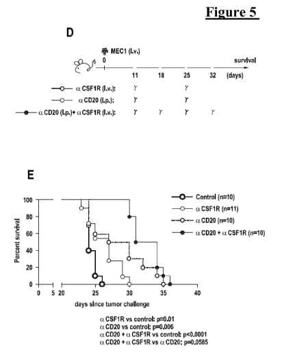
Figure 5
Anti-leukemic effect and survival impact of anti-CSF1R moAb in
the CLL xeno-transplantation system.
(A-B-C) i.v. MEC1 challenged (day 0) Rag2-/-y,-/- mice were
treated i.v. (day +11, +25) with 30mg/Kg of anti-CSF1R moAb
(n=3, white circles) or left untreated (n=4, black circles) and
killed at day 27 (A). The mean value of the percentage of
hCD19+ (B) and of hCD19+ CD20+ (C) cells in PB is shown in
graphs. Data are from one representative experiment of two.
Statistically significant differences were calculated using the
Student t test. **p < 0.01, ***p < 0.001. (D-E) i.v. MEC1
challenged (day 0) Rag2-/-y,-/- mice were left untreated (n=10,
black circles) or treated with: 30mg/Kg i.v. of anti-CSF1R moAb
(n=11, white circles; day +11, +25); or 30mg/Kg i.p. of anti-
CD20 moAb GA101 (n=10, blue circles; day +11, +25); or
CA 02987716 2017-11-29
WO 2016/207312
PCT/EP2016/064611
11
30mg/Kg i.p. of anti-CD20 moAb GA101 (day +11, +25) +
30mg/Kg i.v. of anti-CSF1R moAb (day +18, +32) (n=10, violet
circles) and monitored for survival (D). Kaplan-Meier survival
curve is represented (E), statistical analysis was performed using
Log-Rank test. aCSF1R vs control: p=0.01; aCD20 vs control:
p=0.006; aCD20 + aCSF1R vs control: p<0.0001; aCD20 +
aCSF1R vs aCD20: p=0.0585.
Figure 6 The
impact of macrophage targeting on the microenvironment of
CLL mouse models.
(A-B-C-D-E-F) i.v. MEC1 challenged (day 0) Rag2-/-y,-/- mice
were treated i.v. (day +11, +25) with 30mg/Kg of anti-CSF1R
moAb (n=4, white circles) or left untreated (n=4, black circles)
and killed at day 27 or 29 (A). The mean value of the relative
contribution of CD1 1b+ CSF1R+ cells gated on CD45+ in PB is
shown in graph (B). The mean value of the relative contribution
of CD1 lb+ CSF1R- SSChigh neutrophils gated on CD45+ in PB
is shown in graph (C). The mean value of the relative
contribution of CD1 1b+ Grl+ cells gated on CD45+ in PB is
shown in graph (D). Data are from one representative experiment
of two. The mean value of the relative contribution of monocytic
Ly6C+ Ly6Ggated on CD1 lb+ F4/80low (E) and of granulocytic
Ly6Clow Ly6Ghigh gated on CD1 lb+ F4/80low (F) cells in the
PB is shown in graphs. Statistical analysis: *p < 0.05, **p < 0.01,
***p < 0.001, Student's t test. (G-H-I-J-K) i.v. MEC1 challenged
(day 0) Rag2-/-yc-/- mice were treated i.v. (day +11, +25) with 601A1
clodrolip (n=4, white circles) or left untreated (n=4, black circles)
and killed at day 26 (G). The mean value of the relative
contribution of CD1 1b+ CSF1R- SSChigh neutrophils gated on
CD45+ in PB is shown in graph (H). The mean value of the
relative contribution of CD1 1b+ Grl+ cells gated on CD45+ in
PB is shown in graph (I). The mean value of the absolute number
of CD11b+ CSF1R- SSChigh neutrophils gated on CD45+ in BM
is shown in graph (J). The mean value of the absolute number of
CD1 1b+ Grl+ cells gated on CD45+ in BM is shown in graph
(K). Data are from one representative experiment of two.
Statistical analysis: *p < 0.05, Student's t test. (L-M-N) i.v.
CA 02987716 2017-11-29
WO 2016/207312
PCT/EP2016/064611
12
MEC1 challenged (day 0) Rag2-/-y,-/- mice were treated i.v. (day
+11, +25) with 30mg/Kg of anti-CSF1R moAb (n=4, white
circles) or left untreated (n=4, black circles) and killed at day 27
or 29 (A). The mean value of the absolute number of CD1 lb+
CSF1R+ cells gated on CD45+ in BM is shown in graph (L).
Data are from one representative experiment of two. The mean
value of the absolute number of monocytic Ly6C+ Ly6G- gated
on CD11b+ F4/80low (M) and of granulocytic Ly6Clow
Ly6Ghigh gated on CD1 lb+ F4/80low (N) cells in the BM is
shown in graphs. Statistical analysis: *p < 0.05, Student's t test.
(0-P-Q-R-S-T-U) C57BL/6 mice transplanted i.p. with leukemic
B cells from E[L-TCL1 transgenic mouse were treated i.p. (day
+17, +23) with 30mg/Kg of anti-CSF1R moAb (n=3, white
circles) or left untreated (n=3, black circles) and killed at day 26
(0). The mean value of the relative contribution of
CD44+CD62L+ central and CD44+CD62L1ow/neg effector
memory CD8+ T cells in PB is shown in graph (P). The mean
value of the absolute number of CD44+CD62L+ central and
CD44+CD62L1ow/neg effector memory CD8+ T cells in BM (Q)
and SP (R) is shown in graphs. Intracellular IFNy production was
measured by flow cytometry and the mean value of the absolute
number of CD8+ CD44+ IFNy+ T cells is shown in graph (S).
The mean value of the absolute number of Ann+ PI- cells gated
on CD19+ CD5+ cells to the whole B cell pool in SP is shown in
graph (T). The mean value of the absolute number of CD4+
CD25+ T cells is shown in graph (U). Statistical analysis: *p <
0.05, **p < 0.01, ***p < 0.001, Student's t test. (V-W-X)
C57BL/6 mice transplanted i.p. with leukemic B cells from Ei.t-
TCL1 transgenic mouse were treated i.p. (starting at day 17,
every 3 days) with 50[L1 of clodrolip (n=4, white circles) or left
untreated (n=4, black circles) and killed at day 24 (V). The mean
value of the absolute number of CD44+CD62L+ central and
CD44+CD62L1ow/neg effector memory CD8+ T cells in SP is
shown in graph (W). The mean value of the absolute number of
CD4+ CD25+ T cells in SP is shown in graph (X).
CA 02987716 2017-11-29
WO 2016/207312
PCT/EP2016/064611
13
Figure 7 BM
microenvironment of xeno-transplanted mice: monocyte and
macrophage populations
(A-B-C-D-E-F) Rag2-/-yc-/- mice, uninjected (UN, black circles)
or injected i.v. with MEC1 (white circles) cells (day 0), were
killed at early (n=3) and late (n=3) stage of leukemia and
analyzed by flow cytometry (A). The mean value of the relative
contribution of hCD19+ cells in BM is shown in graph (B). The
mean value of the absolute number of CD1 lb+ CSF1R+ cells
gated on CD45+ in BM is shown in graph (C). The mean value of
the absolute number of CD1 lb+ F4/80+ cells gated on CD45+ in
BM is shown in graph (D). The mean value of the absolute
number of CD1 lb+ MRC1+ cells to the whole macrophage pool
(CD11b+ F4/80+) gated on CD45+ in BM is shown in graph
(E).Data are from one representative experiment of three.
Statistical analysis: *p < 0.05, **p < 0.01, ***p < 0.001,
Student's t test. The mean value of the absolute number of
CD86+ MRC1+ and CSF1R+ MRC1+ cells to the whole CD45+
CD1 lb+ pool in BM is shown in graph (F). Statistical analysis:
*p <0.05, Student's t test.
Figure 8 Cytokine
production by BM stromal cells from CLL xeno-
transplanted mice.
(A-B-C) Rag2-/-yc-/- mice, uninjected (UN, black circles, n=3) or
injected i.v. with MEC1 (white circles) cells (day 0), were killed
at early stage of leukemia (n=5) and analyzed by flow cytometry.
Intracellular cytokine production was measured by flow
cytometry. Left: the mean value of the absolute number of
CD1 lb+ Grl+ cells producing IL-17a gated on CD45+ in BM is
shown in graph; Right: flow cytometry representative plots of
CD1 lb+ Grl+ cells producing IL- 17a gated on CD45+ in BM
(A). Left: the mean value of the absolute number of non-
hematopoietic CD45- cells producing IL-17a; Right: flow
cytometry representative plots of non-hematopoietic CD45- cells
producing IL-17a in BM (B). Left: the mean value of the absolute
number of non-hematopoietic CD45- cells producing IL-2; Right:
flow cytometry representative plots of nonhematopoietic CD45-
CA 02987716 2017-11-29
WO 2016/207312
PCT/EP2016/064611
14
cells producing IL-2 in BM (C). Statistical analysis: *p < 0.05,
**p <0.01, Student's t test.
Figure 9
Transcriptome analysis of MEC1 leukemic cells and
monocytes/macrophages of xeno-transplanted mice
(A-B-C-D-E-F) Rag2-/-yc-/- mice were injected i.v. with MEC1
cells and sacrificed at early stage of leukemia with age-matched
untransplanted Rag2-/-yc-/- mice (n=3/group). RNA was isolated
from BM murine monocytes/macrophages purified by magnetic
separation and processed for Illumina whole-genome gene
expression direct-hybridization assay. Supervised analysis
between monocytes/macrophages from xeno-transplanted and
untransplanted (UN) mice was performed. The heatmaps
represent the hierarchical clustering of differentially expressed
genes (transplanted vs UN) (adjusted P-Value < 0.05). The
expression level of each gene has been standardized by
subtracting the gene's mean expression and then dividing by the
standard deviation across all samples. This scaled expression
value, denoted as the Row Z-score, is plotted in red-blue scale
color, with red indicating high expression. The gene expression
differences among the groups were functionally classified as
follows: nuclear-effectors, tumor suppressors and translation-
related (A); intracellular function (B); membrane/extracellular
proteins (C), apoptosis-related (D) and inflammation mediators
(E). RNA was isolated from BMinfiltrating human CD19+ MEC1
cells purified by magnetic separation and processed for Illumina
whole-genome gene expression direct-hybridization assay.
Supervised analysis between xeno-transplanted and in vitro pre-
injected MEC1 cells was performed. The heatmap represents the
hierarchical clustering of differentially expressed genes
(xenotransplanted vs pre-injected) (adjusted P-Value < 0.05). The
gene expression differences among the groups involved in
monocytes/macrophages and B cells interaction are described (F).
(G) Rag2-/-yc-/- mice injected i.v. with MEC1 cells and
sacrificed at day 21 (n=4, white circles) and age-matched
uninjected (UN) Rag2-/-yc-/- mice (n=4, black circles) were
analyzed by flow cytometry. Left: Flow cytometry representative
CA 02987716 2017-11-29
WO 2016/207312
PCT/EP2016/064611
plots of CD1 lb+ ICAM1+ cells to the whole macrophage pool
(CD1 lb+ F4/80+) gated on CD45+, in the SP, BM and PE. Right:
the mean value of the absolute number of CD1 lb+ ICAM1+ cells
to the whole macrophage pool (CD11b+ F4/80+) gated on
5 CD45+, in SP, BM, PE is shown in graphs. Statistical
significance was analyzed by Student's t test. *p < 0.05, **p <
0.01, ***p <0.001.
Figure 10 Transcriptome analysis suggests a role for
the
monocyte/macrophage cross talk with leukemic cells (related to
10 Figure 9).
(A) Relative mRNA expression of Fcgrl, Saa3, Icaml, Dleu2,
Apoe, T1r7 in monocytes/macrophages from BM of uninjected
(UN) and Rag2-/-yc-/- mice injected i.v. with MEC1 cells and
sacrificed at early stage of leukemia. The expression was
15 normalized to I3-actin levels. Data are shown as mean SD
(n=3/group). (B) Relative mRNA expression of CCL2, CEBPB,
CR2, IL10, IL23R, ADAM10, PTEN, RNASET2 in MEC1 B
cells from BM of xeno-transplanted Rag2-/-yc-/- mice sacrificed
at early stage of leukemia (n=3/group) and in vitro pre-injection
cells. The expression was normalized to I3-actin levels. Data are
shown as mean SD (n=3/group). (C) Fresh peripheral blood
samples were collected from 18 CLL patients and 4 healthy
donors. Monocytes were analyzed by flow cytometry for the
expression of ICAM1. CD14+ monocytes were subdivided in
CD14++ CD16- classical monocytes, CD14++ CD16+
intermediate monocytes and CD14+ CD16++ non-classical
monocytes. The mean value SD of the relative contribution of
ICAM1+ cells to the monocyte populations gated on CD14 is
shown in graph. (D-E) i.v. MEC1 challenged (day 0) Rag2-/-yc-/-
mice were either pre-treated starting from day -1 every 3/4 days
with 2mg/Kg i.v. of ICAM1 blocking moAb clone YN1/1.7.4
(n=3, white triangles) or left untreated (n=3, black circles) and
were analyzed by flow cytometry (D). The mean value of the
percentage of human CD19+ cells in the PB and of the absolute
number of human CD19+ cells in the BM, SP and PE is shown in
CA 02987716 2017-11-29
WO 2016/207312
PCT/EP2016/064611
16
graph (E). Statistical analysis: *p < 0.05, **p < 0.01, ***p <
0.001, Student's t test.
Figure 11 Cell death mechanisms induced on leukemic cells by
macrophage
targeting
(A) Left: MEC1 cells were plated in 96-well plates alone (c), with
increasing concentrations of clodrolip (10 M, 100 M, 1000
M) and with PBS liposomes (v, 1000 M) and a luminescent
assay was performed 24, 48 and 72 hours later to evaluate MEC1
cells' sensitivity to the drug. Right: MEC1 cells were plated in
96-well plates alone (c), with anti-human CSF1R moAb RG7155
(hMab 2F11-e7 IgG1 isotype) (1-10 g/m1) and a luminescent
assay was performed 48 and 72 hours later to evaluate MEC1
cells' sensitivity to the drug.
Figure 12 TNF-dependent macrophage-mediated mechanism of in vivo
leukemic cell death
(A-B-C-D-E-F-G) i.v. MEC1 challenged (day 0) Rag2-/-yc-/-
mice were either pre-treated starting from day -1 every 3 days
with 200 1 i.v. of clodrolip (n=4, white circles) or untreated (n=3,
black circles) and were analyzed by flow cytometry (A). The
mean value of the percentage of human CD19+ cells in SP, BM,
PB, PE is shown in graph (B). The mean value of the relative
contribution of CD1 lb+ CSF1R+ cells gated on CD45 in PB is
shown in graph (C). The mean value of the absolute number or of
the percentage of CD1 lb+ F4/80+ cells gated on CD45+ in SP,
BM, PE (D) and PB (E) is shown in graphs. The mean value of
the relative contribution of Ann+ PI+ cells gated on hCD19+
cells in SP, BM, PE (F) and of the relative contribution of Ann-
P1+ cells gated on hCD19+ cells in PB (G) is shown in graphs.
Statistical significance was analyzed by Student's t test. *p <
0.05, **p <0.01, ***p <0.001.
(H-I) i.v. MEC1 challenged (day 0) Rag2-/-yc-/- mice were left
untreated (n=6, black circles) or treated i.v. (day +11, +25) with
60 1 clodrolip (n=4, white circles) or with 60 1 clodrolip (day
+11, +25) + 10mg/Kg etanercept (i.p., day
CA 02987716 2017-11-29
WO 2016/207312
PCT/EP2016/064611
17
+10,+12,+15,+18,+21,+24) (n=6, red triangles) and killed at day
26 (H). The mean value of the absolute number of human CD19+
cells in the BM is shown in graph (I).Statistical significance was
analyzed by Student's t test. *p < 0.05, **p <0.01, ***p < 0.001.
(J-K) i.v. MEC1 challenged (day 0) Rag2-/-yc-/- mice were left
untreated (n=5, black circles) or treated i.v. (day +11, +25) with
30mg/Kg anti-CSF1R moAb (n=7, white circles) or with
3 Omg/Kg anti-CSF1R moAb (day +11, +25) + 10mg/Kg
etanercept (i.p., day +10,+12,+15,+18,+21,+24) (n=6, red
triangles) and killed at day 29 (J). The mean value of the absolute
number of human CD19+ cells in the SP is shown in graph (K).
Statistical significance was analyzed by Student's t test. *p <
0.05.
Figure 13 Monocytes/macrophages and leukemic cells in humans
(G) hCD19+ CD5+ (black circles) and hCD14+ (white circles)
depletion after 48h (n=4) with anti-CD20 moAb GA101 10 g/ml,
anti-human CSF1R moAb RG7155 (hMab 2F11-e7 IgG1 isotype)
(1-10 g/m1), anti-CD20 moAb GA101 10 g/m1 + anti-human
CSF1R moAb RG7155 (hMab 2F11-e7 IgG1 isotype) (1-
10 g/m1) was calculated by the following formula: 100 - %
remaining cells, where % remaining cells = (Absolute number in
treated samples/Absolute number in untreated samples)x 100.
Horizontal bars represent the mean value (*p<0.05; **p<0.01),
Student's t test.
Figure 14 Cell death mechanisms induced by clodrolip on primary leukemic
cells from CLL patients (related to Figure 13).
(A) Relative mRNA expression of FAS in human primary CLL
cells purified by negative selection from PBMCs of CLL patients
(n=3, Pt #24-26), plated (in triplicates) 24h in 6-well plates alone
(C, black bars) or with 1000 uM of clodrolip (Clo, white bars).
The expression was normalized to 13-actin levels. Data are shown
as mean SD (n=3/group). Statistically significant differences
were calculated using the Student t test. *p < 0.05.
CA 02987716 2017-11-29
WO 2016/207312
PCT/EP2016/064611
18
(B) Relative mRNA expression of TNFR1, FADD, BID, TRAIL-
R2 in human primary CLL cells purified by negative selection
from PBMC of one CLL patient, plated (in triplicates) 24h in 6-
well plates alone (C, black bars) or with 1000 [iM of clodrolip
(Clo, white bars). The expression was normalized to 0- actin
levels. Data are shown as mean SD (n=3/group). Statistically
significant differences were calculated using the Student t test. *p
<0.05
(C) hCD19+ CD5+ depletion after 24h treatment (n=3) with
1001AM, 5001AM, 10001AM of clodrolip, with (black bar) or
without (white bar) etanercept (10n/m1) was calculated by the
following formula: 100 - % remaining cells, where % remaining
cells = (Absolute number in treated samples/Absolute number in
untreated samples) x100. (*p<0.05), Student's t test.
(D) hCD19+ CD5+ depletion after 24h treatment (n=3) with
clodrolip 10001AM, with (black bar) or without (white bar)
blocking anti-TRAIL-R2 moAb (1[Lg/m1) was calculated by the
following formula: 100 - % remaining cells, where % remaining
cells = (Absolute number in treated samples/Absolute number in
untreated samples) x100. (*p<0.05), Student's t test.
Detailed Description of the Invention
Many tumors are characterized by a prominent immune cell infiltrate, including
macrophages. Initially, the immune cells were thought to be part of a defense
mechanism against the tumor, but recent data support the notion that several
immune cell populations including macrophages may, in fact, promote tumor
progression. Macrophages are characterized by their plasticity. Depending on
the
cytokine microenvironment, macrophages can exhibit so-called M1 or M2-
subtypes. M2 macrophages are engaged in the suppression of tumor immunity.
They also play an important role in tissue repair functions such as
angiogenesis and
tissue remodeling which are coopted by the tumor to support growth. In
contrast to
tumor promoting M2 macrophages, M1 macrophages exhibit antitumor activity via
the secretion of inflammatory cytokines and their engagement in antigen
presentation and phagocytosis (Mantovani, A. et al., Curr. Opin. Immunol. 2
(2010) 231-237).
CA 02987716 2017-11-29
WO 2016/207312
PCT/EP2016/064611
19
By secreting various cytokines such as colony stimulating factor 1 (CSF-1) and
IL-10, tumor cells are able to recruit and shape macrophages into the M2-
subtype,
whereas cytokines such as granulocyte macrophage colony stimulating factor
(GM-CSF), IFN-gamma program macrophages towards the M1 subtype. Using
immunohistochemistry, it is possible to distinguish between a macrophage
subpopulation co-expressing CD68 and CD163, which is likely to be enriched for
M2 Macrophages, and a subset showing the CD68+/MHC II+, or CD68+/CD80+
immunophenotype, likely to include M1 macrophages. Cell shape, size, and
spatial
distribution of CD68 and CD163 positive macrophages is consistent with
published
hypotheses on a tumor-promoting role of M2 macrophages, for example by their
preferential location in tumor intersecting stroma, and vital tumor areas. In
contrast,
CD68+/MHC class II+ macrophages are ubiquitously found. Their hypothetical
role in phagocytosis is reflected by clusters of the CD68+/MHC class II+, but
CD163- immunophenotype near apoptotic cells and necrotic tumor areas.
The subtype and marker expression of different macrophage subpopulations is
linked with their functional state. M2 macrophages can support tumorigenesis
by:
a) enhancing angiogenesis via the secretion of angiogenic factors such as
VEGF or bFGF,
b) supporting metastasis
formation via secretion of matrix
metalloproteinases(MMPs), growth factors and migratory factors guiding
the tumor cells to the blood stream and setting up the metastatic niche
(Wyckoff, J. et al., Cancer Res. 67 (2007) 2649-2656),
c) playing a role in building an immunosuppressive milieu by secreting
immunosuppressive cytokines such as IL-4, 11-13, IL-lra and IL-10,
which in turn regulate T regulatory cell function. Conversely CD4
positive T cells have been shown to enhance the activity of tumor
promoting macrophages in preclinical models (Mantovani, A. et al., Eur.
J. Cancer 40 (2004) 1660-1667; DeNardo, D. et al., Cancer Cell 16 (2009)
91-102).
Accordingly, in several types of cancer (e.g. breast, ovarian, Hodgkin's
lymphoma)
the prevalence of M2 subtype tumor associated macrophages (TAMs) has been
associated with poor prognosis (Bingle, L. et al., J. Pathol. 3 (2002) 254-
265; Orre,
M., and Rogers, P.A., Gynecol. Oncol. 1 (1999) 47-50; Steidl, C. et al., N.
Engl. J.
Med. 10 (2010) 875-885). Recent data show a correlation of CD163 positive
macrophage infiltrate in tumors and tumor grade (Kawamura, K. et al., Pathol.
Int.
CA 02987716 2017-11-29
WO 2016/207312
PCT/EP2016/064611
59 (2009) 300-305). TAMs isolated from patient tumors had a tolerant phenotype
and were not cytotoxic to tumor cells (Mantovani, A. et al., Eur. J. Cancer 40
(2004) 1660-1667). However, infiltration of TAMs in the presence of cytotoxic
T
cells correlates with improved survival in non small cell lung cancer and
hence
5
reflects a more prominent M1 macrophage infiltrate in this tumor type (Kawai,
0.
et al., Cancer 6 (2008) 1387-1395).
Recently, a so-called immune signature comprising high numbers of macrophages
and CD4 positive T cells, but low numbers of cytotoxic CD8 positive T cells
was
shown to correlate with reduced overall survival (OS) in breast cancer
patients and
10 to
represent an independent prognostic factor (DeNardo, D. et al., Cancer
Discovery 1 (2011) 54-67).
Consistent with a role for CSF-1 in driving the pro-tumorigenic function of M2
macrophages, high CSF-1 expression in rare sarcomas or locally aggressive
connective tissue tumors, such as pigmented villonodular synovitis (PVNS) and
15
tenosynovial giant cell tumor (TGCT) due in part to a translocation of the CSF-
1
gene, leads to the accumulation of monocytes and macrophages expressing the
receptor for CSF-1, the colony-stimulating factor 1 receptor (CSF-1R) forming
the
majority of the tumor mass (West, R.B. et al., Proc. Natl. Acad. Sci. USA 3
(2006)
690-695). These tumors were subsequently used to define a CSF-1 dependent
20
macrophage signature by gene expression profiling. In breast cancer and
leiomyosarcoma patient tumors this CSF-1 response gene signature predicts poor
prognosis (Espinosa, I. et al., Am. J. Pathol. 6 (2009) 2347-2356; Beck, A. et
al.,
Clin. Cancer Res. 3 (2009) 778-787).
CSF-1R belongs to the class III subfamily of receptor tyrosine kinases and is
encoded by the c-fins proto-oncogene. Binding of CSF-1 or IL-34 induces
receptor
dimerization, followed by autophosphorylation and activation of downstream
signaling cascades. Activation of CSF-1R regulates the survival, proliferation
and
differentiation of monocytes and macrophages (Xiong, Y. et al., J. Biol. Chem.
286
(2011) 952-960).
In addition to cells of the monocytic lineage and osteoclasts, which derive
from the
same hematopoietic precursor as the macrophage, CSF-1R/c-fms has also been
found to be expressed by several human epithelial cancers such as ovarian and
breast cancer and in leiomyosarcoma and TGCT/PVNS, albeit at lower expression
levels compared to macrophages. As with TGCT/PVNS, elevated levels of CSF-1,
CA 02987716 2017-11-29
WO 2016/207312
PCT/EP2016/064611
21
the ligand for CSF-1R, in serum as well as ascites of ovarian cancer patients
have
been correlated with poor prognosis (Scholl, S. et al., Br. J. Cancer 62
(1994) 342-
346; Price, F. et al., Am. J. Obstet. Gynecol. 168 (1993) 520-527).
Furthermore, a
constitutively active mutant form of CSF 1R is able to transform NIH3T3 cells,
one
of the properties of an oncogene (Chambers, S., Future Oncol 5 (2009) 1429-
1440).
Preclinical models provide validation of CSF-1R as an oncology target.
Blockade
of CSF-1 as well as CSF-1R activity results in reduced recruitment of TAMs.
Chemotherapy resulted in elevated CSF-1 expression in tumor cells leading to
enhanced TAM recruitment. Blockade of CSF-1R in combination with paclitaxel
resulted in activation of CD8 positive cytotoxic T cells leading to reduced
tumor
growth and metastatic burden in a spontaneous transgenic breast cancer model
(DeNardo, D. et al., Cancer Discovery 1 (2011) 54-67).
The human CSF-1R (CSF-1 receptor; synonyms: M-CSF receptor; Macrophage
colony-stimulating factor 1 receptor, Fms proto-oncogene, c-fms, SEQ ID NO:
22))
is known since 1986 (Coussens, L., et al., Nature 320 (1986) 277-280). CSF-1R
is
a growth factor and encoded by the c-fms proto-oncogene (reviewed e.g. in
Roth,
P. and Stanley, E.R., Curr. Top. Microbiol. Immunol. 181 (1992) 141-167).
CSF-1R is the receptor for the CSF-1R ligands CSF-1 (macrophage colony
stimulating factor, also called M-CSF) (SEQ ID No.: 86) and IL-34 (SEQ ID
No.: 87) and mediates the biological effects of these cytokines (Shen, C.J.,
et al.,
Cell 41(1985) 665-676; Lin, H., et al., Science 320 (2008) 807-811). The
cloning
of the colony stimulating factor-1 receptor (also called c-fms) was described
for the
first time in Roussel, M.F., et al., Nature 325 (1987) 549-552. In that
publication, it
was shown that CSF-1R had transforming potential dependent on changes in the
C-terminal tail of the protein including the loss of the inhibitory tyrosine
969
phosphorylation which binds Cbl and thereby regulates receptor down regulation
(Lee, P.S., et al., Embo J. 18 (1999) 3616-3628).
CSF-1R is a single chain, transmembrane receptor tyrosine kinase (RTK) and a
member of the family of immunoglobulin (Ig) motif containing RTKs
characterized by 5 repeated Ig-like subdomains Dl-D5 in the extracellular
domain
(ECD) of the receptor (Wang, Z., et al Molecular and Cellular Biology 13
(1993)
5348-5359). The human CSF-1R Extracellular Domain (CSF-1R-ECD) (SEQ ID
NO: 64) comprises all five extracellular Ig-like subdomains D1 ¨D5. The human
CSF-1R fragment delD4 (SEQ ID NO: 65) comprises the extracellular
CA 02987716 2017-11-29
WO 2016/207312
PCT/EP2016/064611
22
Ig-like subdomains D1¨D3 and D5, but is missing the D4 subdomain. The human
CSF-1R fragment D1-D3 (SEQ ID NO: 66) comprises the respective subdomains
D1-D3. The sequences are listed without the signal peptide MGSGPGVLLL
LLVATAWHGQ G (SEQ ID NO: 67). The human CSF-1R fragment D4-D3 (SEQ
ID NO: 85) comprises the respective subdomains D4-D3.
Currently two CSF-1R ligands that bind to the extracellular domain of CSF-1R
are
known. The first one is CSF-1 (colony stimulating factor 1, also called M-CSF,
macrophage; human CSF-1, SEQ ID NO: 86) and is found extracellularly as a
disulfide-linked homodimer (Stanley, E.R. et al., Journal of Cellular
Biochemistry
21 (1983) 151-159; Stanley, E.R. et al., Stem Cells 12 Suppl. 1 (1995) 15-24).
The
second one is IL-34 (human IL-34; SEQ ID NO: 87) (Hume, D. A. , et al, Blood
119 (2012) 1810-1820). Thus in one embodiment the term "CSF-1R ligand" refers
to human CSF-1 (SEQ ID NO: 86) and/or human IL-34 (SEQ ID NO: 87).
For experiments often the active 149 amino acid (aa) fragment of human CSF-1
(aa
33-181 of SEQ ID NO: 86) is used. This active 149 aa fragment of human CSF-1
(aa 33-181 of SEQ ID NO: 86) is contained in all 3 major forms of CSF-1 and is
sufficient to mediate binding to CSF-1R (Hume, D. A. , et al, Blood 119 (2012)
1810-1820).
The main biological effects of CSF-1R signaling are the differentiation,
proliferation, migration, and survival of hematopoietic precursor cells to the
macrophage lineage (including osteoclast). Activation of CSF-1R is mediated by
its
CSF-1R ligands, CSF-1 (M-CSF) and IL-34. Binding of CSF-1 (M-CSF) to CSF-
1R induces the formation of homodimers and activation of the kinase by
tyrosine
phosphorylation (Li, W. et al, EMBO Journal.10 (1991) 277-288; Stanley, E.R.,
et
al., Mol. Reprod. Dev. 46 (1997) 4-10).
The intracellular protein tyrosine kinase domain is interrupted by a unique
insert
domain that is also present in the other related RTK class III family members
that
include the platelet derived growth factor receptors (PDGFR), stem cell growth
factor receptor (c-Kit) and fins-like cytokine receptor (FLT3). In spite of
the
structural homology among this family of growth factor receptors, they have
distinct tissue-specific functions.
CSF-1R is mainly expressed on cells of the monocytic lineage and in the female
reproductive tract and placenta. In addition expression of CSF-1R has been
reported in Langerhans cells in skin, a subset of smooth muscle cells (Inaba,
T., et
CA 02987716 2017-11-29
WO 2016/207312
PCT/EP2016/064611
23
al., J. Biol. Chem. 267 (1992) 5693-5699), B cells (Baker, A.H., et al.,
Oncogene 8
(1993) 371-378) and microglia (Sawada, M., et al., Brain Res. 509 (1990) 119-
124). Cells with mutant human CSF-1R ((SEQ ID NO: 23) are known to proliferate
independently of ligand stimulation.
As used herein, "binding to human CSF-1R" or "specifically binding to human
CSF-1R" or "which binds to human CSF-1R" or "anti-CSF-1R antibody" refers to
an antibody specifically binding to the human CSF-1R antigen with a binding
affinity of KD-value of 1.0 x 10⁻⁸ mo1/1 or lower, in one embodiment of a KD-
value
of 1.0 x10⁻⁹ mo1/1 or lower. The binding affinity is determined with a
standard
binding assay, such as surface plasmon resonance technique (BIAcore0, GE-
Healthcare Uppsala, Sweden). Thus an "antibody binding to human CSF-1R" as
used herein refers to an antibody specifically binding to the human CSF-1R
antigen
with a binding affinity of KD 1.0 x 10⁻⁸ mo1/1 or lower (in one embodiment 1.0
x
10-8 mo1/1 - 1.0 x 10⁻¹³ mo1/1), in on embodiment of a KD 1.0 x10⁻⁹ mo1/1 or
lower
(in one embodiment 1.0 x 10⁻⁹ mo1/1 - 1.0 x 10⁻¹³ mo1/1).
CD20 (also known as B-lymphocyte antigen CD20, B-lymphocyte surface antigen
Bl, Leu-16, Bp35, BM5, and LF5; the sequence is characterized by the SwissProt
database entry P11836) is a hydrophobic transmembrane protein with a molecular
weight of approximately 35 kD located on pre-B and mature B lymphocytes
(Valentine, M.A. et al., J. Biol. Chem. 264 (1989) 11282-11287; Tedder, T.F.,
et
al., Proc. Natl. Acad. Sci. U.S.A. 85 (1988) 208-212; Stamenkovic, I., et al.,
J. Exp.
Med. 167 (1988) 1975-1980; Einfeld, D.A., et al., EMBO J. 7 (1988) 711-717;
Tedder, T.F., et al., J. Immunol. 142 (1989) 2560-2568). The corresponding
human
gene is Membrane-spanning 4-domains, subfamily A, member 1, also known as
MS4A1. This gene encodes a member of the membrane-spanning 4A gene family.
Members of this nascent protein family are characterized by common structural
features and similar intron/exon splice boundaries and display unique
expression
patterns among hematopoietic cells and nonlymphoid tissues. This gene encodes
the B-lymphocyte surface molecule which plays a role in the development and
differentiation of B-cells into plasma cells. This family member is localized
to
llql 2, among a cluster of family members. Alternative splicing of this gene
results
in two transcript variants which encode the same protein.
The terms "CD20" and "CD20 antigen" are used interchangeably herein, and
include any variants, isoforms and species homologs of human CD20 which are
naturally expressed by cells or are expressed on cells transfected with the
CD20
CA 02987716 2017-11-29
WO 2016/207312
PCT/EP2016/064611
24
gene. Binding of an antibody of the invention to the CD20 antigen mediate the
killing of cells expressing CD20 (e.g., a tumor cell) by inactivating CD20.
The
killing of the cells expressing CD20 may occur by one or more of the following
mechanisms: Cell death/apoptosis induction, ADCC and CDC.
Synonyms of CD20, as recognized in the art, include B-lymphocyte antigen CD20,
B-lymphocyte surface antigen Bl, Leu-16, Bp35, BM5, and LF5.
The term "anti-CD20 antibody" according to the invention is an antibody that
binds
specifically to human CD20 antigen. Depending on binding properties and
biological activities of anti-CD20 antibodies to the CD20 antigen, two types
of
anti-CD20 antibodies (type I and type II anti-CD20 antibodies) can be
distinguished according to Cragg, M.S., et al., Blood 103 (2004) 2738-2743;
and
Cragg, M.S., et al., Blood 101 (2003) 1045-1052, see Table 2.
Table 1: Properties of type I and type II anti-CD20 antibodies
type I anti-CD20 antibodies type II anti-CD20 antibodies
type I CD20 epitope type II CD20 epitope
Localize CD20 to lipid rafts Do not localize CD20 to lipid rafts
Increased CDC (if IgG1 isotype) Decreased CDC (if IgG1 isotype)
ADCC activity (if IgG1 isotype) ADCC activity (if IgG1 isotype)
Full binding capacity Reduced binding capacity
Homotypic aggregation Stronger homotypic aggregation
Apoptosis induction upon cross- Strong cell death induction without
linking cross-linking
Examples of type II anti-CD20 antibodies include e.g. humanized B-Lyl antibody
IgG1 (a chimeric humanized IgG1 antibody as disclosed in WO 2005/044859),
11B8 IgG1 (as disclosed in WO 2004/035607), and AT80 IgGl. Typically type II
anti-CD20 antibodies of the IgG1 isotype show characteristic CDC properties.
Type II anti-CD20 antibodies have a decreased CDC (if IgG1 isotype) compared
to
type I antibodies of the IgG1 isotype.
Examples of type I anti-CD20 antibodies include e.g. rituximab, HI47 IgG3
(ECACC, hybridoma), 2C6 IgG1 (as disclosed in WO 2005/103081), 2F2 IgG1 (as
disclosed and WO 2004/035607 and WO 2005/103081) and 2H7 IgG1 (as
disclosed in WO 2004/056312).
CA 02987716 2017-11-29
WO 2016/207312
PCT/EP2016/064611
The "rituximab" antibody (reference antibody; example of a type I anti-CD20
antibody) is a genetically engineered chimeric human gamma 1 murine constant
domain containing monoclonal antibody directed against the human CD20 antigen.
This chimeric antibody contains human gamma 1 constant domains and is
5
identified by the name "C2B8" in US 5,736,137 (Andersen et. al.) issued on
April
17, 1998, assigned to IDEC Pharmaceuticals Corporation. Rituximab is approved
for the treatment of patients with relapsed or refracting low-grade or
follicular,
CD20 positive, B cell non-Hodgkin's lymphoma. In vitro mechanism of action
studies have shown that rituximab exhibits human complement--dependent
10
cytotoxicity (CDC) (Reff, M.E., et. al., Blood 83 (1994) 435-445).
Additionally, it
exhibits significant activity in assays that measure antibody-dependent
cellular
cytotoxicity (ADCC). Rituximab is not afucosylated.
Antibody Amount of fucose
Rituximab (non- >85 %
afucosylated)
Wild type afucosylated >85 %
glyco-engineered
humanized B-Lyl (B-
HH6-B-KV1) (non-
afucosylated)
afucosylated glyco- 45-55 %
engineered humanized B-
Ly1 (B-HH6-B-KV1 GE)
As used herein, "binding to human CD20" or "specifically binding to human
15 CD20"
or "which binds to human CD20" or "anti- CD20 antibody" refers to an
antibody specifically binding to the human CD20 antigen with a binding
affinity of
KD-value of 1.0 x 10⁻⁸ mo1/1 or lower, in one embodiment of a KD-value of 1.0
x10-9 mo1/1 or lower. The binding affinity is determined with a standard
binding
assay, such as surface plasmon resonance technique (BIAcore0, GE-Healthcare
20
Uppsala, Sweden). Thus an "antibody binding to human CD20" as used herein
refers to an antibody specifically binding to the human CD20 antigen with a
binding affinity of KB 1.0 x 10⁻⁸ mo1/1 or lower (in one embodiment 1.0 x 10⁻⁸
mo1/1 - 1.0 x 10⁻¹³ mo1/1), in on embodiment of a KB 1.0 x10⁻⁹ mo1/1 or lower
(in
one embodiment 1.0 x 10⁻⁹ mo1/1 - 1.0 x 10⁻¹³ mo1/1).
25 In one
embodiment the antibody which binds to human C5F-1R used in the
combination therapy described herein is selected from the group consisting of
CA 02987716 2017-11-29
WO 2016/207312
PCT/EP2016/064611
26
hMab 2F11-cl 1 , hMab 2F11-d8 , hMab 2F11-e7 , hMab 2F11412 , and hMab
2F11-gl.
These antibodies are described in W02011/070024 and are characterized in
comprising the following VH and VL sequences as described herein:
Table 2:
anti-CSF-1R antibody amino acid sequence of amino acid sequence of
the heavy chain variable the light chain variable
domain VH, SEQ ID NO: domain VL, SEQ ID NO:
hMab 2F11-c11 23 24
hMab 2F11-d8 31 32
hMab 2F11-e7 39 40
hMab 2F11-f12 47 48
hMab 2F11-gl 55 56
In one preferred embodiment the antibody which binds to human CSF-1R used in
the combination therapy described herein is hMab 2F11-e7 (VH, SEQ ID NO:39;
VL, SEQ ID NO:40).
In one embodiment the antibody which binds to human CD20 used in the
combination therapy described herein is a humanized B-Lyl antibody:
The term "humanized B-Lyl antibody" refers to humanized B-Lyl antibody as
disclosed in WO 2005/044859 and WO 2007/031875, which were obtained from
the murine monoclonal anti-CD20 antibody B-Lyl (see Poppema, S. and Visser,
L., Biotest Bulletin 3 (1987) 131-139) by chimerization with a human constant
domain from IgG1 and following humanization (see WO 2005/044859 and
WO 2007/031875). These "humanized B-Lyl antibodies" are disclosed in detail in
WO 2005/044859 and WO 2007/031875.
In one embodiment, the "humanized B-Lyl antibody" has variable region of the
heavy chain (VH) selected from group of SEQ ID No.69 to SEQ ID No.75 (B-
HH2, BHH-3, B-HH6, B-HH8, B-HL8, B-HL11 and B-HL13 of WO 2005/044859
and WO 2007/031875). In one embodiment, the "humanized B-Lyl antibody" has
variable region of the light chain (VL) of SEQ ID No. 76 (B-KV1 of
WO 2005/044859 and WO 2007/031875).
CA 02987716 2017-11-29
WO 2016/207312
PCT/EP2016/064611
27
These antibodies are characterized in comprising the following VH and VL
sequences as described herein:
Table 3:
anti-CD20 antibody IgG1 amino acid sequence of amino acid sequence of
VH/VL the heavy chain variable the light chain variable
domain VH, SEQ ID NO: domain VL, SEQ ID NO:
B-HH2 / B-KV1 69 76
B-HH3/ B-KV1 70 76
B-HH6/ B-KV1 71 76
B-HH8/ B-KV1 72 76
B-HL8/ B-KV1 73 76
B-HL11/ B-KV1 74 76
B-HL13/ B-KV1 75 76
In one preferred embodiment the antibody which binds to human CD20 used in the
combination therapy described herein is selected the humanized B-Lyl antibody
B-
HH6/B-KV1(comprising a variable region of the heavy chain (VH) of SEQ ID
No.71 (B-HH6)and a variable region of the light chain (VL) of SEQ ID No. 76 (B-
KV1).
Furthermore in one embodiment, the humanized B-Lyl antibody is an afucosylated
antibody of IgG1 isotype.
According to the invention such afucosylated humanized B-Lyl antibodies are
glycoengineered (GE) in the Fc region according to the procedures described in
WO 2005/044859, WO 2004/065540, WO 2007/031875, Umana, P. et al., Nature
Biotechnol. 17 (1999) 176-180 and WO 99/154342. In one embodiment, the
afucosylated glyco-engineered humanized B-Lyl is B-HH6-B-KV1 GE. Such
glycoengineered humanized B-Lyl antibodies have an altered pattern of
glycosylation in the Fc region, preferably having a reduced level of fucose
residues.
In one embodiment, the amount of fucose is 60% or less of the total amount of
oligosaccharides at Asn297 (in one embodiment the amount of fucose is between
40% and 60%, in another embodiment the amount of fucose is 50% or less, and in
still another embodiment the amount of fucose is 30% or less). In another
embodiment, the oligosaccharides of the Fc region are preferably bisected.
These
glycoengineered humanized B-Lyl antibodies have an increased ADCC.
CA 02987716 2017-11-29
WO 2016/207312
PCT/EP2016/064611
28
In one preferred embodiment the antibody which binds to human CD20 used in the
combination therapy described herein is selected the humanized B-Lyl antibody
B-
HH6/B-KV1( comprising a variable region of the heavy chain (VH) of SEQ ID
No.71 (B-HH6)and a variable region of the light chain (VL) of SEQ ID No. 76 (B-
KV1) and is an afucosylated antibody of IgG1 isotype having an altered pattern
of
glycosylation in the Fc region wherein the amount of fucose containing
oligosaccharides is between 40% and 60% of the total amount of
oligosaccharides
at Asn297.
The afucosylated anti-CD20 antibodies according to the invention have an
increased antibody dependent cellular cytotoxicity (ADCC) unlike anti-CD20
antibodies having no reduced fucose.
By "afucosylated anti-CD20 antibody with increased antibody dependent cellular
cytotoxicity (ADCC)" is meant an afucosylated anti-CD20 antibody, as that term
is
defined herein, having increased ADCC as determined by any suitable method
known to those of ordinary skill in the art. One accepted in vitro ADCC assay
is as
follows:
1) the assay uses target cells that are known to express the target antigen
recognized by the antigen-binding region of the antibody;
2) the assay uses human peripheral blood mononuclear cells (PBMCs),
isolated
from blood of a randomly chosen healthy donor, as effector cells;
3) the assay is carried out according to following protocol:
i) the PBMCs are isolated using standard density centrifugation
procedures and are suspended at 5 x 10⁶ cells/ml in RPMI cell culture
medium;
ii) the target cells are grown by standard tissue culture methods, harvested
from the exponential growth phase with a viability higher than 90%,
washed in RPMI cell culture medium, labeled with 100 micro-Curies of
51Cr, washed twice with cell culture medium, and resuspended in cell
culture medium at a density of 105 cells/ml;
iii) 100 microliters of the final target cell suspension above are transferred
to each well of a 96-well microtiter plate;
iv) the antibody is serially-diluted from 4000 ng/ml to 0.04 ng/ml in
cell
culture medium and 50 microliters of the resulting antibody solutions
are added to the target cells in the 96-well microtiter plate, testing in
CA 02987716 2017-11-29
WO 2016/207312
PCT/EP2016/064611
29
triplicate various antibody concentrations covering the whole
concentration range above;
v) for the maximum release (MR) controls, 3 additional wells in the plate
containing the labeled target cells, receive 50 microliters of a 2% (VN)
aqueous solution of non-ionic detergent (Nonidet, Sigma, St. Louis),
instead of the antibody solution (point iv above);
vi) for the spontaneous release (SR) controls, 3 additional wells in the
plate
containing the labeled target cells, receive 50 microliters of RPMI cell
culture medium instead of the antibody solution (point iv above);
vii) the 96-well microtiter plate is then centrifuged at 50 x g for 1 minute
and incubated for 1 hour at 4 C;
viii) 50 microliters of the PBMC suspension (point i above) are added to
each well to yield an effector:target cell ratio of 25: 1 and the plates are
placed in an incubator under 5% CO2 atmosphere at 37 C for 4 hours;
ix) the cell-free supernatant from each well is harvested and the
experimentally released radioactivity (ER) is quantified using a gamma
counter;
x) the percentage of specific lysis is calculated for each antibody
concentration according to the formula (ER-MR)/(MR-SR) x 100,
where ER is the average radioactivity quantified (see point ix above)
for that antibody concentration, MR is the average radioactivity
quantified (see point ix above) for the MR controls (see point V above),
and SR is the average radioactivity quantified (see point ix above) for
the SR controls (see point vi above);
4) "increased ADCC" is defined as either an increase in the maximum
percentage of specific lysis observed within the antibody concentration range
tested above, and/or a reduction in the concentration of antibody required to
achieve one half of the maximum percentage of specific lysis observed
within the antibody concentration range tested above. The increase in ADCC
is relative to the ADCC, measured with the above assay, mediated by the
same antibody, produced by the same type of host cells, using the same
standard production, purification, formulation and storage methods, which
are known to those skilled in the art, but that has not been produced by host
cells engineered to overexpress GnTIII.
Said "increased ADCC" can be obtained by glycoengineering of said antibodies,
that means enhance said natural, cell-mediated effector functions of
monoclonal
CA 02987716 2017-11-29
WO 2016/207312
PCT/EP2016/064611
antibodies by engineering their oligosaccharide component as described in
Umana,
P., et al., Nature Biotechnol. 17 (1999) 176-180 and US 6,602,684.
The term "complement-dependent cytotoxicity (CDC)" refers to lysis of human
tumor target cells by the antibody according to the invention in the presence
of
5
complement. CDC is measured preferably by the treatment of a preparation of
CD20 expressing cells with an anti-CD20 antibody according to the invention in
the presence of complement. CDC is found if the antibody induces at a
concentration of 100 nM the lysis (cell death) of 20% or more of the tumor
cells
after 4 hours. The assay is performed preferably with 51Cr or Eu labeled tumor
cells
10 and
measurement of released 51Cr or Eu. Controls include the incubation of the
tumor target cells with complement but without the antibody.
The term "afucosylated antibody" refers to an antibody of IgG1 or IgG3 isotype
(preferably of IgG1 isotype) with an altered pattern of glycosylation in the
Fc
region at Asn297 having a reduced level of fucose residues. Glycosylation of
15 human
IgG1 or IgG3 occurs at Asn297 as core fucosylated bianntennary complex
oligosaccharide glycosylation terminated with up to 2 Gal residues. These
structures are designated as GO, G1 (a1,6 or a1,3) or G2 glycan residues,
depending from the amount of terminal Gal residues (Raju, T.S., BioProcess
Int. 1
(2003) 44-53). CHO type glycosylation of antibody Fc parts is e.g. described
by
20
Routier, F.H., Glycoconjugate J. 14 (1997) 201-207. Antibodies which are
recombinantely expressed in non glycomodified CHO host cells usually are
fucosylated at Asn297 in an amount of at least 85%. It should be understood
that
the term an afucosylated antibody as used herein includes an antibody having
no
fucose in its glycosylation pattern. It is commonly known that typical
glycosylated
25 residue
position in an antibody is the asparagine at position 297 according to the
EU numbering system ("Asn297").
The "EU numbering system" or "EU index" is generally used when referring to a
residue in an immunoglobulin heavy chain constant region (e.g., the EU index
reported in Kabat et al., Sequences of Proteins of Immunological Interest, 5th
ed.,
30 Public
Health Service, National Institutes of Health, Bethesda, MD (1991)
expressly incorporated herein by reference).
Thus an afucosylated antibody according to the invention means an antibody of
IgG1 or IgG3 isotype (preferably of IgG1 isotype) wherein the amount of fucose
is
60% or less of the total amount of oligosaccharides (sugars) at Asn297 (which
CA 02987716 2017-11-29
WO 2016/207312
PCT/EP2016/064611
31
means that at least 40% or more of the oligosaccharides of the Fe region at
Asn297
are afucosylated). In one embodiment the amount of fucose is between 40% and
60% of the oligosaccharides of the Fe region at Asn297. In another embodiment
the amount of fucose is 50% or less, and in still another embodiment the
amount of
fucose is 30% or less of the oligosaccharides of the Fe region at Asn297.
According to the invention "amount of fucose" means the amount of said
oligosaccharide (fucose) within the oligosaccharide (sugar) chain at Asn297,
related to the sum of all oligosaccharides (sugars) attached to Asn 297 (e. g.
complex, hybrid and high mannose structures) measured by MALDI-TOF mass
spectrometry and calculated as average value (for a detailed procedure to
determine
the amount of fucose, see e.g. WO 2008/077546). Furthermore in one embodiment,
the oligosaccharides of the Fe region are bisected. The afucosylated antibody
according to the invention can be expressed in a glycomodified host cell
engineered
to express at least one nucleic acid encoding a polypeptide having GnTIII
activity
in an amount sufficient to partially fucosylate the oligosaccharides in the Fe
region.
In one embodiment, the polypeptide having GnTIII activity is a fusion
polypeptide.
Alternatively a1,6-fucosyltransferase activity of the host cell can be
decreased or
eliminated according to US 6,946,292 to generate glycomodified host cells. The
amount of antibody fucosylation can be predetermined e.g. either by
fermentation
conditions (e.g. fermentation time) or by combination of at least two
antibodies
with different fucosylation amount. Such afucosylated antibodies and
respective
glycoengineering methods are described in WO 2005/044859, WO 2004/065540,
WO 2007/031875, Umana, P., et al., Nature Biotechnol. 17 (1999) 176-180,
WO 99/154342, WO 2005/018572, WO 2006/116260, WO 2006/114700,
WO 2005/011735, WO 2005/027966, WO 97/028267, US 2006/0134709,
US 2005/0054048, US 2005/0152894, WO 2003/035835, WO 2000/061739. These
glycoengineered antibodies have an increased ADCC. Other glycoengineering
methods yielding afucosylated antibodies according to the invention are
described
e.g. in Niwa, R.. et al., J. Immunol. Methods 306 (2005) 151-160; Shinkawa,
T., et
al., J. Biol. Chem, 278 (2003) 3466-3473; WO 03/055993 or US 2005/0249722.
Thus one aspect of the invention is an afucosylated anti-CD20 antibody of IgG1
or
IgG3 isotype (preferably of IgG1 isotype) specifically binding to CD20 with an
amount of fucose of 60% or less of the total amount of oligosaccharides
(sugars) at
Asn297, for the treatment of cancer in combination with an CSF1R antibody as
described herein. In another aspect of the invention is the use of an
afucosylated
anti-CD20 antibody of IgG1 or IgG3 isotype (preferably of IgG1 isotype)
CA 02987716 2017-11-29
WO 2016/207312
PCT/EP2016/064611
32
specifically binding to CD20 with an amount of fucose of 60% or less of the
total
amount of oligosaccharides (sugars) at Asn297, for the manufacture of a
medicament for the treatment of cancer in combination with an CSF1R antibody
as
described herein. In one embodiment the amount of fucose is between 60% and
20% of the total amount of oligosaccharides (sugars) at Asn297. In one
embodiment the amount of fucose is between 60% and 40% of the total amount of
oligosaccharides (sugars) at Asn297. In one embodiment the amount of fucose is
between 0% of the total amount of oligosaccharides (sugars) at Asn297.
In one embodiment of the invention the antibody which binds to human CSF-1R
used in the combination therapy described herein is characterized in
comprising
a) a heavy chain variable domain VH of SEQ ID NO:23 and a light chain
variable domain VL of SEQ ID NO:24, or
b) a heavy chain variable domain VH of SEQ ID NO:31 and a light chain
variable domain VL of SEQ ID NO:32, or
c) a heavy chain variable domain VH of SEQ ID NO:39 and a light chain
variable domain VL of SEQ ID NO:40, or
d) a heavy chain variable domain VH of SEQ ID NO:47 and a light chain
variable domain VL of SEQ ID NO:48, or
e) a heavy chain variable domain VH of SEQ ID NO:55 and a light chain
variable domain VL of SEQ ID NO:56; and
wherein the antibody which binds to human CD20 used in the combination therapy
is characterized in comprising
a) a heavy chain variable domain VH of SEQ ID NO:69 and a light chain
variable domain VL of SEQ ID NO:76, or
b) a heavy chain variable domain VH of SEQ ID NO:70 and a light chain
variable domain VL of SEQ ID NO:76, or
c) a heavy chain variable domain VH of SEQ ID NO:71 and a light chain
variable domain VL of SEQ ID NO:76, or
d) a heavy chain variable domain VH of SEQ ID NO:72 and a light chain
variable domain VL of SEQ ID NO:76, or
CA 02987716 2017-11-29
WO 2016/207312
PCT/EP2016/064611
33
e) a heavy chain variable domain VH of SEQ ID NO:73 and a light chain
variable domain VL of SEQ ID NO:76, or
f) a heavy chain variable domain VH of SEQ ID NO:74 and a light chain
variable domain VL of SEQ ID NO:76, or
g) a heavy chain variable domain VH of SEQ ID NO :75 and a light chain
variable domain VL of SEQ ID NO:76.
In one embodiment the antibody which binds to human CSF-1R used in the
combination therapy is characterized in comprising
a heavy chain variable domain VH of SEQ ID NO:39 and a light chain
variable domain VL of SEQ ID NO:40;
and
wherein the antibody which binds to human CD20 used in the combination therapy
is characterized in comprising
a heavy chain variable domain VH of SEQ ID NO:71 and a light chain
variable domain VL of SEQ ID NO:76
The term "epitope" denotes a protein determinant of human CSF-1R or CD20
capable of specifically binding to an antibody. Epitopes usually consist of
chemically active surface groupings of molecules such as amino acids or sugar
side
chains and usually epitopes have specific three dimensional structural
characteristics, as well as specific charge characteristics. Conformational
and
nonconformational epitopes are distinguished in that the binding to the former
but
not the latter is lost in the presence of denaturing solvents.
The "variable domain" (light chain variable domain VL, heavy chain variable
domain VH) as used herein denotes each of the pair of light and heavy chain
domains which are involved directly in binding the antibody to the antigen.
The
variable light and heavy chain domains have the same general structure and
each
domain comprises four framework (FR) regions whose sequences are widely
conserved, connected by three "hypervariable regions" (or complementary
determining regions, CDRs). The framework regions adopt a beta-sheet
conformation and the CDRs may form loops connecting the beta-sheet structure.
The CDRs in each chain are held in their three-dimensional structure by the
CA 02987716 2017-11-29
WO 2016/207312
PCT/EP2016/064611
34
framework regions and form together with the CDRs from the other chain the
antigen binding site. The antibody's heavy and light chain CDR3 regions play a
particularly important role in the binding specificity/affinity of the
antibodies
according to the invention and therefore provide a further object of the
invention.
The term "antigen-binding portion of an antibody" when used herein refer to
the
amino acid residues of an antibody which are responsible for antigen-binding.
The
antigen-binding portion of an antibody comprises amino acid residues from the
"complementary determining regions" or "CDRs". "Framework" or "FR" regions
are those variable domain regions other than the hypervariable region residues
as
herein defined. Therefore, the light and heavy chain variable domains of an
antibody comprise from N- to C-terminus the domains FR1, CDR1, FR2, CDR2,
FR3, CDR3, and FR4. Especially, CDR3 of the heavy chain is the region which
contributes most to antigen binding and defines the antibody's properties. CDR
and
FR regions are determined according to the standard definition of Kabat et
al.,
Sequences of Proteins of Immunological Interest, 5th ed., Public Health
Service,
National Institutes of Health, Bethesda, MD (1991) and/or those residues from
a
"hypervariable loop".
The terms "nucleic acid" or "nucleic acid molecule", as used herein, are
intended to
include DNA molecules and RNA molecules. A nucleic acid molecule may be
single-stranded or double-stranded, but preferably is double-stranded DNA.
The term "amino acid" as used within this application denotes the group of
naturally occurring carboxy alpha-amino acids comprising alanine (three letter
code: ala, one letter code: A), arginine (arg, R), asparagine (asn, N),
aspartic acid
(asp, D), cysteine (cys, C), glutamine (gln, Q), glutamic acid (glu, E),
glycine (gly,
G), histidine (his, H), isoleucine (ile, I), leucine (leu, L), lysine (lys,
K), methionine
(met, M), phenylalanine (phe, F), proline (pro, P), serine (ser, S), threonine
(thr, T),
tryptophan (trp, W), tyrosine (tyr, Y), and valine (val, V).
The "Fc part" of an antibody is not involved directly in binding of an
antibody to
an antigen, but exhibit various effector functions. A "Fc part of an antibody"
is a
term well known to the skilled artisan and defined on the basis of papain
cleavage
of antibodies. Depending on the amino acid sequence of the constant region of
their
heavy chains, antibodies or immunoglobulins are divided in the classes: IgA,
IgD,
IgE, IgG and IgM, and several of these may be further divided into subclasses
(isotypes), e.g. IgGl, IgG2, IgG3, and IgG4, IgAl , and IgA2. According to the
CA 02987716 2017-11-29
WO 2016/207312
PCT/EP2016/064611
heavy chain constant regions the different classes of immunoglobulins are
called a,
8, e, 7, and , respectively. The Fc part of an antibody is directly involved
in
ADCC (antibody-dependent cell-mediated cytotoxicity) and CDC (complement-
dependent cytotoxicity) based on complement activation, Clq binding and Fc
5
receptor binding. Complement activation (CDC) is initiated by binding of
complement factor Clq to the Fc part of most IgG antibody subclasses. While
the
influence of an antibody on the complement system is dependent on certain
conditions, binding to Clq is caused by defined binding sites in the Fc part.
Such
binding sites are known in the state of the art and described e.g. by Boackle,
R.J., et
10 al.,
Nature 282 (1979) 742-743; Lukas, T.J., et al., J. Immunol. 127 (1981) 2555-
2560; Brunhouse, R., and Cebra, J.J., Mol. Immunol. 16 (1979) 907-917; Burton,
D.R., et al., Nature 288 (1980) 338-344; Thommesen, J.E., et al., Mol.
Immunol.
37 (2000) 995-1004; Idusogie, E.E., et al., J. Immuno1.164 (2000) 4178-4184;
Hezareh, M., et al., J. Virology 75 (2001) 12161-12168; Morgan, A., et al.,
15
Immunology 86 (1995) 319-324; EP 0 307 434. Such binding sites are e.g. L234,
L235, D270, N297, E318, K320, K322, P331 and P329 (numbering according to
EU index of Kabat, E.A., see below). Antibodies of subclass IgGl, IgG2 and
IgG3
usually show complement activation and Clq and C3 binding, whereas IgG4 do not
activate the complement system and do not bind Clq and C3.
20 In one
embodiment the antibody according to the invention comprises an Fc part
derived from human origin and preferably all other parts of the human constant
regions. As used herein the term "Fc part derived from human origin" denotes a
Fc
part which is either a Fc part of a human antibody of the subclass IgGl, IgG2,
IgG3
or IgG4, preferably a Fc part from human IgG1 subclass, a mutated Fc part from
25 human
IgG1 subclass (in one embodiment with a mutation on L234A + L235A), a
Fc part from human IgG4 subclass or a mutated Fc part from human IgG4 subclass
(in one embodiment with a mutation on 5228P). In one preferred embodiment the
human heavy chain constant region is SEQ ID NO: 58 (human IgG1 subclass), in
another preferred embodiment the human heavy chain constant region is SEQ ID
30 NO: 59
(human IgG1 subclass with mutations L234A and L235A) , in another
preferred embodiment the human heavy chain constant region is SEQ ID NO: 60
(human IgG4 subclass), and in another preferred embodiment the human heavy
chain constant region is SEQ ID NO: 61 (human IgG4 subclass with mutation
5228P). In one embodiment said antibodies have reduced or minimal effector
35
function. In one embodiment the minimal effector function results from an
effectorless Fc mutation. In one embodiment the effectorless Fc mutation is
CA 02987716 2017-11-29
WO 2016/207312
PCT/EP2016/064611
36
L234A/L235A or L234A/L235A/P329G or N297A or D265A/N297A. In one
embodiment the effectorless Fe mutation is selected for each of the antibodies
independently of each other from the group comprising (consisting of)
L234A/L235A, L234A/L235A/P329G, N297A and D265A/N297A.
In one embodiment the antibodies described herein are of human IgG class (i.e.
of
IgG1 , IgG2, IgG3 or IgG4 subclass).
In a preferred embodiment the antibodies described herein are of human IgG1
subclass or of human IgG4 subclass. In one embodiment the described herein are
of
human IgG1 subclass. In one embodiment the antibodies described herein are of
human IgG4 subclass.
In one embodiment the antibody described herein is characterized in that the
constant chains are of human origin. Such constant chains are well known in
the
state of the art and e.g. described by Kabat, E.A., (see e.g. Johnson, G. and
Wu,
T.T., Nucleic Acids Res. 28 (2000) 214-218). For example, a useful human heavy
chain constant region comprises an amino acid sequence of SEQ ID NO: 58. For
example, a useful human light chain constant region comprises an amino acid
sequence of a kappa-light chain constant region of SEQ ID NO: 57.
The invention comprises a method for the treatment of a patient in need of
therapy,
characterized by administering to the patient a therapeutically effective
amount of
an antibody according to the invention.
The invention comprises the use of an antibody according to the invention for
the
described therapy.
One preferred embodiment of the invention are the CSF-1R antibodies of the
present invention for use in the treatment of "CSF-1R mediated diseases" or
the
CSF-1R antibodies of the present invention for use for the manufacture of a
medicament in the treatment of "CSF-1R mediated diseases", which can be
described as follows:
There are 3 distinct mechanisms by which CSF-1R signaling is likely involved
in
tumor growth and metastasis. The first is that expression of CSF-ligand and
receptor has been found in tumor cells originating in the female reproductive
system (breast, ovarian, endometrium, cervical) (Scholl, S.M., et al., J.
Natl.
Cancer Inst. 86 (1994) 120-126; Kacinski, B.M., Mol. Reprod. Dev. 46 (1997) 71-
CA 02987716 2017-11-29
WO 2016/207312
PCT/EP2016/064611
37
74; Ngan, H.Y., et al., Eur. J. Cancer 35 (1999) 1546-1550; Kirma, N., et al.,
Cancer Res 67 (2007) 1918-1926) and the expression has been associated with
breast cancer xenograft growth as well as poor prognosis in breast cancer
patients.
Two point mutations were seen in CSF-1R in about 10-20% of acute myelocytic
leukemia, chronic myelocytic leukemia and myelodysplasia patients tested in
one
study, and one of the mutations was found to disrupt receptor turnover (Ridge,
S.A., et al., Proc. Natl. Acad. Sci USA 87 (1990) 1377-1380). However the
incidence of the mutations could not be confirmed in later studies (Abu-
Duhier,
F.M., et al., Br. J. Haematol. 120 (2003) 464-470). Mutations were also found
in
some cases of hepatocellular cancer (Yang, D.H., et al., Hepatobiliary
Pancreat.
Dis. Int. 3 (2004) 86-89) and idiopathic myelofibrosis (Abu-Duhier, F.M., et
al.,
Br. J. Haematol. 120 (2003) 464-470). Recently, in the GDM-1 cell line derived
from a patient with myelomonoblastic leukemia the Y571D mutation in CSF-1R
was identified (Chase, A., et al., Leukemia 23 (2009) 358-364).
The second mechanism is based on blocking signaling through M-CSF/CSF-1R at
metastatic sites in bone which induces osteoclastogenesis, bone resorption and
osteolytic bone lesions. Breast, multiple myeloma and lung cancers are
examples of
cancers that have been found to metastasize to the bone and cause osteolytic
bone
disease resulting in skeletal complications. M-CSF released by tumor cells and
stroma induces the differentiation of hematopoietic myeloid monocyte
progenitors
to mature osteoclasts in collaboration with the receptor activator of nuclear
factor
kappa-B ligand-RANKL. During this process, M-CSF acts as a permissive factor
by giving the survival signal to osteoclasts (Tanaka, S., et al., J. Clin.
Invest. 91
(1993) 257-263). Inhibition of CSF-1R activity during osteoclast
differentiation
and maturation with an anti-CSF-1R antibody is likely to prevent unbalanced
activity of osteoclasts that cause osteolytic disease and the associated
skeletal
related events in metastatic disease. Whereas breast, lung cancer and multiple
myeloma typically result in osteolytic lesions, metastasis to the bone in
prostate
cancer initially has an osteoblastic appearance in which increased bone
forming
activity results in 'woven bone' which is different from typical lamellar
structure of
normal bone. During disease progression bone lesions display a significant
osteolytic component as well as high serum levels of bone resorption and
suggests
that anti-resorptive therapy may be useful. Bisphosphonates have been shown to
inhibit the formation of osteolytic lesions and reduced the number of skeletal-
related events only in men with hormone-refractory metastatic prostate cancer
but
at this point their effect on osteoblastic lesions is controversial and
bisphosphonates
CA 02987716 2017-11-29
WO 2016/207312
PCT/EP2016/064611
38
have not been beneficial in preventing bone metastasis or hormone responsive
prostate cancer to date. The effect of anti-resorptive agents in mixed
osteolytic/osteoblastic prostate cancer is still being studied in the clinic
(Choueiri,
M.B., et al., Cancer Metastasis Rev. 25 (2006) 601-609; Vessella, R.L. and
Corey,
E., Clin. Cancer Res. 12 (20 Pt 2) (2006) 6285s-6290s).
The third mechanism is based on the recent observation that tumor associated
macrophages (TAM) found in solid tumors of the breast, prostate, ovarian and
cervical cancers correlated with poor prognosis (Bingle, L., et al., J.
Pathol. 196
(2002) 254-265; Pollard, J.W., Nat. Rev. Cancer 4 (2004) 71-78). Macrophages
are
recruited to the tumor by M-CSF and other chemokines. The macrophages can then
contribute to tumor progression through the secretion of angiogenic factors,
proteases and other growth factors and cytokines and may be blocked by
inhibition
of CSF-1R signaling. Recently it was shown by Zins et al (Zins, K., et al.,
Cancer
Res. 67 (2007) 1038-1045) that expression of siRNA of Tumor necrosis factor
alpha (TNF alpha), M-CSF or the combination of both would reduce tumor growth
in a mouse xenograft model between 34% and 50% after intratumoral injection of
the respective siRNA. SiRNA targeting the TNF alpha secreted by the human
SW620 cells reduced mouse M-CSF levels and led to reduction of macrophages in
the tumor. In addition treatment of MCF7 tumor xenografts with an antigen
binding
fragment directed against M-CSF did result in 40% tumor growth inhibition,
reversed the resistance to chemotherapeutics and improved survival of the mice
when given in combination with chemotherapeutics (Paulus, P., et al., Cancer
Res.
66 (2006) 4349-4356).
TAMs are only one example of an emerging liffl( between chronic inflammation
and cancer. There is additional evidence for a liffl( between inflammation and
cancer as many chronic diseases are associated with an increased risk of
cancer,
cancers arise at sites of chronic inflammation, chemical mediators of
inflammation
are found in many cancers; deletion of the cellular or chemical mediators of
inflammation inhibits development of experimental cancers and long-term use of
anti-inflammatory agents reduce the risk of some cancers. A link to cancer
exists
for a number of inflammatory conditions among- those H.pylori induced
gastritis
for gastric cancer, Schistosomiasis for bladder cancer, HHVX for Kaposi's
sarcoma, endometriosis for ovarian cancer and prostatitis for prostate cancer
(Balkwill, F., et al., Cancer Cell 7 (2005) 211-217). Macrophages are key
cells in
chronic inflammation and respond differentially to their microenvironment.
There
are two types of macrophages that are considered extremes in a continuum of
CA 02987716 2017-11-29
WO 2016/207312
PCT/EP2016/064611
39
functional states: M1 macrophages are involved in Type 1 reactions. These
reactions involve the activation by microbial products and consequent killing
of
pathogenic microorganisms that result in reactive oxygen intermediates. On the
other end of the extreme are M2 macrophages involved in Type 2 reactions that
promote cell proliferation, tune inflammation and adaptive immunity and
promote
tissue remodeling, angiogenesis and repair (Mantovani, A., et al., Trends
Immunol.
25 (2004) 677-686). Chronic inflammation resulting in established neoplasia is
usually associated with M2 macrophages. A pivotal cytokine that mediates
inflammatory reactions is TNF alpha that true to its name can stimulate anti-
tumor
immunity and hemorrhagic necrosis at high doses but has also recently been
found
to be expressed by tumor cells and acting as a tumor promoter (Zins, K., et
al.,
Cancer Res. 67 (2007) 1038-1045; Balkwill, F., Cancer Metastasis Rev. 25
(2006)
409-416). The specific role of macrophages with respect to the tumor still
needs to
be better understood including the potential spatial and temporal dependence
on
their function and the relevance to specific tumor types.
Thus one embodiment of the invention are the CSF-1R antibodies described
herein
in for use in the treatment of cancer in combination with an anti-CD20
antibody as
described herein.
Detailed description of the embodiments of the invention
In one embodiment, the invention relates to an antibody which binds to
CSF-1R for use in inducing lymphocytosis of leukemic cells in lymphomas or
leukemias.
"Lymphocytosis of leukemic cells in lymphomas or leukemias" as used
herein refers to an absolute or relative increase of circulating leukemic
cells in the
peripheral blood of patients suffering from/afflicted with lymphomas or
leukemias.
In one embodiment the lymphocytosis increases the percentage of CD19
expressing and/or CD20 expressing circulating leukemic cells in the peripheral
blood. In one preferred embodiment the lymphocytosis increases the percentage
of
CD19 expressing and/or CD20 expressing circulating leukemic cells in the
peripheral blood (see also Hallek M, Cheson BD, Catovsky D, et al. Response
assessment in chronic lymphocytic leukemia treated with novel agents causing
an
increase of peripheral blood lymphocytes. Blood. Epub June 4, 2012). Examples
of
such lmphocytosis are e.g. described below where two administrations of the
anti-
CSF1R moAb (Figure 6A) induced significant lymphocytosis (Figure 6B), a
CA 02987716 2017-11-29
WO 2016/207312
PCT/EP2016/064611
treatment-related compartment shift from infiltrated tissues into peripheral
blood
(PB). Similar lymphocytosis was also described for CLL patients with agents
targeting BCR signaling (e.g. ibrutinib) (Byrd et al., N Engl J Med. 2013 Jul
4;369(1):32-42.), but never seen before for CSF-1R targeting agents.
5 In a
further embodiment, the invention relates to the use of an antibody
which binds to CSF-1R for inducing lymphocytosis of leukemic cells in
lymphomas or leukemias.
In another embodiment the invention is concerned with a method of
treatment, the method comprising the administration of an effective amount of
an
10
antibody which binds to CSF-1R for inducing lymphocytosis of leukemic cells in
lymphomas or leukemias or the use of an antibody which binds to CSF-1R for the
manufacture of a medicament for inducing lymphocytosis of leukemic cells in
lymphomas or leukemias.
The invention further relates to the antibody, use or method as described
15 herein
before, wherein the lymphocytosis increases the percentage of CD19
expressing and/or CD20 expressing circulating leukemic cells in the peripheral
blood. In a further embodiment, the invention relates the antibody, use or
method
as described herein, wherein the lymphocytosis increases the percentage of
CD19
expressing and/or CD20 expressing circulating leukemic cells in the peripheral
20 blood
and renders the lymphoma or leukemia susceptible to a treatment with an
anti-CD19 antibody and /or an anti-CD20 antibody.
The invention also relates to the antibody, use or method according to any
of the preceding embodiments, wherein the lymphocytosis increases the
circulating
leukemic cells expressing CD20, in particular, wherein the lymphocytosis
increases
25 the
percentage of CD20 expressing circulating leukemic cells and renders the
lymphoma or leukemia susceptible to a treatment with an anti-CD20 antibody.
In another embodiment, the invention relates to a combination therapy,
encompassing the co-administration of an antibody that binds to CSF-1R with an
antibody that binds to CD20. The combination therapy is applicable in
30 a) treating cancer,
b) delaying progression of cancer,
c) prolonging the survival of a patient suffering from cancer, or
d) stimulating a cell mediated immune response, particularly
stimulating cytotoxic T-lymphocytes, stimulating T cell activity, or
stimulating
CA 02987716 2017-11-29
WO 2016/207312
PCT/EP2016/064611
41
macrophage activity. In one embodiment of the invention, the stimulation of
the
cell mediated immune response occurs during the treatment of cancer.
In one embodiment, stimulating the cell mediated immune response as
indicated under d) encompasses stimulating the activity of cytotoxic T-
lymphocytes, or stimulating the activity of macrophages.
It has been shown that applying said combination therapy was potent to
improve CSF-1R based cancer therapy in treated individuals suffering from
cancer.
The inventors of the present invention have found that CSF-1R antibodies
enhance
the efficacy of CD20 antibodies to treat cancers, delay progression of a
tumor, or
prolonging the survival of a patient afflicted with cancer e.g. with lymphoma
or
leukemia. The delay of cancer progression, as well as the longer overall
survival
represent a major benefit for patients.
Antibody for use
One aspect of the invention relates to an antibody that binds to CSF-1R, or
an antibody that binds to CSF-1R for use in a combination therapy according to
the
invention.
anti-CSF-1R antibody for use
In one embodiment the invention relates to an antibody that binds to CSF-
1R, wherein the antibody is administered in a combination therapy with an
antibody that binds to CD20, for use in
a) treating cancer,
b) delaying progression of cancer,
c) prolonging the survival of a patient suffering from cancer, or
d) stimulating a cell mediated immune response, particularly
stimulating cytotoxic T-lymphocytes, stimulating T cell activity, or
stimulating
macrophage activity.
In one embodiment the invention relates to an antibody that binds to CSF-
1R, wherein the antibody is administered in a combination therapy with an
antibody that binds to CD20, for use in
a) treating cancer,
b) delaying progression of cancer, or
c) prolonging the survival of a patient suffering from cancer.
CA 02987716 2017-11-29
WO 2016/207312
PCT/EP2016/064611
42
In one embodiment the invention relates to an antibody that binds to CSF-
1R, wherein the antibody is administered in a combination therapy with an
antibody that binds to CD20, for use in treating cancer. In one embodiment the
invention relates to an antibody that binds to CSF-1R, wherein the antibody is
administered in a combination therapy with an antibody that binds to CD20, for
use
in prolonging the survival of a patient suffering from cancer.
anti-CD20 antibody for use
In one embodiment the invention relates to an antibody that binds to CD20,
wherein the antibody is administered in a combination therapy with an antibody
that binds to CSF-1R, for use in
a) treating cancer,
b) delaying progression of cancer,
c) prolonging the survival of a patient suffering from cancer, or
d) stimulating a cell mediated immune response, particularly
stimulating cytotoxic T-lymphocytes, stimulating T cell activity, or
stimulating
macrophage activity. In one embodiment of the invention, the stimulation of
the
cell mediated immune response occurs during the treatment of cancer.
In one embodiment the invention relates to an antibody that binds to CD20,
wherein the antibody is administered in a combination therapy with an antibody
that binds to CSF-1R, for use in
a) treating cancer,
b) delaying progression of cancer, or
c) prolonging the survival of a patient suffering from cancer.
In one embodiment the invention relates to an antibody that binds to CD20,
wherein the antibody is administered in a combination therapy with an antibody
that binds to CSF-1R, for use in treating cancer. In one embodiment the
invention
relates to an antibody that binds to CD20, wherein the antibody is
administered in a
combination therapy with an antibody that binds to CSF-1R, for use in
prolonging
the survival of a patient suffering from cancer.
Methods of treatment
In one embodiment the invention relates to a method for
a) treating cancer,
b) delaying progression of cancer,
CA 02987716 2017-11-29
WO 2016/207312
PCT/EP2016/064611
43
c) prolonging the survival of a patient suffering from cancer, or
d) stimulating a cell mediated immune response, particularly
stimulating cytotoxic T-lymphocytes, stimulating T cell activity, or
stimulating macrophage activity,
wherein the method comprises the step of administering an effective
amount of an antibody that binds to CSF-1R and an effective amount of an
antibody that binds to CD20 to a patient in need thereof.
In one embodiment the invention relates to a method for
a) treating cancer,
b) delaying progression of cancer, or
c) prolonging the survival of a patient suffering from cancer,
wherein the method comprises the step of administering an effective
amount of an antibody that binds to CSF-1R and an effective amount of an
antibody that binds to CD20 to a patient in need thereof.
In one embodiment the invention relates to a method for treating cancer,
wherein the method comprises the step of administering an effective amount of
an
antibody that binds to CSF-1R and an effective amount of an antibody that
binds to
CD20 to a patient in need thereof In one embodiment the invention relates to a
method for prolonging the survival of a patient suffering from cancer, wherein
the
method comprises the step of administering an effective amount of an antibody
that
binds to CSF-1R and an effective amount of an antibody that binds to CD20 to a
patient in need thereof
Use of antibody for manufacture of a medicament
One aspect of the invention relates to an antibody that binds to CSF-1R, or
an antibody that binds to CD20 for use in the manufacture of a medicament for
the
use in a combination therapy according to the invention.
Use of anti-CSF-1R antibody
In one embodiment the invention relates to the use of an antibody that binds
to CSF-1R for the manufacture of a medicament for
a) treating cancer,
b) delaying progression of cancer,
CA 02987716 2017-11-29
WO 2016/207312
PCT/EP2016/064611
44
c) prolonging the survival of a patient suffering from cancer, or
d) stimulating a cell mediated immune response, particularly
stimulating cytotoxic T-lymphocytes, stimulating T cell activity, or
stimulating macrophage activity,
wherein the antibody is for administration in a combination therapy with an
antibody that binds to CD20.
In one embodiment the invention relates to the use of an antibody that binds
to CSF-1R for the manufacture of a medicament for
a) treating cancer,
b) delaying progression of cancer, or
c) prolonging the survival of a patient suffering from cancer,
wherein the antibody is for administration in a combination therapy with an
antibody that binds to CD20.
In one embodiment the invention relates to the use of an antibody that binds
to CSF-1R for the manufacture of a medicament for treating cancer, wherein the
antibody is for administration in a combination therapy with an antibody that
binds
to CD20. In one embodiment the invention relates to the use of an antibody
that
binds to CSF-1R for the manufacture of a medicament for prolonging the
survival
of a patient suffering from cancer, wherein the antibody is for administration
in a
combination therapy with an antibody that binds to CD20.
Use of anti-CD20 antibody
In one embodiment the invention relates to the use of an antibody that binds
to CD20 for the manufacture of a medicament for
a) treating cancer,
b) delaying progression of cancer,
c) prolonging the survival of a patient suffering from cancer, or
d) stimulating a cell mediated immune response, particularly
stimulating cytotoxic T-lymphocytes, stimulating T cell activity, or
stimulating macrophage activity,
wherein the antibody is for administration in a combination therapy with an
antibody that binds to CSF-1R.
CA 02987716 2017-11-29
WO 2016/207312
PCT/EP2016/064611
In one embodiment the invention relates to the use of an antibody that binds
to CD20 for the manufacture of a medicament for
a) treating cancer,
b) delaying progression of cancer, or
5 c) prolonging the survival of a patient suffering from cancer,
wherein the antibody is for administration in a combination therapy with an
antibody that binds to CSF-1R.
In one embodiment the invention relates to the use of an antibody that binds
to CD20 for the manufacture of a medicament for treating cancer, wherein the
10 antibody is for administration in a combination therapy with an antibody
that binds
to CSF-1R. In one embodiment the invention relates to the use of an antibody
that
binds to CD20 for the manufacture of a medicament for prolonging the survival
of
a patient suffering from cancer, wherein the antibody is for administration in
a
combination therapy with an antibody that binds to CSF-1R.
15 Use of an anti-CSF-1R antibody and an anti-CD20 antibody
In one embodiment the invention relates to the use of an antibody that binds
to CSF-1R and an antibody that binds to CD20 for the manufacture of a
medicament for
a) treating cancer,
20 b) delaying progression of cancer,
c) prolonging the survival of a patient suffering from cancer, or
d) stimulating a cell mediated immune response, particularly
stimulating cytotoxic T-lymphocytes, stimulating T cell activity, or
stimulating macrophage activity.
25 In one embodiment the invention relates to the use of an antibody
that binds
to CSF-1R and an antibody that binds to CD20 for the manufacture of a
medicament for
a) treating cancer,
b) delaying progression of cancer, or
30 c) prolonging the survival of a patient suffering from cancer.
In one embodiment the invention relates to the use of an antibody that binds
to CSF-1R and an antibody that binds to CD20 for the manufacture of a
medicament for treating cancer, wherein the antibody is for administration in
a
combination therapy with an antibody that binds to CD20. In one embodiment the
CA 02987716 2017-11-29
WO 2016/207312
PCT/EP2016/064611
46
invention relates to the use of an antibody that binds to CSF-1R and an
antibody
that binds to CD20 for the manufacture of a medicament for prolonging the
survival of a patient suffering from cancer, wherein the antibody is for
administration in a combination therapy with an antibody that binds to CD20.
Article of manufacture
One aspect of the invention relates to an article of manufacture comprising
a container, a composition within the container comprising an antibody that
binds
to CSF-1R, or an antibody that binds to CD20, and a package insert instructing
the
user of the composition to administer the respective antibody in a combination
therapy according to the invention.
Article of manufacture comprising an anti-CSF-1R antibody
In one embodiment the invention relates to an article of manufacture
comprising (a) a container, a composition within the container comprising an
antibody that binds to CSF-1R, and (b) a package insert instructing the user
of the
composition to administer the antibody that binds to CSF-1R to a patient
a) in the treatment of cancer,
b) to delay progression of cancer,
c) to prolong the survival of a patient suffering from cancer, or
d) to stimulate a cell mediated immune response, particularly
stimulating cytotoxic T-lymphocytes, stimulating T cell activity, or
stimulating macrophage activity
wherein the administration of the antibody that binds to CSF-1R is in a
combination therapy with an antibody that binds to CD20.
In one embodiment the invention relates to an article of manufacture
comprising (a) a container, a composition within the container comprising an
antibody that binds to CSF-1R, and (b) a package insert instructing the user
of the
composition to administer the antibody that binds to CD20 to a patient
a) in the treatment of cancer,
b) to delay progression of cancer, or
c) to prolong the survival of a patient suffering from cancer,
wherein the administration of the antibody that binds to CSF-1R is in a
combination therapy with an antibody that binds to CD20.
CA 02987716 2017-11-29
WO 2016/207312
PCT/EP2016/064611
47
In one embodiment the invention relates to an article of manufacture
comprising (a) a container, a composition within the container comprising an
antibody that binds to CSF-1R, and (b) a package insert instructing the user
of the
composition to administer the antibody that binds to CSF-1R to a patient in
the
treatment of cancer, wherein the administration of the antibody that binds to
CSF-
1R is in a combination therapy with an antibody that binds to CD20. In one
embodiment the invention relates to an article of manufacture comprising (a) a
container, a composition within the container comprising an antibody that
binds to
CSF-1R, and (b) a package insert instructing the user of the composition to
administer the antibody that binds to CSF-1R to a patient to prolong the
survival of
a patient suffering from cancer, wherein the administration of the antibody
that
binds to CSF-1R is in a combination therapy with an antibody that binds to
CD20.
Article of manufacture comprising an anti-CD20 antibody
In one embodiment the invention relates to an article of manufacture
comprising (a) a container, a composition within the container comprising an
antibody that binds to CD20, and (b) a package insert instructing the user of
the
composition to administer the antibody that binds to CD20 to a patient
a) in the treatment of cancer,
b) to delay progression of cancer,
c) to prolong the survival of a patient suffering from cancer, or
d) to stimulate a cell mediated immune response, particularly
stimulating cytotoxic T-lymphocytes, stimulating T cell activity, or
stimulating macrophage activity,
wherein the administration of the antibody that binds to CD20 is in a
combination therapy with an antibody that binds to CSF-1R.
In one embodiment the invention relates to an article of manufacture
comprising (a) a container, a composition within the container comprising an
antibody that binds to CD20, and (b) a package insert instructing the user of
the
composition to administer the antibody that binds to CD20 to a patient
a) in the treatment of cancer,
b) to delay progression of cancer, or
c) to prolong the survival of a patient suffering from cancer,
wherein the administration of the antibody that binds to CD20 is in a
combination therapy with an antibody that binds to CSF-1R.
CA 02987716 2017-11-29
WO 2016/207312
PCT/EP2016/064611
48
In one embodiment the invention relates to an article of manufacture
comprising (a) a container, a composition within the container comprising an
antibody that binds to CD20, and (b) a package insert instructing the user of
the
composition to administer the antibody that binds to CD20 to a patient in the
treatment of cancer, wherein the administration of the antibody that binds to
CD20
is in a combination therapy with an antibody that binds to CSF-1R. In one
embodiment the invention relates to an article of manufacture comprising (a) a
container, a composition within the container comprising an antibody that
binds to
CD20, and (b) a package insert instructing the user of the composition to
administer the antibody that binds to CD20 to a patient to prolong the
survival of a
patient suffering from cancer, wherein the administration of the antibody that
binds
to PD-1 is in a combination therapy with an antibody that binds to CSF-1R.
Article of manufacture comprising an anti-CSF-1R antibody and an anti-
CD20 antibody
In one embodiment the invention relates to an article of manufacture
comprising (a) a container, a composition within the container comprising an
antibody that binds to CSF-1R, and (b) a container, a composition within the
container comprising an antibody that binds to CD20, and (c) a package insert
instructing the user of the composition to administer the antibody that binds
to
CSF-1R and the antibody that binds to CD20 to a patient
a) in the treatment of cancer,
b) to delay progression of cancer,
c) to prolong the survival of a patient suffering from cancer, or
d) to stimulate a cell mediated immune response, particularly
stimulating cytotoxic T-lymphocytes, stimulating T cell activity, or
stimulating macrophage activity.
Pharmaceutical composition
In one embodiment the invention relates to a pharmaceutical composition,
comprising an antibody that binds to CSF-1R, and an antibody that binds to
CD20,
wherein each antibody is formulated together with a pharmaceutically
acceptable
carrier.
In one embodiment of said pharmaceutical composition, the antibody that
binds to CSF-1R and the antibody that binds to CD2Oare formulated separately.
In
CA 02987716 2017-11-29
WO 2016/207312
PCT/EP2016/064611
49
another embodiment of said pharmaceutical composition, the antibody that binds
to
CSF-1R and the antibody that binds to CD20 are formulated together.
Method for the manufacture of a pharmaceutical composition
In one embodiment the invention relates to a method for the manufacture of
a pharmaceutical composition, comprising (a) formulating an antibody that
binds to
CSF-1R with a pharmaceutically acceptable carrier, and (b) formulating
separately
an antibody that binds to CD2Owith a pharmaceutically acceptable carrier.
In one embodiment the invention relates to a method for the manufacture of
a pharmaceutical composition, comprising formulating an antibody that binds to
CSF-1R together with an antibody that binds to CD20 in combination with a
pharmaceutically acceptable carrier.
Specific antibodies used in the above embodiments
In one embodiment the antibody which binds to human CSF-1R used in the
combination therapy described herein is selected from the group consisting of
hMab 2F11-cl 1 , hMab 2F11-d8 , hMab 2F11-e7 , hMab 2F11412 , and hMab
2F11-gl.
These antibodies are described in W02011/070024 and are characterized in
comprising the following VH and VL sequences as described herein:
Table 2:
anti-C SF-1R antibody amino acid sequence of amino acid sequence of
the heavy chain variable the light chain variable
domain VH, SEQ ID NO: domain VL, SEQ ID NO:
hMab 2F11-c11 23 24
hMab 2F11-d8 31 32
hMab 2F11-e7 39 40
hMab 2F11-f12 47 48
hMab 2F11-gl 55 56
In one preferred embodiment the antibody which binds to human CSF-1R used in
the combination therapy described herein is hMab 2F11-e7 (VH, SEQ ID NO:39;
VL, SEQ ID NO:40)
CA 02987716 2017-11-29
WO 2016/207312
PCT/EP2016/064611
In one embodiment the antibody which binds to human CD20 used in the
combination therapy described herein is a humanized B-Lyl antibody:
The term "humanized B-Lyl antibody" refers to humanized B-Lyl antibody as
disclosed in WO 2005/044859 and WO 2007/031875, which were obtained from
5 the murine monoclonal anti-CD20 antibody B-Lyl (see Poppema, S. and
Visser,
L., Biotest Bulletin 3 (1987) 131-139) by chimerization with a human constant
domain from IgG1 and following humanization (see WO 2005/044859 and
WO 2007/031875). These "humanized B-Lyl antibodies" are disclosed in detail in
WO 2005/044859 and WO 2007/031875.
10 In one embodiment, the "humanized B-Lyl antibody" has variable region of
the
heavy chain (VH) selected from group of SEQ ID No.69 to SEQ ID No.75 (B-
HH2, BHH-3, B-HH6, B-HH8, B-HL8, B-HL11 and B-HL13 of WO 2005/044859
and WO 2007/031875). In one embodiment, the "humanized B-Lyl antibody" has
variable region of the light chain (VL) of SEQ ID No. 76 (B-KV1 of
15 WO 2005/044859 and WO 2007/031875).
These antibodies are characterized in comprising the following VH and VL
sequences as described herein:
Table 3:
anti-CD20 antibody IgG1 amino acid sequence of amino acid sequence of
VH/VL the heavy chain variable the light chain variable
domain VH, SEQ ID NO: domain VL, SEQ ID NO:
B-HH2 / B-KV1 69 76
B-HH3/ B-KV1 70 76
B-HH6/ B-KV1 71 76
B-HH8/ B-KV1 72 76
B-HL8/ B-KV1 73 76
B-HL11/ B-KV1 74 76
B-HL13/ B-KV1 75 76
20 In one preferred embodiment the antibody which binds to human CD20 used
in the
combination therapy described herein is selected the humanized B-Lyl antibody
B-
HH6/B-KV1( comprising a variable region of the heavy chain (VH) of SEQ ID
No.71 (B-HH6)and a variable region of the light chain (VL) of SEQ ID No. 76 (B-
KV1).
CA 02987716 2017-11-29
WO 2016/207312
PCT/EP2016/064611
51
Furthermore in one embodiment, the humanized B-Lyl antibody is an afucosylated
antibody of IgG1 isotype.
According to the invention such afucosylated humanized B-Lyl antibodies are
glycoengineered (GE) in the Fc region according to the procedures described in
WO 2005/044859, WO 2004/065540, WO 2007/031875, Umana, P. et al., Nature
Biotechnol. 17 (1999) 176-180 and WO 99/154342. In one embodiment, the
afucosylated glyco-engineered humanized B-Lyl is B-HH6-B-KV1 GE. Such
glycoengineered humanized B-Lyl antibodies have an altered pattern of
glycosylation in the Fc region, preferably having a reduced level of fucose
residues.
In one embodiment, the amount of fucose is 60% or less of the total amount of
oligosaccharides at Asn297 (in one embodiment the amount of fucose is between
40% and 60%, in another embodiment the amount of fucose is 50% or less, and in
still another embodiment the amount of fucose is 30% or less). In another
embodiment, the oligosaccharides of the Fc region are preferably bisected.
These
glycoengineered humanized B-Lyl antibodies have an increased ADCC.
In one preferred embodiment the antibody which binds to human CD20 used in the
combination therapy described herein is selected the humanized B-Lyl antibody
B-
HH6/B-KV1( comprising a variable region of the heavy chain (VH) of SEQ ID
No.71 (B-HH6)and a variable region of the light chain (VL) of SEQ ID No. 76 (B-
KV1) and is an afucosylated antibody of IgG1 isotype having an altered pattern
of
glycosylation in the Fc region wherein the amount of fucose containing
oligosaccharides is between 40% and 60% of the total amount of
oligosaccharides
at Asn297.
The term "cancer" as used herein may be, for example, lung cancer, non small
cell
lung (NSCL) cancer, bronchioloalviolar cell lung cancer, bone cancer,
pancreatic
cancer, skin cancer, cancer of the head or neck, cutaneous or intraocular
melanoma,
uterine cancer, ovarian cancer, rectal cancer, cancer of the anal region,
stomach
cancer, gastric cancer, colon cancer, breast cancer, uterine cancer, carcinoma
of the
fallopian tubes, carcinoma of the endometrium, carcinoma of the cervix,
carcinoma
of the vagina, carcinoma of the vulva, Hodgkin's Disease, cancer of the
esophagus,
cancer of the small intestine, cancer of the endocrine system, cancer of the
thyroid
gland, cancer of the parathyroid gland, cancer of the adrenal gland, sarcoma
of soft
tissue, cancer of the urethra, cancer of the penis, prostate cancer, cancer of
the
bladder, cancer of the kidney or ureter, renal cell carcinoma, carcinoma of
the renal
pelvis, mesothelioma, hepatocellular cancer, biliary cancer, neoplasms of the
CA 02987716 2017-11-29
WO 2016/207312
PCT/EP2016/064611
52
central nervous system (CNS), spinal axis tumors, brain stem glioma,
glioblastoma
multiforme, astrocytomas, schwanomas, ependymonas, medulloblastomas,
meningiomas, squamous cell carcinomas, pituitary adenoma, lymphoma,
lymphocytic leukemia, including refractory versions of any of the above
cancers, or
a combination of one or more of the above cancers. In one preferred embodiment
the cancer refers to lymphomas (preferably B-Cell Non-Hodgkin's lymphomas
(NHL)) and lymphocytic leukemias. Such lymphomas and lymphocytic leukemias
include e.g. a) follicular lymphomas, b) Small Non-Cleaved Cell Lymphomas/
Burkitt's lymphoma (including endemic Burkitt's lymphoma, sporadic Burkitt's
lymphoma and Non-Burkitt's lymphoma) c) marginal zone lymphomas (including
extranodal marginal zone B cell lymphoma (Mucosa-associated lymphatic tissue
lymphomas, MALT), nodal marginal zone B cell lymphoma and splenic marginal
zone lymphoma), d) Mantle cell lymphoma (MCL), e) Large Cell Lymphoma
(including B-cell diffuse large cell lymphoma (DLCL), Diffuse Mixed Cell
Lymphoma, Immunoblastic Lymphoma, Primary Mediastinal B-Cell Lymphoma,
Angiocentric Lymphoma-Pulmonary B-Cell Lymphoma) f) hairy cell leukemia, g)
lymphocytic lymphoma, waldenstrom's macroglobulinemia, h) acute lymphocytic
leukemia (ALL), chronic lymphocytic leukemia (CLL)/ small lymphocytic
lymphoma (SLL), B-cell prolymphocytic leukemia, i) plasma cell neoplasms,
plasma cell myeloma, multiple myeloma, plasmacytoma j) Hodgkin's disease.
In another preferred embodiment the term cancer refers to a "CD20 expressing
cancer" in which the cancer cells show an expression of the CD20 antigen. In
another preferred embodiment the term "CD20 expressing cancer" as used herein
refers to lymphomas (preferably B-Cell Non-Hodgkin's lymphomas (NHL)) and
lymphocytic leukemias. Such lymphomas and lymphocytic leukemias include e.g.
a) follicular lymphomas, b) Small Non-Cleaved Cell Lymphomas/ Burkitt's
lymphoma (including endemic Burkitt's lymphoma, sporadic Burkitt's lymphoma
and Non-Burkitt's lymphoma) c) marginal zone lymphomas (including extranodal
marginal zone B cell lymphoma (Mucosa-associated lymphatic tissue lymphomas,
MALT), nodal marginal zone B cell lymphoma and splenic marginal zone
lymphoma), d) Mantle cell lymphoma (MCL), e) Large Cell Lymphoma (including
B-cell diffuse large cell lymphoma (DLCL), Diffuse Mixed Cell Lymphoma,
Immunoblastic Lymphoma, Primary Mediastinal B-Cell Lymphoma, Angiocentric
Lymphoma-Pulmonary B-Cell Lymphoma) f) hairy cell leukemia, g) lymphocytic
lymphoma, waldenstrom's macroglobulinemia, h) acute lymphocytic leukemia
(ALL), chronic lymphocytic leukemia (CLL)/ small lymphocytic lymphoma (SLL),
CA 02987716 2017-11-29
WO 2016/207312
PCT/EP2016/064611
53
B-cell prolymphocytic leukemia, i) plasma cell neoplasms, plasma cell myeloma,
multiple myeloma, plasmacytoma j) Hodgkin's disease.
In another preferred embodiment, the CD20 expressing cancer is a B-Cell Non-
Hodgkin's lymphomas (NHL). In another embodiment, the CD20 expressing
cancer is a Mantle cell lymphoma (MCL), acute lymphocytic leukemia (ALL),
chronic lymphocytic leukemia (CLL), diffuse large B-cell lymphoma (DLBCL),
Burkitt's lymphoma, hairy cell leukemia, follicular lymphoma, multiple
myeloma,
marginal zone lymphoma, post transplant lymphoproliferative disorder (PTLD),
HIV associated lymphoma, waldenstrom's macroglobulinemia, or primary CNS
lymphoma.
In one preferred embodiment the cancer or CD20 expressing cancer is B-Cell Non-
Hodgkin's lymphoma (NHL). In another preferred embodiment the cancer or CD20
expressing cancer is chronic lymphocytic leukemia (CLL), follicular lymphoma,
multiple myeloma. In another preferred embodiment the cancer or CD20
expressing cancer is Hodgkin's disease
Rheumatoid arthritis, psioratic arthritis and inflammatory arthridities are in
itself
potential indications for CSF-1R signaling inhibitors in that they consist of
a
macrophage component and to a varying degree bone destruction (Ritchlin, C.T.,
et
al., J. Clin. Invest. 111 (2003) 821-831). Osteoarthritis and rheumatoid
arthritis are
inflammatory autoimmune disease caused by the accumulation of macrophages in
the connective tissue and infiltration of macrophages into the synovial fluid,
which
is at least partially mediated by M-CSF. Campbell, I., K., et al., J. Leukoc.
Biol. 68
(2000) 144-150, demonstrated that M-CSF is produced by human-joint tissue
cells
(chondrocytes, synovial fibroblasts) in vitro and is found in synovial fluid
of
patients with rheumatoid arthritis, suggesting that it contributes to the
synovial
tissue proliferation and macrophage infiltration which is associated with the
pathogenesis of the disease. Inhibition of CSF-1R signaling is likely to
control the
number of macrophages in the joint and alleviate the pain from the associated
bone
destruction. In order to minimize adverse effects and to further understand
the
impact of the CSF-1R signaling in these indications, one method is to
specifically
inhibit CSF-1R without targeting a myriad other kinases, such as Raf kinase.
Thus another embodiment of the invention are the CSF-1R antibodies being
characterized by the above mentioned amino acid sequences and amino acid
sequence in combination with an anti-CD20 antibody being characterized by the
CA 02987716 2017-11-29
WO 2016/207312
PCT/EP2016/064611
54
above mentioned amino acid sequences and amino acid sequence for use in the
treatment of rheumatoid arthritis, psioratic arthritis, osteoarthritis,
inflammatory
arthridities, and inflammation.
The invention comprises the combination therapy with an antibody binding to
human CSF-1R being characterized by the above mentioned amino acid
sequences and amino acid sequence fragments with an anti-CD20 antibody
being characterized by the above mentioned amino acid sequences and amino
acid sequence fragments for the treatment of cancer.
The invention comprises the combination therapy with an antibody binding to
human CSF-1R being characterized by the above mentioned amino acid
sequences and amino acid sequence fragments with an anti-CD20 antibody
being characterized by the above mentioned amino acid sequences and amino
acid sequence fragments for the prevention or treatment of metastasis.
The invention comprises the combination therapy of antibody binding to human
CSF-1R being characterized by the above mentioned amino acid sequences
and amino acid sequence fragments with an anti-CD20 antibody being
characterized by the above mentioned amino acid sequences and amino acid
sequence fragments for treatment of inflammatory diseases.
The invention comprises the combination therapy of antibody binding to human
CSF-1R being characterized by the above mentioned amino acid sequences
and amino acid sequence fragments with an anti-CD20 antibody being
characterized by the above mentioned amino acid sequences and amino acid
sequence fragments for use in treating or delaying progression of an immune
related disease such as tumor immunity.
The invention comprises the combination therapy of antibody binding to human
CSF-1R being characterized by the above mentioned amino acid sequences
and amino acid sequence fragments with an anti-CD20 antibody being
characterized by the above mentioned amino acid sequences and amino acid
sequence fragments for use in stimulating an immune response or function,
such as T cell activity.
The invention comprises the use of an antibody characterized in comprising the
antibody binding to human CSF-1R being characterized by the above
mentioned amino acid sequences and amino acid sequence fragments for the
CA 02987716 2017-11-29
WO 2016/207312
PCT/EP2016/064611
combination treatment of cancer with an anti-CD20 antibody or alternatively
for the manufacture of a medicament for the combination treatment of cancer
with an anti-CD20 antibody as described herein.
The invention comprises the use of an antibody characterized in comprising the
5 antibody binding to human CSF-1R being characterized by the above
mentioned amino acid sequences and amino acid sequence fragments for the
prevention or treatment of metastasis in the combination with an anti-CD20
antibody as described herein or alternatively for the manufacture of a
medicament for the prevention or treatment of metastasis in the combination
10 with an anti-CD20 antibody as described herein.
The invention comprises the use of an antibody characterized in comprising the
antibody binding to human CSF-1R being characterized by the above
mentioned amino acid sequences and amino acid sequence fragments for
combination treatment of inflammatory diseases with an anti-CD20 antibody
15 as described herein or alternatively for the manufacture of a
medicament for
the combination treatment of inflammatory diseases with an anti-CD20
antibody as described herein.
The invention comprises the use of an antibody characterized in comprising the
antibody binding to human CSF-1R being characterized by the above
20 mentioned amino acid sequences and amino acid sequence fragments for
use
in treating or delaying progression of an immune related disease such as
tumor immunity in combination with an anti-CD20 antibody as described
herein or alternatively for the manufacture of a medicament for use in
treating or delaying progression of an immune related disease such as tumor
25 immunity in combination with an anti-CD20 antibody as described
herein.
The invention comprises the use of an antibody characterized in comprising the
antibody binding to human CSF-1R being characterized by the above
mentioned amino acid sequences and amino acid sequence fragments for use
in stimulating an immune response or function, such as T cell activity in
30 combination with an anti-CD20 antibody as described herein or
alternatively
for the manufacture of a medicament for use in stimulating an immune
response or function, such as T cell activity in combination with an anti-
CD20 antibody as described herein.
CA 02987716 2017-11-29
WO 2016/207312
PCT/EP2016/064611
56
In one preferred embodiment of the invention the antibody which binds to human
CSF-1R used in the above described combination treatments and medical uses of
different diseases is characterized in comprising
a heavy chain variable domain VH of SEQ ID NO:39 and a light chain variable
domain VL of SEQ ID NO:40, and
wherein the antibody which binds to human CD20 used in such combination
treatments is characterized in comprising
a heavy chain variable domain VH of SEQ ID NO:71 and a light chain variable
domain VL of SEQ ID NO:76.
The antibodies described herein are preferably produced by recombinant means.
Such methods are widely known in the state of the art and comprise protein
expression in prokaryotic and eukaryotic cells with subsequent isolation of
the
antibody polypeptide and usually purification to a pharmaceutically acceptable
purity. For the protein expression nucleic acids encoding light and heavy
chains or
fragments thereof are inserted into expression vectors by standard methods.
Expression is performed in appropriate prokaryotic or eukaryotic host cells,
such as
CHO cells, NSO cells, 5P2/0 cells, HEK293 cells, COS cells, yeast, or E. coli
cells,
and the antibody is recovered from the cells (from the supernatant or after
cells
lysis).
Recombinant production of antibodies is well-known in the state of the art and
described, for example, in the review articles of Makrides, S.C., Protein
Expr.
Purif. 17 (1999) 183-202; Geisse, S., et al., Protein Expr. Purif. 8 (1996)
271-282;
Kaufman, R.J., Mol. Biotechnol. 16 (2000) 151-161; Werner, R.G., Drug Res. 48
(1998) 870-880.
The antibodies may be present in whole cells, in a cell lysate, or in a
partially
purified, or substantially pure form. Purification is performed in order to
eliminate
other cellular components or other contaminants, e.g. other cellular nucleic
acids or
proteins, by standard techniques, including alkaline/SDS treatment, CsC1
banding,
column chromatography, agarose gel electrophoresis, and others well known in
the
art. See Ausubel, F., et al., ed. Current Protocols in Molecular Biology,
Greene
Publishing and Wiley Interscience, New York (1987).
CA 02987716 2017-11-29
WO 2016/207312
PCT/EP2016/064611
57
Expression in NSO cells is described by, e.g., Barnes, L.M., et al.,
Cytotechnology
32 (2000) 109-123; Barnes, L.M., et al., Biotech. Bioeng. 73 (2001) 261-270.
Transient expression is described by, e.g., Durocher, Y., et al., Nucl. Acids.
Res. 30
(2002) E9. Cloning of variable domains is described by Orlandi, R., et al.,
Proc.
Natl. Acad. Sci. USA 86 (1989) 3833-3837; Carter, P., et al., Proc. Natl.
Acad. Sci.
USA 89 (1992) 4285-4289; Norderhaug, L., et al., J. Immunol. Methods 204
(1997) 77-87. A preferred transient expression system (HEK 293) is described
by
Schlaeger, E.-J. and Christensen, K., in Cytotechnology 30 (1999) 71-83, and
by
Schlaeger, E.-J., in J. Immunol. Methods 194 (1996) 191-199.
The heavy and light chain variable domains according to the invention are
combined with sequences of promoter, translation initiation, constant region,
3'
untranslated region, polyadenylation, and transcription termination to form
expression vector constructs. The heavy and light chain expression constructs
can
be combined into a single vector, co-transfected, serially transfected, or
separately
transfected into host cells which are then fused to form a single host cell
expressing
both chains.
The control sequences that are suitable for prokaryotes, for example, include
a
promoter, optionally an operator sequence, and a ribosome binding site.
Eukaryotic
cells are known to utilize promoters, enhancers and polyadenylation signals.
Nucleic acid is "operably linked" when it is placed into a functional
relationship
with another nucleic acid sequence. For example, DNA for a presequence or
secretory leader is operably linked to DNA for a polypeptide if it is
expressed as a
preprotein that participates in the secretion of the polypeptide; a promoter
or
enhancer is operably linked to a coding sequence if it affects the
transcription of the
sequence; or a ribosome binding site is operably linked to a coding sequence
if it is
positioned so as to facilitate translation. Generally, "operably linked" means
that
the DNA sequences being linked are contiguous, and, in the case of a secretory
leader, contiguous and in reading frame. However, enhancers do not have to be
contiguous. Linking is accomplished by ligation at convenient restriction
sites. If
such sites do not exist, the synthetic oligonucleotide adaptors or linkers are
used in
accordance with conventional practice.
The monoclonal antibodies are suitably separated from the culture medium by
conventional immunoglobulin purification procedures such as, for example,
protein
A-Sepharose, hydroxylapatite chromatography, gel electrophoresis, dialysis, or
CA 02987716 2017-11-29
WO 2016/207312
PCT/EP2016/064611
58
affinity chromatography. DNA and RNA encoding the monoclonal antibodies are
readily isolated and sequenced using conventional procedures. The hybridoma
cells
can serve as a source of such DNA and RNA. Once isolated, the DNA may be
inserted into expression vectors, which are then transfected into host cells
such as
HEK 293 cells, CHO cells, or myeloma cells that do not otherwise produce
immunoglobulin protein, to obtain the synthesis of recombinant monoclonal
antibodies in the host cells.
As used herein, the expressions "cell", "cell line", and "cell culture" are
used
interchangeably and all such designations include progeny. Thus, the words
"transformants" and "transformed cells" include the primary subject cell and
cultures derived therefrom without regard for the number of transfers. It is
also
understood that all progeny may not be precisely identical in DNA content, due
to
deliberate or inadvertent mutations. Variant progeny that have the same
function or
biological activity as screened for in the originally transformed cell are
included.
In another aspect, the present invention provides a composition, e.g. a
pharmaceutical composition, containing one or a combination of monoclonal
antibodies, or the antigen-binding portion thereof, of the present invention,
formulated together with a pharmaceutically acceptable carrier.
As used herein, "pharmaceutically acceptable carrier" includes any and all
solvents,
dispersion media, coatings, antibacterial and antifungal agents, isotonic and
absorption/resorption delaying agents, and the like that are physiologically
compatible. Preferably, the carrier is suitable for injection or infusion.
A composition of the present invention can be administered by a variety of
methods known in the art. As will be appreciated by the skilled artisan, the
route
and/or mode of administration will vary depending upon the desired results.
Pharmaceutically acceptable carriers include sterile aqueous solutions or
dispersions and sterile powders for the preparation of sterile injectable
solutions or
dispersion. The use of such media and agents for pharmaceutically active
substances is known in the art. In addition to water, the carrier can be, for
example,
an isotonic buffered saline solution.
Regardless of the route of administration selected, the compounds of the
present
invention, which may be used in a suitable hydrated form, and/or the
pharmaceutical compositions of the present invention, are formulated into
CA 02987716 2017-11-29
WO 2016/207312
PCT/EP2016/064611
59
pharmaceutically acceptable dosage forms by conventional methods known to
those of skill in the art.
Actual dosage levels of the active ingredients in the pharmaceutical
compositions
of the present invention may be varied so as to obtain an amount of the active
ingredient which is effective to achieve the desired therapeutic response for
a
particular patient, composition, and mode of administration, without being
toxic to
the patient (effective amount). The selected dosage level will depend upon a
variety
of pharmacokinetic factors including the activity of the particular
compositions of
the present invention employed, or the ester, salt or amide thereof, the route
of
administration, the time of administration, the rate of excretion of the
particular
compound being employed, other drugs, compounds and/or materials used in
combination with the particular compositions employed, the age, sex, weight,
condition, general health and prior medical history of the patient being
treated, and
like factors well known in the medical arts.
The term "a method of treating" or its equivalent, when applied to, for
example,
cancer refers to a procedure or course of action that is designed to reduce or
eliminate the number of cancer cells in a patient, or to alleviate the
symptoms of a
cancer. "A method of treating" cancer or another proliferative disorder does
not
necessarily mean that the cancer cells or other disorder will, in fact, be
eliminated,
that the number of cells or disorder will, in fact, be reduced, or that the
symptoms
of a cancer or other disorder will, in fact, be alleviated. Often, a method of
treating
cancer will be performed even with a low likelihood of success, but which,
given
the medical history and estimated survival expectancy of a patient, is
nevertheless
deemed to induce an overall beneficial course of action.
The terms "administered in combination with" or "co-administration", "co-
administering", "combination therapy" or "combination treatment" refer to the
administration of the anti-CSF-1R as described herein, and the anti-CD20
antibody
as described herein e.g. as separate formulations/applications (or as one
single
formulation/application). The co-administration can be simultaneous or
sequential
in either order, wherein preferably there is a time period while both (or all)
active
agents simultaneously exert their biological activities. Said antibody and
said
further agent are co-administered either simultaneously or sequentially (e.g.
intravenous (i.v.) through a continuous infusion. When both therapeutic agents
are
co-administered sequentially the dose is administered either on the same day
in two
separate administrations, or one of the agents is administered on day 1 and
the
CA 02987716 2017-11-29
WO 2016/207312
PCT/EP2016/064611
second is co-administered on day 2 to day 7, preferably on day 2 to 4. Thus in
one
embodiment the term "sequentially" means within 7 days after the dose of the
first
component, preferably within 4 days after the dose of the first component; and
the
term "simultaneously" means at the same time. The terms "co-administration"
with
5 respect to the maintenance doses of anti-CSF-1R antibody and/or anti-CD20
antibody mean that the maintenance doses can be either co-administered
simultaneously, if the treatment cycle is appropriate for both drugs, e.g.
every
week. Or the further agent is e.g. administered e.g. every first to third day
and said
antibody is administered every week. Or the maintenance doses are co-
10 administered sequentially, either within one or within several days.
It is self-evident that the antibodies are administered to the patient in a
"therapeutically effective amount" (or simply "effective amount") which is the
amount of the respective compound or combination that will elicit the
biological or
medical response of a tissue, system, animal or human that is being sought by
the
15 researcher, veterinarian, medical doctor or other clinician.
The amount of co-administration and the timing of co-administration will
depend
on the type (species, gender, age, weight, etc.) and condition of the patient
being
treated and the severity of the disease or condition being treated. Said anti-
CSF-1R
antibody and further agent are suitably co-administered to the patient at one
time or
20 over a series of treatments e.g. on the same day or on the day after.
Depending on the type and severity of the disease, about 0.1 mg /kg to 50
mg/kg
(e.g. 0.1-20 mg/kg) of said anti-CSF-1R antibody and/or anti-CD20 antibody; is
an
initial candidate dosage for co-administration of both drugs to the patient
The
invention comprises the use of the antibodies according to the invention for
the
25 treatment of a patient suffering from cancer, especially from colon,
lung or
pancreas cancer.
Depending on the type and severity of the disease, about 0.1 mg /kg to 50
mg/kg
(e.g. 0.1-20 mg/kg) of said anti-CSF-1R antibody and/or anti-CD20 antibody; is
an
initial candidate dosage for co-administration of both drugs to the patient
The
30 invention comprises the use of the antibodies according to the invention
for the
treatment of a patient suffering from cancer, especially from colon, lung or
pancreas cancer.
In addition to the anti-CSF-1R antibody in combination with the anti-CD20
antibody also a chemotherapeutic agents or targeted therapies can be
administered.
CA 02987716 2017-11-29
WO 2016/207312
PCT/EP2016/064611
61
In one embodiment such additional chemotherapeutic agents, which may be
administered with anti-CSF-1R antibody as described herein and the anti-CD20
antibody as described herein, include, but are not limited to, anti-neoplastic
agents
including alkylating agents including: nitrogen mustards, such as
mechlorethamine,
cyclophosphamide, ifosfamide, melphalan and chlorambucil; nitrosoureas, such
as
carmustine (BCNU), lomustine (CCNU), and semustine (methyl-CCNU);
Temodal(TM) (temozolamide), ethylenimines/methylmelamine such as
thriethylenemelamine (TEM), triethylene, thiophosphoramide (thiotepa),
hexamethylmelamine (HMM, altretamine); alkyl sulfonates such as busulfan;
triazines such as dacarbazine (DTIC); antimetabolites including folic acid
analogs
such as methotrexate and trimetrexate, pyrimidine analogs such as 5-
fluorouracil
(5FU), fluorodeoxyuridine, gemcitabine, cytosine arabinoside (AraC,
cytarabine),
5-azacytidine, 2,2'-difluorodeoxycytidine, purine analogs such as 6-
merca.rho.topurine, 6-thioguamne, azathioprine, T-deoxycoformycin
(pentostatin),
erythrohydroxynonyladenine (EHNA), fludarabine phosphate, and 2-
chlorodeoxyadenosine (cladribine, 2-CdA); natural products including
antimitotic
drugs such as paclitaxel, vinca alkaloids including vinblastine (VLB),
vincristine,
and vinorelbine, taxotere, estramustine, and estramustine phosphate;
pipodophylotoxins such as etoposide and teniposide; antibiotics such as
actinomycin D, daunomycin (rubidomycin), doxorubicin, mitoxantrone,
idarubicin,
bleomycins, plicamycin (mithramycin), mitomycin C, and actinomycin; enzymes
such as L-asparaginase; biological response modifiers such as interferon-
alpha, IL-
2, G-CSF and GM-CSF; miscellaneous agents including platinum coordination
complexes such as oxaliplatin, cisplatin and carboplatin, anthracenediones
such as
mitoxantrone, substituted urea such as hydroxyurea, methylhydrazine
derivatives
including N- methylhydrazine (MIH) and procarbazine, adrenocortical
suppressants
such as mitotane (o, p-DDD) and aminoglutethimide; hormones and antagonists
including adrenocorticosteroid antagonists such as prednisone and equivalents,
dexamethasone and aminoglutethimide; Gemzar(TM) (gemcitabine), progestin
such as hydroxyprogesterone caproate, medroxyprogesterone acetate and
megestrol
acetate; estrogen such as diethylstilbestrol and ethinyl estradiol
equivalents;
antiestrogen such as tamoxifen; androgens including testosterone propionate
and
fluoxymesterone/equivalents; antiandrogens such as flutamide, gonadotropin-
releasing hormone analogs and leuprolide; and non-steroidal antiandrogens such
as
flutamide. Therapies targeting epigenetic mechanism including, but not limited
to,
histone deacetylase inhibitors, demethylating agents (e.g., Vidaza) and
release of
transcriptional repression (ATRA) therapies can also be combined with the
antigen
CA 02987716 2017-11-29
WO 2016/207312
PCT/EP2016/064611
62
binding proteins. In one embodiment the chemotherapeutic agent is selected
from
the group consisting of taxanes (like e.g. paclitaxel (Taxol), docetaxel
(Taxotere),
modified paclitaxel (e.g., Abraxane and Opaxio), doxorubicin, sunitinib
(Sutent),
sorafenib (Nexavar), and other multikinase inhibitors, oxaliplatin, cisplatin
and
carboplatin, etoposide, gemcitabine, and vinblastine. In one embodiment the
chemotherapeutic agent is selected from the group consisting of taxanes (like
e.g.
taxol (paclitaxel), docetaxel (Taxotere), modified paclitaxel (e.g. Abraxane
and
Opaxio). In one embodiment, the additional chemotherapeutic agent is selected
from 5-fluorouracil (5-FU), leucovorin, irinotecan, or oxaliplatin. In one
embodiment the chemotherapeutic agent is 5-fluorouracil, leucovorin and
irinotecan (FOLFIRI). In one embodiment the chemotherapeutic agent is 5-
fluorouracil, and oxaliplatin (FOLFOX).
Specific examples of combination therapies with additional chemotherapeutic
agents include, for instance, therapies taxanes (e.g., docetaxel or
paclitaxel) or a
modified paclitaxel (e.g., Abraxane or Opaxio), doxorubicin), capecitabine
and/or
bevacizumab (Avastin) for the treatment of breast cancer; therapies with
carboplatin, oxaliplatin, cisplatin, paclitaxel, doxorubicin (or modified
doxorubicin
(Caelyx or Doxil)), or topotecan (Hycamtin) for ovarian cancer, the therapies
with
a multi-kinase inhibitor, MKI, (Sutent, Nexavar, or 706) and/or doxorubicin
for
treatment of kidney cancer; therapies with oxaliplatin, cisplatin and/or
radiation for
the treatment of squamous cell carcinoma; therapies with taxol and/or
carboplatin
for the treatment of lung cancer.
Therefore, in one embodiment the additional chemotherapeutic agent is selected
from the group of taxanes (docetaxel or paclitaxel or a modified paclitaxel
(Abraxane or Opaxio), doxorubicin, capecitabine and/or bevacizumab for the
treatment of breast cancer.
In one embodiment the CSF-1R antibody/CD20 antibody combination therapy is
no chemotherapeutic agents or targeted therapies are administered.
The invention comprises also a method for the treatment of a patient suffering
from
such disease.
In addition to the anti-CSF-1R antibody in combination with the anti-CD20
antibody also a targeted therapies can be administered.
CA 02987716 2017-11-29
WO 2016/207312
PCT/EP2016/064611
63
In addition to the anti-CSF-1R antibody in combination with the anti-CD20
antibody also an anti-PD-Li or anti-PD1 antibody can be administered.
PD-1/PD-L1/PD-L2 pathway:
An important negative co-stimulatory signal regulating T cell activation is
provided
by programmed death ¨ 1 receptor (PD-1)(CD279), and its ligand binding
partners
PD-Li (B7-H1, CD274; SEQ ID NO: 88) and PD-L2 (B7-DC, CD273). The
negative regulatory role of PD-1 was revealed by PD-1 knock outs (Pdcd1-/-),
which are prone to autoimmunity. Nishimura et al., Immunity 11: 141-51 (1999);
Nishimura et al., Science 291: 319-22 (2001). PD-1 is related to CD28 and CTLA-
4, but lacks the membrane proximal cysteine that allows homodimerization. The
cytoplasmic domain of PD-1 contains an immunoreceptor tyrosine-based
inhibition
motif (ITIM, V/IxYxxLN). PD-1 only binds to PD-Li and PD-L2. Freeman et al.,
J. Exp. Med. 192: 1-9 (2000); Dong et al., Nature Med. 5: 1365-1369 (1999);
Latchman et al., Nature Immunol. 2: 261-268 (2001); Tseng et al., J. Exp. Med.
193: 839-846 (2001).
PD-1 can be expressed on T cells, B cells, natural killer T cells, activated
monocytes and dendritic cells (DCs). PD-1 is expressed by activated, but not
by
unstimulated human CD4+ and CD8+ T cells, B cells and myeloid cells. This
stands in contrast to the more restricted expression of CD28 and CTLA-4.
Nishimura et al., Int. Immunol. 8: 773-80 (1996); Boettler et al., J. Virol.
80: 3532-
40 (2006). There are at least 4 variants of PD-1 that have been cloned from
activated human T cells, including transcripts lacking (i) exon 2, (ii) exon
3, (iii)
exons 2 and 3 or (iv) exons 2 through 4. Nielsen et al., Cell. Immunol. 235:
109-16
(2005). With the exception of PD-1 Aex3, all variants are expressed at similar
levels as full length PD-1 in resting peripheral blood mononuclear cells
(PBMCs).
Expression of all variants is significantly induced upon activation of human T
cells
with anti-CD3 and anti-CD28. The PD-1 Aex3 variants lacks a transmembrane
domain, and resembles soluble CTLA-4, which plays an important role in
autoimmunity. Ueda et al., Nature 423: 506-11 (2003). This variant is enriched
in
the synovial fluid and sera of patients with rheumatoid arthritis. Wan et al.,
J.
Immunol. 177: 8844-50 (2006).
The two PD-1 ligands differ in their expression patterns. PD-Li is
constitutively
expressed on mouse T and B cells, CDs, macrophages, mesenchymal stem cells
and bone marrow-derived mast cells. Yamazaki et al., J. Immunol. 169: 5538-45
CA 02987716 2017-11-29
WO 2016/207312
PCT/EP2016/064611
64
(2002). PD-Li is expressed on a wide range of nonhematopoietic cells (e.g.,
cornea, lung, vascular epithelium, liver nonparenchymal cells, mesenchymal
stem
cells, pancreatic islets, placental synctiotrophoblasts, keratinocytes, etc.)
[Keir et
al., Annu. Rev. Immunol. 26: 677-704 (2008)], and is upregulated on a number
of
cell types after activation. Both type I and type II interferons IFN's)
upregulate PD-
Li. Eppihimer et al., Microcirculation 9: 133-45 (2002); Schreiner et al., J.
Neuroimmunol. 155: 172-82 (2004). PD-Li expression in cell lines is decreased
when MyD88, TRAF6 and MEK are inhibited. Liu et al., Blood 110: 296-304
(2007). JAK2 has also been implicated in PD-Li induction. Lee et al., FEBS
Lett.
580: 755-62 (2006); Liu et al., Blood 110: 296-304 (2007). Loss or inhibition
of
phosphatase and tensin homolog (PTEN), a cellular phosphatase that modified
phosphatidylinositol 3-kinase (PI3K) and Akt signaling, increased post-
transcriptional PD-Li expression in cancers. Parsa et al., Nat. Med. 13: 84-88
(2007).
PD-L2 expression is more restricted than PD-Li. PD-L2 is inducibly expressed
on
DCs, macrophages, and bone marrow-derived mast cells. PD-L2 is also expressed
on about half to two-thirds of resting peritoneal B1 cells, but not on
conventional
B2 B cells. Zhong et al., Eur. J. Immunol. 37: 2405-10 (2007). PD-L2+ B1 cells
bind phosphatidylcholine and may be important for innate immune responses
against bacterial antigens. Induction of PD-L2 by IFN-gamma is partially
dependent upon NF- -KB. Liang et al., Eur. J. Immunol. 33: 2706-16 (2003). PD-
L2
can also be induced on monocytes and macrophages by GM-CF, IL-4 and IFN-
gamma. Yamazaki et al., J. Immunol. 169: 5538-45 (2002); Loke et al., PNAS
100:5336-41 (2003).
PD-1 signaling typically has a greater effect on cytokine production than on
cellular proliferation, with significant effects on IFN-gamma, TNF-alpha and
IL-2
production. PD-1 mediated inhibitory signaling also depends on the strength of
the
TCR signaling, with greater inhibition delivered at low levels of TCR
stimulation.
This reduction can be overcome by costimulation through CD28 [Freeman et al.,
J.
Exp. Med. 192: 1027-34 (2000)] or the presence of IL-2 [Carter et al., Eur. J.
Immunol. 32: 634-43 (2002)].
Evidence is mounting that signaling through PD-Li and PD-L2 may be
bidirectional. That is, in addition to modifying TCR or BCR signaling,
signaling
may also be delivered back to the cells expressing PD-Li and PD-L2. While
treatment of dendritic cells with a naturally human anti-PD-L2 antibody
isolated
CA 02987716 2017-11-29
WO 2016/207312
PCT/EP2016/064611
from a patient with Waldenstrom's macroglobulinemia was not found to
upregulate
MHC II or B7 costimulatory molecules, such cells did produce greater amount of
proinflammatory cytokines, particularly TNF-alpha and IL-6, and stimulated T
cell
proliferation. Nguyen et al., J. Exp. Med. 196: 1393-98 (2002). Treatment of
mice
5 with
this antibody also (1) enhanced resistance to transplanted b16 melanoma and
rapidly induced tumor-specific CTL. Radhakrishnan et al., J. Immunol. 170:
1830-
38 (2003); Radhakrishnan et al., Cancer Res. 64: 4965-72 (2004); Heckman et
al.,
Eur. J. Immunol. 37: 1827-35 (2007); (2) blocked development of airway
inflammatory disease in a mouse model of allergic asthma. Radhakrishnan et
al., J.
10
Immunol. 173: 1360-65 (2004); Radhakrishnan et al., J. Allergy Clin. Immunol.
116: 668-74 (2005).
Further evidence of reverse signaling into dendritic cells ("DC's") results
from
studies of bone marrow derived DC's cultured with soluble PD-1 (PD-1 EC
domain fused to Ig constant region ¨ "s-PD-1"). Kuipers et al., Eur. J.
Immunol.
15 36:
2472-82 (2006). This sPD-1 inhibited DC activation and increased IL-10
production, in a manner reversible through administration of anti-PD-1.
Additionally, several studies show a receptor for PD-Li or PD-L2 that is
independent of PD-1. B7.1 has already been identified as a binding partner for
PD-
Li. Butte et al., Immunity 27: 111-22 (2007). Chemical crosslinking studies
20 suggest
that PD-Li and B7.1 can interact through their IgV-like domains. B7.1:PD-
L 1 interactions can induce an inhibitory signal into T cells. Ligation of PD-
Li on
CD4+ T cells by B7.1 or ligation of B7.1 on CD4+ T cells by PD-Li delivers an
inhibitory signal. T cells lacking CD28 and CTLA-4 show decreased
proliferation
and cytokine production when stimulated by anti-CD3 plus B7.1 coated beads. In
T
25 cells
lacking all the receptors for B7.1 (i.e., CD28, CTLA-4 and PD-L1), T cell
proliferation and cytokine production were no longer inhibited by anti-CD3
plus
B7.1 coated beads. This indicates that B7.1 acts specifically through PD-Li on
the
T-cell in the absence of CD28 and CTLA-4. Similarly, T cells lacking PD-1
showed decreased proliferation and cytokine production when stimulated in the
30
presence of anti-CD3 plus PD-Li coated beads, demonstrating the inhibitory
effect
of PD-Li ligation on B7.1 on T cells. When T cells lacking all known receptors
for
PD-Li (i.e., no PD-1 and B7.1), T cell proliferation was no longer impaired by
anti-CD3 plus PD-Li coated beads. Thus, PD-Li can exert an inhibitory effect
on
T cells either through B7.1 or PD-1.
CA 02987716 2017-11-29
WO 2016/207312
PCT/EP2016/064611
66
The direct interaction between B7.1 and PD-Li suggests that the current
understanding of costimulation is incomplete, and underscores the significance
to
the expression of these molecules on T cells. Studies of PD-L1-/- T cells
indicate
that PD-Li on T cells can downregulate T cell cytokine production. Latchman et
al., Proc. Natl. Acad. Sci. USA 101: 10691-96 (2004). Because both PD-Li and
B7.1 are expressed on T cells, B cells, DCs and macrophages, there is the
potential
for directional interactions between B7.1 and PD-Li on these cells types.
Additionally, PD-Li on non-hematopoietic cells may interact with B7.1 as well
as
PD-1 on T cells, raising the question of whether PD-Li is involved in their
regulation. One possible explanation for the inhibitory effect of B7.1:PD-L1
interaction is that T cell PD-Li may trap or segregate away APC B7.1 from
interaction with CD28.
As a result, the antagonism of signaling through PD-L1, including blocking PD-
Li
from interacting with either PD-1, B7.1 or both, thereby preventing PD-Li from
sending a negative co-stimulatory signal to T-cells and other antigen
presenting
cells is likely to enhance immunity in response to infection (e.g., acute and
chronic)
and tumor immunity. In addition, the anti-PD-Li antibodies of the present
invention, may be combined with antagonists of other components of PD-1 :PD-L1
signaling, for example, antagonist anti-PD-1 and anti-PD-L2 antibodies.
Mechanisms of PD-Ll/PD-1 mediated CD8 T-cell dysfunction in the context of
aging-related immune defects in the Eu-TCL1 CLL mouse model are described in
McClanahan F, et al, .Blood. 2015 May 15. blood-2015-02-626754 (Epub ahead of
print- PMID: 25979947). McClanahan F, et al , Blood. 2015 Mar 23.( pii: blood-
2015-01-622936) refers to PD-Li Checkpoint Blockade Prevents Immune
Dysfunction and Leukemia Development in a Mouse Model of Chronic
Lymphocytic Leukemia.
The term "human PD-Li" refers to the human protein PD-Li (SEQ ID NO: 97,
PD-1 signaling typically). As used herein, "binding to human PD-Li" or
"specifically binding to human PD-Li" or "which binds to human PD-Li" or "anti-
PD-Li antibody" refers to an antibody specifically binding to the human PD-Li
antigen with a binding affinity of KD-value of 1.0 x 10⁻⁸ mo1/1 or lower, in
one
embodiment of a KD-value of 1.0 x10⁻⁹ mo1/1 or lower. The binding affinity is
determined with a standard binding assay, such as surface plasmon resonance
technique (BIAcore0, GE-Healthcare Uppsala, Sweden). Thus an "antibody
binding to human PD-Li" as used herein refers to an antibody specifically
binding
CA 02987716 2017-11-29
WO 2016/207312
PCT/EP2016/064611
67
to the human PD-Li antigen with a binding affinity of KD 1.0 x 10⁻⁸ mo1/1 or
lower
(in one embodiment 1.0 x 10⁻⁸ mo1/1 - 1.0 x 10⁻¹³ mo1/1), in on embodiment of
a KD
1.0 x10⁻⁹ mo1/1 or lower (in one embodiment 1.0 x 10⁻⁹ mo1/1 - 1.0 x 10⁻¹³
mo1/1).
In one preferred embodiment , in addition to the anti-CSF-1R antibody in
combination with the anti-CD20 antibody also an anti-PD-Li antibody can be
administered.
In one embodiment the anti-PD-Li antibody ( antibody which binds to human PD-
L1) used in the combination therapy described herein is selected from the
group
consisting of:
243.55.S70, 243.55.H1, 243.55.H12, 243.55.H37, 243.55.H70, 243.55.H89,
243.55.S1, 243.55.5, 243.55.8 , 243.55.30, 243.55.34 , 243.55.S37 , 243.55.49
,
243.55.51, 243.55.62 , and 243.55.84.
These antibodies are described in WO 2010/77634 (sequences are shown in Figure
11 of WO 2010/77634) and are characterized in comprising the following VH and
VL sequences as described herein:
Table 4:
anti-PD-Li antibody amino acid sequence of amino
acid sequence of
the heavy chain variable the
light chain variable
domain VH, SEQ ID NO: domain VL, SEQ ID NO:
243.55.S70 78 81
243.55.H1 79 82
243.55.H12 79 83
243.55.H37 79 84
243.55.H70 79 85
243.55.H89 79 86
243.55.S1 79 87
243.55.5 79 88
243.55.8 79 89
243.55.30 79 90
243.55.34 79 91
243.55.S37 79 92
243.55.49 79 93
243.55.51 79 94
243.55.62 79 95
243.55.84 80 96
CA 02987716 2017-11-29
WO 2016/207312
PCT/EP2016/064611
68
In one embodiment of the invention, the antibody that binds to PD-Li that
is used in a combination therapy according to the invention comprises variable
domain amino acid sequences, selected from the group of:
- variable heavy chain domain VH of SEQ ID NO: 78, and variable
light chain domain VL of SEQ ID NO: 81 (corresponding to the VH
and VL domains of <PD-L1> "243.55.S70" as disclosed herein);
- variable heavy chain domain VH of SEQ ID NO: 79, and variable
light chain domain VL of SEQ ID NO: 82 (corresponding to the VH
and VL domains of <PD-L1> "243.55.H1" as disclosed herein);
- variable heavy chain domain VH of SEQ ID NO: 79, and variable
light chain domain VL of SEQ ID NO: 83 (corresponding to the VH
and VL domains of <PD-L1> "243.55.H12" as disclosed herein);
- variable heavy chain domain VH of SEQ ID NO: 79, and variable
light chain domain VL of SEQ ID NO: 84 (corresponding to the VH
and VL domains of <PD-L1> "243.55.H37" as disclosed herein);
- variable heavy chain domain VH of SEQ ID NO: 79, and variable
light chain domain VL of SEQ ID NO: 85 (corresponding to the VH
and VL domains of <PD-L1> "243.55.H70" as disclosed herein);
- variable heavy chain domain VH of SEQ ID NO: 79, and variable
light chain domain VL of SEQ ID NO: 86 (corresponding to the VH
and VL domains of <PD-L1> "243.55.H89" as disclosed herein);
- variable heavy chain domain VH of SEQ ID NO: 79, and variable
light chain domain VL of SEQ ID NO: 87 (corresponding to the VH
and VL domains of <PD-L1> "243.55.S1" as disclosed herein);
- variable heavy chain domain VH of SEQ ID NO: 79, and variable
light chain domain VL of SEQ ID NO: 88 (corresponding to the VH
and VL domains of <PD-L1> "243.55.5" as disclosed herein);
- variable heavy chain domain VH of SEQ ID NO: 79, and variable
light chain domain VL of SEQ ID NO: 89 (corresponding to the VH
and VL domains of <PD-L1> "243.55.8" as disclosed herein);
CA 02987716 2017-11-29
WO 2016/207312
PCT/EP2016/064611
69
- variable heavy chain domain VH of SEQ ID NO: 79, and variable
light chain domain VL of SEQ ID NO: 90 (corresponding to the VH
and VL domains of <PD-L1> "243.55.30" as disclosed herein);
- variable heavy chain domain VH of SEQ ID NO: 79, and variable
light chain domain VL of SEQ ID NO: 91 (corresponding to the VH
and VL domains of <PD-L1> "243.55.34" as disclosed herein);
- variable heavy chain domain VH of SEQ ID NO: 79, and variable
light chain domain VL of SEQ ID NO: 92 (corresponding to the VH
and VL domains of <PD-L1> "243.55.S37" as disclosed herein);
- variable heavy chain domain VH of SEQ ID NO: 79, and variable
light chain domain VL of SEQ ID NO: 93 (corresponding to the VH
and VL domains of <PD-L1> "243.55.49" as disclosed herein);
- variable heavy chain domain VH of SEQ ID NO: 79, and variable
light chain domain VL of SEQ ID NO: 94 (corresponding to the VH
and VL domains of <PD-L1> "243.55.51" as disclosed herein);
- variable heavy chain domain VH of SEQ ID NO: 79, and variable
light chain domain VL of SEQ ID NO: 95 (corresponding to the VH
and VL domains of <PD-L1> "243.55.62" as disclosed herein); and
- variable heavy chain domain VH of SEQ ID NO: 80, and variable
light chain domain VL of SEQ ID NO: 96 (corresponding to the VH
and VL domains of <PD-L1> "243.55.84" as disclosed herein).
In one preferred embodiment of the invention, the antibody that binds to
PD-Li that is used in a combination therapy according to the invention
comprises
the following variable domain amino acid sequences: variable heavy chain
domain
VH of SEQ ID NO: 78, and variable light chain domain VL of SEQ ID NO: 81
(corresponding to the VH and VL domains of <PD-L1> "243.55.S70" as disclosed
herein).
In one embodiment the anti-PD-Li antibody ( antibody which binds to human PD-
L1) used in the combination therapy described herein is atezolizumab (CAS
Number: 1380723-44-3). In one preferred embodiment of the invention, the
antibody that binds to PD-Li that is used in a combination therapy according
to the
invention comprises the variable domain amino acid sequences of the variable
CA 02987716 2017-11-29
WO 2016/207312
PCT/EP2016/064611
heavy chain domain VH and variable light chain domain VL of atezolizumab (CAS
Number: 1380723-44-3).
In one embodiment, in addition to the anti-CSF-1R antibody in combination
with the anti-CD20 antibody also an anti-PD1 antibody can be administered. In
one
5
embodiment of the invention, the antibody that binds to PD-1 that is used in a
combination therapy according to the invention binds to human PD-1.
In one embodiment of the invention, the antibody that binds to PD-1 that is
used in a combination therapy according to the invention comprises the
following
variable domain amino acid sequences: variable heavy chain domain VH of SEQ
10 ID NO:
98, and variable light chain domain VL of SEQ ID NO: 99 (corresponding
to the VH and VL domains of <PD-1> nivolumab as disclosed herein).
In one embodiment of the invention, the antibody that binds to PD-1 that is
used in a combination therapy according to the invention comprises the
following
variable domain amino acid sequences: variable heavy chain domain VH of SEQ
15 ID NO:
100, and variable light chain domain VL of SEQ ID NO: 101
(corresponding to the VH and VL domains of <PD-1> pembrolizumab as disclosed
herein).
In another preferred embodiment in addition to the anti-CSF-1R antibody in
combination with the anti-CD20 antibody also ibrutinib can be administered.
20
Ibrutinib (1- [(3R)-344-Amino-3-(4-phenoxypheny1)-1H-pyrazolo [3 ,4-d]
pyrimidin-
1 -yl]pip eridin-1 -yl]prop-2-en-1 -one) (USAN, [1] also known as PC I-32765
and
marketed under the name Imbruvica) is an anticancer drug targeting B-cell
malignancies.
Therefore in one embodiment in addition to the anti-CSF-1R antibody in
25
combination with the anti-CD20 antibody in the above described methods and
medical uses ( with the specific CSF1R and CD20 antibodies) also Ibrutinib is
administered and/ or used for combination. (triple combo)
The invention further provides a method for the manufacture of a
pharmaceutical
composition comprising an effective amount of an antibody according to the
30
invention together with a pharmaceutically acceptable carrier and the use of
the
antibody according to the invention for such a method.
CA 02987716 2017-11-29
WO 2016/207312
PCT/EP2016/064611
71
The invention further provides the use of an antibody according to the
invention in
an effective amount for the manufacture of a pharmaceutical agent, preferably
together with a pharmaceutically acceptable carrier, for the treatment of a
patient
suffering from cancer.
The invention also provides the use of an antibody according to the invention
in an
effective amount for the manufacture of a pharmaceutical agent, preferably
together with a pharmaceutically acceptable carrier, for the treatment of a
patient
suffering from cancer.
The following examples, sequence listing and figures are provided to aid the
understanding of the present invention, the true scope of which is set forth
in the
appended claims. It is understood that modifications can be made in the
procedures
set forth without departing from the spirit of the invention.
Description of the Sequences
SEQ ID NO: 1 heavy chain CDR3, Mab 2F11
SEQ ID NO: 2 heavy chain CDR2, Mab 2F11
SEQ ID NO: 3 heavy chain CDR1, Mab 2F11
SEQ ID NO: 4 light chain CDR3, Mab 2F11
SEQ ID NO: 5 light chain CDR2, Mab 2F11
SEQ ID NO: 6 light chain CDR1, Mab 2F11
SEQ ID NO: 7 heavy chain variable domain, Mab 2F11
SEQ ID NO: 8 light chain variable domain, Mab 2F11
SEQ ID NO: 9 heavy chain CDR3, Mab 2E10
SEQ ID NO: 10 heavy chain CDR2, Mab 2E10
SEQ ID NO: 11 heavy chain CDR1, Mab 2E10
SEQ ID NO: 12 light chain CDR3, Mab 2E10
SEQ ID NO: 13 light chain CDR2, Mab 2E10
SEQ ID NO: 14 light chain CDR1, Mab 2E10
SEQ ID NO: 15 heavy chain variable domain, Mab 2E10
SEQ ID NO: 16 light chain variable domain, Mab 2E10
SEQ ID NO: 17 heavy chain CDR3, hMab 2F11-c 1 1
SEQ ID NO: 18 heavy chain CDR2, hMab 2F11-c 1 1
SEQ ID NO: 19 heavy chain CDR1, hMab 2F11-cl 1
SEQ ID NO: 20 light chain CDR3, hMab 2F11-cl 1
SEQ ID NO: 21 light chain CDR2, hMab 2F11-cl 1
CA 02987716 2017-11-29
WO 2016/207312
PCT/EP2016/064611
72
SEQ ID NO: 22 light chain CDR1, hMab 2F11-c11
SEQ ID NO: 23 heavy chain variable domain, hMab 2F11-c11
SEQ ID NO: 24 light chain variable domain, hMab 2F11-c11
SEQ ID NO: 25 heavy chain CDR3, hMab 2F11-d8
SEQ ID NO: 26 heavy chain CDR2, hMab 2F11-d8
SEQ ID NO: 27 heavy chain CDR1, hMab 2F11-d8
SEQ ID NO: 28 light chain CDR3, hMab 2F11-d8
SEQ ID NO: 29 light chain CDR2, hMab 2F11-d8
SEQ ID NO: 30 light chain CDR1, hMab 2F11-d8
SEQ ID NO: 31 heavy chain variable domain, hMab 2F11-d8
SEQ ID NO: 32 light chain variable domain, hMab 2F11-d8
SEQ ID NO: 33 heavy chain CDR3, hMab 2F11-e7
SEQ ID NO: 34 heavy chain CDR2, hMab 2F11-e7
SEQ ID NO: 35 heavy chain CDR1, hMab 2F11-e7
SEQ ID NO: 36 light chain CDR3, hMab 2F11-e7
SEQ ID NO: 37 light chain CDR2, hMab 2F11-e7
SEQ ID NO: 38 light chain CDR1, hMab 2F11-e7
SEQ ID NO: 39 heavy chain variable domain, hMab 2F11-e7
SEQ ID NO: 40 light chain variable domain, hMab 2F11-e7
SEQ ID NO: 41 heavy chain CDR3, hMab 2F11412
SEQ ID NO: 42 heavy chain CDR2, hMab 2F11412
SEQ ID NO: 43 heavy chain CDR1, hMab 2F11412
SEQ ID NO: 44 light chain CDR3, hMab 2F11412
SEQ ID NO: 45 light chain CDR2, hMab 2F11412
SEQ ID NO: 46 light chain CDR1, hMab 2F11412
SEQ ID NO: 47 heavy chain variable domain, hMab 2F11412
SEQ ID NO: 48 light chain variable domain, hMab 2F11-f12
SEQ ID NO: 49 heavy chain CDR3, hMab 2F11-gl
SEQ ID NO: 50 heavy chain CDR2, hMab 2F11-gl
SEQ ID NO: 51 heavy chain CDR1, hMab 2F11-gl
SEQ ID NO: 52 light chain CDR3, hMab 2F11-gl
SEQ ID NO: 53 light chain CDR2, hMab 2F11-gl
SEQ ID NO: 54 light chain CDR1, hMab 2F11-gl
SEQ ID NO: 55 heavy chain variable domain, hMab 2F11-gl
SEQ ID NO: 56 light chain variable domain, hMab 2F11-gl
SEQ ID NO: 57 human kappa light chain constant region
SEQ ID NO: 58 human heavy chain constant region derived from IgG1
CA 02987716 2017-11-29
WO 2016/207312
PCT/EP2016/064611
73
SEQ ID NO: 59 human heavy chain constant region derived from IgG1
mutated on L234A and L235A
SEQ ID NO: 60 human heavy chain constant region derived from IgG4
SEQ ID NO: 61 human heavy chain constant region derived from IgG4
mutated on 5228P
SEQ ID NO: 62 human wildtype CSF-1R (wt CSF-1R) (including signal
sequence)
SEQ ID NO: 63 human mutant CSF-1R L3015 Y969F (including signal
sequence)
SEQ ID NO: 64 human CSF-1R Extracellular Domain (domains D1-D5)
SEQ ID NO: 65 human CSF-1R fragment delD4
SEQ ID NO: 66 human CSF-1R fragment domains D1-D3
SEQ ID NO: 67 signal peptide
SEQ ID NO: 68 Primer
SEQ ID NO: 69 heavy chain variable domain, humanized B-Lyl variant B-
HH2
SEQ ID NO: 70 heavy chain variable domain, humanized B-Lyl variant
B-
HH3
SEQ ID NO: 71 heavy chain variable domain, humanized B-Lyl variant
B-
HH6
SEQ ID NO: 72 heavy chain variable domain, humanized B-Lyl variant
B-
HH8
SEQ ID NO: 73 heavy chain variable domain, humanized B-Lyl variant
B-
HL 8
SEQ ID NO: 74 heavy chain variable domain, humanized B-Lyl variant B-
HL 11
SEQ ID NO: 75 heavy chain variable domain, humanized B-Lyl variant
B-
HL 13
SEQ ID NO: 76 light chain variable domain (VL), humanized B-Lyl
variant
B -KV1
SEQ ID NO: 77 human CD20
SEQ ID NO: 78 variable heavy chain domain VH of <PD-L1> 243.55
variant
1
SEQ ID NO: 79 variable heavy chain domain VH of <PD-L1> 243.55
variant
2
SEQ ID NO: 80 variable heavy chain domain VH of <PD-L1> 243.55
variant
3
CA 02987716 2017-11-29
WO 2016/207312
PCT/EP2016/064611
74
SEQ ID NO: 81 variable light chain domain VL of <PD-L1> 243.55
variant 1
SEQ ID NO: 82 variable light chain domain VL of <PD-L1> 243.55
variant 2
SEQ ID NO: 83 variable light chain domain VL of <PD-L1> 243.55
variant 3
SEQ ID NO: 84 variable light chain domain VL of <PD-L1> 243.55
variant 4
SEQ ID NO: 85 variable light chain domain VL of <PD-L1> 243.55 variant 5
SEQ ID NO: 86 variable light chain domain VL of <PD-L1> 243.55
variant 6
SEQ ID NO: 87 variable light chain domain VL of <PD-L1> 243.55
variant 7
SEQ ID NO: 88 variable light chain domain VL of <PD-L1> 243.55
variant 8
SEQ ID NO: 89 variable light chain domain VL of <PD-L1> 243.55
variant 9
SEQ ID NO: 90 variable light chain domain VL of <PD-L1> 243.55 variant
SEQ ID NO: 91 variable light chain domain VL of <PD-L1> 243.55
variant
11
SEQ ID NO: 92 variable light chain domain VL of <PD-L1> 243.55
variant
12
SEQ ID NO: 93 variable light chain domain VL of <PD-L1> 243.55
variant
13
SEQ ID NO: 94 variable light chain domain VL of <PD-L1> 243.55
variant
14
SEQ ID NO: 95 variable light chain domain VL of <PD-L1> 243.55 variant
SEQ ID NO: 96 variable light chain domain VL of <PD-L1> 243.55
variant
16
SEQ ID NO: 97 human programmed death ligand 1
SEQ ID NO: 98 variable heavy chain domain VH of <PD-1> nivolumab
SEQ ID NO: 99 variable light chain domain VL of <PD-1> nivolumab
SEQ ID NO: 100 variable heavy chain domain VH of <PD-1>
pembrolizumab
SEQ ID NO: 101 variable light chain domain VL of <PD-1>
pembrolizumab
In the following specific embodiments of the invention are described:
1. An antibody which binds to CSF-1R for use in inducing lymphocytosis of
leukemic cells in lymphomas or leukemias.
2. Use of an antibody which binds to CSF-1R for inducing
lymphocytosis of
leukemic cells in lymphomas or leukemias.
CA 02987716 2017-11-29
WO 2016/207312
PCT/EP2016/064611
3. A method of treatment, the method comprising the administration of an
effective amount of an antibody which binds to CSF-1R for inducing
lymphocytosis of leukemic cells in lymphomas or leukemias.
4. Use of an antibody which binds to CSF-1R for the manufacture of a
5 medicament for inducing lymphocytosis of leukemic cells in lymphomas
or
leukemias.
5. The antibody, use or method according to any of the preceding
embodiments, wherein the lymphocytosis increases the percentage of
CD19 expressing and/or CD20 expressing circulating leukemic cells in the
10 peripheral blood.
6. The antibody, use or method according to any of the preceding
embodiments, wherein the lymphocytosis increases the percentage of CD19
expressing and/or CD20 expressing circulating leukemic cells in the
peripheral blood and renders the lymphoma or leukemia susceptible to a
15 treatment with an anti-CD19 antibody and /or an anti-CD20 antibody.
7. The antibody, use or method according to any of the preceding
embodiments, wherein the lymphocytosis increases the circulating leukemic
cells expressing CD20.
8. The antibody, use or method according to any of the preceding
20 embodiments, wherein the lymphocytosis increases the percentage of
CD20
expressing circulating leukemic cells and renders the lymphoma or
leukemia susceptible to a treatment with an anti-CD20 antibody.
9. The antibody, use or method according to any of the preceding
embodiments,
25 wherein the antibody which binds to human CSF-1R used in the
combination therapy comprises
a) a heavy chain variable domain VH of SEQ ID NO:23 and a light chain
variable domain VL of SEQ ID NO:24, or
b) a heavy chain variable domain VH of SEQ ID NO:31 and a light chain
30 variable domain VL of SEQ ID NO:32, or
CA 02987716 2017-11-29
WO 2016/207312
PCT/EP2016/064611
76
c) a heavy chain variable domain VH of SEQ ID NO:39 and a light
chain
variable domain VL of SEQ ID NO:40, or
d) a heavy chain variable domain VH of SEQ ID NO:47 and a light
chain
variable domain VL of SEQ ID NO:48, or
e) a heavy chain variable domain VH of SEQ ID NO:55 and a light chain
variable domain VL of SEQ ID NO:56; and
wherein the antibody which binds to human CD20 used in the combination
therapy comprises
a) a heavy chain variable domain VH of SEQ ID NO:69 and a light chain
variable domain VL of SEQ ID NO:76, or
b) a heavy chain variable domain VH of SEQ ID NO:70 and a light chain
variable domain VL of SEQ ID NO:76, or
c) a heavy chain variable domain VH of SEQ ID NO:71 and a light chain
variable domain VL of SEQ ID NO:76, or
d) a heavy chain variable domain VH of SEQ ID NO:72 and a light chain
variable domain VL of SEQ ID NO:76, or
e) a heavy chain variable domain VH of SEQ ID NO:73 and a light
chain
variable domain VL of SEQ ID NO:76, or
f) a heavy chain variable domain VH of SEQ ID NO :74 and a light chain
variable domain VL of SEQ ID NO:76, or
g) a heavy chain variable domain VH of SEQ ID NO:75 and a light chain
variable domain VL of SEQ ID NO:76.
10. The antibody, use or method according to any of the preceding
embodiments,
wherein the antibody which binds to human CSF-1R used in the combination
therapy comprises
a heavy chain variable domain VH of SEQ ID NO:39 and a light chain
variable domain VL of SEQ ID NO:40,and
CA 02987716 2017-11-29
WO 2016/207312
PCT/EP2016/064611
77
wherein the antibody which binds to human CD20 used in the combination
therapy comprises
a heavy chain variable domain VH of SEQ ID NO:71 and a light chain
variable domain VL of SEQ ID NO:76
11. The antibody, use or method according to any of the preceding embodiments,
wherein the antibody which binds to human CSF-1R used in the combination
therapy comprises
a heavy chain variable domain VH of SEQ ID NO:39 and a light chain
variable domain VL of SEQ ID NO:40,and
wherein the antibody which binds to human CD20 used in the combination
therapy is an afucosylated antibody of IgG1 isotype having an altered pattern
of glycosylation in the Fc region wherein the amount of fucose containing
oligosaccharides is between 40% and 60% of the total amount of
oligosaccharides at Asn297; and comprises a heavy chain variable domain
VH of SEQ ID NO:71 and a light chain variable domain VL of SEQ ID
NO:76.
12. A)
An antibody which binds to human CSF-1R wherein the antibody is for
use in combination with an antibody which binds to human CD20; or
B) Use of an antibody which binds to human CSF-1R for the combined
therapy with an antibody which binds to human CD20; or
C) A method of treatment, the method comprising administering an effective
amount of an antibody which binds to human CSF-1R wherein the antibody
is for use in combination with an antibody which binds to human CD20; or
D) Use of an antibody which binds to human CSF-1R is the manufacture of a
medicament for use in combination with an antibody which binds to human
CD20;
wherein the antibody , the use or method under A), B) C) and D) is
i) for use in the treatment of a CD20 expressing cancer; or
CA 02987716 2017-11-29
WO 2016/207312
PCT/EP2016/064611
78
ii) for use in stimulating an immune response or function, such as T cell
activity; or
iii) for use in stimulating a cell mediated immune response, particularly
stimulating cytotoxic T-lymphocytes, stimulating T cell activity, or
stimulating macrophage activity; or
iv) for use in delaying progression of cancer; or
v) for use in prolonging the survival of a patient suffering from cancer.
and wherein the antibody which binds to human CSF-1R used in the
combination therapy is characterized in comprising
a) a heavy chain variable domain VH of SEQ ID NO:23 and a light chain
variable domain VL of SEQ ID NO:24, or
b) a heavy chain variable domain VH of SEQ ID NO:31 and a light chain
variable domain VL of SEQ ID NO:32, or
c) a heavy chain variable domain VH of SEQ ID NO:39 and a light chain
variable domain VL of SEQ ID NO:40, or
d) a heavy chain variable domain VH of SEQ ID NO:47 and a light chain
variable domain VL of SEQ ID NO:48, or
e) a heavy chain variable domain VH of SEQ ID NO:55 and a light chain
variable domain VL of SEQ ID NO:56;
and wherein the antibody which binds to human CD20 used in the combination
therapy is characterized in comprising
a) a heavy chain variable domain VH of SEQ ID NO:69 and a light chain
variable domain VL of SEQ ID NO:76, or
b) a heavy chain variable domain VH of SEQ ID NO:70 and a light chain
variable domain VL of SEQ ID NO:76, or
c) a heavy chain variable domain VH of SEQ ID NO:71 and a light chain
variable domain VL of SEQ ID NO:76, or
d) a heavy chain variable domain VH of SEQ ID NO:72 and a light chain
variable domain VL of SEQ ID NO:76, or
CA 02987716 2017-11-29
WO 2016/207312
PCT/EP2016/064611
79
e) a heavy chain variable domain VH of SEQ ID NO:73 and a light chain
variable domain VL of SEQ ID NO:76, or
f) a heavy chain variable domain VH of SEQ ID NO:74 and a light chain
variable domain VL of SEQ ID NO:76, or
g) a heavy chain variable domain VH of SEQ ID NO :75 and a light chain
variable domain VL of SEQ ID NO:76.
13. The antibody, use or method according to embodiment 12,
wherein the antibody which binds to human CSF-1R used in the combination
therapy comprises
a heavy chain variable domain VH of SEQ ID NO:39 and a light chain
variable domain VL of SEQ ID NO:40,and
wherein the antibody which binds to human CD20 used in the combination
therapy comprises
a heavy chain variable domain VH of SEQ ID NO:71 and a light chain
variable domain VL of SEQ ID NO:76.
14. The antibody, use or method according to embodiment 12,
wherein the antibody which binds to human CSF-1R used in the combination
therapy comprises
a heavy chain variable domain VH of SEQ ID NO:39 and a light chain
variable domain VL of SEQ ID NO:40,and
wherein the antibody which binds to human CD20 used in the combination
therapy is an afucosylated antibody of IgG1 isotype having an altered pattern
of glycosylation in the Fc region wherein the amount of fucose containing
oligosaccharides is between 40% and 60% of the total amount of
oligosaccharides at Asn297; and comprises a heavy chain variable domain
VH of SEQ ID NO:71 and a light chain variable domain VL of SEQ ID
NO:76.
CA 02987716 2017-11-29
WO 2016/207312
PCT/EP2016/064611
15. A method of targeting a CD20 expressing cancer with an anti-CD20
antibody
in combination with a CSF-1R inhibitor for preventing escape from CD20
targeting therapies by targeting macrophages.
16. The method of embodiment 15 wherein macrophages are targeted with anti-
5 C SF1R antibody.
17. The method of embodiments 15 or 16.
wherein the antibody which binds to human CSF-1R used in the combination
therapy is characterized in comprising
a) a heavy chain variable domain VH of SEQ ID NO:23 and a light chain
10 variable domain VL of SEQ ID NO:24, or
b) a heavy chain variable domain VH of SEQ ID NO:31 and a light chain
variable domain VL of SEQ ID NO:32, or
c) a heavy chain variable domain VH of SEQ ID NO:39 and a light chain
variable domain VL of SEQ ID NO:40, or
15 d) a heavy chain variable domain VH of SEQ ID NO:47 and a light chain
variable domain VL of SEQ ID NO:48, or
e) a heavy chain variable domain VH of SEQ ID NO:55 and a light chain
variable domain VL of SEQ ID NO:56;
and wherein the antibody which binds to human CD20 used in the
20 combination therapy is characterized in comprising
a) a heavy chain variable domain VH of SEQ ID NO:69 and a light chain
variable domain VL of SEQ ID NO:76, or
b) a heavy chain variable domain VH of SEQ ID NO:70 and a light chain
variable domain VL of SEQ ID NO:76, or
25 c) a heavy chain variable domain VH of SEQ ID NO:71 and a light chain
variable domain VL of SEQ ID NO:76, or
d) a heavy chain variable domain VH of SEQ ID NO:72 and a light chain
variable domain VL of SEQ ID NO:76, or
CA 02987716 2017-11-29
WO 2016/207312
PCT/EP2016/064611
81
e) a heavy chain variable domain VH of SEQ ID NO:73 and a light chain
variable domain VL of SEQ ID NO:76, or
f) a heavy chain variable domain VH of SEQ ID NO:74 and a light chain
variable domain VL of SEQ ID NO:76, or
g) a heavy chain variable domain VH of SEQ ID NO :75 and a light chain
variable domain VL of SEQ ID NO:76.
18. The method of embodiments 15 or 16;
wherein the antibody which binds to human CSF-1R used in the combination
therapy comprises
a heavy chain variable domain VH of SEQ ID NO:39 and a light chain
variable domain VL of SEQ ID NO:40,and
wherein the antibody which binds to human CD20 used in the combination
therapy comprises
a heavy chain variable domain VH of SEQ ID NO:71 and a light chain
variable domain VL of SEQ ID NO:76
19. The method of embodiments 15 or 16;
wherein the antibody which binds to human CSF-1R used in the combination
therapy comprises
a heavy chain variable domain VH of SEQ ID NO:39 and a light chain
variable domain VL of SEQ ID NO:40,and
wherein the antibody which binds to human CD20 used in the combination
therapy is an afucosylated antibody of IgG1 isotype having an altered pattern
of glycosylation in the Fc region wherein the amount of fucose containing
oligosaccharides is between 40% and 60% of the total amount of
oligosaccharides at Asn297; and comprises a heavy chain variable domain
VH of SEQ ID NO:71 and a light chain variable domain VL of SEQ ID
NO:76.
CA 02987716 2017-11-29
WO 2016/207312
PCT/EP2016/064611
82
20. The antibody, use or method according to any one of the preceding
embodiments for use in the treatment of lymphomas and lymphocytic
leukemias.
21. The antibody, use or method according to any one of the preceding
embodiments, for use in the treatment of B-Cell Non-Hodgkin's lymphomas
(NHL).
22. The antibody, use or method according to any one of the preceding
embodiments, for use in the treatment of multiple myeloma, of follicular
lymphoma, or of Hodgkin's disease.
23. The antibody, use or method according to any one of the preceding
embodiments, wherein the cancer, lymphoma and leukemia expressed CD20.
24. The antibody, use or method according to any one of the preceding
embodiments, for use in treating or delaying progression of an immune
related disease such as tumor immunity.
25. The antibody, use or method according to any one of the preceding
embodiments, for use in stimulating an immune response or function, such as
T cell activity.
26. The antibody, use or method according to any one of the preceding
embodiments, for use in the prevention or treatment of metastasis.
27. The antibody, use or method according to any one of the preceding
embodiments, for use in the treatment of inflammatory diseases.
28. The antibody, use or method according to any one of the preceding
embodiments, wherein the antibody which binds to human CSF-R and the
antibody which binds to human CD20 are of human IgG1 subclass.
29. The antibody, use or method according to any one of the preceding
embodiments, wherein no additional chemotherapeutic agents and/or targeted
therapy is administered in addition to the anti-CSF-1R antibody and anti-
CD20 antibody combination therapy.
CA 02987716 2017-11-29
WO 2016/207312
PCT/EP2016/064611
83
30. The antibody, use or method according to any one of the preceding
embodiments, wherein the antibody that binds to CSF-1R and the antibody
that binds to human CD20 are co-administered simultaneously.
31. The antibody, use or method according to any one of the preceding
embodiments, wherein the antibody that binds to CSF-1R and the antibody
that binds to human CD20 are co-administered sequentially.
32. The antibody, use or method according to any one of the preceding
embodiments, wherein in addition to the anti-CSF-1R antibody in
combination with the anti-CD20 antibody also an anti-PD-Li antibody is
administered.
33. The antibody, use or method according to embodiment 32, wherein the
antibody that binds to PD-Li that is used comprises variable domain amino
acid sequences, selected from the group of:
- variable heavy chain domain VH of SEQ ID NO: 78, and variable light
chain domain VL of SEQ ID NO: 81 (corresponding to the VH and VL
domains of <PD-L1> "243.55.S70" as disclosed herein);
- variable heavy chain domain VH of SEQ ID NO: 79, and variable light
chain domain VL of SEQ ID NO: 82 (corresponding to the VH and VL
domains of <PD-L1> "243.55.H1" as disclosed herein);
- variable heavy chain domain VH of SEQ ID NO: 79, and variable light
chain domain VL of SEQ ID NO: 83 (corresponding to the VH and VL
domains of <PD-L1> "243.55.H12" as disclosed herein);
- variable heavy chain domain VH of SEQ ID NO: 79, and variable light
chain domain VL of SEQ ID NO: 84 (corresponding to the VH and VL
domains of <PD-L1> "243.55.H37" as disclosed herein);
- variable heavy chain domain VH of SEQ ID NO: 79, and variable light
chain domain VL of SEQ ID NO: 85 (corresponding to the VH and VL
domains of <PD-L1> "243.55.H70" as disclosed herein);
- variable heavy chain domain VH of SEQ ID NO: 79, and variable light
chain domain VL of SEQ ID NO: 86 (corresponding to the VH and VL
domains of <PD-L1> "243.55.H89" as disclosed herein);
CA 02987716 2017-11-29
WO 2016/207312
PCT/EP2016/064611
84
- variable heavy chain domain VH of SEQ ID NO: 79, and variable light
chain domain VL of SEQ ID NO: 87 (corresponding to the VH and VL
domains of <PD-L1> "243.55.S1" as disclosed herein);
- variable heavy chain domain VH of SEQ ID NO: 79, and variable light
chain domain VL of SEQ ID NO: 88 (corresponding to the VH and VL
domains of <PD-L1> "243.55.5" as disclosed herein);
- variable heavy chain domain VH of SEQ ID NO: 79, and variable light
chain domain VL of SEQ ID NO: 89 (corresponding to the VH and VL
domains of <PD-L1> "243.55.8" as disclosed herein);
- variable heavy chain domain VH of SEQ ID NO: 79, and variable light
chain domain VL of SEQ ID NO: 90 (corresponding to the VH and VL
domains of <PD-L1> "243.55.30" as disclosed herein);
- variable heavy chain domain VH of SEQ ID NO: 79, and variable light
chain domain VL of SEQ ID NO: 91 (corresponding to the VH and VL
domains of <PD-L1> "243.55.34" as disclosed herein);
- variable heavy chain domain VH of SEQ ID NO: 79, and variable light
chain domain VL of SEQ ID NO: 92 (corresponding to the VH and VL
domains of <PD-L1> "243.55.S37" as disclosed herein);
- variable heavy chain domain VH of SEQ ID NO: 79, and variable light
chain domain VL of SEQ ID NO: 93 (corresponding to the VH and VL
domains of <PD-L1> "243.55.49" as disclosed herein);
- variable heavy chain domain VH of SEQ ID NO: 79, and variable light
chain domain VL of SEQ ID NO: 94 (corresponding to the VH and VL
domains of <PD-L1> "243.55.51" as disclosed herein);
- variable heavy chain domain VH of SEQ ID NO: 79, and variable light
chain domain VL of SEQ ID NO: 95 (corresponding to the VH and VL
domains of <PD-L1> "243.55.62" as disclosed herein); and
- variable heavy chain domain VH of SEQ ID NO: 80, and variable light
chain domain VL of SEQ ID NO: 96 (corresponding to the VH and VL
domains of <PD-L1> "243.55.84" as disclosed herein).
CA 02987716 2017-11-29
WO 2016/207312
PCT/EP2016/064611
34. The antibody, use or method according to embodiment 32 wherein the
antibody that binds to PD-Li is atezolizumab.
35. The antibody, use or method according to any one of embodiments 32 to
34,
wherein the antibody that binds to CSF-1R, the antibody that binds to human
5 CD20,
and the antibody that binds to human PD-Li are co-administered
simultaneously.
36. The antibody, use or method according to any one of embodiments 32 to
34,
wherein the antibody that binds to CSF-1R, the antibody that binds to human
CD20, and the antibody that binds to human PD-Li are co-administered
10 sequentially.
37. The antibody, use or method according to any one of the preceding
embodiments, wherein in addition to the anti-CSF-1R antibody in
combination with the anti-CD20 antibody also an anti-PD-1 antibody is
administered.
15 38. The
antibody, use or method according to embodiment 37, wherein the
antibody that binds to PD-1 comprises the following variable domain amino
acid sequences:
variable heavy chain domain VH of SEQ ID NO: 98,
and variable light chain domain VL of SEQ ID NO: 99 (corresponding to the
20 VH and VL domains of <PD-1> nivolumab as disclosed herein).
39. The antibody, use or method according to embodiment 38, wherein the
antibody that binds to PD-1 comprises the following variable domain amino
acid sequences:
variable heavy chain domain VH of SEQ ID NO: 100,
25 and
variable light chain domain VL of SEQ ID NO: 101 (corresponding to
the VH and VL domains of <PD-1> pembrolizumab as disclosed herein).
40. The antibody, use or method according to embodiments 37 to 39, wherein the
antibody that binds to CSF-1R, the antibody that binds to human CD20, and
the antibody that binds to human PD-1 are co-administered simultaneously.
30 41. The
antibody, use or method according to embodiments 37 to 39, wherein the
antibody that binds to CSF-1R, the antibody that binds to human CD20, and
the antibody that binds to human PD-1 are co-administered sequentially.
CA 02987716 2017-11-29
WO 2016/207312
PCT/EP2016/064611
86
42. The antibody, use or method according to any one of the preceding
embodiments, wherein in addition to the anti-CSF-1R antibody in
combination with the anti-CD20 antibody also ibrutinib is administered.
Experimental procedures
Mice
All mice were housed and bred in a specific pathogen-free animal facility,
treated
in accordance with the European Union guidelines and with the approval of the
San
Raffaele Scientific Institute Institutional Ethical Committee. Rag2-/-y,-/-
mice on
BALB/c background were kindly provided by CIEA and Taconic, Eu-TCL1
transgenic mice on a C57BL/6 background were kindly provided by Dr. Byrd and
wild-type C57BL/6 mice were supplied by Charles River Laboratories.
Cells and reagents
Human primary samples were obtained from RAI stage 0-1 CLL patients, after
informed consent as approved by the Institutional Ethical Committee (protocol
VIVI-CLL) of San Raffaele Scientific Institute (Milan, Italy) in accordance
with
the Declaration of Helsinki. MEC1 CLL cell line was obtained from Deutsche
Sammlung von Mikroorganismen und Zellkulturen (DMSZ) and cultured in RPMI
1640 medium (Invitrogen) with 10% fetal bovine serum and gentamicin (15
ug/mL; Sigma-Aldrich). Clodrolip and phosphate buffer solution (PBS) liposomes
were purchased from ClodLip B.V. Anti-mouse CSF1R antibody 2G2 and anti-
human CSF1R antibody huMab 2F11-2e 7 were provided by Roche Innovation
Center P enzb erg, Germany. Anti-human CD20 GA101(glyco engineered
humanized B-Lyl antibody BHH6-B-KV1 GE with an amount of fucose between
40 and 60%) was provided by Roche Innovation Center, Schlieren, Switzerland.
In vivo studies
For xenograft studies, eight-week-old Rag2-/-y,-/- mice were challenged either
i.v. or
s.c. with 10x106 MEC1 cells in 0.1 mL of saline through a 27-gauge needle.
Depending on the experiments mice were injected with clodrolip, anti-mouse
CSF1R moAb, anti-human CD20 GA101 moAb. For transgenic transplantation
studies, C57BL/6 male mice were 16 challenged i.p. (day 0) with 10x106 cells
purified from the spleen of leukemic male Et- TCL1 transgenic mice. Depending
CA 02987716 2017-11-29
WO 2016/207312
PCT/EP2016/064611
87
on the experiments, mice were treated with clodrolip or anti-CSF1R moAb at
different doses and schedules.
Murine cell preparations
PB, PE, SP, and femurs were collected from mice and cells were isolated.
Erythrocytes from BM, PE, SP and PB samples were lysed by incubation for 5 min
at room temperature in ammonium chloride solution (ACK) lysis buffer (NH4C1
0,15 M, KHCO3 10 mM, Na2EDTA 0,1 mM, pH 7,2-7,4). After blocking fragment
crystallizable (Fc) receptors with Fc block (BD Biosciences) for 10 minutes at
room temperature, cells from PB, BM, PE, SP were stained with the antibodies
(15
min. at 4 C) listed below and analyzed with a Beckman Coulter FC500 flow
cytometer. For intracellular cytokine detection see below. Absolute numbers
were
obtained by multiplying the percentage of the cells by the total number of
splenocytes, peritoneal cells or BM cells flushed from one femur (except for
Figure
7 ( Figure 1 of paper)B-C-D-E, where the absolute number refers to the total
BM
cells flushed from both femurs and tibias).
Murine cell flow cytometry
Phenotype analysis was performed with the following antibodies: PE-Cy7 or PE
Rat Anti-Mouse CD1 lb (M1/70), APC Rat Anti-Mouse CD5 (53-7.3), PE-Cy7 Rat
Anti-Mouse CD19 (1D3), APC or FITC Rat Anti-Mouse CD45 (30-F11), FITC Rat
Anti-Mouse CD206 (MR5D3), FITC Hamster Anti-Mouse CD54 (3E2), PE Rat
Anti-Mouse CD86 (GL1), FITC or PE-Cy7 Rat Anti-Mouse CD8 (53-6.7), FITC
Rat Anti-Mouse CD44 (IM7), PE Rat Anti-Mouse CD62L (MEL-14), PE-Cy7 Rat-
Anti-Mouse CD4 (GK1.5), APC Rat-Anti-Mouse CD25 (PC61), FITC Rat Anti-
mouse Grl (RB6-8C5), FITC Annexin V Apoptosis Detection Kit obtained by BD
Biosciences; FITC Rat Anti-Mouse Kappa (187.1), PE Rat Anti-Mouse Lambda
(JC5-1) purchased by Beckman Coulter; PE Anti-mouse F4/80 (BM8), FITC Rat
Anti-Mouse Ly6C (HK1.4), APC Rat Anti-Mouse Ly6G (1A8) obtained by
BioLegend; APC Anti-Mouse CSF1R (AFS98) purchased by eBioscience; PE Rat
Anti-Mouse CSF1R (604B5 2E11) purchased by AbD Serotec.
Intracellular cytokine detection
For IFNy detection on CD8 T cells, splenocytes were stimulated in vitro with
ionomycin and Brefeldin A, and stained with FITC anti-CD44, PE-Cy7 anti-CD8
and APC Anti-Mouse IFNy (XMG1.2, BD Biosciences). For IL17a and IL2
CA 02987716 2017-11-29
WO 2016/207312
PCT/EP2016/064611
88
detection on stromal cells, the cell surface was labeled with anti-CD45, anti-
Grl,
anti-CD lib, then cells were washed, fixed and permeabilized with
Cytofix/Cytoperm-Perm/Wash (BD Biosciences) and labeled with PE Anti-Mouse
IL17a (TC11-18H10) or Biotin-Streptavidin-PE Anti-Mouse IL2 (JES6-5H4)
purchased by BD Biosciences.
In vitro cultures and cell-depletion assays from CLL samples
Fresh PBMCs from untreated CLL patients were seeded, as triplicates, at 3x10⁶
cells/ml in culture medium and treated with clodrolip or PBS liposomes (100,
500,
1000 [LM) for 30 min and 24h, in presence or absence of etanercept (10m/m1), a
TNF-a antagonist from Pfizer or anti-TRAIL-R2 (human, 1[tg/m1) moAb (HS201)
from Adipogen AG. For CSF1R inhibition studies, PBMCs from CLL patients
were treated 48h with anti-human CSF1R moAb (1-10m/m1) or with anti-human
CSF1R moAb + anti-CD20 moAb GA101 (10m/m1). The specific percentage of
remaining leukemic CD19+CD5+ or CD14+ cells in treated samples was
calculated as (absolute number in treated samples/absolute number in control
samples) x 100. For each condition, the absolute number of remaining cells was
calculated as total viable cell number (trypan blue exclusion determination) x
% of
viable cells (flow cytometry determination). Then, specific cell depletion was
calculated as follow: 100 - % specific remaining cells, as described
{Laprevotte,
2013 #46}.
Gene Expression Profiling analysis
hCD 19- cells from murine BM were secondary enriched of
monocytes/macrophages by depletion of T, NK, dendritic cells, progenitors,
granulocytes and red blood cells through the Easysep negative selection
monocyte
enrichment kit on EasySep Magnet (StemCell Technologies), following the
manufacturer's indications. RNA extraction was performed using RNeasy Mini Kit
(QIAGEN). Two hundred nanograms of total RNA samples were processed by
Illumina TotalPrep Amplification kit (Ambion-Life technologies) strictly
adhering
to manufacture protocol. Subsequently, biotin-labeled cRNA was hybridized on
MouseWG-6 v2.0 Expression BeadChip (harboring probes for ¨45,000 transcript)
or HumanHT-12 v4 BeadChip (harboring probes for ¨47,000 transcript)
(Illumina0) for 16 hours, followed by Cy3 staining. Following hybridization,
the
BeadChip underwent a washing and streptavidin-Cy3 (GE Healthcare Bio-
Sciences) conjugate staining protocol. Once the BeadChip was processed it was
CA 02987716 2017-11-29
WO 2016/207312
PCT/EP2016/064611
89
scanned with an Illumina BeadArray Reader. In brief, arrays were scanned on a
BeadScan instrument, and fluorescence intensities were extracted and
summarized
using the BeadStudio software (IIlumina) resulting in a set of summarized
fluorescence measurements.
Statistical analysis
Statistical analyses were performed with the use of the Student test. Data
were
expressed as the mean value SD, and comparison of growth curves was
considered statistically significant for P less than .05. Comparison of
survival
curves was performed with the use of the log-rank test.
Mice genotyping
E[t-TCL1 transgenic mice were genotyped for hTCL1 transgene by PCR-based
screening assay as previously described {Bertilaccio, 2011 #1}.
Xenograft studies
Eight-week-old Rag2-/-yc-/- mice male mice were challenged i.v. (day 0) with
10x106 MEC1 cells in 0.1 mL of saline through a 27-gauge needle, as described
(Bertilaccio MTS, Blood, 2010). In some experiments, mice were i.v. injected
with
clodrolip (200 pi) every 3 days, starting at day -1 of the leukemic challenge
or with
PBS liposomes (200 [t1), as control. Depending on the experiments, mice were
monitored once a week for weight and killed at early stage of leukemia (days
18-
21, 7 clodrolip injections) or at late stage of leukemia (day 28-31, 5
clodrolip
injections). PE, PB, SP and femoral BM were analyzed. In selected experiments,
BM cells were flushed from mice femurs for magnetic separation of leukemic
cells
and monocytes/macrophages. In preclinical experiments Rag2-/-yc-/- mice were
challenged s.c. (day 0) in the left flank with 10x106 MEC1 cells in 0.1 mL of
saline. Once mice bearing subcutaneous tumor developed a palpable tumor
draining axillary LN (TDAL), they were s.c. injected with clodrolip (60 pi) at
the
TDAL site (day +39, +45, +52, +56, +60, +63, +66) or with PBS liposomes (60
[t1),
as control. Mice were monitored once a week for weight, tumor and LN growth
(measuring three perpendicular diameters by a caliper) and killed when the
mean
LN volume reached 1000 mm3 or larger before reaching clinical signs and
symptoms, to avoid unnecessary pain and discomfort according to the ethical
guidelines. For pre-clinical purpose, Rag2-/-yc-/- mice transplanted i.v. (day
0)
with 10x106 MEC1 cells, were also i.v. injected with clodrolip (60 pi)
starting at
CA 02987716 2017-11-29
WO 2016/207312
PCT/EP2016/064611
day 11 of the leukemic challenge and sacrificed at day 26-32 (after bi-weekly
clodrolip injection, two weeks treatment; after two clodrolip injections,
every other
week) or monitored for survival (after two clodrolip injections, every other
week).
For in vivo CSF1R inhibition studies, xeno-transplanted mice were i.v.
injected
5 with anti- CSF1R moAb at days +11, +25 and sacrificed at day 27-29. For
survival
experiment xenotransplanted mice were i.v. injected with anti-CSF1R or GA101
moAbs (monoclonal antibodies) at days +11, +25 as single agents or in
combination setting. For functional studies xeno-transplanted mice, either
injected
i.v. with clodrolip or anti- CSF1R moAb (at days +11, +25), were repeatedly
10 administered i.p. with etanercept (Pfizer, 10mg/Kg) and killed at day 26
or 29,
respectively. In one experiment xenotransplanted mice were pre-treated
starting
from day -1 every 3/4 days with 2mg/Kg of anti-ICAM1 blocking moAb
(YN1/1.7.4, Biolegend) and killed at day +19. Transgenic studies Eight-week-
old
syngeneic immunocompetent C57BL/6 male mice were challenged i.p. (day 0)
15 with 10x106 cells purified from the spleen of leukemic male Eu-TCL1
transgenic
mice by the EasySep mouse B-cell enrichment kit. The purity of transplanted
CD19+ CD5+ Igk+ cells was assessed by flow cytometry. Mice were i.p. injected
with clodrolip (200 pi) every 3 days, starting at day -1 of the leukemic
challenge
(day -1, +2, +5, +8, +11, +14, +18) or with PBS liposomes (200 [L1), as
control. In
20 preclinical studies, transplanted mice were administered with clodrolip
(50 pi)
starting at day +16 or +17 of the leukemic challenge every 3/4 days or with
PBS
liposomes (50 ul), as control. Mice were monitored weekly for weight and
leukemia development by flow cytometric analysis of the PB samples and killed
at
day 24-28. PE, PB, and organs (SP, LN, femoral BM) were analyzed. For in vivo
25 CSF1R inhibition studies, transplanted mice were treated i.p. (day +17,
+23) with
30mg/Kg of anti-CSF1R moAb and monitored as described above.
Examples
Example 1
Inhibition of CSF-1-induced CSF-1R phosphorylation in NIH3T3-CSF-1R
30 recombinant cells
4.5x10³ NIH 3T3 cells, retrovirally infected with an expression vector for
full-
length CSF-1R, were cultured in DMEM (PAA Cat. No.E15-011), 2mM
L-glutamine (Sigma, Cat.No.G7513, 2mM Sodium pyruvate, lx nonessential
amino acids, 10% FKS (PAA, Cat.No.A15-649) and 100 g/m1 PenStrep (Sigma,
35 Cat.No. P4333 [10mg/m1]) until they reached confluency. Thereafter cells
were
CA 02987716 2017-11-29
WO 2016/207312
PCT/EP2016/064611
91
washed with serum-free DMEM media (PAA Cat.No.E15-011) supplemented with
sodium selenite [5ng/m1] (Sigma, Cat.No. S9133), transferrin [10 g/m1] (Sigma,
Cat.No. T8158), BSA [400 g/m1] (Roche Diagnostics GmbH, Cat.No. 10735078),
4mM L-glutamine (Sigma, Cat.No.G7513), 2mM sodium pyruvate (Gibco, Cat.No.
11360), lx nonessential amino acids (Gibco, Cat: 11140-035), 2-mercaptoethanol
[0,05mM] (Merck, Cat.No. M7522), 100 g/m1 and PenStrep (Sigma, Cat.
No. P4333) and incubated in 30 1 of the same medium for 16 hours to allow for
receptor up-regulation. 10 1 of diluted anti-CSR-1R antibodies were added to
the
cells for 1.5 h. Then cells were stimulated with 10 1 of 100 ng/ml hu CSF-1
(active 149 aa fragment of human CSF-1 (aa 33-181 of SEQ ID NO: 86) ;Biomol,
DE, Cat.No.60530)for 5 min. After the incubation, supernatant was removed,
cells
were washed twice with 80 1 of ice-cold PBS and 50 1 of freshly prepared ice-
cold lysis buffer (150mM NaC1/ 20mM Tris pH 7.5 / 1mM EDTA/ 1mM EGTA/
1% Triton X-100 /1 protease inhibitor tablet (Roche Diagnostics GmbH Cat.No.1
836 170) per 10 ml buffer/10 1/m1 phosphatase inhibitor cocktail 1 (Sigma
Cat.No. P-2850, 100x Stock)/ 10 1/m1 protease inhibitor 1 (Sigma Cat.No.P-
5726,
100x Stock) /10 1/m1 1 M NaF ) was added. After 30 minutes on ice the plates
were shaken vigorously on a plateshaker for 3 minutes and then centrifuged 10
minutes at 2200 rpm (Heraeus Megafuge 10).
The presence of phosphorylated and total CSF-1 receptor in the cell lysate was
analyzed with Elisa. For detection of the phosphorylated receptor the kit from
R&D
Systems (Cat. No. DYC3268-2) was used according to the instructions of the
supplier. For detection of total CSF-1R 10 1 of the lysate was immobilized on
plate by use of the capture antibody contained in the kit. Thereafter 1:750
diluted
biotinylated anti CSF-1R antibody BAF329 (R&D Systems) and 1:1000 diluted
streptavidin-HRP conjugate was added. After 60 minutes plates were developed
with freshly prepared ABTS solution and the absorbance was detected. Data
were
calculated as % of positive control without antibody and the ratio value
phospho/total receptor expressed. The negative control was defined without
addition of M-CSF-1. Anti CSF-1R SC 2-4A5 (Santa Cruz Biotechnology, US, see
also Shea, C.J. et al., Blood 73 (1989) 1786-1793), which inhibits the ligand-
receptor interaction, was used as reference control.
Table 3:
Calculated 1050 values for the inhibition of CSF-1 receptor phosphorylation.
CA 02987716 2017-11-29
WO 2016/207312
PCT/EP2016/064611
92
IC50 CSF-1R
CSF-1R Mab Phosphorylation
[ng/m1]
Mab 2F11 219.4
Mab 2E10 752.0
Mab 2H7 703.4
Mab 1G10 56.6
SC-2-4A5 1006.6
Example 2
Growth inhibition of NIH3T3-CSF-1R recombinant cells in 3D culture under
treatment with anti-CSF-1R monoclonal antibodies (CellTiterGlo-assay)
NIH 3T3 cells, retrovirally infected with either an expression vector for full-
length
wildtype CSF-1R (SEQ ID NO: 62) or mutant CSF-1R L3015 Y969F (SEQ ID
NO: 63), were cultured in DMEM high glucose media (PAA, Pasching, Austria)
supplemented with 2mM L-glutamine, 2mM sodium pyruvate and non-essential
amino acids and 10% fetal bovine serum (Sigma, Taufkirchen, Germany) on poly-
HEMA (poly(2-hydroxyethylmethacrylate)) (Polysciences, Warrington, PA, USA))
coated dishes to prevent adherence to the plastic surface. Cells are seeded in
medium replacing serum with 5ng/m1 sodium selenite, 10mg/m1 transferrin,
400 g/m1 BSA and 0.05 mM 2-mercaptoethanol. When treated with 10Ong/m1 hu
CSF-1 (active 149 aa fragment of human CSF-1 (aa 33-181 of SEQ ID NO: 86);
Biomol, DE, Cat.No.60530) wtCSF-1R ( expressing cells form dense spheroids
that grow three dimensionally, a property that is called anchorage
independence.
These spheroids resemble closely the three dimensional architecture and
organization of solid tumors in situ. Mutant CSF-1R recombinant cells are able
to
form spheroids independent of the CSF-1 ligand. The anti-CSF-1R antibody
according to the invention hMab 2F11-e7 and the anti-CSF-1R antibodies 1.2.SM
(ligand displacing CSF-1R antibody described in WO 2009/026303), CXIIG6
(ligand displacing CSF-1R antibody described in WO 2009/112245), the goat
polyclonal anti-CSF-1R antibody ab10676 (abcam), and SC 2-4A5 (Santa Cruz
Biotechnology, US- see also Shea, C.J. et al., Blood 73 (1989) 1786-1793) and
Mab R&D-Systems 3291 were investigated. Reference control Mab R&D-Systems
3291 did not show inhibition of mutant CSF-1R recombinant cell proliferation.
Spheroid cultures were incubated for 3 days in the presence of different
concentrations of antibody in order to determine an IC30 (concentration with
30
percent inhibition of cell viability). Maximum concentration was 20 ug/m1 The
CA 02987716 2017-11-29
WO 2016/207312
PCT/EP2016/064611
93
CellTiterGlo assay was used to detect cell viability by measuring the ATP-
content
of the cells.
Table 4:
CSF-1R Mab wtC SF- 1R Mutant CSF-1R
IC30 hug/mg IC30 [ag/m1]
hMab 2F11-e7 4.91 0.54
> 20 ug/m1 (-19%
1.2.SM 1.19 inhibition at 20 ug/m1
= 19% stimulation)
> 20 ug/m1 (21% > 20 ug/m1 (-36%
CXIIG6 inhibition at 20 inhibition at 20 ug/m1
iLig/m1) = 36% stimulation)
>20 iug/m1 (0%
ab10676 14.15
inhibition at 20 ug/m1)
SC 2-4A5 16.62 2.56
Example 3
Inhibition of human macrophage differentiation under treatment with anti-
CSF-1R monoclonal antibodies (CellTiterGlo-assay)
Human monocytes were isolated from peripheral blood using the RosetteSepTM
Human Monocyte Enrichment Cocktail (StemCell Tech. - Cat. No.15028).
Enriched monocyte populations were seeded into 96 well microtiterplates
(2.5x10⁴
cells/well) in 100 1 RPMI 1640 (Gibco - Cat. No.31870) supplemented with 10%
FCS (GIBCO - Cat. No.011-090014M), 4 mM L-glutamine (GIBCO - Cat.
No.25030) and lx PenStrep (Roche Cat. No.1 074 440) at 37 C and 5% CO2 in a
humidified atmosphere. When 150 ng/ml huCSF-1 was added to the medium, a
clear differentiation into adherent macrophages could be observed. This
differentiation could be inhibited by addition of anti-CSF-1R antibodies.
Furthermore, the monocyte survival is affected and could be analyzed by
CellTiterGlo (CTG) analysis. From the concentration dependent inhibition of
the
survival of monocytes by antibody treatment, an IC50 was calculated (see Table
below).
CA 02987716 2017-11-29
WO 2016/207312
PCT/EP2016/064611
94
Table 5:
CSF-1R Mab IC50 [ ing/m1]
Mab 2F11 0.08
In a separate test series humanized versions of Mab 2 F11, e.g. hMab 2F11-c 1
1,
hMab 2F11-d8, hMab 2F11-e7, hMab 2F11-f12, showed IC50 values of 0.07
ug/m1 (hMab 2F11-cl 1), 0.07 ug/m1 (hMab 2F11-d8), 0.04 ug/m1 (hMab 2F11-e7)
and 0.09 ig/m1 (hMab 2F11-f12).
Example 4
Inhibition of human macrophage differentiation under treatment with anti-
CSF-1R monoclonal antibodies (CellTiterGlo-assay)
Human monocytes were isolated from peripheral blood using the RosetteSepTM
Human Monocyte Enrichment Cocktail (StemCell Tech. - Cat. No.15028).
Enriched monocyte populations were seeded into 96 well microtiterplates
(2.5x10⁴
cells/well) in 100 1 RPMI 1640 (Gibco - Cat. No.31870) supplemented with 10%
FCS (GIBCO - Cat. No.011-090014M), 4 mM L-glutamine (GIBCO - Cat.
No.25030) and lx PenStrep (Roche Cat. No.1 074 440) at 37 C and 5% CO2 in a
humidified atmosphere. When 150 ng/ml huCSF-1 was added to the medium, a
clear differentiation into adherent macrophages could be observed. This
differentiation could be inhibited by addition of anti-CSF-1R antibodies.
Furthermore, the monocyte survival is affected and could be analyzed by
CellTiterGlo (CTG) analysis. From the concentration dependent inhibition of
the
survival of monocytes by antibody treatment, an ICso was calculated. Humanized
versions of Mab 2 F11, e.g. hMab 2F11-c 1 1, hMab 2F11-d8, hMab 2F11-e7, hMab
2F11-f12, showed IC50 values of 0.07 ug/m1 (hMab 2F11-cl 1), 0.07 ug/m1 (hMab
2F11-d8), 0.04 ug/m1 (hMab 2F11-e7) and 0.09 ug/m1 (hMab 2F11-f12).
Example 5
Inhibition of human M1 and M2 macrophage differentiation under treatment
with anti-CSF-1R monoclonal antibodies (CellTiterGlo-assay)
Human monocytes were isolated from peripheral blood using the RosetteSepTM
Human Monocyte Enrichment Cocktail (StemCell Tech. - Cat. No.15028).
Enriched monocyte populations were seeded into 96 well microtiterplates
(2.5x10⁴
CA 02987716 2017-11-29
WO 2016/207312
PCT/EP2016/064611
cells/well) in 100 1 RPMI 1640 (Gibco - Cat. No.31870) supplemented with 10%
FCS (GIBCO - Cat. No.011-090014M), 4 mM L-glutamine (GIBCO - Cat.
No.25030) and lx PenStrep (Roche Cat. No.1 074 440) at 37 C and 5% CO2 in a
humidified atmosphere. When 100 ng/ml huCSF-1 was added for 6 days to the
5 medium,
a clear differentiation into adherent, M2 macrophages with elongated
morphology could be observed. When 100 ng/ml huGM-CSF was added to the
medium for 6 days, a clear differentiation into adherent, M1 macrophages with
round morphology could be observed. This differentiation was associated with
the
expression of certain markers such as CD163 for M2 macrophages and CD80 or
10 high
MHC class II for M1 macrophages as assessed by flow cytometry. Cells were
washed with PBS and, if adherent, detached using a 5mM EDTA solution in PBS
(20min at 37 C). Cells were then well resuspended, washed with staining buffer
(5% FCS in PBS) and centrifuged at 300xg for 5min. Pellets were resuspended in
lml staining buffer and cells counted in a Neubauer chamber. Approximately
15 lx10e5
cells were transferred in each FACS tube, centrifuged at 300xg for 5min
and resuspended in staining buffer. Fcy receptors were blocked by incubation
with
liAg human IgG/2,5x10e4 cells (JIR Cat.No.009-000-003) in staining buffer for
20
min on ice. Cells were then mixed with 1,5[L1 antibody/2,5x10e4 cells for CD80
and CD163 detection whereas 5 1 antibody/2,5x10e4 cells for MHC class II
20
detection was used: PE labeled mouse anti human CD163 (BD Bioscience
Cat.No.556018), PE labeled mouse anti human CD80 (BD Bioscience
Cat.No. 557227) and Alexa 647 labeled mouse anti human MHC class II (Dako-
Cat.No. M0775). The Alexa 647 label was conjugated to the antibody by using
the
Zenon Alexa 647 mouse IgG labeling kit (Invitrogen Cat.No. Z25008) After a 1-
25 hour
incubation on ice cells were washed twice with staining buffer, resuspended
and measured at a FACS Canto II.
Exclusively M2 macrophage differentiation which is characterized by the
expression of CD163, absence of CD80 and low MHC class II expression could be
inhibited by addition of humanized anti-CSF-1R antibody hMab 2F11-e7.
30
Furthermore, the M2 but not M1 macrophage survival is affected and could be
analyzed by CellTiterGlo (CTG) analysis. Concentration dependent inhibition of
the survival of macrophages by antibody treatment for 7 days is depicted in
Figure
la. Expression of M1 and M2 macrophage markers assessed by flow cytometry is
shown in Figure lb.
CA 02987716 2017-11-29
WO 2016/207312
PCT/EP2016/064611
96
Example 6
Relationship between M2 subtype tumor associated macrophages (TAMs) and
T cells - Rationale for combining anti-CSF-R1 antibody and a T cell engaging
agents
To investigate the functional relationship between TAMs and T cells we
isolated
TAMs from the MC38 tumor and cocultured them with CD8+ T cells.
TAM suppression assay
TAMs were enriched from single cell suspensions of MC38 tumors after enzymatic
digest using a two-step protocol: Single cells were stained with CD11b-FITC
(clone M1/70) and positively enriched over MACS columns by anti-FITC beads
(Miltenyi). Upon removal from the column, anti-FITC beads were detached using
release buffer protocol as provided the manufacturer. Finally, TAM were
isolated
by adding anti-Ly6G and anti-Ly6C positive selection beads in order to remove
granulocytic and monocytic cells from TAM preparations. Final cell purity was
analyzed and was usually > 90%. Subsequently, TAM were titrated in the
indicated
ratios to total CD3+ T cells labeled with CFSE in U-bottom plates coated with
anti-
CD3 and soluble anti- CD28 was added. Cell proliferation was determined from
CFSElow cells using blank Sphero beads as previously described after 3 days of
incubation (Hoves, S. et al. Monocyte-derived human macrophages mediate anergy
in allogeneic T cells and induce regulatory T cells. J. Immunol. 177, 2691-
2698
(2006)). In the presence of TAMs, T cell expansion induced by activation of
CD3
and CD28 was suppressed. (see Figure 3).
Example 7
TARGETING MONOCYTES/MACROPHAGES BY CSF-1R INHIBITION
CAN BE EXPLOITED AS A THERAPEUTIC STRATEGY IN CLL
To evaluate the therapeutic potential of macrophage targeting, we investigated
the
anti-leukemic effects of a monoclonal antibody that inhibits CSF1R signaling
and
prevents macrophage differentiation from monocyte precursors {Ries, 2014 #16}.
Rag2-/-y,-/- mice transplanted i.v. with MEC1 cells were injected i.v. with
the anti-
CSF1R moAb (30 mg/kg, days +11, +25) and sacrificed at day 27, 48 hours after
the last moAb injection (Figure 4A). Macrophage depletion (Figure 4B-C)
significantly reduced the number of leukemic B cells in the BM (Figure 4D) and
induced the appearance of CD19+ AnnV-PI+ necrotic leukemic cells in the SP
(Figure 4E). To confirm that CSF1R blockade reduces disease severity in the
SP,
CA 02987716 2017-11-29
WO 2016/207312
PCT/EP2016/064611
97
we performed a time course experiment, where mice were sacrificed 48h and 96h
after the last moAb injection, at days 27 and 29, respectively (Figure 4F).
CSF1R
blockade was able to stabilize the disease and induce increasing necrosis of
the
splenic leukemic cells over time (Figure 4G). This anti-leukemic effect was
associated with a remarkable and selective depletion of CSF1R+ MRC1+ M2-like
TAMs (Figure 4H-I). As already shown in solid tumors {Ries, 2014 #16}, CSF1R
blockade, together with monocyte depletion (Figure 6A-B), induced a relative
increase in the PB of SSChigh neutrophil granulocytes (Figure S4C) and Grl+
myeloid cells (Figure 6D) encompassing both monocytic (Ly6C+) and granulocytic
(Ly6G+) subsets (Figure 6E-F). Of note, this effect was not observed with
clodrolip (Figure 6G-H-I-J-K). In the BM, CSF1R blockade decreased monocytes,
identified as either CD11b+CSF1R+ (Figure 6L) or Ly6C+ (Figure 6M-N). In the
Eu-TCL1 tg transplantation model (Figure 60), we observed a pronounced
increase of CD8+ effector T cells in the PB (Figure 6P), BM (Figure 6Q) and SP
(Figure 6R-S), accompanied by a marked increase of apoptotic leukemic cells
(Figure 6T) and a decrease of splenic CD4+CD25+ T cells (Figure 6U).
Conversely, clodrolip did not impact on T cells (Figure 6V-W-X). Two
administrations of the anti-CSF1R moAb (Figure 5A) induced significant
lymphocytosis (Figure 5B-C), a treatment-related process already seen in CLL
patients with agents targeting BCR signaling (e.g. ibrutinib), not associated
to
disease progression (Byrd J.C. NEJM 2013). As moAb treatment increased the
percentage of CD19+CD20+ circulating leukemic cells (Figure 5C), we tested a
two-pronged approach by combining the anti-CSF1R moAb with the
glycoengineered type II CD20 moAb, GA101 (Moessner E Blood, 2010) (Figure
5D). Of note, anti-mouse CSF1R moAb impacted on the survival of mice as single
agent (p=0.01, aCSF1R moAb vs untreated) and, even more significantly in the
combination setting (p<0.0001, aCD20 + aCSF1R moAb vs untreated) (Figure
5E). Together, these findings indicate that CSF1R blockade provides
therapeutic
benefits in mouse models of CLL. Furthermore, they show that macrophage
depletion-associated increase of circulating CD20+ leukemic cells represents a
therapeutic opportunity for combination with anti-CD20 antibodies.
CHARACTERIZATION OF BM MONOCYTES AND MACROPHAGES IN
CLL XENOTRANSPLANTED MICE
We asked whether monocytes/macrophages influence CLL growth in the BM.
Eightweek-old Rag2-/-yc-/- mice were injected intravenously (i.v.; day 0) with
MEC1
cells (a CLL cell line) {Stacchini, 1999 #50} and killed either in the early
(day 21)
CA 02987716 2017-11-29
WO 2016/207312
PCT/EP2016/064611
98
or overt (day 31) phase of leukemia growth (Figure 7A), when the percentage of
human CD19+ leukemic cells in the BM was 33.83 15.03 and 86 9.13,
respectively (Figure 7B). We found lower numbers of CD11b+CSF1R+ monocytes
and CD11b+F4/80+ macrophages in the BM of frank leukemic Rag2-/-y,-/- mice
when compared to early leukemic mice (Figure 7C-D). Of note, the abundance of
CD1 lb+ cells coexpressing the macrophage mannose receptor (MRC1/CD206),
CSF1R and CD86 increased along with leukemia progression (Figure 7E-F), a
feature also associated with progression of solid tumors {De Palma, 2013 #48}
(Noy & Pollard Immunity 2014).
We next evaluated the expression of chemokines, cytokines and growth factors
in
the non-leukemic cells of the BM microenvironment of Rag2-/-y,-/- mice with
early
leukemia compared to uninjected mice. BM cells of xeno-transplanted mice were
depleted of human CD19+ leukemic cells by magnetic beads to perform RNA
extraction and qPCR array analysis (RT2 ProfilerTm). As shown in Figure 7G, a
distinct inflammatory profile was induced by the presence of leukemic cells.
We
observed a significant upregulation of cytokines with leukemia-growth-
promoting
capacity (Yan XJ Blood 2011), such as 116, Cd401g, 112 and 1117a. Grl+ myeloid-
derived suppressor cells (MDSCs) were identified as the source of IL-17a
(Figure
8A) and non-hematopoietic CD45- cells, as the source of IL-17a (Figure 8B) and
IL-2 (Figure 8C) protein production. Also upregulated were I110, a cytokine
with
immunosuppressive activity; Csfl, which promotes monocyte/macrophage
differentiation and survival; and various chemokines/stromal cell
chemoattractants,
like Cell, Cc13, and Cc14 {De Palma, 2013 #48} (Noy & Pollard Immunity 2014).
Such cytokine/chemokine profile is suggestive of a BM microenvironment
conducive to leukemic cell growth.
To specifically characterize the molecular profile of CLL-associated
monocytes/macrophages (Figure 7C-D), a whole genome transcriptional profile
analysis was performed by Illumina hybridization system on
monocytes/macrophages isolated from the BM of Rag2-/-y,-/- mice xeno-
transplanted mice, sacrificed at day 21 (early leukemia).
A complex network of gene regulation involving the modulation of 164
transcripts
(84 up- and 80 downregulated; 1.6% of total transcripts; adjusted P-value <
0.05)
distinguished monocytes/macrophages of mice with leukemia compared to age-
matched, uninjected mice. Differentially expressed genes were organized into
five
putative functional categories including inflammation and
intracellular/extracellular
CA 02987716 2017-11-29
WO 2016/207312
PCT/EP2016/064611
99
function, shown in Figure 9A-E. In parallel, we also analyzed the
transcriptional
changes occurring in leukemic MEC1 cells purified from the same mice.
Particularly relevant was the differential expression of genes supporting the
existence of monocyte/macrophage-leukemic cell cross tall(, including IL10,
RNASET2 and CCL2 (a potent monocyte chemoattractant (Noy & Pollard
Immunity 2014)), besides genes involved in CLL progression, like NOTCH1,
BIRC3, and PTEN (Figure 9F). We also confirmed by qPCR the differential
expression of a panel of selected genes in both murine monocytes/macrophages
(Figure 10A) and human MEC1 cells (Figure 10B). Due to its role in B cell
adhesion, antigen presentation and activation (Batista F Nat Rev Immunol
2009),
we validated ICAM1 upregulation at the protein level, both in mouse
macrophages
from spleen (SP), BM or peritoneal exudate (PE) (Figure 9G) and human
classical
monocytes of CLL patients (Figure 10C). Xeno-transplantation studies using an
ICAM1 blocking moAb, administered every 3/4 days from day -1 (Figure 10D),
demonstrated that its inhibition increased the number of leukemic cells in the
PB,
SP and PE, but not in the BM (Figure 10E), thus suggesting a functional
relevance
of ICAM1 upregulation in different tissues.
Taken together, these findings indicate that CLL cells profoundly sculpt the
BM
microenvironment and modulate the expression of multiple gene transcripts
involved in CLL cell-monocyte/macrophage interaction.
LEUKEMIC CELL DEATH INDUCED BY MACROPHAGE TARGETING
IS TNF DEPENDENT
As a next step we evaluated how leukemic cells were affected by macrophage
targeting. Clodrolip and anti-human CSF-1R moAb (hMab 2F11-e7) have no direct
toxicity on leukemic B cells in vitro, as shown by cytotoxicity assays
performed on
MEC1 cells (Figure 11A). We therefore investigated alternative mechanisms
whereby clodrolip and anti-CSF1R may induce leukemic cell death in vivo. To
address this question, Rag2-/-y,-/- mice transplanted i.v. with MEC1 cells
were
macrophage-depleted by i.v. clodrolip injection every 3 days starting at day -
1 (7
consecutive injections) and sacrificed 1 day after the last treatment (Figure
12A).
The reduction of the leukemic (Figure 12B) and monocyte/macrophage (Figure
12C-D-E) cell burden induced by clodrolip paralleled the induction of
apoptosis of
leukemic cells in all tissues, as shown by the significantly increased
frequency of
CD19+ AnnV+PI+ late apoptotic cells in the SP and BM and of CD19+ AnnV-PI+
necrotic cells in the PB (Figure 12F-G). These results confirmed the increase
of
CA 02987716 2017-11-29
WO 2016/207312
PCT/EP2016/064611
100
CD19+ AnnV-PI+ necrotic cells already observed in the SP of anti-CSF1R moAb-
treated mice (Figure 4G). The interaction between leukemic B cells and
monocytes/macrophages and the induction of leukemic B cells apoptosis upon
clodrolip were also visualized in a time-course experiment whereby GFP-
labelled
monocytes/macrophages were added to leukemic cells from Eu-TCL1 transgenic
mice in the absence or presence of clodrolip. Interestingly, AnnV/PI staining
and
flow cytometry analysis showed that the induction of leukemic cell apoptosis
by
clodrolip occurred only in the presence of monocytes and macrophages (Figure
11B).
To investigate the cell death pathways induced in leukemic cells via clodrolip-
induced macrophage killing, we purified CD19+ MEC1 cells from the BM of
transplanted Rag2-/-y,-/- mice and evaluated, at the RNA level by qPCR, the
expression of key effector molecules involved in TNF, TRAIL, ROS, and
FAS/FASL regulated cell death pathways. As for the TNF pathway, we observed
upregulated expression of TNFR1, FADD, BID, BAX and CASP3 (Figure S5C).
Interestingly we also found upregulated levels of TRAIL-R2 and AIFM1 (Figure
S5C), the latter being involved in ROS-mediated dell death {Joza, 2009 #49}.
To confirm the involvement of TNF (Figure 11C) in the mechanism of leukemia
cell death induced by macrophage targeting, we utilized etanercept, a soluble
TNF
20-/- -/-
receptor fusion protein {Deeg, 2002 #44}, in xeno-transplanted Rag2 y, mice
treated either with clodrolip or CSF1R moAb. Rag2-/-y,-/- mice transplanted
i.v.
with MEC1 cells and injected with clodrolip (i.v., days +11, +25) and with
etanercept (i.p., starting at day +10) were sacrificed 24 hours after the last
clodrolip
injection (Figure 12H). Etanercept abated clodrolip-induced leukemic cell
depletion in the BM (Figure 121). In xeno-transplanted mice treated i.v. with
the
CSF1R moAb (days +11, +25) and sacrificed 96h after the last injection of the
moAb (Figure 7J), the anti-leukemic effect was abrogated in the SP when TNF
pathway was blocked in vivo by etanercept (Figure 12K).
Furthermore, we functionally inactivated the FAS/FASL signaling by using the
blocking Ab (clone Kay10, Biolegend) in the TCL1-transplantation system. FASL
blocking did not abrogate the leukemic cell depletion of transplanted mice
treated
with clodrolip (Figure 11D-E-F-G) or with aCSF1R (hMab 2F11-e7) (Figure 11H-
I-J). The same clodrolip results were confirmed by transplanting leukemic B
cells
into Faslgld mice, carrying a null-function mutation of the Fasl gene (Figure
11E-
F-G).
CA 02987716 2017-11-29
WO 2016/207312
PCT/EP2016/064611
101
These findings let us conclude that macrophage killing sensitizes leukemic
cells to
apoptosis mainly via induction of TNF signaling.
TARGETING HUMAN PRIMARY CLL CELLS BY
MONOCYTE/MACROPHAGE KILLING
Finally, we applied the relevant molecular and functional information gathered
in
mouse models to human samples. We first analyzed by immunohistochemistry LN
sections from CLL patients and observed the proximity of CD68+ macrophages to
proliferating (Ki67+) CLL cells in the proliferation centers (Figure 13A). As
a next
step we evaluated whether human primary leukemic cells could be affected by
clodrolip. Clodrolip had no direct toxic effect in vitro on leukemic B cells,
as
shown by cytotoxicity studies performed on MEC1 cells (Figure 11A) as well as
on
purified human primary CLL cells (Figure 13B). However, when unfractionated
peripheral blood mononuclear cells (PBMCs) of CLL patients (which contained
both leukemic and normal hematopoietic cells) were treated with different
doses of
clodrolip, we observed a marked depletion of both CD14+ monocytes and
leukemic B cells, as early as 30 minutes after treatment (Figure 13C) and even
more markedly after 24h (Figure 13D). The massive leukemic cell death induced
by monocyte killing is shown in Figure 13E. Transwell experiments, performed
by
seeding PBMCs from CLL patients depleted of monocytes, showed that CLL death
is not necessarily induced by cell-cell contacts (Figure 13F). We then
evaluated the
expression of key effector molecules involved in cell death mechanisms in
human
primary leukemic cells. To this aim we purified by magnetic negative selection
leukemic cells from PBMCs of 24h-clodrolip cultures and untreated control
cultures. We observed a significant upregulation of FAS in all the treated
samples
analyzed (Figure 14A, n=3). Besides FAS, we observed the upregulation of
TNFR1, FADD, BID, TRAIL-R2 in one patient's sample (Figure 14B), suggesting
that several pathways of cell death (e.g. FAS/FASL, TRAIL and TNF) may be
involved also in human primary CLL cell killing induced by
monocytes/macrophages.
To investigate the involvement of TNF and TRAIL in the mechanism of leukemia
cell death, we utilized etanercept and a blocking anti-human TRAIL-R2 moAb
(Germano G CC 2013). As shown in Figure 14C-D, in 3 out of 4 samples, the
leukemic cell depletion induced by clodrolip was reduced when TNF (Figure S6C)
and TRAIL signaling (Figure 14D) were blocked. To conclusively test the
efficacy
of a macrophage targeting strategy on primary cells from CLL patients, PBMCs
CA 02987716 2017-11-29
WO 2016/207312
PCT/EP2016/064611
102
were treated with anti-human CSF1R moAb (hMab 2F11-e7) (Figure 13G). We
observed a relevant depletion of both CD14+ monocytes and leukemic B cells
after
48h. More strikingly, the leukemic cell depletion increased significantly when
the
anti-human CSF1R moAb (hMab 2F11-e7) was associated to GA101 (Figure
13G).
Together, these findings indicate that macrophage killing either restores CLL
sensitivity to apoptosis or directly induces their death. They further support
the
rationale translating these findings into novel combination therapies.
Our data on the anti-CSF1R moAb, known to prevent the formation of new
macrophages by inducing apoptosis or inhibiting mono cyte differentiation,
substantiate the dependence of the leukemic clone on monocytes/macrophages
especially in the BM niche, where normally CSF1 induces differentiation and
maturation of monocytes {Ries, 2014 #16;MacDonald, 2010 #36}. The proof of
principle that monocyte/macrophage depletion results in an anti-leukemic
effect
was obtained by clodrolip killing. The results obtained with the
therapeutically
relevant anti-CSF1R moAb paralleled those obtained with clodrolip. In
different
mouse models macrophage targeting impairs CLL cell engraftment and, even more
interestingly associates with a striking anti-leukemic effect and a
significant
improvement of mouse survival.
The depletion of the monocyte/macrophage pool goes along with the apoptosis of
leukemic cells. The molecular mechanisms accounting for the leukemic cell
death
in vivo appear to entail the RNA upregulation of key molecules of the TNF
pathway. Macrophage targeting sensitizes leukemic cells to apoptosis via
induction
of TNF signaling and triggers their death through a TNF-dependent mechanism.
Our in vitro findings on human primary CLL cells and the survival improvement
of
xeno-transplanted mice upon the combined treatment, corroborated the strong
potential of this innovative strategy.