Note: Descriptions are shown in the official language in which they were submitted.
CA 02988137 2017-12-01
Muscular Dystrophy Chimeric Cells and Method for Treating
Muscular Dystrophies
Introduction
[0001] This application claims benefit of priority to U.S.
Provisional Patent Application Serial No. 62/174,122, filed
June 11, 2015.
Background
[0002] Muscular dystrophies (MD) are a group of more than
30 genetic diseases characterized by progressive weakness
and degeneration of the skeletal muscles that control
movement. Some forms of MD are seen in infancy or
childhood, while others may not appear until middle age or
later. The disorders differ in terms of the distribution
and extent of muscle weakness (some forms of MD also affect
cardiac muscle), age of onset, rate of progression, and
pattern of inheritance.
[0003] Duchenne muscular dystrophy (DMD), the most common,
lethal X-chromosome linked progressive muscle-wasting
disorder, is caused by dystrophin gene mutations resulting
in the absence of dystrophin, a protein involved in
maintaining the integrity of muscle. DMD affects 1 in every
3500 male births. Onset is between 3 and 5 years and the
disorder progresses rapidly. Most boys are unable to walk
by age 12, and later need a respirator to breathe. Girls in
these families have a 50 percent chance of inheriting and
passing the defective gene to their children. Debilitated
patients cannot partake in routine activities; most are
wheelchair-dependent by the age of 12. Life expectancy of
DMD patients is 25. Boys with Becker MD (very similar to
-1-
CA 02988137 2017-12-01
WO 2016/201182 PCT/US2016/036821
but less severe than Duchenne MD) have faulty or not enough
dystrophin.
[0004] Facioscapulohumeral MD usually begins in the teenage
years. It causes progressive weakness in muscles of the
face, arms, legs, and around the shoulders and chest. It
progresses slowly and can vary in symptoms from mild to
disabling.
[0005] Myotonic MD is the disorder's most common adult form
and is typified by prolonged muscle spasms, cataracts,
cardiac abnormalities, and endocrine disturbances.
Individuals with myotonic MD have long, thin faces,
drooping eyelids, and a swan-like neck.
[0006] Muscular dystrophies are caused by progressive
degeneration of skeletal muscle fibers. Lack of one of
several proteins located either at the plasma membrane or,
less frequently, within internal membranes increases the
probability of damage during contraction, and eventually
leads to fiber degeneration, accompanied by severe local
inflammation with infiltration of immune-competent cells.
In the most severe forms, such as Duchenne Muscular
Dystrophy, regeneration is exhausted and skeletal muscle is
progressively replaced by fat and fibrous tissue. This
condition leads the patient to progressive weakness and
eventually death by respiratory and/or cardiac failure.
[0007] At present, an effective therapy for MD has not been
found yet, and importance is placed on rehabilitation for
retarding the progression of symptoms or respiratory
management using mechanical ventilators or the like. Drug
therapy includes corticosteroids (steroids), but they have
strong side effects and have not produced sufficient
therapeutic effect. Experimental therapies such as
regenerative therapy (transplantation of stem cells and
myoblasts) (Meregalli, et al. (2010) BioDrugs 24:237-247;
-2-
CA 088137 2017-1
WO 2016/201182 PCT/US2016/036821
US 7,341,719; US 7,887,793; and US 7,452,529), gene therapy
(functional dystrophin gene transfer, antisense morpholino-
mediated skipping of mutated exons), alternative drug
therapy (read-through of nonsense mutations) and the like
have been suggested. However, there is an urgent need to
develop new, more effective strategies to treat patients
with MD.
Summary of the Invention
[0008] This invention provides a Muscular Dystrophy
Chimeric Cell (MDCC) prepared by fusion of (a) a first
myoblast; and (h) a second myoblast, mesenchymal stem cell,
or stromal cell, wherein at least one of the first
myoblast, second myoblast, mesenchymal stem cell, or
stromal cell is from a healthy donor. In some embodiments,
the first myoblast and second myoblast are from different
donors. In other embodiments, the first myoblast, second
myoblast, mesenchymal stem cell, or stromal cell is
autologous or allogeneic. In particular embodiments, the
healthy donor is the subject's father. In further
embodiments, the mesenchymal stem cells are derived from
bone marrow or adipose tissue and the MDCC secretes one or
more immunomodulatory cytokines and growth factors, e.g.,
insulin-like growth factor 1, hepatocyte growth factor and
myostatin. A composition containing the MDCC, a kit, and a
method of administering the MDCC by intravenous injection,
intra-bone injection or intramuscular injection in the
treatment of a muscular dystrophy such as Duchenne Muscular
Dystrophy are also provided.
Brief Description of the Drawings
[0009] Figure 1 shows the ex vivo fusion procedure to
create human MDCC. In one embodiment, human myoblasts are
-3-
CA 02988137 2017-12-01
WO 2016/201182 PCT/US2016/036821
obtained from a DMD patient's muscle and are fused with
myoblasts or mesenchymal stem cells (MSC) from a healthy
donor. Prior to fusion, cells are fluorescently labeled
with PKH26 or PKH67, respectively. Cell fusion of
fluorescently labeled cells is performed using polyethylene
glycol (PEG). Double (PKH26 and PKH67) stained cells that
undergo fusion are selected via fluorescently activated
cells sorting (FACS; BD Asterios). These cells are
delivered through intramuscular injection into DMD patient
deteriorating muscles.
[0010] Figure 2 shows flow cytometry analysis of ex vivo
created (by fusion) human MD chimeric cells (hMDCC). Shown
are dot plots of unstained myoblasts, PKH26-stained
myoblasts, PKH67-stained mesenchymal stem cells (MSC), and
fused PKH26/PKH67-stained cells, confirming creating of
hMDCC.
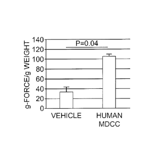
[0011] Figures 3A and 3B show the results of an in vivo
muscular contractility test. Muscle force (Figure 3A) and
percent fatigue (Figure 3B) measurements were normalized
with the muscle weights. hMDCC-treated animals showed
improved muscle force (p=0.04) and fatigue tolerance 90
days after MDCC local injections.
[0012] Figures 4A and 4B show the results of an ex vivo
muscular contractility test. mMDCC-treated gastrocnemius
muscles showed increased contractile strength expressed
after the maximal sine wave (p=0.039; Figure 4A) and
maximal percent strain (Figure 413), compared to control,
untreated muscle.
[0013] Figures 5A and 5B show the results of in vivo
muscular contractility test. Muscle force (Figure 5A) and
Percent fatigue (Figure 5B) measurements were normalized
with the muscle weights. mMDCC-treated animals showed
-4-
CA 088137 2017-1
WO 2016/201182 PCT/US2016/036821
improved muscle force and fatigue tolerance (p=0.05) 30
days after MDCC local injections.
Detailed Description of the Invention
[0014] MDCC compositions and methods for treating muscular
dystrophies in patients have now been developed. MDCC lines
are created by ex vivo fusion of a first population of
myoblasts (autologous or allogeneic) with a second
population of myoblasts, mesenchymal stem cells or stromal
cells. Administration of the MDCC enables simultaneous
delivery of myogenic and mesenchymal origin cells to
patients. Accordingly, this invention provides MDCCs
prepared by fusion of myoblasts from a subject suffering
from a muscular dystrophy and myoblasts from a healthy
donor; myoblasts from a subject suffering from a muscular
dystrophy and mesenchymal stem cells; myoblasts from a
healthy donor and mesenchymal stem cells; myoblasts from a
subject suffering from a muscular dystrophy and stromal
cells; or myoblasts from a healthy donor and stromal cells.
In particular embodiments, the MDCCs of this invention find
use in a method for treating muscular dystrophies.
[0015] This invention differs from other cell-based
therapies since the combined therapy introduces myoblasts
differentiating into myocytes, whereas MSC are known for
reduced alloreactivity, plasticity and potential for de-
differentiation into myoblasts in damaged tissue.
Combination of myoblasts/MSC characteristics, including the
secretion of immunomodulatory cytokines and growth factors,
supports MDCC engraftment, tolerance and regeneration of
muscle under favorable microenvironment conditions. On the
other hand, myoblast/myoblast MDCC characteristics and
capacity to spontaneously fuse offer the ability to either
engraft and resupply the muscle stem cell niche or fuse
-5-
CA 088137 2017-12-01 2016/201182 PCT/US2016/036821
with the recipient myoblast after treatment, providing
better outcomes compared to myoblasts/MSC MDCC. Cells
harvested from DMD patient may be fused with healthy,
dystrophin-positive stem cells from haploidentical male
relative, i.e., father or from haplo-matched donor in cell
banks. In this manner, the MDCC shares surface antigens of
self and haplo-identical origin, reducing the risk of
rejection of transplanted MDCC. Further, given that the
MDCCs are not genetically modified (i.e., by recombinant
methods) and do not require immune suppression, use of the
cells of this invention provides a safe alternative to
conventional therapies.
[0016] For the purposes of this invention, a "chimeric
cell" or "hybrid cell" is a cell that is constructed from a
somatic cell hybridization (or a whole cell hybridization)
of, for example, two or more biological cells (parent
cells). The parent or donor cells can be obtained from
either the same donor or cell lineage or different donors
or cell lineage. While the MDCC of this invention is
referred to as "a chimeric cell," said chimeric cell is
intended to mean a single cell or a population of cells.
[0017] As used herein, a donor is a subject who provides a
cell used in the preparation of a chimeric cell of this
invention. The donor can be a subject with a muscular
dystrophy or a healthy donor, i.e., an individual not
suffering from the same genetic disorder. The donor can be
the genetic father (parent) of a subject (son) with a
muscular dystrophy or a cell bank donor. In particular
embodiments, at least one of the donors is a healthy
subject. In certain embodiments, the healthy donor is the
subject's father. In other embodiments, the first myoblast,
a second myoblast, mesenchymal stem cell or stromal cell is
autologous or allogeneic. Further, the donor may be any
-6-
CA 02988137 2017-12-01
WO 2016/201182 PCT/US2016/036821
mammal including a human, mouse, rat, dog, cat, horse, and
the like. In particular embodiments, the donor is human.
[0018] As is convention in the art, a myoblast refers to a
primitive muscle cell having the potential to develop into
a muscle fiber. Myoblasts are characterized by expression
of desmin and CD56, and can be obtained from fetal or adult
tissue using a method known in the art. See, e.g., WO
93/03768, which discloses the isolation of myoblasts from a
crude cell population by flow cytometry (e.g., FACs).
Alternatively, a myoblast can be obtained by growing and
propagating muscle biopsy-derived myoblasts in culture.
See, e.g., Springer, et al. (1997) In: Current Human
Genetics. Unit 13.4, Boyle Ed. John Wiley & Sons, NY. In
accordance with some embodiments of this invention, a
myoblast from a healthy donor is fused with a myoblast from
a subject with a muscular dystrophy. In particular
embodiments, a first myoblast and a myoblast are from
different donors.
[0019] "Mesenchymal stem cells" (also referred to as
"MSCs") can give rise to connective tissue, bone,
cartilage, and cells in the circulatory and lymphatic
systems. Mesenchymal stem cells are found in the
mesenchyme, the part of the embryonic mesoderm that
consists of loosely packed, fusiform or stellate
unspecialized cells. Mesenchymal stem cells can be obtained
by conventional methods and can be identified one or more
of the following markers: 0329, CD31-, CD34-, CD44 CD45-,
CD51, CD73, 0390/Thy-1, 03105, CD166, Integrin al, PDGF Re,
Nestin, Sca-14-, SCF R/c-Kit, STRO-1, and VCAM-1. In some
embodiments, the mesenchymal stem cells are derived or
obtained from bone marrow (BM) or adipose tissue (ASC). In
particular embodiments, the mesenchymal stem cells are
derived or obtained from human bone marrow.
-7-.
CA 02988137 2017-1
WO 2016/201182 PCT/US2016/036821
[0020] The term "stromal cell" or "adherent stromal cell"
is intended to mean a cell defined by its ability to adhere
and proliferate in tissue-culture treated petri dishes with
or without other cells and/or elements found in loose
connective tissue, including but not limited to,
endothelial cells, pericytes, macrophages, monocytes,
plasma cells, mast cells and adipocytes. Any suitable
method can be used to obtain stromal cells. See, e.g.,
Tondreau, et al. (2005) Stem Cells 23:1105-1112. In
particular embodiments, the stromal cell is derived or
obtained from bone marrow (BM) or cord blood (CB). In other
embodiments, the stromal cell is a CD133 stromal cell.
[0021] The cells used in the preparation of the MDCCs of
this invention can be isolated and optionally purified. As
used herein the term "isolated" is meant to describe a cell
of interest that is in an environment different from that
in which the element naturally occurs. "Purified" as used
herein refers to a cell removed from an environment in
which it was produced and is at least 60% free, preferably
75% free, and most preferably 90% free from other
components with which it is naturally associated or with
which it was otherwise associated with during production.
[0022] Purification and/or identification of cells of
interest can be achieved through any means known in the
art, for example immunologically. Histochemical staining,
flow cytometry, fluorescence activated cell sorting (FACS),
western blot analysis, enzyme-linked immunosorbent assay
(ELISA), etc. may be used. Flow immunocytochemistry may be
used to detect cell-surface markers, immunohistochemistry
(for example, of fixed cells) may be used for intracellular
or cell-surface markers. Western blot analysis may be
conducted on cellular extracts. Enzyme-linked immunosorbent
assay may be used for cellular extracts or products
-8-
CA 02988137 2017-12-01
WO 2016/201182
PCT/US2016/036821
secreted into the medium. Antibodies for the identification
of stem cell markers may be obtained from commercial
sources, for example from Chemicon International,
(Temecula, CA).
[0023] In some embodiments, a donor cell used in the
preparation of the MDCC of this invention is autologous or
heterologous to the subject being treated. In other
embodiments, a donor cell used in the preparation of the
MDCC of this invention is allogeneic to said subject. In
certain embodiments, the donor cells are HLA (human
leukocyte antigen)-matched. Representative sources of donor
cells used for the preparation of the MDCCs of the
invention are listed in Table 1.
TABLE 1
Father Son
MSC Myoblast
Myoblast Myoblast
Bone Marrow (BM) Myoblast
MDSC Myoblast
CD133+ (BM) Myoblast
MSC Muscle-derived stem cell
(MDSC)
________ Myoblast MDSC
Bone Marrow MDSC
MDSC MDSC
CD133+ (BM) MDSC
Myoblast MSC
Myoblast Bone Marrow
ASC Myoblast
ASC ASC
Father Father
MSC Myoblast
CD133+ (BM) Myoblast _____________
Bone Marrow Myoblast
MSC MDSC
CD133 (BM) MDSC
Bone Marrow MDSC
ASC Myoblast
Cell Bank Son
CD133+ (BM) Myoblast __
CD133+ (CB) Myoblast
MSC Myoblast
-9-
CA 02988137 2017-12-01
WO 2016/201182 PCT/US2016/036821
CD133 (BM) MDSC
CD133' (CB) MDSC
___________ MSC MDSC
Cell Bank Father __
CD133+ (BM) Myoblast
CD133+ (CB) Myoblast
MSC Myoblast
CD133+ (BM) MDSC
CD133+ (CB) MDSC
MSC MDSC
[0024] In particular embodiments, the MDCC is produced by
fusing:
a) a human myoblast from a subject suffering from a
muscular dystrophy and a human myoblast from a healthy
donor;
b) a human myoblast from a subject suffering from a
muscular dystrophy and a human mesenchymal stem cell from a
healthy donor;
c) a human myoblast from a healthy donor and a human
mesenchymal stem cell from a healthy donor;
d) a human myoblast from a subject suffering from a
muscular dystrophy and a human stromal cell from a healthy
donor; or
e) a human myoblast from a healthy donor and a human
stromal cell from a healthy donor.
[0025] The MDCC of this invention is prepared by ex vivo
fusion of two different donor cells. By "ex vivo" it is
meant that cells are manipulated outside of the body. Cell
fusion is a process in which two or more cells merge into
one by fusing their plasma membranes. MDCCs can be prepared
by cell fusion methods known in the art, including, but not
limited to, exposure of cells to fusion-promoting
chemicals, such as polyethylene glycol (PEG); the use of
inactivated virus, such as Sendai virus; and the use of
electrical stimulation. See, e.g., Kennett (1979) Methods
-10-
CA 02988137 2017-12-01
WO 2016/201182 PCT/US2016/036821
Enzymol. 58:345-359 for a review of the commonly used
methods based upon Sendai virus induced cell fusion, or
cell fusion induced by polyethylene glycol (PEG). Briefly,
cells to be fused are incubated with a fusogenic agent,
such as Sendai virus or PEG. Centrifugation or agitation
may be used to encourage clumping and close apposition of
the cell membranes. Variables such as time, temperature,
cell concentration and fusogenic agent concentration may be
optimized for each cell combination. With respect to
electro fusion, short electric pulses are passed through
mixtures of cells to stimulate fusion. See, e.g., Neil &
Zimmermann (1993) Methods Enzymol. 220:174-196.
[0026] In certain embodiments, the MDCCs are prepared by
polyethylene glycol cell fusion. After fusion, cell lines
representing either myogenic/myogenic or myogenic/MSC
origin are separated, cultured and characterized to confirm
the myoblast and MSC specific markers and HLA class I types
of cell donor origin.
[0027] Prior to fusion, the donor cells may or may not be
cultured to increase their number. Further, the donor cells
may or may not be labeled (e.g., with a membrane dye) to
monitor fusion of the donor cells. By way of illustration,
myoblasts from a subject suffering from a muscular
dystrophy are labeled with PKH26-red and myoblasts from a
healthy donor are labeled with PKH67-green.
[0028] In some embodiments, the MDCC of this invention
secretes one or more immunomodulatory cytokines and growth
factors. In certain embodiments, the immunomodulatory
cytokines and growth factors include insulin-like growth
factor 1 (IGF-1), hepatocyte growth factor (HGF) and
myostatin. In further embodiments, the MDCC of this
invention produces dystrophin.
-11-
CA 02988137 2017-12-01
WO 2016/201182 PCT/US2016/036821
[0029] The MDCC of this invention is of particular use in
the treatment of muscular dystrophies. Accordingly, this
invention also provides methods of treating muscular
dystrophies in a subject in need thereof by administering
to the subject the MDCC of the invention or a composition
containing the MDCC in an amount effective to treat the
dystrophies. "Treating" a subject having a disease or
disorder means accomplishing one or more of the following:
(a) reducing the severity of the disease; (b) arresting the
development of the disease or disorder; (e) inhibiting
worsening of the disease or disorder; (d) limiting or
preventing recurrence of the disease or disorder in
patients that have previously had the disease or disorder;
(e) causing regression of the disease or disorder; (f)
improving or eliminating the symptoms of the disease or
disorder; and (g) improving survival.
[0030] As indicated herein, muscular dystrophies are a
group of genetic diseases characterized by progressive
weakness and degeneration of the skeletal muscles that
control movement. Examples of muscular dystrophies include
Duchenne Muscular Dystrophy, Becker Muscular Dystrophy,
Limb Girdle Muscular Dystrophy, Myotonic Muscular
Dystrophy, Facioscapulohumeral Muscular Dystrophy,
Oculopharyngeal muscular dystrophy, Emery-Dreifuss muscular
dystrophy, Fukuyama-type congenital muscular dystrophy,
Miyoshi myopathy, Ullrich congenital muscular dystrophy,
Steinert Muscular Dystrophy. In certain embodiments, the
muscular dystrophy is Duchenne muscular dystrophy (DMD).
[0031] In accordance with the method of treatment, a MDCC
or composition containing the same is administered to a
subject having a muscular dystrophy. In some embodiments, a
combination of MDCCs of this invention can be administered.
The MDCC or combination of cells can be administered by
-12-
CA 02988137 2017-12-01
WO 2016/201182 PCT/US2016/036821
engraftment, wherein the cells are injected into the
subject, for example, intravenously, intra-muscularly,
intra-arterially, intra-bone and the like. In certain
embodiments, administration involves engrafting about 102,
104, 106, 107, 108, 108, 1010, 1012, or more cells. The number
of cells engrafted may be chosen based on the route of
administration and/or the severity of the condition for
which the cells are being engrafted. Advantageously, the
MDCC of this invention will successfully engraft and
complement the function of defected muscles of muscular
dystrophy patients.
[0032] Compositions containing the MDCC or combinations of
MDCCs can be prepared by combining the cell or combination
of cells with a pharmaceutically acceptable carrier or
aqueous medium. The phrase "pharmaceutically or
pharmacologically acceptable" refers to molecular entities
and compositions that do not produce adverse, allergic, or
other untoward reactions when administered to an animal or
a human. As used herein, "pharmaceutically acceptable
carrier" includes any and all solvents, dispersion media,
coatings, antibacterial and antifungal agents, isotonic and
the like. The use of such media and agents for
pharmaceutically active substances is well known in the
art. Except insofar as any conventional media or agent is
incompatible with the cells of the present disclosure, its
use in therapeutic compositions is contemplated.
Pharmaceutical compositions can be determined by one
skilled in the art depending upon, for example, the
intended route of administration, delivery format and
desired dosage. See, for example,
REMINGTON'S
PHARMACEUTICAL SCIENCES, 18th Edition, (A. R. Gennaro,
ed.), 1990, Mack Publishing Company.
-13-
CA 02988137 2017-12-01
WO 2016/201182 PCT/US2016/036821
[ 0 0 3 3 ] The compositions of the invention can be
incorporated in an injectable formulation. The formulation
may also include the necessary physiologically acceptable
carrier material, excipient, lubricant, buffer, surfactant,
antibacterial, bulking agent (such as mannitol),
antioxidants (ascorbic acid or sodium bisulfite) and the
like.
[0034] Acceptable formulation materials preferably are
nontoxic to recipients at the dosages and concentrations
employed. The pharmaceutical composition may contain
formulation materials for modifying, maintaining or
preserving, for example, the pH, osmolarity, viscosity,
clarity, color, isotonicity, odor, sterility, stability,
rate of dissolution or release, adsorption or penetration
of the composition. Suitable formulation materials may
include, but are not limited to, amino acids (such as
glycine, glutamine, asparagine, arginine or lysine);
antimicrobials; antioxidants (such as ascorbic acid, sodium
sulfite or sodium hydrogen-sulfite); buffers (such as
borate, bicarbonate, Tris-HCl, citrates, phosphates or
other organic acids); bulking agents (such as mannitol or
glycine); chelating agents (such as ethylenediamine
tetraacetic acid (EDTA; complexing agents (such as
caffeineI polyvinylpyrrolidone, beta-cyclodextrin or
hydroxypropyl-beta-cyclodextrin); fillers; monosaccharides,
disaccharides, and other carbohydrates (such as glucose,
mannose or dextrins); proteins (such as serum albumin,
gelatin or immunoglobulins); coloring, flavoring and
diluting agents; emulsifying agents; hydrophilic polymers
(such as polyvinylpyrrolidone); low molecular weight
polypeptides; salt-forming counterions (such as sodium);
preservatives (such as benzalkonium chloride, benzoic acid,
salicylic acid, thimerosal, phenethyl alcohol,
-14-
CA 02988137 2017-12-01
WO 2016/201182 PCT/US2016/036821
methylparaben, propylparaben, chlorhexidine, sorbic acid or
hydrogen peroxide); solvents (such as glycerin, propylene
glycol or polyethylene glycol); sugar alcohols (such as
mannitol or sorbitol); suspending agents; surfactants or
wetting agents (such as PLURONICS, PEG, sorbitan esters,
polysorbates such as polysorbate 20 and polysorbate 80,
TRITON, trimethamine, lecithin, cholesterol, or tyloxapal);
stability enhancing agents (such as sucrose or sorbitol);
tonicity enhancing agents (such as alkali metal halides,
preferably sodium or potassium chloride, mannitol, or
sorbitol); delivery vehicles; diluents; excipients and/or
pharmaceutical adjuvants. See, for example, REMINGTON'S
PHARMACEUTICAL SCIENCES, Id.
[0035] The primary vehicle or carrier in a pharmaceutical
composition may be either aqueous or nonaqueous in nature.
For example, a suitable vehicle or carrier may be water for
injection, physiological saline solution or artificial
cerebrospinal fluid, possibly supplemented with other
materials common in compositions for parenteral
administration. Neutral buffered saline or saline mixed
with serum albumin are further exemplary vehicles.
Pharmaceutical compositions can comprise Tris buffer of
about pH 7.0-8.5, or acetate buffer of about pH 4.0-5.5,
which may further include sorbitol or a suitable substitute
therefore. Pharmaceutical compositions of the invention may
be prepared for storage by mixing the selected composition
having the desired degree of purity with optional
formulation agents (REMINGTON'S PHARMACEUTICAL SCIENCES,
Id.) in the form of a lyophilized cake or an aqueous
solution.
[0036] The cell or composition can be provided by sustained
release systems, by encapsulation or by implantation
devices. The compositions may be administered by bolus
-15-
CA 02988137 2017-12-01
WO 2016/201182 PCT/US2016/036821
injection or continuously by infusion, or by implantation
device. The composition also can be administered locally
via implantation of a membrane, sponge or another
appropriate material onto which the cell or cells have been
absorbed or encapsulated. Where an implantation device is
used, the device may be implanted into any suitable tissue
or organ. The injections may be given as a one-time
treatment, repeated (daily, weekly, monthly, annually etc.)
in order to achieve the desired therapeutic effect.
[0037] Cell encapsulation methodology has been previously
described which allows transplantation of encapsulated
cells in treatment of Parkinson's disease (Tresco, et al.
(1992) ASAIO J. 38:17-23) or Amyotrophic lateral sclerosis
(Aebischer, et al. (1996) Hum. Gene Ther. 7:851-860). In
accordance with this embodiment, cells are encapsulated by
compounds which form a microporous membrane. Capsules, for
example approximately 1 cm in length, containing the cells
of interest may be prepared employing a hollow microporous
membrane fabricated from poly-ether-sulfone (PES) (Akzo
Nobel Faser AG, Wuppertal, Germany; Deglon, et al. (1996)
Rum. Gene Ther. 7:2135-2146).
[0038] The compositions of the invention can be delivered
parenterally. When parenteral administration is
contemplated, the therapeutic compositions for use in this
invention may be in the form of a pyrogen-free,
parenterally acceptable aqueous solution. A particularly
suitable vehicle for parenteral injection is sterile
distilled water. Preparation can involve the formulation
with an agent, such as injectable microspheres, bio-
erodible particles, polymeric compounds (such as polylactic
acid or polyglycolic acid), beads or liposomes, that may
provide controlled or sustained release of the cell or
cells, which may then be delivered via a depot injection.
-16-
CA 088137 2017-1
WO 2016/201182 PCT/US2016/036821
Formulation with hyaluronic acid has the effect of
promoting sustained duration in the circulation.
Implantable drug delivery devices may be used to introduce
the desired composition.
[0039] These compositions may also contain adjuvants such
as preservative, wetting agents, emulsifying agents and
dispersing agents. Prevention of the action of
microorganisms can be ensured by the inclusion of various
antibacterial and antifungal agents, for example, paraben,
chlorobutanol, phenol sorbic acid and the like. It may also
be desirable to include isotonic agents such as sugars,
sodium chloride and the like.
[0040] Supplementary active ingredients also can be
incorporated into the compositions. The active compositions
of the present disclosure may include classic
pharmaceutical preparations. Administration of these
compositions according to the present disclosure will be
via any common route so long as the target tissue is
available via that route. Such routes include oral, nasal,
buccal, rectal, vaginal or topical route. Alternatively,
administration may be by orthotopic, intradermal,
subcutaneous, intraperitoneal, or intravenous injection.
Intramuscular injection will be preferred. Such
compositions would normally be administered as
pharmaceutically acceptable compositions.
[0041] As used herein, the term "amount effective,"
"effective amount" or a "therapeutically effective amount"
refers to an amount of the cell or composition of the
invention sufficient to achieve the desired result. The
amount of the cell or composition which constitutes an
"effective amount" or "therapeutically effective amount"
may vary depending on the severity of the disease, the
condition, weight, or age of the patient to be treated, the
-17-
CA 088137 2017-1
WO 2016/201182 PCT/US2016/036821
frequency of dosing, or the route of administration, but
can be determined routinely by one of ordinary skill in the
art. A clinician may titer the dosage or route of
administration to obtain the optimal therapeutic effect.
[0042] The present invention is also directed to a kit to
for the treatment of a muscular dystrophy. The kit is
useful for practicing the inventive method of treating a
muscular dystrophy. The kit is an assemblage of materials
or components, including at least one of the inventive
compositions. Thus, in some embodiments the kit a fusogenic
agent for carrying out ex vivo cell fusions, and one or
more donor cells (e.g., donor cells from a cell bank), and
optionally materials for obtaining donor cells, as
described above.
[0043] The exact nature of the components configured in the
inventive kit depends on its intended purpose. For example,
some embodiments are configured for the purpose of treating
a muscular dystrophy. In one embodiment, the kit is
configured particularly for the purpose of treating human
subjects. In another embodiment, the kit is configured
particularly for the purpose of treating adult, human
subjects. In another embodiment, the kit is configured
particularly for the purpose of treating children. In
another embodiment, the kit is configured particularly for
the purpose of treating DMD. In another embodiment, the kit
is configured particularly for the purpose of treating BMD.
In another embodiment, the kit is configured particularly
for the purpose of providing continuous daily use dosages.
In another embodiment, the kit is configured particularly
for the purpose of providing as needed use dosages. In
further embodiments, the kit is configured for veterinary
applications, treating subjects such as, but not limited
to, farm animals, domestic animals, and laboratory animals.
-18-
CA 088137 2017-12-01 2016/201182 PCT/US2016/036821
[0044] Instructions for use may be included in the kit.
"Instructions for use" typically include a tangible
expression describing the technique to be employed in using
the components of the kit to effect a desired outcome, such
as to treat muscular dystrophy, to treat BMD, or to treat
DMD. Optionally, the kit also contains other useful
components, such as, diluents, buffers, pharmaceutically
acceptable carriers, syringes, catheters, applicators,
pipetting or measuring tools, bandaging materials or other
useful paraphernalia as will be readily recognized by those
of skill in the art.
[0045] The materials or components assembled in the kit can
be provided to the practitioner stored in any convenient
and suitable ways that preserve their operability and
utility. For example the components can be in dissolved,
dehydrated, or lyophilized form; they can be provided at
room, refrigerated or frozen temperatures. The components
are typically contained in suitable packaging material (s)
As employed herein, the phrase "packaging material" refers
to one or more physical structures used to house the
contents of the kit. The packaging material is constructed
by well-known methods, preferably to provide a sterile,
contaminant-free environment. The packaging materials
employed in the kit are those customarily utilized in
therapeutic treatment. As used herein, the term "package"
refers to a suitable solid matrix or material such as
glass, plastic, paper, foil, and the like, capable of
holding the individual kit components. The packaging
material generally has an external label which indicates
the contents and/or purpose of the kit and/or its
components.
[0046] The following non-limiting examples are provided to
further illustrate the present invention.
-19-
CA 088137 2017-1
WO 2016/201182 PCT/US2016/036821
Example 1: Ex Vivo Preparation of Human Muscular Dystrophy
Chimeric Cells (hMDCC)
[0047] Ex vivo fusions of allogenic human myoblasts and MSC
or myoblasts from two unrelated donors were performed,
using polyethylene glycol technique (Figure 1). Briefly,
commercially available (Lonza, Inc.) human myoblasts and
MSC were separately cultured for 6 to 10 days. Next, cells
were fluorescently labeled using either PKH-26 (red) or
PKH-67 (green) tracking dye, as shown in Figure 1. Fusion
was performed using PEG. Cells presenting double (PKH26 and
PKH67) staining were selected via fluorescently activated
cell sorting (FACS; BD ASTERIOS). To confirm fusion, double
(PKH26 and PKH67) labeled MDCC were evaluated using
confocal microscopy and flow cytometry (Figure 2).
Morphology of MDCC was assessed using transmission electron
microscopy. The MDCC were confirmed by the presence of two
nuclei, fused cell membrane and fused cytoplasm.
[0048] Flow cytometry was used to assess phenotype changes
of MDCC at 7, 14, 21 and 30 days following fusion. MDCC
were tested for the expression of muscle-specific markers
(Anti-Myogen in, Anti-hMyosin Heavy Chain, Anti-mMYF-5) and
MSC markers (CD105, CD73, CD90). The results are shown in
Table 2. Notably, the MDCC did not express CD45 or CD8
markers, which are characteristic for hematopoietic cells.
TABLE 2
Sample
Phenotype Myoblast Myoblast MDCC after
Donor A Donor B Fusion A+B
Anti-myogenin
Anti-hMyosin Heavy Chain
Anti-mMYF-5
CD105
CD73
CD90
-20-
CA 02988137 2017-12-01
WO 2016/201182 PCT/US2016/036821
[0049] Additionally, MDCC were analyzed by FISH to detect
sex chromosomes and viability staining (Trypan blue).
Further, MDCC were cultured for 30 days to test
proliferation and secretory properties via ELISA assay.
Moreover, using immunofluorescence staining, the expression
of dystrophin was demonstrated in the human MDCC. In
addition, in vitro results confirmed myogenic
differentiation potential of hMDCC. After fusion, hMDCC
were placed in specific myogenic differentiation medium
(low serum medium supplemented with insulin, PROMOCELL) for
7 days. Skeletal myosin heavy chain expression, a marker of
skeletal myocyte differentiation, was observed in all hMDCC
lines.
[0050] To asses genotype and confirm cell fusion,
Polymerize Chain Reaction - Reverse Sequence-Specific Probe
(PCR-rSSOP) and short-tandem repeat - PCR (STR - PCR) was
performed on MDCC, detecting HLA class I and II and
specific gene combination from both donors (Tables 3 and 4,
respectively). This analysis indicated that MDCC presented
HLA alleles derived from both donor cells and showed the
presence of genetic markers specific for both fusion donor
cells.
TABLE 3
Sample
HLA Type Myoblast Myoblast MDCC after Fusion
Donor A Donor B (A+B)
A 01,31 30,30 01,31,30
08,39 42,57 08,39,42,57
Cw 07,07 17,18 07,07,17,18
Bw 6 6,4 6,4
DRB1 03(17),14 08,03(18)
03(17),14,08,03(18)
DQB1 02,03(7) 03(7)/04 02,03(7),03(7),04
DR52, DR53 52 52 52
-21-
CA 02988137 2017-12-01
WO 2016/201182
PCT/US2016/036821
TABLE 4
Sample
Genetic
Myoblast Myoblast MDCC after
Marker
Donor A Donor B Fusion (A+B)
D5S818 11,12 11,12 11,12
D75820 8,10 10 8,10
Th01 8,9,3 6,7 8,9,3,6,7
AMEL X,Y X X,Y
TPDX 9,11 8,9 9,11,8
CSF1P0 11 12 11,12
D21S11 28,30 30,31 28,30,31
[0051] In vitro culturing results showed proliferative
potential, long-term viability and differentiation of DMDCC
to the myocyte lineage. The expression of dystrophin was
maintained by DMDCC up to 30 days post-fusion. Secretion of
cytokines by DMDCC was confirmed by ELISA assay.
[0052] In in vivo studies, engraftment of locally
administered hMDCC in mdx/scid mouse model was assessed.
Five groups of mice were tested (Table 5). The first two
groups included MDCC of myoblast/myoblast or MSC/myoblast
origin (dose 0.5x10) delivered through multiple
intramuscular injections following a standardized template,
to the left gastrocnemius muscle of mdx/scid mice. Control
groups included treatment with vehicle, treatment with
unfused myoblasts (dose 0.5x10⁶), or treatment with mixed
MSCs and myoblasts (dose 0.5x10⁶) via intramuscular
injection. Outcomes measured included in vivo muscle
function, dystrophin expression in treated muscles as well
as MDCC engraftment at 1 week and 12 week time-points.
[0053] Dystrophin expression was used as a specific marker
for hMDCC since it had been confirmed that both hMDCC lines
expressed dystrophin. After 7 days following local
intramuscular delivery of hMDCC, successful engraftment of
hMDCC was shown. In addition, locally increased dystrophin
expression (12%) was observed as early as 7 day post-
-22-
CA 088137 2017-12-01 2016/201182 PCT/US2016/036821
transplant as compared with the lack of dystrophin
expression in mdx/scid controls. Furthermore, at 90 days
17% of dystropin expression was observed.
TABLE 5
Time-point No. of
Treatment Outcomes
(days) Animals
MDCC engraftment and
7 3 dystrophin
Myoblast/MSC MDCC expression (DE)
90 3 Muscle function and
structure, DE
7
MDCC engraftment and
3
Myoblast/Myoblast DE
MDCC 90 3 Muscle function and
structure, DE
7
MDCC engraftment and
3
Myoblasts without DE
fusion 90 3 Muscle function and
structure, DE
7
MDCC engraftment and
3
Myoblast and MSC DE
without fusion 90 3 Muscle function and
structure, DE
7
MDCC engraftment and
3
DE
Vehicle
90 3 Muscle function and
__________________________________________ structure, DE
[0054] Another set of experimental groups (vehicle and
hMDCC treated mdx/scid and wild type snj mice, n=3) were
tested in motor function tests, including grip strength
measurements and a wire hanging test. Ninety days after
hMDCC therapy delivery, mice receiving hMDCC showed
improvement (p=0.037) of grip strength and tolerance to
fatigue on wire (mdx/scid mice, 50gF; hMDCC therapy, 85-
90gF). Functional improvement was observed in groups
treated with both MDCC lines when compared to vehicle
treated groups until day 42. After this time-point only
myoblast/myoblast hMDCC maintained increased muscle
strength throughout the 90 day follow-up period. By
-23-
CA 02988137 2017-12-01
WO 2016/201182 PCT/US2016/036821
comparison, grip strength values returned to baseline
levels after 42 days in mice treated with MSC/myoblast
hMDCC. The unfused cell treatment control groups showed
temporary motor function improvement in the first 21 days
only.
[0055] Gastrocnemius muscles harvested at 90 days after
hMDCC delivery were analyzed in an ex vivo contractility
test to assess muscle strength evoked by electric
stimulation. Results showed that myoblast/myoblast hMDCC
treated (Figure 3A) muscle had significantly stronger
contraction (p=0.04) even under induced strain (Figure 3B).
The same muscle samples were analyzed by confocal
microscopy to detect and quantify dystrophin expression at
90 days after hMDCC treatment. On average, 17% of the cells
were positive for dystrophin expression.
[0056] Currently, there is no effective therapy to treat
DMD, a lethal genetic, neuromuscular disorder affecting 1
in every 3500 newborn boys. Treatment modalities, such as
growth-modulating agents, anti-inflammatory or second-
messenger signal-modulating agents, and molecular devices
designed to skip mutations in the dystrophin gene have been
attempted, however these approaches fail to halt or reverse
disease progression. By comparison, the instant MDCC
therapy represents a universal regenerative medicine
approach for local or systemic application in patients
suffering from DMD. Further, compared to other cell-based
therapies, the unique features of MDCC, created through ex
vivo fusion, are the combination and synergistic effects of
the complementing characteristics of cells of myogenic and
mesenchymal origin, such as high proliferation rate,
capability for myogenic conversion, and secretion of
immunomodulatory cytokines and growth factors facilitating
muscle regeneration. Moreover, the preparation of MDCC does
-24-
CA 02988137 2017-1
WO 2016/201182 PCT/US2016/036821
not require genetic manipulations or introduction of viral
vectors to the cells, thus making it a safer therapy. In
addition, since MDCC therapy is not gene/mutation-specific,
it can be tailored and applied to patients suffering from
other types of muscular dystrophies including, e.g., Becker
dystrophy.
Example 2: Ex Vivo Preparation of Mouse Duchene Muscular
Dystrophy Chimeric Cells (inDMDCC)
[0057] Healthy snj and mdx (dystrophin-deficient) primary
myoblast cultures as well as mdx MSC cultures were
established and expanded in vitro. Three murine
myoblast/MSC and myoblast/myoblast fusions were performed
with polyethylene-glycol technique. Dystrophin expression,
as well as myogenic differentiation capacity, was confirmed
before and after fusion.
[0058] In in vivo studies (Table 6), efficacy of murine
DMDCC engraftment, survival and restoration of dystrophin
expression and motor functions were tested 30 days
following local delivery of mDMDCC to the gastrocnemius
muscle. Of the five groups tested, mDMDCC composed of
myoblast/myoblast and MSC/myoblast origin (dose 0.5x10⁶)
were delivered through multiple intramuscular injections
following a standardized template, to the left
gastrocnemius muscle of mdx mice. Control groups included
treatment with vehicle, treatment with not unfused myoblast
and treatment with mixed MSC and myoblast. Outcome
measurements included in vivo muscle function, dystrophin
expression in treated muscles as well as mDMDCC
engraftment, which were assessed after 4 weeks. After
myoblast/myoblast mDMDCC delivery, dystrophin expressing
cells constituted 37% of total nucleated cells on
immunofluorescence analysis by confocal microscope.
-25-
CA 088137 2017-1
WO 2016/201182
PCT/US2016/036821
Myoblast/myoblast mDMDCC-treated muscle showed dystrophin-
positive and dystrophin-negative areas. Dystrophin-positive
cells were characterized by dystrophin expression on the
membrane (normal pattern) as well as cytoplasmic
expression.
TABLE 6
Time-point No. of
Treatment Outcomes
(days) Animals
Myoblast/MSC MDCC 90 4 Muscle
function and
structure, DE
MDCC engraftment,
Myoblast/Myoblast
MDCC 90 4 muscle
function and
structure, DE
MDCC engraftment,
Myoblasts without
90 4 muscle
function and
fusion
structure, DE
MDCC engraftment,
Myoblast and MSC
90 4 muscle
function and
without fusion
structure, DE
MDCC engraftment,
Vehicle 90 4 muscle
function and
structure, DE
[0059] Interestingly, mDMDCC-treated muscles showed
decreased muscle weight as compared to contralateral
untreated muscles. The lower weight of the treated muscle
can be explained by decreased DMD-related hypertrophy and
fibrosis. Despite the reduced muscle weight, muscle force
measurements, evoked with electric stimulation in situ,
resulted in increased values of muscle strength in vivo
(Figures 4A and 4B) as well as ex vivo (Figures 5A and 5B).
[0060] In vivo motor function evaluated by grip strength
and wire hanging tests also confirmed the efficacy of
mDMDCC with increased grip strength and prolonged time to
resist fatigue. Myoblast/Myoblast mDMDCC-treated muscle
maintained higher strength values than muscles treated with
myoblast/MSC MDCC, which did not differ from control muscle
values 30 days after therapy delivery. Myoblast-derived
-26-
CA 02988137 2017-12-01
WO 2016/201182 PCT/US2016/036821
mDMDCC treatment resulted in increased tolerance for
fatigue, shown by longer hanging time on wire compared to
control animals.
-27-