Note: Descriptions are shown in the official language in which they were submitted.
CA 02988499 2017-12-06
WO 2016/012800 PCT/GB2015/052139
ANTI-DENGUE VACCINES AND ANTIBODIES
Field of the invention
The invention relates to the field of treatment and prevention of Dengue virus
infection and
related compounds and methods.
Summary
to Dengue infects nearly 400 million people annuallyl, with symptoms in 25%
of infections
ranging from mild disease (dengue fever) to severe cases such as dengue
haemorrhagic
fever. The etiological agents are four serologically related viruses from the
flavivirus
genus2, termed dengue virus serotypes 1-4 (DENV1 -4). Infection with one
serotype leads
to lifelong protection against that serotype but not against the other
serotypes. There is
epidemiological evidence that severe disease is more likely to occur during a
secondary
infection than during the first or primary DENV infection 3'4. The enhancement
of disease
upon secondary infection and the need to protect against four diverse
serotypes sets a
high bar for vaccines, which are urgently needed to protect against the 400
million
infections estimated to occur annually 1'5'6.
Most DENV vaccines in development aim to raise virus neutralizing antibodies
and the
DENV envelope is the main focus of this effort 89 The envelope protein is
responsible for
receptor binding and subsequent receptor-mediated endocytosis. In the acidic
environment of the endosome, envelope protein catalyses a membrane fusion
reaction
between the viral envelope and the endosomal membrane, thereby releasing the
viral
genomic RNA into the cytoplasm. The envelope protein is about 500 amino acids
long,
with a large N-terminal ectodomain and two transmembrane (TM) helices at the C-
terminal
end. Its overall structure is conserved among all flaviviruses, with an amino
acid sequence
identity of approximately 65% between the most distant viruses within the
dengue group,
all of which display two conserved N-linked glycosylation sites at positions
N67 and N153.
The 400 amino terminal residues of the ectodomain of the envelope protein
(termed "sE"
for "soluble E") fold into a13-sheet rich three-domain structure typical of
class viral fusion
proteins, which form head-to-tail homodimers' that coat the surface of mature
virions"
The crystal structure of the envelope glycoprotein ectodomain from dengue
virus serotypes 2, 3 and 4 are available in the PDB (Protein Data Bank)
database
under the accession numbers 10AN, 10K8 for dengue virus serotype 2, 1 UZG for
dengue virus serotype 3 and 3UAJ for dengue virus serotype 4.
1
CA 02988499 2017-12-06
WO 2016/012800
PCT/GB2015/052139
However, antibody recognition of DENV particles is complicated by a number of
dramatically different compositions and conformations of the virus capsid that
are
displayed at different phases of the virus life cycle 11'12. The "immature"
virus particle has
a full complement of precursor membrane protein (prM) in 1:1 association with
envelope
and the virion adopts a characteristic spiky appearance where each spike is
made up of a
trimer of prM/E proteins 11-15.
Following furin-mediated prM cleavage the "mature" virus particle adopts a
smooth
appearance with 90 closely packed dimers of envelope protein arranged around
2, 3 and
5 fold axes of symmetry; an expansion of these mature particles into a "bumpy"
form upon
exposure to temperatures above 34 C, in which the envelope protein dimers
rearrange
with respect to each other, has also been recently described 16, 17. Following
internalization
into early endosomes, the acidic environment triggers a major conformational
change of
the envelope protein, which exposes the fusion loop and then trimerizes
irreversibly to
induce membrane fusion 15'19.
An important and additional level of complexity is that prM cleavage is
frequently
incomplete leading to a population of viruses with varying degrees of cleavage
15, 20.
Viruses containing high levels of prM are not infectious whereas viruses with
lower levels
of prM are still infectious and furthermore it has been demonstrated that high-
prM non-
infectious particles can be driven to infect by antibody dependent enhancement
21'22.
It is currently unclear as to what the epitope is that most human neutralising
antibodies
target, for example de Alwis27 suggests the epitope requires virus assembly
for formation,
whilst Rer suggests that the envelope dimer itself is the target.
Antibody dependent enhancement of DENV infection (ADE) is one of the
mechanisms
postulated to increase the severity of disease upon secondary infection 23.
Antibody formed
to the primary infection is proposed to opsonize but not fully neutralize
virus and promote
Fc receptor mediated uptake into myeloid cells driving higher virus loads in
secondary
infection. ADE can be seen at sub-neutralizing concentrations of almost all
antibodies and
its perceived risk complicates vaccine strategies in DENV.
The leading dengue vaccine candidates currently being tested in clinical
trials consist of
tetravalent formulations of live attenuated dengue or dengue/yellow fever
chimeric viruses
24, 25. Raising balanced tetravalent immunity without unacceptable
reactogenicity has
2
CA 02988499 2017-12-06
WO 2016/012800
PCT/GB2015/052139
proved challenging. The most advanced dengue vaccine candidate returned much
lower
vaccine efficacy than anticipated in a recent phase II clinical trial 4, and
did not protect
against DENV-2, whilst in a phase III study the vaccine reduced the incidence
of disease
by 56% ( http://sanofipasteurcom/en/articles/theworld-s-first-larqe-scale-
denque-vaccine-
efficacy-study-successfully-achieved-its-primary-clinical-endpoint.aspx);
Capeding et at
(2014) Lancet Published online July 11, 2014
http://dx.doi.org/10.1016/60140-
6736(14)61060-6, leaving approximately half the population exposed to the
disease. This
disparity creates a pressing need to understand the human antibody response in
natural
dengue infection and following vaccination, and in particular to identify the
epitopes
113 recognized by the most potent cross-reactive antibodies generated in
humans, and
understand the correlates with protection from disease. It is therefore
crucial to provide a
dengue vaccine including the epitopes recognized by the most potent cross-
reactive
antibodies generated in humans.
Recent evidence has indicated that the dengue virus (DENV)-specific serum Ab
response
in humans consists of a large fraction of cross-reactive, poorly neutralizing
Abs and a small
fraction of serotype-specific, potently inhibitory antibodies 27, which bind
to a complex,
quaternary structure epitope that is expressed only when envelope proteins are
assembled
on a virus particle, implying that in order to stimulate an effective immune
response, an
intact viral particle is required.
In contrast to this recent evidence, the inventors of the present invention
have
surprisingly identified and characterised human antibodies obtained by
isolating
rearranged heavy- and light-chain genes from sorted single plasmablasts of
patients
infected with dengue virus which were found to be potently neutralising cross-
reactive
antibodies. In addition, the epitope to which these antibodies bind was found
to be limited
to the envelope protein dimer, and did not require full virus assembly. A
subunit vaccine
comprising a stabilized soluble protein E dimer is therefore a good candidate
for a
successful dengue vaccine, avoiding eliciting antibodies against poorly
immunogenic
regions that are normally not accessible at the surface of an infectious
virion.
The invention, as described below, provides compounds, compositions, methods,
uses
and vaccines in relation to the newly identified antibodies and antigen.
The inventors characterized 145 human monoclonal antibodies from patients with
a
dengue infection. The acute human antibody response was found to be focused on
two
major epitopes; one of which is well described on the fusion loop, and a
second novel
3
CA 02988499 2017-12-06
WO 2016/012800 PCT/GB2015/052139
epitope found on intact virions or dimers of envelope protein, which
encompasses areas
of domains I, II and III. Antibodies reactive with this epitope, the Envelope
Dimer Epitope,
or EDE, were found to be able to fully neutralise virus made in both insect
and primary
human cells in the low picomolar range. This novel epitope has wide ranging
implications
for the treatment and prevention of diseases caused by the dengue virus.
lo
The invention will be described below with reference to various embodiments of
different
aspects of the invention. It is appreciated that certain features of the
invention, which are,
for clarity, described in the context of separate embodiments, may also be
provided in
combination in one or more embodiments or in a single embodiment. Conversely,
various
features of the invention, which are, for brevity, described in the context of
a single
embodiment, may also be provided separately or in any suitable sub-
combination. All
combinations of the embodiments are specifically embraced by the present
invention and
are disclosed herein just as if each and every combination was individually
and explicitly
disclosed. In addition, all sub-combinations are also specifically embraced by
the present
invention and are disclosed herein just as if each and every such sub-
combination was
individually and explicitly disclosed herein.
Thus, in a first aspect of the invention, a compound is provided that
neutralises dengue
virus of more than one serotype of dengue virus.
Preferably the compound neutralises the dengue virus of two serotypes of
dengue virus,
more preferably three types of dengue virus and most preferably four serotypes
of dengue
virus ie all serotypes of dengue virus, for example neutralises two or more
serotypes of
dengue virus from the list comprising DENV-1, DENV-2, DENV-3 and DENV-4.
By a compound we mean any compound that can neutralise more than one serotype
of
Dengue virus. The compound may, for example, be a small molecule, a
polypeptide or
protein (which terms are used interchangeably herein), including a
glycoprotein, a nucleic
acid, a carbohydrate, a fat, an element, for example a metal. In a preferred
embodiment
the compound is a polypeptide, preferably an antibody or antigen binding
portion thereof.
The antigen binding portion may be a Fv fragment; a Fab-like fragment (e.g. a
Fab
fragment, a Fab' fragment, a F(ab)2 fragment, Fv or scFv fragments); or a
domain
4
CA 02988499 2017-12-06
WO 2016/012800
PCT/GB2015/052139
antibody. The antibody binding portion may be derived from the linear amino
acid
sequence present in an intact antibody, or may comprise a set of non-
consecutive amino
acids, optionally interspersed with other amino acids, for example may
comprise particular
amino acids that are required for contact with an epitope, but may for example
not
comprise the amino acids required for the framework of a native antibody,
which, in some
cases, may be replaced by a heterologous scaffold protein, for example. An
antibody
according to the present invention is obtainable by a method comprising a step
of
immunizing a mammal, such as a human, a monkey, a rabbit or a mouse; and/or by
an in
vitro method, for example comprising a phage display selection step, as will
be well known
to those skilled in the art.
By antibody we include the meaning of a substantially intact antibody
molecule, as well as
a chimeric antibody, humanised antibody (wherein at least one amino acid is
mutated
relative to a non-human antibody , for example a naturally occurring non-human
antibody
or antibody assembled from non-human antibody sequences), single chain
antibody, bi-
specific antibody, antibody heavy chain, antibody light chain, homo-dimer or
heterodimer
of antibody heavy and/or light chains, and antigen binding portions and
derivatives of the
same.
zo When the compound is a protein, for example an antibody or fragment
thereof is
administered to a human subject and if the antibody is not a human antibody or
fragment
thereof, then it can be humanized in order to reduce immunogenicity in human.
Methods
for producing humanized antibodies or fragments thereof are known in the art
(Vinckle et
al., 2009).
Further, the bioavailability of the antibody or fragment thereof according to
the present
invention can be improved by conjugating the neutralizing antibody or fragment
thereof to
inert carriers like albumin (Coppieters et a/, 2006) or immunoglobulins
(Harmsen et a/.,
2005).
The term antibody also includes all classes of antibodies, including IgG, IgA,
IgM, IdD and
IgE. The term antibody also includes variants, fusions and derivatives of any
defined
antibodies and antigen binding portions thereof.
The compound may alternatively be a cyclic peptide, for example a polycyclic
peptide, for
example a bicyclic peptide, for example as described in MILLWARD STEVEN W ET
AL:
"Design of cyclic peptides that bind protein surfaces with antibody-like
affinity", ACS
5
CA 02988499 2017-12-06
WO 2016/012800 PCT/GB2015/052139
CHEMICAL BIOLOGY, vol. 2, no. 9, 1 January 2007 (2007-01-01) , pages 625-634,
XP002616292, AMERICAN CHEMICAL SOCIETY, WASHINGTON, DC, US ISSN: 1554-
8929, DOI: 10.1021/CB7001126; HEINIS CHRISTIAN ET AL: "Phage-encoded
combinatorial chemical libraries based on bicyclic peptides" NATURE CHEMICAL
BIOLOGY, vol. 5, no. 7, July 2009 (2009-07), pages 502-507, XP007913181. See
also,
for example W02009098450. Bicyclic peptides with required binding properties
can be
selected by, for example, phage display techniques.
By neutralise we mean reduce the ability of the virus to infect previously
uninfected cells.
The person skilled in the art will be well aware of suitable techniques to
monitor the viral
neutralising ability of a compound. One example of such a method is detailed
in Example
3 and involves allowing one or more serotypes of dengue virus to infect a
population of
potential host cells, wherein the compound under assay is mixed with the
virus, and then
the mixture is incubated with the potential host cells. The number of cells
infected is
assayed which gives a measure of the neutralising ability of the compound,
i.e. the ability
of the compound to prevent infection In one particular example the
neutralising potential
of a compound, for example an antibody or antigen binding portion thereof can
be
determined using the Focus Reduction Neutralization Test (FRNT), where the
reduction in
the number of the infected foci is compared to control (no compound) 22 .
Briefly, the
compound is mixed with the virus and incubated for 1 hr at 37 C. The mixtures
are then
transferred to Vero cells (kidney epithelial cell line from the African Green
Monkey) and
incubated for 3 days. The focus-forming assay can be performed using anti-E
mAb (4G2)
followed by rabbit anti-mouse IgG, conjugated with HRP. The reaction can be
visualized
by the addition of DAB substrate. The percentage focus reduction is calculated
for each
compound. 50% FRNT values can be determined from graphs of percentage
reduction
versus concentration of compound using the probit program from the SPSS
package.
Typically the assay may be performed so that there are approximately 100 foci
in the
absence of the test compound, for example in a 96 well plate well with
confluent cells, for
example just-confluent cells.
Other such examples will be known to those skilled in the art, for example
foci reduction
neutralisation testing (FRNT); plaque reduction neutralisation testing (PRNT;
see WHO
document http://wholibdoc.who.int/hd/2007/who ivb 07.07 end.pdf; FRNT;
techniques
using flow cytometry and in vivo such as mice and monkeys. See, for example,
Figure 30
for examples of FRNT and flow cytometry methods.
6
CA 02988499 2017-12-06
WO 2016/012800
PCT/GB2015/052139
In one embodiment, the compound neutralises the virus to at least 80%,
preferably 90%,
more preferably 95% and most preferably 100%. In a more preferred embodiment,
the
compound neutralises all serotypes of dengue virus to at least 80%, preferably
90%, more
preferably 95% and most preferably 98%, 99% or 100%. The virus may be produced
by
insect cells or in human cancer cell lines (typically considered to produce
high pr-M
containing virus, as discussed further below); or alternatively in human
primary cells, for
example primary human dendritic cells, or in cell lines over-expressing furin
(which are
considered to make low-pr-M containing virus).
By neutralise to a particular level, we include the meaning of neutralise to a
particular level
for a given concentration of compound. It will be appreciated that an
appropriate
concentration of a given compound may depend on the actual compound. For
example,
the concentration of the given compound, for example as used in the assay
above, may
be no more than 100 mM, 10 mM, 1mM, 100 pM, 10 pM, 1 pM, 100 nM, 10 nM or 1
nM;
or no more than 0.01ug/ml, 0.02ug/ml, 0.04ug/ml, 0.05ug/ml, 0.06ug/ml,
0.075ug/ml,
0.1ug/ml, 0.25ug/ml, 0.5ug/ml, 0.75ug/ml, lug/ml, 1.25ug/ml, 1.5ug/ml,
1.75ug/ml, 2ug/ml,
2.25ug/ml, 2.5ug/ml, 2.75ug/ml, 3ug/ml, 3.25ug/ml, 3.5ug/ml, 3.75ug/ml,
4ug/ml,
zo 4.25ug/ml, 4.5ug/ml, 4.75ug/ml, 5ug/ml, 5.25ug/ml, 5.5ug/ml, 5.75ug/ml,
6ug/ml, 6.5ug/ml,
7ug/ml, 7.5ug/ml, 8ug/ml, 8.5ug/ml, 9ug/ml, 9.5ug/m1 or bug/ml, or less than
0.01ug/ml.
Typically the concentration of the compound, for example an antibody, may be
less than
lug/ml, for example.
For example, a compound (for example an antibody) may neutralise the one or
more
serotypes of the virus to 80% at a compound concentration of 0.1ug/ml, and may
neutralise
one or more serotypes of the virus to at least 98%, for example 100%, at a
compound
concentration of 1 ug/ml. Preferably the compound (for example an antibody)
neutralises
one or more serotypes of the virus to 80% at a concentration of 0.05pg/m1 ,or
neutralises
one or more serotypes of the virus to at least 98%, for example 100%, at a
concentration
of 0.5pg/ml.
It will also be appreciated that the level of neutralisation observed for a
given concentration
of a compound may depend on the number of viral particles in the assay. For
example, it
may be expected that for a given concentration of compound, if the number of
viral particles
in the assay is doubled, then the level of neutralisation may reduce (for a
given population
7
CA 02988499 2017-12-06
WO 2016/012800
PCT/GB2015/052139
of host cells). The number of viral particles in the assay will typically be
such as to provide
around 100 foci in the absence of the test compound, for example in a 96 well
microtire
plate well, for example with confluent cells, for example just-confluent
cells.
For example, in one embodiment, the compound neutralises one or more serotypes
of the
virus at a concentration of 1 pg/ml or 0.05ug/m1 or less to a level of at
least 80%, or to a
level of 100% when the viral concentration is sufficient to produce around 100
foci in the
absence of the test compound for example in a 96 well microtire plate well,
for example
with confluent cells, for example just-confluent cells. .
The number of cells in the assay which may be infected by the virus may also
influence
the apparent level of neutralisation. For example, a small number of cells may
exhibit a
larger infection rate, expressed per cell, than a large population of cells.
Therefore the
ratio of compound, virus and host cell number may also be important. The cells
used in
the assay may be confluent. The assay may be carried out in a microtitre well
plate, for
example in a 96-well microtitre plate. The cells may be confluent, for
example, just-
confluent in the container, for example a microtitre plate well, for example a
well of a 96-
well microtitre plate.
Preferably, the compound is able to neutralise virus made in both insect
cells, for example
C6/36 insect cells, or human tumour cell lines (which may typically produce
high pr-M
containing virus) and human cells, for example primary human cells, for
example primary
human dendritic cells, or cells which overexpress furin (which are considered
to make low-
pr-M containing virus). The production of a virus particle, sub-viral particle
or a virus-like
particle in different cell types will be well known to the person skilled in
the art. For example
The ability of the compound to neutralise the virus can be tested as detailed
above and in
the examples. In one embodiment the compound is able to neutralise the virus
made in
primary human cells, for example primary human dendritic cells, or in insect
cells. In
another embodiment the compound is able to neutralise the virus made in
primary human
and insect cells to the same level. By to the same level we include the
meaning that for a
given concentration of compound and/or given concentration of virus and/or
given number
8
CA 02988499 2017-12-06
WO 2016/012800
PCT/GB2015/052139
of potential host cells, the level of neutralisation caused by the compound is
not
significantly different for virus made in both insect and primary human cells,
or that the
level of neutralisation caused by the compound is over a particular
thresholdfor example
over 80%, 90%, 95% or 98% neutralisation in virus from both insect and primary
human
cells. For example, for a given concentration of viral particles, and a given
number of
potential host cells, the 50% FRNT is the same (not significantly different)
for virus made
in insect and primary human cells, for example is 0.05pg/m1 or lower, or
0.5pg/m1 or lower
or 1 pg/ml or lower or 5pg/m1 or lower. In a preferred embodiment, the
compound is able
to neutralise more than one serotype of dengue virus made in primary human and
insect
cells, preferably two serotypes, preferably three serotypes, more preferably
four serotypes
or all serotypes. In a most preferred embodiment the compound is able to fully
neutralise
(i.e. to 100%) all serotypes of dengue virus made in both insect and primary
human cells.
For example, the compound can neutralise virus made in both primary human and
insect
cells to 100%, at a viral concentration sufficient to yield around 100 foci,
as discussed
above at a compound concentration of 0.05ug/ml. By made in both primary human
and
insect cells we include the meaning of virus made independently in primary
human cells
(for example), and virus made independently in insect cells rather than a
particular
population of viral particles that have been produced using both primary human
and insect
cells in the same procedure.
The cross-reactive, highly neutralising compounds identified in the present
invention were
found to bind to a specific epitope which can be found on both the intact
virus and a dimer
of envelope protein, independently of virus formation. Thus, the compounds of
the present
invention can be defined in terms of their ability to bind to this specific
epitope.
Thus, in a further aspect of the invention is provided a compound that binds
to an Envelope
Dimer Epitope (EDE) of a Dengue virus. By EDE we include the meaning of any
EDE
herein defined.
By a compound that binds to an Envelope Dimer Epitope (EDE) we mean any
compound
that can bind to the EDE of a Dengue virus, of one or more serotypes. The
compound
may be a small molecule, a polypeptide, a nucleic acid, a carbohydrate, a fat,
an element,
for example a metal. In a preferred embodiment the compound is a polypeptide,
preferably
an antibody or antigen binding portion thereof. Preferences for the compound
are as
detailed earlier.
9
CA 02988499 2017-12-06
WO 2016/012800
PCT/GB2015/052139
There are four serotypes of dengue virus. Thus it will be appreciated that the
compound
may bind to the EDE of one serotype of dengue virus. In a preferred
embodiment, the
compound will bind to the EDE of more than one serotype of dengue virus, and
will bind
to two serotypes of dengue virus, or three serotypes of dengue virus, or four
serotypes of
dengue virus, le considered to be all serotypes of dengue virus, as discussed
above.
By "bind" we include the meaning of any form of non-covalent bonding between a
compound of the invention and an epitope or molecule or macromolecule or
compound,
and we include the meaning of any significant degree of binding to the EDE as
assessed
by methods usual in the art. In a preferred embodiment the compound
selectively binds
the EDE. By selectively binds the EDE we include the meaning that the compound
does
not, or does not significantly, bind the dengue virus or envelope protein
other than on the
EDE. We also include the meaning that the compound does not bind to, or does
not
significantly bind to, another compound or molecule or macromolecule other
than one
displaying the EDEDetermining whether or not the compound binds the EDE will
be well
within the skill remit of a person skilled in the art. For example, an ELISA-
type assay may
be used, as well known to those skilled in the art. One non- limiting example
of a method
to determine whether the compound binds the EDE is as follows: Intact virus,
of one or
more, preferably of all serotypes of dengue virus, and/or the envelope dimer
of one or
more, preferably of all serotypes of dengue virus, and/or the EDE according to
any of the
definitions described herein, for example a stabilised envelope dimer, or an
EDE
comprising residues from the envelope protein held within a heterologous
scaffold; and
mock uninfected supernatant are captured separately onto a solid support, for
example a
MAX1SORP immunoplate (NUNC) coated anti-E Abs (4G2). The captured wells are
then
incubated with the compound, for example an antibody or antigen binding
portion thereof,
for example a human monoclonal antibody, for example lug/ml of a human mAb,
followed
by incubation with a secondary antibody (that binds to the compound)
conjugated to a
reporter, for example ALP-conjugated anti-human IgG. The reaction is
visualized by, for
example the addition of a suitable substrate, for example PNPP substrate, and
stopped
with NaOH. For ALP/PNPP the absorbance is measured at 405 nm.
By a compound that binds to the EDE we include the meaning of any compound
which
binds to the wells containing the virus or EDE, for example stabilised soluble
protein E
dimer, to any degree above the level of background binding to the wells
containing
uninfected supernatant. Preferably the level of binding obtained to the virus
or EDE, for
example stabilised soluble protein E dimer, is 2 times the level of background
binding to
the uninfected supernatant wells, preferably 4 times, preferably 6 times, more
preferably
CA 02988499 2017-12-06
WO 2016/012800
PCT/GB2015/052139
ten times. To determine if the compound binds to the virus or envelope protein
at a site
other than the EDE, the ability of the compound to bind to the denatured or
monomeric or
recombinant envelope protein may be assessed. If the compound binds to the
denatured
or monomeric or recombinant envelope protein to a significant level, it is
deemed to bind
to the virus or envelope protein at a site other than the EDE. To determine
whether the
compound selectively binds the EDE rather than any other molecule or
macromolecule or
compound, the ability of the compound to bind the EDE can be compared to the
ability of
the compound to bind to a molecule or macromolecule or compound using the
above
detailed method. A compound selectively binds the EDE if it binds the EDE to a
significantly greater extent than it binds to another molecule or
macromolecule or
compound, for example denatured or monomeric envelope protein, for example if
the
compound binds to the EDE with at least 2 times, 4 times, 6 times, 8 times or
10 times
greater affinity than it binds to another molecule, macromolecule or compound,
for
example denatured or monomeric or recombinant envelope protein.
The EDE is an epitope which is considered to be formed on an intact viral
particle spanning
a dimer of envelope proteins, or on a free dimer of envelope proteins, for
example on a
free dimer of soluble envelope proteins, spanning the two polypeptides. The
envelope
protein sequence is detailed in Figure 29 and SEQ ID No: 29, 31, 33 and 35.
In a preferred embodiment, the compound of the invention binds the EDE, either
on the
intact virus or on the free envelope dimer (ie having a molecular weight of
twice that of an
envelope polypeptide monomer), or other structure providing the EDE, as
indicated above
and discussed further below, and does not bind to the monomeric envelope
protein, or
denatured envelope protein. In one embodiment, if the compound binds to the
monomeric
envelope protein or denatured envelope protein, it is not considered a useful
compound
and is not a compound of the invention. Accordingly, one non- limiting method
of
identifying whether a compound is a compound of this embodiment of the
invention is, for
example, by assaying a compound, for example an antibody or antigen binding
portion
thereof, for its ability to bind to denatured envelope protein, for example on
a western blot,
and/or recombinant (monomeric) envelope protein, for example in an ELISA, and
intact
virus particles, and/or a dimer of envelope protein (for example a dimer of
soluble envelope
protein), for example in an ELISA. Preferred compounds of the invention are
considered
to bind to the intact virus or non-denatured dimer, but not (or to a
significantly lesser extent)
11
CA 02988499 2017-12-06
WO 2016/012800
PCT/GB2015/052139
to denatured or monomeric envelope protein. The degree of binding can be
assessed as
described above.
A compound which binds to the fusion loop, and not to the EDE is not
considered to be a
compound of the invention. The fusion loop is a restricted set of residues in
and around
101W defining the previously described or classical fusion loop epitope (FL).
In the fusion
loop, residues 101-WGNG-104 make a distorted a-helical turn that projects the
W101 side
chain towards domain III across the dimer interface. If a compound binds to
the envelope
monomer or to denatured envelope protein (for example determined as described
above),
it may be considered to bind to the fusion loop, though it is possible that
the antibody may
instead bind to a different part of the envelope polypeptide (which could be
checked by
binding to envelope polypeptide mutated in the fusion loop region).
In another embodiment, a compound which binds the fusion loop is one which is
unaffected
(or not significantly affected) by mutation at any one or more of the
following residues in
the envelope protein, particularly DENV-1: E49, Q77, 1161, T200, W391 or F392.
A compound of the present invention, in some embodiments, does not bind to the
denatured EDE, or denatured envelope protein.
In one embodiment the EDE is considered to span the polypeptides of a dengue
virus
envelope polypeptide dimer, for example a soluble envelope polypeptide dimer.
In a
particular embodiment the EDE comprises areas of domains 1, II and III of an
envelope
polypeptide dimer. It will be appreciated that the EDE comprises a quaternary
structure
dependent epitope at the dimer interface of the envelope proteins of one or
more serotypes
of the Dengue virus.
It will be appreciated that envelope proteins from different dengue serotypes
can dimerise,
forming a hybrid dimer. As such, the EDE that the compound binds to in one
embodiment
is made from envelope monomers derived from different dengue serotypes and as
such
the EDE may comprise a homodimer or heterodimer.
It will also be appreciated that the EDE could be presented to the compound as
part of a
virion or a sub-viral particle or a virus-like particle, as the dimer of
envelope protein is found
on the intact virion or virus like particle. Where the EDE is presented as
part of a virion or
a sub-viral particle or a virus-like particle, the compound of the present
invention is one
12
CA 02988499 2017-12-06
WO 2016/012800
PCT/GB2015/052139
that binds the intact virion or sub-viral particle or virus-like particle, but
does not bind
monomeric or denatured envelope protein.
Alternatively, the EDE could be presented to the compound not as part of a
virion, for
example the EDE which is formed from a dimer of two envelope proteins could be
presented to the compound as a free dimer. Thus, in one embodiment, the
compound of
the invention is a compound which binds to the EDE, when the EDE is a free
dimer of
envelope or soluble envelope (sE) protein. In another embodiment, the compound
of the
invention is a compound which binds to the EDE when the EDE is a stabilised
dimer of
envelope or sE protein.
The free dimer may be presented as part of a composition comprising elements
that
stabilise the dimerization of the proteins. For example, particular buffer
components
considered to promote protein association may be used. Alternatively, the
envelope
protein may be presented at high concentrations which promote dimer formation
(see
Example 7).
In addition to external agents which stabilise the envelope dimer, the
envelope protein may
be engineered to have increased stability in the dimer configuration. For
example, the
dimer may be:
- covalently stabilized with at least one, optionally 2, 3, 4, 5, 6, 7, 8,
9, or 10
or more disulphide inter-chain bond between the two envelope or sE monomers
and/or,
- covalently stabilized with at least one, optionally 2, 3, 4, 5, 6, 7, 8,
9, or 10
or more sulfhydryl-reactive crosslinker between the two sE monomers and/or,
covalently stabilized by linking the two envelope or sE monomers through
modified sugars; and/or,
- non-covalently stabilized by substituting at least one amino acid residue
in
the amino acid sequence of at least one envelope or sE monomer with at least
one bulky
side chain amino acid, at the dimer interface or in domain 1 (D1) / domain 3
(D3) linker of
each monomer.
A dengue virus envelope glycoprotein E ectodomain (sE; soluble envelope
polypeptide/glycoprotein) refers to the 1-395 amino acid fragment of the
envelope
glycoprotein E of the dengue virus serotypes 1, 2 and 4, and to the 1-393
amino acid
fragment of the envelope glycoprotein E of the dengue virus serotype 3.
13
CA 02988499 2017-12-06
WO 2016/012800
PCT/GB2015/052139
In a preferred embodiment, the compound binds to the EDE wherein the EDE is a
stabilised dimer of sE, wherein the recombinant dengue virus envelope
glycoprotein E
ectodomain (recombinant sE) monomer is selected from the group consisting of:
the
DENV-1 sE of SEQ ID NO: 132, the DENV-2 sE of SEQ ID NO: 133 the DENV-3 sE of
SEQ ID NO: 134, the DENV-4 sE of SEQ ID NO: 135 and a mutant sE thereof having
at
least one mutation (substitution) selected among H27F, H27W, L107C, F108C,
H244F,
H244W, S255C, A259C, T/S262C, T/A265C, L278F, L292F, L294N, A313C (S313C in
DEN3) and T315C. These mutations are considered to contribute to increased
stability in
the dimer configuration, as detailed below.
Optionally, said mutant sE thereof has further at least one mutation
(substitution) selected
among Q227N, E174N and D329N, preferably the three mutations Q227N, E174N and
D329N. These mutations are considered to allow masking non appropriate
immunogenic
regions and allow the stabilized recombinant sE dimer of the invention to
preferentially
elicit in a subject neutralizing antibodies directed to all four dengue virus
serotypes.
The above detailed mutagenesis of the sE dimer introduces mutations that do
not interfere
with its immunogenicity but provide a higher dimer affinity, including
cysteine mutations at
the dimer contacts to provide stabilization by cross-links, and/or introduces
new
glycosylation sites to allow chemical cross-linking between adjacent sugars on
the dimer
by dick chemistry, and/or substitution of at least one amino acid residue in
the amino acid
sequence of at least one sE monomer with at least one bulky side chain amino
acid to
allow forming cavities at the dimer interface or in domain 1 (D1) / domain 3
(D3) linker of
each monomer.
Unless otherwise specified, the amino acid residue position is numbered
according to sE
amino acid sequence alignment shown in Figure 15.
Nucleic acid sequences encoding DENV-1 sE of SEQ ID NO: 132, DENV-2 sE of SEQ
ID
NO: 133, DENV-3 sE of SEQ ID NO: 134, DENV-4 sE of SEQ ID NO: 135 are
respectively
represented as SEQ ID NO: 136, 137, 138 and 139.
As used herein, the term "recombinant" refers to the use of genetic
engineering methods
(cloning, amplification) to produce a dengue virus envelope glycoprotein E
ectodomain, an
antibody or an antibody fragment of the present invention.
14
CA 02988499 2017-12-06
WO 2016/012800
PCT/GB2015/052139
The dimer can be a homodimer of two identical recombinant sE as defined above
or a
heterodimer of two different recombinant sE as defined above, the dimer being
preferably
a homodimer.
By way of example, it can be a heterodimer of DENV-1 sE and DENV-2 sE as
defined
above. It can also be a heterodimer of DENV-1 sE and a mutant sE of DENV-1 sE
as
defined above.
In one embodiment the compound binds to the EDE wherein the EDE is a
stabilised dimer
of sE, wherein the stabilised dimer of envelope or recombinant sE is
covalently stabilized
with at least one, two or three disulphide inter-chain bonds between the two
sE monomers.
Advantageously, said stabilized dimer involves single cysteine mutant sE
located by the
two-fold molecular axis of the dimer, which gives rise to a single inter-chain
disulphide
bond, or multiple (e.g., double) cysteine mutant sE that can make multiple
(e.g., two)
disulphide bonds away from the two-fold molecular axis. Said disulphide bonds
can be
synthetized under oxidative conditions, for example with a DMSO solution (0.
Khakshoor
et al., 2009) or with oxidative agents such as CdC12 or CuSO4. Therefore, said
stabilized
dimer can be composed of monomers wherein one amino acid residue of each
monomer
by (near) the two-fold molecular axis of the dimer is substituted with a
cysteine. Said
stabilized dimer can also be composed of monomers wherein two amino acid
residues of
each monomer away from the tvvo-fold molecular axis of the dimer are
substituted with a
cysteine. Said stabilized dimer can also be composed of monomers wherein three
amino
acid residues of each monomer away from the two-fold molecular axis of the
dimer are
substituted with a cysteine.
It may be desirable for there to be more than one inter-chain disulphide bond,
as such an
arrangement may limit access to the FLE region and therefore reduce the
ability of the
molecule to raise anti-FLE responses, as discussed further in Example 17.
In another preferred embodiment, the compound binds to the EDE wherein the EDE
is a
stabilised dimer of sE, wherein the stabilised dimer of envelope or
recombinant sE is a
homodimer of mutants sE having each the mutation A259C or S255C as defined
above,
and wherein the residues 259C or 255C are linked together through a disulphide
inter-
chain bond.
CA 02988499 2017-12-06
WO 2016/012800
PCT/GB2015/052139
In another preferred embodiment, wherein the EDE comprises a stabilised dimer
of
recombinant sE, the stabilized recombinant sE dimer is a heterodimer of a
mutant sE
having the mutation A259C as defined above and a mutant sE having the mutation
S255C
as defined above, wherein the residues 259C and 255C are linked together
through a
disulphide inter-chain bond.
In another preferred embodiment, wherein the EDE comprises a stabilised dimer
of
recombinant sE, the stabilized recombinant sE dimer is a homodimer of mutant
sE having
each the mutations F108C and T315C as defined above, or a homodimer of mutants
sE
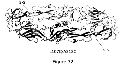
having each the mutations L107C and A313C as defined above, wherein the
residues
108C and 3150 or the residues 107C and 313C are linked together through a
disulphide
inter-chain bond.
In one embodiment the compound binds to the EDE wherein the EDE is a
stabilised dimer
of sE, wherein the stabilised dimer of envelope or recombinant sE is a
heterodimer of a
mutant sE having the mutations F108C and A313C as defined above and a mutant
sE
having the mutations L1 07C and T315C as defined above, wherein the residues
108C and
313C are linked respectively to the residues 315C and 107C through a
disulphide inter-
chain bond between the two sE monomers.
In another preferred embodiment, wherein the EDE comprises a stabilised dimer
of
recombinant sE, the stabilized recombinant sE dimer is selected from the group
consisting
of a homodimer of mutants sE having each the mutations A259C, F108C and T315C,
a
homodimer of mutants sE having each the mutations S255C, F108C and T315C, a
homodimer of mutants sE having each the mutations A259C, L1 07C and A313C, and
a
homodimer of mutants sE having each the mutations A255C, L107C and A313C as
defined above, wherein the residues 259C, 255C, 108C, 315C, 107C and 313C are
linked
respectively to the residues 259C, 2550, 3150, 108C, 313C and 1070 through
disulphide
inter-chain bonds.
In another preferred embodiment, the compound binds to the EDE wherein the EDE
comprises a stabilised dimer of recombinant sE, the stabilized recombinant sE
dimer is a
heterodimer of a mutant sE having the mutations A259C, F108C and T315C as
defined
above and a mutant sE having the mutations S255C, Fl 08C and T315C as defined
above,
wherein the residues 259C, 108C and 315C are linked respectively to the
residues 255C,
3150 and 1080 through disulphide inter-chain bonds.
16
CA 02988499 2017-12-06
WO 2016/012800
PCT/GB2015/052139
In another preferred embodiment, wherein the EDE comprises a stabilised dimer
of
recombinant sE, the stabilized recombinant sE dimer is a heterodimer of a
mutant sE
having the mutations S255C, Ll 07C and A3130 as defined above and a mutant sE
having
the mutations A259C, L107C and A313C as defined above, wherein the residues
255C,
__ 107C and 313C are linked respectively to the residues 259C, 313C and 107C
through
disulphide inter-chain bonds.
As well as stabilisation via disulphide bonds, it will be appreciated that
stabilisation may
also be achieved via sulfhydryl-reactive crosslinkers. Thus, in one
embodiment, wherein
__ the EDE comprises a stabilised dimer of recombinant sE, the stabilized
recombinant sE
dimer of the invention is covalently stabilized with at least one, two or
three, sulfhydryl-
reactive crosslinkers (also called thiol-reactive crosslinkers) between the sE
monomers.
Chemical crosslinking of proteins is well-known in the art (see for review
Hemaprabha,
__ 2012).
Naturally, the sE dimer has two difference faces, one exposed to the
extracellular medium,
where the antibodies bind, and the one exposed to the viral membrane.
__ Advantageously, said stabilized recombinant sE dimer involves candidate
amino acid
residues present in the face of sE exposed to the viral membrane and thus are
not part of
the epitope. One of each candidate amino acid residue of each monomer is
mutated
(substituted) to cysteine, producing a free sulfhydryl group that is the
target of sulfhydryl-
reactive crosslinkers of appropriate lengths.
Thr/Ser262 and Thr/A1a265 are candidate residues. The distance between them in
the
context of the dimer is 12 and 22 A respectively. Further, these residues
(Thr/Ser262,
Thr/A1a265) are not fully conserved. Hence, they can tolerate point mutations.
__ In a preferred embodiment, the compound binds to the EDE wherein the EDE
comprises
a stabilised dimer of recombinant sE, the stabilized recombinant sE dimer is a
homodimer
of mutant sE having each the mutation T/S262C or T/A265C as defined above,
wherein
the residues 262C or 265C are linked together through a sulfhydryl-reactive
crosslinker.
__ In another preferred embodiment wherein the EDE comprises a stabilised
dimer of
recombinant sE, the stabilized recombinant sE dimer is a heterodimer of a
mutant sE
having the mutation T/S262C as defined above and a mutant sE having the
mutation
17
CA 02988499 2017-12-06
WO 2016/012800
PCT/GB2015/052139
T/A265C as defined above, wherein the residues 262C and 265C are linked
together
through a sulfhydryl-reactive crosslinker.
Regions of the recombinant sE which are not considered to be part of the
epitope and
which can be crosslinked are region A consisting of residues 1-9 of sE, region
B consisting
of residues 25-30 of sE, region C consisting of residues 238-282 of sE, region
D consisting
of residues 96-111 of sE and region E consisting of residues 311-318 of sE.
Any of the
residues of these five regions (A to E) of a monomer is at less than 25-30 A
of other residue
of the other monomer in the recombinant sE dimer, and thus these residues can
be
crosslinked.
Advantageously, one or several of the candidate amino acid residues in these
five regions
of each monomer is mutated (substituted) to cysteine, producing a free
sulfhydryl group
that is the target of sulfhydryl-reactive crosslinkers of appropriate lengths
as defined
above.
In another embodiment, the compound binds to the EDE wherein the EDE comprises
a
stabilised dimer of recombinant sE, the stabilized recombinant sE dimer is a
homodimer
or a heterodimer of a mutant sE wherein at least one of the amino acid
residues 1-9, 25-
30, 238-282, 96-111 311-318 of sE is mutated (substituted) to cysteine and a
mutant sE
wherein at least one of the amino acid residues 1-9, 25-30, 238-282, 96-111
311-318 of
sE is mutated (substituted) to cysteine, and wherein the mutated cysteine
residues are
linked together through a sulfhydryl-reactive crosslinker.
The sulfhydryl-reactive crosslinkers are preferably homo-bifunctional reagents
with
identical or non-identical reactive groups, permitting the establishment of
inter-molecular
crosslinkages between the two monomers. Homobifunctional crosslinkers have
identical
reactive groups at either end of a spacer arm, and generally they can be used
in one-step
reaction procedures. The sulfhydryl-reactive crosslinkers of the invention can
be a
maleimide, a haloacetyl (preferably a bromo- or iodo-acetyl), a pyridyl
disulfide, a
vinylsulfone, an alkyl halide or an aziridine compound, an acryloyl
derivative, an arylating
agent, or a thiol-disulfide exchange reagent (Hermanson, 2010; Hemaprabha,
2012), such
as the bis(methanethiosulfonate) (Haberz P. et at., 2006).
Examples of maleimide homo-bifunctional sulfhydryl-reactive crosslinkers
according to the
invention, with spacer of different lengths, include BMOE (1,2-bis-
maleimidoethane), BMB
(1,4-bis-maleimidobutane), BMH (1,6-bis-maleimidohexane), TMEA (tris-(2-
18
CA 02988499 2017-12-06
WO 2016/012800 PCT/GB2015/052139
maleimidoethyl)amine), BM(PEG)2 (1,8-bismaleimidodiethyleneglycol), BM(PEG)3
(1,1 1-
bismaleimidotriethyleneglycol), BMDB (1,4-bismaleimidy1-2,3-dihydroxybutane),
DTME
(dithio-bis-maleimidoethane), and preferably BMH, BM(PEG)2 and BM(PEG)3.
0 0 8
0
BMH 13M(PEG)2 8MfoSG)3
Siam& o flume 1,8-Bisme =nyleneglycol t1IBismk
hyleneglycol
=0 h' '34
Spacer Ann 110 A Spacer Arm 14,7 A Spacer Ann 17.8 A
The maleimide group reacts specifically with the sulfhydryl groups is
performed under mild
buffer and pH conditions, in order to minimize the degree of structural shift
due to
crosslinking reactions. Preferably, the pH of the reaction mixture is between
6.5 and 7.5
leading to the formation of a stable thio-ether linkage that is not reversible
(the bond cannot
be cleaved with reducing agents).
In addition to stabilisation via disulphide bonds and sulfhydryl-reactive
crosslinkers, it will
be appreciated that stabilisation may be obtained through the linking of the
two monomers
through modified sugars. To this end, glycosylation sites are inserted on them
and are
reacted with modified sugars, in order to join them by click-chemistry.
According to this embodiment, the compound binds to the EDE wherein the EDE
comprises a stabilised dimer of recombinant sE, the stabilized recombinant sE
dimer is a
homodimer or heterodimer of mutants sE, wherein:
- one sE monomer has at least one mutation which introduces a glycosylation
site, and wherein the mutated amino acid residue is glycosylated with a
modified sugar
bearing an X functional group, and
- the other sE monomer has at least one mutation which introduces a
glycosylation site, and wherein the mutated amino acid residue is glycosylated
with a
modified sugar bearing a Y functional group,
and wherein both mutated residues are joined together through the modified
sugars by reacting, specifically by click chemistry, the X functional group of
the sugar of
the first sE monomer with the Y functional group of the sugar of the other sE
monomer.
19
CA 02988499 2017-12-06
WO 2016/012800
PCT/GB2015/052139
By X functional group, it is meant a chemical group beared by a sugar which is
able to
react and form a covalent linking by click chemistry with a Y functional
group, said Y
functional group being preferably an azide functional group.
By Y functional group, it is meant a chemical group beared by a sugar which is
able to
react and form a covalent linking by click chemistry with a X functional
group, said X
functional group being preferably a terminal alkyne functional group.
The modified sugars can be synthesized and introduced in the sE monomers as
described
by Laughlin et aL, 2007, and joined together as described by Speer et al.,
2003.
In addition to the abovementioned covalent methods of stabilising the dimer,
non-covalent
means may also be used. Thus, in another embodiment wherein the EDE comprises
a
stabilised dimer of recombinant sE, the dimer is non-covalently stabilized by
filling the
cavities of said dimer at the dimer interface by substituting at least one
amino acid in the
amino acid sequence of one or the two monomers, preferably the two monomers,
with
bulky side chain amino acids. According to this embodiment, cavities unique to
the
quaternary conformation of the recombinant sE dimer are identified and filled
by
engineered hydrophobic substitutions in the monomers.
According to this embodiment, the stabilized recombinant sE dimer is non-
covalently
stabilized by substituting at least one amino acid residue in the amino acid
sequence of at
least one sE monomer with at least one bulky side chain amino acid within
regions forming
cavities at the dimer interface or in domain 1 (D1) / domain 3 (03) linker of
each monomer.
Such substitutions allow increasing hydrophobic interactions between the two
sE
monomers.
In a preferred embodiment wherein the EDE comprises a stabilised dimer of
recombinant
sE, the stabilized recombinant sE dimer is a homodimer or heterodimer,
preferably
homodimer, of two recombinant sE as defined above, wherein one of the
recombinant sE
or the two recombinant sE have at least one mutation (substitution) selected
from the
group consisting of H27F, H27W, H244F, H244W,and L278F. The mutations H27F,
H27W,
H244F, H244W and L278F allow stabilizing the cavity around F279 of the
recombinant sE
dimer, strengthening the dimer interface and mimicking the F279 conformation
in the
virion.
CA 02988499 2017-12-06
WO 2016/012800
PCT/GB2015/052139
Other means of non-covalently stabilising the dimer include, for example non-
covalent
stabilisation in domain 1 (D1) / domain 3 (D3) linker of each monomer, by
substituting
amino acids in the amino acid sequence of one or the two, preferably the two,
monomers
with at least one bulky side chain amino acid.
In a preferred embodiment the compound binds the EDE wherein the EDE comprises
a
stabilised dimer of recombinant sE, the stabilized recombinant sE dimer is a
homodimer
or heterodimer, preferably homodimer, of two recombinant sE as defined above,
wherein
one of the recombinant sE or the two recombinant sE have at least one mutation
(substitution) selected from the group consisting of L292F and L294N. The
mutations
L292F, L294N are considered to allow stabilizing the D1-D3 linker in sE
dimeric
conformation.
In a preferred embodiment where the EDE is stabilised in the dimer
configuration through
engineering, the engineering, such as that described above, does not result in
a change
in the overall 3D structure of the dimer, or does not substantially change the
overall 3D
structure and the residues in the native dimer spatially correspond to the
engineered dimer.
If the native dimer spatially corresponds to the engineered dimer, this means
that when a
3D model of the engineered dimer (or part thereof, for example reflecting
residues of
particular importance in defining the VDR, for example the residues indicated
in Table 2
and/or discussed further below) is superimposed on the 3D model of the native
dimer,
coordinates defining the spatial location of the backbone atoms in the native
dimer vary
from the coordinates defining the analogous backbone atoms in the engineered
dimer by
less than about 10 angstroms. Backbone atoms are those atoms in an amino acid
that
form the peptide backbone, or 3D folding pattern, i.e. does not include the
side chain
atoms, though the position of some or all of the side chain atoms may
similarly not vary
significantly. The 3D structure is key to the immunogenicity of the VDE, and
as such, in a
preferred embodiment, the engineering does not result in a dimer with
decreased
immunogenicity. In one embodiment the engineering does result in a dimer with
a different
3D conformation. Preferably the engineering results in a darner with increased
immunogenicity. Such approaches have been used in ref 84. Thus in one
embodiment,
the compound binds to an engineered EDE, such as those described above.
A 3D model of the native dimer may be formed making use of the information on
crystal
structures for envelope glycoprotein ectodomain from dengue virus serotypes,
for example
21
CA 02988499 2017-12-06
WO 2016/012800
PCT/GB2015/052139
serotypes 2, 3, and 4, available in the Protein Data Bank, for example under
accession
numbers 10AN, 10K8, 1UZG and 3UAJ, as noted above.
Whether or not a particular mutation or modification alters or substantially
alters the 3D
structure could be assessed by different techniques, including monitoring
whether the
antibodies described herein, which are known to bind to the VDE, can still
bind to the
engineered version of the VDE.
The skilled person is able to use computer programs to aid in the
identification of potential
stabilising modifications, for example r
The effect of the engineering on the immunogenicity of the EDE can be assessed
by
comparing the antibody response in a subject when administered an engineered
and non-
engineered EDE or by comparing binding to known anti-EDE antibodies.
Alternatively, the modified envelope protein could be expressed in a dengue
virus and the
ability of the compound to neutralise the virus assessed.
In order to present a stabilised EDE, non-EDE heterologous proteins that have
a similar
three- dimensional structure to the respective EDE (referred to as scaffold
proteins), can
be modified to contain the appropriate residues that enable the modified
protein to hold
the EDE. Thus in one embodiment the compound binds the EDE wherein the EDE is
presented as part of an epitope-scaffold protein. An epitope-scaffold protein
is a chimeric
protein that includes an epitope sequence fused to a heterologous "acceptor"
scaffold
protein. Design of the epitope-scaffold is performed, for example,
computationally in a
manner that preserves the native structure and conformation of the epitope
when it is fused
onto the heterologous scaffold protein. The use of such scaffold proteins is
well known in
the art and such methods and techniques are described in WO 2011/050168 and
refS54'82'83
and the skilled person can follow methods described therein and apply them to
the present
invention.
Accordingly, in one embodiment, the EDE comprises part of an epitope-scaffold
protein,
wherein the scaffold protein comprises a heterologous scaffold protein
covalently linked to
the Envelope Dimer Epitope. Scaffold proteins are useful for creating the EDE
of the
present invention in that they hold contact residues of the EDE in the proper
spatial
orientation to facilitate interaction between such residues and the compound,
for example
between contact residues of the compound when the compound is a protein,
optionally an
22
CA 02988499 2017-12-06
WO 2016/012800
PCT/GB2015/052139
antibody or antigen binding portion thereof. A contact residue is any amino
acid present
in a molecule that interacts directly or indirectly (e.g. forms an ionic bond
either directly, or
indirectly through a salt bridge) with an amino acid in another molecule.
Residues of the
envelope protein which are considered to be potentially important for compound
binding
to the EDE, at least for DENV-1, are detailed in Table 2 The scaffold protein
may present
the entire dimer or may present only the selected residues above. A 3D model
of the
native dimer or parts thereof may be formed making use of the information on
crystal
structures for envelope glycoprotein ectodomain from dengue virus serotypes,
for example
serotypes 2, 3, and 4, available in the Protein Data Bank, for example under
accession
io numbers 10AN, 10K8, 1UZG and 3UAJ, as noted above.
Mutational analysis revealed particular residues of DENV1 and DENV2 which are
important for binding to the antibodies identified in the present invention.
These residues
are:
DENV1: E49,K64,Q77,W101,V122,N134,N153,T155,I161,A162,P169,
T200, K202, E203, L308,K310, Q323,W391, F392;
DENV2: Q77,W101,N153,T155,K310.
All of these residues are considered to be important for binding, and the
Q77,W101,N153,T155,K310
Accordingly, in one embodiment, compound binds the EDE wherein the EDE is part
of a
scaffold protein, wherein the scaffold protein holds at least residues
corresponding to one
or more of E49,K64,Q77,W101,V122,N134,N153,T155,I161,A162,P169,
T200,K202,E203,L308,K310,Q323,W391,F392, of the envelope protein or equivalent
residue of a Dengue virus envelope protein, particularly for DENV-1 and DENV-
2. Certain
residues are considered to be more important, and as such a further embodiment
of the
EDE comprises a scaffold protein which holds at least one or more of residues
corresponding to Q77,W101,N153,T155,K310 of the envelope protein or equivalent
residue of a Dengue virus envelope protein, particularly DENV-1 and DENV-2.
Residues of the envelope protein considered to be important for contacting the
epitope are
given in Figure 31, for example:
23
CA 02988499 2017-12-06
WO 2016/012800
PCT/GB2015/052139
the B7 antibody is considered to contact the DENV2 EDE at residues
N67,T68,T69,T70, E71, S72, R73, L82,V97, D98, R99,W101, G102, N103,G104,1113,
G152, N
153, D154,T155,G 156, K246, K247,Q248, D249;
the Al 1 antibody is considered to contact the DENV2 EDE at residues
N67,168,169,T70, E71, S72, R73, C74, E84,V97,D98, R99,G102,N103,
G104,C105,V114, N
153,D154,T155, G156, H158,K246,K247,Q248,D249,V250;
the C10 antibody is considered to contact the DENV2 EDE at residues
io
R2,H27,G28,E44,L45,146,K47,N67,T68,T69,T70,E71,S72,R73,C74,Q77,S81,L82,N83,E
84,V97, R99,W101, G102, N103, G104, C 105, G106,L 113,T115, K246, K247, Q248,
Q271,V3
09, K310, R323,Q325, D362;
the C10 antibody is considered to contact the DENV4 EDE at residues
R2, H27,G28,G29, E44, L45,T46, N67,T69,T70,A71,T72, R73,C74,Q77,V97, R99,W101,
G1
02,N103,G104,C105,G106,V113,R247,Q248,D249,D271,M278,D309,K310,V324,K323,
K325,T361,N362;
the C8 antibody is considered to contact the DENV2 EDE at residues
N67, T68,T69,T70, E71, S72, R73, C74,Q77, N83, E84,V97, D98, R99,W101, G102,
N103,
G104,C105,G106,L113,E148,H158,K246,K247,Q248,D249,1308,K310,E311,R323,D362,
G374.
Thus residues of the envelope protein that are considered to be important for
binding to
the compound, particularly for DENV2 and DENV4 are:
A71, C105, C74, D154, D249, D271, D309, D362, D98, E148, E311, E44, E71,
E84,G102,G 104
G106,G152, G156, G28, G29,G374, H158, H27,1113,1308,146,
K246,K247,K310,K323,K325
K47, L113, L45, 1_82,M 278, N103, N153, N362, N67, N83, Q248,Q271,Q325,Q77,
R2, R247,
R323, R73, R99, S72,
S81,T115,T155,T361,T46,T68,T69,T70,T72,V113,V114,V250,V309
V324,V97,W101,
or equivalent residue of a Dengue virus envelope protein.
The scaffold protein may present one or more residues selected from both of
the sets of
residues, for example may present at least one or more, for example, at least
1, 2, 3, 4, 5,
6, 7, 8, 9, or 10 or all of:
24
CA 02988499 2017-12-06
WO 2016/012800
PCT/GB2015/052139
E49,K64,Q77,W101,V122,N134,N153,T155,1161,A162,P169,
T200,K202, E203,L308,K310,Q323,W391,F392,
A71,C105,C74, D154, D249, D271, D309, D362, D98, E148, E311,
E44,E71,E84,G102,G104
G106,G152, G156, G28, G29, G374, H158, H27,1113,1308,146, K246, K247, K323,
K325
K47, L113, L45, L82, M278, N103õ N362, N67, N83,Q248, Q271,Q325, R2, R247,
R323, R73, R99, S72,S81,1115õT361,T46,T68,T69,T70,T72,V113,V114,V250,V309
V324,V97, or equivalent residue of a Dengue virus envelope protein.
In addition, the scaffold protein may present any one or more or all of the
following sets of
residues, which as described earlier are considered to increase stability of
the dimer
configuration: H27F, H27W, L107C, F108C, H244F, H244W, S255C, A259C, T/S262C,
T/A265C, L278F, L292F, L294N, A313C and T315C.
The scaffold protein may hold the dimer, or fragment of dimer, and may
comprise any of
the described modifications above which are considered essential for
immunogenicity,
and/or result in increased dimer stability, for example increased disulphide
bonds.
Moreover, the scaffold can be such that an improved EDE is presented. In one
embodiment, the compound therefore binds an improved EDE. For example, as
described
below and in Examples 2 and 5, patients with Dengue infection tend to have
either
antibodies directed towards the VDE, which are considered useful antibodies,
or
antibodies directed towards the Fusion Loop (anti-FL antibodies) which are not
considered
to be useful. Thus a scaffold may be engineered such that only the EDE is
presented, and
is presented in such a way as to exclude the possibility of a compound, for
example an
antibody or antigen binding portion thereof, being raised to the FL.
Therefore, in one
preferred embodiment the EDE is capable of raising antibodies to the EDE and
not to the
FL, optionally by being incorporated into a scaffold protein.
Independently of a scaffold protein, the envelope protein may be engineered
such that an
improved EDE is generated. As detailed above, an EDE which is incapable of
being
recognised by the anti-FL antibodies, and incapable of raising such
antibodies, is
considered to be an improved EDE. This may be accomplished by one or more
mutations,
deletions or insertions in the envelope protein, or by generating a hybrid
protein wherein
the specific epitope, without any antigens which would raise anti-FL
antibodies, fused to a
scaffold protein.
CA 02988499 2017-12-06
WO 2016/012800
PCT/GB2015/052139
In one embodiment, the envelope protein is engineered by modifying the
internal surface
of the dimer (projecting to the inside of the virus) with sugars to make it
less
immunogenic by adding N or 0 linked glycan sequences.
Extensive mutagenic resurfacing of the dimer may be useful to further reduce
the
generation of non-ED suboptimal responses by mutation of residues and/or
addition of
glycan.
As an example, the L278F mutation is considered to re-shape the kl-loop and to
mimic
the virion-like conformation.
Modelling an optimisation of the core EDE epitopes may also be useful to
produce an
optimal sequence to induce the desired EDE response to provide binding and
neutralising antibodies.
It will be appreciated that the EDE may be the naturally occurring envelope
protein held
within a scaffold to effect increased dimer stability. The EDE may also be
engineered
independently of any scaffold to increase dimer stability. The two may be
combined such
that in one embodiment the EDE comprises a dimer wherein the envelope protein
is
engineered to have improved stability in the dimer configuration, which is
held within a
heterologous scaffold protein. Alternatively, the envelope protein may be
engineered such
that only the relevant portions of the protein are present, and this may then
be held in a
heterologous scaffold protein.
A dimer conformation may be stabilised by, for example, creating a long
linker, for example
a glycine-serine-rich liner between two envelope monomers to express as a
single
polypeptide chain comprising two envelope polypeptide domains. Alternatively
or in
addition, a dimeric structure may be stabilised by any antibody (for example)
which binds
to the inner facing surfaces of the dimer or to tags associated with the
dimer.
Any reference to the envelope protein, sE, sE dimer or envelope protein dimer
also
includes within its scope a scaffold protein, or a structure, which comprises
the particular
residues that make up the EDE, held in a particular conformation so as to
present a
suitable EDE.
The envelope nucleotide sequence may be engineered such that the envelope
protein has
any one or more of mutations, insertions or deletions. The nucleotide sequence
may be
26
CA 02988499 2017-12-06
WO 2016/012800
PCT/GB2015/052139
such that it has at least 70%, 80%, 85%, 90%, 95%, 98% or 99% homology to the
native
sequence of the particular envelope protein (or part thereof).
In a further embodiment the envelope protein may be engineered such that it
has at least
70%, 80%, 85%, 90%, 95%, 98% or 99% homology to an envelope protein (or part
or parts
thereof, for example one or more portions of at least 8, 9 or 10 consecutive
amino acids)
from another serotype of dengue virus. In a preferred embodiment, the envelope
protein
is engineered such that it has at least 70%, 80%, 85%, 90%, 95%, 98% or 99%
homology
to two different envelope proteins (or part or parts thereof, for example one
or more
portions of at least 8, 9 or 10 consecutive amino acids), more preferably to
four different
envelope proteins (or part or parts thereof, for example one or more portions
of at least 8,
9 or 10 consecutive amino acids), most preferably to all envelope proteins (or
part or parts
thereof, for example one or more portions of at least 8, 9 or 10 consecutive
amino acids)
from all serotypes of dengue virus.
As described above, the envelope protein may be engineered such that it
actually has very
low homology to the native envelope protein, but wherein the integrity and
conformation of
the EDE is maintained, or is altered in such a way that the EDE is improved,
for example,
is incapable of raising the anti-FL antibodies. Thus, the level of sequence
homology is not
necessarily an indication of the 3D structure homology, or functional
homology. For
example, a particular sequence encoding a structure comprising a EDE may
actually have
a very low level of homology to the native envelope protein, but may
nevertheless be
considered a useful compound of the invention. For example, the protein may
have 10%,
20%, 30%, 40%, 50% or 60% homology to the native envelope protein, and the
nucleotide
sequence which encodes this structure may have a correspondingly low sequence
identity
to the native envelope sequence.
In a preferred embodiment, where the envelope protein, or structure comprising
the EDE
has at least 70%, 80%, 85%, 90%, 95%, 98% or 99% homology to an envelope
protein (or
part or parts thereof, for example one or more portions of at least 8, 9 or 10
consecutive
amino acids) of a dengue virus, or at least 70%, 80%, 85%, 90%, 95%, 98% or
99%
homology to two different envelope proteins (or part or parts thereof, for
example one or
more portions of at least 8, 9 or 10 consecutive amino acids), more preferably
to four
different envelope proteins, most preferably to all envelope proteins from all
serotypes of
dengue virus, or wherein the protein or structure comprising the EDE has at
least 10%,
20%, 30%, 40%, 50% or 60% homology to the native envelope protein of one or
more
27
CA 02988499 2017-12-06
WO 2016/012800
PCT/GB2015/052139
serotypes of dengue virus, the protein comprises one or more of, or optionally
all of:
E49,K64,077,W101,V122,N134,N153,T155,1161,A162,P169,
T200,K202,E203,L308,K310,Q323,W391,F392, or equivalent residue of a Dengue
virus
envelope protein.
Some of these residues are considered to be more important than others, as
such in a
further embodiment of the EDE, the envelope protein, or structure comprising
the EDE
comprises one or more of, or optionally all of: Q77,W101,N153,T155,K310 , or
equivalent
residue of a Dengue virus envelope protein.
lo
It is considered that one or more of
residues
E49,K64,077,W101,V122, N134, N153,T155,1161,A162, P169,
T200,K202,E203,L308,K310,0323,W391,F392, of the envelope protein, or
equivalent
residues of a dengue virus protein are required for binding of the compound to
the EDE.
Thus in one embodiment, the envelope protein or structure comprising the EDE
comprises
one or more or all of these residues.
Whilst the anti-FL antibodies appear, in most cases, to require only residue
W101 out of
the residues mutated in the alanine scanning analysis (Example 2) and are not
affected
by mutation of any of the other residues, the anti-EDE antibodies require a
much larger
epitope, which requires the presence of residue W101, as does the anti-FL
antibodies, but
which are also affected by mutations at many of the other residues. As such,
in one
embodiment the EDE is defined as an epitope in which residues W101 and at
least one or
more of positions E49, K64,Q77,W101,V122, N134,N153,T155,1161,A162, P169,
1200,K202,E203,L308,K310,Q323,W391,F392, or equivalent residue in the Envelope
Dimer Epitope are required for binding of the compound.
In a particular embodiment, the Envelope Dimer Epitope comprises the domain
III residue
K310.
In an embodiment the EDE is glycosylated at position 67 (Asn67 glycan) and/or
at position
153 (Asn153 glycan), for example of each envelope, for example sE, monomer,
preferably
at least at position 67 (Asn67 glycan) of each monomer.
The compound of the invention, according to one embodiment, contacts the N67
glycan
chain of the envelope protein dimer, or the N153 glycan chain of the envelope
protein
28
CA 02988499 2017-12-06
WO 2016/012800
PCT/GB2015/052139
dimer. It will be appreciated that the compound can contact both the N67 and
N153 glycan
chains of the envelope protein dimer.
In a particular example, the compound is an antibody wherein the CDR H2
interacts with
the N67 glycan chain of the envelope protein.
In one embodiment, the compound contacts the EDE at any one or more of
A71,C105,C74, D154, D249, D271, D309, D362, D98, E148, E311, E44, E71, E84,
G102,G104
G106,G152, G156,G28,G29, G374, H 158, H27,1113,1308,146, K246, K247, K310,
K323, K325
K47, L113, L45, L82, M278, N103, N153, N362, N67, N83,
Q248,Q271,Q325,Q77,R2,R247,
R323, R73,R99,S72, S81,T115,T155,T361,T46,T68,T69,T70,T72,V113,V114,V250,V309
V324,V97,W101 in the envelope protein, for example DENV-2 or DENV-4, of one or
more
serotypes of Dengue virus, where present, preferably all serotypes of dengue
virus.
In an embodiment, the Envelope Dimer Epitope comprises a region centred in a
valley
lined by the b strand on the domain II side, and the "150 loop" (see, for
example, Figure
29) on the domain I side (across from the dimer interface), wherein the 150
loop spans
residues 148-159, connecting b-strands EO and FO of domain I, and carries the
N153
glycan, which covers the fusion loop of the partner subunit in the dimer. The
150 loop is
considered to comprise SEQ ID NO: 148 150 loop of Deny-1 QHQVGNETTEHG; SEQ ID
NO: 1149 150 loop of Deny 2 EHAVGNDTGKHG; SEQ ID NO: 150 150 loop of Deny 3
QHQVGNETQG; SEQ ID NO: 151 150 loop of Deny 4 THAVGNDIPNHG.
In some cases, the Envelope Dimer Epitope comprises domain II of the envelope
protein,
optionally further comprising any one or more of the following features of
domain II; the b
strain (residues 67-74), the fusion loop and residues immediately upstream
(residues 97-
106) and the ij loop (residues 246-249), and residues 243-251 and residues 307-
314.
In one embodiment the EDE comprises the five polypeptide segments of the
dengue virus
glycoprotein E ectodomain (sE) consisting of the residues 67J4, residues 97-
106,
residues 148-159, residues 243-251 and residues 307-314.
Thus in one embodiment the invention also provides a compound, for example an
isolated
neutralizing antibody or antigen binding fragment thereof directed against the
stabilized
recombinant sE dimer as defined above, wherein said antibody or fragment
thereof binds
the five polypeptide segments of the dengue virus glycoprotein E ectodomain
(sE)
consisting of the residues 67J4, residues 97-106, residues 148-159, residues
243-251
and residues 307-314.
29
CA 02988499 2017-12-06
WO 2016/012800
PCT/GB2015/052139
The characterization of the binding of an antibody fragment thereof according
to the
present invention to a polypeptide segment or amino acid residue can be
performed by,
for example, crystallization trials as describes in the Examples below.
Preferably, in addition to binding to the EDE the compound is capable of
neutralising the
virus. In a preferred embodiment the compound is capable of neutralising all
serotypes of
Dengue virus, preferably to at least 90% or at least 98%, for example 100%,
and preferably
neutralises all serotypes of Dengue virus made in both insect and human cells
to at least
90% or at least 98%, for example 100%. Preferences for the neutralisation and
neutralisation assay techniques are as described earlier.
In one embodiment the EDE comprises a dimer of full length envelope protein.
In another
embodiment, the EDE comprises a dimer of the envelope ectodomain (sE). In a
further
embodiment the envelope protein comprises the (approximately, as discussed
above) 400
amino terminal residues of the ectodomain of Envelope protein. See, for
example, Figure
28. The preferences for the stability of a dimer of the full length envelope
protein described
above also apply to the truncated ectodomain of envelope protein. Therefore,
the dimer
of ectodomain of envelope protein may be stabilised through engineering or
stabilised by
being incorporated into a scaffold protein, or may comprise a hybrid dimer.
In a further embodiment, the compound of the present invention is one which
will not bind
to dengue virus or virion or sub-viral particle or virus-like particle
incubated at acid pH.
Acidic pH causes the envelope protein to irreversibly adopt a trimer
configuration. The
inventors found that the compounds of the present invention do not bind to
viral particles
incubated at a low pH (see Example 4). Therefore, in one embodiment, the
compound, for
example and antibody or antigen binding portion thereof, does not bind to
dengue virus or
virion or sub-viral particle or virus-like particle, incubated at an acidic
pH. By an acidic pH
we mean any pH below 7, preferably pH 5.5.
As such, the skilled person can readily identify whether a particular compound
is a
compound of the invention according to this embodiment of the invention,
simply by
identifying whether the compound cannot bind to one or more than one of: a) a
virion or
sub-viral particle or a virus-like particle made in cells lacking furin
activity; b) a virion or
sub-viral particle or a virus-like particle having a high percentage of pr11/1
protein, and/or c)
a virion or sub-viral particle or a virus-like particle incubated under acidic
conditions
CA 02988499 2017-12-06
WO 2016/012800
PCT/GB2015/052139
Methods to assay the binding ability of the compound to the virion, sub-viral
particle or
virus-like particle detailed above are provided earlier in relation to
assaying the ability of
the compound to bind to the EDE and is detailed in Example 4 and generally
simply
involves an ELISA against the particular virion or virus like particle to
assay whether or not
the compound can bind. The compound is considered useful if it binds to, or
significantly
binds to, the native EDE or virion or virus like particle, and does not bind
to a virion or sub-
viral particle or a virus-like particle that: a) is made in cells lacking
furin activity; b) have a
high percentage of prM protein, and/or c) are incubated under acidic
conditions.
-ir) The invention further comprises specific compounds. For example, in
one embodiment,
the compound is an antibody comprising the sequence heavy chain SEQ ID No: 1
and
light chain SEQ ID No: 37; or heavy chain SEQ ID No:2 and light chain SEQ ID
No: 38; or
heavy chain SEQ ID No: 3 and light chain SEQ ID No: 39; or heavy chain SEQ ID
No: 4
and light chain SEQ ID No: 40. It will be appreciated that the invention also
includes
truncations and mutations of these antibodies, such that the compound is an
antigen
binding portion thereof. Antibodies with a sequence homology of at least 90%
or at least
95% homology to the above sequences are included in the invention. Particular
sequences of antibodies, light and heavy chains are given in SEQ ID No's: 1-4,
37-141,
141-147 and, for example, Figure 29.
In further embodiments, the compound is an antibody and comprises; heavy chain
SEQ
ID No: 1 and either light chain SEQ ID No: 37, 38, 39 or 40; heavy chain SEQ
ID No: 2
and either light chain SEQ ID No: 37, 38, 39 or 40; heavy chain SEQ ID No: 3
and either
light chain SEQ ID No: 37, 38, 39 or 40; or heavy chain SEQ ID No: 4 and
either light chain
SEQ ID No: 37, 38, 39 or 40.
Particular residues of the specific heavy and light chains are considered to
be important
for binding to the EDE. Thus, in one embodiment, where the sequence homology
is at
least 90% to the above sequences, where present, the following residues are:
SEQ ID No: 1 - T52, E54, D56, S57, A58, K65, G66, T69, E82, N84, S85, Y100,
N102,
F103, Y104, Y105, Y106;
SEQ ID No: 2 - G554, N55, N57, K59, Q62, Q65, G66, R94, R98, F99, Y100, Y101,
D102,
5103,T104, Y106, Y107, P108, D109, 5110, D117, V118;
31
CA 02988499 2017-12-06
WO 2016/012800
PCT/GB2015/052139
SEQ ID No: 3 -V2, S28, N31, D54, S56, T57, R58, K65, G66, R94, R98, F99, Y100,
Y101,
D102, S103, T104, Y106, Y107, P108, D109, 5110, D117, V118;
SEQ ID No: 4 - V2, T28, S31, D54, S56, S57, T58, G66, F68, M69, R94, R98, Y99,
Y100,
Y101, D102, S103, T104, Y106, Y107, P108, D109, N110, D117, V118;
SEQ ID No: 37 - S30, T31, F32, Y49, D50, S52, R54, R66, R91, Y92, N93, W94;
SEQ ID No: 38 - S26, S27, G30, G31, F32, N33, Y34, D52, T54, S55, R56, S62,
S95,
R96, G97;
SEQ ID No: 39 - Y51, R56, P57, S58, G59, S96, R97;
SEQ ID No: 40 - Y51, R56, P57, S58, K97.
An antibody is composed of a light chain and a heavy chain, and within each
light chain
and heavy chain are three variable regions. The most variable part of each of
these
regions is the complementary determining region and is considered to be the
most crucial
for antigen binding and recognition. Therefore, in one embodiment, the
compound
comprises one or more of the following amino acid sequences, having no, one or
two
amino acid substitutions, insertions or deletions:
SEQ ID No: 5 or SEQ ID No: 8 or SEQ ID No: 11 or SEQ ID No: 14
and/or
SEQ ID No: 6 or SEQ ID No: 9 or SEQ ID No: 12 or SEQ ID No: 15
and/or
SEQ ID No: 7 or SEQ ID No: 10 or SEQ ID No: 13 or SEQ ID No: 16
and/or
SEQ ID No: 17 or SEQ ID No: 20 or SEQ ID No: 23 or SEQ ID No: 26
and/or
32
CA 02988499 2017-12-06
WO 2016/012800
PCT/GB2015/052139
SEQ ID No: 18 or SEQ ID No: 21 or SEQ ID No: 24 or SEQ ID No: 27
and/or
SEQ ID No: 19 or SEQ ID No: 22 or SEQ ID No: 25 or SEQ ID No:28.
Particular compounds may comprise the following sequences, having no, one or
two amino
acid substitutions, insertions or deletions;
Heavy chain:
SEQ ID No: 5 and SEQ ID No: 6 and SEQ ID No: 7
or
SEQ ID No: 8 and SEQ ID No: 9 and SEQ ID No: 10
or
SEQ ID No: 11 and SEQ ID No: 12 and SEQ ID No: 13
or
SEQ ID No: 14 and SEQ ID No: 15 and SEQ ID No: 16
And/or
Light chain:
SEQ ID No: 17 and SEQ ID No: 18 and SEQ ID No: 19
or
SEQ ID No: 20 and SEQ ID No: 21 and SEQ ID No: 22
or
SEQ ID No: 23 and SEQ ID No: 24 and SEQ ID No: 25
33
CA 02988499 2017-12-06
WO 2016/012800
PCT/GB2015/052139
Or
SEQ ID No: 26 and SEQ ID No: 27 and SEQ ID No: 28.
In a preferred embodiment, particular compounds may comprise the following
sequences,
having no, one or two amino acid substitutions, insertions or deletions;
io Heavy chain SEQ ID No: 5 and SEQ ID No: 6 and SEQ ID No: 7 and light
chain SEQ ID
No: 17 and SEQ ID No: 18 and SEQ ID No: 19
Or
Heavy chain SEQ ID No: 8 and SEQ ID No: 9 and SEQ ID No: 10 and light chain
SEQ ID
No: 20 and SEQ ID No: 21 and SEQ ID No: 22
Or
Heavy chain SEQ ID No: 11 and SEQ ID No: 12 and SEQ ID No: 13 and light chain
SEQ
ID No: 23 and SEQ ID No: 24 and SEQ ID No: 25
Or
Heavy chain SEQ ID No: 14 and SEQ ID No: 15 and SEQ ID No: 16 and light chain
SEQ
ID No: 26 and SEQ ID No: 27 and SEQ ID No: 28
Or
Heavy chain SEQ ID NO: 11, 12 and 13 and optionally light chain SEQ ID NO: 25
and
optionally the amino acid sequences SEQ ID NO: 23 and 24
Or
Heavy chain SEQ ID NO: 14, 15 and 16 and optionally light chain SEQ ID NO: 28
and
optionally the amino acid sequences SEQ ID NO: 26 and 27
34
CA 02988499 2017-12-06
WO 2016/012800
PCT/GB2015/052139
or
Heavy chain SEQ ID NO: 3 and optionally light chain SEQ ID NO: 25, preferably
the light
chain variable region of SEQ ID NO: 140
or
Heavy chain variable region of SEQ ID NO: 4 and optionally light chain SEQ ID
NO: 28
preferably the light chain variable region of SEQ ID NO: 141.
In a further embodiment, particular residues of the above sequences are
considered
important for antigen binding. As such, in this embodiment, where present the
following
residues are:
SEQ ID No: 6 residue 3 is a T, residue 5 is an E, residue 7 is a D, residue 8
is an S, residue
9 is an A, residue 16 is a K and residue 17 is a G
SEQ ID No: 7 residue 2 is a Y, residue 4 is an N, residue 5 is an F, residue 6
is a Y, residue
7 is a Y and residue 8 is a Y
SEQ ID No: 9 residue 5 is a G, residue 6 is an N, residue 10 is a K, residue
13 is a Q,
residue 16 is a Q and residue 17 is a D
SEQ ID No: 10 residue 5 is a D, residue 6 is a Y, residue 8 is a D, residue 10
is a W,
residue 11 is an F, residue 12 is a P and residue 14 is an L
SEQ ID No: 11 residue 1 is an N
SEQ ID No: 12 residue 5 is a D, residue 7 is an S, residue 8 is a T, residue 9
is an R,
residue 16 is a K and residue 17 is a G
SEQ ID No: 13 residue 4 is an R, residue 5 is an F, residue 6 is a Y, residue
7 is a Y,
residue 8 is a D, residue 9 is an S, residue 10 is a T, residue 12 is a Y,
residue 13 is a Y,
residue 14 is a P, residue 15 is a D and residue 16 is an S
SEQ ID No: 14 residue 1 is an S
CA 02988499 2017-12-06
WO 2016/012800
PCT/GB2015/052139
SEQ ID No: 15 residue 5 is a D, residue 7 is and S, residue 8 is an S, residue
9 is a T and
residue 17 is a G or H
SEQ ID No: 16 residue 4 is an R, residue 5 is a Y, residue 6 is a Y, residue 7
is a Y, residue
8 is a D, residue 9 is an S, residue 10 is a T, residue 12 is a Y, residue 13
is a Y, residue
14 is a P, residue 15 is a D and residue 16 is an N
SEQ ID No: 17 residue 7 is an S, residue 8 is a T and residue 9 is an F
lo
SEQ ID No: 18 residue 1 is a D, residue 3 is an S and residue 5 is an R
SEQ ID No: 19 residue 3 is an R, residue 4 is a Y and residue 5 is an N
SEQ ID No: 20 residue 4 is an S, residue 5 is an S, residue 8 is a G, residue
9 is a G,
residue 10 is an F, residue Ills an N and residue 12 is a Y
SEQ ID No: 21 residue 1 is a D, residue 3 is a T, residue 4 is an S and
residue 5 is an R
SEQ ID No: 22 residue 5 is an S, residue 6 is an R and residue 7 is a G
SEQ ID No: 24 residue 5 is an R, residue 6 is a P and residue 7 is an S
SEQ ID No: 25 residue 6 is an S and residue 7 is an R
SEQ ID No: 27 residue 5 is an R, residue 6 is a P and residue 7 is an S
As described above in relation to the presentation of the antigenic EDE in a
protein
scaffold, the compound, for example a protein, for example an antibody, may
also be part
of a larger structure, for example held within a protein scaffold. Preferences
for the scaffold
are as described earlier. For example, in one embodiment, the antibody or
antigen binding
portion thereof is within a larger polypeptide.
In a preferred embodiment, the compound that binds to the EDE as defined in
any of the
embodiments above also neutralises the dengue virus, preferably to at least
80%,
preferably 90%, more preferably 95% or 98%, and most preferably 100%. In a
more
preferred embodiment, the compound neutralises all serotypes of dengue virus
to at least
36
CA 02988499 2017-12-06
WO 2016/012800
PCT/GB2015/052139
80%, preferably 90%, more preferably 95% or 98% and most preferably 100%.
Preferences for neutralisation, including the concentrations of the compound,
viral or sub-
viral or virus like particles and host cells are as defined earlier in the
first aspect of the
invention.
As for the first aspect of the invention, it is preferred if the compound that
can bind to the
EDE is able to neutralise virus made in both insect cells, for example C6/36
insect cells,
and human cells, for example primary human cells, for example dendritic cells.
Preferably
the compound neutralises dengue virus made in both insect cells, for example
C6/36 insect
io cells, and human cells, for example primary human cells, for example
dendritic cells to the
same level, as discussed above. The ability of the compound to neutralise
virus can be
tested as detailed above and in the examples. In a most preferred embodiment
the
compound is able to fully neutralise (i.e. to 100%) all serotypes of dengue
virus made in
both insect and human cells.
In a preferred embodiment the compound is an antibody or antigen binding
portion thereof.
The antigen binding portion may be a Fv portion; a Fab-like fragment (e.g. a
Fab fragment,
a Fab' fragment or a F(ab)2 fragment); or a domain antibody.
In one embodiment the antibody or antigen binding portion thereof is, or is
derived from, a
monoclonal antibody. In another embodiment the antibody or antigen binding
portion
thereof is, or is derived from a polyclonal antibody. In a further embodiment,
the compound
is a composition comprising a mixture of antibodies or antigen binding
portions thereof,
comprising:
a) a mixture of monoclonal antibodies or antigen binding portion thereof,
or
b) a mixture of polyclonal antibodies or antigen binding portion thereof,
or
c) a
mixture or monoclonal and polyclonal antibodies or antigen binding portion
thereof, for example wherein the ratio of monoclonal to polyclonal antibodies
or antigen
binding portions thereof is 10:1, 8:1, 6:1, 4:1, 2:1, 1:1, 1:2, 1:4, 1:6, 1:8
or 1:10
It will be appreciated that the compound may be a recombinant protein, for
example a
recombinant antibody or antigen binding portion thereof. The compound may also
be
made synthetically. The compound may be a combination of recombinantly and
synthetically produced.
37
CA 02988499 2017-12-06
WO 2016/012800 PCT/GB2015/052139
The present invention also includes the means of making such a compound, for
example
a protein, for example an antibody or antigen binding portion thereof.
it will also be appreciated, therefore, that the compound may be produced by
recombinant
means, for example the compound, for example a polypeptide, for example an
antibody or
antigen binding portion thereof may be produced and isolated or purified from
various
organisms, including:
a) a human cell line, optionally CHO cells, or
b) a mammal, optionally a human, or
c) a microorganism, or
d) an insect cell line.
By isolated or purified we mean that the agent has been removed from its
natural
environment, and does not reflect the extent to which the agent has been
purified.
Therefore the invention includes the isolation or purification of a compound
of the present
invention from various organisms, including from a human cell line, optionally
CHO cells,
or from a mammal, optionally a human, or from a microorganism, or from an
insect cell
line.
Where the compound is a polypeptide, for example an antibody or antigen
binding portion
thereof, or for example included in a protein scaffold, the compound may be
encoded by
a nucleic acid. By nucleic acid we include the meaning of both DNA and RNA,
single or
double stranded and in all their various forms. As such the invention includes
a nucleic
acid encoding any of the proteinaceous compound of the invention. In
particular, SEQ ID
No: 41-48 are included in the present invention. Any sequence derived from or
comprising
SEQ ID No: 41-48 which comprises mutations which would result in a silent
mutation are
included, as are sequences which cover any of the earlier mentioned
possibilities, for
example a nucleic acid sequence comprising a portion which encodes any of:
SEQ ID No: 1, or a sequence with at least 90% homology to SEQ ID No: 1;
SEQ ID No: 2, or a sequence with at least 90% homology to SEQ ID No: 2;
SEQ ID No: 3, or a sequence with at least 90% homology to SEQ ID No: 3;
38
CA 02988499 2017-12-06
WO 2016/012800
PCT/GB2015/052139
SEQ ID No: 4, or a sequence with at least 90% homology to SEQ ID No: 4;
SEQ ID No: 37, or a sequence with at least 90% homology to SEQ ID No: 37;
SEQ ID No: 38, or a sequence with at least 90% homology to SEQ ID No: 38;
SEQ ID No: 39, or a sequence with at least 90% homology to SEQ ID No: 39;
SEQ ID No: 40, or a sequence with at least 90% homology to SEQ ID No: 40;
A sequence with at least 90% homology to SEQ ID No: 1 wherein the following
residues
are - 152, E54, 056, S57, A58, K65, G66, 169, E82, N84, S85, Y100, N102, F103,
Y104,
Y105, Y106;
A sequence with at least 90% homology to SEQ ID No: 2 wherein the following
residues
are- G554, N55, N57, K59, Q62, Q65, G66, R94, R98, F99, Y100, Y101, D102,
S103,
T104, Y106, Y107, P108, D109, 5110, D117, V118;
A sequence with at least 90% homology to SEQ ID No: 3 - wherein the following
residues
are V2, S28, N31, D54, S56, T57, R58, K65, G66, R94, R98, F99, Y100, Y101,
D102,
S103, T104, Y106, Y107, P108, D109, 5110, D117, V118;
A sequence with at least 90% homology to SEQ ID No: 4 -wherein the following
residues
are V2, T28, S31, 054, S56, S57, T58, G66, F68, M69, R94, R98, Y99, Y100,
Y101,
D102, S103, 1104, Y106, Y107, P108, 0109, N110, 0117, V118;
A sequence with at least 90% homology to SEQ ID No: 37 - wherein the following
residues
are S30, T31, F32, Y49, D50, S52, R54, R66, R91, Y92, N93, W94;
A sequence with at least 90% homology to SEQ ID No: 38 -wherein the following
residues
are 526, 527, G30, G31, F32, N33, Y34, D52, T54, 555, R56, 562, 595, R96, G97;
A sequence with at least 90% homology to SEQ ID No: 39 - wherein the following
residues
are Y51, R56, P57, 558, G59, S96, R97;
A sequence with at least 90% homology to SEQ ID No: 40 - wherein the following
residues
are Y51, R56, P57, 558, K97;
39
CA 02988499 2017-12-06
WO 2016/012800
PCT/GB2015/052139
SEQ ID No: 5, or a sequence resulting in the polypeptide having one or two
amino acid
substitutions, insertions or deletions compared to SEQ ID No: 5;
SEQ ID No: 6 or a sequence resulting in the polypeptide having one or two
amino acid
substitutions, insertions or deletions compared to SEQ ID No: 6; optionally
wherein residue
3 is a T, residue 5 is an E, residue 7 is a D, residue 8 is an S, residue 9 is
an A, residue
16 is a K and residue 17 is a G;
SEQ ID No: 7 or a sequence resulting in the polypeptide having one or two
amino acid
substitutions, insertions or deletions compared to SEQ ID No: 7; optionally
wherein residue
2 is a Y, residue 4 is an N, residue 5 is an F, residue 6 is a Y, residue 7 is
a Y and residue
8 is a Y;
SEQ ID No: 8 or a sequence resulting in the polypeptide having one or two
amino acid
substitutions, insertions or deletions compared to SEQ ID No: 8;
SEQ ID No: 9 or a sequence resulting in the polypeptide having one or two
amino acid
substitutions, insertions or deletions compared to SEQ ID No: 9; optionally
wherein residue
5 is a G, residue 6 is an N, residue 10 is a K, residue 13 is a Q, residue 16
is a Q and
residue 17 is a D;
SEQ ID No: 10 or a sequence resulting in the polypeptide having one or two
amino acid
substitutions, insertions or deletions compared to SEQ ID No: 10; optionally
wherein
residue 5 is a D, residue 6 is a Y, residue 8 is a D, residue 10 is a W,
residue 11 is an F,
residue 12 is a P and residue 14 is an L;
SEQ ID No: 11 or a sequence resulting in the polypeptide having one or two
amino acid
substitutions, insertions or deletions compared to SEQ ID No:11; optionally
wherein
residue 1 is an N;
SEQ ID No: 12 or a sequence resulting in the polypeptide having one or two
amino acid
substitutions, insertions or deletions compared to SEQ ID No:12; optionally
wherein
residue 5 is a D, residue 7 is an S, residue 8 is a T, residue 9 is an R,
residue 16 is a K
and residue 17 is a G;
CA 02988499 2017-12-06
WO 2016/012800
PCT/GB2015/052139
SEQ ID No: 13 or a sequence resulting in the polypeptide having one or two
amino acid
substitutions, insertions or deletions compared to SEQ ID No:13; optionally
wherein
residue 4 is an R, residue 5 is an F, residue 6 is a Y, residue 7 is a Y,
residue 8 is a D,
residue 9 is an S, residue 10 is a T, residue 12 is a Y, residue 13 is a Y,
residue 14 is a P,
residue 15 is a D and residue 16 is an S;
SEQ ID No: 14 or a sequence resulting in the polypeptide having one or two
amino acid
substitutions, insertions or deletions compared to SEQ ID No: 14; optionally
wherein
residue 1 is an S;
SEQ ID No: 15 or a sequence resulting in the polypeptide having one or two
amino acid
substitutions, insertions or deletions compared to SEQ ID No: 15; optionally
wherein
residue 5 is a D, residue 7 is and S, residue 8 is an S, residue 9 is a T and
residue 17 is a
G or H
SEQ ID No: 16 or a sequence resulting in the polypeptide having one or two
amino acid
substitutions, insertions or deletions compared to SEQ ID No: 16; optionally
wherein
residue 4 is an R, residue 5 is a Y, residue 6 is a Y, residue 7 is a Y,
residue 8 is a D,
residue 9 is an S, residue 10 is a T, residue 12 is a Y, residue 13 is a Y,
residue 14 is a P,
residue 15 is a D and residue 16 is an N;
SEQ ID No: 17 or a sequence resulting in the polypeptide having one or two
amino acid
substitutions, insertions or deletions compared to SEQ ID No: 17; optionally
wherein
residue 7 is an S, residue 8 is a T and residue 9 is an F;
SEQ ID No: 18 or a sequence resulting in the polypeptide having one or two
amino acid
substitutions, insertions or deletions compared to SEQ ID No: 18; optionally
wherein
residue us a D, residue 3 is an S and residue 5 is an R;
SEQ ID No: 19 or a sequence resulting in the polypeptide having one or two
amino acid
substitutions, insertions or deletions compared to SEQ ID No: 19; optionally
wherein
residue 3 is an R, residue 4 is a Y and residue 5 is an N;
SEQ ID No: 20 or a sequence resulting in the polypeptide having one or two
amino acid
substitutions, insertions or deletions compared to SEQ ID No: 20; optionally
wherein
residue 4 is an S, residue 5 is an S, residue 8 is a G, residue 9 is a G,
residue 10 is an F,
residue 11 is an N and residue 12 is a Y;
41
CA 02988499 2017-12-06
WO 2016/012800
PCT/GB2015/052139
SEQ ID No: 21 or a sequence resulting in the polypeptide having one or two
amino acid
substitutions, insertions or deletions compared to SEQ ID No: 21; optionally
wherein
residue 1 is a D, residue 3 is a T, residue 4 is an S and residue 5 is an R;
SEQ ID No: 22 or a sequence resulting in the polypeptide having one or two
amino acid
substitutions, insertions or deletions compared to SEQ ID No: 22; optionally
wherein
residue 5 is an S, residue 6 is an R and residue 7 is a G;
SEQ ID No: 23 or a sequence resulting in the polypeptide having one or two
amino acid
substitutions, insertions or deletions compared to SEQ ID No: 23;
SEQ ID No: 24 or a sequence resulting in the polypeptide having one or two
amino acid
substitutions, insertions or deletions compared to SEQ ID No: 24; optionally
wherein
residue 5 is an R, residue 6 is a P and residue 7 is an S;
SEQ ID No: 25 or a sequence resulting in the polypeptide having one or two
amino acid
substitutions, insertions or deletions compared to SEQ ID No: 25; optionally
wherein
residue 6 is an S and residue 7 is an R;
SEQ ID No: 26 or a sequence resulting in the polypeptide having one or two
amino acid
substitutions, insertions or deletions compared to SEQ ID No: 26;
SEQ ID No: 27 or a sequence resulting in the polypeptide having one or two
amino acid
substitutions, insertions or deletions compared to SEQ ID No: 27; optionally
wherein
residue 5 is an R, residue 6 is a P and residue 7 is an S;
and
SEQ ID No: 28 or a sequence resulting in the polypeptide having one or two
amino acid
substitutions, insertions or deletions compared to SEQ ID No: 28.
The nucleic acid may or may not contain introns. The nucleic acid may also be
modified to
enable purification of the subsequently translated polypeptide, for example
the open
reading frame of the intended polypeptide may be modified to incorporate a
tag, for
example a myc tag or a his tag, to enable subsequent purification.
42
CA 02988499 2017-12-06
WO 2016/012800
PCT/GB2015/052139
The nucleic acid may also be modified, for example codon optimised, to be
better
translated by the organism which it is to be translated in, without affecting
final polypeptide
sequence.
Nucleic acids of the present disclosure can be produced or modified using a
number of
methods known to those skilled in the art for example, classic mutagenesis,
chemical
treatment, restriction digestion, ligation and PCR.
The nucleic acid of the invention may be incorporated into a vector. Thus the
invention
also comprises a vector comprising the nucleic acid. By vector we mean vehicle
for
cloning of amplification of the nucleic acid, or for insertion into a target
organism, for
example the vector may be a plasmid or may be a nucleic acid used to target
the nucleic
acid of the invention into a target organism, for example into the genome of a
target
organism. The vector may further comprise nucleotide sequences required for
expression
of the polypeptide encoded by the nucleic acid of the invention, for example
promoter
sequences or termination sequences may be operably linked to the nucleic acid
of the
invention, and may also include reporter genes, for example antibiotic
resistance
cassettes. The vector may be single stranded or double stranded, and may be
linear or
circular. In one embodiment the vector is a plasmid.
In addition to providing a compound which can bind to an EDE as indicated
above, a further
aspect of the invention also provides an EDE compound as defined below. The
invention
also provides a nucleic acid, or a vector, which encodes the EDE compound of
the
invention, in addition to a host cell comprising the nucleic acid or vector.
Preferences for
a nucleic acid and vector, for example, indicated above may also be relevant
to the present
aspect of the invention, as will be apparent to the skilled person. Thus the
invention
provides an EDE compound as defined below, or a nucleic acid encoding such an
EDE
compound, or a vector comprising said nucleic acid, or a host cell comprising
said nucleic
acid or vector.
The EDE compound is intended to provide an epitope as described above as a
Envelope
Dependent Epitope. The EDE compound may bind specifically to one or more EDE-
specific antibodies of the invention, for example to a preferred neutralising
antibody as
discussed above, or as exemplified in the Examples. The EDE compound typically
is or
comprises a polypeptide. In one embodiment, the EDE compound is a dimer of
envelope
protein, or envelope ectodomain or the 400 amino terminal residues of the
ectodomain of
Envelope protein. By "400 amino terminal residues" as used herein is included
43
CA 02988499 2017-12-06
WO 2016/012800
PCT/GB2015/052139
approximately 400 amino terminal residues, for example between 350 and 450
residues,
320 and 470 residues, or 330 and 480 residues (or combinations thereof), for
example
between 380 and 420 residues, for example between 390 and 410 residues, for
example
395 or 393 residues, as noted above and as will be apparent to those skilled
in the art..
__ The envelope protein may be any of the envelope proteins from DENV-1, DEN
V2, DENV-
3 and DENV-3, and DENV-4, (SEQ ID No's: 29, 31, 33 or 35), or a protein with
at least
90% homology to the sequences in SEQ ID No's: 29, 31, 33 or 35. The dimer may
be a
homodimer or a heterodimer. In a preferred embodiment the dimer is not
incorporated into
an intact viral particle, or a sub-viral particle, or a virus-like particle,
but rather is a free
__ dimer for example with a molecular weight of twice that of the monomeric
envelope
polypeptide. It will be appreciated that any form of EDE or EDE compound
described
herein, for example, an engineered envelope protein, for example, as part of a
protein
scaffold, may potentially be presented as part of a virus, virus-like
particle, or sub-viral
particle.
In another embodiment, the EDE compound comprises a dimer of envelope protein,
or
envelope ectodomain or the (approximately) 400 amino terminal residues of the
ectodomain of envelope protein which has been engineered to have increased
stability in
the dimer configuration, for example has been engineered to have increased
levels of
__ covalent and/or non-covalent bonds between the dimers;
In a preferred emobodiment, the EDE compound is a stabilised recombinant
dengue virus envelope glycoprotein E ectodomain (recombinant sE) dimer
according to
the earlier aspect of the invention, for example, is a stabilised recombinant
dengue virus
__ envelope glycoprotein E ectodomain (recombinant sE) dimer wherein the dimer
is:
- covalently stabilized with at least one disulphide inter-chain
bond between
the two sE monomers and/or,
- covalently stabilized with at least one sulfhydryl-reactive crosslinker
between the two sE monomers and/or,
- covalently stabilized by linking the two sE monomers through modified
sugars; and/or,
- non-covalently stabilized by substituting at least one amino acid residue
in
the amino acid sequence of at least one sE monomer with at least one bulky
side chain
amino acid, at the dimer interface or in domain 1 (D1) / domain 3 (D3) linker
of each
monomer.
44
CA 02988499 2017-12-06
WO 2016/012800
PCT/GB2015/052139
A dengue virus envelope glycoprotein E ectodomain (sE) refers to the 1-395
amino acid
fragment of the envelope glycoprotein E of the dengue virus serotypes 1, 2 and
4, and to
the 1-393 amino acid fragment of the envelope glycoprotein E of the dengue
virus serotype
a
Thus, as described ear(ier, the EDE compound is a stabilised dimer and may be
any one
or more of:
a) A dimer wherein the monomer is selected from the group consisting of: the
DENV-1 sE
of SEQ ID NO: 132, the DENV-2 sE of SEQ ID NO: 133 the DENV-3 sE of SEQ ID NO:
134, the DENV-4 sE of SEQ ID NO: 135 and a mutant sE thereof having at least
one
mutation (substitution) selected among H27F, H27W, L107C, F108C, H244F, H244W,
S255C, A259C, T/S2620, T/A265C, L278F, L292F, L294N, A3130 and T3150;
optionally, wherein said mutant sE thereof has further at least one mutation
(substitution) selected among Q227N, E174N and D329N, preferably the three
mutations Q227N, E174N and D329N;
b) A dimer wherein the dimer can be a homodimer of two identical recombinant
sE as
defined above or a heterodimer of two different recombinant sE as defined
above, the
dimer being preferably a homodimer, for example, it can be a heterodimer of
DENV-1
sE and DENV-2 sE as defined above. It can also be a heterodimer of DENV-1 sE
and
a mutant sE of DENV-1 sE as defined above;
c) A dimer which is glycosylated at position 67 (Asn67 glycan) and/or at
position 153
(Asn153 glycan) of each sE monomer, preferably at least at position 67 (Asn67
glycan)
of each monomer;
d) A dimer which is covalently stabilized with at least one, two or three
disulphide inter-
chain bonds between the two sE monomers;
e) A dimer which is a homodimer of mutant sE having each the mutation A2590 or
S2550
as defined above, and wherein the residues 259C or 2550 are linked together
through
a disulphide inter-chain bond
f) A dimer which is a heterodimer of a mutant sE having the mutation A2590 as
defined
above and a mutant sE having the mutation S255C as defined above, wherein the
residues 2590 and 255C are linked together through a disulphide inter-chain
bond;
g) A dimer which is a homodimer of mutant sE having each the mutations F1080
and
13150 as defined above, or a homodimer of mutants sE having each the mutations
L1070 and A3130 as defined above, wherein the residues 1080 and 3150 or the
residues 1070 and 3130 are linked together through a disulphide inter-chain
bond;
CA 02988499 2017-12-06
WO 2016/012800
PCT/GB2015/052139
h) A dimer which is a heterodimer of a mutant sE having the mutations Fl 08C
and A313C
as defined above and a mutant sE having the mutations L107C and T315C as
defined
above, wherein the residues 108C and 313C are linked respectively to the
residues
315C and 107C through a disulphide inter-chain bond between the two sE
monomers;
i) A dimer which is selected from the group consisting of a homodimer of
mutants sE
having each the mutations A259C, F108C and 1315C, a homodimer of mutants sE
having each the mutations S255C, F108C and T315C, a homodimer of mutants sE
having each the mutations A259C, L107C and A313C, and a homodimer of mutants
sE
having each the mutations A255C, L107C and A313C as defined above, wherein the
residues 259C, 255C, 108C, 315C, 107C and 313C are linked respectively to the
residues 259C, 255C, 315C, 108C, 313C and 107C through disulphide inter-chain
bonds;
j) A dimer which is a heterodimer of a mutant sE having the mutations A259C,
Fl 08C and
T315C as defined above and a mutant sE having the mutations S255C, F108C and
T315C as defined above, wherein the residues 259C, 108C and 315C are linked
respectively to the residues 255C, 315C and 108C through disulphide inter-
chain
bonds;
k) A dimer which is a heterodimer of a mutant sE having the mutations S255C,
L1 07C and
A313C as defined above and a mutant sE having the mutations A259C, L107C and
A313C as defined above, wherein the residues 255C, 107C and 313C are linked
respectively to the residues 259C, 313C and 107C through disulphide inter-
chain
bonds;
I) A dimer which is covalently stabilized with at least one, two or three,
sulfhydryl-reactive
crosslinkers (also called thiol-reactive crosslinkers) between the sE
monomers;
m)A dimer which is a homodimer of mutant sE having each the mutation T/S262C
or
T/A265C as defined above, wherein the residues 262C or 265C are linked
together
through a sulfhydryl-reactive crosslinker;
n) A dimer which is a heterodimer of a mutant sE having the mutation T/S262C
as defined
above and a mutant sE having the mutation T/A265C as defined above, wherein
the
residues 262C and 265C are linked together through a sulfhydryl-reactive
crosslinker;
o) A dimer which is a homodimer or a heterodimer of a mutant sE wherein at
least one of
the amino acid residues 1-9, 25-30, 238-282, 96-111 311-318 of sE is mutated
(substituted) to cysteine and a mutant sE wherein at least one of the amino
acid
residues 1-9, 25-30, 238-282, 96-111 311-318 of sE is mutated (substituted) to
cysteine, and wherein the mutated cysteine residues are linked together
through a
sulfhydryl-reactive crosslinker;
46
CA 02988499 2017-12-06
WO 2016/012800
PCT/GB2015/052139
p) A dimer which is is covalently stabilized by linking the two monomers
through modified
sugars.
q) A dimer which is a homodimer or heterodimer of mutant sE, wherein:
- one sE monomer has at least one mutation which introduces a glycosylation
site, and
wherein the mutated amino acid residue is glycosylated with a modified sugar
bearing
an X functional group, and the other sE monomer has at least one mutation
which
introduces a glycosylation site, and wherein the mutated amino acid residue is
glycosylated with a modified sugar bearing a Y functional group, and wherein
both
mutated residues are joined together through the modified sugars by reacting,
specifically by click chemistry, the X functional group of the sugar of the
first sE
monomer with the Y functional group of the sugar of the other sE monomer. By X
functional group, it is meant a chemical group beared by a sugar which is able
to react
and form a covalent linking by click chemistry with a Y functional group, said
Y functional
group being preferably an azide functional group. By Y functional group, it is
meant a
chemical group beared by a sugar which is able to react and form a covalent
linking by
click chemistry with a X functional group, said X functional group being
preferably a
terminal alkyne functional group;
r) A dimer which is non-covalently stabilized by filling the cavities of said
dimer at the
dimer interface by substituting at least one amino acid in the amino acid
sequence of
one or the two monomers, preferably the two monomers, with bulky side chain
amino
acids;
s) A dimer which is non-covalently stabilized by substituting at least one
amino acid residue
in the amino acid sequence of at least one sE monomer with at least one bulky
side
chain amino acid within regions forming cavities at the dimer interface or in
domain 1
(D1) / domain 3 (D3) linker of each monomer. Such substitutions allow
increasing
hydrophobic interactions between the two sE monomers;
t) A dimer which is a homodimer or heterodimer, preferably homodimer, of two
recombinant sE as defined above, wherein one of the recombinant sE or the two
recombinant sE have at least one mutation (substitution) selected from the
group
consisting of H27F, H27W, H244F, H244W,and L278F;
u) A dimer which is non-covalently stabilized in domain 1 (D1) / domain 3 (D3)
linker of
each monomer, by substituting amino acids in the amino acid sequence of one or
the
two, preferably the two, monomers with at least one bulky side chain amino
acid;
v) A dimer which is a homodimer or heterodimer, preferably homodimer, of two
recombinant sE as defined above, wherein one of the recombinant sE or the two
recombinant sE have at least one mutation (substitution) selected from the
group
consisting of L292F and L294N.
47
CA 02988499 2017-12-06
WO 2016/012800 PCT/GB2015/052139
In yet another embodiment, the EDE compound presents an improved epitope over
the
naturally occurring envelope dimer within a virus, virus-like particle or sub-
viral particle. By
improved epitope we include the meaning of improved over any epitope naturally
displayed
on an intact viral particle. By improved we include the meaning of being
capable of eliciting
a more beneficial immune response than the native intact dengue virus
particle. An EDE
compound which has increased stability in the dimer configuration, for example
via the
modifications described in a)-v) above, is considered to be an improved
epitope. The EDE
may be improved in other ways, for example, the EDE compound may be an EDE
which
has been engineered, or inserted into a scaffold, such that the FL is
incapable of being
recognised by a compound, for example a polypeptide, for example an antibody
or
antigenic portion thereof, on its own, for example where the EDE is engineered
such that
the FL cannot be recognised by an antibody in isolation from the immediate
neighbours of
the fusion loop, i.e. the fusion loop cannot be recognised in a context
independent of the
quaternary organisation..
In another embodiment, the EDE compound is incorporated into a heterologous
protein
scaffold which conserves the dimer configuration, by, for example, increasing
the level of
covalent and/or non-covalent bonds between the dimers, as described above.
Further,
the VDE compound may comprises a heterologous protein scaffold which may
present
only a portion of the dimer of envelope protein, or envelope ectodomain or the
(approximately) 400 amino terminal residues of the ectodomain of the envelope
protein,
wherein the portion is a sequential portion of the dimer of envelope protein,
or envelope
ectodomain or the (approximately) 400 amino terminal residues of the
ectodomain of the
envelope protein, or wherein the portion comprises select, non-contiguous
residues of the
dimer of envelope protein, or envelope ectodomain or the (approximately) 400
amino
terminal residues of the ectodomain of the envelope protein, as described
above.
For example, in one embodiment, the EDE compound comprises one or more of
positions
E49,K64,Q77,W101,V122,N134, N153,T155,1161,A162, P169,
T200, K202, E203, L308, K310, Q323,W391, F392,
A71, C105,C74, D154, D249, D271,D309,D362,D98, E148,E311,E44,E71,E84,G102,G104
G106,G152,G156,G28,G29,G374,H158,H27,1113,1308,146,K246,K247,K310,K323,K325
K47,L113,L45,L82,M278,N103, N153, N362,N67, N83,Q248,Q271,Q325,Q77,R2,R247,
R323, R73, R99,S72, S81,T115,T155,T361,T46,T68,T69,T70,T72,V113,V114,V250,V309
V324,V97,W101; or equivalent residue of a Dengue virus envelope polypeptide in
a
substantially similar spatial configuration as the residues adopt in the
native the dimer of
48
CA 02988499 2017-12-06
WO 2016/012800
PCT/GB2015/052139
envelope protein, or envelope ectodomain or the (approximately) 400 amino
terminal
residues of the ectodomain of envelope protein. These residues may be within
the
naturally occurring envelope protein, or in an alternate embodiment, they are
held within a
scaffold protein in the appropriate configuration. In a preferred embodiment,
the EDE
compound comprises position W101 and at least one other residue described
above. In
a more preferred embodiment, the EDE compound comprises all of the above
residues.
In one embodiment the EDE compound comprises a N153 glycan. In an alternate
embodiment, the EDE does not comprise a N153 glycan.
In a more preferred embodiment, the EDE compound comprises residues that are
conserved in both amino acid and spatial position across more than one
serotype of
dengue virus, preferably residues that are conserved in both amino acid and
spatial
position across all serotypes of dengue virus, that is, across four serotypes
of dengue
virus.
The EDE compound may comprise the dimer of envelope protein, or envelope
ectodomain
or the (approximately) 400 amino terminal residues of the ectodomain of the
envelope
protein which has been engineered to have increased stability in the dimer
configuration,
and also be held within a protein scaffold as described above.
The inventors have found that particular regions of the envelope dimer are
important for
contact with a compound of the invention, for example an antibody or antigen
binding
portion thereof. Therefore, in some embodiments, the VDE compound comprises a
particular antigenic portion of a dimer of envelope protein, or envelope
ectodomain or the
400 amino terminal residues of the ectodomain of envelope protein.
The EDE compound may be a particular fragment comprising particular residues
of the
dimer of envelope protein, or envelope ectodomain or the (approximately) 400
amino
terminal residues of the ectodomain of the envelope protein which comprises
regions
deemed to be required for antigenicity. This fragment may also have been
engineered to
maintain a particular conformation, or may be held within a protein scaffold,
or may both
be engineered and held within a protein scaffold.
For example, in one embodiment, the EDE compound comprises a region centred in
a
valley lined by the b strand on the domain II side, and the "150 loop" on the
domain I side
(across from the dimer interface), wherein the 150 loop spans residues 148-
159,
connecting b-strands E0 and FO of domain I, and carries the N153 glycan, which
covers
49
CA 02988499 2017-12-06
WO 2016/012800 PCT/GB2015/052139
the fusion loop of the partner subunit in the dimer. In one embodiment this
region
comprises three polypeptide segments of domain II of the reference subunit,
which is
defined as the subunit which contributes the FL to the epitope. These three
segments are:
the b strand (resides 67-74 which bears the N67 glycan), the fusion loop and
residues
immediately upstream (residues 97-106) and the ij loop (residues 246-249).
In another embodiment, in addition to the region described above, (the region
which
comprises the three polypeptide segments of domain 11 of the reference
subunit), the EDE
compound further comprises the 150 loop and the N153 glycan chain of the
second
subunit.
A further embodiment of the EDE compound comprises the region described above,
(the
region which comprises the three polypeptide segments of domain II of the
reference
subunit), and the 150 loop and the A strand of domain III of the second
subunit, in particular
residue K310. The inventors have found that a subset of the useful compounds
defined
here in cause disorder in the 150 loop of the second subunit upon binding to
the EDE.
Thus, in one embodiment the 150 loop may be in the natural configuration found
in the
natural dimer of envelope protein, or envelope ectodomain or the 400 amino
terminal
residues of the ectodomain of the envelope protein, or in another embodiment
the 150 loop
may be in the disordered configuration that the 150 loop adopts on binding to
one of the
compounds of the invention.
It is considered that the N67 glycan is particularly important for dengue
infection of
dendritic cells. Thus an EDE compound comprising this residue in the correct
epitopic
environment, as described herein, is considered to be a preferred embodiment.
In a preferred embodiment, the EDE compound is such that it may raise
antibodies once
administered to a subject, preferably a human, wherein the antibodies are
preferably
capable of binding to all four serotypes of dengue virus, and optionally are
capable of
neutralising all four serotypes of dengue virus, preferably capable of
neutralising all four
serotypes of dengue virus to 100%, and optionally are capable of neutralising
virus made
in both human and insect cells, preferably capable of neutralising all four
serotypes of
dengue virus made in both human and insect cells to 100%.
CA 02988499 2017-12-06
WO 2016/012800
PCT/GB2015/052139
The VDE compound may be an anti-idiotypic antibody (or fragment thereof or
molecule
sharing the binding specificity, as discussed above), as well known to those
skilled in the
art, developed against one or more of the high affinity/neutralising
antibodies provided
herein, for example as indicated in the Examples.
The present invention also provides a method for the synthesis of the EDE
wherein the
EDE is a stabilized recombinant sE dimer of the present invention, comprising
at least one
of the following steps:
a) contacting two single or multiple cysteine mutant sE as defined above,
under
oxidative conditions, and/or,
b) contacting two sE monomers with at least one, two or three, sulfhydryl-
reactive
crosslinkers as defined above, and/or,
c) contacting two sE monomers having glycosylation sites as defined above, by
click chemistry and/or
d) contacting two sE monomers wherein at least one amino acid residue in the
amino acid sequence of at least one sE monomer is substituted with a bulky
side chain
amino acid as defined above.
The present invention also provides a stabilized recombinant sE dimer
obtainable by the
method as defined above.
To ensure the proper formation of the stabilized recombinant sE dimer
according to the
present invention the affinity for the antibodies as described below can be
measured by
ELISA (for the covalently and non-covalently stabilized dimer) or by Surface
Plasmon
Resonance (for the covalently stabilized dimer).
The invention also includes a host cell comprising any of the nucleic acids of
the invention
or the vector of the invention, for example a nucleic acid or vector
comprising a portion of
nucleic acid that encodes the EDE compound or the compound of the invention.
For
example the invention comprises any host cell known to be useful for the
expression of
heterologous proteins, for example a C6/36 insect cell, human dendritic cell,
CHO cell, or
a microorganism, for example a Pichia pastoris cell, which comprises the
vector, for
example a plasmid. The host cell may also comprise a nucleic acid of the
invention which
has been incorporated into the genome of the host cell, optionally by the use
of a viral
vector to target the nucleic acid of the invention to the genome, for example
adenovirus,
adeno-associated virus, cytomegalovirus, herpes virus, poliovirus, retrovirus,
sindbis virus,
vaccinia virus, or any other DNA or RNA virus vector.
51
CA 02988499 2017-12-06
WO 2016/012800
PCT/GB2015/052139
The invention further comprises a non-human transgenic animal comprising at
least one
cell transformed by a nucleic acid of the invention or the vector of the
invention, or the host
cell of the invention, for example by a nucleic acid or vector comprising a
portion of nucleic
acid that encodes the VDE compound or the compound of the invention.
A process for the production of the compound of the invention, preferably a
polypeptide,
preferably an antibody or antigen binding portion thereof, or the EDE compound
of the
invention, is provided herein. The process comprises the following stages:
lo
i) Culture in the appropriate medium of a host cell of the invention,
ii) Recovery of said compound, preferably an antibody or antigen binding
portion
thereof produced, or said EDE compound, wherein said recovery is either from
the culture
medium or said cultured cells.
It will be appreciated that for the purification or isolation of polypeptides,
for example
wherein the compound is a polypeptide, or the EDE compound is a polypeptide,
the skilled
person would readily engineer the nucleotide coding sequence to include
nucleotides
which aid in purification, for example the inclusion of affinity tags, of
epitope tags. Thus in
one embodiment, the process for the production of the compound of the
invention, or the
EDE compound, involves culture of a host cell which comprises the nucleotide
sequence
encoding the compound or the EDE compound, and further comprising nucleotides
that
encode a portion useful in the purification of the compound or EDE compound,
or vector
comprising the nucleotide sequence encoding the compound or the EDE compound,
and
further comprising nucleotides that encode a portion useful in the
purification of the
compound or EDE compound.
It will be appreciated that where the compound is a polypeptide, for example
an antibody
or antigen binding portion thereof, as well as being made by recombinant
means,
polypeptide production can be triggered by the administration of a EDE as
defined in any
of the above embodiments, optionally an EDE compound as defined above, to a
subject.
Following EDE (optionally EDE compound) administration, the natural host
response
would produce the antibodies which can be recovered from the subject's blood.
Preferably
the EDE is not presented as part of an intact virus, or virus like particle or
sub-viral particle.
Preferably the EDE is an envelope polypeptide dimer, as discussed above, or
other EDE
compound as discussed above or below.
52
CA 02988499 2017-12-06
WO 2016/012800
PCT/GB2015/052139
For example, the present invention provides a method of producing a compound
of the
present invention, where the compound is an antibody of the present invention,
comprising
the steps of:
a) contacting a mammal with a stabilized recombinant sE dimer of the present
invention, or an immunogenic composition of the present invention,
b) detecting the presence of an antibody directed to said sE dimer in one or
more
serum samples derived from said mammal,
c) harvesting spleen cells from said mammal,
d) fusing said spleen cells with myeloma cells to produce hybridoma cells,
e) identifying hybridoma cells capable of producing said antibody,
f) culturing said hybridoma cells capable of producing said antibody, and
g) optionally, isolating said antibody.
The present invention also provides an antibody obtainable by any of the
methods defined
above.
The present invention also provides a hybridoma cell obtainable by the method
defined
above.
The present invention also provides the use of a stabilized recombinant sE
dimer of the
present invention for the preparation of hybridoma cells capable of producing
a neutralizing
antibody directed to said dimer as defined above.
In a preferred embodiment the EDE or EDE compound is such that it is has
already been
determined to be capable of raising highly cross reactive and potently
neutralising
antibodies. The antibodies identified in the Examples (Examples 1-6) were
raised to the
intact virus in a natural infection of dengue virus. It is considered that
more specific and
improved antibodies can be raised by the administration of a specific EDE
antigen, which
may be a EDE compound of the invention. For example, in the natural infection,
some
patients did not raise anti-EDE antibodies, and instead produced anti-FL
antibodies which
are considered to be less useful and are less cross-reactive and are less
neutralising. It
is considered that administration of a EDE antigen is more likely to raise the
anti-EDE
useful antibodies. As described earlier, in some embodiments the EDE or EDE
compound
is engineered to have increased stability in the dimer formation, which is
considered to
increase the chances of anti-VDE antibodies being made within the subject. In
addition,
the EDE or EDE compound in some embodiments is engineered, for example
mutations
53
CA 02988499 2017-12-06
WO 2016/012800
PCT/GB2015/052139
within the envelope protein itself, or by the use of a scaffold protein, to
present an improved
epitope, for example by hiding the fusion loop so that anti-FL antibodies are
less likely to
be made. Administration of an EDE or EDE compound which is common to all
serotypes
of dengue virus is likely to raise highly cross-reactive and potently
neutralising antibodies.
These antibodies can be recovered from the subject and used for further
analysis or used
in treatment of dengue fever, or in dengue fever clinical trials.
Therefore one embodiment provides a process for the production of a compound
according to the invention wherein the compound is a polypeptide, or an
antibody or
antigen binding portion thereof, wherein said process comprises the following
stages:
a. administration to a subject a Envelope Dimer Epitope or EDE compound
as defined
in any of the preceding embodiments,
b. recovery and isolation of said antibody or antigen binding portion
thereof from the
subject's blood.
It will be appreciated that the above method of producing compounds, for
example
antibodies or antigen binding portions thereof, of the invention comprising
administering to
a subject an EDE or EDE compound, can also be used as part of a method of
selecting a
suitable antigen for a vaccine. Current vaccines utilise attenuated versions
of all four
serotypes of dengue, and are not particularly effective. Such a vaccine would
also be
capable of triggering the production of the non-useful anti-FL antibodies. A
preferred
vaccine would comprise a single antigen capable of eliciting an immune
response to all
serotypes of dengue virus, wherein the immune response is capable of
neutralising all
serotypes of dengue virus ie considered to be four serotypes of dengue virus.
The inventors of the present invention have, for the first time, identified
highly cross-
reactive and potently neutralising antibodies, and the particular epitope
(EDE) to which
they bind. Thus, the use of this epitope in a vaccine is likely to be
preferable to the current
vaccine strategies.
The present invention thus provides a method of selecting a suitable antigen
for a vaccine
against dengue virus wherein said method comprises characterisation of one or
more
antibodies made in a subject in response to said antigen, optionally wherein
said antigen
has previously been found to bind to a panel of antibodies known to bind the
Envelope
Dimer Epitope as defined in any of the preceding embodiments.
54
CA 02988499 2017-12-06
WO 2016/012800
PCT/GB2015/052139
The identification of highly cross-reactive and potently neutralising
antibodies in a subject
which has been administered a dengue antigen is indicative of that antigens
likelihood of
being useful in a vaccine. In one embodiment, the antigen is not presented as
part of an
intact virus. In a preferred embodiment the antigen is an EDE compound as
described in
any of the earlier embodiments, preferably a dimer of envelope protein,
preferably a
stabilised dimer, optionally as part of a scaffold protein. In a preferred
embodiment, the
antigen is such that it has already been determined to be able to bind to
highly cross-
reactive and potently neutralising antibodies that can bind the EDE, for
example the
antibodies of this present invention, for example as identified in the
Examples.
By administering such an antigen, known to be able to bind to highly useful
antibodies, the
antibodies made in response to the antigen in the subject can be
characterised. It is likely
that such an antigen will cause the production of such useful antibodies
within the subject
and therefore be a suitable candidate antigen for use in vaccine composition.
By
characterisation we include the meaning of determining whether the antibodies
are
considered to bind the fusion loop, by, for example, determining the ability
of the antibody
to bind to linear or denatured or recombinant envelope protein, for example
the ability to
bind to the envelope protein on a western blot or ELISA, and the ability of
the antibody to
bind to a dimer of envelope protein, or an EDE or EDE compound as described
earlier in
previous embodiments. The ability of the antibody to bind to all four
serotypes of dengue
virus may also be assessed, as may the ability of the antibody to neutralise
all four types
of dengue virus. Methods for determining the neutralising ability of an
antibody are
detailed earlier. The ability of the antibody to neutralise dengue virus made
in both human
and insect cells may also be determined, as described earlier and in the
examples.
In one embodiment, an antigen is not considered to be useful as a vaccine if
it raises
predominantly anti-FL antibodies. For example, the antigen is considered
useful if the ratio
of antibodies raised against the FL and antibodies raised against the EDE is
no more than
1:2, 1:4, 1:5, 1:10, 1:50, 1:100, 1:500, 1:1000. The relative amount of anti-
FL antibodies
and anti-EDE antibodies can be determined by methods well known to those
skilled in the
art, for example using ELISA based techniques. The antigen is considered to be
useful if
it raises antibodies capable of binding to the EDE of more than one serotype
of dengue
virus, preferably all 4 types of dengue virus. The antigen is considered to be
useful if it
raises antibodies capable of neutralises more than one serotype of dengue
virus,
preferably capable of neutralising all 4 types of dengue virus, preferably to
100%. The
antigen is also considered useful if it raises antibodies that are capable of
neutralising
CA 02988499 2017-12-06
WO 2016/012800
PCT/GB2015/052139
dengue virus made in both human and insect cells, preferably to the same level
(as
discussed above), preferably neutralises the virus to at least 95% or at least
98%, for
example 100%. The antigen is considered most useful if it:
a) Does not raise, or does not significantly raise anti-FL antibodies, and
b) Binds, to some significant degree, to all 4 serotypes of dengue virus,
and
c) Neutralises, to some significant degree, all 4 serotypes of dengue virus
made in
both human and insect cells to 100%.
Further, in another embodiment, the antigen is considered to be suitable for
use in a
vaccination if the antibodies raised are capable of binding to the EDE as
defined in the
earlier embodiments.
In a further embodiment, the antigen administered to the subject may comprise
an
additional agent to help prevent antibodies being raised to the fusion loop.
The antibodies produced by a subject exposed to the antigen may be obtained
from sorted
single plasma cells of a subject.
It will be appreciated that the identification, for the first time, or highly
cross-reactive and
potently neutralising antibodies against dengue virus presents a unique
opportunity to be
able to treat or prevent this viral disease. In addition, it will allow
clinical trials comprising
live dengue virus to be performed, as until the present invention, there was
no reliable way
to treat the infection caused during the trial. Therefore a further aspect of
the present
invention provides a method of treating or preventing Dengue virus infection
in a subject.
The method comprises the administration of one or more compounds according to
the
present invention, preferably a polypeptide, preferably an antibody or
fragment thereof.
The invention also provides one or more compounds according to the present
invention,
preferably a polypeptide, preferably an antibody or fragment thereof, for use
in the
prevention or the treatment of dengue virus infection. The invention also
provides the use
of a compound of the invention in the manufacture of a medicament for the
treatment or
prevention of dengue infections.
It will be appreciated that for administration, the compound of the invention
may be part of
a composition, for example and pharmaceutical composition. The composition may
further
comprise one or more other therapeutic agents deemed to be useful in either
treating the
56
CA 02988499 2017-12-06
WO 2016/012800 PCT/GB2015/052139
infection itself, for example further anti-viral agents, or one or more agents
deemed to be
useful in treating a symptom of dengue infection, for example].
The term "treating" includes the administration of any of the compounds of the
invention,
for example compound of the invention, EDE compound, vaccine composition,
antibody,
a stabilized recombinant sE dimer or an immunogenic composition of the present
invention
to a patient who has a dengue virus infection or a symptom of dengue virus
infection, with
the purpose to cure, heal, alleviate, relieve, alter, remedy, ameliorate,
improve, or affect
the dengue virus infection and/or the symptoms of the dengue infection. We
include the
meaning of treating of alleviating any one or more of symptoms of dengue
infection.
Treating also includes the meaning of preventing new cells from being
infected. Whether
or not a patient has been successfully treated will be apparent to one skilled
in the art. For
example, viral load may be reduced.
The term "preventing" means that the progression of a dengue virus infection
is reduced
and/or eliminated, or that the onset of a dengue virus infection is delayed or
eliminated.
Symptoms of dengue virus infection and Dengue fever are set out in WHO Fact
sheet no
117, for example. As noted therein, Dengue fever is a severe, flu-like illness
that affects
infants, young children and adults, but seldom causes death. Dengue should be
suspected when a high fever (40 C/ 104 F) is accompanied by two of the
following
symptoms: severe headache, pain behind the eyes, muscle and joint pains,
nausea,
vomiting, swollen glands or rash. Symptoms usually last for 27 days, after an
incubation
period of 4-10 days after the bite from an infected mosquito. Severe dengue is
a potentially
deadly complication due to plasma leaking, fluid accumulation, respiratory
distress, severe
bleeding, or organ impairment. Warning signs occur 37 days after the first
symptoms in
conjunction with a decrease in temperature (below 38 C/ 100 F) and include:
severe
abdominal pain, persistent vomiting, rapid breathing, bleeding gums, fatigue,
restlessness,
blood in vomit. The next 24-48 hours of the critical stage can be lethal;
proper medical care
is needed to avoid complications and risk of death.
Administration of the compound, or a composition comprising the compound is of
an
amount, for example a therapeutically effective amount, which causes the
inhibition of
infection of cells, when the compound is used prophylactically, or inhibition
of further
infection of cells and/or reduces signs and/or symptoms of the disease when
used for
therapeutic purposes.
57
CA 02988499 2017-12-06
WO 2016/012800
PCT/GB2015/052139
A therapeutically effective amount is that which provides subjective relief of
a symptom(s)
or an objectively identifiable improvement as noted by a clinician or other
qualified
observer.
By preventing dengue infection we include the meaning of reducing the level of
infection
by any significant degree. In one embodiment the compound of the present
invention
prevents infection by one serotype of dengue virus to 30%, 50%, 70%, 80%, 90%,
95%,
preferably 100%. In a preferred embodiment the compound of the present
invention
prevents infection by two serotypes of dengue virus, by three serotypes of
dengue virus,
by all four serotypes of dengue virus, to 30%, 50%, 70%, 80%, 90%, 95%,
preferably
100%. In the most preferred embodiment the compound of the present invention
totally
prevents infection by all found serotypes of dengue virus. This may be
assessed by
techniques well known to those skilled in the art, for example by measuring
viral load.
The present invention provides the use of an EDE, preferably of a stabilized
recombinant
sE dimer, or an immunogenic composition according to the present invention for
immunizing an animal (non human), preferably a mammal, such as a monkey, a
rabbit, a
mouse or a camelid (e.g., Llama pacos).
A further embodiment provides one or more compounds according to the present
invention, preferably a polypeptide, preferably an antibody or fragment
thereof for use in
live Dengue vaccine trials, for example with the intention of terminating
infection.
Preferably the compound of the invention is one that is capable of
neutralising all four
serotypes of dengue virus to at least 95% or at least 98%, for example 100%,
made in
both insect and human cells. It is considered that prior administration of the
compound
before exposure to the virus will prevent viral infection.
The compound according to the present invention, for example an antibody or
fragment
thereof, for example that is capable of neutralising all four serotypes of
dengue virus as
noted above may be administered before exposure to the virus, as noted above,
for
example may be used as a prophylactic either in travellers or in outbreaks or
in close
contacts of one more more infected people, for example in the neighbourbood or
home,
who are likely also to be bitten. Alternatively or in addition, the compound
may be
administered when a patient first presents with fever; or when symptoms become
severe.
58
CA 02988499 2017-12-06
WO 2016/012800
PCT/GB2015/052139
All preferences for the compound are as described earlier in the embodiments
of the
invention.
It will be appreciated that the compound of the invention, for example an
antibody or
antigen binding portion thereof may be administered with further therapeutic
agents, for
example one or more T cell vaccines, or other anti-viral agents. These may be
administered as part of the same composition as the compound of the invention,
or may
be administered separately. For example, T cell vaccines are proposed for
protection
against influenze.
The compound of the invention may be administered once, twice or several
times.
Administration may occur over 1 day, 2 days, 1 week, 2 weeks, 1 month, 6
months, 1 year
or more. For treatment after infection, a shorter period, for example up to
one month, may
be appropriate. For prophylaxis, a longer period, for example 6 months of 1
year or more
may be appropriate.
The compound, for example an antibody or antigen binding portion thereof for
use in the
prevention or treatment of dengue infection may be selected using methods of
the
invention. Thus the invention provides a method of selecting a suitable
antibody or
fragment thereof for use in the prevention or treatment of Dengue virus
wherein said
method comprises characterisation of an antibody or fragment thereof made in a
subject
in response to an antigen comprising a Envelope dimer Epitope as defined in
any earlier
embodiment.
The EDE compound as defined in any of the earlier embodiments is likely to be
capable
of raising suitable antibodies following administration of the EDE to a
subject. Thus,
antibodies made in such a subject are likely to be useful in the treatment or
prevention of
dengue infection.
In a preferred embodiment the EDE is an EDE compound of the invention, for
example a
dimer of envelope protein, preferably a stabilised dimer, optionally as part
of a scaffold
protein. In a preferred embodiment, the antigen/EDE compound is such that it
is already
known to be able to bind to highly cross-reactive and potently neutralising
antibodies that
can bind the EDE, for example the antibodies of this present invention. In a
preferred
embodiment the antigen is deemed to be improved over the natural envelope
dimer, for
example by comprising residues in a particular conformation required to raise
anti-EDE
59
CA 02988499 2017-12-06
WO 2016/012800
PCT/GB2015/052139
antibodies that are cross-reacting and potently neutralising, but not
comprising residues,
or particular conformations of residues, which raise anti-FL antibodies.
In another embodiment, as well as administration of the EDE, optionally EDE
compound,
the subject is administered a compound or agent which blocks the formation of
anti-FL
antibodies, for example. A stabilised sE dimer may be useful, for example.
By characterisation we include the meaning of determining whether the
antibodies are
considered to bind the fusion loop, by, for example, determining the ability
of the antibody
to bind to linear or denatured or recombinant envelope protein, for example
the ability to
bind to the envelope protein on a western blot or ELISA, and the ability of
the antibody to
bind to a dimer of envelope protein, or an EDE or EDE compound as described
earlier in
previous embodiments. The ability of the antibody to bind to all four
serotypes of dengue
virus may also be assessed, as may the ability of the antibody to neutralise
all four types
of dengue virus. Methods for determining the neutralising ability of an
antibody are
detailed earlier and in the examples. The ability of the antibody to
neutralise dengue virus
made in both human and insect cells may also be determined.
In one embodiment, an antibody is not considered to be useful if it binds to
the FL. The
antibody is considered to be useful if it is capable of binding to more than
one serotype of
dengue virus, preferably all 4 types of dengue virus, or of binding to more
than one
serotype of EDE as defined in any of the earlier embodiments. The antibody is
considered
to be useful if it is capable of neutralising more than one serotype of dengue
virus,
optionally two serotypes of dengue virus, optionally three serotypes of dengue
virus,
preferably capable of neutralising all 4 types of dengue virus, preferably to
at least 95% or
at least 98%, for example 100%. The antibody is also considered useful if it
is capable of
neutralising dengue virus made in both human cells, optionally dendritic
cells, and insect
cells, optionally C6/36 cells, preferably to the same level, preferably
neutralises the virus
to at least 95% or at least 98%, for example 100%. The antibody is considered
most useful
if it:
a) Does not raise, or does not significantly raise anti-FL antibodies, and
b) Binds, to some significant degree, to all 4 serotypes of dengue virus,
and
c) Neutralises, to some significant degree, all 4 serotypes of dengue virus
made in
both human and insect cells to 100%.
CA 02988499 2017-12-06
WO 2016/012800
PCT/GB2015/052139
As the present inventors found that patients with dengue infection either
produce the useful
anti-EDE antibodies, or the non-useful anti-FL antibodies, a further method of
identifying
antibodies that would be useful to treat or prevent dengue infection is to
simply identify
those antibodies which cannot bind to the envelope protein in its denatured or
linear form.
Any antibodies which cannot do this are likely to be useful compounds of the
invention.
It should be appreciated that the patient may also be treated with a nucleic
acid, vector, or
host cell expressing the polypeptide, preferably an antibody or antigen
binding portion
thereof. For example, a nucleic acid encoding the polypeptide may be inserted
into a
suitable delivery system, for example a viral vector, for example adenovirus,
adeno-
associated virus, cytomegalovirus, herpes virus, poliovirus, retrovirus,
sindbis virus,
vaccinia virus, or any other DNA or RNA virus vector, such that the compound
of the
invention is expressed endogenously within the patient to be treated.
The present invention also provides a method for stratifying patients
according to their
likely need to receive treatment or prophylactic treatment with one or more
compounds of
the present invention. Therefore, herein is provided a method for identifying
patients
suffering from Dengue virus infection as likely to require treatment with, or
an elevated
dose of, the compound or composition according to any one of the preceding
embodiments, wherein the method involves the determination of the levels of
anti-
Envelope Dimer Epitope antibodies and anti-Fusion Loop antibodies in the
subject,
wherein the Envelope Dimer Epitope is as defined in any of the preceding
embodiments.
As identified by the present inventors, patients with dengue infection produce
predominantly anti-EDE antibodies or anti-FL antibodies. The anti-FL
antibodies are not
considered to be useful, whilst the anti-EDE antibodies are considered to be
useful. If a
subject has anti-EDE antibodies, whilst it may still require some additional
therapy with the
compounds of the present invention, a subject with mainly anti-FL antibodies
is likely to
require a higher dose as they have no innate useful antibodies. Thus, a
patient with only
anti-FL antibodies is deemed to be one which is likely to require treatment
with the
compound of the invention. A patient who is already producing the anti-EDE
antibodies
may not require treatment.. In addition a patient who does not produce anti-
EDE
antibodies and only produces anti-FL antibodies is likely to require a higher
dose of the
treatment than patients with anti-EDE antibodies. Also, a patient may make
anti-EDE
antibodies but only to a low level, and may thus require a higher dose of
compound.
61
CA 02988499 2017-12-06
WO 2016/012800 PCT/GB2015/052139
By a higher dose we mean the patient requires 2, 3, 4, 5, 10, 20, 50 times the
dose of the
compound of the present invention than a patient who produces anti-EDE
antibodies
requires.
By "make anti-VDE antibodies to a low level" we mean that the patient, in
comparison to
other patients which make anti-EDE antibodies, has a lower than average level
of anti-
EDE antibodies.
Means to identify whether or not the antibodies bind to the EDE are as
described earlier
and in the Examples, for example determine whether the antibody binds to an
intact
dengue virus, or the EDE, and not to the denatured or linear envelope protein.
Where the
envelope protein has been engineered to have increased dimer stability, or
where the
envelope protein, or residues thereof, are presented as part of a scaffold,
the ability of the
antibodies to bind to that protein can be assessed.
The level of anti-FL and anti-EDE antibodies within a subject can also be used
to assess
the need of that subject for a dengue virus vaccination. Thus in a further
embodiment is
provided a method for assessing the need of a patient for a Dengue virus
vaccination, said
method comprising the identification of the levels of anti-Envelope Dimer
Epitope
antibodies and anti-Fusion Loop antibodies in the subject, wherein the
Envelope Dimer
Epitope is as defined in any of the preceding embodiments. Similar to the
criteria for a
patient requiring treatment with a compound of the invention, or a higher dose
of the
compound, if a patient is determined to have anti-Envelope Dimer Epitope
antibodies,
vaccination is likely unnecessary.
Further, if the patient is determined to have anti-Envelope Dimer Epitope
antibodies the
patient may subjected to a boost dose.
In another embodiment, if the patient does not have anti-Envelope Dimer
Epitope
antibodies, full vaccination is required.
The present invention also provides the use of a stabilized recombinant sE
dimer (used as
an antigen) as defined above, for preparing a preventive or therapeutic
immunogenic (or
vaccine) composition intended for the prevention and/or the treatment of a
dengue virus
infection in a sensitive mammal subject, such as in human.
62
CA 02988499 2017-12-06
WO 2016/012800
PCT/GB2015/052139
Significantly, the inventors, as described above, have identified for the
first time a specific
epitope that is recognised by previously unknown highly cross-reactive and
potently
neutralising antibodies. This epitope is considered to provide a particularly
effective
antigen for vaccination against dengue virus. Methods to select a suitable
antigen for use
in a vaccination against dengue virus are described in earlier embodiments.
The invention
therefore provides a composition presenting a Envelope Dimer Epitope of Dengue
virus,
optionally EDE compound for use in for preparing a preventive or therapeutic
immunogenic
(or vaccine) composition intended for the prevention and/or the treatment of a
dengue virus
infection in a sensitive mammal subject, such as in human, wherein the
Envelope Dimer
Epitope and EDE compound are as defined in any of the preceding embodiments or
identified according to the preceding methods, for example the EDE or EDE
compound
could be identified in the earlier embodiment setting out a method of
selecting a suitable
antigen for use in a vaccine, for example by characterising the antibodies
made following
administration of the potential vaccine candidate EDE/EDE compound to a
subject. This
would be well within the skilled person's remit. Alternatively, the EDE or EDE
compound
may be as set out in the earlier embodiments, for example in one embodiment,
the EDE
or EDE compound is a dimer of envelope protein, or envelope ectodomain or the
(approximately) 400 amino terminal residues of the ectodomain of Envelope
protein. The
envelope protein may be any of the envelope proteins from DENV-1, DENV-2, DENV-
3
zo and DENV-3, and DEN V4, (SEQ ID No's: 29, 31, 33 or 35), or a protein
with at least 90%
homology to the sequences in SEQ ID No's: 29, 31, 33 or 35. The dimer may be a
homodimer or a heterodimer. In a preferred embodiment the dimer is not
incorporated into
an intact viral particle, or a sub-viral particle, or a virus-like particle,
but rather is a free
dimer. It will be appreciated that any form of EDE or EDE compound described
herein, for
example, an engineered envelope protein, for example, as part of a protein
scaffold, may
be presented as part of a virus, virus-like particle, or sub-viral particle.
In a preferred
embodiment the EDE compound is a stabilized recombinant sE dimer as described
in the
earlier embodiments.
In yet another embodiment, the EDE or EDE compound for use in a vaccine
composition
presents an improved epitope over the naturally occurring envelope dimer. For
example,
an EDE/EDE compound which has been engineered, or inserted into a scaffold,
such that
the FL is incapable of being recognised by a compound, for example a
polypeptide, for
example an antibody or antigenic portion thereof, on its own, for example
where the
EDE/EDE compound is engineered such that the FL cannot be recognised by an
antibody
in isolation from the immediate neighbours of the fusion loop, i.e. the fusion
loop cannot
be recognised in a context independent of the quaternary organisation.
63
CA 02988499 2017-12-06
WO 2016/012800 PCT/GB2015/052139
In another embodiment, the EDE or EDE compound for use in a vaccine
composition
comprises a dimer of envelope protein, or envelope ectodomain or the
(approximately)
400 amino terminal residues of the ectodomain of envelope protein which has
been
engineered to have increased stability in the dimer configuration, for example
has been
engineered to have increased levels of covalent and/or non-covalent bonds
between the
dimers, as detailed above; or has been incorporated into a heterologous
protein scaffold
which conserves the dimer configuration, by, for example, increasing the level
of covalent
and/or non-covalent bonds between the dimers, as described above. Further, the
EDE/EDE compound may comprise a heterologous protein scaffold which may
present
only a portion of the dimer of envelope protein, or envelope ectodomain or the
(approximately) 400 amino terminal residues of the ectodomain of the envelope
protein,
wherein the portion is a sequential portion of the dimer of envelope protein,
or envelope
ectodomain or the (approximately) 400 amino terminal residues of the
ectodomain of the
envelope protein, or wherein the portion comprises select, non-contiguous
residues of the
dimer of envelope protein, or envelope ectodomain or the 400 amino terminal
residues of
the ectodomain of the envelope protein, as described above.
For example, in one embodiment, the EDE/EDE compound comprises one or more of
positions E49,K64,Q77,W101,V122,N134,N153,T155,1161,A162,P169,
T200, K202, E203, L308,K310, Q323,W391, F392,
A71,C105,C74,D154,D249,D271,D309,D362,D98,E148,E311,E44,E71,E84,G102,G104
G106,G152,G156,G28,G29,G374,H158,H27,1113,1308,146,K246,K247,K310,K323,K325
K47,L113,L45,L82,M278,N103,N153,N362,N67,N83,Q248,Q271,Q325,Q77,R21R247,
R323, R73 ,R99,S72, S81,T115,T155,T361,T46,T68,T69,T70,T72,V113,V114,V250,V309
V324,V97,W101; or equivalent residue of a Dengue virus envelope polypeptide in
a
substantially similar spatial configuration as the residues adopt in the
native the dimer of
envelope protein, or envelope ectodomain or the (approximately) 400 amino
terminal
residues of the ectodomain of envelope protein. These residues may be within
the
naturally occurring envelope protein, or in an alternate embodiment, they are
held within a
scaffold protein in the appropriate configuration. In a preferred embodiment,
the EDE/EDE
compound comprises position W101 and at least one other residue described
above. In
a more preferred embodiment, the EDE/EDE compound comprises all of the above
residues. In one embodiment the EDE/EDE compound comprises a N153 glycan. In
an
alternate embodiment, the EDE/EDE compound does not comprise a N153 glycan.
64
CA 02988499 2017-12-06
WO 2016/012800
PCT/GB2015/052139
In a more preferred embodiment, the EDE/EDE compound for use in a vaccine
composition comprises residues that are conserved in both amino acid and
spatial position
across more than one serotype of dengue virus, preferably residues that are
conserved in
both amino acid and spatial position across all serotypes of dengue virus,
that is, across
four serotypes of dengue virus.
The EDE/EDE compound may comprise the dimer of envelope protein, or envelope
ectodomain or the (approximately) 400 amino terminal residues of the
ectodomain of the
envelope protein which has been engineered to have increased stability in the
dimer
io configuration, and also be held within a protein scaffold as described
above.
The inventors have found that particular regions of the envelope dimer are
important for
contact with a compound of the invention, for example an antibody or antigen
binding
portion thereof. Therefore, in some embodiments, the EDE/EDE compound
comprises a
particular antigenic portion of a dimer of envelope protein, or envelope
ectodomain or the
(approximately) 400 amino terminal residues of the ectodomain of envelope
protein.
The EDE/EDE compound for use in a vaccine composition may be a particular
fragment
comprising particular residues of the dimer of envelope protein, or envelope
ectodomain
or the (approximately) 400 amino terminal residues of the ectodomain of the
envelope
protein which comprises regions deemed to be required for antigenicity. This
fragment
may also have been engineered to maintain a particular conformation, or may be
held
within a protein scaffold, or may both be engineered and held within a protein
scaffold.
For example, in one embodiment, the EDE/EDE compound for use in a vaccine
composition comprises a region centred in a valley lined by the b strand on
the domain II
side, and the "150 loop" on the domain I side (across from the dimer
interface), wherein
the 150 loop spans residues 148-159, connecting b-strands E0 and FO of domain
I, and
carries the N153 glycan, which covers the fusion loop of the partner subunit
in the dimer.
In one embodiment this region comprises three polypeptide segments of domain
II of the
reference subunit, which is defined as the subunit which contributes the FL to
the epitope.
These three segments are: the b strand (resides 67-74 which bears the N67
glycan), the
fusion loop and residues immediately upstream (residues 97-106) and the ij
loop (residues
246-249).
In another embodiment, in addition to the region described above, (the region
which
comprises the three polypeptide segments of domain II of the reference
subunit), the
CA 02988499 2017-12-06
WO 2016/012800 PCT/GB2015/052139
EDE/EDE compound further comprises the 150 loop and the N153 glycan chain of
the
second subunit.
A further embodiment of the EDE/EDE compound comprises the region described
above,
-- (the region which comprises the three polypeptide segments of domain II of
the reference
subunit), and the 150 loop and the A strand of domain III of the second
subunit, in particular
residue K310. The inventors have found that a subset of the useful compounds
defined
here in cause disorder in the 150 loop of the second subunit upon binding to
the VDE.
Thus, in one embodiment the 150 loop may be in the natural configuration found
in the
lo -- natural dimer of envelope protein, or envelope ectodomain or the
(approximately) 400
amino terminal residues of the ectodomain of the envelope protein, or in
another
embodiment the 150 loop may be in the disordered configuration that the 150
loop adopts
on binding to one of the compounds of the invention..
-- It is considered that the N67 glycan is particularly important for dengue
infection of
dendritic cells. Thus an EDE/EDE compound comprising this residue in the
correct
epitopic environment, as described herein, is considered to be a preferred
embodiment.
In a preferred embodiment, the EDE/EDE compound is such that it may raise
antibodies
-- once administered to a subject, preferably a human, wherein the antibodies
are preferably
capable of binding to all four serotypes of dengue virus, and optionally are
capable of
neutralising all four serotypes of dengue virus, preferably capable of
neutralising all four
serotypes of dengue virus to 100%, and optionally are capable of neutralising
virus made
in both human and insect cells, preferably capable of neutralising all four
serotypes of
-- dengue virus made in both human and insect cells to 100%.
An immunogenic composition comprising an EDE wherein the EDE comprises the
stabilized recombinant sE dimer as described above is particularly suitable
for eliciting in
said subject neutralizing antibodies:
- which recognize exclusively envelope dimer epitopes (EDE) (which show no
binding to recombinant E protein monomer in ELISA tests),
- are cross-reactive, and
- neutralize dengue viruses from the four serotypes (DENV1-4).
The present invention also provides a dengue virus immunogenic composition
-- comprising a therapeutically effective amount of a stabilized recombinant
sE dimer (used
as an antigen) as defined above.
66
CA 02988499 2017-12-06
WO 2016/012800 PCT/GB2015/052139
It will be appreciated that the composition may include the EDE/EDE compound
itself, or
it may include the means to express the EDE/EDE compound within the subject to
be
vaccinated. For example, the invention includes a nucleic acid encoding the
Envelope
Dimer Epitope or EDE compound, for use in vaccination against Dengue virus
infections,
wherein the Envelope Dimer Epitope or EDE compound is as defined in any of the
preceding embodiments. Additionally, the nucleic acid may be part of a vector.
Preferences for the vector and vector components are as detailed above.
For example it is well known in the art that vaccination can be carried out
using a nucleic
acid encoding a particular antigen, for example via direct immunisation with
plasmid DNA.
Such nucleic acids can be delivered via liposomes and immune-stimulating
constructs.
Alternatively, attenuated viral hosts or vectors or bacterial vectors can be
used, for
example adenovirus, adeno-associated virus, cytomegalovirus, herpes virus,
poliovirus,
retrovirus, sindbis virus, vaccinia virus, or any other DNA or RNA virus
vector.
Where the composition for use in vaccination against dengue virus infection is
a nucleic
acid, the nucleic acid can be delivered to the patient in a viral vector for
example
adenovirus, adeno-associated virus, cytomegalovirus, herpes virus, poliovirus,
retrovirus,
sindbis virus, vaccinia virus, or any other DNA or RNA virus vector.
A composition comprising any one or more of the:
a) Envelope Dimer Epitope or EDE compound,
b) nucleic acid encoding the EDE or EDE compound,
c) vector comprising the nucleic acid,
for use in vaccination against Dengue virus infection, wherein the Envelope
Dimer Epitope
or EDE compound is as defined in any of the preceding embodiments is also part
of the
invention.
In one embodiment, the:
a) Envelope Dimer Epitope or EDE compound,
b) nucleic acid encoding the EDE or EDE compound,
C) vector comprising the nucleic acid,
67
CA 02988499 2017-12-06
WO 2016/012800
PCT/GB2015/052139
are, or encode, more than one, optionally 2, optionally 3, optionally 4
serotypes of Dengue
virus.
In a preferred embodiment, the:
a) Envelope Dimer Epitope or EDE compound,
b) nucleic acid encoding the EDE or EDE compound,
c) vector comprising the nucleic acid,
are, or result in the production of a single epitope which can raise
antibodies capable of
neutralising all four serotypes of dengue virus, preferably neutralise all
four serotypes to
100%.
The use of the composition of the present invention in a vaccination against
dengue virus
is intended to reduce or prevent infection with dengue virus.
By reducing or preventing dengue infection we include the meaning of reducing
the level
of infection by any degree. In one embodiment the compound of the present
invention
reduces infection by one serotype of dengue virus by 30%, 50%, 70%, 80%, 90%,
95%,
preferably 100%. In a preferred embodiment the compound of the present
invention
reduces infection by two serotypes of dengue virus, by three serotypes of
dengue virus,
by all four serotypes of dengue virus, by 30%, 50%, 70%, 80%, 90%, 95%,
preferably
100%. In the most preferred embodiment the compound of the present invention
totally
prevents infection by all found serotypes of dengue virus.
The EDE or EDE compound, for example stabilized recombinant sE dimer of the
present
invention, which induces neutralizing antibodies against dengue virus
infection, is
administered to a mammal subject, preferably a human, in an amount sufficient
to prevent
or attenuate the severity, extent of duration of the infection by dengue
virus.
The therapeutically effective amount varies depending on the subject being
treated, the
age and general condition of the subject being treated, the capacity of the
subject's
immune response to synthesize antibodies, the degree of protection desired,
the severity
of the condition to be treated, the particular VDE compound, for example the
particular
stabilized recombinant sE dimer selected and its mode of administration, among
other
factors. An appropriate effective amount can be readily determined by one of
skill in the
art. A therapeutically effective amount will fall in a relatively broad range
that can be
determined through routine trials.
68
CA 02988499 2017-12-06
WO 2016/012800
PCT/GB2015/052139
More particularly the EDE compound, for example stabilized recombinant sE
dimer of the
invention is administered in a therapeutically effective amount that comprises
from 1 to
1000 pg of dimer, preferably 1 to 50 pg.
An optimal amount for a particular vaccine can be ascertained by standard
studies
involving measuring the anti-sE dimer antibody titers in subjects.
The immunogenic composition of the invention may be administered with or
without
adjuvant. Adjuvants can be added directly to the immunogenic composition or
can be
administered separately, either concurrently with or shortly after,
administration of the
vaccine. Such adjuvants include but are not limited to aluminium salts
(aluminium
hydroxide), oil-in-water emulsion formulations with or without specific
stimulating agents
such as muramyl peptides, saponin adjuvants, cytokines, detoxified mutants of
bacteria
toxins such as the cholera toxin, the pertussis toxin, or the E. coli heat-
labile toxin.
The immunogenic composition of the invention may be administered with other
immunogens or immunoregulatory agents, for example, immunoglobulins,
cytokines,
lymphokines and chemokines.
Vaccination programmes often include a boost strategy. Following an initial
vaccination,
subjects may receive one or two booster injections at an appropriate interval
determined
by one of skill in the art. In one embodiment, the vaccination can comprise a
prime
followed by one or more boosts. The antigen, composition, nucleic acid or
vector which
result in the expression of an antigen are included in the present invention
for use in a
boost strategy for vaccination against Dengue virus infection, optionally
wherein the
antigen, compound, nucleic acid, vector or composition is for administration
before (prime)
or after (boost) administration of Dengue virus, optionally attenuated Dengue
virus, and/or
Dengue virus like particle, wherein the Dengue virus or Dengue virus like
particle can be
a collection of one or more serotypes of Dengue virus, and may comprise or
present a
EDE, for example a non-native EDE or EDE compound, as described above. As a
further
example, heterologous flavivirus such as the chimerivax with yellow fever may
be used,
for example followed by one or more of dimer, DNA, vaccinia, adeno virus,
Different orders
and timings of administration of different antigen and/or antigen-encoding
nucleic acid may
be possible, as will be apparent to those skilled in the art, and the present
invention is not
limited to any particular combination or order of administration.
69
CA 02988499 2017-12-06
WO 2016/012800
PCT/GB2015/052139
The invention also comprises a vaccination strategy to provide protection
against Dengue
virus wherein the vaccination strategy comprises, for example:
a) A single time administration of a Envelope Dimer Epitope or EDE
compound as
defined in any of the preceding embodiments, capable of raising antibodies to
all four
serotypes, or the vaccine composition according to the preceding embodiments,
or the
nucleic acid for use in vaccination, or the vector for use in vaccination,
optionally followed
by administration of the attenuated Dengue virus, or
b) Administration of two Envelope Dimer Epitopes or EDE compounds from two
serotypes, as defined in any of the preceding embodiments, followed by
administration of
Envelope Dimer Epitopes or EDE compounds from the other two serotypes,
optionally
followed by administration of the attenuated Dengue virus, or
c) Administration of the attenuated Dengue virus followed by administration
of an
Envelope Dimer Epitope as defined in any of the preceding embodiments, capable
of
raising antibodies to all four serotypes, or
d) Administration of the attenuated Dengue virus followed by
administration of two
Envelope Dimer Epitopes or EDE compounds from two serotypes, as defined in any
of the
preceding embodiments, followed by administration of Envelope Dimer Epitopes
or EDE
compounds from the other two serotypes.
It is also envisaged that a patient which has received a vaccination according
to the present
invention may still require subsequent treatment with a compound or
composition
according to the present invention for use in treating or preventing dengue
infection.
Thus the compound of the present invention is for use in treating or
preventing dengue
infection in a patient which has previously received a dengue vaccination, or
in a patient
which has not previously received a dengue vaccination.
The vaccine is preferably administered prior to symptoms of dengue infection,
or before
the patient is known to have dengue infection, though the vaccination is still
considered to
be useful if the patient already has dengue infection, as the vaccination is
considered to
offer protection to more than one serotype of dengue virus, preferably offer
protection to
all four serotypes of dengue virus.
CA 02988499 2017-12-06
WO 2016/012800
PCT/GB2015/052139
Thus the vaccination is for use in a patient which has not been previously
infected with
dengue, and is not currently, at the time of the administration of the
vaccine, infected with
dengue. Alternatively, the vaccination is for use in a patient which has
previously been
infected with one or more serotypes of dengue infection, but is not infected
at the time of
administration of the vaccine, or the vaccination is for use in a patient
which has previously
been infected with one or more serotypes of dengue virus, and is currently, at
the time of
administration, infected with one or more serotypes of dengue virus.
The vaccination is also for use in a patient which has previously been treated
with a
compound of the invention but is not currently being treated with a compound
of the
invention, and is also for use in a patient which has previously been treated
with a
compound of the invention and is currently being treated with a compound of
the invention.
The vaccination is also for use in a patient which is being treated with a
compound of the
invention for the first time.
The present invention also provides an EDE compound, for example a stabilized
recombinant sE dimer or an immunogenic composition as defined above for use as
a
medicament, preferably for preventing and/or treating a dengue virus
infection.
The present invention also provides the use of an EDE compound, for example a
stabilized
recombinant sE dimer or an immunogenic composition as defined above for the
manufacturing of a medicament, preferably of a preventive or therapeutic
vaccine againt
a dengue virus infection in a subject.
The present invention also provides a method for preventing and/or treating a
dengue virus
infection, comprising administering to a subject in need thereof an EDE
compound, for
example a stabilized recombinant sE dimer or an immunogenic composition as
defined
above, in an amount effective to inhibit dengue virus infection of susceptible
cells so as to
thereby prevent or treat the infection.
The present invention also provides a diagnostic agent comprising or
consisting of an EDE
compound of the invention, for example a stabilized recombinant sE dimer, or a
compound
of the invention, for example an antibody or fragment thereof according to the
present
invention.
71
CA 02988499 2017-12-06
WO 2016/012800 PCT/GB2015/052139
In an embodiment of said diagnostic agent, the compound, for example antibody
or
fragment thereof according to the present invention is linked, directly or
indirectly,
covalently or non-covalently to a detectable marker.
The detectable marker can be directly and covalently linked to the compound,
for example
antibody or fragment thereof, either to one of the terminal ends (N or C
terminus) of said
antibody or fragment thereof, or to the side chain of one of the amino acids
of said antibody
or fragment thereof. The detectable marker can also be indirectly and
covalently linked to
said antibody or fragment thereof through a connecting arm (i.e., a cross-
linking reagent)
either to one of the terminal ends of said antibody or fragment thereof, or to
a side chain
of one of the amino acids of said antibody or fragment thereof. Linking
methods of a
compound of interest to a peptide or antibody are well-known in the art.
Advantageously, said detectable marker is selected from the group consisting
of:
- enzymes such as horseradish peroxidase, alkaline phosphatase, glucose-6-
phosphatase or beta-galactosidase;
- fluorophores such as green fluorescent protein (GFP), blue fluorescent dyes
excited at wavelengths in the ultraviolet (UV) part of the spectrum (e.g. AMCA
(7-amino-
4-methylcoumarin-3-acetic acid); Alexa Fluor 350), green fluorescent dyes
excited by blue
light (e.g. FITC, Cy2, Alexa Fluor 488), red fluorescent dyes excited by green
light (e.g.
rhodamines, Texas Red, Cy3, Alexa Fluor dyes 546, 564 and 594), or dyes
excited with
far-red light (e.g. Cy5) to be visualized with electronic detectors (CCD
cameras,
photomultipliers);
- heavy metal chelates such as europium, lanthanum or yttrium;
- radioisotopes such as (18Fifluorodeoxyglucose, "C-, 1251_, 1311_, 3H-, -
14^
l, , 35S, or
99Tc- labelled compounds.
The present invention also provides the use of an EDE compound, for example a
stabilized
recombinant sE dimer, an antibody or fragment thereof, or a diagnostic agent
according to
the present invention for diagnosing or monitoring a dengue virus infection in
a subject.
The present invention also provides an in vitro method for diagnosing a dengue
virus
infection in a subject, comprising the steps of:
a) contacting in vitro an appropriate biological sample from said subject with
an
antibody or fragment thereof, or a diagnostic agent comprising or consisting
of an antibody
or fragment thereof according to the present invention, and
72
CA 02988499 2017-12-06
WO 2016/012800 PCT/GB2015/052139
b) determining the presence or the absence of a dengue virus envelope
glycoprotein E in said biological sample,
the presence of said dengue virus envelope glycoprotein E indicating that said
subject has dengue virus infection.
Step b) can be carried out by determining the presence or the absence of the
antibody-
antigen complex (i.e., antibody directed to the dengue virus envelope
glycoprotein E ¨
dengue virus envelope glycoprotein E complex).
The present invention also provides an in vitro method for determining the
presence of
dengue virus envelope glycoprotein E in an appropriate biological sample from
a subject,
comprising the steps of
a) contacting in vitro said appropriate biological sample from said subject
with an
antibody or fragment thereof, or a diagnostic agent comprising or consisting
of an antibody
or fragment thereof according to the present invention, and
b) determining the presence or the absence of a dengue virus envelope
glycoprotein E in said biological sample.
The present invention also provides an in vitro method for diagnosing a dengue
virus
infection in a subject, comprising the steps of:
a) contacting in vitro an appropriate biological sample from said subject with
a
stabilized recombinant sE dimer according to the present invention, and
b) determining the presence or the absence of antibodies directed to said
dimer
in said biological sample,
the presence of said antibodies indicating that said subject has dengue virus
infection.
The present invention also provides an in vitro method for determining the
presence of
antibodies directed to dengue virus envelope glycoprotein E in an appropriate
biological
sample from a subject, comprising the steps of:
a) contacting in vitro said appropriate biological sample from said subject
with a
stabilized recombinant sE dimer according to the present invention, and
b) determining the presence or the absence of antibodies directed to said
dimer
in said biological sample.
The present invention also provides an in vitro method for monitoring the
progression or
regression of a dengue virus infection in a subject, comprising the steps of:
73
CA 02988499 2017-12-06
WO 2016/012800 PCT/GB2015/052139
a) contacting in vitro an appropriate biological sample from said subject with
an
antibody or fragment thereof, a diagnostic agent comprising or consisting of
an antibody
or fragment thereof according to the present invention,
b) determining the amount of dengue virus envelope glycoprotein E in said
biological sample, and
c) comparing the amount determined in step (b) with the amount of dengue virus
envelope glycoprotein E previously obtained for said subject,
a significant increase in amount of dengue virus envelope glycoprotein E
constituting a marker of the progression of said dengue virus infection and a
significant
decrease of dengue virus envelope glycoprotein E constituting a marker of the
regression
of said dengue virus infection.
As used herein the terms "significant increase" and "significant decrease"
refer to a higher
amount or lower amount respectively of dengue virus envelope glycoprotein E in
an
appropriate biological sample with respect to the amount of dengue virus
envelope
glycoprotein E in an appropriate biological sample from said subject, that was
previously
determined and used as a reference amount.
Step b) can be carried out by determining the presence or the absence of the
antibody-
antigen complex (i.e., antibody directed to the dengue virus envelope
glycoprotein E ¨
dengue virus envelope glycoprotein E complex).
The present invention also provides an in vitro method for predicting a
favourable
prognosis of the evolution of a dengue virus infection in a subject,
comprising the steps of:
a) contacting in vitro an appropriate biological sample from said subject with
a
stabilized recombinant sE dimer according to the present invention,
b) determining the amount of neutralizing antibodies directed to said dimer in
said
biological sample, and
c) comparing the amount determined in step (b) with the amount of antibodies
directed to said dimer previously obtained for said subject,
a significant increase in amount of neutralizing antibodies directed to said
dimer
constituting a marker of favourable prognosis of the evolution of said dengue
virus
infection.
The present invention also provides an in vitro method for monitoring the
success of a
vaccination protocol against a dengue virus infection in a subject vaccinated
against
dengue virus, comprising the steps of:
74
CA 02988499 2017-12-06
WO 2016/012800
PCT/GB2015/052139
a) contacting in vitro an appropriate biological sample from said subject with
a
stabilized recombinant sE dimer according to the present invention,
b) determining the amount of neutralizing antibodies directed to said dimer in
said
biological sample, and
c) comparing the amount determined in step (b) with the amount of antibodies
directed to said dimer previously obtained for said subject,
a significant increase in amount of neutralizing antibodies directed to said
dimer
constituting a marker of success of said vaccination protocol.
Said appropriate biological sample can be blood, serum, urine or a liver
biopsy, preferably
blood.
Immunological methods for detecting and determining the amount of proteins or
antibodies
are well known in the art. By way of examples, EIA, ELISA, RIA or
immunofluorescence
tests can be used.
The present invention also provides an isolated polynucleotide encoding a
mutant sE as
defined above or a polypeptide of SEQ ID NO: 28, 142, 140, 143, 144, 145, 146
or 147.
Polynucleotides according to the present invention may be obtained by well-
known
methods of recombinant DNA technology and/or of chemical DNA synthesis.
The invention also provides several kits of parts. One embodiment provides a
kit for
diagnosing or monitoring, in a subject, a dengue virus infection, comprising a
stabilized
recombinant sE dimer, or an antibody or fragment thereof according to the
present
invention and an appropriate diagnostic reagent.
The appropriate diagnostic reagent is necessary for performing an assay for
diagnosing
or monitoring, in a subject, a dengue virus infection. The appropriate
diagnostic reagent
can be a solvent, a buffer, a dye, an anticoagulant.
The kit can also comprise a micro-titre plate.
In one embodiment the kit of parts comprises the means to identify patients
requiring
treatment with the compound of the invention, or requiring a higher dose of
the compound
of the invention, according to the preceding embodiments. The kit may provide
means to
identify the presence or absence of anti-EDE and anti-FL antibodies, for
example the kit
may comprise a micro-titre plate, optionally wherein the micro-titre plate is
coated with
CA 02988499 2017-12-06
WO 2016/012800
PCT/GB2015/052139
linear or denatured envelope protein, and separately coated with the EDE
epitope
according to any of the preceding embodiments, and/or may also include
reagents to carry
out an ELISA test, optionally a colourimetric test on a stick. Preferably the
kit contains
means to simply identify the presence or absence of the antibodies, preferably
on a solid
-- support. The kit may also further comprise a compound or composition of the
present
invention for use in treating or preventing dengue infection.
A further kit of parts comprising means to identify patients requiring
vaccination is also
provided. A patient is deemed to require vaccination based on the presence and
absence,
-- and level of, anti-EDE antibodies and anti-FL antibodies. The kit may
therefore provide
means to identify the presence or absence of anti-EDE and anti-FL antibodies,
for example
the kit may comprise a micro-titre plate, optionally wherein the micro-titre
plate is coated
with linear or denatured envelope protein, and separately coated with the EDE
epitope
according to any of the preceding embodiments, and/or may also include
reagents to carry
-- out an ELISA test, optionally a colourimetric test on a stick. Preferably
the kit contains
means to simply identify the presence or absence of the antibodies, preferably
on a solid
support. The kit may also further comprise a composition for use in
vaccination, as
described in the preceding embodiments.
-- A further kit comprises the means to treat or prevent dengue infection, and
includes one
or more compounds of the invention that bind to the EDE, or the composition
comprising
a compound of the invention that binds to the EDE, and optionally includes a
further
therapeutic agent, for example a further anti-viral agent.
-- It will be appreciated that any compound or composition or antigen or
antibody mentioned
herein may be part of a composition. The composition may comprise stabilising
agents,
such a PEG it will be appreciated that a polypeptide component, for example,
may be
covalently modified or conjugated, for example PEGylated, as will be well
known in the art
-- Thus, for example, any compound or antibody for use in treating or
preventing dengue
infection, or any polypeptide or antigen, or nucleic acid or vector encoding
the antigen or
antibody, may be conjugated to one or more further entities, for example may
be
conjugated to a reporter moiety, or may be conjugated to one or more further
therapeutic
agents.
One such further therapeutic agent is an agent to prevent Fc receptor binding.
It is well
known that dengue virus causes antibody dependent enhancement, and this is
thought to
76
CA 02988499 2017-12-06
WO 2016/012800 PCT/GB2015/052139
be due to the production of certain antibodies that can bind to, but not
neutralise the virus.
This leads to internalisation of the antigen via the Fc receptor, leading to a
heightened
response upon reinfection. It is believed that agents which can block Fc
receptor binding
may prevent antibody dependent enhancement. Examples of such agents are and
such
agents are considered to be useful when administered along with (or separately
to) the
compounds of the invention for use in treating or preventing dengue infection,
and the
antigen for use in vaccination. It may also be useful to modify or select the
antibody
molecule such that interaction with Fc receptor is lessened, as will be known
to those
skilled in the art.
It will be appreciated that administration of any agent described herein is
typically
administered as part of a pharmaceutical composition together with a
pharmaceutically
acceptable excipient, diluent, adjuvant, or carrier. Thus, any mention of a
compound,
polypeptide, antibody, antigen binding portion thereof, composition, nucleic
acid, vector,
antigen, host cell, and any mention of a further therapeutic agent, equally
applies to a
pharmaceutically acceptable composition comprising that compound, composition,
nucleic
acid, vector, antigen, host cell, and/or further therapeutic agent (e.g. a
formulation).
The compound, polypeptide, antibody, antigen binding portion thereof,
composition,
nucleic acid, can be part of a nanoparticle.
Routes of administration will be known to those skilled in the art. For
example, the agents
of the invention (compound, polypeptide, antibody, antigen binding portion
thereof,
composition, nucleic acid, vector, antigen, host cell, further therapeutic
agent) can be
administered orally, buccally or sublingually in the form of tablets,
capsules, ovules, elixirs,
solutions or suspensions, which may contain flavouring or colouring agents,
for immediate-
delayed- or controlled-release applications. The compounds of invention may
also be
administered via intracavernosal injection. The compound polypeptide,
antibody, antigen
binding portion thereof, composition, nucleic acid, vector, antigen, host
cell, further
therapeutic agent according to the present invention can be orally
administered to a
mammal subject, preferably a human. They can also be administered to said
subject by
injection, such as intravenous, intraperitoneal, intramuscular, intradermal or
subcutaneous
injection.
The agents may be administered orally or by any parenteral route, in the form
of a
pharmaceutical formulation comprising the active ingredient, optionally in the
form of a
non-toxic organic, or inorganic, acid, or base, addition salt, in a
pharmaceutically
77
CA 02988499 2017-12-06
WO 2016/012800
PCT/GB2015/052139
acceptable dosage form. Depending upon the subject to be treated, as well as
the route
of administration, the agents may be administered at varying doses.
Preferably, the formulation is a unit dosage containing a daily dose or unit,
daily sub-dose or
an appropriate fraction thereof, a weekly dose, a monthly dose, or a 6 monthly
dose of the
agent or active ingredient.
In human therapy, the agents (compound, polypeptide, antibody, antigen binding
portion
thereof, composition, nucleic acid, vector, antigen, host cell, further
therapeutic agent) can
be administered alone but will generally be administered in admixture with a
suitable
pharmaceutical excipient diluent or carrier selected with regard to the
intended route of
administration and standard pharmaceutical practice.
Tablets may contain excipients such as microcrystalline cellulose, lactose,
sodium citrate,
calcium carbonate, dibasic calcium phosphate and glycine, disintegrants such
as starch
(preferably corn, potato or tapioca starch), sodium starch glycollate,
croscarmellose
sodium and certain complex silicates, and granulation binders such as
polyvinylpyrrolidone, hydroxypropylmethylcellulose (HPMC), hydroxy-
propylcellulose
(HPC), sucrose, gelatin and acacia. Additionally, lubricating agents such as
magnesium
stearate, stearic acid, glyceryl behenate and talc may be included. Capsules
or tablets
may also be enteric coated to enhance gastric stability.
Solid compositions of a similar type may also be employed as fillers in
gelatin capsules.
Preferred excipients in this regard include lactose, starch, a cellulose, milk
sugar or high
molecular weight polyethylene glycols. For aqueous suspensions and/or elixirs,
the
compounds of the invention may be combined with various sweetening or
flavouring
agents, colouring matter or dyes, with emulsifying and/or suspending agents
and with
diluents such as water, ethanol, propylene glycol and glycerin, and
combinations thereof.
The agents (compound, polypeptide, antibody, antigen binding portion thereof,
composition, nucleic acid, vector, antigen, host cell, further therapeutic
agent, vaccine) can
also be administered parenterally, for example, intravenously, intra-
arterially,
intraperitoneally, intrathecally, intraventricularly, intrasternally,
intracranially, intra-
muscularly or subcutaneously, or they may be administered by infusion
techniques. They
are best used in the form of a sterile aqueous solution which may contain
other substances,
for example, enough salts or glucose to make the solution isotonic with blood.
The
aqueous solutions should be suitably buffered (preferably to a pH of from 3 to
9), if
78
CA 02988499 2017-12-06
WO 2016/012800
PCT/GB2015/052139
necessary. The preparation of suitable parenteral Formulations under sterile
conditions is
readily accomplished by standard pharmaceutical techniques well-known to those
skilled
in the art.
Formulations suitable for parenteral administration include aqueous and non-
aqueous sterile
injection solutions which may contain anti-oxidants, buffers, bacteriostats
and solutes which
render the Formulation isotonic with the blood of the intended recipient; and
aqueous and
non-aqueous sterile suspensions which may include suspending agents and
thickening
agents. The Formulations may be presented in unit-dose or multi-dose
containers, for
example sealed ampoules and vials, and may be stored in a freeze-dried
(lyophilised)
condition requiring only the addition of the sterile liquid carrier, for
example water for
injections, immediately prior to use. Extemporaneous injection solutions and
suspensions
may be prepared from sterile powders, granules and tablets of the kind
previously described.
For oral and parenteral administration to human subjects, the daily dosage
level of the
agents (compound, polypeptide, antibody, antigen binding portion thereof,
composition,
nucleic acid, vector, antigen, host cell, further therapeutic agent, vaccine)
will usually be
from 1 to 5000 mg per adult, administered in single or divided doses.
Thus, for example, the tablets or capsules comprising the compound,
polypeptide,
antibody, antigen binding portion thereof, composition, nucleic acid, vector,
antigen, host
cell, further therapeutic agent, vaccine of the invention may contain from 1
mg to 1000 mg
(i.a from about 60-120 mg/m2) of active compound for administration singly or
two or more
at a time, as appropriate. The physician in any event will determine the
actual dosage
which will be most suitable for any individual subject and it will vary with
the age, weight
and response of the particular subject. The above dosages are exemplary of the
average
case. There can, of course, be individual instances where higher or lower
dosage ranges
are merited and such are within the scope of this invention.
The agents (compound, polypeptide, antibody, antigen binding portion thereof,
composition, nucleic acid, vector, antigen, host cell, further therapeutic
agent, vaccine) can
also be administered intranasally or by inhalation and are conveniently
delivered in the
form of a dry powder inhaler or an aerosol spray presentation from a
pressurised container,
pump, spray or nebuliser with the use of a suitable propellant, e.g.
dichlorodifluoromethane, trichlorofluoromethane, dichlorotetrafluoro-ethane, a
hydrofluoroalkane such as 1,1,1,2-tetrafluoroethane (HFA 134A3 or
1,1,1,2,3,3,3-
heptafluoropropane (HFA 227EA3), carbon dioxide or other suitable gas. In the
case of a
79
CA 02988499 2017-12-06
WO 2016/012800
PCT/GB2015/052139
pressurised aerosol, the dosage unit may be determined by providing a valve to
deliver a
metered amount. The pressurised container, pump, spray or nebuliser may
contain a
solution or suspension of the compound, polypeptide, antibody, antigen binding
portion
thereof, composition, nucleic acid, vector, antigen, host cell, further
therapeutic agent,
vaccine, e.g. using a mixture of ethanol and the propellant as the solvent,
which may
additionally contain a lubricant, e.g. sorbitan trioleate. Capsules and
cartridges (made, for
example, from gelatin) for use in an inhaler or insufflator may be Formulated
to contain a
powder mix of a compound of the invention and a suitable powder base such as
lactose
or starch.
Aerosol or dry powder formulations are preferably arranged so that each
metered dose or
"puff contains at least 'I mg of an agent (compound, polypeptide, antibody,
antigen binding
portion thereof, composition, nucleic acid, vector, antigen, host cell,
further therapeutic
agent, vaccine) for delivery to the subject. It will be appreciated that he
overall daily dose
with an aerosol will vary from subject to subject, and may be administered in
a single dose
or, more usually, in divided doses throughout the day.
Alternatively, the agents (compound, polypeptide, antibody, antigen binding
portion
thereof, composition, nucleic acid, vector, antigen, host cell, further
therapeutic agent,
vaccine) can be administered in the form of a suppository or pessary, or they
may be
applied topically in the form of a lotion, solution, cream, ointment or
dusting powder. The
compound, polypeptide, antibody, antigen binding portion thereof, composition,
nucleic
acid, vector, antigen, host cell, further therapeutic agent, vaccine of the
invention may also
be transdermally administered, for example, by the use of a skin patch. They
may also be
administered by the ocular route, particularly for treating diseases of the
eye.
For ophthalmic use, the agents (compound, polypeptide, antibody, antigen
binding portion
thereof, composition, nucleic acid, vector, antigen, host cell, further
therapeutic agent,
vaccine), can be formulated as micronised suspensions in isotonic, pH
adjusted, sterile
saline, or, preferably, as solutions in isotonic, pH adjusted, sterile saline,
optionally in
combination with a preservative such as a benzylalkonium chloride.
Alternatively, they
may be formulated in an ointment such as petrolatum.
For application topically to the skin, the agents (compound, polypeptide,
antibody, antigen
binding portion thereof, composition, nucleic acid, vector, antigen, host
cell, further
therapeutic agent, vaccine), can be formulated as a suitable ointment
containing the active
compound, polypeptide, antibody, antigen binding portion thereof, composition,
nucleic
CA 02988499 2017-12-06
WO 2016/012800 PCT/GB2015/052139
acid, vector, antigen, host cell, further therapeutic agent, vaccine suspended
or dissolved
in, for example, a mixture with one or more of the following: mineral oil,
liquid petrolatum,
white petrolatum, propylene glycol, polyoxyethylene polyoxypropylene compound,
emulsifying wax and water. Alternatively, they can be formulated as a suitable
lotion or
cream, suspended or dissolved in, for example, a mixture of one or more of the
following:
mineral oil, sorbitan monostearate, a polyethylene glycol, liquid paraffin,
polysorbate 60,
cetyl esters wax, cetearyl alcohol, 2-octyldodecanol, benzyl alcohol and
water.
Formulations suitable for topical administration in the mouth include lozenges
comprising the
active ingredient in a flavoured basis, usually sucrose and acacia or
tragacanth; pastilles
comprising the active ingredient in an inert basis such as gelatin and
glycerin, or sucrose and
acacia; and mouth-washes comprising the active ingredient in a suitable liquid
carrier.
Generally, in humans, oral or topical administration of the agents (compound,
polypeptide,
antibody, antigen binding portion thereof, composition, nucleic acid, vector,
antigen, host
cell, further therapeutic agent, vaccine) is the preferred route, being the
most convenient.
In circumstances where the recipient suffers from a swallowing disorder or
from
impairment of drug absorption after oral administration, the drug may be
administered
parenterally, e.g. sublingually or buccally.
For veterinary use, the agent (compound, polypeptide, antibody, antigen
binding portion
thereof, composition, nucleic acid, vector, antigen, host cell, further
therapeutic agent,
vaccine) is administered as a suitably acceptable formulation in accordance
with normal
veterinary practice and the veterinary surgeon will determine the dosing
regimen and route
of administration which will be most appropriate for a particular animal.
Conveniently, the formulation is a pharmaceutical formulation. The formulation
may be a
veterinary formulation.
It will be appreciated that the term administration is not restricted to a one
time
administration. The term administration is taken to cover all of, but not
limited to, a single
dose administration, multiple administrations over a period of time, variable
dosage
administrations over a period of time, variable means of administration over a
period of
time, administration in conjunction with one or more further therapeutic
agents.
Administration can be by any means known in the art and includes, but is not
limited to,
oral, intravenous, topically direct to a tumour, sublingually or suppository.
81
CA 02988499 2017-12-06
WO 2016/012800 PCT/GB2015/052139
The listing or discussion of an apparently prior-published document in this
specification
should not necessarily be taken as an acknowledgement that the document is
part of the
state of the art or is common general knowledge.
Preferences and options for a given aspect, feature or parameter of the
invention should,
unless the context indicates otherwise, be regarded as having been disclosed
in
combination with any and all preferences and options for all other aspects,
features and
parameters of the invention. For example, the various definitions for the VDE
are relevant
to all aspects of the invention, for example an epitope comprising the VDE for
use in
vaccination against dengue virus infection could comprise any one or more of:
an epitope-
scaffold protein, wherein the scaffold protein comprises a heterologous
scaffold protein
covalently linked to the Virion Dependent Epitope; at least
Q77,W101,N153,T155,K310
of the envelope protein; or domain II of the envelope protein, optionally
further comprising
any one or more of the following features of domain II; the b strain (residues
67-74), the
fusion loop and residues immediately upstream (residues 97-106) and the ij
loop (residues
246-249).
References
1. Shaft, S et a/ 2013 The global distribution and burden of dengue. Nature
496: 504-
507
2. Lindenbach et a/ 2007 Flaviviridae: the viruses and their replication.
5th edn, Vol.
11101-1152 ( Lippincott Williams & Wilkins)
3. Guzman et a/ 2000. Epidemiologic studies on Dengue in Santiago de Cuba,
1997. Am J Epidemio 1152: 793-799
4. Sangkawibha et al 1984 Risk factors in dengue shock syndrome: a
prospective
epidemiologic study in Rayong, Thailand. I. The 1980 outbreak. Am J Epidemio
1120: 653-
669
5. Simmons et a/ 2012 Dengue. N Engl j Med 366:
1423-1432
6. World Health Organization. Dengue and dengue haemorrhagic fever. Fact
sheet no. 117. http://wvvvv. who. intimediacentre/factsheets/fs117/en/
(2009).
7. Mongkolsapaya et a/ 2003 Original antigenic sin and apoptosis in the
pathogenesis of dengue hemorrhagic fever Nat Med 9: 921-927
82
CA 02988499 2017-12-06
WO 2016/012800
PCT/GB2015/052139
8.
Kuhn et al 2002 Structure of dengue virus: implications for flavivirus
organization, maturation, and fusion Cell 108: 717-725
9. Zhang eta! 2013 Cryo-EM structure of the mature dengue virus at 3.5-A
resolution.
Nat Struct Mol Bid 20: 105-110
10. Dejnirattisai et a/ 2008 A complex interplay among virus, dendritic
cells, T
cells, and cytokines in dengue virus infections. The Journal of Immunology
181: 5865-
5874
11. Kuhn et a/ 2002 Structure of dengue virus: implications for flavivirus
organization, maturation, and fusion. Cell 108: 717-725
12. Mukhopadhyay et a/ 2005 A
structural perspective of
the flavivirus life cycle Nat Rev Micro bid 3: 13-22
13. Li et a/ 2008 The flavivirus precursor membrane-envelope protein
complex:
structure and maturation Science 319: 1830-1834
14. Yu et al 2008 Structure of the immature dengue virus at low pH primes
proteolytic maturation Science 319: 1834-1837
15. Junjhon et a/ 2010 Influence of pr-M cleavage on the heterogeneity of
extracellular dengue virus particles Journal of virology 84: 8353-8358
16. Fibriansah et a/ 2013 Structural Changes in Dengue Virus When Exposed
to a
Temperature of 37C J Virol 87: 7585-7592
17. Zhang et a/ 2013 Dengue structure differs at the temperatures of its
human and
mosquito hosts. Proc Natl Acad Sci USA 110: 6795-6799
18. Bressanelli, S. et al 2004 Structure of a flavivirus envelope
glycoprotein in its low-
pH-induced membrane fusion conformation. EMBO J 23: 728-738
19. Moths et al 2004 Structure
of the dengue
virus envelope protein after membrane fusion. Nature 427: 313-319
20. Plevka et al 2011 Maturation of flaviviruses starts from one or more
icosahedrally independent nucleation centres. EMBO reports 12: 602-606
83
CA 02988499 2017-12-06
WO 2016/012800 PCT/GB2015/052139
21. Rodenhuis-Zybert, et al 2010 Immature dengue virus: a veiled
pathogen? PLoS
Pathog 6: e1000718
22. Dejnirattisai et al 2010 Cross-reacting antibodies enhance dengue virus
infection in humans. Science 328: 745-748
23. Halstead 2003 Neutralization and antibody-dependent enhancement of
dengue viruses. Advances in virus research 60: 421-467
24. Mantel et a/ 2011 Vaccine 29:6629-6635
25. Osorio et a/ 2011 Vaccine 29:7251-7260
26. Sittisombut et a/ 1995 Lack of augmenting effect of interferon-gamma on
dengue virus multiplication in human peripheral blood monocytes. J Med Virol
45: 43-49.
27. de Alwis et al 2012 Identification of human neutralizing antibodies
that bind to
complex epitopes on dengue virions. Proc Nat! Acad Sci USA 109: 7439-7444
28. Wengler & Rey 1999 The isolation of the ectodomain of the alphavirus El
protein
as a soluble hemagglutinin and its crystallization. Virology 257: 472-482
29. Cockburn et a/ 2012 Structural insights into the neutralization
mechanism of a
higher primate antibody against dengue virus. Embo J 31: 767-779
30. Wu & Kabat 1970 An analysis of the sequences of the variable regions of
Bence
Jones proteins and myeloma light chains and their implications for antibody
complementarity. The Journal of experimental medicine 132: 211-250
31. Lefranc et al 2009 IMGT, the international ImMunoGeneTics information
system.
Nucleic Acids Res 37: D1006-1012
32. Smith et a/ 2009 Rapid generation of fully human monoclonal antibodies
specific to a vaccinating antigen. Nat Protoc 4: 372-384
84
CA 02988499 2017-12-06
WO 2016/012800
PCT/GB2015/052139
33. Tiller
et a/ 2008 Efficient generation of monoclonal antibodies from single
human B cells by single cell RT-PCR and expression vector cloning Immunol
Methods
329: 112-124
34.
Balakrishnan et al 2011 Dengue virus activates polyreactive, natural IgG B
cells
after primary and secondary infection PLoS One 6: e29430
35. Wrammert et al 2012 Rapid and massive virus-specific plasmablast
responses
during acute dengue virus infection in humans Virol 86: 2911-2918
lo
36. Lin et a/ 2012 Analysis of epitopes on dengue virus envelope protein
recognized by monoclonal antibodies and polyclonal human sera by a high
throughput
assay PLoS Negl Trop Dis 6: e1447
37. Beltramello
et al 2010 The human immune response to Dengue virus is
dominated by highly cross-reactive antibodies endowed with neutralizing and
enhancing
activity. Cell Host Microbe 8: 271-283
38. Tsai et al 2013 High-avidity and potently neutralizing cross-reactive
human
monoclonal antibodies derived from secondary dengue virus infection. J Virol
87: 12562-
12575
39. Cherrier et a/ 2009 Structural basis for the preferential recognition
of
immature flaviviruses by a fusion-loop antibody. EMBO J 28: 3269-3276
40. Smith et al. 2013 The potent and broadly neutralizing human dengue
virus-
specific monoclonal antibody 1C19 reveals a unique cross-reactive epitope on
the bc loop
of domain II of the envelope protein. mBio 4: e00873-00813
41. Wu et al
2000 Human skin Langerhans cells are targets of dengue virus
infection. Nat Med 6: 816-820
42. Allison et al 1995 Oligomeric rearrangement of tick-borne encephalitis
virus
envelope proteins induced by an acidic pH. J Virol 69: 695-700
43. Nelson et a/ 2008 Maturation of West Nile virus modulates sensitivity
to
antibody-mediated neutralization. PLoS Pathog 4: e1000060
CA 02988499 2017-12-06
WO 2016/012800
PCT/GB2015/052139
44. Backovic et al 2010 Efficient method for production of high yields of
Fab fragments
in Drosophila S2 cells. Protein Eng Des Sel 23: 169-174
45. Gilmartin et al 2012 High-level secretion of recombinant monomeric
murine and
human single-chain Fv antibodies from Drosophila S2 cells. Protein Eng Des Sel
25: 59-
66
46. Yu et a/ 2008 Structure of the immature dengue virus at low pH primes
proteolytic
maturation. Science 319: 1834-1837
47. Cockburn et a/ 2012 Mechanism of dengue virus broad cross-
neutralization by a
monoclonal antibody. Structure 20: 303-314
48. Lok et al 2008 Binding of a neutralizing antibody to dengue virus
alters the
arrangement of surface glycoproteins. Nat Struct Mol Biol 15: 312-317
49. Pokidysheva et a/ 2006 Cryo-EM reconstruction of dengue virus in
complex with
the carbohydrate recognition domain of DC-SIGN. Cell 124: 485-493
50. de Alwis et al 2012 Identification of human neutralizing antibodies
that bind to
complex epitopes on dengue virions. Proc Nati Acad Sci U S A 109: 7439-7444
51. Kaufmann, B. et al. Neutralization of West Nile virus by cross-linking
of its surface
proteins with Fab fragments of the human monoclonal antibody CR4354. Proc Natl
Acad
Sci USA 107, 18950-18955, doi:10.1073/pnas.1011036107 (2010).
52. Fibriansah, G. et al. Structural changes of dengue virus when exposed
to 37 C. J
Virol, doi:10.1128/JVI.00757-13 (2013).
53. Zhang, X. et al. Dengue structure differs at the temperatures of its
human and
mosquito hosts. Proc Natl Acad Sci U S A 110, 6795-6799,
doi:10.1073/pnas.1304300110
(2013).
54. McLellan, J. S. et al. Structure-based design of a fusion glycoprotein
vaccine for
respiratory syncytial virus. Science 342, 592-598, doi:10.1126/science.1f67283
(2013).
86
CA 02988499 2017-12-06
WO 2016/012800 PCT/GB2015/052139
55.
Aricescu, A. R., Lu, W. & Jones, E. Y. A time- and cost-efficient system for
high-
level protein production in mammalian cells. Acta Crystallogr D Blot
Crystallogr 62, 1243-
1250 (2006).
56. Rodenhuis-
Zybert et al 2011 A fusion-loop antibody enhances the infectious
properties of immature flavivirus particles. J Virol 85: 11800-11808
57. McLellan et a/ 2013 Structure of RSV fusion glycoprotein trimer bound
to a
prefusion-specific neutralizing antibody. Science 340: 1113-1117
58. Whittle et al 2011 Broadly neutralizing human antibody that recognizes
the
receptor-binding pocket of influenza virus hemagglutinin. Proc Natl Acad Sci U
S A 108:
14216-14221
59. Ekiert eta!
2011 A highly conserved neutralizing epitope on group 2 influenza A
viruses. Science 333: 843-850
60. Zhou et a/ 2007 Structural definition of a conserved neutralization
epitope on H1V1
gp120. Nature 44: 732-737
61. Zhou et a/ 2010 Structural basis for broad and potent neutralization of
HIV-1 by
antibody VRC01. Science 329: 811-817
62. Papworth et
al 1996 Site-directed mutagenesis in one day with
>80% efficiency. Strategies 9:3-4
63. W02011/050168
64. Sittisombut et al 1995 Lack of augmenting effect of interferon-gamma on
dengue
virus multiplication in human peripheral blood monocytes. Journal of medical
virology 45:
43-49
65. Kabsch et a/ 2010 Acta Crystallogr D Blot Crystallogr 66: 125-132
87
CA 02988499 2017-12-06
WO 2016/012800 PCT/GB2015/052139
66. Evans & Murshudov 2013 How good are my data and what is the
resolution? Acta
Crystallogr D Biol Crystallogr 69, 1204-1214
67. Winn et a/ 2011 Overview of the CCP4 suite and current developments.
Acta
Crystallogr D Biol Crystallogr 67: 235-242
68. McCoy et a/ 2007Phaser crystallographic software. J Appl Crystallogr
40, 658674
lo
69. Navaza, 2001 Implementation of molecular replacement in AMoRe. Acta
Crystallogr D Biol Crystallogr 57: 1367-1372
70. Emsley et a/ 2010. Features and development of Coot. Acta Crystallogr D
Biol
-- Crystallogr 66 486-501
71. Blanc et a/. 2004 Refinement of severely incomplete structures with
maximum
likelihood in BUSTER-TNT. Acta Crystallogr D Biol Crystallogr 60: 2210-2221
72. Winn et al 2003 Macromolecular TLS refinement in REFMAC at moderate
resolutions. Methods in enzymology 374: 300-321
73. Afonine et a/ 2012 Towards automated crystallographic structure
refinement with
phenix.refine. Acta Crystallogr D Biol Crystallogr 68: 352-367
74. Larkin et al 2007 Clustal Wand Clustal X version 2Ø Bioinformatics
23: 2947-2948
75. Goujon et a/ 2010 A new bioinformatics analysis tools framework at EMBL-
EBI.
Nucleic Acids Res 38: W695-699
76. Klungthong et a/ 2008 Molecular genotyping of dengue viruses by
phylogenetic
analysis of the sequences of individual genes. Journal of virological methods
154, 175-
181
77. Tamura et a/ MEGA5: molecular evolutionary genetics analysis using
maximum
likelihood, evolutionary distance, and maximum parsimony methods. Mol Biol
Evol 28:
2731-2739
88
CA 02988499 2017-12-06
WO 2016/012800 PCT/GB2015/052139
78. Gouet et a/ 1999 ESPript: analysis of multiple sequence alignments in
PostScript.
Bioinformatics 15: 305-308
79. Baker et a12001 Electrostatics of nanosystems: application to
microtubules and the
ribosome. Proc Natl Acad Sci U S A 98: 10037-10041
80. Dolinsky et al 2004 PDB2PQR: an automated pipeline for the setup of
Poisson-
Boltzmann electrostatics calculations. Nucleic Acids Res 32, W665-667
81. Rey 2013 Nature 497: 443-444
82. Ofek et al 2010 PNAS 107: 17880-17887
83. Burton 2010 PNAS 107:17859-17860
84. Bommakanti et a/ 2010 PNAS 13701-13706
85. Lee eta/Clinical and Experimental Vaccine Research 2014 3:12-28
Other references:
86. Coller & Clements 20111 Dengue vaccines: progress and challenges.
Current opinion in immunology 23: 391-398
87. Murphy & Whitehead 2011 Immune response to dengue virus and
prospects for a vaccine. Annual review of immunology 29: 587-619
88. Sabchareon et al 2012 Protective efficacy of the recombinant, live-
attenuated,
CYD tetravalent dengue vaccine in Thai schoolchildren: a randomised,
controlled phase
2b trial. The Lancet 380:1559-1567
89. Oliphant et al 2006 Antibody recognition and neutralization
determinants on
domains I and II of West Nile Virus envelope protein. J Vi rot 80: 12149-12159
90. Sukupolvi-Petty et a/ 2010 Structure and function analysis of
therapeutic
monoclonal antibodies against dengue virus type 2. J Virol 84: 9227-9239
89
CA 02988499 2017-12-06
WO 2016/012800
PCT/GB2015/052139
91. Goncalvez & Lai 2004 Epitope determinants of a chimpanzee Fab antibody
that
efficiently cross-neutralizes dengue type 1 and type 2 viruses map to inside
and in close
proximity to fusion loop of the dengue type 2 virus envelope glycoprotein. J
Vi rot 78:
1291 9-1 2928
92. Lai et al 2008 Antibodies to envelope glycoprotein of dengue virus
during the
natural course of infection are predominantly cross-reactive and recognize
epitopes
containing highly conserved residues at the fusion loop of domain II. J Virol
82: 6631-6643
93. Costin et al 2013 Mechanistic Study of Broadly Neutralizing Human
Monoclonal Antibodies against Dengue Virus That Target the Fusion Loop. J Vi
rot 87: 52-
66
94. Teoh et a/ 2012 The Structural Basis for Serotype-Specific
Neutralization of
Dengue Virus by a Human Antibody. Science translational medicine 4: 139ra83
95. Lawrence & Colman 1993 Shape complementarity at protein/protein
interfaces. J
MOI Biol 234: 946-950
96. Tran et al 2012 Structural mechanism of trimeric HIV-1 envelope
glycoprotein
activation. PLoS Pathog 8: e1002797
97. McLellan et al 2011 Structure of HIV-1 gp120 V1N2 domain with broadly
neutralizing antibody PG9. Nature 480: 336-343
98. Chen et a/ 2010 MolProbity: all-atom structure validation for
macromolecular
crystallography. Acta Crystallogr D Biol Crystallogr 66: 12-21
99. Millward et al 2007 Design of cyclic peptides that bind protein surfaces
with
antibody-like affinity", ACS CHEMICAL BIOLOGY, vol. 2, no. 9, pages 625-634,
100. Heinis Christian et al 2009 Ph age-encoded
combinatorial chemical libraries based on bicyclic peptides" NATURE CHEMICAL
BIOLOGY, vol. 5, no. 7, pages 502-507
101. W02009098450
CA 02988499 2017-12-06
WO 2016/012800
PCT/GB2015/052139
REFERENCES
Afonine, P. V. et at., Acta Crystallogr D Biol Crystallogr 68, 352-367 (2012).
Backovic, M. etal., Protein Eng Des Se! 23, 169-174 (2010).
Bhatt, S. etal., Nature 496, 504-507 (2013).
Blanc, E. et al., Acta Crystallogr D Biol Crystallogr 60, 2210-2221(2004).
Chen, V. B. et al. Acta Crystallogr D Biol Crystallogr 66, (2010).
Cockburn, J. J. et al., Structure 20, 303-314 (2012a).
Cockburn, J. J. etal., Embo J31, 767-779 (2012b).
Coppieters, K.T. etal., Arthritis Rheum 54, 1856-66 (2006).
de Alwis, R. etal., Proc Nat! Acad Sci U S A 109, 7439-7444 (2012).
Dejnirattisai, W. et al., Science 328, 745-748 (2010).
Emsley, P. et at., Acta Crystallogr D Biol Crystallogr 66, 486-501 (2010).
Evans, P. R. & Murshudov, G. N., Acta Crystallogr D Biol Crystallogr 69, 1204-
1214(2013).
Fan S.Q. etal., J Biol Chem 287, 11272-81(2012).
Gilmartin, A. A. etal., Protein Eng Des Se! 25, 59-66 (2012).
Goujon, M. etal., Nucleic Acids Res 38, W695-699 (2010).
Haberz P. et at., Organic Letters, 2006, 8, 1275-1278.
Hemaprabha E., Journal of Pharmaceutical and Scientific Innovation 1, 22-26
(2012).
Hermanson G. T., Bioconjugate Techniques, 3rd Edition. Academic Press (2013).
Harmsen, M. etal., Vaccine 23, 4926-34 (2005).
Kabsch, W. Xds., Acta Crystallogr D Biol Crystallogr 66, 125-132 (2010).
Kaufmann, B. et al., Proc Nat! Acad Sci U S A 107, 18950-18955 (2010).
Khakshoor, 0. et at., Org. Lett. 11, 3000-3003 (2009).
Klungthong, C. etal., Journal of virological methods 154, 175-181 (2008).
Kuhn, R. J. etal., Cell 108, 717-725 (2002).
Laughlin, S.T. et al., Nat Protoc 2, 2930-44 (2007).
Larkin, M. A. etal., Bioinformatics 23, 2947-2948 (2007).
Lawrence, M. C. & Colman, P.M. J Mol Biol 234, 946-950 (1993).
Lefranc, M. P. etal., Nucleic Acids Res 37, D1006-1012 (2009).
Lindenbach, B., Thiel, H. & Rice, C. Flaviviridae: the viruses and their
replication. 5th edn,
Vol. 11101-1152 ( Lippincott Williams & Wilkins, 2007).
Lok, S. M. etal., Nat Struct Mol Biol 15, 312-317 (2008).
McCoy, A. J. et al., J Appl Crystallogr 40, 658-674 (2007).
McLellan, J. S. et al., Science 342, 592-598 (2013).
Modis, Y. etal., Proc Natl Acad Sci USA 100, 6986-6991 (2003).
Navaza, J., Acta Crystallogr D Biol Crystallogr 57, 1367-1372 (2001).
Pokidysheva, E. et at., Cell 124, 485-493 (2006).
91
CA 02988499 2017-12-06
WO 2016/012800 PCT/GB2015/052139
Rey, F. A. etal., Nature 375, 291-298 (1995).
Sabchareon, A. et al., Lancet 380, 1559-1567 (2012).
Sittisombut, N. etal., Journal of medical virology 45, 43-49 (1995).
Speers, A.E., etal., J Am Chem Soc 125, 4686-7 (2003).
Tamura, K. et al., Mol Biol Evol 28, 2731-2739 (2011).
Vincke, C. et al., J Biol Chem 284, 3273-84 (2009).
Winn, M. D. et al., Methods in enzymology 374, 300-321 (2003).
Winn, M. D. et a/., Acta Ctystallogr D Biol Crystallogr 67, 235-242 (2011).
Wu, T. T. & Kabat, E. A. The Journal of experimental medicine 132, 211-250
(1970).
Yu, I. M. etal., Science 319, 1834-1837 (2008).
Zhang, Y. etal., Structure 12, 1607-1618 (2004).
Zhang, X. et al., Nat Struct Mol Biol 20, 105-110 (2013).
Figure Legends
Figure 1. Characterization of human anti-DENV monoclonal antibodies.
Serotype specificity and reaction to DENV envelope protein by Western Blot
(WB) of 145
DENV mAbs. (b) Serotype specificity of WB positive and EDE (WB negative)
antibodies.
(c) Schematic of epitope mapping using a panel of mutant VLPs. The position of
mutations
are shown relative to the domain structure of dengue envelope protein with
red, yellow and
blue representing domain I, II and III, respectively. Positions of mutations
marking the
fusion loop around W101 and disrupting the N153 glycosylation motif are shown.
Figure 2. EDE antibodies are potent and highly crossreactive in neutralization
assays.
Neutralization assays performed on Vero cells for 9 representative mAbs
against all four
DENV serotypes produced in C6/36 insect cells (3 each of FL, EDE1 and EDE2).
The data
were from 3 independent experiments.
Figure 3, EDE-specific antibodies have superior neutralizing activities.
Neutralization assays using Vero as the target cell were performed on DENV2
generated
from C6/36 insect cells C6/36-DENV (a) or dendritic cells DC-DENV (b). Red,
blue and
green bars represented FRNT (%reduction) for mAbs at 0.05, 0.5 and 5ug/m1
(final
concentration), respectively. The EDE mAbs used in the accompanying paper are
marked
below the histograms. Antibodies are classified into FL and EDE whereas
antibodies
positive by WB, which failed to map on the VLPs are termed non-FL. Based on
the results
92
CA 02988499 2017-12-06
WO 2016/012800
PCT/GB2015/052139
of VLP mapping five subgroups of the EDE were identified referred to as EDE15,
(c)
Titration curves for binding, measure by capture ELISA, and neutralization of
DC and
C6/36 produced viruses with 2 representative antibodies from the FL and EDE1&2
groups.
The data were from 2 independent experiments and are representative of the
results from
9 each of anti-FL and anti-EDE1 antibodies and 7 anti-EDE2 antibodies.
Figure 4. Antibody binding to viral particles in differing states of
maturation. (a) Anti-
prM and anti-E ELISAs were used to calculate a ratio of prM:E on the various
viral particles
and compared to virus from LoVo cells which was defined as 100% prM content.
(b)
Binding of representative mAbs to DENV2 produced from C6/36, DC, 293T, furin-
transfected 293T, LoVo cells or acid-treated DENV2 were measured by capture
EL1SA.
Two each of FL, VDE1 and VDE2 mAbs are shown together with a VDE4 mAb
sensitive
to acid treated virus. The data were from 2 independent experiments and are
representative of the results from 8 anti-FL, 10 anti-VDE1 and 8 anti-VDE2
antibodies.
Figure 5. FL vs. EDE mAbs from individual patients. (a) Distribution of the FL
and EDE
responses between patients are shown, FL-blue and EDE-red. The number in the
centre
indicates the number of Abs from each patient, one copy of three duplicate
antibodies (1
EDE1 and 2 EDE2) from patient 752 which have identical amino acid sequences
were
excluded from this and all other analyses. (b) ADE, U937 cells were infected
with DENV2,
grown in either C6/36 cells or DC in the presence of titrations of anti-E mAbs
reacting to
the FL or EDE. The results are expressed as median peak enhancement (fold)
from two
independent experiments. The purple, blue, green and red symbols represent
respectively:
752-2C8, 753(3)C10, 747(4)A11 and 747(4)B7 mAbs used in the accompanying paper
(c)
NT50 and NT90 titres between the FL and EDE mAbs are compared on C6/36 and DC
virus.
Figure 6
Schematic of epitope mapping using a panel of mutant VLPs. The position of
mutations
are shown relative to the domain structure of dengue envelope protein with
red, yellow and
blue representing domain I, II and III, respectively. Positions of mutations
marking the
fusion loop around W101 and disrupting the N153 glycosylation motif are shown.
Figure 7
Germline analysis of EDE1 and EDE2 anti-EDE antibodies.
Figure 8
93
CA 02988499 2017-12-06
WO 2016/012800 PCT/GB2015/052139
Recombinant E dimers bind both EDE1 and EDE2 antibodies. a)SEC/MALS analysis
of
recombinant sE protein from the four dengue serotypes in complex or not with
the BNA
(broadly neutralizing antibodies) Fab fragments. MALS showed that the SEC
chromatograms of sE protein (green curves) correspond only to the monomer
fraction, and
the dimer is not detected. The two peaks observed for sE serotypes 1, 3 and 4
correspond
to monomers. The most likely explanation is that the dimer affinity is not
high enough, and
the dimer-monomer equilibrium is disrupted in the gel filtration column, such
that the dimer
dissociates. The complex with the Fab fragments clearly stabilizes the dimeric
form,
allowing the elution of a sE dimer in complex with two Fab fragments (red
curves). We
noted that only the sE monomer fraction eluting late is converted to complex,
whereas the
other peak remains unchanged (this was most clear with DENV-3 sE, but it also
holds for
the other serotypes). For DENV-2 sE, there is a single sE peak with a tail
towards the small
molecular weights, which disappears upon complex formation. Our explanation
for this
behavior is that the exposed fusion loop of the fraction containing sE
competent to form
dimers has a tendency to interact with the support, which is why it elutes so
late, similar to
our previous information with the alphavirus fusion protein E128. b) Real-time
SPR profiles
corresponding to the interactions of sE with tethered Fab fragments of EDE2
All, EDE2
B7, EDE1 C8, EDE1 C10. The binding of Fab 5H2, which is specific for DENV-4,
is shown
as a positive control, given that its well-characterized epitope28 is not at
the dimer interface
and does not require dimer formation for binding, The Fab fragments were
immobilized to
similar densities on a Proteon XPR36 chip. 2pM solutions of DENV sE proteins
from four
serotypes (as indicated), were injected simultaneously over all the Fabs (see
the Online
Extended Methods section). SPR signal is presented in response unit (RU) as a
function
of time in seconds (s). Note that the level of binding is in general agreement
with the
SEC/MALS plots of the corresponding antibodies in panel (a).
Figure 9
Crystallographic statistics
Figure 10
Crystal structure of the unliganded DENV-2 FGA02 sE dimer. a) Comparison with
the
available structure of sE. Of the three available structures of sE in its pre-
fusion form (PDB
codes 10AN, 10KE, 1TG8), the one with the PDB code 10AN displayed the smaller
root
mean square deviation with the unliganded FGA02 sE structure. b) Phylogenetic
tree to
position the two genotypes of DENV-2 represented by the structures.
Figure 11
94
CA 02988499 2017-12-06
WO 2016/012800
PCT/GB2015/052139
DENV2 sE in complex with four EDE anti-EDE antibodies. a) Complex with the
EDE2 Al 1
Fab fragment. The sE dimer is oriented with the 2-fold molecular axes vertical
and with the
viral membrane-facing side below. The sE protomers are shown as surfaces
colored
according to domains (I, II and III red, yellow and blue, respectively), and
the fusion loop
purple (labeled for one protomer in b). Foreground and background sE subunits
are
distinguished in bright and pale colors. The two N-linked glycan chains (not
included in the
surface) are shown as ball-and-stick colored according to atom type (carbon
white, oxygen
red, nitrogen blue) and labeled (N67 and N153). The All Fab is shown in ribbon
representation with heavy and light chains in green and grey, respectively. b)
The
unliganded DENV2 FGA02 sE dimer seen down the 2-fold axis (labeled "2" at the
center).
Green and grey empty ovals (labeled VH and VL) show roughly the contact sites
of heavy
and light chains, respectively, with the VHs closer to the 2-fold molecular
axis. Polypeptide
segments and loops relevant to the description of the epitopes are labeled c).
View down
the empty white arrow shown in (b), highlighting the fusion loop "valley"
encased between
two ridges, the b strand on one subunit and the 150 loop on the other. d-f)
the same view
as in c, showing the complexes with anti-EDE antibodies EDE2 87 (d), EDE1 C8
(e) and
EDE1 C10 (f) (only the variable domains are shown). A white star in (e) and
(f) mark the
region of the 150 loop, which is disordered in those complexes. Note that in
the B7 and
Al 1 complexes, the light chain is too far from domain III to interact with
it, in contrast to
the C8 and C10 complexes.
Figure 12
Overall complexes and imprint of the anti-EDE antibodies on the sE dimer. Each
row
corresponds to a different sE/BNA (broadly neutralizing antibody) complex
(except for the
first one, which shows the unliganded sE dimer) and each column displays the
same
orientation, as labeled. In the first two columns the sE dimer is depicted as
ribbons and
the BNA variable domains as surface colored as in Fig.11. In the side view
(left column)
the viral membrane would be underneath, whereas the bottom view (middle
column)
corresponds to the sE dimer seen from the viral membrane, with the antibodies
visible
across the sE ribbons. The top view (right column) shows the sE surface as
presented to
the immune system on the viral particle, showing the imprint of the antibodies
(green) with
a white depth-cuing fog. For clarity, a white outline delimits the green
imprint on the blue
surface of domain III. As a guide, in the top-left panel the glycan chains of
foreground and
background subunits are labeled in red and black, respectively. The fusion
loop and if the
loop are labeled on the top-middle panel, and can be seen in the other rows in
contact with
the anti-EDE antibodies. A red star in the left panels of rows c to e marks
the location of
the 150 loop, which is disordered in the complexes with the EDE1 anti-EDE
antibodies.
CA 02988499 2017-12-06
WO 2016/012800 PCT/GB2015/052139
This loop bears the N153 glycan recognized by the EDE2 anti-EDE antibodies, as
seen in
row b, left panel (glycan shown as sticks with carbon atoms colored red). In
contrast, all
the anti-EDE antibodies are seen contacting the N67 glycan, with C8 displaying
the most
contacts (row c, left panel, N67 glycan as sticks with carbon atoms yellow). A
blue star in
row c shows a disordered loop in domain III. Note that EDE2 C10 (rows d and e)
inserts
deeper into the sE dimer than the other anti-EDE antibodies.
Figure 13
Buried surface areas in the various BNA complexes
lo
Figure 14
Electrostatic potential of DENV-2 sE complex, epitopes and paratopes. Open
book
representation of the complexes, with negative and positive potential
displayed and
colored according to the bar underneath. Because certain regions are
disordered in the
complexes, the DENV-2 sE dimer model, generated as described in the Online
Methods
section, was used to calculate the surface electrostatic potential of the sE
dimer.
Corresponding areas in contact are indicated by same colored ovals.
Figure 15
Residues involved in BNA/antigen interactions. a) Amino acid sequence
alignment of sE
from the four DENV serotypes, with residues in black or light blue background
highlighting
identity and similarity, respectively, across serotypes. The secondary
structure elements
are displayed and labeled underneath the alignment, with the tertiary
structure
arrangement indicated by colors as in Fig. 11. DENV2 sE residues contacted by
the
various anti-EDE antibodies are indicated above the alignment, indicating the
BNA region
in contact by the code provided in the key. Full symbols correspond to
contacts in the
reference subunit (defined as the one contributing the fusion loop to the
epitope), and
empty symbols to contacts across the dimer interface. Colored boxes on the
sequence
highlight the 5 distinct regions of sE making up the epitopes. Figure 24
provides a
histogram with the number of atomic contacts per sE residue in the complex
with each
BNA. Because the EDE2 B7 and Al 1 contacts are very similar, only the B7
contacts are
shown here. The question mark on the 150 loop indicates that these residues
are likely to
contact the EDE1 anti-EDE antibodies, but are not visible in the structure
because the loop
is disordered.
Alignment of the four anti-EDE antibodies crystallized, numbered according to
the Kabat
definition30 and with the FRW and CDR regions in grey and white background,
respectively.
96
CA 02988499 2017-12-06
WO 2016/012800
PCT/GB2015/052139
The CDRs corresponding to the IMGT convention" are marked with a blue line
over the
sequence and labeled. Somatic mutations are in red with the corresponding
residue in the
germ-line written in smaller font underneath. Residues arising from the VDJ
(or VJ)
recombination process are in green. The sE segment contacted (corresponding to
the
colored boxes in Fig. 15a) is indicated above each sequence, coded as
indicated in the
key. The secondary structure elements of the EDE2 C8 Ig 6-barrels are
indicated above
the sequence, as a guide. The histograms of number of contacts per BNA residue
for each
crystallized complex are provided in Fig. 26.
Figure 16
Comparison of the various BNA interactions with DENV-2 sE. a) The structure of
the
unliganded EDE2 All scFv (red, unbound, 1.7 A resolution) superposed to the
variable
domain of Fab All in complex with DENV-2 sE (yellow, 3.8 A resolution), to
show that the
same conformation is retained in the sE/Fab fragment complex. b) Stereo view
showing
the superposed B7 (green) and All (yellow) variable domains, together with the
150 loop
extracted from the structures of the corresponding Fab / DENV-2 sE complexes.
Note that
the main chain of the 150 loop adopts different conformations in the two
complexes, mainly
because of the hydroxyl group of the Y99 side chain in the CDR H3 of B7 makes
a
hydrogen bond with sE T155. All has a phenylalanine at this position, and so
lacks the
hydroxyl group. The sE protein in the complex with Al 1 displays the same
conformation
as the unliganded sE (not shown). c) Histograms of the atomic contacts of B7
(above the
sE sequence) and Al 1 (below the sequence). d) As in c), but showing the
pattern of
contacts made by BNA C10 (above the sequence) and C8 (below) on the sE protein
primary structure. e) As in c), but comparing C10 (EDE1) and B7 (EDE2) along
the E
protein sequence.
Figure 17
The H3 loop in the EDE anti-EDE antibodies and in anti-HIV broadly
neutralizing antbody
PG9. The Fab or scFv fragments are oriented identically, with the light chain
in grey and
the heavy chain in green, with the H3 loop highlighted in red. For comparison,
the Fab
fragment of the potent anti HIV-1 BNA PG951, which recognizes the glycan
chains to a
large extent and has a very long H3 loop (30 aa as calculated by IMGT) is
displayed in the
same way.
Figure 18
Interactions of the BNA CDRs at sE dimer interface. a) The right panel shows
the sE dimer
as ribbon, with the epitope area enlarged in the left panel, with main
features labeled.
97
CA 02988499 2017-12-06
WO 2016/012800
PCT/GB2015/052139
Rows b-d show the sE surface in a semitransparent representation with the
ribbons visible
through. The glycan residues are displayed as sticks (and were not included in
the
calculation of the surface). The relevant CDR loops of the anti-EDE antibodies
are shown
as ribbons with side chains as sticks on top of the sE protein, colored as in
Fig.11. The
orientation of the left panel in rows b-d corresponds to the left panel of row
a, and the right
panel is a view along the arrow in Fig. 11b. Hydrogen bonds are displayed as
dotted lines.
Figure 19
Interactions with the glycan chains. a) The EDE1 C8 / DENV-2 sE complex shown
as
ribbons with selected side chains as sticks, with the sE surface in semi-
transparent
representation, highlighting the interactions with the N67 glycan. A few
hydrogen bonds
are displayed as dotted lines. The 150 loop is disordered (labeled at the
density break). In
both (a) and (b), the right panel is a view down the arrow of the left panel,
through the
glycan chain. b) The EDE2 B7 BNA / DENV-2 sE complex shown with B7 in the same
orientation as C8 in (a) to highlight that EDE1 and EDE2 anti-EDE antibodies
bind in a
similar way. EDE2 B7 inserts its long CDR H3 into the fusion loop valley,
while its sides
contact the two glycan chains, as seen in the left panel. The H3 a-helix packs
against the
N153 glycan. Also shown are a number of hydrogen bonds between anti-EDE
antibodies
and sE, which are listed in Fig 21c). Key to the sugar connectivity and
nomenclature used
in the text and in panels (a) and (b). d) Contacts of the sugar residues with
the antibodies,
coded according to the key (same as in Fig. 24). e) Dengue variants lacking
the N153
glycan are more readily neutralized by EDE1 anti-EDE antibodies, whereas they
are more
resistant to neutralization by EDE2 anti-EDE antibodies. Mean and s.e.m.
values were
estimated from three independent experiments.
Figure 20
Experimental electron density for the glycan chains. Ribbon representation of
a) the EDE2
All Fab and b) the EDE2 C8 EDE1 Fab in complex with DENV2 sE, colored as in
Fig. 11.
The simulated annealing omit maps contoured at 1 sigma (cyan) or 0.6 sigma
(gold) show
clear density for the N153 (in a) and N67 glycans (in a and b) (red and yellow
arrowheads,
respectively). To create an unbiased map, all glycan atoms were removed from
the
structures, all B factors were reset to 20 A2 and the structures were re-
refined using torsion
dynamics simulated annealing. Note that the antibody footprint spans the two
glycans
across the dimer interface (as also shown in Fig. 11). c) Zoom of complex
viewed down
the red (left panel) and yellow (right panel) arrowheads of panels a) and b),
respectively
for DENV-2/B7 (left panel) and DEN V-2/C8 (right panel). Heavy and light
chains are shown
as surfaces and the glycans (together with the corresponding asparagine) are
labeled with
98
CA 02988499 2017-12-06
WO 2016/012800
PCT/GB2015/052139
the average B factor for each residue. Also, the glycans are ramp-colored from
blue (cold)
to red (hot). Note that, in order to show the omit map for the glycans in the
three complexes,
we have displayed in (c) the electron density for DENV-2/B7 instead of DENV-
2/A11 as in
(a).
Figure 21
Polar interactions between antibody and antigen.
Figure 22
C10 BNA imprints on sE dimers of dengue serotypes 2 and 4. a) Surface
representation
of DENV-2 sE as viewed from outside the virion, with exposed main chain atoms
orange
(top panel) or with main chain atoms + strictly conserved side chains in
orange, and highly
conserved side chains in yellow (bottom panel). The epitopes of EDE1 BNA C10
(black
outline) and a EDE2 BNA B7 (green outline) are indicated. b) The surface of
the DENV-2
(top panel) and DENV-4 (bottom) sE dimer extracted from the complex with C10,
color-
coded by domains as in Fig. 11. The C10 footprint is shown in each case. Note
the
asymmetry in conformation. The bigger "hole" on the right hand side of the
dimer (in both
panels) is due to the # loop being disordered, and the smaller one on the left
(bottom panel,
DENV-4 sE/C10) is due to the k/ loop disordered.
Figure 23- Comparison of the C10 interactions with DENV-2 and DENV-4 sE.
a) The structures of DENV-2 (red) and DENV-4 (grey) sE dimers in complex with
C10 were
superposed on the C10 moiety. The axes of the sE dimers are drawn at the
center, colored
accordingly. For clarity, only the C10 scFv on which the superposition was
made is
displayed per complex. Upon superimposing the antibodies, the sE dimers match
only
locally, resulting in slightly different orientations of the dimer axes, as
drawn. The sE dimers
become rotated with respect to each other by about 6 degrees about the axes
drawn in
blue (labeled with a grey/red curved arrow), which have strikingly different
orientations
when the superposition is done on the scFv on the left (bound to epitope A)
than on the
one on the right (bound to epitope B), highlighting the asymmetry of C10
binding to the sE
dimer. b) The C10 contacts plotted on an alignment of DENV-2 (above) and DENV-
4 sE
(below), showing that there is a very similar pattern of contacts in the two
complexes. The
background of the sequence corresponds to that of Fig. 15 in the main text,
showing the
conservation on the four serotypes.
Figure 24
99
CA 02988499 2017-12-06
WO 2016/012800 PCT/GB2015/052139
Detected asymmetry of BNA binding to the sE dimer. This Figure provides the
histograms
of number of contacts per sE residue in the structures of all of the
independent complexes
analyzed here, in panels (a) through (e): a) EDE1 C8/DENV-2 sE; b) EDE1
CI0/DENV-2
sE; c) EDE2 A11/DENV-2 sE; d) EDE2 B7/DENV-2 sE; e) EDE1 CIO/DENV-4 sE.
Each panel is divided in two portions: part (I) displays the immune complex
viewed down
the 2-fold axis of the sE dimer on the left (for clarity, the constant domain
of the Fab
fragments was removed), and on the right with the antibody removed altogether,
to show
the epitope. This part also defines epitopes A and B used in II. Part (II)
shows the histogram
of contacts corresponding to the A and B epitopes in the dimer, with the
histogram bars
color-coded as indicated in the key, to map the antibody region involved in
the contact (in
parenthesis is the symbol used in Fig. 15a to mark the corresponding contact).
Note that
the contacts pattern remains the same, but the number of contacts is not
identical on the
two epitopes of the sE dimer. The crystals of DENV-2 sE / EDE2 010 had two
complexes
in the asymmetric unit (i.e., two sE dimers, each with two 010 scFv), so that
in total there
are 4 independent views of the epitope, labeled A-D, described in (b).
Figure 25
EDE1 C10 residue Y100 (CDR H3) is likely to interact with F279 on mature
dengue virions.
a) One of the 90 E/M heterotetramers that compose the mature DENV-2 particle
was
extracted from the 3.5 A cryo-EM reconstruction', and is displayed in side
view (2-fold axis
vertical, drawn as a white rod labeled "2") with the two E subunits in grey
and yellow and
the two M subunits in red and salmon. The two black arrows indicate the
connection
between the E ectodomain (which corresponds to sE) with the a-helical membrane
interacting region (the horizontal "stem" a-helices and the vertical TM
ahelices). The N-
terminal segment of M is seen interacting underneath the E dimer (pink arrow).
b) View
down the 2-fold axis, with the region magnified in (c) framed. c) The view has
been slightly
tilted, for clarity, with respect to the view in (b), with the structures of
both 010 complexes
(DENV-2 and -4 sE) superposed onto virion E. The labels match the color of the
corresponding structures (DENV-2 sE / 010 green/mustard; DENV-4 sE / 010
blue/beige,
and virion E as in panels (a) and (b). The M protein is shown as a salmon
surface (labeled
in white). It lies underneath the E dimer, where it buttresses the base of the
kl hairpin
(comprised between the arrows, labeled)) and also the ij hairpin across the
dimer interface,
inducing a different conformation of the kl hairpin, such that F279 (labeled)
points away
from the hydrophobic core of the E protomer (dark grey sticks), whereas in the
sE protein
structures (including unliganded sE, not shown) it is part of the hydrophobic
core (green
and blue sticks, labeled). The side chain of Y100 (labeled) in CDR H3 of 010
has
100
CA 02988499 2017-12-06
WO 2016/012800
PCT/GB2015/052139
alternative conformations because it doesn't find its partner in sE (Y100 is
also illustrated
in Fig. 18d, left panel). The CDR H3 loop is flexible enough so that the Y100
could make
a stacking interaction with F279 (which is conserved across serotypes, see
Fig. 15a) in the
conformation observed on the virion.
Figure 26
Histogram of contacts on the antibody residues. This Figure mirrors Fig. 24,
this time
showing the contacts on the antibody side. (a) C8; b) C10 (from the complex
with DENV-
2 sE) c) All; d) B7; e) C10 (from complex with DENV-4 sE). In each panel, Part
I shows
the BNA variable domain extracted from the corresponding complex, colored grey
(VH
dark grey, VL light grey) with somatic mutations in red and junction residues
arising from
recombination in green. Side chains involved in contacts are displayed in ball
and stick
and labeled. Part 11 shows the histogram of the number of atomic contacts per
residue,
color-coded according to the key to indicate the region of sE that is
contacted (in
parenthesis, the symbol used in Fig. 15b to mark the corresponding contact).
The
sequence numbering and the background corresponds to Kabat convention (as in
Fig.
15b). The CDRs corresponding to the IMGT convention are displayed as dotted
orange
lines above the sequences. Somatic mutations are in red, residues arising from
VDJ (or
VI) recombination are in green.
Figure 27
Comparison with the binding properties of potent anti-EDE antibodies targeting
other
viruses.
Figure 28
Sequences of the Dengue envelope protein from serotypes 1 to 4
Figure 29
Sequences of the EDE1 and EDE2 type antibodies identified in Example 1.
Figure 30
Methods of performing a neutralisation test.
Figure 31
Contact residues in the envelope protein derived from crystal structures.
Figure 32
Covalently cross-linked DV2 E dimers
Figure 33
101
CA 02988499 2017-12-06
WO 2016/012800
PCT/GB2015/052139
Binding of EDE1 to rE DENV2 WT vs MT A259C
Figure 34
Binding of EDE2 to rE DENV2 WT. vs MT A259C
Figure 35
Binding of FLE to rE DENV2 WT vs MT A259C
Figure 36
Binding of Non-FLE to rE DENV2 WT vs MT A259C
Figure 37
Binding of EDE1 to rE DENV2 WT vs MT L107C, A313C
Figure 38
Binding of EDE2 to rE DENV2 WT vs MT L107C, A313C
Figure 39
Binding of FLE to rE DENV2 WT vs MT L107C, A313C
Figure 40
Binding of non-FLE to rE DENV2 WT vs MT L1 07C, A313C
Figure 41
Antibody titration on C6/36 DENV2
Group 1 monomer/monomer
prime and boost with El/VT. E WT
Group 2 dimer/dimer
prime and boost with E A259C mutant
Group 3 VLPNLP
prime and boost with prM/E viral like particle (VLP)
Group 4 dimerNLP
prime with E A259C mutant followed by boosting with VLP
Group 5 VLP/dimer
prime with VLP followed by boosting with E A259C mutant
Group 6 mock
non immunisation
Figure 42
Cross reactivity: Binding to live virus (pooled serum)
Group 1 monomer/monomer
102
CA 02988499 2017-12-06
WO 2016/012800
PCT/GB2015/052139
prime and boost with E WT. E WT
Group 2 dimer/dimer
prime and boost with E A259C mutant
Group 3 VLPNLP
prime and boost with prM/E viral like particle (VLP)
Group 4 dimerNLP
prime with E A259C mutant followed by boosting with VLP
Group 5 VLP/dimer
prime with VLP followed by boosting with E A259C mutant
Group 6 mock
non immunisation
Figure 43
Neutralisation of mouse serum: C6/36 DENV2
Group 1 monomer/monomer
prime and boost with E WT
Group 2 dimer/dimer
prime and boost with E A259C mutant
Group 3 VLPNLP
prime and boost with prM/E viral like particle (VLP)
Group 4 dimerNLP
prime with E A259C mutant followed by boosting with VLP
Group 5 VLP/dimer
prime with VLP followed by boosting with E A259C mutant
Group 6 mock
non immunisation
Figure 44
Neutralisation of mouse serum: DC DENV2
Group 1 monomer/monomer
prime and boost with E WT. E WT
Group 2 monomer/monomer
prime and boost with E A259C mutant (dimer/dimer)
Group 3 VLPNLP
prime and boost with prM/E viral like particle (VLP)
Group 4 dimerNLP
prime with E A259C mutant followed by boosting with VLP
Group 5 VLP/dimer
prime with VLP followed by boosting with E A259C mutant
Group 6 mock
non immunisation
Figure 45
ADE: Pooled mouse serum: U937
Group 1 monomer/monomer
prime and boost with E WT. E WT
Group 2 monomer/monomer
prime and boost with E A259C mutant (dimer/dimer)
Group 3 VLPNLP
103
CA 02988499 2017-12-06
WO 2016/012800
PCT/GB2015/052139
prime and boost with prM/E viral like particle (VLP)
Group 4 dimer/VLP
prime with E A259C mutant followed by boosting with VLP
Group 5 VLP/dimer
prime with VLP followed by boosting with E A259C mutant
Group 6 mock
non immunisation
Examples
Example 1- Human DENV antibodies form two distinct groups based on their
ability
to bind to Dengue envelope protein on a western blot
Samples from 7 patients (Table 1) were used to produce 145 human monoclonal
antibodies reacting to the DENV envelope protein 3233. Plasmablasts (CD3-,
CD2010'-,
CD19+, CD27hi, CD38hi) were sorted from peripheral blood; Elispot demonstrated
5090%
of these cells secreted anti-DENV antibodies, consistent with frequencies
reported by
others 34'35. 84% of these antibodies reacted against all four DENV serotypes,
13% reacted
to 2 or 3 serotypes and only 3% reacted to a single serotype (Fig. 1a).
Frequency Frequency of
of DENV-specific
Day plasmablast B cells vs.
Serotype of s vs. total total IgG+IgM No. of
Patien Severit of Hines CD19+ cells secreting cells anti-E
t id y infection Serology s (%) (%) Abs
IJA
747 DHF DENV2 Secondary 6 64.9 76.9 18
7/11
749 DF DENV1 Secondary 4 56.7 62.4 11 -
1/10
750 DHF DENV1 Secondary 5 - 67.9 71.5 17
5/12
751 DF DENV1 Secondary 4 32.7 75.6 - 15
8/7
752 DHF Unknown Primary 4 68.3 47.0 32
31/1
753 DHF DENV1 Secondary 5 74 89.9 35
17/1
8
758 DHF - Unknown Secondary 5 ND ND 17
6/11
Table 1. Summary of DENV-infected patients enrolled in the study
104
CA 02988499 2017-12-06
WO 2016/012800
PCT/GB2015/052139
The initial antibody screen was performed by ELISA using captured whole
virions, rather
than recombinant protein or fixed cells, to make sure we obtained a fully
representative
panel of antibodies. Only 57% of the antibodies reacted to DENV envelope by
Western
Blot (Fig. la), the WB negative mAbs also failed to react to recombinant E by
ELISA. This
allowed us to group the antibodies into two broad groups, WB reactive and
those which
only recognize an epitope present on the intact virion; from hereon we refer
to these
antibodies as reactive to virion dependent epitopes or Envelope Dimer Epitope
(VDE or
EDE). Most of the WB positive antibodies were fully crossreactive between the
four virus
serotypes whilst for the EDEEDE mAbs 41/62 were fully crossreactive with a
further 17/62
reacting against DENV-1, 2&3 (Fig. 1b). Neutralization assays on virus
produced in C6/36
insect cells for three antibodies from each group are shown in Fig. 2. The
fusion loop and
EDEEDE antibodies were broadly neutralising against all four virus serotypes.
For EDE2
747(4)A11 and 747(4)67 there was lower activity to Den4, which relates to the
lack of N-
linked glycan at position N153 in the Den4 strain H241.
Methods relevant to this and other examples
Samples. Blood samples were collected from inpatients following written
informed
consent. The study protocol was approved by the Scientific and Ethical
Committee of the
Hospital for Tropical Diseases, the Oxford Tropical Research Ethical Committee
and the
Riverside Ethics Committee in the UK. Laboratory confirmation of dengue
infection was
determined by RT-PCR detection of DENV nucleic acid (which also confirmed the
infecting
serotype), NS1 antigen lateral flow test or seroconversion in an IgM ELISA
test. Disease
severity was classified according to 1997 World Health Organization criteria.
Of the
patients enrolled in the study, 2 patients were classified as mild symptom
Dengue Fever
(DF) and 5 patients were classified as severe symptom with plasma leakage and
bleeding
Dengue Heamorrhagic Fever (DHF) (Tablel). Secondary infection were defined
based on
the ratio of dengue specific IgM to IgG less than 1.8 7. Blood samples for B
cell sorting
were collected during the hospitalization period at time points where the
blood plasmablast
population was apparent. PBMCs were isolated from whole blood by Ficoll-
Hypaque
density gradient centrifugation and resuspended in 10% FCS/RPMI for immediate
use.
Cells and antibodies. The C6/36 cell line, derived from the mosquito Aedes
albopictus,
was cultured in Leibovitz L-15 at 28 C. Vero, U937 and 293T or furin-
transfected 2931
cells were grown at 37 C in MEM, RPM! 1640 and DMEM respectively. All media
was
supplemented with 10% heat-inactivated foetal bovine serum (FBS), 100 units/ml
penicillin, 100 pg/ml streptomycin and 2mM L-Glutamine. The furin-deficient
LoVo cell line
105
CA 02988499 2017-12-06
WO 2016/012800
PCT/GB2015/052139
was purchased from ATCC and maintained in F-12 as recommended. Monocytederived
dendritic cells (DC) were prepared as previously described10.
Antibodies against human CD3, CD19, CD20, CD27 and CD38 (BD Pharmingen),
AntiHuman IgG-ALP (Sigma) and anti-Human or mouse IgG-HRP (DAKO) were used in
the experiments. anti-DENV envelope, 4G2, and anti-DENV prM, 1H10, murine
monoclonal Abs were gifts from Dr C. Puttikhunt and Dr W. Kasinrerk
(Puttikhunt, 2003).
anti-DENV NS3, El D8 was a gift from Prof. Eva Harris.
Virus stock. Dengue virus serotype 1 (Hawaii), serotype 2 (16681), serotype 3
(H87) and
serotype 4 (H241) were grown in C6/36 cells. In addition, DENV2 was propagated
in DC,
LoVo, 293T and Furin-transfected 293T and cell-free supernatants were
collected and
stored at -80 C. Viral titres were determined by a focus-forming assay on
Vero cells and
expressed as focus-forming units (FFU) per ml 26
Generation of DENV-specific human monoclonal Abs. DENV-specific human mAbs
were generated from activated B cells/plasmablasts 32'33. Briefly, PBMC were
stained with
anti-CD3, CD19, CD20, CD27 and CD38. Activated antibody secreting cells (ASCs)
were
then gated as CD19+, CD3-, CD20104-, CD271)1gh, CD38high. Single ASCs were
sorted into
each well of 96 well PCR plates containing RNase inhibitor (Promega). Plates
were
centrifuged briefly and frozen on dry ice before storage at -80 C. RT-PCR and
nested PCR
were then performed to amplify Gamma, Lambda and Kappa genes using cocktails
of
primers specific for IgG. PCR products of heavy and light chains were then
digested with
the appropriate restriction endonuclease and cloned into IgG1 , Igi< or IgA
expression
Vectors; gifts from Dr Hedda Wardemann. To express antibodies, heavy and light
chain
plasmids were co-transfected into the 2931 cell line by Polyethylenimine
method and
antibody supernatant was harvested for further characterization.
ELISPOT assay. Elispot plates (Millipore) were coated with either anti-human
Ig
(Invitrogen) or UV inactivated DENV1-4. Plates were washed with RPM! and
blocked with
1% BSA/RPM' for 1 hour. Sorted ASCs were added at 500 cells to the anti-Ig and
the
DENV coated wells and incubated overnight at 37 C in 5% CO2. Plates were
washed and
incubated with biotinylated anti-human IgG and IgM (Sigma) for 2 hrs at room
temperature,
followed by Streptavidin-ALP (Sigma). The reaction was developed and spots
were
counted using an AID Elispot plate reader.
106
CA 02988499 2017-12-06
WO 2016/012800
PCT/GB2015/052139
Detection of DENV-specificity and serotype cross-reactivity by ELISA. DENV1-4
and
mock uninfected supernatant were captured separately onto a MAXISORP
immunoplate
(NUNC) coated anti-E Abs (4G2). DENV captured wells were then incubated with
1ug/m1
of human mAbs followed by ALP-conjugated anti-human IgG. The reaction was
visualized
by the addition of PNPP substrate and stopped with NaOH. The absorbance was
measured at 405 nm.
Recombinant soluble DENV envelope protein ELISA. Plates were directly coated
with
150 ng recombinant soluble E and bovine serum albumin (BSA) was used as
negative
control antigen. Protein coated wells were then incubated with lug/m1 of human
monoclonal Abs followed by ALP-conjugated anti-human IgG. PNPP substrate was
finally
added and the reaction was measured at 405nM.
Western blot analysis. For western blot analysis, DENV supernatant from C6/36
cells
was prepared in non-heated and non-reducing conditions and run on 12% SDS
polyacryramide gels and electroblotted onto nitrocellulose membranes
(Amersham). The
membranes were then blocked with 5% skimmed milk and probed with DENV-specific
human mAbs followed by HRP-conjugated anti-human IgG Abs, membranes were
developed with enhanced chemiluminescence substrate (Amersham).
Example 2 - Mutational analysis reveals that the EDE antibodies and the WB
reactive
antibodies bind distinct epitopes
To gain more insight into the epitopes recognized by the mAbs, we created 65
virus like
particles (VLP's) containing alanine substitutions at solvent exposed residues
predicted to
be on the virion surface. These were taken from the 3D structure of the mature
virus
particles 4' 7' 8. These mutant VLP's were screened against the 145 monoclonal
antibodies
by ELISA 22 . Mutations that resulted in >80% reduction of antibody binding
were deemed
significant. Using this panel 112 of the 145 mAbs were assigned an epitope on
the
envelope protein. Thirty three antibodies, all of which react to E by WB,
remained
unmapped using the mutant VLP panel. The epitope mapping results are shown in
Fig. 1c
and in more detail in Fig. 6. These epitopes can be broadly clustered into two
groups:
Group 1: Fusion Loop; a restricted set of residues in and around 101W defining
the
previously described or classical fusion loop epitope (FL). 46 of the 83
antibodies, which
bound to envelope on WB, were sensitive to mutation at position 101W, which
has been
previously shown to be a key residue for the binding of a number of anti-DENV
mAbs 37'
107
CA 02988499 2017-12-06
WO 2016/012800
PCT/GB2015/052139
38. Of the FL specific mAbs, 40 of the 46 were sensitive to the W101 mutation
only whilst
other epitopes contained combinations of the residues W101, G106, and L107.
The crystal
structure of FL mAb E53 bound to the envelope protein from West Nile virus
showed
contacts with residues 104-110, but not 101W and with resides 74-79 in the
bcloop 38. Only
two of the FL specific mAbs were sensitive to changes in the bc-loop where
binding was
lost when amino acids 76-79 were changed to alanine similar to the 1C19 mAb
48.
Group 2: The EDE antibodies; these could be subdivided into five distinct
subgroups based
upon the pattern of reactivity to the VLP mutants (Fig. 1c and Fig. 6). The
majority of the
EDE antibodies were sensitive to changes in fusion loop residue 101, but are
not to be
confused with the classical "Fusion Loop" specific antibodies described above
as the
epitope is much more extensive, with additional determinants on domains I, II
and Ill. The
majority of EDE antibodies can be divided between two distinct subgroups, EDE1
and
EDE2, differentiated by sensitivity to changes at residues 153 and 155 in
EDE2, which will
disrupt an N-linked glycosylation site. Twelve antibodies constituted three
further
subgroups with different epitopes and function; EDE3 mAbs were similar to EDE1
mAbs
but also sensitive to changes at 107L and 295K. EDE4 were not sensitive to
changes at
101W and reacted best to acidified virus particles (Fig. 4b). Finally, EDE5
mAbs constitutes
a group of 5 mAbs, which bind to the fusion loop 101W only, but the epitope is
only
recapitulated on intact virions, EDE3, 4 and 5 are all poorly neutralizing
antibodies Fig.
3a&b.
The VLP mutagenesis experiments suggest the EDE is a complex quaternary
epitope
encompassing more than one envelope protomer.
Methods
Antibody epitope mapping using virus¨like particle (VLP) mutants. Full length
prM/E
of DENV1 was cloned into the expression vector pHLsec to generate VLP
(constructed by
Dr Aleksandra Flanagan) 55. VLP mutants were generated by PCR-based site-
directed
mutagenesis 82, Mutagenic PCR was performed to substitute selected amino acid
residues
in the E protein with alanine using Pfx DNA polymerase (Invitrogen), if
already alanine,
mutation was made to glycine. After Dpnl (NEB) treatment, PCR products were
transformed into E.coli. All mutations were confirmed by sequencing. Plasmids
were
transfected into the 293T cell lines by Polyethylenimine method and culture
supernatants
were harvested for epitope mapping.
108
CA 02988499 2017-12-06
WO 2016/012800
PCT/GB2015/052139
To identify the epitope-specific Ab, WT and mutant VLPs were captured with
mouse anti-
prM (1H10). DENV-specific human anti-E Abs were then added at 1-5 ug/ml
followed by
anti-human IgG-ALP. Finally, PNPP substrate was added and the reaction was
stopped
with NaOH and absorbance measured at 405 nm. The relative recognition index
was
calculated as [absorbance of mutant VLP/absorbance of WT VLP] (recognized by
the test
mAb) / [absorbance of mutant VLP/Absorbance of WT VLP1 (recognized by a group
of 4
mixed mAbs).
113 Example 3 - The WB reactive antibodies are incapable of fully
neutralising virus
made in human dendritic cells, unlike the EDE antibodies
During a DENV infection, the host is presented with two forms of virus; the
initial exposure
is to virus produced in insect cells, whilst virus produced in human cells
drives subsequent
rounds of infection and represents the vast bulk of virus encountered during
infection. To
look at these two different viral forms we compared neutralization of DENV-2
virus
produced in C6/36 insect cells (C6/36-DENV) or in monocyte derived dendritic
cells (DC-
DENV), which are thought to be infected following injection of virus into the
skin from the
mosquito bite and to be a site of virus replication in the infected human host
20.
Of the 83 WB positive mAbs, 46 were mapped to the FL and 37 recognised an as
yet
unmapped binding site. Surprisingly, all 83 WB positive antibodies were
incapable of fully
neutralizing DC-DENV, even at high concentration, with only one neutralizing
to >80% at
5ug/m1 (Fig. 3a&b). On the other hand, most of the EDE1&2 antibodies were able
to
neutralize DC-DENV to >80% with a number reaching 100% neutralization. Full
binding
and neutralization curves for representative mAbs (Fig. 3c), demonstrate that
anti-FL
mAbs have reduced binding by ELISA to DC-DENV (blue crosses) and fail to fully
neutralize DC virus infection (blue circles), whereas the neutralization and
binding curves
for the EDE mAbs are more closely opposed for C6/36 and DC-DENV.
Methods
Neutralization and enhancement assays. The neutralization potential of mAbs
was
determined using the Focus Reduction Neutralization Test (FRNT), where the
reduction in
the number of the infected foci is compared to control (no antibody) 22.
Briefly, serially-
diluted Ab was mixed with virus and incubated for 1 hr at 37 C. The mixtures
were then
transferred to Vero cells and incubated for 3 days. The focus-forming assay
was then
performed using anti-E mAb (4G2) followed by rabbit anti-mouse IgG, conjugated
with
109
CA 02988499 2017-12-06
WO 2016/012800
PCT/GB2015/052139
HRP. The reaction was visualized by the addition of DAB substrate. The
percentage focus
reduction was calculated for each antibody dilution. 50% FRNT values were
determined
from graphs of percentage reduction versus concentration of Abs using the
probit program
from the SPSS package.
Example 4¨ anti-EDE antibodies cannot bind to virus with a high proportion of
prM
or where the envelope protein has adopted the trimer conformation
To represent these different virus forms, we compared antibody binding to 6
DENV-2
preparations. To assess the degree of prM cleavage, we measured the ratio of
prM:E by
ELISA and normalized this to DENV produced in LoVo cells, which lack furin
activity and
produce almost completely non-infectious mature virus particles with a full
complement of
prM17 (Fig. 4a). The virus preparations were as follows: 1) C6/36-DENV which
has a
relatively high prM content of 56%, 2) DC-DENV which has a prM content of 13%,
3) virus
produced in furin deficient LoVo cells (LoVo-DENV) which have a prM content
approaching
100%22, 4) Virus produced in 293T cells overexpressing furin (Furin-293T-DENV)
which
have a prM content of 5%, 5) virus produced in native 293T cells having 60%prM
(293T-
DENV) and 6) Virus incubated at pH 5.5 which irreversibly adopts the E trimer
conformation (acid-DENV)42.
zo
The EDE1&2 mAbs could not bind to acid-DENV, presumably because trimerization
destroys the conformational epitope, or bind to LoVo-DENV likely because a
full
complement of prM supports prM/E spikes, which may again disrupt the mature
envelope
dimer epitope or may sterically interfere with access to the EDE (Fig. 4b).
The antiFL mAbs
showed reduced binding to the low-prM content viruses; binding curves for DC-
DENV and
293T-Furin-DENV were shifted 1.5-2 logs to the right of C6/36 produced DENV.
Additionally, binding to LoVo-DENV was even more efficient than C6/36DENV,
underscoring the importance of prM for the exposure and efficient binding of
fusion loop
antibodies 99'43. The four EDE4 antibodies, which were isolated from three
separate
individuals, bound most efficiently to acid-treated virus, but showed
negligible
neutralization (Fig. 4b).
Methods
DENV binding EUSA. To determine the binding affinity of Ab to DENV generated
from
different cell types, Mock, DENV2 produced from C6/36, DC, 293T,
furintransfected 293T
or LoVo cells and acid-treated C6/36 DENV2 was captured onto plates coated
with 4G2
and then incubated with serial dilutions of DENV-specific human monoclonal Abs
followed
110
CA 02988499 2017-12-06
WO 2016/012800 PCT/GB2015/052139
by ALP-conjugated anti-human IgG. The reaction was developed by the addition
of PNPP
substrate and stopped with NaOH. The absorbance was measured at 405 nm.
Antigen
loading of the different viral forms and inter-day variation in OD readings
between
experiments was normalised by a control EL1SA using a humanised version of the
well
described 3H5 mAb, which is specific to Domain 111 of DENV2.
Example 6 ¨ The antibodies within a particular patient show immunodominance
The anti-DENV mAbs described here are a complex ensemble of overlapping
specificities
where the EDE overlaps with the more restricted epitope of FL antibodies. When
we
compared these antibody groups (FL vs. EDE) within the individual patients we
found
skewed repertoires showing a preference to pick either the FL or EDE epitopes
(Fig. 5a).
This immunodominance of recognition within an individual was surprising. As
the epitopes
are overlapping, it is possible that the most avid antibody would compete off
other
antibodies, affinity mature and hence dominate the response leading to a
stochastic choice
between FL and EDE. However, the responses to the EDE or FL are polyclonal
(different
VDJ recombinations) within individuals, making this less likely as an
explanation.
Example 6 ¨ anti-EDE antibodies cause a reduced level of antibody dependent
enhancement of infection
We tested the ability of the antibodies to enhance DENV infection in Fc
receptor
expressing U937 cells. All antibodies tested caused ADE, however it was around
4-8 fold
less in the EDE group when compared to FL group; the median peak enhancement
for the
FL vs. the EDE groups were 3745:545 on C6/36-DENV and 2070:480 on DC-DENV (Fig
5b).
Methods
For the ADE assay, serially-diluted Ab was pre-incubated with virus for 1 hr
at 37 C, then
transferred to U937 cells (Fc receptor-bearing human monocyte cell lines) and
incubated
for 4 days. Supernatants were harvested and titrated on Vero cells by a
focusforming
assay. The titres of virus were expressed as focus-forming units (FFU) per ml
and the fold
increment was calculated by comparing the viral titre in the absence of
antibody.
Example 7 - The anti-EDE antibodies bind recombinant sE dimer
111
CA 02988499 2017-12-06
WO 2016/012800 PCT/GB2015/052139
For the structural studies we selected four of the most potent anti-EDE
antibodies
identified: 747(4) All and 747 B7 (EDE2) and 752-2 C8 and 753(3) C10 (EDE1),
from
hereon referred to as Al 1, B7, C8 and C10. Both EDE2 anti-EDE antibodies were
isolated
from the same patient (who had a secondary infection with DENV-2), and are
somatic
variants of the same IgG clone, derived from the IGHV3-74 and /GL V2-23 germ
lines. The
heavy chain has a very long (26 amino acids) complementarity-determining
region 3 (CDR
H3). The EDE1 anti-EDE antibodies were isolated from different patients and
the
corresponding germ lines derive from VH and VL genes IGHV3-64 and IGKV3-11,
(EDE1
08, the patient had a primary infection of undetermined serotype) and IGHV1-3*
and
IGLV2-14 (EDE1 C10, from a patient with secondary DENV-1 infection). The
analysis of
the genes for these antibodies is summarized in Figure 7.
Recombinant sE protein (the 400 amino terminal residues of the ectodomain of
Envelope
protein, termed "sE" for "soluble E") and the antigen binding portions (Fab)
as well as
single-chain variable domains (scFv) of the anti-EDE antibodies were produced
in
Drosophila S2 CellS44'45. Because the anti-EDE antibodies did not react with
recombinant
sE protein in standard ELISA assays, we tested the interaction of the antibody
fragments
with purified recombinant DENV sE in solution at high concentrations to favour
dimer
formation. Size exclusion chromatography (SEC) combined with multi-angle light
scattering (MALS) experiments showed that the dimer/monomer equilibrium of
recombinant DENV-1, -2, -3 and -4 sE was shifted to dimer by the antibody
fragments,
eluting as a complex corresponding an sE dimer with two antibody fragments in
most
cases, in spite of the size-exclusion induced dissociation effect upon
separation of the
various species, as shown in Figure 8a. This was further confirmed by surface
plasmon
resonance (SPR) analysis (Figure 8b).
Example 8 - Crystal structures
We determined in total 7 crystal structures, including the DENV-2 sE dimer in
complex with
fragments of the four selected anti-EDE antibodies and DENV-4 sE in complex
with EDE1
C10 in order to confirm the determinants of cross-reactivity. Because the DENV-
2 sE dimer
used belongs to a different strain from the one for which structures are
already available,
we also crystallized the unliganded sE dimer to detect possible changes in
conformation
induced by the antibodies. In addition, we determined the structure of the
unliganded Al 1
scFv, because it was not clear whether its long CDR H3 maintained the same
conformation
in the absence of antigen. The crystallization procedures are described
Example 15 and
the crystallographic statistics are listed in Figure 9.
112
CA 02988499 2017-12-06
WO 2016/012800
PCT/GB2015/052139
DENV-2 sE strain FGA02, genotype III
We did most of the structural studies with recombinant sE from DENV-2 field
strain FGA02
(isolated in 2002 in French Guiana), which belongs to the Asian/American
genotype III"
within serotype 2. FGA02 sE displays 13 amino acid differences compared to the
previously crystallized DENV-2 sE57, scattered over the 394 residues (3%) of
the
ectodomain. As expected, the 3A resolution structure of FGA02 sE shows only
small
differences with the already available structure of DENV-2 sE in its prefusion
form (Fig.
10), fitting within the range of conformations observed in the various
structures deposited
in the PDB (accessions 10AN, 10KE, 1TG8). The structure of unliganded FGA02 sE
was
useful in assessing regions in which antibody binding induces disorder - in
particular, the
150 loop (see below) - by showing that it is not due to the specific amino
acid sequence of
the E protein of this strain.
Example 9 ¨ The Envelope dimer Epitope
The anti-EDE antibodies bind at the sE dimer interface
The crystal structures of the FGA02 sE immune complexes show that the four
anti-EDE
antibodies bind in a similar way (Fig. 11), interacting with both subunits of
the dimer, (see
also ED Figure 12, which provides the imprints of each anti-EDE antibody on
the sE dimer).
The heavy chain binds closer to the 2-fold axis (i.e., the center of the
dimer) while the light
chain is positioned peripherally. The epitopes largely overlap with the
imprint of the prM
protein on the E dimer in immature DENV particles exposed to low pH46. They
are centered
in a valley lined by the b strand on the domain II side, and the "150 loop" on
the domain I
side (across the dimer interface, Fig 11c). The 150 loop spans residues
148159,
connecting 8-strands Eo and Fo of domain I, and carries the N153 glycan, which
lies above
the fusion loop of the partner subunit in the dimer. The heavy chains span the
distance
between the two glycan chains, N67 and N153, across the dimer interface (Fig
11c-f). The
total buried surface per epitope ranges between 1050 A2 and 1400 A2, and the
surface
complementarity coefficient" is between 0.67 and 0.74, which are values
typical for
antibody/antigen complexes (Figure 13). The surface electrostatic potentials
of epitope
and paratopes are mildly charged, with a relatively complementary charge
distribution
(Figure 14).
Consented residues make up the epitopes
The anti-EDE antibody contacts cluster essentially on highly conserved
residues across
the four serotypes (Fig. 15), explaining their cross-reactivity. The 26-
residue long CDR H3
of B7 and Al 1 accounts for the vast majority of the EDE2 anti-EDE antibody
contacts on
both sE subunits forming the epitope. The H3 loop makes a protrusion in the
paratope,
113
CA 02988499 2017-12-06
WO 2016/012800 PCT/GB2015/052139
adopting a convex shape complementary to the concave surface of the antigen
(Fig. 11d).
The H3 protrusion is pre-formed in the antibody, as shown by the 1.7 A
resolution structure
of the unliganded EDE2 A11 scFv (Fig 9, 16 and 17), indicating that there is
no entropic
cost for binding. On the reference subunit, defined as the one contributing
the fusion loop
to the epitope, both EDE1 and EDE2 anti-EDE antibodies target the same
serotype
invariant residues, which cluster in three main polypeptide segments of domain
II (boxed
in Fig. 15a): the b strand (residues 67-74, bearing the N67 glycan), the
fusion loop and
residues immediately upstream (aa 97-106), and the (/loop (aa 246-249).
Whereas both
light and heavy chains of the EDE1 anti-EDE antibodies interact with the
reference subunit
via all three CDRs, the EDE2 anti-EDE antibodies interact essentially with the
heavy chain,
with only a few light chain contacts from CDR L3 (Fig.18). On the opposite
subunit, across
the interface, the sE segments targeted are different for the two EDE groups.
The EDE2
anti-EDE antibodies interact with the 150 loop and the N153 glycan chain (see
below),
whereas EDE1 anti-EDE antibodies induce disorder of the 150 loop upon binding.
This
allows the light chain in EDE1 anti-EDE antibodies to come closer to sE and to
interact
with domain III in the region of the "A strand" epitope, which has been
structurally
characterized previously for murine DENV cross-reactive antibodies47,48. These
domain III
contacts are centred on the conserved sE residue K310, the side chain of which
makes a
lid covering the indole ring of W101 of the fusion loop, in an important
stabilizing sE dimer
contact (Fig. 18). Although the light chains derive from different VL genes
(Fig. 7), both
EDE1 C10 and C8 use CDR L1 and L2 residues to contact domain III (Fig. 15 and
Fig.
18). In domain I, EDE1 C10 inserts its relatively long CDR H3 (21 aa, Fig. 7)
such that it
interacts with conserved residues underneath the 150 loop (scattered in the N-
terminal 50
amino acids of the E protein, see Fig. 15a ¨ also circled in Fig. 18d, left
panel), whereas
the shorter H3 loop of EDE1 C8 cannot reach this region.
Example 10 -Antibody recognition of the glycan chains
The anti-EDE antibodies make extensive contact with the glycan chains, both at
positions
N67 and N153 of E (Fig. 19 and Fig. 20). All four anti-EDE antibodies interact
with the N67
glycan via CDR H2 contacts, and will therefore interfere with binding to the
DC-SIGN
receptor of dendritic cells, which was shown to interact specifically with the
N67 glycan49.
The DENV-2 sE / EDE1 C8 complex displays the highest ordered N67 glycan
structure,
with the distant mannose residues contacting the framework region 3 of the
heavy chain
(FRW H3, Fig. 15b and Fig 18c). Except for EDE1 C10 (which is very close to
its germ
line, Fig. 7), a number of the FRW H3 residues have undergone changes (Fig.
15b),
suggesting affinity maturation to recognize the sugars. It is possible that if
more glycan
114
CA 02988499 2017-12-06
WO 2016/012800
PCT/GB2015/052139
residues were visible in the structures, they would be seen interacting with
the same FRW
H3 residues of the other anti-EDE antibodies as well.
Although the N150 loop and N153 glycan are disordered in the EDE1 complexes,
the
limited space between the antibody and the remainder of domain 1 (Fig. 19a,
left panel)
suggests that this glycopeptide segment does make contacts with the antibody
(as
indicated by the question mark over the 150 loop in Fig. 15A), but adopting
variable local
conformations in each complex such that it averages out to no resolved
electron density
in the crystal. If the 150 loop remained in place, the CDR H3 loop of the EDE1
anti-EDE
antibodies would collide with the N153 glycan, e.g. with the first GIcNAc
residue in DENV-
2 sE / EDE1 Cl 0 (sugar 1 in Fig. 19).
The electron density is clear for the core 6 sugar residues of the N153 glycan
of sE in the
crystals of the complexes with the EDE2 anti-EDE antibodies (including in omit
maps, as
shown in Fig. 20). The CDR H3 of EDE2 anti-EDE antibodies makes a 2-turn a-
helix
(termed H3 helix, Fig. 11d), with one of the carbonyl groups at its C-terminal
end capped
by a hydrogen bond donated by the N2 atom of the first N153 GIcNAc residue
(Fig. 21).
The H3 helix projects laterally the aromatic side chains of Y99 (F99 in EDE2
Al 1) and
Y100 to pack against the sugar residues 1, 3 and 4 of the N153 glycan. The
most distant
residues of the glycan, mannoses 4, 5 and 6, are in contact with the light
chain, via residues
from CDR L2, including several hydrogen bonds (Fig. 19 and Fig. 21).
The different type of interactions that EDE1 and EDE2 anti-EDE antibodies make
with the
150 loop and N153 glycan is reflected in the contrasting effects of the
absence of glycan
in their neutralization potency. For instance, a DENV-4 isolate having
isoleucine at position
155 (Le., a natural glycosylation mutant, lacking the 153-NDT-155
glycosylation motif), is
more sensitive to neutralization by EDE1 anti-EDE antibodies, as there no
collision of CDR
H3 with the glycan chain. In contrast, this variant is more resistant to
neutralization by the
EDE2 anti-EDE antibodies (Fig. 19e), highlighting the importance of the
observed specific
recognition of the N153 glycan.
Example 11 - The main chain conformation of the fusion loop as binding
determinant
In the fusion loop, residues 101-WGNG-104 make a distorted a-helical turn that
projects
the W101 side chain towards domain III across the dimer interface. In the
complexes with
EDE2 anti-EDE antibodies the helical turn of the fusion loop is under the H3
helix, such
that the carbonyl groups at the C-terminal sides of the two helices face each
other.
115
CA 02988499 2017-12-06
WO 2016/012800 PCT/GB2015/052139
Furthermore, S100C of the CDR H3 caps the helical turn by making main chain
and side
chain hydrogen bonds to the carbonyl group of G102 in the fusion loop. In the
complexes
with EDE1 anti-EDE antibodies, the fusion loop lies right underneath the VHNL
interface,
with the side chains of several aromatic residues of both heavy and light
chains packing
against it. In particular, the VL main chain runs very close by, donating a
hydrogen bond
to the main chain carbonyl group of G104. In EDE1 C8, the main chain amide
proton donor
belongs to N93 from CDR L3, while in EDE1 C10 it belongs to N31 from CDR L1.
Residue
D50 in the CDR L2 of both C10 and C8 makes a salt bridge with K310 (Fig. 19a
and Fig.
18d), which is part of an extensive network of polar interactions in this area
(listed in Fig.
21).
The conformation of the glycine rich fusion loop in the E dimer is such that
it essentially
exposes the main chain, while the side chains are mostly buried. Together with
the main
chain of the ij loop, main chain atoms make a large surface patch that is
augmented by
one edge of the b strand, resulting in an invariant exposed surface recognized
by the anti-
EDE antibodies. The invariant side chains in this region, together with the
exposed main
chain atoms at the E dimer surface (Fig. 22a, lower panel), result in a core
region of the
epitope that is serotype invariant, with non-conserved residues essentially at
the periphery.
The reason of this conservation is likely to be related to the interaction
with prM during
particle maturation46. The least conserved region is the surface of domain III
within the
EDE1 epitope.
Example 12 - Structure of DENV-4 sE in complex with EDE1 C10
To understand in detail how the anti-EDE antibodies can efficiently recognize
multiple viral
serotypes, we turned to DENV-4, since it differs most from the other dengue
serotypes in
amino acid sequence (Fig. 15a), and is also potently neutralized by the EDE
anti-EDE
antibodies (Fig. 19e). The 2.7A resolution crystal structure of DENV-4 sE in
complex with
the C10 scFv confirmed the general pattern observed in the complex with DENV-2
sE
(Figs. 22b and Fig. 23), with the 150 loop disordered. As expected, the anti-
EDE antibody
displays the same interactions with the main chain and with the conserved side
chains of
the epitope. In the more variable lateral region of the EDE1 epitope, on
domain III (Fig.
22a), the contact site includes the side chain of residue 309, which is
aspartic acid in
DENV-4 but valine in DENV-2 (Fig. 15a). In the latter complex there is Van der
Waals
packing between the side chains V309 and T52 of CDR L2, while in the former
there is a
polar interaction, with D309 accepting a hydrogen bond from the T52 side chain
(Fig. 23b
116
CA 02988499 2017-12-06
WO 2016/012800
PCT/GB2015/052139
and Fig. 21). The other contacts with domain III are also maintained, in
particular the one
at position 362, which involves a hydrogen bond to the main chain carbonyl
(Fig. 21).
EDE1 C10 clearly induces disorder of the 150 loop in DENV-2 sE, but in the
case of DENV-
4 sE this loop appears to display an intrinsic higher mobility, as suggested
by its crystal
structure in complex with the Fab fragment of an unrelated chimpanzee antibody
termed
5H2 (ref. 17). Indeed, although the 5H2 epitope is also in domain I, it is at
the side of the
sE dimer and does not overlap with the anti-EDE antibody epitopes described
here, yet
the 150 loop was disordered in that structure. In addition, the structure of
the DENV-4 sE
/ EDE1 C10 complex highlighted a non-negligible degree of asymmetry in the
contacts of
the anti-EDE antibodies with the two epitopes of the dimer (Fig. 23, Fig. 13
and Fig.21).
This asymmetry was also detectable in the complexes with DENV-2 sE, as
displayed in
Fig. 24. It is likely that stochastically, the binding of the first antibody
fragment induces an
asymmetric conformational adjustment of the sE dimer, which affects the second
site such
that when the second one binds it accommodates to the available conformation
of the
second epitope. Taken together, these observations strongly suggest that the
binding
determinants of the EDE1 anti-EDE antibodies lie at the conserved core of the
epitope, in
the region shared with the EDE2 anti-EDE antibodies, and that the contacts at
either edge
adapt to the particular side chains present in each serotype, which do not
compromise
binding. This observed plasticity of the E dimer is in line with reports of
conformational
breathing of the E dimers on virions, exposing normally hidden epitopes for
interaction with
antibodies.
Example 13 - Putative additional EDE1 C10 / E dimer interactions on mature
virions
A close examination of the structure shows that the tip of the CDR H3 of EDE1
C10 reaches the "bottom" of the sE dimer (circled in Fig. 18d, left panel; see
also Fig. 15a
and Fig. 12d-e), a region which, in the context of the intact virion, is
buttressed by protein
M underneath (Fig. 25). The 3.5 A resolution cryo-EM structure of the intact
mature particle
of DENV-2 (ref. 9) shows that the interaction with M results in conserved
residue F279, at
the base of the kl hairpin of E (Fig 15a), to be exposed at the dimer
interface instead of
being buried in the hydrophobic core of domain II (Fig. 25c). Superposition
with DENV-2
sE / C10 structure shows that, when in the context of the virion, the exposed
F279 side
chain could interact with Y100 in the H3 loop. Y100 is seen making different
interactions
with DENV-2 sE compared to DENV-4 (Fig. 25C; compare also panels b and e in ED
Fig.
26), suggesting that it does not find its right partner. Thus the EDE1 C10
binding site on
the E dimer in mature virions appears not to be completely recapitulated by
the
117
CA 02988499 2017-12-06
WO 2016/012800
PCT/GB2015/052139
recombinant sE dimer. This observation likely explains the apparent
discrepancy between
the weak binding of EDE1 010 to the sE dimer (Fig. 8) and its potent binding
and
neutralization of viruses from the four dengue serotypes (NT 50 in the low nM
range, see
accompanying manuscript). Importantly, we note that the conformation of F279
on the
mature virion is similar to that observed in sE bound to a hydrophobic
ligand4, suggesting
that it is possible to induce the right conformation of this region of the
recombinant sE
dimer as immunogen.
Example 14
lo
We have provided snapshots of anti-EDE antibodies interacting with a major new
epitope
targeted by human monoclonal antibodies elicited in dengue infected patients.
These
antibodies appear to have converged toward the same specificity via totally
different
evolutionary pathways: acquiring a heavy chain with a very long CDR H3 that
makes most
of the interactions, as in the EDE2 examples, or a fine-tuned combination of
light and
heavy chains, with the light chain making main-chain contacts to the fusion
loop and to
domain III for the EDE1 anti-EDE antibodies analyzed here. EDE1 and EDE2 anti-
EDE
antibodies comprise nearly one third of the antibodies isolated from dengue
patients in the
accompanying study, and constitute the vast majority of those that recognize
conformation-specific quaternary epitopes at the virion surface. Their common
signature
from the alanine scanning experiments (accompanying manuscript) strongly
indicates that
they all target the same quaternary epitopes described here.
Importantly, the binding determinants of the EDE anti-EDE antibodies are
totally
circumscribed to the E dimer, and do not depend on a higher order arrangement
of dimers
at the virion surface, as recently suggested for the quaternary epitopes on
the DENV
particle' based on studies on a different flavivirus, the West Nile virus51.
Recent cryo-EM
analyses of DENV-2 particles suggest that the herringbone pattern may be
disrupted at
physiological temperatures in humans, with the dimers reorienting with respect
to each
other, loosing the symmetric arrangement and/or presenting a different surface
pattern52,
53. The epitopes described here will therefore be accessible in the E dimers
independent
of swelling or not of the particles and may be the favored target for next
generation
vaccines. As a corollary, our results indicate that it is feasible to design
potent immunogens
by stabilizing the dimer contacts in such a way that only E dimers are
presented to the
immune system, as proposed recently for the respiratory syncytial virus
(RSV)54, thus
avoiding eliciting antibodies against poorly immunogenic regions that are
normally not
accessible at the surface of an infectious virion.
118
CA 02988499 2017-12-06
WO 2016/012800 PCT/GB2015/052139
The principal binding determinant of the EDE anti-EDE antibodies appears to be
the
conformation of the main chain of the fusion loop and its immediate neighbors
in the
context of an intact E dimer. This is in stark contrast with the other major
class of antibodies
isolated from humans in the accompanying manuscript, which recognize the
fusion loop
sequence in a context independent of the quaternary organization. The latter
antibodies
are cross reactive but poorly neutralizing and have a strong infection
enhancing potential'.
A notable feature of the epitopes described here is the number of exposed main
chain
atoms, which accounts for approximately 30% of the total surface area buried
in the
complex in the case of EDE1 and 20% for the EDE2 anti-EDE antibodies (this
lower EDE2
percentage is largely compensated with 40% of invariant glycan composition)
(Fig. 13),
whereas in general main chain atoms contribute between 5% to 15% for most
immune
complexes that we have analyzed. We note that certain very potent neutralizing
antibodies
also recognize a high percentage (around 30%) of main chain atoms in the
antigen (Fig.
27), such as the D25 antibody against the respiratory syncytial virus (RSV),
which binds
to the "antigenic site 0" present exclusively in the pre-fusion form of the
RSV fusion protein,
stabilizing it in that conformation67 as do the EDE anti-EDE antibodies. A
similar pattern is
found with BNA CH65, which neutralizes a broad range of H1 influenza virus
isolates by
binding to the receptor binding pocket of hemagglutinin (HA)58 or CR8020, a
potent group
2 reactive anti influenza human BNA with neutralization activity against H3,
H7 and H10
isolates, by binding to the base of the stem of HI". CR8020 also recognizes
the main
chain conformation of the fusion peptide within the context of the quaternary
structure,
similar to the EDE anti-EDE antibodies described here, in the pre-fusion
trimer
conformation. Finally, two of the very broad anti HIV-1 anti-EDE antibodies,
B12 (ref. 9
and VRCO1 (ref. 61), which recognize the CD4 binding site in the envelope
(ENV) protein,
display 36% and 33% of main chain atoms in the epitope (Fig. 27), suggesting
that
recognition of the main chain conformation is an important aspect shared by
many (but not
all) anti-EDE antibodies. These anti-HIV-I broadly neutralising antibodies,
which also
require the correct quaternary structure of the ENV trimer for efficient
binding29, undergo a
long affinity maturation process, displaying more than 20% divergence from the
germ line,
whereas the EDE anti-EDE antibodies against dengue are at most 9% divergent
from the
germ line (Fig 27), indicating that they are relatively easy to develop within
individuals if an
appropriate immunogen is used for vaccination.
In conclusion, we described a highly conserved binding site for potent highly
cross-reactive
antibodies against dengue viruses. The poor efficacy of a recent live
attenuated polyvalent
dengue vaccine has created a pressing need to better understand protective
responses in
119
CA 02988499 2017-12-06
WO 2016/012800 PCT/GB2015/052139
humans and to design a next generation of efficacious vaccines. Serotype
specific
immunity has often been the goal of dengue vaccines mandating their
tetravalent
formulation. Our results suggest that a subunit vaccine comprising a
stabilized E dimer
should be evaluated, that a single optimized universal immunogen may be
possible and
that the elicitation of anti-EDE antibodies should be considered as a
realizable goal for a
successful vaccine.
Example 15 ¨ Additional methods
The recombinant sE proteins from DENV serotypes 1 through 4, as well as Fab
and scFv
BNA fragments, were produced in Drosophila melanogaster Schneider 2 using
previously
described protocols44,45,29. The binding of the BNA fragments to the sE
proteins was
monitored by SEC/MALS and by SPR (Fig.8). Crystals of the sE/BNA complexes
were
obtained by isolating the complex from a mixture by SEC, or by mixing the two
in a 1:2
sE:antibody stoichiometric ratio in the case of EDE1 C10. Crystallization
trials were made
using a robotized facility. Diffraction data were collected at the synchrotron
sources
SOLEIL and ESRF, and the structures were determined by molecular replacement
using
the search models listed in Fig. 9, which also provides the relevant
crystallographic
statistics. The neutralization tests on the DENV-4 variants were carried out
using the same
procedures outlined in the accompanying paper.
Recombinant sE protein production
Recombinant DENV-1 FGA/89 sE (1-395), DENV-2 FGA02 sE (1-395) and DENV-3
PAH881 sE (1-393) were produced in Drosophila S2 cells essentially as
described earlier
for DENV-4 sE (Den4_Burma/63632/1976)29, with some modifications. Briefly, sE
expression was driven by the metallothionein promoter and was induced by 5pM
of CdC12
in Insect-XPRESS medium (Lonza). The constructs had a Drosophila BiP signal
sequence
fused at the N-terminal end of a prM-sE construct for efficient translocation
into the ER of
the transfected S2 cells. prM was present N-terminal to sE, as in the DENV
polyprotein
precursor, with the N-termini of prM and sE generated by signalase cleavage in
the ER,
where prM (which remains membrane-anchored) plays a chaperone role by masking
the
fusion loop of sE. The prM/sE complex is transported across the acidic
compartments,
where prM is cleaved by furin into pr (N-terminal half, bound to sE) and M
(membrane-
anchored C-terminal half). Upon reaching the external milieu, sE and pr
dissociate, and
the sE component is purfied by affinity chromatography from the cells'
supernatant fluid.
While the DENV-3 and -4 sE constructs had C-terminal fusion with a twin-strep-
tag (IBA,
120
CA 02988499 2017-12-06
WO 2016/012800
PCT/GB2015/052139
http://www.iba-lifesciences.com/twin-strep-tag.html), DENV-1 and 2 sE had a
6xHis C-
terminal tag. Clarified cell supernatants were concentrated 20-fold using
Vivaflow
tangential filtration cassettes (Sartorius, cut-off 10 kDa) and adjusted to
0.5M NaCI before
purification in an AKTA FPLC system with either StrepTactin affinity
purification or HisTrap-
HP chromatography after buffer exchange to remove divalent ions, depending on
the
construct. The His-tagged proteins (DENV-1 and -2 sE) were desalted after
elution of the
HisTrap column and further purified by ion exchange chromatography on MonoQ. A
final
purification SEC step using a Superdex 200 10/300 GL column equilibrated in
50mM Tris
pH8, 500mM NaCI was done with all constructs.
Production of Fabs and ScFvs
The BNA fragments were cloned into plasmids for expression as Fab62 and scFv63
in
Drosophila S2 cells. The constructs contain a twin strep tag fused at the C-
terminus (only
of the heavy chain in the case of the Fab) for affinity purification. The
purification protocol
included the same steps described above for the strep tagged sE proteins, and
the same
buffers were used.
Immune complex formation and isolation
The purified DENV sE proteins were mixed with Fabs or ScFvs (in ¨2-fold molar
excess)
in standard buffer (500mM NaCI, Tris 50mM pH 8.0 buffer). The volume was
brought to
0.2m1 by centrifugation in a Vivaspin 10kDa cutoff, after 30 min incubation at
4 C, the
complex was separated from excess Fab or scFv by SEC, except when a clear peak
for
the complex was not obtained (as with BNA C10, see Fig.8). In this case, a
molar ratio 1:2
antigen:antibody mixture (i.e., with an excess of antibody) was directly used
for
crystallization. In all cases, the buffer was exchanged to 150 mM NaCI, 15mM
Tris, pH 8
for crystallization trials. The protein concentrations used for
crystallization, determined by
measuring the optical density at 280 nm and using an extinction coefficient
estimated from
the amino acid sequences, are listed in Fig. 9.
MALS analysis
150pg of purified DENV-1, -2, -3 and -4 sE were mixed with 300pg of All, B7,
C8 and
C10 Fab fragments and adjusted to a total volume of 100p1. The individual
proteins
(DENV sE or Fabs) were also run separately as controls at the same
concentration.
121
CA 02988499 2017-12-06
WO 2016/012800
PCT/GB2015/052139
Samples were incubated for 15min at RT, and analyzed by MALS as they eluted
from an
SDX200 10/300 GL gel filtration column run at a flow rate 0.4m1/min. The
elution was
followed by refractometry and MALS detection with a DAWN Heleos-Optilab T-rEX
setup
(Wyatt Technology).
Surface piasmon resonance
Real-time SPR measurements of the binding of sE dimers to captured Fab
fragments of
the anti-EDE antibodies were performed using a ProteOn XPR36 instrument
(BioRad).
The Fab fragment of the DENV-4 specific neutralizing antibody 5H2 was used as
control.
Biotinylated anti-human CHI specific antibody (Life Technologies) was
immobilized on a
Neutravidin ProteOn NLC sensor chip, and used to capture similar densities
(400-500
RU) of the different Fab fragments. This anti-CHI antibody recognizes all IgG
subclasses
(1, 2, 3 and 4) independently of the light chain subclass (Kappa/Lambda). We
found that
this anti-CHI antibody also cross reacts with 5H2, a chimpanzee antibody,
although with
a lower affinity. The Fab fragment of an anti-HCV E2 antibody was used as a
negative
control. The chip was rotated 900 following Fab capture, and sE of the four
DENV
serotypes was injected at a concentration of 2pM. Blank injections with
running buffer
(50mM Tris pH8, 500mM NaCI, 0.01% Tween20) were used for double referencing.
SPR
signals were normalized to the amount of Fab captured. A control injection of
the
ectodomain of Rubella virus El glycoprotein at a similar concentration over
all the Fabs
showed no apparent binding (data not shown).
Neutralization assays with DENV-4 glycosylation variants
The neutralization potential of the anti-EDE antibodies was determined using
the Focus
Reduction Neutralization Test (FRNT)22, where the reduction in the number of
infected foci
is compared to control (no antibody). DENV-4 strains H241 (with Ile at
position 155), 1-
0093 and 1-0554 (both with Thr at position 155 thus restoring glycosylation at
Asn153)
were grown in C6/36 cells. Viral titres were determined by a focus-forming
assay on Vero
cells64. Briefly, serially-diluted anti-EDE antibodies were mixed with virus
and incubated for
1 hr at 37 C. The mixtures were then transferred to Vero cells and incubated
for 3 days.
The focus-forming assay was then performed using the murine monoclonal 4G2
antibody
(which cross-reacts with E protein from all flaviviruses) followed by rabbit
anti-mouse IgG,
conjugated with horse radish peroxidase. The reaction was visualized by the
addition of
diaminobenzidine substrate. The percentage foci reduction was calculated for
each
122
CA 02988499 2017-12-06
WO 2016/012800
PCT/GB2015/052139
antibody dilution. 50% FRNT were determined from graphs of percentage
reduction versus
concentration of Abs using
"probit"
(http://www.statisticalassociates.com/probitreqression.htm) with the
statistical package
SPSS.
Crystallization and 3D structure determinations
Crystallization trials were carried out in sitting drops of 400n1. Drops were
formed by mixing
equal volumes of the protein and reservoir solution in the format of 96
Greiner plates, using
a Mosquito robot, and monitored by a Rock-Imager. Crystals were optimized with
a
robotized Matrix Maker and Mosquito setups on 400n1 sitting drops, or manually
in 24 well
plates using 2-3p1 hanging drops (Fig. 9). The crystallization and cryo-
cooling conditions
for diffraction data collection are listed in Fig. 9.
X-ray diffraction data were collected at beam lines PROXIMA-1 and PROXIMA-2 at
the
SOLEIL synchrotron (St Aubin, France), and ID23-2 and ID29 at the European
Synchrotron Radiation Facility (Grenoble, France) (Fig. 9). Diffraction data
were processed
using the XDS package' and scaled with SCALA or AIMLESS66 in conjunction with
other
programs of the CCP4 suite67. The structures were determined by molecular
replacement
with PHASER" and / or AMoRe" using the search models listed in Fig. 9.
Subsequently, careful model building with COOT70, alternating with cycles of
crystallographic refinement with program BUSTER/TNT71, led to a final model.
Refinement
was constrained to respect non crystallographic symmetry, and also used target
restraints
(with high resolution structures of parts of the complexes) and TLS
refinement72 depending
on the resolution of the crystal (see Fig. 9). Final omit maps were calculated
using
Phenix. Refine73.
Analysis of the atomic models and Illustrations
Each complex was analyzed with the CCP4 suite of programs67. For
intermolecular
interactions, the maximal cutoff distance used for the interactions was 4.75A.
Then the
contacts of each residue of the Fab/ScFv or of DENV sE proteins were counted
and plotted
as a proportional bar above the corresponding residue.
The Ab sequences were analyzed by Abysis (www.bionif.org.uk/software) and IMGT
(vvvvw.imgt.org)31 websites for mapping CDR/FWR regions according to Kabat33
and
123
CA 02988499 2017-12-06
WO 2016/012800
PCT/GB2015/052139
IMGT31 conventions, respectively. The analysis of the putative germline and
somatic
maturation events was done with the IMGT website (www.imot.orq).
Multiple sequence alignments and phylogenetic trees were calculated using
ClustalW
(ClustalW and ClustaIX version 2 (ref. 74) on the EBI serverm. The tree was
calculated
using amino acid sequences of sE proteins used in this study: DENV-1 FGA/89 (1-
395),
DENV-2 FGA02, DENV-3 PAH881 (1-393) and DENV-4 (DEN_Burma/63632/1976). For
mapping DENV-2 genotypes, the database from 76 was used to extract amino acid
sequences of sE ectodomains and extended to include DENV2 FGA02 sE and DENV-2
10AN sE. For simplicity of representation sub-roots were collapsed to the
level of individual
genotype for DENV-2. The tree was then rooted with the DENV-4 sE sequence and
drawn
to scale using the MEGA5 software package.
For Fig. 22a and Fig. 14 and for analysis purposes, a model of DENV-2 sE dimer
without
gaps in the sequence was used. The model was built using the complete protomer
A of
the DENV-2 sE/B7 complex.
Figures were prepared using Program ESPriptm and the PyMOL Molecular Graphics
System, Version 1.5Ø4 Schrodinger, LLC. (pymol.sourceforge.net) with APBS79
and
PDB2PQR
Finally, current vaccine strategies employ a tetravalent formulation with the
aim of raising
a balanced type specific response against all four serotypes. The description
here of such
potent and crossreactive antibodies points the way for subunit vaccines
containing the
desired epitope and possibly heterologous prime boost strategies to
recapitulate
responses seen in natural sequential infections.
124
CA 02988499 2017-12-06
WO 2016/012800
PCT/GB2015/052139
Example 16: sequence information
SEQ ID NO's
SEQ ID NO: 1
Full seq of antibody C8 Heavy chain
EVQLVESGGGLVQPGGSLRLSCSASGFTFSTYSMHVVVRQAPGKGLEYVSAITGEGDS
AFYADSVKGRFTISRDNSKNTLYFEMNSLRPEDTAVYYCVGGYSNFYYYYTMDVVVGQ
GTTVTV
SEQ ID NO:2
Full seq of antibody C10 Heavy chain
EVQLVESGAEVKKPGASVKVSCKASGYTFT5YAMHVVVRQAPGQRLEWMGW1NAGNG
NTKYSQKFQDRVTITRDTSASTAYMELSSLRSEDTAIYYCARDKVDDYGDYWFPTLWY
FDYWGQGTLVTV
SEQ ID NO:3
Full seq of antibody All Heavy
EVQLVESGGGLVRPGGSLRLSCAASGFSYSNHWMHWVRQAPGKGLVVVVSRINSDGS
TRNYADFVKGRFTISRDNAENTLYLEMNSLTADDTAVYYCVRDGVRFYYDSTGYYPDS
FFKYGMDVWGQGTIVTV
SEQ ID NO:4
Full seq of antibody B7 Heavy chain
EVQLVESGGGLVQPGGSLKLSCAASGFTFSSHWMHWVRQAPGKGLVVVVSRTNSDGS
STSYADSVKGRFMISRDNSKNTVYLHMNGLRAEDTAVYFCARDGVRYYYDSTGYYPD
NFFQYGLDVVVGQGTT
SEQ ID NO:5
C8 CDR H1
TYSMH
SEQ ID NO:6
C8 CDR H2
AITGEGDSAFYADSVKG
SEQ ID NO:7
C8 CDR H3
GYSNFYYY
SEQ ID NO:8
C10 CDR H1
SYAMH
SEQ ID NO:9
125
CA 02988499 2017-12-06
WO 2016/012800
PCT/GB2015/052139
C10 CDR H2
W1NAGNGNTKYSQKFQD
SEQ ID NO:10
C10 CDR H3
DKVDDYGDYWFPTLW
SEQ ID NO:11
All CDR H1
NHWMH
SEQ ID NO:12
All CDR H2
RINSDGSTRNYADFVKG
SEQ ID NO:13
All CDR H3
DGVRFYYDSTGYYPDSFFKY
SEQ ID NO:14
B7 CDR HI
SHWMH
SEQ ID NO:15
87 CDR H2
RTNSDGSSTSYADSVKG
SEQ ID NO:16
B7 CDR H3
DGVRYYYDSTGYYPDNFFQY
SEQ ID NO:17
C8-CDR Ll
RASQSISTFLA
SEQ ID NO:18
C8 CDR L2
DASTRAT
SEQ ID NO:19
C8 CDR L3
126
CA 02988499 2017-12-06
WO 2016/012800
PCT/GB2015/052139
QQRYNWPPYT
SEQ ID NO:20
C10 CDR Ll
TGTSSDVGGFNYVS
SEQ ID NO:21
C10 CDR L2
DVTSRPS
SEQ ID NO:22
SSHTSRGTVVVF
SEQ ID NO:23
All CDR Ll
TGTSSNADTYNLVS
SEQ ID NO:24
All CDR L2
EGTKRPS
SEQ ID NO:25
All CDR L3
CSYATSRTLVF
SEQ ID NO:26
B7 CDR Ll
TG1SSDVETYNLVS
SEQ ID NO:27
87 CDR L2
EASKRPS
SEQ ID NO:28
B7 CDR L3
CSYAGGKSLV
SEQ ID NO:29
Full length envelope protein sequence DENV1
127
CA 02988499 2017-12-06
WO 2016/012800
PCT/GB2015/052139
>DENV1 strain Hawaii
MRCVGIGNRDFVEGLSGGIVVVDVVLEHGSCVITMAKDKPILDIELLKTEVTNPAVLRKL
CIEAKISN I i I DSRCPTQGEATLVEEQDANFVCRRTFVDRGWGNGCGLFGKGSLITCAK
FKCVTKLEGKIVQYENLKYSVIVTVHTGDQHQVGNETTEHGTIATITPQAPTSEIQLTDYG
ALTLDCSPRTGLDFNEMVLLTMKEKSWLVHKQWFLDLPLPVVTSGASTPQE11NNREDL
LVTFKTAHAKKQEVVVLGSQEGAMHTALTGATEIQTSGTTKIFAGHLKCRLKMDKLTLK
GMSYVMCTGSFKLEKEVAETQHGTVLVQVKYEGTDAPCKIPFSTQDEKGVTQNGRLIT
ANPIVTDKEKPVNIEAEPPFGESYIVVGAGEKALKLSWFKKGSSIGKMLEATARGARRM
AILGDTAWDFGSIGGVFTSVGKLVHQIFGTAYGVLFSGVSWTMKIGIGILLTWLGLNSRS
TSLSMTCIAVGMVTLYLGVMVQA
SEQ ID NO:30
full length envelope nucleotide sequence DENV1
SEQ ID NO:31
full length envelope protein sequence DENV2
>DENV2 strain 16681
MRCIGMSNRDFVEGVSGGSVVVDIVLEHGSCVTTMAKNKPILDFELIKTEAKQPATLRKY
ClEAKLTNITTESRCPTQGEPSLNEEQDKRFVCKHSMVDRGWGNGCGLFGKGGIVTC
AMFRCKKNMEGKVVQPENLEYTIVITPHSGEEHAVGNDTGKHGKEIKITPQSSITEAELT
GYGTVTMECSPRTGLDFNEMVLLQMENKAWLVHRQWFLDLPLPWLPGADTQGSNWI
QKETLVTFKNPHAKKQDVVVLGSQEGAMHTALTGATEIQMSSGNLLFIGHLKCRLRMD
KLQLKGMSYSMCTGKFKVVKEIAETQHGTIVIRVQYEGDGSPCKIPFEIMDLEKRHVLGR
LITVNPIVTEKDSPVNIEAEPPFGDSYIIIGVEPGQLKLNWFKKGSSIGQMFETTMRGAKR
MAILGDTAWDFGSLGGVFTSIGKALHQVFGAIYGAAFSGVSINTMKILIGVII1VVIGMNSR
STSLSVTLVLVGIVTLYLGVMVQA
SEQ ID NO:32
full length envelope nucleotide sequence DENV2
SEQ ID NO:33
full length envelope protein sequence DENV3
>DENV3 stain H87
MRCVGVGNRDFVEGLSGATVVVDVVLEHGGCVTTMAKNKPTLDIELQKTEATQLATLRK
LCIEGKITNITTDSRCPTQGEAILPEEQDQNYVCKHTYVDRGWGNGCGLFGKGSLVTCA
KFQCLESIEGKVVQHENLKYT1/11TVHIGDQHQVGNETQGVTAEITSQASTAEAILPGYG
TLGLECSPRTGLDFNEMILLTMKNKAWMVHRQWFFDLPLPVVTSGATTETPTINNRRELL
VTFKNAHAKKQEVVVLGSQEGAMHTALTGATEIQTSGGTSIFAGHLKCRLKMDKLELKG
MSYAMCLNTFVLKKEVSETQHGTILIKVEYKGEDAPCKIPFSTEDGQGKAHNGRLITANP
VVTKKEEPVNIEAEPPFGESNIVIGIGDKALKINVVYRKGSSIGKMFEATARGARRMAILGD
TAWDFGSVGGVLNSLGKMVHQIFGSAYTALFSGVSWIMKIGIGVLL11NIGLNSKNTSMS
FSCIAIGIITLYLGVVVQA
SEQ ID NO:34
full length envelope nucleotide sequence DENV3
128
CA 02988499 2017-12-06
WO 2016/012800
PCT/GB2015/052139
SEQ ID NO:35
full length envelope protein sequence DENV4
>DENV4 strain 241
MRCVGVGNRDFVEGVSGGAVVVDLVLEHGGCVTTMAQGKPTLDFELIKTTAKEVALLRT
YCIEASISNITTATRCPTQGEPYLKEEQDQQVICRRDVVDRGWGNGCGLFGKGGVVIC
AKFSCSGKITGNLVQIENLEYTVVVIVHNGDTHAVGNDIPNHGVTATITPRSPSVEVKLP
DYGELTLDCEPRSGIDFNEMILMKMKKKTWLVHKQWFLDLPLPWAAGADTSEVHWNY
KERMVTFKVPHAKRQDVIVLGSQEGAMHSALTGATEVDSGDGNHMFAGHLKCKVRME
KLRIKGMSYTMCSGKFSIDKEMAETQHGTTVVKVKYEGAGAPCKVPIEIRDVNKEKWG
RIISSTPFAEYTNSVTNIELEPPFGDSYIVIGVGDSALTLHWFRKGSSIGKMLESTYRGVK
RMAILGETAWDFGSVGGLFTSLGKAVHQVFGSVYTTMFGGVSWMVRILIGFLVLWIGTN
SRNTSMAMTCIAVGGITLFLGFTVHA
SEQ ID NO:36 - full length envelope nucleotide sequence DENV4
SEQ ID NO:37
C8 light chain - See table below
SEQ ID NO:38
Full seq of antibody C10 light chain - See table below
SEQ ID NO:39
Full seq of antibody All light chain ¨ See table below
SEQ ID NO:40
87 light chain See table below
SEQ ID NO: 37--131 antibody light and heavy chain sequences from the table
below
Seq epit SEQ Sequence AA (H chain) SEQ Sequence
AA (L
uenc ope ID ID
chain)
e ID NO: NO:
747( EDE 40 QVQLQESGPGLMKPSETLSLTCSVSGVSIS 86
QTVVTQEPSLTVSP
4) 1 THYWSWIRQPPGKGLEWIGFIYNSGGTHY
GGTVTLTCGSNTG
83 NPSLKSRVTISADTSKNQFALTLSSVTAADT
PVTNGHYPYWFQQ
AVYYCARGRRAYDSSGYVKYYYFYGVDVVV
KSGQAPRTLIYDTT
GQGTTVTVSS
NRQSVVTPVRFSGS
LLGGKAALTLSGAQ
PEDEADYHCLLSYS
DGLVFGGGTKLTVL
747 EDE 41 EVQLVESGSELKKPGASVKVSCRASGFTFT 87 CMTPAPSTLAVTPG
Al 2 1 SYTFNVVVRQAPGQGLEWMGWIDTKSGRP
EPAS1SCRSTQSLL
TYAQGFTGRFVLSLDTSVSTAYLQ1NSLKVE
HSDGYNYLDWYLQ
DTAMYYCARVHTGGYPPELRYYYYGMDV
KPGQSPHLLIYLGS
WGQGTTVTVSS
HRASGVPDRFSGS
GSDTDFTLKISRVE
AEDVGVYYCMQPL
,
129
CA 02988499 2017-12-06
WO 2016/012800 PCT/GB2015/052139
RTPPTFGQGTKLEI
752 EDE 42 EVQLVESGGGLVQPGGSLRLSCSASGFTF 88 E1VLTQSPATLSLSA
B10 1 STYSMHWVRQAPGKGLEYVSAITTDGNSA GDRATLSCRASQDI
FYADSVKGRFTISRDNSKNTMYFHMNSLRP SSFLAWYQQKPGQ
EDTAVYYCVGGYSSFYYYYTIVIDVVVGQGTT APRLLMYDTSNRAT
VTVSS GVPARFSGSRSGT
DFTLTISTLEPEDVA
VYYCQHRYNWPPY
TFGQGTKVEIK
752 EDE 43 QVQLVESGGGLVQPGGSLRLSCSASGFTF 89 E1VLTQSPATLSLSP
B11 1 STYSMHVVVRQAPGKGLEYVSAITTDGDSA GERATLSCRASQSI
FYADSVKGRFT1SRDNSKNTMFFHMSNLRP SSFLAWYQQKPGQ
EDTAVYYCVGGYSSFYYYYTLDVVVGQGTT APRLLIYDASNRVT
VTVSS GVPARFSGSRSGT
DFTLT1STLEPEDFA
VYYCQHRYNWPPY
TFGQGTKVE1K
752 EDE 44 EVQLVESEGGLVQPGGSLRLSCSASGFTF 90 EIVLIQSPATLSLSP
09 1 STYSMHWVRQAPGKGLEYVSA1TTNGDST GERATLSCRASQSI
FYADSVKGRFTISRDNSKNTLYFQMSSLRA STYLAWYQQKPGQ
EDTGVYYCVGGYSSFYYYYTMDVVVGQGT APRLL1YDASNRAT
TVTVSS GVPARFSGSRSGT
DFTLTISTLEPEDFA
VYYCQQRYNWPPY
TFGQGTKVEIK
752( EDE 45 EVQLVQSGPEMRKPGASVKVSCKASGYTF 91 DIQMTQSPSSLSAS
2) 1 TSHGINWVRQVPGQGPEWMGWSSSYTDN VGDRVTITCRASQT
A2 TNYAQKFKGRVTM'TTDPSTSTAYMELRSLR 1SGSLSVVYQHKPG
SDDTA1YFCARGFYSGSYYPTAPFDIWGQG KAPKLLIYAASSLQS
TLVTVSS GVPSRFSGSGSGT
DFTLTISSLQPEDFA
TFYCQQSYSTPYTF
GQGTKVEIK
752( EDE 46 EVQLVQSGAEVKKPGASVKVSCKASGYTF 92 DIQMTQSPSSLSAS
2) 1 TTYGLSWVRQAPGQGLEWMGWCSSYNDN IGDRVTITCRASESI
AS TNYAQKFKGRVTMTTDTSTNTAYMELRSLR SSQLHVVYQQKPGK
SDDTAVYYCARVFYSGSYYPNSPFDYWGQ APRLLIYAASSLQG
GTLVTVSS GVPSRFSGSGSGT
DFTLTISGLQPEDF
ATYCCQQSI-1 1PYT
FGQGTKVEIK
r 752( EDE 47 QVQLQESGPGLVKPSQTLSLTCTVSGDSIS 93 EIVMTQSPATLSAS
2) 1 SNNYQWNWIRQPAGKGLEWLGRIDTTGST PGERATLSCRASQ
A7 NYNPSLKSRISIS1DTSKKQFSLRLNSVTAAD DVSTFVAWFQQNP
TAVYYCARSLWSGELWGGPLGYVVGQGTL GQAPRLL1YDASTR
VTVSS APGIPARFSGSRSG
TEFTLTINSLQSEDF
AVYYCQQYYNVVPP
VVTFGQGTKVEIK
752( EDE 48 EVQLVESGAEVKNPGASVKVSCKASGYTFI 94 DIQMTQSPSSVSAS
2) 1 GYYIHVVVRQAPGQGLEWMGWINPNSGAT VGDRVTISCRASQD
A8 YSAQKFQGRVTLTGDASPSTVYMELSSLRS ISASLGWYQQKPG
DDTA1YYCAGRSYNWNDVFYYYYMDVWG KAPKWYRASNLE
QGTTVTVSS GGVPSRFRGSGSG
130
CA 02988499 2017-12-06
WO 2016/012800 PCT/GB2015/052139
TDFTLTISSLQPEDF
ATYYCLQANSFPLT
FGGGTKVEIK
752( EDE 49 EVQ LVESGPGLVKPSETLSLTCTISGVSISD 95 DI Q MTQ SPSSL
SAS
2) 1 YYVVIWIRQPPGKGLEWIGNIYNTGSTNYNP VG DSVTVA C RAS Q
810 SLKSRVAIWMDTSKNKFSLRLTSVTSADTA PIYRNLNWYQQKP
VYYCARVEGGPKYYFGSGDFYNLWGRGSL GKAPKLLIYDASTL
\MSS QSGVPARFSGSGS
GTDFTLTISSLQAE
DFATYYCQQSYSS
PRTFGQGTKVEIK
752( EDE 50 SQVQLVQSGAELKKPGASVKVSCKTSGYT 96 DIQMTQSPSTLSAS
2) 1 FSYYI HVVVRQAPGQG LEWMAM I N PTSGST VGDRVTITCRASQS
C2 SYAQRFQGRVTMTRDTPTN'TVYMEVRSLR ISTYLAWYQQKPGK
SDDTAVYFCASRGYNWNDVQYYYTMDVVV APKLLIYKASSLEIG
GQGTTVTVSS VPSRFSGSGSGTE
FTLTISSLQPDDFAI
YYCQQYNNYSPPV
TFGGGTKVEIK
752( EDE 51 SEVQLVQSGAELKKPGASVKVSCKASGYT 97 D1QMTQSPSTLSAS
2) 1 FSYYI HVVVRQAPGQGLEWMAI I N PTSGSTS VGDRVTITCRASQS
04 YAQRFQGRVTMTRDTSTNTVYMELSSLISE ISTYLAWYQQKPGK
DTAVYYCASRGYNWNDVHYYYTMDVVVGQ APKLLIYKASTLESG
GTTVTVSS VPLRFSGSGSGTEF
TLTISSLQPDDFAIY
YCQQYNNYSPPVT
FGGGTKVEIK
752( EDE 52 QVQLVESGAEVKKPGSSVKVSCKASGYTF 98 DIQMTQSPSSLSAS
2) 1 TTYGLSVVVRQAPGQGLEWMGWCSSYEDN VGDAVSITCRASES
B11 TNYAPRFKGRVTMTTDTSTNTAYMELRSLR VSRQLNWYQQKPG
FDDTAVYYCARVFYSGSYYPNSPFDSW KAPNLLIYAASSLQ
GGVPSRFSGSGSG
TDFTLTISGLQPEDF
ATYYCQQGYSTPY
SFGQGTKVEIK
752- EDE 53 QVQLVESGGGLVQPGGSLRLSCSASGFTF 99 EIVLTQSPATLSLSA
2 A2 1 STYSMHVWRQAPGKGLEYISAITTDGDSAF GERATLSCRASQSI
YADSVKGRFTISRDNSKNTMYFHMNSLRPE SSYLAWYQQKPGQ
DTAVYYCVGGYSSFYYYYTMDVVVGQGTTV APRLLIYDASNRAT
TVSS GVPARFSGSQSGT
DFTLTISTLEPEDFA
VYYCQLRYNWPPY
TFGQGTKVEIK
752- EDE 54 EVQLVESGAEVKKPGASVKVSCKASGYTFT 100 DIQMTQSPSPLSAS
2 A4 1 SYGINVVVRQAPGQGLEWMGWISSDSGHT VGDRVTITCRASQS
NYARKLKGRVTMTTDTS iIIAYMELRSLRS ISSHLNWYQQKSG
DDTAVYYCARGLYSVSYYPTSPFDYVVGQG KVPKLLIYAASSLQS
STVTVSS GVPSRFSGSGSGT
DFTLTITSLQPEDFA
TYYCQQSDTTPYTF
GQGTKVE1K
752- EDE 55 QVQLVESGAEVKKPGSSVKVSCRASGYTF 101 DIQMTQSPSSLSAS
2 AS 1 TTYGLSVWRQAPGQGLEWMGWCSSYNDN VGDAVSITCRASESI
TNYAQKFKGRVTMTTDTSTNTAYMELRSLR ARQLNWYQQKPGK
APNLLIYAASSLQG
131
CA 02988499 2017-12-06
WO 2016/012800 PCT/GB2015/052139
SDDTAVYYCARVFYSGSYYPNSPFDSWGQ GVPSRFSGSGSGA 1
GTLVTVSS DFTLTISGLQPEDF
ATYYCQQGYSTPY
TFGQGTKVEIK
752- -EDE 56 EVQLVESGGGLVQPGGSLRLSCSASGFTF 102 EIVLTQSPATLSLSA
2 A9 1 STYSMHVVVRQAPGKGLEYVSAITTDGDSA GERATLSCRASQDI
FYADSVKGRFTISRDNSKNTMYFHMNSVRP STFLAWYQQKPGQ
EDTAVYYCVGGYSSFYYYYTMDVWGQGTT APRLLIYDTSTRAT
VTVSS GVPARFSGSRSGT
DFTLTITTLEPEDFA
VYYCQHRYNWPPY
TFGQGTKVEIK
752- EDE 57 EVQLVESGGGLVQPGGSLRLSCSASGFTF 103 EIVLTQSPATLSLSA
2 B2 1 STYSMHVVVRQAPGKGLEYVSAITTDGDSA GERATLSCRASQSI
FYADSVKGRFTISRDNSKNTMYFHMNSLRP SSYLAWYQQKPGQ
EDTAVYYCVGGYSSFYYYYTMDVWGQGTT APRLLIYDASNRAT
VTVSS GVPARFSGSRSGT
DFTLTISTLEPEDFA
VYYCQHRYNWPPY
TFGQGTKVEIK
752- EDE 58 EVQLLESGGGLVQPGGSLRLSCSASGFTF 104 EIVLTQSPATLSLSP
2 B3 1 STYSMHWVRQAPGKGLEYVSAISTDGDSA GERATLSCRASHSI
FYADSVKGRFTISRDNSKNTLYFHMSSLRA STFLAVVYQQKPGQ
EDTAVYYCLGGYSTFYYYYTMDVWGQGTT APRLLIYDTSTRAT
VTVSS GVPARFSGSRSGT
DFTLTINTLEPEDFA
VYYCQQRYNWPPY
TFGQGTKVEIK
752- EDE 59 QVQLVESGGGLVQPGGSLRLSCSASGFPF 105 EIVLTQSPATLSLSP
2 B4 1 STYSMHWVRQAPGKGLEYVSAITTNGDST GERATLSCRASQSI
FYADSVKGRFTISRDNSKNTVYFQLSSLRA SSFLAVVYQQKPGQ
EDTAVYYCVGGYSSFYFYYTMDVW APRLLIYDTSNRAT
GVPARFSGSRSGT
DFTLTISTLEPEDFA
IYYCQHRYNWPPY
TFGQGTKVEIK
752- EDE 60 EVQLVQSGAEVKKPGASVKVSCKASGYTY 106 DIQMTQSPSSLSAS
2 B7 1 TNYGLSVVVRQAPGQGLEWMGWMSSYND VGDRVTITCRASQS
NTNYSQKFKGRVTMTTDPSTTTAYMELRSL ISRSLNVVYQQKPG
RSDDTAVYYCARGLYSGSHYPTSPLDYNNG KAPKWYAASTLQS
QGTLVTVSS GVPSRFSGSGSGT
DFALTISSLQPEDFA
TYSCQQSDRTPYT
FGQGTKVEIK
752- EDE 61 EVQLVESGGGLVQPGGSLRLSCSASGFTF 107 EIVLTQSPATLSLSP
2 1 TTYSLH1NVRQTPGKGLEYVSAITTDGDSAF GERATLSCRASQSI
B11 YADSVKGRFTISRDNSKNTMYFHMSSLRPE STYLVWYQQKPGQ
DTAVYYCVGGYSSFYYFYTVDVWGQGTTV APRLLIYDASTRAT
TVSF GVPARFSGSRSGT
DFTLTISTLEPEDFA
VYYCQHRYNWPPY
TFGRGTKVEIK
752- EDE 62 SQVQLVESGAELKKPGASVKVSCKASGYT 108 DIQMTQSPSTLSAS
2 C4 1 FSYYMHVVVRQAPGQGLEWMAIINPTSGST VGDRVTITCRASQS
TYAQRFQGRVTMTRDTSTSTVYMELSSLR ISTYLAWYQQKVGK
132
CA 02988499 2017-12-06
WO 2016/012800 PCT/GB2015/052139
SEDTAVYYCASRGYNWNDVHYYYTMDVW APKLLIYKASTLEGG
GQGTTVTVSS VPSRFSGSGSGTE
FTLTISSLQPEDFAI
YYCQQYNNYSPPV
TFGGGTKVE1K
752- EDE 1 EVQLVESGGGLVQPGGSLRLSCSASGFTF 37 EIVLTQSPATLSLSP
2 C8 1 STYSMHVVVRQAPGKGLEYVSAITGEGDSA GERATLSCRAS QS(
FYADSVKGRFTISRDNSKNTLYFEMNSLRP STFLAWYQHKPGQ
EDTAVYYCVGGYSNFYYYYTMDVWGQGTT APRLL1YDASTRAT
VTVSS GVPARFSGSRSGT
DFTLTISTLEPEDFA
VYYCQQRYNWPPY
TFGQGTKVEIK
753( EDE 2 EVQLVESGAEVKKPGASVKVSCKASGYTFT 38 QSALTQPASVSGS
3) 1 SYAMHWVRQAPGQRLEWMGW1NAGNGNT PGQSIT1SCTGTSS
C10 KYSQKFQDRVT1TRDTSASTAYMELSSLRS DVGGFNYVSWFQQ
EDTAIYYCARDKVDDYGDYWFPTLVVYFDY HPGKAPKLMLYDVr
WGQGTLVTVSS SRPSGVSSRFSGS
KSGNTASLTISGLQ
AEDEADYYCSSHT
SRGTWVFGGGTKL
TVL
753( EDE 63 EVQLVESGPEVKKPGASVKVSCKTSGYTFI 109 DIVMTQSPLSLSVT
3) 1 NYYIHVVVRQAPGQGLEWLGLINPRGGNTN PGEPAS1SCRSSQS
B10 YAEKFEDRVTMTRDTSTSTVNMELSSLTSE LVYSDGNKYLDVVY
DTAVYYCARPLAHTYDFWSGYHRATGYGM VQKPGQSPQLL1YL
DVWGQG I I VTVSS TSTRASGVPDRFS
GSASGTDFTLK1SR
VEAEDVGLYYCMQ
ALQTPFTFGPGTKV
DIK
758 EDE 64 EVQLVESGGGLVQPGGSLRLSCAAFGFTF 110 EIVMTQSPATLSVS
P6A 1 VN YAM NINVR QAPGKG PE VVVAVI YAAG D G PGERAILTCRASQT
1 ANYGDSVKGRFTISRDNSRNTLYLQMNSLR 1STFLAVVYQQKPGQ
AEDTAIYYCAKPAHYDDSGYPYMAYFDSW PPRLLIYDTSTRAT
GQGTLVTVSS G1PGRFSGSRSGTE
FTLTISSLQSEDVAV
YYCQHYYNWPPVVT
FGQGTKVEIK
758 EDE 65 QVQLVQSGAEVKKPGSSVKVSCKASGGFF 111 QSALTQPPSASGS
P6A 1 SSYAM/VVRQAPGQGLEWMGGIIPDYDSAK PGQSVT1SCTGSSS
3 YAQKFQGRVTITADESTSTAYLELRSLRSE DIGGNEYVSWYQL
DTAVYYCARRHCSSTSCSDPWTFFPSWGQ QPGKAPKLMIYEVT
GTLVTSPQ KRPSGVPNRFSGS
KSGNTASLTVSGLQ
SEDEGDYYCSSYA
DNSVLFGGGTTLTV
758 EDE 66 EVQLVESGAEMKKPGSSVKVSCKASGATF 112 QSVLTQPPSASGS
P6A 1 TSFAMYI/WRQAPGQGLEWMGRIIPMFASA PGQSVTISCTGTSS
12 EYAQKFQGRLTMTADESTTTAYMELSSLRS DVGAYYYVSWYQQ
DDTAVYYCAGRYCSSTSCSDPWTYFPHW HPGKAPKLI1YEVNK
GQGTLVTVSS RPSGVPARFSGSK
SGNTASLTVSGLQ
GEDEADYYCTSYA
133
CA 02988499 2017-12-06
WO 2016/012800 PCT/GB2015/052139
GSNTVIFGGGTKLT
VL
758 EDE 67 EVQLVQSGATVRKPGASVTISCKTSGYTFT 113 EIVLTQSPVTLSLSP
P6B 1 DYALHVVVRQAPGQRLEWMGWLIPGSGYT GE RATLSCRAS QT
4 KFAENFQGRVTITRATSAHTAYMELSNLRS VDSTYLAWYQQKP
EDTAVYYCARWGGDCNAGSCYGPYQYRG GRAPRLLIYGASNR
LDAWGQGTTVTVSS AIGVPSRFTGSGSG
TDFTLTISRLEPEDF
ALYYCQQSDGSLFT
FGPGTKVDIK
758 EDE 68 EVQLVQSGAEVKKPGASVKVSCKASGYSFI 114 DIQMTQSPASVSAS
P6B 1 GYYLHVVVRQAPGQGLEWMGRINPNSGGID VGDRVTI SC RAS QG
YGQTFQGRVTMTRDMSSSTVYLELTRLRS IASWLAWYQQKPG
DDTARYYCAGRSDNWNDVYYNYALDVVVG KAPRLLIYGASSLQ
QGTTVTVSS SGVPSRFRGSGSG
TDFTLTISSLQPEDF
ATYYCQQANSFPFT
FGPGTKVDIK
758 EDE 69 EVQLLESGGGVVQPGRSLKLSCAASGFTF 115 QSALTQPASVSGS
P6B 1 SGYAMHWVRQAPGKGLEWLAV1SYDAI I i PGQSITISCTGTSS
11 YYTPSVKGRFTISRDNSKNTLYLQINSLRAE DVGRYNVVSVVYQQ
DAAVYYCAKE1SYCGGDCQNFFFYYNMDV HPGKAPKLIIYGSTK
WGQGTTVIVSS RPSGVSYRFSASK
SGNTASLTISGLQA
EDEAEYHCCSYAS
GSVVVVFGGGTKLT
VL
758 EDE 70 QVQLVQSGAEVKKPGASVKVSCKASGYTF 116 QSALTQPPSASGS
P6C 1 TAYYIHWVRQAPGQGLEWMGSINPNNGGT PG QSVTI SCTGTS S
4 NYAQGFQGRVTMTRDTSIRTVYMELSKLRS DVGGYNYVSWYQH
DDTALYYCARDLGAMGYYLCSAGNCPFDY HPGKAPKLIIYEVSK
WGQGTLVTVSS RPSGVPHRFSGSK
SGNTASLTVSGLQA
EDEAEYYCSSYAG
SNTFTFGGGTKLTV
747 EDE 71 QVQLVESGGALVKPGGSLRLSCAASGFTF 117 QSALTQTASVSGSP
B8 2 RSHWMHVVVRQAPGKGLVVVVSRINSDGSS GQSIT1SCTGTSSD
TNYADFVKGRFTTSRDNAENTLYLEMNSLT AEIYNLVSWYQQHP
ADDTAVYYCVRDGVRYYYDSSGYYPDSFF GKAPKLIIYEGSKRP
KYGMDVVVGQGTTVTVSS SGVSNRFSASKSA
GAASLR1SGLQPED
EADYYCCSYATSKT
LVFGGGTKLTVV
747 EDE 72 EVQLVESGGGLVQPGGSLRLSCAASGFTF 118 DVVMTQSPLSLPVT
C2 2 RSSAMYVVVRQAPGKGLEFVSCIRSNGVTH LGQPASISCRSSRS
YADSVKGRFTISRDNSKNTLHLQMGGLRPD LLNSDGNTYLNWF
DMAVYYCTRDDGPYSGYDWPWASSMDV HQRPGQSPRRLIFK
WGQGTTVTVSS LSNRDSGVPDRFS
GSGSGTDFTLKISR
VEAEDVG1YYCMQ
GTHWPVTFGGGTK
VEIK
747 EDE 73 EVQLVESGGGLVQPGGSLRLSCAASGFIFS 119 QSALTQPASVSGS
D8 2 NHWMHWVRQAPGKGLVVVVSRTNSDGSST _ PGQSIT1SCTGTSS
134
CA 02988499 2017-12-06
WO 2016/012800 PCT/GB2015/052139
SYADFVKGRFTISRDNAKNTLHLQINSLRAD GVGSYNLVSWYQQ
DTAVYYCARDGVRYYYDSTGYYPDSYYEY HPGKAPKFIIYEGSK
GLDVWGQGTTVTVSS RPSGVSNRFSGSN
SGNTASLTISGLQA
EDEADYYCCSYAG
SKTLVFGGGTKVTV
747( EDE 74 EVQLVESGGGLVQPGGSLRLSCAASGFIFN 120 QSVLTQPASVSGS
4) 2 RHWMHVVVRQGPGKGLVVVVSRINSDGSST PGQSITISCTGTSS
A3 SYADSVKGRFTISRDNAKNTLHLQINSLRAE DVGSYNLVSWYQQ
DTAVYYCARDGVRYYYDSTGYYPDSYYEY HPGKAPKFIIYEGSK
GMDVWGQGTTVTVSS RPSGVSNRFSGSN
SGNTASLTISGLQA
EDEADYYCCSYAG
SKTLVFGGGTKVTV
747( EDE - 75 QVQLVQSGGALVKPGGSLRLSCVASGFTF 121 QSALTQPASVSGS
4) 2 GSHWMHVVVRQAPGKGLVVVVSRVNSDGS PGQSITISCTGTSS
Al 0 STNYADFVKGRFTTSRDNAENTLYLEMNSL DI GIYNLVSWYQQH
TADDTAVYYCVRDGVRYYYDSSGYYPDSF PGKAPKLIIYEGSKR
FKYGMDVWGQGTTVTVSS PSGVSNRFSASKS
AGAASLTISGLQPE
DEADYYCCSYATS
KTLVFGGGTKLTVV
747( EDE 3 EVQLVESGGGLVRPGGSLRLSCAASGFSY 39 QSVLTQPASVSGS
4) 2 SNHWMHVVVRQAPGKGLVVVVSRINSDGST PGQSITISCTGTSS
All RNYADFVKGRFTISRDNAENTLYLEMNSLT NADTYNLVSWYQQ
ADDTAVYYCVRDGVRFYYDSTGYYPDSFF RPGKAPKLMIYEGT
KYGMDVWGQGTTVTVSS KRPSGVSNRFSAS
KSATAASLTISGLQ
PEDEADYYCCSYA
TSRTLVFGGGTKLT
VV
747( EDE 76 QVQLQESGPGLVRPSETLSLTCTVSGLSVS 122 EIVMTQSPATLSVS
4) 2 TYYWSWIRQPPGKGLEWIAYVYSRGGTNY PGERATLSCRASQ
B4 NPSLESRVTISVDTATNQFSLRLRSVTAADT SVKSNLAWYQQKP
AVYFCARATNYFDSSGYFFAPWFDPWGQG GQAPRLLMYGAST
I LVTVSS RVVTIPARFSGSGS
GTEFTLTISSLQSED
FAVYYCQQYNKWP
LTFGGGTKVEIK
747( EDE 77 QVQLVQSGAEVKKPGSSVKVSCKASGGTR 123 QSALTQPASVSGS
4) 2 SSYAISWVRRAPGRGLEWMGVI I PFFGTAN PGQSITISCTGTSS
B6 YAQ I FQGRLTITADESTSIANMELTSLTPEDT DIGGFNYVSWYQQ
AI YYCASGGGGYAGYNWFDPWGQGTLVTV HPGKAPKVMIFDVS
SS NRPSGVSNRFSGS
KSGNTASLTISGLQ
AEDEADYYCSSYTT
RTTYVFGTGTKVTV
747( EDE 4 EVQLVESGGGLVQPGGSLKLSCAASGFTF 40 QSALTQPASVSGS
4) 2 SSHWMHVVVRQAPGKGLVVVVSRTNSDGSS PGQSITISCTGISSD
B7 TSYADSVKGRFMISRDNSKNTVYLHMNGLR VETYNLVSVVYEQH
AEDTAVYFCARDGVRYYYDSTGYYPDNFF PGKAPKLI I YEASKR
QYGLDVWGQGT1VTVSS PSGVSNRFSGSKS
135
CA 02988499 2017-12-06
WO 2016/012800 PCT/GB2015/052139
GNTASLAISGLQAE
DEADYYCCSYAGG
KSLVFGGGTRLTVL
747( EDE 78 EVQLVQSGGGLIQPGGSLKLSCAASGFSFR 124 QSALTQPASVSGS
4) 2 NHWMHVVVRQAPGKGLVVVVSRVNSDGYS PGQSITISCSGFSS
D6 TSYADSVKGRFTISRDNAKNTLYLQMNSLR DVGGDKVVSVVYEQ
PEDTAVYFCARDGVRFYSDSTGYYPDNYF HPGKVPKLI1YEGSK
PYGMDVVVGQG'TTVTA/SS RPSGVSNRFSGSK
SGNTASLTISGLQA
EDEADYYCCSYAG
PKTLVFGGGTKVTV
747 EDE 79 EVQLVESGGGLVQPGGSLRLSCKVSGFTF 125 NSPLSLSASVGDRV
B2 2 KAYVVMHWVRQAPGKGLVVVVSRINGLGSS TITCRASRTIDNFLH
RDYADSVRGRFT1SRDDAENTVYLQMNSLT VVYQQKPGKAPNLLI
AEDTAMYYCARDVXFHDSSGYYRXGFXAP YAASSLQSGVPSRF
WG RGSGSGTDFTLTIN
SVQPEDFATYYCQ
QSYTIPPTFGGGTK
VEIR
747 EDE 80 EVQLVESGGGLVQPGGSLRLSCAASGFAF 126 QSALTQPASVSGSL
C4 2 SNHWMHVVVRQAPGKGLVWVSR1NSDGSS GQSITISYTGTAIDV
17YADSVKGRFTISRDNAKNTLSLELNSLRA GSYNLVSWYQQHP
EDTAIYYCARDGVRFYYDSTGYYPDPYFQY GKVPKLMIYEGSKR
GLDVVVGQGTTVTVSS PSGVSNRFFGSKS
GNTASLTISGLQSE
DEAEYYCCSYGGS
RTLLFGGGTKUTVL
747 EDE 81 EVQLVESGGGLVQPGASLRVSCAASGFTF 127 DIVMTQSPLSLPVT
C7 2 STYNMNVVVRQAPGKGLEVVVSYISSRSSTIY LGEPASISCRSSRS
YADSVQGRFTISRDNAKNSLYLQMNSLRAE LLHSNGYNYLDWY
DTAVYYCARDIGHYYDSSGYFHYSFGMDV LQKPGQSPQLLIYL
WGQGTTVTVSS GSNRASGVPDRFS
GSGSGTDFTLKISR
VEAEDVGVYYCMQ
ARQTPVTFGGGTK
VE1K
747 EDE 82 EVQLVESGGGLVQPGGSLRLSCAASGFIFR 128 GPFTLSASVGDRVT
D5 2 NYVVMHWVRQAPGKGLVVVVSRINGLGSTT ITCRASRSINTFLN
TYADSVEGRFTITRDDAKNTIFLQMNSLRAE VVYQQKTGSAPKLLI
DTAVYYCARDVNFYDSSGYYREGWFDSW YGASTLQSGVPSR
GPGTTVTVSS FSGSGSGTDFALTI
TSLQPDDFAAYYC
QQSYTTPLTFGGG
TRVEIK
747 EDE 83 EVQLLESGAEVKKPGSSVKISCKASGGTFS 129 SYELTQPPSVSVAP
D11 2 NYAISVVVRQAPGRGLEWLGGIIPIFGTPNYA GKTATITCGGDNIG
QRFQGRVTITADESTSTAYMELNSLTSDDT SKTVHVVYQQKPGQ
AlYYCARDHPTVINPTFVGSWFDPWGQGTL APLLVIYYNGDRPP
VTVSS GIPERFSGSNSGNT
ATLT1TRVEAGDEA
DYCCQIWDSRSSH
PVFGGGTKLTVL
752 EDE 84 QVQLVESGAEVKKPGASVKVSCKASGFTF 130 DIVMTQSPLSLPVT
86 2 , TSYY1HWVRQAPGQGLEWMGVINPSGGTTI PGEPAS1SCRSSQS
136
CA 02988499 2017-12-06
WO 2016/012800
PCT/GB2015/052139
YARNLQGRVTMTRDTSTTTVYMELSSLKSE
LLHTNGYNFLDVVY
DTAVYYCARAHSGNYDFWSGSNYHYYYG
VQKPGQSPQLLIYL
MDVVVGQGTTVTVSS
GSSRASGVPDRFS
GSGSGTDFTLKISR
VEAEDVGLYYCMQ
ALHTPRTFGQGTKV
EIK
752( EDE 85 EVQLVESGAEVKKPGASVKVSCKASGFTFT 131 DIVMTQSPLSLPVT
2) 2 SYYIHVVVRQAPGQGLEWMGVINPSGGTTIY PGEPASISCRSSQS
D2 AQNFQGRVTMTRDTSTTTVYMELSSLKSE
LLHTNGYNFLDVVY
DTAVYYCARAHSGNYDFWSGSNYHYYYG
VQKPGQSPQLLIYL
MDVVVGQGTTVTVSS
GSSRASGVPDRFS
GSGSGTDFTLK1SR
VEAEDVGLYYCMQ
ALQTPRTFGQGTK
VEIK
SEQ ID NO:132
envelope ectodomain protein sequence DEN Vi
FHLTTRGGEPHMIVSKQERGKS LLFKTSAGVNMCTLIAMDLGELCEDTMTYKC
PRI TEAEPDDVDCWCNATDTWVTYGTCS QTGEHRRDKRSVALAPHVGLGLETR
TETWMS SEGAWKQ I QKVETWALRHPGFTVIALFLAHAI GTS I TQKGI I FI LLM
LVT P SMANIRCVGI GNRDFVEGL SGATWVDVVLEHGS CVTTMAKNKPTLDI ELL
KT EVTNPAVLRKLC IEAKI SNT T TD SRC PTQGEAT LVEEQDANFVCRRTVVDR
GWGNGCGLFGKGS LLTCAKFKCVTKLEGKIVQYENLKYSVIVTVHTGDQHQVG
NETTEHGT IAT I T PQAPT S E I QLTDYGTLTLDC S PRTGLDFNEVVLLTMKEKS
WLVHKQWFLD L P L PWT S GAS T S QETWNRQDLLVT FKTAHA.KKQEVVVLG S QEG
AMHTALTGATE I QT SGTTT I FAGHLKCRLKMDKLTLKGMSYVMCTGSFKLEKE
VAETQHGTVLVQVKYEGTDAP CKI PF STQDEKGVTQNGRL I TANPIVTDKEKP
INI ETEPPFGE SY I IVGAGEKALKLSWFKKG
SEQ ID NO:133
envelope ectodomain protein sequence DENV2
MRC I GI SNRDFVEGVSGGSWVD IVLEHGS CVTTMAKNKPTLDFEL I KTEAKQP
ATLRKYC I EAKLTNTTTESRC PTQGEP S LNEEQDKRF I CKHSMVDRGWGNGCG
LFGKGGIVTCAKFTCKKNMEGKIVQPENLEYTIVI TPHSGEEHAVGNDTGKHG
KE I KI T PQ S STTEAELTGYGTVTME C SPRTGLDFNEMVLLQMEDKAWLVHRQW
FLDLPLPWLPGADTQGSNWIQKETLVTFKNPHAKKQDVVVLGSQEGAMHTALT
GATE I QMS SGNLLFTGHLKCRLRMDKLQLKGMSYS MCTGKFKIVKE IAETQHG
T IVIRVQYEGDGSPCKI PFEITDLEKRHVLGRLI TVNP IVTEKDSPVNIEAEP
PFGD SY I IVGVEPGQLKLNWFKRG
SEQ ID NO:134
envelope ectodomain protein sequence DENV3
137
CA 02988499 2017-12-06
WO 2016/012800
PCT/GB2015/052139
FHLTSRDGEPRMIVGKNERGKSLLFKTASGINMCTLIAMDLGEMCDDTVTYKC
PH I TEVE PED IDCWCNLT STWVTYGTCNQAGEHRRDKRSVALAPHVGMGLDTR
TQTWMSAE GAWRQVEKVETWALRHPGFT ILALFLAHY I GTS LTQKVV I F I LLM
LVTP SMTMRCVGVGNRDFVEGLS GATWVDVVLEHGGCVTTlvIAKNKPTLD I ELQ
KTEATQLATLRKLC I EGKI TNI T TDSRC PTQGEAILPEEQDQNYVCKHTYVDR
GWGNGCGLFGKGSLVTCAKFQCLES IEGKVVQHENLKYTVI ITVHTGDQHQVG
NETQGVTAE ITS QAS TAEAI L PEYGTLGLEC S PRTGLDFNEMILLTMKNKAWM
VHRQWF FDLPL PWTSGATTKT PTWNRKELLVTFKNAHAKKQEVVVLGS QEGAM
HTALTGATE IQTSGGTS I FAGHLKCRLKMDKLKLKGMS YAMCLNTFVLKKEVS
ETQHGT I L I KVEYKGEDAPC KI P FS TEDGQGKAHNGRL I TANPVVTKKEE PVN
I EAE PP FGE SN IVI GIGDKALKINWYRKG
SEQ ID NO:135
envelope ectodomain protein sequence DENV4
FS L S TRDGE PLMIVAKHERGRP LL FKTTEGINKCTLIAMDLGEMCEDTVTYKC
PLLVNTEPEDIDCWCNLTSTWVMYGTCTQSGERRREKRSVALTPHSGMGLETR
AETWMS S EGAWKHAQRVES WI LRNPG FALLAG FMAYMIGQTG I QRTVFFVLMM
LVAPS YGMRCVGVGNRDFVEGVS GGAWVDLVLEHGGCVTTIvIAQGKPTLDFELT
KTTAKEVALLRTYC I EAS I SNI TTATRCPTQGEPYLKEEQDQQYICRRDVVDR
GWGNGCGLFGKGGVVTCAKFS CSGKITGNLVQ IENLEYTVVVTVHNGDTHAVG
NDTSNHGVTAM I TPRS PSVEVKLPDYGELTLDCEPRSGIDFNEMILMKMKKKT
WLVHKQWFLDL PL PWTAGAD TS EVHWNYKERNNT FKVPHAKRQDVTVLG S Q EG
AMHSALAGATEVDS GDGNHMFAGHLKCKVRMEKLRIKGMSYTMCSGKFS ID KE
MAE TQHGTTVVKVKYEGAGAPC KVP IE IRDVNKEKVVGRI IS S TPLAENTNSV
TNIELEPPFGDSYIVIGVGNSALTLHWFRKG
SEQ ID NO:136
envelope ectodomain nucleotide sequence DENV1
ttccatttga ccacacgagg gggagagcca cacatgatag ttagtaagca ggaaagagga
aagtcactct tgttcaagac ctctgcaggt gtcaatatgt gcactctcat tgcgatggat
ttgggagagt tatgtgagga cacaatgact tacaaatgcc cccggatcac tgaggcggaa
ccagatgacg ttgactgctg gtgcaatgcc acagacacat gggtgaccta tgggacgtgt
tctcaaaccg gtgaacaccg acgagacaaa cgttccgtgg cactggcccc acacgtggga
cttggtctag aaacaagaac cgaaacatgg atgtcctctg aaggcgcctg gaaacaaata
caaaaagtgg agacttgggc tttgagacac ccaggattca cggtgatagc tcttttttta
gcacatgcca taggaacatc catcactcag aaagggatca ttttcattct gctgatgctg
gtaacaccat caatggccat gcgatgcgtg ggaataggca acagagactt cgttgaagga
138
CA 02988499 2017-12-06
WO 2016/012800
PCT/GB2015/052139
ctgtcaggag caacgtgggt ggacgtggta ttggagcatg gaagctgcgt caccaccatg
gcaaaaaata aaccaacatt ggacattgaa ctcttgaaga cggaggtcac gaaccctgcc
gtcttgcgca aattgtgcat tgaagctaaa atatcaaaca ccaccaccga ttcaagatgt
ccaacacaag gagaggctac actggtggaa gaacaagacg cgaactttgt gtgtcgacga
acggttgtgg acagaggctg gggcaatggc tgcggactat ttggaaaagg aagcctactg
acgtgtgcta agttcaagtg tgtgacaaaa ctggaaggaa agatagttca atatgaaaac
ttaaaatatt cagtgatagt cactgtccac acaggggacc agcaccaggt gggaaacgag
actacagaac atggaacaat tgcaaccata acacctcaag ctcctacgtc ggaaatacag
ttgacagact acggaaccct tacactggac tgctcaccca gaacagggct ggactttaat
gaggtggtgc tattgacaat gaaagaaaaa tcatggcttg tccacaaaca atggtttcta
gacttaccac tgccttggac ttcgggggct tcaacatccc aagagacttg gaacagacaa
gatftgctgg tcacattcaa gacagctcat gcaaagaagc aggaagtagt cgtactggga
tcacaggaag gagcaatgca cactgcgttg accggggcga cagaaatcca gacgtcagga
acgacaacaa tctttgcagg acacctgaaa tgcagattaa aaatggataa actgacttta
aaagggatgt catatgtgat gtgcacaggc tcatttaagc tagagaagga agtggctgag
acccagcatg gaactgtcct agtgcaggtt aaatacgaag gaacagatgc gccatgcaag
atcccctttt cgacccaaga tgagaaagga gtgacccaga atgggagatt gataacagcc
aatcccatag ttactgacaa agaaaaacca atcaacattg agacagaacc accttttggt
gagagctaca tcatagtagg ggcaggtgaa aaagcfttga aactaagctg gttcaagaaa
gga
SEQ ID NO:137
envelope ectodomain nucleotide sequence DENV2
ttccatttaa ccacacgtaa cggagaacca cacatgatcg tcagtagaca agagaaaggg
aaaagtcttc tgtttaaaac agaggatggt gtgaacatgt gtaccctcat ggccatggac
cttggtgaat tgtgtgaaga tacaatcacg tacaagtgtc cttttctcag gcagaatgaa
ccagaagaca tag attgttg gtgcaactct acgtccacat gggtaactta tgggacgtgt
accaccacag gag aacacag aagagaaaaa agatcagtgg cactcgttcc acatgtggga
139
CA 02988499 2017-12-06
WO 2016/012800
PCT/GB2015/052139
atgggactgg agacacgaac tgaaacatgg atgtcatcag aaggggcctg gaaacatgcc
cagagaattg aaacttggat cttgagacat ccaggcttta ccataatggc agcaatcctg
gcatacacca taggaacgac acatttccaa agagccctga ttttcatctt actgacagct
gtcgctcctt caatgacaat gcgttgcata ggaatatcaa atagagactt tgtagaaggg
gtttcaggag gaagctgggt tgacatagtc ttagaacatg gaagctgtgt gacgacgatg
gcaaaaaaca aaccaacatt ggattttgaa ctgataaaaa cagaagccaa acaacctgcc
actctaagga agtactgtat agaggcaaag ctgaccaaca caacaacaga ttctcgctgc
ccaacacaag gagaacccag cctaaatgaa gagcaggaca aaaggttcgt ctgcaaacac
tccatggtgg acagaggatg gggaaatgga tgtggattat ttggaaaagg aggcattgtg
acctgtgcta tgttcacatg caaaaagaac atgaaaggaa aagtcgtgca accagaaaac
ttggaataca ccattgtgat aacacctcac tcaggggaag agcatgcagt cggaaatgac
acaggaaaac atggcaagga aatcaaaata acaccacaga gttccatcac agaagcagag
ttgacaggct atggcactgt cacgatggag tgctctccga gaacgggcct cgacttcaat
gag atggtgt tgctgcaaat ggaaaataaa gcttggctgg tgcacaggca atggttccta
gacctgccgt tgccatggct gcccggagcg gacacacaag gatcaaattg gatacagaaa
gagacattgg tgactttcaa aaatccccat gcgaagaaac aggatgttgt tgttttggga
tcccaagaag gggccatgca cacagcactc acaggggcca cagaaatcca gatgtcatca
ggaaacttac tgttcacagg acatctcaag tgcaggctga ggatggacaa actacagctc
aaaggaatgt catactctat gtgcacagga aagtttaaag ttgtgaagga aatagcagaa
acacaacatg gaacaatagt tatcagagta caatatgaag gggacggttc tccatgtaag
atcccttttg agataatgga tttggaaaaa agacatgttt taggtcgcct gattacagtc
aacccaatcg taacagaaaa agatagccca gtcaacatag aagcagaacc tccattcgga
gacagctaca tcatcatagg agtagagccg ggacaattga agctcaactg gtttaagaaa
gga
SEQ ID NO:138
envelope ectodomain nucleotide sequence DENV3
ttccacttaa cttcacgaga tggagagccg cgcatgattg tggggaagaa tgaaagagga
140
CA 02988499 2017-12-06
WO 2016/012800
PCT/GB2015/052139
aaatccctac tttttaagac agcctctgga atcaacatgt gcacactcat agccatggat
ttgggagaga tgtgtgatga cacggtcact tacaaatgcc cccacattac cgaagtggag
cctgaagaca ttgactgttg gtgcaacctt acatcgacat gggtgactta tggaacatgc 240
aatcaagctg gagagcatag acgcgataag agatcagtgg cgttagctcc ccatgtcggc 300
io atgggactgg acacacgcac tcaaacctgg atgtcggctg aaggagcttg gagacaagtc 360
gagaaggtag agacatgggc ccttaggcac ccagggttta ccatactagc cctatttctt 420
gcccattaca taggcacttc cttgacccag aaagtggtta tttttatact attaatgctg 480
gttaccccat ccatgacaat gagatgtgtg ggagtaggaa acagagattt tgtggaaggc 540
ctatcgggag ctacgtgggt tgacgtggtg ctcgagcacg gtgggtgtgt gactaccatg 600
gctaagaaca agcccacgct ggacatagag cttcagaaga ctgaggccac tcagctggcg 660
accctaagga agctatgcat tgagggaaaa attaccaaca taacaaccga ctcaagatgt 720
cccacccaag gggaagcgat tttacctgag gagcaggacc agaactacgt gtgtaagcat 780
acatacgtgg acagaggctg gggaaacggt tgtggtttgt ttggcaaggg aagcttggtg 840
acatgcgcga aatttcaatg tttagaatca atagagggaa aagtggtgca acatgagaac 900
ctcaaataca ccgtcatcat cacagtgcac acaggagacc aacaccaggt gggaaatgaa 960
acgcagggag ttacggctga gataacatcc caggcatcaa ccgctgaagc cattttacct 1020
gaatatggaa ccctcgggct agaatgctca ccacggacag gtttggattt caatgaaatg 1080
attttattga caatgaagaa caaagcatgg atggtacata gacaatggtt ctttgactta '1140
cccctaccat ggacatcagg agctacaaca aaaacaccaa cttggaacag gaaagagctt 1200
cttgtgacat ttaaaaatgc acatgcaaaa aagcaagaag tagttgtcct tggatcacaa 1260
gagggagcaa tgcatacagc actgacagga gctacagaga tccaaacctc aggaggcaca 1320
agtatttttg cggggcactt aaaatgtaga ctcaagatgg acaaattgaa actcaagggg 1380
atgagctatg caatgtgctt gaataccttt gtgttgaaga aagaagtctc cgaaacgcag 1440
catgggacaa tactcattaa ggttgagtac aaaggggaag atgcaccctg caagattcct 1500
ttctccacgg aggatggaca agggaaagct cacaatggca gactgatcac agccaatcca 1560
gtggtgacca agaaggagga gcctgtcaac attgaggctg aacctccttt tggggaaagt 1620
aatatagtaa ttggaattgg agacaaagcc ctgaaaatca actggtacag gaagggaa
141
CA 02988499 2017-12-06
WO 2016/012800 PCT/GB2015/052139
SEQ ID NO:139
envelope ectodomain nucleotide sequence DENV4
ttttccctca gcacaagaga tggcgaaccc ctcatgatag tggcaaaaca tgaaaggggg 60
agacctctct tgtttaagac aacagagggg atcaacaaat gcactctcat tgccatggac 120
ttgggtgaaa tgtgtgagga cactgtcacg tataaatgcc ccctactggt caataccgaa 180
cctgaagaca ttgattgctg gtgcaacctc acgtctacct gggtcatgta tgggacatgc 240
acccagagcg gagaacggag acgagagaag cgctcagtag ctttaacacc acattcagga 300
atgggattgg aaacaagagc tgagacatgg atgtcatcgg aaggggcttg gaagcatgct 360
cagagagtag agagctggat actcagaaac ccaggattcg cgctcttggc aggatttatg 420
gcttatatga ttgggcaaac aggaatccag cgaactgtct tctttgtcct aatgatgctg 480
gtcgccccat cctacggaat gcgatgcgta ggagtaggaa acagagactt tgtggaagga 540
gtctcaggtg gagcatgggt cgacctggtg ctagaacatg gaggatgcgt cacaaccatg 600
gcccagggaa aaccaacctt ggattttgaa ctgactaaga caacagccaa ggaagtggct 660
ctgttaagaa cctattgcat tgaagcctca atatcaaaca taactacggc aacaagatgt 720
ccaacgcaag gagagcctta tctgaaagag gaacaggacc aacagtacat ttgccggaga 780
gatgtggtag acagagggtg gggcaatggc tgtggcttgt ttggaaaagg aggagttgtg 840
acatgtgcga agttttcatg ttcggggaag ataacaggca atttggtcca aaftgagaac 900
cttgaataca cagtggttgt aacagtccac aatggagaca cccatgcagt aggaaatgac 960
acatccaatc atggagttac agccatgata actcccaggt caccatcggt ggaagtcaaa 1020
ttgccggact atggagaact aacactcgat tgtgaaccca ggtctggaat tgactttaat 1080
gagatgattc tgatgaaaat gaaaaagaaa acatggctcg tgcataagca atggtttttg 1140
gatctgcctc ttccatggac agcaggagca gacacatcag aggttcactg gaattacaaa 1200
gagagaatgg tgacatttaa ggttcctcat gccaagagac aggatgtgac agtgctggga 1260
tctcaggaag gagccatgca ttctgccctc gctggagcca cagaagtgga ctccggtgat 1320
ggaaatcaca tgtttgcagg acatcftaag tgcaaagtcc gtatggagaa attgagaatc 1380
aagggaatgt catacacgat gtgttcagga aagttttcaa ttgacaaaga gatggcagaa 1440
acacagcatg ggacaacagt ggtgaaagtc aagtatgaag gtgctggagctccgtgtaaa 1500
gtccccatag agataagaga tgtaaacaag gaaaaagtgg ttgggcgtat catctcatcc 1560
142
CA 02988499 2017-12-06
WO 2016/012800
PCT/GB2015/052139
acccctttgg ctgagaatac caacagtgta accaacatag aattagaacc cccctttggg 1620
gaoagctaca tagtgatagg tgttggaaac agcgcattaa cactccattg gttcaggaaa 1680
ggg
SEQ ID NO:140
All Light chain
QSVLTQPVSVSGS PGQS ITIS CTGTS SNADTYNLVSWYQQRPGKAPKLMIYEG
TKRP S GVSNRFSAS KSATAAS L T I SGLQ PEDEADYYCC SYATS RTLVFGGGTK
LTVV
SEQ ID NO:141
B7 Light chain
RS QSALTQ PASVSGS PGQS IT IS CTGI S SDVETYNLVSWYEQHPGKAPKL I SY
EAS KRPS GVSNRF SGS KS GNTAS LAI SGLQAEDEADYYCC SYAGGKSLVFGGG
TRLTVLGQPKAAPSVTLFPPS S EELQANKATLVCL I S DFYPGAVTVAWKADS S
PVKAGVETTTPSKQSNNKYAAS SYLSLTPEQWKSHRSYSCQVTHEGSTVEKTV
APTECS
SEQ ID NO:142
vH chain of Al 1
EVQLVESGGGLVRPGGS LRLSCAASGFSYSNHWMHWVRQAPGKGLVWVSRINS
DGS TRNYAD FVKGRFT I SRDNAENTLYLEMNSLTADDTAVYYCVRDGVRFYYD
S TGYYPDS FFKYGMDVWGQGTTVTV
SEQ ID NO:143
vH B7
EVQLVESGGGLVQPGGSLKLS CAASGFTFS SHWMHWVRQAPGKGLVTATVSRTNS
DGS STSYADSVKGRFMI SRDNSKNTVYLHMNGLRAEDTAVYFC.ARDGVRYYYD
S TGYYPDNFFQYGLDVWGQGTTVTV
SEQ ID NO:144
vH C8
EVQLVESGGGLVQPGGSLRLS CSASGFTFSTYSMHWVRQAPGKGLEYVSAITG
EGDSAFYADSVKGRFT I SRDNSKNTLYFEMNSLRPEDTAVYYCVGGYSNFYYY
YTMDVWGQGTTVTV
SEQ ID NO:145
vLight C8
143
CA 02988499 2017-12-06
WO 2016/012800 PCT/GB2015/052139
EIVLTQSPATLSLSPGERATLSCRASQSISTFLAWYQHKPGQAPRLLIYDAST
RATGVPAR.FSGSRSGTDFTLTISTLEPEDFAVYYCQQRYNWPPYTFGQGTKVE
IK
SEQ ID NO:146
vH C10
EVQLVESGAEVKKPGASVKVSCKASGYTFTSYAMHWVRQAPGQRLEWMGWINA
GNGNTKYSQKFQDRVTITRDTSASTAYMELSSLRSEDTAIYYCARDKVDDYGD
YWFPTLWYFDYWGQGTLVTV
SEQ ID NO:147
vL C10
QSALTQPASVSGSPGQSITISCTGTSSDVGGFNYVSWFQQHPGKAPKLMLYDV
TSRPSGVSSRFSGSKSGNTASLTISGLQAEDEADYYCSSHTSRGTWVFGGGTK
LTVL
SEQ ID NO: 148
150 loop of Deny-1
QHQVGNETTEHG
SEQ ID NO: 149
150 loop of Deny 2
EHAVGNDTGKHG
SEQ ID NO: 150
150 loop of Deny 3
QHQVGNETQG
SEQ ID NO: 151
150 loop of Deny 4
THAVGNDIPNHG
Example 17
Site-directed mutagenesis of the DV2 E protein in order to obtain stable E
dimers
Based on the 3D structure, we generated 3 different E mutants in order to
create disulphide
bonds to stabilise the E dimer. The first had A259C, the second S255C and the
third had
two simultaneous changes: L107C and A313C (Fig. 32). The mutants were
expressed in
insect S2 cells using the same procedure that we had developed for wild-type
DV-2 E
protein, as described above. Mutant A259C gave the highest yields of purified
dimeric
144
CA 02988499 2017-12-06
WO 2016/012800
PCT/GB2015/052139
protein, but the other two constructs also yielded reasonable amounts of cross-
linked
dimers. The mutants were compared to wild type in antibody-binding and in mice
immunization experiments.
1. Binding of FLE and EDE mAbs to the mutants.
A panel of FLE (fusion loop epitope), EDE (envelope dimer epitope) and other
(non FLE)
mAbs were tested on the mutant and WT protein by EL1SA. Figures 33-36 show the
binding
activity of FLE and EDE mAbs on the A259C mutant and WT.
Figure 37-40 shows the binding activity of FLE and EDE mAbs on the L107/A313
mutant
and WT
2 Mice immunisation with the A259C mutant
Mice were set into 6 groups and immunised as prime followed by boost as
describe below
Group 1 prime and boost with E WT. E WT (monomerlmonomer)
Group 2, prime and boost with E A259C mutant (dimer/dimer)
Group 3 prime and boost with prM/E viral like particle (VLP) (VLPNLP)
Group 4 prime with E A259C mutant followed by boosting with VLP (dimerNLP)
Group 5 prime with VLP followed by boosting with E A259C mutant (VLP/dimer)
Group 6 control mice (mock)
Figures 41-46 shows the anti E antibody titre, binding to yeast expressing E
domain 1 to
3 (all 3 domains) , domain 1-2 and domain 3, serotype cross reactivity,
neutralisation on
insect and DC virus and ADE on insect virus.
Methods
Recombinant soluble DENV envelope protein binding ELISA
To determine the binding affinity of human monoclonal Abs to recombinant
soluble DENV
envelope protein (rE), the Nunc Immobilizer Amino plates (436006, Thermo
Scientific)
were directly coated with 50u1 of bug/m1 rE DENV2 wild type monomer (WT),
mutant
dimer (A259C or L107C/A313C) or bovine serum albumin (BSA; negative control)
in 50mM
carbonate buffer pH 9.6 (C3041, Sigma). Following overnight incubation at 4 C,
plates
were washed 3 times with wash buffer (PBS+0.1% Tween-20) and blocked with
200u1
blocking buffer (PBS+3%BSA) for 1 hr at the room temperature followed by 50u1
of 1-
bug/m1 human monoclonal Abs in blocking buffer at 37 C for 1 hr. Afterwards,
Plates
were washed again 3 times and further incubated with 50u1 of ALP-conjugated
anti-human
IgG at 1:10,000 dilutions in blocking buffer (A9544, Sigma) for 1 hr at 37 C.
Finally, after
3X washing, 100u1 of PNPP substrate (N2770, Sigma) was added and left for 1 hr
at the
room temperature. The reaction was measured at 405nm.
145
CA 02988499 2017-12-06
WO 2016/012800
PCT/GB2015/052139
Mice
Female C57BL/6 mice were obtained from Harlan UK (Bicester, UK). Mice were
used at
6-8 weeks of age. All animal experiments were performed in accordance with
United
Kingdom governmental regulations (Animal Scientific Procedures Act 1986) and
were
approved by the United Kingdom Home Office.
Immunization experiment
Mice were intra-peritoneally administered with 1% viv of antigen (5 pg) co-
adsorbed on
2% alhydrogel (Invivogen). The antigen-alum mix was allowed to stand for about
5 min
io prior to injection. At 3 weeks post priming, a booster injection was
given with 5pg antigen
similarly adsorbed on alum. Serum samples were collected at 3 weeks following
the boost
and tested in various assays. 0V2-VLP supernatant was generated by PEI
mediated
transfection of HEK293T cells with pHLsec-prM-E plasmid DNA. The VLP
supernatant
collected in UltraDome protein free medium (Lonza, USA) was concentrated and
buffer
exchanged to PBS using Centricon (100 KDa cut-off). E-protein was estimated
using
capture ELISA. Briefly VLP supernatant was captured using mouse anti-FL (4G2)
and
detected using DEN V-specific human antibody to E protein, 30-E2 (from
patients); followed
by AP-conjugated antibody to human IgG (A9544; Sigma). The colorimetric
reaction was
developed using PNPP substrate and absorbance measured at 405 nm. E-protein in
the
VLP supernatant was quantified based on non-linear regression analysis of
standard curve
generated with purified E-protein monomer. The E-protein equivalent used for
immunization was ¨7ng/mouse corresponding to a total protein concentration of
¨5mg/mouse. The total protein concentration for VLP preparation was performed
by
Bradford method using BSA as standard.
Measurement of anti E antibody titre on live virus
Virus from supernatants of C636 cells infected with various Dengue serotypes
was
captured on Maxisorp immunoplate (442404; NUNC) coated with 10 pg/ml human
anti-
prM antibody, 3-147. Wells were then incubated with various dilutions of mouse
serum
diluted in 1% BSA, followed by 1:2000 dilution of Fe-specific goat anti-mouse
IgG-alkaline
phosphatase conjugate (A2429, Sigma). Reaction was visualized by the addition
of PNPP
substrate and read for absorbance at 405 nm after the reaction was stopped
with 0.4N
NaOH. Data was plotted and analysed using GraphPad prism v6.03.
Neutralization assay
146
CA 02988499 2017-12-06
WO 2016/012800
PCT/GB2015/052139
The neutralization potential of mouse sera was determined using the Focus
Reduction
Neutralization Test (FRNT), where the reduction in the number of the infected
foci is
compared to control (no antibody). For FRNT, ENREF 17Fifty-five microlitres
of DENV-
derived C6/36 cells (C6/36 DENV) or DENV-derived DC (DC-DENV) were mixed with
an
equal volume of serial 3-fold dilutions of mouse sera (from 1:50 to 1:36450
and incubated
for 1 hr at 37 C. Fifty microlitres of the mixtures were then transferred to
Vero cell
monolayer in duplicate in 96-well plate and incubated for 3 days at 37 C. The
focus-
forming assay was then performed by washing the cell monolayer with 200u1 of
PBS twice.
Cells were then fixed with 100u1 of 3.7% formaldehyde in PBS for 10 min at the
room
io temperature and then permeabilized with 100u1 of 2% TritonX-100 in PBS
for 10 min at
the room temperature. Following 2 times wash with PBS, 50u1 of mouse
monoclonal anti-
DENV envelope Ab (4G2) was added to each well and incubated for 2 hrs, at 37
C. Cells
were washed again with PBS and incubated for 1 hr at 37 C with 50u1 of HRP-
conjugated
goat anti-mouse IgG (P0447, Dako) at 1:1,000 dilutions in 0.05% tween-20/2%FBS
in
PBS. The reaction was visualized by the addition of DAB substrate
(PBS+0.05g/m1
DAB+0.03% H202+0.32% N1Cl2). The percentage focus reduction was calculated for
each
antibody dilution.
Antibody dependent enhancement assay
Serially diluted heat-inactivated mouse serum or control antibody (anti FL:
4G2) was pre-
incubated with DV2-virus for 1 h at 37 C. The virus-antibody complexes were
then
transferred to U937 cells (Fc receptor¨bearing human monocyte cell lines)
plated at 1x10⁵
cells/well. Cells were incubated with virus-antibody complexes for 4 days and
viral titres
determined by titration on Vero cells by a focus-forming assay using anti-FL,
4G2 antibody
for detection. The virus titres were read out as focus-forming units per ml
and fold
enhancement of infection calculated based on the titres observed in the
absence of
antibody. Data was plotted and analysed using GraphPad prism v6.03.
Conclusions
The further data in this Example shows that we can make dimer; that it is
correctly folded;
binds to the EDE antibodies and is immunogenic.
Figure 32 shows the locations of single and double site mutations. The A259C
mutant
binds to the panel of EDE 1 antibodies (Figure 33). Likewise EDE2 panel
antibodies bind
also bind to the A259C mutant (Figure 34). However (Figure 35) FLE panel
antibodies also
bind (which is considered less desirable) probably because the E monomers can
pivot
147
CA 02988499 2017-12-06
WO 2016/012800
PCT/GB2015/052139
around the central cysteine link allowing access to the FLE. The "non-FLE"
panel of
antibodies (antibodies which have not been mapped, termed non-fusion loop
epitope (non
FLE) mAb)) likewise bind to the A259C dimer (Figure 36).
The double mutant L107C and A313C likewise forms a stable dimer which binds
EDE1 antibodies (Figure 37) and EDE2 antibodies (Figure 38). However, this
double
mutant is locked at both ends and is much less recognised by the FLE
antibodies (Figure
39), which is ideal as one would prefer an immunogen that did not promote the
generation
of a FLE response. There is also less non-FLE recognition (Figure 40) which is
also good.
A series of mouse immunisations with different combinations of monomer, dimer
and vlp
are shown in Figure 41. Dimer +/- VLP generate good serum antibodies
recognising
DENV2 virus particles (Figure 41). Combinations with VLP initiate good cross
reactive
binding responses (Figure 42), and are expected to provide good
neutralisation. A
tetravalent or prime boost approach may be required to generate broad
neutralisation.
VLP was used at 5pg E protein equivalent ie amount of E WT, mutant and VLP
were 5 pg.
E WT and mutant were protein and measured concentration based on OD whereas E
conc
on VLP prep was measured by ELISA and WT E protein was used for setting up a
standard
curve. Thus, E-protein was estimated using capture ELISA. Briefly VLP
supernatant was
captured using mouse anti-FL (4G2) and detected using DEN V-specific human
antibody
to E protein, 30-E2 (from patients); followed by AP-conjugated antibody to
human IgG
(A9544; Sigma). The colorimetric reaction was developed using PNPP substrate
and
absorbance measured at 405 nm. E-protein in the VLP supernatant was quantified
based
on non-linear regression analysis of standard curve generated with purified E-
protein
monomer. 5pg of VLP is considered to correspond to total protein containing
about 7 ng of E
protein equivalent.
The VLP may induce anti-prM activity that will not have been induced by
monomer or
dimer. The anti-prM activity induced by the VLP may contribute to the virion
binding, cross
reactivity, ADE and neutralisation results.
Neutralisation results of DENV2 show superior response from the dimer above
the
monomer on both insect (high prM; Figure 43) and DC virus (low prM; Figure 44)
viruses.
Figure 45 indicates that antibodies raised to the A259C dimer can still cause
ADE
(Antibody dependent enhancement of DENV infection). The A259C dimer also
reacts with
FL-Abs and thus could elicit FL-like Abs which cause strong ADE and displace
EDE Abs.
It is not yet known whether the L107C/A313C dimer induces ADE.
148
CA 02988499 2017-12-06
WO 2016/012800
PCT/GB2015/052139
One possibility to test which component is important for ADE is to deplete
serum from DIII
binding Abs and to perform ADE tests again.
We are also performing other cavity filling approaches to the stable dimer in
order to
enhance the desired EDE response and minisise the less desirable FLE and
nonFLE/nonEDE responses. Extensive mutagenic resurfacing of the dimer is also
performed to further reduce the generation of non-EDE suboptimal responses by
mutation
of residues or addition of glycan (to assist in masking the less desirable FLE
and
nonFLE/nonEDE epitopes/responses).
Modelling and optimisation of the core EDE epitope is also performed to
produce an
optimal sequence to induce BNA's (broadly neutralising antibodies).
Priming and boosting with a variety of heterolougus techniques may be required
to focus
in on the EDE.
A further dimer that is considered to be useful is a A259C/S255C double
mutant, which
may (similarly to the L107C/T313C double mutant) provide a dimer in which the
FLE is
less accessible.
A further mutation that is considered to reshape the kl-loop and to mimick the
virion-like
conformation is L278F as discussed above. Combinations of such a mutation and
one or
more mutations to establish cysteine links between monomers to form a dimer
may be
useful.
zo As noted above, a molecule displaying the EDE, for example a stabilised
dimer, may be
useful in screening for broadly neutralising antibodies, for example.
Example 18
Further strategies for optimising EDE constructs or binding compounds
Protein folding
In order to promote proper folding and assembly of stabilised dimer molecules,
it may be
useful to co-express EDE-binding compounds, for example Fabs or scFv in the
same cells
as the E protein. This is considered to aid protein folding and may assist in
eliminating or
reducing protein aggregates.
Reduction in prM level
149
CA 02988499 2017-12-06
WO 2016/012800 PCT/GB2015/052139
It may be desirable and possible to produce VLPs that lack prM, thereby
potentially
increasing their immunogenicity.
scFv optimisation
A yeast display screen, for example, could be used to screen for optimised
scFvs. Already-
identified scFvs, for example, can be randomly (or non-randomly) mutated and
expressed
in yeast. Recombinant stabilised E dimers from the four serotypes can be
prepared and
each tagged with a different colour. The scFv-expressing yeast can be stained
using these
tagged proteins, and yeast cells that carry all four colours selected (as the
scFvs are able
to bind to the stabilised E dimer from each of the four serotypes.
Yeast staining may be carried based on the following. Yeast cells expressing E-
protein
domain 1+2 or domain 3 or all 3 domains were washed with PBS. Cells were re-
suspended
in FAGS buffer (PBS containing 1% FCS, 0.5% BSA) and aliquoted in 96-well U
bottom
plates. Mouse serum samples (diluted to 1:300) were added to cells and cells
were
incubated overnight at 4 degrees. Cells were washed and stained with 1:150
dilution of
PE- conjugated (Fab)2 fragment of rabbit anti-mouse Ig (Dako R0439). Cells
were stained
for 30 min at 4 degrees, washed well in PBS and fixed using 1% PFA in PBS.
Data were
acquired using a FACSvERsE (Becton-Dickinson, Mountain View, CA) and analysed
using FlowJo software, (TreeStar, Ashland, OR).
150