Note: Descriptions are shown in the official language in which they were submitted.
CA 02988602 2017-12-06
WO 2016/201051 PCT/US2016/036608
Title of the Invention:
Combination Therapy for the Treatment of Cancer
Cross-Reference to Related Applications:
[0001] This Application claims priority to U.S. Provisional Patent Appin.
Serial Nos.
62/175,039 (filed on June 12, 2015; pending), 62/211,109 (filed on August 28,
2015;
pending), and 62/242,640 (filed on October 16,2015; pending), which
applications are
hereby incorporated by reference herein in their entirety.
Reference to Sequence Listing:
[0002] This application includes one or more Sequence Listings pursuant to
37 C.F.R.
1.821 et seq., which are disclosed in computer-readable media (file name: 1301-
0120PCT ST25.txt, created on May 23, 2016, and having a size of 82,289 bytes),
which
file is herein incorporated by reference in its entirety.
Background of the Invention:
Field of the Invention:
[0003] This invention relates to a pharmaceutical composition that
comprises a first
molecule that specifically binds to HER2/neu and a second molecule that
specifically binds
to a cell-surface receptor that is involved in regulating an immune checkpoint
(or the ligand
thereof). The invention particularly relates to the embodiment wherein the
second molecule
binds to PD-1. The invention also relates to the use of such pharmaceutical
compositions
to treat cancer and other diseases.
Description of the Related Art:
I. HER2/neu and HER2/neu Receptors
[0004] Cellular growth and differentiation processes involve growth factors
that exert
their actions through specific receptors such as the tyrosine kinases. The
binding of ligand
to a tyrosine kinase receptor triggers a cascade of events that eventually
leads to cellular
proliferation and differentiation (Carpenter, G. et at. (1979) "Epidermal
Growth Factor,"
-1-
CA 02988602 2017-12-06
WO 2016/201051 PCT/US2016/036608
Annu Rev Biochem. 48:193-216; Sachs et al. (1987) "Cell Differentiation And
Bypassing
Of Genetic Defects In The Suppression Of Malignancy," Cancer Res. 47:1981-
1986).
Tyrosine kinase receptors can be classified into several groups on the basis
of sequence
similarity and distinct features. One such family is the ErbB or epidermal
growth factor
receptor family, which includes multiple receptors known as HER-1 (also known
as erbB-1
or EGFR) HER2/neu (also known as HER-2, erbB-2, c-neu, or p185), HER-3 (also
known
as erbB-3), and HER-4 (also known as erbB-4) (see, e.g., Carpenter, G. et al.
(1979)
"Epidermal Growth Factor ," Annu. Rev. Biochem. 48:193-216; Semba, K. et al.
(1985) "A
v-erbB-Related Protooncogene, c-erbB-2, Is Distinct From The c-erbB-
1/Epidermal
Growth Factor-Receptor Gene And Is Amplified In A Human Salivary Gland
Adenocarcinoma," Proc. Natl. Acad. Sci. (U.S.A.) 82:6497-6501; Coussens, L. et
al. (1985)
"Tyrosine Kinase Receptor With Extensive Homology To EGF Receptor Shares
Chromosomal Location With neu Oncogene," Science 230:1132-1139; Bargmann, C.I.
et
at. (1986) "Multiple Independent Activations Of The Neu Oncogene By A Point
Mutation
Altering The Transmembrane Domain Of p185," Cell 45:649-657; Kraus, M.H. et
al. (1989)
"Isolation And Characterization Of ERBB3, A Third Member Of The ERBB/Epidermal
Growth Factor Receptor Family: Evidence For Overexpression In A Subset Of
Human
Mammary Tumors," Proc. Natl. Acad. Sci. (U.S.A.) 86:9193-9197; Carraway, K.L.
et at.
(1994) "The erbB3 Gene Product Is A Receptor For Heregulin," J. Biol. Chem.
269:14303-
14306; Plowman, G.D. et al. (1993) "Heregulin Induces Tyrosine Phosphorylation
Of
HER4/p180erbB4," Nature 366: 473-475; and Tzahar, E. et al. (1994) "ErbB-3 and
ErbB-4
Function As The Respective Low And High Affinity Receptors Of All neu
Differentiation
Factor/Heregulin Isoforms," Biol. Chem. 269: 25226-25233).
[0005] The ErbB receptors play important roles in propagating signals
regulating cell
proliferation, differentiation, motility, and apoptosis, both in normal
developmental
processes and in human tumorigenesis (Slamon, D.J. et al. (1989) "Studies Of
The HER-
2/neu Proto-Oncogene In Human Breast And Ovarian Cancer," Science 244:707-
712). For
example, the activation of erbB receptors is coupled to and stimulates
downstream MAPK-
Erk1/2 and phosphoinositide-3-kinase (PI3K)/AKT growth and survival pathways.
The
deregulation of these pathways in cancer has been linked to disease
progression and
refractoriness to therapy (Fukazawa, T. et al. (1996) "Tyrosine
Phosphorylation Of Cbl
Upon Epidermal Growth Factor (EGF) Stimulation And Its Association With EGF
Receptor
- 2 -
CA 02988602 2017-12-06
WO 2016/201051 PCT/US2016/036608
And Downstream Signaling Proteins," J. Biol. Chem. 271:14554-14559; Tzahar, E.
et at.
(1996) "A Hierarchical Network Of Interreceptor Interactions Determines Signal
Transduction By neu Differentiation Factor/Neuregulin And Epidermal Growth
Factor,"
Mol. Cell. Biol. 16:5276-5287; Lange, C.A. et al. (1998) "Convergence Of
Progesterone
And Epidermal Growth Factor Signaling In Breast Cancer. Potentiation Of
Mitogen-
Activated Protein Kinase Pathways," J. Biol. Chem. 273:31308-31316; Olayioye,
M.A. et
al. (1998) "ErbB-1 And ErbB-2 Acquire Distinct Signaling Properties Dependent
Upon
Their Dimerization Partner," Mol. Cell. Biol. 18:5042-5051; Hackel, P.O. et
al. (1999)
"Epidermal Growth Factor Receptors: Critical Mediators Of Multiple Receptor
Pathways,"
Curr. Opin. Cell Biol. 11:184-189). Activation of PI3K/AKT promotes cell
survival and
enhanced tumor aggressiveness, and AKT2 was reported to be activated and
overexpressed
in HER2/neu-overexpressing breast cancers (Shak, S. (1999) "Overview Of The
Trastuzumab (Herceptin) anti-HER2 Monoclonal Antibody Clinical Program In HER2-
Overexpressing Metastatic Breast Cancer," Semin. Oncol. Suppl 12:71-77; Huang,
S.M. et
al. (2000) "Modulation Of Radiation Response After Epidermal Growth Factor
Receptor
Blockade In Squamous Cell Carcinomas: Inhibition Of Damage Repair, Cell Cycle
Kinetics,
And Tumor Angiogenesis," Clinical Cancer Res. 7:2166-2174; Bacus, S.S. et al.
(2002)
"AKT2 Is Frequently Upregulated In HER-2/neu-Positive Breast Cancers And May
Contribute To Tumor Aggressiveness By Enhancing Cell Survival," Oncogene
21:3532-
3540).
[0006] Signaling by the ErbB family of receptors is initiated by ligand
binding which
triggers homo- or hetero-receptor dimerization, reciprocal tyrosine
phosphorylation of the
cytoplasmic tails, and activation of intracellular signal transduction
pathways (Citri, A. et
al. (2003) "The Deaf And The Dumb: The Biology Of ErbB-2 And ErbB-3," Exp.
Cell Res.
284:54-65). The availability of ligands that bind to and activate the ErbB
receptors is
mediated by various metalloproteases, such as the ADAM (a disintegrin and
metalloprotease) family of zinc-dependent metalloproteases, which catalyze
cell-surface
ectodomain shedding of specific proteins (see Chang, C. and Werb, Z. (2001)
"The Many
Faces Of Metalloproteases: Cell Growth, Invasion, Angiogenesis And
Metastasis," Trends
in Cell Biology 11:S37-S43; Moss, M.L. et al. (2002) "Shedding Of Membrane
Proteins By
ADAM Family Proteases," Essays in Biochemistry 38:141-153; Seals, D.F. et al.
(2003)
"The ADAMs Family Of Metalloproteases: Multidomain Proteins With Multiple
- 3 -
CA 02988602 2017-12-06
WO 2016/201051 PCT/US2016/036608
Functions," Genes and Development 17:7-30). Specifically, the ADAM family has
been
shown to cleave ligands responsible for activating the ErbB receptors, such as
APP and
Notch (Blobel, C.P. (2005) "ADAMs: Key Components In EGFR Signalling And
Development," Nat. Rev. Mol. Cell. Biol. 6:32-43).
[0007] HER2/neu is an important member of the ErbB family. It is a 185 kDa
receptor
protein that was originally identified as the product of the ERBB2
transforming gene from
neuroblastomas of chemically treated rats. HER2/neu has been extensively
investigated
because of its role in several human carcinomas and in mammalian development
(Hynes,
N.E. et al. (1994) "The Biology Of erbB-2/neu/HER-2 And Its Role In Cancer,"
Biochim. et
Biophys. Acta 1198:165-184; Dougall, W.C. et al. (1994) "The neu-Oncogene:
Signal
Transduction Pathways, Transformation Mechanisms And Evolving Therapies,"
Oncogene
9:2109-2123; Lee, K.F. et al. (1995) "Requirement For Neuregulin Receptor
erbB2 In
Neural And Cardiac Development," Nature 378:394-398). The human HER2/neu gene
and
HER2/neu protein are described in Semba, K. et al. (1985) "A v-erbB-Related
Protooncogene, c-erbB-2, Is Distinct From The c-erbB-1/Epidermal Growth Factor-
Receptor Gene And Is Amplified In A Human Salivary Gland Adenocarcinoma,"
Proc. Natl.
Acad. Sci. (U.S.A.) 82: 6497-6501 and Yamamoto, T. et al. (1986) "Similarity
Of Protein
Encoded By The Human c-erb-B-2 Gene To Epidermal Growth Factor Receptor,"
Nature
319:230-234, and the sequence is available in GenBank, as accession number
X03363.
HER2/neu comprises four domains: an extracellular domain to which ligand
binds; a
lipophilic transmembrane domain; a conserved intracellular tyrosine kinase
domain; and a
carboxyl-terminal signaling domain harboring several tyrosine residues that
can be
phosphorylated (Plowman, G.D. et al. (1993) "Ligand-Specific Activation Of
HER4/p180erbB4, A Fourth Member Of The Epidermal Growth Factor Receptor
Family,"
Proc. Natl. Acad. Sci. (U.S.A.) 90:1746-1750). The sequence of the HER2/neu
extracellular
domain (ECD) was described by Franklin, M.C. et al. (2004) "Insights Into ErbB
Signaling
From The Structure Of The ErbB2-Pertuzumab Complex," Cancer Cell. 5(4):317-
328, and
is available in Protein DataBank Record 1S78 (2004).
[0008] HER2/neu functions as a growth factor receptor and is often
expressed by cancer
cells of breast cancer, ovarian cancer or lung cancer. HER2/neu is
overexpressed in 25-
30% of human breast and ovarian cancers, and its overexpression is associated
with
aggressive clinical progression and poor prognosis in affected patients
(Slamon, D.J. et al.
- 4 -
CA 02988602 2017-12-06
WO 2016/201051 PCT/US2016/036608
(1987) "Human Breast Cancer: Correlation Of Relapse And Survival With
Amplification Of
The HER-2/neu Oncogene," Science 235:177-182; Slamon, D.J. et al. (1989)
"Studies Of
The HER-2/neu Proto-Oncogene In Human Breast And Ovarian Cancer," Science
244:707-
712). Overexpression of HER2/neu has also been observed in cancer cells of
other
carcinomas including carcinomas of the stomach, endometrium, salivary gland,
lung,
kidney, colon, thyroid, pancreas and bladder (See, e.g., King, C.R. et al.
(1985)
"Amplification Of A Novel v-erbB-Related Gene In A Human Mammary Carcinoma,"
Science 229:974; McCann, A. et al. (1990) "c-erbB-2 Oncoprotein Expression In
Primary
Human Tumors," Cancer 65:88-92; Yonemura, Y. et al. (1991) "Evaluation Of
Immunoreactivity For erbB-2 Protein As A Marker Of Poor Short Term Prognosis
In
Gastric Cancer" Cancer Research 51:1034).
[0009] Activation of HER2/neu has been correlated with reduced clinical
responsiveness
to hormone therapy in breast cancer patients (Wright, C. et. al. (1989)
"Expression Of c-
erbB-2 Oncoprotein: A Prognostic Indicator In Human Breast Cancer," Cancer
Res.
49:2087-2090; Kurokawa, H. et al. (2001) "Inhibition Of erbB Receptor (HER)
Tyrosine
Kinases As A Strategy To Abrogate Antiestrogen Resistance In Human Breast
Cancer,"
Clin. Cancer Res. 7:4436s-42s, 4411s-4412s). Indeed, HER2/neu expression is
sufficient
to convey anti-estrogen resistance (Benz, C.C. et al. (1993) "Estrogen-
Dependent,
Tamoxifen-Resistant Tumorigenic Growth Of MCF-7 Cells Transfected With
HER2/neu,"
Breast Cancer Res. Treat. 24:85-95). HER2/neu, as well as HER-3, appears to be
involved
in the onset of hormone resistance in prostate cancer patients. Approximately
one-third of
prostate cancer patients receive hormone therapy treatment aimed at disrupting
the action
of testicular and adrenal androgens. As with breast cancer, resistance is
inevitable. Recent
data suggests that signals emanating from HER2/neu and HER-3 induce a "hormone-
refractory" state (Mellinghoff, I.K. et al. (2004) "HER2/neu Kinase-Dependent
Modulation
Of Androgen Receptor Function Through Effects On DNA Binding And Stability,"
Cancer
Cell 6:517-527).
[0010] Several truncated and spliced versions of HER2/neu are known. For
example, a
truncated ECD located in the perinuclear cytoplasm of some gastric carcinoma
cells is
produced by an alternative transcript generated by use of a polyadenylation
signal within an
intron (Yamamoto, T. et al. (1986) "Similarity Of Protein Encoded By The Human
c-erb-B-
2 Gene To Epidermal Growth Factor Receptor," Nature 319:230-234; and Scott,
G.K. et al.
- 5 -
CA 02988602 2017-12-06
WO 2016/201051 PCT/US2016/036608
(1993) "A Truncated Intracellular HER2/neu Receptor Produced By Alternative
RNA
Processing Affects Growth Of Human Carcinoma Cells," Mol. Cell. Biol. 13:2247-
2257).
The ECD of HER2/neu can also be proteolytically shed from breast cancer cells
in culture,
and is found in the serum of some cancer patients and may be a serum marker of
metastatic
breast cancer and overall poor prognosis (Petch, L.A. et al. (1990) "A
Truncated, Secreted
Form Of The Epidermal Growth Factor Receptor Is Encoded By An Alternatively
Spliced
Transcript In Normal Rat Tissue," Mol. Cell. Biol. 10:2973-2982; Leitzel, K.
et al. (1992)
"Elevated Soluble c-erbB-2 Antigen Levels In The Serum And Effusions Of A
Proportion Of
Breast Cancer Patients," J. Clin. Oncol. 10:1436-1443; Scott, G.K. et al.
(1993) "A
Truncated Intracellular HER2/neu Receptor Produced By Alternative RNA
Processing
Affects Growth Of Human Carcinoma Cells," Mol. Cell. Biol. 13:2247-2257; and
Lee, H.
et al. (1998) "Isolation And Characterization Of Four Alternate c-erbB3
Transcripts
Expressed In Ovarian Carcinoma-Derived Cell Lines And Normal Human Tissues,"
Oncogene 16:3243-3252). In some HER2/neu-overexpressing cancer cells, the well-
known
metalloprotease activator, 4-aminophenylmercuric acetate (APMA), activates
metalloproteases such as ADAM10 and ADAM15 to cleave the HER2/neu receptor
into
two parts: a truncated membrane-associated receptor known as p95, and a
soluble ECD
known as p105 or ECD105 (see, e.g., Molina, M.A. et al. (2001) "Trastuzumab
(Herceptin),
A Humanized anti-Her2 Receptor Monoclonal Antibody, Inhibits Basal And
Activated Her2
Ectodomain Cleavage In Breast Cancer Cells," Cancer Res. 61:4744-4749; U.S.
Patent
Publication No. 2004/0247602). Loss of the ECD renders the p95 receptor a
constitutively
active tyrosine kinase that can deliver growth and survival signals to cancer
cells (see, e.g.,
U.S. Patent No. 6,541,214).
[0011] Studies have shown that in HER2/neu-overexpressing breast cancer
cells,
treatment with antibodies specific to HER2/neu in combination with
chemotherapeutic
agents (e.g., cisplatin, doxoubicin, taxol) elicits a higher cytotoxic
response than treatment
with chemotherapy alone (Hancock, M.C. et al. (1991) "A Monoclonal Antibody
Against
The c-erbB-2 Protein Enhances The Cytotoxicity Of Cis-Diamminedichloroplatinum
Against Human Breast And Ovarian Tumor Cell Lines," Cancer Res. 51:4575-4580;
Arteaga, C.L. et al. (1994) "p185c-erbB-2 Signal Enhances Cisplatin-Induced
Cytotoxicity
In Human Breast Carcinoma Cells: Association Between An Oncogenic Receptor
Tyrosine
Kinase And Drug-Induced DNA Repair," Cancer 54:3758-3765; Pietras, R.J. et al.
(1994)
- 6 -
CA 02988602 2017-12-06
WO 2016/201051 PCT/US2016/036608
"Antibody to HER-2/neu receptor Blocks DNA Repair After Cisplatin In Human
Breast And
Ovarian Cancer Cells," Oncogene 9:1829-1838). Possible mechanisms by which
HER2/neu antibodies might enhance a response to chemotherapeutic agents are
through the
modulation of HER2/neu protein expression or by interfering with DNA repair
(Stancovski,
I. et al. (1991) "Mechanistic Aspects Of The Opposing Effects Of Monoclonal
Antibodies
To The ERBB2 Receptor On Tumor Growth," Proc. Natl. Acad. Sci. (U.S.A.)
88:8691-8695;
Bacus, S.S. et al. (1992) "A Ligand For The erbB-2 Oncogene Product (gp30)
Induces
Differentiation Of Human Breast Cancer Cells," Cell Growth & Diff. 3:401-411;
Bacus,
S.S. et al. (1993) "Neu Differentiation Factor (Heregulin) Induces Expression
Of
Intercellular Adhesion Molecule 1: Implications For Mammary Tumors," Cancer
Res.
53:5251-5261; Klapper, L.N. et al. (1997) "A Subclass Of Tumor-Inhibitory
Monoclonal
Antibodies To ErbB-2/HER2 Blocks Crosstalk With Growth Factor Receptors,"
Oncogene
14:2099-2109; Klapper, L.N. et al. (2000) "Tumor-Inhibitory Antibodies To HER-
2/ErbB-
2 May Act By Recruiting c-Cbl And Enhancing Ubiquitination Of HER-2," Cancer
Res.
60:3384-3388; Arteaga, CL. et al. (2001) "The Epidermal Growth Factor
Receptor: From
Mutant Oncogene In Nonhuman Cancers To Therapeutic Target In Human Neoplasia,"
J
Clinical Oncology 19(18s):32s-40s).
[0012] A number of monoclonal antibodies and small molecule tyrosine kinase
inhibitors
targeting HER-1 or HER2/neu have been developed. For example, a murine
monoclonal
antibody known as Murine Antibody "4D5" recognizes an extracellular epitope
(amino
acids 529 to 627) in the cysteine-rich II domain of HER2/neu that resides very
close to the
transmembrane region. Treatment of breast cancer cells with murine 4D5 and
humanized
4D5 partially blocks NDF/heregulin activation of HER2/neu-HER-3 complexes, as
measured by receptor phosphorylation assays (Carter, P. et al. (1992)
"Humanization Of An
anti-p185HER2 Antibody For Human Cancer Therapy," Proc. Natl. Acad. Sci.
(U.S.A.)
89:4285-4289; Sliwkowski, M.X. et al. (1999) "Nonclinical Studies Addressing
The
Mechanism Of Action Of Trastuzumab (Herceptin)," Sem. in Oncol. 26:60-70; Ye,
D. et al.
(1999) "Augmentation Of A Humanized anti-HER2 mAb 4D5 Induced Growth
Inhibition By
A Human-Mouse Chimeric anti-EGF Receptor mAb C225," Oncogene 18:731-738;
Vogel,
C.L. et al. (2001) "First-Line Herceptin Monotherapy In Metastatic Breast
Cancer,"
Oncology 61(suppl 2):37-42; Vogel, C.L et al. (2002) "Efficacy And Safety Of
Trastuzumab
As A Single Agent In First-Line Treatment Of HER2-Overexpressing Metastatic
Breast
-7-
CA 02988602 2017-12-06
WO 2016/201051 PCT/US2016/036608
Cancer," J. Clin. Oncol. 20(3):719-726; Fujimoto-Ouchi, K. et al. (2002)
"Antitumor
Activity Of Combinations Of anti-HER-2 Antibody Trastuzumab And Oral
Fluoropyrimidines Capecitabine/5'-Dfurd In Human Breast Cancer Models," Cancer
Chemother. Pharmacol. 49:211-216). Administration of murine 4D5 to humans,
however,
was a clinical failure because patients quickly developed human anti-murine
antibody
(HAMA) responses, so humanized forms of murine 4D5 were developed. The
sequence
and crystal structure of humanized 4D5 antibody have been described in U.S.
Pat. No.
6,054,297, Carter, P. et al. (1992) "Humanization Of An anti-p185HER2 Antibody
For
Human Cancer Therapy," Proc. Natl. Acad. Sci. (U.S.A.) 89:4285-4289; and
Eigenbrot, C.
et al. (1993) "X-ray Structures Of The Antigen-Binding Domains From Three
Variants Of
Humanized anti-p185HER2 Antibody 4D5 And Comparison With Molecular Modeling,"
J.
Mol. Biol. 229:969-995.
[0013] A humanized form of Murine 4D5 Antibody known as "trastuzumab" (sold as
Hercepting by Genentech, Inc.) was developed and has been approved for
treating cancers
that involve the overexpression or gene amplification of HER2/neu, including
breast cancer
(Cobleigh, M.A. et al. (1999) "Multinational Study Of The Efficacy And Safety
Of
Humanized anti-HER2 Monoclonal Antibody In Women Who Have HER2-Overexpressing
Metastatic Breast Cancer That Has Progressed After Chemotherapy For Metastatic
Disease," J. Clin. Oncol. 17:2639-2648). Trastuzumab inhibits the APMA-
mediated
cleavage of HER2/neu into the ECD and p95 portions in vitro, and is believed
to work in
vitro through different mechanisms, including the possible inhibition of
HER2/neu shedding
(Pegram, M.D. et al. (1998) "Phase II Study Of Receptor-Enhanced
Chemosensitivity Using
Recombinant Humanized anti-p185HER2/neu Monoclonal Antibody Plus Cisplatin In
Patients With HER2/neu-Overexpressing Metastatic Breast Cancer Refractory To
Chemotherapy Treatment," J. Clin. Oncology 16(8):2659-2671; Baselga, J. et al.
(2001)
"Mechanism Of Action Of Trastuzumab And Scientific Update," Seminars in
Oncology
28(5)(suppl. 16):4-11; Baselga, J. et al. (2001) "Mechanism Of Action Of anti-
HER2
Monoclonal Antibodies," Ann. Oncol. 12 (suppl. 1):535-541). Trastuzumab
therapy has
various drawbacks, however, such as cardiotoxicity and development of human
anti-
humanized antibody (HAHA) responses in some patients.
[0014] New and improved forms of anti-HER2/neu antibodies for use in cancer
therapies, for example engineered chimeric 4D5 antibodies having increasing
affinity or
- 8 -
CA 02988602 2017-12-06
WO 2016/201051 PCT/US2016/036608
specificity, reduced potential for HAMA or HAHA responses, enhanced effector
functions,
and the like are provided herein and have been described in PCT Publication WO
2009/123894. Such engineered 4D5 antibodies have been shown to exhibit
enhanced
ADCC activity against HER2/neu positive tumors, including low HER2/neu
expressors,
independently of the FcyR variant for the effector cells in pre-clinical
studies (Nordstrom,
J.L. et al. (2011) "Anti-tumor Activity And Toxicokinetics Analysis Of MGAH22,
An anti-
HER2 Monoclonal Antibody With Enhanced Fcy Receptor Binding Properties,"
Breast
Cancer Research 13(6):R123). In addition, phase I studies indicate that such
antibodies are
well tolerated with promising activity in patients with breast cancer and
gastroesophageal
cancer who have failed prior HER-2/neu therapies and in patients with HER2/neu-
expressing tumors for which trastuzumab is considered ineffective (Burris,
H.A.. (2013)
"Phase I Study Of margetuximab (MGAH22), An FC-Modified Chimeric Monoclonal
Antibody (MAb), in Patients (pts) With Advanced Solid Tumors Expressing The
HER2
Oncoprotein," J. Clin. Oncol. Suppl: abstr. 3004). Thus, such improved anti-
HER2/neu
antibodies open up new treatment options for patients whose tumors express low
levels of
HER2/neu or who have failed on other HER2/neu therapies.
Cell-Mediated Immune Responses
[0015] The immune system of humans and other mammals is responsible for
providing
protection against infection and disease. Such protection is provided both by
a humoral
immune response and by a cell-mediated immune response. The humoral response
results
in the production of antibodies and other biomolecules that are capable of
recognizing and
neutralizing foreign targets (antigens). In contrast, the cell-mediated immune
response
involves the activation of macrophages, natural killer cells (NK), and antigen-
specific
cytotoxic T-lymphocytes by T-cells, and the release of various cytokines in
response to the
recognition of an antigen (Dong, C. et al. (2003) "Immune Regulation by Novel
Costimulatory Molecules," Immunolog. Res. 28(1):39-48).
[0016] The ability of T-cells to optimally mediate an immune response
against an antigen
requires two distinct signaling interactions (Viglietta, V. et al. (2007)
"Modulating Co-
Stimulation," Neurotherapeutics 4:666-675; Korman, A.J. et al. (2007)
"Checkpoint
Blockade in Cancer Immunotherapy," Adv. Immunol. 90:297-339). First, antigen
that has
been arrayed on the surface of Antigen-Presenting Cells (APC) must be
presented to an
- 9 -
CA 02988602 2017-12-06
WO 2016/201051 PCT/US2016/036608
antigen-specific naive CD4+ T-cell. Such presentation delivers a signal via
the T-Cell
Receptor (TCR) that directs the T-cell to initiate an immune response that
will be specific
to the presented antigen. Second, a series of costimulatory and inhibitory
signals, mediated
through interactions between the APC and distinct T-cell surface molecules,
triggers first
the activation and proliferation of the T-cells and ultimately their
inhibition. Thus, the first
signal confers specificity to the immune response whereas the second signal
serves to
determine the nature, magnitude and duration of the response.
[0017] The immune system is tightly controlled by costimulatory and co-
inhibitory
ligands and receptors. These molecules provide the second signal for T-cell
activation and
provide a balanced network of positive and negative signals to maximize immune
responses
against infection while limiting immunity to self (Wang, L. et at. (2011)
"VISTA, A Novel
Mouse Ig Superfamily Ligand That Negatively Regulates T-Cell Responses," J.
Exp. Med.
10.1084/jem.20100619:1-16; Lepenies, B. et at. (2008) "The Role Of Negative
Costimulators During Parasitic Infections," Endocrine, Metabolic & Immune
Disorders -
Drug Targets 8:279-288). The inhibitory pathways crucial for maintaining self-
tolerance
and modulating the duration and amplitude of immune responses are collectively
referred
to as immune checkpoints. Of particular importance is binding between the B7.1
(CD80)
and B7.2 (CD86) ligands of the Antigen-Presenting Cell and the CD28 and CTLA-4
receptors of the CD4+ T-lymphocyte (Sharpe, A.H. et at. (2002) "The B7-CD28
Superfamily," Nature Rev. Immunol. 2:116-126; Dong, C. et at. (2003) "Immune
Regulation
by Novel Costimulatory Molecules," Immunolog. Res. 28(1):39-48; Lindley, P.S.
et at.
(2009) "The Clinical Utility Of Inhibiting CD28-Mediated Costimulation,"
Immunol. Rev.
229:307-321). Binding of B7.1 or of B7.2 to CD28 stimulates T-cell activation;
binding of
B7.1 or B7.2 to CTLA-4 inhibits such activation (Dong, C. et at. (2003)
"Immune
Regulation by Novel Costimulatory Molecules," Immunolog. Res. 28(1):39-48;
Lindley,
P.S. et at. (2009) "The Clinical Utility Of Inhibiting CD28-Mediated
Costimulation,"
Immunol. Rev. 229:307-321; Greenwald, R.J. et al. (2005) "The B7 Family
Revisited," Ann.
Rev. Immunol. 23:515-548). CD28 is constitutively expressed on the surface of
T-cells
(Gross, J., et at. (1992) "Identification and Distribution Of The
Costimulatory Receptor
CD28 In The Mouse," J. Immunol. 149:380-388), whereas CTLA-4 expression is
rapidly
upregulated following T-cell activation (Linsley, P. et at. (1996)
"Intracellular Trafficking
Of CTLA4 and Focal Localization Towards Sites Of TCR Engagement," Immunity
4:535-
- 10 -
CA 02988602 2017-12-06
WO 2016/201051 PCT/US2016/036608
543). Since CTLA-4 is the higher affinity receptor (Sharpe, A.H. et al. (2002)
"The B7-
CD28 Superfamily," Nature Rev. Immunol. 2:116-126), binding first initiates T-
cell
proliferation (via CD28) and then inhibits it (via nascent expression of CTLA-
4), thereby
dampening the effect when proliferation is no longer needed.
[0018] Further investigations into the ligands of the CD28 receptor have
led to the
identification and characterization of a set of related B7 molecules (the "B7
Superfamily")
(Sharpe, A.H. et al. (2002) "The B7-CD28 Superfamily," Nature Rev. Immunol.
2:116-126;
Greenwald, R.J. et al. (2005) "The B7 Family Revisited," Ann. Rev. Immunol.
23:515-548;
Collins, M. et al. (2005) "The B7 Family Of Immune-Regulatory Ligands," Genome
Biol.
6:223.1-223.7; Loke, P. et al. (2004) "Emerging Mechanisms Of Immune
Regulation: The
Extended B7 Family And Regulatory T-Cells." Arthritis Res. Ther. 6:208-214;
Korman, A. J.
et al. (2007) "Checkpoint Blockade in Cancer Immunotherapy," Adv. Immunol.
90:297-
339; Flies, D.B. et al. (2007) "The New B7s: Playing a Pivotal Role in Tumor
Immunity," J.
Immunother. 30(3):251-260; Agarwal, A. et al. (2008) "The Role Of Positive
Costimulatory
Molecules In Transplantation And Tolerance," Curr. Opin. Organ Transplant.
13:366-372;
Wang, S. et al. (2004) "Co-Signaling Molecules Of The B7-CD28 Family In
Positive And
Negative Regulation Of T Lymphocyte Responses," Microbes Infect. 6:759-766).
There are
currently several known members of the family: B7.1 (CD80), B7.2 (CD86), the
inducible
co-stimulator ligand (ICOS-L), the programmed death-1 ligand (PD-Li; B7-H1),
the
programmed death-2 ligand (PD-L2; B7-DC), B7-H3, B7-H4 and B7-H6 (Collins, M.
et al.
(2005) "The B7 Family Of Immune-Regulatory Ligands," Genome Biol. 6:223.1-
223.7;
Flajnik, M.F. et al. (2012) "Evolution Of The B7 Family: Co-Evolution Of B7H6
And Nkp30,
Identification Of A New B7 Family Member, B7H7, And Of B7's Historical
Relationship
With The MHC," Immunogenetics 64(8):571-90).
III. PD-1
[0019] Programmed Death-1 ("PD-1") is an approximately 31 kD type I membrane
protein member of the extended CD28/CTLA-4 family of T-cell regulators that
broadly
negatively regulates immune responses (Ishida, Y. et al. (1992) "Induced
Expression Of
PD-1, A Novel Member Of The Immunoglobulin Gene Superfamily, Upon Programmed
Cell
Death," EMBO J. 11:3887-3895; United States Patent Application Publication No.
- 11 -
CA 02988602 2017-12-06
WO 2016/201051 PCT/US2016/036608
2007/0202100; 2008/0311117; 2009/00110667; United States Patents Nos.
6,808,710;
7,101,550; 7,488,802; 7,635,757; 7,722,868; PCT Publication No. WO 01/14557).
[0020] PD-1 is expressed on activated T-cells, B-cells, and monocytes (Agata,
Y. et at.
(1996) "Expression Of The PD-1 Antigen On The Surface Of Stimulated Mouse T
And B
Lymphocytes," Int. Immunol. 8(5):765-772; Yamazaki, T. et at. (2002)
"Expression Of
Programmed Death 1 Ligands By Murine T-Cells And APC," J. Immunol. 169:5538-
5545)
and at low levels in natural killer (NK) T-cells (Nishimura, H. et at. (2000)
"Facilitation Of
Beta Selection And Modification Of Positive Selection In The Thymus Of PD-1-
Deficient
Mice," J. Exp. Med. 191:891-898; Martin-Orozco, N. et at. (2007) "Inhibitory
Costimulation And Anti-Tumor Immunity," Semin. Cancer Biol. 17(4):288-298).
[0021] The extracellular region of PD-1 consists of a single immunoglobulin
(Ig)V
domain with 23% identity to the equivalent domain in CTLA-4 (Martin-Orozco, N.
et at.
(2007) "Inhibitory Costimulation And Anti-Tumor Immunity," Semin. Cancer Biol.
17(4):288-298). The extracellular IgV domain is followed by a transmembrane
region and
an intracellular tail. The intracellular tail contains two phosphorylation
sites located in an
immunoreceptor tyrosine-based inhibitory motif and an immunoreceptor tyrosine-
based
switch motif, which suggests that PD-1 negatively regulates TCR signals
(Ishida, Y. et at.
(1992) "Induced Expression Of PD-1, A Novel Member Of The Immunoglobulin Gene
Superfamily, Upon Programmed Cell Death," EMBO J. 11:3887-3895; Blank, C. et
at.
(2006) "Contribution Of The PD-Li/PD-1 Pathway To T-Cell Exhaustion: An Update
On
Implications For Chronic Infections And Tumor Evasion Cancer," Immunol.
Immunother.
56(5):739-745).
[0022] PD-1 mediates its inhibition of the immune system by binding to B7-H1
and B7-
DC (Flies, D.B. et al. (2007) "The New B7s: Playing a Pivotal Role in Tumor
Immunity," J.
Immunother. 30(3):251-260; United States Patents Nos. 6,803,192; 7,794,710;
United
States Patent Application Publication Nos. 2005/0059051; 2009/0055944;
2009/0274666;
2009/0313687; PCT Publication Nos. WO 01/39722; WO 02/086083).
[0023] B7-H1 and B7-DC are broadly expressed on the surfaces of human and
murine
tissues, such as heart, placenta, muscle, fetal liver, spleen, lymph nodes,
and thymus as well
as murine liver, lung, kidney, islets cells of the pancreas and small
intestine (Martin-Orozco,
N. et at. (2007) "Inhibitory Costimulation And Anti-Tumor Immunity," Semin.
Cancer Biol.
- 12 -
CA 02988602 2017-12-06
WO 2016/201051 PCT/US2016/036608
17(4):288-298). In humans, B7-H1 protein expression has been found in human
endothelial
cells (Chen, Y. et al. (2005) "Expression of B7-H1 in Inflammatory Renal
Tubular Epithelial
Cells," Nephron. Exp. Nephrol. 102:e81-e92; de Haij, S. et at. (2005) "Renal
Tubular
Epithelial Cells Modulate T-Cell Responses Via ICOS-L And B7-H1" Kidney Int.
68:2091-
2102; Mazanet, M.M. et at. (2002) "B7-H1 Is Expressed By Human Endothelial
Cells And
Suppresses T-Cell Cytokine Synthesis," J. Immunol. 169:3581-3588), myocardium
(Brown,
J.A. et at. (2003) "Blockade Of Programmed Death-1 Ligands On Dendritic Cells
Enhances
T-Cell Activation And Cytokine Production," J. Immunol. 170:1257-1266),
syncyciotrophoblasts (Petroff, M.G. et at. (2002) "B7 Family Molecules: Novel
Immunomodulators At The Maternal-Fetal Interface," Placenta 23: S95- S101).
The
molecules are also expressed by resident macrophages of some tissues, by
macrophages that
have been activated with interferon (IFN)-y or tumor necrosis factor (TNF)-a
(Latchman,
Y. et at. (2001) "PD-L2 Is A Second Ligand For PD-1 And Inhibits T-Cell
Activation," Nat.
Immunol 2:261-268), and in tumors (Dong, H. (2003) "B7-H1 Pathway And Its Role
In The
Evasion Of Tumor Immunity," J. Mol. Med. 81:281-287).
[0024] The interaction between B7-H1 and PD-1 has been found to provide a
crucial
negative costimulatory signal to T- and B-cells (Martin-Orozco, N. et at.
(2007) "Inhibitory
Costimulation And Anti-Tumor Immunity," Semin. Cancer Biol. 17(4):288-298) and
functions as a cell death inducer (Ishida, Y. et at. (1992) "Induced
Expression Of PD-1, A
Novel Member Of The Immunoglobulin Gene Superfamily, Upon Programmed Cell
Death,"
EMBO J. 11:3887-3895; Subudhi, S.K. et at. (2005) "The Balance Of Immune
Responses:
Costimulation Verse Coinhibition," J. Molec. Med. 83:193-202). More
specifically,
interaction between low concentrations of the PD-1 receptor and the B7-H1
ligand has been
found to result in the transmission of an inhibitory signal that strongly
inhibits the
proliferation of antigen-specific CD8+ T-cells; at higher concentrations, the
interactions
with PD-1 do not inhibit T-cell proliferation but markedly reduce the
production of multiple
cytokines (Sharpe, A.H. et at. (2002) "The B7-CD28 Superfamily," Nature Rev.
Immunol.
2:116-126). T-cell proliferation and cytokine production by both resting and
previously
activated CD4 and CD8 T-cells, and even naive T-cells from umbilical-cord
blood, have
been found to be inhibited by soluble B7-H1-Fc fusion proteins (Freeman, G.J.
et al. (2000)
"Engagement Of The PD-1 Immunoinhibitory Receptor By A Novel B7 Family Member
Leads To Negative Regulation Of Lymphocyte Activation," J. Exp. Med. 192:1-9;
Latchman,
- 13 -
CA 02988602 2017-12-06
WO 2016/201051 PCT/US2016/036608
Y. et at. (2001) "PD-L2 Is A Second Ligand For PD-1 And Inhibits T-Cell
Activation,"
Nature Immunol. 2:261-268; Carter, L. et at. (2002) "PD-1:PD-L Inhibitory
Pathway
Affects Both CD4(+) and CD8(+) T-cells And Is Overcome By IL-2," Eur. J.
Immunol.
32(3):634-643; Sharpe, A.H. et at. (2002) "The B7-CD28 Superfamily," Nature
Rev.
Immunol. 2:116-126).
[0025] The role of B7-H1 and PD-1 in inhibiting T-cell activation and
proliferation has
suggested that these biomolecules might serve as therapeutic targets for
treatments of
inflammation and cancer. Thus, the use of anti-PD-1 antibodies to treat
infections and
tumors and up-modulate an adaptive immune response has been proposed (see,
United
States Patent Application Publication Nos. 2010/0040614; 2010/0028330;
2004/0241745;
2008/0311117; 2009/0217401; United States Patents Nos. 7,521,051; 7,563,869;
7,595,048;
PCT Publications Nos. WO 2004/056875; WO 2008/083174). Antibodies capable of
specifically binding to PD-1 have been reported by Agata, T. et at. (1996)
"Expression Of
The PD-1 Antigen On The Surface Of Stimulated Mouse T And B Lymphocytes," Int.
Immunol. 8(5):765-772; and Berger, R. et at. (2008) "Phase I Safety And
Pharmacokinetic
Study Of CT-011, A Humanized Antibody Interacting With PD-1, In Patients With
Advanced
Hematologic Malignancies," Clin. Cancer Res. 14(10):3044-3051 (see, also,
United States
Patent Nos. 8,008,449 and 8,552,154; US Patent Publication Nos. 2007/0166281;
2012/0114648; 2012/0114649; 2013/0017199; 2013/0230514 and 2014/0044738; and
PCT
Patent Publication Nos. WO 2003/099196; WO 2004/004771; WO 2004/056875; WO
2004/072286; WO 2006/121168; WO 2007/005874; WO 2008/083174; WO 2009/014708;
WO 2009/073533; WO 2012/135408, WO 2012/145549; and WO 2013/014668).
IV. HER2/neu-Expressing Cancers
[0026] Amplification or overexpression of HER2/neu occurs in approximately
25-30%
of breast cancers (Mitri, Z. et at. (2012). "The HER2 Receptor in Breast
Cancer:
Pathophysiology, Clinical Use, and New Advances in Therapy," Chemother Res
Pract
2012:742193; Burstein, H.J. (2005) "The Distinctive Nature of HER2-Positive
Breast
Cancers," N. Engl. J. Med. 353 (16): 1652-4). Overexpression is also known to
occur in
ovarian, stomach, and aggressive forms of uterine cancer (see for example,
Yonemura, Y.
et at. (1991) "Evaluation Of Immunoreactivity For erbB-2 Protein As A Marker
Of Poor
Short Term Prognosis In Gastric Cancer" Cancer Research 51:1034; Lanitis, E.
(2012)
- 14 -
CA 02988602 2017-12-06
WO 2016/201051 PCT/US2016/036608
"Primary Human Ovarian Epithelial Cancer Cells Broadly Express HER2 At
Immunologically-Detectable Levels," PloS One 7(11):e49829; and Tan, M. et al.
(2007).
"Molecular Mechanisms Of erbB2-Mediated Breast Cancer Chemoresistance," Adv.
Exp.
Med. Biol. 608: 119-29). As stated above, the overexpression of HER2/neu is
strongly
associated with increased disease recurrence and a poor prognosis. However,
HER2/neu is
also an important target for anti-HER2/neu drugs, including monoclonal
antibodies that
target the extracellular domain of the receptor, such as trastuzumab and
margetuximab, and
small molecule adenosine triphosphate (ATP) competitors able to block tyrosine
kinase
(TK) activity within the intracellular domain of HER2 target specific agents,
such as
lapatinib (Gandhi, M.D. et al. (2014) "Targeted Treatment Of Head And Neck
Squamous-
Cell Carcinoma: Potential Of Lapatinib," Onco. Targets Ther. 7:245-251; Opdam,
F.L. et
al. (2012) "Lapatinib For Advanced Or Metastatic Breast Cancer," Oncologist
17(4):536-
542; Liao, J. et al. (2010) "Lapatinib: New Opportunities For Management Of
Breast
Cancer," Breast Cancer (Dove Med Press) 2:79-91).
[0027] Although effective targeting of cancers overexpressing HER2/neu has
improved
progression-free survival (PFS) and overall survival (OS) rates, HER2/neu-
expressing
metastatic breast cancer remains an incurable disease. Indeed, many breast
cancer patients
relapse after treatment with HER2/neu targeted agents such as trastuzumab and
lapatinib,
indicating the presence of de novo or acquired resistance (Tan, M. et al.
(2007). "Molecular
Mechanisms Of erbB2-Mediated Breast Cancer Chemoresistance," Adv. Exp. Med.
Biol.
608: 119-29; Singh et al. (2014) "HER2-Positive Advanced Breast Cancer:
Optimizing
Patient Outcomes And Opportunities For Drug Development," British Journal of
Cancer
111:1888-1898; Formisano, L. et al. (2014) "Epidermal Growth Factor-Receptor
Activation Modulates Src-Dependent Resistance To Lapatinib In Breast Cancer
Models,"
Breast Cancer Research 16:R45). Furthermore, low HER2/neu expression can also
be
associated with a poor prognosis (Gilcrease M.Z. et al. (2009) "Even Low-Level
HER2
Expression May Be Associated With Worse Outcome In Node-Positive Breast
Cancer," Am
J Surg Pathol. 2009 33(5):759-67). However, no HER2/neu targeted treatment
therapies
have been approved for patients with cancers expressing low levels of
HER2/neu. These
findings highlight the importance of developing improved therapies for cancers
expressing
HER2/neu.
- 15 -
CA 02988602 2017-12-06
WO 2016/201051 PCT/US2016/036608
[0028] Thus,
despite prior advances, a need remains for improved compositions, and
methods for treating cancers expressing HER2/neu, and particularly metastatic
breast cancer
and cancers expressing low levels of HER2/neu. The present invention is
directed to such
compositions and to methods for their use in the treatment of HER2/neu-
positive breast
cancer and other cancers expressing HER2/neu.
Summary of the Invention:
[0029] This
invention relates to a pharmaceutical composition that comprises a first
molecule that specifically binds to HER2/neu and a second molecule that
specifically binds
to a cell-surface receptor that is involved in regulating an immune checkpoint
(or the ligand
thereof). The invention particularly relates to the embodiment wherein the
second molecule
binds to PD-1. The invention also relates to the use of such pharmaceutical
compositions
to treat cancer and other diseases.
[0030] In
detail, the invention provides a method of treating a cancer comprising
administering to a subject in need thereof, an antibody that specifically
binds HER2/neu and
a molecule that specifically binds a cell-surface receptor, or a ligand
thereof, that regulates
an immune checkpoint.
[0031] The invention particularly concerns embodiments of such methods wherein
the
antibody that specifically binds HER2/neu is a "Variant Chimeric 4D5 Antibody"
comprising a light chain variable domain having the amino acid sequence of SEQ
ID NO:4
and a heavy chain having an amino acid sequence selected from the group
consisting of
SEQ ID NO:9, SEQ ID NO:!!, and SEQ ID NO:13.
[0032] The
invention particularly concerns the embodiment of such methods wherein the
molecule that specifically binds a cell-surface receptor, or a ligand thereof,
that regulates an
immune checkpoint is an anti-PD-1 antibody, or an antigen-binding fragment
thereof.
[0033] The
invention further concerns embodiments of such methods wherein the anti-
PD-1 antibody, or antigen-binding fragment thereof:
(a) competes for PD-1 binding with nivolumab, pembrolizumab,
pidilizumab, antibody EH12.2H7, antibody hPD-1 mAb 2, antibody
hPD-1 mAb 7, antibody hPD-1 mAb 9, antibody hPD-1 mAb 15, or
with another anti-PD-1 antibody selected from Table 1; or
- 16 -
CA 02988602 2017-12-06
WO 2016/201051 PCT/US2016/036608
(b) has the three heavy chain CDRs and the three light chain CDRs of
nivolumab, pembrolizumab, pidilizumab, antibody EH12.2H7,
antibody hPD-1 mAb 2, antibody hPD-1 mAb 7, antibody hPD-1
mAb 9, antibody hPD-1 mAb 15, or of another anti-PD-1 antibody
selected from Table 1; or
(c) has the heavy chain variable domain and the light chain variable
domain of nivolumab, pembrolizumab, pidilizumab, antibody
EH12.2H7, antibody hPD-1 mAb 2, antibody hPD-1 mAb 7,
antibody hPD-1 mAb 9, antibody hPD-1 mAb 15, or of another anti-
PD-1 antibody selected from Table 1.
[0034] The invention further concerns embodiments of such methods wherein
the anti-
PD-1 antibody, or antigen-binding fragment thereof comprises an Fc Region. The
invention
further concerns the embodiments of such methods wherein the Fc Region
comprises one
or more amino acid modifications that reduce the affinity of the variant Fc
Region for
FcyRIIIa (CD16A) and/or reduces ADCC activity. The invention further concerns
the
embodiments of such methods, wherein the modifications comprise the
substitution of
L234A; L235A; or L234A and L235A.
[0035] The invention further concerns embodiments of such methods wherein
the anti-
PD-1 antibody is nivolumab, pembrolizumab, pidilizumab, antibody EH12.2H7,
antibody
hPD-1 mAb 2, antibody hPD-1 mAb 7, antibody hPD-1 mAb 9, antibody hPD-1 mAb
15,
or another anti-PD-1 antibody selected from Table 1. The invention further
concerns
embodiments of such methods wherein the antigen-binding fragment of the anti-
PD-1
antibody is an antigen-binding fragment of nivolumab, pembrolizumab,
pidilizumab,
antibody EH12.2H7, antibody hPD-1 mAb 2, antibody hPD-1 mAb 7, antibody hPD-1
mAb
9, antibody hPD-1 mAb 15, or of another anti-PD-1 antibody selected from Table
1.
[0036] The invention additionally concerns embodiments of such methods
wherein the
antibody that specifically binds HER2/neu (particularly a Variant Chimeric 4D5
Antibody)
is administered at a dosage of approximately 6-18 mg/kg body weight every
three weeks
and the molecule that specifically binds a cell-surface receptor, or ligand
thereof, that
regulates an immune checkpoint (particularly an anti-PD-1 antibody) is
administered at a
fixed dosage of approximately 200 mg every three weeks. The invention also
concerns
- 17 -
CA 02988602 2017-12-06
WO 2016/201051 PCT/US2016/036608
embodiments of such methods wherein the antibody that specifically binds
HER2/neu
(particularly a Variant Chimeric 4D5 Antibody) is administered at a dosage of
approximately 6-18 mg/kg body weight every three weeks and the molecule that
specifically
binds a cell-surface receptor, or ligand thereof, that regulates an immune
checkpoint
(particularly an anti-PD-1 antibody) is administered at a dosage of
approximately 1-10
mg/kg body weight every three weeks. The invention further concerns
embodiments of
such methods wherein the antibody that specifically binds HER2/neu
(particularly a Variant
Chimeric 4D5 Antibody) is administered at a dosage of approximately 6 mg/kg
body weight,
approximately 10 mg/kg body weight, approximately 15 mg/kg body weight, or
approximately 18 mg/kg body weight every three weeks. The invention further
concerns
embodiments of such methods wherein the molecule that specifically binds a
cell-surface
receptor, or ligand thereof, that regulates an immune checkpoint (particularly
an anti-PD-1
antibody) is administered at a dosage of approximately 1 mg/kg body weight,
approximately
2 mg/kg body weight, approximately 3 mg/kg body weight, or approximately 10
mg/kg
body weight.
[0037] The invention additionally concerns embodiments of such methods
wherein the
antibody that specifically binds HER2/neu (particularly a Variant Chimeric 4D5
Antibody)
and the molecule that specifically binds a cell-surface receptor, or ligand
thereof, that
regulates an immune checkpoint (particularly an anti-PD-1 antibody) are
administered
concurrently to the subject in a single pharmaceutical composition.
[0038] The invention additionally concerns embodiments of such methods
wherein the
antibody that specifically binds HER2/neu (particularly a Variant Chimeric 4D5
Antibody)
and the molecule that specifically binds a cell-surface receptor, or ligand
thereof, that
regulates an immune checkpoint (particularly an anti-PD-1 antibody) are
administered
concurrently in separate compositions such that both compositions are
administered within
a 24-hour period.
[0039] The invention additionally concerns embodiments of such methods
wherein the
antibody that specifically binds HER2/neu (particularly a Variant Chimeric 4D5
Antibody)
and the molecule that specifically binds a cell-surface receptor, or ligand
thereof, that
regulates an immune checkpoint (particularly an anti-PD-1 antibody) are
administered
sequentially to the subject in separate pharmaceutical compositions,
particularly wherein
- 18 -
CA 02988602 2017-12-06
WO 2016/201051 PCT/US2016/036608
second administered composition is administered at least 24 hours, or more,
after the
administration of the first administered composition.
[0040] The invention particularly concerns embodiments of such methods
wherein the
cancer is a HER2/neu-expressing cancer. Invention further concerns embodiments
of such
methods wherein the cancer is breast cancer, gastric cancer, prostate cancer,
uterine cancer,
ovarian cancer, colon cancer, endometrial cancer, adrenal carcinoma, non-small
cell lung
cancer, head and neck cancer, laryngeal cancer, liver cancer, renal cancer,
glioblastoma, or
pancreatic cancer.
[0041] The invention additionally concerns embodiments of such methods
further
comprising the step of administering a third therapeutic agent, particularly
wherein the third
therapeutic agent is an anti-angiogenic agent, an anti-neoplastic agent, a
chemotherapeutic
agent, or a cytotoxic agent.
[0042] The invention further concerns embodiments of such methods wherein
the third
therapeutic agent is administered concurrently with, or separately from, the
antibody that
specifically binds HER2/neu (particularly a Variant Chimeric 4D5 Antibody)
and/or the
molecule that specifically binds a cell-surface receptor, or ligand thereof,
that regulates an
immune checkpoint (particularly an anti-PD-1 antibody).
[0043] The invention further concerns embodiments of such methods wherein
the
antibody that specifically binds HER2/neu is margetuximab and the molecule
that
specifically binds a cell-surface receptor, or ligand thereof, that regulates
an immune
checkpoint is the anti-PD-1 antibody pembrolizumab.
[0044] The invention further concerns embodiments of such methods wherein
the
antibody that specifically binds HER2/neu is margetuximab and the molecule
that
specifically binds a cell-surface receptor, or ligand thereof, that regulates
an immune
checkpoint is the anti-PD-1 antibody nivolumab.
[0045] The invention further concerns embodiments of such methods wherein
the
antibody that specifically binds HER2/neu is margetuximab and the molecule
that
specifically binds a cell-surface receptor, or ligand thereof, that regulates
an immune
checkpoint is the anti-PD-1 antibody pidilizumab.
- 19 -
CA 02988602 2017-12-06
WO 2016/201051 PCT/US2016/036608
[0046] The invention further concerns embodiments of such methods wherein
the
antibody that specifically binds HER2/neu is margetuximab and the molecule
that
specifically binds a cell-surface receptor, or ligand thereof, that regulates
an immune
checkpoint is the anti -PD- 1 antibody EH 1 2 . 2H7 .
[0047] The invention further concerns embodiments of such methods wherein
the
antibody that specifically binds HER2/neu is margetuximab and the molecule
that
specifically binds a cell-surface receptor, or ligand thereof, that regulates
an immune
checkpoint is the anti-PD-1 antibody hPD-1 mAb 2.
[0048] The invention further concerns embodiments of such methods wherein
the
antibody that specifically binds HER2/neu is margetuximab and the molecule
that
specifically binds a cell-surface receptor, or ligand thereof, that regulates
an immune
checkpoint is the anti-PD-1 antibody hPD-1 mAb 7.
[0049] The invention further concerns embodiments of such methods wherein
the
antibody that specifically binds HER2/neu is margetuximab and the molecule
that
specifically binds a cell-surface receptor, or ligand thereof, that regulates
an immune
checkpoint is the anti-PD-1 antibody hPD-1 mAb 9.
[0050] The invention further concerns embodiments of such methods wherein
the
antibody that specifically binds HER2/neu is margetuximab and the molecule
that
specifically binds a cell-surface receptor, or ligand thereof, that regulates
an immune
checkpoint is the anti-PD-1 antibody hPD-1 mAb 15.
[0051] The invention further concerns embodiments of such methods wherein
the
antibody that specifically binds HER2/neu is margetuximab and the molecule
that
specifically binds a cell-surface receptor, or ligand thereof, that regulates
an immune
checkpoint is an anti-PD-1 antibody selected from Table 1.
[0052] Additional advantages and features of the present invention will be
apparent from
the following detailed description, drawings and examples, which illustrate
preferred
embodiments of the invention.
- 20 -
CA 02988602 2017-12-06
WO 2016/201051 PCT/US2016/036608
Brief Description of the Drawings:
[0053] Figure 1 depicts a sequence alignment comparing the sequences of the
light chain
variable domain of the "Chimeric 4D5 Antibody" (SEQ ID NO:4) with the light
chain
variable domain of "Murine 4D5 Antibody" (SEQ ID NO:3) and the light chain
variable
domain of "Humanized 4D5 Antibody" (SEQ ID NO:5).
[0054] Figure 2 depicts a comparison between the sequences of the heavy
chain of a
"Chimeric 4D5 Antibody," having a wild-type ("WT") Fc Region (SEQ ID NO:7),
the
heavy chain of "Variant Chimeric 4D5 Antibody MT1," which has a first variant
Fc Region
("MT1") (SEQ ID NO:9), the heavy chain of "Variant Chimeric 4D5 Antibody MT2,"
which has a second variant Fc Region ("MT2") (SEQ ID NO:!!), and the heavy
chain of
"Variant Chimeric 4D5 Antibody MT3," which has a third variant Fc Region
("MT3")
(SEQ ID NO:13). Residues of the CDRs are indicated with black bars shown
underneath
such residues.
[0055] Figure 3 (Panels A-C) depicts a BIACoreg analysis of the Chimeric
4D5
Antibody having a wild-type Fc ("ch4D5-wild-type Fc") (Panel A), 4D5 (Panel B)
and
trastuzumab (Panel C) binding.
[0056] Figure 4 (Panels A-D) depicts the effect of ch4D5-Ag (Panels A and
B) and
Ch4D-FcMT1 (Panels B and D) on the proliferation of CD16-158F+ (Panels A and
C) or
CD16-158V+ (Panels B and D) SKBR3 cells in vitro.
[0057] Figure 5 depicts the enhanced anti-tumor activity of various
antibodies of the
present invention in non-transgenic mice.
[0058] Figure 6 depicts the enhanced anti-tumor activity of various
antibodies of the
present invention in hCD16A transgenic mice.
[0059] Figure 7 (Panels A-B) depicts the role of mFcRIV and hCD16A in tumor
growth
inhibition by various antibodies of the present invention in non-transgenic
and transgenic
mice.
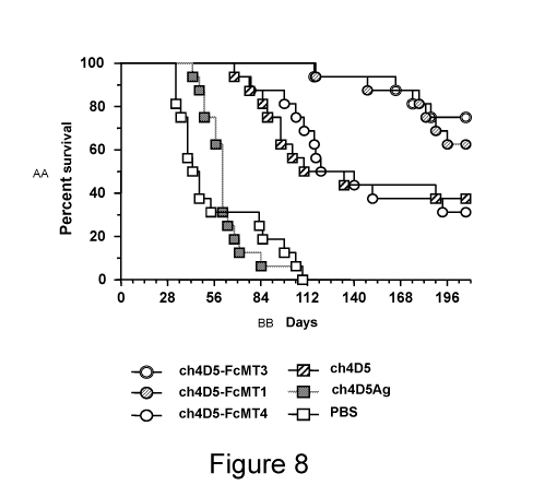
[0060] Figure 8 depicts the enhanced anti-tumor activity of various
antibodies of the
present invention in hCD16A transgenic mice.
-21 -
CA 02988602 2017-12-06
WO 2016/201051 PCT/US2016/036608
[0061] Figure 9 (Panels A-M) illustrates representative immunohistochemical
staining
of cells from various cancer cell lines for HER2/neu. Panels A-L represent the
different
cell lines, i.e., Panel A: MDA-MB-435; Panel B: MDA-MB-231; Panel C: A549;
Panel
D: OVCAR-8; Panel E: MCF-7; Panel F: BT-20; Panel G: HT-29; Panel H: ZR75-1;
Panel!: JIMT-1; Panel J: MDA-MB-453; Panel K: BT-474; Panel L: SKBR-3; and
Panel
M: mSKOV-3.
[0062] Figure 10 (Panels A-B) depicts the results of ADCC assays performed
to test the
ability of Chimeric 4D5 Antibody variants of the present invention to mediate
ADCC in
cancer cell lines (MDA-MB-435 in Panel A; MDA-MB-231 in Panel B) having very
low
or no HER2/neu expression levels (DAKO score of 0).
[0063] Figure 11 (Panels A-E) depicts the results of ADCC assays performed
to test the
ability of Variant Chimeric 4D5 Antibodies of the present invention to mediate
ADCC in
cancer cell lines (A549 in Panel A; OVCAR-8 in Panel B; MCF-7 in Panel C; BT-
20 in
Panel D; HT-29 in Panel E) having low HER2/neu expression levels (DAKO score
of 1+).
[0064] Figure 12 (Panels A-B) depicts the results of ADCC assays performed
to test the
ability of Variant Chimeric 4D5 Antibodies of the present invention to mediate
ADCC in
cancer cell lines (ZR75-1 in Panel A; JIMT-1 in Panel B) having moderate
HER2/neu
expression levels (DAKO score of 2+).
[0065] Figure 13 (Panels A-C) depicts the results of ADCC assays performed
to test the
ability of Variant Chimeric 4D5 Antibodies of the present invention to mediate
ADCC in
cancer cell lines (MDA-MB-453 in Panel A; BT-474 in Panel B; SKBR-3 in Panel
C;
mSKOV-3 in Panel D) having high HER2/neu expression levels (DAKO score of 3+).
[0066] Figure 14 shows a diagram of the protocol for assessing the ability of
anti-PD-1
antibodies to enhance the proliferation of T-cells.
[0067] Figure 15 shows that the addition of PD-1 mAb 1 (5C4; BMS-936558;
Bristol-
Myers Squibb, nivolumab), PD-1 mAb 2 (MK-3475; Merck, pembrolizumab (formerly
lambrolizumab)) and PD-1 mAb 3 (EH12.2H7; Dana Farber) at the start of the
allo-MLR
assay, induced a strong T-cell proliferation response compared to IgG1 isotype
control
antibody. Also shown are the proliferative responses obtained with PD-1 mAb 4
(CT-011;
- 22 -
CA 02988602 2017-12-06
WO 2016/201051 PCT/US2016/036608
CureTech, pidilizumab), an anti-CTLA mAb and LAG-3 mAb. Responder (R) cells
are pan
T-cells; stimulator (S) cells are mature dendritic cells (mDCs).
Detailed Description of the Invention:
[0068] This invention relates to a pharmaceutical composition that
comprises a first
molecule that specifically binds to HER2/neu and a second molecule that
specifically binds
to a cell-surface receptor that is involved in regulating an immune checkpoint
(or the ligand
thereof). The invention particularly relates to the embodiment wherein the
second molecule
binds to PD-1. The invention also relates to the use of such pharmaceutical
compositions
to treat cancer and other diseases.
[0069] In particular, the present invention provides a pharmaceutical
composition that
comprises:
(I) a first antibody that specifically binds to HER2/neu so as to be useful
as a
selective cytotoxic agent for HER2/neu-overexpressing cells (for example, a
Variant Chimeric 4D5 Antibody to HER2/neu having reduced glycosylation
and altered effector functions as compared to known 4D5 antibodies); and
(II) a second antibody that specifically binds to PD-1 so as to be useful
to
antagonize or block PD-1/PD-L1 engagement and thereby maintain T-cell
responses by preventing the delivery of a negative signal toward T-cells.
[0070] The invention also provides methods of using such compositions in
the diagnosis,
prognosis and therapy of diseases such as cancer.
[0071] Without being limited to any particular theory, the methods and
compositions of
the present invention, which combine a potent targeted anti-HER2/neu antibody
with an
anti-PD-1 antibody are capable of directly targeting the tumor by binding to
HER2/neu on
cancer cells thereby reducing/blocking NDF/heregulin activation of HER2/neu-
HER-3
complexes and/or enhancing ADCC activity against HER2/neu positive tumors, and
directly
enhancing endogenous anti-tumor immune responses, for example by binding to
cell-
surface PD-1 molecules that are present on the surfaces of exhausted and
tolerant tumor-
infiltrating lymphocytes, and thereby impairing the ability of such cell-
surface molecules to
bind to their receptor ligands and thereby promoting the activation of the
immune system.
- 23 -
CA 02988602 2017-12-06
WO 2016/201051 PCT/US2016/036608
These attributes permit such treatments and compositions to have utility in
the treatment of
cancer.
[0072] Reference will now be made in detail to the presently preferred
embodiments of
the invention, which, together with the drawings and the following examples,
serve to
explain the principles of the invention. These embodiments are described in
sufficient detail
to enable those skilled in the art to practice the invention, and it is to be
understood that
other embodiments may be utilized, and that structural, biological, and
chemical changes
may be made without departing from the spirit and scope of the present
invention. Unless
otherwise defined, all technical and scientific terms used herein have the
same meaning as
commonly understood by one of ordinary skill in the art to which this
invention belongs.
[0073] The practice of the present invention will employ, unless otherwise
indicated,
conventional techniques of molecular biology (including recombinant
techniques),
microbiology, cell biology, biochemistry and immunology, which are within the
skill of the
art. Such techniques are explained fully in the literature, such as: MOLECULAR
CLONING: A
LABORATORY MANUAL, Fourth Edition (Sambrook et at. Eds., 2012) Cold Spring
Harbor
Press, Cold Spring Harbor, NY; CURRENT PROTOCOLS IN MOLECULAR BIOLOGY
(Ausubel,
F.M. et at., Eds., 1987) Greene Pub. Associates, New York, NY; OLIGONUCLEOTIDE
SYNTHESIS: METHODS AND APPLICATIONS (Methods in Molecular Biology),
IMMUNOBIOLOGY 7 (Janeway, C.A. et at. 2007) Garland Science, London, UK;
MONOCLONAL ANTIBODIES: A PRACTICAL APPROACH (Shepherd, P. et at. Eds., 2000)
Oxford University Press, USA, New York NY; USING ANTIBODIES: A LABORATORY
MANUAL (Harlow, E. et at. Eds., 1998) Cold Spring Harbor Laboratory Press,
Cold Spring
Harbor, NY; and DEVITA, HELLMAN, AND ROSENBERG'S CANCER: PRINCIPLES & PRACTICE
OF ONCOLOGY, EIGHTH EDITION, DeVita, V. et at. Eds. 2008, Lippincott Williams
&
Wilkins, Philadelphia, PA. Antibody engineering is discussed in U.S.
Provisional Patent
Application Nos. 60/781,564; 60/945,523; 61/015,106; and 61/019,051; and in US
20040185045; US 20040197347; US 20040197866; US 20050037000; US 20050064514;
US 20050215767; US 20060134709; US 20060177439; US 20070004909; US
20070036799; US 20070037216; US 20070077246; US 20070244303; US 20080044429;
US 20080050371; 11/869,410; 11/952,568; U.S. Patent No. 7,112,439; WO
04/063351;
WO 06/088494; WO 07/024249; WO 06/113665; WO 07/021841; WO 07/106707; and
WO/2008/140603.
- 24 -
CA 02988602 2017-12-06
WO 2016/201051 PCT/US2016/036608
V. Definitions
[0074] This invention relates to the use of a pharmaceutical composition
that comprises
a first molecule that specifically binds to HER2/neu and a second molecule
that specifically
binds to a cell-surface receptor that is involved in regulating an immune
checkpoint (or the
ligand thereof) for the treatment of diseases such as cancer. The invention
particularly
relates to the embodiment wherein the second molecule binds to PD-1.
[0075] As used herein, the term "ADCC" refers to Antibody-Dependent
Cellular
Cytotoxicity, an in vitro cell-mediated reaction in which nonspecific
cytotoxic cells that
express FcyRs (e.g., monocytic cells such as natural killer (NK) cells and
macrophages)
recognize bound antibody on a target cell and subsequently cause lysis of the
target cell.
[0076] As used herein, the term "antibody" refers to an immunoglobulin
molecule
capable of specific binding to a polypeptide or protein or a non-protein
molecule due to the
presence on such molecule of a particular domain or moiety or conformation (an
"epitope").
An epitope-containing molecule may have immunogenic activity, such that it
elicits an
antibody production response in an animal; such molecules are termed
"antigens". Epitope-
containing molecules need not necessarily be immunogenic.
[0077] As used herein, the term "antibody" encompasses monoclonal
antibodies,
multispecific antibodies, human antibodies, humanized antibodies, synthetic
antibodies,
chimeric antibodies, polyclonal antibodies, camelized antibodies, single-chain
Fvs (scFv),
single-chain antibodies, immunologically active antibody fragments (e.g.,
antibody
fragments capable of binding to an epitope, e.g., Fab fragments, Fab'
fragments, F(ab')2
fragments, Fv fragments, fragments containing a VL and/or VH Domain, or that
contain 1,
2, or 3 of the complementary determining regions (CDRs) of such VL Domain (i .
e . , CDRL 1,
CDRL2, and/or CDRL3) or VH Domain (i.e., CDRH1, CDRH2, and/or CDRH3)) that
specifically bind an antigen, etc., bi-functional or multi-functional
antibodies, disulfide-
linked bispecific Fvs (sdFv), intrabodies, and diabodies, and epitope binding
fragments of
any of the above. In particular, the term "antibody" is intended to encompass
immunoglobulin molecules and immunologically active fragments of
immunoglobulin
molecules, i.e., molecules that contain an antigen-binding site.
Immunoglobulin molecules
can be of any type (e.g., IgG, IgE, IgM, IgD, IgA and IgY), class (e.g., IgGi,
IgG2, IgG3,
IgAi and IgA2) or subclass (see, e.g., United States Patent Publication Nos.:
- 25 -
CA 02988602 2017-12-06
WO 2016/201051 PCT/US2016/036608
20040185045; 20050037000; 20050064514; 20050215767; 20070004909; 20070036799;
20070077246; and 20070244303). The last few decades have seen a revival of
interest in
the therapeutic potential of antibodies, and antibodies have become one of the
leading
classes of biotechnology-derived drugs (Chan, C.E. et at. (2009) "The Use Of
Antibodies In
The Treatment Of Infectious Diseases," Singapore Med. J. 50(7):663-666). Over
200
antibody-based drugs have been approved for use or are under development.
[0078] The
term "chimeric antibody" refers to an antibody in which a portion of a heavy
and/or light chain is identical to or homologous with an antibody from one
species (e.g.,
mouse) or antibody class or subclass, while the remaining portion is identical
to or
homologous with an antibody of another species (e.g., human) or antibody class
or subclass,
so long as they exhibit the desired biological activity. Chimeric antibodies
of interest herein
include "primatized" antibodies comprising variable domain antigen binding
sequences
derived from a non-human primate (e.g., Old World Monkey, Ape, etc.) and human
constant
region sequences.
[0079] The
term "monoclonal antibody" as used herein refers to an antibody of a
population of substantially homogeneous antibodies, i.e., the individual
antibodies
comprising the population are identical except for possible antibodies
possessing naturally
occurring mutations that may be present in minor amounts, and the term
"polyclonal
antibody" as used herein refers to an antibody obtained from a population of
heterogeneous
antibodies. The term "monoclonal" indicates the character of the antibody as
being a
substantially homogeneous population of antibodies, and is not to be construed
as requiring
production of the antibody by any particular method (e.g., by hybridoma, phage
selection,
recombinant expression, transgenic animals, etc.). The
term includes whole
immunoglobulins as well as the fragments etc. described above under the
definition of
"antibody." Methods of making monoclonal antibodies are known in the art. One
method
which may be employed is the method of Kohler, G. et at. (1975) "Continuous
Cultures Of
Fused Cells Secreting Antibody Of Predefined Specificity," Nature 256:495-497
or a
modification thereof. Typically, monoclonal antibodies are developed in mice,
rats or
rabbits. The antibodies are produced by immunizing an animal with an
immunogenic
amount of cells, cell extracts, or protein preparations that contain the
desired epitope. The
immunogen can be, but is not limited to, primary cells, cultured cell lines,
cancerous cells,
proteins, peptides, nucleic acids, or tissue. Cells used for immunization may
be cultured for
- 26 -
CA 02988602 2017-12-06
WO 2016/201051 PCT/US2016/036608
a period of time (e.g., at least 24 hours) prior to their use as an immunogen.
Cells may be
used as immunogens by themselves or in combination with a non-denaturing
adjuvant, such
as Ribi (see, e.g., Jennings, V.M. (1995) "Review of Selected Adjuvants Used
in Antibody
Production," ILAR J. 37(3):119-125). In general, cells should be kept intact
and preferably
viable when used as immunogens. Intact cells may allow antigens to be better
detected than
ruptured cells by the immunized animal. Use of denaturing or harsh adjuvants,
e.g., Freud's
adjuvant, may rupture cells and therefore is discouraged. The immunogen may be
administered multiple times at periodic intervals such as, bi-weekly, or
weekly, or may be
administered in such a way as to maintain viability in the animal (e.g., in a
tissue
recombinant). Alternatively, existing monoclonal antibodies and any other
equivalent
antibodies that are specific for a desired pathogenic epitope can be sequenced
and produced
recombinantly by any means known in the art. In one embodiment, such an
antibody is
sequenced and the polynucleotide sequence is then cloned into a vector for
expression or
propagation. The sequence encoding the antibody of interest may be maintained
in a vector
in a host cell and the host cell can then be expanded and frozen for future
use. The
polynucleotide sequence of such antibodies may be used for genetic
manipulation to
generate the monospecific or multispecific (e.g., bispecific, trispecific and
tetraspecific)
molecules of the invention as well as an affinity optimized, a chimeric
antibody, a
humanized antibody, and/or a caninized antibody, to improve the affinity, or
other
characteristics of the antibody.
[0080] The term "humanized antibody" refers to a chimeric molecule,
generally
prepared using recombinant techniques, having an antigen-binding site of an
immunoglobulin from a non-human species and a remaining immunoglobulin
structure of
the molecule that is based upon the structure and /or sequence of a human
immunoglobulin.
The antigen binding site may comprise either complete variable domains fused
onto constant
domains or only the CDRs grafted onto appropriate framework regions in the
variable
domains. Antigen-binding sites may be wild-type or modified by one or more
amino acid
substitutions. This eliminates the constant region as an immunogen in human
individuals,
but the possibility of an immune response to the foreign variable region
remains (LoBuglio,
A.F. et at. (1989) "Mouse/Human Chimeric Monoclonal Antibody In Man: Kinetics
And
Immune Response," Proc. Natl. Acad. Sci. (U.S.A.) 86:4220-4224). Another
approach
focuses not only on providing human-derived constant regions, but modifying
the variable
- 27 -
CA 02988602 2017-12-06
WO 2016/201051 PCT/US2016/036608
regions as well so as to reshape them as closely as possible to human form. It
is known that
the variable regions of both heavy and light chains contain three CDRs which
vary in
response to the antigens in question and determine binding capability, flanked
by four
framework regions (FRs) which are relatively conserved in a given species and
which
putatively provide a scaffolding for the CDRs. When non-human antibodies are
prepared
with respect to a particular antigen, the variable regions can be "reshaped"
or "humanized"
by grafting CDRs derived from a non-human antibody on the FRs present in the
human
antibody to be modified. Application of this approach to various antibodies
has been
reported by Sato, K. et at. (1993) "Reshaping A Human Antibody To Inhibit The
Interleukin
6-Dependent Tumor Cell Growth," Cancer Res 53:851-856. Riechmann, L. et at.
(1988)
"Reshaping Human Antibodies for Therapy," Nature 332:323-327; Verhoeyen, M. et
at.
(1988) "Reshaping Human Antibodies: Grafting An Antilysozyme Activity,"
Science
239:1534-1536; Kettleborough, C. A. et at. (1991)"Humanization Of A Mouse
Monoclonal
Antibody By CDR-Grafting: The Importance Of Framework Residues On Loop
Conformation," Protein Engineering 4:773-3783; Maeda, H. et at. (1991)
"Construction Of
Reshaped Human Antibodies With HIV-Neutralizing Activity," Human Antibodies
Hybridoma 2:124-134; Gorman, S. D. et at. (1991) "Reshaping A Therapeutic CD4
Antibody," Proc. Natl. Acad. Sci. (U.S.A.) 88:4181-4185; Tempest, P.R. et at.
(1991)
"Reshaping A Human Monoclonal Antibody To Inhibit Human Respiratory Syncytial
Virus
Infection in vivo," Bio/Technology 9:266-271; Co, M. S. et at. (1991)
"Humanized
Antibodies For Antiviral Therapy," Proc. Natl. Acad. Sci. (U.S.A.) 88:2869-
2873; Carter,
P. et at. (1992) "Humanization Of An Anti-p185her2 Antibody For Human Cancer
Therapy," Proc. Natl. Acad. Sci. (U.S.A.) 89:4285-4289; and Co, M.S. et at.
(1992)
"Chimeric And Humanized Antibodies With Specificity For The CD33 Antigen," J.
Immunol. 148:1149-1154. In some embodiments, humanized antibodies preserve all
CDR
sequences (for example, a humanized mouse antibody which contains all six CDRs
from
the mouse antibodies). In other embodiments, humanized antibodies have one or
more
CDRs (one, two, three, four, five or six) that are altered in their amino acid
sequence(s)
relative to the original antibody, which are also termed one or more CDRs
"derived from"
one or more CDRs from the original antibody (i.e., derived from such CDRs,
derived from
knowledge of the amino acid sequecnes of such CDRs, etc.). A polynucleotide
sequence
that encodes the variable domain of an antibody may be used to generate such
derivatives
and to improve the affinity, or other characteristics of such antibodies. The
general principle
- 28 -
CA 02988602 2017-12-06
WO 2016/201051 PCT/US2016/036608
in humanizing an antibody involves retaining the basic sequence of the antigen-
binding site
of the antibody, while swapping the non-human remainder of the antibody with
human
antibody sequences. There are four general steps to humanize a monoclonal
antibody. These
are: (1) determining the nucleotide and predicted amino acid sequence of the
starting
antibody light and heavy variable domains (2) designing the humanized antibody
or
caninized antibody, i.e., deciding which antibody framework region to use
during the
humanizing or canonizing process (3) the actual humanizing or caninizing
methodologies/techniques and (4) the transfection and expression of the
humanized
antibody. See, for example, U.S. Patents Nos. 4,816,567; 5,807,715; 5,866,692;
and
6,331,415.
[0081]
Natural antibodies (such as IgG antibodies) are composed of two Light Chains
complexed with two Heavy Chains. Each light chain contains a Variable Domain
(VL)
and a Constant Domain (CL). Each heavy chain contains a Variable Domain (VH),
three
Constant Domains (CH!, CH2 and CH3), and a "Hinge" Domain ("H") located
between
the CH1 and CH2 Domains. The
basic structural unit of naturally occurring
immunoglobulins (e.g., IgG) is thus a tetramer having two light chains and two
heavy
chains, usually expressed as a glycoprotein of about 150,000 Da. The amino-
terminal ("N-
terminal") portion of each chain includes a Variable Domain of about 100 to
110 or more
amino acids primarily responsible for antigen recognition. The carboxy-
terminal ("C-
terminal") portion of each chain defines a constant region, with light chains
having a single
Constant Domain and heavy chains usually having three Constant Domains and a
Hinge
Domain. Thus, the structure of the light chains of an IgG molecule is n-VL-CL-
c and the
structure of the IgG heavy chains is n-VH-CH1-H-CH2-CH3-c (where n and c
represent,
respectively, the N-terminus and the C-terminus of the polypeptide). The
ability of an
antibody to bind an epitope of an antigen depends upon the presence and amino
acid
sequence of the antibody's VL and VH Domains. Interaction of an antibody light
chain and
an antibody heavy chain and, in particular, interaction of its VL and VH
Domains forms one
of the two antigen-binding sites of a natural antibody. Natural antibodies are
capable of
binding to only one epitope species (i.e., they are monospecific), although
they can bind
multiple copies of that species (i.e., exhibiting bivalency or multivalency).
The Variable
Domains of an IgG molecule consist of the complementarity determining regions
(CDR),
which contain the residues in contact with epitope, and non-CDR segments,
referred to as
- 29 -
CA 02988602 2017-12-06
WO 2016/201051 PCT/US2016/036608
framework segments (FR), which in general maintain the structure and determine
the
positioning of the CDR loops so as to permit such contacting (although certain
framework
residues may also contact antigen). Thus, the VL and VH Domains have the
structure n-
FR1-CDR1-FR2-CDR2-FR3-CDR3-FR4-c. Polypeptides that are (or may serve as) the
first, second and third CDR of an antibody Light Chain are herein respectively
designated
CDRL1 Domain, CDRL2 Domain, and CDRO Domain. Similarly, polypeptides that are
(or may serve as) the first, second and third CDR of an antibody heavy chain
are herein
respectively designated CDRH1 Domain, CDRH2 Domain, and CDRH3 Domain. Thus,
the terms CDRL1 Domain, CDRL2 Domain, CDRL3 Domain, CDRH1 Domain, CDRH2
Domain, and CDRH3 Domain are directed to polypeptides that when incorporated
into a
protein cause that protein to be able to bind to a specific epitope regardless
of whether such
protein is an antibody having light and heavy chains or a diabody or a single-
chain binding
molecule (e.g., an scFv, a BiTe, etc.), or is another type of protein.
Accordingly, as used
herein, the term "Antigen-Binding Domain" refers to that portion of an antigen-
binding
molecule that is responsible for the ability of such molecule to specifically
bind an epitope
of an antigen. An antigen-binding fragment may contain 1, 2, 3, 4, 5 or all 6
of the CDR
Domains of such antibody and, although capable of specifically binding to such
epitope,
may exhibit a specificity, affinity or selectivity toward such epitope that
differs from that of
such antibody. Preferably, however, an antigen-binding fragment will contain
all 6 of the
CDR Domains of such antibody. An antigen-binding fragment of an antibody may
be a
single polypeptide chain (e.g., an scFv), or may comprise two or more
polypeptide chains,
each having an amino terminus and a carboxy terminus (e.g., a diabody, a Fab
fragment, an
F(a1302 fragment, etc.).
[0082] As used herein, the term "diabody" refers to a complex of two or
more
polypeptide chains or proteins, each comprising at least one VL and one VH
domain or
fragment thereof, wherein both domains are comprised within a single
polypeptide chain,
but are separated by an intervening linker that is too short to permit their
association to form
an epitope binding site; thus at least two polypeptide chains or proteins are
required in order
to form a diabody. In certain embodiments a "diabody molecule" includes
molecules
comprising an "Fe" or a "hinge-Fc Region" of an antibody. The polypeptide
chains in the
complex may be the same or different, e.g., the diabody molecule may be a homo-
multimer
or a hetero-multimer. In specific aspects, a "diabody molecule" includes
dimers or tetramers
- 30 -
CA 02988602 2017-12-06
WO 2016/201051 PCT/US2016/036608
or said polypeptide chains containing both a VL and VH domain (e.g., a
homodimer diabody
molecule, a heterodimer diabody molecule, etc.). The individual polypeptide
chains
comprising the multimeric proteins may be covalently joined to at least one
other peptide of
the multimer by interchain disulfide bonds.
[0083] As used herein, the term "cancer" refers to a disease characterized
by the
presence of a neoplasm or tumor resulting from abnormal uncontrolled growth of
cells (such
cells being "cancer cells"). As used herein, the term cancer explicitly
includes, leukemias
and lymphomas. In some embodiments, the term cancer refers to a disease
characterized by
the presence of a benign tumor, which has remained localized. In preferred
embodiments,
however, the term cancer refers to a disease characterized by the presence of
a malignant
tumor that has invaded neighboring body structures. Such tumors may
additionally possess
the ability to spread to distant sites. In some embodiments, the cancer is
associated with
cancer cells that express a specific cancer antigen. In some aspects, the term
cancer as used
herein specifically refers to a cancer expressing HER2/neu. Thus, the term
"HER2/neu-
expressing cancer" as used herein, refers to cancers that are characterized by
the presence
of cancer cells that express detectable levels of HER2/neu. The cancer cells
of such
HER2/neu-expressing cancers may express a high level of HER2/neu or a low
level of
HER2/neu. A "high level of HER2/neu" as used herein refers to a cancer
characterized by
the presence of cancer cells that exhibit a score of 2+ or more when using a
HERCEPTEST
(Dako Cytomation California Inc., Carpenteria, CA) classification, or a
subject or patient
possessing cancer cells that have been identified as overexpressing Her2/neu,
for example,
by fluorescence in situ hybridization (FISH). A "low level of HER2" as used
herein, refers
to a cancer characterized by cancer cells that exhibit a score of less than 2+
(e.g., 1+) in the
HERCEPTEST (Dako Cytomation California Inc., Carpenteria, CA) classification.
Various
diagnostic/prognostic assays are available to determine the level of HER2
expression by the
cancer cells of a tumor. In one aspect, HER2 overexpression can be analyzed by
immunohistochemistry (IHC), e.g., by using HERCEPTEST (Dako). Accordingly,
paraffin-
embedded tissue sections from a tumor biopsy can be subjected to the IHC assay
and
accorded a HER2 protein staining intensity criteria. Alternatively, or
additionally, FISH
assays such as the INFORMTm (sold by Ventana, Ariz.) or PATHVISIONTm (Vysis,
Ill.)
can be carried out on formalin-fixed, paraffin-embedded tumor tissue to
determine the
extent (if any) of HER2 overexpression in the tumor.
- 31 -
CA 02988602 2017-12-06
WO 2016/201051 PCT/US2016/036608
[0084] As used herein, the terms "disorder" and "disease" are used
interchangeably to
refer to a condition in a subject. In particular, the term "autoimmune
disease" is used
interchangeably with the term "autoimmune disorder" to refer to a condition in
a subject
characterized by cellular, tissue and/or organ injury caused by an immunologic
reaction of
the subject to its own cells, tissues and/or organs. The term "inflammatory
disease" is used
interchangeably with the term "inflammatory disorder" to refer to a condition
in a subject
characterized by inflammation, preferably chronic inflammation. Autoimmune
disorders
may or may not be associated with inflammation. Moreover, inflammation may or
may not
be caused by an autoimmune disorder. Thus, certain disorders may be
characterized as both
an autoimmune disorder and an inflammatory disorder, whereas other disorders
may be
characterized as being either only an autoimmune disorder or only an
inflammatory
disorder. Cancer is an example of a "proliferative disorder" (i.e., a disorder
that is
associated with some degree of abnormal cell proliferation).
[0085] As used herein, an "effective amount" of a pharmaceutical
composition is an
amount sufficient to effect beneficial or desired results including, without
limitation, clinical
results such as shrinking the size of a tumor (in the cancer context, for
example, a tumor of
breast, gastric or prostate cancer), retardation of cancer cell growth,
delaying the
development of metastasis, decreasing a symptom resulting from the disease,
increasing the
quality of life of those suffering from the disease, decreasing the dose of
other medications
required to treat the disease, enhancing the effect of another medication such
as via targeting
and/or internalization, delaying the progression of the disease, and/or
prolonging survival
of an individual. An effective amount can be administered in one or more
administrations.
For purposes of this invention, an effective amount of drug, compound, or
pharmaceutical
composition is an amount sufficient to reduce the proliferation of (or
destroy) cancer cells
or to reduce and /or delay the development, or growth, of metastases of cancer
cells, either
directly or indirectly. In some embodiments, an effective amount of a drug,
compound, or
pharmaceutical composition may or may not be achieved in conjunction with
another drug,
compound, or pharmaceutical composition. Thus, an "effective amount" may be
considered
in the context of administering one or more chemotherapeutic agents, and a
single agent
may be considered to be given in an effective amount if, in conjunction with
one or more
other agents, a desirable result may be or is achieved. While individual needs
vary,
- 32 -
CA 02988602 2017-12-06
WO 2016/201051 PCT/US2016/036608
determination of optimal ranges of effective amounts of each component is
within the skill
of the art. Typical dosages are discussed below.
[0086] The term "effector activity" refers to biological activities
attributable to the
interaction of an antibody Fc Region with an Fc receptor or ligand. An
antibody may have
one or more effector functions. Non-limiting examples of antibody effector
functions
include ADCC, Clq binding, complement dependent cytotoxicity (CDC), down
regulation
of cell-surface receptors (e.g., B-cell receptor; BCR), opsonization,
opsonophagocytosis,
cell binding, and rosetting. Effector functions include both those that
operate after the
binding of an antigen and those that operate independent of antigen binding.
[0087] The term "effector cell" as used herein refers to a cell of the
immune system that
expresses one or more Fc receptors and mediates one or more effector
functions. Effector
cells include but are not limited to monocytes, macrophages, neutrophils,
dendritic cells,
eosinophils, mast cells, platelets, B-cells, large granular lymphocytes,
Langerhans' cells,
natural killer (NK) cells, and may be from any organism including but not
limited to humans,
mice, rats, rabbits, and monkeys.
[0088] The terms "Fc receptor" or "FcR" are used herein to describe a
receptor that
binds to the Fc Region of an antibody. An exemplary FcR is a native sequence
human FcR.
An FcR may be one which binds an IgG antibody (a gamma receptor, "FcyR") and
includes
receptors of the FcyRI (CD64), FcyRII (CD32), FcyRIII (CD16), and FcyRIV
subclasses,
including allelic variants and alternatively spliced forms of these receptors,
e.g., there are at
least two known FcyRII receptors, FcyRIIA and FcyRIIB. The term FcR also
includes the
neonatal receptor, FcRn, which is responsible for the transfer of maternal
IgGs to the fetus.
[0089] The term "glycosylation site" refers to an amino acid residue or
residues
recognized by a mammalian cell as a location for the attachment of an
oligosaccharide (i.e.,
carbohydrates containing two or more simple sugars linked together). Amino
acid residues
to which carbohydrates, such as oligosaccharides, are attached are usually
asparagine (N-
linkage), serine (0-linkage), and threonine (0-linkage) residues. N-linked
glycosylation
refers to the attachment of an oligosaccharide moiety to the side chain of an
asparagine
residue. 0-linked glycosylation refers to the attachment of an oligosaccharide
moiety to a
hydroxyamino acid, e.g., serine or threonine. The molecules of the invention
may comprise
- 33 -
CA 02988602 2017-12-06
WO 2016/201051 PCT/US2016/036608
one or more glycosylation sites, including N-linked and 0-linked glycosylation
sites. Any
glycosylation site for N-linked or 0-linked glycosylation known in the art may
be used in
accordance with the instant invention. The specific sites of attachment
usually have a
characteristic sequence of amino acids, referred to as a "glycosylation site
sequence." The
glycosylation site sequence for N-linked glycosylation is: N-X-S or N-X-T,
where N
indicates asparagine, X can be any of the conventional amino acids other than
proline, S
indicates serine and T indicates threonine. The Fc Region of native human IgG
has two N-
linked glycosylation sites, one in each of the CH2 domains, at the asparagine
at position 297
(Asn 297). Glycosylation sites may be introduced into a molecule of the
invention using
methods well known in the art to which this invention pertains (see for
example, IN VITRO
MUTAGENESIS, RECOMBINANT DNA: A SHORT COURSE, J. D. Watson, et at. W.H.
Freeman
and Company, New York, 1983, chapter 8, pp. 106-116, which is incorporated
herein by
reference in its entirety. An exemplary method for introducing a glycosylation
site into a
molecule of the invention may comprise: modifying or mutating an amino acid
sequence of
the molecule so that the desired N-X-S or N-X-T sequence is obtained,
likewise,
glycosylation sites may be removed by modifying or mutating an amino acid
sequence of
an existing glycosylation site, for example, to alter an existing N-X-S or N-X-
T sequence.
[0090] As used herein, the term "Human Anti-Mouse Antibody ("HAMA") response"
refers to a deleterious immunogenic response that occurs when a human immune
system
recognizes a murine antibody as a foreign molecule and mounts an inflammatory
response
against it. A HAMA response can cause toxic shock or death. Chimeric and
humanized
antibodies reduce the likelihood of a HAMA response by decreasing the non-
human
portions of administered antibodies, but there is still potential for a Human
Anti-Human
Antibody response ("HAHA response") immune response to such antibodies.
[0091] As used herein, the term "heterologous" nucleic acid denotes DNA,
RNA, etc.
that is introduced into a host cell. The nucleic acid may be derived from any
of a variety of
sources including genomic DNA, mRNA, cDNA, synthetic DNA and fusions or
combinations of these. The nucleic acid may include a polynucleotide from the
same cell
or cell type as the host or recipient cell or a polynucleotide from a
different cell type, for
example, from a mammal or plant, and may, optionally, include marker or
selection genes,
for example, antibiotic resistance genes, temperature resistance genes, etc.
- 34 -
CA 02988602 2017-12-06
WO 2016/201051 PCT/US2016/036608
[0092] As used herein, the term "immunomodulatory agent" and variations
thereof
refer to an agent that modulates a host's immune system. In certain
embodiments, an
immunomodulatory agent is an immunosuppressant agent. In certain other
embodiments,
an immunomodulatory agent is an immunostimulatory agent. Immunomodulatory
agents
include, but are not limited to, small molecules, peptides, polypeptides,
fusion proteins,
antibodies, inorganic molecules, mimetic agents, and organic molecules.
[0093] As used herein, a molecule (e.g., an antibody) is said to
"specifically" bind a
region of another molecule (i.e., an "epitope") if it reacts or associates
more frequently,
more rapidly, with greater duration and/or with greater affinity with that
region relative to
alternative regions of the molecule or alternative molecules. For example, an
antibody that
specifically binds to a HER2/neu epitope is an antibody that binds such
HER2/neu epitope
with greater affinity, avidity, more readily, and /or with greater duration
than it binds to
other HER2/neu epitopes or to a non-HER2/neu epitope. Likewise, an antibody
that
specifically binds to an epitope of PD-1 binds such epitope with greater
affinity, avidity,
more readily, and /or with greater duration than it binds to other PD-1
epitopes or to a non-
PD-1 epitope. It is also understood by reading this definition that, for
example, an antibody
(or moiety or epitope) that specifically binds to a first target may or may
not specifically or
preferentially bind to a second target. As such, "specific" binding does not
necessarily
require (although it can include) exclusive binding. Generally, unless the
context clearly
evidences to the contrary, reference to "binding" means "specific binding."
The ability of
an antibody to specifically bind to an epitope of an antigen may be determined
by, for
example, an immunoassay.
[0094] As used herein, the term "nucleic acid molecule" include DNA
molecules (e.g.,
cDNA or genomic DNA), RNA molecules (e.g., mRNA), combinations of DNA and RNA
molecules or hybrid DNA/RNA molecules, and analogs of DNA or RNA molecules.
Such
analogs can be generated using, for example, nucleotide analogs, which
include, but are not
limited to, inosine or tritylated bases. Such analogs can also comprise DNA or
RNA
molecules comprising modified backbones that lend beneficial attributes to the
molecules
such as, for example, nuclease resistance or an increased ability to cross
cellular membranes.
The nucleic acids or nucleotide sequences can be single-stranded, double-
stranded, may
contain both single-stranded and double-stranded portions, and may contain
triple-stranded
portions, but preferably is double-stranded DNA.
- 35 -
CA 02988602 2017-12-06
WO 2016/201051 PCT/US2016/036608
[0095] As used herein, the term "substantial sequence identity," refers to
two or more
sequences or subsequences (e.g., domains) that have at least about 80% amino
acid residue
identity, preferably at least about 90%, or at least about 95% identity when
compared and
aligned for maximum correspondence. Sequence identity between two similar
sequences
(e.g., antibody variable domains) can be measured by algorithms such as that
of Smith, T.F.
& Waterman, M.S. (1981) "Comparison Of Biosequences," Adv. Appl. Math. 2:482
[local
homology algorithm]; Needleman, S.B. & Wunsch, C.D. (1970) "A General Method
Applicable To The Search For Similarities In The Amino Acid Sequence Of Two
Proteins,"
J. Mol. Biol. 48:443 [homology alignment algorithm], Pearson, W.R. & Lipman,
D.J. (1988)
"Improved Tools For Biological Sequence Comparison," Proc. Natl. Acad. Sci.
(U.S.A.)
85:2444 [search for similarity method]; or Altschul, S.F. et al., (1990)
"Basic Local
Alignment Search Tool," J. Mol. Biol. 215:403-10 [BLAST algorithm]. When using
any of
the aforementioned algorithms, the default parameters (for Window length, gap
penalty,
etc.) are used. A first amino acid sequence is said to be "substantially
similar" to a second
amino acid sequence when the degree of sequence identity is at least about 70%
identical,
preferably at least about 80%, or at least about 90%, or even at least about
95%, identical.
A nucleic acid sequence is said to be "substantially similar" to a second
sequence when
either: (1) the degree of sequence identity is at least about 70% identical,
preferably at least
about 80%, or at least about 90%, or even at least about 95%, identical, or
(2) a nucleic acid
molecule comprising that nucleic acid sequence encodes a polypeptide that is
at least about
70% identical, preferably at least about 80%, or at least about 90%, or even
at least about
95%, identical to the polypeptide encoded by a nucleic acid molecule
comprising the second
sequence. Sequences that are substantially identical are also substantially
similar.
[0096] When referring to antibodies, the assignment of amino acids to each
domain is in
accordance with Kabat et al., SEQUENCES OF PROTEINS OF IMMUNOLOGICAL INTEREST,
5th
Ed. (National Institutes of Health, Bethesda, MD., (1991) ("Kabat et al."),
which is
expressly incorporated herein by reference. Throughout the present
specification, the
numbering of the constant residues in an IgG heavy chain "according to Kabat"
refers to
the numbering of the human IgG1 EU antibody as described in Kabat et al.
[0097] The term "Murine 4D5 Antibody" refers to the murine IgG1 antibody
disclosed
in US Patent No. 5,677,171 as ATCC CRL 10463. Murine 4D5 Antibody binds
Her2/neu
and has a light chain variable domain having the amino acid sequence of SEQ ID
NO:3 and
- 36 -
CA 02988602 2017-12-06
WO 2016/201051 PCT/US2016/036608
a heavy chain variable domain having the amino acid sequence of SEQ ID NO:47.
The
term "Humanized 4D5 Antibody" refers to the IgG antibody disclosed in Carter,
P. et at.
(1992) ("Humanization Of An Anti-P185her2 Antibody For Human Cancer Therapy,"
Proc.
Natl. Acad. Sci. (U.S.A.) 89:4285-4289). Humanized 4D5 Antibody is reported to
be
capable of binding Her2/neu; it has a light chain variable region having the
amino acid
sequence of SEQ ID NO:5 and a heavy chain variable region having the amino
acid
sequence of SEQ ID NO:48.
[0098] The amino acid sequence of the Light Chain Variable Domain of Murine
4D5
Antibody is (SEQ ID NO:3) (CDRL residues are underlined):
DIVMTQSHKF MSTSVGDRVS ITCKASQDVN TAVAWYQQKP GHSPKLLIYS
ASFRYTGVPD RFTGNRSGTD FTFTISSVQA EDLAVYYCQQ HYTTPPTFGG
GTKLEIK
[0099] The amino acid sequence of the Heavy Chain Variable Domain of Murine
4D5
Antibody is (SEQ ID NO:47) (CDRH residues are underlined):
QVQLQQSGPE LVKPGASLKL SCTASGFNIK DTYIHWVKQR PEQGLEWIGR
IYPTNGYTRY DPKFQDKATI TADTSSNTAY LQVSRLTSED TAVYYCSRWG
GDGFYAMDYW GQGASVTVSS
[00100] The amino acid sequence of the Light Chain Variable Domain of
Humanized 4D5
Antibody is (SEQ ID NO:5) (CDRL residues are underlined):
DIQMTQSPSS LSASVGDRVT ITCRASQDVN TAVAWYQQKP GKAPKLLIYS
ASFLESGVPS RFSGSRSGTD FTLTISSLQP EDFATYYCQQ HYTTPPTFGQ
GTKVEIK
[00101] The amino acid sequence of the Heavy Chain Variable Domain of
Humanized
4D5 Antibody is (SEQ ID NO:48) (CDRH residues are underlined):
EVQLVESGGG LVQPGGSLRL SCAASGFNIK DTYIHWVRQA PGKGLEWVAR
IYPTNGYTRY ADSVKGRFTI SADTSKNTAY LQMNSLRAED TAVYYCSRWG
GDGFYAMDVW GQGTLVTVSS
[00102] The term "Chimeric 4D5 Antibody" refers to an IgG antibody that binds
human
Her2/neu and has a light chain having the amino acid sequence of SEQ ID NO:2
and a
heavy chain having a wild-type Fc Region; the amino acid sequence of the heavy
chain of
Chimeric 4D5 Antibody is shown in SEQ ID NO:7. A "Variant Chimeric 4D5
Antibody"
is an IgG antibody that binds Her2/neu and has a light chain and/or a heavy
chain whose
amino acid sequence(s) differ(s) from those of Chimeric 4D5 Antibody (e.g., an
IgG
antibody comprising a light chain having the amino acid sequence of SEQ ID
NO:2, and a
- 37 -
CA 02988602 2017-12-06
WO 2016/201051 PCT/US2016/036608
heavy chain having the amino acid sequence of SEQ ID NO:9, SEQ ID NO:!! or SEQ
ID
NO: !3).
VI. Binding Molecules
A. Molecules That Specifically Bind HER2/neu
[00103] Molecules that specifically bind HER2/neu that are encompassed by the
present
invention include anti-HER2/neu antibodies capable of specifically binding to
a continuous
or discontinuous (e.g., conformational) epitope of human HER2/neu. The
HER2/neu
antibodies used in the methods of the present invention will preferably also
exhibit the
ability to bind to the HER2/neu molecules of one or more non-human species,
especially,
murine, rodent, canine, and primate species (especially cynomolgus monkey).
[00104] Antibodies to HER2/neu are provided below. Additional desired
antibodies may
be made by mutating a nucleic acid molecule that encodes a polypeptide chain
of such
antibodies and then screening for expressed antibodies that exhibit the
ability to specifically
bind to Her2/neu, by isolating new antibody-secreting hybridomas elicited
using HER2/neu
or a peptide fragment thereof, or by other means. The human HER/2 sequence has
been
described Yamamoto, T. et al. (1986) "Similarity Of Protein Encoded By The
Human c-erb-
B-2 Gene To Epidermal Growth Factor Receptor," Nature 319:230-234, and the
sequence
is available in GenBank as accession number X03363.
[00105] The present invention particularly encompasses variants of Chimeric
4D5
Antibody, and more particularly variants of such Chimeric 4D5 Antibody that
specifically
bind to HER2/neu, preferably human HER2/neu and that exhibit reduced
glycosylation
relative to Murine 4D5 Antibody, due to the removal of a glycosylation site in
the variable
domain of the light chain. In particular, the preferred chimeric antibodies of
the present
invention lack a glycosylation site in the variable domain of the light chain
of Murine 4D5
Antibody, which in the light chain of Murine 4D5 Antibody comprises an N-R-S
sequence
at positions 65, 66 and 67. Preferably the antibodies have enhanced binding
affinity for
HER2/neu, and more preferably the Variant Chimeric 4D5 Antibodies of the
present
invention have enhanced effector function, or both enhanced binding affinity
for HER2/neu
and enhanced effector function as compared to a murine 4D5 antibody.
- 38 -
CA 02988602 2017-12-06
WO 2016/201051 PCT/US2016/036608
[00106] In a preferred embodiment, the preferred variants of Chimeric 4D5
Antibody
comprise a light chain (Chimeric 4D5 light chain) having or comprising the
amino acid
sequence of SEQ ID NO:2.
[00107] Amino acid sequence of Chimeric 4D5 Antibody light chain (SEQ ID NO:2)
(CDRL residues are underlined):
DIVMTQSHKF MSTSVGDRVS ITCKASQDVN TAVAWYQQKP GHSPKLLIYS
ASFRYTGVPD RFTGSRSGTD FTFTISSVQA EDLAVYYCQQ HYTTPPTFGG
GTKVEIKRTV AAPSVFIFPP SDEQLKSGTA SVVCLLNNFY PREAKVQWKV
DNALQSGNSQ ESVTEQDSKD STYSLSSTLT LSKADYEKHK VYACEVTHQG
LSSPVTKSFN RGEC
[00108] An exemplary nucleic acid molecule that encodes the light chain of
preferred
Variant Chimeric 4D5 Antibodies is SEQ ID NO:!:
gacatcgtga tgacccagtc ccacaagttc atgtccacct ctgtgggcga
tagggtcagc atcacctgca aggccagcca ggatgtgaat actgctgtag
cctggtatca gcagaaacca ggacattctc ccaaactgct gatttactcc
gcatccttcc ggtacactgg agtccctgat cgcttcactg gcagcagatc
tgggacagat ttcactttca ccatcagcag tgtgcaggct gaagacctgg
cagtttatta ctgtcagcaa cattatacta cacctcccac cttcggaggg
ggtaccaagg tggagatcaa acgtacggtg gctgcaccat ctgtcttcat
cttcccgcca tctgatgagc agttgaaatc tggaactgcc tctgttgtgt
gcctgctgaa taacttctat cccagagagg ccaaagtaca gtggaaggtg
gataacgccc tccaatcggg taactcccag gagagtgtca cagagcagga
cagcaaggac agcacctaca gcctcagcag caccctgacg ctgagcaaag
cagactacga gaaacacaaa gtctacgcct gcgaagtcac ccatcagggc
ctgagctcgc ccgtcacaaa gagcttcaac aggggagagt gttag
[00109] Antibodies having such light chain amino acid sequence have a
modification at
positions 65 of the VL region and as such lack an N-linked glycosylation site
found in
Murine 4D5 Antibody (see Figure 1, which depicts an exemplary comparison
between the
VL region amino acid sequences of a Chimeric 4D5 Antibody having an N65S
modification
(SEQ ID NO:4), and the murine (SEQ ID NO:3) and humanized (SEQ ID NO:5) 4D5
antibodies). In another preferred embodiment, the Variant Chimeric 4D5
Antibodies of the
present invention have a VL region amino acid sequence of SEQ ID NO:4.
[00110] Amino acid sequence of Chimeric 4D5 VL region (SEQ ID NO:4) (CDRL
residues are underlined):
DIVMTQSHKF MSTSVGDRVS ITCKASQDVN TAVAWYQQKP GHSPKLLIYS
ASFRYTGVPD RFTGSRSGTD FTFTISSVQA EDLAVYYCQQ HYTTPPTFGG
GTKVEIK
- 39 -
CA 02988602 2017-12-06
WO 2016/201051 PCT/US2016/036608
[00111] The Chimeric 4D5 Antibody comprises a heavy chain ("Chimeric 4D5 heavy
chain") that has a wild-type Fc Region (SEQ ID NO:7), which may be encoded by
the
nucleic acid sequence of SEQ ID NO:6. These sequences are presented below:
[00112] Amino acid Sequence of Chimeric 4D5 heavy chain having wild-type Fc
Region
(SEQ ID NO:7) (CDRH residues are underlined):
QVQLQQSGPE LVKPGASLKL SCTASGFNIK DTYIHWVKQR PEQGLEWIGR
IYPTNGYTRY DPKFQDKATI TADTSSNTAY LQVSRLTSED TAVYYCSRWG
GDGFYAMDYW GQGASVTVSS ASTKGPSVFP LAPSSKSTSG GTAALGCLVK
DYFPEPVTVS WNSGALTSGV HTFPAVLQSS GLYSLSSVVT VPSSSLGTQT
YICNVNHKPS NTKVDKRVEP KSCDKTHTCP PCPAPELLGG PSVFLFPPKP
KDTLMISRTP EVTCVVVDVS HEDPEVKFNW YVDGVEVHNA KTKPREEQYN
STYRVVSVLT VLHQDWLNGK EYKCKVSNKA LPAPIEKTIS KAKGQPREPQ
VYTLPPSRDE LTKNQVSLTC LVKGFYPSDI AVEWESNGQP ENNYKTTPPV
LDSDGSFFLY SKLTVDKSRW QQGNVFSCSV MHEALHNHYT QKSLSLSPGK
[00113] An exemplary nucleic acid molecule that encodes Chimeric 4D5 heavy
chain
having a wild-type Fc Region (SEQ ID NO:6):
caggttcagc tgcagcagtc tggccctgag ctggtgaagc caggggcctc
actcaagttg tcctgtacag cttctggctt caacatcaaa gacacctata
tccactgggt gaaacagagg cctgaacagg gcctggaatg gattggaagg
atttatccta ccaatggcta tactagatat gacccaaagt tccaggacaa
ggccactatc acagcagaca catcctccaa cacagcctac ctgcaagtca
gccgcctgac atctgaggac actgccgtct attactgctc ccggtgggga
ggggacggct tctatgctat ggactactgg ggtcagggag cctccgtgac
cgtgagctcc gcctccacca agggcccatc ggtcttcccc ctggcaccct
cctccaagag cacctctggg ggcacagcgg ccctgggctg cctggtcaag
gactacttcc ccgaaccggt gacggtgtcg tggaactcag gcgccctgac
cagcggcgtg cacaccttcc cggctgtcct acagtcctca ggactctact
ccctcagcag cgtggtgacc gtgccctcca gcagcttggg cacccagacc
tacatctgca acgtgaatca caagcccagc aacaccaagg tggacaagag
agttgagccc aaatcttgtg acaaaactca cacatgccca ccgtgcccag
cacctgaact cctgggggga ccgtcagtct tcctcttccc cccaaaaccc
aaggacaccc tcatgatctc ccggacccct gaggtcacat gcgtggtggt
ggacgtgagc cacgaagacc ctgaggtcaa gttcaactgg tacgtggacg
gcgtggaggt gcataatgcc aagacaaagc cgcgggagga gcagtacaac
agcacgtacc gtgtggtcag cgtcctcacc gtcctgcacc aggactggct
gaatggcaag gagtacaagt gcaaggtctc caacaaagcc ctcccagccc
ccatcgagaa aaccatctcc aaagccaaag ggcagccccg agaaccacag
gtgtacaccc tgcccccatc ccgggatgag ctgaccaaga accaggtcag
cctgacctgc ctggtcaaag gcttctatcc cagcgacatc gccgtggagt
gggagagcaa tgggcagccg gagaacaact acaagaccac gcctcccgtg
ctggactccg acggctcctt cttcctctac agcaagctca ccgtggacaa
gagcaggtgg cagcagggga acgtcttctc atgctccgtg atgcatgagg
ctctgcacaa ccactacacg cagaagagcc tctccctgtc tccgggtaaa
tga
- 40 -
CA 02988602 2017-12-06
WO 2016/201051 PCT/US2016/036608
[00114] In other embodiments, the invention contemplates employing a Variant
Chimeric
4D5 Antibody whose heavy chain comprises a variant Fc Region, and more
preferably, an
"FcMT1," "FcMT2," or "FcMT3" variant Fc Region. These sequences are presented
below:
[00115] Amino acid Sequence of the heavy chain of a Variant Chimeric 4D5
Antibody
having the FcMT1 variant Fc Region (SEQ ID NO:9) (CDRH residues are
underlined)):
QVQLQQSGPE LVKPGASLKL SCTASGFNIK DTYIHWVKQR PEQGLEWIGR
IYPTNGYTRY DPKFQDKATI TADTSSNTAY LQVSRLTSED TAVYYCSRWG
GDGFYAMDYW GQGASVTVSS ASTKGPSVFP LAPSSKSTSG GTAALGCLVK
DYFPEPVTVS WNSGALTSGV HTFPAVLQSS GLYSLSSVVT VPSSSLGTQT
YICNVNHKPS NTKVDKRVEP KSCDKTHTCP PCPAPELLGG PSVFLLPPKP
KDTLMISRTP EVTCVVVDVS HEDPEVKFNW YVDGVEVHNA KTKPPEEQYN
STLRVVSILT VLHQDWLNGK EYKCKVSNKA LPAPIEKTIS KAKGQPREPQ
VYTLPPSRDE LTKNQVSLTC LVKGFYPSDI AVEWESNGQP ENNYKTTPLV
LDSDGSFFLY SKLTVDKSRW QQGNVFSCSV MHEALHNHYT QKSLSLSPGK
[00116] An exemplary nucleic acid molecule that encodes a heavy chain of a
Variant
Chimeric 4D5 Antibody having the FcMT1 variant Fc Region (SEQ ID NO:8):
caggttcagc tgcagcagtc tggccctgag ctggtgaagc caggggcctc
actcaagttg tcctgtacag cttctggctt caacatcaaa gacacctata
tccactgggt gaaacagagg cctgaacagg gcctggaatg gattggaagg
atttatccta ccaatggcta tactagatat gacccaaagt tccaggacaa
ggccactatc acagcagaca catcctccaa cacagcctac ctgcaagtca
gccgcctgac atctgaggac actgccgtct attactgctc ccggtgggga
ggggacggct tctatgctat ggactactgg ggtcagggag cctccgtgac
cgtgagctcc gcctccacca agggcccatc ggtcttcccc ctggcaccct
cctccaagag cacctctggg ggcacagcgg ccctgggctg cctggtcaag
gactacttcc ccgaaccggt gacggtgtcg tggaactcag gcgccctgac
cagcggcgtg cacaccttcc cggctgtcct acagtcctca ggactctact
ccctcagcag cgtggtgacc gtgccctcca gcagcttggg cacccagacc
tacatctgca acgtgaatca caagcccagc aacaccaagg tggacaagag
agttgagccc aaatcttgtg acaaaactca cacatgccca ccgtgcccag
cacctgaact cctgggggga ccgtcagtct tcctcttacc cccaaaaccc
aaggacaccc tcatgatctc ccggacccct gaggtcacat gcgtggtggt
ggacgtgagc cacgaagacc ctgaggtcaa gttcaactgg tacgtggacg
gcgtggaggt gcataatgcc aagacaaagc cgccggagga gcagtacaac
agcacgctcc gtgtggtcag catcctcacc gtcctgcacc aggactggct
gaatggcaag gagtacaagt gcaaggtctc caacaaagcc ctcccagccc
ccatcgagaa aaccatctcc aaagccaaag ggcagccccg agaaccacag
gtgtacaccc tgcccccatc ccgggatgag ctgaccaaga accaggtcag
cctgacctgc ctggtcaaag gcttctatcc cagcgacatc gccgtggagt
gggagagcaa tgggcagccg gagaacaact acaagaccac gcctctcgtg
ctggactccg acggctcctt cttcctctac agcaagctca ccgtggacaa
gagcaggtgg cagcagggga acgtcttctc atgctccgtg atgcatgagg
-41-
CA 02988602 2017-12-06
WO 2016/201051 PCT/US2016/036608
ctctgcacaa ccactacacg cagaagagcc tctccctgtc tccgggtaaa
tga
[00117] Amino acid Sequence of the heavy chain of a Variant Chimeric 4D5
Antibody
having the FcMT2 variant Fc Region (SEQ ID NO:!!) (CDRH residues are
underlined):
QVQLQQSGPE LVKPGASLKL SCTASGFNIK DTYIHWVKQR PEQGLEWIGR
IYPTNGYTRY DPKFQDKATI TADTSSNTAY LQVSRLTSED TAVYYCSRWG
GDGFYAMDYW GQGASVTVSS ASTKGPSVFP LAPSSKSTSG GTAALGCLVK
DYFPEPVTVS WNSGALTSGV HTFPAVLQSS GLYSLSSVVT VPSSSLGTQT
YICNVNHKPS NTKVDKRVEP KSCDKTHTCP PCPAPELVGG PSVFLLPPKP
KDTLMISRTP EVTCVVVDVS HEDPEVKFNW YVDGVEVHNA KTKPPEEQYN
STLRVVSVLT VLHQDWLNGK EYKCKVSNKA LPAPIEKTIS KAKGQPREPQ
VYTLPPSRDE LTKNQVSLTC LVKGFYPSDI AVEWESNGQP ENNYKTTPLV
LDSDGSFFLY SKLTVDKSRW QQGNVFSCSV MHEALHNHYT QKSLSLSPGK
[00118] An exemplary nucleic acid molecule that encodes a heavy chain of a
Variant
Chimeric 4D5 Antibody having the FcMT2 variant Fc Region (SEQ ID NO:10):
caggttcagc tgcagcagtc tggccctgag ctggtgaagc caggggcctc
actcaagttg tcctgtacag cttctggctt caacatcaaa gacacctata
tccactgggt gaaacagagg cctgaacagg gcctggaatg gattggaagg
atttatccta ccaatggcta tactagatat gacccaaagt tccaggacaa
ggccactatc acagcagaca catcctccaa cacagcctac ctgcaagtca
gccgcctgac atctgaggac actgccgtct attactgctc ccggtgggga
ggggacggct tctatgctat ggactactgg ggtcagggag cctccgtgac
cgtgagctcc gcctccacca agggcccatc ggtcttcccc ctggcaccct
cctccaagag cacctctggg ggcacagcgg ccctgggctg cctggtcaag
gactacttcc ccgaaccggt gacggtgtcg tggaactcag gcgccctgac
cagcggcgtg cacaccttcc cggctgtcct acagtcctca ggactctact
ccctcagcag cgtggtgacc gtgccctcca gcagcttggg cacccagacc
tacatctgca acgtgaatca caagcccagc aacaccaagg tggacaagag
agttgagccc aaatcttgtg acaaaactca cacatgccca ccgtgcccag
cacctgaact cgtgggggga ccgtcagtct tcctcttacc cccaaaaccc
aaggacaccc tcatgatctc ccggacccct gaggtcacat gcgtggtggt
ggacgtgagc cacgaagacc ctgaggtcaa gttcaactgg tacgtggacg
gcgtggaggt gcataatgcc aagacaaagc cgccggagga gcagtacaac
agcacgctcc gtgtggtcag cgtcctcacc gtcctgcacc aggactggct
gaatggcaag gagtacaagt gcaaggtctc caacaaagcc ctcccagccc
ccatcgagaa aaccatctcc aaagccaaag ggcagccccg agaaccacag
gtgtacaccc tgcccccatc ccgggatgag ctgaccaaga accaggtcag
cctgacctgc ctggtcaaag gcttctatcc cagcgacatc gccgtggagt
gggagagcaa tgggcagccg gagaacaact acaagaccac gcctctcgtg
ctggactccg acggctcctt cttcctctac agcaagctca ccgtggacaa
gagcaggtgg cagcagggga acgtcttctc atgctccgtg atgcatgagg
ctctgcacaa ccactacacg cagaagagcc tctccctgtc tccgggtaaa
tga
[00119] Amino acid Sequence of the heavy chain of a Variant Chimeric 4D5
Antibody
having the FcMT3 variant Fc Region (SEQ ID NO:13) (CDRH residues are
underlined):
- 42 -
CA 02988602 2017-12-06
W02016/201051 PCT/US2016/036608
QVQLQQSGPE LVKPGASLKL SCTASGFNIK DTYIHWVKQR PEQGLEWIGR
IYPTNGYTRY DPKFQDKATI TADTSSNTAY LQVSRLTSED TAVYYCSRWG
GDGFYAMDYW GQGASVTVSS ASTKGPSVFP LAPSSKSTSG GTAALGCLVK
DYFPEPVTVS WNSGALTSGV HTFPAVLQSS GLYSLSSVVT VPSSSLGTQT
YICNVNHKPS NTKVDKRVEP KSCDKTHTCP PCPAPELLGG PSVFLLPPKP
KDTLMISRTP EVTCVVVDVS HEDPEVKFNW YVDGVEVHNA KTKPPEEQYN
STLRVVSVLT VLHQDWLNGK EYKCKVSNKA LPAPIEKTIS KAKGQPREPQ
VYTLPPSRDE LTKNQVSLTC LVKGFYPSDI AVEWESNGQP ENNYKTTPPV
LDSDGSFFLY SKLTVDKSRW QQGNVFSCSV MHEALHNHYT QKSLSLSPGK
[00120] An exemplary nucleic acid molecule that encodes a heavy chain of a
Variant
Chimeric 4D5 Antibody having the FcMT3 variant Fc Region (SEQ ID NO:12):
caggttcagc tgcagcagtc tggccctgag ctggtgaagc caggggcctc
actcaagttg tcctgtacag cttctggctt caacatcaaa gacacctata
tccactgggt gaaacagagg cctgaacagg gcctggaatg gattggaagg
atttatccta ccaatggcta tactagatat gacccaaagt tccaggacaa
ggccactatc acagcagaca catcctccaa cacagcctac ctgcaagtca
gccgcctgac atctgaggac actgccgtct attactgctc ccggtgggga
ggggacggct tctatgctat ggactactgg ggtcagggag cctccgtgac
cgtgagctcc gcctccacca agggcccatc ggtcttcccc ctggcaccct
cctccaagag cacctctggg ggcacagcgg ccctgggctg cctggtcaag
gactacttcc ccgaaccggt gacggtgtcg tggaactcag gcgccctgac
cagcggcgtg cacaccttcc cggctgtcct acagtcctca ggactctact
ccctcagcag cgtggtgacc gtgccctcca gcagcttggg cacccagacc
tacatctgca acgtgaatca caagcccagc aacaccaagg tggacaagag
agttgagccc aaatcttgtg acaaaactca cacatgccca ccgtgcccag
cacctgaact cctgggggga ccgtcagtct tcctcttacc cccaaaaccc
aaggacaccc tcatgatctc ccggacccct gaggtcacat gcgtggtggt
ggacgtgagc cacgaagacc ctgaggtcaa gttcaactgg tacgtggacg
gcgtggaggt gcataatgcc aagacaaagc cgccggagga gcagtacaac
agcacgctcc gtgtggtcag cgtcctcacc gtcctgcacc aggactggct
gaatggcaag gagtacaagt gcaaggtctc caacaaagcc ctcccagccc
ccatcgagaa aaccatctcc aaagccaaag ggcagccccg agaaccacag
gtgtacaccc tgcccccatc ccgggatgag ctgaccaaga accaggtcag
cctgacctgc ctggtcaaag gcttctatcc cagcgacatc gccgtggagt
gggagagcaa tgggcagccg gagaacaact acaagaccac gcctcccgtg
ctggactccg acggctcctt cttcctctac agcaagctca ccgtggacaa
gagcaggtgg cagcagggga acgtcttctc atgctccgtg atgcatgagg
ctctgcacaa ccactacacg cagaagagcc tctccctgtc tccgggtaaa
tga
[00121] In one embodiment, the invention is directed to the use of a Variant
Chimeric
4D5 Antibody having a heavy chain that has a modification in the Fc Region,
and is encoded
by the nucleic acid sequence of SEQ ID NO:8 or that comprises the amino acid
sequence
of SEQ ID NO:9, or is encoded by the nucleic acid sequence of SEQ ID NO:10 or
- 43 -
CA 02988602 2017-12-06
WO 2016/201051 PCT/US2016/036608
comprises the amino acid sequence of SEQ ID NO:!!, or is encoded by the
nucleic acid
sequence of SEQ ID NO:12 or comprises the amino acid sequence of SEQ ID NO:13.
[00122] In one embodiment, the invention is directed to the use of an anti-
HER2/neu
antibody that comprises an immunoglobulin light chain having an N65S
modification in the
VL Domain, and an immunoglobulin heavy chain having a modified Fc Region.
Preferably,
such an anti-HER2/neu antibody will be a Chimeric 4D5 Antibody or a Variant
Chimeric
4D5 Antibody that comprises a light chain having the amino acid sequence of
SEQ ID
NO:2, and a heavy chain having an amino acid sequence selected from the group
consisting
of SEQ ID NO:7, SEQ ID NO:9, SEQ ID NO:!!, and SEQ ID NO:13. In some
embodiments, an anti-HER2/neu antibody of the invention further comprises a
light chain
constant domain fused to a light chain variable domain, which in some
embodiments
comprises at least SEQ ID NO:4. In other embodiments, the antibody is
modified, a
fragment, or a modified fragment.
[00123] Chimeric 4D5 antibodies were constructed in accordance with the
various
embodiments of the invention, to enhance binding to activating low-affinity Fc
receptors,
and to not alter, or only minimally increase, binding to the low-affinity
inhibitor receptor
CD32B (FcyRIIb). The antibodies include the following wild-type and Fc-
optimized
antibodies:
= ch4D5-wild-type Fc, which has a light chain having an amino acid sequence
of SEQ
ID NO:2, and a heavy chain having an amino acid sequence of SEQ ID NO:7.
ch4D5-wild-type Fc has an N65S substitution on the light chain, which results
in a
de-glycosylated light chain.
= ch4D5-FcMT1, which has a light chain having an amino acid sequence of SEQ
ID
NO:2, and a heavy chain having an amino acid sequence of SEQ ID NO:9. ch4D5-
FcMT1 has an N65S substitution on the light chain, which results in a de-
glycosylated light chain, and F243L, R292P, Y300L, V305I, and P396L
substitutions on the heavy chain (all numbered according to Kabat). ch4D5-
FcMT1
exhibits a 10-fold increase in binding to human CD16A (FcyRIII-A), and binding
to
CD16-158Phe is enhanced in a proportionally greater fashion than binding to
CD16-
158va1.
- 44 -
CA 02988602 2017-12-06
WO 2016/201051 PCT/US2016/036608
= ch4D5-FcMT2 ("margetuximab," CAS Reg. No.:1350624-75-7), which has a
light
chain having an amino acid sequence of SEQ ID NO:2, and a heavy chain having
an amino acid sequence of SEQ ID NO:!!. Margetuximab has an N65S substitution
on the light chain, which results in a de-glycosylated light chain, and L235V,
F243L,
R292P, Y300L, and P396L substitutions on the heavy chain (all numbered
according
to Kabat). This antibody is a further refinement of the ch4D5-FcMT1 antibody,
and
has similar CD16A binding properties, but also has a more favorable reduction
in
binding to CD32B (FcyRIIB).
= ch4D5-FcMT3, which has a light chain having an amino acid sequence of SEQ
ID
NO:2, and a heavy chain having an amino acid sequence of SEQ ID NO: !3. ch4D5-
FcMT3 has an N65S substitution on the light chain, which results in a de-
glycosylated light chain, and F243L, R292P, and Y300L substitutions on the
heavy
chain (all numbered according to Kabat). This antibody is a further refinement
of
the ch4D5-FcMT1 antibody, and has similar CD16A binding properties, but also
has
a more favorable reduction in binding to CD32B (FcyRIIB).
= ch4D5-N297Q (also referred to herein as "ch4D5-Ag"), which has a light
chain
having an amino acid sequence of SEQ ID NO:2, and a heavy chain having an
N297Q substitution (numbered according to Kabat).
[00124] A comparison of the heavy chain sequences of the ch4D5-wild-type Fc
and the
Fc-optimized variants ch4D5-FcMT1, ch4D5-FcMT2, and ch4D5-FcMT3 is shown in
Figure 2. The CDRs are indicated with black bars underneath the pertinent
residues.
B. Molecules That Specifically Bind PD-1
[00125] Molecules that specifically bind PD-1 encompassed by the invention
include anti-
PD-1 antibodies capable of binding to a continuous or discontinuous (e.g.,
conformational)
portion (epitope) of human PD-1. The PD-1 antibodies used in the methods of
the present
invention will preferably also exhibit the ability to bind to the PD-1
molecules of one or
more non-human species, especially, murine, rodent, canine, and primate
species.
Antibodies that are specific for PD-1 are known (see, e.g., United States
Patent Application
No. 62/198,867; United States Patents No. 5,952,136; 7,488,802; 7,521,051;
8,008,449;
- 45 -
CA 02988602 2017-12-06
WO 2016/201051 PCT/US2016/036608
8,088,905; 8,354,509; 8,552,154; 8,779,105; 8,900,587; 9,084,776; PCT Patent
Publications WO 2004/056875; WO 2006/121168; WO 2008/156712; WO 2012/135408;
WO 2012/145493; WO 2013/014668; WO 2014/179664; WO 2014/194302; and WO
2015/112800). Additional desired antibodies may be made by isolating antibody-
secreting
hybridomas elicited using PD-1 or a peptide fragment thereof Human PD-1
(including a
20 amino acid residue signal sequence (shown underlined) and the 268 amino
acid residue
mature protein) has the amino acid sequence (SEQ ID NO:14):
MQIPQAPWPV VWAVLQLGWR PGWFLDSPDR PWNPPTFSPA LLVVTEGDNA
TFTCSFSNTS ESFVLNWYRM SPSNQTDKLA AFPEDRSQPG QDCRFRVTQL
PNGRDFHMSV VRARRNDSGT YLCGAISLAP KAQIKESLRA ELRVTERRAE
VPTAHPSPSP RPAGQFQTLV VGVVGGLLGS LVLLVWVLAV ICSRAARGTI
GARRTGQPLK EDPSAVPVFS VDYGELDFQW REKTPEPPVP CVPEQTEYAT
IVFPSGMGTS SPARRGSADG PRSAQPLRPE DGHCSWPL
[00126] Preferred anti-PD-1 antibodies possess the VL and/or VH Domains of
anti-human
PD-1 monoclonal antibodies "PD-1 mAb 1" (nivolumab, CAS Reg. No. :946414-94-4,
also
known as 5C4, BMS-936558, ONO-4538, MDX-1106, and marketed as OPDIVO by
Bristol-Myers Squibb); "PD-1 mAb 2" (pembrolizumab, (formerly known as
lambrolizumab), CAS Reg. No.:1374853-91-4, also known as MK-3475, SCH-900475,
and
marketed as KEYTRUDA by Merck); "PD-1 mAb 3" (EH12.2H7; Dana Farber), "PD-1
mAb 4" (pidilizumab, CAS Reg. No.: 1036730-42-3 also known as CT-011,
CureTech,);
or any of the anti-PD-1 antibodies provided in Table 1; and more preferably
possess 1, 2 or
all 3 of the CDRs of the VL Region and/or 1, 2 or all 3 of the CDRs of the VH
Domain of
such anti-PD-1 monoclonal antibodies. Additional anti-PD-1 antibodies
possessing unique
binding characteristics useful in the methods and compositions of the instant
inventions have
recently been identified (see, United States Patent Application No.
62/198,867).
Particularly, preferred are PD-1-binding molecules which possess a humanized
VH and/or
VL Domain of the anti-PD-1 antibody "PD-1 mAb 5" (hPD-1 mAb 2, MacroGenics);
"PD-
1 mAb 6" (hPD-1 mAb 7, MacroGenics); "PD-1 mAb 7" (hPD-1 mAb 9, MacroGenics);
"PD-1 mAb 8" (hPD-1 mAb 15, MacroGenics); and more preferably possess 1, 2 or
all 3
of the CDRs of the VL Region and/or 1, 2 or all 3 of the CDRs of the VH Domain
of such
anti-PD-1 monoclonal antibodies. Such preferred anti-PD-1 antibodies include
antibodies
having variant Fc Regions, bispecific (or multispecific) antibodies, chimeric
or humanized
antibodies, BiTes, diabodies, etc.
- 46 -
CA 02988602 2017-12-06
WO 2016/201051 PCT/US2016/036608
[00127] Amino acid sequence of the heavy chain variable domain of PD-1 mAb 1
(SEQ
ID NO:15) (CDRH residues are underlined):
QVQLVESGGG VVQPGRSLRL DCKASGITFS NSGMHWVRQA PGKGLEWVAV
IWYDGSKRYY ADSVKGRFTI SRDNSKNTLF LQMNSLRAED TAVYYCATND
DYWGQGTLVT VSS
[00128] CDRH1 of PD-1 mAb 1 (SEQ ID NO:16) NSGMH
[00129] CDRH2 of PD-1 mAb 1 (SEQ ID NO:17) VIWYDGSKRYYADSVKG
[00130] CDRH3 of PD-1 mAb 1 (SEQ ID NO:18) NDDY
[00131] Amino acid sequence of the light chain variable domain of PD-1 mAb 1
(SEQ ID
NO:19) (CDRL residues are underlined):
EIVLTQSPAT LSLSPGERAT LSCRASQSVS SYLAWYQQKP GQAPRLLIYD
ASNRATGIPA RFSGSGSGTD FTLTISSLEP EDFAVYYCQQ SSNWPRTFGQ
GTKVEIK
[00132] CDRL1 of PD-1 mAb 1 (SEQ ID NO:20) RASQSVSSYLA
[00133] CDRL2 of PD-1 mAb 1 (SEQ ID NO:21) DASNRAT
[00134] CDRL3 of PD-1 mAb 1 (SEQ ID NO:22) QQSSNWPRT
[00135] Amino acid sequence of the heavy chain variable domain of PD-1 mAb 2
(SEQ
ID NO:23) (CDRH residues are underlined):
QVQLVQSGVE VKKPGASVKV SCKASGYTFT NYYMYWVRQA PGQGLEWMGG
INPSNGGTNF NEKFKNRVTL TTDSSTTTAY MELKSLQFDD TAVYYCARRD
YRFDMGFDYW GQGTTVTVSS
[00136] CDRH1 of PD-1 mAb 2 (SEQ ID NO:24) NYYMY
[00137] CDRH2 of PD-1 mAb 2 (SEQ ID NO:25) GINPSNGGTNFNEKFKN
[00138] CDRH3 of PD-1 mAb 2 (SEQ ID NO:26) RDYRFDMGFDY
[00139] Amino acid sequence of the light chain variable domain of PD-1 mAb 2
(SEQ ID
NO:27) (CDRL residues are underlined):
EIVLTQSPAT LSLSPGERAT LSCRASKGVS TSGYSYLHWY QQKPGQAPRL
LIYLASYLES GVPARFSGSG SGTDFTLTIS SLEPEDFAVY YCQHSRDLPL
TFGGGTKVEIK
- 47 -
CA 02988602 2017-12-06
WO 2016/201051 PCT/US2016/036608
[00140] CDRL1 of PD-1 mAb 2 (SEQ ID NO:28) RASKGVSTSGYSYLH
[00141] CDRL2 of PD-1 mAb 2 (SEQ ID NO:29) LASYLES
[00142] CDRL3 of PD-1 mAb 2 (SEQ ID NO:30) QHSRDLPLT
[00143] Amino acid sequence of the heavy chain variable domain of PD-1 mAb 3
(SEQ
ID NO:31) (CDRH residues are underlined):
QVQLQQSGAE LAKPGASVQM SCKASGYSFT SSWIHWVKQR PGQGLEWIGY
IYPSTGFTEY NQKFKDKATL TADKSSSTAY MQLSSLTSED SAVYYCARWR
DSSGYHAMDY WGQGTSVTVSS
[00144] CDRH1 of PD-1 mAb 3 (SEQ ID NO:32) SSWIH
[00145] CDRH2 of PD-1 mAb 3 (SEQ ID NO:33) YIYPSTGFTEYNQKFKD
[00146] CDRH3 of PD-1 mAb 3 (SEQ ID NO:34) RWRDSSGYHAMDY
[00147] Amino acid sequence of the light chain variable domain of PD-1 mAb 3
(SEQ ID
NO:35) (CDRL residues are underlined):
DIVLTQSPAS LTVSLGQRAT ISCRASQSVS TSGYSYMHWY QQKPGQPPKL
LIKFGSNLES GIPARFSGSG SGTDFTLNIH PVEEEDTATY YCQHSWEIPY
TFGGGTKLEI K
[00148] CDRL1 of PD-1 mAb 3 (SEQ ID NO:36) RASQSVSTSGYSYMH
[00149] CDRL2 of PD-1 mAb 3 (SEQ ID NO:37) FGSNLES
[00150] CDRL3 of PD-1 mAb 3 (SEQ ID NO:38) QHSWE I PYT
[00151] Amino acid sequence of the heavy chain variable domain of PD-1 mAb 4
(SEQ
ID NO:39) (CDRH residues are underlined):
QVQLVQSGSE LKKPGASVKI SCKASGYTFT NYGMNWVRQA PGQGLQWMGW
INTDSGESTY AEEFKGRFVF SLDTSVNTAY LQITSLTAED TGMYFCVRVG
YDALDYWGQG TLVTVSS
[00152] CDRH1 of PD-1 mAb 4 (SEQ ID NO:40) NYGMN
[00153] CDRH2 of PD-1 mAb 4 (SEQ ID NO:41) WINTDSGESTYAEEFKG
[00154] CDRH3 of PD-1 mAb 4 (SEQ ID NO:42) VGYDALDY
- 48 -
CA 02988602 2017-12-06
WO 2016/201051 PCT/US2016/036608
[00155] Amino acid sequence of the light chain variable domain of PD-1 mAb 4
(SEQ ID
NO:43) (CDRL residues are underlined):
EIVLTQSPSS LSASVGDRVT ITCSARSSVS YMHWFQQKPG KAPKLWIYRT
SNLASGVPSR FSGSGSGTSY CLTINSLQPE DFATYYCQQR SSFPLTFGGG
TKLEIK
[00156] CDRL1 of PD-1 mAb 4 (SEQ ID NO:44) SARSSVSYMH
[00157] CDRL2 of PD-1 mAb 4 (SEQ ID NO:45) RTSNLAS
[00158] CDRL3 of PD-1 mAb 4 (SEQ ID NO:46) QQRSSFPLT
[00159] Amino acid sequence of the heavy chain variable domain of PD-1 mAb 5
(SEQ
ID NO:53) (CDRH residues are underlined):
EVQLVESGGG LVQPGGSLRL SCAASGFVFS SFGMHWVRQA PGKGLEWVAY
ISSGSMSISY ADTVKGRFTI SRDNAKNTLY LQMNSLRTED TALYYCASLS
DYFDYWGQGT TVTVSS
[00160] CDRH1 of PD-1 mAb 5 (SEQ ID NO:54) SFGMH
[00161] CDRH2 of PD-1 mAb 5 (SEQ ID NO:55) Y I SSGSMS I SYAD TVKG
[00162] CDRH3 of PD-1 mAb 5 (SEQ ID NO:56) LSDYFDY
[00163] Amino acid sequence of the light chain variable domain of PD-1 mAb 5
(SEQ
ID NO:57) (CDRL residues are underlined):
DVVMTQSPLS LPVTLGQPAS ISCRSSQSLV HSTGNTYLHW YLQKPGQSPQ
LLIYRVSNRF SGVPDRFSGS GSGTDFTLKI SRVEAEDVGV YYCSQTTHVP
WTFGQGTKLE IK
[00164] CDRL1 of PD-1 mAb 5 (SEQ ID NO:58) RSSQSLVHSTGNTYLH
[00165] CDRL2 of PD-1 mAb 5 (SEQ ID NO:59) RVSNRFS
[00166] CDRL3 of PD-1 mAb 5 (SEQ ID NO:60) SQTTHVPWT
[00167] Amino acid sequence of the heavy chain variable domain of PD-1 mAb 6
(SEQ
ID NO:61 (CDRH residues are underlined):
QVQLVQSGAE VKKPGASVKV SCKASGYSFT SYWMNWVRQA PGQGLEWXGV
IHPSDSETWL DQKFKDRVTI TVDKSTSTAY MELSSLRSED TAVYYCAREH
YGTSPFAYWG QGTLVTVSS
- 49 -
CA 02988602 2017-12-06
WO 2016/201051 PCT/US2016/036608
wherein X is I or A
[00168] CDRH1 of PD-1 mAb 6 (SEQ ID NO:62) SYWMN
[00169] CDRH2 of PD-1 mAb 6 (SEQ ID NO:63) VIHPSDSE TWLDQKFKD
[00170] CDRH3 of PD-1 mAb 6 (SEQ ID NO:64) EHYGTSPFAY
[00171] Amino acid sequence of the light chain variable domain of PD-1 mAb 6
(SEQ
ID NO:65) (CDRL residues are underlined):
EIVLTQSPAT LSLSPGERAT LSCRAXiESVD NYGMSFMNWF QQKPGQPPKL
L I HAASNX2GS GVPSRFSGSG SGTDFTLTIS SLEPEDFAVY FCQQSKEVPY
TFGGGTKVE I K
wherein Xi is N or S and X2 is Q or R; or Xi is N and X2 is Q; or Xi is S and
X2 is Q; or
Xi is S and X2 is R
[00172] CDRL1 of PD-1 mAb 6 (SEQ ID NO:66) RAXiESVDNYGMSFMN
wherein Xi is as indicated above.
[00173] CDRL2 of PD-1 mAb 6 (SEQ ID NO:67) AASNX2GS
wherein X2 is as indicated above.
[00174] CDRL3 of PD-1 mAb 6 (SEQ ID NO:68) QQSKEVPYT
[00175] In particular embodiments PD-1 mAb 6 comprises:
(a) SEQ ID NO:61, wherein X is I; and SEQ ID NO:65, wherein Xi is N and
X2 is Q; or
(b) SEQ ID NO:61, wherein X is I; and SEQ ID NO:65, wherein Xi is S and
X2 is Q.
[00176] Amino acid sequence of the heavy chain variable domain of PD-1 mAb 7
(SEQ
ID NO:69) (CDRH residues are underlined):
EVQLVESGGG LX1RPGGSLKL SCAASGFTFS SYLVX2WVRQA PGKGLEWX3AT
ISGGGGNTYY SDSVKGRFTI SRDNAKNSLY LQMNSX4RAED TATYYCARYG
FDGAWFAYWG QGTLVTVSS
wherein Xi is V or A; X2 is S or G; X3 iS V or T; X4 is L or A; Xi is V, X2 is
S, X3
is V, and X4 is L; or Xi is A, X2 is G, X3 is T, and X4 is A
[00177] CDRH1 of PD-1 mAb 7 (SEQ ID NO:70) SYLVX2
- 50 -
CA 02988602 2017-12-06
WO 2016/201051 PCT/US2016/036608
wherein X2 is as indicated above.
[00178] CDRH2 of PD-1 mAb 7 (SEQ ID NO:71) TISGGGGNTYYSDSVKG
[00179] CDRH3 of PD-1 mAb 7 (SEQ ID NO:72) YGFDGAWFAY
[00180] Amino acid sequence of the light chain variable domain of PD-1 mAb 7
(SEQ
ID NO:73) (CDRL residues are underlined):
DIQMTQSPSS LSASVGDRVT ITCRASENIY XiYLAWYQQKP GKAPKLLIYX2
AKTLAAGVPS RFSGSGSGTD FTLTISSLQP EDFATYYCQH HYAVPWTFGQ
GTKLEIK
wherein Xi is N or S and X2 is N or D; Xi is S and X2 is N; or Xi is N and X2
is D
[00181] CDRL1 of PD-1 mAb 7 (SEQ ID NO:74) RASENIYX1YLA
wherein Xi is as indicated above.
[00182] CDRL2 of PD-1 mAb 7 (SEQ ID NO:75) X2AKTLAA
wherein X2 is as indicated above.
[00183] CDRL3 of PD-1 mAb 7 (SEQ ID NO:76) QHHYAVPWT
[00184] In particular embodiments PD-1 mAb 7 comprises:
(a) SEQ ID NO:69, wherein Xi is V, X2 is 5, X3 is V, and X4 is L; and SEQ
ID
NO:73, wherein Xi is S and X2 is N; or
(b) SEQ ID NO:69, wherein Xi is A, X2 is G, X3 is T, and X4 is A; and SEQ
ID
NO:73, wherein Xi is N and X2 is D.
[00185] Amino acid sequence of the heavy chain variable domain of PD-1 mAb 8
(SEQ
ID NO:77) (CDRH residues are underlined):
EVQLVESGGG LVRPGGSLRL SCAASGFTFS SYLISWVRQA PGKGLEWVAA
ISGGGADTYY ADSVKGRFTI SRDNAKNSLY LQMNSLRAED TATYYCARRG
TYAMDYWGQG TLVTVSS
[00186] CDRH1 of PD-1 mAb 8 (SEQ ID NO:78) SYLIS
[00187] CDRH2 of PD-1 mAb 8 (SEQ ID NO:79) AI SGGGAD TYYADSVKG
[00188] CDRH3 of PD-1 mAb 8 (SEQ ID NO:80) RGTYAMDY
- 51 -
CA 02988602 2017-12-06
WO 2016/201051 PCT/US2016/036608
[00189] Amino acid sequence of the light chain variable domain of PD-1 mAb 8
(SEQ
ID NO:81) (CDRL residues are underlined):
DIQMTQSPSS LSASVGDRVT ITCRASENIY NYLAWYQQKP GKAPKLLIYD
AKTLAAGVPS RFSGSGSGTD FTLTISSLQP EDFATYYCQH HYAVPWTFGQ
GTKLEIK
[00190] CDRL1 of PD-1 mAb 8 (SEQ ID NO:82) RASENIYNYLA
[00191] CDRL2 of PD-1 mAb 8 (SEQ ID NO:83) DAKTLAA
[00192] CDRL3 of PD-1 mAb 8 (SEQ ID NO:84) QHHYAVPWT
Table 1: Additional Anti-PD-1 Antibodies
PD-1 Antibodies Reference
PD1-17; PD1-28; PD1-33; PD1-35; and PD1-F2 US Patent No. 7,488,802;
7,521,051
8,088,905; and PCT Patent
Publication WO 2004/056875
17D8; 2D3; 4H1; 5C4; 4A11; 7D3; and 5F4 US Patent No. 8,008,449;
8,779,105; 9,084,776; and PCT
Patent Publication WO
2006/121168
hPD-1.08A; hPD-1.09A; 109A; KO9A; 409A; US Patent No. 8,354,509;
h409A11; h409A16; h409A17; Codon optimized 8,900,587; 5,952,136; and PCT
109A; and Codon optimized 409A Patent Publication WO
2008/156712
1E3; 1E8; and 1H3 US Patent Publication
2014/0044738; and PCT Patent
Publication WO 2012/145493
9A2; 10B11; 6E9; APE1922; APE1923; PCT Patent Publication WO
APE1924; APE1950; APE1963; and APE2058 2014/179664
GAl; GA2; GB1; GB6; GH1; A2; C7; H7; SH- US Patent Publication
A4; SH-A9; RG1H10; RG1H11; RG2H7; 2014/0356363; and PCT Patent
RG2H10; RG3E12; RG4A6; RG5D9; RG1H10- Publication WO 2014/194302
H2A-22-1S; RG1H1O-H2A-27-25; RG1H10-3C;
RG1H10-16C; RG1H10-17C; RG1H10-19C;
RG1H10-21C; and RG1H10-23C2
- 52 -
CA 02988602 2017-12-06
WO 2016/201051 PCT/US2016/036608
Table 1: Additional Anti-PD-1 Antibodies
PD-1 Antibodies Reference
H1M7789N; H1M7799N; H1M7800N; US Patent Publication
H2M7780N; H2M7788N; H2M7790N; 2015/0203579; and PCT Patent
H2M7791N; H2M7794N; H2M7795N; Publication WO 2015/112800
H2M7796N; H2M7798N; H4H9019P;
H4xH9034P2; H4xH9035P2; H4xH9037P2;
H4xH9045P2; H4xH9048P2; H4H9057P2;
H4H9068P2; H4xH9119P2; H4xH9120P2;
H4Xh9128p2; H4Xh9135p2; H4Xh9145p2;
H4Xh8992p; H4Xh8999p; and H4Xh9008p;
PD-1 mAb 1; PD-1 mAb 2; hPD-1 mAb 2; PD-1 US Patent Application No.
mAb 3; PD-1 mAb 4; PD-1 mAb 5; PD-1 mAb 6; 62/198,867
PD-1 mAb 7; hPD-1 mAb 7; PD-1 mAb 8; PD-1
mAb 9; hPD-1 mAb 9; PD-1 mAb 10; PD-1 mAb
11; PD-1 mAb 12; PD-1 mAb 13; PD-1 mAb 14;
PD-1 mAb 15; and hPD-1 mAb 15
[00193] In certain embodiments PD-1 antibodies useful in the methods and
compositions
of the instant inventions comprise the VL and VH Domains of any of the
antibodies provided
above (e.g., PD-1 mAb 1, PD-1 mAb 2, PD-1 mAb 3, PD-1 mAb 4, PD-1 mAb 5, PD-1
mAb 6, PD-1 mAb 7, PD-1 mAb 8, or any of the anti-PD-1 antibodies in Table 1),
a kappa
CL Domain, and an IgG4 Fc Domain, optionally lacking the C-terminal lysine
residue. Such
antibodies will preferably comprise an IgG4 CH1 Domain and Hinge, and more
preferably
comprise a stabilized IgG4 Hinge comprising an 5228P substitution (wherein the
numbering
is according to the EU index as in Kabat).
[00194] The amino acid sequence of a kappa CL Domain (SEQ ID NO:86) is:
RTVAAPSVFI FPPSDEQLKS GTASVVCLLN NFYPREAKVQ WKVDNALQSG
NSQESVTEQD SKDSTYSLSS TLTLSKADYE KHKVYACEVT HQGLSSPVTK
SFNRGEC
[00195] The amino acid sequence of an IgG4 CH1 Domain and Stabilized Hinge
(SEQ
ID NO:87) is:
ASTKGPSVFP LAPCSRSTSE STAALGCLVK DYFPEPVTVS WNSGALTSGV
HTFPAVLQSS GLYSLSSVVT VPSSSLGTKT YTCNVDHKPS NTKVDKRVES
KYGPPCPPCP
[00196] The amino acid sequence of IgG4 CH2-CH3 Domains (SEQ ID NO:52) is
presented below.
- 53 -
CA 02988602 2017-12-06
WO 2016/201051 PCT/US2016/036608
[00197] An exemplary anti-PD-1 antibody designated "PD-1 mAb 6-ISQ" comprises:
a
light chain having the VL Domain of PD-1 mAb 6 (SEQ ID NO:65) wherein Xi is S
and
X2 is Q and a kappa CL (SEQ ID NO:86); and a heavy chain having the VH Domain
of
PD-1 mAb 6 (SEQ ID NO:61) wherein Xi is I, an IgG4 CH1 Domain, a stabilized
IgG 4
Hinge (SEQ ID NO:87), and IgG4 CH2-CH3 Domains (SEQ ID NO:52).
[00198] The amino acid sequence of the complete light chain of PD-1 mAb 6-ISQ
(SEQ
ID NO:87) is shown below (CDRL residues are underlined):
EIVLTQSPAT LSLSPGERAT LSCRASESVD NYGMSFMNWF QQKPGQPPKL
LIHAASNQGS GVPSRFSGSG SGTDFTLTIS SLEPEDFAVY FCQQSKEVPY
TFGGGTKVEI KRTVAAPSVF IFPPSDEQLK SGTASVVCLL NNFYPREAKV
QWKVDNALQS GNSQESVTEQ DSKDSTYSLS STLTLSKADY EKHKVYACEV
THQGLSSPVT KSFNRGEC
[00199] The amino acid sequence of the complete heavy chain of PD-1 mAb 6-ISQ
(SEQ
ID NO:88) is shown below (CDRH residues are underlined):
QVQLVQSGAE VKKPGASVKV SCKASGYSFT SYWMNWVRQA PGQGLEWIGV
IHPSDSETWL DQKFKDRVTI TVDKSTSTAY MELSSLRSED TAVYYCAREH
YGTSPFAYWG QGTLVTVSSA STKGPSVFPL APCSRSTSES TAALGCLVKD
YFPEPVTVSW NSGALTSGVH TFPAVLQSSG LYSLSSVVTV PSSSLGTKTY
TCNVDHKPSN TKVDKRVESK YGPPCPPCPA PEFLGGPSVF LFPPKPKDTL
MISRTPEVTC VVVDVSQEDP EVQFNWYVDG VEVHNAKTKP REEQFNSTYR
VVSVLTVLHQ DWLNGKEYKC KVSNKGLPSS IEKTISKAKG QPREPQVYTL
PPSQEEMTKN QVSLTCLVKG FYPSDIAVEW ESNGQPENNY KTTPPVLDSD
GSFFLYSRLT VDKSRWQEGN VFSCSVMHEA LHNHYTQKSL SLSLG
[00200] Another exemplary anti-PD-1 antibody is PD-1 mAb 1 (nivolumab), which
is a
human antibody comprising a light chain having a VL Domain (SEQ ID NO:19) and
a
kappa CL Domain (see for example, SEQ ID NO:86); and a heavy chain having a VH
Domain (SEQ ID NO:15), an IgG4 CH1 Domain and stabilized hinge (see for
example,
SEQ ID NO:87), and IgG4 CH2-CH3 Domains (see for example, SEQ ID NO:54).
[00201] Another exemplary anti-PD-1 antibody is PD-1 mAb 2 (pembrolizumab),
which
is a humanized antibody comprising a light chain having a VL Domain (SEQ ID
NO:27)
and a kappa CL Domain (see for example, SEQ ID NO:86); and a heavy chain
having a
VH Domain (SEQ ID NO:23); an IgG4 CHI and stabilized IgG4 Hinge (see for
example,
SEQ ID NO:87); and IgG4 CH2-CH3 Domains (see for example, SEQ ID NO:52).
- 54 -
CA 02988602 2017-12-06
WO 2016/201051 PCT/US2016/036608
C. Antibody Variants
[00202] It is also contemplated that antibody variants can be prepared. The
variants may
possess sequence modifications (e.g., substitutions, deletions and/or
additions) at desired
positions within their amino acid sequences relative to the native amino acid
sequence.
Those skilled in the art will appreciate that amino acid changes may alter
post-translational
processes of an antibody such as changing the number or position of
glycosylation sites or
altering the membrane anchoring characteristics. In a preferred embodiment,
the antibody
and variants are Fc Region variants.
[00203] Variants may have the same or altered activity as compared to a native
antibody.
For example, it may be desirable that the variant have the same activity, but
be modified in
a manner so that it is more stable or has a longer half-life in vivo, for
example by conjugating
the antibody with albumin or a salvage receptor binding epitope, as described,
e.g., in U.S.
Patent No. 5,739,277. Or, for example, it may be desirable that an antibody
have an
increased binding affinity to antigen, but the same effector function as a
native antibody, or
it may be desirable that an antibody have the same binding affinity to
antigen, but a
decreased effector function. Activity may be tested by, e.g., using in vitro
assays such as
ELISA assays, surface plasmon resonance assays, radiolabeled protein binding
assays
(MA), or immunoprecipitation assays.
[00204] Substantial modifications in function or immunological identity may be
accomplished by selecting modifications that differ significantly in their
effect on
maintaining: (a) the structure of the polypeptide backbone in the area of the
modification,
for example, as a sheet or helical conformation, (b) the charge or
hydrophobicity of the
molecule at the target site, or (c) the bulk of the side chain. Scanning amino
acid analysis
can also be employed to identify one or more amino acids along a contiguous
sequence, for
example as described by Cunningham and Wells (1989) Science 244:1081-1085.
Among
the preferred scanning amino acids are relatively small, neutral amino acids,
such as alanine,
glycine, serine, and cysteine. Alanine is typically a preferred scanning amino
acid among
this group because it is the most common amino acid, is frequently found in
both buried and
exposed positions, and because it eliminates the side chain beyond the beta-
carbon and is
less likely to alter the main-chain conformation of the variant. If alanine
substitution does
not yield adequate amounts of variant, an isoteric amino acid can be used.
Further, any
- 55 -
CA 02988602 2017-12-06
WO 2016/201051 PCT/US2016/036608
cysteine residue not involved in maintaining the proper conformation of the
antibody or
polypeptide may be substituted, generally with serine, to improve the
oxidative stability of
the molecule and prevent aberrant crosslinking. However, in certain
circumstances,
particularly where the antibody is an antibody fragment such as an Fv
fragment, cysteine
bond(s) may be added to the antibody or polypeptide to improve its stability.
[00205] The fact that a single amino acid alteration of a CDR residue can
result in loss of
functional binding (Rudikoff, S. etc. (1982) "Single Amino Acid Substitution
Altering
Antigen-Binding Specificity," Proc. Natl. Acad. Sci. (USA) 79(6):1979-1983)
provides a
means for systematically identifying alternative functional CDR sequences. In
one
preferred method for obtaining such variant CDRs, a polynucleotide encoding
the CDR is
mutagenized (for example via random mutagenesis or by a site-directed method
(e.g.,
polymerase chain-mediated amplification with primers that encode the mutated
locus)) to
produce a CDR having a substituted amino acid residue. By comparing the
identity of the
relevant residue in the original (functional) CDR sequence to the identity of
the substituted
(non-functional) variant CDR sequence, the BLOSUM62.iij substitution score for
that
substitution can be identified. The BLOSUM system provides a matrix of amino
acid
substitutions created by analyzing a database of sequences for trusted
alignments (Eddy,
S.R. (2004) "Where Did The BLOSUM62 Alignment Score Matrix Come From?," Nature
Biotech. 22(8):1035-1036; Henikoff, J.G. (1992) "Amino acid substitution
matrices from
protein blocks," Proc. Natl. Acad. Sci. (USA) 89:10915-10919; Karlin, S. et
al. (1990)
"Methods For Assessing The Statistical Significance Of Molecular Sequence
Features By
Using General Scoring Schemes," Proc. Natl. Acad. Sci. (USA) 87:2264-2268;
Altschul,
S.F. (1991) "Amino Acid Substitution Matrices From An Information Theoretic
Perspective," J. Mol. Biol. 219, 555-565. Currently, the most advanced BLOSUM
database
is the BLOSUM62 database (BLOSUM62.iij). Table 2 below presents the
BLOSUM62.iij
substitution scores (the higher the score the more conservative the
substitution and thus the
more likely the substitution will not affect function). If an antigen-binding
fragment
comprising the resultant CDR fails to bind to ROR1, for example, then the
BLOSUM62.iij
substitution score is deemed to be insufficiently conservative, and a new
candidate
substitution is selected and produced having a higher substitution score.
Thus, for example,
if the original residue was glutamate (E), and the non-functional substitute
residue was
- 56 -
CA 02988602 2017-12-06
WO 2016/201051 PCT/US2016/036608
histidine (H), then the BLOSUM62.iij substitution score will be 0, and more
conservative
changes (such as to aspartate, asparagine, glutamine, or lysine) are
preferred.
Table 2: BLOSUM62.ii: Substitution Scores
ARNDCQEGH I LKMF P S T WY V
A +4 -1 -2 -2 0 -1 -1 0 -2 -1 -1 -1 -1 -2 -1 +1 0 -3 -2 0
R -1 +5 0 -2 -3 +1 0 -2 0 -3 -2 +2 -1 -3 -2 -1 -1 -3 -2 -3
N -2 0 +6 +1 -3 0 0 0 +1 -3 -3 0 -2 -3 -2 +1 0 -4 -2 -3
D -2 -2 +1 +6 -3 0 +2 -1 -1 -3 -4 -1 -3 -3 -1 0 -1 -4 -3 -3
C 0 -3 -3 -3 +9 -3 -4 -3 -3 -1 -1 -3 -1 -2 -3 -1 -1 -2 -2 -1
Q -1 +1 0 0 -3 +5 +2 -2 0 -3 -2 +1 0 -3 -1 0 -1 -2 -1 -2
E -1 0 0 +2 -4 +2 +5 -2 0 -3 -3 +1 -2 -3 -1 0 -1 -3 -2 -2
G 0 -2 0 -1 -3 -2 -2 +6 -2 -4 -4 -2 -3 -3 -2 0 -2 -2 -3 -3
H -2 0 +1 -1 -3 0 0 -2 +8 -3 -3 -1 -2 -1 -2 -1 -2 -2 +2 -3
I -1 -3 -3 -3 -1 -3 -3 -4 -3 +4 +2 -3 +1 0 -3 -2 -1 -3 -1 +3
L -1 -2 -3 -4 -1 -2 -3 -4 -3 +2 +4 -2 +2 0 -3 -2 -1 -2 -1 +1
K -1 +2 0 -1 -3 +1 +1 -2 -1 -3 -2 +5 -1 -3 -1 0 -1 -3 -2 -2
M -1 -1 -2 -3 -1 0 -2 -3 -2 +1 +2 -1 +5 0 -2 -1 -1 -1 -1 +1
F -2 -3 -3 -3 -2 -3 -3 -3 -1 0 0 -3 0 +6 -4 -2 -2 +1 +3 -1
P -1 -2 -2 -1 -3 -1 -1 -2 -2 -3 -3 -1 -2 -4 +7 -1 -1 -4 -3 -2
S +1 -1 +1 0 -1 0 0 0 -1 -2 -2 0 -1 -2 -1 +4 +1 -3 -2 -2
T 0 -1 0 -1 -1 -1 -1 -2 -2 -1 -1 -1 -1 -2 -1 +1 +5 -2 -2 0
W -3 -3 -4 -4 -2 -2 -3 -2 -2 -3 -2 -3 -1 +1 -4 -3 -2 +11 +2 -3
Y -2 -2 -2 -3 -2 -1 -2 -3 +2 -1 -1 -2 -1 +3 -3 -2 -2 +2 +7 -1
/ 0 -3 -3 -3 -1 -2 -2 -3 -3 +3 +1 -2 +1 -1 -2 -2 0 -3 -1 +4
[00206] The invention thus contemplates the use of guided or random
mutagenesis to
identify improved CDRs.
D. Fc Region Variants
[00207] In traditional immune function, the interaction of antibody-antigen
complexes
with cells of the immune system results in a wide array of responses, ranging
from effector
functions such as ADCC, mast cell degranulation, and phagocytosis to
immunomodulatory
signals such as regulating lymphocyte proliferation and antibody secretion.
All of these
interactions are initiated through the binding of the Fc Region of antibodies
or immune
complexes to specialized cell-surface receptors on hematopoietic cells. The
diversity of
cellular responses triggered by antibodies and immune complexes results from
the structural
heterogeneity of the three Fc receptors: FcyRI (CD64), FcyRII (CD32), and
FcyRIII (CD16).
FcyRI (CD64), FcyRIIA (CD32A) and FcyRIII (CD16) are activating (i.e., immune
system
enhancing) receptors; FcyRIM (CD32B) is an inhibiting (i.e., immune system
dampening)
receptor.
- 57 -
CA 02988602 2017-12-06
WO 2016/201051 PCT/US2016/036608
[00208] The amino acid sequence of the IgG1 Fc Region is shown below (as SEQ
ID
NO:49, numbered according to Kabat):
231 240 250 260 270 280
APELLGGPSV FLFPPKPKDT LMI SRTPEVT CVVVDVSHED PEVKFNWYVD
290 300 310 320 330
GVEVHNAKTK P REEQYN S TY RVVSVLTVLH QDWLNGKEYK CKVSNKAL PA
340 350 360 370 380
P I EKT I SKAK GQPREPQVYT LP P SREEMTK NQVSLTCLVK GFYPSDIAVE
390 400 410 420 430
WESNGQPENN YKTTPPVLDS DGSFFLYSKL TVDKSRWQQG NVFSCSVMHE
440 447
ALHNHYTQKS LS LS PGX
as numbered by the EU index according to Kabat, wherein, X is a lysine (K) or
is absent.
[00209] The amino acid sequence of the CH2-CH3 domain of an exemplary human
IgG2
is (SEQ ID NO:50):
231 240 250 260 270 280
AP PVA- GP SV FLFPPKPKDT LMI SRTPEVT CVVVDVSHED PEVQFNWYVD
290 300 310 320 330
GVEVHNAKTK PREEQFNSTF RVVSVLTVVH QDWLNGKEYK CKVSNKGLPA
340 350 360 370 380
P I EKT I SKTK GQPREPQVYT LP P SREEMTK NQVSLTCLVK GFYP SDI SVE
390 400 410 420 430
WESNGQPENN YKTTPPMLDS DGSFFLYSKL TVDKSRWQQG NVFSCSVMHE
440 447
ALHNHYTQKS LS LS PGX
as numbered by the EU index according to Kabat, wherein, X is a lysine (K) or
is absent.
[00210] The amino acid sequence of the CH2-CH3 Domain of an exemplary human
IgG3
is (SEQ ID NO:51):
231 240 250 260 270 280
APELLGGPSV FLFPPKPKDT LMI SRTPEVT CVVVDVSHED PEVQFKWYVD
290 300 310 320 330
GVEVHNAKTK PREEQYNSTF RVVSVLTVLH QDWLNGKEYK CKVSNKAL PA
340 350 360 370 380
P I EKT I SKTK GQPREPQVYT LP P SREEMTK NQVSLTCLVK GFYPSDIAVE
390 400 410 420 430
WES S GQPENN YNTTPPMLDS DGSFFLYSKL TVDKSRWQQG NI FS CSVMHE
- 58 -
CA 02988602 2017-12-06
WO 2016/201051 PCT/US2016/036608
440 447
ALHNRFTQKS LSLSPGX
as numbered by the EU index according to Kabat, wherein, X is a lysine (K) or
is absent.
[00211] The amino acid sequence of the CH2-CH3 domain of an exemplary human
IgG4
is (SEQ ID NO:52):
231 240 250 260 270 280
APEFLGGPSV FLFPPKPKDT LMI SRTPEVT CVVVDVSQED PEVQFNWYVD
290 300 310 320 330
GVEVHNAKTK PREEQFNSTY RVVSVLTVLH QDWLNGKEYK CKVSNKGLPS
340 350 360 370 380
S I EKT I SKAK GQPREPQVYT LP P SQEEMTK NQVSLTCLVK GFYPSDIAVE
390 400 410 420 430
WESNGQPENN YKTTPPVLDS DGSFFLYSRL TVDKSRWQEG NVFSCSVMHE
440 447
ALHNHYTQKS LSLSLGX
as numbered by the EU index according to Kabat, wherein, X is a lysine (K) or
is absent.
[00212] Polymorphisms have been observed at a number of different positions
within
antibody constant regions (e.g., Fc positions, including but not limited to
positions 270, 272,
312, 315, 356, and 358 as numbered by the EU index as in Kabat), and thus
slight differences
between the presented sequence and sequences in the prior art can exist.
Polymorphic forms
of human immunoglobulins have been well-characterized. At present, 18 Gm
allotypes are
known: Glm (1, 2, 3, 17) or Glm (a, x, f, z), G2m (23) or G2m (n), G3m (5,6,
10, 11, 13,
14, 15, 16, 21, 24, 26, 27, 28) or G3m (bl, c3, b3, b0, b3, b4, s, t, gl, c5,
u, v, g5) (Lefranc,
G. et at., in THE HUMAN IGG SUBCLASSES: MOLECULAR ANALYSIS OF STRUCTURE,
FUNCTION AND REGULATION, (Shakib, F. (ed.) 1990, pp. 43-78, Pergamon, Oxford,
;
Lefranc, G. et at., (1979) "Gm, Am And Km Immunoglobulin Allotypes Of Two
Populations
In Tunisia," Hum. Genet. 50:199-211). It is specifically contemplated that the
antibodies of
the present invention may be incorporate any allotype, isoallotype, or
haplotype of any
immunoglobulin gene, and are not limited to the allotype, isoallotype or
haplotype of the
sequences provided herein. Furthermore, in some expression systems the C-
terminal amino
acid residue (bolded and underlined in SEQ ID NOs:49-52 above) of the CH3
Domain may
be post-translationally removed. Accordingly, the C-terminal residue of the
CH3 Domain
(bolded and underlined above) is an optional amino acid residue in the
antibodies used in
the methods of the invention.
- 59 -
CA 02988602 2017-12-06
WO 2016/201051 PCT/US2016/036608
[00213] The molecules of the present invention may have variant Fc Regions.
Modification of the Fc Region normally leads to an altered phenotype, for
example altered
serum half-life, altered stability, altered susceptibility to cellular enzymes
or altered effector
function. It may be desirable to modify the antibody of the invention with
respect to effector
function, so as to enhance the effectiveness of the antibody in treating
cancer, for example.
Reduction or elimination of effector function is desirable in certain cases,
for example in
the case of antibodies whose mechanism of action involves blocking or
antagonism, but not
killing of the cells bearing a target antigen. Increased effector function is
generally desirable
when directed to undesirable cells, such as tumor and foreign cells, where the
FcyRs are
expressed at low levels, for example, tumor specific B-cells with low levels
of FcyRIM
(e.g., non-Hodgkin's lymphoma, CLL, and Burkitt's lymphoma). In said
embodiments,
molecules of the invention with conferred or enhanced effector function
activity are useful
for the treatment and/or prevention of a disease, disorder or infection where
an enhanced
efficacy of effector function activity is desired.
[00214] In certain embodiments, the molecules of the invention comprise one or
more
modifications to the amino acids of the Fc Region, which reduce the affinity
and avidity of
the Fc Region and, thus, the molecule of the invention, for one or more FcyR
receptors. In
other embodiments, the molecules of the invention comprise one or more
modifications to
the amino acids of the Fc Region, which increase the affinity and avidity of
the Fc Region
and, thus, the molecule of the invention, for one or more FcyR receptors. In
other
embodiments, the molecules comprise a variant Fc Region wherein said variant
confers or
mediates increased ADCC activity and/or an increased binding to FcyRIIA,
relative to a
molecule comprising no Fc Region or comprising a wild-type Fc Region. In
alternate
embodiments, the molecules comprise a variant Fc Region wherein said variant
confers or
mediates decreased ADCC activity (or other effector function) and/or an
increased binding
to FcyRIM, relative to a molecule comprising no Fc Region or comprising a wild-
type Fc
Region.
[00215] In some embodiments, the invention encompasses molecules comprising a
variant Fc Region, which variant Fc Region does not show a detectable binding
to any FcyR,
relative to a comparable molecule comprising the wild-type Fc Region. In other
embodiments, the invention encompasses molecules comprising a variant Fc
Region, which
- 60 -
CA 02988602 2017-12-06
WO 2016/201051 PCT/US2016/036608
variant Fe Region only binds a single FcyR, preferably one of FcyRIIA,
FcyRIIB, or
FcyRIIIA.
[00216] The molecules of the present invention may comprise altered affinities
for an
activating and/or inhibitory Fey receptor. In one embodiment, the antibody or
molecule
comprises a variant Fe Region that has increased affinity for FcyRIIB and
decreased affinity
for FcyRIIIA and/or FcyRIIA, relative to a comparable molecule with a wild-
type Fe
Region. In another embodiment, the molecules of the present invention comprise
a variant
Fe Region, which has decreased affinity for FcyRIIB and increased affinity for
FcyRIIIA
and/or FcyRIIA, relative to a comparable molecule with a wild-type Fe Region.
In yet
another embodiment, the molecules of the present invention comprise a variant
Fe Region
that has decreased affinity for FcyRIIB and decreased affinity for FcyRIIIA
and/or FcyRIIA,
relative to a comparable molecule with a wild-type Fe Region. In still another
embodiment,
the molecules of the present invention comprise a variant Fe Region, which has
unchanged
affinity for FcyRIM and decreased (or increased) affinity for FcyRIIIA and/or
FcyRIIA,
relative to a comparable molecule with a wild-type Fe Region.
[00217] In certain embodiments, the invention encompasses immunoglobulins
comprising a variant Fe Region with an altered affinity for FcyRIIIA and/or
FcyRIIA such
that the immunoglobulin has an enhanced effector function, e.g., ADCC. Non-
limiting
examples of effector cell functions include ADCC, antibody dependent cellular
phagocytosis (ADCP), phagocytosis, opsonization, opsonophagocytosis, cell
binding,
rosetting, Clq binding, and CDC.
[00218] In particularly preferred embodiments, the invention encompasses
chimeric anti-
HER2/neu antibodies that comprise a variant Fe Region wherein said variant
confers or has
an increased ADCC activity and/or an increased binding to FcyRIIIA (CD16A), as
measured
using methods known to one skilled in the art and exemplified herein. The ADCC
assays
used in accordance with the methods of the invention may be NK dependent or
macrophage
dependent.
[00219] In particularly preferred embodiments, the invention encompasses
molecules,
that specifically bind PD-1 that comprise a variant Fe Region wherein said
variant confers
or has a reduced ADCC activity and/or a decreased binding to FcyRIIIA (CD16A),
as
measured using methods known to one skilled in the art and exemplified herein.
The ADCC
- 61 -
CA 02988602 2017-12-06
WO 2016/201051 PCT/US2016/036608
assays used in accordance with the methods of the invention may be NK
dependent or
macrophage dependent. In additional preferred embodiments, the invention
encompasses
molecules, that specifically bind PD-1 that comprise a variant Fc Region
wherein said
variant confers or has a reduced CDC activity and/or an decreased binding to C
1 q, as
measured using methods known to one skilled in the art and exemplified herein.
[00220] In a preferred embodiment, the alteration in affinity or effector
function is at least
2-fold, preferably at least 4-fold, at least 5-fold, at least 6-fold, at least
7-fold, at least 8-
fold, at least 9-fold, at least 10-fold, at least 50-fold, or at least 100-
fold, relative to a
comparable molecule comprising a wild-type Fc Region. In other embodiments of
the
invention, the variant Fc Region specifically binds one or more FcRs with at
least 65%,
preferably at least 70%, at least 75%, at least 80%, at least 85%, at least
90%, at least 95%,
at least 100%, at least 125%, at least 150%, at least 175%, at least 200%, at
least 225%, or
at least 250% greater affinity relative to a molecule comprising a wild-type
Fc Region. Such
measurements can be in vivo or in vitro assays, and in a preferred embodiment
are in vitro
assays such as ELISA or surface plasmon resonance assays.
[00221] In different embodiments, the molecules comprise a variant Fc Region
wherein
said variant agonizes at least one activity of an FcyR receptor, or
antagonizes at least one
activity of an FcyR receptor. In a preferred embodiment, the molecules
comprise a variant
that agonizes (or antagonizes) one or more activities of FcyRIIB, for example,
B-cell
receptor-mediated signaling, activation of B-cells, B-cell proliferation,
antibody production,
intracellular calcium influx of B-cells, cell cycle progression, FcyRIIB -
mediated inhibition
of FccRI signaling, phosphorylation of FcyRIIB, SHIP recruitment, SHIP
phosphorylation
and association with Shc, or activity of one or more downstream molecules
(e.g., MAP
kinase, JNK, p38, or Akt) in the FcyRIIB signal transduction pathway. In
another
embodiment, the molecules comprise a variant that agonizes (or antagonizes)
one or more
activities of FccRI, for example, mast cell activation, calcium mobilization,
degranulation,
cytokine production, or serotonin release.
[00222] In certain embodiments, the molecules comprise an Fc Region comprising
domains or regions from two or more IgG isotypes (e.g., IgGl, IgG2, IgG3 and
IgG4). The
various IgG isotypes exhibit differing physical and functional properties
including serum
half-life, complement-fixation, FcyR binding affinities and effector function
activities (e.g.
- 62 -
CA 02988602 2017-12-06
WO 2016/201051 PCT/US2016/036608
ADCC, CDC, etc.) due to differences in the amino acid sequences of their hinge
and/or Fc
Regions, for example as described in Flesch, B.K. and Neppert, J. (1999)
"Functions Of The
Fc Receptors For Immunoglobulin G," J. Clin. Lab. Anal. 14:141-156; Chappel,
M.S. et al.
(1993) "Identification Of A Secondary Fc Gamma RI Binding Site Within A
Genetically
Engineered Human IgG Antibody," J. Biol. Chem. 33:25124-25131; Chappel, M.S.
et al.
(1991) "Identification Of The Fc Gamma Receptor Class I Binding Site In Human
IgG
Through The Use Of Recombinant IgG 1 /IgG2 Hybrid And Point-Mutated
Antibodies," Proc.
Natl. Acad. Sci. (U.S.A.) 88:9036-9040; Braggemann, M. et al. (1987)
"Comparison Of The
Effector Functions Of Human Immunoglobulins Using A Matched Set Of Chimeric
Antibodies," J. Exp. Med 166:1351-1361. This type of variant Fc Region may be
used alone,
or in combination with an amino acid modification, to affect Fc-mediated
effector function
and/or binding activity. In combination, the amino acid modification and IgG
hinge/Fc
Region may display similar functionality (e.g., increased affinity for
FcyRIIA) and may act
additively or, more preferably, synergistically to modify the effector
functionality in the
molecule of the invention, relative to a molecule of the invention comprising
a wild-type Fc
Region. In other embodiments, the amino acid modification and IgG Fc Region
may display
opposite functionality (e.g., increased and decreased affinity for FcyRIIA,
respectively) and
may act to selectively temper or reduce a specific functionality in the
molecule of the
invention, relative to a molecule of the invention not comprising an Fc Region
or comprising
a wild-type Fc Region of the same isotype.
[00223] In a preferred specific embodiment, the molecules comprise a variant
Fc Region,
wherein said variant Fc Region comprises at least one amino acid modification
relative to a
wild-type Fc Region, such that said molecule has an altered affinity for an
FcR, provided
that said variant Fc Region does not have a substitution at positions that
make a direct
contact with FcyR based on crystallographic and structural analysis of Fc-FcR
interactions
such as those disclosed by Sondermann, P. et al. (2000) "The 3.2-A Crystal
Structure Of
The Human IgG1 Fc Fragment-Fc GammaRIII Comp/ex,"Nature 406:267-273. Examples
of positions within the Fc Region that make a direct contact with FcyR are
amino acid
residues 234-239 (hinge region), amino acid residues 265-269 (B/C loop), amino
acid
residues 297-299 (C'/E loop), and amino acid residues 327-332 (F/G loop). In
some
embodiments, the molecules of the invention comprise variant Fc Regions
comprise
- 63 -
CA 02988602 2017-12-06
WO 2016/201051 PCT/US2016/036608
modification of at least one residue that does not make a direct contact with
an FcyR based
on structural and crystallographic analysis, e.g., is not within the Fc-FcyR
binding site.
[00224] Variant Fc Regions are well known in the art, and any known Fc variant
may be
used in the present invention to confer or modify the effector function
exhibited by a
molecule of the invention comprising an Fc Region (or portion thereof) as
functionally
assayed, e.g., in an NK dependent or macrophage dependent assay. For example,
Fc Region
variants identified as altering effector function are disclosed in the
Antibody Engineering
Technology Art, and any suitable variant disclosed therein may be used in the
present
molecules.
[00225] In certain embodiments, the molecules comprise a variant Fc Region,
having one
or more amino acid modifications in one or more regions, which modification(s)
alter
(relative to a wild-type Fc Region) the Ratio of Affinities of the variant Fc
Region to an
activating FcyR (such as FcyRIIA or FcyRIIIA) relative to an inhibiting FcyR
(such as
FcyRIIB):
Wild-Type to Variant Change in Affinity to FcyR Activating
Ratio of Affinities _ ___________________________________________
Wild-Type to Variant Change in Affinity to FcyRinhibiting
[00226] Where an Fc variant has a Ratio of Affinities greater than 1, the
methods of the
invention have particular use in providing a therapeutic or prophylactic
treatment of a
disease, disorder, or infection, or the amelioration of a symptom thereof,
where an enhanced
efficacy of effector cell function (e.g., ADCC) mediated by FcyR is desired,
e.g., cancer or
infectious disease. Where an Fc variant has a Ratio of Affinities less than 1,
the methods of
the invention have particular use in providing a therapeutic or prophylactic
treatment of a
disease or disorder, or the amelioration of a symptom thereof, where a
decreased efficacy
of effector cell function mediated by FcyR is desired, e.g., autoimmune or
inflammatory
disorders. Table 3 lists exemplary single, double, triple, quadruple and
quintuple mutations
by whether their Ratio of Affinities is greater than or less than 1, and more
information
concerning these mutations may be found in the Antibody Engineering Technology
Art.
- 64 -
CA 02988602 2017-12-06
WO 2016/201051 PCT/US2016/036608
Table 3
Exemplary Single and Multiple Mutations Listed by Ratio of Affinities
Single Double Triple Quadruple Quintuple
Ratio of Affinities > 1
F243L F243L & F243L, P247L & L234F, F243L, L235V,
F243L,
R292P N421K R292P & Y300L R292P, Y300L
& P396L
D270E F243L & F243L, R292P & L2351, F243L, L235P,
F243L,
Y300L Y300L R292P & Y300L R292P, Y300L
& P396L
R292G F243L & F243L, R292P & L235Q, F243L, F243L,
R292P,
P396L V3051 R292P & Y300L V305I, Y300L &
P396L
R292P D270E & F243L, R292P & F243L, P247L,
P396L P396L D270E & N421K
R292P & F243L, Y300L & F243L, R255L,
Y300L P396L D270E & P396L
R292P & P247L, D270E & F243L, D270E,
V3051 N421K G316D & R416G
R292P & R255L, D270E & F243L, D270E,
P396L P396L K392T & P396L
Y300L & D270E, G316D & F243L, D270E,
P396L R416G P396L & Q419H
P396L & D270E, K392T & F243L, R292P,
Q419H P396L Y300L, & P396L
D270E, P396L & F243L, R292P,
Q419H V3051 & P396L
V284M, R292L P247L, D270E,
& K370N Y300L & N421K
R292P, Y300L & R255L, D270E,
P396L R292G & P396L
R255L, D270E,
Y300L & P396L
D270E, G316D,
P396L & R416G
- 65 -
CA 02988602 2017-12-06
WO 2016/201051 PCT/US2016/036608
Table 3
Exemplary Single and Multiple Mutations Listed by Ratio of Affinities
Single Double Triple Quadruple Quintuple
Ratio of Affinities < 1
Y300L F243L & F243L, R292P &
P396L V3051
P396L P247L &
N421K
R255L &
P396L
R292P &
V3051
K392T &
P396L
P396L &
Q419H
[00227] In a specific embodiment, in variant Fc Regions, any amino acid
modifications
(e.g., substitutions) at any of positions 235, 240, 241, 243, 244, 247, 262,
263, 269, 298,
328, or 330 and preferably one or more of the following residues: A240, 1240,
L241, L243,
H244, N298, 1328 or V330. In a different specific embodiment, in variant Fc
Regions, any
amino acid modifications (e.g., substitutions) at any of positions 268, 269,
270, 272, 276,
278, 283, 285, 286, 289, 292, 293, 301, 303, 305, 307, 309, 331, 333, 334,
335, 337, 338,
340, 360, 373, 376, 416, 419, 430, 434, 435, 437, 438 or 439 and preferably
one or more of
the following residues: H280, Q280, Y280, G290, S290, T290, Y290, N294, K295,
P296,
D298, N298, P298, V298, 1300 or L300.
[00228] In a preferred embodiment, in variant Fc Regions that bind an FcyR
with an
altered affinity, any amino acid modifications (e.g., substitutions) at any of
positions 255,
256, 258, 267, 268, 269, 270, 272, 276, 278, 280, 283, 285, 286, 289, 290,
292, 293, 294,
295, 296, 298, 300, 301, 303, 305, 307, 309, 312, 320, 322, 326, 329, 330,
332, 331, 333,
334, 335, 337, 338, 339, 340, 359, 360, 373, 376, 416, 419, 430, 434, 435,
437, 438 or 439.
Preferably, the variant Fc Region has any of the following residues: A256,
N268, Q272,
D286, Q286, S286, A290, S290, A298, M301, A312, E320, M320, Q320, R320, E322,
A326, D326, E326, N326, S326, K330, T339, A333, A334, E334, H334, L334, M334,
Q334, V334, K335, Q335, A359, A360 or A430.
- 66 -
CA 02988602 2017-12-06
WO 2016/201051 PCT/US2016/036608
[00229] In a different embodiment, in variant Fe Regions that bind an FcyR
(via its Fe
Region) with a reduced affinity, any amino acid modifications (e.g.,
substitutions) at any of
positions 252, 254, 265, 268, 269, 270, 278, 289, 292, 293, 294, 295, 296,
298, 300, 301,
303, 322, 324, 327, 329, 333, 335, 338, 340, 373, 376, 382, 388, 389, 414,
416, 419, 434,
435, 437, 438, or 439.
[00230] In a different embodiment, in variant Fe Regions that bind an FcyR
(via its Fe
Region) with an enhanced affinity, any amino acid modifications (e.g.,
substitutions) at any
of positions 280, 283, 285, 286, 290, 294, 295, 298, 300, 301, 305, 307, 309,
312, 315, 331,
333, 334, 337, 340, 360, 378, 398, or 430. In a different embodiment, in
variant Fe Regions
that binds FcyRIIA with an enhanced affinity, any of the following residues:
A255, A256,
A258, A267, A268, N268, A272, Q272, A276, A280, A283, A285, A286, D286, Q286,
S286, A290, S290, M301, E320, M320, Q320, R320, E322, A326, D326, E326, S326,
K330, A331, Q335, A337 or A430.
[00231] Preferred variants include one or more modifications at any of
positions: 228,
230, 231, 232, 233, 234, 235, 239, 240, 241, 243, 244, 245, 247, 262, 263,
264, 265, 266,
271, 273, 275, 281, 284, 291, 296, 297, 298, 299, 302, 304, 305, 313, 323,
325, 326, 328,
330 or 332.
- 67 -
CA 02988602 2017-12-06
WO 2016/201051 PCT/US2016/036608
[00232] Particularly preferred variants include one or more modifications
selected from
groups A-AI:
A 228E, 228K, 228Y or 228G;
B 230A, 230E, 230Y or 230G;
C 231E, 231K, 231Y, 231P or 231G;
D 232E, 232K, 232Y, 232G;
E 233D;
F 2341 or 234F;
G 235D, 235Q, 235P, 2351 or 235V;
H 239D, 239E, 239N or 239Q;
I 240A, 2401, 240M or 240T;
J 243R, 243, 243Y, 243L, 243Q, 243W, 243H or 2431;
K 244H;
L 245A;
M 247G, 247V or 247L;
N 262A, 262E, 2621, 262T, 262E or 262F;
0 263A, 2631, 263M or 263T;
P 264F, 264E, 264R, 2641, 264A, 264T or 264W;
Q 265F, 265Y, 265H, 2651, 265L, 265T, 265V, 265N or 265Q;
R 266A, 2661, 266M or 266T;
S 271D, 271E, 271N, 271Q, 271K, 271R, 271S, 271T, 271H, 271A, 271V, 271L,
2711, 271F, 271M, 271Y, 271W or 271G;
T 2731;
U 275L or 275W;
/ 281D, 281K, 281Y or 281P;
W 284E, 284N, 284T, 284L, 284Y or284M;
X 291D, 291E, 291Q, 291T, 291H, 2911 or 291G;
Y 299A, 299D, 299E, 299F, 299G, 299H, 2991, 299K, 299L, 299M, 299N, 299P,
299Q, 299R, 299S, 299V, 299W or 299Y;
Z 3021;
AA 304D, 304N, 304T, 304H or 304L
AB 3051;
AC 313F;
AD 3231;
AE 325A, 325D, 325E, 325G, 325H, 3251, 325L, 325K, 325R, 325S, 325F, 325M,
325T, 325V, 325Y, 325W or 325P;
AF 328D, 328Q, 328K, 328R, 328S, 328T, 328V, 3281, 328Y, 328W, 328P, 328G,
328A, 328E, 328F, 328H, 328M or 328N;
AG 330L, 330Y, 3301 or 330V;
AH 332A, 332D, 332E, 332H, 332N, 332Q, 332T, 332K, 332R, 332S, 332V, 332L,
332F, 332M, 332W, 332P, 332G or 332Y; and
AT 336E, 336K or 336Y
- 68 -
CA 02988602 2017-12-06
WO 2016/201051 PCT/US2016/036608
[00233] Still more particularly preferred variants include one or more
modifications
selected from groups 1-105:
Group Variant Group Variant
1 A330L /1332E 54 5239D / D265L / N297D /
1332E
2 D265F / N297E / I332E 55 5239D / D265T / N297D /
1332E
3 D265Y / N297D / I332E 56 5239D / D265V / N297D /
1332E
4 D265Y / N297D / T299L / 57 5239D / D265Y / N297D /
1332E 1332E
F241E / F243Q / V262T / 58 5239D / I332D
V264F
6 F241E / F243Q / V262T / 59 5239D / I332E
V264E / 1332E
7 F241E / F243R / V262E / 60 5239D / I332E / A330I
V264R
8 F241E / F243R / V262E / 61 5239D / I332N
V264R / 1332E
9 F241E / F243Y / V262T / 62 5239D / I332Q
V264R
F241E / F243Y / V262T / 63 5239D / N297D / 1332E
V264R / 1332E
11 F241L / F243L / V262I/ V264I 64 5239D / N297D / I332E /
A330Y
12 F241L / V262I 65 5239D / N297D / I332E /
A330Y/F2415 /F243H/
V262T / V264T
13 F241R / F243Q / V262T / 66 5239D / N297D / 1332E /
V264R K326E
14 F241R / F243Q / V262T / 67 5239D / N297D / I332E /
V264R / I332E L235D
F241W / F243W / V262A / 68 5239D/ 5298A / I332E
V264A
16 F241Y / F243Y / V262T / 69 5239D / V264I/ A330L /
V264T 1332E
17 F241Y / F243Y / V262T / 70 5239D / V264I/ I332E
V264T / N297D / 1332E
18 F243L / V262I / V264W 71 5239D / V264I / 5298A /
1332E
19 P243L / V264I 72 5239E / D265N
L328D /1332E 73 5239E / D265Q
21 L328E /1332E 74 5239E / I332D
22 L328H /1332E 75 5239E / I332E
23 L328I / I332E 76 5239E / I332N
24 L328M /1332E 77 5239E / I332Q
- 69 -
CA 02988602 2017-12-06
WO 2016/201051 PCT/US2016/036608
Group Variant Group Variant
25 L328N / I332E 78 S239E / N297D / I332E
26 L328Q / I332E 79 S239E / V264I / A330Y / 1332
27 L328T / I332E 80 S239E / V264I / 1332 E
28 L328V / I332E 81 S239E / V264I / S298A /
A330Y / I332E
29 N297D / A330Y / I332E 82 S239N / A330L / I332E
30 N297D / I332E 83 S239N / A330Y / I332E
31 N297D / I332E / S239D / A330L 84 S239N / I332D
32 N297D / S298A / A330Y / I 85 S239N / I332E
332E
33 N297D / T299L / I332E 86 S239N / I332N
34 N297D / T299F / I332E / N297D 87 S239N / I332Q
/ T299H / 1332E
35 N297D / T299I / I332E 88 S239N1S298A / I332E
36 N297D / T299L / I332E 89 S239Q / I332D
37 N297D / T299V / I332E 90 S239Q / I332E
38 N297E / I332E 91 S239Q / I332N
39 N297S / I332E 92 S239Q / I332Q
40 P230A / E233D / I332E 93 S239Q / V264I / I332E
41 P244H / P245A / P247V 94 S298A / I332E
42 S239D / A330L / I332E 95 V264E / N297D / I332E
43 S239D / A330Y / I332E 96 V264I / A330L / I332E
44 S239D / A330Y / I332E / K326E 97 V264I / A330Y / I332E
45 S239D / A330Y / I332E / K326T 98 V264I / I332E
46 S239D / A330Y / I332E / L234I 99 V2641/ S298A / I332E
47 S239D / A330Y / I332E / L235D 100 Y296D / N297D / I332E
48 S239D / A330Y / I332E / V240I 101 Y296E / N297D / 1332 E
49 S239D / A330Y / I332E / V264T 102 Y296H / N297D / I332E
50 S239D / A330Y / 1332E / V266I 103 Y296N / N297D / 1332E
51 S239D / D265F / N297D / I332E 104 Y296Q / N297I / I332E
52 S239D / D265H / N297D / 105 Y296T / N297D /1332E
1332E
53 S239D / D265I / N297D / 1332E
[00234] In one embodiment, a molecule that specifically binds HER2/neu (e.g.,
an anti-
HER2/neu antibody), and/or a molecule that specifically binds PD-1 (e.g., an
anti-PD-1
antibody) will comprise a variant Fc Region having at least one modification
in the Fc
Region. In one embodiment, a molecule that specifically binds HER2/neu (e.g.,
an anti-
- 70 -
CA 02988602 2017-12-06
WO 2016/201051 PCT/US2016/036608
HER2/neu antibody), will comprise a variant Fe Region having at least one
modification
that enhances binding to FcyRIIA and/or enhances ADCC activity relative to the
same
antibody comprising a wild-type Fe Region. In certain embodiments, the variant
Fe Region
comprises at least one substitution selected from the group consisting of
L235V, F243L,
R292P, Y300L, V305I, and P396L, wherein said numbering is that of the EU index
as in
Kabat.
[00235] In a specific embodiment, the variant Fe Region comprises:
(A) at least one substitution selected from the group consisting
of F243L,
R292P, Y300L, V3051, and P396L;
(B) at least two substitutions selected from the group
consisting of:
(1) F243L and P396L;
(2) F243L and R292P; and
(3) R292P and V3051;
(C) at least three substitutions selected from the group
consisting of:
(1) F243L, R292P and Y300L;
(2) F243L, R292P and V3051;
(3) F243L, R292P and P396L; and
(4) R292P, V3051 and P396L;
(D) at least four substitutions selected from the group
consisting of:
(1) F243L, R292P, Y300L and P396L; and
(2) F243L, R292P, V3051 and P396L; or
(E) at least the five substitutions selected from the group
consisting of:
(1) F243L, R292P, Y300L, V3051 and P396L; and
(2) L235V, F243L, R292P, Y300L and P396L.
[00236] In another specific embodiment, the variant Fe Region comprises
substitutions
of:
(A) F243L, R292P, and Y300L;
(B) L235V, F243L, R292P, Y300L, and P396L; or
(C) F243L, R292P, Y300L, V3051, and P396L.
[00237] In one embodiment, a molecule that specifically binds PD-1 (e.g., an
anti-PD-1
antibody), will comprise a variant Fe Region having at least one modification
that reduces
- 71 -
CA 02988602 2017-12-06
WO 2016/201051 PCT/US2016/036608
binding to FcyRIIIA (CD16A) and/or reduces ADCC activity relative to the same
antibody
comprising a wild-type Fc Region. In certain embodiments, the variant Fc
Region
comprises at least one substitution selected from the group consisting of
L234A, L235A,
D265A, N297Q, and N297G, wherein said numbering is that of the EU index as in
Kabat.
In a specific embodiment, the variant Fc Region comprises the substitution of
L234A and
L235A.
[00238] Alternatively, an Fc Region which inherently exhibits decreased (or
substantially
no) binding to FcyRIIIA (CD16A) and/or reduced effector function (relative to
the binding
exhibited by the wild-type IgG1 Fc Region (SEQ ID NO:!)) is utilized. In a
specific
embodiment, a molecule that specifically binds HER2/neu (e.g., an anti-
HER2/neu
antibody), and/or a molecule that specifically binds PD-1 (e.g., an anti-PD-1
antibody) will
comprise an IgG2 Fc Region (SEQ ID NO:50) or an IgG4 Fc Region (SEQ ID NO:52),
optionally lacking the C-terminal amino acid residues. Where an IgG4 Fc Region
in utilized
the instant invention also encompasses the introduction of a stabilizing
mutation such as
S228P, as numbered by the EU index as set forth in Kabat (Lu et al., (2008)
"The Effect Of
A Point Mutation On The Stability Of Igg4 As Monitored By Analytical
Ultracentrifugation," J. Pharmaceutical Sciences 97:960-969) to reduce the
incidence of
strand exchange. Other stabilizing mutations known in the art may be
introduced into an
IgG4 Fc Region (Peters, P et al., (2012) "Engineering an Improved IgG4
Molecule with
Reduced Disulfide Bond Heterogeneity and Increased Fab Domain Thermal
Stability," J.
Biol. Chem. 287:24525-24533; PCT Patent Publication No. WO 2008/145142). In a
specific embodiment, a molecule that specifically binds PD-1 (e.g., an anti-PD-
1 antibody),
will comprise an IgG4 Fc Region and an 5228P mutation.
[00239] In other embodiments, the invention encompasses the use of any Fc
variant
known in the art, such as those disclosed in Jefferis, R. et al. (2002)
"Interaction Sites On
Human IgG-Fc For FcgammaR: Current Models," Immunol. Lett. 82:57-65; Presta,
L.G.
et al. (2002) "Engineering Therapeutic Antibodies For Improved Function,"
Biochem. Soc.
Trans. 30:487-90; Idusogie, E.E. et al. (2001) "Engineered Antibodies With
Increased
Activity To Recruit Complement," J. Immunol. 166:2571-75; Shields, R.L. et al.
(2001)
"High Resolution Mapping Of The Binding Site On Human IgG1 For Fc Gamma RI, Fc
Gamma RII, Fc Gamma RIII, And FcRn And Design Of IgG1 Variants With Improved
Binding To The Fc gamma R," J. Biol. Chem. 276:6591-6604; Idusogie, E.E. et
al. (2000)
- 72 -
CA 02988602 2017-12-06
WO 2016/201051 PCT/US2016/036608
"Mapping Of The C 1 q Binding Site On Rituxan, A Chimeric Antibody With A
Human IgG
Fc," J. Immunol. 164:4178-84; Reddy, M.P. et at. (2000) "Elimination Of Fc
Receptor-
Dependent Effector Functions Of A Modified IgG4 Monoclonal Antibody To Human
CD4,"
J. Immunol. 164:1925-1933; Xu, D. et at. (2000) "In Vitro Characterization of
Five
Humanized OKT3 Effector Function Variant Antibodies," Cell. Immunol. 200:16-
26;
Armour, K.L. et at. (1999) "Recombinant human IgG Molecules Lacking Fcgamma
Receptor I Binding And Monocyte Triggering Activities," Eur. J. Immunol.
29:2613-24;
Jefferis, R. et at. (1996) "Modulation Of Fc(Gamma)R And Human Complement
Activation
By IgG3-Core Oligosaccharide Interactions," Immunol. Lett. 54:101-04; Lund, J.
et at.
(1996) "Multiple Interactions Of IgG With Its Core Oligosaccharide Can
Modulate
Recognition By Complement And Human Fc Gamma Receptor I And Influence The
Synthesis Of Its Oligosaccharide Chains," J. Immunol. 157:4963-4969; Hutchins
et at.
(1995) "Improved Biodistribution, Tumor Targeting, And Reduced Immunogenicity
In Mice
With A Gamma 4 Variant Of Campath-1H," Proc. Natl. Acad. Sci. (U.S.A.)
92:11980-84;
Jefferis, R. et at. (1995) "Recognition Sites On Human IgG For Fc Gamma
Receptors: The
Role Of Glycosylation," Immunol. Lett. 44:111-17; Lund, J. et at. (1995)
"Oligosaccharide-
Protein Interactions In IgG Can Modulate Recognition By Fc Gamma Receptors,"
FASEB
J. 9:115-19; Alegre, M.L. et at. (1994) "A Non-Activating "Humanized" Anti-CD3
Monoclonal Antibody Retains Immunosuppressive Properties In Vivo,"
Transplantation
57:1537-1543; Lund et at. (1992) "Multiple Binding Sites On The CH2 Domain Of
IgG For
Mouse Fc Gamma R11," Mol. Immunol. 29:53-59; Lund et at. (1991) "Human Fc
Gamma
RI And Fc Gamma RII Interact With Distinct But Overlapping Sites On Human
IgG," J.
Immunol. 147:2657-2662; Duncan, A.R. et at. (1988) "Localization Of The
Binding Site
For The Human High-Affinity Fc Receptor On IgG," Nature 332:563-564; US Patent
Nos.
5,624,821; 5,885,573; 6,194,551; 7,276,586; and 7,317,091; and PCT Patent
Publications
No. WO 00/42072 and WO 99/58572. In some embodiments, the molecules of the
invention
further comprise one or more glycosylation sites, so that one or more
carbohydrate moieties
are covalently attached to the molecule. Preferably, the molecules of the
invention with one
or more glycosylation sites and/or one or more modifications in the Fc Region
confer or
have an enhanced antibody mediated effector function, e.g., enhanced ADCC
activity,
compared to the unmodified molecule. In some embodiments, the invention
further
comprises molecules comprising one or more modifications of amino acids that
are directly
or indirectly known to interact with a carbohydrate moiety of the Fc Region,
including but
- 73 -
CA 02988602 2017-12-06
WO 2016/201051 PCT/US2016/036608
not limited to amino acids at positions 241, 243, 244, 245, 245, 249, 256,
258, 260, 262,
264, 265, 296, 299, and 301. Amino acids that directly or indirectly interact
with a
carbohydrate moiety of an Fc Region are known in the art, see, e.g., Jefferis,
R. et al. (1995)
"Recognition Sites On Human IgG For Fc Gamma Receptors: The Role Of
Glycosylation,"
Immunol. Lett. 44:111-17.
[00240] In another embodiment, the invention encompasses molecules that have
been
modified by introducing one or more glycosylation sites into one or more sites
of the
molecules, preferably without altering the functionality of the molecules,
e.g., binding
activity to target antigen or FcyR. Glycosylation sites may be introduced into
the variable
and/or constant region of the molecules of the invention.
[00241] Thus, in some embodiments, the invention encompasses methods of
modifying
the carbohydrate content of a molecule of the invention by adding or deleting
a glycosylation
site. Methods for modifying the carbohydrate content of antibodies (and
molecules
comprising antibody domains, e.g., Fc Region) are well known in the art and
encompassed
within the invention, see, e.g., U.S. Patent No. 6,218,149; EP 0 359 096 B 1 ;
U.S. Patent
Publication No. US 2002/0028486; WO 03/035835; U.S. Patent Publication No.
2003/0115614; U.S. Patent No. 6,218,149; U.S. Patent No. 6,472,511; all of
which are
incorporated herein by reference in their entirety. In other embodiments, the
invention
encompasses methods of modifying the carbohydrate content of a molecule of the
invention
by deleting one or more endogenous carbohydrate moieties of the molecule. In a
specific
embodiment, the invention encompasses shifting the glycosylation site of the
Fc Region of
an antibody, by modifying positions adjacent to 297. In a specific embodiment,
the invention
encompasses modifying position 296 so that position 296 and not position 297
is
glycosylated.
[00242] Effector function can be modified by techniques such as those
described in the
Antibody Engineering Technology Art, or by other means. For example, cysteine
residue(s)
may be introduced in the Fc Region, thereby allowing interchain disulfide bond
formation
in this region, resulting in the generation of a homodimeric antibody that may
have
improved internalization capability and/or increased complement-mediated cell
killing and
ADCC. See Caron, P.C. et al. (1992) "Engineered Humanized Dimeric Forms Of IgG
Are
More Effective Antibodies," J. Exp. Med. 176:1191-1195; Shopes, B. (1992) "A
Genetically
- 74 -
CA 02988602 2017-12-06
WO 2016/201051 PCT/US2016/036608
Engineered Human IgG Mutant With Enhanced Cytolytic Activity," J. Immunol.
148(9):2918-2922. Homodimeric antibodies with enhanced anti-tumor activity may
also be
prepared using heterobifunctional cross-linkers as described in Wolff, E.A. et
at. (1993)
"Monoclonal Antibody Homodimers: Enhanced Antitumor Activity In Nude Mice,"
Cancer
Research 53:2560-2565. Alternatively, an antibody can be engineered which has
dual Fc
Regions and may thereby have enhanced complement lysis and ADCC capabilities
(Stevenson, G.T. et at. (1989) "A Chimeric Antibody With Dual Fc Regions
(bisFabFc)
Prepared By Manipulations At The IgG Hinge," Anti-Cancer Drug Design 3:219-
230.
E. Polypeptide Conjugates
[00243] The molecules of the present invention may be recombinantly fused or
chemically conjugated (including both covalent and non-covalent conjugations)
to
heterologous polypeptides or portions thereof to generate fusion proteins.
Preferably, the
molecule of the present invention (especially an antibody) is fused to at
least 10, at least 15,
at least 20, at least 25, at least 30, at least 40, at least 50, at least 60,
at least 70, at least 80,
at least 90 or at least 100 amino acids of the heterologous polypeptide to
generate a desired
fusion protein. The fusion does not necessarily need to be direct, but may
occur through
linker sequences. The molecules (e.g., antibodies and polypeptides) may be
conjugated to
a therapeutic agent in order to modify a given biological response, affect
(e.g., increase) the
serum half-life of the therapeutic agent, or target the therapeutic agent to a
particular subset
of cells. They may also be fused to marker sequences (e.g., a hexa-histidine
peptide or a
"flag" tag) to facilitate purification. Techniques for conjugating such
therapeutic moieties
to antibodies are well known; see, e.g., Hellstrom et at., "Antibodies For
Drug Delivery",
in CONTROLLED DRUG DELIVERY (2nd ed., Robinson et al. (eds.), 1987, pp. 623-
53, Marcel
Dekker, Inc.).
[00244] Additional fusion proteins may be generated through the techniques of
gene-
shuffling, motif-shuffling, exon-shuffling, and/or codon-shuffling
(collectively referred to
as "DNA shuffling"). DNA shuffling may be employed to alter the activities of
molecules
of the invention (e.g., antibodies with higher affinities and lower
dissociation rates).
Molecules of the invention, or their encoding nucleic acids, may be further
altered by being
subjected to random mutagenesis by error-prone PCR, random nucleotide
insertion or other
methods prior to recombination. One or more portions of a polynucleotide
encoding a
- 75 -
CA 02988602 2017-12-06
WO 2016/201051 PCT/US2016/036608
molecule of the invention, may be recombined with one or more components,
motifs,
sections, parts, domains, fragments, etc. of one or more heterologous
molecules.
F. Diabodies and DART Diabodies
[00245] Diabodies and dual affinity retargeting reagents (and particularly
DART
diabodies (MacroGenics, Inc.)) are also provided by the present invention.
Accordingly,
the present invention additionally encompasses diabody (especially, DART
diabody)
molecules that comprise at least two covalently bonded polypeptide chains
which form at
least two epitope binding sites, one of which specifically binds to HER2/neu
and a second
of which binds to a cell-surface receptor (or a ligand thereof) that regulates
an immune
checkpoint. Preferably, such diabodies will bind to HER2/neu and PD-1. In
particular,
diabodies and DARTs comprising antigen-binding domains from an anti-HER2/neu
antibody and an anti-PD-1 antibody of the invention are encompassed.
[00246] The design and construction of homodimeric diabodies and stable,
covalently
bonded heterodimeric non-mono-specific diabodies is described in, for example,
United
States Patent Publications No. 2013-0295121; 2010-0174053 and 2009-0060910;
European
Patent Publication No. EP 2714079; EP 2601216; EP 2376109; EP 2158221 and PCT
Publications No. WO 2012/162068; WO 2012/018687; WO 2010/080538; WO
2008/157379; WO 2006/113665 and Sloan, D.D. et al. (2015) "Targeting HIV
Reservoir in
Infected CD4 T Cells by Dual-Affinity Re-targeting Molecules (DARTs) that Bind
HIV
Envelope and Recruit Cytotoxic T Cells," PLoS Pathog. 11(11):e1005233. doi:
10.1371/journal.ppat.1005233; Al Hussaini, M. et al. (2015) "Targeting CD123
In AML
Using A T-Cell Directed Dual-Affinity Re-Targeting (DARTED) Platform," Blood
127(1):122-131; Chichili, G.R. et al. (2015) "A CD3xCD 123 Bispecific DART For
Redirecting Host T Cells To Myelogenous Leukemia: Preclinical Activity And
Safety In
Nonhuman Primates," Sci. Transl. Med. 7(289):289ra82; Moore, P.A. et al.
(2011)
"Application Of Dual Affinity Retargeting Molecules To Achieve Optimal
Redirected T-Cell
Killing Of B-Cell Lymphoma," Blood 117(17):4542-4551; Veri, M.C. et al. (2010)
"Therapeutic Control Of B Cell Activation Via Recruitment Of Fcgamma Receptor
lib
(CD32B) Inhibitory Function With A Novel Bispecific Antibody Scaffold,"
Arthritis Rheum.
62(7): 1933-1943 ; Johnson, S. et al. (2010) "Effector Cell Recruitment With
Novel Fv-Based
Dual-Affinity Re-Targeting Protein Leads To Potent Tumor Cytolysis And in vivo
B-Cell
- 76 -
CA 02988602 2017-12-06
WO 2016/201051 PCT/US2016/036608
Depletion," J. Mol. Biol. 399(3):436-449; Marvin, J.S. et al. (2005)
"Recombinant
Approaches To IgG -Like Bispecific Antibodies," Acta Pharmacol. Sin. 26:649-
658; Olafsen,
T. et at. (2004) "Covalent Disulfide-Linked Anti-CEA Diabody Allows Site-
Specific
Conjugation And Radiolabeling For Tumor Targeting Applications," Prot. Engr.
Des. Se!.
17:21-27; Holliger, P. et al. (1993) "Diabodies': Small Bivalent And
Bispecific Antibody
Fragments," Proc. Natl. Acad. Sci. (U.S.A.) 90:6444-6448. Each polypeptide
chain of a
diabody molecule comprises a VL Region and a VH Region, from the same or
different
antibodies, which are covalently linked such that the domains are constrained
from self-
assembly. Interaction of two of the polypeptide chains will produce two VL-VH
pairings,
forming two epitope binding sites, i.e., a bivalent molecule. The domains may
be separated
by a peptide linker, and the polypeptide chains may be engineered to comprise
at least one
cysteine residue on each chain, so that interchain disulfide bonds may be
formed to stabilize
the diabody.
[00247] In preferred embodiments, the first polypeptide chain of the diabody
comprises:
(i) a domain (A) comprising a binding region of a light chain variable
domain
of a first immunoglobulin (VL1) specific for an epitope of HER2/neu;
(ii) a domain (B) comprising a binding region of a heavy chain variable
domain
of a second immunoglobulin (VH2) specific for an epitope of PD-1; and
(iii) optionally, a domain (C).
The second polypeptide chain of such a diabody comprises:
(i) a domain (D) comprising a binding region of a light chain variable
domain
of the second immunoglobulin (VL2) specific for such epitope of PD-1;
(ii) a domain (E) comprising a binding region of a heavy chain variable
domain
of the first immunoglobulin (VH1) specific for such epitope of HER2/neu;
and
(iii) optionally, a domain (F).
[00248] The diabody domains (A) and (B) do not associate with one another to
form an
epitope binding site. Similarly, the diabody domains (D) and (E) do not
associate with one
another to form an epitope binding site. Rather, diabody domains (A) and (E)
associate to
form a binding site that binds the HER2/neu epitope and the diabody domains
(B) and (D)
associate to form a binding site that binds the PD-1 epitope.
- 77 -
CA 02988602 2017-12-06
WO 2016/201051 PCT/US2016/036608
[00249] The variable domains of the first and second polypeptide chains may
alternatively
be reversed, such that the first polypeptide chain of the diabody comprises:
(i) a domain (A) comprising a binding region of a light chain variable
domain
of the second immunoglobulin (VL2) specific for an epitope of PD-1;
(ii) a domain (B) comprising a binding region of a heavy chain variable
domain
of the first immunoglobulin (VH1) specific for an epitope of HER2/neu; and
(iii) optionally, a domain (C);
and the second polypeptide chain of such a diabody comprises:
(i) a domain (D) comprising a binding region of a light chain variable
domain
of a first immunoglobulin (VL1) specific for such epitope of HER2/neu;
(ii) a domain (E) comprising a binding region of a heavy chain variable
domain
of a second immunoglobulin (VH2) specific for such epitope of PD-1; and
(iii) optionally, a domain (F).
[00250] In the reversed configuration, diabody domains (A) and (E) associate
to form a
binding site that binds the PD-1 epitope and the diabody domains (B) and (D)
associate to
form a binding site that binds the HER2/neu epitope.
[00251] When present, Domains (C) and (F) are covalently associated together.
Domain
(C) and (F) may be heterodimer-promoting domains which facilitate the
interaction of the
first and second polypeptide chains. Heterodimerization domains useful in the
productions
of diabodies are described in, for example, PCT Publication Nos.: WO
2012/162068; WO
2012/018687; WO 2010/080538; WO 2008/157379; and WO 2006/113665, each
incorporated herein by reference. Domains (C) and/or (F) may comprise an Fc
domain or
portion thereof (e.g. a CH2 domain, or CH3 domain). The Fc domain or portion
thereof
may be derived from any immunoglobulin isotype or allotype including, but not
limited to,
IgA, IgD, IgG, IgE and IgM. In preferred embodiments, the Fc domain (or
portion thereof)
is derived from IgG. In specific embodiments, the IgG isotype is IgGl, IgG2,
IgG3 or IgG4
or an allotype thereof In one embodiment, the diabody molecule comprises an Fc
domain,
which Fc domain comprises a CH2 domain and CH3 domain independently selected
from
any immunoglobulin isotype (i.e., an Fc domain comprising the CH2 domain
derived from
IgG and the CH3 domain derived from IgE, or the CH2 domain derived from IgG1
and the
CH3 domain derived from IgG2, etc.). The Fc domain may be engineered into a
polypeptide
chain comprising the diabody molecule of the invention in any position
relative to other
- 78 -
CA 02988602 2017-12-06
WO 2016/201051 PCT/US2016/036608
domains or portions of said polypeptide chain (e.g., the Fc domain, or portion
thereof, may
be C-terminal to both the VL and VH domains of the polypeptide of the chain;
or it may be
N-terminal to both the VL and VH domains; or it may be N-terminal to one
domain and C-
terminal to another (i.e., between two domains of the polypeptide chain),
etc.).
[00252] The Fc domains in the polypeptide chains of the diabody molecules
preferentially
dimerize, resulting in the formation of a diabody molecule that exhibits
immunoglobulin-
like properties, e.g., Fc-FcyR, interactions. Fc comprising diabodies may be
dimers, e.g.,
comprised of two polypeptide chains, each comprising a VH domain, a VL domain
and an
Fc domain. Dimerization of said polypeptide chains results in a bivalent
diabody
comprising an Fc domain, albeit with a structure distinct from that of an
unmodified bivalent
antibody. Such diabody molecules will exhibit altered phenotypes relative to a
wild-type
immunoglobulin, e.g., altered serum half-life, binding properties, etc. In
other
embodiments, diabody molecules comprising Fc domains may be tetramers. Such
tetramers
comprise two 'heavier' polypeptide chains, i.e., a polypeptide chain
comprising a VL, a VH
and an Fc domain, and two 'lighter' polypeptide chains, i.e., polypeptide
chain comprising
a VL and a VH. The lighter and heavier chains interact to form a monomer, and
said
monomers interact via their unpaired Fc domains to form an Ig-like molecule.
Such an Ig-
like diabody is tetravalent.
[00253] Formation of a tetraspecific diabody molecule as described supra
requires the
interaction of four differing polypeptide chains. Such interactions are
difficult to achieve
with efficiency within a single cell recombinant production system, due to the
many variants
of potential chain mispairings. One solution to decrease the probability of
mispairings, is
to engineer "knobs-into-holes" type mutations into the desired polypeptide
chain pairs.
Such mutations favor heterodimerization over homodimerization. For example,
with
respect to Fc-Fc-interactions, an amino acid substitution (preferably a
substitution with an
amino acid comprising a bulky side group forming a "knob," e.g., tryptophan)
can be
introduced into the CH2 or CH3 domain such that steric interference will
prevent interaction
with a similarly mutated domain and will obligate the mutated domain to pair
with a domain
into which a complementary, or accommodating mutation has been engineered,
i.e., the
"hole" (e.g., a substitution with glycine). Such sets of mutations can be
engineered into any
pair of polypeptides comprising the diabody molecule, and further, engineered
into any
portion of the polypeptides chains of said pair. Methods of protein
engineering to favor
- 79 -
CA 02988602 2017-12-06
WO 2016/201051 PCT/US2016/036608
heterodimerization over homodimerization are well known in the art, in
particular with
respect to the engineering of immunoglobulin-like molecules, and are
encompassed herein
(see e.g., Ridgway et at. (1996) "'Knobs-Into-Holes' Engineering Of Antibody
CH3
Domains For Heavy Chain Heterodimerization," Protein Engr. 9:617-621, Atwell
et at.
(1997) "Stable Heterodimers From Remodeling The Domain Interface Of A
Homodimer
Using A Phage Display Library," J. Mol. Biol. 270: 26-35, and Xie et at.
(2005) "A New
Format Of Bispecific Antibody: Highly Efficient Heterodimerization, Expression
And
Tumor Cell Lysis," J. Immunol. Methods 296:95-101; each of which is hereby
incorporated
herein by reference in its entirety.
[00254] The invention also encompasses diabody molecules comprising variant Fc
or
variant hinge-Fc domains (or portion thereof), which variant Fc domain
comprises at least
one amino acid modification (e.g. substitution, insertion deletion) relative
to a comparable
wild-type Fc domain or hinge-Fc domain (or portion thereof). Molecules
comprising variant
Fc domains or hinge-Fc domains (or portion thereof) (e.g., antibodies)
normally have altered
phenotypes relative to molecules comprising wild-type Fc domains or hinge-Fc
domains or
portions thereof. The variant phenotype may be expressed as altered serum half-
life, altered
stability, altered susceptibility to cellular enzymes or altered effector
function as assayed in
an NK dependent or macrophage dependent assay. Fc domain modifications
identified as
altering effector function are disclosed above.
[00255] The present invention also encompasses molecules comprising a hinge
domain.
The hinge domain be derived from any immunoglobulin isotype or allotype
including IgA,
IgD, IgG, IgE and IgM. In preferred embodiments, the hinge domain is derived
from IgG,
wherein the IgG isotype is IgGl, IgG2, IgG3 or IgG4, or an allotpye thereof.
Said hinge
domain may be engineered into a polypeptide chain comprising the diabody
molecule
together with an Fc domain such that the diabody molecule comprises a hinge-Fc
domain.
In certain embodiments, the hinge and Fc domain are independently selected
from any
immunoglobulin isotype known in the art or exemplified herein. In other
embodiments the
hinge and Fc domain are separated by at least one other domain of the
polypeptide chain,
e.g., the VL domain. The hinge domain, or optionally the hinge-Fc domain, may
be
engineered in to a polypeptide of the invention in any position relative to
other domains or
portions of said polypeptide chain. In certain embodiments, a polypeptide
chain of the
invention comprises a hinge domain, which hinge domain is at the C-terminus of
the
- 80 -
CA 02988602 2017-12-06
WO 2016/201051 PCT/US2016/036608
polypeptide chain, wherein said polypeptide chain does not comprise an Fc
domain. In yet
other embodiments, a polypeptide chain of the invention comprises a hinge-Fc
domain,
which hinge-Fc domain is at the C-terminus of the polypeptide chain. In
further
embodiments, a polypeptide chain of the invention comprises a hinge-Fc domain,
which
hinge-Fc domain is at the N-terminus of the polypeptide chains.
[00256] Although not intending to be bound by a particular mechanism of
action, the
diabody molecules of the invention exhibit enhanced therapeutic efficacy
relative to
therapeutic antibodies known in the art, in part, due to the ability of
diabody to specifically
bind a target cell which expresses a particular antigen (e.g., Her2/neu or PD-
1) at reduced
levels, for example, by virtue of the ability of the diabody to remain on the
target cell longer
due to an improved avidity of the diabody-epitope interaction. Thus, the
diabodies of the
invention have particular utility in treatment, prevention or management of a
disease or
disorder, such as cancer, in a sub-population, wherein the target antigen is
expressed at low
levels in the target cell population.
[00257] The diabody molecules can be produced using a variety of methods,
including de
novo protein synthesis and recombinant expression of nucleic acids encoding
the binding
proteins. The desired nucleic acid sequences can be produced by recombinant
methods
(e.g., PCR mutagenesis of an earlier prepared variant of the desired
polynucleotide) or by
solid-phase DNA synthesis. Preferably recombinant expression methods are used.
Because
of the degeneracy of the genetic code, a variety of nucleic acid sequences
encode each
immunoglobulin amino acid sequence, and the present invention includes all
nucleic acids
encoding the binding proteins described herein.
G. Production of Antibodies
[00258] The antibodies of the preferred embodiments of the invention may be
produced
or obtained in any of a variety of ways. For example, such antibodies may be
obtained from
plasma, synthetically, recombinantly or transgenically, via cell (e.g.,
hybridoma culture),
etc. The production of synthetic proteins has been described in, e.g., Dawson,
P.E. et at.
(2000) "Synthesis Of Native Proteins By Chemical Ligation," Annu. Rev Biochem.
69:923-
960; Wilken, J. et at. (1998) "Chemical Protein Synthesis," Curr. Opin.
Biotechnol.
9(4):412-426; and Kochendoerfer, G.G. et at. (1999) "Chemical Protein
Synthesis,"Curr.
Opin. Chem. Biol. 3(6):665-671.
- 81 -
CA 02988602 2017-12-06
WO 2016/201051 PCT/US2016/036608
[00259] Antibodies may be made recombinantly by first isolating the antibodies
made
from host animals, obtaining the gene sequence, and using the gene sequence to
express the
antibody recombinantly in host cells (e.g., CHO cells). Vectors containing
polynucleotides
of interest can be introduced into the host cell by any of a number of
appropriate means,
including electroporation, transfection employing calcium chloride, rubidium
chloride,
calcium phosphate, DEAE-dextran, or other substances; microprojectile
bombardment;
lipofection; and infection (e.g., wherein the vector is an infectious agent
such as vaccinia
virus). The choice of introducing vectors or polynucleotides will often depend
on features
of the host cell.
[00260] Any host cells capable of overexpressing heterologous DNAs can be used
for the
purpose of isolating the genes encoding the antibody, polypeptide or protein
of interest.
Non-limiting examples of suitable mammalian host cells include but are not
limited to COS,
HeLa, and CHO cells. Preferably, the host cells express the cDNAs at a level
of about 5-
fold higher, more preferably 10-fold higher, even more preferably 20-fold
higher than that
of the corresponding endogenous antibody or protein of interest, if present,
in the host cells.
Screening the host cells for a specific binding to HER2/neu or PD-1 is
effected by an
immunoassay or FACS. Production of antibodies via cell (e.g., hybridoma)
culture has been
described in, e.g., Laffly, E. et at. (2005) "Monoclonal And Recombinant
Antibodies, 30
Years After...," Hum. Antibodies. 14(i-2):33-55; Al dington, S. et at. (2007)
"Scale-Up Of
Monoclonal Antibody Purification Processes," J. Chromatogr. B Analyt. Technol.
Biomed.
Life Sci. 848(1):64-78; S.S. Farid (2006) J. Chromatogr. B Analyt. Technol.
Biomed. Life
Sci. 848(1):8-18; Birch, J.R. et at. (2006) "Antibody Production," Adv. Drug
Deliv. Rev.
58(5-6):671-685; Even, M.S. et at. (2006) "Serum-Free Hybridoma Culture:
Ethical,
Scientific And Safety Considerations," Trends Biotechnol. 24(3):105-108;
Graumann, K. et
at. (2006) "Manufacturing Of Recombinant Therapeutic Proteins In Microbial
Systems,"
Biotechnol. J. 1(2):164-86; U.S. Patent No. 7,112,439; and U.S. Patent
Publications Nos.
20070037216 and 20040197866.
[00261] Another method that may be employed is to express the antibody
sequence in
plants (e.g., tobacco) or transgenic milk. Suitable methods for expressing
antibodies
recombinantly in plants or milk have been disclosed (see, for example,
Peeters, K. et at.
(2001) "Production Of Antibodies And Antibody Fragments In Plants," Vaccine
19:2756;
Lonberg, N. et at. (1995) "Human Antibodies From Transgenic Mice," Int. Rev.
Immunol
- 82 -
CA 02988602 2017-12-06
WO 2016/201051 PCT/US2016/036608
13:65-93; and Pollock et al. (1999) "Transgenic Milk As A Method For The
Production Of
Recombinant Antibodies," J. Immunol Methods 231:147-157).
[00262] Suitable methods for making derivatives of antibodies, e.g.,
humanized,
optimized, single-chain, etc. are known in the art. Derivatives of antibodies
having for
example increased affinity for its antigen may be produced via phage display
methods. The
technology, referred to as affinity maturation, employs mutagenesis or CDR
walking and
re-selection using the cognate antigen to identify antibodies that bind with
higher affinity to
the antigen when compared with the initial or parental antibody (see, e.g.,
Glaser, S.M. et
at. (1992) "Antibody Engineering By Codon-Based Mutagenesis In A Filamentous
Phage
Vector System," J. Immunology 149:3903; Wu, H. et at. (1998) "Stepwise in
vitro Affinity
Maturation Of Vitaxin, An AlphaV Beta3-mAb," Proc. Natl. Acad. Sci. (U.S.A.)
95:6037-
6042; Yelton, D.E. et at. (1995) "Affinity Maturation Of The BR96 Anti-
Carcinoma
Antibody By Codon-Based Mutagenesis," J. Immunology 155:1994-2004; Schier, R
et at.
(1996) "Isolation Of Picomolar Affinity anti-c-erbB-2 Single-Chain Fv By
Molecular
Evolution Of The Complementarity Determining Regions In The Center Of The
Antibody
Binding Site," J. Mol. Bio. 263:551-567).
[00263] Fully human antibodies (also referred to as completely human
antibodies) may
be produced using transgenic mice that are incapable of expressing endogenous
immunoglobulin heavy and light chains genes, but which can express human heavy
and
light chain genes. An overview of this technology for producing human
antibodies is
described in, for example, Lonberg, N. et at. (1995) "Human Antibodies From
Transgenic
Mice," Int. Rev. Immunol. 13:65-93, and U.S. Patent No. 5,633,425. Fully human
antibodies can also be produced using other techniques known in the art,
including phage
display libraries (as described by Hoogenboom, H.R. et at. (1991) "By-Passing
Immunisation. Human Antibodies From Synthetic Repertoires Of Germline Vx Gene
Segments Rearranged In Vitro," J. Mol. Biol. 227:381 and Marks, J.D. et at.
(1991) "By-
Passing Immunization. Human Antibodies From V-Gene Libraries Displayed On
Phage,"
J. Mol. Biol. 222:581) or "guided selection" (as described by, e.g., Jespers,
L.S. et al. (1994)
"Guiding The Selection Of Human Antibodies From Phage Display Repertoires To A
Single
Epitope Of An Antigen," Biotechnology 12:899-903). Transgenic animals that are
designed
to produce a more desirable (e.g., fully human antibodies) or more robust
immune response
may also be used for generation of humanized or human antibodies. Examples of
such
- 83 -
CA 02988602 2017-12-06
WO 2016/201051 PCT/US2016/036608
technology are XENOMOUSETm (Abgenix, Inc., Fremont, CA) and HuMAE-MousEg and
TC M0UsETM (both from Medarex, Inc., Princeton, NJ).
[00264] The invention includes modifications to the antibodies described
herein (i.e., anti-
HER2/neu antibodies and anti-PD-1 antibodies), including functionally
equivalent
antibodies and fusion polypeptides that do not significantly affect the
properties of such
molecules as well as variants that have enhanced or decreased activity.
Modification of
polypeptides is routine practice in the art and need not be described in
detail herein.
Examples of modified polypeptides include polypeptides with conservative
substitutions of
amino acid residues, one or more deletions or additions of amino acids which
do not
significantly deleteriously change the functional activity, or use of chemical
analogs. Amino
acid residues which can be conservatively substituted for one another include
but are not
limited to: glycine/alanine; serine/threonine; valine/isoleucine/leucine;
asparagine/
glutamine; aspartic acid/glutamic acid; lysine/arginine; and
phenylalanine/tyrosine. These
polypeptides also include glycosylated and non-glycosylated polypeptides, as
well as
polypeptides with other post-translational modifications, such as, for
example,
glycosylation with different sugars, acetylation, and phosphorylation.
Preferably, the amino
acid substitutions would be conservative, i.e., the substituted amino acid
would possess
similar chemical properties as that of the original amino acid. Such
conservative
substitutions are known in the art, and examples have been provided above.
Amino acid
modifications can range from changing or modifying one or more amino acids to
complete
redesign of a region, such as the variable region. Changes in the variable
region can alter
binding affinity and/or specificity. Other methods of modification include
using coupling
techniques known in the art, including, but not limited to, enzymatic means,
oxidative
substitution and chelation. Modifications can be used, for example, for
attachment of labels
for immunoassay, such as the attachment of radioactive moieties for
radioimmunoassay.
Modified polypeptides are made using established procedures in the art and can
be screened
using standard assays known in the art.
H. Characterization of Binding Molecules
[00265] The binding molecules such as antibodies may be characterized in a
variety of
ways. In particular, antibodies may be assayed for the ability to specifically
bind to an
antigen, e.g., HER2/neu, PD-1, or, where the molecule comprises an Fc Region
(or portion
- 84 -
CA 02988602 2017-12-06
WO 2016/201051 PCT/US2016/036608
thereof) for the ability to exhibit Fc-FcyR interactions, i.e., specific
binding of an Fc Region
(or portion thereof) to an FcyR.
[00266] Immunoassays which can be used to analyze specific binding, cross-
reactivity,
and Fc-FcyR interactions include, but are not limited to, competitive and non-
competitive
assay systems using techniques such as western blots, radioimmunoassays, ELISA
(enzyme
linked immunosorbent assay), "sandwich" immunoassays, immunoprecipitation
assays,
precipitin reactions, gel diffusion precipitin reactions,
immunochromatographic assays,
immunodiffusion assays, agglutination assays, complement-fixation assays,
immunoradiometric assays, fluorescent immunoassays, and protein A
immunoassays, etc.
(see, e.g., CURRENT PROTOCOLS IN MOLECULAR BIOLOGY (Ausubel, F.M. et al.,
Eds., 1987)
Greene Pub. Associates, New York, NY).
[00267] Binding affinity for a target antigen is typically measured or
determined by
standard antibody-antigen assays, such as BIAcore competitive assays,
saturation assays, or
immunoassays such as ELISA or RIA. Fluorescence activated cell sorting (FACS),
using
any of the techniques known to those skilled in the art, may be used for
immunological or
functional based assays to characterize molecules of the invention. Surface
plasmon
resonance-based assays may be used to characterize the kinetic parameters of
an antigen
binding domain or Fc-FcyR binding.
[00268] Characterization of binding to FcyR by molecules comprising an Fc
Region (or
portion thereof) and/or comprising epitope binding domain specific for an FcyR
may be
performed according to the methods described in the Antibody Engineering
Technology
Art. Assays for effector cell functions are well known, for example as
described in Perussia,
B. et al. (2000) "Assays for antibody-dependent cell-mediated cytotoxicity
(ADCC) And
Reverse ADCC (Redirected Cytotoxicity) In Human Natural Killer Cells," Methods
Mol.
Biol. 121:179-192; Lehmann, A.K. et al. (2000) "Phagocytosis: Measurement By
Flow
Cytometry," J. Immunol. Methods 243(1-2):229-242; Baggiolini, M. et al. (1998)
"Cellular
Models For The Detection And Evaluation Of Drugs That Modulate Human Phagocyte
Activity," Experientia 44(10):841-848; Brown, E.J. (1994) "In Vitro Assays Of
Phagocytic
Function Of Human Peripheral Blood Leukocytes: Receptor Modulation And Signal
Transduction," Methods Cell Biol. 45:147-164; and Munn, D.H. et al. (1990)
"Phagocytosis
- 85 -
CA 02988602 2017-12-06
WO 2016/201051 PCT/US2016/036608
Of Tumor Cells By Human Monocytes Cultured In Recombinant Macrophage Colony-
Stimulating Factor," J. Exp. Med. 172:231-237.
VII. Methods of Treatment
[00269] Molecules that specifically bind to HER2/neu and molecules that
specifically
bind a cell-surface receptor (or its ligand) that is involved in regulating an
immune
checkpoint (especially PD-1) may be used for therapeutic purposes in
individuals with
cancer or other diseases. In one embodiment, molecules(s) having such binding
specificity
are administered concurrently. As used herein, such "concurrent"
administration is intended
to denote:
(A) the administration of a single pharmaceutical composition that contains
both a
molecule that specifically binds HER2/neu and a molecule that specifically
binds a
cell-surface receptor (or its ligand) that is involved in regulating an immune
checkpoint (in particular PD-1). Such molecules may be the same molecule
(e.g., a
diabody), or may be distinct (e.g., an anti-HER2/neu antibody, or antigen-
binding
fragment thereof, and an anti-PD-1-antibody, or antigen-binding fragment
thereof).
or
(B) the separate administration of two or more pharmaceutical compositions,
one
composition of which contains a molecule that specifically binds HER2/neu and
another composition of which contains a molecule that specifically binds a
cell-
surface receptor (or its ligand) that is involved in regulating an immune
checkpoint
(in particular PD-1), wherein the compositions are administered within a 24-
hour
period.
[00270] In a second embodiment, two distinct molecules are employed, and the
molecules
are administered "sequentially" (e.g., an anti-HER2/neu antibody is
administered and, at a
later time, an anti-PD-1 antibody is provided, or vice versa). In such
sequential
administration, the second administered composition is most preferably
administered at
least 24 hours, or more, after the administration of the first administered
composition.
[00271] Providing a therapy or "treating" refers to any indicia of success in
the treatment
or amelioration of an injury, pathology or condition, including any objective
or subjective
parameter such as abatement, remission, diminishing of symptoms or making the
injury,
pathology or condition more tolerable to the patient, slowing in the rate of
degeneration or
- 86 -
CA 02988602 2017-12-06
WO 2016/201051 PCT/US2016/036608
decline, making the final point of degeneration less debilitating, or
improving a patient's
physical or mental well-being. The treatment or amelioration of symptoms can
be based on
objective or subjective parameters, including the results of a physical
examination,
neuropsychiatric exams, and/or a psychiatric evaluation.
[00272] Preferred subjects for treatment include animals, most preferably
mammalian
species such as humans or other primates, and domestic animals such as dogs,
cats and the
like, subject to disease and other pathological conditions. A "patient" refers
to a subject,
preferably mammalian (including human).
[00273] In one embodiment, a monoclonal anti-HER2/neu antibody and a
monoclonal
anti-PD-1 antibody can be used for immunotherapy directed at cancer cells of
different
tissues expressing HER2/neu, and particularly cancer cells such as breast
cancer,
glioblastoma, uterine cervical carcinoma, metastatic colorectal cancer,
gastric cancer,
hepatocellular carcinoma, leukemia, lung cancer, metastatic melanoma,
vascularizing
pancreatic cancer, and metastatic prostate cancer. Such immunotherapy may, for
example,
be sufficient to reduce cell division in the cancer cell, delay the
development (e.g., onset
and extent) of metastasis, and/or to promote the activity of the immune system
on the cancer
cells.
[00274] It is understood that the molecules are administered at a
concentration that
promotes binding at physiological (e.g., in vivo) conditions. The molecules
(e.g., antibodies
or diabodies) may be administered with additional agents that enhance or
direct an
individual's own immune response, such as an agent that strengthens ADCC.
[00275] In yet another embodiment, one or more of such molecules (e.g.,
antibodies or
diabodies) may be conjugated to or associated with a radioactive molecule,
toxin (e.g.,
calicheamicin), chemotherapeutic molecule, liposomes or other vesicles
containing
chemotherapeutic compounds and administered to an individual in need of such
treatment
to target these compounds to the cancer cell containing the antigen recognized
by the
antibody and thus eliminate cancer or diseased cells. Without being limited to
any particular
theory, the antibody (e.g., the anti-HER2/neu antibody) is internalized by the
cell bearing
HER2/neu at its surface, thus delivering the conjugated moiety to the cell to
induce the
therapeutic effect and the molecule that specifically binds to a cell-surface
receptor (or its
- 87 -
CA 02988602 2017-12-06
WO 2016/201051 PCT/US2016/036608
ligand) that is involved in regulating an immune checkpoint (especially PD-1)
promotes the
activation of the immune system.
[00276] In yet another embodiment, such molecules (e.g., antibodies or
diabodies) can be
employed as an adjuvant therapy at the time of the surgical removal of a tumor
in order to
delay, suppress or prevent the development of metastasis. The molecules can
also be
administered before surgery (neoadjuvant therapy) in order to decrease the
size of the tumor
and thus enable or simplify surgery, spare tissue during surgery, and /or
decrease any
resulting disfigurement.
[00277] The anti-HER2/neu antibodies of the invention are particularly useful
for the
treatment and/or prevention of a disease, or disorder where an effector cell
function (e.g.,
ADCC) mediated by FcyR is desired (e.g., cancer). For example, the anti-
HER2/neu
antibodies of the invention may bind a cell-surface antigen and an FcyR (e.g.,
FcyRIIIA) on
an immune effector cell (e.g., NK cell), stimulating an effector function
(e.g., ADCC, CDC,
phagocytosis, opsonization, etc.) against said cell. In some embodiments, the
anti-
HER2/neu antibodies of the invention are especially suited for the treatment
of cancers. The
efficacy of standard monoclonal antibody therapy depends on the FcyR
polymorphism of
the subject. Cartron, G. et at. (2002) "Therapeutic Activity Of Humanized Anti
-CD20
Monoclonal Antibody And Polymorphism In IgG Fc Receptor FcgammaRIIIa Gene,"
Blood
99:754-758; Weng, W.K. et at. (2003) "Two Immunoglobulin G Fragment C Receptor
Polymorphisms Independently Predict Response To Rituximab In Patients With
Follicular
Lymphoma," J Clin Oncol. 21(21):3940-3947. These receptors are expressed on
the surface
of the effector cells and mediate ADCC. High affinity alleles improve the
effector cells'
ability to mediate ADCC. In particular, the anti-HER2/neu antibodies of the
invention
comprise a variant Fc Region that exhibits enhanced affinity to FcyR (relative
to a wild-type
Fc Region) on effector cells, thus providing better immunotherapy reagents for
patients
regardless of their FcyR polymorphism.
VIII. Treatable Disorders
[00278] Exemplary disorders that may be treated by various embodiments of the
present
invention include, but are not limited to, proliferative disorders, and
especially cancer (and
more especially, a HER2/neu-expressing cancer). In various embodiments, the
invention
encompasses methods and compositions for treatment, prevention or management
of a
- 88 -
CA 02988602 2017-12-06
WO 2016/201051 PCT/US2016/036608
disease or disorder in a subject, comprising administering to the subject a
therapeutically
effective amount a molecule that specifically binds HER2/neu and a molecule
that
specifically binds a cell-surface receptor (or its ligand) that is involved in
regulating an
immune checkpoint (e.g., PD-1). For example, molecules of the invention are
particularly
useful for the prevention, inhibition, reduction of growth or regression of
primary tumors,
and metastasis of cancer cells. Although not intending to be bound by a
particular
mechanism of action, molecules of the invention may mediate effector function
against
cancer cells, promote the activation of the immune system against cancer
cells, cross-link
cell-surface antigens and/or receptors on cancer cells and enhance apoptosis
or negative
growth regulatory signaling, or a combination thereof, resulting in tumor
clearance and/or
tumor reduction.
[00279] Antibodies with a decreased affinity for FcyRIIB and an increased
affinity for
FcyRIIIA and/or FcyRIIA may lead to an enhanced activating response upon FcyR
binding
and thus have enhanced therapeutic efficacy for treating and/or preventing
cancer. Non-
limiting examples of cancers treatable by the methods herein include acute
myeloid
lymphoma, adrenal carcinoma, adenocarcinoma, basal cancer, bladder cancer,
bone cancer,
bone and connective tissue sarcoma, brain cancer, breast cancer, bronchial
cancer, cervical
cancer, choriocarcinoma, chronic lymphocytic leukemia, chronic my el ogenous
leukemia,
colon cancer, colorectal cancer, endometrial cancer, esophageal cancer, eye
cancer,
fallopian tube cancer, gall bladder cancer, gastrointestinal cancer, glioma,
hairy cell
leukemia, hepatoma, Hodgkin's disease, intrahepatic bile duct cancer, joint
cancer, Kaposi's
sarcoma, kidney cancer, larynx cancer, liver cancer, leukemia, lung cancer,
lymphoblastic
leukemia, lymphoma, malignant mesothelioma, medullobastoma, melanoma,
mesothelioma, middle ear cancer, multiple myeloma, myeloma, myxosarcoma, nasal
cavity
cancer, nasopharynx cancer, neuroblastoma, Non-Hodgkin's lymphoma, non-small
cell lung
cancer, nose cancer, oral cavity cancer, ovarian cancer, pancreatic cancer,
penal cancer,
peritoneum cancer, pharynx cancer, pituitary gland cancer, prostate cancer,
rectal cancer,
renal cancer, salivary gland cancer, skin cancer, soft tissue sarcoma,
squamous cell
carcinoma, stomach cancer, testicular cancer, thyroid cancer, urinary cancer,
uterine cancer,
vaginal cancer, vesticular cancer, vulval cancer, and Wilm's tumor.
[00280] In some embodiments, the cancer is a hematopoietic cancer or blood-
related
cancer, such as lymphoma, leukemia, myeloma, lymphoid malignancy, cancer of
the spleen,
- 89 -
CA 02988602 2017-12-06
WO 2016/201051 PCT/US2016/036608
and cancer of the lymph nodes. In a preferred embodiment, the cancer is a B-
cell associated
cancer, such as, for example, high, intermediate or low grade lymphoma
(including B-cell
lymphoma such as, for example, Burkitt's lymphoma, diffuse large cell
lymphoma, follicular
lymphoma, Hodgkin's lymphoma, mantle cell lymphoma, marginal zone lymphoma,
mucosa-associated-lymphoid tissue B-cell lymphoma, non-Hodgkin's lymphoma,
small
lymphocytic lymphoma, and T-cell lymphomas) and leukemias (including chronic
lymphocytic leukemia, such as B-cell leukemia (CD5+ B lymphocytes), chronic
myeloid
leukemia, lymphoid leukemia, such as acute lymphoblastic leukemia,
myelodysplasia,
myeloid leukemia, such as acute myeloid leukemia, and secondary leukemia),
multiple
myeloma, such as plasma cell malignancy, and other hematological and/or B-cell
or T-cell-
associated cancers. Other exemplary cancers are cancers of additional
hematopoietic cells,
including polymorphonuclear leukocytes, such as basophils, eosinophils,
neutrophils and
monocytes, dendritic cells, platelets, erythrocytes and natural killer cells.
[00281] In some embodiments, the cancer is a cancer in which HER2/neu is
expressed. In
some embodiments, the cancer is a breast cancer, gastric cancer, prostate
cancer, uterine
cancer, ovarian cancer, colon cancer, endometrial cancer, adrenal carcinoma,
non-small cell
lung cancer, head and neck cancer, laryngeal cancer, liver cancer, renal
cancer,
glioblastoma, or pancreatic cancer in which HER2/neu is expressed.
IX. Pharmaceutical Compositions
[00282] Various formulations of the molecules of the invention (e.g.,
antibodies or
diabodies) may be used for administration as the "active ingredients" of a
pharmaceutical
composition. In some embodiments, such molecules may be administered neat. In
addition
to the pharmacologically active agent(s), the compositions of the present
invention may
contain suitable pharmaceutically acceptable carriers comprising excipients
and auxiliaries
that are well known in the art and are relatively inert substances that
facilitate administration
of a pharmacologically effective substance or which facilitate processing of
the active
compounds into preparations that can be used pharmaceutically for delivery to
the site of
action. For example, an excipient can give form or consistency, or act as a
diluent. Suitable
excipients include, but are not limited to, stabilizing agents, wetting and
emulsifying agents,
salts for varying osmolarity, encapsulating agents, buffers, and skin
penetration enhancers.
The compositions can be in any suitable form, for example tablets, pills,
powders, lozenges,
- 90 -
CA 02988602 2017-12-06
WO 2016/201051 PCT/US2016/036608
sachets, cachets, elixirs, suspensions, emulsions, solutions, syrups, aerosols
(as a solid or in
a liquid medium), ointments containing, for example, up to 10% by weight of
the active
compound, soft and hard gelatin capsules, suppositories, sterile injectable
solutions, and
sterile packaged powders, to name just a few non-limiting alternatives. Such
compositions
may be prepared by any known method, for example by admixing the active
ingredient with
the carrier(s) or excipient(s) under sterile conditions.
[00283] Suitable formulations for parenteral administration include aqueous
solutions of
the active compounds in water-soluble form, for example, water-soluble salts.
In addition,
suspensions of the active compounds as appropriate for oily injection
suspensions may be
administered. Suitable lipophilic solvents or vehicles include fatty oils, for
example, sesame
oil, or synthetic fatty acid esters, for example, ethyl oleate or
triglycerides. Aqueous
injection suspensions may contain substances that increase the viscosity of
the suspension
and include, for example, sodium carboxymethyl cellulose, sorbitol, and /or
dextran.
Optionally, the suspension may also contain stabilizers. Liposomes can also be
used to
encapsulate the agent for delivery into the cell.
[00284] The pharmaceutical formulation for systemic administration according
to the
invention may be formulated for enteral, parenteral or topical administration.
Indeed, all
three types of formulation may be used simultaneously to achieve systemic
administration
of the active ingredient. Excipients as well as formulations for parenteral
and nonparenteral
drug delivery are set forth in REMINGTON: THE SCIENCE AND PRACTICE OF
PHARMACY, 21st
Edition, Lippincott Williams & Wilkins Publishing (2005). Suitable
formulations for oral
administration include hard or soft gelatin capsules, pills, tablets,
including coated tablets,
elixirs, suspensions, syrups or inhalations and controlled release forms
thereof. Generally,
these agents are formulated for administration by injection (e.g.,
intraperitoneally,
intravenously, subcutaneously, intramuscularly, etc.), although other forms of
administration (e.g., oral, mucosal, etc.) can be also used. Accordingly,
molecules of the
invention (e.g., anti-HER2/neu antibodies, anti-PD-1 antibodies) are
preferably combined
with pharmaceutically acceptable vehicles such as saline, Ringer's solution,
dextrose
solution, and the like.
[00285] The pharmaceutical compositions can also be formulated so as to
provide quick,
sustained or delayed release of their active ingredients after administration
to the patient by
- 91 -
CA 02988602 2017-12-06
WO 2016/201051 PCT/US2016/036608
employing procedures known in the art. The physical and chemical
characteristics of the
compositions of the invention may be modified or optimized according to the
skill in the
art, depending on the mode of administration and the particular disease or
disorder to be
treated. The compositions may be provided in unit dosage form, a sealed
container, or as
part of a kit, which may include instructions for use and/or a plurality of
unit dosage forms.
[00286] In particular embodiments, the therapeutic agents can be incorporated
into a
composition, by, e.g., encapsulation in liposomes, microparticles,
microcapsules,
recombinant cells capable of expressing the antibody or fusion protein,
receptor-mediated
endocytosis (See, e.g., Wu G.Y. and Wu C.H. (1987) "Receptor-Mediated In Vitro
Gene
Transformation By A Soluble DNA Carrier System," J. Biol. Chem. 262:4429-
4432),
construction of a nucleic acid as part of a retroviral or other vector, etc.
In another particular
embodiment, the therapeutic agents are supplied as a dry sterilized
lyophilized powder or
water free concentrate in a hermetically sealed container and can be
reconstituted, e.g., with
water or saline to the appropriate concentration for administration to a
subject.
[00287] Preferably, the therapeutic agent is supplied as a dry sterile
lyophilized powder
in a hermetically sealed container at a unit dosage of at least 5 mg, more
preferably at least
mg, at least 15 mg, at least 25 mg, at least 35 mg, at least 45 mg, at least
50 mg, or at
least 75 mg. The lyophilized powder should be stored at between 2 C and 8 C in
its original
container and the molecules should be parenterally administered within 12
hours, preferably
within 6 hours, within 5 hours, within 3 hours, or within 1 hour after being
reconstituted. In
an alternative embodiment, the therapeutic agents are supplied in liquid form
in a
hermetically sealed container indicating the quantity and concentration of the
therapeutic
agent. Preferably, the liquid form is supplied in a hermetically sealed
container at least 1
mg/ml, more preferably at least 2.5 mg/ml, at least 5 mg/ml, at least 8 mg/ml,
at least 10
mg/ml, at least 15 mg/kg, at least 25 mg/ml, at least 50 mg/ml, at least 100
mg/ml, at least
150 mg/ml, at least 200 mg/ml of the molecules.
[00288] Where more than one therapeutic agent is to be administered the agents
may be
formulated together in the same formulation or may be formulated into separate
compositions. Accordingly, in some embodiments the molecule that specifically
binds
HER2/neu and the molecule that specifically binds a cell-surface receptor (or
its ligand) that
is involved in regulating an immune checkpoint (e.g., PD-1) are formulated
together in the
- 92 -
CA 02988602 2017-12-06
WO 2016/201051 PCT/US2016/036608
same pharmaceutical composition. In alternative embodiments, the molecules are
formulated in separate pharmaceutical compositions.
X. Kits
[00289] The compositions may also be included in a kit. The kit can include,
in non-
limiting aspects, a pharmaceutical composition comprising a therapeutic agent,
instructions
for administration and/or other components. In preferred embodiments, the kit
can include
a composition ready for administration. Containers of the kits can include a
bottle,
dispenser, package, compartment, or other types of containers, into which a
component may
be placed. The container can include indicia on its surface. The indicia, for
example, can
be a word, a phrase, an abbreviation, a picture, or a symbol. The containers
can dispense a
pre-determined amount of the component (e.g. compositions of the present
invention). The
composition can be dispensed in a spray, an aerosol, or in a liquid form or
semi-solid form.
The containers can have spray, pump, or squeeze mechanisms. In certain
aspects, the kit
can include a syringe for administering the compositions of the present
invention.
[00290] Where there is more than one component in the kit, they may be
packaged
together, or the kit also will generally contain a second, third or other
additional containers
into which the additional components may be separately placed. The kits of the
present
invention also can include a container housing the components in close
confinement for
commercial sale. Such containers may include injection or blow-molded plastic
containers
into which the desired bottles, dispensers, or packages are retained. A kit
can also include
instructions for employing the kit components as well the use of any other
compositions,
compounds, agents, active ingredients, or objects not included in the kit.
Instructions may
include variations that can be implemented. The instructions can include an
explanation of
how to apply, use, and maintain the products or compositions, for example.
XI. Administration and Dosage
[00291] A variety of administration routes for the compositions of the present
invention
are available. The particular mode selected will depend, of course, upon the
particular
therapeutic agent selected, whether the administration is for prevention,
diagnosis, or
treatment of disease, the severity of the medical disorder being treated and
dosage required
for therapeutic efficacy. The methods of this invention may be practiced using
any mode of
administration that is medically acceptable, and produces effective levels of
the active
- 93 -
CA 02988602 2017-12-06
WO 2016/201051 PCT/US2016/036608
compounds without causing clinically unacceptable adverse effects. Such modes
of
administration include, but are not limited to, oral, buccal, sublingual,
inhalation, mucosal,
rectal, intranasal, topical, ocular, periocular, intraocular, transdermal,
subcutaneous, intra-
arterial, intravenous, intramuscular, parenteral, or infusion methodologies.
In a specific
embodiment, it may be desirable to administer the pharmaceutical compositions
of the
invention locally to the area in need of treatment; this may be achieved by,
for example, and
not by way of limitation, local infusion, by injection, or by means of an
implant, said implant
being of a porous, non-porous, or gelatinous material, including membranes,
such as
sialastic membranes, or fibers.
[00292] As used herein, the term "therapeutically effective amount" means the
total
amount of each active component of the pharmaceutical composition or method
that is
sufficient to show a meaningful patient benefit, i.e., healing or amelioration
of chronic
conditions, a reduction in symptoms, an increase in rate of healing of such
conditions, or a
detectable change in the levels of a substance in the treated or surrounding
tissue. When
applied to an individual active ingredient, administered alone, the term
refers to that
ingredient alone. When applied to a combination, the term refers to combined
amounts of
the active ingredients that result in the therapeutic effect, whether
administered in
combination, serially, or simultaneously.
[00293] The precise dose to be employed in the formulations of the present
invention will
depend on the route of administration, and the seriousness of the condition,
and should be
decided according to the judgment of the practitioner and each patient's
circumstances and
can be determined by standard clinical techniques. Effective doses (i.e.,
doses sufficient to
be effective in the treatment, prevention or amelioration of one or more
symptoms
associated with a disorder) may be extrapolated from dose-response curves
derived from in
vitro or animal model test systems. The particular dosage regimen, i.e., dose,
timing and
repetition, will thus depend on the particular individual and that
individual's medical history,
as well as the route of administration. The dosage and frequency of
administration of the
molecules of the invention may be reduced or altered by enhancing their uptake
and/or tissue
penetration, such as, for example, by lipidation.
[00294] In a preferred embodiment, the therapeutic agents of the invention are
administered in metronomic dosing regimens, either by continuous infusion or
frequent
- 94 -
CA 02988602 2017-12-06
WO 2016/201051 PCT/US2016/036608
administration without extended rest periods. Such metronomic administration
can involve
dosing at constant intervals without rest periods. Typically, the therapeutic
agents, in
particular cytotoxic agents, are used at lower doses. Such dosing regimens
encompass the
chronic daily administration of relatively low doses for extended periods of
time, which can
minimize toxic side effects and eliminate rest periods. In certain
embodiments, the
therapeutic agents are delivered by chronic low-dose or continuous infusion
ranging from
about 24 hours to about 2 days, to about 1 week, to about 2 weeks, to about 3
weeks to about
1 month to about 2 months, to about 3 months, to about 4 months, to about 5
months, to
about 6 months. The scheduling of such dose regimens can be optimized by the
skilled
oncologist.
[00295] Preferably the molecules of the present invention are administered
using a
treatment regimen comprising one or more doses, wherein the treatment regimen
is
administered over 2 days, 3 days, 4 days, 5 days, 6 days or 7 days. In certain
embodiments,
the treatment regimen comprises intermittently administering doses of the
effective amount
of such molecules (for example, administering a dose on day 1, day 2, day 3
and day 4 of a
given week and not administering doses of the molecule on other days of the
week.
Especially encompassed is the administration of such molecules on day 5, day 6
and day 7
of the same week. Typically, there are 1, 2, 3, 4, 5 or more courses of
treatment. Each
course may be the same regimen or a different regimen.
[00296] In another embodiment, the administered dose escalates over the first
quarter,
first half or first two-thirds or three-quarters of the regimen(s) (e.g., over
the first, second,
or third regimens of a 4 course treatment) until the daily prophylactically or
therapeutically
effective amount of the molecule is achieved.
[00297] The dosage of such molecules administered to a patient may be
calculated for use
as a single agent therapy. Alternatively, the molecule may be used in
combination with
other therapeutic compositions and the dosage administered to a patient are
lower than when
said molecules are used as a single agent therapy. The dosage of such
molecules (or a
combination of such molecules) administered to a patient is typically at least
about at least
about 1.0 mg/kg body weight, at least about 3 mg/kg body weight, at least
about 5 mg/kg
body weight, at least about 10 mg/kg body weight, or at least about 20 mg/kg
body weight.
For antibodies encompassed by the invention, the dosage administered to a
patient is
- 95 -
CA 02988602 2017-12-06
WO 2016/201051 PCT/US2016/036608
typically 1.0 mg/kg to 100 mg/kg of the patient's body weight. Preferably, the
dosage
administered to a patient is between 1.0 mg/kg body weight and 20 mg/kg body
weight, 1.0
mg/kg body weight and 10 mg/kg body weight, 1.0 mg/kg body weight and 5 mg/kg
body
weight, 2.0 mg/kg body weight and 20 mg/kg body weight, or 5 mg/kg body weight
and 20
mg/kg of the patient's body weight. In one embodiment, the dosage administered
to a
patient is between 6 mg/kg body weight and 18 mg/kg body weight. In another
embodiment,
the dosage administered to a patient is 6 mg/kg body weight, 10 mg/kg body
weight, 15
mg/kg body weight, or 18 mg/kg body weight. The calculated dose will be
administered
based on the patient's body weight at baseline. Significant (> 10%) change in
body weight
from baseline or established plateau weight should prompt recalculation of
dose.
[00298] Alternatively, a fixed dosage of such molecules (or combination of
such
molecules) is administered to a patient regardless of body weight. For
antibodies
encompassed by the invention, the fixed dosage administered to a patient is
typically
between 50 mg to 500 mg. Preferably, the fixed dosage administered to a
patient is between
50 mg and 300 mg, 100 mg and 300 mg, or 100 mg and 200 mg. In one embodiment,
the
fixed dosage administered to a patient is 200 mg.
[00299] Empirical considerations, such as the half-life, generally will
contribute to the
determination of the dosage. Antibodies, which are compatible with the human
immune
system, such as humanized antibodies or fully human antibodies, may be used to
prolong
half-life of the antibody and to prevent the antibody being attacked by the
host's immune
system. Frequency of administration may be determined and adjusted over the
course of
therapy, and is based on reducing the number of cancer cells, maintaining the
reduction of
cancer cells, reducing the proliferation of cancer cells, or delaying the
development of
metastasis. Alternatively, sustained continuous release formulations of anti-
HER2/neu
antibodies may be appropriate. Various formulations and devices for achieving
sustained
release are known in the art.
XII. Combination Therapies
[00300] The invention further encompasses administering a molecule that
specifically
binds HER2/neu and a molecule that specifically binds a cell-surface receptor
(or its ligand)
that is involved in regulating an immune checkpoint (e.g., PD-1) in further
combination with
other therapies known to those skilled in the art for the treatment or
prevention of cancer,
- 96 -
CA 02988602 2017-12-06
WO 2016/201051 PCT/US2016/036608
autoimmune disease, inflammation, or infectious disease, including but not
limited to,
current standard and experimental chemotherapies, hormonal therapies,
biological
therapies, immunotherapies, radiation therapies, or surgery. In some
embodiments, the
molecules of the invention (e.g. anti-HER2/neu and anti-PD-1 antibodies of the
invention)
are administered in combination with a therapeutically or prophylactically
effective amount
of one or more therapeutic agents known to those skilled in the art for the
treatment and/or
prevention of cancer, in particular a HER2/neu-expressing cancer.
[00301] As used herein, the term "combination" refers to the use of more than
one
therapeutic agent. The use of the term "combination" does not restrict the
order in which
therapeutic agents are administered to a subject with a disorder, nor does it
mean that the
agents are administered at exactly the same time, but rather it is meant that
an antibody or
polypeptide of the invention and the other agent are administered to a mammal
in a sequence
and within a time interval such that the antibody or polypeptide of the
invention can act
together with the other agent to provide an increased benefit than if they
were administered
otherwise. For example, each therapeutic agent (e.g., chemotherapy, radiation
therapy,
hormonal therapy or biological therapy) may be administered at the same time
or
sequentially in any order at different points in time; however, if not
administered at the same
time, they should be administered sufficiently close in time so as to provide
the desired
therapeutic or prophylactic effect. Each therapeutic agent can be administered
separately,
in any appropriate form and by any suitable route, e.g., one by the oral route
and one
parenterally.
[00302] In various embodiments, a first therapeutic agent can be administered
prior to
(e.g., 5 minutes before, 15 minutes before, 30 minutes before, 45 minutes
before, 1 hour
before, 2 hours before, 4 hours before, 6 hours before, 12 hours before, 24
hours before, 48
hours before, 72 hours before, 96 hours before, 1 week before, 2 weeks before,
3 weeks
before, 4 weeks before, 5 weeks before, 6 weeks before, 8 weeks before, or 12
weeks
before), concomitantly with, or subsequent to (e.g., 5 minutes after, 15
minutes after, 30
minutes after, 45 minutes after, 1 hour after, 2 hours after, 4 hours after, 6
hours after, 12
hours after, 24 hours after, 48 hours after, 72 hours after, 96 hours after, 1
week after, 2
weeks after, 3 weeks after, 4 weeks after, 5 weeks after, 6 weeks after, 8
weeks after, or 12
weeks after) the administration of a second (or subsequent) therapeutic agent
to a subject
with a disorder. In preferred embodiments, two or more agents are administered
within the
- 97 -
CA 02988602 2017-12-06
WO 2016/201051 PCT/US2016/036608
same patient visit, or no more than 12 hours apart, no more than 24 hours
apart, or no more
than 48 hours apart.
[00303] Although, as discussed above, various dosing and administration routes
may be
employed in order to provide a combination of a molecule that specifically
binds HER2/neu
and a molecule that specifically binds a cell-surface receptor, or a ligand
thereof, that
regulates an immune checkpoint to recipient subjects in need thereof in
accordance with the
present invention, certain combinations, dosing and administrative routes are
particularly
preferred for use in such treatment. The use of a Variant Chimeric 4D5
Antibody of the
invention (e.g., margetuximab) in combination with an anti-PD-1 antibody of
the invention
(e.g., pembrolizumab) in such dosing and administrative is particularly
preferred.
[00304] A combination of a dose of a Variant Chimeric 4D5 Antibody and a dose
of an
anti-PD-1 antibody may be administered once or multiple times (wherein each
administration of such a combination treatment regimen is herein referred to
as a "cycle")
each of which will comprise administration of 6 to 18 mg, preferably 6 mg, 10
mg, 15 mg
or 18 mg of a Variant Chimeric 4D5 Antibody per kg of patient body weight, and
either 1
to 10 mg, preferably 1 mg, 2 mg, 3 mg or 10 mg of an anti-PD-1 antibody per kg
patient
body weight, or a fixed 200 mg dose of an anti-PD-1 antibody. Most preferably
a cycle
will occur once every three weeks ( 3 days) until remission of disease or
unmanageable
toxicity is observed.
[00305] In particularly preferred embodiments, a Variant Chimeric 4D5 Antibody
(e.g.,
margetuximab) and an anti-PD-1 antibody (e.g., pembrolizumab) are administered
to the
subject by IV infusion about every three weeks ( 3 days) for a duration of at
least 1 month
or more, at least 3 months or more, or at least 6 months or more, or at least
12 months or
more. A treatment duration of at least 6 months or more, or for at least 12
months or more,
or until remission of disease or unmanageable toxicity is observed, is
particularly preferred.
In such IV administration the Variant Chimeric 4D5 Antibody and the anti PD-1
antibody
may be administered together or sequentially. In particularly preferred
embodiments, the
Variant Chimeric 4D5 Antibody and the anti-PD-1 antibody are administered to
the subject
sequentially by IV infusion no more than 24 hours apart. In such sequential
administration
the Variant Chimeric 4D5 Antibody may be administered prior to, or subsequent
to, the
administration of the anti-PD-1 antibody.
- 98 -
CA 02988602 2017-12-06
WO 2016/201051 PCT/US2016/036608
[00306] It is particularly preferred to provide a subject with multiple doses
of a
combination of the Variant Chimeric 4D5 Antibody and the anti-PD-1 antibody. A
treatment regimen may thus comprise 1 cycle, at least 2 cycles or more than 2
cycles, at
least 3 cycles or more than 3 cycles, at least 4 cycles or more than 4 cycles,
at least 5 cycles
or more than 5 cycles, or at least 6 cycles or more than 6 cycles. The dosage
of each antibody
in each such cycle may be the same or may vary from the prior administered
dosage. Thus,
for example, the therapy may comprise the administration of a "first" (or
"loading") dose of
the Variant Chimeric 4D5 Antibody followed by a lowered "second" dose of the
Variant
Chimeric 4D5 Antibody.
[00307] In some embodiments, the Variant Chimeric 4D5 Antibody is administered
at a
first dose of approximately 6, 10, 15 or 18 mg/kg, followed by administration
of a second
lower dose, wherein the second dose is administered about three weeks ( 3
days) following
the administration of the first dose. For example, where the first dose of the
Variant
Chimeric 4D5 Antibody is approximately 18 mg/kg body weight, the second dose
will be
less than 18 mg/kg body weight, (e.g., approximately 3 mg/kg body weight,
approximately
6 mg/kg body weight, approximately 8 mg/kg body weight, approximately 10 mg/kg
body
weight, or approximately 15 mg/kg body weight). In some embodiments,
additional
subsequent doses of the Variant Chimeric 4D5 Antibody are administered,
wherein the
subsequent doses are administered at three weeks ( 3 days) following the
administration of
the second dose, or previous subsequent dose. In some embodiments, the
subsequent doses
are administered at the same concentration as the second lower dose. In
preferred
embodiments, the same dose of Variant Chimeric 4D5 Antibody is administered
over the
entire course of treatment.
[00308] It is preferred that the antibodies not be administered as an IV push
or bolus, but
rather that such administration be accomplished by IV infusion. The antibodies
are thus
preferably diluted into an infusion bag comprising a suitable diluent, e.g.,
0.9% sodium
chloride. Since infusion or allergic reactions may occur, premedication for
the prevention
of such infusion reactions is recommended and precautions for anaphylaxis
should be
observed during the antibody administration. It is particularly preferable for
the IV infusion
to be administered to the subject over a period of between 30 minutes and 24
hours. In
certain embodiments, the IV infusion is preferably delivered over a period of
30-180
minutes, or 30-120 minutes, or 30-90 minutes, or over a period of 60 minutes,
or over a
- 99 -
CA 02988602 2017-12-06
WO 2016/201051 PCT/US2016/036608
lesser period, if the subject does not exhibit signs or symptoms of an adverse
infusion
reaction.
[00309] Accordingly, a preferred method of treating cancer is provided, the
method
comprising administering to a subject in need thereof a Variant Chimeric 4D5
Antibody at
a dosage of approximately 6 to 18 mg/kg body weight and an anti-PD-1 antibody
at a fixed
dosage of approximately 200 mg, wherein each of the antibodies is administered
every three
weeks ( 3 days). In one embodiment, the Variant Chimeric 4D5 Antibody is
administered
at a dosage of approximately 6, 10, 15, or 18 mg/kg body weight. In a further
embodiment,
the Variant Chimeric 4D5 Antibody is administered at a dosage of approximately
6 mg/kg
body weight and the anti-PD-1 antibody is administered at a fixed dosage of
200 mg. In a
further embodiment, the Variant Chimeric 4D5 Antibody is administered at a
dosage of
approximately 10 mg/kg body weight and the anti-PD-1 antibody is administered
at a fixed
dosage of approximately 200 mg. In a further embodiment, the Variant Chimeric
4D5
Antibody is administered at a dosage of approximately 15 mg/kg body weight and
the anti-
PD-1 antibody is administered at a fixed dosage of approximately 200 mg. In a
further
embodiment, the Variant Chimeric 4D5 Antibody is administered at a dosage of
approximately 18 mg/kg body weight and the anti-PD-1 antibody is administered
at a fixed
dosage of approximately 200 mg. In any of the above embodiments, the Variant
Chimeric
4D5 Antibody and the anti-PD-1 antibody are administered by IV infusion within
a 24-hour
period. In any of the above embodiments, the cancer is a HER2/neu expressing
cancer. In
any of the above embodiments, the Variant Chimeric 4D5 Antibody is
margetuximab and
the anti-PD-1 antibody is pembrolizumab. In any of the above embodiments, the
Variant
Chimeric 4D5 Antibody is margetuximab and the anti-PD-1 antibody is nivolumab.
In any
of the above embodiments, the Variant Chimeric 4D5 Antibody is margetuximab
and the
anti-PD-1 antibody is pidilizumab. In any of the above embodiments, the
Variant Chimeric
4D5 Antibody is margetuximab and the anti-PD-1 antibody is EH12.2H7. In any of
the
above embodiments, the Variant Chimeric 4D5 Antibody is margetuximab and the
anti-PD-
1 antibody is hPD-1 mAb 2. In any of the above embodiments, the Variant
Chimeric 4D5
Antibody is margetuximab and the anti-PD-1 antibody is hPD-1 mAb 7. In any of
the above
embodiments, the Variant Chimeric 4D5 Antibody is margetuximab and the anti-PD-
1
antibody is hPD-1 mAb 9. In any of the above embodiments, the Variant Chimeric
4D5
Antibody is margetuximab and the anti-PD-1 antibody is hPD-1 mAb 15. In any of
the above
- 100 -
CA 02988602 2017-12-06
WO 2016/201051 PCT/US2016/036608
embodiments, the Variant Chimeric 4D5 Antibody is margetuximab and the anti-PD-
1
antibody is selected from the antibodies provide in Table 1.
[00310] Another preferred method of treating cancer is provided, the method
comprising
administering to a subject in need thereof a Variant Chimeric 4D5 Antibody at
a dosage of
approximately 6 to 18 mg/kg body weight and an anti-PD-1 antibody at a dosage
of
approximately 1 to 10 mg/kg, wherein each of the antibodies is administered
every three
weeks ( 3 days). In one embodiment, the Variant Chimeric 4D5 Antibody is
administered
at a dosage of approximately 6, 10, 15, or 18 mg/kg body weight. In a further
embodiment,
the anti-PD-1 antibody is administered at a dosage of approximately 1, 2, 3 or
10 mg/kg
body weight. In a further embodiment, the Variant Chimeric 4D5 Antibody is
administered
at a dosage of approximately 6 mg/kg body weight and the anti-PD-1 antibody is
administered at a dosage of approximately 1 mg/kg body weight. In a further
embodiment,
the Variant Chimeric 4D5 Antibody is administered at a dosage of approximately
6 mg/kg
body weight and the anti-PD-1 antibody is administered at a dosage of
approximately 2
mg/kg body weight. In a further embodiment, the Variant Chimeric 4D5 Antibody
is
administered at a dosage of approximately 6 mg/kg body weight and the anti-PD-
1 antibody
is administered at a dosage of approximately 10 mg/kg body weight. In a
further
embodiment, the Variant Chimeric 4D5 Antibody is administered at a dosage of
approximately 10 mg/kg body weight and the anti-PD-1 antibody is administered
at a dosage
of approximately 1 mg/kg body weight. In a further embodiment, the Variant
Chimeric 4D5
Antibody is administered at a dosage of approximately 10 mg/kg body weight and
the anti-
PD-1 antibody is administered at a dosage of approximately 2 mg/kg body
weight. In a
further embodiment, the Variant Chimeric 4D5 Antibody is administered at a
dosage of
approximately 10 mg/kg body weight and the anti-PD-1 antibody is administered
at a dosage
of approximately 10 mg/kg body weight. In a further embodiment, the Variant
Chimeric
4D5 Antibody is administered at a dosage of approximately 15 mg/kg body weight
and the
anti-PD-1 antibody is administered at a dosage of approximately 1 mg/kg body
weight. In
a further embodiment, the Variant Chimeric 4D5 Antibody is administered at a
dosage of
approximately 16 mg/kg body weight and the anti-PD-1 antibody is administered
at a dosage
of approximately 2 mg/kg body weight. In a further embodiment, the Variant
Chimeric 4D5
Antibody is administered at a dosage of approximately 15 mg/kg body weight and
the anti-
PD-1 antibody is administered at a dosage of approximately 10 mg/kg body
weight. In any
- 101 -
CA 02988602 2017-12-06
WO 2016/201051 PCT/US2016/036608
of the above embodiments, the Variant Chimeric 4D5 Antibody and the anti-PD-1
antibody
are administered by IV infusion within a 24-hour period. In any of the above
embodiments,
the cancer is a HER2/neu expressing cancer. In any of the above embodiments,
the Variant
Chimeric 4D5 Antibody is margetuximab and the anti-PD-1 antibody is
pembrolizumab. In
any of the above embodiments, the Variant Chimeric 4D5 Antibody is
margetuximab and
the anti-PD-1 antibody is nivolumab. In any of the above embodiments, the
Variant
Chimeric 4D5 Antibody is margetuximab and the anti-PD-1 antibody is
pidilizumab. In any
of the above embodiments, the Variant Chimeric 4D5 Antibody is margetuximab
and the
anti-PD-1 antibody is EH12.2H7. In any of the above embodiments, the Variant
Chimeric
4D5 Antibody is margetuximab and the anti-PD-1 antibody is hPD-1 mAb 2. In any
of the
above embodiments, the Variant Chimeric 4D5 Antibody is margetuximab and the
anti-PD-
1 antibody is hPD-1 mAb 7. In any of the above embodiments, the Variant
Chimeric 4D5
Antibody is margetuximab and the anti-PD-1 antibody is hPD-1 mAb 9. In any of
the above
embodiments, the Variant Chimeric 4D5 Antibody is margetuximab and the anti-PD-
1
antibody is hPD-1 mAb 15. In any of the above embodiments, the Variant
Chimeric 4D5
Antibody is margetuximab and the anti-PD-1 antibody is selected from the
antibodies
provide in Table 1.
[00311] In certain embodiments, the therapeutic agents are cyclically
administered to a
subject. Such cycling therapy involves the administration of a first agent for
a period of
time, followed by the administration of a second agent and/or third agent for
a period of
time and repeating this sequential administration. Cycling therapy can reduce
the
development of resistance to one or more of the therapies, avoid or reduce the
side effects
of one of the therapies, and/or improves the efficacy of the treatment.
Exemplary cycles are
about once every week, about once every 10 days, about once every two weeks,
and about
once every three weeks. Each cycle can comprise at least 1 week of rest, at
least 2 weeks
of rest, at least 3 weeks of rest. The number of cycles administered is from
about 1 to about
12 cycles, more typically from about 2 to about 10 cycles, and more typically
from about 2
to about 8 cycles.
[00312] In an embodiment for the treatment of a cell proliferative disorder,
an molecule
of the present invention (e.g., anti-HER2/neu antibody, anti-PD-1 antibody) is
conjugated
to, or administered in further combination with, another therapeutic agent,
such as, but not
limited to, an alkylating agent (e.g., mechlorethamine or cisplatin),
angiogenesis inhibitor,
- 102 -
CA 02988602 2017-12-06
WO 2016/201051 PCT/US2016/036608
anthracycline (e.g., daunorubicin/daunomycin or doxorubicin), antibiotic
(e.g.,
dactinomycin, bleomycin, or anthramycin), antibody (e.g., an anti-VEGF
antibody such as
bevacizumab (sold as AVASTIN by Genentech, Inc.), an anti-EGFR antibody such
as
panitumumab (sold as VECTIBIXTm by Amgen, Inc.), or an anti-integrin antibody
such as
natalizumab (sold as TYSABRI by Biogen Idec and Elan Pharmaceuticals, Inc.)),
an
antimetabolite (e.g., methotrexate or 5-fluorouracil), an anti-mitotic agent
(e.g., vincristine
or paclitaxel), a cytotoxin (e.g., a cytostatic or cytocidal agent), a hormone
therapy agent
(e.g., a selective estrogen receptor modulator (e.g., tamoxifen or
raloxifene), aromatase
inhibitor, luteinizing hormone-releasing hormone analog, progestational agent,
adrenocorticosteroid, estrogen, androgen, anti-estrogen agent, androgen
receptor blocking
agent, 5-alpha reductase inhibitor, adrenal production inhibitor, etc.), a
matrix
metalloprotease inhibitor, a radioactive element (e.g., alpha-emitters, gamma-
emitters, etc.),
or any other chemotherapeutic agent.
[00313] Non-limiting examples of suitable angiogenesis inhibitors include ABT-
627;
angiostatin (plasminogen fragment); angiozyme; antiangiogenic antithrombin
III; Bay 12-
9566; benefin; bevacizumab; BMS-275291; bisphosphonates; cartilage-derived
inhibitor
(CDI); CAI; CD59 complement fragment; CEP-7055; Col 3; combretastatin A-4;
endostatin
(collagen XVIII fragment); farnesyl transferase inhibitors (FTI); fibronectin
fragment; gro-
beta; halofuginone; heparinases; heparin hexasaccharide fragment; H1V1V833;
human
chorionic gonadotropin (hCG); IM-862; interferon alpha/beta/gamma; interferon
inducible
protein (IP-10); interleukin-12; kringle 5 (plasminogen fragment); marimastat;
metalloproteinase inhibitors (TIMPs); 2-methoxyestradiol; MMI 270 (CGS
27023A);
MoAb IMC-1C11; neovastat; NM-3; panzem; PI-88; placental ribonuclease
inhibitor;
plasminogen activator inhibitor; platelet factor-4 (PF4); prinomastat;
prolactin 16kDa
fragment; proliferin-related protein (PRP); PTK 787/ZK 222594; retinoids;
solimastat;
squalamine; SS 3304; SU 5416; SU6668; SU11248; tetrahydrocortisol-S;
tetrathi omol yb date; thalidomide; thrombospondin-1 (T SP-1); TNP-470;
transforming
growth factor-beta (TGF-b); vasculostatin; vasostatin (calreticulin fragment);
ZD6126; and
ZD 6474.
[00314] Non-limiting examples of additional antibodies for the treatment of a
cell
proliferative disorder include antibodies to 17-1A, avf33, AFP, CD3, CD18,
CD20, CD22,
CD33, CD44, CD52, CEA, CTLA-4, DNA-associated proteins, EGF receptor, Ep-CAM,
- 103 -
CA 02988602 2017-12-06
WO 2016/201051 PCT/US2016/036608
GD2-ganglioside, gp IIIb/IIIa, gp72, HLA-DR 10 beta, HLA-DR antigen, IgE,
ganglioside
GD3, MUC-1, nuC242, PEM antigen, SK-1 antigen, tumor antigen CA125, tumor
antigen
MUC1, VEGF, and VEGF-receptor.
XIII. Examples
[00315] Having now generally described the invention, the same will be more
readily
understood through reference to the following examples, which are provided by
way of
illustration and are not intended to be limiting of the present invention
unless specified.
EXAMPLE 1
BIACore Affinity Determinations
[00316] The kinetic parameters of the binding of eluted and purified
antibodies were
analyzed using a BIAcore assay (BIAcoreg instrument 1000, BIAcore Inc.,
Piscataway,
N.J.) and associated software. HER-2 was immobilized on one of the four flow
cells (flow
cell 2) of a sensor chip surface through amine coupling chemistry (by
modification of
carboxymethyl groups with mixture ofNHS/EDC) such that about 1000 response
units (RU)
of receptor was immobilized on the surface. Following this, the unreacted
active esters were
"capped off' with an injection of 1M Et-NH2. Once a suitable surface was
prepared, ch4D5-
FcWT (wild-type Fc), ch4D5, and trastuzumab (control) were injected at
concentrations of
6.25 ¨200 nM over the surface at a flow rate of 70 mL/min for 180 sec.
[00317] Once an entire data set was collected, the resulting binding curves
were globally
fitted and the rate constants and apparent equilibrium binding constant were
calculated using
computer algorithms supplied by the manufacturer, as described in the
BIAevaluation
Software Handbook available from BIAcore, Inc. Figure 3 shows the graphical
results of
the SPR analysis, and the calculated constants are provided in Table 4.
Table 4: Kinetic and Equilibrium Constants Calculated from BIAcore Data
Analyte Kal (1/mole*s) Kdl (1/s) KD (nm)
ch4D5-wild-type Fc 1.7 x 10⁵ ¨3.2 x 10⁻⁷ (est.)
ch4D5 1.1 x 10⁵ ¨6.3 x 10⁻⁶ (est.)
trastuzumab 1.6x 10⁵ 1.3 x 10' 0.8
- 104 -
CA 02988602 2017-12-06
WO 2016/201051
PCT/US2016/036608
EXAMPLE 2
Apoptosis
[00318] Various cell lines were incubated overnight with ch4D5 and ch4D5-
FcMT1.
Apoptosis was assayed by FACS analysis, and results are shown in Table 5.
Table 5
Experiment 1 Experiment 2
Cell Lines ch4D5 ch4D5 FcMT1 ch4D5 ch4D5
FcMT1
SKBR3 35 0 3000 1500 10 0
1000 2-30 0 ,10-30
BT474 0 0 0 0
MCF-7 0 0 0 0
MDA MB 435 0 0 0 0
MDA MB 468 1O boo 0o 0
MDA MB 361 0 0 1100 10 0
MDAMB4S 20 o 2O 2O 2O
MDA MB 231 0 0 0 0
ZR-75-1 0 0 0 0
A549 0 0 0 0
SKOV3 0 0 0 0
HT-29 0 0 0 0
OVCAR-' 1000 14 o I9
OVCAR-8 0 0 0 0
BT-2Q: 421),*
EXAMPLE 3
Proliferation
[00319] [3El]Thymidine ([41]TdR) incorporation into DNA was used as a
biochemical
index of SKBR3 cell proliferation, to compare the effects of various Chimeric
4D5
antibodies of the present invention. The effect of ch4D5-Ag, ch4D5, and Ch4D-
FcMT1 on
CD16-158F+ and CD16-158V+ cells were studied and compared to controls. Results
are
depicted in Figure 4.
EXAMPLE 4
Anti-Tumor Activity in Mice (Breast Cancer Model)
[00320] Anti-tumor activity of various antibodies was studied in a breast
cancer model
using non-transgenic and transgenic (hCD16A) mice. Fifty Balb/c RAG2-/- non-
transgenic
mice were injected subcutaneously (s.c.) at day 0 with JMT-1 breast cancer
cells. Mice
were divided into five groups of 10 mice each, and treated intraperitoneously
(IP) weekly
for 8 weeks with ch4D5 N297Q, ch4D5-wild-type Fc, ch4D5-FcMT1, ch4D5-FcMT2, or
PBS (negative control). Tumor development is monitored twice per week, using
calipers,
- 105 -
CA 02988602 2017-12-06
WO 2016/201051 PCT/US2016/036608
and tumor weight is estimated by the following formula: tumor weight = (length
x width2)/2.
Results are shown in Figure 5. Twenty-three Balb/c RAG2-/- mCD16-/- hCD16A+
transgenic mice were injected s.c. at day 0 with JIMT-1 breast cancer cells.
Mice were
divided into three groups, and treated intraperitoneously (IP) weekly for 8
weeks with
ch4D5-wild-type Fc (n=8), ch4D5-FcMT1 (n=8), or PBS (negative control; n=7).
Tumor
development is monitored twice per week, using calipers, and tumor weight is
estimated by
the following formula: tumor weight = (length x width2)/2. Results are shown
in Figure 6.
EXAMPLE 5
Anti-Tumor Activity in Mice (Ovarian Cancer Model)
[00321] Anti-tumor activity of various antibodies was studied in an ovarian
cancer model
using non-transgenic and transgenic (hCD16A) mice. 22 R3-/- N/N non-transgenic
mice
from MacroGenics breeding colony were injected s.c. at day 0 with SKOV-3
ovarian cancer
cells. Mice were divided into four groups, and treated intraperitoneously (IP)
weekly for 8
weeks with ch4D5 N297Q (n=5), ch4D5-wild-type Fc (n=6), ch4D5-FcMT1 (n=6), or
PBS
(negative control; n=5). Tumor development is monitored twice per week, using
calipers,
and tumor weight is estimated by the following formula: tumor weight = (length
x width2)/2.
The effect of such treatment on survival is shown in Figure 7, Panel A. 32 R3-
/- N/N
hCD16A+ transgenic mice from MacroGenics breeding colony were injected s.c. at
day 0
with SKOV-3 ovarian cancer cells. Mice were divided into four groups, and
treated
intraperitoneously (IP) weekly for 8 weeks with ch4D5 N297Q (n=8), ch4D5-wild-
type Fc
(n=8), ch4D5-FcMT1 (n=8), or PBS (negative control; n=8). Tumor development is
monitored twice per week, using calipers, and tumor weight is estimated by the
following
formula: tumor weight = (length x width2)/2. The effect of such treatment on
survival is
shown in Figure 7, Panel B. 96 mCD16-/- huCD16A FoxN1-/- (nu/nu) transgenic
mice
from MacroGenics breeding colony were injected s.c. at day 0 with SKOV-3
ovarian cancer
cells. Mice were divided into six groups of 16 mice each, and treated
intraperitoneously (IP)
weekly for 8 weeks with ch4D5-FcMT3, ch4D5-FcMT1, ch4D5-FcMT4, ch4D5, ch4D5Ag,
or PBS (negative control). Tumor development is monitored twice per week,
using calipers,
and tumor weight is estimated by the following formula: tumor weight = (length
x width2)/2.
The effect of such treatment on survival is shown in Figure 8.
- 106 -
CA 02988602 2017-12-06
WO 2016/201051 PCT/US2016/036608
EXAMPLE 6
ADCC Assays in Various Cancer Cell Lines
[00322] Figure 9 illustrates representative immunohistochemical staining of
various
cancer cell lines for HER2/neu. Cell lines were ranked according to their
HER2/neu staining
intensity as specified in the HER2/neu test kit sold as DAKO HerceptTestTm
(DakoCytomation, Glostrup, Denmark): missing HER2/neu staining (DAKO score 0);
weak
HER2/neu staining (DAKO score 1+); moderate HER2/neu staining (DAKO score 2+);
and
strong HER2/neu staining (DAKO score 3+). Panels A-M represent the various
cell lines,
as shown in Table 6.
Table 6: DAKO Staining of Various Cancer Cell Lines in Figure 9
Panel Cell Line Description Sites/Cell Score
A MDA-MB-43 5 Breast carcinoma 4.7 x 10³ 0
MDA-MB-23 1 Breast adenocarcinoma 1.6 x 10⁴ 0
A549 Lung adenocarcinoma 3.4 x 10⁴ 1+
OVCAR-8 Ovarian carcinoma 4.4 x 10⁴ 1+
MCF-7 Breast adenocarcinoma 4.5 x 10⁴ 1+
BT-20 Ductal carcinoma 6.9 x 10⁴ 1+
HT-29 Colon/Colorectal cancer 9.4 x 10⁴ 1+
ZR75 - 1 Ductal carcinoma 1.4 x 10⁵ 2+
JIMT- 1 Breast carcinoma 2.0 x 10⁵ 2+
MDA-MB-453 Breast carcinoma 2.8 x 10⁵ 3+
BT-474 Ductal carcinoma 2.0 x 10⁶ 3+
SKBR-3 Breast carcinoma 3.0 x 10⁶ 3+
mSKOV-3 Ovarian cancer 4.0 x 10⁶ 3+
[00323] Several ch4D5 antibodies including ch4D5 antibodies having Fc variant
domains
were tested for the ability to mediate ADCC in the cancer cell lines,
including ch4D5-
FcMT1, ch4D5-FcMT2, ch4D5-FcMT3, ch4D5-FcWT (wild-type Fc), ch4D5 N297Q and
trastuzumab (as a control). Data from valid assays (SR 20% MR, AICC 50% MR) is
reported in Table 7, where ECso estimates were considered valid only if the
model fit a max
lysis of >20%. Comparison of ECso and max lysis parameters was performed by
asking
whether the best fit values obtained for the Fc-optimized antibodies were
statistically
different from those obtained for the Fc wild-type ch4D5 antibody by the sum-
of-squares F
test. Data were also fitted to sigmoidal dose-response models as shown in
Figures 10-13.
- 107 -
CA 02988602 2017-12-06
WO 2016/201051 PCT/US2016/036608
Table 7: ADCC Assays in Various Cell Lines
Cell Line Antibody EC50 P Max P Figure
(ng/mL) Lysis
(Panel)
(%)
MBA-MB- ch4D5-FcMT1 ND - 5 NS 10 (A)
435
ch4D5-FcMT2 ND - 13 NS
ch4D5-FcMT3 ND - 7 NS
ch4D5-FcWT ND - 7 -
trastuzumab ND - 7 NS
MBA-MB- ch4D5-FcMT1 4 NS 27 NS 10 (B)
231
ch4D5-FcMT2 12 NS 29 NS
ch4D5-FcMT3 ? ? 24 NS
ch4D5-FcWT 9 - 27 -
trastuzumab 7 NS 22 NS
A549 ch4D5-FcMT1 14 - 34 <0.01 11 (A)
ch4D5-FcMT2 21 - 24 <0.01
ch4D5-FcMT3 > 100 - 23 <0.01
ch4D5-FcWT ND - 6 -
trastuzumab ND - 5 NS
OVC AR-8 ch4D5-FcMT1 14 <0.01 43 <0.01 11 (B)
ch4D5-FcMT2 21 <0.05 40 <0.01
ch4D5-FcMT3 26 NS 36 <0.01
ch4D5-FcWT 57 - 16 -
trastuzumab 37 NS 13 NS
MCF -7 ch4D5-FcMT1 4 <0.05 55 <0.01 11 (C)
ch4D5-FcMT2 9 NS 51 <0.01
ch4D5-FcMT3 8 NS 48 <0.01
ch4D5-FcWT 23 NS 32 -
trastuzumab 9 - 21 NS
BT-20 ch4D5-FcMT1 42 <0.01 66 <0.01 11 (D)
ch4D5-FcMT2 78 <0.01 62 <0.01
ch4D5-FcMT3 67 <0.01 55 <0.01
ch4D5-FcWT >100 - 33 -
trastuzumab >100 NS 25 NS
- 108 -
CA 02988602 2017-12-06
WO 2016/201051 PCT/US2016/036608
Table 7: ADCC Assays in Various Cell Lines
Cell Line Antibody EC50 P Max P Figure
(ng/mL) Lysis
(Panel)
(%)
HT-29 ch4D5-FcMT1 0.4 - 43 <0.01 11 (E)
ch4D5-FcMT2 0.5 - 44 <0.01
ch4D5-FcMT3 1 - 38 <0.01
ch4D5-FcWT ND - 13 -
ZR75-1 ch4D5-FcMT1 14 <0.01 78 <0.01 12(A)
ch4D5-FcMT2 20 NS 67 <0.01
ch4D5-FcMT3 26 <0.01 63 <0.01
ch4D5-FcWT 38 - 38 -
trastuzumab ND - 23 <0.01
ch4D5-FcMT1 8 NS 73 <0.01
JIMT-1 12 (B)
ch4D5-FcMT2 7 <0.05 70 <0.01
ch4D5-FcMT3 10 NS 65 <0.01
ch4D5-FcWT 22 - 43 -
trastuzumab 10 NS 34 NS
ch4D5-FcMT1 3 <0.05 59 <0.01
MBA-MB- 13(A)
453 ch4D5-FcMT2 4 <0.05 58 <0.01
ch4D5-FcMT3 6 NS 57 <0.01
ch4D5-FcWT 11 - 45 -
trastuzumab 3 <0.05 31 <0.01
ch4D5-FcMT1 3 <0.01 73 <0.01
BT-474 13 (B)
ch4D5-FcMT2 3 <0.05 58 NS
ch4D5-FcMT3 4 <0.05 71 NS
ch4D5-FcWT 11 - 64 -
trastuzumab 7 NS 60 NS
ch4D5-FcMT1 0.4 <0.01 64 NS
SKBR-3 13 (C)
ch4D5-FcMT3 0.8 <0.01 61 NS
ch4D5-FcWT 6 - 62 -
- 109 -
CA 02988602 2017-12-06
WO 2016/201051 PCT/US2016/036608
Table 7: ADCC Assays in Various Cell Lines
Cell Line Antibody EC50 p Max p Figure
(ng/mL) Lys is
(Panel)
1%)
ch4D5-FcMT1 1.2 NS 71 <0.01
m SKOV-3 13 (D)
ch4D5-FcMT2 7 <0.05 43 <0.05
ch4D5-FcMT3 0.9 <0.05 56 NS
ch4D5-FcWT 3 58
EXAMPLE 7
Activity of Monoclonal Antibodies Against Costimulatory or Checkpoint Targets
on
T-Cell Proliferation
[00324] Within the context of the allo-MLR assay, T-cells are induced to
proliferate in
response to HLA-mismatching (Latchman, Y.E. et al. (2004) "PD-Li-Deficient
Mice Show
That PD-Li On T-Cells, Antigen-Presenting Cells, And Host Tissues Negatively
Regulates
T-Cells." Proc. Natl. Acad. Sci. (U.S.A.) 101(29):10691-10696; Wang, W. et al.
(2008)
"PD-Ll/PD-1 Signal Deficiency Promotes Allogeneic Immune Responses And
Accelerates
Heart Allograft Rejection," Transplantation 86(6):836-44) or mitogenic/
pharmacological
stimulation. Agonist antibodies that target costimulatory molecules are known
to induce
proliferative responses by re-enforcing T-cell signaling and stabilizing
transcription factors
that promote or drive T-cell effector function (Melero, I. et al. (2013)
"Agonist Antibodies
to TNFR Molecules That Costimulate T and NK Cells," Clin. Cancer Res.
19(5):1044-1053).
Similarly, antagonist antibodies that target key checkpoint molecules that
negatively
regulate T-cell responses (checkpoint inhibitors) can induce proliferative
responses by
maintaining T-cell signaling and effector function and thereby improving anti-
tumor
immunity (Capece, D. et al. (2012) "Targeting Costimulatory Molecules to
Improve
Antitumor Immunity," J. Biomed. Biotech. 2012:926321). The effect of
monoclonal
antibodies against costimulatory or checkpoint targets on proliferation in
response to
alloantigen can be easily measure in short-term mixed lymphocyte (allo-MLR)
reactions by
following the incorporation of 41-thymidine. To address ability of antibodies
against
checkpoint inhibitors to enhance proliferation, anti-PD-1 or anti-LAG-3 mAbs
were
generated, purified, and exogenously added at the initiation of allo-MLR assay
at 20, 10, 5,
2.5, and 1.25 pg/m1 (Figure 14). At the end of 5-6 days, the 96-well plated
was pulse with
41-thymidine and cultured for 18 hrs to measure proliferation. Several
benchmark
- 110 -
CA 02988602 2017-12-06
WO 2016/201051 PCT/US2016/036608
antibodies against human PD-1, LAG-3, and CTLA-4 were evaluated in their
capacity to
enhance T-cell proliferation in response to allo-antigen stimulation. As shown
in Figure
15, the addition of PD-1 mAb 1 (5C4 (BMS-936558), PD-1 mAb 2 (MK-3475; Merck,
lambrolizumab), or PD-1 mAb 3 (EH12.2H7; Dana Farber) at the start of the allo-
MLR
assay, induced strong T-cell proliferation compared to IgG1 isotype control
antibody or the
control wells containing responders and stimulators. Wells containing
irradiated stimulator
cells alone demonstrated no proliferation. Although a dose dependent
proliferative response
was observed, PD-1 mAb 4 (CT-011; CureTech, BAT-1) showed minimal
proliferation
compared to PD-1 mAb 1 (5C4 (BMS-936558), PD-1 mAb 2 (MK-3475; Merck,
lambrolizumab), or PD-1 mAb 3 (EH12.2H7; Dana Farber). A slight dose dependent
proliferative response was also observed with LAG-3 mAb 1 (25F7; BMS-986016,
Medarex/BMS), which compared similarly to Yervoy ipilimumab, an anti-CTLA-4
mAb
(Bristol-Myers Squib).
EXAMPLE 8
Dose-Escalation Study Of Margetuximab And Pembrolizumab
[00325] A dose escalation study is performed to determine the Maximum
Tolerated Dose
(MTD) or Maximum Administered Dose (MAD) (if no MTD is defined) of escalating
doses
of margetuximab administered in combination with a fixed dose of approximately
200 mg
pembrolizumab. This may be followed by a cohort expansion phase to further
define the
safety and initial efficacy of the combination with the margetuximab dose
established in the
dose escalation study. Both margetuximab and pembrolizumab are administered
once every
3 weeks. Both agents are administered on the same day, with pembrolizumab
administered
first, followed by margetuximab. Each cycle of therapy is defined as 3 weeks,
in which
margetuximab and pembrolizumab are given on Day 1. Tumor assessments may be
performed during the study, preferably at the end of every two cycles of
treatment (i.e.,
every 6 weeks [end of Cycles 2, 4, 6, etc.]).
[00326] Margetuximab may be evaluated in two sequential escalating doses,
approximately 10 mg/kg body weight and approximately 15 mg/kg body weight, in
combination with 200 mg pembrolizumab in cohort patients. If it is determined
that the
MTD is exceeded in the first dose cohort, a dose de-escalation cohort to
evaluate a lower
dose of margetuximab (6 mg/kg) in combination with 200 mg pembrolizumab may be
- 111 -
CA 02988602 2017-12-06
WO 2016/201051 PCT/US2016/036608
utilized. A higher dose of margetuximab (e.g., 18 mg/kg) may be explored
during the dose
escalation portion of the study.
[00327] For a cohort expansion phase additional patients are enrolled and will
receive
margetuximab at the MTD (or MAD) established from the dose escalation phase of
the study
in combination with 200 mg pembrolizumab.
[00328] All publications and patents mentioned in this specification are
herein
incorporated by reference to the same extent as if each individual publication
or patent
application was specifically and individually indicated to be incorporated by
reference in its
entirety. While the invention has been described in connection with specific
embodiments
thereof, it will be understood that it is capable of further modifications and
this application
is intended to cover any variations, uses, or adaptations of the invention
following, in
general, the principles of the invention and including such departures from
the present
disclosure as come within known or customary practice within the art to which
the invention
pertains and as may be applied to the essential features hereinbefore set
forth.
- 112 -