Note: Descriptions are shown in the official language in which they were submitted.
DIRECT LIGHT DIFFERENTIAL MEASUREMENT SYSTEM
This application claims the benefit of and priority to U.S. Provisional Patent
Application
nos. 62/184,680 filed June 25, 2015 and 621185,373 filed June 26, 2015.
TECHNICAL FIELD
[0001] The disclosure generally relates to optical blood monitoring systems
used to
monitor extracorporeal patient blood flow and take real-time measurement of
nematocrit,
oxygen saturation levels and/or other blood constituents. The disclosure more
particularly is
directed to improving the reliability and accuracy of such systems.
BACKGROUND
[0002] Patients with kidney failure or partial kidney failure typically
undergo
hemodialysis treatment in order to remove toxins and excess fluids from their
blood. To do
this, blood is taken from a patient through an intake needle or catheter which
draws blood
from an artery or vein located in a specifically accepted access location -
e.g.,. a shunt
surgically placed in an arm, thigh, suhclavian and the like. The needle or
catheter is
connected to extracorporeal tubing that is fed to a peristaltic pump and then
to a dialyzer that
cleans the blood and removes excess fluid. The cleaned blood is then returned
to the patient
through additional extracorporeal tubing and another needle or catheter.
Sometimes, a
heparin drip is located in the hemodialysis loop to prevent the blood from
coagulating,
[0003] As the drawn blood passes through the dialyzer, it travels in straw-
like tubes
within the dialyzer that serve as semi-permeable passageways for the unclean
blood. Fresh
dialysate solution enters the dialyzer at its downstream end. The dialysate
surrounds the
straw-like tubes and flows through the dialyzer in the opposite direction of
the blood flowing
through the tubes. Fresh dialysate collects toxins passing through the straw-
like tubes by
diffusion and excess fluids in the blood by ultra filtration. Dialysate
containing the removed
toxins and excess fluids is disposed of as waste. The red cells remain in the
straw-like tubes
and their volume count is unaffected by the process.
[0004] A blood monitoring system is often used during hemodialysis
treatment or other
treatments involving extracorporeal blood flow, One example is the CRIT-LINE
monitoring system produced by Fresenius Medical Care of Waltham, MA. The CRIT-
L,INg8)
Date recue / Date received 2021-11-02
2
blood monitoring system uses optical techniques to non-invasively measure in
real-time the
hematocrit and the oxygen saturation level of blood flowing through the
hemodialysis
system. The blood monitoring system measures the blood at a sterile blood
chamber attached
in-line to the extracorporeal tubing, typically on the arterial side of the
dialyzer.
[0005] In general, blood chambers along with the tube set and dialyzer are
replaced for
each patient. The blood chamber is intended for a single use. The blood
chamber defines an
internal blood flow cavity comprising a substantially flat viewing region and
two opposing
viewing lenses, LED emitters and photodetectors for the optical blood monitor
are clipped
into place onto the blood chamber over the lenses. Multiple wavelengths of
light may be
directed through the blood chamber and the patient's blood flowing through the
chamber with
a photodetector detecting the resulting intensity of each wavelength.
[0006] Suitable wavelengths to measure bematocrit are about 810 nm, which
is
substantially isobestic for red blood cells, and about 1300 rim, which is
substantially isobestic
for water. A ratiomettie technique implemented in the MT-LINO controller,
substantially
as disclosed in U.S. Patent No. 5,372,136 entitled "System and Method for Non-
Invasive
Hematocrit Monitoring," which issued on December 13, 1999,
uses this light intensity information to calculate the patient's hematocrit
value in
real-time, The hematocrit value, as is widely used in the art, is a percentage
determined by
the ratio between (1) the volume of the red blood cells in a given whole blood
sample and (2)
the overall volume of the blood sample.
[00071 In a clinical setting, the actual percentage change in blood volume
occurring
during hemodialysis can be determined, in real-time, from the change in the
measured
hematocrit. Thus, an optical blood monitor is able to non-invasively monitor
not only the
patient's hematocrit level but also the change in the patient's blood volume
in real-time during
a hemodialysis treatment session. The ability to monitor real-time change in
blood volume
helps facilitate safe, effective hemodialysis.
[0008] To monitor blood in real time, Light Emitting Diodes (LEDs) and
photodetectors
for them are mounted on two opposing heads of a sensor clip assembly that fit
over the blood
chamber. For accuracy of the system, the LEDs and the photodetectors are
located in a
predetermined position and orientation each time the sensor clip assembly is
clipped into
place over the Hood chamber, The predetermined position and orientation
ensures that light
traveling from the LEDs to the photodetectors travels through a lens of the
blood chamber.
Date recue / Date received 2021-11-02
CA 02988658 2017-12-06
WO 2016/210282
PCT/1JS2016/039283
3
[0009] in existing systems, the optical monitor is calibrated for the
specific dimensions of
the blood chamber and the specific position and orientation of the sensor clip
assembly with
respect to the blood chamber. For this purpose, the heads of the sensor clips
are designed to
mate to the blood chamber so that the LEDs and the photodetectors are at known
positions
and orientations with respect to one another.
[0010] While there are numerous light emitters which can be used, LEDs are
often
preferred due to their cost factors with their wide use in industry. In most
non-medical
applications, precise amplitude of the generated light is not important. For
example, indicator
lights showing that a device is on is only required to glow so that it is
visible to the end user.
Whether the amplitude (brightness) of the light changes slightly over time or
temperature is
of no consequence in this use. Another example where precision of amplitude is
less critical
is in driving fiber optic cables to propagate phone calls, video and the like
over extended
distance. In this application, the light source is commonly keyed on and off
in patterns or
time widths creating modulations where detection is by light amplitude,
thresholds. If the
light amplitude is high enough to exceed the threshold, one digital state is
registered. If not,
then the opposite digital state is registered. A slight change in amplitude
where the threshold
is still crossed is of no consequence to the operation of the system,
10011] However, the use of LEDs (or any light source) in blood monitoring
systems such
as described herein requires knowing the precise amplitude. All small
variations in the
amplitude are accounted for. Otherwise, errors can result in the measurements
of blood
parameters. For blood parameters to be repeatedly measured with acceptable
accuracy,
effects on the amplitude of the light that are acceptable in some applications
such as
telecommunications must be dealt with in blood monitoring systems.
[00121 Changes in the amplitude of the light from LEDs can be attributed to
three of their
physical properties,
100131 The first property gives an effect of a "short term" amplitude
shift, which affects
the amplitude. During the manufacturing process of LEDs, specially formulated
Silicon or
Indium Gallium Arsenide compounds are melted together to form electrical
junctions,
making the device an LED. impurities in the environment during the
manufacturing process,
although the process is performed in a clean room, can contaminate the
junction. The effect
is to change the amplitude that would otherwise be obtained if the junction is
pure when
energized with the proper current. Over time, with heat applied during normal
operation of
CA 02988658 2017-12-06
WO 2016/210282
PCMJS2016/039283
4
the junction, the impurities are "burned off;" causing the LED to change its
output amplitude
as the impurities diminish.
[0014] The second property causes a "long term" amplitude shift. This shift
results from
the quantum mechanics of the materials in the LEDs as they change with age.
There is
nothing to be done about this effect. The shift is small and requires several
years for it to
have an effect on the amplitude that would be noticeable in the context of
applications such
as blood monitoring systems.
[0015] The third property causing changes to the amplitude of the light is
temperature
sensitivity. The temperature at the internal LED junction directly affects the
speed of the
electro-chemieal reaction at the junction, which in turn affects the number of
electrons
changing orbit. The energy released by this action is selected by the
compounds used to
make the LED to yield a specific wavelength of light. For example, at higher
temperatures
there is more electron activity in the device junction, resulting in more
electron movement
and, thus, greater amplitude of the light.
10016] To address the "short term" effect on amplitude, conventional blood
monitoring
systems often rely on a base calibration model to yield a known, quantified
amplitude for an
LED. A "burn-in" process deliberately raises the LED junction temperatures
using high
current (but not high enough to harm the device's junction) to rapidly
dissipate any
manufacturing impurities in the junction and bring "short term" stability to
the LED.
[0017] To address the "long term" effect on amplitude, the variation is
slow enough that
conventional blood monitoring systems are usually returned for service or for
other reasons
prior to this effect become noticeable in the context of the system's
performance.
[0018] The temperature effect on the amplitude of the Rat from LEDS is
addressed in
many conventional blood monitoring systems by employing a compensation model
that relies
on a relationship between temperature and amplitude variations established
through
measurements. The blood monitoring system uses a thermistor sensor mounted in
close
proximity to the LEDs to measure the average temperature of the LEDs. The
temperature
signal from the thermistor is provided to the compensation model that
compensates for
variations in the amplitude of the light from the LEDs as a function of their
temperatures.
The compensation model includes empirical data collected for each LED. The
compensation
model of each blood monitor system is calibrated for the temperature profile
of its LEDs.
Thus, each monitor channel has a temperature calibration model based on the
temperature
profile for the LED for which it provides compensation. Moreover, the average
temperature
CA 02988658 2017-12-06
WO 2016/210282
PCMJS2016/039283
of all LEDs in a system is typically used for the compensation, causing errors
in measurement
in the event of a single LED fluctuation. Also, measuring light output by
sensing the
temperature profiles of the LEDs and then mapping the actual temperatures to
light amplitude
can become inaccurate as the LEDs age (the "long term" effect).
SUMMARY
[0019] According to one aspect of the blood monitoring system described
herein, the
system compensates for the variation in the light amplitude level from the
LEDs in the optical
monitor without requiring calibration of each monitor to account for
individual LED
characteristics.
[0020] A first advantage of an embodiment is that the system is self-
normalizing.
Regardless of temperature changes, an embodiment provides a ratio of a
received light
measurement to an initial reference light measurement. Such an embodiment
obviates the
need for creating a calibration model to account for temperature variations.
[0021] A second advantage of an embodiment is that the system does not
become
uncalibrated in the event an LED changes output amplitude due either to age or
as the result
of impurities in the manufacturing process, or transients in the LED operating
current. That
is, an LED whose light amplitude may have changed over time for any reason can
still he
used for accurate measurement.
[00221 A third advantage of an embodiment is that it avoids the need to
"burn-in" LEDs.
Embodiments of the present invention allow for accurate system operation
without such burn-
in because a real time reference light measurement normalizes any short term
changes in
LED amplitude output.
[0023] A fourth advantage of an embodiment is that it permits the use of
LEDs with
minor spectral variations in wavelength energies and bandwidths.
[0024] In one illustrated embodiment, the light level from the LEDs is
measured directly
and provided for comparison with light levels measured through a blood flow
channel.
Measurements are based on the ratio of the light amplitude before and after
the light is passed
through the blood flow channel, thus normalizing the measurement to account
for variations
in the light from the LEDs. In this regard, one feature of the illustrated
embodiment is that
the direct measurement of the LED output amplitudes keeps the monitor in
proper calibration
for a longer time and extends the life cycle of the monitor.
CA 02988658 2017-12-06
WO 2016/210282
PCMJS2016/039283
6
[00251 Directly measuring the LED light output eliminates a significant
calibration
problem caused by a time dynamic characteristic of monitors using thermistors
to map
temperature into light output amplitude compensation. Also, direct measurement
of the LED
light allows for the use of less precise LEDs hi contrast to temperature-
tested and stable
LEDs whose costs may make them impractical flar commercial use in blood
monitoring
systems. The ability to rely on less precise LEDs leads to the expeditious
addition of
wavelengths for measuring absorption characteristics of other blood
constituents.
[09261 The blood monitoring system described herein measures blood
characteristics and
includes a controller, an emitter (e.g,, an LED), a sensor, a reference photo
sensor and a mask.
for optically isolating the reference photo sensor from light other than light
directly sourcing
from the emitter. The emitter emits light at a plurality of wavelengths that
enters a blood
flow channel from a first side of the channel and exits the channel on a
second side. The
sensor is provided on the second side of the blood flow channel and detects
characteristics of
the light that are affected by the blood constituents in the channel. The
reference photo
sensor is provided on the first side of the blood flow channel and receives
light from the
emitter before is passes through the channel, The mask isolates the reference
photo sensor
from light sources other than the emitter (e.g., other light source or
reflection). The controller
uses information from the reference photo sensor to compensate for changes in
the light from
the emitter so that measurements from the sensor are thereby "normalized" to
be
measurements only of the effects on the light from the blood constituents.
[00271 in an embodiment, the system uses a Indium Gallium Arsenide
photodiode as the
reference photo sensor to directly measure light from the emitter (e.g., LED)
and the direct
measurement is used to normalize the measurement of the light at the sensor,
thereby
eliminating any need for an indirect normalization such as a temperature proxy
measurement
and associated calibration. By directly measuring LED light amplitude, the
blood monitoring
system does not need to wait for the LED temperatures to stabilize before
using the system.
if the monitor is used immediately after it is turned on, this direct
measurement ensures the
measured effect on the light from the blood constituents is free of any
influence from changes
at the LED. With the indirect approach to normalizing the measurements, the
blood
monitoring system has to stabilize to a condition expected by the electronics
providing the
indirect normalization, which usually takes a few minutes. In contrast, the
direct
measurement. can reliably normalize the measurement immediately so that no
warm up or
stabilizing time period is necessary. Furthermore, in some clinical settings,
blood monitoring
CA 02988658 2017-12-06
WO 2016/210282
PCMJS2016/039283
7
systems are left on continually, which leads to faster aging of the LEDs. Here
again, the
direct measurement approach normalizes the measurement to account for this
faster aging.
[0028] An embodiment of the disclosure provides for a system for measuring
blood
characteristics, comprising: an emitter emitting light at a plurality of
wavelengths from a first
side of a blood flow channel to a second side of the blood flow channel; a
sensor on the
second side of the blood flow channel; a reference photo sensor on the first
side of the blood
flow channel positioned to receive the light from the emitter; and a mask on
the first side of
the blood flow channel blocking light from external or externally reflected
sources, other than
the light directly from the emitter, to the reference photo sensor, a
controller configured to
compensate measurements of light by the sensor based upon measurements of
light by the
reference photo sensor.
[00291 In an embodiment of the system, the blood flow channel of the
measurement
system includes a removable cartridge through which blood flows.
[0030] In an embodiment of the system, a transparent dome covers the
emitter and
reference photo sensor on the first side.
[0031] In an embodiment of the system, the mask covers a portion of the
transparent
dome.
[0032] In an embodiment of the system, the mask is inside the transparent
dome.
[0033] In an embodiment of the system, the mask covers the reference photo
sensor
except in a space between the mask and the emitter.
[0034] in an embodiment of the system, a memory stores calibration
parameters used by
the controller to compensate the measurements from the sensor based upon the
measurements
from the reference photo sensor.
[0035] In an embodiment of the system, the controller uses changes in the
measurements
from the reference photo sensor to continuously compensate for changes in the
measurements
from the sensor caused by changes in the light emitted from the emitter.
[0036] In an embodiment of the system, the memory stores a log of the
calibration
parameters used by the controller.
100371 In an embodiment of the system, the controller is enabled to perform
a calibration
that generates a new set of calibration parameters for each new blood flow
channel used.
[00381 The disclosure further provides for a method for measuring blood
characteristics,
comprising: emitting, by an emitter, light from a side of a. blood flow
channel to a second side
of the blood flow channel; blocking, by a mask on the first side, receipt of
light sourced other
CA 02988658 2017-12-06
WO 2016/210282
PCMJS2016/039283
8
than directly from the emitter by a reference photo sensor positioned on the
side of the blood
flow channel with the emitter; measuring, at a sensor on another side of the
blood flow
channel, characteristics of light received from the emitter after the light
has passed through
blood flowing through the blood flow channel; measuring, at the reference
photo sensor
characteristics of light received from the emitter without the light passing
through the blood
flowing through the blood flow chamber; and compensating, by a controller, tbr
measurements taken from the measuring of the characteristics of the light by
the sensor
caused by changes of the light at the emitter using the measuring of light by
the reference
photo sensor,
[00391 In an embodiment of the method, the measurement method further
comprising:
determining, at a time when blood is not disposed within the blood flow
channel, a
calibration ratio between measurements from the reference photo sensor and the
measurements from the sensor; and wherein the compensating by the controller
is further
based on the calibration ratio.
[00401 In an embodiment of the method, the amplitude of the light emitted
by the emitter
is unknown prior to determining the calibration ratio.
[0041:1 in an embodiment of the method, the amplitude of the light emitted
by the emitter
at the time of calibration differs from the amplitude of the light emitted by
the emitter at the
time of measuring the light passing through the blood flowing through the
blood flow
channel
[0042] The disclosure still further provides a method for calibrating
measurements of
blood characteristics, comprising: emitting, by an emitter light at a
plurality of wavelengths
from a first side of a blood flow channel to a second side of the blood flow
channel;
measuring, at a reference sensor on the first side of the blood flow channel,
quantifiable
characteristics of light received from the emitter; measuring, at a sensor on
the second side of
the blood flow channel and while no blood chamber is disposed within the blood
flow
channel, quantifiable characteristics of light received from the emitter;
determining, by a
controller, a calibration ratio between the measurements from the reference
sensor and the
measurements from the sensor; measuring, at the sensor and while an empty
blood chamber
is disposed within the blood flow channel, quantifiable characteristics of
light received from
the emitter; determining, by the controller, a profile of light loss as a
function of the
calibration ratio, the measurements from the reference detector, and the
measurements from
the sensor; and determining, by the controller and while blood is flowing
within a blood
CA 02988658 2017-12-06
WO 2016/210282
PCMJS2016/039283
9
chamber disposed in the blood flow channel, a profile of blood characteristics
as a function of
the profile of light loss, the calibration ratio, the measurements from the
reference sensor, and
the measurements from the sensor.
100431 In an embodiment of the method, the amplitude of the light emitted
by the emitter
is unknown prior to measurement by the system.
[0044] in an embodiment of the method, the temperature profile of the
emitter is
unknown,
[00451 The disclosure yet further provides a dialysis system for filtering
blood of a
patient, the system comprising: arterial extracorporeal tubing for
transporting blood from the
patient; a dialyzer, for receiving patient blood via the arterial tubing and
filtering the patient
blood; venous extracorporeal tubing for transporting cleaned blood from the
dialyzer to the
patient; a pump for causing patient blood to be circulated through the
arterial tubing, dialyzer,
and venous tubing; and a blood measurement system for measuring
characteristics of blood in
the dialysis system, the blood measurement system comprising: an emitter
emitting light
into the blood flow channel; a sensor for receiving the light after it has
passed through the
blood flow channel; a reference sensor receiving the light from the emitter
without the light
passing through the blood flow channel; a mask blocking light sourcing other
than directly
from the emitter to the reference sensor: and a controller configured to
compensate
measurements from the sensor based upon measurements from the reference
sensor,
[00461 In an embodiment of the dialysis system, the emitter and reference
sensor are on a
common side of the blood chamber that is opposite another side of the blood
chamber
occupied by the sensor.
100471 In an embodiment of the dialysis system, the mask covers a portion
of a
transparent dome covering the emitter and the reference sensor.
BRIEF DESCRIPTION OF THE SEVERAL VIEWS OF THE DRAWINGS
[00481 The present invention will be described in even greater detail below
based on the
exemplary figures and embodiments. The invention is not limited to the
exemplary
embodiments. All features described and/or illustrated herein can be used
alone or combined
in different combinations in embodiments of the invention. The features and
advantages of
various embodiments of the present invention will become apparent by reading
the following
detailed description with reference to the attached drawings which illustrate
the following:
CA 02988658 2017-12-06
WO 2016/210282
PCT/1JS2016/039283
[00491 Figure 1 illustrates an exemplary blood monitoring system as part of
a dialysis
treatment system.
[00501 Fig, 2 illustrates an exemplary control interface of the blood
monitoring system in
Fig, 1,
[0051] Fig. 3 is a schematic of an aspect of the blood monitoring system
according to an
embodiment in which the system self calibrates by relying on direct
measurement of the light
output from the system's light sources used for monitoring blood constituents.
[0052] Fig. 4 is a flowchart referring to the schematic of Fig. 3 and
describing the
calibration of the blood monitoring system.
[0053] Fig. 5 is an isometric illustration of a clip assembly mated to a
blood chamber
according to the blood monitoring system in Figure 1,
[0054] Fig. 6 is a plan view of the clip assembly and mating blood chamber
taken along
the line 6-6 in Fig. 5, generally illustrating in gray scale a circuit board
housed by the clip
assembly and including the hardware schematically illustrated in Fig. 3.
[0055] Fig. 7 is a sectional view of the clip assembly and mating blood
chamber taken
along the line 7-7 of Fig. 6, showing details of the area where the clip
assembly and blood
chamber mate.
[0056] Fig. 8 is an isolated and enlarged view of the Detail A in the
sectional view of the
clip assembly and mating blood chamber as illustrated in Fig. 7, showing a
sensor for directly
measuring light from a LED in the clip assembly.
[0057] Fig. 9 is an isolated and further detail of the detail shown in Fig.
8 of the clip
assembly and mating blood chamber, showing the structure the LED mounted to a
circuit
board housed in the clip assembly and adjacent the sensor for directly
measuring light from
the LED.
[0058] Figs. 10a and 10b illustrate an alternative embodiment of the clip
assembly with
respect to that illustrated in Figs. 7-9 for directly sensing light from the
light source in the clip
assembly.
[0059] Fig. 11 illustrates another alternative embodiment for directly
sensing the light
from the light sources in the clip assembly.
[0060] Fig. 12 illustrates still another alternative embodiment for
directly sensing the
light from the light sources in the clip assembly.
CA 02988658 2017-12-06
WO 2016/210282
PCMJS2016/039283
11
DETAILED DESCRIPTION
[0061] Figure 1 illustrates an exemplary environment usable with
embodiments of a
blood monitoring system incorporating the invention. It will be appreciated by
those skilled
in the art that the monitoring system has applications other than in a
dialysis system, such as
for example, measuring hetnatocrit and oxygen levels during perfusion with a
heart-lung
machine, or during extracorporeal membrane oxygenation (FA7M0), or continuous
renal
replacement therapy (CRRT).
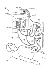
100621 In a conventional manner, a patient 10 in Figure 1 is attached to
the dialysis
treatment system 12 via a blood extraction needle 16 and blood injection
needle 26. During a
dialysis treatment with the dialysis treatment system 12, blood is extracted
from the patient
via blood extraction needle 16, passed through the blood pump 20, the blood
chamber 32
and dialyzer blood filter 22 using tubes 18, and then returned hack to the
patient 10 via tube
24 and blood injection needle 26, The dialyzer 22 filters the blood by fluid
exchange with
dialysis solution from fresh dialysis tube 28 and deposits filtered waste out
to used dialysis
tube 30.
[0063l A blood monitoring system 14 incorporating the invention is used
with a dialysis
treatment system 12 for monitoring certain blood characteristics relevant to
the dialysis
process. The blood monitoring system 14 includes a display 36, a cable 37 and
a clip
assembly 34 that mates to a blood chamber 32 in the blood flow path provided
by the tubes
18. The clip assembly 34 includes light sources and detectors that are
positioned on opposite
sides of the blood chamber 32 when the clip assembly is mated to the blood
chamber. Light
passing through the blood chamber from the light sources in the clip assembly
34 is absorbed
by the blood undergoing dialysis. Detectors in the clip assembly 34 detect the
absorption and
circuitry in either the clip assembly or the display 36 process absorption
signals from the
detectors to provide information at the display meaningfill to the clinician
responsible for the
dialysis process.
[00641 Fig. 2 illustrates an embodiment of the display 36 of the blood
monitoring system
14. The illustrated embodiment of the display 36 includes a screen 100 for
displaying
information such as, for example, a plot 112 of change of blood volume (BV)
versus time,
current elapsed time 102 of a dialysis session (assuming the system is in
place with a dialysis
system), current hematocrit (FICT) measurement 104, current oxygen saturation
(SAT)
measurement 106, current estimated hemoglobin (HOE) level 108, current BVA
measurement
110, and the like measurements useful to the clinician during the dialysis
process. A user
12
may operate the blood monitoring system 14 via the display 36, for example, to
change the
types of information displayed by the display, or the manner of the display
(plots or
alphanumeric text).
[0965] The illustrated display 36 includes various control buttons for
control of the blood
monitoring system 14. Alternatively or in addition, the screen 100 may be a
touch screen and
control of the blood monitoring system 14 can be accomplished using the touch
screen 100 as
a control interface. in other embodiments not illustrated, the blood
monitoring system 14 is
controlled or monitored using remote and/or other non-contact interface
mechanisms. See,
for example US 2014/0267003 Al to Wane et al., entitled "Wireless Controller
to Navigate
and Activate Screens on a Medical Device," US 2014/0266983 Al to Christensen,
entitled
"Wearable Interface for Remote Monitoring and Control of a Medical Device,"
and US
2015/0253860 Al to Merics et al. entitled "F,-field Sensing of Non-contact
Gesture Input for
Controlling a Medical Device."
[00661 Fig. 3 schematically illustrates the blood monitoring system 14. The
system 14
includes a controller 310 that may be located either within the clip assembly
34 or the display
36. The system 14 also includes at the clip assembly 34 an LED emitter 340, a
sensor 330 for
sensing light from the LED after it passes through the blood chamber 32, a
reference photo
sensor 350 for directly sensing light from the LED and a mask 370 for
shielding the photo
sensor from light other than from the LED 340. The LED emitter 340 emits light
at a
plurality of wavelengths from a first side of the blood chamber 32 and through
a blood flow
path 900 provided by the chamber. The light exits the blood chamber 32 at a
second side of
the chamber after traveling a fixed distance 380 of "d" between the LED 340
and the sensor
330. The distance "d" can be arbitrary to the system design. But once the
distance is
selected, it remains constant. The LED 340 and sensor 330 are each supported
on opposing
arms of the clip assembly 34 at the area of the clip assembly that mates with
the blood
chamber 32, The reference photo sensor 350 is also supported by the clip
assembly 34 and is
specifically supported on the same side as the LED 340, The mask 370 is also
supported by
the clip assembly 34 and on the same side as the LED 340 and the reference
photo sensor
350. The mask may be realized in several alternative embodiments, including
those
illustrated herein. In the various embodiments, the mask 370 shields the
reference photo
sensor 350 from receiving light other than directly from the LED emitter 340 -
e.g,, no light
externally reflected light or light from other sources.
Date recue / Date received 2021-11-02
CA 02988658 2017-12-06
WO 2016/210282
PCT/13S2016/039283
13
[0067] The controller 310 synchronizes and controls the monitoring system
14 as a
whole. Measurements of the light reaching the sensor 330 are processed by
signal processing
hardware and fed to the controller 310. Similarly, supporting signal
processing hardware
feeds compensation measurements from the reference photo sensor 350 to the
controller 310.
The controller 310 than normalizes the "raw" measurement from the sensor 330
using the
measurement received from the photo sensor 350. The reference photo sensor 350
and the
sensor 330 may each be a Silicon or a Indium Gallium Arsenide photodiode, or
each may be
an array of Silicon or Indium Gallium Arsenide photodiodes.
[00681 In the illustrated embodiments, the emitter 340 includes a light-
emitting-diode
(LED) or an array of LEDs. The emitter 340 may include other light sources,
such as
LASER emitters, fluorescent light sources, incandescent light sources and the
like.
[00691 The blood flow chamber 32 can be made of polycarbonate. The purpose
of the
blood chamber is to provide a window into the blood flow during a process
(e.g., dialysis) to
be monitored and to maintain the spacing "d" 380 as a constant during the
measurement
process involved in the monitoring.
[0070] in one embodiment as illustrated in Fig. 3, a dome 360 is covering
the LED
emitter 340. One part of the dome 360 contains the mask 370 surrounding the
reference
photo sensor 350 in all directions other than in the direction of the LED 340.
The dome 360
may be of various shapes, such as rectangular and semi-spherical. The dome 360
may also be
of various materials, such as epoxy, plastic, glass or plexi-glass, or other
inorganic materials
that are reproducible with respect to their optical properties. The
transparent dome 360
provides some protection for the LED emitter 340, such as against dust
contamination,
thermal stress, electrical shorts, and mechanical damages from moving parts
while providing
a path for light from the LED to illuminate the first side of the blood
chamber 32, The dome
360 provides the same protections for the reference photo detector 350 under
the masked 370
portion of the dome.
10071] In the embodiment illustrated in Fig. 3, the mask 370 covers a
portion of the
transparent dome 360. The mask 370 may be a portion of the transparent dome
360 that is
coated with a dense, light stopping material, on the outer surface of the dome
or on its inner
surface if the dome is hollow. The mask 370 may be an opaque coating of the
dome 360 that
blocks reflected light, both visible and infrared, ensuring that the only
light visible to the
reference photo sensor 350 is light emitted from the LED emitter 340.
14
[0072] Alternatively, the mask 370 may stand alone without the transparent
dome 360 or
separated from the transparent dome 360. The precise mechanical structure of
the mask can
have these and other variations as long as the mask functions to isolate the
reference photo
sensor 350 from light originating from sources other than the LED emitter 340.
[0073j In the illustrated embodiment of Fig. 3, the light amplitude
intensity for the LED
emitter 340 is controlled by a LED current source 305 with the intensity set
by the current set
resistor 315. The controller 310 is able to further adjust the LED current
source 305 to
compensate the light intensity using transmitter control 308 based on the
reference signal 356
that is developed from the signal provided by the reference photo sensor 350.
100741 Light passes from the LED emitter 340 through the unmasked portion
of the dome
360 in Fig. 3 and the blood chamber 32 body to the photo sensor 330. The blood
flow path
900 across the blood chamber has the fixed distance "d" 380 to ensure proper
calibration.
Blood parameters absorb and scatter the light, which attenuates the amplitude
of the light at
different wavelengths arriving at the photo sensor 330. The amount of
amplitude attenuation
at predetermined wavelengths is used to determine blood properties such as
hematocrit. The
determination process is more completely described in U.S. Patent Nos.
5,372,136 and
6,246,894.
[00751 In response to the light reaching it after passing through the blood
in the blood
chamber 32, the photo sensor 330 generates in a conventional manner a current
signal
proportional to the intensity of the light it receives and sends the current
signal to signal
processing circuitry to be processed for use by the controller 310. For
example, in the
illustrated embodiment in Fig. 3, a trans-impedance amplifier 331 receives the
current signal
and amplifies it as necessary and converts the signal to a voltage signal. The
voltage signal is
then applied to a sensor receiver 332 where it is filtered and conditioned for
passing on to an
analog-to-digital (A to D) voltmeter 333. This voltmeter 333 converts the
measured voltage
proportional to the light received at the photo sensor 330 to a final digital
sensor signal 336
formatted to be an input to the controller 310.
[00761 Similarly, light from the LED emitter 340 that reaches the reference
photo sensor
350 under the mask area 370 of the dome 360 causes the reference photo sensor
to react by
generating a current signal, which is processed by signal processing circuitry
in a manner
similar to the current signal from the photo sensor 330. All material in the
optical path from
the LED emitter 340 to the reference photo sensor 350 have unchanging optical
properties
Date recue / Date received 2021-11-02
CA 02988658 2017-12-06
WO 2016/210282
PCMJS2016/039283
such that the signal received at the reference photo sensor 350 varies solely
with changes in
the emission characteristics of the LED emitter. The mask 370 prevents
reflections from
outside the dome 360 and light sourcing from other than the LED emitter from
summing into
the direct signal between the reference photo sensor 350 and the LED emitter
340.
[0077] in the embodiment illustrated in Fig. 3, the light from the LED
emitter 340
received at the reference photo sensor 350 is converted in a conventional
manner to a
proportional current signal by the reference photo sensor 350. This current
signal is applied
to a trans-impedance amplifier 351 where it is amplified as necessary and
converted to a
voltage output signal. The voltage signal is then applied to a reference
receiver 352 where it
is filtered and conditioned as a voltage measurement by an analog-to-digital
(A to D)
voltmeter 353. The voltmeter 353 converts the measured voltage proportional to
the light
received to a digital reference signal 356, which is read by the controller
310.
[0078] The controller 310 compensates for the measurements from the sensor
330 at the
sensor signal 336 that source from changes in the intensity of the light at
the LED emitter
340, using the measurements provided by the reference signal 356 from the
reference photo
sensor 350. The compensation accounts for variations in the light emitted from
the LED
emitter 340 and is continuous and substantially in real time.
[00791 The controller 310 in the embodiment illustrated in Fig. 3 also has
the option to
adjust the amplitude of the light at the LED emitter 340 by adjusting the LED
current source
305 to provide more power to the LED emitter. A transmitter control 308 signal
from the
controller 310 to the LED current source 305 accomplishes this task.
10080] The LED emitter 340 may experience short term or long term
variations in the
amplitude of its emitted light for various reasons. For example, there may be
power
fluctuations in the LED emitter 340, which causes the light intensity from the
LED emitter to
change according to the power fluctuations. Or light from the LED emitter 340
may
gradually intensify or fade in intensity due to degradation of the LED
emitter. The system in
the illustrated embodiment of Fig. 3 continuously compensates for these
variations during
operation by providing the controller 310 with the ability to compensate for
changes in
measurements from the reference photo sensor 350, which thereby results in the
system to
maximize its accurate measurements of the attenuation of the light at the
photo sensor 330
caused only by the properties of the blood in the blood flow path 900.
[0081] The schematic illustration of an embodiment of the blood monitoring
system 14 in
Fig. 3 includes a memory 320 for storing calibration parameters and executable
instruction
CA 02988658 2017-12-06
WO 2016/210282
PCMJS2016/039283
16
(such as instructions programmed to perform the steps shown in Fig. 4) used by
the
controller 3.10. The calibration parameters and executable instructions
compensate for the
attenuation of the light between the LED emitter 340 and the photo sensor 330
across the
fixed distance "d" when there is no blood flood in the path 900.
[00821 In the embodiment of Fig. 3, the memory 320 stores a log of the
calibration
parameters used by the controller 310. The log may be used for system
diagnostic purposes.
For example, the memory 320 may keep a running log of compensation parameter
values
needed to normalize the sensor signal 336. If the log evidences the
compensation required to
normalize the sensor signal 336 is gradually increasing while the reference
signal 356 from
the reference photo sensor 350 is gradually decreasing over time, then the
logged data
suggests the LED emitter 340 is failing or burning out and needs to be
replaced. In the
illustrated embodiment of Fig. 3, the controller 310 is programmed to detect
diagnostic
events based upon the log of the calibration parameters used by the controller
310, and to
alert operators.
[0083] In the embodiment of Fig. 3, the controller 310 performs a
calibration to generate
a new set of calibration parameters for each new blood channel used.
[0084] The controller 310 may include various components, such as a
processor, non-
transitory computer readable medium for storing computer code/programs to
perform
measurement method and/or calibration methods provided throughout in this
disclosure, as
well as user interface devices, such as keyboard, mouse, touchpad, displays,
speakers and the
like. For example, in the embodiment illustrated in Fig, 3, program memory 320
is a non-
transitory computer readable medium. Serial interface 311 in Fig. 3 is an
example of a
communications interface for the controller 310. It passes the blood data
developed by the
monitoring system to the outside world for display and further analysis. Such
as data port
can be any of a variety of known formats and interfaces, including R.S-232,
Universal Serial
Bus (UBS) and the like.
[0085] In the embodiment illustrated in Figure 1, the blood data is
delivered to the
display 36 via cable 37, where the data is used to generate a graphical
display of information
useful to the clinician such as hematocrit values as suggested in Fig. 2. An
example of a
suitable display is, for example, the display of the Crit-Line Monitor Ill by
Fresenius Medical
Care, North America, Waltham, MA.
[0086] As an alternative or in addition to the cable 37 in Figure 1 for
communicating
data, the controller 310 may be coupled to a communication module that enables
the
CA 02988658 2017-12-06
WO 2016/210282
PCMJS2016/039283
17
transmitting and/or receiving of data and/or other information that may be
controlled or used
by the controller 310 and/or stored on the memory 320. In an embodiment, the
communication module 318 includes a wireless transceiver that wirelessly
transm its or
receives the data or other information. In an example, the wireless
transceiver enables
wireless communication between the blood monitoring system 14 and the dialysis
treatment
system 12, or component thereof; performing the dialysis treatment and/or
other peripheral
devices that record or otherwise use data or other information concerning the
dialysis
treatment.
[00871 In an embodiment, the communication module 318 includes components
for
short-range wireless communications between the blood monitoring system 14 and
the
dialysis treatment system 12 via known short-range wireless technology
protocol such as, for
example, a Bluctooth protocol or an REID protocol - - e.g., a near field
communication
(NFC) protocol. in other embodiments, wireless communication to and from the
blood
monitoring system 12 may be facilitated using other wireless technologies,
such as via WIFi
and/or via an implementation utilizing telecommunication networks.
[00881 In connection with the transmission, either via cable 37 or via
wireless
transmission, the data may be secured and/or encrypted via the controller 310
using
appropriate security and encryption protocols according to applicable laws and
regulations
governing transmission of sensitive data and/or protected medical information.
[00891 The blood monitoring system 14 eliminates the need for temperature-
based
measurements to calibrate or normalize the sensor signal 336. By directly
measuring a
portion of light emitted by the LED emitter 340 for use in compensating for
changes in the
light caused by effects such as temperature changes, the system does not need
to wait long for
the LED emitter 340 temperatures to stabilize before performing measurements.
[00901 Additionally, normalizing the sensor signal 336 using direct
measurement of the
emitted light keeps the controller 310 in proper calibration for a much longer
time, making
the life cycle of the system 14 longer. This approach also allows the use of
lower cost LEDs
(e.g., LEDs having higher variations in light intensity than would othenvise
be possible) for
LED emitter 340, allowing for reduced development time of many additional
possible
wavelengths for measuring additional blood characteristics.
[00911 The LED emitter 340 may be an array of diodes such that the emitted
light
comprises a plurality of wavelengths that enters the blood chamber 32 from a
first side,
passes through the blood flow channel 900 and exits the blood chamber from a
second side.
CA 02988658 2017-12-06
WO 2016/210282
PCMJS2016/039283
18
The sensor 330 on the second side of the blood chamber 32 receives the light
from the LED
emitter 340 after the amplitude of its plurality of wavelength has been
affected by passing
through the blood flow channel 900. The reference photo sensor 350 directly
measures the
light from the array comprising the LED emitter 340. The mask 370 ensures that
only light
from the LED emitter 340 arrives at the reference photo sensor 350. The
controller 310
controls the measurement hardware and compensates measurements from the sensor
330
based upon measurements from the reference photo sensor 350, for example by
measuring a
ratio between readings from the reference photo sensor 350 and the sensor 330
prior to blood
entering the blood chamber 32, and applying the ratio to readings from sensor
330 during
dialysis while blood is in the channel 900.
[00921 Notably, the intensity of emitted light is inversely proportional to
the square of the
distance it travels. Thus, the distance "d" 380 between the LED emitter 340
and the sensor
330 must remain constant so that any change in intensity of sensed light
during the
calibration process and during actual usage is dependent entirely on the
medium between the
sensor 330 and LED emitter 340 and not characteristics of light propagation.
The distance
"d" is selected to be the distance separating the LED emitter 340 and the
sensor 330 when the
blood chamber 32 is inserted into the jaw of the clip assembly 34, which
include opposing
arms housing the LED emitter 340 and the sensor 330. The arms of the clip
assembly 34 flex
so that they can function as a jaw or clamp fitted over the blood chamber 32
at an area of the
blood chamber that serves as a window into the blood flow channel 900. Because
the arms
flex, the distance between the LED emitter 340 and the sensor 330 is variable
unless it is
fixed such as, tbr example, by positioning the blood chamber 32 in the jaw
formed by the
arms of the clip assembly 34.
[00931 Referring now to calibrating the monitoring system 14, Fig. 4
illustrates an
embodiment of a calibration method 400. The method 400 begins at block 410 by
obtaining
amplitude measurements at the reference photo sensor 350 of the light from the
LED emitter
340 and sending the resulting reference signal 356 (Fig. 3) to the controller
310.
At block 420 of Fig. 4, the sensor 330 obtains measurements of light from the
LED emitter
340 with the blood chamber 32 removed while holding the distance "d" 380
between the
sensor and the LED emitter. The sensor 330 provides a sensor signal 336.
[0094] At block 430, the controller 310 determines a calibration ratio
between each
processed signal derived from reference signal 356 and the sensor signal 336
while nothing is
between the sensor 330 and the LED emitter 340 held at the distance "d" 380.
CA 02988658 2017-12-06
WO 2016/210282
PCT/1JS2016/039283
19
[00951 At block 440, the photo sensor 330 obtains a light measurement
from LED emitter
340, with the blood chamber 32 in the measurement path but with the blood flow
channel 900
being empty (only air present),
[00961 At block 450, a controller 310 determines a calibration constant
between each
received and processed reference signal 356 and each sensor signal 336 with
the blood
chamber 32 in the light path but with nothing in the blood flow path 900
except air.
[00971 At block 460, the controller 310 determines a composite
ratiometric Calibration
Coefficient for each wavelength from the measurements at Hocks 430 and 450.
These
composite Calibration Coefficients are used to normalize the measurements of
light across
the blood flow 900 in the blood chamber 32 by illuminating the blood with LED
emitters 340
and receiving the modified amplitude of the light at the photo sensor 330
through the
absorption and scattering of the blood. At the same time, variations in the
amplitudes of the
LED emitters 340 themselves are measured by the reference photo sensors 350 to
complete
the normalization.
[00981 The modeling of calibration and compensation functions for each
wavelength is
illustrated as follows:
[00991 Light measured by the reference photo sensor 350 may be a
function according to
Beer's Law:
e-"'-dEr
.= where,
tr is measurement of light intensity at the reference photo sensor 350,
Io is the actual intensity of light radiated by the LED emitter 340,
tiEr is light loss coefficient from the LED emitter 340 to the reference photo
sensor due to the material of the dome 360, and
dEr is the distance light travels from LED emitter 340 to the reference photo
sensor 350 in dome 360.
¨ dE
e 'a, r r may be considered a constant Kr.
Which simplifies to:
loKr (2)
[001001 Beer's Law equation may be similarly applied for light measured by the
photo
sensor 330 with more loss components:
CA 02988658 2017-12-06
WO 2016/210282
PCMJS2016/039283
= Aro e ¨a gmdEm e-amictm,, e-an,.2a,n2 e-apidpi e---abdb e-ap2dp2 (3)
where,
im is measurement of light intensity at the photo sensor 330,
aEm is light loss coefficient from the LED emitter 340 to the epoxy-air
boundary
of the dome. 360 along a ray toward sensor 330 within the dome due to the
material of the
dome 360 (in the example, the same as
(113m is the ray distance light travels from LED emitter 340 to exit the
transparent
dome 360,
ami is the light loss coefficient from the exit (surface) of the dome 360 to
the side
wall of the blood chamber 32 on the first (illumination) side due to the
medium material light
properties through which the light travels,
dml is the distance light travels from the surface of the dome 360 to the
blood
chamber 32 on the first (illumination) side,
ani, is light loss coefficient from the second (receiving) side wall of blood
chamber 32 toward the photo sensor 330 due to the medium material light
properties through
which the light travels,
dm2 is the distance light travels from second side (receiving) wall of the
blood
chamber 32 to the photo sensor 330,
a is light loss coefficient of first side (illumination) thickness
of the blood
chamber 32 outside wall to the blood flow channel 900 based on the light
propagation
properties of the blood chamber 32 material,
is the distance light travels in the first side (illumination) from the
outside
side wall of the blood chamber 32 to the blood flow channel 900,
ab is light loss coefficient of the blood in the blood flow channel 900,
db is the distance light travels through the blood in the blood flow channel
900
(which is the inside channel thickness of the blood flow channel 900),
aP2 is light loss coefficient of second side (receiving) thickness of the
blood
chamber 32 from the blood flow channel 900 to the outside wall based on the
light
propagation properties of the blood chamber 32 material, and
CA 02988658 2017-12-06
WO 2016/210282
PCMJS2016/039283
21
dp2 is the distance light travels in the second side (receiving) from the
blood flow
channel 900 to the outside side wall of the blood chamber 32.
[001011 Equation (3) can be simplified to:
tm = Km e -ap e ¨abdb e-cip,dpz
(4)
[001021 Combining equations (2) and (4):
ra e---abdb e-ap2dp2
(5)
/0Kr
[001031 Canceling 10 from equation (5) yields:
.i4ke001.40 3. I.!
(6)
Kr
[00104] Without the presence of blood and the blood chamber in the flow
channel 900, the
ratio becomes:
tin = K7T1
(7)
ir Kr
[001051 During calibration, the Composite Calibration light propagation
constant for each
wavelength Sc for Kmac. may be derived by taking calibration measurements of
the
reference photo sensor 350 and the sensor 330 (obtaining without the
presence of
blood and the blood chamber in the flow channel and holding constant the
distance "d" (380)
between the LED 340 and the photo sensor 330.
[001061 Plugging in Se=7-1(õ1"<õ into equation (6), the function for photo
sensor 330
measurements becomes:
(scir)e-apidp1e-andbe-a12ap2
(8)
where e-a'PidPie-aP2dP2 is also constant.
[00107] Assigning constant Kp e-avidp, e-ap2dpz, Kp
may be derived by taking
calibration measurements of the reference photo sensor 350 and the photo
sensor 330, with
the blood flow channel 900 of the blood chamber 32 being empty and present in
the optical
path between LED emitter 340 and sensor 330.
[001081 During calibration, Kp can be derived for each new blood chamber 32
with the
blood flow channel 900 being empty. Assuming tight controls are possible in
the molding, of
the blood chamber 32, Kp can be assumed to be constant across different blood
chambers
unless there is a change in the molding properties of the blood chamber. This
is another
CA 02988658 2017-12-06
WO 2016/210282
PCT/1JS2016/039283
22
feature of this embodiment in that changes in the blood chamber 32 can be made
and the
blood monitoring systems 14 in the field can compensate for any change in
calibration rather
than having to return the systems to the factory for completing calibration
adjustments,
[00109] Thus, equation (8) can be simplified to:
= (SC )Kp e¨abd (9)
and
K (10)
P (3c r)
when ab equals zero (no blood equals blood chamber empty) and db is the normal
light path
length through an empty blood chamber which is in the sensor.
001.101 Additionally, erclbdb is a function dependent upon blood
characteristics, and
may be profiled independently ahead of time and stored in controller 310 for
use, for
example, by using a standard set of blood samples for calibration in labs, and
pre-
programming the profile function of e¨abdb into controller 310, as algorithms
or a set of
lookup tables. A set of these calibrations is unique and required -for each
active wavelength.
[00111] As db is also assumed to be constant and could be measured and/or
inputted into
controller 310, the controller 310 can solve for ab:
=
õ
ab = ........................................................... (n)
a!,
[001121 Equation (11) can be used to derive ab ibr blood of various blood
characteristics
at various concentrations and different light wavelengths. For example,
polynomial fitting
may be used to derive HCT value, using the following:
tiCT = A(.21")2 B (4441 C (12)
where,
a800 is ab derived from measurements taken at a wavelength of 800 rim emitted
from LED emitter 340,
[00113] a1300 is ab derived - from measurements taken at a wavelength of
1300 rim
emitted from LED emitter 340.
[00114] Standard samples of known i-ICT levels are measured in Human blood and
are
used to derive the FICT calibration polynomial coefficients A., B, and C
through regression
CA 02988658 2017-12-06
WO 2016/210282
PCMJS2016/039283
-73
techniques, These coefficients A, B, and C are then programmed into the
controller 310
algorithm for ongoing I-ICT calculations.
[00115] During operation, the controller 310 may take measurements to derive
asoo and
a1300 for a specific blood sample of a specific patient, and solve for the HCT
results,
[001161 Thus, according to the embodiments above, the differential measurement
system
based upon direct LED emitter 340 light monitoring and the resulting
normalization of photo
sensor 330 readings can provide accurate blood characteristic measurements
with simple
calibration.
[00117] The identical system can he used with the ratio of similarly
derived light loss
coefficients for an approximately 660rim wavelength and an approximately
800rim
wavelength to create the model and algorithms for measurement of oxygen
saturation of the
blood.
[00H8] Turning to Fig. 5, an enlarged and isolated view is shown of the clip
assembly 34
mated to the blood chamber 32 as shown more generally in Figure 1. The clip
assembly 34
may incorporate components of the blood monitoring system 14 as discussed
hereinafter. In
accordance with one or more embodiments, the LED emitter 340 (e.g., an array
of LEDs) is
located on a circuit board within one side 530 of the clip assembly 34, while
the photo sensor
330 is located on a circuit board within the opposing side 540 of the
assembly. The reference
photo sensor 350 is co-located with the LED emitter 340 on side 530. When the
clip
assembly 34 is attached to the blood chamber 32, light emitted from the first
side 530 by the
LED emitter 340 passes through the blood flow path 900 of the blood chamber 32
and is
detected by the photo sensor 330 on the opposing side 540 of the clip
assembly. Various
physical properties of blood flowing through the chamber 520 in the blood flow
path affect
the intensity of the light received at the photo sensor 330 on the second side
540,
[0011.91 Fig. 6 illustrates the clip assembly 34 and mating blood chamber
32 in Fig. 5,
showing the interior of the side 540 of the clip in accordance with an
embodiment. The
circuit boards 535 and 541 are housed in side 540 of the assembly 34, Circuit
board 541
supports the photo sensor 330 and circuit board 535 supports substantially all
of the circuitry
illustrated in Fig. 3.
[001201 A circuit board 537 is housed in side 530 of the clip assembly 34 as
best
illustrated in the cross sectional view of Fig. 7, The circuit boards 536 and
537 support the
light transmitter portion of the monitoring system illustrated in Fig. 3, In
particular, the
24
circuit board 536 supports the light emitters 340 (e.g., LEDs) and the
reference sensor 350 in
an area within the side 530 of the clip assembly 34 that positions the
emitters and the photo
sensor 330 on opposite sides of the blood chamber 32 as schematically
illustrated in Fig. 3.
A ribbon cable 538 connects the circuit board 537 to the circuitry supported
on the circuit
board 535 housed in side 540.
[001.21.] The cross section of the mated clip assembly 34 and blood chamber 32
illustrated
in Fig, 7 provides additional information about the spatial relationships
among the light
emitters 340, the photo sensor 330 of the clip assembly 34 and the blood flow
path 900 of the
blood chamber 32. One side 530 of the clip assembly 34 mates to one side of
the blood
chamber 32, while the second side 540 of the clip assembly mates to the other
side of the
blood chamber. The first side 530 of the clip assembly 34 includes the circuit
boards 537 and
536 with mounted light emitters 340 (e.g., LEDs), while the second side 540
contains the
photo sensor 330 for detecting light passing through the blood flow path 900
of the blood
chamber 32.
[00122] Fig. 8 is an enlarged view of the area in Fig. 7 that includes the
interface between
the sides 530 and 540 of the clip assembly 34 and the blood chamber 32. Each
of the sides is
an arm of the clip assembly 34. On the first side 530 (or arm 530) of the clip
assembly 34,
the circuit board 536 supports both the LED emitters 340 and the reference
photo sensor 350
under a light diffusing window 542. Similarly, a light diffusing window 539 in
the side 540
allows light from the emitters 340 that passes through the blood chamber 34 to
be received by
the photo sensor 330 mounted on a circuit board 541 within the side 540. A
ribbon cable 543
best seen in Fig. 7 connects the circuit board 541 to the board 535. Further
detail of the clip
assembly 34 and the blood chamber 32 can be found at US Patent No. 8,743,354,.
Specifically, the
embodiment of the clip assembly 34 and the blood chamber 32 illustrated herein
is shown in
further detail in Figures 25A through 29E of the '354 patent.
100123] The partially transparent epoxy dome 360 covers the emitter 340 and
reference
sensor 350. A portion of dome 360 is used as the mask 370, which shields the
reference
sensor 350 from any externally reflected light or other light other than
direct light from the
LED emitter 340. The reference photo sensor 350 may be each be a Silicon or an
Indium
Gallium Arsenide photodiode, or each an array of Silicon or Indium Gallium
Arsenide
photodiodes, such as those manufactured by Hamamatsu Fhotonics K..K,,
Hamamatsu City,
Japan.
Date recue /Date received 2021-11-02
CA 02988658 2017-12-06
WO 2016/210282
PCT/1JS2016/039283
[001241 Light passes from the LED emitter 340 through the unmasked portion of
the dome
360 to the blood chamber 32 and the blood flow path 900 inside the chamber to
the photo
sensor 330 located on the second side (receiving side or arm 540) of the clip
assembly 34.
Blood in path 900 and its parameters absorb and scatter the light, thereby
modifying the
amplitudes of light at different wavelengths arriving at the photo sensor 330.
[001.251 in still further detail, an enlarged and isolated view of the dome
360 is shown in
Fig. 9. The epoxy dome 360 is placed on the circuit board 536, covering the
light emitter 340
and the reference photo sensor 350. The portion of the dome 360 over the
reference photo
sensor 350 is coated with an opaque surface 370 so that any reflected light
from outside dome
360 that otherwise might reach the reference photo sensor 350 is blocked, In
this
embodiment, the reference photo sensor 350 is placed on the circuit board 536
in a
conventional manner such that its direction of primary detection is
approximately
perpendicular to the light emitter 340. However, sufficient light from the
light emitter 340
reaches the reference photo sensor 350 to effectively detect light from the
emitter when the
detector is protected from ambient or external reflected light by the mask
370. Alternatively,
and as illustrated in Figs. 11 and 12 discussed hereinafter, a reference photo
sensor (1140 and
1230, respectively) can be mounted to the circuit board 536 to face the light
emitter 340,
which then increases the sensitivity and noise immunity of the sensor.
However, the edge
mounting of the sensor is typically a more expensive mounting technique.
[001261 Additional embodiments are described with reference to Figs. 10 13.
1001271 Referring to Figs. 10a and lob, an array of LEDs 1010 are mounted to a
circuit
board 1020, which is located approximately where the circuit board 536 is
located in Figs. 6-
9, The LEDs 1010 need not be of any particular brightness or quality standard.
in this
embodiment, a shield 1030 is spaced at a fixed distance from the circuit board
1020 through
the use of spacers 1040, The shield 1030 is made of material such that it
blocks all reflected
light from the LEDs 1010, which light passes through an opening 1050 in the
shield that
allows the light to pass to the blood chamber. A reference photo sensor 1060
is mounted on
the underside of the shield 1030. Not shown in Fig. 10 is the blood chamber 32
or the photo
sensor 330 in the arm of the clip assembly 34 opposite the arm housing the
LEDs 1010. The
calibration and measurement work in a similar manner as described above in
reference to the
embodiment of Figs. 6-9, using the difference in light intensity between the
reference photo
sensor 1060 and the photo sensor 330 to determine levels of hematocrit, oxygen
saturation,
and/or other blood constituents.
CA 02988658 2017-12-06
WO 2016/210282
PCMJS2016/039283
26
100128] In another alternative embodiment, not shown, the reference photo
sensor is
placed directly next to the LEDs on the circuit board, or sufficiently close
to the LEDs that
the intensity of the direct light from the LEDs themselves is much greater
than any optical
noise from reflections and/or ambient light. Using such an embodiment
increases the
sensitivity of the reference photo sensor and may reduce or render
insignificant the optical
noise such that the mask is unnecessary.
[00129] The embodiment in Fig. 11 uses an epoxy dome 1110 like the embodiment
of
Figs. 6-9. The dome 1110 is placed on the circuit board 1120, which is
positioned in the clip
assembly 34 similarly to the position of the circuit board 536 in Figs. 6-9.
The dome 1110
covers the array of LEDs 1130 and a reference photo sensor 1140. The reference
photo
sensor 1.140 is preferably a photo diode placed on its edge, so that the
sensor more directly
faces the light emitted by the LEDs 1130 then it would otherwise if mounted
fiat on the
circuit hoard 1120. The portion of the dome 1110 over the reference photo
sensor 1140 is
coated with an opaque material, so that any external reflected light that
otherwise might reach
the reference photo sensor 1140 (both visible and infrared) is blocked. Not
shown in Fig. 11,
the blood chamber 32 runs parallel to the circuit board 1120, such that light
(upwardly
pointing arrow) from the LEDs 1130 passes through the non-opaque portion of
the dome
1110, through the blood chamber, and is detected by the photo sensor (not
shown) in the
opposing arm of the clip assembly 34.
[00130] in accordance with another embodiment, a solid enclosure 1210 in
Fig. 12 is
mounted on a circuit board 1220 positioned in the arm 530 of the clip assembly
34 similarly
to the position of the circuit board 535 in Figs. 6-9, Like the embodiment
illustrated in Fig,
11, the reference photo sensor 1230 is placed on its edge on the circuit board
1220. The solid
enaosure 1210 surrounds the reference photo sensor 1230 on all sides, with the
exception of
an opening facing the array of LEDs 1240. In this embodiment, the solid
enclosure 1210 may
be made of metal, or other material that is impervious to all light. Not shown
in Fig, 12, the
blood chamber 34 is oriented to be parallel to the circuit board 1220, such
that light from the
LEDs 1240 passes through the blood chamber for measurement by the photo sensor
on an
opposing side.
[001311 Although the embodiments of Figs, 11 and 12 are illustrated as
including masks or
shrouds intended to optically isolate the reference photo sensors, the
increased sensitivity
achieved by edge mounting the sensors to the circuit boards may increase the
signal to noise
immunity of the sensor such that the masks or shrouds are not required.
27
[001321 [Blank]
100133] The use of the terms "a" and "an" and "the" and "at least one" and
similar
referents in the context of describing the invention (especially in the
context of the fiallowing
claims) are to be construed to cover both the singular and the plural, unless
otherwise
indicated herein or clearly contradicted by context. The use of the term "at
least one"
followed by a list of one or more items (for example, "at least one of A and
B") is to be
construed to mean one item selected from the listed items (A or B) or any
combination of two
or more of the listed items (A and B), unless otherwise indicated herein or
clearly
contradicted by context. The terms "comprising," "having," "including," and
"containing"
are to be construed as open-ended terms (i.e., meaning "including, but not
limited to,") unless
otherwise noted. Recitation of ranges of values herein are merely intended to
serve as a
shorthand method of referring individually to each separate value falling
within the range,
unless otherwise indicated herein, and each separate value is incorporated
into the
specification as if it were individually recited herein. All methods described
herein can be
performed in any suitable order unless otherwise indicated herein or otherwise
clearly
contradicted by context. The use of any and all examples, or exemplary
language (e.g., "such
as") provided herein, is intended merely to better illuminate the invention
and does not pose a
limitation on the scope of the invention unless otherwise claimed. No language
in the
specification should be construed as indicating any non-elaimed element as
essential to the
practice of the invention.
[001.341 Preferred embodiments of this invention are described herein,
including the best
mode known to the inventors for carrying out the invention. Variations of
those preferred
embodiments may become apparent to those of ordinary skill in the art upon
reading the
foregoing description. The inventors expect skilled artisans to employ such
variations as
appropriate, and the inventors intend for the invention to be practiced
otherwise than as
specifically described herein. Accordingly, this invention includes all
modifications and
equivalents of the subject matter recited in the claims appended hereto a.s
permitted by
applicable law. Moreover, any combination of the above-described elements in
all possible
variations thereof is encompassed by the invention unless otherwise indicated
herein or
otherwise clearly contradicted by context.
Date recue / Date received 2021-11-02