Note: Descriptions are shown in the official language in which they were submitted.
CA 02988715 2017-12-07
[DESCRIPTION]
[Invention Title]
An antibody or a fragment specifically binding to peptide derived from
vimentin
[Technical Field]
The present invention relates to an antibody specifically binding to the
peptide of SEQ
ID NO: 1, and specifically, to an antibody specifically binding to an isolated
peptide of SEQ ID
NO: 1 or a fragment binding to the peptide, a polynucleotide encoding the
antibody or the
fragment binding to the peptide, a vector containing the polynucleotide, a
cell introduced with
the vector, a method of producing the antibody or the fragment binding to the
peptide using the
cell, a recombinant antibody or a fragment binding to the peptide produced by
the method, an
antiviral composition containing the antibody or the fragment binding to the
peptide, a
composition for preventing or treating inflammatory diseases containing the
antibody or the
fragment binding to the peptide, and a method of treating infectious viral
diseases or
inflammatory diseases using the composition.
[Background Art]
Unlike bacterial diseases, viral diseases are difficult to treat because
antibiotics are not
particularly effective against viruses, and viral diseases are consistently
emerging as one of the
main causes of diseases and deaths in humans. Additionally, infectious viral
diseases induce
inflammation, thereby causing various inflammatory diseases.
The therapeutic agents developed for the treatment of such infectious viral
diseases may
be categorized into chemical materials and bio-derived materials, and most of
the chemical
materials are developed to be effective only against specific viral diseases,
exhibiting various
adverse effects and disadvantages such as the frequent emergence of resistant
viruses.
Meanwhile, well-known examples of the bio-derived materials may include
cytokines
such as interferon (IFN). Of these, interferon was first discovered among the
cytokines
produced in virus-infected cells and is known as the cytokine which has the
superior antiviral
activity. It has been reported that interferon can be used for treating
various diseases such as
chronic hepatitis B or C, blood cancer, multiple sclerosis, etc. Recently, the
effect of IFN on
1
CA 02988715 2017-12-07
human immunodeficiency virus (HIV) patients has been reported. As such, in an
attempt to
develop antiviral agents and immune booster, for the past several years,
research on genetic
engineering or bioengineering methods has been ongoing for the mass production
of interferon,
and recently, research has been actively conducted to search for compounds
which can induce
interferon expression from natural or synthetic materials (Alearo S et al.,
Bioorg Med Chem.
2005, 13 (10), 3371 - 3378). As a result, a strong interferon inducer named
Imiquimod was
developed by 3M Pharmaceuticals, but its development has been discontinued due
to various
adverse effects observed during clinical trials.
Although interferon is known to be a strong antiviral agent, interferon has a
disadvantageous in that it cannot be used for more than 6 months, because it
induces
inflammatory responses such as infiltration of immune cells, etc.; almost all
cells express
receptors for interferon at all times, showing various adverse effects such as
an anti-cellular
effect; and it requires large amounts to treat at clinical trials.
Accordingly, there is a need for the development of a therapeutic agent which
can
selectively act on virus-infected cells rather than normal cells; which is
applicable in small
amounts and to various types of viruses rather than specific types of viruses;
and which
simultaneously has both antiviral and anti-inflammatory activity by
suppression of the
infiltration of immune cells.
[Disclosure]
[Technical Problem]
The present inventors have endeavored to develop a therapeutic agent which can
specifically act on virus-infected cells and have antiviral and anti-
inflammatory activity capable
of inhibiting inflammation by suppressing the infiltration of immune cells. As
a result, the
present inventors have confirmed that not only does humanized virus-
suppressing factor (hzVSF),
which is a novel humanized antibody, have the ability to specifically act on
various types of
viruses, as well as inhibitory activity against the infiltration of immune
cells and superior for
antiviral activity, but also, as a humanized antibody having reduced
immunogenicity, it is a safe
agent without any adverse effects when administered to humans, thereby
completing the present
invention.
[Technical Solution]
2
CA 02988715 2017-12-07
An object of the present invention is to provide a novel antibody specifically
binding to
the peptide of SEQ ID NO: 1 or a fragment binding to the peptide.
Another object of the present invention is to provide a polynucleotide
encoding the
antibody or the fragment binding to the peptide, a vector containing the
polynucleotide, a cell
introduced with the vector, a method of producing the antibody or the fragment
binding to the
peptide using the cell, and an antibody or a fragment binding to the peptide
produced by the
method.
Still another object of the present invention is to provide an antiviral
composition
containing the antibody or the fragment binding to the peptide.
Still another object of the present invention is to provide a method for
preventing or
treating infectious viral diseases using the antiviral composition.
Still another object of the present invention is to provide a composition for
preventing or
treating inflammatory diseases containing the antibody or the fragment binding
to the peptide.
Still another object of the present invention is to provide a method for
treating
inflammatory diseases using the composition for preventing or treating
inflammatory diseases.
[Advantageous Effects]
The antibody specifically binding to the peptide of SEQ ID NO: 1 or the
fragment
binding to the peptide can be provided as a new humanized antibody therapeutic
agent having
excellent antiviral and anti-inflammatory activity because they can
selectively act on
virus-infected cells, thus requiring only a small amount at the time of
treatment, without any
adverse effects, and can also inhibit inflammation by suppressing the
infiltration of immune
cells.
[Brief Descriptions of the Drawings]
FIG. 1 shows a schematic diagram of a vector for preparing chimeric virus
suppressing
factor (VSF).
FIG. 2 shows a schematic diagram of chimeric VSF.
FIG. 3 shows the results confirming the expression of chimeric VSF.
FIG. 4 shows the result illustrating the DNA sequence of single-chain Fv
(scFv) of VSF.
FIG. 5 shows schematic diagrams illustrating the cloning of scFv of VSF into a
vector.
FIG. 6 shows the results confirming scFv of VSF after purification.
3
CA 02988715 2017-12-07
FIG. 7 shows the results confirming the antiviral activity of VSF, scFv, and
anti-EMC-D
virus antibodies based on the amount of viable cells remaining after viral
infection.
FIG. 8 shows the result confirming the antiviral activity of chimeric VSF.
FIG. 9 shows a schematic diagram of a vector for preparing hzVSF, which is a
humanized antibody.
FIG. 10 shows a schematic diagram of hzVSF, which is a humanized antibody of
the
present invention.
FIG. 11 shows the results confirming the expression of hzVSF, which is a
humanized
antibody.
FIG. 12 shows the results confirming the antiviral activity of hzVSF, which is
a
humanized antibody.
FIG. 13 shows the results of reducing and non-reducing SDS-PAGE confirming the
physical properties of hzVSF_var13, which is a humanized antibody.
FIG. 14 shows the results of LC/MS confirming the physical properties of
hzVSF_var13,
which is a humanized antibody.
FIG. 15 shows the results of SEC-HPLC confirming the physical properties of
hzVSF_var13, which is a humanized antibody.
FIG. 16 shows the results illustrating isoelectric focusing (IEF) confirming
the physical
properties of hzVSF_var13, which is a humanized antibody.
FIG. 17 shows the results illustrating the number of donors with T-cell
proliferation in
response to KLH, hzVSF_var12, and hzVSF_var13 among 51 blood donors.
FIG. 18 shows the results illustrating the degree of T-cell proliferation of
the 51 donors
in response to hzVSF_var12 and hzVSF_varl 3, which are representative variants
of hzVSF, a
humanized antibody.
FIG. 19 shows the results illustrating the T-cell proliferation induced by
KLH,
hzVSF_varl 2, and hzVSF van 3 represented by a mean stimulation index (SI).
FIG. 20 shows the results of SDS-PAGE of hzVSF_var12 and hzVSF var13, which
are
representative variants of hzVSF, a humanized antibody.
FIG. 21 shows the results of HPLC confirming the physical properties of
hzVSF_var12
and hzVSF_var13, which are representative variants of hzVSF, a humanized
antibody.
FIG. 22 shows the results illustrating the pharmacodynamics of hzVSF_var13,
which is
a representative variant of hzVSF, a humanized antibody.
4
CA 02988715 2017-12-07
FIG. 23 shows the result confirming the antiviral activity of hzVSF, and
hzVSF_var12
and hzVSF_var13, which are representative variants of hzVSF, a humanized
antibody.
FIG. 24 shows the results confirming the cell viability of hzVSF_var13, which
is a
humanized antibody, and hIFN-a in human cells.
FIG. 25 shows the images confirming the inhibitory activity of hzVSF, which is
a
humanized antibody, against the infiltration of inflammatory cells in viral
diabetes.
FIG. 26 shows a result confirming the anti-HBV effect of mVSF.
FIG. 27 shows the images confirming the expression feature of VSF receptors in
HBV-
infected human liver tissue.
FIG. 28 shows the images confirming the expression feature of VSF receptors in
HCV-
infected human liver tissue.
FIG. 29 shows the images confirming the expression of VSF receptors in
influenza
virus-infected cells.
FIG. 30 shows the images confirming the expression of VSF receptors in EMC-D
virus-infected cells.
FIG. 31 shows the results confirming the antiviral effect of hzVSF_var13
against
hepatitis B virus represented by the amount of cccDNA using real-time
quantitative PCR.
FIG. 32 shows the results confirming the antiviral effect of hzVSF_var13
against
hepatitis B virus represented by the amount of extracellular HBV DNA using
real-time
quantitative PCR.
FIG. 33 shows the results confirming the antiviral effect of hzVSF_var13
against
hepatitis B virus represented by the amount of intracellular HBV DNA using
real-time
quantitative PCR.
FIG. 34 shows the results confirming the antiviral effect of lizVSF against
hepatitis C
virus using FACS.
FIG. 35 shows the results confirming the antiviral effect of hzVSF against
hepatitis C
virus using real-time quantitative PCR.
FIG. 36 shows the results confirming the antiviral effect of hzVSF_var13
against
hepatitis C virus genotype la using real-time quantitative PCR and western
blot.
FIG. 37 shows the results confirming the long-term antiviral effect of
hzVSF_var13
against hepatitis C virus genotype la using real-time quantitative PCR.
FIG. 38 shows the results confirming the antiviral effect of hzVSF_var13
against
CA 02988715 2017-12-07
hepatitis C virus genotype lb using real-time quantitative PCR and western
blot.
FIG. 39 shows the results confirming the antiviral effect of hzVSF_var13
against
hepatitis C virus genotype 2a using real-time quantitative PCR and western
blot.
FIG. 40 shows the results confirming the long-term antiviral effect of
hzVSF_var13
against hepatitis C virus genotype 2a using real-time quantitative PCR.
FIG. 41 shows the inhibitory effect of hzVSF_var13, which is a humanized
antibody,
against the proliferation of influenza virus (H1N1) confirmed in mice.
FIG. 42 shows the results confirming the therapeutic effect of hzVSF_var13,
which is a
representative variant of hzVSF, on lung tissue after administering to
influenza virus-infected
mice.
FIG. 43 shows the results confirming the protective effect of hzVSF_var13,
which is a
representative variant of hzVSF, on mucosal epithelial cells and cilia of
influenza virus-infected
mice.
FIG. 44 shows the results confirming the inhibitory effect of hzVSF_var13,
which is a
representative variant of hzVSF, against pneumonia in influenza virus-infected
mice, according
to administration time and dose.
FIG. 45 shows the inhibitory effect of hzVSF_var13, which is a representative
variant of
hzVSF, against the infiltration of CD4 immune cells after administering to
influenza
virus-infected (100,000 pfu) mice.
FIG. 46 shows the inhibitory effect of hzVSF_var13, which is a representative
variant of
hzVSF, against the infiltration of CD4 immune cells after administering to
influenza
virus-infected (10,000,000 pfu) mice.
FIG. 47 shows the inhibitory effect of hzVSF_var13, which is a representative
variant of
hzVSF, against the infiltration of macrophages after administering to
influenza virus-infected
(100,000 pfu) mice.
FIG. 48 shows the inhibitory effect of hzVSF_var13, which is a representative
variant of
hzVSF, against the infiltration of macrophages after administering to
influenza virus-infected
(10,000,000 pfu) mice.
FIG. 49 shows the results illustrating the inhibitory effect of hzVSF against
the secretion
of inflammatory cytokines after viral infection in mice.
FIG. 50 shows the results illustrating the antiviral activity of hzVSF_v13
confirmed by
MVIT assay after infecting MCF-7 cells, which do not express VSF receptors,
with virus
6
CA 02988715 2017-12-07
following the overexpression of wild-type VR and mutant-type VR therein.
FIG. 51 shows the results illustrating the antiviral activity of hzVSF_v13
confirmed by
WST assay after infecting MCF-7 cells, which do not express VSF receptors,
with virus
following the overexpression of wild-type VR and mutant-type VR therein.
FIG. 52 shows the results illustrating the antiviral activity of hzVSF_v13
confirmed by
MVIT assay after infecting MCF-7 cells, which do not express VSF receptors,
with virus
following the overexpression of wild-type VR and mutant-type VR therein.
FIG. 53 shows the images of the binding between VSF receptors and hzVSF_v13
confirmed by immunofluorescent staining after infecting MCF-7 cells, which do
not express
VSF receptors, with virus following the overexpression of wild-type VR and
mutant-type VR
therein.
FIG. 54 shows the images of the binding between hzVSF_v13 and wild-type and
mutant-type VSF receptors (vimentin) purified after overexpression in E. coli
confirmed by
pull-down assay.
FIG. 55 shows the images of the binding between hzVSF_v13 and wild-type and
mutant-type VSF receptors (vimentin) purified after overexpression in HEK293T
cells
confirmed by immunoprecipitation.
FIG. 56 shows a schematic diagram simulating the binding region between
vimentin and
VSF.
FIG. 57 shows schematic diagrams simulating the binding between vimentin and
VSF.
FIG. 58 shows schematic diagrams simulating the binding region between
vimentin and
hzVSF v13.
[Best Mode]
In order to achieve the above objects, in an aspect, the present invention
provides an
antibody specifically binding to the isolated peptide of SEQ ID NO: 1 or a
fragment binding to
the peptide.
Examples of the antibody may include mouse antibodies, chimeric antibodies, or
humanized antibodies, but are not limited thereto.
The humanized antibody or the fragment binding to the peptide of the present
invention
has superiority in inhibiting human anti-mouse antibody (HAMA) reaction in the
human body
while maintaining the original affinity and specificity of mouse antibody by
transplanting the
7
CA 02988715 2017-12-07
complementarity-determining region (CDR) of the variable region of a mouse
monoclone or
monoclonal antibody, which binds directly to an antigen, to a human antibody
backbone.
Additionally, the humanized antibodies of the present invention have lowered
immunogenicity
by de-immunization, and thus can be used as a safe agent when administered to
humans by
significantly lowering the immunogenicity. That is, the humanized antibodies
of the present
invention can treat target cells more efficiently by better interacting with
the human immune
system while responding to and influencing the cells in which the peptide
region of SEQ ID
NO: 1 is exposed to the Surface of cell membrane, for example, preventing
complement-dependent cytotoxicity (CDC) or antibody-dependent cell-mediated
cytotoxicity
(ADCC) while responding to virus-infected cells. Additionally, the humanized
antibodies of
the present invention have an advantage in that the human immune system does
not recognize
the humanized antibodies as proteins of foreign origin due to the lowered
immunogenicity.
Additionally, the humanized antibodies of the present invention also have an
advantage
in that the half-lives of the humanized antibodies in the human circulatory
system are similar to
those of naturally occurring antibodies, even when the drug is administered in
a smaller dose or
less frequently.
In the present invention, the mouse antibodies which specifically bind to the
isolated
peptide of SEQ ID NO: 1 may be collectively referred to as "mouse virus
suppressing factor
(mVSF)"; chimeric antibodies as "chimeric virus suppressing factor (chVSF)";
and humanized
antibodies as "humanized virus suppressing factor (hzVSF)". As used herein,
the term
"humanized antibody hzVSF" or -variants thereof' can be used interchangeably,
and hzVSF can
be used interchangeably with a wild-type hzVSF (hzVSF_wt) and a variant of
hzVSF (e.g.,
indicated as hzVSF van, hzVSF vi, hzVSF 1, etc.).
In the present invention, the isolated peptide of SEQ ID NO: 1 corresponds to
the amino
acid sequence of vimentin at amino acid positions 142 to 294, and the peptide
may include not
only the above amino acid sequence but also any amino acid sequences which
have a homology
to the above sequence of 80% or higher, preferably 90% or higher, more
preferably 95% or
higher, and even more preferably 97% or higher, as long as the antibody of the
present invention
or a peptide-binding fragment thereof can bind thereto. The isolated peptide
of SEQ ID NO: 1
is an antigenic region including an epitope, and it may be an amino acid
sequence of vimentin at
amino acid positions 142 to 211 or at amino acid positions 211 to 294, as long
as the peptide can
8
CA 02988715 2017-12-07
exhibit a function similar to that of the present invention, by binding to an
antibody or a
peptide-binding fragment. Additionally, it is obvious that any amino acid
sequences having any
of the homologies described above can belong to the scope of the present
invention, although the
sequence may have deletion, modification, substitution, or addition in part of
the sequence.
Vimentin, which is a protein encoded by the VIM gene, supports and anchors
intracellular
organelles in place, and is known to be mainly involved in maintenance of cell
shape, transport
of proteins, and cell signaling. Vimentin is also known to be used as a cancer
marker; however,
it is not known whether antibodies capable of binding to vimentin can exhibit
antiviral activity.
The antibody, which specifically binds to the isolated peptide of SEQ ID NO: 1
of the
present invention, or the fragment binding to the peptide specifically
responds to virus-infected
cells, and the antibody and binding fragment both bind to the receptors of
virus-suppressing
factor (VSF) which are exposed to the cell surface in the virus-infected
cells. The antibody or
the fragment binding to the peptide of the present invention exhibits
antiviral and
anti-inflammatory activities by the specific binding to virus-infected cells,
and can thus be used
effectively as an antiviral composition and field of preventing or treating
infectious viral diseases
and inflammatory diseases.
Specifically, the antibody or the fragment binding to the peptide may be one
which
specifically binds to the amino acid residue at the 9th, the 45th, the 54th,
the 76th, the 94th, or the
129th position of the peptide of SEQ ID NO: 1, and more specifically, one
which specifically
binds to the amino acid residue at the 9th, the 45th, the 54th, the 76th, the
94th, and the 1296
positions of the peptide of SEQ ID NO: 1, but is not limited thereto as long
as they can
specifically bind to the isolated peptide of SEQ ID NO: 1.
As used herein, the term -antibody" immunologically refers to a protein
molecule which
has the role of a ligand specifically recognizing an antigen, including an
immunoglobulin
molecule having reactivity to a specific antigen, and it may include all of a
polyclonal antibody,
a monoclonal antibody, a whole antibody, and an antibody fragment.
Additionally, the term
"antibody" may include a chimeric antibody (e.g., a humanized murine
antibody), and a bivalent
or bispecific molecule (e.g., a bispecific antibody), a diabody, a triabody,
and a tetrabody.
Additionally, the term "antibody" may include a single-chain antibody having
FcRn-binding
affinity, scAb, a derivative of a constant region of an antibody, and an
artificial antibody based
on a protein scaffold. A whole antibody has the structure consisting of two
full-length light
9
CA 02988715 2017-12-07
chains and two full-length heavy chains, where each light chain is linked to a
heavy chain by a
disulfide bond. The whole antibody includes IgA, IgD, IgE, IgM, and IgG, and
subtypes of
IgG include IgGl, IgG2, IgG3, and IgG4. As used herein, the terms "fragment",
"fragment
binding to a peptide", and "antibody fragment- refer to any fragment of the
antibodies or
peptide-binding fragment of the present invention having antigen-binding
activity and these
terms may be used interchangeably. In an exemplary embodiment, the antibody
fragment may
include a single-chain antibody, Fd, Fab, Fab', F(ab)2, dsFv, or scFv, but is
not limited thereto.
The Fd refers to a heavy chain part included in the Fab fragment. Fab has a
structure
consisting of variable regions of the heavy chain and the light chain,
constant regions of the light
chain, and the first constant region of the heavy chain (CH1 domain), and has
a single
antigen-binding site. Fab" differs from Fab in that Fab' has a hinge region
containing at
least one cysteine residue at the C-terminus of the heavy chain CH1 domain.
The F(a112
antibody is produced when the cysteine residue in the hinge region of Fab'
forms a disulfide
bond. As used herein, the term "variable fragment (Fv)" refers to a minimum
antibody
fragment having only the variable region of a heavy chain and the variable
region of a light
chain. Disulfide-stabilized Fv (dsFv) is characterized in that the variable
region of a heavy
chain and the variable region of a light chain are linked by a disulfide bond,
and single-chain Fv
(scFv) is characterized in that the heavy chain variable region and the light
chain variable region
are generally linked by a covalent bond through a linker. These antibody
fragments may be
obtained using a protease (for example, papain restriction cleavage of the
whole antibody can
yield Fab while pepsin cleavage of the whole antibody can yield F(abi)2
fragments), and may
preferably be prepared using genetic recombinant technology.
As used herein, the term "monoclonal antibody" refers to an antibody molecule
consisting of a single molecule obtained from substantially the same antibody
group, and the
monoclonal antibody exhibits a single binding specificity and affinity to a
particular epitope.
Typically, immunoglobulins have heavy chains and light chains, and each of the
heavy
chains and light chains includes a constant region and a variable region (also
known as a domain).
The variable regions of the light chain and the heavy chain include three
highly variable regions,
which are called complementarity-determining regions (hereinafter, "CDR"), and
four
framework regions (hereinafter, "FR"). The CDR mainly has a role of binding to
the epitope of
an antigen. The CDR in each chain is called CDR1, CDR2, and CDR3, typically
starting from
the N-terminus in this order, and these are identified by the chain in which a
particular CDR is
CA 02988715 2017-12-07
located.
Additionally, when the antibody of the present invention contains a constant
region, a
constant region derived from IgG, IgA, IgD, IgE, and IgM, or a constant region
by a combination
thereof or a hybrid thereof may be included.
As used herein, the term "combination" refers to the formation of a binding
between a
polypeptide encoding a single-chain immunoglobulin constant region of the same
origin and a
single-chain polypeptide of a different origin when forming a dimer or
multimer. For example,
a dimer or multimer may be formed from two or more constant regions selected
from the group
consisting of the constant regions of IgG, IgA, IgD, IgE, and IgM.
As used herein, the term "hybrid" refers to the presence of sequences
corresponding to
two or more immunoglobulin heavy chain constant regions of different origins
within a
single-chain immunoglobulin heavy chain constant region, and for example, a
hybrid consisting
of one to four domains selected from the group consisting of the CH1, CH2,
CH3, and CH4 of
IgG, IgA, IgD, IgE, and IgM may be possible.
The humanized antibody of the present invention may be humanized based on
human
immunoglobulin y4, although humanization is not limited thereto, and it may
have an advantage
in that it does not cause CDC due to lack of complement activation.
The humanized antibody or the fragment binding to the peptide may include:
a heavy chain variable region including a heavy chain CDR1 of SEQ ID NO: 2; a
heavy
chain CDR2 of SEQ ID NO: 3 or SEQ ID NO: 14 (in which the 9th amino acid of
SEQ ID NO: 3,
threonine, is substituted with aspartic acid); and a heavy chain CDR3 of SEQ
ID NO: 4 or SEQ
ID NO: 15 (in which the 4th amino acid of SEQ ID NO: 4, threonine, is
substituted with
asparagine); and
a light chain variable region including a light chain CDR1 of SEQ ID NO: 5; a
light
chain CDR2 of SEQ ID NO: 6, SEQ ID NO: 16 (in which the 3rd amino acid of SEQ
ID NO: 6,
threonine, is substituted with aspartic acid), SEQ ID NO: 17 (in which the 3rd
amino acid of SEQ
ID NO: 6, threonine, is substituted with aspartic acid and the 6th amino acid
of SEQ ID NO: 6,
alanine, is substituted with glycine), or SEQ ID NO: 18 (in which the 3' amino
acid of SEQ ID
NO: 6, threonine, is substituted with aspartic acid; the 5th amino acid of SEQ
ID NO: 6, leucine,
is substituted with arginine; and the 6th amino acid of SEQ ID NO: 6, alanine,
is substituted with
glycine); and a light chain CDR3 of SEQ ID NO: 7 or SEQ ID NO: 19 (in which
the 6th amino
11
CA 02988715 2017-12-07
acid of SEQ ID NO: 7, serine, is substituted with threonine).
Additionally, the humanized antibody or the fragment binding to the peptide,
which
includes a human framework region (FR), may be human immunoglobulin gamma of
SEQ ID
NO: 29, SEQ ID NO: 30, and SEQ ID NO: 31, or a heavy chain variable region,
which includes
a heavy chain framework region 1 (FR1) of SEQ ID NO: 20, a heavy chain FR2 of
SEQ ID
NO: 21, a heavy chain FR3 of SEQ ID NO: 22 or SEQ ID NO: 28 (in which the 8th
amino acid of
SEQ ID NO: 22, lysine, is substituted with threonine; and the 10th amino acid
of SEQ ID NO: 22,
isoleucine, is substituted with alanine), and a heavy chain FR4 of SEQ ID NO:
23; and a light
chain variable region, which includes a light chain framework region 1 (FR1)
of SEQ ID NO: 24,
a light chain FR2 of SEQ ID NO: 25, a light chain FR3 of SEQ ID NO: 26, and a
light chain FR4
of SEQ ID NO: 27, but is not limited thereto.
Specifically, the humanized antibody or the fragment binding to the peptide
may include:
(a) a heavy chain CDR1, a heavy chain CDR2, and a heavy chain CDR3 of SEQ ID
NO: 2, SEQ ID NO: 3, and SEQ ID NO: 4, respectively; and a light chain CDR1, a
light chain
CDR2, and a light chain CDR3 of SEQ ID NO: 5, SEQ ID ,NO: 6, and SEQ ID NO: 7,
respectively;
(b) a heavy chain CDR1, a heavy chain CDR2, and a heavy chain CDR3 of SEQ ID
NO: 2, SEQ ID NO: 3, and SEQ ID NO: 4, respectively; and a light chain CDR1, a
light chain
CDR2, and a light chain CDR3 of SEQ ID NO: 5, SEQ ID NO: 16, and SEQ ID NO: 7,
respectively;
(c) a heavy chain CDR1, a heavy chain CDR2, and a heavy chain CDR3 of SEQ ID
NO: 2, SEQ ID NO: 3, and SEQ ID NO: 4, respectively; and a light chain CDR1, a
light chain
CDR2, and a light chain CDR3 of SEQ ID NO: 5, SEQ ID NO: 17, and SEQ ID NO: 7,
respectively;
(d) a heavy chain CDR1, a heavy chain CDR2, and a heavy chain CDR3 of SEQ ID
NO: 2, SEQ ID NO: 3, and SEQ ID NO: 4, respectively; and a light chain CDR1, a
light chain
CDR2, and a light chain CDR3 of SEQ ID NO: 5, SEQ ID NO: 18, and SEQ ID NO: 7,
respectively;
(e) a heavy chain CDR1, a heavy chain CDR2, and a heavy chain CDR3 of SEQ ID
NO: 2, SEQ ID NO: 3, and SEQ ID NO: 4, respectively; and a light chain CDR1, a
light chain
12
CA 02988715 2017-12-07
CDR2, and a light chain CDR3 of SEQ ID NO: 5, SEQ ID NO: 18, and SEQ ID NO:
19,
respectively;
(1) a heavy chain CDR1, a heavy chain CDR2, and a heavy chain CDR3 of SEQ ID
NO: 2, SEQ ID NO: 14, and SEQ ID NO: 4, respectively; and a light chain CDR1,
a light chain
CDR2, and a light chain CDR3 of SEQ ID NO: 5, SEQ ID NO: 6, and SEQ ID NO: 7,
respectively;
(g) a heavy chain CDR 1 , a heavy chain CDR2, and a heavy chain CDR3 of SEQ ID
NO: 2, SEQ ID NO: 3, and SEQ ID NO: 4, respectively; heavy chains FR1, FR2,
FR3, and FR4
of SEQ ID NO: 20, SEQ ID NO: 21, SEQ ID NO: 28, and SEQ ID NO: 23,
respectively; a light
chain CDR1, a light chain CDR2, and a light chain CDR3 of SEQ ID NO: 5, SEQ ID
NO: 6, and
SEQ ID NO: 7, respectively; and light chains FR1, FR2, FR3, and FR4 of SEQ ID
NO: 24, SEQ
ID NO: 25, SEQ ID NO: 26, and SEQ ID NO: 27, respectively;
(h) a heavy chain CDR1, a heavy chain CDR2, and a heavy chain CDR3 of SEQ ID
NO: 2, SEQ ID NO: 14, and SEQ ID NO: 4, respectively; heavy chains FR1, FR2,
FR3, and FR4
of SEQ ID NO: 20, SEQ ID NO: 21, SEQ ID NO: 28, and SEQ ID NO: 23,
respectively; a light
chain CDR1, a light chain CDR2, and a light chain CDR3 of SEQ ID NO: 5, SEQ ID
NO: 6, and
SEQ ID NO: 7, respectively; and light chains FR1, FR2, FR3, and FR4 of SEQ ID
NO: 24, SEQ
ID NO: 25, SEQ ID NO: 26, and SEQ ID NO: 27, respectively;
(i) a heavy chain CDR1, a heavy chain CDR2, and a heavy chain CDR3 of SEQ ID
NO: 2, SEQ ID NO: 14, and SEQ ID NO: 15, respectively; heavy chains FR1, FR2,
FR3, and
FR4 of SEQ ID NO: 20, SEQ ID NO: 21, SEQ ID NO: 28, and SEQ ID NO: 23,
respectively; a
light chain CDR1, a light chain CDR2, and a light chain CDR3 of SEQ ID NO: 5,
SEQ ID NO: 6,
and SEQ ID NO: 7, respectively; and light chains FRl, FR2, FR3, and FR4 of SEQ
ID NO: 24,
SEQ ID NO: 25, SEQ ID NO: 26, and SEQ ID NO: 27, respectively;
(j) a heavy chain CDR1, a heavy chain CDR2, and a heavy chain CDR3 of SEQ ID
NO: 2, SEQ ID NO: 14, and SEQ ID NO: 4, respectively; heavy chains FR1, FR2,
FR3, and FR4
of SEQ ID NO: 20, SEQ ID NO: 21, SEQ ID NO: 28, and SEQ ID NO: 23,
respectively; a light
chain CDR1, a light chain CDR2, and a light chain CDR3 of SEQ ID NO: 5, SEQ ID
NO: 18,
and SEQ ID NO: 7, respectively; and light chains FR1, FR2, FR3, and FR4 of SEQ
ID NO: 24,
SEQ ID NO: 25, SEQ ID NO: 26, and SEQ ID NO: 27, respectively;
(k) a heavy chain CDR1, a heavy chain CDR2, and a heavy chain CDR3 of SEQ ID
NO: 2, SEQ ID NO: 14, and SEQ ID NO: 15, respectively; heavy chains FR1, FR2,
FR3, and
13
CA 02988715 2017-12-07
FR4 of SEQ ID NO: 20, SEQ ID NO: 21, SEQ ID NO: 28, and SEQ ID NO: 23,
respectively; a
light chain CDR1, a light chain CDR2, and a light chain CDR3 of SEQ ID NO: 5,
SEQ ID
NO: 18, and SEQ ID NO: 7, respectively; and light chains FR1, FR2, FR3, and
FR4 of SEQ ID
NO: 24, SEQ ID NO: 25, SEQ ID NO: 26, and SEQ ID NO: 27, respectively;
(1) a heavy chain CDR1, a heavy chain CDR2, and a heavy chain CDR3 of SEQ ID
NO: 2, SEQ ID NO: 14, and SEQ ID NO: 4, respectively; heavy chains FR1, FR2,
FR3, and FR4
of SEQ ID NO: 20, SEQ ID NO: 21, SEQ ID NO: 28, and SEQ ID NO: 23,
respectively; a light
chain CDR1, a light chain CDR2, and a light chain CDR3 of SEQ ID NO: 5, SEQ ID
NO: 18,
and SEQ ID NO: 19, respectively; and light chains FR1, FR2, FR3, and FR4 of
SEQ ID NO: 24,
SEQ ID NO: 25, SEQ ID NO: 26, and SEQ ID NO: 27, respectively;
(m) a heavy chain CDR1, a heavy chain CDR2, and a heavy chain CDR3 of SEQ ID
NO: 2, SEQ ID NO: 14, and SEQ ID NO: 15, respectively; heavy chains FR1, FR2,
FR3, and
FR4 of SEQ ID NO: 20, SEQ ID NO: 21, SEQ ID NO: 28, and SEQ ID NO: 23,
respectively; a
light chain CDR1, a light chain CDR2, and a light chain CDR3 of SEQ ID NO: 5,
SEQ ID
NO: 18, and SEQ ID NO: 19, respectively; and light chains FR1, FR2, FR3, and
FR4 of SEQ ID
NO: 24, SEQ ID NO: 25, SEQ ID NO: 26, and SEQ ID NO: 27, respectively; and
(n) a heavy chain CDR1, a heavy chain CDR2, and a heavy chain CDR3 of SEQ ID
NO: 2, SEQ ID NO: 14, and SEQ ID NO: 4, respectively; and a light chain CDR1,
a light chain
CDR2, and a light chain CDR3 of SEQ ID NO: 5, SEQ ID NO: 16, and SEQ ID NO: 7,
respectively.
The antibody (a) may include hzVSE_WT, antibody (b) may include hzVSF_varl,
antibody (c) may include hzVSF_var2, antibody (d) may include hzVSF_var3,
antibody (e) may
include hzVSF_var4, antibody (f) may include hzVSF_var5, antibody (g) may
include
hzVSF_var6, antibody (h) may include hzVSF_var7, antibody (i) may include
h7VSF_var8,
antibody (j) may include hzVSF var9, antibody (k) may include hzVSF_varl 0,
antibody (1) may
include hzVSF varl 1, antibody (m) may include hzVSF_var12, and antibody (n)
may include
hzVSF_var13.
The humanized antibody or the fragment binding to the peptide may include a
heavy
chain variable region and a light chain variable region of SEQ ID NO: 10 and
SEQ ID NO: 12;
SEQ ID NO: 32 and SEQ ID NO: 34; SEQ ID NO: 36 and SEQ ID NO: 38; SEQ ID NO:
40 and
SEQ ID NO: 42; SEQ ID NO: 44 and SEQ ID NO: 46; SEQ ID NO: 48 and SEQ ID NO:
50;
14
CA 02988715 2017-12-07
SEQ ID NO: 52 and SEQ ID NO: 54; SEQ ID NO: 56 and SEQ ID NO: 58; SEQ ID NO:
60 and
SEQ ID NO: 62; SEQ ID NO: 64 and SEQ ID NO: 66; SEQ ID .NO: 68 and SEQ ID NO:
70;
SEQ ID NO: 72 and SEQ ID NO: 74; SEQ ID NO: 76 and SEQ ID NO: 78; or SEQ ID
NO: 80
and SEQ ID NO: 82, respectively, but is not limited thereto.
Specifically, the mouse antibody may include a heavy chain variable region
including a
heavy chain CDR1 of SEQ ID NO: 137; a heavy chain CDR2 of SEQ ID NO: 138; and
a heavy
chain CDR3 of SEQ ID NO: 139; and a light chain variable region including a
light chain CDR1
of SEQ ID NO: 134; a light chain CDR2 of SEQ ID NO: 135; and a light chain
CDR3 of SEQ
ID NO: 136, and more specifically, include a heavy chain variable region of
SEQ ID NO: 9 and a
light chain variable region of SEQ ID NO: 8, but is not limited thereto.
Specifically, the chimeric antibody may include a heavy chain variable region
of SEQ
ID NO: 141 or SEQ ID NO: 142 and a light chain variable region of SEQ ID NO:
140, and more
specifically, a heavy chain of SEQ ID NO: 146 or SEQ ID NO: 148 and a light
chain of SEQ ID
NO: 144, but is not limited thereto.
The scFv may also include the scFv prepared for the safety of mVSF, but is not
limited
thereto, and for example, the scFv may be prepared by the sequence shown in
FIG. 4.
Additionally, the scFv may be in a form where the heavy chain variable region
of SEQ ID
NO: 131 and the light chain variable region of SEQ ID NO: 133 are linked by a
linker.
Additionally, the scFv may be in a form where the heavy chain variable region
encoding the
nucleotide sequence of SEQ ID NO: 130 and the light chain variable region
encoding the
nucleotide sequence of SEQ ID NO: 132 are linked by a linker. These scFv may
be cloned into
an E. coli expression vector with SEQ ID NO: 150.
In an exemplary embodiment, the present inventors prepared humanized
antibodies (i.e.,
hzVSF_wt, three alternatives, and 13 variants thereof) and confirmed their
antiviral activity
(Example 6). Additionally, as a result of the comparison of the antigenicity
of the humanized
antibodies with therapeutic antibodies which have received FDA approval and
are commercially
available, it was confirmed that the antigenicity of the humanized antibodies
was similar to that
of Humira, which has the lowest antigenicity among the above commercial
therapeutic
antibodies (Table 7), thus confirming that the humanized antibodies can be
used as safe antivirals
or drugs without any adverse effects that may occur when they are used as
antivirals or
anti-inflammatory agents. Additionally, it was confirmed that the above
humanized antibodies
=
CA 02988715 2017-12-07
do not significantly affect T cell proliferation by T cell analysis using
hzVSF_variants (Table 8),
and thus it was confirmed that they have a low risk of adverse reactions by
acting as antigens
when they are used in clinical trials. Additionally, it was confirmed that the
humanized
antibodies have sufficiently long duration of in vivo half-lives to be used
for clinical studies by
pharmacokinetic analysis (Example 8). Additionally, as a result of the
comparison of
cytotoxicity of the humanized antibodies with interferon, they did not exhibit
any cytotoxicity, at
a concentration of 4 nM or higher, thus confirming that the humanized
antibodies have fewer
adverse effects, unlike interferon (Example 11). Additionally, it was
confirmed that the hzVSF
antibodies (both wild-type and variants) of the present invention have an
antiviral effect against
EMC-D virus infection and an inhibitory effect against infiltration of immune
cells in mice, and
thus the hzVSF antibodies can significantly inhibit the destruction of islets
of Langerhans and
treat diabetes caused by viral infection (Example 12). Additionally, it was
confirmed that
hzVSF antibodies can also significantly inhibit hepatitis virus (Examples 13
to 16).
Additionally, it was confirmed that hzVSF antibodies also have antiviral and
anti-inflammatory
effects against influenza virus without the infiltration of immune cells
(Example 17) and have
antiviral effects against various viruses (Table 15), and thus it was
confirmed that hzVSF
antibodies can be used as universal antiviral agents. Additionally, it was
confirmed that hzVSF
antibodies can inhibit the secretion of proinflammatory cytokines in a virus-
infected mouse
model (Example 19), and thus hzVSF antibodies can be used as a therapeutic
agent for treating
various kinds of inflammatory diseases.
Another aspect of the present invention provides a polynucleotide encoding the
antibody
or the fragment binding to the peptide, a vector containing the
polynucleotide, a cell introduced
with the vector, a method of producing the antibody or the fragment binding to
the peptide using
the cell, and an antibody or a fragment binding to the peptide produced by the
method.
The antibody and the fragment binding to the peptide are the same as described
above.
The vector containing a polynucleotide encoding the antibody provided in the
present
invention may be a vector which can replicate and/or express the
polynucleotide in eukaryotic
cells or prokaryotic cells including mammalian cells (e.g., cells of humans,
monkeys, rabbits,
rats, hamsters, mice, etc.), plant cells, yeast cells, insect cells, or
bacterial cells (e.g., E. coli), and
preferably a vector which can be operably connected to a suitable promoter in
a host cell for the
expression of the nucleotide and which has at least one selective marker, but
is not particularly
16
CA 02988715 2017-12-07
limited thereto. For example, the vector may be in a form where the
polynucleotide was
introduced into a phage, a plasmid, a cosmid, a mini-chromosome, a virus, or a
retroviral vector,
etc.
The vector containing the polynucleotide encoding the antibody may be an
expression
vector which includes a polynucleotide encoding the heavy chain of the
antibody or a
polynucleotide encoding the light chain of the antibody, respectively, or an
expression vector
which includes both polynucleotides encoding the heavy chain and the light
chain of the
antibody.
Examples of the cells introduced with the expression vector
(transformants/transfectants)
provided in the present invention may include a cell of bacteria such as E.
coli, Streptomyces,
and Salmonella typimurium; a cell of yeasts, a cell of fungi such as Pichia
pastoris; a cell of
insects such as Drosophila and Spodoptera Sf9; a cell of animals such as
Chinese hamster ovary
(CHO), SP2/0 (mouse myeloma), human lymphoblastoid, COS, NSO (mouse myeloma),
293T,
melanoma, HT-1080, baby hamster kidney (BHK), human embryonic kidney (HEK),
and
PERC.6 (human retina); or a cell of plants, introduced with the expression
vector and
transformed, but are not particularly limited thereto.
As used herein, the term "introduction" refers to a method of delivering a
vector
containing a polynucleotide encoding the above antibody to a host cell. The
introduction may
be performed by various methods known in the art, such as calcium phosphate-
DNA
co-precipitation, DEAE-dextran-mediated transfection, polybrene-mediated
transfection,
electroporation, microinjection, liposome fusion, lipofectamine transfection,
and protoplast
fusion. Additionally, transduction refers to delivery of a target material
into a cell via infection
using a virus particle. Additionally, the vector may be introduced into a host
cell by methods
such as gene bombardment, etc. In the present invention, the term introduction
may be used
interchangeably with transformation.
In still another aspect, the present invention provides an antiviral
composition containing
the antibody or the fragment binding to the peptide.
The antibody and the fragment binding to the peptide are the same as explained
above.
As used herein, the term "antiviral" refers to an effect of alleviating,
inhibiting, or
preventing viral infection by the inhibition of proliferation or replication
of a pathogenic virus,
but is not limited thereto. The "pathogenic virus", the proliferation or
replication of which is
17
CA 02988715 2017-12-07
inhibited by the antiviral activity, is characterized in that a part of
vimentin in a host cell is
exposed to the surface of the host cell membrane by viral infection, but is
not limited thereto.
Examples of the pathogenic virus, which causes a disease in animals or humans,
may include a
virus of the family Orthotnyxoviridae, a virus of the family Picornaviridae, a
virus of the family
Retroviridae, a virus of the family Herpesviridae, a virus of the family
Filoviridae, a virus of the
family Coronaviridae, a virus of the family Hepadnaviridae, a virus of the
family Flaviviridae, a
virus of the family Bunyaviridae, etc. Examples of the pathogenic virus may
include influenza
virus, hepatitis B and C virus, encephalomyocarditis virus, Mengovirus, Ebola
virus, severe
acute respiratory syndrome (SARS) coronavirus, Middle East respiratory
syndrome (MERS)
coronavirus, reovirus, human immunodeficiency virus (HIV), human
cytomegalovirus (HCMV),
or hantaan virus, but are not limited thereto. Specifically, the hzVSF
according to the present
invention exhibited antiviral activity not only in Mengovirus of the family
Picornaviridae, but
also in influenza virus of the family Orthomyxoviridae, which has a gcnomic
structure and a life
cycle significantly different from those of EMC virus of the family
Picornaviridae, and
additionally, the hzVSF shows universal antiviral activity including the
effective inhibition of
HIV (belonging to the family Retroviridae) proliferation (Table 15).
The composition may be in the form of a pharmaceutical composition, a quasi-
drug
composition, and a functional health food composition.
The pharmaceutical composition may further include a pharmaceutically
acceptable
carrier.
As used herein, the term "pharmaceutically acceptable carrier" refers to a
carrier or
diluent which does not inhibit the biological activities or properties of a
compound to be
administered to an organism without causing irritation to the organism.
Examples of the
pharmaceutically acceptable carrier used in the composition to be formulated
into a liquid
solution, as ones suitable for sterilization and in vivo use, saline, sterile
water, Ringer's solution,
buffered saline, an albumin injection solution, a dextrose solution, a
maltodextrin solution,
glycerol, ethanol, and a mixture of at least one component thereof, and other
conventional
additive(s) such as an antioxidant, a buffer, and a bacteriostatic agent may
be further added as
necessary. Additionally, the composition may be formulated into injection
formulations (e.g.,
an aqueous solution, a suspension, an emulsion, etc.), pills, capsules,
granules, or tablets by
additionally adding a diluent, a dispersant, a surfactant, a binder, a
lubricant, etc.
The pharmaceutical composition may be prepared in various oral or parenteral
18
formulations. For the preparation of these formulations, the pharmaceutical
composition may
be formulated in combination with a diluent or excipient such as a filler, an
extender, a binder, a
humectant, a disintegrating agent, a surfactant, etc. Solid formulations for
oral administration
may include tablets, pills, powders, granules, capsules, etc., and these solid
formulations may be
prepared by adding at least one excipient, e.g., starch, calcium carbonate,
sucrose or lactose,
gelatin, etc. In addition to a simple excipient, a lubricant such as magnesium
stearate, talc, etc.,
may be used. Liquid formulations for oral administration may include
suspensions, oral
solutions, emulsions. syrups, etc., and in addition to a simple diluent such
as water or liquid
paraffin, various excipients, such as humectants, sweeteners, aromatics,
preservatives, etc. may
be contained in the liquid preparations. Formulations for parenteral
administration may include
sterile aqueous solutions, non-aqueous solvents, suspensions, emulsions,
lyophilized
formulations, suppositories. Examples of the non-aqueous solvents and
suspensions may
include propylene glycol, polyethylene glycol, vegetable oils such as olive
oil, an injectable ester
such as ethyl oleate, etc. Examples of the bases for suppositories may include
Witepsol,
macrogol, TweenTm 61, cacao butter, laurinum, glycerogelatin, etc.
The pharmaceutical composition may have any formulation type selected from the
group
consisting of tablets, pills, powders, granules, capsules, suspensions, oral
solutions, emulsions,
syrups, sterile aqueous solutions, non-aqueous solvents, suspensions,
lyophilized formulations,
and suppositories.
The composition of the present invention is administered in a pharmaceutically
effective
dose.
As used herein, the term "pharmaceutically effective dose" refers to an amount
sufficient
for the treatment of diseases at a reasonable benefit/risk ratio applicable to
a medical treatment,
and the level of the effective dose may be determined based on the factors
including the kind of a
subject, severity of illness, age, sex, kind of disease(s), drug activity,
drug sensitivity,
administration time, administration route and dissolution rate, duration of
treatment, factors
including drug(s) to be simultaneously used in combination, and other factors
well-known in the
medical field. The composition of the present invention may be administered as
an individual
therapeutic agent, in combination with other therapeutic agent(s), and
sequentially or
simultaneously with a conventional therapeutic agent(s), and may be
administered in a single
dose or multiple doses. It is important to administer an amount to obtain the
maximum effect
with a minimum amount without adverse effects considering the factors
described above, and
19
Date Recue/Date Received 2020-04-21
CA 02988715 2017-12-07
these factors can easily be determined by one of ordinary skill in the art.
The other therapeutic
agent may be interferon but is not limited thereto.
The composition may be a composition carrying out the prevention or treatment
of
infectious viral diseases by antiviral actions.
In the present invention, the infectious viral diseases may include diseases
which cause a
part of vimentin in a host cell to be exposed to the host cell membrane upon
viral infection, and
for example, they may include not only hepatitis, AIDS, pneumonia, and
diabetes, but also all
diseases that may occur by the infection of a virus of the family
Orthornyxoviridae, a virus of the
family Picornaviridae, a virus of the family Reiroviridae, a virus of the
family Filoviridae, a
virus of the family Coronaviridae, a virus of the family Hepadnaviridae, a
virus of the family
Flaviviridae, a virus of the family Bunyaviridae, and a virus of the family
Herpesviridae.
As used herein, the term "prevention" may refer to any action resulting in
suppression or
delay of the onset of a disease by the administration of the composition, and
the term "treatment"
may refer to all kinds of actions associated with the improvement or
advantageous changes in
symptoms of a disease by the administration of the composition.
The composition may be one that specifically acts on virus-infected cells.
The composition may be one which suppresses the infiltration of immune cells
or may be
one which inhibits inflammatory reactions (FIGS. 45 to 48). The composition of
the present
invention was confirmed to significantly inhibit proinflammatory cytokines,
such as IL-6, TNF-a,
IFN-y, and CCL2 (MCP-1) (FIG. 49).
In still another aspect, the present invention provides a method for treating
infectious
viral diseases including administering the antiviral composition to a subject
in need thereof.
The antiviral composition and infectious viral diseases are the same as
explained above.
The method for treating infectious viral diseases may be a method including
administering a pharmaceutical composition, which contains an antibody or
additional
pharmaceutically acceptable carrier, to a subject having an infectious viral
disease or suspected
of having the same. The pharmaceutically acceptable carrier is the same as
explained above.
Preferably, the method for treating the infectious viral diseases may be a
method for treating
infectious viral diseases including administering the composition containing
an antibody to a
subject infected with an infectious viral disease.
CA 02988715 2017-12-07
The subject may include mammals, birds, etc., such as cattle, pigs, sheep,
chickens, dogs,
and humans, and may include without limitation any subject in which infectious
viral diseases
can be treated by administering the composition of the present invention.
In particular, the composition may be administered in a pharmaceutically
acceptable dose
in a single or multiple administrations. The composition may be administered
in the form of
liquids, powders, aerosols, capsules, enteric coated tablets, capsules, or
suppositories.
Examples of the administration routes may include intraperitoneal,
intravenous, intramuscular,
subcutaneous, endothelial, oral, topical, intranasal, intrapulmonary, or
intrarectal administration,
etc., but is not limited thereto. However, since peptides are digested when
being administered
orally, the oral composition must be formulated so that the active ingredient
can be coated or
protected from degradation in the stomach. Additionally, the pharmaceutical
composition may
be administered using any device which can transport the active ingredient to
the target cell.
In still another aspect, the present invention provides a composition for
preventing or
treating inflammatory diseases containing the antibody or the fragment binding
to the peptide.
The composition for preventing or treating inflammatory diseases may be in the
form of a
pharmaceutical composition, a quasi-drug composition, and a functional health
food
composition.
The inflammatory diseases may be caused by viral infection.
In still another aspect, the present invention provides a method for treating
inflammatory
diseases including administering the composition for preventing or treating
inflammatory
diseases to a subject in need thereof.
In still another aspect, the present invention provides an antiviral use of
the antibody or
the fragment binding to the peptide.
IModes for Carrying Out Invention]
The present invention will be described in detail with reference to
accompanying
examples herein below. However, the Examples disclosed herein are only for
illustrative
purposes and should not be construed as limiting the scope of the present
invention.
21
CA 02988715 2017-12-07
Example 1: Preparation of a novel humanized antibody 'VSF
Example 1-1: Preparation of chimeric VSF (chVSF)
Based on the assumption that the major functional part of the mouse VSF (mVSF)
is a
monoclonal antibody, mouse/human chimeric antibody (chAb) was chimerized by
genetic
engineering using the mVSF and the human immunoglobulin.
Specifically, for the preparation of a chimeric antibody, the constant regions
of the light
and heavy chains of mVSF were replaced with the constant regions of human
immunoglobulin
antibody (lc, y2 or y4). For chVSF, an expression vector was prepared using
the pCAGGS
vector as a template (FIG. 1). The heavy chain variable region of mVSF (mVH)
(SEQ ID
NO: 9) was amplified by PCR including the Sad l and Kpnl restriction enzyme
sites. The light
chain variable region (mVL) (SEQ ID NO: 8) including Clal and Xhol restriction
enzyme sites
was amplified by PCR. The primers used in PCR are described in Table 1, and
the PCR was
performed for a total of 35 cycles (94 C for 45 sec, 60 C for 45 sec, and 72
C for 45 sec) and
at 72 C for 10 min.
[Table 1]
Primer Sequence SEQ ID NO
mVH F cgagctcatgggatggagctggatc 124
mVH R c ggtac ctgaggagacggtgactg 125
KpnI_delR gggc c cttggtggaagctg aggagacggtgactgagg 126
mVL F catcgatatgagtgtgcccactcag 127
mVL R cetcgagittgataccagettgg 128
Xho modR agatggtgcagccacc gtgc gtttgatttc c agettggtge c 129
The human heavy chain (SEQ ID NO: 11) was cloned using Kpnl and S'phi
restriction
enzyme sites, and the light chain (SEQ ID NO: 13) was cloned using XhoI and
Bgll restriction
enzyme sites. For the simultaneous expression of both heavy and light chains,
an internal
ribosome entry site (IRES) was cloned between the light chain and the heavy
chain using the
Sphl and C/oI restriction enzyme sites. A selectable marker was inserted into
the Sall
restriction enzyme site. As such, chVSF was prepared as illustrated in the
schematic diagram
of FIG. 2.
22
CA 02988715 2017-12-07
Example 1-2: Expression of chVSF using a two-vector expression system
15 jig of pCAGGS-GFP was transfected into HEK 293T cells using 1 mg/mL of
polyethylenimine (PEI) to examine the levels of transfection and expression.
The chVSF
prepared in Example 1-1 was transfected into HEK 293T cells in the same
manner, and after 6
hours, the medium was replaced with a medium containing 2% FBS. The cell
culture
supernatant was collected every 3 days and impurities contained therein were
removed using a
filter (0.45 um). The chVSF was purified using nProtein A Sepharose beads.
The chVSF
was eluted with 0.2 M glycine/HC1 buffer (pH 2.5) and 1 M Tris-C1 buffer (pH
9.0) was used as
a neutralization buffer. Specifically, the VSF culture supernatant was passed
through a column
using homogenized resin with a 10-fold volume of 1 M Iris-CI buffer (pH 8.0)
relative to the
resin volume. The resultant was washed by flowing thereinto an at least 5-fold
volume of
0.1 M Tris-C1 buffer (pH 8.0) relative to the colunin volume. The resultant
was eluted by
flowing thereinto a 5-fold volume of 0.2 M glycine/HC1 buffer (pH 2.5)
relative to the resin
volume, and purified VSFs were thereby obtained in a tube in which a
neutralization buffer was
added in advance. The purified VSF was confirmed by SDS-PAGE.
As a result, as illustrated in FIG. 3, it was confirmed that chVSF has a
structure
consisting of a heavy chain (50 kDa) and a light chain (25 kDa), which have
the characteristics
of immunoglobulin.
Example 2: Preparation of single-chain variable fragment (scFv) and
confirmation
of antiviral effect thereof
The single-chain variable fragment (scFv) was prepared using the variable
regions of
VSF. The scFv had the DNA sequence of SEQ ID NO: 150 and the scFv was prepared
by
cloning the DNA into pET-22b (+), an E. coli expression vector (FIGS. 4 and
5).
Specifically, the scFV was prepared by linking the VH and VL of mVSF by a
linker,
inserted into the bacterial expression vector, pET-22b (+), treated with IPTG
to induce its
expression, and purified with a Ni-NTA column (FIG. 6).
The antiviral activity of the scFV was confirmed using the purified scFv.
Specifically,
the L929 cells, which were infected with EMC-D virus, were incubated with
scFv, VSF, or
anti-EMC-D virus antibodies at 37 C for 30 hours. Then, the supernatant was
removed and
23
treated with CellTiter96 AQueous OneTM Solution, and the absorbance was
measured at OD450.
As a result, it was confirmed that all groups treated with VSF (5 ng to 500
ng), say (5 pg to
jig), and anti-EMC-D virus antigen (1:20 dilution) exhibited antiviral effects
(FIG. 7).
Example 3: Confirmation of antiviral activity of chVSF
In order to confirm the antiviral activity of the chVSF prepared in Example 1,
an MV1T
assay was performed.
Specifically, the L929 cells, which are mouse fibroblasts seeded into a 96-
well plate at
the amount of 2 x 10⁴ cells, were infected with EMC-D virus (100 pfu) for one
hour using
Dulbecco's Modified Eagle's Medium (DMEM) containing 2% fetal bovine serum
(FBS) and
then treated with VSF (4 lig/mL) by 2-fold dilutions. After 48 hours, the
cells were fixed with
10% formalin for 10 minutes and stained with 1% crystal violet for 10 minutes.
The stained
cells were washed with PBS and the viability of the cells was evaluated based
on the degree of
staining. When virus proliferation was inhibited, all cells became viable and
formed a uniform
layer, which was also stained with crystal violet. In contrast, when the cells
are lysed by viral
infection, the cells were detached and thus there was almost no stained layer.
As a result, as illustrated in FIG. 8, the chVSF of the present invention
exhibited
antiviral activity against EMC-D virus. These results support that not only
the existing mouse
VSFs but also the chVSF of the present invention, which was prepared by
chimerization of the
existing mouse VSF with the constant region of human immunoglobulin antibody
based on the
assumption that the mVSF is a monoclonal antibody, can also be used as an
antiviral agent.
Example 4: Preparation of humanized antibody VSF
A humanized antibody, hzVSF, was prepared using the chVSF based on Examples 1
and
3.
In particular, pdCMV-dhfr vector, which corresponds to a two-gene expression
vector,
i.e., an expression system used for expressing two different kinds of
recombinant proteins using
a eukaryotic cell (FIG. 9), was used. The vector consists of two different
transcription units for
two different kinds of genes in a single vector and thereby expresses the two
different genes
using the promoter and polyA signal in each transcription unit, and it is a
vector system utilizing
the cytomegalovirus (CMV) promoter, a strong mammalian promoter. The hzVSF was
24
CA 2988715 2019-04-02
CA 02988715 2017-12-07
prepared using the promoter, as illustrated in FIG. 10.
In this regard, the amino acid sequence of the heavy chain variable region of
the hzVSF
was indicated by SEQ ID NO: 10, that of the heavy chain region by SEQ ID NO:
11, whereas the
amino acid sequence of the light chain variable region was indicated by SEQ ID
NO: 12 and that
of the light chain was indicated by SEQ ID NO: 13.
15 pz of pCAGGS-GFP was transfected into HEK 293T cells using 1 mg/mL of
polyethylenimine (PEI) to examine the levels of transfection and expression.
The chVSF and
hzVSF were transfected into HEK 293T cells in the same manner, and after 6
hours, the medium
was replaced with media containing 2% FBS. The cell culture supernatant was
collected every
3 days and impurities contained therein were removed using a filter (0.45 pm).
The chVSF and
hzVSF were purified using nProtein A Sepharose beads. The chVSF and hzVSF
were eluted
with 0.2 M glyeine/HC1 buffer (pH 2.5) and 1 M Tris-CI buffer (pH 9.0) was
used as a
neutralization buffer. Specifically, the VSF culture was passed through a
column using
homogenized resin with a 10-fold volume of 1 M Tris-C1 buffer (pH 8.0)
relative to the resin
volume. The resultant was washed by flowing thereinto an at least 5-fold
volume of 0.1 M
Tris-Cl buffer (pH 8.0) relative to the column volume. The resultant was
eluted by flowing
thereinto a 5-fold volume of 0.2 M glycine/HC1 buffer (pH 2.5) relative to the
resin volume, and
purified VSFs were thereby obtained in a tube in which a neutralization buffer
was added in
advance. The purified VSFs were confirmed by SDS-PAGE and their activities
were confirmed
by MVIT assay.
The VSFs used in the experiment are shown in Table 2 below.
[Table 2]
VSF Type Expressed Cell mg/L (Harvested sup.)
mVSF Mouse hybridoma 4.14
*rmVSF 11EK293T 5.71
chVSFy2 HEK293T 5.15
chVSFy4 HEK293T 7.32
hzVSFy2 HEK293T 0 5.01
hzVSFy4 HEK293T 9.38
* rmVSF: a recombinant of mouse VSF
CA 02988715 2017-12-07
As a result, as can be seen in FIG. 11, it was confirmed that chVSFy2 and
chVSFy4, and
hzVSFy2 and hzVSF-y4 consist of a heavy chain (50 kDa) and a light chain (25
kDa),
respectively, which have the characteristics of immunoglobulin.
Additionally, as can be seen in FIG. 12, it was confirmed that hzVSF also has
an
antiviral effect. These results suggest that hzVSF, as a humanized antibody,
has an antiviral
effect similar to that of VSF.
Example 5: Confirmation of physical properties of humanized antibody VSF
The physical properties of hzVSF prepared in Example 4 Were confirmed as
follows.
Example 5-1: Confirmation of basic molecular weight patterns and purity
Molecular weight patterns and purities were confirmed by reducing and non-
reducing
SDS-PAGE. Specifically, hzVSF_v13 was stained by Coomassie staining in SDS-
PAGE
according to molecular weight, and thereby the molecular Weight and purity
were confirmed.
As a result, as illustrated in FIG. 13, in lane 1, being a non-reducing gel, a
major band
was observed in the position where IgG antibody (150 kDa) was expected to
appear; and in lane
2, being a reducing gel, the bands corresponding to the positions of the heavy
chain (about
50 kDa) and the light chain (about 25 kDa) of immunoglobulin G (IgG) antibody
were observed,
thus confirming that hzVSF_v13 shows a general IgG antibody pattern.
Example 5-2: Confirmation of molecular weight, glycosylation pattern, size
variation, etc.
In order to confirm the molecular weight, glycosylation pattern, size
variation, etc., of
hzVSF v13, liquid chromatography/mass spectrometry was performed. A small
amount of
hzVSF_v13 was injected into 11PLC and the peaks were observed.
As a result, it was confirmed that hzVSF v13 exhibited the characteristics of
IgG
(FIG. 14). In Intact Mass, the total molecular weight (about 140 kDa) of
hzVSF_v13 was
observed, and the peak patterns corresponding to general glycosylated IgG
(e.g., GO/GO, GOF/G1,
G1 /G1, etc.) were observed. Additionally, the heavy chain (about 49 kDa) and
the light chain
26
CA 02988715 2017-12-07
(about 23 kDa) after deglycosylation were observed. Summarizing the molecular
weights of
the heavy chain, where the glycan was removed by treating with PNGase F, and
the heavy chain,
which was without PNGase F treatment, a general glycan pattern of IgG could be
confirmed
(GOF, G1F, and G2F). =
Example 5-3: Confirmation of purity and aggregation
In order to confirm the purity and aggregation of hzVSF_v13, SEC-HPLC was
performed.
SEC-HPLC conditions are as follows:
- HPLC system: Dionex Ultimate 3000
- Column: Tosoh TSKgel G3000 SWx1
- Mobile phase: phosphate buffer, 0.5 mL/min
- Injection volume: 10
As a result, 92.44% of the major peak was observed at the position
corresponding to the
monomers of a typical IgG antibody (at the retention time of about 16 minutes)
and about 6.84%
of the peaks were observed at the position corresponding to the dimers of a
typical IgG antibody
(at the retention time of about 13 minutes) (FIG. 15).
Example 5-4: Confirmation of pI and charge heterogeneity
In order to confirm the isoelectric point of hzVSF_v13, electrophoresis was
performed
using a gel exhibiting a gradient of pH 3 to pH 10.
As a result, as illustrated in FIG. 16, the hzVS_v 1 3F was shown to have a pI
of 7.7, and
acidic/basic isoforms were also observed in addition to the major bands. This
corresponds to
the isomers generally observed in IgG antibodies (e.g., deamination at the C-
terminal region).
The above results support that the humanized antibodies of the present
invention,
hzVSF_v13s, have physical properties similar to those of IgG antibodies.
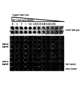
Example 6: Preparation of hzVSF variants which are humanized antibodies with
reduced immunogenicitv while having their virus-inhibitory activity maintained
or
27
CA 02988715 2017-12-07
enhanced
Example 6-1: Preparation of hzVSF alternatives
Three hzVSF alternatives were prepared based on the hzVSF prepared in Example
4.
The activity of each alternative was similar to or lower than that of the wild-
type (0.5 << 1 U <
1 mg/mL) (Tables 3 and 4). The amino acid sequences of CDR 1 to CDR 3 for each
of the
alternatives are shown in Table 3 and the amino acid sequences of FR1 to FR4
of each of the
variants are shown in Table 4.
[Table 3]
Antibody CDR1 CDR2 CDR3
NIDPYYGSTTYAQ
GYNMN
ETGTRAMDY
hzVSF WT Heavy chain KFQG
(SEQ ID NO: 2) (SEQ ID NO: 4)
(SEQ ID NO: 3)
RASENIYSNLA VATNLAD QHFYGSPRT
Light chain
(SEQ ID NO: 5) (SEQ ID NO: 6) (SEQ ID
NO: 7)
NIDPYYGSTTYAQ
GYNMN
ETGTRAMDY
hzVSF ¨al Heavy chain KFQG
(SEQ ID NO: 2) (SEQ ID NO: 4)
(SEQ ID NO: 3)
RASENIYSNLA VATNLAD QHFYGSPRT
Light chain
(SEQ ID NO: 5) (SEQ ID NO: 6) (SEQ ID
NO: 7)
NTDPYYGSTTYAQ
GYNMN
ETGTRAMDY
hzVSF a2 Heavy chain KF2G
(SEQ ID NO: 2) (SEQ ID NO: 4)
(SEQ ID NO: 3)
RASENIYSNLA VATNLAD QHFYGSPRT
Light chain
(SEQ ID NO: 5) (SEQ ID NO: 6) (SEQ ID
NO: 7)
NIDPYYGSTTYAQ
GYNMN
ETGTRAMDY
hzVSF a3 Heavy chain KFQG
(SEQ ID NO: 2) (SEQ ID NO: 4)
(SEQ ID NO: 3)
RASENIYSNLA VATNLAD QHFYGSPRT
Light chain
(SEQ ID NO: 5) (SEQ ID NO: 6) (SEQ ID
NO: 7)
28
CA 02988715 2017-12-07
[Table 4]
Antibody FR1 FR2 FR3 FR4
Heavy
hzVSF WT SEQ ID NO: 20 SEQ ID NO: 21 SEQ ID NO: 22 SEQ ID NO: 23
¨ chain
Light
SEQ ID NO: 24 SEQ ID NO: 25 SEQ ID NO: 26 SEQ ID NO: 27
chain
Heavy
hzVSF al SEQ ID NO: 151 SEQ ID NO: 21 SEQ ID NO: 22 SEQ ID NO: 23
¨ chain
Light
SEQ ID NO: 24 SEQ ID NO: 25 SEQ ID NO: 26 SEQ ID NO: 27
chain
Heavy
hzVSF a2 SEQ ID NO: 20 SEQ ID NO: 152 SEQ ID NO: 22 SEQ ID NO: 23
¨ chain
Light
SEQ ID NO: 24 SEQ ID NO: 25 SEQ ID NO: 26 SEQ ID NO: 27
chain
Heavy
hzVSF a3 SEQ ID NO: 151 SEQ ID NO: 152 SEQ ID NO: 22 SEQ ID NO: 23
¨ chain
Light
SEQ ID NO: 24 SEQ ID NO: 25 SEQ ID NO: 26 SEQ ID NO: 27
chain
Example 6-2: Preparation of hzVSF variants
Based on the hzVSF prepared in Example 4, hzVSF variants for actual use in
vivo were
prepared via immunogenicity reduction and affinity maturation. As a result, a
total of 13
variants were prepared (Tables 5 and 6). The amino acid sequences of CDR 1 to
CDR 3 for
each of the variants are shown in Table 5 and the amino acid sequences of FR1
to FR4 of each of
the variants are shown in Table 6.
[Table 5]
Antibody CDR I CDR2 CDR3
hzVSF WT Heavy GYNMN
NIDPYYGSTTYAQKFQG ETGTRAMDY
chain (SEQ ID NO: 2) (SEQ ID NO: 3) (SEQ ID
NO: 4)
Light RASENIYSNLA VATNLAD QHFYGSPRT
29
CA 02988715 2017-12-07
chain (SEQ ID NO: 5) (SEQ ID NO: 6) (SEQ ID
NO: 7)
hzVSF_varl Heavy GYNMN
NIDPYYGSTTYAQKFQG ETGTRAMDY
chain (SEQ ID NO: 2) (SEQ ID NO: 3) (SEQ ID
NO: 4)
Light RASENIYSNLA VADNLAD QHFYGSPRT
chain (SEQ ID NO: 5) (SEQ ID NO: 16) (SEQ ID
NO: 7)
hzVSF_var2 Heavy GYNMN
NIDPYYGSTTYAQKFQG ETGTRAMDY
chain (SEQ ID NO: 2) (SEQ ID NO: 3) (SEQ ID
NO: 4)
Light RASENIYSNLA VADNLGD QHFYGSPRT
chain (SEQ ID NO: 5) (SEQ ID NO: 17) (SEQ ID
NO: 7)
hzVSF_var3 Heavy GYNMN
NIDPYYGSTTYAQKFQG ETGTRAMDY
chain (SEQ ID NO: 2) (SEQ ID NO: 3) (SEQ ID
NO: 4)
Light RASENIYSNLA VADNRGD QHFYGSPRT
chain (SEQ ID NO: 5) (SEQ ID NO: 18) (SEQ ID
NO: 7)
hzVSF_var4 Heavy GYNMN
NIDPYYGSTTYAQKFQG ETGTRAMDY
chain (SEQ ID NO: 2) (SEQ ID NO: 3) (SEQ ID
NO: 4)
QHFYGTPRT
Light RASENIYSNLA VADNRGD
(SEQ ID
chain (SEQ ID NO: 5) (SEQ ID NO: 18)
NO: 19)
hzVSF var5 Heavy GYNMN
NIDPYYGSDTYAQKFQG ETGTRAMDY
chain (SEQ ID NO: 2) (SEQ ID NO: 14) (SEQ ID
NO: 4)
Light RASENIYSNLA VATNLAD QHFYGSPRT
chain (SEQ ID NO: 5) (SEQ ID NO: 6) (SEQ ID
NO: 7)
hzVSF_var6 Heavy GYNMN
NIDPYYGSTTYAQKFQG ETGTRAMDY
chain (SEQ ID NO: 2) (SEQ ID NO: 3) (SEQ ID
NO: 4)
Light RASEN1YSNLA VATNLAD QHFYGSPRT
chain (SEQ ID NO: 5) (SEQ ID NO: 6) (SEQ ID
NO: 7)
hzVSF_var7 Heavy GYNMN
NIDPYYGSDTYAQKFQG ETGTRAMDY
chain (SEQ ID NO: 2) (SEQ ID NO: 14) (SEQ ID
NO: 4)
Light RASENIYSNLA VATNLAD QHFYGSPRT
chain (SEQ ID NO: 5) (SEQ ID NO: 6) (SEQ ID
NO: 7)
hzVSF_var8 Heavy GYNMN
NIDPYYGSDTYAQKFQG ETGNRAMD
CA 02988715 2017-12-07
chain (SEQ ID NO: 2) (SEQ ID NO: 14) (SEQ ID
NO: 15)
Light RASENIYSNLA VATNLAD QHFYGSPRT
chain (SEQ ID NO: 5) (SEQ ID NO: 6) (SEQ ID NO: 7)
hzVSF_var9 Heavy GYNMN NIDPYYGSDTYAQKFQG ETGTRAMDY
chain (SEQ ID NO: 2) (SEQ ID NO: 14) (SEQ ID NO: 4)
Light RASENIYSNLA VADNRGD QHFYGSPRT
chain (SEQ ID NO: 5) (SEQ ID NO: 18) (SEQ ID NO: 7)
ETGNRAMDY
Heavy GYNMN NIDPYYGSDTYAQKFQG
hzVSF var10 (SEQ ID
chain (SEQ ID NO: 2) (SEQ ID NO: 14)
NO: 15)
Light RASENIYSNLA VADNRGD QHFYGSPRT
chain (SEQ ID NO: 5) (SEQ ID NO: 18) (SEQ Ill NO: 7)
Heavy GYNMN NIDPYYGSDTYAQKFQG ETGTRAMDY
hzVSF varl 1
chain (SEQ ID NO: 2) (SEQ ID NO: 14) (SEQ ID NO: 4)
QHFYGTPRT
Light RASENIYSNLA VADNRGD
(SEQ ID NO:
chain (SEQ ID NO: 5) (SEQ ID NO: 18)
19)
ETGNRAMDY
Heavy GYNMN NIDPYYGSDTYAQKFQG
hzVSF var12 (SEQ ID
chain (SEQ ID NO: 2) (SEQ ID NO: 14)
NO: 15)
QHFYGTPRT
Light RASENIYSNLA VADNRGD
(SEQ ID
chain (SEQ ID NO: 5) (SEQ ID NO: 18)
NO: 19)
Heavy GYNMN NIDPYYGSDTYAQKFQG ETGTRAMDY
hzVSF var13
chain (SEQ ID NO: 2) (SEQ ID NO: 14) (SEQ ID NO: 4)
Light RASENIYSNLA VADNLAD QHFYGSPRT
chain (SEQ ID NO: 5) (SEQ ID NO: 16) (SEQ ID NO: 7)
[Table 6]
Antibody FR1 FR2 FR3 FR4
31
CA 02988715 2017-12-07
Heavy
SEQ ID NO: 20 SEQ ID NO: 21 SEQ ID NO: 22 SEQ ID NO: 23
chain
hzVSF WT
Light
SEQ ID NO: 24 SEQ ID NO: 25 SEQ ID NO: 26 SEQ ID NO: 27
chain
Heavy
SEQ ID NO: 20 SEQ ID NO: 21 SEQ ID NO: 22 SEQ ID NO: 23
hzVSF var chain
1 Light
= SEQ ID NO: 24 SEQ ID NO: 25 SEQ ID NO: 26 SEQ ID NO: 27
chain
Heavy
SEQ ID NO: 20 SEQ ID NO: 21 SEQ ID NO: 22 SEQ ID NO: 23
hzVSF_var chain
2 Light
SEQ ID NO: 24 SEQ ID NO: 25 SEQ ID NO: 26 SEQ ID NO: 27
chain
Heavy
SEQ ID NO: 20 SEQ ID NO: 21 SEQ ID NO: 22 SEQ ID NO: 23
hzVSF_var chain
3 Light
SEQ ID NO: 24 SEQ ID NO: 25 SEQ ID NO: 26 SEQ ID NO: 27
chain
Heavy
SEQ ID NO: 20 SEQ ID NO: 21 SEQ ID NO: 22 SEQ ID NO: 23
hzVSF var chain
4 Light
SEQ ID NO: 24 SEQ ID NO: 25 SEQ ID NO: 26 SEQ ID NO: 27
chain
Heavy
SEQ ID NO: 20 SEQ ID NO: 21 SEQ ID NO: 22 SEQ ID NO: 23
hzVSF var chain
Light
SEQ ID NO: 24 SEQ ID NO: 25 SEQ ID NO: 26 SEQ ID NO: 27
chain
Heavy SEQ ID NO: 28
SEQ ID NO: 20 SEQ ID NO: 21 SEQ ID
NO: 23
hzVSF_var chain (K74T, 176A)
6 Light
SEQ ID NO: 24 SEQ ID NO: 25 SEQ ID NO: 26 SEQ ID NO: 27
chain
Heavy SEQ ID NO: 28
hzVSF var SEQ ID NO: 20 SEQ ID NO: 21 SEQ ID
NO: 23
chain (K74T, I76A)
7
Light SEQ ID NO: 24 SEQ ID NO: 25 SEQ ID NO: 26 SEQ ID NO: 27
32
CA 02988715 2017-12-07
chain
Heavy SEQ ID NO: 28
SEQ ID NO: 20 SEQ ID NO: 21 SEQ ID
NO: 23
hzVSF_var chain (K74T, I76A)
8 Light
SEQ ID NO: 24 SEQ ID NO: 25 SEQ ID NO: 26 SEQ ID NO: 27
chain
Heavy SEQ ID NO: 28
SEQ ID NO: 20 SEQ ID NO: 21 SEQ ID
NO: 23
hzVSF_var chain (K74T, I76A)
9 Light
SEQ ID NO: 24 SEQ ID NO: 25 SEQ ID NO: 26 SEQ ID NO: 27
chain
Heavy SEQ ID NO: 28
SEQ ID NO: 20 SEQ ID NO: 21 SEQ ID
NO: 23
hzVSF_var chain (K74T, I76A)
Light
SEQ ID NO: 24 SEQ ID NO: 25 SEQ ID NO: 26 SEQ ID NO: 27
chain
Heavy SEQ ID NO: 28
SEQ ID NO: 20 SEQ ID NO: 21 SEQ ID
NO: 23
hzVSF_var chain (K74T, I76A)
11 Light
SEQ ID NO: 24 SEQ ID NO: 25 SEQ ID NO: 26 SEQ ID NO: 27
chain
Heavy SEQ ID NO: 28
SEQ ID NO: 20 SEQ ID NO: 21 SEQ ID
NO: 23
hzVSF_var chain (K74T, I76A)
12 Light
SEQ ID NO: 24 SEQ ID NO: 25 SEQ ID NO: 26 SEQ ID NO: 27
chain
Heavy
SEQ ID NO: 20 SEQ ID NO: 21 SEQ ID NO: 22 SEQ ID NO: 23
hzVSF_var chain
13 Light
SEQ ID NO: 24 SEQ ID NO: 25 SEQ ID NO: 26 SEQ ID NO: 27
chain
It was confirmed that all of the 13 variants prepared above had reduced
immunogenicity
while having their antiviral activity and anti-inflammatory activity
maintained or enhanced
compared to those of the wild-type.
hzVSF_var12, which has the lowest immunogenicity among the above variants,
showed
antiviral activity such that 1 unit showed antiviral activity at 500 ng/unit,
the same as in hzVSF
wild-type. In contrast, hzVSF_var13, which has a relatively low immunogenicity
but relatively
33 ,
1
CA 02988715 2017-12-07
high antiviral activity, showed antiviral activity at 250 ng/unit.
Example 6-3: Confirmation of epitope counts of hzVSF wild-type and variants
thereof
The epitope counts of hzVSF wild-type; hzVSF_var12 and hzVSF_var13, which are
the
representative variants among the variants with reduced immunogenicity of the
wild-type; and
four kinds of blockbuster antibody drugs currently available in the
pharmaceutical market were
compared. As a result, the possibility of relative immunogenicity in each HLA
class II was
confirmed, as shown in Table 7.
[Table 7]
,
Protein Type DRB1 DRB 3/4/5 DQ DP Total
hzVSF w 31 17 1 3 52
hzVSF_varl 30 18 1 3 52
hzVSF var2 30 17 0 3 50
hzVSF_var3 30 16 0 3 49
hzVSF_var4 28 16 0 3 47
hzVSF var5 30 16 1 3 50
hzVSF_var6 ' 29 16 1 3 49
hzVSF_var7 28 15 1 3 47
hzVSF var8 27 14 1 3 45
hzVSF_var9 27 14 0 3 44
hzVSF_var10 26 13 0 3 42
hzVSF_varl 1 25 14 0 3 42
hzVSF_var12 24 13 0 3 40
hzVSF_var13 29 17 0 3 49
IIumira (human) 25 12 4 1 42
Remicade (chimeric) 74 37 3 1 115
Rituxan (chimeric) 65 33 9 3 110
Herceptin (humanized) 40 20 3 1 64
34
CA 02988715 2017-12-07
In Table 7, a higher total value means that there is a higher likelihood of
adverse effects
due to the HLA class II. From the above results, it was confirmed that the
epitope counts of
hzVSF var12 and hzVSF var13 with reduced immunogenicity of the present
invention were
similar to that of Humira, which has the lowest epitope count of the four
kinds of pharmaceutical
drugs. These results suggest that the humanized antibodies of the present
invention will have a
very low level of serious side effects that may arise when they are used as
antiviral agents, and
thus support the safety of the humanized antibodies of the present invention
as safe antiviral or
anti-inflammatory drugs.
Additionally, the biological activity of each of the variants (from var 1 to
var12) was
similar to that of wild-type (0.5 mg/U), but the activity of var13 was shown
to be higher than that
of the wild-type.
Example 6-4: Confirmation of T cell analysis of hzVSF variants
In order to evaluate the immunogenicity of hzVSF_var12 and hzVSF_var13, it was
confirmed whether the material examined by in vitro T cell analysis of Lonza
can have an effect
on T cell proliferation using the blood samples of 51 healthy donors.
The total peripheral blood mononuclear cells (PBMC) of each donor were treated
with
hzVSF_var12, hzVSF_var13, or Keyhole limpet hemocyanin (KLH) and cultured for
7 days.
KLH is an oxygen-carrying metalloprotein that can be used as a carrier protein
in the production
of antibodies, and it was used as a positive control for its ability to
effectively elicit immune
responses. Then, the ratio of T cells stained with CD3 'CD4+Edu was measured.
As a result,
KLH showed T cell proliferation in 45 blood donors (a response rate of 88%),
whereas
hzVSF_varl 2 and hzVSF_var13 induced T cell proliferation in only 3 subjects
(a response rate
of 5.8%), in response to the material (FIG. 17).
Meanwhile, the stimulation index (SI) of T cell proliferation induced by KLH,
hzVSF_v12, or hzVSF_v13 was calculated using the PBMC cultured as a control
for 7 days.
When the SI value is greater than 2, the immunogenicity is expected to be
large when applied to
humans. In contrast, when the SI value is less than 0.5, T cell proliferation
is expected to be
inhibited. As a result, it was confirmed that hzVSF_v12 and hzVSF_v13 showed
low SI values
of 1.12 and 1.03, respectively, and also showed SI values in the range of
being equal to or greater
than 0.6 and being equal to or less than 2. In contrast, KLH, which was used
as a positive
control, showed a high SI value of 3.91 (FIGS. 18 and 19; and Table 8).
[Table 8]
Product (Antigen) Mean SI p-Value
hzVSF_var12 1.12 0.008
hzVSF_var13 1.05 0.3229
KLI I 3.91 <0.0001
From the above results, it is expected that hzVSF_v12 and hzVSF_v13 will have
less
immunogenicity when administered to virus-infected patients and subsequently
have few side
effects.
Example 7: Confirmation of binding epitopes of hzVSF and variants thereof
An attempt was made to identify the peptides to which the hzVSF and variants
thereof
prepared in the above Examples bind.
As a result, it was confirmed that the hzVSF and variants thereof prepared in
the above
Examples were bound to the isolated peptide of SEQ ID NO: 1, corresponding to
the amino acid
sequence of vimentin at amino acid positions 142 to 294 (FIGS. 54 to 58).
Example 8: Confirmation of physical properties and pharmacokinetics of hzVSF
variants
In order to confirm the physical properties and pharmacokinetics of the hzVSF
variants
prepared in Example 6, experiments were performed regarding the representative
variants
hzVSF_var12 and hzVSF_var13.
Example 8-1: Confirmation of molecular weight patterns and purities of hzVSF
variants
The molecular weight pattern and purity of hzVSF_var12 and hzVSF_var13 were
confirmed by SDS-PAGE. The reduced sample was prepared by mixing the NuPageTM
4X
LDS sample buffer (Invitrogen, NP0007) and NuPageTM 10X sample reducing agent
(Invitrogen,
NP0009) with hzVSF_var12 and hzVSF_var13, respectively, followed by heating at
70 C for 10
minutes. The non-reducing sample was prepared by omitting the steps of adding
a reducing
36
CA 2988715 2019-04-02
agent and heating. 10 pt each of control antibodies, hzVSF_var12, and
hzVSF_var13 at a
concentration of 1 mg/mE, respectively, was electrophoresed on 4% to 12% SDS-
PAGE, the gel
was stained with InstantBlueTM (TripleRed, ISBO 1 L), and the purity of
hzVSF_var12 and
hzVSF_var13 was confirmed.
As a result, as illustrated in FIG. 20, it was confirmed that, in lanes 2 and
4
(non-reducing samples), the major bands were observed in the position where
IgG antibody
(about 150 kDa) was expected to appear; and in lanes 3 and 5 (reducing
samples), the bands
corresponding to the positions of the heavy chain (about 50 kDa) and the light
chain (about
25 kDa) of an immunoglobulin G (IgG) antibody were observed, thus confirming
that the hzVSF
variants also show a general IgG antibody pattern, as is the ease for hzVSF.
Additionally, when
IgG4 was used as a control, the pattern observed was the same as those
observed in lane 6 (a
non-reducing sample) and lane 7 (a reducing sample). Additionally, it was
confirmed that both
hzVSF_var12 and hzVSF_var13 showed good purity.
Additionally, in order to confirm the purity and aggregation of hzVSF_var12
and
hzVSF_var13, hzVSF_var12 and hzVSF_var13 were analyzed by size exclusion
high-performance liquid chromatography (SE-HPLC) using HPLC along with a
ZorbaxTM
GF-250 gm 9.2 mm ID x 25 cm column (Agilent 1200 series). Before injection
into HPLC, the
sample at a concentration of 1 mg/mL was purified by a 0.2 gm filter to remove
impurities.
Each of the samples (100 gL) was injected and HPLC was operated at a rate of 1
mL/min for 15
minutes. Analysis was performed using the Chemstation software.
As a result, the major peak was observed at the position corresponding to the
monomers
of a typical IgG antibody (at the retention time of about 8.65 minutes) and
the purity of
hzVSF_var12 and hzVSF_var13 was confirmed to be 95% or higher (FIG. 21). About
3.72%
and 4.43% of the peaks were observed at the position corresponding to the
dimers of a typical
IgG antibody (at the retention time of about 7.89 to 7.93 minutes), thus
confirming that the peak
shape was identical to that of the IgG antibody; this result is consistent
with the SDS-PAGE
result.
Example 8-2: Analysis of pharmacokinetics of hzVSF variants
37
CA 2988715 2019-04-02
CA 02988715 2017-12-07
The following experiment was carried out in order to obtain objective indices
for
reference during clinical tests by determining suitable in vivo dosage,
administration intervals,
and administration formulations of hzVSF_var13 via quantitative prediction of
in vivo
performance of hzVSF_var13 after its administration into mice, i.e.,
concentration in blood,
half-life, metabolic rate, etc.
As a control, 6.25 jig (0.31 mg/kg) of human IgG (polyclonal) was used, while
in the
experimental groups, 6.25 jig (25 U; 0.31 mg/kg), 62.5 jig (250 U; 3.10
mg/kg), and 625 jig
(2500 U; 31.0 mg/kg) of hzVSF_var13 was injected into the Mouse caudal vein.
After the
injection, blood samples were collected from the mice at days 1, 2, 4, 8, 14,
21, 28, and 35, and
the sera were separated and used for measuring the blood concentration of
hzVSF var13 using
ELISA.
For ELISA assay, anti-human IgG (y-specific) was fixed on a plate at 37 C for
2 hours
or at 4 C overnight, and was then incubated with 3% bovine serum albumin
(BSA) at 37 C for
2 hours to block the uncoated parts. The sera obtained from the blood
collected from the mice
were reacted at 37 C for 1 hour and reacted with anti-human IgG (K-specific),
to which
horseradish peroxidase (HRP) was conjugated, at 37 C for 30 minutes. The
resultant was
incubated in a darkroom at room temperature for. 9.5 minutes using a
3,3',5,5'-tetramethylbenzidine (TMB) solution as a substrate, and the amount
of hzVSF_var13 in
the blood was measured according to its OD value at 450 nm. The PK parameters
when a
single dose of hzVSF_var13 was administered to mice at each concentration are
indicated in
Table 9 and in the graphs of FIG. 22.
[Table 9]
Sham Control hzVSF_var13 hzVSF_var13 hzVSF_var13
(IgG 0.31 mg/kg) (0.31 mg/kg) (3.10 mg/kg) (31.0
mg/kg)
Female
6 6 6 6
Cmax (,tg/mL) 5.5 0.1 5.5 0.2 57.2 1.0 607.2 22.2
AUC
178.1 53.9 113.0 17.8 948.3 296.7 8686.9 2958.8
(1.1g=day/mL)
38
CA 02988715 2017-12-07
tin, (days) 20.1 2.9 13.4 2.4 10.8 3.8 10.2 3.0
CL
1.8 0.4 2.8 0.4 3.5 + 1.1 4.0 1.5
(mL/day/kg)
Male
5 5 5
C,,, ax (lg/mL) 5.5 0.2 5.2 0.3 54.6 1.4 563.0 8.2
AUC
121.8 15.3 78.8 20.8 798.1 274.5 6939.9 2311.8
(pig day/mL)
t112 (days) 16.6 2.4 10.1 3.1 9.0 4.0 8.9 3.7
CL
2.6 0.3 4.1 0.8 4.2 1.0 4.9 1.2
(mL/day/kg)
As a result, when hzVSF_var13 was administered at a single dose of 25 U, the
trend of
hzVSF concentration in blood was decreased in the same manner as in a sham
control (IgG)
administered with the same dose. The maximum concentration (Cmax) and the area
under the
concentration curve (AUC) were increased along with the increase in the
administration dose and
were thus confirmed to be dose-dependent. The in vivo half-life (t112) of
hzVSF_var13 (25 U)
was 10 days for males and 13 days for females, being slightly lower than the
sham control (16
days for males and 20 days for females), and the half-life was shown to be
slightly decreased
along with the increase in the administration dose. The Cõ, AUC, in vivo half-
life (11/2), in
vivo systemic and clearance value (CLt) were all shown to be higher in females
in each
concentration for administration than in males. At 35 days after the
administration, all mice
were autopsied to conduct measurement of organ weight, histopathological
examination,
immunotoxicity test, and neutralizing capability to examine the presence of in
vivo toxicity
caused by hzVSF_var13, but no adverse effects were observed.
The above results support that hzVSF and variants thereof according to the
present
invention have half-lives similar to those of other humanized antibodies, and
that they will have
no problem for clinical applications.
Example 9: Confirmation of virus-inhibiting activity of hzVSF and variants
thereof
Mouse L929 cells were infected with EMC-D virus, treated with hzVSF wild-type
and
39
CA 02988715 2017-12-07
hzVSF var12 and hzVSF_var13, which are the representative variants of hzVSF
variants, and
their antiviral effect was confirmed by MVIT assay of Example 2.
As a result, as illustrated in FIG. 23, the hzVSF wild-type showed 100%
antiviral effect
at a concentration of 0.51.1g/mL; and hzVSF_var12 and hzVSF_var13 showed 100%
antiviral
effect at concentrations of 0.2 i_tg/mL and 0.1 i.tg/mL, respectively.
Accordingly, the minimum amount contained in 1 mL of the VSF exhibiting 100%
antiviral effect when L929 cells were infected with EMC-D virus and
simultaneously treated
with VSF was determined as 1 unit of biological activity.
In this regard, the biological activity of each of the VSFs is shown in Table
10 below.
[Table 10]
VSF Type Biological Activity (1 unit) MW (kDa)
mVSF 0.09 g 163
hzVSF_wt 0.5 [ig 147
hzVSF_var12 0.2 it g 147
hzVSF_var13 0.1 tg 147
These results suggest that the hzVSF variants, which were newly prepared by
performing humanization of a monoclonal antibody, reduction of immunogenicity,
and affinity
maturation, can exhibit excellent antiviral and anti-inflammatory effects with
almost no adverse
effects compared to the hzVSF wt, although the newly prepared hzVSF variants
also have a
lower titer than the mVSF.
Example 10: Confirmation of affinity of hzVSF
The affinity for the receptors of hzVSF was measured by the method described
below.
L929 cells were seeded at the amount of 6 x 10⁵ cells and infected with 2 MOI
of
EMC-D virus for 0, 2, 6, and 10 hours, respectively. Four units of ligands
(mVSF, hzVSF_wt,
and hzVSF v13) were added thereto and cultured for 1 hour, and the culture
supernatants were
collected and their Ka values were obtained. The difference between the amount
of each ligand
added and the amount of the collected ligand after reaction is the amount of
the ligand bound to
CA 02988715 2017-12-07
=
the receptors, and based on the result, the affinity constant (Ka) and the
dissociation constant (Kd)
were calculated (Table 11).
[Table 11]
VSF Type Affinity Constant (Ka) Dissociation Constant (Kd)
mVSF 2.1 x 101 L/mol 4.7 x 10⁻¹¹M
hzVSF wt 8.2 x 10⁹ L/mol to
1.2>< 10- M
hzVSF v13 1.7 x 10¹⁰ L/mol 5.9 x 10⁻¹¹M
As a result, hzVSF was confirmed to have an affinity constant (Ka) of 1 x 10⁹
or higher,
corresponding to potential biologics, and thus, as biologics, both hzVSF and
their variants were
also confirmed to have high binding affinity for the receptors.
Example 11: Confirmation of anticellular activity of hIFN-a and hzVSF wt in
human cells
Human fibroblasts, WI-38 cells, were infected with EMC-D and treated with
human
interferon and hzVSF wt at various concentrations, and the antiviral effect
and viability of each
cell were measured by the MTS method.
In detail, the cells were seeded into a 96-well plate and cultured while
attached to the
bottom for 24 hours. The cells were infected with EMC-D virus (100 pfu) and
treated with
interferon or hzVSF. After culturing for 48 hours, the cells were treated
with
[3 -(4,5 -dimethylthi azo I -2-y1)-5-(3 -e arb oxy m eth oxypheny1)-2-(4-
sulfopheny1)-2H-tetrazo lium]
(MTS). The cells were further incubated for 2 or 3 hours and their viability
was confirmed by
measuring the absorbance at 490 nm. As illustrated in FIG. 24, the cells
treated with human
interferon showed about 90% viability even with a lesser amount than the cells
treated with
hzVSF_wt; however, when the cells were treated at a high concentration (4 nM
or higher) of
human interferon, the cells showed cytotoxicity. In contrast, when the cells
were treated with
hzVSF_wt, the cells showed about 100% viability at a concentration of about 4
nM and the cells
did not show any cytotoxicity at a concentration of 4 nM or higher.
From the above results, it is expected that hzVSF will have fewer adverse
effects, unlike
human interferon.
41
CA 02988715 2017-12-07
Example 12: Confirmation of the inhibitory activity of hzVSF wt against immune
cell infiltration and inflammatory response in viral diabetes
In order to examine the efficacy of hzVSF on diabetes in mice according to the
concentration of hzVSF, 5-week-old male DBA/2N mice were infected with EMC-D
virus
(100 pfu) by intraperitoneal injection. Then, the hzVSF_wt at concentrations
of 2, 4, and 16
units, respectively, were injected into the caudal veins and the glucose
levels in the blood and
urine were examined from the 3r1 day of post infection. Additionally, the mice
were sacrificed
on the 3rd and the 7th days post infection, and their pancreases were
surgically removed and
subjected to a biopsy followed by a histological examination.
As a result, as illustrated in FIG. 25, when the pancreases were removed from
the mice
after infecting them with EMC-D virus and stained with Herriatoxylin & Eosin,
the group
infected with only virus (virus control) showed the infiltration of immune
cells and a decrease in
the size of islets due to destruction of the islets thereof, whereas the group
administered with at
least 4 units of hzVSF_wt (virus + hzVSF) showed no infiltration of immune
cells, and the islets
also remained normal. Accordingly, it is speculated that hzVSF wt inhibited
the proliferation
of EMC-D and suppressed the infiltration of immune cells, thereby protecting
the islets from
being destroyed.
Additionally, as a result of the detection of blood glucose levels, diabetes
was not
developed in the group administered with at least 4 units of hzVSF_wt, and
blood glucose levels
were below 200 mg/dL, which corresponds to the range of normal mice. Regarding
the urine,
urine glucose was not detected in the group administered with at least 8 units
of hzVSF_wt
(Tables 12 to 14).
In viral diabetes, the incidence rate of diabetes according to the
concentration of the
hzVSF wt is shown in Table 12, the blood glucose levels according to the
concentration of the
hzVSF_wt are shown in Table 13, and the urine glucose levels according to the
concentration of
the hzVSF_wt are shown in Table 14.
[Table 12]
Group Incidence
Post-Infection
Rate of
42
CA 02988715 2017-12-07
Diabetes
3 Days 4 Days 5 Days 6 Days 7 Days
Virus Control - 4/4 4/4 4/4 4/4 100%
2 Unit/Mouse - - - 4/4 4/4 100%
4 Unit/Mouse - - - - . - 0%
8 Unit/Mouse - - - - - 0%
16 Unit/Mouse - - - - - 0%
* When a mouse has a blood glucose level of 300 mg/dL or higher and the urine
glucose level of
"+" or higher, the mouse is considered to have diabetes.
[Table 13]
Group Post-Infection
3 Days 4 Days 5 Days 6 Days 7 Days
Virus Control 157 1 25 541 84 540 84 526 91 529 100
2 Unit/Mouse 162 24 157 23 186 53 484 21 505 26
4 Unit/Mouse 149 42 148 6 135 11 145 8 122 16
8 Unit/Mouse 171 42 148 6 135 11 145 8 122 16
16 Unit/Mouse 163 21 160 4 144 9 139 8 157 22
* When a mouse has a blood glucose level of 300 mg/dL or higher, the mouse is
considered to
have diabetes.
[Table 14]
Group Post-Infection
3 Day 4 Day 5 Day 6 Day 7 Day
Virus Control - ++++ ++++ ++++ ++++
2 Unit/Mouse - - - +++ +++
4 Unit/Mouse - - - - -
8 Unit/Mouse - - - - -
16 Unit/Mouse - - - - -
The above results confirmed that hzVSF and various variants thereof of the
present
43
CA 02988715 2017-12-07
invention, due to their antiviral effect against EMC-D virus infection and
inhibitory activity
against the infiltration of immune cells, significantly inhibited the
destruction of the Langerhans
islets and were confirmed to be capable of significantly treating diabetes
induced by viral
infection. That is, the above results suggest that hzVSF and various variants
thereof of the
present invention not only exhibit an antiviral effect without inducing the
infiltration of immune
cells, but also have the capability to treat viral diabetes and may thus be
used as an agent for
treating various kinds of inflammatory diseases.
Example 13: Confirmation of anti-HBV effect of mVSF
In order to confirm the antiviral effect of mVSF against human hepatitis B
virus (HBV),
HepG2.2.15 cells, which are human hepatocareinoma cells expressing HBV, were
used.
The HepG2.2.15 cells were seeded into a plate and treated with mVSF, and the
amount
of the surface antigens of HBV (IIBsAg), which were proliferated in the cells
and released into
the culture, was quantitated every 3 days using ELISA. The result was
converted into the
amount of virus per cell. At the same time, the number of cells was counted
and the relative
amount of virus per cell was measured.
As a result, it was confirmed that the mVSF treatment distinctively reduced
the amount
of HBsAg, thus suggesting that mVSF has an anti-HBV effect (FIG. 26). These
results support
the fact that hzVSF and various variants thereof of the present invention can
effectively inhibit
hepatitis viruses such as HBV, and thus have an antiviral effect against
various viruses.
Additionally, the mVSF treatment for 9 days was shown to inhibit the viral
replication in each
cell by 80% or higher and is expected to further inhibit inflammation in
humans, thereby
suggesting that it will be particularly effective in treating clinical
symptoms.
Example 14: Confirmation of expression patterns of VSF receptors in
virus-infected cells
First, in order to confirm whether VSF receptors can be expressed in virus-
infected cells,
a human liver tissue of a hepatitis B patient (a part of the liver tissue
without cancer, although the
liver progressed into cancer due to hepatitis B) and a human liver tissue of a
hepatitis C patient (a
part of the liver tissue without cancer, although the liver progressed into
cancer due to hepatitis C)
were stained with mVSF antibodies. A human liver tissue not infected with
virus (without
44
=
CA 02988715 2017-12-07
cancer) was used as control. As a result, VSF receptors were shown to be
expressed in the liver
tissues obtained from patients infected with HBV or HCV, but VSF receptors
were not expressed
in the control tissue, thus suggesting that the VSF receptors are expressed in
a virus-infection
specific manner. These results were shown in the same pattern when the
experiment was
repeatedly performed using the human liver tissues obtained from other
patients (FIGS. 27 and
28).
Subsequently, in order to confirm the expression period of VSF receptors
according to
viral infection, H1N1 influenza virus was infected into the MDCK cell line,
and the viral
replication and the expression of VSF receptors were observed by fluorescent
staining. As a
result, it was confirmed that the virus replication occurred according to time
after influenza virus
infection, and the expression of VSF receptors increased following the
infection (FIG. 29).
Based on these results, it was confirmed that VSF receptors are induced by
viral infection.
Additionally, human WI-38 fibroblasts were infected with EMC-D virus and then
the
expression of VSF receptors and antiviral effect of VSF were examined (FIG.
30). The images
on the left of FIG. 30 show the expression of VSF receptors by the EMC-D
infection, and the
images on the right show the anti-EMC-D effect and the expression of VSF
receptors by the
hzVSF_wt treatment. Based on the above results, it was confirmed that VSF can
exhibit an
antiviral effect and also express its receptors in an infection-specific
manner with respect to the
EMC-D virus infection.
Conclusively, it was confirmed that virus-infected cells can express VSF
receptors,
although the expression time may vary depending on cells, and also that VSF
treatment can
induce an antiviral effect, although it does not affect the expression of the
receptors.
Example 15: Confirmation of antiviral effect of hzVSF against hepatitis B
virus
In order to examine the effect of hzVSF against human hepatitis B virus (HBV),
experiments were performed using hzVSF_var13, a representative variant of
hzVSF, as
described below.
Example 15-1: Analysis of covalently closed circular DNA (cccDNA)
A HBV-infected HepG2.2.15 cell line was treated with hzVSF_varl 3 and an HBV
drug
(lamivudine; Lami) according to each dosage, the number of cells was counted
every week, and
the same amount of cells were harvested.
In order to obtain the cccDNA of HBV from the above cells, the HepG2.2.15
cells, upon
recovery, were resuspended in a lysis buffer (25 mM EDTA, 10 mM Tris-HCl; pH
7.5, 100 mM
NaCI, 1% SDS, 0.1 mg/mL proteinase K) and reacted at 55 C for 1 hour. After
purifying the
total HBV DNA using the MEGA quick-spin total fragment DNA purification kit
(Intron), the
resultant was reacted at 37 C with plasmid-safe ATP-dependent DNase for 1
hour, and the
enzyme was inactivated at 70 C for 30 minutes. Then, HBV relaxed circular DNA
(re.DNA)
was removed and only the HBV cccDNA was recovered. Subsequently, the HBV
cccDNA was
subjected to quantitative analysis by qPCR using the AccupowerTM 2x greenstar
qPCR Master
mix (Bioneer, Korea).
The PCR primers used were a forward primer 5'-TGAATCCYGCGGACGACC-3' (SEQ
ID NO: 153) and a reverse primer 5'-CAGCTTGGAGGCTTGAACAG-3' (SEQ ID NO: 154)
(nucleotides 1862-1881) (Y = C/T).
As a result, it was confirmed that the cccDNA content of HBV-infected cells
was
decreased in a concentration-dependent manner when treated with hzVSF_var13,
and in
particular, there was almost no cccDNA content when treated with 10 ttg/mL of
hzVSF_var13
(FIG. 31).
Accordingly, it was confirmed that hzVSF administration can significantly
reduce the
amount of intracellular cccDNA, and thus has an excellent effect as an agent
for treating HBV.
Example 15-2: Confirmation of inhibition of HBV DNA
The HBV-infected HepG2.2.15 cell line was treated with hzVSF_var13 and an HBV
drug (lamivudine; Lami) according to each dosage, the number of cells was
counted every week,
and the same amount of cells were harvested.
Intracellular HBV DNA was purified as follows. The HepG2.2.15 cells obtained
above
were resuspended in a lysis buffer (25 mM EDTA, 10 mM Tris-HC1; pH 7.5, 100 mM
NaCl, 1%
SDS, 0.1 mg/mL proteinase K) and reacted at 55 C for 1 hour. Intracellular HBV
DNA was
purified using the MEGA quick-spin total fragment DNA purification kit
(Intron).
In order to obtain extracellular HBV DNA, a certain amount of the cell culture
supernatant was obtained, treated with a lysis buffer, and extracted. Then,
the HBV total DNA
was subjected to quantitative analysis by qPCR using the Accupower 2x
greenstar qPCR Master
mix (Bioneer, Korea).
The PCR primers used were a forward primer 5'-CCTCTTCATCCTGCTGCT-3' (SEQ
46
CA 2988715 2019-04-02
CA 02988715 2017-12-07
ID NO: 155) and a reverse primer 5'-AACTGAAAGCCAAACAGTG-3 (SEQ ID NO: 156).
As a result, in the case of the extracellular HBV DNA, the amount of HBV DNA
was
significantly reduced when treated with hzVSF_var13 at a very low
concentration (0.1 pg/mL)
compared to the control, which was treated with Lamivudine (FIG. 32). In the
case of the
intracellular HBV DNA, HBV DNA was inhibited in a dose-dependent manner of
hzVSF var13,
and the hzVSF_var13 treatment showed a significantly excellent efficacy
compared to the
control, which was treated with Lamivudine (FIG. 33).
Conclusively, it was confirmed that hzVSF administration can significantly
reduce the
amount of both intracellular and extracellular HBV DNA, and thus hzVSF has
excellent activity
as a therapeutic agent for HBV treatment.
Example 16: Confirmation of antiviral effect of hzVSF against hepatitis C
virus
Example 16-1: Confirmation of antiviral effect of hzVSF_wt against hepatitis C
virus
In order to examine the effect of hzVSF wt against human hepatitis C virus
(HCV),
experiments were performed as described below.
Hepatitis C virus ,IFH-1 strain was infected into human liver cells, Huh7.5
cells, at a
concentration of 0.1 multiplicity of infection (MOI), and was treated with
interferon-13 (3 ng/mL)
and hzVSF wt (500, 1000, and 2000 units) on the 3r1 day of post. infection,
and the cell culture
supernatants and cells were collected on the 4th day. The cells were measured
using anti-HCV
NS5A antibody staining of HCV NS5A by FACS analysis, and the cell culture
supernatants were
measured with respect to their HCV RNA titers by real-time quantitative PCR.
As a result, the hzVSF_wt(1000 U/mL) treatment showed an anti-HCV effect
similar to
that of interferon-13 in FACS analysis (FIG. 34). Additionally, when the virus
titer was
measured by real-time quantitative PCR, hzVSF wt showed an about 50% anti-HCV
effect at
1000 U/mL, but the effect was shown to be weaker compared to that of
interferon-13 (FIG. 35).
However, while interferon actually has adverse effects in vivo, hzVSF does not
have any
adverse effects and is also expected to treat symptoms due to inflammation,
and thus hzVSF will
show marked excellence in real applications. Therefore, at the early stages of
viral infection,
the combination of a chemical agent and interferon is necessary for inhibiting
viral growth; and
47
CA 02988715 2017-12-07
from the mid-stage of viral infection or the appearance of symptoms, the
combination of a
chemical agent and hzVSF is necessary for inhibiting not only
immunopathological phenomena
but also the virus growth. That is, the above results suggest the possibility
of a combination
therapy of a chemical drug having an excellent inhibitory effect against virus
growth (or
interferon) and hzVSF_wt and various variants thereof of the present invention
having an
excellent inhibitory effect against immunopathological phenomena. However,
these results also
suggest the application of hzVSF_wt and various variants thereof of the
present invention alone
as an antiviral agent without adverse effects.
Example 16-2: Confirmation of antiviral effect of hzVSF variants against
hepatitis
C virus (HCV la)
Then, to examine the effect of hzVSF variants against human hepatitis C virus
(HCV),
experiments were performed using hzVSF_var13, a representative variant of
hzVSF, as
described below.
First, in order to confirm the inhibitory effect of the hzVSF variant against
the HCV la
replication, the Huh 7.5 cells infected with HCV TN cell-culture (TNce)
(genotype la) were
treated with hzVSF_var13 according to each dosage, and the cells were
collected on day 3, day 6,
day 9, and day 12. Then, after collecting the HCV-infected cells, the total
RNA was extracted
using trizol. Subsequently, cDNA was synthesized by reverse transcription, and
HCV RNA
and GAPDH (a housekeeping gene) for normalization were subjected to
quantitative analysis by
qPCR using the Accupower 2x greenstar qPCR Master mix (Bioncer, Korea).
The HCV-targcting PCR primers used were a forward primer
51-GGGCTATAAGGTGCTAGTGC-3' (SE() ID NO: 157) and a reverse primer
'-GGCTGCCAGTGGTA ATTGTT-3' (SEQ ID NO: 158), and the GAPDH PCR primers used
were a forward primer 5'-TCCCTGAGCTGAACGGGAAG-3 (SEQ ID NO: 159) and a reverse
primer 5'-GGAGGAGTGGGTGTCGCTGT-3' (SEQ ID NO: 160).
As a result, it was confirmed that the content of HCV la RNA decreased with
time when
hzVSF_var13 was treated at a concentration of 0.1 U/mL or higher and also that
the inhibitory
effect against the HCV 1 a replication was in a dose-dependent manner of
hzVSF_var13
(FIG. 36A).
Furthermore, when hzVSF van 3 was treated, the content of HCV core protein was
confirmed by western blot analysis, and as a result, it was confirmed that the
protein content was
48
CA 02988715 2017-12-07
decreased in a dose-dependent manner (FIG. 36B).
Then, the effect of hzVSF_varl 3 compared to that of HCV drugs was examined.
The
Huh 7.5 cells infected with HCV TNcc (genotype la) were treated with
hzVSF_var13 and HCV
drugs (sofosbuvir and simeprevir) according to the dosage, and the HCV-
infected cells were
recovered on a weekly basis and the total RNA was extracted using trizol.
Then, cDNA was
synthesized by reverse transcription, and HCV total RNA (the primers of SEQ ID
NOS: 157 and
158 were used) and GAPDH (a housekeeping gene) for normalization were
subjected to
quantitative analysis by qPCR using the Accupower 2x greenstar qPCR Master mix
(Bioneer,
Korea).
As a result, it was confirmed that when hzVSF_var13 was treated at a
concentration of
1 Rg/mL, the rate of inhibition against the HCV la replication was
significantly higher compared
to the control drugs (FIG. 37).
Accordingly, the administration of the hzVSF variant to the HCV TNcc (genotype
I a)-infected cells reduced HCV gene (RNA) and HCV core protein, and thus it
was confirmed
that hzVSF variants have an antiviral effect against HCV.
Example 16-3: Confirmation of antiviral effect of hzVSF variants against
hepatitis
C virus (HCV lb)
Then, in order to confirm the inhibitory effect of hzVSF variants against the
HCV lb
replication, the Huh7 cells, which express the subgenomic replicon of HCV
genotype lb, were
treated with hzVSF van l 3 and HCV drugs (sofosbuvir and simeprevir) according
to the dosage,
the HCV-infected cells were recovered on a weekly basis, and the total RNA was
extracted using
trizol. Then, cDNA was synthesized by reverse transcription, and HCV total RNA
and
GAPDH (a housekeeping gene) for normalization were subjected to quantitative
analysis by
qPCR using the Accupower 2x greenstar qPCR Master mix (Bioneer, Korea).
The HCV-targeting PCR primers used were a forward primer
5'-ATGCAGCCCAAGGGTATAAG-3' (SEQ ID NO: 161) and a reverse primer
5'-GGTTCTGATGTTAGGGTCGATAC-3' (SEQ ID NO: 162), and the GAPDH PCR primers
used were the primers of SEQ ID NOS: 159 and 160.
As a result, it was confirmed that the hzVSF van 3 treatment inhibited the HCV
lb
replication in a dose-dependent manner, and in particular, when hzVSF var13
was treated at a
49
CA 02988715 2017-12-07
concentration of 1 vg/mL, the inhibition was effective at a level similar to
that of the control
drugs (sofosbuvir and simeprevir) (FIG. 38B). Additionally, when the level of
HCV NS5A was
confirmed by western blot according to the concentration of hzVSF_var13
treatment, the amount
of HCV NS5A was confirmed to decrease in a dose-dependent manner (FIG. 38A).
Accordingly, the administration of the hzVSF_var13 to the HCV replicon
(genotype
1b)-expressing cells reduced the amount of HCV gene (RNA) and HCV NS5A
protein, and thus
it was confirmed that hzVSF_var13 has an antiviral activity against HCV.
Example 16-4: Confirmation of antiviral effect of hzVSF variants against
hepatitis
C virus (HCV 2a)
Then, in order to confirm the inhibitory effect of hzVSF variants against the
HCV 2a
replication, the JFH- I cells infected with HCV genotype 2a were treated with
hzVSF_var13 and
HCV drugs (sofosbuvir and simeprevir) according to the dosage, the HCV-
infected cells were
recovered on a weekly basis, and the total RNA was extracted using trizol.
Then, cDNA was
synthesized by reverse transcription, and subsequently HCV total RNA and GAPDH
(a
housekeeping gene) for normalization were subjected to quantitative analysis
by qPCR using the
Accupower 2x greenstar qPCR Master mix (Bioneer, Korea). The HCV-targeting PCR
primers
used were the primers of SEQ ID NOS: 157 and 158, and the GAPDH PCR primers
used were
the primers of SEQ ID NOS: 159 and 160.
As a result, it was confirmed that when hzVSF_var13 was treated, the
inhibition against
the HCV 2a replication was similar to that of the control drugs without a
significant difference in
the concentration of treatment at the 7-week time-point (FIG. 39B).
Furthermore, when the
above result was examined on a long-term basis, i.e., up to the 21' week, it
was confirmed that
when hzVSF van l 3 was treated at a concentration of 1 [ig/mL, the inhibitory
effect against HCV
2a replication was maintained at a level similar to when treated with
sofosbuvir and simeprevir
(FIG. 40).
Summarizing the above results, it was confirmed that the administration of the
hzVSF_var13 to the HCV JFH-1 (genotype 2a)-infected cells reduced the amount
of HCV gene
(RNA) and IICV core protein (FIG. 39A), and thus hzVSF_varl 3 has an antiviral
activity
against HCV.
Example 17: Confirmation of therapeutic effect of hzVSF on influenza virus
CA 02988715 2017-12-07
=
infection
In order to confirm the antiviral and anti-inflammatory effects of
hzVSF_var13, a
representative variant of hzVSF, against influenza virus, experiments were
performed in mice as
follows.
Four-week-old female Balb/c mice were infected with 111Ni Influenza A/Puerto
Rico/8/34 virus (1 x 10⁵ pfu) into nasal cavities, and 25, 100, and 400 units
of hzVSF_var13
were administered into caudal veins on day 2, 4, or 6 after the infection. On
the 7th day and the
9th
day after the viral infection, the mice were sacrificed. Thereafter, weight of
lung and body,
mucosal epithelial cells, and cilia were examined by H&E staining, and the
infiltration of the
immune cells was examined by an immunohistochemical staining.
As a result, the group administered with 500 U of hzVSF_var13 showed an at
least
100-fold decrease in influenza titer compared to the control group (FIG. 41).
Additionally, the group infected with H1NI influenza virus showed the
infiltration of
immune cells in the alveolar sacs while the groups administered with 100 U and
400 U of
hzVSF_var13 showed recovery of the symptoms by the influenza virus similar to
the lung tissue
of an uninfected group (healthy lung) regardless of administration period.
Even the
administration of 25 U of hzVSF_var13 was shown to suppress the infiltration
of immune cells,
although there was a difference according to the administration period, and
thus the
administration of 25 U of hzVSF_var13 was shown to have a therapeutic effect
(FIG. 42).
Additionally, when the cilia in the respiratory epithelial cells of mouse
lungs were
observed, the uninfected group and the group treated with hzVSF_var13 (400 U)
were shown to
have epithelial cells with cilia on the trachea in the lungs while the
epithelial layer was shown to
be destroyed in the virus-infected lungs due to viral infection (FIG. 43).
Additionally, when the ratio between the weight of lungs and body weight of
mice was
measured, the virus-infected group showed an increase in the ratio of lung
weight relative to the
body weight due to the pneumonic symptom of the lungs filling with fluid,
whereas the group
treated with hzVSF_var13 showed a decrease in the ratio of the lung weight
relative to the body
weight of healthy mice, in proportion to the concentration of hzVSF_var13 to
some extent
(FIG. 44).
Lastly, the inhibitory effect of hzVSF var13 against the infiltration of
immune cells was
confirmed by an immunohistochemical method using markers of CD4 T cells (FIGS.
45 and 46)
51
CA 02988715 2017-12-07
and macrophage (FIGS. 47 and 48) (in particular, the infection of 107 pfu in
FIGS. 46 and 48).
When the mouse lungs were stained on the 711 day after the H1N1 influenza
virus infection
(107 pfu), the infiltration of CD4 T cells and macrophages was = observed,
whereas the group
treated with hzVSF_var13 showed a significant decrease in the infiltration of
CD4 T cells and
macrophages (FIGS. 45 to 48).
These results confirmed that the hzVSF and various variants thereof of the
present
invention not only have an excellent antiviral effect against influenza virus
but also have an
anti-inflammatory effect capable of suppressing the infiltration of immune
cells, thereby
suggesting that they can be used as antiviral and anti-inflammatory agents
without adverse
effects.
Example 18: Confirmation of antiviral effect of hzVSF .against various viruses
In order to confirm whether hzVSF_wt and variants thereof of the present
invention
have an antiviral effect against various viruses, the effects of hzVSF wt and
hzVSF_var13,
which are representative variants of hzVSF wt, were examined in vitro and in
vivo. The in
vitro experiments are related to an antiviral effect, and the in vivo
experiments are related to
antiviral and anti-inflammatory effects.
The results are shown in Table 15.
[Table 15]
Virus Antiviral Effect
In vitro In vivo
Encephalomyocarditis virus
+++ +++
(EMCV)
Mengovirus +++ NT
Reovirus NT
Influenza virus +++
HIV ++ NT
HCMV NT
Mouse Hepatitis Virus (MHV) ++ +++
52
CA 02988715 2017-12-07
Hantaan Virus ++ NT
HBV ++ NT
HCV ++ NT
*NT: not tested
As a result, hzVSF was shown to have excellent antiviral and anti-inflammatory
effects
against encephalomyocarditis virus (EMCV) both in vitro and in vivo. The hzVSF
showed an
excellent antiviral effect against mengovirus in vitro. The hzVSF also showed
an antiviral
effect against reovirus in vitro. The hzVSF also showed an antiviral effect
against HIV in vitro.
The hzVSF also showed antiviral and anti-inflammatory effects against HCMV.
hzVSF also
showed effects against Hantaan virus in vitro and showed an antiviral effect
against hepatitis B
and C virus in vitro. The hzVSF also showed antiviral and anti-inflammatory
effects against
mouse hepatitis virus (MHV), which causes hepatitis in mice by a mechanism
similar to that of
human hepatitis, specifically in vivo. The hzVSF also showed antiviral and
anti-inflammatory
effects against influenza virus both in vitro and in vivo, and the effects
were more remarkable in
in vivo.
Example 19: Confirmation of inhibitory effect of mVSF administration against
secretion of inflammatory cytokines in a mouse model
In order to confirm the effect of mVSF on the generation of inflammatory
cytokines,
mice were infected with EMC-D virus and administered with mVSF, and after 3
days, the
amount of inflammatory cytokines in sera was measured by ELISA. As illustrated
in Table 16,
the levels of inflammatory cytokines such as IL-6, TNF-a, IFN-y, and MCP-1
increased after the
viral infection; however, the mVSF administration inhibited the expression of
these
inflammatory cytokines.
[Table 16]
Sample Cytokine (pg/mL):
Measured 3 Days After Viral Infection
CCL2
IL-12 IL-6 TNF-a IFN-y
(MCP-1)
Normal ND 2.9 4.6 1.3 13.8
EMC-B 1.1 190.3 44.3 91.4 1752.7
53
CA 02988715 2017-12-07
EMC-D 1.6 21.3 15.8 29.2 609.3
EMC-D + VSF
1.2 6.5 8.8 6.2 124.9
(1000 Unit)
* ND: not detected
From the above results, it was confirmed that mVSF has an anti-inflammatory
effect that
inhibits inflammation induced by viral infection.
Additionally, in order to confirm the effect of mVSF on the generation of
inflammatory
cytokines in a mouse with acute hepatitis, the mouse was infected with mouse
hepatitis virus and
treated with VSF, and after 1 or 3 day of post infcction, the amount of
inflammatory cytokines in
sera was measured by ELTSA. As illustrated in Table 17, the levels of
inflammatory cytokines
such as IL-6, TNF-a, IFN-7, and MCP-1 increased after the viral infection;
however, the VSF
administration inhibited the expression of these inflammatory cytokines.
Additionally, a single
combined administration of IFN-a was shown to have a more significant
synergistic effect.
[Table 17]
Cytokines (pg/mL)
CCL2
Sample IL-12 TNF-a INF-y 11-10 IL-6
(MCP-1)
1 Day
Post-Infe Normal ND 5.4 1.2 ND ND 1.2
ction
Virus
14.2 40.0 166.2 1165.5 ND 205.6
Infection
Virus + VSF ND 8.4 1.9 16.9 ND 5.0
Virus + IFN-a
ND 9.1 1.6 13.0 ND 1.5
once + VSF
2 Day
Post-Infe Normal ND 2.1 ND ND ND ND
ction
54
CA 02988715 2017-12-07
Virus
8.7 38.0 724.3 344.5 ND 13.7
Infection
Virus + VSF ND 32.4 74.5 129.9 ND 9.2
Virus + IFN-u
ND 4.0 20.3 13.9 ND 1.4
once + VSF
From the above results, it was confirmed that VSF also has an anti-
inflammatory effect
capable of inhibiting the inflammation induced by viral infection.
Example 20: Confirmation of inhibitory effect of hzVSF administration against
secretion of inflammatory cytokines in a mouse model
In order to confirm whether the hzVSF of the present invention can inhibit the
secretion
of inflammatory cytokines for the treatment of inflammatory diseases,
experiments were
performed as follows.
The lung of a mouse infected with H1N1 (Influenza A/PR8) influenza virus (1 x
105 pfu)
and the lung of a mouse administered with hzVSFIvar13 (500 units) one day
after the influenza
virus infection was harvested. Greenberger lysis buffer (GLB) was homogenized
with the lungs
and the resultant was placed on ice for 30 minutes, and centrifuged at 3,470x
g at 4 C for 7
minutes. After collecting the supernatant, it was centrifuged at 420xg at 4 C
for 10 minutes,
mixed with the supernatant from lung specimens, capture beads, and PE
detection reagent in an
amount of 50 [IL each at a 1:1:1 ratio, and reacted at room temperature for 2
hours while
blocking light. Upon reaction, the beads were resuspended in a wash buffer and
the samples
were measured using the FACS canto II, and the data was collected.
As a result, it was confirmed that the amount of IL-6, TNF-u, IFN-y, and CCL2
(MCP-1), which belong to pro-inflammatory cytokines, present in the lungs of
the mice was
significantly increased on the 7th day after the influenza virus. infection.
However, it was
confirmed that the treatment of the hzVSF_var13 of the present invention
significantly inhibited
the generation of cytokines in the lungs of the mice (FIG. 49).
That is, it was suggested that the hzVSF_varl 3 of the present invention can
inhibit
various kinds of inflammatory cytokines, and thus can be used as an agent for
treating various
inflammatory diseases caused by viral infection.
= CA 02988715 2017-12-07
Example 21: Confirmation of the effect of hzVSF after EMC-D infection in a
vimentin-overexpressing cell line (a stable cell line)
MCF-7 cells, which do not express vimentin, were transfected with vimentin
(wt) or
vimentin (mt), in which the hzVSF-binding domain was mutated, to prepare a
vimentin (wt)- or
vimentin (mt)-overexpressing cell line. Then, an MVIT assay was performed
using the selected
cells with a similar level of vimentin expression.
Specifically, 2 x 10⁴ cells were seeded per well, and the cells were infected
with EMC-D
virus (2 MOI), and 2 hours thereafter, treated with hzVSFvarl 3 at
concentrations from 256 U
and at 4-fold serial dilutions thereafter, and cultured for 2 days. Then, the
cultured cells were
fixed with methanol, stained with 0.5% crystal violet, and air-dried for
observation.
As a result, it was confirmed that the effect of hzVSF against EMC-D virus
infection
was not observed in MCF-7 parent cells, Mock, and vimentin (mt), whereas the
MCF-7 cells.
which can express vimentin (wt), were induced to have a structural change of
vimentin into VSF
receptors (VR) by the viral infection, thus confirming the antiviral effect of
hzVSF (FIGS. 50
and 51).
Then, a WST assay was performed additionally to confirm cytotoxicity by EMC-D
infection.
The vimentin (wt) cells and vimentin (mt) cells were cultured for three days
in the same
condition as that of the MVIT assay, treated with tetrazolium salt (WST-1),
cultured for an
additional 4 hours, and the absorbance was measured at 450 nm. After
measurement, the
supernatant was removed and the cells were fixed with methanol, stained with
0.5% crystal
violet for 20 minutes, and then air-dried. The stained cells were dissolved in
methanol and the
absorbance was measured at 540 mn.
As a result, it was confirmed that the effect of hzVSF against EMC-D virus
infection
was not observed in MCF-7 parent cells, Mock, and vimentin (mt), whereas the
111CF-7 cells,
which can express vimentin (wt), were induced to have a structural change of
vimentin into VSF
receptors (VR) by the viral infection, thus confirming the antiviral effect of
hzVSF (FIG. 52).
Then, the expression feature of VSF receptors (VR) following EMC-D infection
was
examined in a vimentin-overexpressing cell line (a stable cell line). In this
regard, each of the
stable MCF-7 cells, i.e., vimentin (wt) and vimentin (mt) cells, was infected
with EMC-D (5
56
CA 02988715 2017-12-07
MOI) for 9 hours, and then the cells were immunostained with hzVSF_var13. The
cells were
then fixed and permeabilized, and the hzVSF van 3, which was diluted in a
1:250 ratio, was
reacted with the cells. Then, the cells were reacted with FITC conjugated-goat
human IgG as a
secondary antibody, and the expression of the VSF receptors (VR) for hzVSF in
the cells was
examined under a fluorescence microscope (500x magnification).
As a result, as illustrated in FIG. 55, it was confirmed that hzVSF_var13 did
not bind to
the vimentin-expressing cells, in which the Mock- and hzVSF-binding domains
were mutated,
but the VR was expressed in the wild-type vimentin-expressing cells by viral
infection (FIG. 53).
Example 22: Confirmation of the binding VSF with VR WT and VR MT purified
in E. coli
The vimentin recombinant proteins (5 mg) purified by the Ni-NTA system and
hzVSF_var13 were mixed in a 2:1 molar ratio and filled with phosphate buffered
saline (PBS) to
a final volume of 700 mL, and incubated in an orbital shaker at 4 C for 3
hours. Then, protein
A beads (50% slurry) were added thereto, incubated at 4 C for 1 hour, and the
protein
A-hzVSF-VR complex was centrifuged at 3,000xg at 4 C for 3 minutes. Then, the
supernatant
was removed and the beads were washed with PBS, and centrifuged again at
3,000xg at 4 C for
3 minutes, and the process was repeated 3 times. Subsequently, 2X SDS-sample
buffer was
added thereto, and boiled for 5 minutes to perform SDS-PAGE and western blot
analyses.
As a result, it was confirmed that the proteins, in which the 6 amino acid
residues of
vimentin, Y150, R186, Q195, R217, K235, and R270, were subjected to
substitution mutation
into F, K, N, K, R, and K, respectively, showed a distinct decrease in the
binding with hzVSF
(FIG. 54). Accordingly, it was confirmed that hzVSF can bind to Y150, R186,
Q195, R217,
K235, and R270 of vimentin.
Example 23: Confirmation of the binding between VR, which was overexpressed in
HEK293T cells, and VSF
HEK293T cells were transfected with vimentin WT or vimentin MT to examine
their
binding with hzVSF_v13.
Specifically, the HEK293T cells (2 x 10⁶) were seeded into 100 mm culture
dishes,
transfected with vimentin WT (wild-type) and MT (mutant-type) DNA (vector:
pcDNA3.1mycHisC), and incubated for 48 hours. Then, the cells were harvested,
treated with a
57
CA 02988715 2017-12-07
1% CHAPS lysis buffer (1 mL), and placed on ice for 20 minutes for the lysis
of the cells. The
lysed cells were centrifuged at 13,000 rpm at 4 C for 15 minutes, and then
the supernatant was
transferred into a fresh tube.
The protein from the supernatant (700 mg) was treated with protein A beads
with a 50%
slurry and allowed to preclear at 4 C for 1 hour. Then, the* resultant was
centrifuged at
3,000xg at 4 C for 3 minutes and the supernatant was transferred to a fresh
tube. The resultant
was treated with hzVSF_varl 3 at a 1:100 ratio and incubated at 4 C overnight
to allow the
binding between VSF and VR. Then, the protein A beads with a 50% slurry were
added thereto
and reacted at 4 C for 1 hour. A protein A-VSF-VR complex was obtained as a
result, and to
wash the beads, it was centrifuged at 3,000xg at 4 C for 3 minutes and the
supernatant was
removed. Subsequently, the precipitated complex was washed 3 times by adding a
1% CHAPS
lysis buffer thereto. The resultant was treated with a 2X SDS-sample buffer,
and boiled to
perform SDS-PAGE and immunoblot analyses.
As a result, it was confirmed that the proteins, in which the 6 amino acids of
vimentin,
Y150, R186, Q195, R217, K235, and R270, were subjected to substitution
mutation into F, K, N,
K, R, and K, respectively, showed a distinct decrease in the binding with
hzVSF (FIG. 55).
Accordingly, it was confirmed that hzVSF can bind to Y150, R186, Q195, R217,
K235, and
R270 of vimentin.
The weak binding with VSF in the lane, in which the 6 amino acid residues were
substituted by mutation, is presumed to be ascribed to the dimerization and
tetramerization with
wild-type vimcntin, which is endogenous to the HEK293T cells, and the
overexpressed, mutant
vimentin.
Further to the above results, the vimentin-VSF binding model was examined
using
computer programs (FIGS. 56 to 58). The specific programs used for the
analysis are shown
below.
VR dimer modeling: Protein Homology/analogY Recognition Engine V 2.0
(Component software Template detection: HHpred 1.51
Secondary structure prediction: Psi-pred 2.5
Disorder prediction: Disopred 2.4
Transmembrane prediction: Memsat SVM
58
CA 02988715 2017-12-07
Multi-template modeling and ab initio: Poing 1.0
Re-orientation of structures for easy viewing: OVOP)
Complex modeling of VR dimer and VSF: Pymol
Complex modeling of VR_dimer and chemical: autodock vina
The structure in which VR and hzVSF were bound together was deduced by the VR
dimer modeling, and conclusively, it was confirmed that hzVSF binds to the
residues of Y150,
R186, Q195, R217, K235, and R270.
The above results suggest that the antibody of the present invention, which
specifically
binds to the peptide of SEQ ID NO: 1, or a fragment binding to the peptide
which specifically
binds to the peptide, can exhibit antiviral and anti-inflammatory effects
against a broad range of
viral diseases. Unlike the existing antiviral agents such as interferon or
chemotherapeutic
agents, they can be selectively applied to virus-infected cells without any
adverse effects, require
a small amount for treatment, and have anti-inflammatory activity by
suppressing the infiltration
of immune cells, thereby suggesting that they can be used as agents for
treating various viral
diseases.
From the foregoing, a skilled person in the art to which the present invention
pertains
will be able to understand that the present invention may be embodied in other
specific forms
without modifying the technical concepts or essential characteristics of the
present invention.
In this regard, the exemplary embodiments disclosed herein are only for
illustrative purposes and
should not be construed as limiting the scope of the present invention. On the
contrary, the
present invention is intended to cover not only the exemplary embodiments but
also various
alternatives, modifications, equivalents, and other embodiments that may be
included within the
spirit and scope of the present invention as defined by the appended claims.
59