Note: Descriptions are shown in the official language in which they were submitted.
CA 02988822 2017-12-07
1
DESCRIPTION
MULTIFOCAL SPECTROMETRIC MEASUREMENT DEVICE AND OPTICAL
SYSTEM FOR MULTIFOCAL SPECTROMETRIC MEASUREMENT DEVICE
TECHNICAL FIELD
[00011
The present invention relates to a multifocal spectrometric measurement device
for simultaneously measuring beams of signal light, such as fluorescence or
Raman
scattering light, coming from a plurality of points, as well as an optical
system for such a
multifocal spectrometric measurement device.
BACKGROUND ART
[0002]
In recent years, a simultaneous multi-sample measurement method, called the
"high
throughput screening" (HTS), for analyzing a number of samples within a short
period of
time has been attracting attention in pharmaceutical and other areas. In the
simultaneous
multi-sample measurement method, a spectrometric measurement is widely used,
in which
signal light emitted from each sample is dispersed to obtain a spectrum for
each sample,
and the composition, molecular structure and other aspects of the samples are
analyzed
from their respective spectra. Examples of the signal light include:
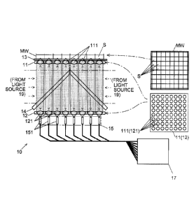
fluorescence or Raman
scattering light emitted from each sample when the sample is irradiated with
irradiation
light; and chemiluminescent light emitted from a sample without irradiating
the sample
with light.
[0003]
Patent Literature 1 and Non Patent Literature 1 disclose a multifocal
spectrometric
measurement device. Although these documents contain no description on the
simultaneous multi-sample spectrometric measurement method, the device is
potentially
applicable for the simultaneous multi-sample spectrometric measurement method.
It is a
type of device called the "Raman spectroscopic microscope". In this device,
the irradiation
light is divided into a plurality of beams with a microlens array (which will
be described
later). The irradiation beams are delivered to different positions on a single
sample, and
Raman scattering light obtained at each position is dispersed into a spectrum.
By mapping
CA 02988822 2017-12-07
2
the intensity of the Raman scattering light having a specific amount of Raman
shift, an
image showing the distribution of a sample component corresponding to the
Raman shift is
obtained. This device may possibly be applied for a simultaneous measurement
of a
plurality of samples by casting each irradiation beam onto a different sample
instead of
delivering those beams to different positions on a single sample.
[0004]
The multifocal spectrometric measurement device disclosed in Patent Literature
1
and Non Patent Literature 1 is hereinafter described in detail using Fig. 15.
In this
multifocal spectrometric measurement device 90, a laser beam generated by a
laser source
91 is divided into a plurality of light fluxes with a micro lens array 92
having microlenses
921 arranged in a matrix form with eight rows and eight columns. Each of those
light fluxes
is reflected by an edge filter 93 and passes through a pinhole array 94 having
one hole
provided for each light flux, as well as a relay lens 95 and an objective lens
96, which are
common to all light fluxes, to be eventually cast onto the sample S. From this
sample S, the
same number of beams of signal light as the light fluxes produced by dividing
the
irradiation light are emitted. In each beam of signal light, the reflected
light which has the
same wavelength as the irradiation light, and the Raman scattering light which
has a
different wavelength from the irradiation light, are superposed on each other.
The beams of
signal light travel through the objective lens 96, relay lens 95 and holes of
the pinhole array
94, reaching the edge filter 93. Due to the difference in wavelength, the
reflected light is
reflected by the edge filter 93 and cannot pass through; only the Raman
scattering light is
allowed to pass through. After passing through the edge filter 93, the fluxes
of the Raman
scattering light pass through a lens system 97 consisting of a pair of lenses
and enter a fiber
bundle 98 which has the same number of optical fibers as the micro lenses 921
bundled in
homeomorphic relation to the microlens array 92. The inlet ends of the optical
fibers in the
fiber bundle 98 are arranged in a matrix form, with each inlet end receiving
one flux of
Raman scattering light. The outlet ends of the fiber bundle 98 are arranged in
a row. The
light exiting from each outlet end is individually dispersed into a spectrum
by a
spectrograph 99.
[0005]
Patent Literature 2 discloses a simultaneous multipoint spectrometric
measurement
device, in which: irradiation light is cast from a single objective lens onto
a sample S; the
signal light (Raman scattering light) emitted from the sample S is received by
a plurality of
CA 02988822 2017-12-07
3
optical fibers in a fiber bundle; and the light exiting from the output end of
each optical
fiber is individually dispersed into a spectrum by a spectrograph. This device
differs from
the one disclosed in Patent Literature 1 and Non Patent Literature 1 in that
the irradiation
light is not divided, and the signal light is also not divided until it
reaches the fiber bundle.
Patent Literature 2 also includes no description on the simultaneous multi-
sample
measurement method.
CITATION LIST
PATENT LITERATURE
[0006]
Patent Literature 1: JP 2012-237647 A
Patent Literature 2: JP 2010-151801 A
NON PATENT LITERATURE
.. [0007]
Non Patent Literature 1: Masanari Okuno and Hiroo Harnaguchi, "Tashouten
Kyoushouten Raman Bunkou Kenbikyou No Kaihatsu (Development of Multifocal
Confocal Raman Spectroscopic Microscope)", Abstract of Oral Presentation No.
1B17 in
the Fourth Annual Meeting for Japan Society of Molecular Science (2010),
[online], July
2010, Japan Society for Molecular Science, [accessed on November 25, 2014; re-
accessed
on January 29, 2016], the Internet <URL:
http://molsci.center.ims.acjp/area/2010/bk2010/papers/1B17_w.pdf>
SUMMARY OF INVENTION
TECHNICAL PROBLEM
[0008]
The signal light generated from a sample is isotropically emitted in a
spherical form.
Therefore, an objective lens can collect only a portion of the entire signal
light. However,
the collection efficiency of the signal light should be as high as possible
for a
high-sensitivity measurement. Since the collection efficiency becomes higher
as the
numerical aperture NA of the objective lens becomes larger, a microscopic
optical system
having a high numerical aperture (NA) is required. In the case of analyzing a
number of
samples using a microscopic optical system, the microspectroscopic measurement
is
CA 02988822 2017-12-07
4
simultaneously performed for multiple points by (i) sequentially performing
the
microspectroscopic measurement while scanning the sample with a single focal
point, or
(ii) simultaneously performing the microspectroscopic measurement on multiple
points by a
multifocal system as in Patent Literature 1 and Non Patent Literature 1. In
case (i), the
measurement time increases with the number of samples. In particular, for a
faint light as in
the Raman spectroscopy, a long exposure time is required, so that an enormous
amount of
time is needed for the spectrometric measurement of all samples. In case (ii),
increasing the
numerical aperture NA of the objective lens causes an increase in the
magnification of the
lens, which reduces the observable area on the sample. That is to say, there
is a trade-off
relationship between the numerical aperture NA and the magnification of the
objective lens,
which also means that there is a trade-off relationship between the
measurement sensitivity
and the size of the observable area.
[0009]
The multipoint spectrometric measurement device disclosed in Patent Literature
1
and Non Patent Literature 1 is aimed at obtaining Raman scattering light from
a plurality of
different positions within a single sample. Therefore, the area to be observed
is
comparatively small. Since the size of the area to be observed in this device
is
approximately one dozen pun square, there is no problem with the measurement
sensitivity
as far as the aforementioned aim is concerned. However, a device to be applied
in a
simultaneous measurement of multiple samples needs to observe a larger area.
As a specific
example, a sample holder called the "multiwell", which is commercially offered
for
simultaneous multi-sample measurements, has several tens to several hundreds
of wells for
holding individual samples arranged in a matrix form, with the entire area
measuring
approximately a several cm to one dozen cm each side. In order to observe the
entire area
of such a multiwell through a single objective lens as used in the device
disclosed in Patent
Literature 1 and Non Patent Literature 1, the objective lens must have a low
magnification,
which lowers the measurement sensitivity. Therefore, this device is not
suitable for
simultaneous multi-sample measurements.
[0010]
The problem to be solved by the present invention is to provide a multifocal
spectrometric measurement device capable of performing a simultaneous multi-
sample
measurement with high sensitivity and with no restriction on the
magnification, as well as
an optical system for such a multifocal spectrometric measurement device.
CA 02988822 2017-12-07
SOLUTION TO PROBLEM
[0011]
The multifocal spectrometric measurement device according to the present
5 invention developed for solving the previously described problem is a
device in which
beams of signal light emitted from a plurality of predetermined observation
areas on a
sample or samples placed in a sample placement section are introduced into a
spectrograph
and thereby dispersed into spectra, the device including:
a plurality of objective light-condensing sections individually located at
positions
which respectively and optically face the plurality of observation areas; and
spectrograph input sections provided in such a manner that each of the
plurality of
objective light-condensing sections has one corresponding spectrograph input
section, for
introducing signal light passing through the corresponding objective light-
condensing
sections into the spectrograph.
[0012]
The "positions which optically face observation areas" are positions at which
beams
of signal light from the observation areas arrives. If no other optical
element, such as a
light-condensing section or reflector, is present between the observation
areas and the
objective light-condensing sections, those positions are positions which
(literally) face the
observation areas. If such an optical element (e.g. a magnification-converting
section which
will be described later) is present between the observation areas and the
objective
light-condensing sections, those positions are positions at which the signal
light arrives
after passing through that optical element.
[0013]
In the multifocal spectrometric measurement device according to the present
invention, the plurality of objective light-condensing sections are provided
so that one
objective light-condensing section optically faces one observation area in the
sample. The
plurality of observation areas may be entirely included in one sample, or they
may be
distributed over a plurality of samples; having a single observation area on
each sample is
also possible. In any case, one objective light-condensing section corresponds
to one
observation area. Each objective light-condensing section collects signal
light from the
observation area which optically faces that section. The signal light
collected by each
objective light-condensing section is sent to the corresponding spectrograph
input section.
CA 02988822 2017-12-07
6
Thus, a spectrometric measurement of the signal light is performed for each
observation
area. Each spectrograph input section may be placed so that it (literally)
faces the
corresponding objective light-condensing section, or there may be some optical
element
(e.g. another light-condensing section, reflector or the like) placed in the
space between the
objective light-condensing sections and the spectrograph input sections, as in
the space
between the observation areas and the objective light-condensing sections.
[0014]
The numerical aperture NA of a lens is defined as NA¨n=sinO, where 0 is the
maximum angle made by the light incident on the lens from the focal point with
respect to
the optical axis, and n is the refractive index of the medium which is present
between the
focal point and the lens. Similarly, the numerical aperture NA of a light-
condensing section
can be defined as NA=n=sin0', where 0' is the angular radius of the light-
condensing
section as viewed from the point where the light condensed by the light-
condensing section
is focused (focal point), and n is the refractive index of the medium which is
present in the
intermediate area.
[0015]
By the present invention, a number of samples or a large-size sample can be
simultaneously observed, since each objective light-condensing section only
needs to
observe a single observation area. Therefore, it is possible to lower the
magnification, i.e. to
increase the area of the observable area, as well as to increase the numerical
aperture NA of
the individual objective light-condensing section, i.e. to improve the
measurement
sensitivity.
[0016]
The multifocal spectrometric measurement device according to the present
.. invention may preferably be configured as follows: at each of some or all
of the plurality of
objective light-condensing sections, a spectrograph-side light-condensing
section is
provided between the objective light-condensing section and the corresponding
spectrograph input section, a point in the observation area optically facing
the objective
light-condensing section is located at the position on which the signal light
between the
objective light-condensing section and the spectrograph-side light-condensing
section is
focused after passing through the objective light-condensing section, and the
spectrograph
input section corresponding to the objective light-condensing section is
located at the
position on which the signal light is focused after passing through the
spectrograph-side
CA 02988822 2017-12-07
7
light-condensing section. This configuration resolves the trade-off between
the numerical
aperture NA and the magnification, making it possible to optimize the
measurement
sensitivity with no restriction on the numerical aperture NA of the objective
light-condensing section, numerical aperture NA of the spectroscope-side light-
condensing
section, size of the measurement area, and intervals of the measurement
points. That is to
say, since the numerical aperture NA of the objective light-condensing section
can be
increased independently of the magnification, the collection efficiency of the
signal light
can be improved, and furthermore, since the numerical aperture NA of the
spectrograph-side light-condensing section can also be set independently of
the
magnification, the collected signal light can efficiently enter the
spectrograph with an
optimum numerical aperture NA which yields high utilization efficiency. For
these reasons,
the signal light from the observation areas can be efficiently introduced to
the spectrograph
input sections. Furthermore, since the trade-off between the numerical
aperture NA and the
magnification is resolved, the size and interval of the beams of signal light
to be incident on
the spectrograph can also be suitably determined for the configuration of the
spectrograph,
which allows the measurement points to be increased as needed even when the
same
spectrograph is used. These favorable effects cannot be achieved by
conventional imaging
optical systems as shown in Fig. 15.
[0017]
As in the case of the fluorescence or Raman scattering light, if the signal
light is
obtained by irradiating a sample with irradiation light having a predetermined
wavelength,
the multifocal spectrometric measurement device according to the present
invention should
include a light source for casting the irradiation light onto the sample or
samples. The light
source may be placed at a position from which the irradiation light is cast
through the
objective light-condensing sections onto the sample, or at a position from
which the
irradiation light is cast onto the sample without passing through the
objective
light-condensing sections.
As in the case of the signal light generated by chemiluminescence, if the
signal light
is obtained without irradiating the sample with light, it is unnecessary to
provide the
multifocal spectrometric measurement device according to the present invention
with a
light source for casting the irradiation light onto the sample or samples.
CA 02988822 2017-12-07
8
[0018]
The arrangement of the plurality of observation areas, and that of the
objective
light-condensing sections which optically face the respective observation
areas, may be
either a one-dimensional or two-dimensional arrangement. The observation areas
and the
.. objective light-condensing sections may be arranged either at regular or
irregular intervals.
If the observation areas and the objective light-condensing sections are
arranged in a
two-dimensional form, they may be arranged either at random or in an ordered
form, such
as a square lattice (matrix), triangular lattice or radial form. In the case
of using a multiwell
in which a number of wells for holding individual samples are arranged in a
matrix form as
mentioned earlier, the multifocal spectrometric measurement device according
to the
present invention may preferably include an objective-light-condensing-section
array in
which the plurality of objective light-condensing sections are arranged in a
matrix form. In
the case of using this objective-light-condensing-section array along with the
spectrograph-side light-condensing sections, the device may preferably include
a
.. spectrograph-side light-condensing-section array in which the plurality of
spectrograph-side
light-condensing sections are arranged in a matrix form, with each
spectrograph-side
light-condensing section optically facing one objective light-condensing
section.
[0019]
In the multifocal spectrometric measurement device according to the present
invention, if the above configuration is simply used, not only the signal
light but also the
irradiation light reflected by the sample can enter the spectrograph input
sections. The
irradiation light entering the spectrograph can be removed by analytical
processing.
However, if the signal light is Raman scattering light, the signal light has a
different
wavelength from that of the irradiation light, and therefore, it is possible
to remove only the
irradiation light by a filter. The same also applies in the case of the
fluorescence emission
whose wavelength differs from that of the irradiation light. That is to say,
the multifocal
spectrometric measurement device according to the present invention may
include a filter
placed between the sample placement section and the spectrograph input
sections, for
allowing light having a wavelength of the signal light to pass through while
reflecting light
having a wavelength of the irradiation light, along with the light source for
casting the
irradiation light onto the sample or samples.
CA 02988822 2017-12-07
9
[0020]
The irradiation light to be incident on the filter may be a single beam.
However, the
filter may preferably be arranged so that a plurality of beams of the
irradiation light are
incident on the filter and each of the plurality of beams of the irradiation
light reflected by
the filter is cast onto a different subset of the objective light-condensing
sections. The
plurality of beams of the irradiation light may be generated from a plurality
of different
light sources, or they may be generated by providing multiple paths from a
single source of
light. The use of the plurality of beams of the irradiation light allows for
the reduction of
the size of the filter, which is advantageous for improving the surface
accuracy of the filter
and reducing the production cost as compared to the use of a single
irradiation beam and a
large-area filter. If a plurality of different light sources is used to
generate a plurality of
beams of the irradiation light, it is possible to deliver a stronger
irradiation beam to each
sample and thereby improve the sensitivity of the spectrometric measurement.
[0021]
In the present invention, it is not always necessary to deliver one separate
irradiation
beam to each sample; casting a single irradiation beam on the entire area of
the sample or
samples is also possible. However, it is preferable to adopt the configuration
that the filter is
arranged between the objective light-condensing sections and the spectrograph
input
sections so that the irradiation light cast from the light source is reflected
by the filter into
the direction of the optical axes of the objective light-condensing sections.
This
configuration makes the irradiation light be condensed on the observation
areas through the
objective light-condensing sections, so that the irradiation light will be
exhaustively
utilized.
[0022]
The multifocal spectrometric measurement device according to the present
invention may include a magnification-converting section placed between the
plurality of
observation areas and the plurality of objective light-condensing sections,
for changing the
size of an image formed by the signal light from each of the plurality of
observation areas.
By using the magnification-converting section, measurements of a number of
small
observation areas can be simultaneously performed. Similarly, the multifocal
spectrometric
measurement device according to the present invention may include a
spectrograph-input-section-side magnification-converting section placed
between the
plurality of objective light-condensing sections and the spectrograph input
sections, for
CA 02988822 2017-12-07
changing the size of an image formed by the signal light from each of the
plurality of
objective light-condensing sections. As the magnification-converting section
and the
spectrograph-input-section-side magnification-converting section, a light-
condensing
section consisting of a single lens (or the like) or a light-condensing
section consisting of a
5 set of lenses (or the like) arranged in the propagating direction of the
signal light can be
used.
[0023]
The multifocal spectrometric measurement device according to the present
invention may also be configured as follows:
10 the spectrograph input sections are arranged at matrix points in such a
manner that
each of the plurality of objective light-condensing sections has one
corresponding
spectrograph input section; and
the rows and columns of the matrix are non-parallel to the wavelength-
dispersing
direction of a dispersing element included in the spectrograph. A diffraction
grating, prism
or similar element can be used as the dispersing element. By this
configuration, beams of
diffracted light can be produced from the respective beams of signal light by
means of a
single dispersing element without overlapping each other. This spectrometric
optical system
can be used not only as the optical system for the multifocal spectrometric
measurement
device according to the present invention, but can also be used, for example,
in the devices
described in Patent Literature 1 or 2.
[0024]
The multifocal spectrometric measurement device according to the present
invention may include a moving means for changing the relative position of the
sample or
samples and the plurality of objective light-condensing sections along a plane
containing
the sample or samples placed in the sample placement section. This allows for
an
observation of the sample over a larger area than in the case where no such
moving means
is used, making it possible to perform a spectrometric imaging operation for
creating an
image which shows spectrometric data. The change in the relative position of
the sample
and the plurality of objective light-condensing sections may be made in a
linear
(one-dimensional), planer (two-dimensional) or steric (three-dimensional)
form.
[0025]
The optical system for a multifocal spectrometric measurement device according
to
the present invention is an optical system to be used in a device in which
beams of signal
CA 02988822 2017-12-07
11
light emitted from a plurality of predetermined observation areas on a sample
or samples
placed in a sample placement section are introduced into a spectrograph and
thereby
dispersed into spectra, the optical system including:
a plurality of objective light-condensing sections configured to be installed
in the
.. device in such a manner as to be individually placed at positions which
respectively and
optically face the plurality of observation areas.
ADVANTAGEOUS EFFECTS OF THE INVENTION
[0026]
By the present invention, a multifocal spectrometric measurement device
capable of
simultaneously performing a measurement of multiple samples or a large sample
with high
sensitivity and with no restriction on the magnification can be obtained.
BRIEF DESCRIPTION OF DRAWINGS
[0027]
Fig. 1 is a schematic configuration diagram showing the first embodiment of
the
multifocal spectrometric measurement device according to the present
invention.
Fig. 2 is a schematic configuration diagram showing the light source and
beam-diameter-increasing optical system used in the first embodiment.
Fig. 3 is a vertical sectional view of another example of a lens array.
Figs. 4A and 4B are CCD images and graphs showing the results of a
fluorescence
spectrum measurement (Fig. 4A) and Raman spectrum measurement (Fig. 4B)
performed
using the multifocal spectrometric measurement device in the first embodiment.
Fig. 5 is a schematic configuration diagram showing the second embodiment of
the
multifocal spectrometric measurement device according to the present
invention.
Fig. 6 is a schematic configuration diagram showing the third embodiment of
the
multifocal spectrometric measurement device according to the present
invention.
Figs. 7A-7C are schematic configuration diagrams showing variations of the
multifocal spectrometric measurement device according to the third embodiment.
Fig. 8 is a schematic configuration diagram showing the fourth embodiment of
the
multifocal spectrometric measurement device according to the present
invention.
Fig. 9 is a schematic configuration diagram showing the fifth embodiment of
the
multifocal spectrometric measurement device according to the present
invention.
CA 02988822 2017-12-07
12
Fig. 10 is a schematic configuration diagram showing the sixth embodiment of
the
multifocal spectrometric measurement device according to the present
invention.
Fig. 11 is a schematic configuration diagram showing a variation of the first
embodiment.
Fig. 12 is a plan view showing a variation of the arrangement of the objective
lenses
in the multifocal spectrometric measurement device according to the present
invention.
Fig. 13A is a schematic configuration diagram showing one example of the
spectrograph to be used in the multifocal spectrometric measurement device in
each
embodiment and other multifocal spectrometric measurement devices, and Fig.
13B is a
diagram showing beams of signal light exiting from a pinhole array and beams
of diffracted
light incident on a photodetector.
Fig. 14A is a CCD image showing the result of a simultaneous dispersion of a
plurality of beams of signal light using the spectrograph shown in Fig. 13,
Fig 14B is a
partially enlarged image of the same CCD image, and Fig. 14C is an extracted
spectrum.
Fig. 15 is a schematic configuration diagram showing one example of a
conventional multifocal spectrometric measurement device.
DESCRIPTION OF EMBODIMENTS
[0028]
Embodiments of the multifocal spectrometric measurement device according to
the
present invention are hereinafter described using Figs. 1-14C.
[0029]
(1) Multifocal Spectrometric Measurement Device According to First Embodiment
(1-1) Configuration of multifocal spectrometric measurement device according
to first
embodiment
As shown in Fig, 1, the multifocal spectrometric measurement device 10
according
to the first embodiment has a sample holder 13 on which a multiwell having
wells for
holding samples S arranged in a matrix form is mounted. The bottom wall of the
multiwell
MW and the sample holder 13 are made of glass which is transparent to both
irradiation
light and signal light. An objective lens array (objective light-condensing-
section array) 11
having a plurality of objective lenses (objective light-condensing sections)
111 arranged in
a matrix form is provided, facing the sample holder 13. The plurality of
objective lenses
111 are provided in such a manner that each objective lens 111 faces one well
when the
CA 02988822 2017-12-07
13
multiwell MW is held on the sample holder 13. Each of those wells becomes an
observation
area for one sample S. Each objective lens 111 is arranged in such a manner
that, when a
parallel beam as the signal light is incident on the lens from the side
opposite to the sample
holder 13, the lens focuses the beam on a point within the well which faces
the lens in the
.. multiwell MW held on the sample holder 13.
[0030]
The multifocal spectrometric measurement device 10 also has a second lens
array
(spectrograph-side lens array; spectrograph-side light-condensing-section
array) 12 facing
the objective lens array 11. The second lens array 12 has second lenses
(spectrograph-side
lenses; spectrograph-side light-condensing sections) 121 arranged in a matrix
form in such
a manner that each of the objective lenses 111 has one second lens 121 facing.
[0031]
The multifocal spectrometric measurement device 10 further includes
spectrograph
input sections 151 provided in such a manner that each of the second lenses
121 has one
.. spectrograph input section 151 facing. Each individual spectrograph input
section 151
consists of the input end of one optical fiber. Each spectrograph input
section 151 is placed
at a position where signal light (parallel beam) which falls from the side
opposite to the
spectrograph input section 151 onto the second lens 121 correspondingly facing
this section
is focused. Those spectrograph input sections 151 arranged in a matrix form
and facing the
second lenses 12 constitute the spectrograph-input-section assembly 15. The
output ends of
all optical fibers are arranged in a row in such a manner that the beams of
light from those
output ends are cast at different positions on the surface of a diffraction
grating in a
spectrograph 17.
[0032]
Between the objective lens array 11 and the second lens array 12, a filter 14
is
provided which allows light having wavelengths within a predetermined
wavelength band
to pass through while reflecting light having wavelengths within other
wavelength bands.
The predetermined wavelength band mentioned earlier does not include the
wavelength of
the irradiation light but includes the wavelength of the signal light.
Accordingly, the filter
.. 14 reflects the irradiation light while allowing the signal light to pass
through. The filter 14
consists of two quadrilateral plate members. One plate member covers one half
of the
columns of the objective lenses 111 (in Fig. 1, four columns on the left side)
and is tilted at
an angle of 45 degrees to the optical axes of the objective lenses 111, with
its distance from
CA 02988822 2017-12-07
14
the objective lens array 11 being larger at the end of the columns of the
objective lenses 111
than at the center of the objective lens array 11. The other plate member
covers the other
half of the columns of the objective lenses 111 (in Fig. 1, four columns on
the right side)
and is tilted at an angle of 90 degrees to the former plate member (and 45
degrees to the
optical axes of the objective lenses 111).
[0033]
The multifocal spectrometric measurement device 10 in the present embodiment
has
two light sources (laser sources) 19 of the irradiation light. As shown in
Fig. 2, each light
source 19 is provided with a diameter-increasing optical system 191, which
includes a
diameter-increasing lens 1911 for increasing the diameter of the laser light
from the light
source 19 and a parallel beam formation lens 1912 for collimating the laser
light whose
diameter has been increased by the diameter-increasing lens 1911 into a
parallel beam. One
of the two light sources 19 is arranged so as to cast the laser beam onto one
of the plate
members of the filter 14 from the direction at an angle of 90 degrees to the
optical axes of
the objective lenses 111 (in Fig. 1, from the left side). The other light
source 19 is arranged
so as to cast the laser beam onto the other plate member of the filter 14 from
the direction at
an angle of 90 degrees to the optical axes of the objective lenses 111 (in
Fig. 1, from the
right side).
[0034]
The objective lenses 111 shown in Fig. 1 are arranged in eight rows and eight
columns. The number of objective lenses 111 is not limited to this shown
example. For
example, a commercially available multiwell has wells arranged in 16 rows and
24 columns,
with a total of 384 wells (24x16). In the case where each of all wells in this
multiwell is
used as an observation area, the objective lenses 111 can also be arranged in
16 rows and
24 columns. The second lenses 121 and the spectrograph input sections 151
should also be
similarly arranged. The entire size of this commercially available multiwell
is 72 mm by
108 mm. Its area is approximately 107 times as large as the entire observation
area in the
case of Patent Literature 1, which is approximately one dozen um square.
[0035]
In Fig. 1, each of the individual objective lenses 111 in the objective lens
array 11 is
independently provided. Fig. 3 shows another possible example, in which a
plurality of
convex portion 111C are provided on the surface of a plate 112 which is
transparent to both
irradiation light and signal light. In this configuration, the individual
convex portions 111C
CA 02988822 2017-12-07
can be used as objective lenses. The plate 112 and the plurality of convex
portions 111C
forming a single part can be handled as an objective lens array 11P. The
second lens array
12 can also be similarly created.
[0036]
5 (1-2)
Operation of multifocal spectrometric measurement device according to first
embodiment
An operation of the multifocal spectrometric measurement device 10 according
to
the first embodiment is hereinafter described.
A sample S is placed in each well of the multiwell MW. This multiwell MW is
held
10 on the
sample holder 13. In this state, irradiation light (laser light) is cast from
each of the
two light sources 19 through the diameter-increasing optical system 191 onto
the entire
surface of the corresponding plate member of the filter 14. The irradiation
light is
represented by arrowed broken lines on the optical paths in the figure. The
irradiation light
is reflected by the filter 14 into the direction parallel to the optical axes
of the objective
15 lenses 111
and falls onto all objective lenses 111. At each objective lens 111, the
irradiation
light is focused on the well (observation area) which faces the lens. Thus,
the light is cast
onto the sample S.
[0037]
The sample S absorbs the energy of the irradiation light, or scatters the
irradiation
light, emitting signal light, such as fluorescence or Raman scattering light,
whose
wavelength differs from that of the irradiation light. The signal light is
represented by
arrowed solid lines on the optical paths in the figure. The signal light
emitted from each
sample S is collected by the objective lens 111 facing the well (observation
area) which
holds that sample. The signal light collected by each objective lens 111 is
collimated into a
parallel beam, which passes through the filter 14 and falls onto the second
lens 121. The
objective lens 111 collects not only the signal light but also the irradiation
light reflected by
the sample S (reflected light). However, this reflected light is removed by
the filter 14 and
does not fall onto the second lens 121.
[0038]
The signal light incident on each second lens 121 is focused on the
spectrograph
input section 151 which respectively faces the lens, i.e. on the input end of
an optical fiber,
and is cast from the output end of the same optical fiber onto the
spectrograph 17. Each
signal light is diffracted on the surface of the diffraction grating in the
spectrograph 17 and
CA 02988822 2017-12-07
16
dispersed into a spectrum in which each wavelength is located at a different
position on the
light-receiving surface of a detector.
[0039]
In the multifocal spectrometric measurement device 10 according to the first
embodiment, each individual objective lens 111 observes a single well
(observation area).
As compared to the case of using a single objective lens to observe all
observation areas,
the area to be observed through each individual objective lens 111 is small.
Therefore, it is
possible to increase the magnification as well as increase the numerical
aperture NA of
each individual objective lens. Consequently, the collection efficiency, i.e.
the proportion of
the amount of light collected through the objective lens to the entire amount
of signal light
emitted from the sample within one observation area, becomes high, and the
measurement
sensitivity also becomes high.
[0040]
In the present embodiment, since the filter 14 consists of a plurality of
plate
members, the area of the filter per plate member can be smaller than in the
case of a filter
consisting of a single plate member. Therefore, it is easier to improve the
surface accuracy
of the filter and thereby reduce the production cost. Furthermore, since there
are two laser
sources each of which casts irradiation light onto one half of the plurality
of objective
lenses 111, the intensity of the irradiation light is higher than in the case
of casting the
irradiation light from a single laser source onto all objective lenses 111.
Consequently, the
intensity of the signal light becomes high, and the measurement sensitivity
also becomes
high. Although two irradiation beams are used in the present embodiment, three
or more
irradiation beams may be used.
[0041]
(1-3) Results of experiment of fluorescence and Raman scattering light
measurements using
multifocal spectrometric measurement device according to first embodiment
An experiment to observe fluorescence and Raman scattering light has been
performed using the multifocal spectrometric measurement device 10 according
to the first
embodiment. In the experiment, 96 wells arranged in 8 rows and 12 columns in
the
multiwell MW were used as measurement areas. The objective lenses 111, second
lenses
121 and spectrograph input sections 151 were also arranged in 8 rows and 12
columns. As
the sample S, rhodamine 6G was used in the fluorescence measurement, and
ethanol in the
Raman scattering light measurement. In each measurement, the same kind of
sample was
CA 02988822 2017-12-07
17
placed in all of the 96 wells. The group of wells in which the sample was
placed in the
experiment corresponds to only a portion of the entire multiwell MW. However,
the entire
observation area formed by the group of 96 observation areas had a
considerable size of
36.0 mm by 54.0 mm, which is approximately 107 times the entire observation
area in the
case of Patent Literature 1.
[0042]
Fig. 4 shows the experimental results. Fig. 4A shows the result of the
fluorescence
measurement, while Fig. 4B shows that of the Raman scattering light
measurement. In both
Figs. 4A and 4B, the upper photograph shows an image of the diffracted light
from the
diffraction grating, taken with a CCD camera in the spectrograph 17. Each
photograph
shows 96 lines vertically arranged, each line extending horizontally with
varying light-dark
levels. Each of the 96 lines shows a fluorescence spectrum (Fig. 4A) or Raman
scattering
spectrum (Fig. 4B) of the signal light from a different well. The horizontal
position
corresponds to the wavelength of the diffracted light. The light-dark level of
the line
indicates the intensity of the spectrum. The graph in the lower portion of
each of Figs. 4A
and 4B is a graphical representation of the fluorescence spectrum or Raman
scattering
spectrum on the fourteenth line from the top in the upper photograph. Those
photos and
graphs demonstrate that clear spectra of the fluorescence and Raman scattering
light could
be obtained with the multifocal spectrometric measurement device 10 according
to the first
embodiment.
[0043]
(2) Multifocal Spectrometric Measurement Device According to Second Embodiment
A multifocal spectrometric measurement device 10A according to the second
embodiment is hereinafter described using Fig. 5. In the present multifocal
spectrometric
measurement device 10A, a filter 14A consisting of a single quadrilateral
plate member is
provided at an angle of 45 degrees to the optical axes of the objective lenses
111 in such a
manner that the plate member covers all objective lenses 111, in place of the
filter 14 of the
multifocal spectrometric measurement device 10 in the first embodiment. Only
one light
source 19 is used. The laser light from the light source 19 is cast onto the
filter 14A at the
entire surface which faces the objective lens array 11. After being reflected
by the filter
14A, the light falls onto each objective lens 111. Except this filter 14A, the
configuration
and operation of the multifocal spectrometric measurement device 10A according
to the
CA 02988822 2017-12-07
18
second embodiment are identical those of the multifocal spectrometric
measurement device
in the first embodiment.
[0044]
A comparison of the multifocal spectrometric measurement device 10A according
5 to the second embodiment with the multifocal spectrometric measurement
device 10
according to the first embodiment demonstrates that the former device is
favorable for
reducing the filter-production cost by improving the surface accuracy due to
the use of the
filter consisting of the plate members having a smaller area, as well as for
increasing the
signal-light intensity by increasing the intensity of the irradiation light.
The latter device is
10 favorable for simplifying the device configuration.
[0045]
(3) Multifocal Spectrometric Measurement Device According to Third Embodiment
A multifocal spectrometric measurement device 10B according to the third
embodiment is hereinafter described using Fig. 6. The multifocal spectrometric
measurement device 10B in the present embodiment has a configuration for
casting the
irradiation light onto samples S from the back side (the side opposite to the
objective lens
array 11) of the multiwell MW, using a multiwell MW made of a material which
is
transparent to the irradiation light. The multifocal spectrometric measurement
device 10B
has irradiation light output ends 131A arranged in a matrix form at the same
intervals as the
wells in the multiwell MW. Each irradiation light output end 131A is the
output end of an
optical fiber which is different from the one provided in the spectrograph
input section 151.
A light source (not shown) is provided so that the irradiation light is
delivered to the inlet
ends of those optical fibers. The irradiation light output ends 131A are
embedded in the
sample holder 13A, with their end faces exposed on the top surface of the
sample holder 13A.
[0046]
Between the objective lens array 11 and the second lens array 12 in the
multifocal
spectrometric measurement device 10B, a filter 14B consisting of a plate
member arranged
perpendicular to the optical axes of the objective lenses 111 is provided.
Unlike the filters
in the first and second embodiments, the filter 14B in the present embodiment
does not
directly receive irradiation light from the light source, yet performs a
similar function; i.e.
the filter 14B allows the signal light to pass through, while removing the
irradiation light
which exits from the irradiation light output ends 131A and reaches the filter
14B after
passing through the wells.
CA 02988822 2017-12-07
19
[0047]
Except the light source, irradiation light output ends 131A, sample holder 13A
and
filter 14B mentioned thus far, the configuration of the multifocal
spectrometric
measurement device 10B is identical that of the multifocal spectrometric
measurement
device 10 in the first embodiment.
[0048]
In the multifocal spectrometric measurement device 108 according to the
present
embodiment, the irradiation light is cast from the irradiation light output
ends 131A onto
the samples S in the wells without passing through the objective lenses 111.
In the present
embodiment, the irradiation light is cast from the irradiation light output
ends 131A onto
the samples S without being condensed. However, it is also possible to cast
the irradiation
light onto the samples S through lenses (which are different from the
objective lenses 111)
by providing those lenses between the irradiation light output ends 131A and
the wells. The
signal light generated from each sample S irradiated with the irradiation
light is collected by
the objective lens 111 which faces the sample. After passing through the
filter 14B, the
signal light reaches the second lens 121. Subsequently, the signal light is
guided from the
second lens 121 through the spectrograph input section 151 to be eventually
dispersed into
a spectrum by the spectrograph 17, as in the multifocal spectrometric
measurement device
10 according to the first embodiment.
[0049]
In the multifocal spectrometric measurement device 10B according to the
present
embodiment, the irradiation light is directly cast from the irradiation light
output ends 131A
onto the closely positioned wells, using the optical fibers. Therefore, the
irradiation light
can be used with a minimum of waste.
[0050]
In the multifocal spectrometric measurement device 10B according to the
present
embodiment, the filter 14B may be provided between the second lens array 12
and the
spectrograph input sections 151 (Fig. 7A), instead of providing it between the
objective lens
array 11 and the second lens array 12. In any case, the irradiation light from
the light source
is removed by the filter 14B and does not enter the spectrograph input
sections 151. If the
filter 14B is provided between the second lens array 12 and the spectrograph
input sections
151, then, as shown in Fig. 7B, a double-sided lens array 1112 can be used,
which is a
single part including an objective lens array and a second lens array. The
double-sided lens
CA 02988822 2017-12-07
array 1112 consists of a plate member 1112P which is transparent to the signal
light, with a
plurality of convex portions 1112C formed on both obverse and reverse sides so
as to face
each other. Alternatively, as shown in Fig. 7C, an array of double-sided
lenses 1112A may
be used, each of which is a single part including one objective lens 111 and
one second lens
5 121.
[0051]
(4) Multifocal Spectrometric Measurement Device According to Fourth Embodiment
A multifocal spectrometric measurement device 10C according to the fourth
embodiment is hereinafter described using Fig. 8. In the multifocal
spectrometric
10 measurement device 10C according to the present embodiment, an objective
lens array 11A
(which will be hereinafter described) is used in place of the objective lens
array 11 in the
multifocal spectrometric measurement device 10B according to the third
embodiment. The
objective lens array 11 A has a plurality of objective lenses 111A arranged in
a manner
similar to the objective lenses 111 of the objective lens array 11 in the
third embodiment.
15 Each objective lens 111A collects signal light from the sample S in the
well (observation
area) which faces the lens, and focuses the signal light on the focal point on
the side
opposite to the well. At this focal point, the corresponding spectrograph
input section 151 is
placed. Between the objective lenses 111A and the spectrograph input sections
151, a filter
14C which allows the signal light to pass through while blocking the
irradiation light is
20 placed, but no second lens is provided. Except the features described so
far, the multifocal
spectrometric measurement device 10C according to the fourth embodiment has
the same
configuration as the multifocal spectrometric measurement device 10B according
to the
third embodiment.
[0052]
The operation of the multifocal spectrometric measurement device 10C according
to
the present embodiment is identical to that of the multifocal spectrometric
measurement
device 10B according to the third embodiment except the operation of the
objective lenses
111A as well as the omission of the second lenses. Due to the omission of the
second lenses,
the multifocal spectrometric measurement device 10C according to the present
embodiment
can be constructed in a simpler form.
CA 02988822 2017-12-07
21
[0053]
(5) Multifocal Spectrometric Measurement Device According to Fifth Embodiment
A multifocal spectrometric measurement device according to the fifth
embodiment
is shown using Fig. 9. The multifocal spectrometric measurement device 10D in
the present
embodiment includes a magnification-converting section 21 consisting of a pair
lenses
(light-condensing sections) 211 and 212 between the objective lens array 11
and the sample
holder 13A. The magnification-converting section 21 enlarges the image of the
signal light
from the samples held in the sample holder 13A and introduces it to the
objective lens array
11. By this system, samples with small observation areas can be observed. The
multifocal
spectrometric measurement device 10D also includes a spectrograph-input-
section-side
magnification-converting section 22 consisting of a pair of lenses (light-
condensing
sections) 221 and 222 placed between the second lens array 12 and the
spectrograph-input-section assembly 15, for reducing the image of the signal
light from the
objective lenses. This allows the objective lens array and the second lens to
be larger in size
.. than the spectrograph input section 151 which is the inlet end of an
optical fiber. Providing
the multifocal spectrometric measurement device 10D with only either the
magnification-converting sections 21 or the spectrograph-input-section-side
magnification-converting section 22 is also possible. For a sample with a
large observation
area, a magnification-converting section which reduces the image of the signal
light from
the sample may be used in place of the aforementioned magnification-converting
section 21.
A spectrograph-input-section-side magnification-converting section which
enlarges the
image of the signal light from the objective lenses may also be used in place
of the
aforementioned spectrograph-input-section-side magnification-converting
section 22.
[0054]
(6) Multifocal Spectrometric Measurement Device According to Sixth Embodiment
A multifocal spectrometric measurement device according to the sixth
embodiment
is shown using Fig. 10. The multifocal spectrometric measurement device 10E in
the
present embodiment includes a moving means for moving the sample holder 13B in
the
horizontal direction in Fig. 10 as well as in the direction perpendicular to
the plane of paper.
The multiwell MW placed on the sample holder 13B has a sufficiently large
number of
wells compared to the number of objective lenses 111 in the objective lens
array 11. The
multifocal spectrometric measurement device 10E having such a moving means can
perform an analysis for a larger number of samples. By an automatic control of
the moving
CA 02988822 2017-12-07
22
means, an automatic high-speed measurement of a large number of samples can be
performed. A spectroscopic imaging measurement of a large-size sample can be
performed
by placing the sample on the sample holder 13B in place of the multiwell MW.
Instead of
moving the sample holder 13B as in the present multifocal spectrometric
measurement
device 10E, the objective lens array 11, second lens array 12, spectrograph-
input-section
assembly 15 and filter 14 may be moved as one unit.
[0055]
(7) Variations
The multifocal spectrometric measurement device according to the present
invention is not limited to the previous embodiments.
For example, in any of the previous embodiments, the objective lenses 111
(111A)
are provided so that one lens faces each sample S contained in the well. It is
also possible to
provide one objective lens for each of a plurality of observation areas on a
single sample. In
this case, the second lenses and the spectrograph input sections should be
provided so that
one lens and one section correspond to one objective lens.
[0056]
In the first embodiment, a multiwell MW having a bottom wall made of a
material
transparent to both irradiation light and signal light is used, and the
irradiation light is cast
from the bottom side onto the samples S. Alternatively, as shown in Fig. 11,
the irradiation
light may be cast from the top side of the multiwell MW onto the samples S. In
this case, a
multiwell MW having a non-transparent bottom wall can be used. Similarly, in
the second
embodiment, the irradiation light may be cast from the top side of the
multiwell MW onto
the samples S.
[0057]
In the first embodiment, a total of two light sources 19 corresponding to the
two
plate members in the filter 14 are used, with one light source for each plate
member. It is
also possible to divide the light from a single light source into two beams
and cast one
beam onto each of the two plate members. This reduces the number of light
sources 19 used
and lowers the device cost. The use of the filter consisting of plate members
having a
smaller area improves the surface accuracy and thereby reduces the cost of the
filter
production, as noted in the first embodiment. It is also possible to divide
the light from a
single light source into three or more beams and cast them onto the same
number of plate
members in the filter, with one beam onto each plate member.
CA 02988822 2017-12-07
23
[0058]
In any of the previous embodiments, the objective lenses 111 (111A) are
arranged
in a matrix form. It is also possible to arrange them in a non-matrix form
according to the
observation areas on the sample or samples. For example, the objective lenses
may be
arranged in a triangular lattice form (Fig. 12), in a radial form, or at
random positions.
Arranging the objective lenses in a row is also possible. The objective lens
array 11X in
which the objective lenses 111X are arranged in a triangular lattice form can
be used for a
multiwell in which a number of regular hexagonal wells are arranged. In this
objective lens
array 11X, the entire area where the objective lenses 111X are arranged has an
approximately circular shape. Therefore, a laser beam having a circular cross
section
generated from a light source can be efficiently cast onto that area as the
irradiation light.
[0059]
In any of the previous embodiments, no optical element other than the filter
14 (14A
or 14B), second lens 121, magnification-converting section 21 and
spectrograph-input-section-side magnification-converting section 22 is placed
within the
space between the objective lenses 111 (111A) and the corresponding
spectrograph input
sections 151. However, the present invention does not exclude the possibility
of arranging
another optical system, such as a reflector or lens, within that space. In any
of the first
through third embodiments, the second lenses 121 are arranged so as to face
the objective
lenses 111, and the spectrograph input sections 151 are arranged so as to face
the second
lenses 121; and in the fourth embodiment, the spectrograph input sections 151
are arranged
so as to face the objective lenses 111. These mutually facing relationships do
not need to be
present if the aforementioned optical system, such as a reflector or lens, is
present within
the space between the objective lenses 111 (111A) and the corresponding
spectrograph
input sections 151.
[0060]
In any of the previous embodiments, the light source 19 for casting
irradiation light
onto the samples S is used. It is unnecessary to use the light source 19 if
the signal light can
be obtained without irradiating the sample with light, as in the case of the
signal light
generated by cherniluminescence.
CA 02988822 2017-12-07
24
[0061]
(8) Another Example of Spectrograph
The spectrograph used in the previous embodiments is of the same type as
described in Patent Literature 1. Hereinafter described is another example of
the
configuration of the spectrograph. Figs. 13A and 13B are schematic
configuration diagrams
of the present spectrograph. This spectrograph has a diffraction grating 42.
Fig. 13A is a
view from one direction parallel to the surface of the diffraction grating 42.
The diffraction
grating 42 has grating lines extending in the direction perpendicular to the
plane of paper of
the drawing. In the previous stage to the diffraction grating 42, a pinhole
array 41 having a
matrix of pinholes for allowing signal light exiting from the second lenses
121 of the
second lens array 12 to pass through is provided. The pinhole array 41
corresponds to the
spectrograph input sections mentioned earlier. Referring to Fig. 13A, there
are two pinholes
PHI and PH2 neighboring each other in the direction indicated by the arrowed
solid line in
Fig. 13A. These two pinholes PHI and PH2 are displaced from each other in the
direction
perpendicular to the plane of paper of the drawing. The line connecting these
two pinholes
PHi and PH2 corresponds to one row of the matrix in which pinholes are
arranged.
Therefore, this row is inclined to the plane of paper of the drawing (i.e. it
is neither parallel
nor perpendicular to the plane of paper). On the other hand, the dispersing
direction of the
wavelength at the diffraction grating 42 is perpendicular to the grating
lines, or parallel to
the plane of paper of the drawing. Accordingly, the rows of the matrix are non-
parallel to
the dispersing direction of the wavelength at the diffraction grating 42. The
same applies to
the columns of the matrix. A lens 441 is provided between the pinhole array 41
and the
diffraction grating 42. The signal light which has passed through each pinhole
forms a
spreading beam, which is subsequently collimated by the lens 441 and falls
onto the
diffraction grating 42. On the surface of the diffraction grating 42, the
signal light is
diffracted at a different angle depending on its wavelength. Each diffracted
light is focused
on a photodetector 43 by a lens 442.
[0062]
In Fig. 13A, signal light from pinhole PHI is represented by thin broken
lines, while
signal light from pinhole PH2 is represented by thick broken lines. For
pinhole PHI, three
kinds of signal light with different wavelengths are shown as an example, and
each beam of
light resulting from the diffraction of the three kinds of signal light by the
diffraction
grating 42 is represented by thin broken lines. Those beams of diffracted
light having
CA 02988822 2017-12-07
different wavelengths fall onto the photodetector 43 at positions displaced
from each other
in the direction indicated by the arrowed broken line in Fig. 13A. Fig. 13B
illustrates how
the incidence of light onto the photodetector 43 occurs, taking the example of
the beams of
light produced by diffracting signal light coming from a large number of
pinholes. Since the
5 rows and
columns of the matrix in the pinhole array 41 are non-parallel to the grating
lines
of the diffraction grating 42, the beams of light produced by diffracting the
signal light from
the pinholes are displaced from each other in the vertical direction of the
figure. On the
other hand, each beam of diffracted light (for example, diffracted light DLI
for pinhole PHI
and diffracted light DL2 for pinhole PH2 are indicated in Fig. 13B) is
detected in a
10 horizontally expanded form in the figure according to wavelengths. Thus,
the beams of
diffracted light coming from different pinholes are vertically displaced from
each other,
with each ray extending in the horizontal direction. Therefore, they can be
detected without
overlapping each other.
[0063]
15 Figs. 14A
and 14B show images, taken with a CCD camera, of spectra
simultaneously produced from a plurality of beams of signal light using the
spectrograph
shown in Figs. 13A and 13B. In Fig. 14A, the beams of diffracted light DL
incident on the
photodetector 43 are visible in the image. Fig. 14B is an enlargement of the
beams of
diffracted light DL included in the region indicated by the white frame in
Fig. 14A. This
20 image demonstrates that the components of each diffracted light DL are
projected at
different horizontal positions in the image depending on their wavelengths,
while the beams
of diffracted light DL originating from different samples are vertically
separated from each
other in the image. Each beam of diffracted light DL in Figs. 14A and 148 has
an intensity
distribution in the horizontal direction of those figures. By graphically
representing this
25 intensity
distribution, a wavelength spectrum can be obtained, as shown by an example in
Fig. 14C.
[00641
In the spectrograph shown in Figs. 13A and 13B, a prism may be used in place
of
the diffraction grating 42. The spectrograph shown in Figs. 13A and 13B can
also be used
in a conventional type of simultaneous multipoint (multifocal) spectrometric
measurement
device, such as the devices described in Patent Literature 1 or 2. In that
case, the fiber
bundle 98 (Fig. 15), with the output ends rearranged in a matrix form, can be
used as the
spectrograph input sections.
CA 02988822 2017-12-07
26
REFERENCE SIGNS LIST
[0065]
10, 10A, 10B, 10C, 10D, 10E... Multifocal Spectrometric Measurement Device
11, 11A, 11P, 11X... Objective Lens Array (Objective Light-Condensing-Section
Array)
111, 111A, 111X... Objective Lens (Objective Light-Condensing Section)
111C, 1112C... Convex Portion of Lens Array
1112P, 112... Plate of Lens Array
1112... Double-Sided Lens Array
1112A... Double-Sided Lens
12... Second Lens Array (Spectrograph-Side Lens Array; Spectrograph-Side
Light-Condensing-Section Array)
121... Second Lens (Spectrograph-Side Lens; Spectrograph-Side Light-Condensing
Section)
13, 13A, 13B... Sample Holder (Sample Placement Section)
131A... Irradiation Light Output End
14, 14A, 14B, 14C... Filter
15... Spectrograph-Input-Section Assembly
151... Spectrograph Input Section
17... Spectrograph
19, 91... Laser Source
191... Diameter-Increasing Optical System
1911... Diameter-Increasing Lens
1912... Parallel Beam Formation Lens
21... Magnification-Converting Section
211... Lens (Light-Condensing Section) of Magnification-Converting Section
22... Spectrograph-Input-Section-Side Magnification-Converting Section
221... Lens (Light-Condensing Section) of Spectrograph-Input-Section-Side
Magnification-Converting Section
41... Pinhole Array (Spectrograph Input Section)
42... Diffraction Grating
43... Photodetector
441, 442... Lens
CA 02988822 2017-12-07
27
90... Conventional Multifocal Spectrometric Measurement Device
92... Microlens Array
921... Microlens
93... Edge Filter
.. 94... Pinhole Array
95... Relay Lens
96... Objective Lens in Conventional Multifocal Spectrometric Measurement
Device
97... Lens System
98... Fiber Bundle
99... Spectrograph
DL, DLi, DL2... Diffracted Light
MW... Multiwell
PHI, PH2... Pinhole
S... Sample