Note: Descriptions are shown in the official language in which they were submitted.
CA 02989103 2017-12-11
0 I, 40 0 4 abed TCT/PL 2016/000 006¨ 04.02.2017
zg go Li.oz zoi7o le Peleldwoo sem 0 40 0 sit-u
Lox. cu vu - =cu Lwc cu vu uuliJna
Prostate biopsy needle
The present invention relates to the prostate biopsy needle for performing
transrectal prostate biopsy.
Prostate cancer is the most common malignancy diagnosed in men in
Europe. Australia and North America. The average chance to develop this cancer
during lifetime is approximately 15% and its incidence rate is estimated to be
about 150 per 100 000 male. It is associated with high costs of diagnosis and
treatment, which are estimated at 8.43 billion E per year in Europe.
Transrectal ultrasound-guided prostate biopsy is the standard method for
pathological diagnosis of prostate cancer and is one of the most common
urological procedures performed around the world. Only in the United States of
America, there are approximately 800 000 prostate biopsies performed annually.
Patent application US 5014717 discloses a punch-biopsy apparatus for
prostate biopsy comprising a cannula, a stylet that is mounted slidably in the
cannula, handles for a biopsy gun which are located on the back side of the
needle. Known prostate biopsy needle is inserted into the prostate under
transrectal ultrasound guidance. Triggering the spring mechanism results in a
rapid sliding of the stylet, and then pulling the cutting cam-luta on the
stylet. This
enables to collect the tissue sample from the desired area of a prostate.
During the
biopsy at least eight cores should be taken from different parts of the
prostate.
This multiplies the risk of complications mentioned below.
The most common complications following transrectal prostate biopsy
performed by known prostate biopsy needles are hematuria and hematospermia_
However, the most serious clinical problems are infectious complications occur
after the prostate biopsy. During the procedure, after introduction of the
needle
through the rectum, the intestinal bacteria are transferred into the blood and
the
prostate. Patients undergoing prostate biopsy are exposed to urinary tract
infections, prostatitis, and even severe septic complications. Therefore, it
is
AMENDED SHEET
OTOLJ V/ISNVGD VNINHOHIII0d 06TZLT7CS28T700 IV,I 2r:OT
LTOg gO/T70
CA 02989103 2017-12-11
WO 2016/118026
PCT/PL2016/000006
2
recommended that periprocedural oral antibacterial prophylaxis should be
administrated. The most commonly used antibiotics in antibacterial prophylaxis
prior transrectal prostate biopsy are oral fluoroquinolones. In recent years,
numerous scientific publications have reported the rapid spread of intestinal
bacterial strains resistant to this group of antibiotics. This phenomenon is
responsible for an increase in the percentage of severe infectious
complications
occurring after the prostate biopsy performed by the known biopsy prostate
needle.
Hitherto, an effective and fully accepted strategy to reduce the infections
caused by transrectal prostate biopsy still has not been defined. Attempts to
use
oral or intravenous antibiotics from other groups appear to be insufficiently
effective. While, simultaneous use of multiple antibiotics in prophylaxis can
result
in significant side effects.
The known prostate biopsy needle does not allow to overcome the problem
of hematuria, hematospermia and infectious complications which occur as a
result
of transrectal invasive procedures of prostate biopsy.
A known solution of this problem is a transperineal prostate biopsy.
However this method has some significant disadvantages including the need for
epidural or general analgesia, which is an additional burden for the patient
and
special equipment requirements, which increases the costs of diagnosis.
There are known multiple methods to modify the surface of medical
devices for insertion into human organs and body cavities for long-term
period.
Examples of such medical devices include: stents, vascular grafts, catheters,
urological, orthopedic implants and contact lenses. Their surface is usually
coated
with a bioactive layer. The presence of a bioactive layer enables to release
biologically active substances from the surface of such devices. Biologically
active agents are usually used to reduce the risk of vessels occlusion (stents
and
vascular grafts) or prevent infection (orthopedic implants or dental
catheters,
CA 02989103 2017-12-11
WO 2016/118026
PCT/PL2016/000006
3
urology, contact lenses, vascular prostheses) that can develop due to long
exposure to medical device implanted into the body.
There are also known urological catheters, contact lenses, vascular,
orthopedic or dental implants, which surface is coated with a bioactive layer
releasing antimicrobial agents. Furthermore, there are known medical devices
such as catheters, urological and vascular stents, which surface is coated
with an
additional protective layer that enables stable release of active agents and /
or with
additional binder layer which strengthens the connection of bioactive layer
with
the surface of the medical device.
The application of beta-cyclodextrin complex with vancomycin on the
surface of the vascular prosthesis made of poly(ethylene terephthalate) is
known
from the publication [Blanchemain N et al. European Journal of Vascular and
Endovascular Surgery 2005, 29, 628-632]. Cyclodextrins are cyclic
oligosaccharides with a toroid-shaped molecular structure, characterized by a
hydrophilic outer surface and a lipophilic central cavity which enable them to
form inclusion compounds with antibiotics.
The formation of the antimicrobial bioactive layer on the surface of
titanium medical implant using poly(vinyl alcohol), chitosan and silver ions
is
known from the publication [Mishra SK et al. Mechanically stable antimicrobial
chitosan-PVA-silver nanocomposite coatings Deposited on Titanium Implants,
Carbohydrate Polymers, 2015, 121, 37-48]. The contact lenses with a bioactive
layer formed of poly(L-glutamic acid) containing ciprofloxacin are known from
literature [Ciolino JB et al., Drug-Eluting A Contact Lens. Investigative
Ophthalmology and Visual Science, 2009, 50 (7), 3346-3352].
CA 02989103 2017-12-11
WO 2016/118026
PCT/PL2016/000006
4
Patent No PL214742 discloses a method for preparing an antibacterial
layer on the surface of the catheters by immobilization of antimicrobial
substances
due to fixed connection through the use of the polysaccharide.
There are known silicone catheters with antibacterial properties, where the
bioactive layer is formed by immersing the catheter in a solution composed of
polymers: poly(ethylene-co-vinyl acetate), poly(ethyleneoxide) and
poly(dimethylsiloxane) containing antibiotics [Park III et al., Journal of
Biomaterials Science, Polymer Edition, 2003, 14 (9), 951-962].
All of the known medical devices coated with bioactive layer are designed
for implantation into the human body permanently or for a long period of time.
The biopsy prostate needle is not such a device since it is introduced into
the body
for relatively short time only during duration of procedure.
The object of the invention is to provide the prostate biopsy needle, which
reduces the periprocedural complications. The invention therefore relates to
prostate biopsy needle, which provide protection against potential infection
and
perioperative bleeding complications. In particular the biopsy needle with
specific
construction that allow the administration of biologically active agents
during the
prostate biopsy.
Unexpectedly, we found that adequate and very distinctive coating of
biopsy needle with bioactive layer or relevant mechanical construction of a
biopsy
needle enables release or direct application of the biological active
compounds to
a prostate gland.
Thus according to the invention the prostate biopsy needle comprising a
cannula, a pointed stylet mounted slidably in the cannula, with biopsy gun
handles
located on the back side of the needle, and has the characteristic that an
inner
CA 02989103 2017-12-11
WO 2016/118026
PCT/PL2016/000006
surface of the cannula and / or an outer surface of the cannula and / or a
surface of
the stylet is coated with at least one active layer comprising a biologically
active
agent. The active layer preferably forms a biodegradable structure enabling
controlled release of the biologically active agent.
In preferred embodiment of the invention, the inner surface of the cannula
and / or the outer surface of the cannula and / or the surface of the stylet
form an
extended surface, preferable grooved and / or rough and / or porous surface.
In embodiment, the extended surface forms pits with depth ranged from
0.001 mm to 0.1 mm, preferably from 0.01 mm to 0.06 mm.
In further embodiment, the part of inner surface of the cannula and / or the
part of outer surface of cannula is coated with the active layer, wherein the
part is
an area ranged from 0.1 cm to 10 cm in length, preferably from 4 cm to 8 cm,
preferably extended from a tip of the cannula and / or the front part of the
surface
of stylet is coated with the active layer, wherein the part is the area ranged
from
0.1 cm to 10 cm in length, preferably from 4 cm to 8 cm, preferably extended
from the tip of stylet.
In further embodiment, the active layer is applied on the binder layer
wherein the inner surface of the cannula and / or the outer surface of the
cannula
and / or the surface of stylet is coated with the binder layer.
In further embodiment, the active layer contains binder agent.
In further embodiment, the surface of active layer is coated with the
protective layer enabling stable release of the biologically active agent,
preferably
in the form of polymer layer.
CA 02989103 2017-12-11
WO 2016/118026
PCT/PL2016/000006
6
In embodiment, the protective layer has a form of a fine mesh with the
cells size from 1 gm to 500 gm, preferably 10 gm to 100 gm.
In further embodiment, the biologically active agent of the active layer is
an antibacterial substance and/or an anti-inflammatory substance and/or an
analgesic substance and/or an anesthetic substance and/or the antihemorrhagic
substance.
In embodiment, the antibacterial substance and / or the anti-inflammatory
substance is an antibiotic and / or the chemotherapeutic agent and / or zinc
ions
and / or silver ions.
To solved the problem, the present invention also provide prostate biopsy
needle comprising a cannula and pointed stylet mounted slidably in the cannula
which is characterized by longitudinal pass-through-hole channel formed in the
wall of the cannula. The channel has closed profile in cross-section view, and
the
channel extends through the entire length of the wall of cannula or the part
the
length thereof. In preferred embodiment of this invention, the channel is
circular-
shaped in cross-section with the diameter of 0.1 mm to 2.0 mm, preferably from
0.5 mm to 0.7 mm.
In further embodiment, the channel protrudes over the outer surface of the
cannula, preferably protrudes over the area of 130 mm to 155 mm from the
cannula.
According to the invention, biopsy needle enables for direct delivery
surface elution of different antibiotics, and/or anti-inflammatory drugs
and/or
other substances with a biological activity into the prostate. This does not
require
additional punctures of the prostate since biologically active agents are
released
simultaneously during prostate biopsy. The positive effect of this invention
is that
CA 02989103 2017-12-11
WO 2016/118026
PCT/PL2016/000006
7
biologically active agents are introduced through the needle precisely to the
starting point of potential infection. This allows for reduction or complete
elimination of oral or intravenous antimicrobial prophylaxis prior transrectal
prostate biopsy. Furthermore, the effective dose of drugs administered
directly
into the prostate may be significantly lower than in intravenous or oral form.
The
invention enables simultaneous topical application of a set of antibiotics,
which
can broaden the spectrum of antimicrobial protection, with a lower risk of
side
effects. The invention set a completely new approach of periprocedural
antimicrobial prophylaxis for prostate biopsy. The invention also enables the
simultaneous administration of several active agents like anesthetics and / or
anti-
inflammatory and / or antibacterial and / or antihemmorrhagic substances.
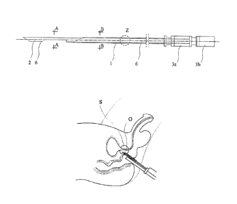
The invention is shown in more details in the examples and drawings
wherein Fig. 1 shows a biopsy needle in a top view, Fig. 2 biopsy needle in a
side
view, Fig. 3 a cross section of the stylet taken along the line A-A in Fig. 1,
Fig. 4
a cross-section through the stylet and cannula along line B-B in Fig. 1, Fig.
5
enlarged longitudinal section of the stylet and the cannula at the point Z in
Fig. 1
and Fig. 2; Fig. 6 biopsy needle in a side view, in a further embodiment, Fig.
7
enlarged longitudinal section of the stylet and the cannula at the point Y of
Fig. 6;
Fig. 8 biopsy needle in a side view, in a further embodiment, Fig. 9 enlarged
longitudinal section of the stylet and the cannula at the point X of Fig. 8;
Fig. 10
biopsy needle in a side view, in a further embodiment, Fig. 11 enlarged
longitudinal section through the stylet and cannula in place W of Fig. 10;
Fig. 12
biopsy needle in a side view, in a further embodiment, Fig. 13 enlarged
longitudinal section of the stylet and the cannula at the point V of Fig. 13;
Fig. 14
biopsy needle in a side view, in a further embodiment, Fig. 15 enlarged
longitudinal section of the stylet and the cannula at the point U of Fig. 14;
Fig. 16
biopsy needle in a side view, in a further embodiment, Fig. 17 enlarged
longitudinal section of the stylet and the cannula at the point T of Fig. 16;
Fig. 18
biopsy needle in a side view, in a further embodiment, Fig. 19 enlarged
CA 02989103 2017-12-11
WO 2016/118026
PCT/PL2016/000006
8
longitudinal section of the stylet and the cannula at the point S of Fig. 18,
Fig. 20
enlarged cannula surface; Fig. 21 biopsy needle in a side view, in a further
embodiment, Fig. 22 enlarged longitudinal section of the stylet and the
cannula at
the point R of Fig. 21, Fig. 23 enlarged axonometric view of the extended
layers
from segment I of the biopsy needle from Fig. 21; Fig. 24 biopsy needle in a
side
view, in a further embodiment, Fig. 25 enlarged longitudinal section of the
stylet
and the cannula at the point Q of Fig. 24, Fig. 26 enlarged axonometric view
of
the extended layers from segment II of the biopsy needle from Fig. 24; Fig. 27
biopsy needle in a side view, in a further embodiment, Fig. 28 enlarged
longitudinal section of the stylet and the cannula at the point P of Fig. 27;
Fig. 29
biopsy needle in a side view, in a further embodiment, in Fig. 30 enlarged
longitudinal section of the stylet and the cannula at the point 0 from Fig.
29, Fig.
31 enlarged axonometric view of the extended layers from segment III of the
biopsy needle from Fig. 29; Fig. 32 biopsy needle in a top view, in a further
embodiment, Fig. 33 biopsy needle in a side view, Fig. 34 cross-section of the
stylet taken along the line CC of Fig. 32, 33, 37 and 38; Fig. 35 is a cross
section
of the stylet and the cannula along the line DD of Fig. 32, 33, 37 and 38;
Fig. 36
enlarged longitudinal section through the stylet and cannula N of Fig. 32;
Fig. 37
biopsy needle in a top view, in a further embodiment, Fig. 38 biopsy needle in
a
side view, Fig. 39 anatomical chart of prostate surrounding during biopsy.
Example 1
A steel biopsy needle with 200 mm length, comprises a pointed cannula 1,
pointed stylet 2 mounted slidably in the cannula 1 and in the back the
polypropylene holders for the biopsy gun - a cannula holder 3a and stylet
holder
3b. The outer diameter of the cannula 1 is 1.93 mm, the inner diameter of the
cannula 1 is 1.70 mm, and the stylet 2 diameter is 1.65 mm.
The inner surface 4 of the cannula 1 and the outer surface 5 of the cannula 1
and
the surface of the stylet 2, over the entire length of the steel needles are
coated
CA 02989103 2017-12-11
WO 2016/118026
PCT/PL2016/000006
9
with the active layer 6 containing the biologically active agents, as shown in
Figs.
1-5. Active layer 6 has a biodegradable form allows for the controlled release
of
the biologically active agent by dissolving in water contained in the blood
and
biopted tissue. The active layer contains two antibiotics - ciprofloxacin and
amikacin which form inclusion complex with 13-cyclodextrin. An active layer 6
was obtained by mixing in a centrifuge (5 min, 30 rpm), ciprofloxacin,
amikacin
and p-cyclodext-rin in a molal ratio 1: 1: 2. The mixture of compounds was
dissolved in 0.14% aqueous solution of a nitrogen hydride (III). The obtained
solution was filtered. After immersion of the steel cannula 1 and stylet 2 in
solution, the freeze-drying process was carried out to perform the active
layer 6
fixation. As shown in Fig. 39 by using the biopsy gun and a biopsy needle
under
transrectal ultrasound, prostate cores s were collected in a standard way.
During
the procedure, ciprofloxacin and amikacin were directly release from the
biodegradable active layer 6 into the prostate tissue by disintegration of
inclusion
complex of 13-cyclodextrin with antibiotics. In vitro and in vivo studies
confirmed
the release of drugs from the active layer 6 applied to the steel surface of
the
biopsy needle and the antibacterial effect in the action area o of bioactive
agents.
Example 2
The needle is made as described in Example 1, except that the inner
surface 4 of the cannula 1 and part of the outer surface 5 of the cannula 1,
5cm
long from the tip of it, is coated with an active layer 6 as shown in Fig. 6
and Fig.
7. The active layer 6 contains two antibiotics: ciprofloxacin and amikacin, as
described in Example I.
Example 3
The needle is made as described in Example 1, except that the part of
stylet 2 surface, 7cm long from the tip of it, is coated with an active layer
6, as
CA 02989103 2017-12-11
WO 2016/118026
PCT/PL2016/000006
shown in Fig. 8 and Fig. 9. The active layer 6 contains two antibiotics:
ciprofloxacin and amikacin, as described in Example 1.
Example 4
The needle is made as described in Example 1, except that the active layer
6 is applied on the binder layer 7, which is placed on the outer surface 5 of
the
cannula 1 and the inner surface 4 of the cannula 1 and the surface of the
stylet 2,
as shown in Fig. 10 and Fig. 11. The binder layer 7 is made of poly(vinyl
alcohol).
The binder layer 7 was obtained by dipping the cannula I in a solution of
acetone
and drying, and then dipping in an aqueous solution of 0.5 mM/I, of poly(vinyl
alcohol) (molecular weight of 49,000 g mol-1). On such binder layer 7 the
active
layer 6, obtained as described in Example 1, was applied.
Example 5
The needle is made as described in Example 1, except that the active layer
6 is applied on the binder layer 7, which covers the outer surface 5 of the
cannula
1, as shown in Figs. 12 and Fig. 13. The binder layer 7 is made of
polyethylene
(vinyl alcohol). Binder layer 7 was obtained as described in Example 4 except
that
in the solution was immersed temporarily sealed cannula 1. Then, the outer
surface 5 of the cannula 1 was coated by spraying with antihemorrhagic
substance
- potassium aluminum sulfate dodecahydrate, thus leading to formation of an
active layer 6. In vitro and in vivo studies have confirmed the role of this
biopsy
needle to accelerate the coagulation process.
Example 6
The needle is made as described in Example 1, except that the outer
surface 5 of the cannula 1 is coated with the active layer 6, as shown in Fig.
14
and Fig. 15. The active layer 6 contains a biologically active agent -
fibrinogen, a
CA 02989103 2017-12-11
WO 2016/118026
PCT/PL2016/000006
11
protein which is involved in the coagulation process. Temporarily sealed
cannula
1 was dipped ten times in an aqueous solution of fibrinogen (10 mg/m1), and
dried
temporarily. In vitro and in vivo studies have confirmed the role of this
biopsy
needle to accelerate the coagulation process and thus minimize the risk of
bleeding.
Example 7
The needle is made as described in Example 1, except that the active layer
6 is applied to the binder layer 7, which is applied on the outer surface 5 of
the
cannula I and the surface of the stylet 2, as shown in Fig. 16 and Fig. 17.
The
active layer 6 contains a biologically active antiseptic agent - zeolite,
comprising
of 2.5% silver ions and 14% zinc ions. The binder layer 7 is made of
poly(vinyl
alcohol). The binder layer 7 was obtained by biopsy needle immersion in
acetone
solution and drying. Then temporarily sealed cannula 1 was immersed in an
aqueous solution of 0.5 mM/L of poly(vinyl alcohol) (molecular weight about 49
000 g = mol- 1). Then, the binder layer 7 was coated by spraying with zeolit.
The
needle was dried in 50 C for 1 hour to obtain the active layer 6 fixation.
Example 8
The needle is made as described in Example 1, except that the outer
surface 5 of the cannula 1 is coated with active layer 6 as shown in Figs. 18-
20.
The surface of the active layer 6 is covered with a protective layer 8 to
achieve
stable release of the biologically active agent during whole prostate biopsy
procedure. The protective layer 8 is made of poly(glycolic acid). The surface
of
the active layer 6 was coated by spraying with poly(glycolic acid) which forms
a
net-like protective layer 8. This layer delays the release of the biologically
active
agent from the active layer 6. The active layer 6 was obtained by mixing in a
centrifuge (5 min, 30 rpm) antibiotic - levofloxacin with P-cyclodextrin in
molal
CA 02989103 2017-12-11
WO 2016/118026
PCT/PL2016/000006
12
ratio 1:1. Further procedure was as described in Example 1, except that in the
solution was immersed the temporarily sealed cannula.
Example 9
The needle is made as described in Example 1, except that the outer
surface 5 of the cannula 1 and stylet 2 surface have an extended form with
porous
pits, as shown in Figs. 21-23. Pores 9a were obtained by micro laser engraving
with a diameter of 0.1 mm and a depth of 0.05 mm. The active layer 6 is
applied
on an extended porous fowl 9a of the outer surface 5 of the cannula 1 and on
an
extended porous surface 9a of the stylet 2. The outer surface 5 of the cannula
1
and stylet surface 2 were coated with a mixture of I3-cyclodextrin with
ciprofloxacin in the molal ratio 1:1. Further procedure was as described in
Example 1, except that in the solution was immersed the temporarily sealed
cannula.
Example 10
The needle is made as described in Example 1, except that the outer
surface 5 of the cannula 1 and surface of stylet 2 have an extended form with
grooved pits. The active layer 6 containing a complex of f3-cyclodextrin with
an
antibiotic - ciprofloxacin was formed on extended grooved form 9b of the outer
surface 5 of the cannula I and on the extended grooved form 9b of stylet 2, as
shown in Fig. 24-26. Grooves 9b were formed parallel to the axis of the
cannula 1
and stylet 2 to accumulate the antibiotic complex what allow an increase dose
of
active compound of the layer. The dimensions of the grooves 9b were 0.05 mm x
0.05 mm x 100 mm. Grooves 9b were obtained by micro laser engraving from the
tip of the cannula 1 and stylet 2. The active layer 6 is applied on an
extended
grooved form 9b of the outer surface 5 of the cannula 1 and on an extended
grooved surface 9b of stylet 2. The outer surface 5 of the cannula 1 and
stylet
CA 02989103 2017-12-11
WO 2016/118026
PCT/PL2016/000006
13
surface 2 were coated with a mixture of f3-cyclodextrin with ciprofloxacin in
the
molal ratio 1:1. Further procedure was as described in Example 1, except that
in
the solution was immersed the temporarily sealed cannula.
Example 11
The needle is made as described in Example 1, except that the active layer
6 is applied on the outer surface 5 of the cannula 1, as shown in Fig. 27 and
Fig.
28. The active layer 6 contains a biologically active antimicrobial agent -
levofloxacin. The active layer 6 was obtained by mixing in a centrifuge (5
min, 30
rpm) antibiotic - levofloxacin with 13-cyclodextrin in rnolal ratio 1 : 1.
Further
procedure was as described in Example 1, except that in the solution was
immersed the temporarily sealed cannula. The surface of the active layer 6 is
coated by spraying with the aqueous solution of 0.5 mM/L of poly(vinyl
alcohol)
(molecular weight about 49 000 g mol- 1) to form a protective layer 8 which
was
made to achieve stable release of the biologically active agent. The
protective
layer 8 had a thickness of 0.02 mm.
Example 12
The biopsy needle is made as described in Example 1, except that the outer
surface 5 of the cannula 1 is coated with two active layers 6, as shown in
Figs. 29-
31. The first active layer 6 is applied to the binder layer 7 which is located
on the
outer surface 5 of the cannula 1. Binder layer 7 was obtained by dipping the
cannula 1 in a solution of acetone and drying, and then dipping in an aqueous
solution of 0.5 mM/L of poly(vinyl alcohol) (molecular weight of 49,000 g -
mol-
l). On such binder layer 7 the first active layer 6, obtained as described in
Example 1, was applied, except that the cannula 1 was temporarily sealed.
Then,
the first active layer 6 was coated by spraying with poly(glycolic acid) which
forms a net-like protective layer 8. This layer delays the release of the
biologically
active agent from the first active layer 6. The second active layer 6, was
applied
CA 02989103 2017-12-11
WO 2016/118026
PCT/PL2016/000006
14
on the protective layer 8. The second active layer was obtained as in Example
1,
except that the cannula 1 was temporarily sealed.
Example 13
A steel biopsy needle with 200 mm length, comprises a pointed cannula 1,
pointed stylet 2 slidably located in the cannula 1 and in the back the
polypropylene holders for the biopsy gun - a cannula holder 3a and stylet
holder
3b. The outer width of the cannula 1 is 1.52 mm, its height is 2.22 mm, and
the
stylet 2 diameter is 1.27 mm as shown in Figs. 32-36. The cannula 1 has a
circular
cross-section and one axis of symmetry. The cannula 1 contains main channel
with diameters of 1.32 mm (for the stylet) and a longitudinal pass-through-
hole
channel 10 of cannula 1. The additional channel 10 of circular shaped in cross
section with the diameter of 0.5 mm, passes through 150 mm of cannula 1,
beginning at its pointed end, protrudes over the outer surface 5 of the
cannula 1
and then transfer into the steel connector 11. The connector 11 has a tube
form, a
length of 25 mm, an inner diameter of 0.5 mm and an external diameter 0.65 mm.
The connector 11 of the channel 10 is connected to a flexible tube 12, made of
poly(vinyl chloride), with an internal diameter of 0.5 mm and a length of 200
mm.
The flexible tube 12 is connected with a 5m1 syringe 13. As shown in Fig. 39
by
using the biopsy gun and a biopsy needle under transrectal ultrasound,
prostate
cores s were collected in a standard way. During the procedure, at each
sequence
of collecting biopsy samples 0.1 ml of an aqueous solution of levofloxacin (5
mg/m1) and 0.1 ml of lidocaine hydrochloride (20 mg/ml) were administered
through the additional channel 10. In vitro and in vivo studies confirmed the
antibacterial effect in the action area o of bioactive agents. The analgesic
effect
was confirmed in vivo.
CA 02989103 2017-12-11
WO 2016/118026
PCT/PL2016/000006
Example 14
A steel biopsy needle with 200 mm length, comprises a pointed cannula 1,
pointed stylet 2 slidably located in the cannula 1 and in the back the
polypropylene holders for the biopsy gun - a cannula holder 3a and stylet
holder
3b. The outer width of the cannula 1 is 1.52 mm, its height is 2.22 mm, and
the
stylet 2 diameter is 1.27 mm as shown in Figs. 34-38. Cannula 1 has a circular
cross-section and one axis of symmetry. The cannula 1 contains main channel
with diameters of 1.32 mm (for the stylet) and a longitudinal pass-through-
hole
channel 10 of cannula 1. The additional channel 10 of circular shaped in cross
section with the diameter of 0.5 mm, passes through the entire length of the
cannula 1, from its pointed end to the holder 3a. As shown in Fig. 39 by using
the
biopsy gun and a biopsy needle under transrectal ultrasound, prostate cores s
were
collected in a standard way. During the procedure, at each sequence of
collecting
biopsy samples 0.1 ml of an aqueous solution of levofloxacin (5 mg/m1) and 0.1
ml of lidocaine hydrochloride (20 mg/ml) were administered through the
additional channel 10. In vitro and in vivo studies confirmed the
antibacterial
effect in the action area o of bioactive agents. The analgesic effect was
confirmed
in vivo.