Note: Descriptions are shown in the official language in which they were submitted.
CA 02989144 2017-12-11
WO 2016/201425 PCT/US2016/037207
TREATMENT OF CANCER BY COMBINED BLOCKADE OF
THE PD-1 AND CXCR4 SIGNALING PATHWAYS
Throughout this application, various publications are referenced in
parentheses by
author name and date, or by Patent No. or Patent Publication No. Full
citations for these
publications may be found at the end of the specification immediately
preceding the
claims. The disclosures of these publications are hereby incorporated in their
entireties by
reference into this application in order to more fully describe the state of
the art as known
to those skilled therein as of the date of the invention described and claimed
herein.
However, the citation of a reference herein should not be construed as an
acknowledgement that such reference is prior art to the present invention.
CROSS REFERENCE TO RELATED APPLICATIONS
This application is entitled to priority pursuant to 35 U.S.C. 119(e) to U.S.
Provisional Application No. 62/174,931, filed June 12, 2015, which is
incorporated herein
in its entirety.
FIELD OF THE INVENTION
This invention relates to methods for treating a cancer in a subject
comprising
administering to the subject a combination of an antibody that blocks the
Programmed
Death-1 (PD-1)/Programmed Death Ligand-1 (PD-L1) signaling pathway and an
antibody
that blocks the C-X-C Chemokine Receptor 4 (CXCR4)/C-X-C motif chemokine 12
(CXCL12) signaling pathway.
BACKGROUND OF THE INVENTION
Human cancers harbor numerous genetic and epigenetic alterations, generating
neoantigens potentially recognizable by the immune system (Sjoblom et al.,
2006). The
adaptive immune system, comprised of T and B lymphocytes, has powerful anti-
cancer
potential, with a broad capacity and exquisite specificity to respond to
diverse tumor
antigens. Further, the immune system demonstrates considerable plasticity and
a memory
component. The successful harnessing of all these attributes of the adaptive
immune
system makes immunotherapy unique among all cancer treatment modalities.
Until recently, cancer immunotherapy had focused substantial effort on
approaches that enhance anti-tumor immune responses by adoptive-transfer of
activated
effector cells, immunization against relevant antigens, or providing non-
specific immune-
stimulatory agents such as cytokines. In the past decade, however, intensive
efforts to
1
CA 02989144 2017-12-11
WO 2016/201425 PCT/US2016/037207
develop specific immune checkpoint pathway inhibitors have provided new
immunotherapeutic approaches for treating cancer, including the development of
an
antibody (Ab), ipilimumab (YERVOYg), that binds to and inhibits Cytotoxic T-
Lymphocyte Antigen-4 (CTLA-4) for the treatment of patients with advanced
melanoma
(Hodi et al., 2010) and the development of Abs such as nivolumab (OPDIV0g) and
pembrolizumab (KEYTRUDA4D) that bind specifically to the PD-1 receptor, a cell
surface negative regulatory molecule expressed by activated T and B
lymphocytes, and
block the inhibitory PD-1/PD-1 ligand pathway (Topalian et al., 2012a, b;
Topalian et al.,
2014; Hamid et al., 2013; Hamid and Carvajal, 2013; McDermott and Atkins,
2013). This
pathway can also be disrupted by Abs that bind specifically to PD-L1,
including BMS-
936559 (PCT Publication No. WO 2013/173223) and atezolizumab (TECENTRIQ41);
Fehrenbacher et al., 2016).
Nivolumab (previously designated BMS-936558, MDX-1106, or ONO-4538, and
designated 5C4 in U.S. Patent No. 8,008,449) is a fully human immunoglobulin
(Ig) G4
(S228P) monoclonal antibody (mAb) that selectively prevents interaction with
the PD-1
ligands, PD-L1 and PD-L2 (U.S. Patent No. 8,008,449; Wang et al., 2014),
thereby
blocking the down-regulation of antigen-specific T cell responses directed
against both
foreign (including tumor) and self antigens and enhancing an immune response
against
these antigens (McDermott and Atkins, 2013). Nivolumab has received approval
recently
for metastatic melanoma, squamous non-small cell lung cancer (NSCLC), renal
cell
carcinoma (RCC) and classical fiodgkin lymphoma Will.), and is currently being
clinically evaluated as monotherapy or in combination with ipilimumab or other
anti-
cancer agents for efficacy in various tumor types, including pancreatic cancer
(PAC),
small cell lung cancer (SCLC), head and neck cancer, bladder cancer and
hematological
malignancies (see, e.g., Topalian et al., 2012b; WO 2013/173223; Ansell et
al., 2015; and
NCT02309177, NCT01928394, NCT02105636, NCT02387996, and NCT02329847 on
the Clinical Trials Web site, http://www.clinicaltrials.gov). However,
combinations of
nivolumab with other targeted therapies may further improve response rates and
prolong
survival in a higher percentage of patients. Specifically, for example, the
combination of
nivolumab with therapies targeting the protective stromal microenvironment
surrounding
the tumor may allow for enhanced infiltration of activated immune cells to the
tumor site,
thereby increasing tumor cell killing and broadening the spectrum of patients
able to
benefit from these therapies.
2
CA 02989144 2017-12-11
WO 2016/201425 PCT/US2016/037207
Ulocuplumab (previously designated BMS-936564 or MDX-1338, and designated
F7 in WO 2008/060367) is a fully human IgG4 (S224P) mAb specific for CXCR4,
which
is expressed on leukocytes, platelets and other non-hematopoietic cells that
comprise the
tumor stromal microenvironment (Balkwill, 2004). CXCR4 is also over-expressed
in the
majority of human cancers and, together with its endogenous ligand CXCL12,
plays a
fundamental role in cancer pathogenesis including proliferation, adhesion,
metastasis,
angiogenesis and survival (Domanska et al., 2013; Duda et al., 2011; Balkwill,
2004; Pitt
et al., 2015; Passoro et al., 2015; WO 2008/060367). Ulocuplumab has been
evaluated in
two Phase 1 clinical trials in subjects with various hematological
malignancies including
acute myeloid leukemia (AML), multiple myeloma (MM), chronic lymphocytic
leukemia
(CLL), follicular lymphoma (FL) and diffuse large B cell lymphoma (DLBCL) with
a
safe and tolerable profile. Efficacy data from the AML and MM cohorts has been
presented and show encouraging results for the addition of ulocuplumab to
standard
therapy (Becker et al., 2014; Ghobrial et al., 2014).
Evidence has been presented suggesting that CXCL12 may be
immunosuppressive and may support the stroma surrounding the tumor, shielding
it from
immune mechanisms that would otherwise result in tumor cell killing (Domanska
et al.,
2013; Duda et al., 2011; Burger and Kipps, 2006). The refractory nature of
many
metastatic tumors, including PAC and SCLC, may result from an
immunosuppressive
environment surrounding the tumor that prevents activated lymphocytes from
accessing
the tumor site. It is, therefore, of interest to determine whether disruption
of the stromal
microenvironment via CXCR4 blockade with an anti-CXCR4 Ab could increase the
tumor's susceptibility to immune-targeted therapies and allow for the
penetration of
immune cells to the tumor site. Furthermore, ulocuplumab may be involved in
direct
cytotoxicity against the tumor since it has demonstrated direct in vitro cell
killing activity
of CXCR4-expressing tumor cells (Kuhne et al., 2013; WO 2013/071068). CXCR4 is
also over-expressed on immune-suppressive regulatory T cells (Tregs) and
myeloid-
derived suppressor cells (MDSCs) in cancer patients (Wang et al., 2012;
Obermajer et al.,
2011; Katoh and Watanabe, 2015), and anti-CXCR4-mediated depletion of Tregs
and/or
MDSCs may contribute to enhancement of an anti-tumor effect.
The present disclosure relates to studies evaluating Ab-mediated dual blockade
of
the PD-1/PD-L1 and CXCR4/CXCL12 signaling pathways to determine whether the
combined inhibition of these pathways benefit cancers that are poorly treated
by standard
3
CA 02989144 2017-12-11
WO 2016/201425
PCT/US2016/037207
therapies. The combination of the mechanisms of action of anti-CXCR4/anti-
CXCL12
and anti-PD-1/anti-PD-L1 offers a unique opportunity to simultaneously target
the
immunosuppressive tumor microenvironment and the activation of T cells, thus
increasing tumor cell killing.
SUMMARY OF THE INVENTION
The present disclosure provides a method for treating a subject afflicted with
a
cancer comprising administering to the subject a combination of
therapeutically effective
amounts of: (a) an Ab or an antigen-binding portion thereof that binds
specifically to PD-
1 or to PD-L1; and (b) an Ab or an antigen-binding portion thereof that binds
specifically
to CXCR4 or to CXCL2. In certain embodiments, the Ab that binds specifically
to PD-1
or to PD-L1 disrupts the interaction between PD-1 and PD-L1, and inhibits PD-
1/PD-L1
signaling. In other embodiments, the Ab that binds to CXCR4 or CXCL2 disrupts
the
interaction between CXCR4 and CXCL12, and inhibits CXCR4/CXCL12 signaling. In
further embodiments, the cancer is a solid tumor such as PAC, SCLC or
hepatocellular
carcinoma (HCC). In certain embodiments of any of the therapeutic methods
disclosed
herein, the Ab that binds to PD-1 is nivolumab or pembrolizumab. In certain
other
embodiments, the Ab that binds specifically to PD-L1 is BMS-936559,
atezolizumab,
durvalumab, STI-A1014 or avelumab. In yet other embodiments, the Ab that the
Ab that
binds specifically to CXCR4 is ulocuplumab, or preferably, ulocuplumab
modified to
comprise an Fc region with effector functions, for example an Fc region of a
human IgG1
or human IgG3 isotype. In further embodiments, the Ab that binds specifically
to CXCL2
is the mAb designated 2A5 in U.S. Patent No. 8,496,931.
In certain embodiments of the methods comprising use of an anti-PD-1 Ab in
combination with an anti-CXCR4 Ab, the therapeutically effective dosage of the
anti-PD-
1 Ab or antigen-binding portion thereof ranges from about 0.1 to about 20
mg/kg body
weight administered by intravenous infusion about once every 2, 3 or 4 weeks.
In certain
preferred embodiments, the anti-PD-1 Ab is administered at a dose of about 2
mg/kg or
about 3 mg/kg once every 2 or 3 weeks. In certain other embodiments of these
methods
the therapeutically effective dosage of the anti-CXCR4 Ab or antigen-binding
portion
thereof ranges from a flat dose of about 50 to about 2000 mg administered
weekly by
intravenous infusion. In certain preferred embodiments, the anti-CXCR4 Ab is
administered at a flat dose of about 400 or about 800 mg weekly.
The disclosure also provides a kit for treating a subject afflicted with a
cancer, the
4
CA 02989144 2017-12-11
WO 2016/201425
PCT/US2016/037207
kit comprising: (a) one or more dosages ranging from about 0.1 to about 20
mg/kg body
weight of an Ab or an antigen-binding portion thereof that binds specifically
to PD-1 or to
PD-L1; (b) one or more dosages ranging from about 50 to about 2000 mg of an Ab
or an
antigen-binding portion thereof that binds specifically to CXCR4 or to CXCL12;
and (c)
instructions for using the Ab or portion thereof that binds specifically to PD-
1 or to PD-
L1 and the Ab or portion thereof that binds specifically to CXCR4 or to
CXCL12.
Other features and advantages of the instant invention will be apparent from
the
following detailed description and examples which should not be construed as
limiting.
The contents of all cited references, including scientific articles, GenBank
entries, patents
and patent applications cited throughout this application are expressly
incorporated herein
by reference.
BRIEF DESCRIPTION OF THE FIGURES
Figure 1 shows an assessment of CXCR4 expression on mouse Kpl and Kp3
SCLC cell lines by flow cytometry.
Figure 2 shows an assessment of CXCR4 expression on the MC38 mouse colon
adenocarcinoma cell line by flow cytometry.
Figure 3 shows an assessment of CXCR4 expression on CD8+ T cells, T effector
cells and regulatory T cells (Tregs) by flow cytometry.
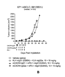
Figure 4 shows the effects on tumor growth of anti-mCXCR4 and anti-mouse PD-
1 Abs used alone or in combination in a syngeneic endogenous CXCR4-expressing
mouse SCLP model derived from a KP1 tumor cell line (p53; Rbl; p130 null;
B6129S
Fl mice). A, Median change in tumor volume from treatment with single Abs
compared
to controls. B, Median change in tumor volume from treatment with combination
of Abs
compared to controls. Vehicle: saline; KLH mIgG1 (or mIgG1 KLH): anti-Keyhole
Limpet Hemocyanin (KLH) mAb having mouse IgG1 isotype; mIgG2a KLH: anti- KLH
mAb having mouse IgG2a isotype; mCXCR4 mIgG1 (4.8): anti-mouse CXCR4 Ab
(clone 4.8) having mouse IgG1 isotype; mCXCR4 mIgG2a: anti-mouse CXCR4 Ab
(clone 4.8) having mouse IgG2a isotype; mPD-1 mIgG1 (or simply "PD-1"): anti-
PD-1
mAb 4H2 having mouse IgG1 isotype. Similar abbreviations are used in the other
figures
relating to anti-tumor efficacy studies in mouse tumor models.
Figure 5 shows the effects on tumor growth of anti-mCXCR4 IgG2a and anti-
mouse PD-1 Abs used alone or in combination in a syngeneic endogenous CXCR4-
nonexpressing mouse SCLP model derived from a Kp3 tumor cell line (P53; Rbl;
p130
5
CA 02989144 2017-12-11
WO 2016/201425 PCT/US2016/037207
null; B6129S Fl mice). A, Median change in tumor volume from treatment with
single
Abs compared to controls. B, Median change in tumor volume from treatment with
combination of Abs compared to controls.
Figure 6 shows the effects of anti-mCXCR4 and anti-mouse PD-1 Abs used alone
or in combination in a CXCR4-nonexpressing mouse colon carcinoma model derived
from a MC38 tumor cell line (BC57BI/6 mice). A, Median change in tumor volume
from
treatment with single Abs compared to controls. B, Median change in tumor
volume
from treatment with combination of Abs compared to controls.
Figure 7 shows the effects of the combination of anti-mCXCR4 mIgG2a and anti-
mPD-1 mIgG1D265A Abs in combination in inhibiting the growth of a CXCR4-
nonexpressing H22 liver cancer mouse model. A, Change in tumor volume in eight
individual mice from treatment with anti-mCXCR4 plus anti-mPD-1. B, Change in
tumor
volume in eight individual mice from treatment with anti-mPD-1. C, Change in
tumor
volume in eight individual mice from treatment with combination of isotype
controls. D,
Median changes in tumor volumes for the treatments shown in (A) to (C).
Figure 8 shows a schematic summarizing the design of a Phase 1/2 study of
ulocuplumab in combination with nivolumab to evaluate the safety and efficacy
of this
combination of therapeutic Abs in subjects with SCLC and PAC.
Figure 9 shows the receptor occupancy (RO) on circulating CD3+ cells (T cells)
in
the patient cohort dosed with a combination of 200 mg ulocuplumab weekly and 3
mg/kg
nivolumab every two weeks. Data are depicted as absolute % RO by ulocuplumab
on
circulating CD3+ cells. Gray horizontal lines indicate median values. Each dot
represents
a subject sample.
DETAILED DESCRIPTION OF THE INVENTION
The present invention relates to methods for treating solid tumors in a
subject
comprising administering a combination of an anti-PD-1 or an anti-PD-L1 Ab and
an
anti-CXCR4 or anti-CXCL12 Ab to the subject.
Terms
In order that the present disclosure may be more readily understood, certain
terms
are first defined. As used in this application, except as otherwise expressly
provided
herein, each of the following terms shall have the meaning set forth below.
Additional
definitions are set forth throughout the application.
6
CA 02989144 2017-12-11
WO 2016/201425
PCT/US2016/037207
"Administering" refers to the physical introduction of a composition
comprising a
therapeutic agent to a subject, using any of the various methods and delivery
systems
known to those skilled in the art. A preferred route for administration of
therapeutic Abs
such as anti-PD-1 and anti-CXCR4 Abs is intravenous administration. Other
routes of
administration include intramuscular, subcutaneous, intraperitoneal, or other
parenteral
routes of administration, for example by injection or infusion. The phrase
"parenteral
administration" as used herein means modes of administration other than
enteral and
topical administration. Administering can also be performed, for example,
once, a
plurality of times, and/or over one or more extended periods.
An "adverse event" (AE) is any new untoward medical occurrence or worsening
of a preexisting medical condition in a clinical investigation subject
administered study
drug and need not have a causal relationship with this treatment. An AE can
therefore be
any unfavorable and unintended sign (such as an abnormal laboratory finding),
symptom,
or disease temporally associated with the use of study drug, whether or not
considered
related to the study drug. The causal relationship to study drug is determined
by a
physician and is used to assess all AEs. The causal relationship can either
"related" (i.e.,
there is a reasonable causal relationship between study drug administration
and the AE),
or "not related" (i.e., there is not a reasonable causal relationship between
study drug
administration and the AE). The term "reasonable causal relationship" means
there is
evidence to suggest a causal relationship. Reference to methods or dosages for
"reducing
adverse events" means a treatment regime, e.g., a combination of an anti-PD-
1/anti-PD-
L1 Ab and an anti-CXCR4/anti-CXCL12 Ab, that decreases the incidence and/or
severity
of one or more AEs associated with the use of a different treatment regime,
e.g.,
monotherapy with an anti-PD-1/anti-PD-L1 or an anti-CXCR4/anti-CXCL12 Ab.
A "serious adverse event" (SAE) is any untoward medical occurrence that at any
dose results in death, is life-threatening (defined as an event in which the
subject was at
risk of death at the time of the event; it does not refer to an event which
hypothetically
might have caused death if it were more severe), requires inpatient
hospitalization or
causes prolongation of existing hospitalization, results in persistent or
significant
disability/incapacity, is a congenital anomaly/birth defect, and/or is an
important medical
event (defined as a medical event(s) that may not be immediately life-
threatening or result
in death or hospitalization but, based upon appropriate medical and scientific
judgment,
may jeopardize the subject or may require intervention to prevent a more
serious
7
CA 02989144 2017-12-11
WO 2016/201425 PCT/US2016/037207
outcome). Examples of such important medical events include, but are not
limited to,
intensive treatment in an emergency room or at home for allergic bronchospasm,
blood
dyscrasias or convulsions that do not result in hospitalization, and potential
drug-induced
liver injury (DILI).
An "antibody" (Ab) shall include, without limitation, a glycoprotein
immunoglobulin (Ig) which binds specifically to an antigen and comprises at
least two
heavy (H) chains and two light (L) chains interconnected by disulfide bonds,
or an
antigen-binding portion thereof. Each H chain comprises a heavy chain variable
region
(abbreviated herein as VH) and a heavy chain constant region. The heavy chain
constant
region of an IgG Ab comprises three constant domains, CHi, CH2 and CH3. Each
light
chain comprises a light chain variable region (abbreviated herein as VL) and a
light chain
constant region. The light chain constant region of an IgG Ab comprises one
constant
domain, CL. The VH and VL regions can be further subdivided into regions of
hypervariability, termed complementarity determining regions (CDRs),
interspersed with
regions that are more conserved, termed framework regions (FR). Each VH and VL
comprises three CDRs and four FRs, arranged from amino-terminus to carboxy-
terminus
in the following order: FR1-CDR1-FR2-CDR2-FR3-CDR3-FR4. The variable regions
of
the heavy and light chains contain a binding domain that interacts with an
antigen. The
constant regions of the Abs may mediate the binding of the immunoglobulin to
host
tissues or factors, including various cells of the immune system (e.g.,
effector cells) and
the first component (Clq) of the classical complement system.
An Ig may derive from any of the commonly known isotypes, including but not
limited to IgA, secretory IgA, IgG and IgM. IgG subclasses are also well known
to those
in the art and include but are not limited to human IgGl, IgG2, IgG3 and IgG4.
"Isotype"
refers to the Ab class or subclass (e.g., IgM, IgGl, or IgG4) that is encoded
by the heavy
chain constant region genes. The term "antibody" includes, by way of example,
both
naturally occurring and non-naturally occurring Abs; monoclonal and polyclonal
Abs;
chimeric and humanized Abs; human or nonhuman Abs; wholly synthetic Abs; and
single
chain Abs. A nonhuman Ab may be humanized partially or fully by recombinant
methods
to reduce its immunogenicity in man. Where not expressly stated, and unless
the context
indicates otherwise, the term "antibody" also includes an antigen-binding
fragment or an
antigen-binding portion of any of the aforementioned immunoglobulins, and
includes a
monovalent and a divalent fragment or portion, and a single chain Ab.
8
CA 02989144 2017-12-11
WO 2016/201425 PCT/US2016/037207
An "isolated" Ab refers to an Ab that is substantially free of other Abs
having
different antigenic specificities (e.g., an isolated Ab that binds
specifically to PD-1 is
substantially free of Abs that bind specifically to antigens other than PD-1).
An isolated
Ab that binds specifically to PD-1 may, however, have cross-reactivity to
other antigens,
such as PD-1 molecules from different species. Moreover, an isolated Ab may be
purified
so as to be substantially free of other cellular material and/or chemicals.
The term "monoclonal" Ab (mAb) refers to a non-naturally occurring preparation
of Ab molecules of single molecular composition, i.e., Ab molecules whose
primary
sequences are essentially identical, which exhibits a single binding
specificity and affinity
for a particular epitope. A mAb is an example of an isolated Ab. MAbs may be
produced
by hybridoma, recombinant, transgenic or other techniques known to those
skilled in the
art.
A "chimeric" Ab refers to an Ab in which the variable regions are derived from
one species and the constant regions are derived from another species, such as
an Ab in
which the variable regions are derived from a mouse Ab and the constant
regions are
derived from a human Ab.
A "human" mAb (HuMAb) refers to a mAb having variable regions in which both
the framework and CDR regions are derived from human germline immunoglobulin
sequences. Furthermore, if the Ab contains a constant region, the constant
region also is
derived from human germline immunoglobulin sequences. The human Abs of the
invention may include amino acid residues not encoded by human germline
immunoglobulin sequences (e.g., mutations introduced by random or site-
specific
mutagenesis in vitro or by somatic mutation in vivo). However, the term
"human" Ab, as
used herein, is not intended to include Abs in which CDR sequences derived
from the
germline of another mammalian species, such as a mouse, have been grafted onto
human
framework sequences. The terms "human" Abs and "fully human" Abs are used
synonymously.
A "humanized" mAb refers to a mAb in which some, most or all of the amino
acids outside the CDR domains of a non-human mAb are replaced with
corresponding
amino acids derived from human immunoglobulins. In one embodiment of a
humanized
form of an Ab, some, most or all of the amino acids outside the CDR domains
have been
replaced with amino acids from human immunoglobulins, whereas some, most or
all
amino acids within one or more CDR regions are unchanged. Small additions,
deletions,
9
CA 02989144 2017-12-11
WO 2016/201425 PCT/US2016/037207
insertions, substitutions or modifications of amino acids are permissible as
long as they
do not abrogate the ability of the Ab to bind to a particular antigen. A
"humanized" Ab
retains an antigenic specificity similar to that of the original Ab.
An "anti-antigen" Ab refers to an Ab that binds specifically to an antigen.
For
example, an anti-PD-1 Ab is an Ab that binds specifically to PD-1, whereas an
anti-
CXCR4 Ab is an Ab that binds specifically to CXCR4. As used herein, an "anti-
PD-
1/anti-PD-L1" Ab is an Ab that is used to disrupt the PD-1/PD-L1 signaling
pathway,
which is an anti-PD-1 Ab or an anti-PD-L1 Ab. Similarly, an "anti-CXCR4/anti-
CXCL12" Ab is an Ab that is used to disrupt the CXCR4/CXCL12 signaling
pathway,
which is an anti-CXCR4 Ab or an anti-CXCL12 Ab.
An "antigen-binding portion" of an Ab (also called an "antigen-binding
fragment") refers to one or more fragments of an Ab that retain the ability to
bind
specifically to the antigen bound by the whole Ab.
A "cancer" refers a broad group of various diseases characterized by the
uncontrolled growth of abnormal cells in the body. Unregulated cell division
and growth
divide and grow results in the formation of malignant tumors that invade
neighboring
tissues and may also metastasize to distant parts of the body through the
lymphatic system
or bloodstream.
"C-X-C Chemokine Receptor 4" (CXCR4; also known in the art as, for example,
LESTR, Fusin or CD184) refers to a 7-transmembrane G-protein coupled receptor
expressed on leukocytes, platelets and other non-hematopoietic cells that
comprise the
tumor stromal microenvironment. It is also over-expressed in the majority of
human
cancers and on Tregs and MDSCs. CXCR4 binds to a single ligand, CXCL12. The
term
"CXCR4" as used herein includes human CXCR4 (hCXCR4), variants, isoforms, and
species homologs of hCXCR4, and analogs having at least one common epitope
with
hCXCR4. The complete hCXCR4 amino acid sequence can be found under
GENBANK Accession No. CAA12166.
"C-X-C motif chemokine 12" (CXCL12; also known as stromal cell-derived
factor 1 or SDF-1) is a chemokine that is the only known ligand for the CXCR4
receptor
though it may also serve as a ligand for a second receptor, CXCR7 (RDC1).
CXCL12 is
strongly chemotactic for lymphocytes, and plays an important role in
angiogenesis by
recruiting endothelial progenitor cells from the bone marrow through a CXCR4-
dependent mechanism. It is also thought to be involved in directing metastasis
of
CA 02989144 2017-12-11
WO 2016/201425 PCT/US2016/037207
CXCR4+ tumor cells to organs such as lymph node, lung, liver and bone that
highly
express CXCL12. The term "CXCL12" as used herein includes human CXCL12
(hCXCL12), variants, isoforms, and species homologs of hCXCL12, and analogs
having
at least one common epitope with hCXCL12. Human CXCL12 is produced in three
forms, CXCL12a, CXCL12b and CXCL12c, by alternate splicing of the same gene.
The
complete amino acid sequence of exemplary CXCL12a, CXCL12b and CXCL12c
isoforms can be found under GENBANK Accession Nos. NP 954637, NP 000600
and NP 001029058, respectively.
The term "immunotherapy" refers to the treatment of a subject afflicted with,
or at
risk of contracting or suffering a recurrence of, a disease by a method
comprising
inducing, enhancing, suppressing or otherwise modifying an immune response.
"Treatment" or "therapy" of a subject refers to any type of intervention or
process
performed on, including the administration of an active agent to, the subject
with the
objective of reversing, alleviating, ameliorating, inhibiting, slowing down or
preventing
the onset, progression, development, severity or recurrence of a symptom,
complication
or condition, or biochemical indicia associated with a disease.
"Programmed Death-1" (PD-1) refers to an immunoinhibitory receptor
belonging to the CD28 family that is expressed predominantly on previously
activated T
cells in vivo, and binds to two ligands, PD-L1 and PD-L2. The term "PD-1" as
used
herein includes human PD-1 (hPD-1), variants, isoforms, and species homologs
of hPD-
1, and analogs having at least one common epitope with hPD-1. The complete hPD-
1
amino acid sequence can be found under GENBANK Accession No. U64863.
"Programmed Death Ligand-1" (PD-L1) is one of two cell surface glycoprotein
ligands for PD-1 (the other being PD-L2) that downregulate T cell activation
and
cytokine secretion upon binding to PD-1. The term "PD-L1" as used herein
includes
human PD-L1 (hPD-L1), variants, isoforms, and species homologs of hPD-L1, and
analogs having at least one common epitope with hPD-L1. The complete hPD-L1
sequence can be found under GENBANK Accession No. Q9NZQ7.
A "subject" includes any human or nonhuman animal. The term "nonhuman
animal" includes, but is not limited to, vertebrates such as nonhuman
primates, sheep,
dogs, and rodents such as mice, rats and guinea pigs. In preferred
embodiments, the
subject is a human. The terms "subject" and "patient" are used interchangeably
herein.
A "therapeutically effective amount" or "therapeutically effective dosage" of
a
11
CA 02989144 2017-12-11
WO 2016/201425 PCT/US2016/037207
drug or therapeutic agent is any amount of the drug or agent that, when used
alone or in
combination with another therapeutic agent, protects a subject against the
onset of a
disease or promotes disease regression evidenced by a decrease in severity of
disease
symptoms, an increase in frequency and duration of disease symptom-free
periods, or a
prevention or reduction of impairment or disability due to the disease
affliction. In
addition, the terms "effective" and "effectiveness" with regard to a treatment
includes
both pharmacological effectiveness and physiological safety. Pharmacological
effectiveness refers to the ability of the drug to promote disease regression,
e.g., cancer
regression, in the patient. Physiological safety refers to an acceptable level
of toxicity,
or other adverse physiological effects at the cellular, organ and/or organism
level
(adverse effects) resulting from administration of the drug. The efficacy of a
therapeutic
agent can be evaluated using a variety of methods known to the skilled
practitioner, such
as in human subjects during clinical trials, in animal model systems
predictive of
efficacy in humans, or by assaying the activity of the agent in in vitro
assays.
By way of example for the treatment of tumors, a therapeutically effective
amount of an anti-cancer agent preferably inhibits cell growth or tumor growth
by at
least about 20%, more preferably by at least about 40%, even more preferably
by at least
about 60%, and still more preferably by at least about 80% relative to
untreated subjects.
In other preferred embodiments of the invention, tumor regression may be
observed and
continue for a period of at least about 20 days, more preferably at least
about 40 days, or
even more preferably at least about 60 days. Notwithstanding these ultimate
measurements of therapeutic effectiveness, evaluation of immunotherapeutic
drugs must
also make allowance for "immune-related" response patterns.
An "immune-related" response pattern refers to a clinical response pattern
often
observed in cancer patients treated with immunotherapeutic agents that produce
antitumor
effects by inducing cancer-specific immune responses or by modifying native
immune
processes. This response pattern is characterized by a beneficial therapeutic
effect that
follows an initial increase in tumor burden or the appearance of new lesions,
which in the
evaluation of traditional chemotherapeutic agents would be classified as
disease
progression and would be synonymous with drug failure. Accordingly, proper
evaluation
of immunotherapeutic agents may require long-term monitoring of the effects of
these
agents on the target disease.
A therapeutically effective amount of a drug includes a "prophylactically
12
CA 02989144 2017-12-11
WO 2016/201425 PCT/US2016/037207
effective amount," which is any amount of the drug that, when administered
alone or in
combination with an another therapeutic agent to a subject at risk of
developing a
disease (e.g., a subject having a pre-malignant condition who is at risk of
developing a
cancer) or of suffering a recurrence of the disease, inhibits the development
or
recurrence of the disease (e.g., a cancer). In preferred embodiments, the
prophylactically
effective amount prevents the development or recurrence of the disease
entirely.
"Inhibiting" the development or recurrence of a disease means either lessening
the
likelihood of the disease's development or recurrence, or preventing the
development or
recurrence of the disease entirely.
The use of the alternative (e.g., "or") should be understood to mean either
one,
both, or any combination thereof of the alternatives. As used herein, the
indefinite
articles "a" or "an" should be understood to refer to "one or more" of any
recited or
enumerated component.
The term "about" refers to a numeric value, composition or characteristic that
is
within an acceptable error range for the particular value, composition or
characteristic as
determined by one of ordinary skill in the art, which will depend in part on
how the value,
composition or characteristic is measured or determined, i.e., the limitations
of the
measurement system. For example, "about" can mean within 1 or within more than
1
standard deviation per the practice in the art. Alternatively, it can mean a
range of plus or
minus 20%, more usually a range of plus or minus 10%. When particular values,
compositions or characteristics are provided in the application and claims,
unless
otherwise stated, the meaning of "about" should be assumed to be within an
acceptable
error range for that particular value, composition or characteristic.
The term "substantially the same" or "essentially the same" refers to a
sufficiently
high degree of similarity between two or more numeric values, compositions or
characteristics that one of skill in the art would consider the difference
between these
values, compositions or characteristics to be of little or no biological
and/or statistical
significance within the context of the property being measured. The difference
between
numeric values being measured may, for example, be less than about 50%,
preferably less
than about 30%, and more preferably less than about 10%.
As described herein, any concentration range, percentage range, ratio range or
integer range is to be understood to include the value of any integer within
the recited
range and, when appropriate, fractions thereof (such as one tenth and one
hundredth of an
13
CA 02989144 2017-12-11
WO 2016/201425 PCT/US2016/037207
integer), unless otherwise indicated.
Various aspects of the invention are described in further detail in the
following
subsections.
Therapeutic Methods
This disclosure provides a method for treating a subject afflicted with a
cancer
comprising administering to the subject a combination of therapeutically
effective
amounts of: (a) an Ab or an antigen-binding portion thereof that binds
specifically to PD-
1 or to PD-L1; and (b) an Ab or an antigen-binding portion thereof that binds
specifically
to CXCR4 or to CXCL2. In preferred embodiments of any of the present methods,
the
subject is a human patient.
The present disclosure provides a method for treating a subject afflicted with
a
cancer comprising administering to the subject a combination of
therapeutically effective
amounts of: (a) an Ab or an antigen-binding portion thereof that binds
specifically to PD-
1 or to PD-L1; and (b) an Ab or an antigen-binding portion thereof that binds
specifically
to CXCR4 or to CXCL2. In certain embodiments, the Ab that binds to PD-1 or to
PD-L1
disrupts the interaction between PD-1 and inhibits PD-1/PD-L1 signaling. In
other
embodiments, the Ab that binds to CXCR4 or CXCL2 disrupts the interaction
between
CXCR4 and CXCL12 and inhibits CXCR4/CXCL12 signaling.
In certain embodiments of the disclosed methods, the Ab or antigen-binding
portion thereof that the Ab that binds to PD-1 or to PD-L1 disrupts the
interaction
between PD-1 and PD-L1, and thereby inhibits PD-1/PD-L1 signaling.
In certain other embodiments, the Ab or antigen-binding portion thereof that
binds
to CXCR4 or to CXCL12 disrupts the interaction between CXCR4 and CXCL12, and
thereby inhibits CXCR4/CXCL12 signaling. In other embodiments, blockade of the
interaction between CXCR4 expressed on immunosuppressant Tregs and/or MDSCs
and
CXL12 expressed on tumor cells decreases the trafficking of these
immunosuppressant
cells to the tumor environment, resulting in reduced tumor growth. In yet
other
embodiments, the Ab that binds specifically to CXCR4 induces apoptosis and/or
inhibits
growth of CXCR4 + tumor cells in vivo (as described in WO 2013/071068). In
further
embodiments, the anti-CXCR4 Ab comprises an Fc region that mediates effector
functions such as Ab-dependent cellular cytotoxicity (ADCC), Ab-dependent
cellular
phagocytosis (ADCP) and complement-dependent cytotoxicity (CDC) (for example,
the
14
CA 02989144 2017-12-11
WO 2016/201425 PCT/US2016/037207
Ab is of a human IgG1 or IgG3 isotope), binds to CXCR4 on Tregs and/or MDSCs,
and
mediates the depletion of these immunosuppressant Tregs and/or MDSCs, thereby
enhancing an anti-tumor response). Effector functions mediated by the Fc
region can also
be increased by certain mutations. Numerous mutations have been made in the
CH2
domain of IgG and their effect on ADCC and CDC tested in vitro. For example,
an
E333A or E333S mutation was reported to increase both ADCC and CDC (Idusogie
et
al., 2001).
Anti-PD-1 and anti-PD-L1 Abs suitable for use in the disclosed methods
Anti-PD-1 Abs suitable for use in the present methods include Abs that bind to
PD-1 with high specificity and affinity, block the binding of PD-L1 and/or PD-
L2 to PD-
1, and inhibit the immunosuppressive effect of the PD-1 signaling pathway.
Similarly,
anti-PD-L1 Abs suitable for use in these methods are Abs that bind to PD-L1
with high
specificity and affinity, block the binding of PD-L1 to PD-1, and inhibit the
immunosuppressive effect of the PD-1 signaling pathway. In any of the
therapeutic
methods disclosed herein, an anti-PD-1 or anti-PD-L1 Ab includes an antigen-
binding
portion or fragment that binds to the PD-1 receptor or PD-L1 ligand,
respectively, and
exhibits functional properties similar to those of whole Abs in inhibiting
receptor-ligand
binding and reversing the inhibition of T cell activity, thereby upregulating
an immune
response.
Anti-PD-1 Abs
MAbs that bind specifically to PD-1 with high affinity have been disclosed in
U.S.
Patent No. 8,008,449. Other anti-PD-1 mAbs have been described in, for
example, U.S.
Patent Nos. 7,488,802, 8,168,757 and 8,354,509, and PCT Publication No. WO
2012/145493. The anti-PD-1 mAbs disclosed in U.S. Patent No. 8,008,449 have
been
demonstrated to exhibit several or all of the following characteristics: (a)
binding to
human PD-1 with a KD of about 5 x 10⁻⁹ M or lower, as determined by the
surface
plasmon resonance (Biacore) biosensor system; (b) not substantially binding to
human
CD28, CTLA-4 or ICOS; (c) increasing T-cell proliferation, interferon-y
production and
IL-2 secretion in a Mixed Lymphocyte Reaction (MLR) assay; (d) binding to
human PD-
1 and cynomolgus monkey PD-1; (e) inhibiting the binding of PD-L1 and PD-L2 to
PD-1;
(f) releasing inhibition imposed by Treg cells on proliferation and interferon-
y production
of CD4+CD25- T cells; (g) stimulating antigen-specific memory responses; (h)
stimulating Ab responses; and (i) inhibiting tumor cell growth in vivo. Anti-
PD-1 Abs
CA 02989144 2017-12-11
WO 2016/201425 PCT/US2016/037207
usable in the disclosed methods of treatment include mAbs that bind
specifically to
human PD-1 with high affinity and exhibit at least five, and preferably all,
of the
preceding characteristics. For example, an anti-PD-1 Ab suitable for use in
the therapeutic
methods disclosed herein (a) binds to human PD-1 with a KD of about 5 x 10⁻⁹
to 1 x 10⁻¹⁰
M, as determined by surface plasmon resonance (Biacore); (b) increases T-cell
proliferation, interferon-y production and IL-2 secretion in a MLR assay; (c)
inhibits the
binding of PD-L1 and PD-L2 to PD-1; (d) reverses inhibition imposed by Tregs
on
proliferation and interferon-y production of CD4+CD25- T cells; (e) stimulates
antigen-
specific memory responses; and (f) inhibits tumor cell growth in vivo.
Anti-PD-1 Abs usable in the disclosed methods also include isolated Abs that
bind
specifically to human PD-1 and cross-compete for binding to human PD-1 with
any one
of the following anti-PD-1 reference Abs: nivolumab (5C4), the mAbs designated
17D8,
2D3, 4H1, 4A11, 7D3 and 5F4 (see, e.g., U.S. Patent No. 8,008,449; WO
2013/173223),
and pembrolizumab (designated h409A11 in U.S. Patent No. 8,354,509). The
ability of
Abs to cross-compete for binding to an antigen, e.g., PD-1, indicates that
these Abs bind
to the same epitope region of the antigen and sterically hinder the binding of
other cross-
competing Abs to that particular epitope region. These cross-competing Abs are
expected
to have functional properties very similar to the properties of the reference
Abs by virtue
of their binding to substantially the same epitope region of PD-1. Abs that
cross-compete
with a reference Ab, e.g., nivolumab or pembrolizumab, for binding to an
antigen, in this
case human PD-1, can be readily identified in standard PD-1 binding assays
such as
Biacore analysis, ELISA assays or flow cytometry (see, e.g., WO 2013/173223).
Anti-PD-1 Abs usable in the methods of the disclosed invention also include
antigen-binding portions of the above Abs. It has been amply demonstrated that
the
antigen-binding function of an Ab can be performed by fragments of a full-
length Ab.
Examples of binding fragments encompassed within the term "antigen-binding
portion"
of an Ab include (i) a Fab fragment, a monovalent fragment consisting of the
VL, VH, CL
and CH1 domains; (ii) a F(ab')2 fragment, a bivalent fragment comprising two
Fab
fragments linked by a disulfide bridge at the hinge region; (iii) a Fd
fragment consisting
of the VH and CH1 domains; and (iv) a Fv fragment consisting of the VL and VH
domains
of a single arm of an Ab.
These fragments, obtained initially through proteolysis with enzymes such as
papain and pepsin, have been subsequently engineered into monovalent and
multivalent
16
CA 02989144 2017-12-11
WO 2016/201425 PCT/US2016/037207
antigen-binding fragments. For example, although the two domains of the Fv
fragment,
VL and VH, are coded for by separate genes, they can be joined, using
recombinant
methods, by a synthetic linker peptide that enables them to be made as a
single protein
chain in which the VL and VH regions pair to form monovalent molecules known
as single
chain variable fragments (scFv). Divalent or bivalent scFvs (di-scFvs or bi-
scFvs) can be
engineered by linking two scFvs in within a single peptide chain known as a
tandem scFv
which contains two VH and two VL regions. ScFv dimers and higher multimers can
also
be created using linker peptides of fewer than 10 amino acids that are too
short for the
two variable regions to fold together, which forces the scFvs to dimerize and
produce
diabodies or form other multimers. Diabodies have been shown to bind to their
cognate
antigen with much higher affinity than the corresponding scFvs, having
dissociation
constants up to 40-fold lower than the KD values for the scFvs. Very short
linkers (< 3
amino acids) lead to the formation of trivalent triabodies or tetravalent
tetrabodies that
exhibit even higher affinities for to their antigens than diabodies. Other
variants include
minibodies, which are scFv-CH3 dimers, and larger scFv-Fc fragments (scFv-CH2-
CH3
dimers), and even an isolated CDR may exhibit antigen-binding function. These
Ab
fragments are engineered using conventional recombinant techniques known to
those of
skill in the art, and the fragments are screened for utility in the same
manner as are intact
Abs. All of the above proteolytic and engineered fragments of Abs and related
variants
(see Hollinger and Hudson, 2005; Olafsen and Wu, 2010, for further details)
are intended
to be encompassed within the term "antigen-binding portion" of an Ab.
In certain embodiments, the anti-PD-1 Ab or antigen-binding portion thereof
comprises a heavy chain constant region which is of a human IgGl, IgG2, IgG3
or IgG4
isotype. In certain preferred embodiments, the anti-PD-1 Ab or antigen-binding
portion
thereof comprises a heavy chain constant region which is of a human IgG4
isotype. In
other embodiments, the anti-PD-1 Ab or antigen-binding portion thereof is of a
human
IgG1 isotype. In certain other embodiments, the IgG4 heavy chain constant
region of the
anti-PD-1 Ab or antigen-binding portion thereof contains an S228P mutation
(numbered
using the Kabat system; Kabat et al., 1991) which replaces a serine residue in
the hinge
region with the proline residue normally found at the corresponding position
in IgG1
isotype Abs. This mutation, which is present in nivolumab, prevents Fab arm
exchange
with endogenous IgG4 Abs, while retaining the low affinity for activating Fc
receptors
associated with wild-type IgG4 Abs (Wang et al., 2014). In yet other
embodiments, the
17
CA 02989144 2017-12-11
WO 2016/201425 PCT/US2016/037207
Ab comprises a light chain constant region which is a human kappa or lambda
constant
region.
In other embodiments of the present methods, the anti-PD-1 Ab or antigen-
binding portion thereof is a mAb or an antigen-binding portion thereof. For
administration to human subjects, the anti-PD-1 Ab is preferably a chimeric Ab
or, more
preferably, a humanized or human Ab. Such chimeric, humanized or human mAbs
can be
prepared and isolated by methods well known in the art, e.g., as described in
U.S. Patent
No. 8,008,449.
In certain preferred embodiments of any of the therapeutic methods described
herein comprising administration of an anti-PD-1 Ab, the anti-PD-1 Ab is
nivolumab. The
VH amino acid sequence of nivolumab is provided herein as SEQ ID NO: 1 and the
VL
amino acid sequence is provided herein as SEQ ID NO: 2. The amino acid
sequences of
the heavy and light chains of nivolumab are shown in SEQ ID Nos. 3 and 4,
respectively.
(The sequence shown for the nivolumab heavy chain does not include the encoded
carboxy-terminal lysine residue as this lysine gets cleaved off to varying
degrees
depending on the host cell and culture conditions, but it essentially
completely cleaved off
in the Chinese Hamster Ovary (CHO) cell lines used for Ab production. The same
applies
to the heavy chain sequences disclosed herein for the anti-PD-L1 mAb, BMS-
936559, the
anti-CXCR4 mAb, ulocuplumab, and the anti-CXCL12 mAb, 2A5.) In other preferred
embodiments, the anti-PD-1 Ab is pembrolizumab (h409A11 in U.S. Patent No.
8,354,509). In other embodiments, the anti-PD-1 Ab is chosen from the human
Abs
17D8, 2D3, 4H1, 4A11, 7D3 and 5F4 described in U.S. Patent No. 8,008,449.
Anti-PD-1 Abs comprising VH and VI, regions having amino acid sequences that
are highly similar or homologous to the amino acid sequences of nivolumab or
any of the
above anti-PD-1 Abs and which retain the functional properties of these Abs
are also
suitable for use in the present methods. For example, suitable Abs include
mAbs
comprising a VH and VI, region each comprising consecutively linked amino
acids having
a sequence that is at least 80% identical to the amino acid sequence set forth
in SEQ ID
Nos. 1 and/or 2, respectively. In certain embodiments, the VH and/or VL amino
acid
sequences exhibits at least 85%, 90%, 95%, or 99% identity to the sequences
set forth in
SEQ ID Nos. 1 and/or 2, respectively. As used herein, the percent sequence
identity
between two amino acid sequences is a function of the number of identical
positions
shared by the sequences relative to the length of the sequences compared
(i.e.,% identity
18
CA 02989144 2017-12-11
WO 2016/201425 PCT/US2016/037207
= number of identical positions/total number of positions being compared x
100), taking
into account the number of any gaps, and the length of each such gap,
introduced to
maximize the degree of sequence identity between the two sequences. The
comparison of
sequences and determination of percent identity between two sequences can be
accomplished using mathematical algorithms that are well known to those of
ordinary
skill in the art (see, e.g., U.S. Patent No. 8,008,449).
Anti-PD-L1 Abs
Because anti-PD-1 and anti-PD-L1 target the same signaling pathway and have
been shown in clinical trials to exhibit comparable levels of efficacy in a
variety of
cancers (see, e.g., Brahmer et al., 2012; Topalian et al., 2012b; WO
2013/173223), an
anti-PD-L1 Ab may be substituted for the anti-PD-1 Ab in the combination
therapy
methods disclosed herein.
MAbs that bind specifically to PD-L1 with high affinity have been disclosed in
U.S. Patent No. 7,943,743. Other anti-PD-L1 mAbs have been described in, for
example,
U.S. Patent No. 8,217,149 and PCT Publication Nos. WO 2011/066389, WO
2012/145493, WO 2013/079174 and WO 2013/181634. The anti-PD-1 HuMAbs
disclosed in U.S. Patent No. 7,943,743 have been demonstrated to exhibit one
or more of
the following characteristics: (a) binding to human PD-L1 with a KD of about 5
x 10-9 M
or lower, as determined by surface plasmon resonance; (b) increasing T-cell
proliferation,
interferon-y production and IL-2 secretion in a MLR assay; (c) stimulating Ab
responses;
(d) inhibiting the binding of PD-L1 to PD-1; and (e) reversing the suppressive
effect of
Tregs on T cell effector cells and/or dendritic cells. Anti-PD-L1 Abs for use
in the
therapeutic methods disclosed herein include Abs that bind specifically to
human PD-L1
with high affinity and exhibit at least three, and preferably all, of the
preceding
characteristics. For example, an anti-PD-L1 Ab suitable for use in these
methods (a) binds
to human PD-1 with a KD of about 5 x 10⁻⁹ to 1 x 10⁻¹⁰ M, as determined by
surface
plasmon resonance (Biacore); (b) increases T-cell proliferation, interferon-y
production
and IL-2 secretion in a MLR assay; (c) inhibits the binding of PD-L1 to PD-1;
and (d)
reverses the suppressive effect of Tregs on T cell effector cells and/or
dendritic cells.
A preferred anti-PD-L1 Ab for use in the present methods is BMS-936559
(formerly MDX-1105; designated 12A4 in U.S. Patent No. 7,943,743). The VH and
VL
amino acid sequences of BMS-936559 are set forth in SEQ ID Nos. 5 and 6,
respectively,
and sequences of the heavy and light chains of BMS-936559 are shown in SEQ ID
Nos. 7
19
CA 02989144 2017-12-11
WO 2016/201425 PCT/US2016/037207
and 8, respectively. Other anti-PD-L1 Abs suitable for use in the present
methods include
mAbs comprising a VH and VI, region each having an amino acid sequence that is
at least
80% identical to the amino acid sequence set forth in SEQ ID Nos. 5 and/or 6,
respectively, and which retain the functional properties of BMS-936559. In
certain
embodiments, the VH and/or VI, amino acid sequences exhibit at least 85%, 90%,
95%, or
99% identity to the sequences set forth in SEQ ID Nos. 5 and/or 6,
respectively. Yet other
suitable anti-PD-L1 Abs include atezolizumab (formerly MPDL3280A; Herbst et
al.,
2014; designated YW243.55570 in U.S. Patent No. 8,217,149), durvalumab
(formerly
MEDI4736; Segal et al., 2014; designated 2.14H9OPT in WO 2011/066389), STI-
A1014
(designated H6 in WO 2013/181634), and avelumab (designated A09-246-2 in WO
2013/079174).
Anti-PD-L1 Abs suitable for use in the disclosed methods also include isolated
Abs that bind specifically to human PD-L1 and cross-compete for binding to
human PD-
L1 with any one of the following reference Abs: BMS-936559 (12A4), the Abs
designated 3G10, 10A5, 5F8, 10H10, 1B12, 7H1, 11E6, 12B7 and 13G4 (see, e.g.,
U.S.
Patent No. 7,943,743; WO 2013/173223), atezolizumab (YW243.55570 in U.S.
Patent
No. 8,217,149), durvalumab (2.14H9OPT in WO 2011/066389), STI-A1014 (H6 in WO
2013/181634), and avelumab (A09-246-2 in WO 2013/079174). The ability of an Ab
to
cross-compete with a reference Ab for binding to human PD-L1 demonstrates that
such
Ab binds to the same epitope region of PD-L1 as the reference Ab and is
expected to have
very similar functional properties to that of the reference Ab by virtue of
its binding to
substantially the same epitope region of PD-L1. For example, cross-competing
anti-PD-
L1 mAbs 3G10, 1B12, 13G4, 12A4 (BMS-936559), 10A5, 12B7, 11E6 and 5F8 (see WO
2013/173223) have been shown to have similar functional properties (see U.S.
Patent No.
7,943,743 at Examples 3-11), whereas mAb 10H10, which binds to a different
epitope
region (see WO 2013/173223), behaves differently (U.S. Patent No. 7,943,743 at
Example 11). Cross-competing Abs can be identified in standard PD-L1 binding
assays
that are well known to persons skilled in the art.
In certain preferred embodiments, the anti-PD-L1 Abs for use in the present
methods are mAbs. In other preferred embodiments, these cross-competing Abs
are
chimeric Abs, humanized or human Abs. Chimeric, humanized and human Abs can be
prepared and isolated by methods well known in the art, e.g., as described in
U.S. Patent
No. 7,943,743.
CA 02989144 2017-12-11
WO 2016/201425 PCT/US2016/037207
In certain embodiments, the anti-PD-L1 Ab or antigen-binding portion thereof
comprises a heavy chain constant region which is of a human IgGl, IgG2, IgG3
or IgG4
isotype. In certain other embodiments, the anti-PD-L1 Ab or antigen-binding
portion
thereof is of a human IgG1 of IgG4 isotype. In further embodiments, the
sequence of the
IgG4 heavy chain constant region of the anti-PD-L1 Ab or antigen-binding
portion
thereof contains an S228P mutation. In other embodiments, the Ab comprises a
light
chain constant region which is a human kappa or lambda constant region.
Anti-PD-L1 Abs of the invention also include antigen-binding portions of the
above Abs, including Fab, F(ab')2, Fd, Fv, and scFv, di-scFv or bi-scFv, and
scFv-Fc
fragments, diabodies, triabodies, tetrabodies, and isolated CDRs.
Anti-CXCR4 and anti-CXCL12 Abs suitable for use in the disclosed methods
Anti-CXCR4 and anti-CXCL12 Abs suitable for use in the disclosed methods are
Abs that bind specifically to CXCR4 and CXCL12, respectively, with high
specificity and
affinity. In certain embodiments, such anti-CXCR4 Abs block the binding of
CXCR4 and
CXCL12, and inhibit the activity of CXCR4. In certain other embodiments, the
anti-
CXCR4 Ab induces apoptosis and/or inhibits growth of CXCR4 + tumor cells in
vivo. In
yet other embodiments, the anti-CXCR4 Ab binds to CXCR4 on Tregs and/or MDSCs
and mediates the destruction of these immunosuppressant cells by either direct
apoptosis
or depletion via ADCC, ADCP and/or CDC mechanisms.
Anti-CXCL12 Abs usable in these methods bind to the CXCL12 ligand with high
specificity and affinity. Similar to anti-CXCR4, such anti-CXCL12 Abs block
the binding
of CXCR4 and CXCL12, and inhibit the activity of the CXCR4 receptor.
Anti-CXCR4 Abs
Anti-CXCR4 mAbs that bind specifically to CXCR4 with high affinity,
specifically mAbs F7 (ulocuplumab; also previously designated BMS-936564 and
MDX-
1338), F9, D1 and E2, have been exemplified in WO 2008/060367. Methods of
using
these Abs to treat hematological malignancies are also described in WO
2008/060367 and
WO 2013/071068. Other anti-CXCR4 mAbs have been described in, for example, WO
2008/142303, WO 2010/037831, WO 2009/140124, WO 2013/013025, and U.S.
Publication No. 2015/0037328.
The anti-CXCR4 mAbs disclosed in WO 2008/060367 have been demonstrated to
exhibit one or more of the following characteristics: (a) binding to human
CXCR4 on a
21
CA 02989144 2017-12-11
WO 2016/201425
PCT/US2016/037207
surface of a cell with an EC50 of less than about 100 nM (e.g., about 20-80
nM); (b)
inhibiting binding of CXCL12 to CXCR4 with an EC50 of less than about 30 nM
(e.g.,
about 2-29 nM); (c) inhibiting CXCL12-induced calcium flux in cells expressing
CXCR4
with an EC50 of less than about 1 nM (e.g., about 0.3-0.9 nM); (d) inhibiting
CXCL12-
induced migration of cells expressing CXCR4 with an EC50 of less than about 20
nM
(e.g., about 12-19 nM); (e) inhibiting capillary tube formation by human
umbilical vein
endothelial cells; (f) inducing apoptosis in cells expressing CXCR4; (g)
inhibiting
proliferation of CXCR4 + tumor cells in vitro; (h) inhibiting CXCR4 + tumor
cell
proliferation and/or inducing CXCR4 + tumor cell apoptosis in vivo; (i)
inhibiting
metastases of CXCR4 + tumor cells; and (j) increasing survival time of a CXCR4
+ tumor-
bearing subject. Anti-CXCR4 Abs usable in the methods of present invention
include
mAbs that bind specifically to human CXCR4 expressed on a cell surface with
high
affinity, for example, with a KD of 1 x 10⁻⁸ M or less, preferably with a KD
of 5 x 10⁻⁹ M
or less, and exhibit at least five, and preferably all, of the other preceding
characteristics.
For example, an anti-CXCR4 Ab suitable for use in the disclosed methods of
treatment (a) binds to human PD-1 with a KD of about 5 x 10⁻⁹ to 1 x 10⁻¹⁰ M,
as
determined by surface plasmon resonance (Biacore); (b) inhibits binding of
CXCL12 to
CXCR4 with an EC50 of less than about 10 nM (e.g., about 1-10 nM); (c) induces
apoptosis in cells expressing CXCR4; (d) inhibits proliferation of CXCR4 +
tumor cells in
vitro; (e) inhibits CXCR4 + tumor cell proliferation and/or induces CXCR4 +
tumor cell
apoptosis in vivo; and (f) inhibits metastases of CXCR4 + tumor cells. In
certain preferred
embodiments, the anti-CXCR4 Ab comprises an Fc region (e.g., human IgG1 or
IgG3)
that possesses effector functions including ADCC, ADCP and/or CDC and mediates
the
depletion of immunosuppressant Tregs and/or MDSCs. These immunosuppressant
cells
are known to overexpress CXCR4 (see Figure 3). Thus, preferred anti-CXCR4
reverse
inhibition imposed by Tregs and/or MDSCs on proliferation and interferon-y
production
of CD4+CD25- T cells.
A suitable anti-CXCR4 Ab for use in the methods disclosed herein is
ulocuplumab, which comprises VH and VL regions having the amino acid sequences
set
forth in SEQ ID Nos. 9 and 10, respectively, corresponding to the VH and VL
sequences
of F7GL in WO 2008/060367. (As described in WO 2008/060367, the N-terminal
(FR1)
region of the VH and VI, regions of the exemplified anti-CXCR4 Abs, F7, F9, D1
and E2,
contained amino acid substitutions compared to the germline sequences from
which they
22
CA 02989144 2017-12-11
WO 2016/201425 PCT/US2016/037207
were derived because these non-germline residues were encoded by the primers
used to
create the phage display libraries from which genes encoding the Abs were
isolated. The
substituted framework residues in the N-terminal regions of the VH and VL
regions were
"back-mutated" to restore the f germline sequences (referred to as "GL" forms,
for
germline), and these "back-mutated" sequences are present in ulocuplumab. The
sequences disclosed herein for the 2A5 heavy and light chains similarly
reflect the "back-
mutation" of the N-terminal FR1 regions to their germline configuration; see
U.S. Patent
No. 8,496,931). The sequences of the complete heavy and light chains of
ulocuplumab
are set forth in SEQ ID Nos. 11 and 12, respectively. Other suitable anti-
CXCR4 Abs
include, for example, the Abs designated c414H5 and c515H7 (WO 2010/037831),
the
Abs designated Antibody I, Antibody II, Antibody III, Antibody IV, and
Antibody V
(U.S. Patent No. 7,892,546), the Ab designated 6C7 (WO 2013/013025), and
humanized
3G10 Abs, e.g., the Abs designated h3G1 0.A57.A58, h3G10.1.91.A58A and
h3G10.1.91.A58B (U.S. Publication No. 2015/0037328).
Related anti-CXCR4 Abs comprising VH and VL regions having amino acid
sequences that are at least 80% identical to the amino acid sequence set forth
in SEQ ID
Nos. 11 and/or 12, respectively, and which retain the functional properties of
ulocuplumab are also suitable for use in the present methods. In certain
embodiments, the
VH and/or VL amino acid sequences exhibit at least 85%, 90%, 95%, or 99%
identity to
the sequences set forth in SEQ ID Nos. 11 and/or 12, respectively.
The data from mouse tumor models disclosed herein indicate that an anti-CXCR4
Ab comprising an Fc region that mediates effector functions is able to
synergize with an
anti-PD-1 Ab to produce a significantly enhanced anti-tumor effect (see
Examples 2-5).
Accordingly, in certain preferred embodiments, the anti-CXCR4 Ab suitable for
use in
the disclosed methods comprises an Fc region (e.g., human IgG1 or IgG3) that
possesses
effector functions. For example, the heavy chain sequence of the human IgGlf
variant of
ulocuplumab is set forth in SEQ ID NO:13, and the heavy chain sequence of the
human
IgG3b0 variant of ulocuplumab is set forth in SEQ ID NO:14. The corresponding
light
chain sequences of these IgG1 and IgG3 variants would be the same as in
ulocuplumab,
i.e., the sequence set forth in SEQ ID NO:12.
Additional anti-CXCR4 Abs usable in the disclosed methods include Abs that
bind specifically to human CXCR4 and cross-compete for binding to human CXCR4
with
a reference Ab which is ulocuplumab (F7) or any of the Abs designated F9, D1
and E2
23
CA 02989144 2017-12-11
WO 2016/201425 PCT/US2016/037207
(see, e.g., WO 2008/060367; WO 2013/071068). These cross-competing Abs are
expected to have functional properties very similar those of ulocuplumab, F9,
D1 or E2,
respectively, by virtue of their binding to substantially the same epitope
region of
CXCR4. Cross-competing Abs can be readily identified based on their ability to
cross-
compete with a reference Ab, e.g., ulocuplumab, in standard CXCR4 binding
assays such
as Biacore analysis, ELISA assays or flow cytometry.
The anti-CXCR4 Abs suitable for use in the disclosed methods are preferably
mAbs. In certain embodiments, the anti-CXCR4 Ab or antigen-binding portion
thereof is
a chimeric, humanized or human monoclonal Ab or a portion thereof. In certain
preferred
embodiments for treating a human subject, the Ab is a humanized Ab. In other
preferred
embodiments, the Ab is a human Ab. Such chimeric, humanized or human mAbs can
be
prepared and isolated by methods well known in the art, e.g., as described in
WO
2008/060367.
In certain embodiments, the anti-CXCR4 Ab or antigen-binding portion thereof
is
of a human IgGl, IgG2, IgG3 or IgG4 isotype. In further embodiments, the Ab or
antigen-binding portion thereof is of a human IgG1 of IgG4 isotype. In certain
embodiments, the IgG4 heavy chain constant region of the anti-CXCR4 Ab or
antigen-
binding portion thereof contains an S228P mutation. In certain preferred
embodiments,
the Ab or antigen-binding portion thereof comprises an Fc region that mediates
effector
functions, for example it is of a human IgG1 or human IgG3 isotype, or
comprises a
mutation (e.g., E333A or E3335; Idusogie et al., 2001) that increases effector
functions.
In other embodiments, the Ab comprises a light chain constant region which is
a human
kappa or lambda constant region.
Anti-CXCR4 Abs usable in the methods of the disclosed invention also include
antigen-binding portions of the above Abs, such as Fab, F(ab')2, Fd, Fv, and
scFv, di-scFv
or bi-scFv, and scFv-Fc fragments, diabodies, triabodies, tetrabodies, and
isolated CDRs.
Anti-CXCL12 Abs
MAbs that bind specifically to CXCL12 with high affinity have been disclosed
in
U.S. Patent No. 8,496,931. These anti-CXCL12 mAbs disclosed in U.S. Patent No.
8,496,931 have been demonstrated to exhibit one or more of the following
characteristics:
(a) binding to human CXCL12 with a KD of about 1.3 x 10⁻⁹ M or lower, as
determined
by surface plasmon resonance; (b) blocking the binding of CXCL12 to CEM (human
T
cell leukemia) cells; (c) blocking CXCL12-induced calcium flux in CEM cells;
(d)
24
CA 02989144 2017-12-11
WO 2016/201425
PCT/US2016/037207
blocking CXCL12-induced migration of CEM cells; and (e) blocking capillary
tube
formation in HuVEC cells. This indicates that anti-CXCL12 exhibits several of
the
properties of anti-CXCR4 such as blocking the binding of CXCL12 to CXCR4,
blocking
CXCL12-induced calcium flux in, and blocking CXCL12-induced migration of,
CXCR4-
expressing cells. However, unlike anti-CXCR4, anti-CXCL12 was shown to not
inhibit
tumor growth cells, leading to the conclusion that anti-tumor control is not
dependent on
blockade of the CXCL12/CXCR4 axis (WO 2013/071068). In contrast, Pitt et al.
(2015)
reported that Cxcl12 deletion from vascular endothelial cells impeded growth
of T cell
acute lymphoblastic leukemia (T-ALL) tumor cells. In any event, as discussed
herein, the
rationale for combining blockade of the PD-1 and CXCR4 signaling pathways is
not
dependent on anti-tumor activity of the CXCR4 blocker, but may rely more on
the ability
of the CXCR4/CXCL12 inhibitor to enhance penetration of activated immune cells
to the
tumor site (see, also, Feig et al., 2013; Fearon, 2014; WO 2015/019284; Chen
et al.,
2015). Without being bound by any particular theory or mechanism of action,
anti-
CXCL12 Abs usable in the present invention include mAbs that bind specifically
to
human CXCL12 and exhibit at least three, and preferably all, of the
characteristics of
anti-CXCL12 mAbs listed above. A preferred anti-CXCL12 Ab for use in the
methods
disclosed herein is the mAb designated 2A5 in U.S. Patent No. 8,496,931. MAb
2A5
comprises a VH and VI, region comprising consecutively linked amino acid
having the
sequences set forth in SEQ ID Nos. 15 and 16, respectively (corresponding to
the 2A5 VH
and VL sequences in FR1 "back-mutated" to their germline configuration; see
U.S. Patent
No. 8,496,931). The sequences of the complete heavy and light chains of mAb
2A5 are
set forth in SEQ ID Nos. 17 and 18, respectively. Other usable Abs include the
mAbs
designated 1D3, 1H2 and 106 in U.S. Patent No. 8,496,931.
Anti-CXCL12 Abs comprising VH and VI, regions having amino acid sequences
that are at least 80% identical to the amino acid sequence set forth in SEQ ID
Nos. 15
and/or 16, respectively, and which retain the functional properties of the 2A5
mAb, are
also suitable for use in the present methods. In certain embodiments, the VH
and/or VL
amino acid sequences exhibit at least 85%, 90%, 95%, or 99% identity to the
sequences
set forth in SEQ ID Nos. 15 and/or 16, respectively.
Additional anti-CXCL12 Abs suitable for use in the disclosed methods include
Abs that bind to substantially the same epitope region of either the monomer
or dimer of
CXCL12a as mAbs 2A5 and 106 on the one hand, or mAbs 1D3 and 1 H2 on the other
CA 02989144 2017-12-11
WO 2016/201425 PCT/US2016/037207
hand. MAbs 106 and 2A5 are recognize two epitope peptides, one near the N-
terminal
region amino acid residues 7-19, which is also the known receptor binding
site, and the
other one on the third beta strand between residues 37-50, whereas mAbs 1D3
and 1H2
block the heparin binding site, and appear to bind predominantly to the
CXCL12a dimer
interface binding site, between residues 24-30 of the first and the second
monomer where
heparin also binds (U.S. Patent No. 8,496,931). The Arg8 residue is critical
in epitope
binding by mAbs 106 and 2A5. Abs that bind to the same epitope region of
CXCL12 are
expected to have functional properties very similar those of the 106/2A5 and
1D3/12
reference Abs, respectively.
Also suitable for use in the disclosed methods are Abs that bind specifically
to
human CXCL12 and cross-compete for binding to human CXCL12 with any of the Abs
designated 1D3, 1H2, 106 and 2A5 (see U.S. Patent No. 8,496,931). These cross-
competing Abs are expected to have functional properties very similar those of
1D3, 1H2,
106 and 2A5, respectively, by virtue of their binding to substantially the
same epitope
region of CXCL12. Such cross-competing anti-CXCL12 Abs can be readily
identified
based on their ability to cross-compete with 1D3, 1H2, 106 or 2A5 in standard
CXCL12
binding assays such as Biacore analysis, ELISA assays or flow cytometry (see
U.S. Patent
No. 8,496,931).
In preferred embodiments, the anti-CXCL12 Abs suitable for use in the
disclosed
methods are mAbs. In certain embodiments, these anti-CXCL12 Abs are chimeric
Abs,
preferably humanized Abs, or more preferably human Abs. Such chimeric,
humanized or
human mAbs can be prepared and isolated by methods well known in the art,
e.g., as
described in U.S. Patent No. 8,496,931.
In certain embodiments, the anti-CXCL12 Ab or antigen-binding portion thereof
comprises a heavy chain constant region which is of a human IgGl, IgG2, IgG3
or IgG4
isotype. In certain other embodiments, the anti-CXCL12 Ab or antigen-binding
portion
thereof is of a human IgG1 of IgG4 isotype. In further embodiments, the
sequence of the
IgG4 heavy chain constant region of the anti-CXCL12 Ab or antigen-binding
portion
thereof contains an 5228P mutation. In yet other embodiments, the Ab comprises
a light
chain constant region which is a human kappa or lambda constant region.
Antigen-binding portions of the above anti-CXCL12 Abs may also be used, such
as Fab, F(ab')2, Fd, Fv, and scFv, di-scFv or bi-scFv, and scFv-Fc fragments,
diabodies,
triabodies, tetrabodies, and isolated CDRs.
26
CA 02989144 2017-12-11
WO 2016/201425 PCT/US2016/037207
Cross-competing Abs
The ability of a pair of Abs to "cross-compete" for binding to an antigen
indicates
that a first Ab binds to substantially the same epitope region of the antigen
as, and
reduces the binding of, a second Ab to that particular epitope region and,
conversely, the
second Ab binds to substantially the same epitope region of the antigen as,
and reduces
the binding of, the first Ab to that epitope region. Thus, the ability of a
test Ab to
competitively inhibit the binding of, for example, nivolumab to human PD-1,
demonstrates that the test Ab binds to substantially the same epitope region
of human PD-
1 as does nivolumab.
A first Ab is considered to bind to "substantially the same epitope" or
"substantially the same determinant" as does a second Ab if the first Ab
reduces the
binding of the second Ab to an antigen by at least about 40%. Preferably, the
first Ab
reduces the binding of the second Ab to the antigen by more than about 50%
(e.g., at least
about 60% or at least about 70%). In more preferred embodiments, the first Ab
reduces
the binding of the second Ab to the antigen by more than about 70% (e.g., at
least about
80%, at least about 90%, or about 100%). The order of the first and second Abs
can be
reversed, i.e. the "second" Ab can be first bound to the surface and the
"first" is thereafter
brought into contact with the surface in the presence of the "second" Ab. The
Abs are
considered to "cross-compete" if a competitive reduction in binding to the
antigen is
observed irrespective of the order in which the Abs are added to the
immobilized antigen.
Cross-competing Abs are expected to have similar functional properties by
virtue
of their binding to substantially the same epitope region of an antigen such
as a PD-1 or
CXCR4 receptor. The higher the degree of cross-competition, the more similar
will the
functional properties be. For example, two cross-competing Abs are expected to
have
essentially the same functional properties if they each inhibit binding of the
other to an
epitope by at least about 80%. This similarity in function is expected to be
even closer if
the cross-competing Abs exhibit similar affinities for binding to the epitope
as measured
by the dissociation constant (KD).
Cross-competing anti-antigen Abs can be readily identified based on their
ability
to detectably compete in standard antigen binding assays, including surface
plasmon
resonance (BIAcoreg) analysis, ELISA assays or flow cytometry, using either
recombinant antigen molecules or cell-surface expressed antigen molecules. By
way of
example, a simple competition assay to identify whether a test Ab competes
with
27
CA 02989144 2017-12-11
WO 2016/201425 PCT/US2016/037207
nivolumab for binding to human PD-1 may involve: (1) measuring the binding of
nivolumab, applied at saturating concentration, to a BIAcore chip (or other
suitable
medium for surface plasmon resonance analysis) onto which human PD-1 is
immobilized,
and (2) measuring the binding of nivolumab to a human PD-1-coated BIAcore chip
(or
other medium suitable) to which the test Ab has been previously bound. The
binding of
nivolumab to the PD-1-coated surface in the presence and absence of the test
Ab is
compared. A significant (e.g., more than about 40%) reduction in binding of
nivolumab in
the presence of the test Ab indicates that both Abs recognize substantially
the same
epitope such that they compete for binding to the KIR2DL1 target. The
percentage by
which the binding of a first Ab to an antigen is inhibited by a second Ab can
be calculated
as: [1-(detected binding of first Ab in presence of second Ab)/(detected
binding of first
Ab in absence of second Ab)] x 100. To determine whether the Abs cross-
compete, the
competitive binding assay is repeated except that the binding of the test Ab
to the PD-1-
coated chip in the presence of nivolumab is measured.
Cancers Amenable to Treatment by Disclosed Methods
Immuno-oncology, which relies on using the practically infinite flexibility of
the
immune system to attack and destroy cancer cells, is applicable to treating a
very broad
range of cancers (see, e.g., Callahan et al., 2016; Vick and Mahadevan, 2016;
Lesokhin
et al., 2015; Yao et al., 2013; Chen and Mellman, 2013; Pardoll, 2012). The
anti-PD-1
Ab, nivolumab, has been shown to be effective in inhibiting many different
types of
cancers (see, e.g., Topalian et al., 2012b; WO 2013/173223), and is currently
undergoing
clinical trials in multiple solid and hematological cancers. Accordingly, the
disclosed
methods employing dual blockade of the PD-1/PD-L1 and CXCR4/CXCL12 signaling
pathways are applicable to treating a wide variety of both solid and liquid
tumors. The
initial focus of these methods, however, is for the treatment of two solid
tumors, SCLC
and PAC, for which there is a large unmet need for effective therapies.
Unmet medical need in small cell lung cancer (SCLC)
Standard-of-care therapies for different types of cancer are well known by
persons
of skill in the art. For example, the National Comprehensive Cancer Network
(NCCN), an
alliance of 21 major cancer centers in the USA, publishes the NCCN Clinical
Practice
Guidelines in Oncology (NCCN GUIDELINES ) that provide detailed up-to-date
information on the standard-of-care treatments for a wide variety of cancers
(see NCCN
28
CA 02989144 2017-12-11
WO 2016/201425 PCT/US2016/037207
GUIDELINES , 2015). SCLC accounts for approximately 15% of new cases of lung
cancer, and an estimated 31,000 cases are expected to be diagnosed in the
United States
in 2015 (Siegel et al., 2015; NCCN GUIDELINES , Version 1.2016 ¨ Small Cell
Lung
Cancer). When compared with NSCLC, SCLC generally has a more rapid doubling
time,
a higher growth fraction, and earlier development of widespread metastases. In
patients
with limited stage (LD) disease, the goal of treatment is cure using
chemotherapy plus
thoracic radiotherapy (NCCN GUIDELINES , Version 1.2016 ¨ Small Cell Lung
Cancer; Sorensen et al., 2010). In patients with extensive stage (ED) disease,
chemotherapy can prolong survival in most patients; however, long term
survival is rare
(NCCN GUIDELINES , Version 1.2016 ¨ Small Cell Lung Cancer; Sorensen et al.,
2010; Janne et al., 2002; Chute et al., 1999). Despite the activity of several
agents in
SCLC, an etoposide and platinum (e.g., cisplatin)-containing regimen remains
standard
for SCLC because of its higher activity compared to other chemotherapy
regimens and
the ease of combining it with radiation. Initial response rates can be robust
with 70-90%
responders in LD-SCLC and 50-70% responders in ED-SCLC (Califano et al.,
2012).
However, disease typically recurs rapidly which is reflected by the median
survival rates
of 9 to 11 months for ED-SCLC and the 2-year survival rate is less than 5%
(NCCN
GUIDELINES , Version 1.2016 ¨ Small Cell Lung Cancer; Sorensen et al., 2010).
Second-line (2L) therapy generally involves single-agent chemotherapy and
provides
palliative care in many patients. Innovative treatment strategies that can
enhance the
clinical benefit and prolong survival and quality of life in SCLC are urgently
needed.
Rationale for combined blockade of PD-1 and CXCR4 signaling in SCLC
Nivolumab and pembrolizumab have been approved for treatment of NSCLC, and
several checkpoint inhibitors are being evaluated in both NSCLC and SCLC. The
preliminary efficacy observed has supported further evaluation in both forms
of lung
cancer. A randomized Phase 2 trial in SCLC demonstrated that ipilimumab (10
mg/kg), in
combination with paclitaxel/carboplatin, significantly prolonged progression-
free survival
(PFS) in the front-line setting (Reck et al., 2013). A Phase 3 study is
ongoing comparing
ipilimumab in combination with etoposide/carboplatin or etoposide/cisplatin as
first-line
(1L) treatment in ED-SCLC (NCT01450761). Similarly, nivolumab has been
approved in
squamous and non-squamous NSCLC, and early trials are being evaluated in SCLC
patients that have failed prior chemotherapy (Topalian et al., 2012b;
NCT01928394).
While the efficacy of checkpoint inhibitors in these trials is promising, the
combination
29
CA 02989144 2017-12-11
WO 2016/201425
PCT/US2016/037207
with other novel targeted agents may be required to maximize response rates
and/or
improve survival outcomes.
In SCLC, the tumor stroma contributes to the refractory nature of SCLC and
therapies that target the stromal compartment are being evaluated in this
disease (Burger
and Kipps, 2006; Burger et al., 2011). CXCR4 is a stromal cell marker that is
overexpressed in a high percentage of primary tumors and cell lines, and
constitutive
secretion of its ligand, CXCL12, by stromal cells induces migration and
adhesion of
SCLC cells via CXCR4-dependent pathways (Burger et al., 2003; Gangadhar et
al.,
2010). Furthermore, stromal cells may protect SCLC from chemotherapy-induced
apoptosis which can be antagonized by CXCR4 inhibitors (Hartmann et al.,
2005). In a
pre-clinical mouse model, a small peptidic CXCR4 inhibitor suppressed
pulmonary
metastases of CXCR4-expressing SCLC in size and number (Otani et al., 2012),
supporting CXCR4 blockade in the treatment of SCLC. In contrast, however, a
small
molecule CXCR4 inhibitor did not demonstrate efficacy in a recent Phase 2
clinical trial
in ED-SCLC patients when combined with chemotherapy (Spigal et al., 2014).
Together,
these data suggest that additional immune-mediated mechanisms combined with
CXCR4-targeting agents may be required to overcome resistance and provide
clinical
benefit for SCLC patients.
The results of experiments to evaluate the combination of anti-PD-1 and anti-
CXCR4 in mouse SCLC, colon and liver cancer models described herein (Examples
2-5)
support the efficacy of this combination for treating SCLC. These experiments
indicate
that anti-PD-1 and anti-CXCR4 interact synergistically to produce anti-tumor
effects that
are more potent than either antibody alone. The most pronounced synergism was
observed with the combination of a depleting mIgG2a anti-CXCR4 Ab in
combination
with anti-PD-1 in a CXCR4-expressing syngeneic Kpl tumor model (Figure 4B).
Multiple mechanisms of action may contribute to this strong synergistic
interaction. For
example, anti-CXCR4 may directly induce apoptosis of tumor cells as shown in
WO
2013/071068. Anti-CXCR4-mIgG2a may also mediate the depletion of tumor cells
by
ADCC, ADCP and/or CDC. The much weaker anti-tumor effect seen with a non-
depleting anti-CXCR4-mIgG1 Ab in combination with anti-PD-1 (Figure 4B)
suggests
that apoptosis of SCLC cells may not be a major factor in this model system.
A lower level of synergism was observed in the Kp3 CXCR4-nonexpressing
SCLC mouse model (Figure 5B). But the finding that anti-CXCR4-mIgG2a shows
CA 02989144 2017-12-11
WO 2016/201425 PCT/US2016/037207
activity as monotherapy (Figure 5A) and in combination with anti-PD-1 (Figure
5B)
suggests that anti-CXCR4 may act on CXCR4-expressing cells other than the
tumors cells
themselves. As it is known that the immunosuppressant Tregs (Figure 3; Wang et
al.,
2012; Obermajer et al., 2011; Katoh and Watanabe, 2015) and MDSCs express high
levels on CXCR4, anti-CXCR4-mediated depletion of Tregs and/or MDSCs may
reverse
immunosuppression by these cells types and contribute to an anti-tumor effect.
Additionally, there is some evidence that Tregs and MDSCs may blunting T cell
function
via a mechanism involving the PD-1/PD-L1 signaling pathway. Depletion of Tregs
and/or
MDSCs with a depleting anti-CXCR4 antibody such as anti-CXCR4 IgG2a may
indirectly contribute to alleviating the immunoinhibitory effect of PD-1/PD-L1
and
thereby potentiate the effects of an anti-PD-1 or anti-PD-L1 Ab.
In the CXCR4-nonexpressing MC38 mouse model, low anti-tumor activity was
observed with either of anti-CXCR4 IgG1 or anti-CXCR4 IgG2a (Figure 6A), but
potent
activity was observed with both anti-CXCR4 isotype combinations (IgG1 or
IgG2a) with
anti-PD-1 (Figure 6B), with the anti-CXCR4 IgG2a plus anti-PD1 being slightly
more
pronounced than the anti-CXCR4 IgG1 plus anti-PD-1. The combination of anti-
CXCR4
IgG2a plus anti-PD1 also produced a robust anti-tumor effect in a H22 liver
cancer model
(Example 5; Figure 7). The high level of synergism exhibited by the non-
depleting anti-
CXCR4 IgG1 with anti-PD-1 in a CXCR4- tumor model suggests yet another
possible
mechanism of action. Blockade of the interaction between CXCR4 expressed on
Tregs
and/or MDSCs on the one hand and CXCL12 expressed in tumors on the other hand
may
decrease the trafficking of Tregs or MDSCs to the tumor microenvironment,
thereby
reducing the level of immune suppression.
Without being bound by any particular mechanism of action, these mouse data
indicate that the combination of anti-PD-1 and anti-CXCR4 may be effective for
treating
various cancers, including SCLC, colon cancer and liver cancer. These data
suggest that a
depleting anti-CXCR4 Ab, for example an Ab having effector functions such as a
human
IgG1 or human IgG3 variant of ulocuplumab, may be highly effective in this
combination.
Unmet medical need in PAC
Pancreatic cancer is the fourth most common cause of cancer-related death in
the
United States with a rising incidence during the past several decades. An
estimated
48,960 people will be diagnosed with PAC, and approximately 40,560 will die of
their
31
CA 02989144 2017-12-11
WO 2016/201425 PCT/US2016/037207
disease (Siegel et al., 2015). The 1- and 5-year survival rates for newly
diagnosed patients
are 15% and less than 5%, respectively. If disease is diagnosed early (Stage
I, Stage II),
radical surgery with curative intent is the treatment goal (NCCN GUIDELINES ,
Version 2.2015 ¨Pancreatic Adenocarcinoma; Tempero et al., 2012). For patients
with
locally advanced or metastatic disease, the following systemic therapies have
proven
clinical benefit: FOLFIRINOX (a combination of folinic acid [FOL],
fluorouracil [F],
irinotecan [IRIN] and oxaliplatin [OX]), gemcitabine and the combination of
gemcitabine
plus albumin-bound paclitaxel. Phase 3 studies with gemcitabine demonstrated a
median
survival of 6.2 months and a 1-year survival rate of 20%. The Phase 3 PRODIGE
trial
comparing FOLFIRINOX to gemcitabine in metastatic patients with good
performance
status showed significant improvement in median PFS (6.4 vs. 3.3 months) and
median
OS (11.1 vs 6.8 months) with FOLFIRINOX compared to gemcitabine (Conroy et
al.,
2011). The Phase 3 IMPACT trial demonstrated improved PFS with the combination
of
gemcitabine/albumin-bound paclitaxel versus gemcitabine monotherapy (5.5 vs.
3.7
months) (Von Hoff et al., 2013). Second-line options in PAC include
gemcitabine for
patients that received FOLFIRINOX in the 1L and fluoropyrimidine-containing
options
for patients that received gemcitabine-based regimens in the 1L. However, no
established
standard of care exists for subjects who progress after 1L therapy in the
advanced or
metastatic disease setting.
Rationale for combined blockade of PD-1 and CXCR4 signaling in PAC
Evidence supports targeting immune checkpoints in PAC due to the upregulation
of the PD-1 pathway in pancreatic tumor biopsies and the correlation of PD-L1
expression with poor prognosis (Nomi et al., 2007). Similar to SCLC, pre-
clinical
evidence suggests that combination strategies with targeted agents may be
required to
overcome the refractory nature of the disease. For example, the combination of
checkpoint inhibitors with therapies targeting the tumor microenvironment may
allow for
enhanced penetration of activated immune cells to the tumor site, thereby
increasing
tumor cell killing and prolonging survival. Pancreatic tumor biopsies express
high levels
of CXCR4 and this expression is associated with poor prognosis (Wang et al.,
2013; Gao
et al., 2010). CXCL12 promotes the growth of pancreatic tumor cells and is
also reported
to be an immunosuppressive component of the stromal microenvironment (Gao et
al.,
2010; Feig et al., 2013). In a pre-clinical mouse model of PAC, targeting the
PD-1/PD-L1
pathway was only effective in the presence of concomitant inhibition of the
32
CA 02989144 2017-12-11
WO 2016/201425 PCT/US2016/037207
CXCR4/CXCL12 pathway, further supporting this hypothesis (Feig et al., 2013;
WO
2015/019284). It is, therefore, of interest to determine whether an anti-PD-
1/anti-PD-L1
Ab such as nivolumab in conjunction with an anti-CXCR4/anti-CXCL12 Ab such as
ulocuplumab provides an innovative combination regimen to improve response
rates in
PAC patients. Notably, the data obtained in mouse models of SCLC, colon cancer
and
liver cancer (Examples 2-5) show that an anti-CXCR4 Ab having effector
functions, e.g.,
an IgG1 or IgG3 variant of ulocuplumab, may be more effective in synergizing
with an
anti-PD-1 Ab in inhibiting tumor growth.
Preclinical rationale for the dual inhibition of CXCR4 and PD-1 signaling
Pre-clinical xenograft tumor model studies were conducted with human cancer
cell lines representing a number of hematologic malignancies including AML, MM
and
non-Hodgkin lymphomas (NHLs) such as CLL, FL, DLBCL and Burkitt's lymphoma,
treated with ulocuplumab. Tumor growth inhibition was observed when
ulocuplumab was
administered as a single agent in these models (Kuhne et al., 2013; WO
2013/071068). In
contrast, weak efficacy was observed with ulocuplumab monotherapy in solid
tumor
xenograft models including glioblastoma, melanoma, mesothelioma, pancreatic,
breast
carcinoma and SCLC (data not shown). In these solid tumor studies, tumor
growth
inhibition ranged from approximately 0-40% with the most convincing activity
seen with
the SCLC and triple negative breast carcinoma models.
Several malignancies present with tumors that contain fibroblast activating
protein-positive (FAP+) carcinoma-associated fibroblasts that are major
components of
the tumor microenvironment. These malignancies express CXCL12 on the tumors
and
lack T-cells in the tumor nest (Fearon, 2014). These tumors, which tend to be
refractory
to standard treatments, include PAC, ovarian and colorectal cancer. Based on a
PAC
model, it was hypothesized that secretion of CXCL12 by FAP+ stromal cells
resulted in
CXCL12 binding to tumor cells, which interaction provided an immunosuppressive
environment by inhibiting the recruitment of T-cells. This immunosuppression
was
overcome by using a combination of a small molecule CXCR4 antagonist and an
anti-PD-
L1 Ab, resulting in recruitment of CD3+ T-cells and significant tumor growth
control
(Feig et al., 2013; WO 2015/019284).
A similar finding and evidence for an important role for the CXCL12/CXCR4
pathway in immune surveillance was also recently reported using an orthotopic
model of
HCC (Chen et al., 2015). It was shown that sorafenib, the standard of care for
HCC,
33
CA 02989144 2017-12-11
WO 2016/201425 PCT/US2016/037207
induced hypoxia which led to upregulation of CXCL12 and PD-L1 expression by
tumor
cells. Following treatment with sorafenib, a small-molecule CXCR4 inhibitor
and anti-
PD-1 mAb resulted in reduced tumor growth and lung metastasis and increased
CD8+ T-
cell recruitment in tumors. It was concluded that blockade of CXCR4 and PD-1
pathway
prevents suppression of immune cell function, increases recruitment of immune
cells into
the tumor and ultimately delays progression of HCC (Chen et al., 2015).
Collectively, the weak monotherapy activity observed with anti-CXCR4 in solid
tumor animal models, and indications of a role of CXCR4/CXCL12 in immune
surveillance with supportive in vivo efficacy data, provides a rationale for
the testing of
an anti-CXCR4 Ab (e.g., ulocuplumab) in combination with an anti-PD-1 Ab
(e.g.,
nivolumab). The mouse data suggest that an IgG1 or IgG3 variant of ulocuplumab
may be
a batter choice for this study but such a variant is not yet available for
clinical testing.
However, surprising and unexpected complications have sometimes been observed
when
immunotherapeutics are combined with other anti-cancer agents. For example, 1L
therapy
of two melanoma patients carrying BRAF V600E mutations with anti-PD-1 agents
(nivolumab and pembrolizumab, respectively) did not cause significant
toxicity, but
treatment with vemurafenib (ZELBORADD) upon disease progression resulted in
severe
hypersensitivity drug eruptions with multi-organ injury early in their
vemurafenib
treatment course (Johnson et al., 2013). One patient subsequently developed
acute
inflammatory demyelinating polyneuropathy and the other developed anaphylaxis
upon
low-dose vemurafenib rechallenge.
Similarly, in a Phase 1 dose-escalation trial of the combination of sunitinib,
an
anti-angiogenic tyrosine kinase inhibitor, and tremelimumab, an anti-CTLA-4 Ab
(Ribas,
2010; U.S. Patent No. 6,682,736), in 28 subjects with metastatic RCC, an
unexpected
toxicity of rapid-onset renal failure was observed in 4 subjects out of 13 who
received
sunitinib 37.5 mg daily in combination with 10 mg/kg or 15 mg/kg tremelimumab
once
every 12 weeks, and one of these patients suffered a sudden death (Rini et
al., 2011).
Although a 43% partial response rate was observed, the toxicity of the
combination at the
maximal tolerated dose (MTD; sunitinib 37.5 mg daily plus tremelimumab 10
mg/kg q 12
weeks) was deemed unacceptable.
Thus, the combination of an immune checkpoint inhibitor drug such as an anti-
PD-1/anti-PD-L1 Ab with another anti-cancer therapy such as an anti-CXCR4/anti-
CXCL12 Ab is unpredictable. Despite a sound rationale for combining such
drugs, it was
34
CA 02989144 2017-12-11
WO 2016/201425 PCT/US2016/037207
not known prior to the studies described herein whether the combination of an
anti-PD-
1/anti-PD-L1 Ab and a CXCR4/CXCL12 Ab would be significantly more effective in
treating refractory cancers in human subjects than treatment of these cancers
with the
individual agents.
Overall risk/benefit assessment
There is very little treatment success for PAC patients failing 1L
chemotherapy.
Second-line treatment options include capecitabine and other chemotherapy-
based
options, none of which has demonstrated a survival benefit. Furthermore, no
targeted
agent has been approved for this disease in either newly diagnosed or
refractory patient
populations. For newly diagnosed SCLC patients, platinum-based chemotherapy is
effective with significant response rates; however, most responses are not
durable. Time
to relapse after primary response to platinum-based agents is informative when
determining the success rates to subsequent treatment options. For platinum-
sensitive
patients who have progressed, some responses can be seen after 2L chemotherapy
but all
patients eventually relapse. However, in platinum-refractory patients, very
little success is
anticipated when using a 2L chemotherapy agent, which represents a significant
unmet
need in this patient population. The refractory nature of PAC and SCLC may be
a result,
in part, of the immunosuppressive stromal microenvironment that prevents
activated
lymphocytes from infiltrating the tumor site. The combination of an anti-
CXCR4/anti-
CXCL12 Ab with an anti-PD-1/anti-PD-L1 Ab, as described herein, offers a
unique
opportunity to target both the stromal microenvironment and the activation of
tumor-killing T cells. The ability of an anti-CXCR4/anti-CXCL12 Ab to increase
the
sensitivity of these tumor types to checkpoint inhibition with an anti-PD-1/
anti-PD-L1
Ab may increase the treatment options in these refractory patient populations.
Ulocuplumab has demonstrated a manageable safety profile in two Phase 1
clinical trials in hematological malignancies (Becker et al., 2014; Ghobrial
et al., 2014).
Other therapeutic agents that target the CXCR4/CXCL12 pathway, including the
approved drug plerixafor (AMD3100; MOZOBIL ), have demonstrated an acceptable
toxicity profile in combination with background SOC in similar patient
populations. In
SCLC, a small peptide CXCR4 inhibitor was recently found to be safe and
tolerable in a
large, randomized Phase 2 SCLC trial (Spigal et al., 2014). While the safety
of CXCR4
inhibition has been repeatedly demonstrated with multiple agents, limited
clinical activity
has been demonstrated. In both ulocuplumab trials in hematological
indications, modest
CA 02989144 2017-12-11
WO 2016/201425 PCT/US2016/037207
preliminary clinical activity was observed when combined with systemic
chemotherapy
or SOC. Other CXCR4 inhibitors have failed to meet primary endpoints in
randomized
controlled trials, and plerixafor has reported very limited efficacy data
outside of the
primary indication. However, there exists the potential that combination with
other
classes of targeted agents may enhance the activity of CXCR4 antagonists. The
present
disclosure relates to treatment of cancer patients with an anti-CXCR4 Ab such
as
ulocuplumab, or an anti-CXCL12 Ab such as 2A5 (U.S. Patent No. 8,496,931) to
block
the immunosuppressive stromal microenvironment surrounding solid tumors as a
means
of enhancing the activity of an immune checkpoint inhibitor, specifically an
anti-PD-1 Ab
such as nivolumab, or an anti-PD-L1 Ab such as BMS-936559 (WO 2013/173223) and
increasing tumor cell killing.
Nivolumab has demonstrated a manageable safety profile in more than 4000
patients in numerous early and late stage clinical trials. Preliminary data
from a Phase 2
study of nivolumab monotherapy in SCLC and PAC show a similar toxicity profile
compared to other solid tumor types. While there is clear benefit of nivolumab
in many
cancer patients, a significant proportion of patients fail to respond to
monotherapy.
Furthermore, there are some tumor types that have yet to show significant
responses to
checkpoint inhibition. The combination of nivolumab with agents that target
the
immunosuppressive microenvironment has the potential to benefit subjects with
tumors
that show low response to nivolumab monotherapy.
Broad spectrum of cancers amenable to treatment
Whereas the present disclosure exemplifies the treatment of SCLA and PAC by
dual blockade of the PD-1 and CXCR4 signaling pathways, other cancers may be
amendable to this combination therapy. For example, data reported by Chen et
al. (2015)
suggest that HCC may also be amenable to treatment. In addition, given the
demonstrated
efficacy of nivolumab a broad range of cancers, many other cancers may be
treatable
using the present combination of Abs. Thus, in certain embodiments, the
disclosed
combination therapy methods may be used to treat a cancer which is a solid
tumor. In
certain preferred embodiments, the solid tumor is SCLC or PAC. In other
preferred
embodiments, the solid tumor is HCC. In further embodiments, the solid tumor
is a
cancer selected from squamous cell carcinoma, non-small cell lung cancer,
squamous
non-small cell lung cancer (NSCLC), non squamous NSCLC, glioma,
gastrointestinal
cancer, renal cancer, ovarian cancer, liver cancer, colorectal cancer,
endometrial cancer,
36
CA 02989144 2017-12-11
WO 2016/201425 PCT/US2016/037207
kidney cancer, prostate cancer, thyroid cancer, neuroblastoma, glioblastoma,
stomach
cancer, bladder cancer, hepatoma, breast cancer, colon carcinoma, head and
neck cancer,
gastric cancer, germ cell tumor, pediatric sarcoma, sinonasal natural killer,
melanoma,
skin cancer, bone cancer, cervical cancer, uterine cancer, carcinoma of the
fallopian tubes,
carcinoma of the endometrium, carcinoma of the cervix, carcinoma of the
vagina,
carcinoma of the vulva, cancer of the anal region, testicular cancer, cancer
of the
esophagus, cancer of the small intestine, cancer of the endocrine system,
cancer of the
parathyroid gland, cancer of the adrenal gland, sarcoma of soft tissue, cancer
of the
urethra, cancer of the ureter, cancer of the penis, carcinoma of the renal
pelvis, neoplasm
of the central nervous system (CNS), primary CNS lymphoma, tumor angiogenesis,
spinal axis tumor, brain cancer, brain stem glioma, pituitary adenoma,
Kaposi's sarcoma,
epidermoid cancer, squamous cell cancer, solid tumors of childhood,
environmentally-
induced cancers, virus-related cancers, cancers of viral origin, and any
combination of
these cancers. In certain embodiments, the cancer is an advanced,
unresectable,
metastatic, refractory cancer, and/or recurrent cancer.
Both nivolumab and ulocuplumab have exhibited efficacy in early stage clinical
trials in patients afflicted with hematological malignancies (Ansell et al.,
2015; Becker et
al., 2014; Ghobrial et al., 2014). Recently, it was demonstrated that CXCL12
from bone
marow stroma, endothelium or osteoblasts promotes T cell acute lymphoblastic
leukemia
(T-ALL) survival while CXCR4 is required for T-ALL homing, and deletion of
Cxcr4 or
Cxcl12 genes or inhibition of CXCR4 with a small molecule antagonist in mouse
models
inhibited T-ALL progression (Pitt et al., 2015; Passaro et al., 2015). Thus,
without being
bound by any particular theory or mechanism of action, therapeutic methods
disclosed
herein combining blockade of the PD-1 and CXCR4 signaling pathways may also be
used
to treat hematological malignancies.
Hematological malignancies include liquid tumors derived from either of the
two
major blood cell lineages, i.e., the myeloid cell line (which produces
granulocytes,
erythrocytes, thrombocytes, macrophages and mast cells) or the lymphoid cell
line (which
produces B, T, NK and plasma cells), including all types of leukemias,
lymphomas, and
myelomas. Accordingly, hematological malignancies that may be treated using
the
present methods include, for example, cancers selected from acute, chronic,
lymphocytic
(lymphoblastic) and/or myelogenous leukemias, such as acute lymphoblastic
leukemia
(ALL), acute myelogenous leukemia (AML), chronic lymphocytic leukemia (CLL),
and
37
CA 02989144 2017-12-11
WO 2016/201425 PCT/US2016/037207
chronic myelogenous leukemia (CML); lymphomas, such as Hodgkin's lymphoma (HL;
Hodgkin disease), non-Hodgkin's lymphomas (NHLs), of which about 85% are B
cell
lymphomas, including diffuse large B-cell lymphoma (DLBCL), follicular
lymphoma
(FL), chronic lymphocytic leukemia (CLL)/small lymphocytic lymphoma (SLL),
mantle
cell lymphoma, marginal zone B-cell lymphomas (mucosa-associated lymphoid
tissue
(MALT) lymphoma, nodal marginal zone B-cell lymphoma, and splenic marginal
zone
B-cell lymphoma), Burkitt's lymphoma, lymphoplasmacytoid lymphoma (LPL; also
known as Waldenstrom's macroglobulinemia (WM)), hairy cell lymphoma, and
primary
central nervous system (CNS) lymphoma, NHLs that are T cell lymphomas,
including
precursor T-lymphoblastic lymphoma/leukemia, T-lymphoblastic lymphoma/leukemia
(T-Lbly/T-ALL), peripheral T-cell lymphomas such as cutaneous T-cell lymphoma
(CTLC, i.e., mycosis fungoides, Sezary syndrome and others), adult T-cell
lymphoma/leukemia, angioimmunoblastic T-cell lymphoma, extranodal natural
killer/T-
cell lymphoma nasal type, enteropathy-associated intestinal T-cell lymphoma
(EATL),
anaplastic large-cell lymphoma (ALCL), and peripheral T-cell lymphoma
unspecified,
acute myeloid lymphoma, lymphoplasmacytoid lymphoma, monocytoid B cell
lymphoma, angiocentric lymphoma, intestinal T-cell lymphoma, primary
mediastinal B-
cell lymphoma, post-transplantation lymphoproliferative disorder, true
histiocytic
lymphoma, primary effusion lymphoma, diffuse histiocytic lymphoma (DHL),
immunoblastic large cell lymphoma, and precursor B-lymphoblastic lymphoma;
myelomas, such as multiple myeloma, smoldering myeloma (also called indolent
myeloma), monoclonal gammopathy of undetermined significance (MGUS), solitary
plasmocytoma, IgG myeloma, light chain myeloma, nonsecretory myeloma, and
amyloidosis; and any combinations of said hematological malignancies. The
present
methods are also applicable to treatment of advanced, metastatic, refractory
and/or
recurrent hematological malignancies.
Rationale for study design
The clinical study disclosed herein is a Phase 1/2 open-label study of
ulocuplumab
combined with nivolumab to estimate the safety and efficacy in subjects with
SCLC and
PAC. Since this is the first time evaluating ulocuplumab in solid tumors, a
dose limiting
toxicity (DLT) evaluation period is conducted for the first 3-6 subjects at
each dose (400
mg, 800 mg and 1600 mg weekly ulocuplumab combined with nivolumab). For the
sentinel dose level (400 mg weekly ulocuplumab), both tumor types are combined
for the
38
CA 02989144 2017-12-11
WO 2016/201425
PCT/US2016/037207
safety evaluation. For the 800 mg and 1600 mg weekly ulocuplumab dose levels,
each
tumor type is evaluated for safety independently in the event that tumor
specific AE may
emerge. A Rolling-6 design is utilized for the DLT evaluation period, which
allows for a
range of 3-6 evaluable subjects to contribute to the DLT evaluation depending
on how
many subjects are enrolled and still being evaluated during the DLT period
(Skolnik et
al., 2007). This design is particularly useful in SCLC and PAC, where subjects
often
discontinue due to disease progression prior to completion of the DLT period.
After completion of the DLT period, the Dose Evaluation Phase simultaneously
evaluates two different ulocuplumab doses (800 and 1600 mg) and two different
ulocuplumab schedules for 1600 mg (weekly and every 2 weeks). A recommended
dose
and schedule is selected for the Dose Expansion Phase and is based on the
totality of
safety and efficacy data across three cohorts, within each tumor type. The
level of
efficacy at the recommended dose also dictates the type of study design
selected for the
Dose Expansion Phase. One option is to continue with a single arm study at the
recommended dose level if only moderate efficacy is observed during the Dose
Evaluation Phase. However, if substantial efficacy is observed, a randomized
Phase 2
design with a comparative arm is initiated. This adaptive approach allows for
the rapid
implementation of confirmatory efficacy studies and proactively plans for
performing the
most informative studies with this combination regimen for these advanced
tumor types.
Pharmaceutical Compositions and Dosage Regimens
Abs used in the methods disclosed herein may be constituted in a composition,
e.g., a pharmaceutical composition containing an Ab and a pharmaceutically
acceptable
carrier. As used herein, a "pharmaceutically acceptable carrier" includes any
and all
solvents, dispersion media, coatings, antibacterial and antifungal agents,
isotonic and
absorption delaying agents, and the like that are physiologically compatible.
Preferably,
the carrier for a composition containing an Ab is suitable for intravenous,
intramuscular,
subcutaneous, parenteral, spinal or epidermal administration (e.g., by
injection or
infusion). A pharmaceutical composition of the invention may include one or
more
pharmaceutically acceptable salts, anti-oxidants, aqueous and non-aqueous
carriers,
and/or adjuvants such as preservatives, wetting agents, emulsifying agents and
dispersing
agents.
Dosage regimens are adjusted to provide the optimum desired response, e.g., a
maximal therapeutic response and/or minimal adverse effects. For
administration of an
39
CA 02989144 2017-12-11
WO 2016/201425 PCT/US2016/037207
anti-PD-1, anti-PD-L1, anti-CXCR4 Ab or anti-CXCL12, including for combination
use,
the dosage may range from about 0.01 to about 20 mg/kg, preferably from about
0.1 to
about 15 mg/kg, of the subject's body weight. For example, dosages can be
about 0.1, 0.3,
1, 2, 3, 5, 10 or 15 mg/kg body weight, and more preferably, about 0.3, 1, 3,
or 10 mg/kg
body weight. Alternatively, a fixed or flat dose, e.g., about 50-2000 mg of
the Ab, instead
of a dose based on body weight, may be administered weekly or once every two
weeks.
The dosing schedule is typically designed to achieve exposures that result in
sustained
receptor occupancy (RO) based on typical pharmacokinetic properties of an Ab.
An
exemplary treatment regime entails administration once per week, once every 2
weeks,
once every 3 weeks, once every 4 weeks, once a month, once every 3-6 months or
longer.
In certain preferred embodiments, the anti-PD-1 or anti-PD-L1 Ab is
administered to the
subject once every 2 weeks. In other preferred embodiments, the Ab is
administered once
every 3 weeks. The dosage and scheduling may change during a course of
treatment.
When used in combinations, a subtherapeutic dosage of one or both Abs, e.g., a
dosage of an anti-PD-1, anti-PD-L1, anti-CXCR4 and/or anti-CXCL12 Ab lower
than the
typical or approved monotherapy dose, may be used. For example, a dosage of
nivolumab
that is significantly lower than the approved 3 mg/kg every 2 weeks, for
instance, 1.0
mg/kg or less every 3 or 4 weeks, is regarded as a subtherapeutic dosage. RO
data from
15 subjects who received 0.3 mg/kg to 10 mg/kg dosing with nivolumab indicate
that PD-
1 occupancy appears to be dose-independent in this dose range. Across all
doses, the
mean occupancy rate was 85% (range, 70% to 97%), with a mean plateau occupancy
of
72% (range, 59% to 81%) (Brahmer et al., 2010). Thus, 0.3 mg/kg dosing may
allow for
sufficient exposure to lead to significant biologic activity.
A synergistic interaction between the anti-PD-1/anti-PD-L1 and anti-CXCR4/anti-
CXCL12 Abs favors the administration of one or both of these therapeutics to a
patient at
subtherapeutic dosages, i.e., a dose of the therapeutic agent that is
significantly lower than
the typical or approved dose when administered as monotherapy for the
treatment of the
cancer. In certain embodiments of the disclosed combination therapy methods,
the anti-
PD-1/anti-PD-L1 Ab or antigen-binding portion thereof is administered to a
cancer
patient at a subtherapeutic dose. In other embodiments, the anti-CXCR4/anti-
CXCL12 Ab
is administered at a subtherapeutic dose. In further embodiments, the anti-PD-
1/anti-PD-
L1 and anti-CXCR4/anti-CXCL12 Abs or antigen-binding portions thereof are each
administered to the patient at a subtherapeutic dose.
CA 02989144 2017-12-11
WO 2016/201425 PCT/US2016/037207
The administration of such a subtherapeutic dose of one or both Abs may reduce
adverse events compared to the use of higher doses of the individual Abs in
monotherapy.
Thus, the success of the disclosed methods of combination therapy may be
measured not
only in improved efficacy of the combination of Abs relative to monotherapy
with these
Abs, but also in increased safety, i.e., a reduced incidence of adverse
events, from the use
of lower dosages of the drugs in combination relative to the monotherapy
doses.
Dose selection of nivolumab
For nivolumab, a dosage of 3 mg/kg every 2 weeks has been determined to be
safe
and tolerable as a monotherapy in multiple solid tumor programs and is the
approved
dosage in melanoma and NSCLC. This dosage is being evaluated in multiple
clinical
studies in various hematological malignancies and other solid tumors,
including PAC and
SCLC, and has not demonstrated any dose-related toxicity. Nivolumab has also
been
evaluated with various combination partners at this dosage and has not
revealed any
unexpected safety concerns. Therefore, the dosage of 3 mg/kg every 2 weeks is
expected
to be tolerable as a combination partner with an anti-CXCR4 or anti-CXCL12 Ab.
Dose selection of ulocuplumab
Ulocuplumab has been evaluated in over 140 subjects across various
hematological malignancies at dose levels ranging from 0.3 mg/kg to 10 mg/kg
with a
safe and tolerable profile. The majority of subjects have received the 10
mg/kg dose and
no exposure-related AEs have been observed. No MTD was identified. Results
from a
preliminary population PK analysis have suggested that body weight had only
modest
effects on the disposition of ulocuplumab. The allometric coefficients of
baseline body
weight for ulocuplumab clearance (CL) and central volume of distribution (Vc)
were
estimated to be 0.33 and 0.41, respectively. It has been reported that a flat
dosing regimen
may provide more uniform exposures when the estimated allometric exponent of
body
weight on CL and Vc in the population PK model are less than 0.5 (Bai et al.,
2012). This
information supports a flat-dose schedule, as opposed to body weight-
normalized dosing.
Simulations based on the population PK model indicated that a dose of 800 mg
weekly
(equivalent to 10 mg/kg weekly for an 80-kg subject) would provide exposures
largely
within the concentration ranges observed in subjects who received 10 mg/kg
ulocuplumab
in the two Phase 1 studies (Becker et al., 2014; Ghobrial et al., 2014).
Peripheral ulocuplumab CXCR4 RO was measured in subjects with AML and the
exposure-RO analysis showed that high RO was achieved over much of
concentrations
41
CA 02989144 2017-12-11
WO 2016/201425 PCT/US2016/037207
following the 10-mg/kg dose. Correspondingly, simulations based on the
population PK
and exposure-RO models have suggested that ulocuplumab exposures following the
800-
mg weekly dose would also provide high median RO (greater than 90%) in the
tumor
tissues throughout the dosing period and, therefore, is expected to be an
efficacious dose.
Dosage regimens employed in the present methods
In certain embodiments of the disclosed methods, the anti-PD-1 or anti-PD-L1
Ab
or antigen-binding portion thereof is administered to the subject at a dose
ranging from
about 0.1 to about 20.0 mg/kg body weight once every 2, 3 or 4 weeks. In
certain
preferred embodiments, the anti-PD-1 Ab or antigen-binding portion thereof is
administered at a dose of about 2 or about 3 mg/kg body weight once every 2 or
3 weeks,
whereas the anti-PD-L1 Ab or antigen-binding portion thereof is administered
at a dose of
about 10 or about 15 mg/kg body weight once every 2 or 3 weeks. In certain
embodiments of the methods employing nivolumab, this Ab is administered at the
approved dose of 3 mg/kg every 2 weeks. Similarly, in certain embodiments
employing
pembrolizumab, this Ab is administered at the approved dose of 2 mg/kg every 3
weeks.
In certain embodiments of the present methods, the anti-CXCR4 or anti-CXCL12
Ab or antigen-binding portion thereof is administered to the subject at a at a
flat dose of
about 50-2000 mg weekly. In certain other embodiments, the anti-CXCR4 Ab or
anti-
CXCL12 or antigen-binding portion thereof is administered at a flat dose of
about 200,
about 400, about 800, or about 1600 mg weekly. In certain preferred
embodiments, the
anti-CXCR4 Ab or anti-CXCL12 or antigen-binding portion thereof is
administered at a
flat dose of about 400 or about 800 mg weekly. In certain other embodiments,
the anti-
CXCR4 Ab or anti-CXCL12 or antigen-binding portion thereof is administered at
a flat
dose of about 1600 mg once every 2 weeks.
In further embodiments, the anti-CXCR4/anti-CXCL12 Ab or antigen-binding
portion thereof is administered at a dose ranging from about 0.1 to about 20.0
mg/kg body
weight once every 2, 3 or 4 weeks. In certain preferred embodiments, the anti-
CXCR4/anti-CXCL12 Ab or antigen-binding portion thereof is administered at a
dose of
about 3 or about 10 mg/kg body weight once every 2 or 3 weeks.
Although there is no evidence to suggest that the combination of nivolumab and
ulocuplumab in the combination clinical study described herein would result in
overlapping or synergistic toxicities, given the first-in-human nature of this
combination,
dosing is initiated for ulocuplumab at 400 mg weekly (i.e., half of the
highest tolerated
42
CA 02989144 2017-12-11
WO 2016/201425 PCT/US2016/037207
dose to date of 800 mg weekly). In the current study, following a safety
evaluation period
for the 400 mg weekly starting dose, in the event of dose limiting toxicity
(DLT), a lower
dose of 200 mg weekly is also evaluated. Conversely, in the event the 400 mg
weekly
dose is deemed to be safe and tolerable, higher dose levels of 800 mg weekly
and 1600
mg weekly are also evaluated sequentially. The 1600 mg weekly dose, based on
the
current model and sensitivity analysis, is expected to provide sustained, near
maximum
RO in the majority of the subjects. In addition, in the event the 1600 mg
weekly dose is
determined to be safe following the DLT period, a regimen comprising 1600 mg
administered every 2 weeks is evaluated. Administration of ulocuplumab once
every 2
weeks aligns with the dosing schedule for nivolumab and would allow for
improved
patient convenience.
Accordingly, certain embodiments of the present combination therapy methods
comprise administering to the subject a combination of: (a) an Ab or an
antigen-binding
portion thereof that binds to PD-1 and inhibits PD-1/PD-L1 signaling, wherein
the anti-
PD-1 Ab or portion thereof is administered at a dose of about 2 or about 3
mg/kg body
weight once every 2 or 3 weeks; and (b) an Ab or an antigen-binding portion
thereof that
binds to CXCR4 and inhibits CXCR4/CXCL12 signaling, wherein the anti-CXCR4 Ab
or
portion thereof is administered at a flat dose of about 400 or about 800 mg
weekly. In
certain preferred embodiments, the anti-PD-1 Ab is nivolumab which is
administered at a
dose of about 3 mg/kg body weight once every 2 weeks, and the anti-CXCR4 Ab is
ulocuplumab which is administered at a flat dose of about 400-800 mg weekly.
In certain
other embodiments, the anti-PD-1 Ab is pembrolizumab which is administered at
a dose
of about 2 mg/kg body weight once every 3 weeks, and the anti-CXCR4 Ab is
ulocuplumab which is administered at a flat dose of about 400-800 mg weekly.
In certain embodiments of any of the methods disclosed herein, the anti-PD-1,
anti-PD-L1, anti-CXCR4 and/or anti-CXCL12 Abs are formulated for intravenous
administration. In certain embodiments, the anti-PD-1/anti-PD-L1 Ab or antigen-
binding
portion thereof and the anti-CXCR4/anti-CXCL12 Ab or antigen-binding portion
thereof
are administered sequentially to the subject. "Sequential" administration
means that one
of the anti-PD-1/anti-PD-L1 and anti-CXCR4/anti-CXCL12 Abs is administered
before
the other. Typically, the Ab administered second is administered while the
activity of the
first-administered Ab is ongoing in the subject. Either Ab may be administered
first; i.e.,
in certain embodiments, the anti-PD-1/anti-PD-L1 Ab is administered before the
anti-
43
CA 02989144 2017-12-11
WO 2016/201425 PCT/US2016/037207
CXCR4/anti-CXCL12 Ab, whereas in other embodiments, the anti-CXCR4/anti-CXCL12
is administered before the anti-PD-1/anti-PD-L1 Ab. Typically, each Ab is
administered
by intravenous infusion over a period of about 60 minutes.
In certain embodiments of sequential administration, for the convenience of
the
patient, the anti-PD-1/anti-PD-L1 and anti-CXCR4/anti-CXCL12 Abs or portions
thereof
are administered within 30 minutes of each other. Typically, when both the
anti-PD-
1/anti-PD-L1 and anti-CXCR4/anti-CXCL12 Abs are to be administered on the same
day,
separate infusion bags and filters are used for each infusion. After the
administration of
the first Ab, say, ulocuplumab, the ulocuplumab infusion is promptly followed
by a saline
flush to clear the line of ulocuplumab before starting the infusion of the
second Ab, e.g.,
nivolumab. In other embodiments, the two Abs are administered within 1, 2, 4,
8, 24 or
48 hours of each other.
Because checkpoint inhibitor Abs have been shown to produce very durable
responses, in part due to the memory component of the immune system (see,
e.g., WO
2013/173223; Lipson et al., 2013; Wolchok et al., 2013), the activity of an
administered
anti-PD-1/anti-PD-L1 Ab may be ongoing for several weeks, several months, or
even
several years. In certain embodiments, the present combination therapy methods
involving sequential administration entail administration of an anti-
CXCR4/anti-CXCL12
Ab to a patient who has been previously treated with an anti-PD-1/anti-PD-L1
Ab. In
further embodiments, the anti-CXCR4/anti-CXCL12 Ab is administered to a
patient who
has been previously treated with, and progressed on, an anti-PD-1/anti-PD-L1
Ab. In
other embodiments, the present combination therapy methods involving
sequential
administration entail administration of an anti-PD-1/anti-PD-L1 Ab to a
patient who has
been previously treated with an anti-CXCR4/anti-CXCL12 Ab, optionally a
patient
whose cancer has progressed after treatment with the anti-CXCR4/anti-CXCL12
Ab.
In certain other embodiments, the anti-PD-1/anti-PD-L1 and anti-CXCR4/anti-
CXCL12 Abs are administered concurrently, either admixed as a single
composition in a
pharmaceutically acceptable formulation for concurrent administration, or
concurrently as
separate compositions with each Ab in formulated in a pharmaceutically
acceptable
composition.
Factors affecting dosing regimens
Dosage and frequency vary depending on the half-life of the Ab in the subject.
In
general, human Abs show the longest half-life, followed by humanized Abs,
chimeric
44
CA 02989144 2017-12-11
WO 2016/201425 PCT/US2016/037207
Abs, and nonhuman Abs. The dosage and frequency of administration can vary
depending
on whether the treatment is prophylactic or therapeutic. In prophylactic
applications, a
relatively low dosage is typically administered at relatively infrequent
intervals over a
long period of time. Some patients continue to receive treatment for the rest
of their lives.
In therapeutic applications, a relatively high dosage at relatively short
intervals is
sometimes required until progression of the disease is reduced or terminated,
and
preferably until the patient shows partial or complete amelioration of
symptoms of
disease. Thereafter, the patient can be administered a prophylactic regime.
Actual dosage levels of the active ingredients in the pharmaceutical
compositions
of the present invention may be varied so as to obtain an amount of the active
ingredient
which is effective to achieve the desired therapeutic response for a
particular patient,
composition, and mode of administration, without being unduly toxic to the
patient. The
selected dosage level depends upon a variety of pharmacokinetic factors
including the
activity of the particular compositions of the present invention employed, the
route of
administration, the time of administration, the rate of excretion of the
particular
compound being employed, the duration of the treatment, other drugs, compounds
and/or
materials used in combination with the particular compositions employed, the
age, sex,
weight, condition, general health and prior medical history of the patient
being treated,
and like factors well known in the medical arts.
Methods of reducing adverse events
In certain embodiments of the present methods, the anti-PD-1/anti-PD-L1 Ab or
antigen-binding portion thereof is administered at a subtherapeutic dose. In
certain other
embodiments, the anti-CXCR4/anti-CXCL12 Ab or antigen-binding portion is
administered at a subtherapeutic dose. In further embodiments, the anti-PD-
1/anti-PD-L1
Ab or antigen-binding portion thereof and the anti-CXCR4/anti-CXCL12 Ab or
antigen-
binding portion thereof are each administered at a subtherapeutic dose. The
administration of at least one of the Abs at a subtherapeutic dose may reduce
adverse
events in the subject, for example, compared to the incidence of adverse
events when the
Ab is administered at its typical or approved dose in monotherapy.
Accordingly, this
disclosure provides a method for treating a subject afflicted with a cancer
comprising
administering to the subject a combination of: (a) an Ab or an antigen-binding
portion
thereof that disrupts the interaction between PD-1 and PD-L1 and inhibits PD-
1/PD-L1
signaling; and (b) an Ab or an antigen-binding portion thereof that disrupts
the interaction
CA 02989144 2017-12-11
WO 2016/201425
PCT/US2016/037207
between CXCR4 and CXCL12 and inhibits CXCR4/CXCL12 signaling, wherein at least
one of the Abs or portions thereof is administered at a subtherapeutic dose,
which
subtherapeutic dose or doses reduces adverse events in the subject.
The disclosure also provides a method for reducing adverse events in a subject
undergoing treatment for cancer comprising administering to the subject a
combination
of: (a) an Ab or an antigen-binding portion thereof that disrupts the
interaction between
PD-1 and PD-L1 and inhibits PD-1/PD-L1 signaling; and (b) an Ab or an antigen-
binding
portion thereof that disrupts the interaction between CXCR4 and CXCL12 and
inhibits
CXCR4/CXCL12 signaling, wherein at least one of the Abs or portions thereof is
administered at a subtherapeutic dose.
In certain embodiments of any of the therapeutic methods disclosed herein,
administration of the combination of Abs is continued for as long as clinical
benefit is
observed or until unmanageable toxicity or disease progression occurs.
Medical uses of anti-PD-1/anti-PD-L1 and anti-CXCR4/anti-CXCL12 Abs
This disclosure also provides an anti-PD-1/anti-PD-L1 Ab or an antigen-binding
portion thereof and an anti-CXCR4/anti-CXCL12 Ab or an antigen-binding portion
thereof for use in combination in treating a subject afflicted with cancer
comprising dual
inhibition of the PD-1/PD-L1 and CXCR4/CXCL12 signaling pathway. These Abs may
be used in combination therapy of the full range of cancers disclosed herein.
In certain
preferred embodiments, the cancer is SCLC. In other preferred embodiments, the
cancer
is PAC. In yet other preferred embodiments, the cancer is HCC.
One aspect of the disclosed invention is the combined use of an anti-PD-1/anti-
PD-L1 Ab or an antigen-binding portion thereof and an anti-CXCR4/anti-CXCL12
Ab or
an antigen-binding portion thereof for the preparation of a medicament for
treating a
subject afflicted with a cancer. Uses of any such anti-PD-1/anti-PD-L1 Ab and
anti-
CXCR4/anti-CXCL12 Ab in combination for the preparation of medicaments are
broadly
applicable to the full range of cancers disclosed herein. In certain preferred
embodiments
of these uses, the cancers are SCLC, PAC and HCC.
This disclosure also provides medical uses of an anti-PD-1/anti-PD-L1 Ab in
combination with an anti-CXCR4/anti-CXCL12 Ab corresponding to all the
embodiments of the methods of treatment employing this combination of Abs
described
herein.
46
CA 02989144 2017-12-11
WO 2016/201425 PCT/US2016/037207
Kits
Also within the scope of the present invention are kits comprising an anti-PD-
1/anti-PD-L1 Ab and an anti-CXCR4/anti-CXCL12 Ab for therapeutic uses. Kits
typically include a label indicating the intended use of the contents of the
kit and
instructions for use. The term label includes any writing, or recorded
material supplied on
or with the kit, or which otherwise accompanies the kit. Accordingly, this
disclosure
provides a kit for treating a subject afflicted with a cancer, the kit
comprising: (a) one or
more dosages ranging from about 0.1 to about 20 mg/kg body weight of an Ab or
an
antigen-binding portion thereof that disrupts the interaction between PD-1 and
PD-L1 and
inhibits PD-1/PD-L1 signaling; (b) one or more dosages ranging from about 400
to about
800 mg of an Ab or an antigen-binding portion thereof that disrupts the
interaction
between CXCR4 and CXCL12 and inhibits CXCR4/CXCL12 signaling; and (c)
instructions for using the Ab or portion thereof that inhibits PD-1/PD-L1
signaling and
the Ab or portion thereof that inhibits CXCR4/CXCL12 signaling in any of the
combination therapy methods disclosed herein. In certain embodiments, the Abs
may be
co-packaged in unit dosage form. In certain preferred embodiments for treating
human
patients, the kit comprises an anti-human PD-1 Ab disclosed herein, e.g.,
nivolumab or
pembrolizumab. In other preferred embodiments, the kit comprises an anti-human
CXCR4 Ab disclosed herein, e.g., ulocuplumab.
The present invention is further illustrated by the following examples which
should not be construed as further limiting. The contents of all references
cited
throughout this application are expressly incorporated herein by reference.
EXAMPLE 1
USE OF SYNGENEIC MOUSE TUMOR MODELS TO STUDY ANTI-TUMOR
ACTIVITY OF ANTIBODIES
Tumor efficacy studies in Kpl, Kp3 and MC38 mice
The Kpl and Kp3 cell lines were derived from SCLC-like lung tumors of
transgenic mice in which three oncogenes, p53, Rb andp/30, had been
inactivated
(Schaffer et al., 2010; Jahchan et al., 2013). The Kpl and Kp3 mouse SCLC cell
lines
(Jahchan et al., 2013) were kindly provided by Dr. Julien Sage of Stanford
University.
Mouse cell lines Kpl (SCLC), Kp3 (SCLC) or MC38 (a mouse colon carcinoma
cell derived from C57BL6/J mice) were cultured in Dulbecco's modified Eagle's
medium
(DMEM) (Corning Life Sciences, Manassas, VA) supplemented with 10% fetal
bovine
47
CA 02989144 2017-12-11
WO 2016/201425 PCT/US2016/037207
serum. Cells were maintained in a humidified atmosphere at 37 C and 5% CO2.
All cell
lines were harvested in their exponential growth phase, and the cell number
and viability
assessed using a Cedex automated cell counter (Roche Diagnostics,
Indianapolis, IN). All
cell lines for in vivo studies were confirmed to be free of mycoplasma and
rodent viral
pathogens (IMPACT test).
For tumor studies, 5 x 10⁶ cells were implanted subcutaneously (s.c.) with 50%
MATRIGELTm (Becton Dickinson, San Jose, CA) into the flank of either B6129S Fl
(Kpl or Kp3) or C57B16 mice (MC38). Mice were randomized into cohorts
(typically 6-
mice/group) when tumors reached a median size of approximately 25-50 mm3. All
10 test agents (single agents or combinations) were administered
intraperitoneally (i.p.) at
doses and schedules indicated in the Figures. Tumor volumes, body weights and
clinical
observations were noted to establish efficacy and tolerability of test agents.
Tumor caliper
measurements were converted into tumor volumes using the formula: volume = 1/2
(length
x width x height). Tumor growth and body weight were monitored for up to 47
days after
initial dosing.
On study, mice received sterile rodent chow and water ad libitum and were
housed
in sterile filter-top cages with 12-h light/dark cycles. All experiments were
conducted in
accordance with the guidelines of the Association for Assessment and
Accreditation of
Laboratory Animal Care International.
Tumor efficacy studies in H22 mice
The H22 (liver cancer) mouse cell line was maintained in vitro in RPMI-1640
medium (Corning Life Sciences) supplemented with 10% fetal bovine serum. The
tumor
cells were routinely sub-cultured twice weekly. Cells were harvested in their
exponential
growth phase and counted for tumor inoculation. Each mouse was inoculated s.c.
at the
right lower flank region with 2 x 10⁶ H22 tumor cells in 0.1 ml of PBS for
tumor
development. Mice were randomized into cohorts of 8 mice/group when tumors
reached a
mean size of about 169 mm3, and test agents (single agents or combinations)
were
administered i.p. to the tumor-bearing mice twice a week for five doses with
the date of
the first dosing denoted as Day O. The isotype control group was treated with
mouse
IgG2a plus mouse IgG1D265A (a non-FcyR-binding mutant IgG1 isotype containing
a
D265A mutation; Clynes et al., 2000) each at 10 mg/kg. The PD-1 group was
treated
with anti-mouse PD-1 mouse IgG1D265A at 10 mg/kg. The CXCR4 and PD-1
combination group was treated with anti-mouse CXCR4 mIgG2a and anti-mouse PD-1
48
CA 02989144 2017-12-11
WO 2016/201425 PCT/US2016/037207
IgG1D265A each at 10 mg/kg. Tumor growth and body weight were monitored for 42
days after initial dosing.
Flow cytometry
Cell lines (KP cells, MC38) were harvested during their exponential growth
phase, and cell number and viability were assessed using a Cedex automated
cell counter.
For FACS analysis, cells (106 per well) were transferred into an U-Bottom
plate
(Polystyrene 96-well plate, Falcon REF# 351177). Cells were washed with 200 pi
of
FACS buffer (PBS, 2% FBS, 0.1%NaN3) and centrifuged at 2,000 rpm for 1 min.
Cells
were Fc-blocked for 10 min on ice with purified rat anti-mouse CD16/CD32
(Mouse BD
Fc block; BD Cat. No. 553142 (lOug/m1)). CXCR4 immunostaining was conducted
with
Abs for mouse CXCR4 (anti-mouse CXCR4 PE, R&D Cat. No. FAB21651P) or isotype
(rat IgG2b isotype control PE, R&D Cat. No. 1C013P) and live/dead stain (Aqua
fluorescent reactive dye, Life Science Cat. No. L34957) (1:500). Staining was
conducted
for 30 min in the dark on ice (in 100 pi per well). Cells were then washed
twice with
FACS buffer as previously described. Cells were fixed with 4% PFA for 30 min
on ice,
followed by an additional wash with 200 pi of FACS buffer (2,000 rpm for 1
min). The
cells were then resuspended in 150 pi of FACS buffer prior to either FACS
Array or BD
Canto II analysis. Data analysis was conducted using flowjo software.
The expression of CXCR4 on the Kpl, Kp3 and MC38 cell lines was assessed by
flow cytometry. As shown in Figure 1, the Kpl cell line expresses CXCR4 on the
cell
surface whereas and Kp3 shows no surface expression of CXCR4. Figure 2 shows
that
the MC38 cell line also does not express CXCR4 on the cell surface. The
expression of
CXCR4 on various types of human T cells was also measured by flow cytometry.
As
shown in Figure 3, human Tregs express considerably higher levels of CXCR4
than
CD8+ T cells and T effector cells.
EXAMPLE 2
ANTI-TUMOR ACTIVITY OF ANTI-CXCR4 IN COMBINATION WITH ANTI-PD-1
IN CXCR4-EXPRESSING MOUSE KP1 TUMOR MODEL
The anti-tumor activity of an anti-mouse CXCR4 Ab was assessed, either alone
or
in combination with an anti-mouse PD-1 Ab, in the Kpl CXCR4 + mouse SCLC model
as
described in Example 1.
The CXCR4 Ab used in this and the subsequent Examples was a mouse anti-
49
CA 02989144 2017-12-11
WO 2016/201425 PCT/US2016/037207
mouse CXCR4 mAb, clone 4.8, constructed from a rat IgG2b anti-mouse CXCR4 mAb
(Clone # 247506; Cat. No. MAB21651; R&D Systems, Minneapolis, MN) in which the
Fc portion was replaced with an Fc portion from a mouse IgG1 or mouse IgG2a
isotype.
The mIgG1 format of the anti-mCXCR4 mAb was intended to mimic the non-
depleting
biological properties of ulocuplumab which has a human IgG4 isotype, while the
mIgG2a
format (corresponding to human IgG1) was designed for potentially mediating
depletion
of cells to which the mAb binds.
The PD-1 Ab used in the Examples was mAb 4H2 with an engineered
IgG1D265A isotype. Mab 4H2 is a chimeric rat-mouse anti-mPD-1 mAb constructed
from a rat IgG2a anti-mouse PD-1 Ab in which the Fc portion was replaced with
an Fc
portion from a mouse IgG1 isotype (WO 2006/121168). In the mouse tumor
experiments
described herein that employed anti-mouse PD1, mAb 4H2 comprising the
mIgG1D265A
Fc portion was used. 4H2-mIgG1D265A has been shown to block binding of mPD-L1
and mPD-L2 to mPD-1, stimulate a T cell response, and exhibit the strongest
inhibitory
effect on MC38 tumor growth compared to the other mouse isotypes (WO
2006/121168).
The changes in median tumor volumes of the mice are plotted in Figures 4A and
4B. The anti-CXCR4 mIgG1 isotype shows practically no inhibition of tumor
growth in
this model system, with the median tumor volumes being similar to those in
mice treated
with mouse anti-Keyhole Limpet Hemocyanin (KLH) IgG1 mAb and vehicle (saline)
negative controls (Figure 4A), whereas the mIgG2a isotype of the anti-CXCR4 Ab
exhibits the most robust inhibitory effect on Kpl tumor growth (Figure 4A).
Anti-PD1
(mAb 4H2 mIgG1) shows a low level of anti-tumor activity (Figure 4A).
When combined with anti-PD-1, the anti-CXCR4 IgG1 Ab showed a low degree
of tumor inhibition compared to the controls (Figure 4B). In contrast, the
combination of
the anti-CXCR4 IgG2a mAb with anti-PD-1 produced essentially total inhibition
of tumor
growth throughout the monitoring period (Figure 4B). Thus, in this Kpl CXCR4-
expressing model, the combination of anti-CXCR4-IgG2a and anti-PD1 shows a
strong
synergistic effect in inhibiting growth of mouse SCLS tumor cells whereas the
combination of anti-CXCR4-IgG1 and anti-PD1 did not significantly enhance the
low
level of anti-tumor activity of anti-PD-1 in this murine model (Figures 4A and
4B). A
combination of Abs is considered synergistic if the antitumor effect of the
combination is
greater than the effect observed with monotherapy with the more efficacious Ab
or
greater than the sum of the level of inhibition exhibited by each Ab
individually.
CA 02989144 2017-12-11
WO 2016/201425 PCT/US2016/037207
EXAMPLE 3
ANTI-TUMOR ACTIVITY OF ANTI-CXCR4 IN COMBINATION WITH ANTI-PD-1
IN CXCR4-NONEXPRES SING MOUSE KP3 TUMOR MODEL
The anti-tumor activity of different isotypes of the anti-mouse CXCR4 Ab was
__ assessed, either alone or in combination with anti-mouse PD-1, in a CXCR4-
Kp3 mouse
SCLC tumor model as described in Example 1. A non-fucosylated (nf) anti-
diphtheria
toxin (DT) Ab with a human IgG1 Fc region, the anti-KLH IgG1 and anti-KLH
IgG2a
mAbs (simply designated "IgGl" or "IgG2a" in Figure 5) were used as non-
binding
control Abs. The nf modification typically enhances ADCC activity.
In this experiment, a low level of tumor growth inhibition was observed with
multiple non-binding control Abs compared to saline "vehicle"). See Figure 5.
The
results for the controls Abs and single agents (anti-CXCR4 or anti-PD-1) are
shown in
Figure 5A. This figure illustrates that anti-CXCR4 mIgG2a administered as a
single
agent exhibits appreciable anti-tumor activity, more than the level seen with
anti-PD-1,
__ despite the lack of expression of CXCR4 on Kp3 cells. Figure 5B shows the
effects of
treatment with the combination of anti-CXCR4 and anti-PD-1 in the same
experiment. It
is evident that in this Kp3 tumor cell model that is relatively refractory to
anti-PD1
treatment, there is still a modest enhancement in the level of anti-tumor
activity with the
anti-CXCR4 IgG2a plus anti-PD1 combination treatment compared to treatment
with
__ anti-CXCR4 IgG2a or anti-PD-1 alone (Figure 5B). The lack of CXCR4
expression by
tumor cells in this Kp3 model suggests that anti-CXCR4 may act on CXCR4-
expressing
targets other than the tumor itself, for example, Tregs and/or MDSCs. Blockade
of the
interaction between CXCR4 expressed on Tregs or MDSCs and CXCL12 expressed in
tumors may decrease the recruitment of Tregs or MDSCs to the tumor, reducing
the level
__ of immune suppression. Binding of anti-CXCR4 IgG2a to CXCR4 on Tregs and/or
MDSCs may also result in apoptosis and ADCC-, ADCP- and/or CDC-mediated
depletion of these immunosuppressant cells, thereby enhancing the anti-tumor
response of
anti-PD-1.
The present results contrast with the data shown in Figure 4B, where a strong
__ synergistic interaction, evidenced by a massive enhancement of anti-tumor
activity, was
seen between anti-CXCR4 IgG2a and anti-PD1 in the CXCR4 + Kpl tumor model.
This
suggests that in the Kpl model additional mechanisms of anti-CXCR4 action may
be
involved. For example, CXCR4-expressing tumor cells may be destroyed by anti-
CXCR4
51
CA 02989144 2017-12-11
WO 2016/201425
PCT/US2016/037207
IgG2a directly by apoptosis and/or by ADCC- and/or CDC-mediated mechanisms.
EXAMPLE 4
ANTI-TUMOR ACTIVITY OF ANTI-CXCR4 IN COMBINATION WITH ANTI-PD-1
IN CXCR4-NONEXPRESSING MOUSE MC38 TUMOR MODEL
The anti-tumor activity of different isotypes of the anti-mouse CXCR4 Ab was
assessed, either alone or in combination with anti-mouse PD-1, in a CXCR4-
MC38
mouse colon adenocarcinoma model as described in Example 1. Anti-KLH in the
mIgG1
and mIgG2a mAbs formats were used, singly or in combination, as non-binding
control
Abs.
The results for the controls Abs and single agents (anti-CXCR4 or anti-PD-1)
are
shown in Figure 6A and the results for the combination treatments are shown in
Figure
6B. Whereas, like the Kp3 tumor model, MC38 cells do not express CXCR4 on the
cell
surface (see Example 1), MC38 tumors are fairly sensitive to anti-PD1 IgG1
treatment as
disclosed in WO 2014/089113 and confirmed in Figure 6A, unlike the Kp3 model
(cf.
Figure 5A). A low level of single-agent activity was observed with CXCR4 IgG1
and
CXCR4 IgG2a (Figure 6A). In contrast, anti-PD-1 interacted synergistically
with either
anti-CXCR4 Ab isotype (IgG1 or IgG2a) to produce potent anti-tumor activity in
this
MC38 tumor model, with the anti-CXCR4 IgG2a combination being more efficacious
than the anti-CXCR4 IgG1 combination (Figure 6B). This result reinforces the
indications
from Example 3 that anti-CXCR4 may target CXCR4-expressing cells other than
tumor
cells including, for example, Tregs and/or MDSCs. The mIgG2a isotype of anti-
CXCR4
may kill CXCR4 + Tregs and/or MDSCs by ADCC, ADCP and/or CDC in mice in
addition to the mechanisms employed by the IgG1 isotype, including direct
killing by
apoptosis decreasing the trafficking of Tregs and/or MDSCs to the CXCL12-
expressing
tumor.
EXAMPLE 5
ANTI-TUMOR ACTIVITY OF ANTI-CXCR4 IgG2A IN COMBINATION WITH
ANTI-PD-1 IN CXCR4-NONEXPRESSING MOUSE H22 TUMOR MODEL
The anti-tumor activity of the anti-mouse CXCR4 mIgG2a Ab was assessed in
combination with anti-mouse PD-1 mIgG1D265A in a CXCR4- H22 mouse liver cancer
model as described in Example 1. Anti-KLH in the mIgG1D265A and mIgG2a
formats,
corresponding to the isotypes of the anti-CXCR4 and anti-PD1 Abs used in the
52
CA 02989144 2017-12-11
WO 2016/201425 PCT/US2016/037207
combination arms, were included as controls for isotype effects (secondary Fc-
mediated
interactions).
Figures 7 shows tumor growth curves for individual mice treated with the
combination of anti-PD-1 and anti-CXCR4 IgG2a (A), anti-PD-1 monotherapy (B)
and
the combination of anti-KLH isotype controls (C), and the median tumor growth
curves
are shown in Figure 7D. Anti-PD-1 produced strong inhibition of tumor growth
(Figure
7B) compared to the controls which showed minimal inhibition of tumor growth
(Figure
7C), with three out of eight of the anti-PD-1-treated mice being tumor-free
(TF) by Day
38. The combination with anti-mCXCR4 mIgG2a enhances the efficacy of anti-PD1
in
the H22 model (Figure 7A), with seven out of 8 mice TF by Day 31 for the
combination
versus three out of eight TF mice for PD-1 alone by Day 38 (Figure 7B). This
enhancement is clearly depicted in the median tumor growth curves shown in
Figure 7D.
These data are consistent with the data obtained with the CXCR4- Kp3 (Example
3) and
MC38 (Example 4) tumors, substantiating the evidence that anti-CXCR4 can
synergize
with anti-PD-1 in augmenting the inhibition of tumor growth even of tumors
that do not
express CXCR4, probably by causing direct apoptosis or depletion of
immunosuppressive
MDSCs and/or Tregs.
EXAMPLE 6
DESIGN OF PHASE 1/2 CLINICAL STUDY OF ULOCUPLUMAB
COMBINED WITH NIVOLUMAB TO TREAT SCLC AND PAC
Study Design and Duration
This is an open-label, multicenter Phase 1/2 study of ulocuplumab in
combination
with nivolumab designed to independently evaluate the safety and efficacy in
subjects
with SCLC and PAC. The study design consists of a Dose Evaluation Phase (Stage
1) that
includes a DLT evaluation for the dose levels of 400, 800 mg and 1600 mg
weekly
followed by a parallel evaluation of three cohorts to assess two dose levels
(800 mg and
1600 mg weekly) and an additional schedule for 1600 mg (every 2 weeks). 1f2 or
more
DLT are seen with any dose during the DLT evaluation period, a lower dose is
evaluated
as a single arm. A recommended dose is selected based on the safety and
efficacy data
from Stage 1 and proceeds to Dose Expansion in the form of a Simon optimal 2-
stage-like
design or a randomized Phase 2 study with comparative arm if high efficacy is
observed
(Simon, 1989).
The study consists of Screening, Treatment, and Follow-up. All subjects
undergo
53
CA 02989144 2017-12-11
WO 2016/201425 PCT/US2016/037207
a screening period to determine eligibility within 28 days prior to initial
dosing. During
the treatment phase, ulocuplumab is administered weekly or every two weeks
(1600 mg
dose only) and nivolumab is administered every two weeks. The treatment period
continues until disease progression or occurrence of unacceptable toxicity.
During follow-
up, subjects are monitored for disease activity and safety. The duration of
the study is
anticipated to be approximately 2 years.
The study design schematic is presented in Figure 8.
Dose Evaluation Phase (Stage 1)
The Dose Evaluation Phase consists of a DLT evaluation period followed by an
evaluation of up to three cohorts with various doses and schedules of
ulocuplumab
combined with nivolumab (see Table 1). The DLT evaluation period is conducted
in the
first 3-6 subjects with either PAC or SCLC at dose level 1 (DL1; 400 mg weekly
of
ulocuplumab combined with nivolumab), followed by 3-6 subjects each with PAC
and
SCLC at DL2 (800 mg weekly ulocuplumab combined with nivolumab), followed by 3-
6
subjects each with PAC and SCLC at DL3A (1600 mg weekly ulocuplumab combined
with nivolumab) for 6 weeks. For DL1, both tumor types are combined for the
safety
evaluation. For DL2 and DL3A, each tumor type is evaluated for safety
independently in
the event that tumor specific AEs emerge. Enrollment during the DLT evaluation
phase
allow for concurrent accrual of up to 6 subjects in each dose/tumor cohort
(i.e., Rolling
Six design) (Skolnik et al., 2007). This design allows for 3-6 evaluable
subjects to
contribute to the DLT evaluation depending upon how many are enrolled and
still being
evaluated during the DLT period. Decisions as to whether to enroll a new
participant onto
the current dose level or next highest dose level are based on available data
at the time of
new participant enrollment. Study stopping rules for the DLT evaluation period
and the
decision to proceed with the Dose Evaluation Phase include the following:
Enrollment in the active cohort proceeds if there are: fewer than 3 subjects
enrolled, up to a maximum of 6 subjects; and 1 DLT in 2 or up to 5 subjects
evaluable
for toxicity.
Enrollment in the active cohort is paused if there are: a maximum of 6
subjects
enrolled (including evaluable and non-evaluable).
Active cohort is deemed intolerable and enrollment will be permanently stopped
if
there are: 2 or more DLTs in up to 6 subjects evaluable for toxicity.
Active cohort is deemed tolerable and enrollment proceeds to next step if
there
54
CA 02989144 2017-12-11
WO 2016/201425 PCT/US2016/037207
are: 0 DLT in 3 or up to 6 subjects evaluable for toxicity; and 1 DLT in 6
subjects
evaluable for toxicity.
Subjects who are not evaluable for DLT (i.e., discontinuation due to disease
progression) are replaced with a concurrently enrolled subject.
Table 1. Dose levels for ulocuplumab and nivolumab
Dose Level Ulocuplumab Nivolumab
DL-1 200 mg weekly 3 mg/kg every 2 weeks
DL1 400 mg weekly 3 mg/kg every 2 weeks
DL2 800 mg weekly 3 mg/kg every 2 weeks
DL3A 1600 mg weekly 3 mg/kg every 2 weeks
DL3B 1600 mg every 2 weeks 3 mg/kg every 2 weeks
Depending on the number of DLTs observed during the DLT evaluation period,
escalation or de-escalation of ulocuplumab may be warranted. Dose
escalation/de-
escalation at the 800 mg weekly and 1600 mg weekly ulocuplumab dose levels
occurs
independently for each tumor type. No dose modification of nivolumab is
allowed in this
study.
If the toxicity at DL1 and DL2 and DL3A is acceptable, enrollment proceeds
with
three randomized cohorts (DL2, DL3A, and DL3B) to complete Stage 1.
If the toxicity at DL3A is unacceptable, enrollment proceeds at DL2 to
complete
Stage 1.
If the toxicity at DL2 is unacceptable, enrollment proceeds at DL1 to complete
Stage 1.
If the toxicity at DL1 is unacceptable, a new DLT evaluation period is
initiated at
DL-1.
If the toxicity of DL-1 is unacceptable, enrollment is stopped for that tumor
type.
If the toxicity at DL-1 is acceptable, enrollment proceeds at DL-1 to complete
Stage 1 at a single dose level.
Decision Rules to Proceed with Dose Expansion Phase
An interim analysis (IA) is carried out when all subjects in the Dose
Evaluation
Phase in an individual tumor type have at least three months of treatment, or
are
discontinued prematurely. This IA is conducted independently for each tumor
type.
CA 02989144 2017-12-11
WO 2016/201425
PCT/US2016/037207
Investigator-assessed objective response rate (ORR) is used to guide the
decision making
for the Stage 2 portion of the study. However, all available efficacy and
safety data are
used to select the recommended dose that is further evaluated in the Dose
Expansion
Phase. Furthermore, if the level of efficacy observed at the recommended dose
in the
Dose Evaluation Phase does not warrant stopping evaluation of that tumor type,
it is used
to select the appropriate Expansion Phase study design, either proceeding with
a Simon 2-
stage-like design or conducting a randomized Phase 2 study with comparative
arm. The
efficacy thresholds (see Table 2) used for the IA analysis are based on the
preliminary
efficacy data from the ongoing Phase 1/2 study evaluating nivolumab
monotherapy in
SCLC and PAC and the level of activity reported for 2L options (NCT01928394;
Hurwitz
et al., 2015). The determination of low, moderate or high efficacy is based
primarily on
the response rates observed with ulocuplumab and nivolumab, but the totality
of available
safety and efficacy data is considered.
If the number of responders per tumor type at the recommended dose level is
consistent with low efficacy, the evaluation of that tumor type is placed on
hold pending
final review of the data.
If the number of responders per tumor type at the recommended dose level is
consistent with moderate efficacy, the Dose Expansion Phase continues with a
single-arm
evaluation.
If the number of responders per tumor type at the recommended dose level is
consistent with high efficacy, the Dose Expansion Phase continues with a
randomized
Phase 2 study with comparative arm.
Table 2. Stage 1 efficacy threshold for each tumor type
Efficacy Threshold SCLC PAC
Low 3 responders / 19 subjects
1 responders / 21 subjects
Moderate 4-8 responders / 19 subjects
2-5 responders / 21 subjects
High 9 responders / 19 subjects
6 responders / 21 subjects
Dose Expansion Phase (Stage 2)
Based on the results of the IA, the Dose Expansion Phase consists of a second
stage of a Simon 2-stage like single arm study (moderate efficacy) or a
randomized Phase
2 study with comparative arm (high efficacy).
The second stage of a Simon 2-stage like design expands enrollment at the
56
CA 02989144 2017-12-11
WO 2016/201425
PCT/US2016/037207
recommended dose level in a single arm study. An additional 25 SCLC subjects
and 20
PAC subjects are enrolled to complete this evaluation. The primary endpoint is
investigator-assessed ORR for both tumor types, and PFS is considered a
secondary
endpoint.
The randomized Phase 2 study compares the combination therapy at the
recommended dose level versus a comparative arm appropriate for that tumor
type. The
primary endpoint of this study is dictated by the tumor type, where ORR is the
endpoint
for a randomized Phase 2 study in SCLC and overall survival (OS) for a
randomized
Phase 2 study in PAC. For ORR, an independent radiology review committee
(IRRC)
performs blinded independent review of the imaging per Response Evaluation
Criteria in
Solid Tumors (RECIST 1.1) criteria.
SCLC
A randomized Phase 2 study with comparative arm in SCLC compares the
recommended dose of ulocuplumab combined with nivolumab versus nivolumab
monotherapy. The main goal of this comparison is to determine whether the
combination
therapy is superior to nivolumab monotherapy. The primary endpoint of this
study is
evaluation of IRRC-assessed ORR. Safety, tolerability and PFS are considered
as
secondary endpoints. A randomized Phase 2 study requires an additional 50
subjects per
arm (i.e., 100 for the two arms). The SCLC subjects included in the Dose
Evaluation
Phase are not part of the efficacy analysis of the randomized Phase 2 study. A
stratification factor is used for this portion of the study to balance
recruitment and
includes performance status (ECOG 0 vs. 1).
PAC
A randomized Phase 2 study with comparative arm in PAC compares the
recommended dose of ulocuplumab combined with nivolumab versus investigator's
choice 2L chemotherapy. The main goal of this comparison is to determine if
ulocuplumab plus nivolumab combination therapy is superior to 2L chemotherapy.
The
primary endpoint of this study is OS. Safety, tolerability and PFS are
considered as
secondary endpoints. For PAC, a randomized Phase 2 study requires an
additional 125
subjects per arm (i.e., 250 for the two arms). IRRC-assessed ORR is considered
an
exploratory endpoint. The PAC subjects included in the Dose Evaluation Phase
are not
considered in the analysis of the randomized Phase 2 study. Investigator's
choice
chemotherapy options in this study are based on NCCN guidelines for PAC and
include
57
CA 02989144 2017-12-11
WO 2016/201425 PCT/US2016/037207
the following (NCCN GUIDELINES , Version 2.2015 ¨ Pancreatic Adenocarcinoma;
Tempero et al., 2012):
Subjects that fail FOLFIRINOX or other fluoropyrimidine-based regimens can
consider gemcitabine-based therapies for this study; and
Subjects that fail gemcitabine-based regimens can consider
fluoropyrimidine-based regimens for this study.
Stratification factors are used for this portion of the study and include
performance status (ECOG 0 vs. 1) and type of chemotherapy used in the 1L
setting
(fluoropyrimidine-containing vs. gemcitabine-containing regimens).
Dose Limiting Toxicity
The incidence of DLT(s) assessed in the first 3-6 evaluable subjects per tumor
type (if applicable) during the first 6 weeks is used to initially determine
whether a dose
level is tolerable. A subject is considered evaluable for DLT if they receive
at least 5 out
of 6 ulocuplumab doses and at least 2 out of 3 nivolumab doses in a 6-week
dosing period
or experience a DLT. DLT is not an AE considered by the investigator to be
disease
related. The following drug-related AE (whether related to one or both agents)
is
considered a DLT:
Any drug-related non-hematological AE of Grade 3, including laboratory
abnormalities. If a subject has baseline AST or ALT within the Grade 2
toxicity
range, a DLT is considered for drug-related elevations in AST and/or ALT > 2x
baseline or > 8x ULN;
Any drug-related hematological AE of Grade 4;
Any toxicity managed by discontinuation of ulocuplumab;
Any toxicity managed by discontinuation of nivolumab.
During the DLT period, subject withdrawal is required for any ulocuplumab
dosing delay of more than 14 days.
Treatment beyond Disease Progression
Accumulating evidence indicates that subjects treated with immunotherapy may
derive clinical benefit despite evidence of progressive disease (PD).
Accordingly, subjects
are permitted to continue with treatment beyond initial RECIST 1.1-defined PD
as long as
they show investigator-assessed clinical benefit and the subject is tolerating
the study
58
CA 02989144 2017-12-11
WO 2016/201425 PCT/US2016/037207
drugs. The assessment of clinical benefit takes into account whether the
subject is
clinically deteriorating and unlikely to receive further benefit from
continued treatment.
Subjects discontinue study therapy upon evidence of further progression,
defined
as an additional 10% or greater increase in tumor burden from time of initial
progression
(including all target lesions and new measurable lesions). New lesions are
considered
measurable at the time of initial progression if the longest diameter is at
least 10 mm
(except for pathological lymph nodes, which must have a short axis of at least
15 mm).
Any new lesion considered non-measurable at the time of initial progression
may become
measurable and therefore included in the tumor burden measurement if the
longest
diameter increases to at least 10 mm (except for pathological lymph nodes,
which must
have an increase in short axis to at least 15 mm).
For statistical analyses that include the investigator-assessed progression
date,
subjects who continue treatment beyond initial investigator-assessed, RECIST
1.1-
defined progression are considered to have investigator-assessed progressive
disease at
the time of the initial progression event. Subjects who have tumor shrinkage
following
RECIST 1.1-defined progression are also descriptively summarized separately
since these
immune responses may be used in decision rules for selecting Dose Expansion
Phase
(Stage 2) study design.
EXAMPLE 7
EFFICACY ASSESSMENTS
Baseline tumor assessments are performed within 28 days prior to the first
dose
utilizing contrast-enhanced Computed Tomography (CT) or magnetic resonance
imaging
(Mill) scans. In addition to chest, abdomen, pelvis, and brain, all known
sites of disease
are assessed at baseline. Subsequent assessments include chest, abdomen, and
pelvis, and
all known sites of disease and use the same imaging method as was used at
baseline.
Subjects are evaluated for tumor response beginning 6 weeks ( 1 week) from
first dose
and continuing every 6 weeks ( 1 week) for the first 24 weeks and every 12
weeks ( 1
week) thereafter, until disease progression is documented or treatment is
discontinued
(whichever occurs later). Tumor assessments for ongoing study treatment
decisions are
completed by the investigator using RECIST 1.1 criteria.
Primary Efficacy Assessment
The primary efficacy endpoint is ORR, as determined by the investigators, for
the
59
CA 02989144 2017-12-11
WO 2016/201425 PCT/US2016/037207
Dose Evaluation Phase and if a Simon 2-stage like design is selected for the
Expansion
Phase. If an open label randomized Phase 2 with a comparative arm is selected
for the
Expansion Phase, the primary efficacy endpoint is ORR for SCLC and OS for PAC.
If a
randomized Phase 2 study is initiated for either tumor type, a blinded
independent review
of all imaging scans is used to determine ORR, best overall response (BOR) and
the
magnitude of reduction in tumor volume. For OS, every effort is made to
collect survival
date on all randomized subjects (including subjects who withdraw from
treatment for any
reason) who are eligible to participate in the study and who have not
withdrawn consent
for survival data collection. If the death of a subject is not reported, every
date collected
in this study representing a date of subject contact is used in determining
the subject's last
known alive date.
Endpoints
Primary endpoint(s)
The incidence of DLTs is the primary safety endpoint during the DLT evaluation
phase.
In terms of efficacy, the primary endpoint for SCLC is investigator-assessed
ORR
for the Dose Evaluation Phase and the single arm Dose Expansion Phase. If the
randomized Phase 2 study in SCLC subjects is triggered, an IRRC performs
blinded
independent review of the imaging per RECIST 1.1 criteria for the assessment
of ORR.
The ORR is defined as the number of subjects with a best overall response
(BOR) of
complete response (CR) or partial response (PR) divided by the number of
treated
subjects (the number of randomized subjects for the randomized Phase 2 study
with
comparative arm). The BOR is defined as the best response designation, as
determined by
the investigator, recorded between the first dosing date (randomization date
for the
randomized Phase 2 study with comparative arm) and the date of objectively
documented
progression per RECIST 1.1 or the date of subsequent anti-cancer therapy,
whichever
occurs first. CR or PR determinations included in the BOR assessment are
confirmed by a
second scan no less than 4 weeks after the criteria for response are first
met. For subjects
without documented progression or subsequent therapy, all available response
designations contribute to the BOR assessment. For subjects who continue
treatment
beyond progression, the BOR is determined based on response designations
recorded up
to the time of the initial RECIST 1.1-defined progression.
CA 02989144 2017-12-11
WO 2016/201425 PCT/US2016/037207
For PAC, the primary endpoint is investigator assessed ORR for the Dose
Evaluation Phase and the single arm Dose Expansion Phase. OS is the primary
endpoint
for the randomized two-arm Phase 2 study. The ORR is defined as above, and OS
is
defined as the time between the randomization date and the date of death due
to any
cause. A subject who has not died is censored at the last known alive date.
Secondary endpoint(s)
Safety and tolerability are analyzed through the incidence of DLTs, adverse
events, serious adverse events, and specific laboratory abnormalities (worst
grade).
Toxicities are graded using the NCI CTCAE version 4Ø
PFS is defined as the time from first dosing date (randomization date for the
randomized Phase 2 study with comparative arm) to the date of the first
documented
tumor progression, as determined by the investigator (per RECIST 1.1), or
death due to
any cause, whichever occurs first. Subjects who die without a reported prior
progression
are considered to have progressed on the date of their death. Subjects who did
not
progress or die are censored on the date of their last evaluable tumor
assessment. Subjects
who did not have any on-study tumor assessments and did not die are censored
on the
date of their first dosing date (randomization date for the randomized Phase 2
study with
comparative arm). Subjects who started anti-cancer therapy without a prior
reported
progression are censored on the date of their last evaluable tumor assessment
prior to the
initiation of subsequent anti-cancer therapy.
Exploratory endpoint(s)
Duration of response (DOR) is computed for subjects with a BOR of PR or CR
and is defined as the time from when measurement criteria are first met for CR
or PR
(whichever status is recorded first) to the date of the first documented tumor
progression
as determined using RECIST 1.1 criteria or death due to any cause, whichever
occurs
first. For subjects who neither progress nor die, the DOR is censored on the
date of their
last evaluable tumor assessment.
Disease control rate is defined as the ORR above except that its definition
includes a BOR of PR or CR or stable disease (for at least 6 weeks, present on
2
consecutive scans, the second scan a minimum of 10 weeks from baseline)
divided by the
number of treated subjects (the number of randomized subjects if a randomized
Phase 2
study with comparative arm is initiated).
61
CA 02989144 2017-12-11
WO 2016/201425
PCT/US2016/037207
OS as an exploratory endpoint is defined as for the primary endpoint,
considering
the randomization date for the randomized Phase 2 study with comparative arm
and the
dose start date for the other designs.
ORR is further characterized by the magnitude of reduction in tumor volume.
The
magnitude of reduction in tumor volume is defined as the percent decrease in
tumor
volume from baseline to nadir, observed up until the time of the first
documented tumor
progression or death. As an exploratory objective for the randomized Phase 2
study in
PAC subjects, IRRC-assessed ORR is used.
Analyses
All analyses are presented separately by tumor type.
Efficacy analyses
For the Dose Evaluation Phase, efficacy analyses are summarized using the All
Dose-evaluation Randomized Treated Subjects by randomized cohort (primary
population). Additionally, analyses including all efficacy data collected
during that Phase
using the All Treated Subjects population are provided by regimen. Analyses
are
presented as-treated.
If the form of the Expansion Phase is a Simon 2-stage like design, efficacy
analyses are summarized for the regimen recommended for the Dose Expansion
Phase,
pooling data from the related randomized cohort from the All Dose-evaluation
Randomized Treated Subjects during Stage 1 with the Stage 2 data (primary
population).
Additionally, analyses using the All Treated Subjects and including all
efficacy data
collected for that regimen during the Dose Evaluation and the Expansion Phases
are
provided. Analyses are presented as-treated.
If a randomized Phase 2 study with comparative arm is initiated, efficacy
analyses
are presented separately by treatment arm using the All Expansion Phase
Randomized
Subjects. Analyses are presented as-randomized.
Primary endpoint methods
ORR is summarized by a binomial response rate and corresponding two-sided
90% exact CI using the Clopper and Pearson method. If a randomized Phase 2
study with
comparative arm is initiated, ORR is compared between the treatment arms using
a one-
sided alpha level of 0.10 with Cochran-Mantel-Haenszel (CMH) test stratified
by the
62
CA 02989144 2017-12-11
WO 2016/201425 PCT/US2016/037207
stratification factors defined for each tumor type. A two-sided, 80% CI for
the difference
in response rates is also computed, adjusting for the stratification factors.
The primary analysis of OS as primary endpoint for PAC is a comparison of the
OS of subjects randomized to ulocuplumab plus nivolumab to that of subjects
randomized
to the investigator's choice chemotherapy using a one-sided alpha level of
0.10 log-rank
test stratified by the stratification factors defined for each tumor type. The
hazard ratio
and associated two-sided 80% confidence interval are computed using an
univariate Cox
proportional hazards model with treatment as the sole covariate. Further
analyses of OS
are summarized descriptively using Kaplan-Meier methodology. Median values of
OS,
along with two-sided 95% CIs using the Brookmeyer and Crowley method
considering a
log-log transformation, are calculated. OS rates at 3, 6, 9, 12, 18 and 24
months are
estimated as well as associated two-sided 95% CIs considering a log-log
transformation.
Secondary endpoint methods
PFS as a secondary endpoint is descriptively summarized as for OS. PFS rates
at
3, 6, 9, 12, and 18 months are estimated as well as associated two-sided 95%
CIs
considering a log-log transformation.
Exploratory endpoint methods
ORR as exploratory endpoint is descriptively summarized as for the primary
endpoint. DOR is summarized for subjects who achieve confirmed PR or CR using
the
Kaplan-Meier (KM) product-limit method. The median value along with two-sided
95%
CI using the Brookmeyer and Crowley method considering a log-log
transformation is
also calculated. In addition, the percentage of responders still in response
at different time
points (3, 6, 12, and 18 months) is presented based on the KM plot.
The magnitude of reduction in tumor burden is summarized descriptively.
Disease control rate is summarized by a binomial response rate and
corresponding
two-sided 95% exact CI using the Clopper and Pearson method.
OS as an exploratory endpoint for SCLC subjects is descriptively summarized as
for the primary endpoint for PAC subjects.
Safety analyses
Except where indicated, safety analyses are performed using the All Treated
Subjects population and are presented as-treated.
63
CA 02989144 2017-12-11
WO 2016/201425 PCT/US2016/037207
During the DLT evaluation Phase, the primary analysis consists of the
incidence
of DLTs among DLT-evaluable Subjects but all available safety and tolerability
data are
used to assess the safety of the regimens.
For the Dose Evaluation Phase, safety analyses are summarized by randomized
cohort. Additionally, analyses including all safety data collected during that
Phase are
provided by regimen.
If the form of the Expansion Phase is a Simon 2-stage like design, safety
analyses
are summarized for the regimen recommended for the Dose Expansion, pooling
data from
both stages of the study. Additionally, analyses including all safety data
collected for that
regimen during the Dose Evaluation and the Expansion Phases are provided.
If a randomized Phase 2 study with comparative arm is initiated, safety
analyses
are presented separately by treatment arm.
Events (AEs or laboratory) are counted as on-study if the event occurred
within
100 days of the last dose of ulocuplumab or within 100 days of the last dose
of
nivolumab, whichever is later. All on-study AEs, treatment-related AEs, SAEs,
treatment-
related SAEs, AEs leading to discontinuation and treatment-related AEs leading
to
discontinuation are tabulated (All Grades and Grade 3-4) using worst grade per
NCI
CTCAE v 4.0 criteria by system organ class and preferred term. On-study
laboratory
abnormalities including hematology, chemistry, liver function, and renal
function are
summarized (All Grades and Grade 3-4) using worst grade NCI CTCAE v 4.0
criteria.
Interim Analyses
Within each tumor type, an interim analysis (IA) is conducted when all
subjects in
the Dose Evaluation Phase have a minimum of 3 months of treatment or
discontinued
prematurely. The objectives of this IA are: (1) to determine if further study
of
ulocuplumab combined with nivolumab is warranted in the tumor type; (2) if
further
study is warranted, to select a recommended dose for the Dose Expansion Phase;
and (3)
if the Dose Expansion Phase is to be completed, to determine whether to
conduct a single
arm second stage of a Simon optimal 2-stage like design or an open label
randomized
Phase 2 design with a comparative arm.
The decision to further study ulocuplumab combined with nivolumab in each
tumor type is primarily based on the pre-defined Simon 2-stage design
thresholds for the
Dose Evaluation Phase (at least 4 responders for SCLC and at least 2
responders for
PAC). In addition, the selection of the recommended dose is based on all
available safety
64
CA 02989144 2017-12-11
WO 2016/201425
PCT/US2016/037207
and efficacy data for that IA from both tumor types. The decision to proceed
from the
Dose Evaluation Phase to the Dose Expansion Phase is conducted for each tumor
type
independently. Consideration may be given to evaluating final data before a
decision is
reached to stop further study of the combination to ensure that the full
characterization of
the response pattern is evaluated.
The decision to proceed with an open label randomized two-arm Phase 2 design
rather than completing the second stage of a Simon optimal 2-stage like design
for the
Expansion Phase is taken if, among the treated subjects in the recommended
dose selected
during the Dose Evaluation Phase, a "high" frequency of responders is
observed. For
SCLC, this "high" number is at least 9 responders and, for PAC, at least 6
responders.
This number of responders has been defined considering clinical input but,
ensuring that the related proportion of responders also presents with a 90%
exact CI lower
limit above 25% for SCLC or above 12% for PAC. These percentages correspond to
the
minimum proportion of responders that would be needed at the end of a Simon 2-
stage
design in order to further evaluate the drug (for SCLC, 11 responders among
the 44
subjects is 25% and, for PAC, 5 responders among the 41 subjects is 12%).
During the Expansion phase, an IA is conducted when all subjects of the second
stage of the Simon 2-stage like design have a minimum of 3 months of treatment
or
discontinued prematurely. If a randomized Phase 2 study with comparative arm
is
initiated, IA is conducted for the DMC as specified in the DMC charter on a
regular basis.
EXAMPLE 8
PHARNIACOKINETIC AND IMUUNOGENICITY ASSESSMENTS
A detailed schedule of PK and immunogenicity evaluations is provided in Table
3
and Table 4. Pre-dose samples are taken within 30 minutes prior to the start
of the first
infusion for the day. End of infusion samples are taken just prior to the end
of infusion,
preferably within 2 min, of the respective study drug. All other time points
are relative to
the start of infusion for the respective study drug. All on-treatment PK time
points are
intended to align with days on which study drug is administered; if dosing
occurs on a
different day due to minor scheduling shifts, the PK sampling is adjusted
accordingly.
For a randomized Phase 2 study with comparative arm in SCLC, nivolumab PK
and immunogenicity sample collection follow Table 4 for the nivolumab
monotherapy
comparator arm. For a randomized Phase 2 study with comparative arm in PAC, no
PK
and immunogenicity samples are collected for the comparator arm with the
Investigator's
CA 02989144 2017-12-11
WO 2016/201425 PCT/US2016/037207
Choice 2L chemotherapy.
Table 3. Pharmacokinetic and immunogenicity sampling schedule for ulocuplumab
and
nivolumab in Dose Evaluation Phase (Stage 1)
Sample collection Time Time Time
Immunogenicity
hour:min hour:min PK Sample
Samplea
(Relative to (Relative to
Study Day Time (Event) start of
start of
ii
ii
ulocuplumab nivolumab E E
E
E
infusion) infusion)
o o
(.)' (.)
o o
5 5
Day 1, Week 1 0 h (Pre-dose) 00:00 00:00 X X
X X
1 h (EOI
01:00 X
Ulocuplumab)b
2.5 h (EOI
02:30 01:00 X X
Nivolumab)c
4h 04:00 X
6h 06:00 X
Day 2, Week 1 24 h 24:00 X
Day 3, Week 1 48 h 48:00 X
Day 4, 5 or 6,
72-120 h 72:00-120:00 X
Week 1
0 h (Pre-dose) 00:00/
Day 1, Week 2X
/168 hd 168:00c
0 h (Pre-dose) 00:00 00:00 X X X X
1 h (EOI
Day 1, Week 3,
Ulocuplumab)b 01:00 X
5, 7, 13, 19, 25
2.5 h (EOI
02:30 01:00 X X
Nivolumab)c
Day 1 of every 0 h (Pre-dose) 00:00 00:00 X X
X X
12th week
(starting from 1 h (EOI 01:00 X
Week 37) Ulocuplumab)b
2.5 h (EOI
02:30 01:00 X X
Nivolumab)c
End of
Treatment/ X X X
X
Discontinuation
Follow-upe X X X
X
a Serum sample for immunogenicity assessment is collected within 30 min before
start of
the first infusion of the day.
66
CA 02989144 2017-12-11
WO 2016/201425 PCT/US2016/037207
b End of infusion (ulocuplumab): This sample is taken immediately prior to
stopping the
ulocuplumab infusion (preferably within 2 min prior to end of infusion). If
the end of
ulocuplumab infusion is delayed, the collection of the infusion is delayed
accordingly.
c End of infusion (nivolumab): This sample is taken immediately prior to
stopping the
nivolumab infusion (preferably within 2 min prior to end of infusion). The 2.5-
h time
point takes into account 30 min in between ulocuplumab and nivolumab dosing.
If the
end of nivolumab infusion is delayed, the collection of this sample is delayed
accordingly.
d For ulocuplumab weekly dosing, a pre-dose sample (relative time is 00:00) is
collected;
for ulocuplumab given every 2 weeks, a 168-h sample (relative time is 168:00)
is
collected.
e First 2 follow-up visits (up to 100 days from end of treatment visit except
for subjects
that withdraw consent).
Table 4. Pharmacokinetic and immunogenicity sampling schedule for ulocuplumab
and
nivolumab in Dose Expansion Phase (Stage 2)
Sample collection Time Time Time PK Sample
Immunogenicity
hour:min hour:min Samplea
Study Day Time (Event) (Relative to (Relative
start of to start of
ulocuplumab nivolumab
infusion) infusion)
Day 1 of Weeks 0 h (Pre-dose) 00:00 00:00 X X X X
1, 3, 5, 7, 13,
1 h (EOI
19, 25 01:00 X
Ulocuplumab)b
2.5 h (EOI
02:30 01:00 X
Nivolumab)
0 h (Pre-dose) 00:00 00:00 X X X X
Day 1 of 12th 1 h (EOI X
0100
(week starting Ulocuplumab)b
from Week 37)
2.5 h (EOI
02:30 01:00 X
Nivolumabf
End of
Treatment/ X X X X
Discontinuation
Follow-upd X X X X
a Serum sample for immunogenicity assessment is collected within 30 min before
start of
the first infusion of the day.
b End of infusion (ulocuplumab): This sample is taken immediately prior to
stopping the
ulocuplumab infusion (preferably within 2 min prior to end of infusion). If
the end of
ulocuplumab infusion is delayed, the collection of the infusion is delayed
accordingly.
67
CA 02989144 2017-12-11
WO 2016/201425 PCT/US2016/037207
C End of infusion (nivolumab): This sample is taken immediately prior to
stopping the
nivolumab infusion (preferably within 2 min prior to end of infusion). The 2.5-
h time
point takes into account 30 min in between ulocuplumab and nivolumab dosing.
If the
end of nivolumab infusion is delayed, the collection of this sample is delayed
accordingly
d Follow-up visits (up to 100 days from end of treatment visit except for
subjects that
withdraw consent).
Pharmacokinetic analyses
The ulocuplumab and nivolumab concentration data obtained in this study may be
combined with data from other studies in the clinical development program to
develop or
refine a population PK model. This model is used to evaluate the effects of
intrinsic and
extrinsic covariates on the PK of ulocuplumab and nivolumab and to determine
measures
of individual exposure (such as steady-state peak, trough, and time-averaged
concentration). In addition, model determined exposures may be used for
exposure-
response analyses. Results of population PK and exposure response-analyses are
reported
separately.
EXAMPLE 9
BIOMARKER ASSESSMENTS
Peripheral blood and tumor tissue are collected prior to therapy and at
selected
time points on treatment. Biomarker sampling schedules are provided in Table 5
and
Table 6.
Soluble biomarkers
Inflammatory cytokines, chemokines and other exploratory serum-based
biomarkers are characterized and quantified prior to treatment and at selected
time points
post-treatment as potential PD markers. Two cancer-related biomarkers, C-
Reactive
Protein (CRP) and cancer antigen 19.9 (CA19.9), are evaluated prior to
treatment and at
selected time points post-treatment as potential markers of disease activity.
Immunophenotyping
The proportion of specific lymphocyte subsets and expression levels of T cell
co-
stimulatory markers in peripheral blood mononuclear cell (PBMC) preparations
is
quantified by flow cytometry. Analyses may include, but not necessarily be
limited to, the
proportion of T, B, and NK cells, proportion of myeloid-derived suppressor
cells
(MDSCs), proportion of memory and effector T cell subsets, and expression
levels of PD-
68
CA 02989144 2017-12-11
WO 2016/201425 PCT/US2016/037207
1, PD-L1, ICOS, and Ki67.
Table 5. Biomarker sampling schedule for dose evaluation phase (Stage 1)
Whole Blood
Whole Blood
Tumor for Receptor +
Sample Collection Time for CD34
Biopsy Occupancy and
Cell Counts
T cell counts
Study Day Time (Event)
Screening X
0 h (pre-dose) X X
Day 1, Week 1
4.0 h X X
Day 2, Week 1 24h X X
Day 3, Week 1 48h X X
Day 4, 5 or 6, X
72-120h X
Week 1
Day 1, Week 2 0 h (pre-dose)/168 h X X
Day 1, Week 3, 5, 0 h (pre-dose) X X
7, 13, 19, 25 1 h (EOI) X X
a For ulocuplumab weekly dosing, collect a pre-dose sample (relative time is
00:00); for
ulocuplumab given every 2 weeks, collect a 168 hour sample (relative time is
168:00).
Table 6. Biomarker sampling schedule for dose expansion phase (Stage 2)
Whole Blood
Tumor Whole Blood
Serum
Sample collection Timefor PBMC
Biopsy
for RNAAnalysis
isolation
Study Day Time (Event)
Screening X
Day 1, Week 1 0 h (pre-dose) X X X
Day 1, Week 13 0 h (pre-dose) X X X
Day 1, Week 25 0 h (pre-dose) X X X
Peripheral blood gene expression
The expression level of genes related to response to nivolumab monotherapy and
nivolumab/ulocuplumab combination therapy are quantified using whole blood
samples.
Analysis may include, but not necessarily be limited to, genes associated with
immune-
related pathways, such as T cell activation and antigen processing and
presentation.
69
CA 02989144 2017-12-11
WO 2016/201425 PCT/US2016/037207
Receptor occupancy analysis
CXCR4 RO analysis is performed on circulating T cells as a surrogate biomarker
of target binding by ulocuplumab. Data from these analyses is also used to
facilitate
interpretation of corresponding PK data. Absolute T cell and CD34+ cell counts
are also
assessed. Increases in absolute T cell and CD34+ cell counts post-dose are
used together
with the RO assay to confirm CXCR4 engagement and inhibition by ulocuplumab.
Preliminary RO data have been obtained in this ongoing clinical study for 8
subjects in the 200 mg ulocuplumab dose cohort. Within 4 h post dose with
ulocuplumab,
100% RO (median value) was achieved and maintained at essentially all
subsequent time
points analyzed (Figure 9). In one subject, % RO dropped to 23% at Day 1 of
Week 5, but
this was due to a delay of dosing due to a SAE unrelated to study drug at Day
1 of Week
4.
Tumor biomarkers
Tumor biopsy specimens (fresh or archived material) are required from all
subjects prior to treatment to characterize immune cell populations,
expression of selected
tumor markers, and for gene expression analysis. These samples are also used
to assess
expression and localization of CXCR4 and, if technically feasible, FAP and
CXCL12,
within the tumor and surrounding stroma. Biopsy samples are used for
characterizing
tumor infiltrating lymphocytes (TILs) and tumor antigens, analysis of T cell
repertoire,
and gene expression profiling.
Characterization of TILs and tumor antigens
Immunohistochemistry (IHC) is used to assess the number and composition of
immune infiltrates in order to define the immune cell subsets present within
tumor tissue
before and after exposure to therapy. These IHC analyses may include, but not
necessarily be limited to, the following markers: CD4, CD8, FOXP3, PD-1, PD-
L1, and
PD-L2.
T cell repertoire analysis
In order to explore whether a diverse T cell repertoire is predictive of
response to
therapy, DNA isolated from tumor tissue is sequenced to quantify the
composition of the
T cell repertoire prior to, and during, monotherapy and combination therapy.
Gene expression profiling
Tumor biopsies are examined for expression of selected immune related genes
CA 02989144 2017-12-11
WO 2016/201425 PCT/US2016/037207
pre- and post-treatment.
Characterization of CXCL12, CXCR4 and FAP expression
To ascertain whether CXCL12-mediated T cell inhibition is functional in solid
tumors, the expression of CXCR4 and, if technically feasible, FAP and CXCL12
in tumor
tissue, is assessed pre- and at post-treatment. Expression of CXCR4 and FAP is
assessed
by IHC, and CXCL12 expression is assessed via RNAscope.
Sequence Listing Summary
SEQ ID NO: Description
1 VH amino acid sequence of nivolumab (anti-PD-1)
2 VL amino acid sequence of nivolumab (anti-PD-1)
3 Heavy chain amino acid sequence of nivolumab (anti-PD-1)
4 Light chain amino acid sequence of nivolumab (anti-PD-1)
5 VH amino acid sequence of BMS-936559 (anti-PD-L1)
6 VI, amino acid sequence of BMS-936559 (anti-PD-L1)
7 Heavy chain amino acid sequence of BMS-936559 (anti-PD-L1)
8 Light chain amino acid sequence of BMS-936559 (anti-PD-L1)
9 VH amino acid sequence of ulocuplumab (anti-CXCR4)
VL amino acid sequence of ulocuplumab (anti-CXCR4)
11 Heavy chain amino acid sequence of ulocuplumab (anti-CXCR4)
12 Light chain amino acid sequence of ulocuplumab (anti-CXCR4)
13 Heavy chain amino acid sequence of IgGlf variant of
ulocuplumab
(anti-CXCR4)
14 Heavy chain amino acid sequence of IgG3b0 variant of
ulocuplumab
(anti-CXCR4)
VH amino acid sequence of 2A5 (anti-CXCL12)
16 VL amino acid sequence of 2A5 (anti-CXCL12)
17 Heavy chain amino acid sequence of 2A5 (anti-CXCL12)
18 Light chain amino acid sequence of 2A5 (anti-CXCL12)
71
CA 02989144 2017-12-11
WO 2016/201425
PCT/US2016/037207
REFERENCES
Anse11 SM, Lesokhin AM, Borrello I, Halwani A, Scott EC (2015) PD-1 Blockade
with
nivolumab in relapsed or refractory Hodgkin's lymphoma. N Engl J Med 372:311-
319.
Bai S, Jorga K, Xin Y, Jin D, Zheng Y et al. (2012) A guide to rational dosing
of monoclonal
antibodies. Clin Pharmacokinet 51(2):119-35.
Balkwill F (2004) The significance of cancer cell expression of the chemokine
receptor
CXCR4. Semin Cancer Biol 14:171-9.
Becker PS, Foran J, Altman J, Yacoub A, Castro J et al. (2014) Targeting the
CXCR4
pathway: Safety, tolerability and clinical activity of BMS-936564
(ulocuplumab), an anti-
,.
CXCR4 Antibody, in Relapsed Refractory Acute Myeloid Leukemia. 56th American
Society of Hematology (ASH) Annual Meeting, San Francisco, December 6-9, 2014,
Oral
Presentation No. 386.
= Brahmer JR, Drake CG, Wollner I, Powderly JD, Picus J et al. (2010) Phase
I study of single-
agent anti-programmed death-1 (MDX-1106) in refractory solid tumors: safety,
clinical
= activity, pharmacodynamics, and immunologic correlates. J Clin Oncol
28:3167-75.
= Brahmer JR, Tykodi SS, Chow LQ, Hwu WJ, Topalian SL et al. (2012) Safety
and activity of
= anti-PD-L1 antibody in patients with advanced cancer. N Engl J Med
366:2455-65.
Burger JA, Kipps TJ (2006) CXCR4: a key receptor in the crosstalk between
tumor cells and
= their microenvironment. Blood 107:1761-7.
= Burger M, Glodeck A, Hartmann T, Schmitt-Graff A, Silberstein LE et al.
(2003) Functional
expression of CXCR4 9CD184) on small-cell lung cancer cells mediates
migration,
integrin activation and adhesion to stromal cells. Oncogene 22: 8093-101.
Burger JA, Stewart DJ, Wald 0, Peled A (2011) Potential of CXCR4 antagonists
for the
treatment of metastatic lung cancer. Expert Rev Anticancer Ther 11(4):621-30.
=
Califano R, Abidin AZ, Peck R, Faivre-Finn C, Lorigan P (2012) Management of
small cell
lung cancer: Recent developments for optimal care. Drugs 72: 471-90.
Chen DS, Mellman I (2013) Oncology meets immunology: the cancer-immunity
cycle. =
Immunity 39(1):1-10.
Chen Y, Ramjiawan RR, Reiberger T, Ng MR, Hato T et al. (2015) CXCR4
inhibition in
= tumor microenvironment facilitates anti-PD-1 immunotherapy in sorafenib-
treated HCC
in mice. Hepatology 61(5):1591-602.
Chute JP, Chen T, Feigal E, Simon R, Johnson BE (1999) Twenty years of phase
III trials for
patients with extensive-stage small-cell lung cancer: perceptible progress. J
Clin Oncol
17:1794-801.
= 72
RECTIFIED SHEET (RULE 91)
CA 02989144 2017-12-11
WO 2016/201425
PCT/US2016/037207
Clynes RA, Towers TL, Presta LG, Ravetch JV (2000) Inhibitory Fc receptors
modulate in
vivo cytotoxicity against tumor targets. Nature Med 6(4):443-6.
Conroy T, Desseigne F, Ychou M et al. (2011) FOLFIRINOX versus gemcitabine for
metastatic pancreatic cancer. New Engl J Med 364(19):1817-25.
Domanska UM, Kruizinga RC, Nagengast WB, Timmer-Bosscha H et al. (2013) A
review on
CXCR4/CXCL12 axis in oncology: No place to hide. Eur J Cancer 49:219-30.
Duda DG, Kozin SV, Kirkpatrick ND, Xu L et al. (2011) CXCL12 (SDF-1)-
CXCR4/CXCR7
= pathway inhibition: An emerging sensitizer for anticancer therapies? Clin
Cancer Res
17:2074-80.
Fearon DT (2014) The carcinoma-associated fibroblast expressing fibroblast
activation
protein and escape from immune surveillance. Cancer Immunol Res 2(3):187-93.
Feig C, Jones JO, Kraman M, Wells RJB, Deonarine A et al. (2013) Targeting
CXCL12 from
FAP-expressing carcinoma-associated fibroblasts synergizes with anti-PD-L1
immunotherapy in pancreatic cancer. Proc Nati Acad Sci USA 110 (50):20212-7.
Fehrenbacher L, Spira A, Ballinger M, Kowanetz M et al. (2016) Atezolizumab
versus
docetaxel for patients with previously treated non-small-cell lung cancer
(POPLAR): a
multicentre, open-label, phase 2 randomised controlled trial. Lancet
387(10030):1837-46.
Gangadhar T, Nandi S, Salgia R (2010) The role of chemokine receptor CXCR4 in
lung
cancer. Cancer Biol Ther 15:9(6):409-16.
Gao Z, Wang X, Wu K, Zhao Y, Hu G (2010) Pancreatic stellate cells increase
the invasion
of human pancreatic cancer cells through the stromal cell-derived factor-
1/CXCR4 axis.
Pancreatology 10(23):186-93.
Ghobrial I, Perez R, Baz R, Richardson P, Anderson K et al. (2014) Phase lb
study of the
novel anti-CXCR4 antibody ulocuplumab (BMS-936564) in combination with
lenalidomide plus low-dose dexamethasone, or with bortezomib plus
dexarnethasone in
subjects with relapsed or refractory multiple myeloma. 56th American Society
of
Hematology (ASH) Annual Meeting, San Francisco, December 6-9, 2014, Poster
Presentation No. 3483.
Hamid 0, Carvajal RD (2013) Anti-programmed death-1 and anti-programmed death-
ligand
1 antibodies in cancer therapy. Expert Opin Biol Ther 13(6):847-61.
Hamid 0, Robert C, Daud A, Hodi FS et al. (2013) Safety and tumor responses
with
lambrolizumab (anti-PD-1) in melanoma. New Engl J Med 369(2):134-44.
Hartmann TN, Burger JA, Glodeck A, Fujii N, Burger M et al. (2005) CXCR4
chemokine
receptor and integrin signaling co-operate in mediating adhesion and
chemoresistance in
73
RECTIFIED SHEET (RULE 91)
CA 02989144 2017-12-11
WO 2016/201425
PCT/US2016/037207
small cell lung cancer (SCLC) cells. Oncogene 24:4462-71.
Herbst RS, Soria JC, Kowanetz M, Fine GD, Hamid 0 et al. (2014) Predictive
correlates of
response to the anti-PD-Ll antibody MPDL3280A in cancer patients. Nature 515:
563-7.=
Hodi FS, O'Day SJ, McDermott DF, Weber RW et al. (2010) Improved survival with
= ipilimumab in patients with metastatic melanoma. N Engl J Med 363:711-23.
Hollinger P, Hudson PJ (2005) Engineered antibody fragments and the rise of
single
domains. Nature Biotech 23(9):1126-36.
Hurwitz H, Uppal N, Wagner SA, Bendell JC, Beck JT et al. (2015) A randomized
double =
-
blind phase 2 study of ruxolitinib (RUX) or placebo (PBO) with capecitabine
(CAPE) as
second-line therapy in patients with metastatic pancreatic cancer. American
Society of
Clinical Oncology (ASCO) oral presentation , Chicago, IL, May 29-June 2, 2015.
Idusogie EE, Wong PY, Presta LG, Gazzano-Santoro et al. (2001) Engineered
antibodies
with increased activity to recruit complement. J Immunol 166(4):257175.
Janne PA, Freidlin B, Saxman S et al. (2002) Twenty-five years of clinical
research for
patients with limited-stage small cell lung carcinoma in North America. Cancer
95:1528-
38.
Johnson ,DB, Wallender EK, Cohen DN, Likhari SS, Zwerner JP et al. (2013)
Severe
cutaneous and neurologic toxicity in melanoma patients during vemurafenib
administration following anti-PD-1 therapy. Cancer Immunol Res 1:373-77.
Kabat EA, Wu TT, Perry H, Gottesman K, Foel ler C et al. (1991) Sequences of
Proteins of
Immunological Interest, Fifth Edition, U.S. Department of Health and Human
Services,
NM Publication No. 91-3242.
Katoh H, Watanabe M (2015) Myeloid-derived suppressor cells and therapeutic
strategies in
cancer. Mediators of Inflammation 2015, Article ID 159269, 12 pages.
Kuhne MR, Mulvey T, Belanger B, Chen S et al. (2013) BMS-936564/MDX-1338: A
fully
human anti-CXCR4 antibody induces apoptosis in vitro and shows antitumor
activity in
= vivo in hematologic malignancies. Clin Cancer Res 19:357-366.
Lesokhin AM, Callahan MK, Postow MA, Wolchok JD (2015) On being less tolerant:
enhanced cancer immunosurveillance enabled by targeting checkpoints and
agonists of T
cell activation. Sci Transl Med 7(280):280srl.
Lipson EJ, Sharfman WH, Drake CG, Wollner I, Taube JM et al. (2013) Durable
cancer
regression off-treatment and effective reinduction therapy with an anti-PD-1
antibody.
Clin Cancer Res 19:462-8.
= McDermott DF, Atkins MB (2013) PD-1 as a potential target in cancer
therapy. Cancer Med
74
RECTIFIED SHEET (RULE 91)
CA 02989144 2017-12-11
WO 2016/201425
PCT/US2016/037207
2(5):662-73.
NCCN Guidelines Version 2.2015 ¨ Pancreatic Adenocarcinoma.
NCCN Guidelines Version 1.2016 ¨ Small Cell Lung Cancer.
= NCCN GUIDELINES (2015), available at:
http://www.nccn.ora/professionals/physician_gls/f guidelines.asp#site, last
accessed June
8,2015.
Nomi T, Sho M, Akahori T, Hamada K, Kubo A et al. (2007) Clinical significance
and
therapeutic potential of the Programmed Death-1 Ligand/Programmed Death-1
pathway
in human pancreatic cancer. Clin Cancer Res 13(7):2151-7.
Obermajer N, Muthuswamy R, Odunsi K, Edwards RP, Kalinski P (2011) PGE2-
induced
CXCL12 production and CXCR4 expression controls the accumulation of human
MDSCs
in ovarian cancer environment. Cancer Res 71(24):7463-70.
Olafsen T, =Wu AM (2010) Antibody vectors for imaging. Semin Nucl Med
40(3):167-81.
Otani Y, Kijima T, Kohmo S, Oishi S, Minami T et al. (2012) Suppression of
metastates of
= small cell lung cancer cells in mice by a peptidic CXCR4 inhibitor
TF14016. FEBS Lett
586:3639-44.
Pardo]] DM (2012) The blockade of immune checkpoints in cancer immunotherapy.
Nat Rev
Cancer 12:252-64.
Passaro D, Irigoyen M, Catherinet C, Gachet S, Da Costa De Jesus C et al.
(2015) CXCR4 is
required for leukemia-initiating cell activity in T cell acute lymphoblastic
leukernia.
Cancer Cell 27(6):769-79.
PCT Publication No. WO 2008/060367, published May 22, 2008 by Medarex, Inc.
PCT Publication No. WO 2008/142303, published November 27, 2008 by Pierre
Fabre
Medicament.
PCT Publication No. WO 2009/140124, published November 19, 2009 by Eli Lilly
and Co.
PCT Publication No. WO 2010/037831, published April 8, 2010 by Pierre Fabre
Medicament. =
PCT Publication No. WO 2011/066389, published June 3, 2011 by Medimmune Ltd.
et al.
PCT Publication No. WO 2012/145493, published October 26, 2012 by Amplimmune,
Inc.
= PCT Publication No. WO 2013/013025, published January 24, 2013 by
MedImmune Ltd.
PCT Publication No. WO 2013/071068, published May 16, 2013 by Bristol-Myers
Squibb
= Co.
PCT Publication No. WO 2013/079174, published June 6, 2013 by Merck Patent
GmbH.
PCT Publication No. WO 2013/173223, published November 21, 2013 by Bristol-
Myers
RECTIFIED SHEET (RULE 91)
CA 02989144 2017-12-11
WO 2016/201425
PCT/US2016/037207
Squibb Co.
PCT Publication No. WO 2013/181634, published December 5, 2013 by Sorrento
Therapeutics, Inc.
PCT Publication No. WO 2014/089113, published June 12, 2014 by Bristol-Myers
Squibb
Co.
PCT Publication No. WO 2015/019284, published February 12, 2015 by Cambridge
Enterprise Ltd.
Pitt LA, Tikhonova AN, Hu H, Trimarchi T, King B et al. (2015) CXCL12-
Producing
Vascular Endothelial Niches Control Acute T Cell Leukemia Maintenance. Cancer
Cell
27(6):755-68.
Reck M, Bondarenko I, Luft A, Serwatowski P, Barlesi F et al. (2013)
Ipilimumab in
combination with paclitaxel and carboplatin as first-line therapy in extensive-
disease-
small-cell-lung-cancer: results from a randomized, double-blind, multicenter
phase 2 trial.
Ann Oncol 24:75-83.
Ribas A (2010) Clinical development of the anti-CTLA-4 antibody tremelimumab.
Semin
Oncol 37(5):450-4.
Rini BI, Stein M, Shannon P, Eddy S, Tyler A et al. (2011) Phase 1 dose-
escalation trial of
tremelimumab plus sunitinib in patients with metastatic renal cell carcinoma.
Cancer
117:758-67.
Segal NH, Antonia SJ, Brahmer JR, Maio M, Blake-Haskins A et al. (2014)
Preliminary data
from a multi-arm expansion study of MED14736, an anti-PD-L1 antibody. J Clin
Oncol
32 (suppl. 5S); abstr 3002.
Siegel R, Miller KM, Jemal A (2015) Cancer statistics, 2015. CA Cancer J Clin
65(1):5-29.
Sjoblom T, Jones S, Wood LD, Parsons DW et al. (2006) The consensus coding
sequences of
human breast and colorectal cancers. Science 314:268-74.
Simon R (1989) Optimal two-stage designs for Phase II clinical trials. Control
Clin Trials
10:1-10.
Schaffer BE, Park KS, Yiu G, Conklin JF, Lin C et al. (2010) Loss of p130
accelerates tumor
development in a mouse model for human small-cell lung carcinoma. Cancer Res
70(10):3877-83.
Skolnik JIM, Barrett JS, Jayaraman B, Patel D, Adams PC (2007) Shortening the
timeline of
pediatric Phase I trials: The Rolling Six design. J Clin Oncol 26 (2):190-195.
Sorensen M, Pijls-Johannesma M, Felip E (2010) Small-cell lung cancer: ESMO
clinical
Practice guidelines for diagnosis, treatment and follow-up. Ann Oncol (Suppl
5):v120-5.
76
RECTIFIED SHEET (RULE 91)
CA 02989144 2017-12-11
WO 2016/201425
PCT/US2016/037207
Spigal DR, Weaver R, McCleod M, Harrid 0, Stille JR et al. (2014) Phase 2
study of
carboplatin/etoposide plus LY2510924, a CXCR4 peptide antagonist, versus
carboplatin/etoposide in patients with extensive stage small cell lung cancer.
Ann Oncol
25 (Suppl. 4):iv511-iv516.
Tempero MA, Arnoletti JP, Behrman SW, Ben-Josef E, Benson AB 3rd. (2012)
Pancreatic =
Adenocarcinoma, version 2.2012: featured updates to the NCCN Guidelines. J
Natl
Compr Canc Netw 10:703-13.
Topalian SL, Drake CG, Pardoll DM (2012a) Targeting the =PD-1/B7-H1(PD-L1)
pathway to
activate anti-tumor immunity. Curr Opin Immunol 24(2):207-12.
= Topalian SL, Hodi FS, Brahmer JR, Gettinger SN et al. (2012b) Safety,
activity, and immune
correlates of anti-PD-1 antibody in cancer. New Engl J Med 366:2443-54.
Topalian SL, Sznol M, McDermott DF, Kluger HM et al. (2014) Survival, durable
tumor
remission, and long-term safety in patients with advanced melanoma receiving
nivolumab. J Clin Oncol 32(10):1020-30.
U.S. Patent No. 6,682,736, issued January 27, 2004 to Hanson et al.
U.S. Patent No. 7,488,802, issued February 10, 2009 to Collins et al.
U.S. Patent No. 7,892,546, issued February 22, 2011 to Dickerson et al.
U.S. Patent No. 7,943,743, issued May 17, 2011 to Korman et al.
U.S. Patent No. 8,008,449, issued August 30, 2011 to Korman et al.
U.S. Patent No. 8,168,757, issued May 1, 2012 to Finnefrock et al.
U.S. Patent No. 8,217,149, issued July 10, 2012 to Irving et al.
U.S. Patent No. 8,354,509, issued January 15, 2013 to Carven et al.
U.S. Patent No. 8,496,931, issued July 30, 2013 to Pogue et al.
U.S. Publication No. 2015/0037328, published February 5, 2015 by Pfizer, Inc.
Vick E, Mahadevan D (2016) Programming the immune checkpoint to treat
hematologic
malignancies. Expert Opin Investig Drugs 2016 Apr 25:1-16. [Epub ahead of
print].
= Von Hoff DD, Ervin T, Arena FP, Chiorean EG, Infante J et al. (2013)
Increased survival in
pancreatic cancer with nab-paclitaxel plus gemcitabine. New Engl J Med
369:1691-702.
Wang C, Lee JH, Kim CH (2012) Optimal population of FoxP3+ T cells in tumors
requires an
antigen priming-dependent trafficking receptor switch. PLoS One 7, e30793.
Wang C, Thudium KB, Han M, Wang XT et al. (2014) In vitro characterization of
the anti-
PD-1 antibody nivolumab, BMS-936558, and in vivo toxicology in non-human
primates.
= Cancer Imm Res 2(9):846-56.
Wang Z, Ma Q, Li P, Sha H, Li X, Xu J (2013) Aberrant expression of CXCR4
and13-catenin
77
RECTIFIED SHEET (RULE 91)
CA 02989144 2017-12-11
WO 2016/201425
PCT/US2016/037207
in pancreatic cancer. Anticancer Res 33(9):4103-10.
Wolchok JD, Weber JS, Maio M, Neyns B, Harmankaya K et al. (2013) Four-year
survival
rates for patients with metastatic melanoma who received ipilimumab in phase
11 clinical
trials. Ann Oncol 24(8):2174-80.
Yao S, Zhu Y, Chen L (2013) Advances in targeting cell surface signalling
molecules for
immune modulation. Nature Rev Drug Discov 12:130-46.
78
RECTIFIED SHEET (RULE 91)