Note: Descriptions are shown in the official language in which they were submitted.
CA 02989444 2017-12-13
WO 2017/011727 PCT/US2016/042426
DEVICE FOR DETECTING MISFOLDED PROTEINS AND METHODS OF USE
THEREOF
Background of the Invention
[0001] According to the American College of Obstetricians and
Gynecologists,
hypertensive disorders of pregnancy including preeclampsia complicate
approximately 10% of
pregnancies throughout the world and are a leading cause of maternal and fetal
morbidity and
mortality [ref: Hypertension in Pregnancy, Report of the American College of
Obstetricians and
Gynecologists' Task Force on Hypertension in Pregnancy, Obstetrics and
Gynecology 122 VOL.
122, NO. 5, NOVEMBER 2013 (the ACOG 2013 guildelines)]. Furthermore, these
conditions
are a leading cause of premature births and associated perinatal complications
[ref: Ananth CV,
Vintzileos AM. J Matern Fetal Neonatal Med. 2006:19(12):773-82]. Hypertension
in pregnancy
can be categorized as 1) preeclampsia-eclampsia, 2) chronic hypertension 3)
chronic
hypertension with superimposed preeclampsia or 4) gestational hypertension.
[0002] Preeclampsia-eclampsia is a poorly understood pregnancy-related
condition that is
a leading cause of maternal mortality, premature birth, and rising healthcare
costs for maternity.
Globally, the death of 76,000 expectant mothers is due to preeclampsia.
Preeclampsia is
responsible for one-fifth of deaths related to pregnancy in the U.S., and the
condition can lead to
seizures, organ failure and death. It most commonly occurs after about 20
weeks of pregnancy,
and women are at risk through the postpartum period. The condition can result
in seizures or
convulsions known as eclampsia. Preeclampsia may be categorized as mild,
severe, less severe,
more severe or as preeclampsia without severe features, or preeclampsia with
severe features.
HELLP syndrome, a preeclampsia subtype, is characterized as patients with
symptoms of
hemolysis, elevated liver enzymes and low platelet count. Preeclampsia that
presents with an
unusual compilation of symptoms is known as atypical preeclampsia.
[0003] The only known cure is to deliver the baby, and as a result,
preeclampsia is the
leading cause of pre-term births that are medically indicated, estimated to be
17% of all preterm
births. Costs to the U.S. healthcare system are estimated to be over $13
billion for delivery and
care of mother and infant due to preeclampsia. Today, preeclampsia remains a
challenge to
diagnose, as it is characterized only by its symptoms: most often, high blood
pressure and the
presence of urine protein. Research towards improving the diagnosis of
preeclampsia has
1
CA 02989444 2017-12-13
WO 2017/011727 PCT/US2016/042426
commonly searched for known biomarkers in blood which are up- or down-
regulated, but few if
any findings have yielded globally useful diagnostic products.
[0004] Research utilizing urine specimens of women with severe
preeclampsia that
required medically indicated delivery due to a diagnosis of preeclampsia
(MIDPE) and an
unbiased mass spectrometry protein profiling approach and found unique non-
random cleavage
products of SERPINA-1 and albumin. Knowledge of the tendency of SERPINA-1
fragments to
misfold and form supramolecular aggregates led to the proposal that
preeclampsia may be a
misfolding disorder, not unlike Alzheimer's disease [See U.S. Patent No.
8,263,342 and
Buhimschi et al., Am J Obstet Gynecol. 2008 November; 199(5): 551.e1-551.16.
doi:10.1016/j.ajog.2008.07.006.]
Furthermore, misfolded protein based on binding of the proteins to Congo Red
(CR) dye
("congophilia") were found in urine from women with preeclampsia. These
misfolded protein(s)
or "supramolecular aggregates" bound to conformational state-dependent anti-
amyloid aggregate
antibodies were associated with a highly active amyloid precursor protein
(APP) processing
pathway and amyloid-like protein deposits in placentas from preeclamptic
women. [See
Buhimschi et at., Sci. Transl. Med. 6, 245ra92 (2014).] A dot blot affinity
assay measured the
proportion of CR retained (due to binding to misfolded protein) after washing
(as % of original
CR) and results were reported as % Congo Red Retention (CRR).
[0005] In a feasibility study of 80 women (40 who required medically
indicated delivery
and 40 were "control" healthy pregnancies), %CRR was significantly higher in
severe
preeclampsia urine (P<0.001) with 100% sensitivity and specificity. In a
validation study of 582
women (in cross sectional and longitudinal cohorts), women with severe
preeclampsia and
preeclampsia superimposed on existing high blood pressure or proteinuria had
higher %CRR
than all other clinical classifications (P<0.001). Furthermore, 75% of women
diagnosed with
mild preeclampsia, 89% with severe preeclampsia and 91% with superimposed
preeclampsia had
CRR results higher than all other groups (P<0.05). Overall, CRR alone in the
validation cohort
had 85.9% sensitivity and 85.00 specificity, positive likelihood ratio of 95%
and negative
likelihood ratio of 95% in prediction of preeclampsia necessitating MIDPE. CRR
was superior
to clinical screening methods currently used for preeclampsia (P<0.001
compared to blood
pressure or urine protein dipstick; P=0.004 compared to combined blood
pressure combined with
2
CA 02989444 2017-12-13
WO 2017/011727 PCT/US2016/042426
urine protein at American College of Obstetricians and Gynecologists (ACOG)
recommended
cutoffs) (Buhimschi et at., Sci. Transl. Med. 6, 245ra92 (2014) and U.S.
Patent No. 9,229,009.)
[0006] Congo Red also binds to cellulose which was used to create a
simple paper based
assay. Normal urine-dye mixtures applied to test paper results in dye binding
to the cellulose in
the paper visualized as a red tightly centered dot. In contrast, urine from
women with
congophilic urine proteins results in the dye no longer binding to cellulose
because it is bound to
the proteins and instead dispersing in a diffuse fashion visualized as a halo.
(See U.S. Patent
Application Publication No. 20150293115.) In a clinical study of 346 women
referred to a labor
and delivery triage center to rule-out preeclampsia, patient urine was tested
using the CR simple
paper assay (CRD). The CRD test demonstrated a 79% sensitivity 89%
specificity, negative
predictive value of 91%, positive predictive value of 74% for the diagnosis of
preeclampsia as
defined by the ACOG 2013 guidelines [ref: Rood et al 2016 AJOG Volume 214,
Issue 1,
Supplement, Pages S24¨S25]. The CRD test requires a step of mixing urine with
dye before
applying the urine to the test paper and the results can be challenging to
read and interpret.
[0007] In view of the above, there is still a highly significant and
unmet need for a simple
diagnostic device that may be used at the point of care to detect possible
preeclampsia in
pregnant mammals and especially women. Such a device could potentially save
the lives of
thousands of pregnant women as well as their unborn fetuses by providing early
information as
to whether a woman is at risk for preeclampsia or has preeclampsia and should
therefore receive
immediate therapeutic intervention.
[0008] All patents and publications referred to herein are hereby
incorporated in their
entirety by reference.
Brief Summary of the Invention
[0009] The present invention includes a diagnostic device for detection
of at least one
protein in a biological sample of a mammal. The device comprises a) a sample
receiving
material, wherein the sample receiving material is capable of receiving a
biological sample; b) a
detection reagent, which is reactive with (i.e., binds to) at least one
protein present in the
biological sample; c) a trap which is in contact with the sample receiving
material and is able to
3
CA 02989444 2017-12-13
WO 2017/011727 PCT/US2016/042426
separate the detection reagent bound to the at least one protein in the
biological sample from the
detection reagent that is not bound to the at least one protein in the
biological sample, whereby
the detection reagent bound to the at least one protein in the biological
sample is able to flow
through the trap, and whereby the detection reagent that is not bound to the
at least one protein in
the biological sample is captured by the trap; d) a capillary bed which is in
contact with the trap,
and is configured to contain the biological sample after the biological sample
flows through the
trap. The capillary bed displays the bound detection reagent if the at least
one protein is detected
in the biological sample. The sample receiving material, trap, and capillary
bed are configured to
be in contact in sequence. The sample receiving material of the device may
comprise, for
example, the detection reagent. Also, the detection reagent may be on or
within the sample
receiving material.
[0010] Further, the device may be encased in a housing or cassette. The
housing or
cassette may comprise a well (or other entity) for biological sample
application and may also
contain a window for reading the results obtained. The device may be used at
the point of care
in a variety of clinical and non-clinical settings or in a clinical
laboratory.
[0011] As noted above, the detection reagent binds to at least one
protein in the
biological sample. This at least one protein may be, for example, a misfolded
protein, a protein
aggregate, a supramolecular protein aggregate as well as mixtures thereof and
fragments of each
protein. The at least one protein may comprise a beta sheet structure.
Additionally, the at least
one protein may be congophilic. The misfolded protein may comprise, for
example, alpha-1
antitrypsin (SerpinA1), ceruloplasmin, heavy-chain IgG, light-chain IgG,
interferon-inducible
protein 6-16 (IF16-6, G1P3), albumin, mixtures thereof or fragments thereof,
or fragments of
each protein. However, the misfolded protein is not limited to these proteins.
For example, the
misfolded protein may be any protein (or combination of proteins) which causes
or is associated
with a protein-misfolding disorder.
[0012] The detection reagent may be, for example, an azo dye, Thioflavin
T or an analog
of an azo dye. An example of an azo dye that may be utilized in connection
with the present
invention is Congo Red (i.e., di sodium 4-amino-3-[4-[4(1-amino-4-sulfonato-
naphthalen-2-
yl)diazenylphenyl]phenyl]diazenyl-naphthalene-l-sulfonate). (An analog of
Congo Red may
also be utilized. For example, secondary diazo dyes of the formula
C32H22N6Na206S2 may also
4
CA 02989444 2017-12-13
WO 2017/011727 PCT/US2016/042426
be used as the detection reagent described herein in connection with the
device of the present
invention.) The Congo Red may be pre-loaded onto the sample receiving material
referred to
above.
[0013]
The sample receiving material of the device of the present invention may
comprise, for example, nitrocellulose, cellulose, a glass fiber, cotton, a
woven mesh, a nonwoven
material, a porous plastic, a polymer and/or a polyester. The polyester may
be, for example,
polyethylene.
[0014]
The trap of the device of the present invention may comprise, for example,
nitrocellulose, cellulose, a glass fiber, a cotton/glass fiber, a woven mesh,
a nonwoven material, a
polymer, and/or a polysulfone.
[0015]
The capillary bed of the device of the present invention may comprise a
material
such as, for example, nitrocellulose, a chromatographic paper, polysulfone
and/or cellulose.
[0016]
It should be noted that the device of the present invention provides a test
result in
approximately 10 minutes or less, preferably approximately 5 minutes or less,
more preferably
approximately 3 minutes or less, and most preferably 1 minute or less.
Further, one may obtain a
qualitative or semi-quantitative result by visualization. Moreover, one may
also obtain a semi-
quantitative or quantitative result such that the amount of the at least one
protein is measured, if
desired.
[0017]
The present invention also includes a diagnostic device, as described above,
which is utilized for the detection of misfolded protein in a biological
sample, for the detection
of aggregated protein in a biological sample and/or for the detection of
supramolecular
aggregated protein in a biological sample, wherein the sample is obtained from
a mammal, for
example, a human, primate or genetically-engineered mammal. In some instances,
the mammal
may be pregnant. The device, as described above, may be used for the detection
of preeclampsia
which may be diagnosed when the detection reagent is reactive (i.e., binds) to
a misfolded
protein, aggregate protein and/or supramolecular aggregate protein (i.e.,
proteins associated with
preeclampsia in pregnant mammals) contained with the biological sample. The
trap of the
device may be configured to competitively bind to the detection reagent of the
device. The
device may be configured as a lateral flow device or a strip comprising the
sample receiving
material, the trap and the capillary bed.
CA 02989444 2017-12-13
WO 2017/011727 PCT/US2016/042426
[0018] Additionally the present invention encompasses a method of
detecting at least one
protein in a biological sample of a mammal comprising the steps of: a)
applying a biological
sample of said mammal to the sample receiving material of the diagnostic
device of the present
invention for a time and under conditions sufficient to allow the at least one
protein to bind to the
detection reagent; and b) detecting presence of detection reagent on the
capillary bed, wherein
presence of detection reagent on the capillary bed indicates presence of the
at least one protein
present in said biological sample.
[0019] The method may be utilized for detecting at least one protein in a
biological
sample of a mammal having a protein-misfolding disorder or at risk of having a
protein-
misfolding disorder. This method comprises the steps of: (a) applying a
biological sample of the
mammal to the sample receiving material of the diagnostic device described
above for a time and
under conditions sufficient to allow the at least one protein to bind to the
detection reagent and
(b) detecting presence of bound detection reagent on the capillary bed,
wherein presence of
detection reagent on the capillary bed indicates presence of the at least one
protein present in the
biological sample, and indicates that the mammal has the protein-misfolding
disorder. Again,
the at least one protein may be, for example, a misfolded protein, an
aggregated protein, a
supramolecular aggregated protein, or a mixture thereof, or a protein with a
beta sheet structure,
such as a congophilic protein. The at least one protein may be congophilic
and/or may have a
beta sheet structure. More specifically, the misfolded protein may be, for
example, alpha-1
antitrypsin (SerpinA1), ceruloplasmin, heavy-chain IgG, light-chain IgG,
interferon-inducible
protein 6-16 (IF16-6,G1P3), albumin, mixtures or fragments thereof, or
fragments of each
protein. The protein-misfolding disorder may be, for example, preeclampsia,
Alzheimer's
disease, prion disease or Parkinson's disease. Misfolded proteins found in
other diseases or
conditions characterized as protein-misfolding disorders may also be detected
using the device of
the present invention. As to the diagnosis of preeclampsia using the device of
the present
invention, one may diagnose different forms of preeclampsia including, for
example, mild
preeclampsia, severe preeclampsia, atypical preeclampsia, hemodialysis-
elevated liver enzyme-
low platelet count (HELLP) syndrome and eclampsia. Further, a patient may be
suffering from a
hypertensive disorder of pregnancy. Thus, the present method may be utilized
to differentially
diagnose certain hypertensive disorders of pregnancy such as differentiating
preeclampsia from
6
CA 02989444 2017-12-13
WO 2017/011727 PCT/US2016/042426
hypertensive conditions such as chronic hypertension or gestational
hypertension or hypertension
due to other causes, or differentiating the types of preeclampsia noted above.
[0020] The biological sample used in the above method and applied to the
sample
receiving pad may be, for example, urine (clean or natural catch), blood,
saliva, tissue, interstitial
fluid, serum, plasma, cerebrospinal fluid, amniotic fluid or an extracted
substance (e.g., extracted
from nasal secretions, ear wax, fecal material and tissue). The method is
utilized in connection
with biological samples from mammals, for example, humans, primates and
genetically-
engineered mammals. The mammal may be pregnant. In the case of a human, the
method may
be utilized in connection with a pregnant woman who is approximately 8 to 42
weeks pregnant
(i.e., gestational age), preferably about 18 to 41 weeks pregnant, and more
preferably about 20 to
41 weeks pregnant or 20 weeks to delivery. However, the method of the present
invention may
also be utilized in connection with a postpartum mammal. It should be noted
that the least one
protein detected by the method, utilizing the device, may be detected by
visualization in order to
obtain a qualitative or semi-quantitative result or detected by measurement in
order to obtain a
semi-quantitative or a quantitative result. Subsequent to visualization, the
at least one protein
may be measured in order to obtain a semi-quantitative or a quantitative
result.
[0021] Additionally, the present invention includes a kit comprising the
above-described
device. This kit may also comprise a calibrator or control reagent as well as
instructions for use
of the device. Also, the kit may comprise a sample applicator.
Brief Description of the Drawings
[0022] Figure 1 shows photographs of device test strips of the invention
in a cassette or
housing. The left panel of Figure 1 illustrates the device in a housing prior
to contact with a
biological sample, with a sample well, a results window and control line. The
right panel shows
the device results of testing urine samples known to be negative, weak
positive and positive for
misfolded proteins associated with preeclampsia.
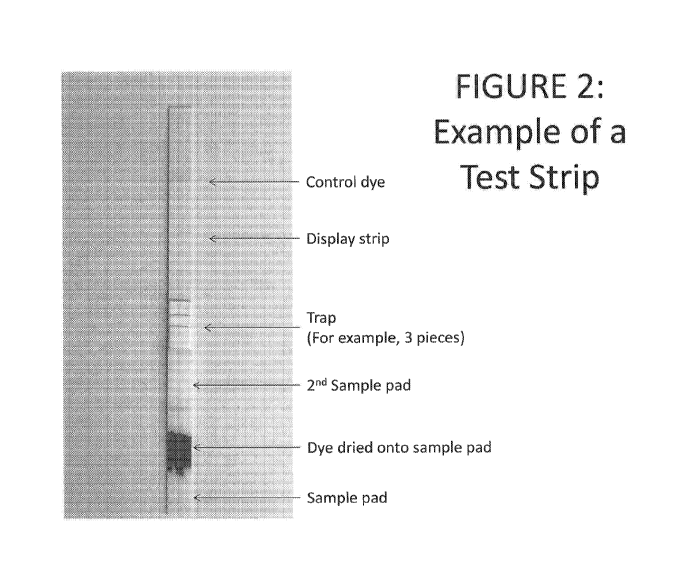
[0023] Figure 2 shows an example of a test strip embodiment of the
inventive device.
The strip comprises a control dye, a display strip (or capillary bed), a trap
(e.g., three pieces of
material), a second sample pad (or sample receiving material), a first sample
pad (or sample
receiving material) and the dye dried onto the sample pad.
7
CA 02989444 2017-12-13
WO 2017/011727 PCT/US2016/042426
[0024] Figure 3 shows an embodiment of the diagnostic device of the
present invention.
Fig. 3A shows a top view with a sample pad, a trap and a display strip. Fig.
3B shows the same
embodiment as in Fig. 3A viewed from the side. The sample pad, trap and
display strip may be
butted together. Fig. 3C shows an embodiment with the sample pad, trap and
display strip
overlapping each other (side view).
[0025] Figure 4 shows different material configurations within the test
strip. Fig. 4A
shows an alternative embodiment of the invention having two sample pads, and a
trap and
display strip (capillary bed) in sequence each piece having an overlap with
the adjacent piece.
Fig. 4B shows another, alternative embodiment of the invention having two
sample pads, a triple
trap and a display strip overlapping in sequence, with each piece having an
overlap with the
adjacent piece.
[0026] Figure 5 shows an example of a dipstick configuration of the test
strip of the
present invention. This embodiment is configured with a cover tape on top of
the test strip. At
the top of the test strip, the tape is opaque in color and may be used to hold
the strip. In the
middle, the tape is clear to provide a viewing window for the results. The
tape may be placed so
that the clear window is, for example, approximately 10 mm above the trap. The
tape at the
window may be surrounded by a color (e.g., white) which allows for easy
viewing of the results.
Below the trap, the tape is opaque in color to cover the trap. At the bottom
of the strip, the tape
may display an arrow indicating which end of the strip to dip into the test
sample and may
display a line indicating the maximum level to dip the test strip into the
sample. The bottom end
of the test strip may be free of cover tape to facilitate wicking of test
sample upon dipping.
Detailed Description of the Invention
[0027] The present invention is a device as well as methods of utilizing
this device.
More specifically, the device is a lateral flow chromatographic rapid test
that may be used in
several clinical and non-clinical settings in order to detect proteins of
interest in a biological
specimen. The device may be used to detect protein-misfolding disorders in
mammals. The
mammal may be suspected of having or at risk of having one or more such
disorders.
[0028] The device has several, different embodiments which will be
described herein.
Basically, it comprises a test strip for detection of a protein or proteins of
interest in a biological
sample (see, for example, Figure 1). The detection is carried out by means of
a sequential series
8
CA 02989444 2017-12-13
WO 2017/011727 PCT/US2016/042426
of reactions. The test strip comprises a length of lateral flow assay or
chromatographic material
having capillarity and has a first end at which chromatographic solvent
transport begins. It also
has a second end at which chromatographic solvent transport ends. The strip
includes a plurality
of zone or regions which are positioned between the first and second ends
(see, for example,
Figures 2 and 3). The zones include a first zone which is impregnated with a
detection reagent,
for example, a dye. This detection reagent specifically binds with the protein
or proteins of
interest in the biological sample. The first zone also receives the biological
sample. In
comparison, the second zone, which is downstream of the first zone, retains
the detection reagent
which is not bound to the protein or proteins of interest in the biological
sample while permitting
detection reagent bound to the protein or proteins of interest in the
biological sample to be
transported to a third zone. The third zone of the test strip, located
downstream of the second
zone, receives the sample after it passes through the second zone. The third
zone will display the
detection reagent if the proteins of interest are present in the sample. It
also comprises a means
for detecting the detection reagent bound protein as a measure of the protein
or proteins in the
biological sample. In one embodiment, the device determines the presence of
misfolded proteins
in the biological sample from a patient and allows for determination of
whether a patient has or
does not have a protein-misfolding disorder. The presence of the protein or
proteins can be
qualitatively or semi-quantitatively determined via visualization or may be
semi-quantified or
quantified by use of a measuring entity which may be present within the
device. After the
biological sample is applied to the first zone, the first zone releases the
detection reagent in the
sample, and the second zone separates the detection reagent bound to the
protein or proteins from
unbound detection reagent, and permits only bound detection reagent to be
transported to the
third zone which then displays the bound detection reagent for viewing or
measurement. The
first zone may be a sample receiving material. The second zone may be a trap.
The third zone
may be a capillary bed or display strip.
The specific elements or components of the device and the characteristic or
properties of
these elements are described, in detail, as follows:
9
CA 02989444 2017-12-13
WO 2017/011727 PCT/US2016/042426
Materials Used In Components of Device
[0029] The sample receiving material, the trap, and the capillary bed can
be made from
the same or different materials. The materials are generally known in the art
of lateral flow
devices and chromatography [see Ref: EMD Millipore Rapid Lateral Flow Test
Strips
Considerations for Product Development, available from EMD Millipore,
Billerica, MA].
Membranes are selected based on physical and chemical properties that impact
capillary flow
and therefore reagent deposition and assay performance. The materials include,
for example, but
are not limited to, nitrocellulose, filter papers, chromatography papers,
cellulose, plastic
polymers, asymmetric polysulfone membrane, cotton, linters and/or glass
fibers, polyesters,
polyethylene and polysulfone. Membranes may be made of polymers including, for
example,
nitrocellulose, polyvinylidene fluoride, nylon and polyethersulfone. Pad
materials are often used
as sample receiving material to provide controlled and even receipt of the
sample and facilitate
flow to the contiguous strip materials of the device. The pad materials are
porous, often made
with cellulose (i.e. filter papers), glass fibers, woven meshes and synthetic
nonwoven material or
polyesters. Filter matrices may be used for sample receipt particularly if it
is desirable to
separate out extraneous material contained in the sample from that part of the
sample to be
assayed, for example, to separate out cellular material from fluid. These
filter matrixes may be,
for example, cellulose, asymmetric polysulfone membrane (including but not
limited to Vivid'
Plasma separation membrane and asymmetric sub-micron (BTS) polysulfone
membrane).
Absorbent pads may be used, for example, as a wick at the end of the device
strip to pull sample
through the lateral flow strip, and may increase the amount of sample assayed
and enhance assay
sensitivity. These absorbent pads are often cellulose or cotton linters and
optimally selected
based on thickness, compressibility, and uniformity of bed volume. The entire
strip may be
assembled on a backing card often a card of a plastic backing and adhesive.
While these various
materials are often thought of for specific purposes in lateral flow devices,
as described herein,
each material may be considered for suitable properties for the purposes of
the receiving
material, trap and display strip of the present invention. See Examples for
further discussion of
materials.
[0030] Other materials utilized in the configuration of the test strip,
specifically the
display strip, are known in the art of lateral flow technology and
chromatography.
CA 02989444 2017-12-13
WO 2017/011727 PCT/US2016/042426
Elements or Components of the Device
Sample Receiving Material (Sample Pad)
[0031] The first element of the device (hereinafter referred to as the
sample receiving
material or the sample pad) acts as a sponge and holds an excess of sample
fluid to be tested. The
receiving material absorbs sample, but also permits it to flow or to wick to
the next contiguous
material. It is typically inert to, and thus does not react with, proteins of
interest that may be
present in the sample, as well as the detection reagent (e.g., dye), allowing
proteins and detection
reagent to flow or to wick through the material to contiguous material in the
lateral flow device.
[0032] The sample receiving material may be dipped into the sample from
the mammal
or patient or, alternatively, the sample may be indirectly or directly applied
to the sample
receiving material. The sample may be applied to the sample receiving material
by, for example,
a dropper with a metered tip, a pipette, a transfer pipette, or a pipette
capable of repeated
dispensing of the patient sample. If the sample receiving material is
configured to be dipped into
the biological sample, (see Figure 5, for example), then the sample receiving
material may be
relatively long (for example, but not limited to, about 10mm). If the sample
receiving material is
configured to receive the sample, applied by, for example, a dropper or
pipette, then the sample
receiving material may be relatively short (between, but not limited to, about
5mm and about
10mm). Commonly, the width may be from 2mm to 10mm, and most commonly, 2.5 to
5mm
(+0.5mm). Variations in the length and width of the sample receiving material
are possible and
depend upon such factors as the size of the cassette or housing as well as the
ability of the
biological sample to sufficiently mix with the detection reagent.
[0033] In particular, the sample receiving pad acts as a capillary matrix
in which the
biological sample and a detection reagent (e.g., dye) can freely mix. The
sample pad may also
have a detection reagent in a dried format suitable for an optimized chemical
reaction between
the analyte (e.g., protein of interest to be detected in the biological
sample) and the detection
reagent. The detection reagent may be pre-loaded onto the sample receiving
material. In one
embodiment, the biological sample (e.g., the urine) when added to the sample
receiving pad
dissolves the detection reagent, and then the sample and detection reagent dye
mix are
11
CA 02989444 2017-12-13
WO 2017/011727 PCT/US2016/042426
transported across the device by flowing through the sample pad to contiguous
material such as
the trap.
[0034] Alternatively, the sample pad comprises a series of two or more
sample pads (see,
for example, Figure 4). For example, the first pad may receive the sample and
the second may
contain the detection reagent, whereby the biological sample migrates from the
first to the
second element (pad) containing a detection reagent in a dried format suitable
for an optimized
chemical reaction between the analyte of interest and the detection reagent.
If two or more
sample receiving materials are utilized, either one may comprise the detection
reagent.
Preferably, the first sample receiving material comprises the detection
reagent and the second
sample receiving material does not.
[0035] In yet another embodiment, a first sample pad receives the
biological sample and
is designed to separate out or retain insoluble material that may be present
in the sample. The
filtered sample then flows through the first sample pad or to the second pad
and the detection
reagent is incorporated in either the first or second sample pad and allows
for suitable mixing of
the detection reagent and sample. The second pad may have the same or
different composition
as the first pad.
[0036] Further, in yet another embodiment, the first sample pad receives
the sample and
also contains the detection reagent, and the second pad provides for
additional time for the
mixing of detection reagent with the analyte of interest before entering the
next contiguous
material in the strip for example, the trap.
[0037] In an additional embodiment, the sample receiving material
comprises a substrate
for the detection reagent and retains the substrate upon drying. The detection
reagent may be on
or within the sample receiving material. Once the patient sample is added, the
detection reagent
is released. The substrate does not react with or absorb the patient sample
which, when applied,
moves through the matrix and onto contiguous material, for example, the trap.
[0038] It should be noted that the sample may be applied or placed into a
cassette or
housing, for example, through a sample well or other entity of the device for
receiving the
sample. The sample well or entity may be positioned over the sample receiving
material of, for
example, a test strip when it is assembled or encased inside a cassette or
housing. (See Figure 1.)
12
CA 02989444 2017-12-13
WO 2017/011727 PCT/US2016/042426
[0039] Materials useful as sample receiving material or sample pads are
generally known
in the art of lateral flow devices and chromatography [see Ref: EMD Millipore
Rapid Lateral
Flow Test Strips Considerations for Product Development, available from EMD
Millipore,
Billerica, MA] and are selected based on physical and chemical properties that
impact sample
receipt, controlled and even capillary flow, and sample filtering.
Additionally, if the sample
receiving material also contains the detection reagent, ideally, the material
is a suitable matrix for
holding the detection reagent and optimally releasing it upon addition of the
test sample. The
pad materials are porous, often made with cellulose (e.g., filter papers),
glass fibers, woven
meshes, synthetic, nonwoven material or porous plastic, for example,
polyesters. Other materials
that can be used as sample receiving material are, for example, polysulfone
asymmetric
membranes, cotton/glass fibers materials such as Ahlstrom 8950, plastic
polymer membranes,
for example, polyethylene, (e.g. high density polyethylene),
polytetrafluoroethylene, and porous
glass fiber membranes (see, for example Porex, Fairburn, GA). See Examples for
further
description of materials (i.e., Examples 2 and 6).
[0040] It should be noted that the sample receiving material used in a
device of the
present invention, when the detection reagent is a dye with affinity to
cellulose, may be cellulose
provided enough detection reagent is present for binding to the modified
protein or proteins of
interest in the biological sample. More specifically, the cellulose cannot be
permitted to "out
compete" the detection reagent in connection with binding to the protein of
interest, e.g., the
misfolded protein or proteins in the biological sample. Alternatively,
cellulose may be used for
the sample receiving material if it is present in a matrix which is less
reactive with the detection
reagent than the modified proteins or protein of interest. (Cellulose, for
purposes herein, is
defined as an organic compound with the formula (C6E11005)n and, in
particular, is a
polysaccharide consisting of a linear chain of several hundred to many
thousands of 0(1->4)
linked D-glucose units.)
Trap
[0041] Next, the fluid (e.g. sample or sample mixed with detection
reagent) flows from
the sample receiving material or sample pad through a filter (hereinafter
referred to as a "trap")
designed to retain any unbound detection reagent. In particular, the trap
serves to separate free
13
CA 02989444 2017-12-13
WO 2017/011727 PCT/US2016/042426
detection reagent (e.g., dye) from protein-bound detection reagent in the
lateral flow device.
Specifically, the trap material permits the flow of sample through to the next
contiguous material
but retains, retards the flow of, or binds to the unbound detection reagent if
the protein or
proteins of interest are not present in the biological sample.
[0042] The trap abuts but preferably overlaps with the sample pad that
contains the
detection reagent (e.g., dye) or the series of sample pad elements (see, for
example, Figure 3).
The trap functions to separate the detection reagent that is bound to the test
sample protein or
proteins of interest (e.g., misfolded protein or proteins) from detection
reagent that is not bound
to test sample protein or proteins, thus permitting the bound detection
reagent to flow through
while retaining the unbound detection reagent. Alternatively, the trap may be
a series of one or
more filters of the same or different materials.
[0043] Not to be bound by theory, the trap material may contain a
substrate for the
detection reagent such that unbound detection reagent binds the trap material
and does not flow
to the next material. Detection reagent that is already bound to proteins does
not bind to the
substrate in the trap and does flow to the next material in the strip. The
substrate may be the trap
material (e.g. cellulose) or it may be a chemical modification or addition to
the trap material.
Alternatively, a structural feature of the trap material composition may
provide for the retention
of unbound detection reagent.
[0044] Also, the trap may be comprised of multiple pieces of material
overlapped or in
succession to optimize for retaining unbound detection reagent (see Figure 4).
The multiple
pieces may be the same or made from different materials. Filter matrices may
be used for the
trap, for example cellulose, thermoplastic polymers such as asymmetric
polysulfone membrane
(including but not limited to Vivid plasma separation membrane and asymmetric
sub-micron
(BTS) polysulfone membrane). See Examples for further description of trap
materials (e.g.
Examples 2, 4, 5, 8, 13, 14 and16).
[0045] It was unexpected and quite surprising that there were, indeed,
many
nitrocellulose materials that actually worked well and permitted flow of urine
and dye-bound
proteins through the device. For example, Whatmang AE99 nitrocellulose
membrane worked
very well. (See Table 1.) There were also cellulose materials that worked
reasonably well (for
example, Ahlstrom 601, 319, 247, Whatmang CF1, CF3, CF4; EMI11513, 5475,
5493), but
14
CA 02989444 2017-12-13
WO 2017/011727 PCT/US2016/042426
there were also some cellulose materials that did not perform well (for
example, Ahlstromg
270). Surprisingly, among the materials that performed well for allowing
protein-bound CR dye
to flow while retaining unbound dye were VividTM Plasma separation materials
(Pall
Corporation) and asymmetric sub-micron (BTS) polysulfone membrane (Pall
Corporation). (See
Tables 1 and 2.)
[0046] When the detection reagent is a dye such as Congo red, the trap
material may be,
for example, filter papers, cellulose based and/or materials such as EMI
11513, EMI 5475, EMI
5493, 1281, 642, Standard 17, C048, LF1, LF1, VF2, CFI, CF3, Ahlstrom 319. The
trap may
be, for example, about 5-10 mm in length, or it may be a series of pieces each
5-10mm in length.
[0047] In one embodiment, the trap retains free detection reagent (e.g.,
dye) but allows
protein-bound dye to flow through. In a specific embodiment, the trap is
comprised of cellulose
and the detection reagent is Congo Red.
Detection Reagent
[0048] The detection reagent is a substance which is reactive with a
protein or proteins of
interest in the sample. For example, the detection reagent may be a substance
which is reactive
with or has a binding affinity for the misfolded protein or proteins (e.g.,
congophilic proteins),
aggregated proteins and/or supramolecular aggregated proteins present in a
biological sample
from a mammal, e.g. the patient sample. The detection reagent may be preloaded
onto a reagent
pad (for example, applied onto the reagent pad, or the reagent pad dipped into
the detection
reagent or dye. The reagent pad may be the sample receiving material or sample
receiving pad.
[0049] In one aspect, the detection reagent is a dye that stains the
subset of proteins of
interest in the biological sample, if present. In one embodiment, the
detection reagent may react
with misfolded proteins. For example, the dye may be an azo dye such as Congo
Red (CR), or
analog thereof, either buffered or unbuffered. Alternatively, other dyes could
be used as
detection reagents as long as these dyes have an affinity for (and can bind to
or react with) the
misfolded proteins, aggregated proteins and/or supramolecular protein or
proteins of interest in
the biological sample or patient sample. Examples of such dyes include but are
not limited to
Congo Red analogs such as those described in the following publications:
Sellarajah S et al,
CA 02989444 2017-12-13
WO 2017/011727 PCT/US2016/042426
Synthesis of analogues of Congo red and evaluation of their anti-prion
activity, J Med Chem.
2004 Oct 21;47(22):5515-34; and Helene Rudyk et al, Screening Congo Red and
its analogues
for their ability to prevent the formation of PrP-res in scrapie-infected
cells, Journal of General
Virology (2000), 81, 1155-1164. The detection reagent for detecting misfolded
protein or
proteins may also be, for example, Thioflavin T.
[0050] Further, in one aspect of the invention, the detection reagent is
present in a dried
form in the device, but may be present in other forms as well. The form of
detection reagent is
suitable for optimally mixing with the sample when applied and allowing
binding to the protein
of interest. The form of the detection reagent is suitable for long term
stability or shelf life of the
device. In one embodiment, the dried detection reagent is a dye. Furthermore,
the dye may be
an Congo Red and may be present in the device in an amount of, for example, 0.
lug to 800ug,
more preferably, 0.2ug to 480ug, even more preferably lug to 400 ug, and even
more preferred
2.5 to 12Oug.
[0051] Congo Red (CR) (e.g., buffered or non-buffered) may be pre-applied
to the
sample receiving material. For example, a CR solution can be applied to the
material during kit
manufacture and dried before assembly and packaging (see Figure 2 and Example
6).
[0052] The detection reagent is detectable, i.e., visible to the naked
eye, or otherwise
detected, for example, by visual examination and/or mechanical or electronic
reader(s).
[0053] The present invention provides for the detection reagent to be
incorporated into
the test device, for example, during manufacturing or assembly of the device.
This is an
improvement over previous devices for detecting misfolded proteins, where dye
and biological
sample needed to be mixed prior to adding to a test. In a preferred
embodiment, the test strip
contains a sample receiving material that contains the detection reagent. When
the biological
sample is added to the test device, into the sample receiving material, it
mixes with the detection
reagent in the sample receiving material and the biological sample-detection
reagent mixture
flows through the device.
Capillary Bed ("Display Strip")
[0054] A sample (e.g., urine) with or without protein-bound-detection
reagent passes
16
CA 02989444 2017-12-13
WO 2017/011727 PCT/US2016/042426
through the trap and onto a capillary bed (hereinafter referred to as a
"display strip") where it
accumulates. Thus, the display strip permits the flow of sample up the strip
and displays the
presence or absence of the detection reagent. In particular, the display strip
permits the flow of
sample up the strip and displays the presence or absence of the detection
reagent -bound analyte.
The detection reagent or bound reagent can then be visualized by human or
mechanical means
(to obtain a qualitative result) and/or then measured (i.e., semi-
quantitatively or quantitatively).
The display strip optimally provides for even flow of the sample throughout
and relatively
homogeneous display of the detection reagent when protein of interest is
present. In certain
embodiments, the intensity or concentration of the detection reagent on the
display strip may
correspond to the amount of protein of interest in the sample. In another
embodiment, the
distance the detection reagent flows up the display strip may be indicative of
the amount of
protein of interest in the biological sample. Furthermore, both the intensity
of detection reagent
and the distance up the display strip may be indicative the concentration of
proteins in the
sample. Furthermore, in the case of detection of misfolded protein aggregates
or supramolecular
aggregates, both the intensity of detection reagent and the distance up the
display strip may be
indicative the size of protein aggregates present in the sample. Suitable
materials for the
capillary bed include, for example, nitrocellulose or chromatography papers.
Also, polysulfone
asymmetric membranes provide suitable display strips.
Other suitable materials include
CytoSepg membranes such as CytoSepg 1660 and MN-260. The capillary bed of the
lateral
flow strip is aligned under a results viewing window of the lateral flow strip
cassette or housing,
described below. See Examples for further details of materials (e.g. Example
2, 3 and 9). If
protein bound detection reagent is present, then the detection reagent is
visualized within the
window. (See Figure 1.)
Wick
[0055] Optionally, the device contains a wick. The wick may be positioned
after the
third zone or the display strip in the strip or device of the invention. When
in use, the biological
sample (e.g., fluid) applied to the device continues to migrate from the
display strip into a final
porous absorbent material, the "wick", that acts as a sample accumulator and
also may function
to pull sample along the strip. Absorbent pads may be used, for example, as a
wick at the end of
17
CA 02989444 2017-12-13
WO 2017/011727 PCT/US2016/042426
the device strip to pull sample through the lateral flow strip, and may
increase the amount of
sample assayed and enhance assay sensitivity. These absorbent pads are often
cellulose or cotton
linters and optimally selected based on thickness, compressibility and
uniformity of bed volume.
Backing Card
[0056] The device may have a backing card. The entire strip may be
assembled on a
backing card (for example, those available from Lohmann, Orange, VA) which is
often a card of
a plastic backing and adhesive.
Housing/Cassette
[0057] In one embodiment of the present invention, the diagnostic device
is housed,
encased or encapsulated in a housing or cassette. The device may further
comprise a housing or
cassette, including but not limited to, a cartridge, plastic device or
extruded plastic piece
configured for the purpose, that encases the device. Several generic cassette
housings are
commercially available (for example, from Kanani Biologicals, Gujarat, India
or EASE-
Medtrend Biotech LTD, Shanghai, China) or may be custom-produced for the
purpose at hand.
(See, for example, U.S. Design Patent Appin. Ser. No. 29/533,647.) The device
may be
configured in a housing or cassette having a sample well for receiving the
sample, wherein the
sample well is positioned over the sample receiving material of the strip when
it is
assembled/housed inside the cassette. Furthermore, the device may be
configured such that,
when assembled in the housing or cassette, the display strip or capillary bed
of the strip is
positioned beneath a result viewing window of the housing.
[0058] In one embodiment, the device of the invention also comprises an
electronic
reader that is able to quantify the result, e.g. the intensity of the
detection reagent (e.g., dye) on
the capillary bed (i.e., in the results window) and may further comprise a
display screen (e.g., an
LED screen) to display the results. This reader may be part of the housing, or
an element that is
assembled integrally with the housing, for example, in the results display
window. Such readers
are described, for example, in Venkatraman, Biosensors and Bioelectronics
Volume 74, 15
December 2015, Pages 150-155, PCT Application No. W02013083686 Al, PCT
Application
18
CA 02989444 2017-12-13
WO 2017/011727 PCT/US2016/042426
No. W02004010143 A2, and PCT Application No. W02006010072 A2.
Cover Tape
[0059] The device with or without a housing may further comprise a cover
such as a
protective adhesive tape (for example, available from Lohmann, Orange, VA) or
other material
capable of protecting the device from damage and providing for proper reading
of test results.
For example, the device may be configured to be used as a dipstick such as a
urine dipstick. In
this embodiment, the lateral flow strip may be covered with a protective
adhesive tape. (See
Figure 5 and Example 11)
Run Control Reagent
[0060] In a preferred embodiment, a control reagent may be present on the
capillary bed
visible in the view window before the device is used. (See Figure 1 and Figure
2, Example 12.)
When the biological sample (e.g., fluid) flows through the capillary bed, the
control reagent
dissolves, the control reagent line disseminates and/or the control is carried
away, up the
display strip so that no control reagent is visible in the results window, or
it is blurred, or some
other difference may be visualized or measured. The change in the control
reagent in the view
window indicates that the test sample (e.g., fluid) was added and has run
through the device
properly, e.g., serves as a run control. (See Example 12.) The control reagent
is detectable
visually e.g. by the naked eye or otherwise detected or measured, for example,
by mechanical
examination and/or an electronic reader. In one embodiment, the control
reagent is tartrazine.
Other dyes that may be used as the run control reagent include: FD&C Blue No.
1 ¨ Brilliant
Blue FCF, E133 (blue shade), FD&C Blue No. 2 ¨ Indigotine, E132 (indigo
shade), FD&C
Green No. 3 ¨ Fast Green FCF, E143 (turquoise shade), FD&C Red No. 3 ¨
Erythrosine, E127
(pink shade), FD&C Red No. 40 ¨ Allura Red AC, E129 (red shade), FD&C Yellow
No. 5 ¨
Tartrazine, E102 (yellow shade), FD&C Yellow No. 6 ¨ Sunset Yellow FCF, E110
(orange
shade).
19
CA 02989444 2017-12-13
WO 2017/011727 PCT/US2016/042426
Detected Proteins of Interest
[0061] As noted above, the present invention is directed to a device and
methods of
utilizing this device for detection of proteins of interest, more
specifically, misfolded proteins,
aggregated proteins and/or supramolecular protein aggregates. It is known that
the alpha helix is
the prominent structural motif of the functional protein in its native
conformation. In contrast, a
conformational change in a protein can lead to a beta sheet structural motif
(i.e., beta sheet
structure) or a misfolded protein that then tends to cause protein aggregation
and toxicity. The
misfolded proteins may therefore be in the form of protein aggregates or
supramolecular
aggregates and may be associated with misfolded protein disorders such as
preeclampsia,
Alzheimer's disease, prion disease and Parkinson's disease.
[0062] In particular, these misfolded proteins, protein aggregates and/or
supramolecular
aggregates associated with preeclampsia which are detected by the device and
methods of the
present invention may include, but are not limited to, for example, alpha-1
antitrypsin
(SerpinA1), ceruloplasmin, heavy-chain IgG, light-chain IgG, interferon-
inducible protein 6-16
(IF16-6,G1P3), albumin as well as fragments of each protein, mixtures thereof,
and fragments of
such mixtures. These proteins have binding affinity for the detection reagent
(to be described
below) utilized in the device of the present invention. For example, these
misfolded proteins are
congophilic, having an affinity for the dye referred to as Congo Red.
[0063] The device of the present invention may also be used to detect
protein-misfolding
disorders other than preeclampsia. For example, the device may be utilized to
detect misfolded
proteins in such misfolded protein disorders or conditions as Alzheimer's
disease, Cerebral beta-
amyloid angiopathy, Retinal ganglion cell degeneration in glaucoma, Prion
diseases, Parkinson's
disease and other synucleinopathies, Tauopathies, Frontotemporal lobar
degeneration (FTLD),
FLTD-FUS, Amyotrophic lateral sclerosis (ALS), Huntington's disease and other
triplet, repeat
disorders, Dementia (familial British and Danish), Hereditary cerebral
hemorrhage with
amyloidosis, CADASIL, Alexander disease, various amyloidoses, Serinopathies,
Type II
diabetes, Inclusion body myositis/myopathy, cataracts, Retinitis pigmentosa
with rhodopsin
mutations, Medullary thyroid carcinoma, Pituitary prolactinoma, Hereditary
lattice corneal
dystrophy, Mallory bodies, Pulmonary alveolar proteinosis, Odontogenic tumor
amyloid, Cystic
fibrosis, Sickle cell disease and Critical illness myopathy.
CA 02989444 2017-12-13
WO 2017/011727 PCT/US2016/042426
Biological Sample
[0064] The protein or proteins of interest to be detected may be found in
a biological
sample from a mammal. The biological sample may be, for example, urine
obtained from a
clean or natural catch, cerebrospinal fluid, amniotic fluid or any bodily
fluid sample potentially
comprising the protein or proteins of interest (e.g., blood, saliva, amniotic
fluid, cerebrospinal
fluid, plasma or serum). The sample may also be an extract of excretions from
a patient, for
example, from nasal secretions, fecal material, or ear wax, or tissue
specimens extracted with
appropriate solutions and applied to the device. The proteins of interest may
be found, for
example, in a biological sample from a pregnant or postpartum mammal.
Patient
[0065] The patient may be a mammal. Furthermore, the mammal may be
pregnant, for
example, a pregnant woman, a pregnant primate or a genetically-engineered
animal model
designed to have the physical symptoms and signs of preeclampsia such as those
utilized in
laboratory studies (e.g., high blood pressure and protein in the urine).
Preferably, for the
diagnosis of preeclampsia, the patient may be any pregnant woman. The pregnant
woman may
be suspected of having preeclampsia or at risk of having preeclampsia. For
example the
suspicion may be based upon the following: 1) exhibiting the signs and
symptoms of
preeclampsia, for example, as set forth in the American College of Obstetrics
and Gynecology
Guidelines (ACOG) (Hypertension in Pregnancy, Report of the American College
of
Obstetricians and Gynecologists' Task Force on Hypertension in Pregnancy,
Obstetrics and
Gynecology 122 VOL. 122, NO. 5, NOVEMBER 2013), specifically, for example
TABLE E-1
(the "ACOG 2013 guidelines") and/or 2) having one or more risk factors for
preeclampsia (e.g.,
a woman having a previous pregnancy involving preeclampsia, a woman carrying
multiple
fetuses, a woman with cardiovascular or renal abnormalities or a woman having
an autoimmune
disease such as lupus).
Kit
[0066] The present invention also includes a kit for detecting proteins
of interest in a
21
CA 02989444 2017-12-13
WO 2017/011727 PCT/US2016/042426
sample. In one embodiment, the kit comprises a device for detection of
misfolded proteins
associated with preeclampsia, present in a sample from a pregnant mammal. The
kit comprises
the device of the invention as described above in any alternative embodiments
described above.
The kits may also comprise a means for applying the patient sample to the
sample receiving
material, for example, a pipette (for example Fine tip transfer pipette
available from Genesee
Scientific, San Diego, CA) or dropper, a control as well as instructions for
use of the device.
Thus, not only may the device be utilized as a stand-alone entity, it may also
be used in kit form
which may be more advantageous in some clinical or non-clinical settings. The
kit may be
packaged in a foil or mylar pouch. Kit pouches may furthermore be packaged
singly, or in
multiples, e.g., 2, 5, 10, 15, 25, 50 or 100 kits per package.
Settings For Use of Device and Methods Utilizing Device
[0067] The device of the present invention may be used in, for example,
clinical
laboratories (either within a hospital setting or outside a hospital setting),
immediate care
settings, physician office laboratories, emergency departments (e.g., within a
hospital) or as a
near-patient testing or point-of-care device by medical personnel, non-medical
professionals or
even by the patient herself when the patient is a human. Further, the device
may be used in
combination with other diagnostic assays (e.g., immunoassays such as those
that detect other
proteins (i.e., biomarkers) associated with preeclampsia such as, for example,
sFlt-1, P1GF, PP-
A, PP13, pentraxin, inhibin-A and soluble endoglin), and/or other methods or
observations
commonly utilized in the diagnosis of preeclampsia (e.g., blood pressure
readings, clinical tests
used in the diagnosis of preeclampsia including platelet count, serum
creatinine concertation,
serum ALT (alaninine aminotransferase) and AST (aspartate aminotransferase),
and other signs
or symptoms such as weight gain, dizziness, headaches, blurred vision, etc.
[0068] For example, the present invention includes a method of diagnosis
of
preeclampsia or performing a differential diagnosis in a patient suffering
from a hypertensive
disorder of pregnancy or a patient who may be suspected of having preeclampsia
comprising the
steps of: a) determining a blood pressure of the pregnant patient, wherein a
blood pressure
greater than 140/90 mm/Hg in the pregnant patient may indicate preeclampsia in
the pregnant
patient and b) applying a biological sample from the patient to the device of
the present
22
CA 02989444 2017-12-13
WO 2017/011727 PCT/US2016/042426
invention, whereby detection of at least one protein of interest in the
biological sample of the
patient provides or supports a diagnosis of preeclampsia in the patient or
acts as a differential
diagnosis.
[0069]
Further, the present invention also encompasses a method of treating a
pregnant
mammal suspected of having preeclampsia comprising the steps of: a) applying a
biological
sample from the mammal to the device of the present invention in order
determine the presence
of the at least one protein indicating the pregnant mammal has preeclampsia;
and b) delivering
the pregnant mammal in order to treat the preeclampsia. Further, the device
may also be used
post-delivery to determine if the patient is expressing the misfolded protein
or proteins in the
biological sample. If the misfolded protein or proteins are present, further
patient management
or therapeutic intervention may be needed (e.g., administration of magnesium
sulfate or other
anti-hypertensive agents) to treat the preeclampsia or the patient may be
monitored using the
device of the present invention to indicate when proteins are no longer
detectable in the
biological sample. Additionally, the device of the present invention may be
utilized after
therapeutic intervention to determine if treatment was successful or to
measure a change (e.g.,
decrease, absence or increase) in the amount or presence of protein of
interest present in the
biological sample.
Thus, the device of the present invention may be used pre- and post-
treatment of the patient to determine whether further therapeutic intervention
is necessary, to
determine whether therapeutic intervention has been effective and/or whether
to administer an
alternative form of therapeutic intervention or an increased dosage of the
therapeutic agent to
resolve the preeclampsia or other protein-misfolding disorder.
Advantages of Device and Methods Utilizing The Device
[0070]
The devices of the invention are advantageous over existing devices for at
least
the following reasons: 1) the device provides for a simplified testing
procedure with fewer steps;
2) the device provides for the use of standardized test materials for optimal
manufacturing; 3) the
device proides for improved stability of the detection reagent in the test kit
to provide a longer
shelf life; 4) results are simpler and easier to read all while 5) retaining
relatively low cost; and
6) providing fast results suitable for point-of-care use or use in clinical
laboratory settings.
[0071]
In particular, the provided device and methods are vastly superior to known
paper
23
CA 02989444 2017-12-13
WO 2017/011727 PCT/US2016/042426
kits for detecting possible preeclampsia (see, e.g., U.S. Patent Appin.
Publication No.
20150293115) in that the provided device requires fewer steps for the user,
results are easier to
read and is more stable than the paper kits resulting in a long shelf life of
at least 6 months,
preferably 1 year, even more preferably 2 years, even more preferably 3 years,
even more
preferably 4 years, and even more preferably 5 years. The device of the
invention also provides
fast results (i.e., within 3 minutes, preferably within 2 minutes, and more
preferably within 1
minute or less from application of the biological sample (e.g., urine) to the
device) suitable for
point-of-care users with minimal training.
[0072] The present invention may be illustrated by the use of the
following non-limiting
examples:
EXAMPLES
Example 1
Sample Devices
[0073] Figure 1 shows photographs of device test strips of the invention
in a cassette or
housing. The left panel of Figure 1 illustrates the device in a housing prior
to contact with a
biological sample. The right panel shows the device results of testing urine
samples known to be
negative, weak positive and positive for misfolded proteins associated with
preeclampsia.
[0074] Figure 2 shows an example of a test strip embodiment of the
inventive device.
The strip comprises a control dye, a display strip, a trap (e.g., three pieces
of material), a second
sample pad, a first sample pad and the dye dried onto the sample pad.
[0075] Figure 3A shows one embodiment of the diagnostic device of the
present
invention. Figure 3A shows a top view. Figure B shows the same embodiment as
in Figure 3A
but viewed from the side. The sample pad, trap and display strip may be butted
together. Figure
3C shows an embodiment with the sample, pad, trap and display strip
overlapping each other
(side view).
[0076] Figure 4 shows different material configurations within the test
strip. Figure 4A
shows an alternative embodiment of the invention having two sample pads, a
trap and a display
strip (capillary bed) in sequence. Each piece has an overlap with the adjacent
piece. Figure 4B
shows another, alternative embodiment of the invention having two sample pads,
a triple trap and
a display strip which overlap in sequence, each piece having an overlap with
the adjacent piece.
24
CA 02989444 2017-12-13
WO 2017/011727 PCT/US2016/042426
[0077] Figure 5 shows an example of a dipstick configuration of the test
strip of the
present invention. This embodiment is configured with a cover tape on top of
the test strip. At
the top of the test strip, the tape is opaque in color and may be used to hold
the strip. In the
middle, the tape is clear to provide a viewing window for the results. The
tape may be placed so
that the clear window is, for example, approximately 10 mm above the trap. The
tape at the
window may be surrounded by a color (e.g., white) which allows for easy
viewing of the results.
Below the trap, the tape is opaque in color to cover the trap. At the bottom
of the strip, the tape
may display an arrow indication as to which end of the strip to dip into the
test sample and may
display a line indicating the maximum level to dip the test strip into the
sample. The bottom end
of the test strip may be free of cover tape to facilitate wicking of test
sample upon dipping.
Example 2
Materials Testing
[0078] Various paper-like materials were tested for ability to
differentiate urine samples
from pregnant women with and without preeclampsia. Congo Red (CR) dye (Sigma.
St. Louis,
MO) was added to urine samples and a drop was added to the material to assess
characteristics
suitable for sample receiving, trap and display strip. The resulting spot was
visualized after
about 3 minutes and evaluated for a visual difference between urine from
preeclampsia patients
and that from normal control pregnancies (Table 1). A result of "excellent"
indicates the
material was suitable in providing a visually observable difference between CR-
urine from
preeclampsia patients and CR-urine from control pregnant patients such that
the material could
be useful in the diagnostic test device. A result of "poor" indicates the
material was not suitable
in providing a visually observable difference between CR-preeclampsia positive
and CR-control
urines. From a scale from a result of "poor" being the least suitable
material, "subtle" was
slightly better but not ideal, "okay" better yet, "good" even better, "better"
more improved and
"excellent" being the most suitable materials. It was determined that the
polysufone asymmetric
materials provided the best results. Certain cotton materials such as CF3 and
CF4 worked well
whereas other cottons like Ahlstrom 270 did not work well. Nitrocellulose and
glass fiber
materials generally did now work well for this purpose.
CA 02989444 2017-12-13
WO 2017/011727
PCT/US2016/042426
Table 1
Manufacturer Material Composition Results
Pall Corp, Port Vivid polysulfone asymmetric Excellent
Washington, New
York
Pall Corp BTS polysulfone asymmetric Excellent
Whatman, GE AE98 Nitrocellulose Okay
Healthcare Bio-
Sciences,
Pittsburgh, PA
Whatman AE99 Nitrocellulose Better
Whatman FF120 nitrocellulose on plastic Good
Whatman FF170HP nitrocellulose on plastic Poor
Whatman FF85 nitrocellulose on plastic Subtle
Whatman FF8OHP nitrocellulose on plastic Subtle
Whatman Prima 40 Nitrocellulose Okay
Whatman Prima 125 Nitrocellulose Less so
Whatman Immunopore RP Subtle
Whatman Standard 14 glass fiber Poor
Whatman Standard 17 glass fiber Poor
Whatman CF3 Cotton Okay
Whatman CF4 Cotton Okay
Whatman VF2 Poor
Whatman LF1 Subtle
Millipore, EMD HF75 Nitrocellulose Modest
Millipore,
Billerica, MA
Millipore HF90 Nitrocellulose Modest
Millipore HF120 Nitrocellulose Poor
Millipore HF135 Nitrocellulose Poor
Millipore HF170 Nitrocellulose Poor
Millipore HF180 Nitrocellulose Poor
Sartorius Stedim, CN150 Nitrocellulose Subtle
Bohemia, New
York
Sartorius Stedim, CN140 Nitrocellulose Subtle
Sartorius Stedim, CN95 Nitrocellulose Subtle
Ahlstrom, 270 Cotton Poor
Helsinki
Finland
Ahlstrom 222 Poor
Ahlstrom 320 Poor
26
CA 02989444 2017-12-13
WO 2017/011727 PCT/US2016/042426
Ahlstrom 111 Glass Poor
Ahlstrom 142 Glass Poor
Ahlstrom 21 Poor
Ahlstrom 141 Glass Poor
Ahlstrom 6613 glass or polyester Poor
Ahlstrom 6615 Poor
Ahlstrom 181 Poor
Ahlstrom 169 Poor
Ahlstrom 161 Poor
Ahlstrom 151 Poor
Ahlstrom 131 Poor
Ahlstrom 601 Cotton Good
Ahlstrom 237 Okay
Ahlstrom 238 Cotton Subtle
Ahlstrom 8950 cotton or glass Poor
Ahlstrom 8951 glass fiber or polyester Poor
Ahlstrom 8964 glass fiber Poor
Ahlstrom CytoSep 1662 Proprietary Good
Ahlstrom CytoSep 1663 Proprietary Good
Ahlstrom CytoSep 1660 Proprietary Good
Ahlstrom ReliaFlow 319 Subtle
Ahlstrom ReliaFlow 800 Poor
Ahlstrom ReliaFlow 1281 Subtle
Macherey Nagel MN-260 Good
Bethlehem, PA
Macherey Nagel MN-321 Subtle
Macherey Nagel MN-615 Good
Macherey Nagel MN-616g Okay
Macherey Nagel MN-640 Okay
Macherey Nagel MN-6176 Okay
Lypore 9334 Poor
Rochester, NH
Lypore 9390 Poor
Lypore 9389 Poor
Example 3
Test Strip
[0079] Test strips were assembled using a Ahlstrom 8950 (a cotton/glass
fiber
composition)("8950") sample pad and a BTS or VividTM running strip or
capillary bed, as BTS
27
CA 02989444 2017-12-13
WO 2017/011727 PCT/US2016/042426
and Vivid had demonstrated excellent results in providing a visually
observable difference
between CR-urine from preeclampsia patients and CR-urine from control pregnant
patients.
Congo Red was added to urine samples, the sample was vortexed, and then
samples were applied
to the 8950 end of the test strip. Results were observed on the BTS or Vivid
membrane run
strip. Urine from women with preeclampsia applied to VividTM membrane
containing strips
resulted in very clear signal (pink/red Congo Red staining) with negative
urines showing no
signal. Signal was volume dependent with decreasing volumes resulting in
decreasing signal.
The BTS strips showed no difference between positive and negative urines with
both resulting in
positive staining at the higher sample volume. Signal decreased equally for
the positive and
negative urines with decreasing sample volumes applied.
Table 2
Run Material Positive Urine Staining Results Negative Urine Results
Vivid 90u1 Positive 90u1 Negative
Vivid 50u1 Weak positive 50u1 Negative
Vivid 30u1 Negative 30u1 Negative
BTS 90u1 Positive 90u1 Positive
BTS 50u1 Weak positive 50u1 Weak positive
BTS 30u1 Negative 30u1 Negative
These results indicate that the VividTM material was suitable in the
diagnostic test device of the
present invention whereas the BTS did not perform well.
Example 4
Trap Materials Evaluation
[0080] Vivid' and BTS membranes were evaluated as materials for the trap,
that is
positioned between the sample pad and the run strip (i.e., capillary bed or
display strip) to
determine if they aided in the retention of CR dye when negative urines (i.e.,
urine not
containing the proteins of interest from a woman without preeclampsia) were
applied, allowing
CR with preeclampsia-positive urine to flow through to the display strip. Test
strips were
assembled to include 8950 as a sample pad with either Vivid' membrane or BTS
membrane
followed by a CytoSep 1660 run strip. Urine with added CR was applied to the
8950 and
results were observed on the CytoSep 1660 run strip.
28
CA 02989444 2017-12-13
WO 2017/011727 PCT/US2016/042426
[0081] CR staining was seen on all strips when positive urine (i.e.,
urine containing the
proteins of interest from a woman with preeclampsia)¨Congo Red dye was
applied. While strips
made with a VividTM membrane trap were negative when negative urine-Congo Red
was applied,
strips made with a BST membrane trap showed some dye staining. The VividTM
material was an
effective trap to retain CR dye when negative urine was tested, while letting
through the CR dye
when positive urine was tested. BTS was a less effective trap.
Example 5
Evaluation of materials for use for pre-loaded detection reagent in test
strips
[0082] Congo Red dye was applied to sample pad materials and allowed to
dry. Positive
urine was applied and the results on the dye release from the sample pad were
observed.
Table 3
Material Results
POREX 4894 (Porex, Good dye release and flow
Fairbum, GA)
Porex X-4897 Good dye release and flow
Porex D3883B Modest dye release, spot
still retained
Porex PVA Poor dye release, no flow
Fusion 5-1 (GE Healthcare Modest dye release spot
Life Sciences, Pittsburgh still retained
PA)
Fusion 5-2 (GE Healthcare Modest dye release spot
Life Sciences,) still retained
GF DVA 1 (GE Healthcare Poor dye release
Life Sciences,)
GF DVA 2 (GE Healthcare Poor dye release
Life Sciences,)
Whatman 33 (GE Modest dye release, spot
Healthcare Life Sciences,) still retained
[0083] The detection reagent (i.e., dye in this instance) ideally is
released from the
sample pad material upon application of the biological sample, i.e., urine.
Several materials
including POREX 4894 and Porex X-4897 provided for good dye release and
therefore are
highly suitable in the diagnostic test device. Additionally, several materials
also provided dye
29
CA 02989444 2017-12-13
WO 2017/011727 PCT/US2016/042426
release and are suitable, such as Fusion materials (glass microfibers) and
Whatman 33.
Example 6
Evaluation of sample pads with dye incorporated into test strips
[0084] Sample pad materials including Porex , X4897 and POREX 4894
infused with
Congo Red dye were incorporated into test strips with a VividTM x trap and an
MN-260 run or
display strip. Urine was added to the sample pad of each strip and results
observed on the MN-
260 display strip.
[0085] Results: The sample pad materials released dye when urine was
added. Adding
preeclampsia-positive urines resulted in positive staining of the MN260
material. In most cases,
adding preeclampsia-negative urines resulted in no staining of the MN260;
however, in some
cases there was some staining at the beginning of the MN260 strip. These
results indicate that
the configuration of Porex , VividTM, MN260 materials could provide means for
differentiating
preeclampsia positive from preeclampsia negative urines, providing for a
useful diagnostic test
device.
Example 7
Test strip evaluation in lateral flow assay cassette
[0086] Strips were assembled of dye infused POR-4899 sample pad, placed
on top of
Fusion 5, followed by Vivid X membrane serving as a trap and then MN260
display strip. Strips
were placed in generic cassette with the sample pad directly underneath sample
port of the
cassette and the MN260 visible in the cassette results window. [75u1] Seventy-
five microliters of
urine were added in the sample port and results were observed in the results
window. When
positive urines were added. Red staining was visible in the results window
whereas, when
negative urines were added, no staining was observed. This configuration
provided for an
effective diagnostic device differentiating preeclampsia positive from
preeclampsia negative
urines.
Example 8
Refining the trap
CA 02989444 2017-12-13
WO 2017/011727 PCT/US2016/042426
[0087] At times, negative urine tested on the strip resulted in some
streaking of dye up
the results strip, particularly over time. Additional trap configurations were
evaluated to see if
better separation of positive vs. negative samples could be achieved and if
this streaking with
negative urines could be eliminated. Strips were assembled with dye infused
POR-4899 sample
pad placed on top of Fusion 5, a trap of two offset but overlapping layers of
Vivid X and
MN260 results strip. When tested by applying negative urines, the double layer
trap prevented
dye leakage and streaking thereby providing for an improved diagnostic test
device for
differentiating preeclampsia positive from preeclampsia negative urines.
Example 9
Device strip configurations
[0088] POR-4899 was striped with Congo Red dye solution dispensed at 6
1/cm using a
Kinematic dispensing machine. POR-4588 was striped at 4 1/cm. Pads were dried
at 37 C for
1 hour and stored in a desiccant cabinet. Test strips were assembled using the
striped POR
material, one layer Vivid' X trap and MN260. The POR-4899 strip performed well
when
testing urines; however, the strip containing POR-4588 resulted in dye leakage
onto the display
strip when testing negative urines. POR-4588 was further evaluated when
striped at 4 1/cm and
resulted in clean negative urine run. Then, POR-4588 was evaluated when
striped at 6 or 8
1/cm assembled with a double Vivid' trap. This strip assembly with 6 1/cm dye
strip gave
the best results when testing positive urines and the least amount of dye
leakage or streaking
when testing negative urines. Further, evaluation of strip with a 3 layer
Vivid' trap and POR-
4588 striped at 8 1/cm gave good results and a stronger positive signal.
Table 4
Dye pad Congo red Trap Pos/neg
dispensing rate differentiation
POR-4899 6u1/cm Single VividTM trap Ai
POR-4588 4u1/cm Single VividTM trap False positive
POR-4588 6u1/cm Double VividTM
trap
31
CA 02989444 2017-12-13
WO 2017/011727 PCT/US2016/042426
POR-4588 8u1/cm Double Vivid TM A/
trap
[0089] The device configured with POR-4599 with Congo Red applied at
8u1/cm and a
double vivid trap resulted in a clean, negative result when preeclampsia
negative urine was tested
and a strong positive result when preeclampsia positive urine was tested and
therefore is a
preferred diagnostic test device configuration.
Example 10
Evaluation of a dipstick format test design
[0090] Test strips were assembled using POR-4588 striped at 6, 8 or 10
nl/cm with a 3
layer Vivid trap and MN260. Strips were dipped into 60 n1 of urine. Urine
successfully
wicked up the strip and the version with 8 nl/cm stripping provided the best
positive test results
with clean negative test results. Therefore, a suitable dipstick format of the
diagnostic test
device can be configured in this way and is useful for differentiating
preeclampsia positive urine
from preeclampsia negative urine for diagnostic purposes.
Example 11
Further evaluation of diagnostic test device configuration
Test strips made of two Porex sample pads, one striped with dye; 3 Vivid'
layer trap, and
MN260 were tested in a cassette/housing and as dipsticks for testing urine.
The cassette housing
improved results, perhaps because of controlled flow of sample onto central
spot on sample pad,
good uptake of dye, flow up center of strip, and more homogeneous staining of
run strip result.
Five negative urines all tested negative; 5 positive urines resulted in 2 weak
positive, 3 positive
results. As a dipstick, the test strip was placed into 100 n1 urine in a test
tube. At 3 minutes, all
results for both positive and negative urines were negative. However, given
more time, by 5
minutes the positive urines results were 2 weak positives, 3 positives, while
negative urines
remained negative. Therefore, these test configurations are suitable
preeclampsia diagnostic test
devices.
32
CA 02989444 2017-12-13
WO 2017/011727 PCT/US2016/042426
Example 12
Evaluation of Run Control
[0091] The purpose of this experiment was to investigate a control that
indicates when
the sample has run up the strip successfully, a "run control". When a negative
biological sample
is tested and no staining is observed on the display or results strip it is
difficult to tell if it is a
negative result, or the test failed to run correctly (i.e., no sample was
actually added, insufficient
sample was added or insufficient wicking of the sample up the strip occurred).
A stripe of a
water soluble coloring was sprayed across the run strip material, positioned
so that it would be
visible in the top of the cassette results window when assembled in the
housing. Urine samples
were applied to the sample pad through the sample port and flow observed
through the results
window. When sample reached the control dye stripe, the stripe dissolved and
ran with the
sample. The disappearance of the run control stripe provided an easily visible
indication that
sample had run through the strip successfully. The control did not interfere
with reading results
of the urine test. An example of control strip dye is tartrazine. Other dyes
that may be used as
the run control include: FD&C Blue No. 1 ¨ Brilliant Blue FCF, E133 (blue
shade), FD&C Blue
No. 2 ¨ Indigotine, E132 (indigo shade), FD&C Green No. 3 ¨ Fast Green FCF,
E143 (turquoise
shade), FD&C Red No. 3 ¨ Erythrosine, E127 (pink shade), FD&C Red No. 40 ¨
Allura Red
AC, E129 (red shade), FD&C Yellow No. 5 ¨ Tartrazine, E102 (yellow shade),
FD&C Yellow
No. 6 ¨ Sunset Yellow FCF, E110 (orange shade).
Example 13
Investigation of alternative materials for trap
[0092] Test strips were assembled with 2 Porex sample pads, the first
striped with
Congo Red (CR) dye, a variety of alternative trap materials as listed in Table
5 and MN260 run
strip. Negative urine sample were tested on the assembled test strips and the
ability of each trap
to: 1) retain the CR dye when proteins of interest in the sample are not
present and 2) not retain
protein bound CR were evaluated.
33
CA 02989444 2017-12-13
WO 2017/011727 PCT/US2016/042426
Table 5
Trap Material Results testing Negative Results testing Positive
sample sample
EMI 11513 (EMI Specialty Negative, slight dye run off Positive
Papers, Redding, CT)
EMI 5475 (EMI Specialty Negative, very little dye run Positive
Papers) off
EMI 5493 (EMI Specialty Negative, slight dye run off Positive
Papers)
ReliaFlow 1281 Negative, slight dye run off Positive (streaky)
Ahlstrom 642 Negative, very little dye run Positive
off
Whatmang Standard 17 Negative, modest dye run off Positive (streaky)
Millipore C048 Negative, very little dye run Weak positive
off
Whatmang LF1 Negative, slight dye run off Weak positive
Whatmang VF2 Negative, very little dye run Weak positive
off
Whatmang CF1 Negative, slight dye run off Positive
Whatmang CF3 Negative, very little dye run Positive
off
[0093] Results: Most of these trap materials showed promise in
differentiating
congophilic protein-positive biological samples (congophilic protein positive
urine) from
congophilic protein-negative biological samples (congophilic protein-negative
urine).
[0094] To further improve the retention of detection reagent (CR) when
negative samples
are tested, test strips were then made using select trap material described in
Table 6 in three
layers.
Table 6
Trap Material Results testing Negative Results testing Positive
sample sample
EMI 11513 Negative Weak Positive
642 Negative Positive
C048 Negative Weak positive
CF1 Negative, slight run off Positive
[0095] Results: A triple layer trap was effective in retaining unbound CR
detection
34
CA 02989444 2017-12-13
WO 2017/011727
PCT/US2016/042426
reagent from entering the display strip. However, triple trap configuration
made with EMI
11513 and C048 also reduced signal results intensity when testing positive
samples. Triple traps
made with 642 provided the best results for both positive and negative
samples, amongst those
traps tested.
Example 14
Further investigation of trap materials
[0096] To further investigate suitable materials for use as a trap in the
diagnostic test
device of the present invention, strips with 2 Porex sample pads, the first
striped with CR dye
(8u1/cm), a trap as described in Table 7 and MN260 run strip were assembled.
Strips of each
configuration were tested with a congophilic negative urine sample, two weak
positive
congophilic protein samples and two strong positive congophilic protein
samples.
Table 7 Investigation of trap materials as single layer
Material Neg Weak positive Strong Weak
positive Strong
Positive Positive
EMI 11513 - +- +- ++
237 - + ++ + ++
319 - + ++ + ++
CFI - +- ++ +- +
Vivid Tm - ++ ++ +- ++
5475 - + ++ +- ++
5493 - +- ++ +- ++
C048 - +-- + +-- +
CF3 - +-- + +-- +
238 - + + +- +
601 - + + + +
1281 - +- + +- ++
642 - +- + +-- +
MN640m - +- + +-- +
MN615 - +- ++ +- +
[0097] Surprisingly, 5493 results were less intense than 5475, with 5493
about 4 times
more absorbent and 2 times faster capillary rise characteristics than 5475.
These results suggest
that less absorption and a slower capillary rise is preferable for robust
signal. Similarly, CF3 is
weaker than CF I, having two times the absorption. Material 238 has a weaker
signal than 237
CA 02989444 2017-12-13
WO 2017/011727 PCT/US2016/042426
and has twice the wick rate, and 319 showed the strongest color results of
this paper line and had
the slowest wick rate. MN640 which is a faster running material has weaker
signal than MN615.
[0098] Results show that the materials that have less absorbency, slower
capillary rise or
wick rate may be preferred materials for more robust signal.
Example 15
Evaluation of diagnostic test devices
[0099] A complete test strip was assembled, comprising a dye infused
Porex sample
pad placed on top of Fusion 5 (a proprietary single layer matrix membrane made
by GE), a
Vivid membrane strip trap and MN260 capillary bed membrane. The strip was
housed in a
generic LFA cassette case. Urine samples were added to the circular sample
well and results
were observed in the rectangular window at 3 minutes.
[00100] Results: No dye was observed in the window for negative urine, a
light pink was
observed for weak positive urine and a strong pink was observed for the strong
positive urine
sample. These results indicate this is a suitable configuration for a
diagnostic test device for
detection of congophilic proteins in a sample for the diagnosis of
preeclampsia.
Example 16
Further evaluation of optimal trap materials
[00101] A complete test strip was assembled, comprising a dye infused
Porex sample
pad a second Porex pad (without dye), a trap of single or double layers of
11513, 5475, 319,
247, or CFI, a MN260 membrane capillary bed with a wick of Whatman 470. The
MN260
contained a run control stripe of tartrazine dye. The test strip was housed in
a LFA-like cassette
optimized for addition of test sample onto dye infused Porex pad, and a view
window centered
over the MN260 membrane such that the tartrazine dye is observable prior to
running the test and
not observed after the test is run. Urine samples were added to the circular
sample well and
results were observed in the rectangular window at 3 minutes.
36
CA 02989444 2017-12-13
WO 2017/011727 PCT/US2016/042426
Results
[00102] No dye was observed in the window for negative urine, a light pink
was observed
for weak positive urine and a strong pink/red was observed for the strong
positive urine sample
at 3 minutes. Furthermore, the yellow tartrazine dye stripe was evident prior
to adding sample,
could be observed to travel along with the wicking of the sample, and was
barely visible or
completely gone at 3 minutes. These results indicate this is a suitable
configuration for a
diagnostic test device for detection of congophilic proteins in a sample for
the diagnosis of
preeclampsia.
Example 17
Results of present device versus CRD test results
[00103] The test device of the present invention was evaluated by testing
105 clinical
urine specimens from a study of the Congo Red dot (CRD) test in women
suspected of
preeclampsia (Rood et al., 2016 AJOG, 214(1)s24-s25). Samples were chosen to
provide 27
strong positives, 28 negatives and 50 weak positive samples. The
negative/positive status of the
samples was determined by %CRR (Irma A. Buhimschi et at., Sci. Transl. Med. 6,
245ra92
(2014)). Samples were added to the test device and results were visually
scored at 3minutes as
either negative (- or 0), weak positive (+ or 1) or strong positive (++ or 2).
[00104] Results showed that the test device results were concordant with
the CRD result
in 103/105 of the samples (98%). The test result of the device of the present
invention, when
compared to an adjudicated diagnosis of preeclampsia (based on ACOG guidance:
Hypertension
in Pregnancy Report of the American College of Obstetricians and
Gynecologists' Task Force on
Hypertension in Pregnancy, 2013 Obstetrics & Gynecology 122(5): 1122-1131),
showed a
sensitivity of 91%; specificity of 64%, accuracy of 82% with a positive
predictive value of 83%
and a negative predictive value of 79%.
[00105] Upon adding the test sample to the sample well of the test, the
front of the liquid
traveling up the strip was visible by 30 seconds. The front hit the yellow dye
control stripe at
about 1 minute and traveled to the end of the window by about 1.5 min. Strong
positive results
were clearly visible with the front of liquid traveling into the results
window by 30 seconds and
37
CA 02989444 2017-12-13
WO 2017/011727 PCT/US2016/042426
beyond. The full results window was colored by 1.5 min. Weak positive results
take longer to
resolve than the strong positives as the liquid front commonly appeared
negative with color
developing in the results window at about 1-3 minutes after addition of the
sample. Figure 1
shows examples of results.
Example 18
Evaluation of device using urine sample controls
[00106] The device test strip was assembled, comprising a Porex 4588
sample pad dye
infused 12Oug/device, a second Porex pad (without dye), a trap of double
layers of Ahlstrom
319, a MN260 membrane capillary bed with a wick of Whatman 470. The MN260
contained a
run control stripe of tartrazine dye. The test strip was housed in a LFA-like
cassette optimized
for addition of test sample onto dye infused Porex pad, and a view window
centered over the
MN260 membrane such that the tartrazine dye is observable prior to running the
test and not
observed after the test is run.
[00107] Urine specimens from five (5) patients with confirmed preeclampsia
were pooled
to create a positive assay control. The presence of congophilic proteins was
confirmed by the
Congo Red dot blot test (Buhimschi 2014). Phosphate buffered saline (10 mM
phosphate, pH
7.4, 150 mM NaC1) was used as a negative control and for making the dilution
series of the
positive assay control. The following dilution were prepared (in ratio of
sample to total volume
of buffer): undiluted, 1:4, 1:8, 1:15, 1:40. Each urine sample of the dilution
series was tested in
duplicate on the device.
[00108] Results: Each replicate for each urine sample for a particular
dilution exhibit the
same density of staining on the devices. The results of the testing are
presented in the following
table.
38
CA 02989444 2017-12-13
WO 2017/011727 PCT/US2016/042426
[00109] Table 8: Testing results from a dilution series of a pooled urine
sample
Dilution Test Results
Undiluted +++
1:4 ++
1:8
1:10
1:15
1:40
These data demonstrate that the diagnostic device can detect congophilic
proteins, and that the
effect is titratable. These data also indicate that device exhibits a
progressive visual scale of red
color corresponding to increasing concentration of congophilic proteins.
39