Note: Descriptions are shown in the official language in which they were submitted.
CA 02990513 2017-12-21
WO 2017/001379 PCT/EP2016/064960
1
Devices and kits for assisting in open surgeries
This application claims benefit of European Patent Application no.
15382341.4 filed on June 29, 2015.
The present disclosure relates to devices and kits for assisting in a medical
procedure, particularly open surgeries.
BACKGROUND
Surgeries can generally be grouped in laparoscopic or minimally-invasive
surgeries and open surgeries. In this sense, open surgeries should be
understood as traditional surgeries which require a relatively long incision
in
order for the surgeon to insert instruments and visualize the surgery through
the incision, whereas minimally-invasive surgeries are much less invasive
and involve much smaller incisions. With an open approach, e.g. the incision
for a typical appendectomy is approximately 10 cm (4 inches) long. While,
using minimally-invasive techniques, the incisions may range from 0.5 to 1.5
cm (1/4 to 1/2 inch) or for some surgeries even no incisions at all.
The key element in laparoscopic surgery is the use of a laparoscope, i.e. a
long fiber optic cable system which allows viewing of the affected area.
Laparoscopic surgeries belong to the field of endoscopy, i.e. using an
endoscope which is an instrument used to examine the interior of a hollow
organ or cavity of the body. In laparoscopic surgery, the surgeon is actually
performing the surgery from outside the patient's body, removed from the
actual intervention site. Surgical instruments are controlled from a distance
and reach the intervention site through a tubular body, e.g. a catheter.
Illumination devices for illuminating medical procedures are known. Since
practitioners in the several medical fields of specialization usually need
enhanced visualization of a body tissue and/or body cavities, a variety of
illumination systems have been designed to address this issue.
There are currently several ways to illuminate a medical procedure, such as
e.g. open surgeries in an operating room. One option is overhead lighting
mounted in a ceiling. Surgical lights may be fixed in the ceiling or be
CA 02990513 2017-12-21
WO 2017/001379 PCT/EP2016/064960
2
suspended from a ceiling with an arm that can be manipulated to reposition
and reorient the light. However, these kinds of lighting usually provide a
general and diffuse kind of illumination, which may lack the precision needed
to point towards and adequately illuminate the desired target depending on
the kind of intervention. The light may encounter obstacles that cast a shadow
over said target, particularly in interventions in bodily cavities or
openings.
Another option is the use of lighting devices held in a person's hand. In such
a case, surgeons or other operating room personnel may employ handheld
lighting devices, such as surgical flashlights. However, during the time that
the surgeon is holding the device, his/her hand that is holding the lamp is
unable to perform other actions. If instead such a device is held by other
personnel, the accuracy in lighting the target might not correspond to the
surgeon's specific demands. As a solution to overcome some of the
aforementioned limitations, surgical headlights may be used. However,
surgical headlights commonly are heavy, and can be uncomfortable. They
also need positioning and possibly adjustments during surgery, again
requiring a free hand to do so. Furthermore, the headlight and all related
apparatus are unsterile, so that precautions must be taken in order to avoid
contamination of the surgical field.
In a similar manner, during open surgeries practitioners usually need ways to
indicate instruments, a body tissue and/or body cavities to other
practitioners
or personnel involved. Identification of the correct intervention point and
the
direction of a required movement (e.g., incision, needle advancement) can be
of pivotal importance for completing these procedures. Verbal communication
sometimes may not adequately provide the guidance. Laser pens are
sometimes used for such indications.
In the course of an open surgery, many combinations of surgical instruments
may be needed. For example, it is quite often that one or more blood vessels
need to be severed in the course of an open surgery. In these cases,
normally after introducing the scalpel the surgeon needs to provide means for
closing and cauterizing such blood vessels. To do this, various surgical
elements may be needed. Alternatively, it is known to use an electric scalpel
or electric surgical pencil. This way, the blood vessels can be severed and
CA 02990513 2017-12-21
WO 2017/001379 PCT/EP2016/064960
3
cauterized in a single step. However, during cauterization smoke may hinder
the view of the surgeon. Thus a further surgical instrument may be needed,
namely a surgical smoke aspirator which will normally be operated by a
surgical assistant.
Radio-guided surgery is also known, in which a patient is administered a
radioactive material. The radioactive material concentrates e.g. in cancer
cells. By using a radioactive probe, a tumour and affected tissue can be
located and removed with precision. However, when the surgeon is holding
the probe, his/her hands are unable to perform other actions. Thus normally
the radioactive probe will be held / operated by a surgical assistant. Often
both medical professionals cannot access the opening for the surgery at the
same time, i.e. one person uses the radioactive probe to locate affected
tissue and tells the surgeon where to cut.
Currently, many such combinations of surgical instruments, including, but not
limited to, cutting, suturing, aspiration, clamping, cauterizing, irrigating,
and
various forms of tissue manipulation within a patient's body, may be needed
simultaneously. And in all these and other combinations of surgical
instruments for open surgery, visibility is very important, but often not
ideal
because more than one person is needed to handle different tools.
It is an object of examples of the present disclosure to provide alternative
devices and kits for assisting in open surgeries, particularly for assisting
or
illuminating in open surgeries that at least partially overcome some of the
aforementioned drawbacks.
SUMMARY
According to a first aspect, a device for assisting in an open surgery is
provided. The device comprises: a base configured to be mounted on a
portion of a handheld medical tool, an illumination device mounted on the
base such that when the base is attached to the handheld medical tool, the
illumination device is directed towards a distal end of the handheld medical
tool. The device further comprises an appendage to the base having a
receptacle configured to receive an auxiliary medical device.
CA 02990513 2017-12-21
WO 2017/001379 PCT/EP2016/064960
4
According to this aspect, a device is provided which can be coupled to a
handheld medical tool (e.g. an electrical scalpel) to improve visualization
during open surgery, and at the same allow auxiliary devices to be coupled to
the handheld medical tool. Such an auxiliary device may be e.g. a surgical
aspirator or smoke/liquid evacuator, a laser pointer, a handheld ultrasound
system, an irrigator for electrocoagulation, a further illuminating device, a
radioactive probe, a camera or a sensor among others.
In general, these devices may particularly be instruments that assist the
surgeon while performing the surgery (e.g. interacting with the tissue) with
the
(principal) handheld medial tool.
No further medical personnel is needed to hold such an auxiliary device. The
surgical procedure can thus be more effective, quicker and safer.
The attachment of the base with appendage locally increases the cross-
sectional dimensions of the medical tool. However, in examples of the present
disclosure, the base with appendage may remain outside the patient's body
while the medical tool and auxiliary tool reach sufficiently inside the body.
In
such cases, there is thus no need to increase a surgical incision.
In some examples, the receptacle may be a through-hole. Optionally, the
appendage may comprise a tubular or annular portion defining the through-
hole. Optionally, the tubular or annular portion may be made of a resilient
material, such that the auxiliary medical device can be received with a
friction
fit. In more alternatives, the receptacle may be open having e.g. a
substantially C-shaped or U-shaped portion.
In some examples, the appendage may be made from a rigid material. In
others, they may be made of a resilient material. A resilient material will
allow
a secure friction fit. Depending on the resiliency, auxiliary devices with
slightly
different dimensions can be fitted in the same through-hole. A single
appendage may thus be suitable for fitting various different auxiliary medical
devices, i.e. auxiliary medical devices having different diameters.
In some examples, the device may comprise an appendage that may be
CA 02990513 2017-12-21
WO 2017/001379 PCT/EP2016/064960
integrally formed with the base. In other examples, the appendage may be
removably fixed to the base.
In some examples, a single base may comprise a first appendage and a
5 second appendage, the first appendage having a receptacle configured to
receive a first auxiliary medical device, and the second appendage having a
receptacle configured to receive a second auxiliary medical device. In some
surgical methods, two auxiliary devices may be needed simultaneously, e.g. a
laser pointer and a surgical aspirator.
Depending on circumstances, devices comprising a base and several
appendages of different types can be used, e.g. having different sizes and/or
involving rigid and/or resilient materials may be foreseen.
In more examples, an appendage may be or comprise an antiskid strap
attached to the base. Antiskid straps herein can be straps, bands, slings or
loops that have a friction fit with an auxiliary medical case. In some of
these
cases, the straps may be provided with Velcro-type fasteners. In some cases,
elastic bands may be foreseen as antiskid straps. The elastic bands achieve
a friction fit when stretched due to the tendency to return to the natural
relaxed state. In still further alternatives, combinations of appendages such
as appendages integrally formed with the base and/or removably fixed to the
bases and/or straps separated from the base may be foreseen.
In a further aspect, a kit for assisting in an open surgery is provided, the
kit
comprising a base configured to be mounted on a portion of a handheld
medical tool, and an appendage configured to be removably fixed to the base
and having a receptacle configured to receive an auxiliary medical device.
In some examples according to any of these aspects, the problems of
illuminating may be addressed by a device attached to the medical tool or
instrument, namely the base with an illumination device. The medical tool or
instrument may in a direct manner and in proximity of the medical procedure
provide the illumination needed to properly operate during the medical
intervention or surgery.
CA 02990513 2017-12-21
WO 2017/001379 PCT/EP2016/064960
6
The light source may enhance visualization of e.g. a surgical field ¨ which is
a
key issue in surgery - while being lightweight and comfortable to use. The
fact
that the device or the kit can be mounted in a variety of medical instruments
results in a versatile tool which may be added to a tool or instrument and
used whenever convenient. Moreover, this permits to supplement the tools
already at disposal and may highly improve the equipment performance at a
reduced cost.
The device (and/or appendages) may be disposable and used only once. In
other examples, the device or the kit may be used multiple times and may be
configured to be sterilized after each use. Either way, contamination of the
surgical field can effectively be avoided.
In some examples, the illumination device may be a LED. LEDs offer a cost-
effective solution while keeping a high illumination standard. They require
little maintenance and are cold, which avoids overheating or undesired
temperature variations (for example, if a thermometer should be close to the
light source). They may be provided with variable intensity control, so that
they can adapt to changing demands. Alternatively, the illumination device
may be a fiber-optic light. In further examples, the base may comprise two or
more illumination devices.
In further examples, one or more incandescent light bulbs may be used.
In yet a further example, the base may comprise a central opening configured
to be mounted around a portion of the handheld medical tool. Optionally, the
base may comprise an 0-ring or a diaphragm made of a relatively flexible
material surrounding at least a portion of the central opening. This feature
makes the device or kit adaptable to several handheld medical tools,
particularly tools used in open surgery and in particular it may be adapted to
be mounted on scalpel-like instrumentation.
In alternative examples, the base may comprise a hinge between a first half
and a second half of the base, and wherein a biasing element forces the first
and second half to contact each other. This feature allows moving conveying
the external part of the base in the desired direction.
CA 02990513 2017-12-21
WO 2017/001379 PCT/EP2016/064960
7
In yet other examples, the base may comprise a mounting ring arranged at or
near an edge of the base for mounting on a medical tool.
During an open surgery, a surgeon may take a decision based on the specific
circumstances whether to mount the base around the medical tool or not. As
the surgery progresses, and if the situation changes, a surgeon may again
judge to mount or dismount the base (and/or the appendages) from the tools
he/she is using at that moment.
Flexibility or resiliency in the mounting arrangement makes it possible for
the
same base to be mounted on different tools.
In some examples, the device or the kit may further comprise a power cable
for connecting to a power source. A plurality of controls and/or switches may
be provided on the power source. As for switching the respective lamps on
and off or activating the same, it can be advantageous if the lamps can be
switched on individually or in groups. This results in a special option of
controlling the light intensity on the one hand and the wavelength emitted on
the other, as well as controlling the use of a further element, e.g. a laser
pointer, that may further be provided at the base independently of the light
sources or not. The device/kit can thus be adapted optimally to suit the
respective application.
In some examples, the electrical cable comprises a plurality of fasteners for
attaching to the handheld medical tool. The fasteners, which may be of
several kinds, allow for a compact and handy mounting of the base onto the
medical tool, so that the cable is not disturbing to the medical professional.
In
alternative examples, a plurality of fasteners may be attached to the medical
tool, e.g. to a handle portion of the medical tool. For example, eyelets may
be
used for guiding the electrical cable.
The several feasible configurations may be adapted to the requirements of a
plurality of procedures and/or applications, depending on the most useful and
frequent uses. Thus, in some examples the further auxiliary device may be a
sensor, a smoke and/or liquid evacuator, an ultrasonography sensor or
CA 02990513 2017-12-21
WO 2017/001379 PCT/EP2016/064960
8
another imaging device (e.g. video or photo camera).
In some examples, such a kit may comprise a first appendage configured to
be removably fixed to the base and having a first receptacle configured to
receive a first auxiliary medical device, and a second appendage configured
to be removably fixed to the base and having a second receptacle configured
to receive a second auxiliary medical device, wherein the first receptacle is
of
a different size or shape than the second receptacle.
Such a surgical kit comprises different appendages for fitting different
auxiliary devices or different types of auxiliary devices. The base may
comprise a first fastening element, and the appendages comprise a second
fastening element, wherein the first and second fastening elements are
configured to mate. In some cases, a single fastening element is provided for
coupling with either the first or the second appendage. In some other cases,
the base comprises a first and a third fastening element for coupling with the
first and the second appendage simultaneously.
In some examples, a laser pointer may be mounted on the base such that
when the base is attached to the handheld medical tool, the laser pointer is
directed substantially towards a distal end of the handheld medical tool. The
device or the kit may further comprise a power supply to power the laser
pointer.
According to this aspect, a tool for pinpointing biological tissue of body
parts
of interest is provided in proximity of a bodily cavity. Furthermore, the
laser
pointer may be easily controlled without distracting the medical professional.
According to another aspect, a device for illuminating a medical procedure is
provided. The device may include a base that is attachable to a handheld
medical tool, an illumination device mounted on the base, a laser pointer
mounted on the base such that when the base is attached to the handheld
medical tool, the laser pointer is directed substantially towards a distal end
of
the handheld medical tool, and further includes a connection for connecting to
a power supply to feed the illumination device and the laser pointer.
CA 02990513 2017-12-21
WO 2017/001379 PCT/EP2016/064960
9
In yet a further aspect, a method for replacing a first auxiliary medical tool
mounted in a receptacle of an appendage of a device for assisting in open
surgery, when a base of the device is mounted on a portion of a handheld
medical tool is provided. The method comprises providing the handheld
medical tool together with the appendage supporting the first auxiliary
medical tool in a surgery position; retracting the handheld medical tool from
the surgery position; removing the first auxiliary medical tool; mounting a
second auxiliary medical tool on the device for assisting in open surgery; and
positioning the handheld medical tool in the surgery position. In accordance
with this aspect, the method is carried out during an open surgery.
Herein, the surgical position is to be understood as a position in which the
handheld medical tool protrudes beyond the incision in the skin of the patient
to be able to perform a surgical action.
In accordance with this aspect, in examples, as the surgery progresses, and
when the situation changes, a surgeon may judge to mount or dismount
different auxiliary medical devices to the appendage, thus being able to use
different combinations of tools rather rapidly during an open surgery. In a
laparoscopic surgery, such procedures would be much more complicated
since the surgical tool is further away from the surgeon.
In some examples, removing the first auxiliary medical tool may comprise
removing the appendage from the device for assisting in open surgery. In
some of these examples, mounting the second auxiliary medical tool on the
device for assisting in open surgery comprises attaching a second
appendage to the device for assisting in open surgery. Substituting one
auxiliary medical tool for another auxiliary medical device may thus involve
changing an appendage to quickly dismount and mount auxiliary medical
tools. In other examples, a first auxiliary medical device may be removed from
an appendage and a second auxiliary medical device may be fixed attached
CA 02990513 2017-12-21
WO 2017/001379 PCT/EP2016/064960
to the same appendage.
In yet a further aspect, a method for changing an auxiliary medical tool
mounted in a receptacle of an appendage of a device for assisting in open
5 surgery, when a base of the device is mounted on a portion of a first
handheld
medical tool is provided. The method comprises providing the first handheld
medical tool together with the appendage supporting the first auxiliary
medical device in a surgery position; retracting the first handheld medical
tool
from the surgery position; removing the device for assisting in open surgery
10 from the first handheld medical tool; providing a second handheld
medical
tool; mounting the device for assisting in open surgery on the second
handheld medical tool; and positioning the second handheld medical tool in
the surgery position. In accordance with this aspect, the method is carried
out
during an open surgery.
In accordance with this aspect, during an open surgery, a device for assisting
in open surgery may be removed from the principal handheld medical tool
and then be attached to another handheld medical tool while still carrying the
same auxiliary medical tools. Particularly, in the case the device comprises a
base having a central opening for being mounted around a portion of the
handheld medical tool (e.g. comprises a ring or diaphragm of flexible
material), the device can be slid off the medical tool and be slid back onto a
different medical tool. In laparoscopic surgeries, such a change would be
much more complicated.
In yet a further aspect, a device for illuminating a medical procedure is
provided, this device comprising: a base that is attachable to a handheld
medical tool, an illumination device mounted on the base; and optionally a
power supply to feed the illumination device. The illuminating device
according to this aspect may incorporate one or more of the features
described in connection with the other aspects. For example:
CA 02990513 2017-12-21
WO 2017/001379 PCT/EP2016/064960
11
The handheld medical tool may be an electric scalpel. The illumination device
may be one or more LEDs or fiber-optic light. The base may also comprises
two or more illumination devices, optionally diametrically opposed
illumination
devices.
Optionally, the base may comprise a central opening configured to be
mounted around a portion of the handheld medical tool. And in some cases,
the base may comprise a ring made of a relatively flexible material (e.g.
rubber or silicone gel or a combination thereof) surrounding the central
opening.
In some cases, the power supply can comprise a battery mounted within a
housing of the base. In other cases, the power supply can comprise an
electrical cable configured to be connected to a power source.
In some examples of this illumination device, a further auxiliary device for
use
during a medical procedure may be mounted on the base, and optionally
further including a switch to selectively activate the further auxiliary
device.
Such an auxiliary device may be a temperature sensor, e.g. a thermometer or
an infrared radiation sensor, a spectrophotometer, a flow meter, a video or
photo camera, a laser pointer or a suction device. Several of these could also
be combined.
In yet a further aspect, an electrical scalpel is provided comprising an
elongate shaft having a receptacle at or near the distal end of the elongate
shaft, a blade configured to be received in the receptacle, and an
illumination
device as hereinbefore described mounted on the receptacle. In particular,
the illumination device may be removably mounted on the receptacle.
BRIEF DESCRIPTION OF THE DRAWINGS
CA 02990513 2017-12-21
WO 2017/001379 PCT/EP2016/064960
12
Non-limiting examples of the present disclosure will be described in the
following, with reference to the appended drawings, in which:
Figure 1A schematically illustrates an example of a device for assisting in an
open surgery procedure in combination with an electrical scalpel;
Figure 1B illustrates a cross-sectional view of the device on the scalpel of
figure 1A;
Figure 10 schematically illustrates a detail of a device according to the
example of Figure 1A;
Figure 1D illustrates a frontal view of the device of the example of figure
1A;
Figures 2A and 2B schematically illustrate details of examples of devices for
assisting in open surgery;
Figures 3A ¨ 30 schematically illustrate different views of a further example
of
a device for assisting in an open surgery;
Figures 4A and 4B schematically illustrate a further example of a device for
assisting in open surgery with different handheld medical devices and
auxiliary medical tools;
Figures 5A ¨ 50 schematically illustrate different auxiliary medical devices
which may be used with examples of the devices for assisting in open
surgery;
Figures 6A ¨ 60 show another example of a device for assisting in an open
surgery;
Figures 7A and B show a further example of a device for assisting in an open
surgery substantially as hereinbefore described; and
Figures 8A and 8B show a still further example of a device for assisting in an
open surgery substantially as hereinbefore described.
CA 02990513 2017-12-21
WO 2017/001379 PCT/EP2016/064960
13
DETAILED DESCRIPTION OF EXAMPLES
Handheld medical devices may include a variety of tools or instruments for
applications in several fields. Such tools generally may comprise a handle
portion or grip portion near a proximal portion of an elongated shaft and a
working tool at a distal end of the shaft. For example such a tool can be an
electrical scalpel or electrocautery knife which can be used in electro
surgery.
Other tools for medical treatment include further surgical tools, or tools in
dentistry or orthodontics.
Electrosurgery is commonly used in dermatological, gynaecological, cardiac,
plastic, ocular, spine, ear-nose-throat (ENT), maxillofacial, orthopaedic,
urological, neuro- and general surgical procedures as well as certain dental
procedures. Electrosurgery is performed using an electrosurgical generator
(also referred to as power supply or waveform generator) and a hand piece
including one or several electrodes, sometimes referred to as a
radiofrequency knife (RF knife).
Most of the examples of the present disclosure are shown in combination with
an electrical scalpel. It should be clear however that the same and similar
devices may be used in combination with other handheld medical tools. Other
suitable medical tools include e.g. orthodontic and dental tools, probes,
surgical graspers or tweezers, and surgical retractors. Most of the examples
of the present disclosure are adapted and suitable for tools and procedures in
which the illumination device or further auxiliary device do not enter
completely into a bodily cavity, but rather stay outside the cavity or barely
enters the cavity.
Examples of the present disclosure are generally configured for open
surgeries wherein an opening is created or has been created in the skin of a
patient.
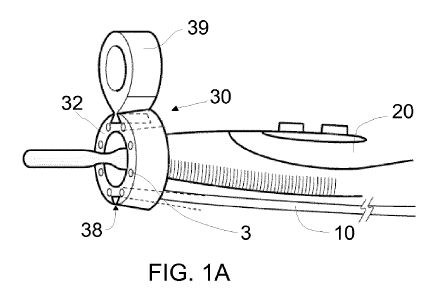
Figures 1A and 1 B show an example of a kit or a device 30 for illuminating
and assisting in a medical procedure, e.g. an open surgery, in combination
with a handheld surgical tool. In this example, the handheld surgical tool is
an
electrical scalpel, which comprises an elongate shaft 20, having a receptacle
CA 02990513 2017-12-21
WO 2017/001379 PCT/EP2016/064960
14
25 with a distal open end. The elongate shaft 20 may be held by a medical
professional near a proximal end of the shaft. The proximal portion of the
shaft 20 may have an ergonomic shape configured for gripping or handling.
The gripping portion may extend from a proximal end to approximately a mid-
portion of the shaft. The ergonomic shape may be adapted for a pencil grip.
The receptacle 25 of the electrical scalpel is configured to receive e.g. an
electrocautery blade 24. The receptacle 25 may be made from an electric
insulating material. The receptacle 25 may be made from a rubbery or
polymeric material.
A power supply system for powering the scalpel, and a cable connecting a
proximal end of the shaft with the power supply system may generally also be
provided. A power switch allows turning on and off the power supply or
otherwise regulating the power flow.
The kit or device 30 for assisting in open surgery in this example comprises a
cylindrical base 32, which has a central opening 2a configured to be mounted
around a portion of the handheld medical tool, for instance, around the
receptacle 25. Thus, the base 32 can be positioned by introducing the
receptacle 25 into the central opening 2a. As a result, the base is positioned
distally with respect to a handle or gripping portion of the medical tool. The
cylindrical base in some examples may have a diameter of e.g. approximately
1 cm, 1.5 cm or 2 cm.
This is also illustrated in figures 10 and 1D.
In this example, eight LEDs 3 are provided. Because of their distal position
with respect to the hand of a medical professional, the hand cannot create
any shadows. The LEDs may be strategically positioned so that regardless of
the precise orientation of the scalpel, sufficient light is provided to the
surgical
field. In other examples, different numbers of LEDs may be provided.
The position on the receptacle 25 of the electrical scalpel thus makes
visualization possible, but at the same time does not prevent access of the
scalpel to the surgical procedure. The device can be positioned sufficiently
CA 02990513 2017-12-21
WO 2017/001379 PCT/EP2016/064960
proximally such that in case of small bodily openings or cavities, it stays
outside of the patient.
In this case, the base has a generally round, rather flat frustoconical shape.
5 Nevertheless, the base might have any geometry as long as it would be
suitable for its purpose. An example of geometry requirements might include
being light enough to keep the ensemble illumination device-medical tool
manageable and not being too sharp or too bulky as to hamper the
practitioner while using said ensemble.
In this example, the base 32 comprises a diaphragm 2 surrounding the central
opening 2a. Alternatively, an 0-ring may be used. The diaphragm 2 may be
made of a relatively flexible material, so that it can adapt to several
handheld
medical tools. As the base 32 is pressed against or pulled away from the
receptacle 25 of the shaft, the diaphragm 2 can deform to admit or release the
receptacle 25 from the central opening 2a. Moreover, the flexibility of the
diaphragm may adapt to a certain range of distal end diameters or geometries
in a variety of handheld medical tools. This provides versatility to the
kit/device, so that it may be used on different handheld medical tools
according to need.
The kit or device 30 also has a connection for connecting to the power
source, namely a cable 10 connecting a rear part of the base 32 with a power
source. Some switches in a control panel may allow to control the different
systems on the device, e.g., turning them on and off, individually or
simultaneously.
At the rear part of the base, a power supply module (not shown) receives
electrical power from the connection (i.e. cable 10) and feeds whatever
devices are mounted on the base. In this case, illumination devices 3 are
powered. However, in a further example, a laser pointer may also be
integrated in the base, e.g. arranged in between the illumination devices.
Alternatively, one of the shown illumination devices may be replaced with a
laser pointer. The power supply may thus also receive electrical power from
the same source.
CA 02990513 2017-12-21
WO 2017/001379 PCT/EP2016/064960
16
Infrared laser pointers or green laser pointers may be useful. The integration
of a laser pointer in the assisting and illuminating device offers several
advantages compared to the normal use of pen-size laser pointers. The laser
pointer may in some examples be turned on and off through switches
provided at the power source or at the cable without the need for the
practitioner to use a hand each time that he needs the laser pointer.
Moreover, he or she may thus use the laser with better precision than if
having to ask someone else to do so.
In addition, conventional laser pointers are unsterile, so that precautions
must
be taken in order to avoid contamination of the surgical field. In the
examples
of the present disclosure, the laser pointer is part of an assisting device
that
may be orderly sterilized and thus be treated as any other medical
instrumentation. In particular, such a feature is an advantage in the medical
practice, since it may lower the risk for contamination of the surgical area,
and as a consequence of undesired complications during the patient's
treatment.
In examples, the rear side of the base may comprise a printed circuit board
which acts as a control system for the device.
The light sources according to examples of the present disclosure may be,
e.g., a light emitting diode (LED), a fiber-optic light, incandescent light
bulbs
or other. The light source may be shaped and/or oriented to promote
projection of light toward the area where the blade 24 is being used. As for
the choice of one type of light source or the other, a choice may be made in
accordance with circumstances balancing e.g. energy consumption, light
output, colour temperature and light source life (including lumen
maintenance).
LEDs offer advantages for the surgical environment such as bright and highly
uniform illumination. Its superior thermal management enables both high
intensity light output and a long life (they yield a durability of e.g. 50,000
hours of LED headlight versus 3,000 to 7,000 hours of a fiber-optic
headlight). Another advantage is the low replacement cost of single LED
lamps. Fiber-optic light, instead, has the features of being a thin and
flexible
CA 02990513 2017-12-21
WO 2017/001379 PCT/EP2016/064960
17
material, which may be an advantage for certain applications. These are the
most common light sources for medical applications in the market nowadays,
due to their well-suited technical features. However, other light sources
might
be considered for being implemented in the device or kit, depending on the
user's requirements and/or limitations.
If fibre optic light is used, an additional fibre optic cable may be provided
from
the base to a light source. In examples, wherein only fibre optic light is
provided (i.e. no laser pointer of further auxiliary device), no power supply
would be needed on the base.
In the example of figure 1, the kit/device 30 comprises a base 32 and an
appendage 39 removably fixed to the base 32. The base 32 in this example
has two dovetail slots 38 which are diametrically opposite to each other. The
appendage 39 has a suitable protrusion 37 with a shape complementary to
the slot 38 to be slidably fitted in the slot 38. In this example, two
appendages
may be fitted to the base 32 at the same time.
In examples, the protrusion may be slightly larger than the slot such that a
friction fit is established. A friction fit is one of the ways in which it may
be
ensured that the appendage does not move in an undesirable manner with
respect to base 32.
Each of the appendages has a through-hole which serves as a receptacle for
receiving an auxiliary medical tool. In this case, the "primary" medical tool
is
the electrical scalpel. The auxiliary medical tool may be any tool which may
be used during an open surgery in combination with the electrical scalpel.
Examples include:
- a video or photo camera,
- a laser pointer (for pinpointing tissue. A laser might in examples be
integrated in the base. In other examples, it is an auxiliary device fitted
in the appendage),
- sensors comprising thermometers, flow meters or the like. The sensors
may be arranged at any suitable location in the base. A suitable
position for placing sensors such as thermometers or flow meters
would be on the side of the base which more easily can be in contact
CA 02990513 2017-12-21
WO 2017/001379 PCT/EP2016/064960
18
with the body tissue. An infrared radiation sensor may be used for
determining temperature differences between tissues which may
indicate vascularization. A flow meter may be based on ultrasound
technology. The (ultrasonic) flow meter specifically in an
implementation on an electrical scalpel may serve to locate blood
vessels. A further or alternative sensor that may be incorporated in the
device for the same purpose is a spectrophotometer.
- an irrigator (in case of electrocoagulation),
- a radioactive probe (e.g. when dissecting a tumour), or
- a liquid/gas aspirator (e.g. for aspirating smoke development when
cutting using the scalpel).
In the latter case, such an aspirator may be combined with a REIMS
spectrometer to immediately analyse the smoke. See figure 6A
In examples, the auxiliary medical device or tool supports and/or enhances
the functioning of the medical tool on which the device is mounted.
Figures 2A and 2B illustrate details which may be incorporated in examples of
the devices (or kits) for assisting in an open surgery. Figure 2A illustrates
that
the protrusion 37 which is configured to mate with the slot 38 may have one
or more local protuberance 37a which may fit in complementary local
recesses 38a along slot 38. The engagement of the protuberance 37A in local
recess 38A can ensure that the appendage 39 is fitted to the base 32 in the
correct position and stays in that position.
In examples, the engagement of the protrusion 37 into the slot 38 may further
provide electrical power to provide energy supply to the auxiliary medical
device being received in the appendage.
Figure 2B illustrates that in examples, the base may have a portion that
functions as an electrical connector. In the example of figure 2B, a bottom
portion of the appendage may incorporate conductive stripes 37b. If or when
the base is provided with electrical power, the same electrical power can be
made available for e.g. an auxiliary device mounted in the appendage.
CA 02990513 2017-12-21
WO 2017/001379 PCT/EP2016/064960
19
In yet further examples, a portion of the appendage may be shaped as an
electrical plug (or socket) and the base may comprise a complementary
socket (or plug), again to provide electrical power to an auxiliary medical
device through the base.
Figures 3A ¨ 30 schematically illustrate different views of a further example
of
a device or kit for assisting in an open surgery. Figure 3A illustrates an
example of a device/kit for assisting in open surgery which has an increased
length as compared to the example of figure 1. Depending on which handheld
medical tool is used as "primary" surgical tool, the geometry of the base 32
and kit/device 30 may be adapted to fit to the tool. Also in this example, the
base 32 tapers outwardly from a distal end to a proximal end.
Figure 3B illustrates that, similarly, the protrusion 37 may taper outwardly
from a distal end 371 to proximal end 372. An aspect of such a tapered
protrusion (and mating slot) is that the correct position of the appendage 39
with respect to the base 32 can be ensured.
Again in this example, two appendages can be fitted to base 32. In further
examples, a base may be provided that is configured for only a single
appendage, or for three or more appendages. In still further examples, the
appendage(s) may be integrally formed with the base. See figures 7A ¨ 8B.
Figures 4A and 4B schematically illustrate a further example of a kit/device
for assisting in an open surgery with different handheld medical devices and
auxiliary medical tools. In figure 4A, the kit/device 30 is fitted to an
ultrasonic
scalpel 40.
Figure 4B serves to illustrate that complementary to the LEDs 3, an additional
illumination device 50, e.g. an incandescent light bulb may be temporarily
positioned in appendage 39 when increased illumination is required. The
further illumination device in this case thus acts as the auxiliary medical
tool.
Figures 5A - 50 schematically illustrate three further auxiliary medical
devices
which may be used with examples of the kits/devices for assisting in an open
surgery. Particularly figure 5A shows a laser pointer 60. In particular, an
CA 02990513 2017-12-21
WO 2017/001379 PCT/EP2016/064960
infrared laser or green laser pointer are used in operating rooms. In the
examples of the present disclosure, such laser pointers or laser pens may be
inserted in the receptacle of appendage 39 such that they point towards a
distal end of the primary medical tool.
5
Figure 5B serves to illustrate that on occasions, it might be preferable to
have
light 51 of a different wavelength, e.g. infrared or UV light instead of white
light. On such occasions, a suitable LED may simply be inserted in an
appendage 39 to provide the required illumination. And figure 50 serves to
10 illustrate that on occasions, it might be preferable to have a
radioactive probe
in order to measure radioactive radiation and thus precisely determine the
position of cells and tissue affected by cancer.
Figures 6A ¨ 6B show a kit or device according to a different example
15 comprising two appendages 39 and 39. The example shown in figure 6A thus
differs from that of figure 1A in that a second appendage 39' is also
removably fixed to the base 32 substantially as explained in connection with
figure 1A. Furthermore, a laser pointer 60 is fitted in the appendage 39
substantially as explained in connection with figure 5A. Further in this
20 example, the second appendage 39' is open, i.e. it has a substantially C-
shaped portion, as clearly shown in figure 6B which shows a partial front view
of the kit/device illustrated in figure 6A.
If the portion with the substantially C-shaped cross-section is sufficiently
closed and/or has sufficient resiliency, an auxiliary medical device can be
positioned in the corresponding through-hole. The auxiliary medical device
may in examples be mounted with a snap-fit.
And the kit/device of figure 6A further differs from that of figure 1A in that
a
tube 80 aspirating the generated smoke and leading towards a REIMS
spectrometer may also be attached to the base by an elastic strap 81 that is
connected to the base 32, between the first 39 and second 39' appendages.
Figure 6B illustrates how an elastic band or strap can function as an
appendage. The elastic band can be forced to elongate to create sufficient
space for fitting an auxiliary medical device. The auxiliary medical device in
CA 02990513 2017-12-21
WO 2017/001379 PCT/EP2016/064960
21
this sense is then clamped between the base 32 and the elastic band.
Figure 60 shows that in further alternatives a strap 82 provided with a Velcro-
type fastener may be foreseen e.g. for attaching the electrical cable to the
shaft 20 of the electrical scalpel. The electrical cable thus will not hinder
movements of the surgeon or of the medical devices.
Figures 7A ¨ 8B show two alternative devices for assisting in an open
surgery. Figures 7A and 8A show perspective views of this examples and
figures 7B and 8B show exploded view of the same. These examples differ
from the examples above in that an appendage 90 (figure 7), or 91 (figure 8)
is integrally formed with the substantially cylindrical base 900 and 910.
In both cases the base 900 and 910 comprises a central opening 901 and
911 for receiving a handheld medical tool such as the electrical scalpel of
the
example of figure 1A. Alternatively, other handheld medical tools in
particular
surgical tools may be foreseen.
Further in these examples, an auxiliary through-hole working channel 905,
915 may be integrally formed with the base 900, 910. Alternatively, the
working channel may comprise a C-shape or U-shape such that it can grip an
auxiliary medical tool, substantially as explained in connection with figures
6A
or 6B.
The example of figures 7A and 7B comprises three LEDs 902 equidistantly
arranged at 120 angles between them around the central opening 901. And
the example of figures 8A and 8B comprises five LEDs 912 equidistantly
arranged (at 72 angles between them) around the central opening 911.
Figures 7B and 8B further show that these examples are made from three
pieces 90a, 91a; 90b, 91b and 90c, 91c made of e.g. a resilient material.
Polyamide is one of the materials suitable for this case.
In the example of figure 7B, spaces 904 for accommodating LEDs 902 are
provided between pieces 90a and 90b, and a grooved space 903 for housing
e.g. any required wiring is further defined between pieces 90a and 90b. In the
CA 02990513 2017-12-21
WO 2017/001379 PCT/EP2016/064960
22
example of figure 8B, piece 91a is provided with spaces 913 for
accommodating the LEDs 912. Furthermore, a grooved space 914 for housing
required wirings is defined between pieces 91a and 91b.
In this example, the device may be relatively easy manufactured and
assembled. For assembly, the three pieces may be glued together. In these
examples, the device incorporates base and appendage in a single integrally
formed body, but in other examples may be separate elements to be
assembled just prior or during surgery.
In these examples, a power cable (not shown) for connecting to a power
source may further be attached to e.g. a rear side of respectively pieces 90a
or 91a so as to supply power to the LEDs. In some cases a laser pointer may
be integrated in the base 900, 910 such that it points towards a distal end of
a
primary medical tool that may be placed around the central opening 901, 911.
The laser pointer may be arranged e.g. replacing one of the LEDs or in
between LEDs substantially as explained in connection with figure 1.
In any of the examples disclosed herein, different types of light may be used.
In some implementations, white light may be preferred.
In some examples, a device or kit substantially as hereinbefore described
mounted or mountable on a medical tool may include illumination systems
configured to emit light in different ranges of wavelength. In some examples,
the wavelength of an illumination system may be varied in use.
In any of the hereinbefore described examples, the cable 10 providing
electrical power to the kit/device 30 may comprise a fastener for attachment
of the cable to the shaft 20 of the electrical scalpel. Suitable fasteners
include: a mounting clip, adhesives, clamps, cable ties, or Velcro TM
fasteners,
to mention some possibilities. In some examples, a mounting clip may be
attached to the shaft through a snap-on connection. The mounting clip can
also have an opening to receive the cable or connection to the power source.
The clip may be positioned at a variety of places on the shaft 20. A clip
according to this example could be attached to the shaft 20 by, e.g., sliding
the clip onto the shaft. Analogously, the clip may be easily removed from the
CA 02990513 2017-12-21
WO 2017/001379 PCT/EP2016/064960
23
shaft by sliding it off the shaft. A plurality of mounting clips may be used,
depending on the length and geometry of the medical tool.
In any of the hereinbefore described examples, the device may have a timer
linked to the lighting system provided. The device may be programmed to
automatically turn off the light(s) and/or automatically disconnect electrical
power after a predetermined period of time.
Although only a number of examples have been disclosed herein, other
alternatives, modifications, uses and/or equivalents thereof are possible.
Furthermore, all possible combinations of the described examples are also
covered. Thus, the scope of the present disclosure should not be limited by
particular examples, but should be determined only by a fair reading of the
claims that follow.