Note: Descriptions are shown in the official language in which they were submitted.
CA 02990745 2017-12-22
WO 2017/005880
PCT/EP2016/066200
INFLUENZA POTENCY ASSAYS
[1] This application claims the benefit of European Patent Application No.
15175765.5 (filed 7th July
2015) and European Patent Application No. 16152829.4 (filed 26th January
2016), the complete contents
of which are hereby incorporated herein by reference for all purposes.
FIELD OF THE INVENTION
[2] This invention relates generally to vaccines, more specifically to
assays for influenza vaccines.
BACKGROUND TO THE INVENTION
[3] Recent outbreaks of influenza highlight the need to rapidly produce and
release adequate amounts
of influenza vaccines to protect the general public from this disease, which
has potentially deadly
complications.
[4] The standard assay for hemagglutinin (HA) content in inactivated
influenza vaccines is based on
single radial immunodiffusion ("SRID") (refs. 1 & 2) which was recommended by
the WHO in 1978 to replace
tests based on agglutination of erythrocytes.
[5] Although the SRID assay is well established, it is slow to perform, has
poor dynamic range, is
susceptible to considerable variability, and it can take a long time to
prepare and calibrate the required
specific anti-HA serum. As the influenza strains in vaccines change every
season, this creates a bottleneck
for influenza vaccine lot release because these reference reagents need to be
prepared and calibrated
anew for every strain change. This is particularly problematic in the case of
an influenza pandemic where
influenza vaccines need to be prepared as quickly as possible.
[6] Another drawback of the SRID assay is that it may not reliably
distinguish between immunogenically
active forms of the influenza hemagglutinin (HA) antigen and those which are
not as immunogenic because
the antisera used in the assay may not be completely specific and may react
with both forms, although it is
generally thought that such antisera can be adjusted to preferentially
recognize the native, immunogenic
form in the SRID assay (ref. 125). As the immunogenicity (hence the
immunoprotection) of an influenza
vaccine is determined by the amount of immunogenically active HA, it is
desirable for an assay to be able
to specifically measure the immunogenically active form of HA.
[7] Reference 3 suggests an alternative to a SRID assay in which
ultrafiltration is followed by reverse
phase high pressure liquid chromatography (RP-HPLC), and references 4 and 5
teach high pressure liquid
chromatography (HPLC) based assays. References 6 and 7 developed quantitative
mass spectrometry
based assays. These assays could accurately quantify total HA and did not
depend on strain-specific
antisera, but failed to differentiate immunologically active HA from inactive
HA. An ELISA assay was able
to specifically quantify immunologically active HA but relied on generation of
strain specific antibodies (ref.
8), which significantly increases the time needed before the vaccine can be
released.
1
CA 02990745 2017-12-22
WO 2017/005880
PCT/EP2016/066200
SUMMARY OF THE INVENTION
[8] The present invention encompasses identifying the source of problems
with the existing influenza
potency assays. Thus, the invention is based at least in part on the
recognition that conventional methods
used to quantify influenza virus antigens for vaccine production do not
accurately measure the amount of
influenza virus proteins included in vaccines as immunogens. Work presented
herein and elsewhere
suggests that an isolated influenza viral protein may act as an antigen in in
vitro immunoassays, but not as
a functional immunogen to elicit an immune response in vivo. There is dire
need to differentially measure
these functionally and structurally distinct forms of influenza proteins, so
influenza vaccines can be
manufactured to reflect accurate amounts of functional immunogens contained
therein. To that end, the
inventors of the present disclosure sought to develop an assay based on
biophysical measurements (as
opposed to immunochemical measurements) to distinguish structural differences
between active,
immunogenic conformations and inactive counterparts. The rationale for this
approach includes: i) more
direct assessment of the proteins/antigens themselves; ii) speed by which such
assays can be carried out;
ill) simplicity by which a sample with multiple antigens can be simultaneously
processed; and/or, iv) no
necessity for reliance on the availability of corresponding antisera
(typically sheep antisera).
[9] Accordingly, the methods provided herein enable vaccine manufacturers
to accurately indicate how
much immunogenic antigen is contained in their respective vaccines, not merely
the total amount of proteins
included in the products. This is important from a public health perspective,
because it is necessary to
determine how much immunogenic HA is present and also is desirable to know how
much non-functional
HA is present in vaccines, and because it helps to shift the focus more on the
purity and efficacy of vaccines.
[10] It is therefore an object of the invention to provide an influenza
potency assay which is faster than
the traditional SRID assay and further provides a more reliable assessment of
the amount of
immunogenically active HA in an influenza vaccine.
[11] The invention further provides an improved SRID assay, in which the
traditional SRID assay has
been modified to take advantage of the benefit of biological proteolysis
described herein, i.e., the ability to
differentiate between the immunogenic form of HA and the poorly immunogenic
form of HA. Thus, another
aspect of the invention provides, an SRID assay which incorporates a step of
biological proteolysis (e.g.,
trypsin pre-treatment) prior to SRID, thereby improving accuracy of the assay
in determining the amount of
immunologically active HA in a sample. Suitable samples may be samples as
defined herein. Suitable
samples may be obtained from, for example, antigen bulk preparations (such as
monobulk), intermediate
preparations, during manufacture and/or after final formulation, final vaccine
formulations and/or products
prior to release, vaccine products after storage, etc. SRID with pre-treatment
by biological proteolysis, as
described herein, advantageously avoids overestimation of immunologically
active HA seen in traditional
SRID formats.
[12] The methods described herein are suitable for measuring the amount of
influenza viral antigens
during the manufacture of influenza vaccines, as well as for quality control
purposes, e.g., evaluating the
potency of samples (including intermediate preparations, bulk preparations,
and final vaccine products)
after durations of time, e.g., after storage.
2
CA 02990745 2017-12-22
WO 2017/005880
PCT/EP2016/066200
BRIEF DESCRIPTION OF DRAWING
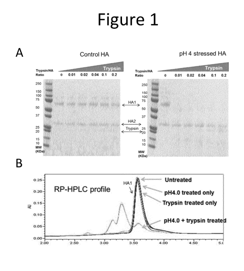
[13] Figure 1 shows that low pH-induced post fusion HA1 is sensitive to
trypsin digestion while control
pre-fusion HA1 is resistant to trypsin even at high protease concentration, as
shown by both reduced SDS-
PAGE (A) and RP-H PLC (B).
[14] Figure 2 provides a flow chart of the influenza potency assay.
[15] Figure 3 provides results from pH 4-stressed quadrivalent influenza
vaccine (QIV) sample tested by
an assay of the present invention and SRID. (A) RPLC chromatogram of the
differentially treated samples
(annotation of the peaks are based on retention times of the standard
monobulks); (B) assay results by the
current assay (left panel) and SRID (right panel). Potency of each strain at -
/- control is shown as 100%.
[16] Figure 4 provides results from pH 11-stressed quadrivalent influenza
vaccine (QIV) sample tested
by an assay of the present invention and SRID. (A) RPLC chromatogram of the
differentially treated
samples (annotation of the peaks are based on retention times of the standard
monobulks); (B) assay
results by the current assay (left panel) and SRID (right panel). Potency of
each strain at -/- control is shown
as 100%.
[17] Figure 5 provides results from heat (56 C)-stressed quadrivalent
influenza vaccine (QIV) sample
tested by the current assay and SRID. (A) RPLC chromatogram of the
differentially treated samples
(annotation of the peaks is based on retention times of the standard
monobulks); (B) assay results by the
current assay (left panel) and SRID (right panel). Potency of each strain at -
/- control is shown as 100%.
[18] Figure 6 shows percentage of immunogenic HA recovered following acetone
precipitation for
A/Victoria, A/Brisbane and B/Brisbane strains.
[19] Figure 7 provides a table detailing the percentage of digested inactive
HA peptides that remain
following different washing protocols. Samples were washed i) three times in
acetone; ii) twice in acetone
followed by a single ethanol wash; or iii) three times in ethanol. Washing
three times in ethanol produced
the best result by removing the greatest amount of digested inactive HA
peptides.
[20] Figure 8 provides graphs showing immunogenicity in mice of two injections
of 1 pg egg-produced
A/Texas/50/2012 (H3N2) HA maintained at pH 7.2 or transiently exposed to pH
4.0, with and without trypsin
digestion. (A) HI titers using A/Texas/50/2012 (H3N2) virus and turkey blood
cells. (B) Microneutralization
titers in the same set of sera as A using A/Texas/50/2012 (H3N2) virus to
infect MDCK cells.
[21] Figure 9 shows SDS-PAGE, RP-HPLC, ELISA and SRID analysis of egg-produced
A/Texas/50/2012
(H3N2) HA maintained at pH 7.2 or transiently exposed to pH 4.0, with and
without trypsin digestion. (A)
SDS-PAGE of non-reduced (left) and reduced (right) samples. (B) Analytical RP-
HPLC chromatograms of
the same set of samples as in (A). (C) ELISA of the same set of samples as in
(A), performed with HA
coated on plates and detected by the sheep polyclonal antiserum used in SRID.
(D) SRID gel image for
the same set of samples as in (A). (E) Summary of HA quantification by RP-
HPLC, ELISA and SRID with
HI titer from immunogenicity in mice.
3
CA 02990745 2017-12-22
WO 2017/005880
PCT/EP2016/066200
[22] Figure 10 shows SRID and trypsin/RP-HPLC analyses for homogeneous samples
of egg-produced
A/Perth/16/2009 (H3N2) HA maintained at pH 7.2 or transiently exposed to pH
4.0 and for mixtures of the
two samples. (A) Image of an SRID gel assaying non-stressed HA, low-pH
stressed HA and non-stressed
HA spiked with 2x, 1.5x, lx and 0.5x of low-pH stressed HA. (B) Relative
quantification of HA from the
SRID gel in A and from trypsin/RP-HPLC assay of the stressed samples and their
mixtures.
[23] Figure 11 shows SRID and RP-HPLC analysis for HA maintained at pH 7.2 or
transiently exposed
to pH 4.0 or the mixture of the two HA samples with and without trypsin
digestion. (A) SRID image for egg-
produced B/Brisbane/60/2008 samples subject to these treatments. (B) HA
quantification by SRID and RP-
HPLC for egg-produced B/Brisbane/60/2008 HA; (C) A/California/07/2009 (H1N1)
HA; (D)
A/Texas/50/2012 (H3N2) HA; and (E) B/Massachusetts/02/2012 HA; subject to
these treatments.
[24] Figure 12 provides images of SRID analysis of IRDye-labeled
A/Texas/50/2012 (H3N2) HA. (A)
Non-stressed HA and low-pH stressed HA, each labeled with IRDye800, were
analyzed by SRID. The
labeled protein was tracked in the SRID gel by infrared fluorescent imaging.
Non-labeled HA was also
detected by western blotting with an anti-H3 antibody on a nitrocellulose
membrane used to blot the SRID
gel. (B) Non-stressed HA was labeled with IRDye800, and low-pH stressed HA was
labeled with IRDye680.
The non-stressed HA, stressed HA, and a mixture of non-stressed and stressed
HA with and without trypsin
treatment were detected in SRID gel through the green and red channels of the
imager.
DETAILED DESCRIPTION OF PREFERRED EMBODIMENTS
[25] The invention provides methods for quantifying immunogenic HA in a
sample, which are faster, more
accurate and do not require the use of antisera. According to the invention,
immunogenic HA and inactive
HA can be separated due to their different conformation, which is reflected in
their differential sensitivity to
proteolysis. These methods are independent from antisera and exploit a
biophysical pretreatment to
selectively remove immunologically inactive HA (e.g., poorly immunogenic,
stressed or post-fusion
conformations), followed by separation and quantification of the immunogenic
HA. These methods are
therefore significantly faster than the standard SRID assay and are further
more accurate because only the
amount of immunogenic HA is measured.
[26] The invention provides a method comprising the steps of:
a) providing a sample comprising immunogenic HA, inactive HA, or
combination thereof;
b) subjecting the sample to biological proteolysis, wherein the inactive HA
is digested and the
immunogenic HA remains undigested;
c) separating the digested inactive HA from the undigested immunogenic HA
in the sample;
d) subjecting the undigested immunogenic HA to analytical proteolysis, so
as to provide fragments of
digested immunogenic HA; and,
e) carrying out liquid chromatography-electrospray ionization-tandem mass
spectrometry (LC-ESI-
MS) in the presence of at least one labeled reference HA peptide, to quantify
the amount of immunogenic
HA in the sample.
4
CA 02990745 2017-12-22
WO 2017/005880
PCT/EP2016/066200
[27] Further provided is a method for quantifying immunogenic influenza HA in
a sample, comprising the
steps of:
a) subjecting the sample to biological proteolysis;
b) separating the immunogenic HA from other components in the sample; and,
c) quantifying the immunogenic HA in the sample.
[28] In a particularly preferred embodiment, step (c) is carried out using
mass spectrometry, in particular
liquid chromatography-electrospray ionization-tandem mass spectrometry (LC-ESI-
MS).
[29] The quantification of the immunogenic HA can in principle be performed
using any method for protein
quantification known in the art. Certain embodiments described above would
also be compatible with
quantification by SRID.
[30] Also provided are methods as described herein wherein a step of
separating the immunogenic HA
from other components in the sample (e.g. separating the digested inactive HA
from the undigested
immunogenic HA in the sample) is dispensed with. Such methods may be an
alternative to the methods
described herein that involve a separation step, particularly the methods
wherein quantification is by mass
spectrometry. Such methods have the advantage of reducing the number of
processing or handling steps
(e.g. by eliminating a step of separation by protein precipitation) and
minimizing sample loss. The inventors
have found that one can further differentiate the quantity of immunogenic HA
from inactive HA in a sample
if different proteases, or different selections of proteases, having different
substrate specificities (or
selections of substrate specificities), are used in each of the biological and
analytical proteolysis steps. In
such methods, the inactive HA is digested by one or more proteases in the
biological proteolysis step, to
produce digested inactive HA, while the immunogenic HA (or substantially all
of the immunogenic HA),
remains undigested. The mixture of undigested immunogenic HA and digested
inactive HA is then
subjected to analytical proteolysis by one or more proteases. Importantly, the
analytical proteolysis is
carried out using a protease or selection of proteases that cannot cleave the
immunogenic HA at one or
more cleavage site(s) which can be cleaved in the inactive HA during
biological proteolysis. In this way,
the analytical proteolysis can provide fragments of digested immunogenic HA
that comprise immunogenic
HA-derived peptide(s) that is/are distinguishable from the inactive HA-derived
peptides. For example, the
fragments of digested immunogenic HA will comprise one or more immunogenic HA-
derived peptide(s)
which contain at least one cleavage site that can be cleaved by at least one
protease, wherein the at least
one protease was used in the biological proteolysis step, but not in the
analytical proteolysis step.
[31] Accordingly, the invention also provides a method comprising the steps
of:
a) providing a sample comprising immunogenic HA, inactive HA, or
combination thereof;
b) subjecting the sample to biological proteolysis by one or more
proteases, wherein the inactive HA
is digested and the immunogenic HA remains undigested;
c) subjecting the mixture of undigested immunogenic HA and digested
inactive HA to analytical
proteolysis using one or more proteases, wherein the analytical proteolysis
cannot cleave the immunogenic
HA at one or more cleavage site(s) which can be cleaved in the inactive HA
during biological proteolysis,
so as to provide fragments of digested immunogenic HA that comprise
immunogenic HA-derived peptide(s)
that is/are distinguishable from the inactive HA-derived peptides; and,
5
CA 02990745 2017-12-22
WO 2017/005880
PCT/EP2016/066200
d) carrying out liquid chromatography-electrospray ionization-tandem
mass spectrometry (LC-ESI-
MS) in the presence of at least one labeled reference HA peptide, to quantify
the amount of immunogenic
HA in the sample.
[32] In preferred embodiments of these methods, one protease (e.g. a
chymotrypsin-like protease) is
used the biological proteolysis step and a different protease having a
different substrate specificity (e.g. a
trypsin-like protease) is used in the analytical proteolysis step.
Alternatively, more than one different
protease (e.g. two) may be used in the biological proteolysis step and a
single protease may be used in the
analytical proteolysis step. If a set of more than two different proteases
(e.g. three, four or more) is used
in the biological proteolysis step, a set of fewer different proteases may be
used in the analytical proteolysis
step.
[33] Such methods may, for example, involve subjecting the sample to
biological proteolysis by a first
protease and a second protease, wherein the substrate specificity of the
second protease is different to
that of the first protease. The methods may then involve subjecting the
mixture of undigested immunogenic
HA and digested inactive HA to analytical proteolysis by only the first
protease. Alternatively, a third
protease having a substrate specificity that is different to that of both the
first and second proteases may
be used in the analytical proteolysis. In some embodiments, first and second
proteases are used for the
biological proteolysis, wherein the first protease is a trypsin-like protease
(e.g. trypsin) and the second
protease is a chymotrypsin-like protease (e.g. chymotrypsin).
[34] The analytical proteolysis may use a single protease. In particularly
preferred embodiments, the
single protease used for analytical proteolysis is a trypsin-like protease
(e.g. trypsin).
[35] Further examples of suitable proteases are provided below and would be
known to a person skilled
in the art. Suitable combinations of proteases for use in the invention can be
easily determined by a skilled
person in a pilot experiment. It will also be appreciated by a person skilled
in the art that a reference /
surrogate HA peptide(s) used for immunogenic HA quantification may be selected
to comprise an HA
sequence which contains at least one cleavage site that can be cleaved by at
least one protease, wherein
said at least one protease is used in the biological proteolysis step, but not
in the analytical proteolysis step.
For example, a surrogate HA peptide used for immunogenic HA quantification may
be selected which
comprises an HA sequence which is cleaved by a chymotrypsin-like protease
(e.g. chymotrypsin) when the
chymotrypsin-like protease is used in the biological proteolysis step (e.g.
either alone or along with a trypsin-
like protease such as trypsin), but not in the analytical proteolysis step.
[36] In another aspect, the invention provides improved SRID methods for
quantifying immunogenic HA
in a sample, which are more accurate than standard SRID assays. As explained
above, immunogenic HA
and inactive HA can be separated due to their different conformation, which is
reflected in their differential
sensitivity to proteolysis. The SRID methods of the invention typically use an
antiserum (i.e., they are not
independent from antisera) and also exploit a biophysical pretreatment to
enable selective removal of
immunologically inactive HA (e.g., poorly immunogenic, stressed or post-fusion
conformations). In the
SRID methods of the invention, this pretreatment (biological proteolysis) is
followed by quantification of the
immunogenic HA.
6
CA 02990745 2017-12-22
WO 2017/005880
PCT/EP2016/066200
[37] Thus, the invention further provides a method for quantifying immunogenic
influenza HA in a sample,
comprising the steps of:
a) subjecting the sample to biological proteolysis; and,
b) quantifying the amount of immunogenic HA in the sample from (b) by a
SRID assay.
[38] In some embodiments, the SRID assay of step (b) is carried out with the
use of an antiserum, such
as polyclonal antisera (e.g., sheep polyclonal antisera) and/or a monoclonal
antibody antiserum (e.g.,
comprising suitable monoclonal antibodies). Suitable antiserum or antisera
is/are strain-specific and may
be HA-specific. Thus, the SRID assay may be carried out with the use of strain-
specific, anti-HA, polyclonal
(sheep) antisera.
[39] The invention further provides a method for manufacturing an influenza
vaccine, the method
comprising steps of:
a) providing a sample from a bulk preparation comprising an influenza HA;
b) quantifying the amount of immunogenic HA according to a method of the
invention; and,
c) packaging unit dosage forms from the bulk preparation according to the
amount of immunogenic
HA in the sample.
[40] The invention further provides a method for preparing an influenza
vaccine, comprising the steps of:
a) quantifying the amount of HA in a bulk vaccine by a method of the
invention; and
b) preparing a vaccine from the bulk.
Biological proteolysis and analytical proteolysis
[41] Influenza viral surface HA primarily exists as an oligomer (such as a
trimer) in the pre-fusion state,
which is the most immunologically relevant state. Under various stress
conditions, HA can undergo an
irreversible transition to post-fusion state, which does not elicit a good
immune response. The preparation
of influenza vaccines often results in the presence of post-fusion HA and the
standard SRID assay cannot
distinguish between pre-fusion (immunogenic) and post-fusion (inactive) HA,
thus resulting in an
overestimation of the actual amount of immunogenic HA contained in the
vaccine. The methods of the
invention distinguish between these different forms of HA by biological
proteolysis so that only the
immunogenic form is quantified.
[42] Accordingly, as used herein, "biological proteolysis" refers to an enzyme-
based digestion of HA,
whereby immunologically active forms of the protein (e.g., HA in the pre-
fusion state) remain intact (i.e.
undigested), while inactive forms of the protein (e.g., HA in the post-fusion
state) become digested. A step
of biological proteolysis therefore achieves differential digestion, depending
on the conformation of the
protein. HA proteins that have not undergone a denaturation step are resistant
to biological proteolysis.
Thus, a step of biological proteolysis, as used herein, achieves controlled or
limited digestion of the protein,
depending on the structural integrity or conformation of the protein.
7
CA 02990745 2017-12-22
WO 2017/005880
PCT/EP2016/066200
[43] By contrast, as used herein, "analytical proteolysis" refers to
fragmentation (i.e., digestion) of a target
protein regardless of its conformation, typically for purposes of subsequent
analytical step(s), such as mass
spec analyses. Analytical proteolysis involves a step of denaturing the
protein before digestion.
[44] It was reported in reference 9 that the conformation of HA changed
depending on the pH and that
some forms of HA were susceptible to protease digestion whilst others were
not. This could be attributed
to the well-packed structure and dense glycosylation coat on the surface of
immunogenic HA. The inventors
have confirmed that pre-fusion HA is very resistant to proteolytic degradation
in its native state even at a
high protease concentration (HA:Trypsin = 5:1). In contrast, low pH induced
post fusion HA1, even when
native, was very sensitive to proteolytic degradation.
[45] By including a step of biological proteolysis before quantification, the
methods of the invention allow
a distinction between immunogenic HA and inactive HA. In particular,
immunogenic HA is protease-
resistant whilst the inactive HA is protease-sensitive. This means that
substantially all of the inactive HA
becomes digested whereas substantially all of the immunogenic HA remains
structurally intact when a
protease is added to the sample (for example, where trypsin is added at an
enzyme:substrate ratio of 1:20
and incubated at 37 C for 2 hours). The biological proteolysis step therefore
digests substantially all of the
inactive (post-fusion) HA whilst substantially all of the immunogenic (pre-
fusion) HA remains undigested.
In this respect, it will be understood that the immunogenic HA is not entirely
resistant to protease-dependent
cleavage per se. In particular, HAO can be cleaved by certain proteases (such
as trypsin) into HA1 and
HA2 but the protein does not become dissociated into fragments. Rather, HA1
and HA2 remain associated
as a complex which maintains the same structural integrity (e.g., pre-fusion
state). These HA1/HA2
complexes are still considered to be immunogenic. The cleaved pre-fusion HA
can be distinguished from
the digested products of inactive HA in that substantially all of the inactive
HA is fragmented into peptides
by the protease.
[46] "Substantially" in the context of proteolysis (digestion) means that at
least 80%, 85%, 90%, 95%,
96%, 97%, 98%, or 99% of the inactive (post-fusion) HA present in a sample is
digested. Likewise,
"substantially" in the context of immunogenic HA means that at least 80%, 85%,
90%, 95%, 96%, 97%,
98%, or 99% of the immunogenic (pre-fusion) HA remains undigested.
[47] The immunogenic HA is in the form of a pre-fusion HA trimer, a pre-fusion
HA oligomer (e.g., so
called rosettes) or combinations thereof. The inactive HA is an HA monomer, a
post-fusion HA trimer, a
denatured protein, an aggregated protein, or a combination thereof. The pre-
fusion and post fusion HA
configurations can be identified, for example, using methods known in the art
such as crystallography.
[48] The immunogenic HA will generally elicit a much higher immune response
compared to the inactive
HA. For example, the geometric mean titre (GMT) obtained from injecting a
subject with the immunogenic
HA may be at least 2 times, at least 4 times, at least 8 times or at least 16
times higher with the immunogenic
HA compared to the inactive HA. Thus, it should be readily understood by those
skilled in the art that the
"inactive HA" as used herein (e.g., post-fusion, or stressed forms of HA) does
not necessarily mean it
completely lacks immunogenicity; rather, it is poorly immunogenic as compared
to pre-fusion, non-stressed
forms of HA that are trypsin-resistant.
8
CA 02990745 2017-12-22
WO 2017/005880
PCT/EP2016/066200
[49] The biological proteolysis can be performed with any protease that can
digest inactive HA. Such
enzymes are known in the art and include, for example, serine proteases (such
as trypsin), threonine
proteases, cysteine proteases, aspartate proteases, glutamic acid proteases,
and metalloproteases.
Exemplary serine proteases include, without limitation, trypsin-like
proteases, chymotrypsin-like proteases,
elastase-like proteases and subtilisin-like proteases. All of these enzymes
are expected to work in the
methods of the invention as the different proteolytic activity in respect of
immunogenic HA and inactive HA
is due to the well packed structure and dense glycosylation coat on the
surface of immunogenic HA which
physically prevents proteases from digesting the protein. Indeed, the
inventors have also shown that
chymotrypsin can digest inactive (post-fusion) HA in a biological proteolysis
step. Thus, biological
proteolysis may use trypsin and/or chymotrypsin. The methods of the invention
may be practiced using
two, three, four or more proteases.
[50] Methods for determining whether a protease can digest inactive HA are
well known in the art. For
example, a skilled person can provide inactive HA using the method described
in reference 9 and test
different proteases to establish which one can digest the inactive HA.
[51] A group of proteases known to digest inactive HA are serine proteases.
These are enzymes that
cleave peptide bonds in proteins, in which serine serves as the nucleophilic
amino acid at the active site.
Such proteases are required for influenza viral infection of host cells in
vivo and function by splitting the
precursor HAO into the HA1 and HA2 forms, thus allowing the influenza virus to
infect the host cells by
promoting membrane fusion. As serine proteases are known to digest influenza
HA they are preferred for
use in the invention.
[52] The most commonly used serine protease for digesting influenza HA is
trypsin and the use of this
protease in the methods of the invention is particularly preferred. However,
other serine proteases which
can digest HA can also be used. Examples of such proteases include TMPRSS2 and
HAT (ref. 10).
[53] The protease is preferably added directly to the sample. This is
preferred because it makes the
quantification process easier and further avoids any overestimation of the
amount of immunogenic HA due
to manipulation of the sample. In some circumstances it may be desirable,
however, to optimize the
conditions for biological proteolysis in the sample before the protease is
added. This may be necessary, for
example, where the buffer in the sample does not allow for optimal protease
activity. In these embodiments,
the buffer in the sample may be exchanged through standard methods in the art
such as, for example,
dialysis. It is also possible to dilute the sample with additional buffer. It
will be understood that care must
be taken not to create conditions where additional inactive HA is formed, for
example by lowering the
buffer's pH as this could result in inaccurate quantification. Where this is
unavoidable, it is still possible to
quantify HA using the methods of the invention by performing a pilot
experiment to determine the relative
loss in immunogenic HA and correcting the result obtained from quantification
by this amount.
[54] Where influenza viruses are grown in cell culture, proteases such as
trypsin are routinely added
during the growth of the influenza viruses. The production process for
influenza vaccines includes
purification steps and so only negligible amounts of residual protease will be
present in an influenza vaccine
9
CA 02990745 2017-12-22
WO 2017/005880
PCT/EP2016/066200
prepared from the influenza viruses. For the avoidance of doubt, the step of
biological proteolysis cannot
rely on residual protease which may be present but a protease needs to be
added to the sample.
[55] Suitable conditions for digestion can easily be determined by a skilled
person. For example, the
methods of the invention may be performed using about 2, 5, 10, 15, 20, 25,
30, 35, 40, 45, or 50 U/mL of
a protease, such as trypsin. The methods of the invention may be performed
using about 2, 5, 10, 15, 20,
25, 30, 35, 40, 45, 50, 60, 70, 80, 90, or 100 U/mL of a protease, such as
trypsin. Many proteases need
an optimal temperature of between 32 C and 40 C, between 34 C and 38 C or
about 37 C and so digestion
may be performed at that temperature. The exact time for digestion may vary
but the reaction will generally
be allowed to proceed until substantially all of the inactive HA has been
digested. For example, the sample
may be incubated with the protease for 10 minutes, 20 minutes, 30 minutes, 40
minutes, 50 minutes, 60
minutes, 70 minutes, 80 minutes, 90 minutes, 100 minutes, 110 minutes, 120
minutes, 130 minutes, 140
minutes, 150 minutes, 160 minutes or 170 minutes. The time needed for
digestion can be easily determined
by a skilled person in a pilot experiment.
[56] A step of biological proteolysis as discussed above can also be
beneficial in the vaccine
manufacturing process. In particular, it is possible to include such a step in
the vaccine manufacturing
process which has the advantage that the resulting vaccine will predominantly
contain immunogenic HA as
the inactive HA would be selectively removed. Such methods will involve
purification steps to remove the
protease prior to formulation of the vaccine.
[57] Accordingly, the invention encompasses a method for manufacturing a
vaccine intermediate, the
method comprising a step of preparing a bulk preparation comprising an antigen
(such as HA mono-bulk),
subjecting the bulk preparation or portion thereof to biological proteolysis
so as to digest stressed or inactive
forms of the antigen. The resulting intermediate can be subsequently used to
formulate a vaccine product,
which is enriched with protease-resistant, immunologically active form of the
antigen. Thus, the invention
includes a method for manufacturing a vaccine product, comprising steps of:
preparing a bulk preparation
comprising an antigen (such as HA mono-bulk), subjecting the bulk preparation
or portion thereof to
biological proteolysis; and, formulating a vaccine using the bulk preparation
or portion thereof, which has
been subjected to biological proteolysis. The antigen may be influenza
antigen. However, the invention
can be useful for any antigens, which can exist in multiple conformations such
that protease-sensitivity (or
protease-resistance) correlates with biological activities of interest (e.g.,
immunogenicity). In some
embodiments, the vaccine product contains at least 60% of the antigen in
immunologically active
conformation, e.g., at least 60%, at least 70%, at least 80%, and at least
90%. In some embodiments, such
vaccine products enriched with immunologically active antigen(s) may contain
less-than-standard amounts
of the antigen(s) but are able to elicit equivalent or greater immune
responses in subjects, as compared to
standard products that are not enriched with immunologically active forms of
the antigen(s). For example,
as compared to standard influenza vaccines with 15 pg HA per strain per dose,
the vaccine products of the
present invention may have equivalent or better efficacy or effectiveness with
lower total antigens, e.g.,
less than 12 pg, less than 9 pg, less than 7.5 pg, less than 5 pg, less than
3.75 pg, HA per strain per dose.
CA 02990745 2017-12-22
WO 2017/005880
PCT/EP2016/066200
[58] The step of protease digestion may also provide an additional benefit of
reversing some aggregation
that may be present in an antigen preparation, thereby reducing the loss and
increasing the yield of antigen
per preparation.
[59] As discussed above, in some embodiments, more than one protease may be
used in the methods
of the invention. Different proteases, or different sets of proteases, may be
used in each of the biological
and analytical proteolysis steps described herein. Each different protease may
have different substrate
specificity. For example, trypsin-like proteases, chymotrypsin-like proteases,
elastase-like proteases and
subtilisin-like proteases typically have different substrate specificities
from each other. Proteases may be
considered to have different substrate specificities if one is capable of
cleaving a given peptide at a given
cleavage site, while the other is not, under identical conditions. For
example, trypsin-like proteases typically
cleave peptides at the carboxyl side of the amino acids lysine or arginine
(except when either is followed
by proline). Chymotrypsin-like proteases typically cleave peptides at the
carboxyl side of a large
hydrophobic amino acid (e.g. tyrosine, tryptophan, phenylalanine, leucine).
Elastase-like proteases
typically cleave peptides at the carboxyl side of small, hydrophobic amino
acids (e.g. glycine, alanine, and
valine).
[60] In certain embodiments, where quantification is by mass spectrometry,
more than one different
protease may be used in the analytical proteolysis step. As described herein,
analytical proteolysis may
be preceded by a separation step. In alternative embodiments described herein,
the separation step may
be dispensed with. In either case, the analytical proteolysis may use more
than one different protease,
having different substrate specificities (i.e. different cleavage sites). The
use of more than one different
protease in the analytical proteolysis stage may produce fragments of digested
immunogenic HA that
comprise immunogenic HA-derived peptide(s) that are shorter than the
immunogenic HA-derived peptide(s)
that would be produced when using fewer different proteases. Advantageously,
shorter reference /
surrogate peptides may therefore be used for quantification, thus providing
greater freedom to choose
reference peptide(s) having a sequence that is/are conserved in the
immunogenic HA to be quantified. This
technique may also be used to increase the availability of reference
peptide(s) that do not contain amino
acids that are prone to modification (e.g. chemical or post-translational
modifications that could undesirably
influence quantification results). In some preferred embodiments, where the
analytical proteolysis is
preceded by a separation step, the biological proteolysis uses one protease
(e.g. a trypsin-like protease,
such as trypsin) and the analytical proteolysis uses the same type of protease
used in biological proteolysis
and one or more different protease(s) having a different substrate specificity
(e.g. a chymotrypsin-like
protease, such as chymotrypsin).
Separation
[61] Following digestion it is preferable that the undigested immunogenic HA
is separated from other
components in the sample. In particular, it is highly desirable that the
undigested immunogenic HA is
separated from the digested inactive HA. This is advantageous because it makes
the downstream
quantification easier. In particular, separating the immunogenic HA from the
digested inactive HA allows
quantification of immunogenic HA by methods such as mass spectrometry.
11
CA 02990745 2017-12-22
WO 2017/005880
PCT/EP2016/066200
[62] Nevertheless, as discussed above, in some embodiments of the invention,
the separation step may
be dispensed with, e.g. where analytical proteolysis provides fragments of
digested immunogenic HA that
comprise immunogenic HA-derived peptide(s) that is/are distinguishable from
the inactive HA-derived
peptides.
[63] Methods for separating the immunogenic HA from other components in the
sample are well known
in the art and include, for example, reverse phase chromatography, size
exclusion chromatography and ion
exchange chromatography.
[64] Whilst these prior art separation methods are suitable for use in some
embodiments of the invention
(in particular those which do not rely on mass spectrometry to quantify the
immunogenic HA), the inventors
have found that initial attempts using reverse phase and size exclusion
chromatography were
unsatisfactory due to a number of reasons: 1) neither chromatography was able
to achieve optimal baseline
resolution; 2) substantial sample loss on columns was observed; 3) significant
variation introduced during
fraction collection; 4) intense labor and lowered assay throughput associated
with fraction collection, buffer
exchange, and volume reduction.
[65] It is therefore preferred that the immunogenic HA in the sample is
separated from other components
in the sample (in particular the digested inactive HA) by protein
precipitation. The advantage of this
approach includes same-tube sample preparation/processing, which minimizes
sample loss; a reduced
introduction of artefacts; and the convenience of resuspending recovered
protein pellets in a desired
volume of compatible buffer for downstream sample preparation. The inventors
found that protein
precipitation consistently recovered nearly 100% of the immunogenic HA in the
sample.
[66] Various methods for precipitating proteins are known in the art. They
include salting out, isoelectric
point precipitation, precipitation with organic solvents, non-ionic
hydrophilic polymers, and flocculation by
polyelectrolytes. Thus, in some embodiments, the step of separating comprises
removing digested inactive
HA fragments from the sample that retains intact, undigested immunogenic HA.
[67] The inventors have seen good results with organic solvents, in particular
with acetone which resulted
in nearly 100% recovery of immunogenic HA from the sample. In a preferred
embodiment the step of
separating the (undigested) immunogenic HA comprises a step of adding an
organic solvent, in particular
a ketone or alcohol. The organic solvent may be acetone, ethanol or methanol.
The methods of the
invention may further include a step of washing the precipitated protein with
an alcohol. The added alcohol
may have a temperature of less than 4 C. The alcohol is preferably ethanol.
The inventors found that this
step effects complete removal of the digested peptides derived from the
inactive HA. The precipitant may
then be dried, for example by air drying or vacuum centrifugation.
[68] Following protein precipitation, the precipitated protein is resuspended.
This can be achieved, for
example, by adding a buffer that introduces strong denaturing conditions, such
as a strong denaturing
guanidine buffer. The sample may further be heated to facilitate protein
resuspension. Such methods are
standard in the art and a skilled person can therefore easily put them into
practice.
12
CA 02990745 2017-12-22
WO 2017/005880
PCT/EP2016/066200
[69] As shown in the Examples below, where the step of quantifying immunogenic
influenza HA uses a
SRID assay, the SRID assay itself may enable separation of immunogenic HA from
other components in
the sample (e.g. inactive HA).
Quantification
[70] The quantification of the immunogenic HA can in principle be performed
using any method for protein
quantification known in the art. For example, the methods of the invention
would be compatible with
quantification by SRID as the step of digesting the inactive HA, as described
above, allows for a more
accurate determination of the amount of immunogenic HA in the sample. Thus, in
methods of the invention,
a step of quantifying the immunogenic HA may comprise the use of SRID.
However, as mentioned above,
SRID has the drawback that it relies on the use of strain-specific antisera
which takes weeks or even months
to produce. It is therefore preferred that the step of quantifying the
immunogenic HA does not comprise the
use of SRID.
[71] The invention therefore preferably utilizes quantification methods which
do not require the use of
strain-specific antisera. Such methods avoid the current bottleneck for
influenza vaccine production as they
do not require the preparation and calibration of reference antigens every
season. Such methods include
chromatographic methods like high performance liquid chromatography (HPLC), in
particular reverse-
phase high performance liquid chromatography (RP-HPLC), but also mass
spectrometry methods, like
liquid chromatography¨mass spectrometry (LC-MS) and liquid chromatography¨mass
spectrometry/mass
spectrometry (LC-MS-MS) and two-dimensional gel electrophoresis (2-DE).
RP-HPLC
[72] RP-HPLC is a form of chromatography which applies a liquid (mobile phase,
such as a solvent) to a
chromatographic column (stationary phase), with retention on the column
depending on the interactions
between the stationary phase and components present in a sample. A pump moves
the liquid phase
through the column and, as conditions change, different molecules can elute
from the column at different
times. RP-HPLC has a non-polar stationary phase and an aqueous, moderately
polar mobile phase. RP
HPLC retention times can generally be increased by increasing the proportion
of water in the mobile phase
(thereby making the affinity of a hydrophobic analyte for a hydrophobic
stationary phase stronger relative
to the now more hydrophilic mobile phase); conversely they can be decreased by
increasing the proportion
of non-polar or less-polar organic solvent (e.g., methanol, acetonitrile).
[73] The RP-HPLC column and elution conditions are selected such that the HA1
can be resolved from
these other proteins. The ability of RP-HPLC to achieve this resolution is
already known from e.g., see
reference 11.
[74] Various forms of RP-HPLC are available. Where RP-HPLC is used it can
conveniently be performed
on a column of 10 pm polystyrenedivinylbenzene (PSDVB) particles with a 4000 A
pore size, but other
support materials (e.g., other hydrophobic polymers, such as n alkyl
hydrophobic chains of octadecyl, decyl
or butyl covalently bonded to silanol groups in silica), particle sizes (e.g.,
3-50 pm) and pore sizes (e.g.,
13
CA 02990745 2017-12-22
WO 2017/005880
PCT/EP2016/066200
between 250-5000 A) can be used, and the properties of PSDVB can be changed by
changing the ratio of
PS and DVB during copolymerization, or 13-derivatisation (e.g.,
sulfoacylation). Suitable RP-HPLC supports
can readily be selected based on their ability to retain and elute HA and to
separate it from other materials
which are present in a sample. Supports with beads having two pore classes can
be used: large
"throughpores" which allow convection flow to occur through the particles
themselves, quickly carrying
sample molecules to short "diffusive" pores inside. This pore arrangement
reduces the distance over which
diffusion needs to occur and reduces the time required for sample molecules to
interact with binding sites.
Thus diffusion can be non-limiting and flow rates can be increased (e.g., 1000-
5000 cm/hour) without
compromising resolution or capacity.
[75] Various elution buffers can be used e.g. using an acetonitrile gradient.
Suitable flow rates can readily
be selected e.g. between 0.1 and 5m1/min (e.g., between 0.5 and 1.5 ml/min, or
about 0.8 ml/min). Elution
can take place at room temperature but elution in the range of 50-70 C is
helpful, e.g., between 55-65 C,
or at about 60 C.
[76] The RP-HPLC eluate can be monitored (e.g., for UV absorbance at about 214
nm, or for intrinsic
fluorescence using excitation at about 290 nm and emission at about 335 nm) to
detect any HA in the
sample. The area under the HA peak on a HPLC elution chromatogram can be used
to quantify the HA.
By using samples of known volume, the amounts of HA determined by these
methods can then be used to
calculate the HA concentration in the original material from which the sample
was taken, e.g., in a bulk
antigen preparation, or in an individual vaccine dose. Due to the potential
peak overlaps of individual
strains, this measurement technique is most reliable for measuring monovalent
rather than multivalent
vaccine preparations.
Mass spectrometry
[77] The most preferred method for quantifying HA according to the invention
is mass spectrometry (MS),
in particular liquid chromatography mass spectrometry (LC-MS) techniques such
as liquid chromatography-
electrospray ionization-tandem mass spectrometry (LC ESI-MS).
[78] A significant advantage of using LC-MS is that it allows for the specific
quantification of proteins in a
sample. Furthermore, it allows for the simultaneous measurement of HAs from
multiple influenza strains
at the same time. This is particularly advantageous where a multivalent
influenza vaccine is analyzed as it
avoids the need to analyze each HA individually. The methods have the further
advantage that they are
compatible with the presence of adjuvants, such as MF59 which may interfere
with the traditional SRID
assay.
[79] Methods for quantifying proteins by mass spectrometry are well known in
the art and have been
described, for example, in reference 12. These methods general involve an
initial step of protease digestion
of a denatured sample to provide peptides of the protein that is to be
quantified. These peptides are then
usually chromatographically separated and then analyzed by MS.
14
CA 02990745 2017-12-22
WO 2017/005880
PCT/EP2016/066200
[80] The initial step of analytical proteolysis may be performed using an
endoprotease. Suitable
endoproteases have been described in reference 13 and include trypsin,
chymotrypsin, endoproteinase
Asp-N, endoproteinase Arg-C, endoproteinase Glu-C, endoproteinase Lys-C,
pepsin, thermolysin,
elastase, papain, proteinase K, subtilisin, clostripain, exopeptidase,
carboxypeptidase A, B, P, or Y,
cathepsin C, acylamino-acid-releasing enzyme, pyroglutamate aminopeptidase, or
combinations thereof.
[81] The immunogenic HA is typically digested in an aqueous solution which
denatures the HA. The
aqueous solution may comprise an inorganic or organic acid. An inorganic acid
may be selected from the
group consisting of guanidine hydrochloride, nitric acid, phosphoric acid,
sulfuric acid, ammonium chloride,
ammonium bicarbonate, and combinations thereof. Where an organic acid is used
this may be selected
from the group consisting of oxalic acid, malonic acid, tartaric acid, acetic
acid, formic acid, lactic acid,
propionic acid, phthalic acid, benzoic acid, citric acid, succinic acid, salts
thereof, and combinations thereof.
The exact nature of the acid is not critical as the main purpose of it is to
denature the protein to facilitate
digestion.
[82] Where the methods of the invention involve a step of protein
precipitation, the precipitated protein
may be directly resuspended into the buffer used for analytical proteolysis.
Alternatively, it may be
resuspended into a different buffer and additional components (such as the
inorganic or organic acid) may
be added later to the resuspended protein.
[83] Following digestion, the reaction may be quenched using known quenching
agents such as, for
example, trifluoroacetic acid.
[84] Before the obtained peptides are analyzed by MS, labelled surrogate
peptides may be added to the
reaction mixture. The use of these surrogate peptides has the advantage that
they facilitate the
quantification of the immunogenic HA as it is not necessary to run a control
experiment in parallel. Surrogate
peptides therefore can be used as reference peptides (i.e., control) to which
fragments of interests are
compared for quantitation purposes. Typically, surrogate peptides are
synthetic polypeptides of
predetermined amino acid sequences. Any suitable surrogate peptide may be used
as a reference, which
provides a known shift in its mass such that it can be detected with ease. For
example, a surrogate peptide
may include one or more chemical moiety or moieties of known mass in addition
to the core amino acid
stretch. In some embodiments, a surrogate peptide may contain one or more
modified amino acids such
that they have slightly different masses as compared to their natural
counterparts. In some embodiments,
surrogate peptides may be isotopically labelled. Preferably, the isotope label
is selected from the list
consisting of 15N and 130.
[85] Methods for preparing surrogate peptides are well known in the art. These
surrogate peptides are
preferably chosen so that they have a good retention time on the liquid
chromatography (LC), have
acceptable ionization efficiency on the ESI, and are free from potential post-
translational modifications
(such as N-linked glycans and methionine).
[86] Where the quantity of more than one influenza antigen is assessed,
several strain-specific surrogate
peptides may be added. For example, where a sample comprises antigens from n
influenza strains, n types
CA 02990745 2017-12-22
WO 2017/005880
PCT/EP2016/066200
of strain-specific surrogate peptides can be added. The number of strain-
specific surrogate peptides may
also differ from the number of antigens from different strains in the sample.
For example, a skilled person
may wish to analyze only two antigens in a quadrivalent sample.
[87] Suitable labels for the surrogate peptides include fluorine, a
fluorescent label, such as rhodamine,
Oregon green or others known in the art, radioactive labels, mass labels (ref.
13). A calibration curve is
optionally used and represents a mathematical relationship between a known
amount of at least one
immunogenic antigen fragment peptide and a ratio; wherein the ratio is the
quotient of the known amount
of the at least one peptide and a constant amount of at least one standard
peptide.
[88] The sample may be analyzed using liquid chromatography (LC) followed by a
step of mass
spectrometry (MS). Suitable LC methods are known to a skilled person and
include high performance LC
(HPLC), ultra-high performance LC (UPLC), and standard column or slab gel
chromatography techniques.
Preferably, the peptides are separated using UPLC.
[89] Following chromatography, the peptides may be detected using mass
spectrometry. This has the
advantage that it allows the specific detection of the peptides of interest
and thus provides a more accurate
quantification. In particular, the eluate from chromatography columns often
contains contaminants and
adding a step of MS avoids the over-quantification of the immunogenic HA in
the sample due to these
contaminants.
[90] The methods of the invention may be practiced using any MS technique.
Suitable detection and
quantitation systems include electrospray, matrix assisted laser desorbtion
ionization (MALDI), time of flight
(TOF), multiple quadrupole, and other types of mass spectrometry systems known
in the art. Illustratively,
a Waters Q-Tof Premier TOF quadrupole tandem mass spectrometer available from
Waters, Corp. or an
API 4000-Q trap triple quadrupole tandem mass spectrometer (Applied
Biosystems, Foster City, CA) are
each suitable for use in the present invention.
[91] A particularly preferred method for quantifying immunogenic HA according
to the invention is the
liquid chromatography selected reaction monitoring (LC-SRM) assay (ref. 14).
Modified single-radial immunodiffusion (SRID) assay
[92] As described above, protease digestion of antigen samples selectively
degrades inactive forms of
antigens, so that an otherwise conformationally insensitive biophysical
quantification technique, such as
reversed-phase high pressure liquid chromatography (RP-HPLC), can be used to
specifically quantify
protease-resistant, immunologically active antigens (also see Examples below).
Based in part on the
recognition that protease can be used to selectively degrade undesirable forms
of antigens, the invention
in another aspect provides "modified" SRID assay that achieves improved
accuracy.
[93] According to this aspect of the invention, biological proteolysis (e.g.,
protease digestion) can be
incorporated into the otherwise standard SRID protocol to achieve more
accurate assay results. As detailed
in the Examples below, trypsin digestion can improve the specificity of SRID
so that it can quantify
immunologically active, pre-fusion HA when it is mixed with immunologically
inactive, post-fusion HA. The
16
CA 02990745 2017-12-22
WO 2017/005880
PCT/EP2016/066200
SRID assay, which remains the standard in vitro potency assay in the field, is
believed to specifically detect
immunologically active HA. As demonstrated in the Examples, with
conformationally homogeneous HA
preparations, the SRID assay can be used to specifically detect native, pre-
fusion HA, which elicit influenza
neutralizing and hemagglutination inhibiting antibodies in mice, and it does
not detect low-pH stressed,
post-fusion HA, which was selectively removed from the SRID gel during a
blotting step and was not
immunologically active. Work disclosed herein has surprisingly revealed that
this selective detection is due
to the SRID format itself, but not due to conformational specificity of the
sheep antiserum used in the SRID,
as the same antiserum can detect non-stressed and low-pH-stressed HA similarly
when used in an ELISA
format. However, when low-pH stressed HA is mixed with non-stressed HA, SRID
can detect both forms,
leading to over-quantification of immunologically active HA.
[94] Accordingly, the invention provides methods and intermediates drawn to an
improved SRID assay.
The invention thus includes a method comprising a step of subjecting a sample
containing HA to biological
proteolysis (e.g., trypsin digestion) prior to quantifying HA by SRID. The
invention further includes use of
a sample, which has been subjected to biological proteolysis (as defined
herein), in carrying out SRID assay
(e.g., to quantify the immunogenic HA in the sample).
Sample
[95] The sample is usually an influenza vaccine or a vaccine bulk antigen
preparation. This can either be
a sample obtained from a bulk vaccine or a unit dose of a vaccine albeit the
methods will usually be
performed on the bulk vaccine as the quantification is used to ensure that a
full HA dose (usually 15 pg per
strain for an adult dose of a seasonal influenza vaccine) is present in dose
volume of the vaccine (usually
0.5 mL).
[96] The methods of the invention can be performed on bulk vaccines or on the
final vaccine. They may
also be performed on intermediate products found during the production
process.
[97] Tests on the final vaccine may be performed to ensure that an accurate
amount of immunogenic HA
is present. The methods of the invention may also be performed on bulk
vaccines to ensure that the correct
amount of immunogenic HA is added to the final vaccine. The bulk or the
vaccine may be monovalent. It
may also be multivalent, for example following mixing of two, three, four,
five or six monobulks. As seasonal
influenza vaccines are typically trivalent or quadrivalent, the multivalent
bulk or the vaccine will usually be
trivalent or quadrivalent. The methods of the invention may be performed
before or after sterile filtration of
the bulk or the vaccine. They may further be performed before or after
addition of an adjuvant. Where the
sample is a vaccine, the methods may be performed before or after packaging.
[98] It can also be useful to perform the methods of the invention on bulk
vaccines (monovalent or
multivalent) or vaccines which have been stored. The bulk or vaccine may have
been stored at a
temperature below 10 C (for example 4 C) or below 0 C (for example -20 C), for
example for a period of
more than 1 week, more than 2 weeks, more than 3 weeks, more than 4 weeks etc.
It is possible that
storage results in conformational changes in the HA from the immunogenic state
to the inactive HA. By
performing the methods of the invention on samples which have been stored, it
can be ensured that
17
CA 02990745 2017-12-22
WO 2017/005880
PCT/EP2016/066200
accurate amounts of immunogenic HA are found in the final vaccine. The methods
of the invention also
allow for the shelf-life of a sample to be assessed. In particular, a sample
may be stored and fractions of
the sample may be tested at several time points for the amount of immunogenic
HA using the methods of
the invention to assess at which point the amount of immunogenic HA drops. The
higher the stability of the
sample, the longer it will take for the amount of immunogenic HA to decrease
significantly. Samples where
this takes longer will be considered to have a higher stability.
[99] Various forms of influenza virus vaccine are currently available, and
vaccines are generally based
either on live virus or on inactivated virus. Inactivated vaccines may be
based on whole virions, split virions,
or on purified surface antigens. Influenza antigens can also be presented in
the form of virosomes or can
be expressed in a recombinant host (e.g., in an insect cell line using a
baculovirus vector) and used in
purified form (ref. 15). The invention can be used with any of these types of
vaccine, but will typically be
used with inactivated vaccines.
[100] The antigen may take the form of a whole attenuated virus or an
inactivated virus. Chemical means
for inactivating a virus include treatment with an effective amount of an
inactivation agent, such as one or
more of the following agents: detergents, formaldehyde, peroxides, formalin,
beta propiolactone, or UV
light. Beta-propiolactone has the advantage that it can be easily removed from
the preparation and this
agent is therefore preferred. Additional chemical means for inactivation
include treatment with methylene
blue, psoralen, carboxyfullerene (C60) or a combination of any thereof. Other
methods of viral inactivation
are known in the art, such as for example binary ethylamine, acetyl
ethyleneimine, or gamma irradiation.
The INFLEXALTM product is a whole virion inactivated vaccine.
[101] Where an inactivated virus is used, the vaccine may comprise whole
virion, split virion, or purified
surface antigens (including hemagglutinin and, usually, also including
neuraminidase).
[102] Virions can be harvested from virus containing fluids by various
methods. For example, a purification
process may involve zonal centrifugation using a linear sucrose gradient
solution that includes detergent to
disrupt the virions. Antigens may then be purified, after optional dilution,
by diafiltration.
[103] Split virions are obtained by treating virions with detergents (e.g.,
ethyl ether, polysorbate 80,
deoxycholate, tri N-butyl phosphate, Triton X-100, Triton N101,
cetyltrimethylammonium bromide, etc.) to
produce subvirion preparations, including the Tween-ether' splitting process.
Methods of splitting influenza
viruses are well known in the art, e.g., see refs. 16-21, etc. Splitting of
the virus is typically carried out by
disrupting or fragmenting whole virus, whether infectious or non-infectious
with a disrupting concentration
of a splitting agent. The disruption results in a full or partial
solubilization of the virus proteins, altering the
integrity of the virus. Preferred splitting agents are non-ionic and ionic
(e.g., cationic) surfactants, e.g.,
alkylglycosides, alkylthioglycosides, acyl sugars, sulphobetaines, betains,
polyoxyethylenealkylethers,
N,N-dialkyl-Glucamides, Hecameg, alkylphenoxy-polyethoxyethanols, quaternary
ammonium compounds,
sarcosyl, CTABs (cetyl trimethyl ammonium bromides), tri-N-butyl phosphate,
Cetavlon,
myristyltrimethylammonium salts, lipofectin, lipofectamine, and DOT-MA, the
octyl- or nonylphenoxy
polyoxyethanols (e.g., the Triton surfactants, such as Triton X-100 or Triton
N101), polyoxyethylene
sorbitan esters (the Tween surfactants), polyoxyethylene ethers,
polyoxyethlene esters, etc. One useful
18
CA 02990745 2017-12-22
WO 2017/005880
PCT/EP2016/066200
splitting procedure uses the consecutive effects of sodium deoxycholate and
formaldehyde, and splitting
can take place during initial virion purification (e.g., in a sucrose density
gradient solution). Split virions can
usefully be resuspended in sodium phosphate-buffered isotonic sodium chloride
solution. The AFLURIATM,
BEGRIVACTM, FLUARIXTM, FLUZONETM and FLUSHIELDTM products are split vaccines.
[104] Purified surface antigen vaccines comprise the influenza surface
antigens HA and, typically, also
neuraminidase. Processes for preparing these proteins in purified form are
well known in the art. The
FLUVIRIN TM AGRIPPALTM, FLUADTM, FLUCELVAXTM, and INFLUVACTM products are
subunit vaccines.
[105] Influenza antigens can also be presented in the form of virosomes (ref.
22) (nucleic acid free viral-
like liposomal particles), as in the INFLEXAL VTM and INVAVACTM products.
[106] The invention can also be used with recombinant influenza vaccines. An
example of such a vaccine
is FlublokTM.
[107] The influenza virus may be attenuated. The influenza virus may be
temperature-sensitive. The
influenza virus may be cold adapted. These three possibilities apply in
particular for live viruses.
[108] Influenza virus strains for use in vaccines change from season to
season. In the current inter-
pandemic period, vaccines typically include two influenza A strains (H1N1 and
H3N2) and one or two
influenza B strain, and trivalent or quadrivalent vaccines are typical. The
invention can be use with these
vaccines. It is also useful for viruses from pandemic strains (i.e., strains
to which the vaccine recipient 5
and the general human population are immunologically naïve), such as H2, H5,
H7 or H9 subtype strains
(in particular of influenza A virus), and influenza vaccines for pandemic
strains may be monovalent or may
be based on a normal trivalent vaccine supplemented by a pandemic strain.
Depending on the season and
on the nature of the antigen included in the vaccine, however, the invention
may be used with vaccines that
protect against one or more of influenza A virus hemagglutinin subtypes H1,
H2, H3, H4, H5, H6, H7, H8,
H9, H10, H11, H12, H13, H14, H15 or H16. The invention may be used with
vaccines that protect against
one or more of influenza A virus NA subtypes Ni, N2, N3, N4, N5, N6, N7, N8 or
N9.
[109] Other strains that can usefully be included in vaccine compositions are
strains which are resistant to
antiviral therapy (e.g., resistant to oseltamivir (ref. 23) and/or zanamivir),
including resistant pandemic
strains (ref. 24).
[110] As discussed above, HA can undergo a transition to post-fusion state
during the vaccine production
process either due to natural stability limitations or due to manufacturing
steps necessary when producing
the vaccine (such as inactivation). The invention may be practiced with
samples in which at least 60%, at
least 65%, at least 70%, at least 75%, 80%, at least 85%; at least 90%, at
least 95%, or at least 99% of HA
in the sample is in an active/immunogenic (pre-fusion) form and/or in which
less than 20%, 15%, 10%, 5%
or 1% of HA in the sample is in an inactive (post-fusion) form. The ratio of
active to inactive HA in the
sample may be at least 4:1, 10:1, 20:1, 50:1 or 100:1.
[111] Generally, it is useful for the sample to contain equal to or more than
30 pg/mL of active HA per
strain as a standard adult dose of the influenza vaccine requires 15 pg HA per
0.5mL of the antigen per
19
CA 02990745 2017-12-22
WO 2017/005880
PCT/EP2016/066200
strain. Having a sample with a HA concentration of 30 pg/mL of active HA per
strain makes the vaccine
production process easier as the antigen does not need to be concentrated to
provide a human dose.
Samples with a concentration of less than 30 pg/mL of active HA per strain may
also be used, for example
at least 25 pg/mL of active HA per strain, 20 pg/mL of active HA per strain,
at least 15 pg/mL of active HA
per strain, at least 10 pg/mL of active HA per strain etc. In this case, the
final vaccine may have a larger
dose volume to accommodate a final amount of 15 pg HA per dose, the antigen
may be concentrated using
standard methods in the art or the final vaccine may contain a lower amount of
HA. A lower amount of HA
may be used for pandemic influenza vaccines, for example, in which case the
vaccine may be adjuvanted.
The sample may contain less than 4 pg, 3 pg, 2 pg, 1 pg, 0.5 pg or 0.25 pg of
inactive HA.
[112] The final vaccine product may contain no more than 15 pg of total HA per
strain per dose, e.g., no
more than 14 pg, no more than 13 pg, no more than 12 pg, no more than 11 pg,
no more than 10 pg, no
more than 9 pg, no more than 8 pg, no more than 7 pg, no more than 6 pg, no
more than 5 pg of total HA
per strain. In some embodiments, the total HA in such a product, at least 50%
of the antigen is in
immunogenic forms, e.g., at least 60%, at least 70%, at least 80%, at least
90%. In some embodiments,
no more than 50% of the total HA contained in a final vaccine product is in
trypsin-sensitive forms, e.g., no
more than 40%, no more than 30%, no more than 20%, no more than 10% of the
total HA.
[113] The viruses used as the source of the antigens can be grown either on
eggs or on cell culture. The
current standard method for influenza virus growth uses embryonated hen eggs,
which may be specific
pathogen-free, with virus being purified from the egg contents (allantoic
fluid). More recently, however,
viruses have been grown in animal cell culture and, for reasons of speed and
patient allergies, this growth
method is preferred. If egg-based viral growth is used then one or more amino
acids and/or steroids may
be introduced into the allantoid fluid of the egg together with the virus
(ref. 25).
[114] When cell culture is used, the viral growth substrate will typically be
eukaryotic cells, such as a cell
line of mammalian origin. Suitable mammalian cells include, but are not
limited to, hamster, cattle, primate
(including humans and monkeys) and dog cells. Various cell types may be used,
such as fibroblasts and
epithelial cells. Non-limiting examples of suitable cell types include kidney
cells, fibroblasts, retinal cells,
lung cells, etc. Examples of suitable hamster cells are the cell lines having
the names BHK21 or HKCC.
Suitable monkey cells are e.g. African green monkey cells, such as kidney
cells as in the Vero cell line.
Suitable dog cells are e.g. kidney cells, as in the MDCK cell line. Thus
suitable cell lines include, but are
not limited to: MDCK; CHO; 293T; BHK; Vero; MRC-5; PER.C6; WI-38; etc..
Preferred mammalian cell
lines for growing influenza viruses include: MDCK cells (refs. 26-29), derived
from Madin Darby canine
kidney; Vero cells (refs. 30-32), derived from African green monkey
(Cercopithecus aethiops) kidney; or
PER.C6 cells (ref. 33), derived from human embryonic retinoblasts. These cell
lines are widely available
e.g. from the American Type Cell Culture (ATCC) collection (ref. 34), from the
Coriell Cell Repositories (ref.
35), or from the European Collection of Cell Cultures (ECACC). For example,
the ATCC supplies various
different Vero cells under catalog numbers CCL 81, CCL 81.2, CRL 1586 and CRL-
1587, and it supplies
MDCK cells under catalog number CCL 34. PER.C6 is available from the ECACC
under deposit number
96022940.
CA 02990745 2017-12-22
WO 2017/005880
PCT/EP2016/066200
[115] Influenza viruses can also be grown on avian cells lines (e.g., refs. 36-
38), including avian embryonic
stem cells (refs. 36 & 39) and cell lines derived from ducks (e.g., duck
retina), or from hens. Suitable avian
embryonic stem cells include the EBx cell line derived from chicken embryonic
stem cells, EB45, EB14,
and EB14-074 (ref. 40). Chicken embryo fibroblasts (CEF) can also be used. The
most preferred avian
cell line is the EB66 cell line, which is derived from duck embryonic stem
cells. This cell line has been
reported to work well for producing influenza antigens (ref. 41).
[116] The most preferred cell lines for growing influenza viruses are MDCK
cells. The original MDCK cell
line is available from the ATCC as CCL 34, but derivatives of this cell line
may also be used. For instance,
reference 26 discloses a MDCK cell line that was adapted for growth in
suspension culture ('MDCK 33016',
deposited as DSM ACC 2219). Similarly, reference 42 discloses a MDCK-derived
cell line that grows in
suspension in serum free culture ('B-702', deposited as FERM BP-7449).
Reference 43 discloses non-
tumorigenic MDCK cells, including `MDCK-S' (ATCC PTA-6500), `MDCK-SF101' (ATCC
PTA-6501),
`MDCK-5F102' (ATCC PTA-6502) and `MDCK-5F103' (PTA-6503). Reference 44
discloses MDCK cell
lines with high susceptibility to infection, including `MDCK.5F1' cells (ATCC
CRL 12042). Any of these
MDCK cell lines can be used.
[117] Where virus has been grown on a cell line then the composition will
advantageously be free from
egg proteins (e.g., ovalbumin and ovomucoid) and from chicken DNA, thereby
reducing allergenicity.
[118] Where virus has been grown on a cell line then the culture for growth,
and also the viral inoculum
used to start the culture, will preferably be free from (i.e., will have been
tested for and given a negative
result for contamination by) herpes simplex virus, respiratory syncytial
virus, parainfluenza virus 3, SARS
coronavirus, adenovirus, rhinovirus, reoviruses, polyomaviruses, birnaviruses,
circoviruses (in particular
porcine circoviruses), and/or parvoviruses (ref. 45). Absence of herpes
simplex viruses is particularly
preferred.
[119] Where virus has been grown on a cell line then the composition
preferably contains less than 10 ng
(preferably less than 1 ng, and more preferably less than 100 pg) of residual
host cell DNA per dose
(typically 0.25 mL for children or 0.5 mL for adults), although trace amounts
of host cell DNA may be
present. In general, the host cell DNA that it is desirable to exclude from
compositions of the invention is
DNA that is longer than 100 bp.
[120] Measurement of residual host cell DNA is now a routine regulatory
requirement for biologicals and
is within the normal capabilities of the skilled person. The assay used to
measure DNA will typically be a
validated assay (refs. 46 & 47). The performance characteristics of a
validated assay can be described in
mathematical and quantifiable terms, and its possible sources of error will
have been identified. The assay
will generally have been tested for characteristics such as accuracy,
precision, specificity. Once an assay
has been calibrated (e.g., against known standard quantities of host cell DNA)
and tested then quantitative
DNA measurements can be routinely performed. Three principle techniques for
DNA quantification can be
used: hybridization methods, such as Southern blots or slot blots (ref. 48);
immunoassay methods, such as
the Threshold TM System (ref. 49); and quantitative PCR (ref. 50). These
methods are all familiar to the
skilled person, although the precise characteristics of each method may depend
on the host cell in question,
21
CA 02990745 2017-12-22
WO 2017/005880
PCT/EP2016/066200
e.g., the choice of probes for hybridization, the choice of primers and/or
probes for amplification, etc. The
Threshold TM system from Molecular Devices is a quantitative assay for
picogram levels of total DNA, and
has been used for monitoring levels of contaminating DNA in biopharmaceuticals
(ref. 49). A typical assay
involves non-sequence-specific formation of a reaction complex between a
biotinylated ssDNA binding
protein, a urease-conjugated anti-ssDNA antibody, and DNA. All assay
components are included in the
complete Total DNA Assay Kit available from the manufacturer. Various
commercial manufacturers offer
quantitative PCR assays for detecting residual host cell DNA e.g. AppTecTm
Laboratory Services,
BioReliance TM , Althea Technologies, etc. A comparison of a chemiluminescent
hybridisation assay and the
total DNA Threshold TM system for measuring host cell DNA contamination of a
human viral vaccine can be
found in reference 51.
[121] Contaminating DNA can be removed during vaccine preparation using
standard purification
procedures, e.g., chromatography, etc. Removal of residual host cell DNA can
be enhanced by nuclease
treatment e.g. by using a DNase. A convenient method for reducing host cell
DNA contamination is
disclosed in references 52 & 53, involving a two-step treatment, first using a
DNase (e.g., Benzonase),
which may be used during viral growth, and then a cationic detergent (e.g.,
CTAB), which may be used
during virion disruption. Treatment with an alkylating agent, such as 6-
propiolactone, can also be used to
remove host cell DNA, and advantageously may also be used to inactivate
virions (ref. 54). Methods using
two steps of treatment with an alkylating agent or a combination of a DNase
and an alkylating agent have
also been described (ref. 55).
[122] Vaccines containing no more than 10 ng of residual host cell DNA per
dosage are preferred. For
example, vaccines containing <10 ng (e.g., <1 ng, <100 pg) host cell DNA per
15 pg of hemagglutinin are
preferred, as are vaccines containing <10 ng (e.g., <1 ng, <100 pg) host cell
DNA per 0.25 ml volume.
Vaccines containing <10 ng (e.g., <1 ng, <100 pg) host cell DNA per 50 pg of
hemagglutinin are more
preferred, as are vaccines containing <10 ng (e.g., <1 ng, <100 pg) host cell
DNA per 0.5 ml volume.
[123] It is preferred that the average length of any residual host cell DNA is
less than 500 bp, e.g., less
than 400 bp, less than 300 bp, less than 200 bp, less than 100 bp, etc.
[124] For growth on a cell line, such as on MDCK cells, virus may be grown on
cells in suspension (refs.
26, 56 & 57) or in adherent culture. One suitable MDCK cell line for
suspension culture is MDCK 33016
(deposited as DSM ACC 2219). As an alternative, microcarrier culture can be
used.
[125] Cell lines supporting influenza virus replication are preferably grown
in serum free culture media
and/or protein free media. A medium is referred to as a serum-free medium in
the context of the present
invention in which there are no additives from serum of human or animal
origin. Protein-free is understood
to mean cultures in which multiplication of the cells occurs with exclusion of
proteins, growth factors, other
protein additives and non-serum proteins, but can optionally include proteins
such as trypsin or other
proteases that may be necessary for viral growth. The cells growing in such
cultures naturally contain
proteins themselves.
22
CA 02990745 2017-12-22
WO 2017/005880
PCT/EP2016/066200
[126] Cell lines supporting influenza virus replication are preferably grown
below 37 C (ref. 58) (e.g., 30
36 C, or at about 30 C, 31 C, 32 C, 33 C, 34 C, 35 C, 36 C), for example
during viral replication.
[127] The method for propagating virus in cultured cells generally includes
the steps of inoculating the
cultured cells with the strain to be cultured, cultivating the infected cells
for a desired time period for virus
propagation, such as for example as determined by virus titer or antigen
expression (e.g., between 24 and
168 hours after inoculation) and collecting the propagated virus. The cultured
cells are inoculated with a
virus (measured by PFU or TCID50) to cell ratio of 1:1000 to 1:1, 1:500 to
1:1, 1:100 to 1:5, or 1:50 to 1:10.
The virus is added to a suspension of the cells or is applied to a monolayer
of the cells, and the virus is
absorbed on the cells for at least 60 minutes but usually less than 300
minutes, preferably between 90 and
240 minutes at 25 C to 40 C, preferably 28 C to 37 C. The infected cell
culture (e.g., monolayers) may be
removed either by freeze-thawing or by enzymatic action to increase the viral
content of the harvested
culture supernatants. The harvested fluids are then either inactivated or
stored frozen. Cultured cells may
be infected at a multiplicity of infection ("m.o.i.") of about 0.0001 to 10,
preferably 0.002 to 5, more preferably
to 0.001 to 2. Still more preferably, the cells are infected at an m.o.i of
about 0.01. Infected cells may be
harvested 30 to 60 hours post infection. Preferably, the cells are harvested
34 to 48 hours post infection,
for example 38 to 40 hours post infection. Proteases (typically trypsin) are
generally added during cell
culture to allow viral release, and the proteases can be added at any suitable
stage during the culture.
[128] The influenza virus may be a reassortant strain, and may have been
obtained by reverse genetics
techniques. Reverse genetics techniques (e.g., refs. 59-63) allow influenza
viruses with desired genome
segments to be prepared in vitro using plasmids or linear expression
constructs. Typically, it involves
expressing (a) DNA molecules that encode desired viral RNA molecules e.g. from
poll promoters, and (b)
DNA molecules that encode viral proteins e.g. from polli promoters, such that
expression of both types of
DNA in a cell leads to assembly of a complete intact infectious virion. The
DNA preferably provides all of
the viral RNA and proteins, but it is also possible to use a helper virus to
provide some of the RNA and
proteins. Plasmid-based methods using separate plasmids for producing each
viral RNA are preferred (refs.
64-66), and these methods will also involve the use of plasmids to express all
or some (e.g., just the PB1,
PB2, PA and NP proteins) of the viral proteins, with 12 plasmids being used in
some methods. The use of
linear expression constructs is also possible (ref. 67).
[129] To reduce the number of plasmids needed, a recent approach (ref. 68)
combines a plurality of RNA
polymerase I transcription cassettes (for viral RNA synthesis) on the same
plasmid (e.g., sequences
encoding 1, 2, 3, 4, 5, 6, 7 or all 8 influenza A vRNA segments), and a
plurality of protein coding regions
with RNA polymerase ll promoters on another plasmid (e.g., sequences encoding
1, 2, 3, 4, 5, 6, 7 or all 8
influenza A mRNA transcripts). Preferred aspects of the reference 68 method
involve: (a) PB1, PB2 and
PA mRNA encoding regions on a single plasmid; and (b) all 8 vRNA encoding
segments on a single
plasmid. Including the NA and HA segments on one plasmid and the six other
segments on another plasmid
can also facilitate matters.
[130] As an alternative to using poll promoters to encode the viral RNA
segments, it is possible to use
bacteriophage polymerase promoters (ref. 69). For instance, promoters for the
5P6, T3 or T7 polymerases
can conveniently be used. Because of the species specificity of poll
promoters, bacteriophage polymerase
23
CA 02990745 2017-12-22
WO 2017/005880
PCT/EP2016/066200
promoters can be more convenient for many cell types (e.g., MDCK), although a
cell must also be
transfected with a plasmid encoding the exogenous polymerase enzyme.
[131] In other techniques it is possible to use dual poll and polli promoters
to simultaneously code for the
viral RNAs and for expressible mRNAs from a single template (refs. 70 & 71).
[132] Thus an influenza A virus may include one or more RNA segments from a
NPR/8/34 virus (typically
6 segments from NPR/8/34, with the HA and NA segments being from a vaccine
strain, i.e. a 6:2
reassortant), particularly when viruses are grown in eggs. It may also include
one or more RNA segments
from a A/WSN/33 virus, or from any other virus strain useful for generating
reassortant viruses for vaccine
preparation. References 72 and 73 also discuss suitable backbones for
reassorting influenza A and B
strains.
[133] Typically, the invention protects against a strain that is capable of
human-to-human transmission,
and so the strain's genome will usually include at least one RNA segment that
originated in a mammalian
(e.g., in a human) influenza virus. It may include an NS segment that
originated in an avian influenza virus.
[134] Hemagglutinin (HA) is the main immunogen in inactivated influenza
vaccines, and vaccine doses are
standardised by reference to HA levels, typically as measured by a single
radial immunodiffusion (SRID)
assay. Vaccines typically contain about 15 pg of HA per strain, although lower
doses are also used e.g. for
children, or in pandemic situations. Fractional doses such as 1/2 (i.e., 7.5
pg HA per strain), 1/4 and 1/8 have
been used (refs. 74 & 75), as have higher doses (e.g., 3x or 9x doses (refs.
76 & 77)). Thus vaccines may
include between 0.1 and 150 pg of HA per influenza strain, preferably between
0.1 and 50 pg, e.g., 0.1-20
pg, 0.1-15 pg, 0.1-10 pg, 0.1-7.5 pg, 0.5-5 pg, etc. Particular doses include
e.g. about 45, about 30, about
15, about 10, about 7.5, about 5, about 3.8, about 1.9, about 1.5, etc. per
strain. These lower doses are
most useful when an adjuvant is present in the vaccine, as with the invention.
The components of the
vaccines, kits and processes of the invention (e.g., their volumes and
concentrations) may be selected to
provide these antigen doses in final products.
[135] For live vaccines, dosing is measured by median tissue culture
infectious dose (TCID50) rather than
HA content, and a TCID50 of between 106 and 108 (preferably between 1065-1075)
per strain is typical.
[136] HA used with the invention may be a natural HA as found in a virus, or
may have been modified. For
instance, it is known to modify HA to remove determinants (e.g., poly-basic
regions around the cleavage
site between HA1 and HA2) that cause a virus to be highly pathogenic in avian
and other species, as these
determinants can otherwise prevent a virus from being grown in eggs.
[137] Compositions may include detergent e.g. a polyoxyethylene sorbitan ester
surfactant (known as
Tweens'), an octoxynol (such as octoxynol-9 (Triton X-100) or t
octylphenoxypolyethoxyethanol), a cetyl
trimethyl ammonium bromide ('CTAB'), or sodium deoxycholate, particularly for
a split or surface antigen
vaccine. The detergent may be present only at trace amounts. Thus the vaccine
may include less than 1
mg/ml of each of octoxynol 10, a-tocopheryl hydrogen succinate and polysorbate
80. Other residual
components in trace amounts could be antibiotics (e.g., neomycin, kanamycin,
polymyxin B).
24
CA 02990745 2017-12-22
WO 2017/005880
PCT/EP2016/066200
[138] An inactivated but non whole cell vaccine (e.g., a split virus vaccine
or a purified surface antigen
vaccine) may include matrix protein, in order to benefit from the additional T
cell epitopes that are located
within this antigen. Thus a non-whole cell vaccine (particularly a split
vaccine) that includes hemagglutinin
and neuraminidase may additionally include M1 and/or M2 matrix protein. Where
a matrix protein is present,
inclusion of detectable levels of M1 matrix protein is preferred.
Nucleoprotein may also be present.
[139] The antigen in the sample will typically be prepared from influenza
virions but, as an alternative,
antigens such as hemagglutinin can be expressed in a recombinant host (e.g.,
in yeast using a plasmid
expression system, or in an insect cell line using a baculovirus vector) and
used in purified form (refs. 78 &
79). In general, however, antigens will be from virions.
[140] The sample may have been, or may be suspected to have been exposed to a
stress condition. The
methods of the invention may be used to ensure that an accurate amount of
immunogenic HA is present in
the sample, when the sample has been exposed to one or more of a range of
stress conditions. In some
embodiments, the stress condition is selected from pH below 6.5, freeze-and-
thaw and vortexing. In some
embodiments, the stress condition is selected from pH below 6.5, freeze-and-
thaw and vortexing and the
quantification is performed using RP-HPLC, as described herein. In some
embodiments, the stress
condition is selected from pH below 6.5, a pH above 7.5, a temperature above
50 C, or freeze-and-thaw.
In some embodiments, the stress condition is selected from pH below 6.5, a pH
above 7.5, a temperature
above 50 C, or freeze-and-thaw and the quantification is performed using mass
spectrometry, as described
herein. The mass spectrometry method may be a method as described herein
wherein the separation step
(e.g. precipitation) is dispensed with. A pH below 6.5 includes a pH of 4 to 6
(e.g. between pH 4.0 and pH
6.0). Freeze-and-thaw may be in PBS buffer. Freeze-and-thaw may be in Tris
buffer. In preferred
embodiments, the stress condition is pH below 6.5 (e.g. pH 4 to pH 6).
Preferably, the sample contains, or
is suspected to contain inactive HA (e.g. post-fusion HA).
Pharmaceutical compositions
[141] Vaccines prepared according to the invention are pharmaceutically
acceptable. They may include
components in addition to the antigen and adjuvant, e.g., they will typically
include one or more
pharmaceutical carrier(s) and/or excipient(s). A thorough discussion of such
components is available in
reference 80. The carrier(s)/excipient(s) used in mucosal vaccines may be the
same as or different from
those used in parenteral vaccines.
[142] Compositions may include preservatives such as thiomersal or 2-
phenoxyethanol. It is preferred,
however, that the vaccines should be substantially free from (i.e., less than
5 ug/m1) mercurial material e.g.
thiomersal-free (refs. 20 & 81). Vaccines containing no mercury are more
preferred.
[143] To control tonicity, particularly in injectable vaccines, it is
preferred to include a physiological salt,
such as a sodium salt. Sodium chloride (NaCI) is preferred, which may be
present at between 1 and 20
mg/ml. Other salts that may be present include potassium chloride, potassium
dihydrogen phosphate,
disodium phosphate dehydrate, magnesium chloride, calcium chloride, etc.
CA 02990745 2017-12-22
WO 2017/005880
PCT/EP2016/066200
[144] Compositions for injection will generally have an osmolality of between
200 mOsm/kg and 400
mOsm/kg, preferably between 240-360 mOsm/kg, and will more preferably fall
within the range of 290-310
mOsm/kg. Osmolality has previously been reported not to have an impact on pain
caused by vaccination
(ref. 82), but keeping osmolality in this range is nevertheless preferred.
[145] Compositions may include one or more buffers. Typical buffers include: a
phosphate buffer; a Tris
buffer; a borate buffer; a succinate buffer; a histidine buffer; or a citrate
buffer. Buffers will typically be
included in the 5-20 mM range.
[146] The pH of a composition will generally be between 5.0 and 8.1, and more
typically between 6.0 and
8.0, e.g., between 6.5 and 7.5, or between 7.0 and 7.8. A process of the
invention may therefore include a
step of adjusting the pH of the bulk vaccine prior to packaging.
[147] The composition is preferably sterile. The composition is preferably non
pyrogenic, e.g., containing
<1 EU (endotoxin unit, a standard measure) per dose, and preferably <0.1 EU
per dose. The composition
is preferably gluten free.
[148] The composition may include material for a single immunization, or may
include material for multiple
immunizations (i.e., a `multidose' kit). The inclusion of a preservative is
preferred in multidose
arrangements. As an alternative (or in addition) to including a preservative
in multidose compositions, the
compositions may be contained in a container having an aseptic adaptor for
removal of material.
[149] Influenza vaccines are typically administered in a dosage volume of
about 0.5 ml, although a half
dose (i.e., about 0.25 ml) may be administered to children. For intranasal
administration, this total dosage
volume can be split between nostrils, e.g., 1/2 in each nostril.
[150] Compositions and kits are preferably stored at between 2 C and 8 C. They
should not be frozen.
They should ideally be kept out of direct light.
Adjuvants
[151] One of the advantages of the methods of the invention is that they allow
HA quantification of
immunogenic HA in adjuvanted influenza vaccines. Thus the sample which is used
in the methods of the
invention may be an adjuvanted influenza vaccine. The adjuvant is preferably
an oil-in-water emulsion
adjuvant as they have been shown to work well with influenza antigens.
Oil-in-water emulsion adjuvants
[152] Oil-in-water emulsions have been found to be particularly suitable for
use in adjuvanting influenza
virus vaccines. Various such emulsions are known, and they typically include
at least one oil and at least
one surfactant, with the oil(s) and surfactant(s) being biodegradable
(metabolisable) and biocompatible.
The oil droplets in the emulsion are generally less than 5 pm in diameter, and
may even have a sub-micron
diameter, with these small sizes being achieved with a microfluidiser to
provide stable emulsions. Droplets
with an average size less than 220 nm are preferred as they can be subjected
to filter sterilization.
26
CA 02990745 2017-12-22
WO 2017/005880
PCT/EP2016/066200
[153] In preferred embodiments, the oil-in-water emulsion is uniform. A
uniform emulsion is characterized
in that a majority of droplets (particles) dispersed therein is within a
specified size range (e.g., in diameter).
Suitable specified size range can be, for example, between 50-220 nm, between
50-180 nm, between 80-
180 nm, between 100-175 nm, between 120-185 nm, between 130-190 nm, between
135-175 nm, between
150-175 nm. In some embodiments, the uniform emulsion contains 10% of the
number of droplets
(particles) that are outside of the specified range of diameters. In some
embodiments, mean particle size
of oil droplets in the oil-in-water emulsion preparation is between 135-175
nm, e.g., 155 nm 20 nm as
measured by dynamic light scattering, and such a preparation contains not more
than 1 x 10⁷ large particles
per mL of the preparation, as measured by optical particle sensing. "Large
particles" as used herein mean
those having diameters >1.2 pm, typically between 1.2-400 pm. In preferred
embodiments, the uniform
emulsion contains less than 10%, less than 5%, or less than 3% of the droplets
that fall outside of the
preferred size range. In some embodiments, the mean droplet size of particles
in an oil-in-water emulsion
preparation is between 125-185 nm, e.g., about 130 nm, about 140 nm, about 150
nm, about 155 nm, about
160 nm, about 170 nm, or about 180 nm, and the oil-in-water emulsion is
uniform in that less than 5% of
the number of droplets in the preparation fall outside the 125-185 nm range.
[154] The invention can be used with oils such as those from an animal (such
as fish) or vegetable source.
Sources for vegetable oils include nuts, seeds and grains. Peanut oil, soybean
oil, coconut oil, and olive
oil, the most commonly available, exemplify the nut oils. Jojoba oil can be
used e.g. obtained from the
jojoba bean. Seed oils include safflower oil, cottonseed oil, sunflower seed
oil, sesame seed oil and the
like. In the grain group, corn oil is the most readily available, but the oil
of other cereal grains such as wheat,
oats, rye, rice, teff, triticale and the like may also be used. 6-10 carbon
fatty acid esters of glycerol and 1,2-
propanediol, while not occurring naturally in seed oils, may be prepared by
hydrolysis, separation and
esterification of the appropriate materials starting from the nut and seed
oils. Fats and oils from mammalian
milk are metabolizable and may therefore be used in the practice of this
invention. The procedures for
separation, purification, saponification and other means necessary for
obtaining pure oils from animal
sources are well known in the art. Most fish contain metabolizable oils which
may be readily recovered. For
example, cod liver oil, shark liver oils, and whale oil such as spermaceti
exemplify several of the fish oils
which may be used herein. A number of branched chain oils are synthesized
biochemically in 5-carbon
isoprene units and are generally referred to as terpenoids. Shark liver oil
contains a branched, unsaturated
terpenoids known as squalene, 2,6,10,15,19,23-hexamethy1-2,6,10,14,18,22-
tetracosahexaene, which is
particularly preferred herein. Squalane, the saturated analog to squalene, is
also a preferred oil. Fish oils,
including squalene and squalane, are readily available from commercial sources
or may be obtained by
methods known in the art. Other preferred oils are the tocopherols (see
below). Mixtures of oils can be
used.
[155] Surfactants can be classified by their `I-ILB' (hydrophile/lipophile
balance). Preferred surfactants of
the invention have a HLB of at least 10, preferably at least 15, and more
preferably at least 16. The invention
can be used with surfactants including, but not limited to: the
polyoxyethylene sorbitan esters surfactants
(commonly referred to as the Tweens), especially polysorbate 20 and
polysorbate 80; copolymers of
ethylene oxide (BO), propylene oxide (PO), and/or butylene oxide (BO), sold
under the DOWFAXTM
tradename, such as linear EO/PO block copolymers; octoxynols, which can vary
in the number of repeating
27
CA 02990745 2017-12-22
WO 2017/005880
PCT/EP2016/066200
ethoxy (oxy-1,2-ethanediy1) groups, with octoxynol-9 (Triton X-100, or t-
octylphenoxypolyethoxyethanol)
being of particular interest; (octylphenoxy)polyethoxyethanol (IGEPAL CA-
630/NP-40); phospholipids such
as phosphatidylcholine (lecithin); polyoxyethylene fatty ethers derived from
lauryl, cetyl, stearyl and oleyl
alcohols (known as Brij surfactants), such as triethyleneglycol monolauryl
ether (Brij 30); and sorbitan
esters (commonly known as the SPANs), such as sorbitan trioleate (Span 85) and
sorbitan monolaurate.
Non-ionic surfactants are preferred. Preferred surfactants for including in
the emulsion are Tween 80
(polyoxyethylene sorbitan monooleate), Span 85 (sorbitan trioleate), lecithin
and Triton X-100.
[156] Mixtures of surfactants can be used, e.g., Tween 80/Span 85 mixtures. A
combination of a
polyoxyethylene sorbitan ester such as polyoxyethylene sorbitan monooleate
(Tween 80) and an octoxynol
such as t-octylphenoxypolyethoxyethanol (Triton X-100) is also suitable.
Another useful combination
comprises laureth 9 plus a polyoxyethylene sorbitan ester and/or an octoxynol.
[157] Preferred amounts of surfactants (% by weight) are: polyoxyethylene
sorbitan esters (such as Tween
80) 0.01 to 1%, in particular about 0.1 %; octyl- or nonylphenoxy
polyoxyethanols (such as Triton X-100, or
other detergents in the Triton series) 0.001 to 0.1 %, in particular 0.005 to
0.02%; polyoxyethylene ethers
(such as laureth 9) 0.1 to 20 %, preferably 0.1 to 10 % and in particular 0.1
to 1 % or about 0.5%.
[158] The most preferred oil-in-water emulsions are squalene-in-water
emulsions, preferably submicron
squalene-in-water emulsions.
[159] Specific oil-in-water emulsions useful with the invention include, but
are not limited to, the following,
from which squalene-containing emulsions are preferred:
[160] A submicron emulsion of squalene, polysorbate 80, and sorbitan
trioleate. The emulsion may include
citrate ions in the aqueous phase, e.g., 10 mM sodium citrate buffer. The
emulsion may comprise 3.2-4.6
mg/ml squalene, 4.1-5.3 mg/ml polysorbate 80, and 4.1-5.3 mg/ml sorbitan
trioleate. The composition of
the emulsion by volume can be about 4.6% squalene, about 0.45% polysorbate 80
and about 0.5% sorbitan
trioleate. The adjuvant known as "MF59" (refs. 83-85) is described in more
detail in Chapter 10 of reference
86 and chapter 12 of reference 87. Squalene, polysorbate 80 and sorbitan
trioleate may be present at a
weight ratio of 9750:1175:1175. Concentrations of about 39 mg/mL squalene,
about 4.7 mg/mL polysorbate
80, and about 4.7 mg/mL sorbitan trioleate are typical. A Z-average droplet
size of between 155-185 nm is
preferred, with a polydispersity of <0.2.
[161] An emulsion comprising squalene, a tocopherol (in particular, DL-a-
tocopherol), and polysorbate 80.
The emulsion may include phosphate buffered saline. These emulsions may have
by volume from 2 to 10%
squalene, from 2 to 10% tocopherol and from 0.3 to 3% polysorbate 80, and the
weight ratio of
squalene:tocopherol is preferably <1 (e.g., 0.90) as this can provide a more
stable emulsion. Squalene and
polysorbate 80 may be present volume ratio of about 5:2 or at a weight ratio
of about 11:5. Thus the three
components (squalene, tocopherol, polysorbate 80) may be present at a weight
ratio of 1068:1186:485 or
around 55:61:25. One such emulsion ("A503") includes 4.3% by weight squalene,
4.8% by weight
tocopherol, and 2% by weight polysorbate 80. Concentrations of about 42.7
mg/mL squalene, about 47.4
mg/mL DL-a-tocopherol, and about 19.4 mg/mL polysorbate 80 are typical. A Z-
average droplet size of
28
CA 02990745 2017-12-22
WO 2017/005880
PCT/EP2016/066200
between 140-170 nm is preferred. The emulsion may also include a 3-de-0-
acylated monophosphoryl lipid
A (3d MPL). Another useful emulsion of this type may comprise, per human dose,
0.5-10 mg squalene, 0.5-
11 mg tocopherol, and 0.1-4 mg polysorbate 80 (ref. 88), e.g., in the ratios
discussed above.
[162] An emulsion comprising squalene, an aqueous solvent, a polyoxyethylene
alkyl ether hydrophilic
nonionic surfactant (e.g., polyoxyethylene (12) cetostearyl ether) and a
hydrophobic nonionic surfactant
(e.g., a sorbitan ester or mannide ester, such as sorbitan monoleate or 'Span
80'). The emulsion is
preferably thermoreversible and/or has at least 90% of the oil droplets (by
volume) with a size less than
200 nm (ref. 89). The emulsion may also include one or more of: alditol; a
cryoprotective agent (e.g. a
sugar, such as dodecylmaltoside and/or sucrose); and/or an alkylpolyglycoside.
The emulsion may include
a TLR4 agonist (ref. 90). Such emulsions may be lyophilized. A preferred
emulsion includes squalene,
sorbitan oleate, polyoxyethylene cetostearyl ether and mannitol (e.g., 32.5%
squalene, 4.82% sorbitan
oleate, 6.18% polyoxyethylene cetostearyl ether and 6% mannitol; %s by
weight), with an average droplet
size below 150 nm. Concentrations of about 49.6 mg/mL squalene, about 7.6
mg/mL sorbitan oleate, and
about 9.6 mg/mL polyoxyethylene cetostearyl ether, and 9.2 mg/mL mannitol are
typical.
[163] An emulsion comprising squalene, phosphatidylcholine, poloxamer 188,
glycerol and an ammonium
phosphate buffer (ref. 91), optionally also including an a-tocopherol ('SE').
[164] An emulsion of squalene, a tocopherol, and a Triton detergent (e.g.
Triton X-100). The emulsion may
also include a 3d-MPL (see below). The emulsion may contain a phosphate
buffer.
[165] An emulsion comprising a polysorbate (e.g., polysorbate 80), a Triton
detergent (e.g., Triton X-100)
and a tocopherol (e.g., an a-tocopherol succinate). The emulsion may include
these three components at
a mass ratio of about 75:11:10 (e.g., 750 ug/m1 polysorbate 80, 110 ug/m1
Triton X-100 and 100 ug/m1 a-
tocopherol succinate), and these concentrations should include any
contribution of these components from
antigens. The emulsion may also include squalene. The aqueous phase may
contain a phosphate buffer.
[166] An emulsion of squalane, polysorbate 80 and poloxamer 401 ("Pluronic TM
L121"). The emulsion can
be formulated in phosphate buffered saline, pH 7.4. This emulsion is a useful
delivery vehicle for muramyl
dipeptides, and has been used with threonyl-MDP in the "SAF-1" adjuvant (ref.
92) (0.05-1% Thr-MDP, 5%
squalane, 2.5% Pluronic L121 and 0.2% polysorbate 80). It can also be used
without the Thr-MDP, as in
the "AF" adjuvant (ref. 93) (5% squalane, 1.25% Pluronic L121 and 0.2%
polysorbate 80). Microfluidisation
is preferred.
[167] An emulsion of squalene, poloxamer 105 and Abil-Care (ref. 94). The
final concentration (weight) of
these components in adjuvanted vaccines are 5% squalene, 4% poloxamer 105
(pluronic polyol) and 2%
Abil-Care 85 (Bis-PEG/PPG-16/16 PEG/PPG-16/16 dimethicone; caprylic/capric
triglyceride).
[168] An emulsion having from 0.5-50% oil, 0.1-10% phospholipid, and 0.05-5%
non-ionic surfactant. As
described in reference 95, preferred phospholipid components are
phosphatidylcholine,
phosphatidylethanolamine, phosphatidylserine, phosphatidylinositol,
phosphatidylglycerol, phosphatidic
acid, sphingomyelin and cardiolipin. Submicron droplet sizes are advantageous.
29
CA 02990745 2017-12-22
WO 2017/005880
PCT/EP2016/066200
[169] A submicron oil-in-water emulsion of a non-metabolizable oil (such as
light mineral oil) and at least
one surfactant (such as lecithin, Tween 80 or Span 80). Additives may be
included, such as QuilA saponin,
cholesterol, a saponin-lipophile conjugate (such as GPI-0100, described in
reference 96, produced by
addition of aliphatic amine to desacylsaponin via the carboxyl group of
glucuronic acid),
dimethyidioctadecylammonium bromide and/or N,N-dioctadecyl-N,N-bis (2-
hydroxyethyl)propanediamine.
[170] An emulsion in which a saponin (e.g., QuilA or Q521) and a sterol (e.g.,
a cholesterol) are associated
as helical micelles (ref. 97).
[171] An emulsion comprising a mineral oil, a non-ionic lipophilic ethoxylated
fatty alcohol, and a non-ionic
hydrophilic surfactant (e.g., an ethoxylated fatty alcohol and/or
polyoxyethylene-polyoxypropylene block
copolymer) (ref. 98).
[172] An emulsion comprising a mineral oil, a non-ionic hydrophilic
ethoxylated fatty alcohol, and a non-
ionic lipophilic surfactant (e.g., an ethoxylated fatty alcohol and/or
polyoxyethylene-polyoxypropylene block
copolymer) (ref. 98).
[173] To make a vaccine for injection these emulsions will generally be mixed
with an aqueous immunogen
preparation. This mixing typically involves the emulsion in aqueous form with
the immunogen in aqueous
form at a 1:1 volume ratio, in which case the proportion of the emulsion's
components will be halved in a
final vaccine. For instance, an emulsion with 5% by volume squalene can be
mixed at a 1:1 ratio with an
antigen solution to give a vaccine with a final concentration of 2.5% by
volume. Other mixing ratios are, of
course, possible e.g. using a volume ratio of the two liquids for mixing
between 5:1 and 1:5. Thus in a
vaccine composition the concentrations of components of the emulsions noted
above may be modified by
dilution (e.g., by an integer, such as 2 or 3) in which their ratios stay the
same. For example, pediatric
vaccines may contain lower concentrations of an adjuvant, e.g., 4%, 3.5%, 3%,
2.5%, 2%, 1.5%, or 1% by
volume squalene.
[174] After the antigen and adjuvant have been mixed, hemagglutinin antigen
will generally remain in
aqueous solution but may distribute itself around the oil/water interface. In
general, little if any hemagglutinin
will enter the oil phase of the emulsion.
[175] Where a composition includes a tocopherol, any of the a, 13, y, 6, E or
C tocopherols can be used, but
a-tocopherols are preferred. The tocopherol can take several forms, e.g.,
different salts and/or isomers.
Salts include organic salts, such as succinate, acetate, nicotinate, etc. D-a-
tocopherol and DL-a-tocopherol
can both be used. Tocopherols are advantageously included in vaccines for use
in elderly patients (e.g.,
aged 60 years or older) because vitamin E has been reported to have a positive
effect on the immune
response in this patient group (ref. 99). They also have antioxidant
properties that may help to stabilize the
emulsions (ref. 100). A preferred a-tocopherol is DL-a-tocopherol, and the
preferred salt of this tocopherol
is the succinate. The succinate salt has been found to cooperate with TNF-
related ligands in vivo.
Moreover, a-tocopherol succinate is known to be compatible with influenza
vaccines and to be a useful
preservative as an alternative to mercurial compounds (ref. 20). Preservative-
free vaccines are particularly
preferred.
CA 02990745 2017-12-22
WO 2017/005880
PCT/EP2016/066200
General
[176] The term "comprising" encompasses "including" as well as "consisting"
e.g. a composition
"comprising" X may consist exclusively of X or may include something
additional e.g. X + Y.
[177] The word "substantially" does not exclude "completely" e.g. a
composition which is "substantially
free" from Y may be completely free from Y. Where necessary, the word
"substantially" may be omitted
from the definition of the invention.
[178] The term "about" in relation to a numerical value x is optional and
means, for example, x+10%.
[179] Unless specifically stated, a process comprising a step of mixing two or
more components does not
require any specific order of mixing. Thus components can be mixed in any
order. Where there are three
components then two components can be combined with each other, and then the
combination may be
combined with the third component, etc.
[180] Where animal (and particularly bovine) materials are used in the culture
of cells, they should be
obtained from sources that are free from transmissible spongiform
encaphalopathies (TSEs), and in
particular free from bovine spongiform encephalopathy (BSE). Overall, it is
preferred to culture cells in the
total absence of animal derived materials.
[181] It will be understood that the invention has been described by way of
example only and modifications
may be made whilst remaining within the scope and spirit of the invention. The
invention encompasses the
following non-limiting embodiments:
1) A method comprising the steps of: (a) providing a sample comprising
immunogenic HA, inactive HA,
or combination thereof; (b) subjecting the sample to biological proteolysis,
wherein the inactive HA is
digested and the immunogenic HA remains undigested; (c) separating the
digested inactive HA from the
undigested immunogenic HA in the sample; (d) subjecting the undigested
immunogenic HA to analytical
proteolysis, so as to provide fragments of digested immunogenic HA; and, (e)
carrying out a liquid
chromatography-electrospray ionization-tandem mass spectrometry (LC-ESI-MS) in
the presence of at
least one labeled reference HA peptide, to quantify the amount of immunogenic
HA in the sample.
2) A method comprising the steps of: (a) providing a sample comprising
immunogenic HA, inactive HA,
or combination thereof; (b) subjecting the sample to biological proteolysis by
one or more proteases,
wherein the inactive HA is digested and the immunogenic HA remains undigested;
(c) subjecting the mixture
of undigested immunogenic HA and digested inactive HA to analytical
proteolysis using one or more
proteases, wherein the analytical proteolysis cannot cleave the immunogenic HA
at one or more cleavage
site(s) which can be cleaved in the inactive HA during biological proteolysis,
so as to provide fragments of
digested immunogenic HA that comprise immunogenic HA-derived peptide(s) that
is/are distinguishable
from the inactive HA-derived peptides; and, (d) carrying out liquid
chromatography-electrospray ionization-
tandem mass spectrometry (LC-ESI-MS) in the presence of at least one labeled
reference HA peptide, to
quantify the amount of immunogenic HA in the sample.
31
CA 02990745 2017-12-22
WO 2017/005880
PCT/EP2016/066200
3) The method of embodiment 1, wherein the undigested immunogenic HA of
step (d) is in a stressed
form.
4) The method of embodiment 3, wherein the stressed form of undigested
immunogenic HA is obtained
by exposure to a pH below 6.5, a pH above 7.5, a temperature above 50 C; or
freeze-and-thaw.
5) The method of embodiment 4, wherein the low pH is below pH 6Ø
6) The method of embodiment 5, wherein the low pH is between pH 4.0 and
6Ø
7) The method of any preceding embodiment, wherein the analytical
proteolysis step comprises
digestion with a serine protease under conditions sufficient to generate
fragments of the immunogenic HA.
8) The method of embodiment 7, wherein the analytical proteolysis is
performed at about 37 C for up
to 18 hours.
9) The method of any preceding embodiment, as dependent on embodiment 1,
wherein the protease
in step (b) and the protease in step (d) are the same, or different.
10) A method for quantifying immunogenic influenza HA in a sample,
comprising the steps of:
subjecting the sample to biological proteolysis; separating the immunogenic HA
from other components in
the sample; and, quantifying the immunogenic HA in the sample.
11) The method of any preceding embodiment, as dependent on embodiment 1,
wherein the biological
proteolysis comprises proteolysis with a protease.
12) The method of embodiment 11, wherein the protease is a serine
protease.
13) The method of embodiment 12, wherein the serine protease is trypsin.
14) The method of any preceding embodiment, wherein the biological
proteolysis is carried out at 37 C.
15) The method of any one of embodiments 10-14, wherein the biological
proteolysis is carried out for
30 minutes, 60 minutes, 90 minutes or 120 minutes.
16) The method of any one of embodiments 10-15, wherein the immunogenic HA
is quantified by liquid
chromatography-electrospray ionization-tandem mass spectrometry (LC-ESI-MS).
17) The method of any preceding embodiment, as dependent on embodiment 1 or
10, wherein the step
of separating the immunogenic HA comprises protein precipitation.
32
CA 02990745 2017-12-22
WO 2017/005880
PCT/EP2016/066200
18) The method of embodiment 17, wherein the protein precipitation
comprises a step of adding an
organic solvent.
19) The method of embodiment 18, wherein the organic solvent is a ketone or
alcohol.
20) The method of embodiment 19, wherein the organic solvent is acetone,
ethanol or methanol.
21) The method of any preceding embodiment, as dependent on embodiment 1 or
10, wherein the
method comprises a step of washing the precipitated protein with an alcohol.
22) The method of embodiment 21, wherein the alcohol is ethanol.
23) The method of any preceding embodiment, wherein the sample is selected
from the group consisting
of: a whole virion influenza vaccine, a split influenza vaccine, a subunit
influenza vaccine, and a
recombinant influenza vaccine.
24) The method of embodiment 23, wherein the sample comprises an adjuvant.
25) The method of embodiment 24, wherein the adjuvant is an oil-in-water
emulsion adjuvant.
26) The method of any preceding embodiment, wherein the sample comprises
HA from one, two, three,
four or more influenza strains.
27) The method of any preceding embodiment, wherein the sample was taken from
a monovalent bulk
preparation, a multivalent bulk preparation, monovalent product, or
multivalent product.
28) The method of any one of embodiments 1-9 or 16-27, wherein the LC-ESI-
MS is Isotope Dilution
Mass Spectrometry (IDMS).
29) The method of any preceding embodiment, wherein the labeled reference
HA peptide comprises an
isotope label.
30) The method of embodiment 29, wherein the isotope label is selected from
the list consisting of 15N
and 130.
31) A method for quantifying immunogenic influenza HA in a sample,
comprising the steps of: (a)
subjecting the sample to biological proteolysis; and, (b) quantifying the
amount of immunogenic HA in the
sample from (a) by a SRID assay.
33
CA 02990745 2017-12-22
WO 2017/005880
PCT/EP2016/066200
32) The method of embodiment 31, wherein step (b) is carried out with the
use of an antiserum, such as
polyclonal antisera (e.g., sheep antisera) and/or suitable monoclonal
antibodies.
33) The method of embodiment 32, wherein the biological proteolysis
comprises proteolysis with a
protease (e.g. a serine protease, such as trypsin).
34) The method of any one of embodiments 31-33, wherein the biological
proteolysis is carried out at
37 C.
35) The method of any one of embodiments 31-34, wherein the sample is selected
from the group
consisting of: a whole virion influenza vaccine, a split influenza vaccine, a
subunit influenza vaccine, and a
recombinant influenza vaccine.
36) The method of any preceding embodiment, wherein the sample comprises HA
from one, two, three,
four or more influenza strains.
37) The method of any preceding embodiment, wherein the sample was taken
from a monovalent bulk
preparation, a multivalent bulk preparation, monovalent product, or
multivalent product.
38) The method of any preceding embodiment, wherein: at least 60% of HA in the
sample/vaccine is in
an active/immunogenic form; and/or, less than 20% of HA in the sample/vaccine
is in an inactive form;
and/or, the ratio of active:inactive HA in the sample is at least 4:1.
39) A method for manufacturing an influenza vaccine, the method comprising
steps of: providing a
sample from a bulk preparation comprising an influenza HA; quantifying the
amount of immunogenic HA
according to the method of any preceding embodiment; and, packaging unit
dosage forms from the bulk
preparation according to the amount of immunogenic HA in the sample.
40) A method for preparing an influenza vaccine, comprising the steps of:
quantifying the amount of HA
in a bulk vaccine by the method of any one of embodiments 1 to 38; and
preparing a vaccine from the bulk.
41) The method of embodiment 39 or 40, further comprising a step of
filtering so as to provide a sterile
preparation.
42) The method of any one of embodiments 39-41, further comprising a step of
combining with an
adjuvant.
43) The method of any one of embodiments 39-42, wherein the unit dosage
are in the form of a liquid, a
lyophilized solid, a lyophilized powder, or a nasal aerosol.
34
CA 02990745 2017-12-22
WO 2017/005880
PCT/EP2016/066200
44) The method of any one of embodiments 39-43, further comprising a step
of repeating the method of
embodiment 1 following step (c).
45) The method of any one of embodiments 39-44, wherein the bulk is
monovalent or multivalent.
EXAMPLES
[182] This invention is further illustrated by the following examples, which
should not be construed as
limiting.
Example 1: Pre-treatment by biological trypsinization.
[183] Influenza viral surface HA primarily exists as a trimer in the pre-
fusion state, which is the most
immunologically relevant state. HA can undergo an irreversible transition to
post-fusion state under various
stress conditions. Post-fusion HA does not elicit a strong neutralizing immune
response.
[184] The sensitivity of pre-fusion HA and post-fusion HA to trypsin digestion
was compared by adding
freshly prepared trypsin (enzyme:substrate ratio at 1:20) to samples and
incubating at 37 C for 2 hours.
[185] As shown by SDS-PAGE under reducing conditions (Figure 1A), the control
pre-fusion HA (HA1 and
HA2) is very resistant to trypsin digestion, even at high protease
concentration (HA:Trypsin = 5:1). In
contrast, low pH induced post-fusion HA1, which underwent conformation
changes, became very sensitive
to trypsin. Post-fusion HA2, however, still retained its protease resistance.
[186] Samples were also analysed by reverse phase high performance liquid
chromatography (RP-HPLC).
For RP-HPLC, DTT-reduced samples were resolved by a Waters Alliance HPLC with
UV detection using a
Pros R1/10 column (Applied Biosystems) and a gradient of 30-35 % ACN (0.1 %
TFA).
[187] RP-HPLC clearly showed that the HA1 peaks remained unchanged in control
conditions (pre-fusion
control, pH 4.0 treatment only, and trypsin treatment only). However, the HA1
peak significantly diminished
in the low pH and trypsin double-treated samples.
[188] A potency assay was developed by combining the pre-treatment step
(dubbed biological
trypsinization) with quantitative mass spectrometry. Optimization of
biological trypsinization focused on
achieving the maximum level of digestion of post-fusion HA1 into short
peptides in a short period of time,
while not affecting pre-fusion HA1.
Example 2: Protein precipitation, analytical digestion and IDMS
[189] After biological trypsinization, the digested post-fusion HA1 peptides
are separated from intact pre
fusion HA molecules. A protein precipitation approach was developed and
optimized. For protein
precipitation, 10 mM Na-Tosyl-L-lysine chloromethyl ketone hydrochloride
(TLCK) freshly made in 1 mM
HCI was added to samples that had undergone biological trypsinization, to a
final concentration of 100 pM
and incubated at room temperature for 10 min. Four fold volume of cold acetone
was added to the solution
and incubated at -20 C for 2 hours. The mixture was subsequently centrifuged
at ¨21k RCF to pellet the
CA 02990745 2017-12-22
WO 2017/005880
PCT/EP2016/066200
precipitant. Supernatant was carefully removed. The precipitant was then
washed three times in 1mL of
cold ethanol. Caution was taken not to disturb the precipitant. Finally, the
precipitant was air-dried for 10
min.
[190] The advantage of this approach includes the fact that sample preparation
is in the same tube, which
minimized sample loss. This also provided convenient re-suspension of
recovered protein pellets in desired
volume of compatible buffer for downstream sample preparation. This approach
led to the introduction of
fewer artefacts.
[191] With this optimized protocol, nearly 100% intact HA molecules were
recovered consistently, in most
cases with acetone protein precipitation (see Figure 6), and removed 97-100%
of digested peptides with
ethanol washing (see Figure 7).
[192] Following separation, the recovered protein pellets were re-suspended in
6 M guanidine HCI (in 50
mM Tris, pH 8.0) and heated at 70 C for 5 min for complete re-solubilization.
Before digestion, the
guanidine buffer was diluted to 0.6 M by adding 100 mM Tris (pH 8.0).
Trypsin/Lys-C mixture was then
added (enzyme:substrate ratio of 1:5) and incubated at 37 C, to begin the
analytical digestion that
generates peptides for direct isotope dilution mass spectrometry (IDMS)
analysis. Time course study
showed that under the selected conditions, most if not all targeted surrogate
peptides signals reached
plateau after 4 h. The digestion was quenched by adding 20% TFA to a final
concentration of 2%.
[193] A cocktail of labelled surrogate peptides was added to the reaction
mixture and serial concentrations
of standard surrogate peptides were made in digestion buffer to generate
standard calibration curves.
Liquid chromatography standard reaction monitoring (LC-SRM) was performed
using a Thermo TSQ
Endura Triple Quadrupole MS equipped with an Electrospray Ionization Source
(ESI) and coupled to a
Dionex Ultimate 3000 UHPLC. Chromatography was performed using a Waters
Acquity BEH C18 (2.1 x 50
mm, 1.7 pm particles) at flow rate of 0.25 mL/min at 50 C column temperature.
SRM was performed
essentially as described in the literature (ref. 14) with 3 transitions
monitored for each peptide. Data were
analyzed by Skyline software and Thermo Qual Browser.
[194] Since the HA2 subunit was resistant to proteolysis, surrogate peptides
were selected only from the
HA1 sequence. Depending on the purpose of the assay, selection criteria for
surrogate peptides vary. If the
goal was to quantify total HAs, then conserved sequences across strains were
selected as surrogate
peptides. If the goal was to quantify specific strains in multivalent samples,
then sequences unique to each
strain of interest were selected as surrogate peptides. Generally, at least 2
surrogate peptides were used
for each strain.
[195] A summary flow chart of the whole assay is shown in Figure 2.
Example 3: Assay compatibility with adjuvant
[196] Adjuvants have been widely used in vaccines to boost their efficacy. The
oil-in-water emulsion
adjuvant MF59 was one of only a couple of adjuvants that have been used in
influenza vaccines and had
36
CA 02990745 2017-12-22
WO 2017/005880
PCT/EP2016/066200
been linked to significantly improved protection in immuno-
deficient/compromised population (ref. 101).
Thus, it will be advantageous if the alternative potency assay is compatible
with adjuvants.
[197] Three different HA monobulks were tested in the presence or absence of
an MF59 adjuvant using
the assay of the invention to determine whether the assay is compatible with
adjuvants. The data show that
the presence of MF59 did not interfere with the assay (Table 1).
Table 1: Potency assay is compatible with MF59.
Strain Name Condition Protein concentration (pg/mL) Ratio
(MF59/Control)
With MF59 (n=3) 11.48
A Brisbane 95%
No MF59 (n=3) 12.12
With MF59 (n=3) 20.38
B Brisbane 103%
No MF59 (n=3) 19.79
With MF59 (n=3) 11.13
A Victoria 109%
No MF59 (n=3) 10.16
Example 4: Quantification of HA in stressed multivalent vaccines.
[198] The assay was used to test influenza vaccines stressed under low pH,
high pH, and elevated
temperature to confirm the stability-indicating feature of the assay. Due to
the nature of LC-SRM based
quantification, which is highly selective and capable of quantitating
thousands of SRM transitions during a
single run, the current assay has the intrinsic advantages in simultaneously
quantitating different strains in
a multivalent vaccine as long as unique surrogate peptides are used for each
strain. Thus, a quadrivalent
vaccine was used in this assay, which included 30 pg (based on SRID values)
each of A/Victoria/210/2009,
A/Brisbane/59/2007, B/Brisbane/60/2008, and B/Wisconsin/1/2010. Stressed or
non-stressed control
samples, each with or without pre-treatment (biological trypsinization) were
tested in parallel by RP-HPLC,
SRID, and the current assay.
Low pH stress
[199] Low pH triggers conformational transition of HA from pre-fusion to post-
fusion (ref. 9). Low pH
stressing was initiated by adding 0.5 M sodium acetate (pH 4.0) to a QIV
sample (-120 pg/mL HA, final pH
¨4.1), and the sample was further incubated at room temperature for 1 hour.
Stressing was quenched by
adding 1 M Tris (pH 8.5) to neutralize the pH to ¨7.1. The low pH negative'
control samples were treated
the same way except that H20 was used instead of sodium acetate.
[200] For the low pH stressing experiment, RP-HPLC showed that the HA1 peaks
corresponding to all 4
strains almost disappeared in pH 4.0 and biological trypsinization double
treated samples (low
pH+/trypsin+), which indicated that biological trypsinization removed post-
fusion HAs. For low pH-
37
CA 02990745 2017-12-22
WO 2017/005880
PCT/EP2016/066200
/trypsin+' sample, the HA1 peaks were largely intact, indicating that
biological trypsinization did not affect
the pre-fusion HA.
[201] For the samples without biological trypsinization, the HA1 peaks were
also unchanged regardless of
the presence or absence of low pH stressing, which was expected since RP-HPLC
could not differentiate
between pre-fusion or post-fusion HAs (see Figure 3A).
[202] Quantification results with the current assay showed dramatic drops for
all 4 strains in the double
treated samples, while results for low pH+/trypsin-' samples remained
unchanged, which indicated the
necessity of the biological trypsinization step.
[203] Only very slight decrease for selected strains was detected for low pH-
/trypsin+' sample, presumably
due to low levels of pre-existing post-fusion HAs in the control sample
(Figure 3B).
[204] SRID testing of the same set of samples showed dramatic drops in low pH
treated samples
regardless of presence/absence of biological trypsinization (Figure 3B),
confirming low pH induced post-
fusion conversion.
[205] This dataset indicates that HA proteins from different strains were all
extremely sensitive to low pH
stressing. Furthermore, when employing a biological trypsinization step, the
assay of the present invention
can readily differentiate immunologically active pre-fusion HA from
immunologically inactive post-fusion
HA.
High pH and heat stress
[206] The same experimental scheme was carried under high pH stress
(deamidation) and heat stress
(56 C) conditions.
[207] High pH stressing was initiated by adding 0.2 M of N-cyclonhexy1-3-
aminopropanesulfonic acid
(CAPS) buffer to a QIV sample (final pH ¨11), and incubated at 37 C for 2
hours, after which sodium
acetate (pH 4.0) was added to neutralize the pH. 'High pH negative' control
samples were included using
H20 instead of CAPS buffer. For heat stress, samples were treated at 56 C for
6 hours. For `trypsin
negative' samples, blank buffer was added instead of trypsin.
[208] As for low pH stress, excellent correlation between the assay of the
present invention and RP-
HPLC/SRID results confirmed that the biological trypsinization step provided
the stability-indicating feature
to the proposed alternative potency assay (Figures 4-5).
[209] Under these conditions, the data suggested that HA proteins were less
sensitive to high pH and heat
stress, and the sensitivity seemed to be strain-specific. For example,
A/Victoria/210/2009 (H3N2) was
highly sensitive to elevated temperature, while A/Brisbane/59/2007 (H1N1)
potency was not significantly
affected by 6 hours heating at 56 C.
[210] In addition, the data set confirmed the capability of the current assay
to quantify specific strains in
multivalent vaccines without cross-interference.
38
CA 02990745 2017-12-22
WO 2017/005880
PCT/EP2016/066200
Example 5: Measuring mixed monobulks.
[211] B/Brisbane, A/Brisbane and A/Vitoria monobulk samples were tested using
the assay of the present
invention and compared with a mixed monobulk sample of the same strains mixed
in a 1:1:1 ratio.
[212] The quantification of HA in the mixed monobulk sample was the same as in
the single monobulk
sample (see Table 2). This indicates that the assay is able to quantify
specific strains in multivalent
samples.
Table 2: Comparison of signature peptides in single or mixed strain digests.
Strain Peptide Ratio Ratio Ratio
(native/standard) (native/standard) Comparison
in Single in Mixture
(Single/Mixture)
A/Brisbane YAFALSR 9.41 8.33 1.13
TLDFHDSNVK 0.34 0.30 1.13
B/Brisbane GILLPQK 0.38 0.38 1.00
NLNSLSELEVK 0.56 0.58 0.97
A/Victoria NSFFSR 0.77 0.79 0.97
Example 6: Trypsin pre-treatment corrects SRID over-estimation of
immunologically active HA
caused by mixed immunoprecipitin rings.
[213] Each monomeric HA subunit, consisting of disulfide-linked HAI and HA2
fragments, has two
domains exposed outside the virus envelope (or cell membrane): a globular
"head" composed entirely of
HAI residues, an elongated "stem" composed of residues from HAI and HA2, and
transmembrane and
cytoplasmic domains composed of HA2 residues (Wiley and Skehel 1977). HA
generally maintains a
"metastable," pre-fusion conformation at neutral pH. Once an energy barrier is
overcome by low pH, HA
refolds irreversibly to a more stable, post-fusion conformation (Ruigrok,
Aitken et al. 1988, Bu!lough,
Hughson et al. 1994, Skehel and Wiley 2000). Heat can also trigger HA
rearrangement (Ruigrok, Martin et
al. 1986, Wharton, Skehel et al. 1986). Immunization with HA in the pre-fusion
conformation elicits
significant immunity against influenza; immunization with HA in the post-
fusion conformation fails to elicit
binding and neutralizing antibodies in mice (Quan, Li et al. 2011).
[214] The protective efficacy and immunogenicity of inactivated influenza
vaccines ("I1Vs"), in principle,
can be assessed by immunizing experimental animals, but such in vivo potency
testing is time-consuming
and imprecise. Instead, more practical in vitro potency assays have been
developed to determine the
quantity of HA in IlVs that is immunologically active (able to elicit
neutralizing or hemagglutination inhibiting
[HI] antibody responses). Single radial immunodiffusion (SRID) as a surrogate
in vitro potency test for
influenza vaccine antigen content was developed and validated in the 1970s
(Schild, Wood et al. 1975,
39
CA 02990745 2017-12-22
WO 2017/005880
PCT/EP2016/066200
Wood, Schild et al. 1977, Williams 1993). This modified Ouchterlony test
quantifies HA based on the
diameter of the immunoprecipitin ring that forms when vaccine antigen (or an
antigen standard homologous
to the precise strain in the vaccine) diffuses radially from a circular well
into an agarose gel that has been
cast with a homogeneous concentration of a strain-specific sheep antiserum.
The immunopreciptin ring is
detected by Coomassie blue staining, after free antigen and antibody are
removed by blotting with filter
paper. Although HA in IIVs forms rosettes and other complexes, Zwittergent is
added to the antigen to
disperse HA to homogenous trimers so that the ring size is more directly
proportional to the HA
concentration (Williams 1993).
[215] The strain-specific antisera in the agarose SRID gels are generated by
immunizing sheep multiple
times with HA cleaved from whole virus by bromelain (Brand and Skehel 1972).
SRID is believed to
produce a readable immunoprecipitin ring only with a native form of HA,
because the antisera are raised
against HA cleaved from virions and presumed to be native (Minor 2015).
Correlation has been shown
between SRID-measured vaccine potency and vaccine immunogenicity in clinical
trials, although the
correlation is relatively weak (Ennis, Mayner et al. 1977, La Montagne, Noble
et al. 1983, Rowlen 2015).
SRID has been accepted by regulatory agencies and used for influenza vaccine
manufacture for IIV
formulation, release, and stability testing for four decades.
[216] In contrast, biophysical assays for HA quantity, such as reversed-phase
high pressure liquid
chromatography (RP-HPLC), isotope dilution mass spectrometry (IDMS), and SDS-
PAGE, denature HA
before quantification, so that they do not distinguish different
conformational states of HA. The Examples
above have shown that trypsin digestion can be used as a pre-selection step to
confer conformational
specificity on an alternative, biophysical influenza potency assays that does
not require strain-specific
antibodies and would not otherwise be conformationally sensitive (Wen, Han et
al. 2015). The basis for
this pre-treatment is the trypsin resistance of native, pre-fusion HA and the
trypsin sensitivity of post-fusion
HA and of HA subjected to other stresses (Skehel, Bayley et al. 1982, Ruigrok,
Martin et al. 1986). Trypsin
selectively degrades immunologically inactive HA.
[217] This Example further demonstrates that, with conformationally homogenous
HA preparations, SRID
does indeed detect and quantify non-stressed, presumably pre-fusion HA that is
immunogenic in mice and
does not detect low-pH-stressed, presumably post-fusion HA that is not
immunogenic in mice. The post-
fusion HA is selectively removed from the SRID gel during the blotting step.
The sheep antiserum used in
SRID detects both forms of HA equivalently when used in an ELISA format,
suggesting that the
conformational selectivity of SRID is due to the SRID format, not due to any
conformational selectivity of
the antiserum. When low-pH stressed HA is mixed with non-stressed HA, SRID
does not distinguish
between the pre-fusion and post-fusion forms, detecting both conformers
efficiently in mixed
immunoprecipitin rings and over-estimating the content of immunologically
active HA in the sample. Just
as trypsin pre-treatment allows RP-HPLC to specifically quantify
immunologically active HA, trypsin
digestion also improves the accuracy of SRID, allowing the assay to quantify
immunologically active HA
when mixed with immunologically inactive HA.
CA 02990745 2017-12-22
WO 2017/005880
PCT/EP2016/066200
Results
Immunogenicity of HA exposed to reduced pH in mice
[218] To assess the immunogenicity of HA in pre-fusion and post-fusion
conformation, mice were
immunized twice with 0.1 pg of egg-produced A/Texas/50/2012 (H3N2) HA
incubated in pH 4.0 buffer and
then returned to pH 7.2 (assumed to be in the post-fusion conformation) or
maintained at a constant pH 7.2
(assumed to be in the pre-fusion conformation). HI was performed to evaluate
the titers of antibodies
present in mouse immune sera that could block hemagglutination of turkey red
blood cells by
A/Texas/50/2012 (H3N2) virus. HA maintained at pH 7.2 elicited a GMT HI titer
of 3x10², and HA transiently
exposed to pH 4.0 elicited a GMT HI titer of <10 (Fig. 8A).
Microneutralization assays were also used to
quantify the neutralizing antibody titers blocking infection of Madin-Darby
Canine Kidney (MDCK) cells with
A/Texas/50/2012 (H3N2) virus, showing similar results (Fig. 8B). HA stressed
by low pH did not elicit a
detectable neutralizing response, and HA maintained at neutral pH elicited a
significant neutralizing
antibody titer. Thus, HA in the pre-fusion conformation was significantly more
immunogenic in mice than
HA in the post-fusion conformation, consistent with previous findings (Quan,
Li et al. 2011). The HA
samples were also digested with trypsin under native conditions and used to
immunize mice. Both HI and
microneutralization assays showed that the HI and neutralizing antibody
responses to HA were not affected
by trypsin digestion (Fig. 8).
Characterization of HA exposed to reduced pH by SOS-PAGE, RP-HPLC, ELISA and
SRID
[219] The same A/Texas/50/2012 (H3N2) HA samples were further characterized by
SDS-PAGE under
both non-reducing and reducing conditions. SDS-PAGE band intensities for HA
maintained at pH 7.2 and
transiently exposed to pH 4.0 were comparable (Fig. 9A). Consistent with our
previous findings (Wen, Han
et al. 2015). After trypsin treatment under native conditions, SDS-PAGE bands
for HA (with non-reduced
samples) and HA1 (with reduced samples) stressed by low pH disappeared while
those for HA maintained
at pH 7.2 were unchanged. Similarly, RP-HPLC HA1 peaks from un-digested HA
with and without low-pH
treatment were almost identical (Fig. 9B). However, after trypsin digestion,
only the HA1 peaks for HA
treated by low pH disappeared. ELISA with SRID reference antiserum against
A/Texas/50/2012 (H3N2)
HA was performed on these HA samples. Un-digested HA that was un-stressed or
stressed with low pH
were recognized similarly by the SRID antiserum in the ELISA format (Fig. 8C).
Trypsinization slightly
decreased the ELISA signal (<15% decrease) for HA maintained at pH 7.2, but
significiantly decreased the
ELISA signal (>50% decrease) for low-pH stressed HA. These results suggested
that although SDS-PAGE,
RP-HPLC and ELISA showed similar results for low-pH stressed and non-stressed
HA, when coupled with
trypsinization, these assays were able to distinguish HA maintained at neutral
pH from HA stressed by low
pH, yielding results correlating with those from the immunogenicity study
(Fig. 9E).
[220] SRID was also performed on these HA samples. As expected, distinct SRID
rings were detected for
HA maintained at neutral pH with and without trypsinization, and no SRID rings
were detected for HA
exposed to low pH regardless of whether the samples were trypsin digested
(Fig. 9D). Therefore, SRID
self-sufficiently distinguished non-stressed HA immunogenic in mice from low-
pH stressed HA significant
less immunogenic (Fig. 8), so were SDS-PAGE, RP-HPLC and ELISA once combined
with the
trypsinization step (e.g., biological proteolysis) (Fig. 9E).
41
CA 02990745 2017-12-22
WO 2017/005880
PCT/EP2016/066200
SRID quantification of non-stressed HA spiked with low-pH stressed HA
[221] Egg-produced A/Perth/16/2009 (H3N2) HA was evaluated by SRID, and the
same results were
obtained as for A/Texas/50/2102 (H3N2): non-stressed HA was detected with
positive SRID rings, and low-
pH stressed HA produced no rings (Fig. 10A). Surprisingly, when non-stressed
HA was spiked with low-
pH stressed HA, the SRID rings reduced in intensity but expanded in a dose-
response fashion with the
increasing amount of added low-pH treated HA (Fig. 10A). Quantification of
ring sizes confirmed that the
detected HA quantity increased 25% to 70% with 0.5- to 2¨fold amounts of low-
pH stressed HA added to
non-stressed HA (Fig. 10B). This set of samples was also quantified with RP-
HPLC following trypsinization,
and the relative HA quantities were maintained at 100% compared to non-
stressed HA alone (Fig. 10B).
These results showed that, when stressed and non-stressed HA were mixed, SRID
detected both forms of
HA, losing its specificity for immunologically active HA.
[222] We next investigated whether this SRID over-estimation of
immunologically active HA in mixtures of
stressed and non-stressed HA was reproducible with additional HA samples of
different influenza strains.
Egg-produced B/Brisbane/60/2008 HA was evaluated by SRID, and produced
positive SRID rings for non-
stressed HA and negative rings for low-pH stressed HA, when non-stressed and
stressed HA preparations
were analyzed separately (Fig. 11A). However, when non-stressed
B/Brisbane/60/2008 HA was mixed
with an equal amount of low-pH stressed HA, the SRID rings were enlarged,
leading to a relative measured
HA quantity of 150% compared to non-stressed HA alone (Fig. 11B). Trypsin
treatment before SRID did
not change the relative HA quantity for non-stressed HA nor stressed HA alone,
but decreased the
measured HA content for the mixture of non-stressed and stressed HA to the
amount of non-stressed HA
alone and made the immunoprecipitin rings more distinct (Fig. 11A). As
expected, RP-HPLC quantified the
total HA content in the mixture at 200% of the non-stressed quantity alone;
with trypsin pre-treatment,
quantification of the mixture by RP-HPLC also decreased to the amount of non-
stressed HA only.
[223] Additional HA samples, egg-produced A/California/07/2009 (Hi Ni),
A/Texas/50/2012 (H3N2),
B/Massachusetts/02/2012 were also quantified with SRID and RP-HPLC (Figs. 110,
11D and 11E). In
each case, the addition of the equal amount of low-pH stressed HA into non-
stressed HA led to 30 to 50%
increases of HA quantities measured by SRID. Trypsin digestion as pre-step
brought the relative SRID
quantification back to 100% of the non-stressed sample. RP-HPLC alone
quantified total HA in the mixture
with relative quantity as 200% of the non-stressed sample. Trypsinization also
decreased the quantity by
RP-HPLC to 100%. These results confirmed that, when low-pH stressed HA is
mixed with non-stressed
HA, SRID quantified the stressed HA at 20-50% of non-stressed HA instead of
0%. In contrast, in vivo
potency testing indicated that the low-pH stressed HA was immunologically
inactive (Fig. 8). Trypsin
treatment not only enabled RP-HPLC to selective quantify non-stressed HA but
also corrected the SRID
over-estimation of immunologically active HA.
Mechanism of SRID quantification of HA
[224] To understand the mechanism of SRID better, we labeled HA and tracked it
as it diffused into an
SRID gel. Egg-produced A/Texas/50/2012 (H3N2) HA was transiently exposed to
low pH or maintained at
pH 7.2, then conjugated with IRDye 8000W maleimide through one free cysteine
located on each HA2.
SDS-PAGE with infrared fluorescent scanning confirmed the HA2-specific
labelling and equal labelling
42
CA 02990745 2017-12-22
WO 2017/005880
PCT/EP2016/066200
efficiency on non-stressed and stressed HA (data not shown). IRDye-labeled HA
and non-labeled HA were
evaluated by SRID assay. HA samples were loaded in the wells of an SRID gel
and allowed to diffuse into
the gel for 16 hours at room temperature. IRDye-labeled HA, either non-
stressed or low-pH stressed,
diffused in the gel to a similar distance from the wells (Fig. 12A). Instead
of filter paper, a nitrocellulose
membrane was used to blot away free antigen and antibody. The fluorescent
signal on the membrane for
low-pH stressed HA was stronger than that for non-stressed HA. The western
blotting with anti-H3 antibody
to the blotting membrane for the non-labeled HA confirmed that more low-pH
stressed HA than non-
stressed HA was removed onto the blotting membrane. The fluorescent signal of
post-blotting SRID gel
showed distinct immunoprecipitation rings for non-stressed HA samples and
weak, smeared signals for
low-pH stressed HA. Coomassie blue staining of the same SRID gel confirmed the
expected SRID ring for
non-stressed HA and no ring for low-pH stressed HA. These results suggested
that the negative SRID
signal for low-pH stressed HA was caused by its removal by blotting during the
SRID assay. Coomassie
blue staining of SRID gels for labeled and non-labeled HA showed identical
SRID ring profiles, confirming
that labeling of HA has no detectable impact on HA quantification by SRID.
[225] We next labeled non-stressed HA with IRDye800SW, which has a green
fluorescent signal, and low-
pH stressed HA with IRDye6800T, which has a red fluorescent signal, and
analyzed the samples by SRID
(Fig. 12B). The green signal showed an SRID ring formed by immune complexes
containing non-stressed
HA, but no red ring corresponding to immune complexes containing low-pH
stressed HA was visible.
Coomassie blue staining showed equivalent results. For a mixed sample
containing non-stressed and
stressed HA with ratio of one-to-one, non-stressed HA formed a green ring
larger than the one observed in
the absence of low-pH stressed HA. Low-pH stressed HA in the mixture also
formed a well-defined large
red ring. The overlayed image showed that the rings for non-stressed HA and
low-stressed HA overlapped,
suggesting that non-stressed and stressed HA associated together directly or
indirectly in mixed
immunoprecipitin rings. The larger ring was also detected by Coomassie blue
staining indicating that more
total HA diffused further away from the well, reflecting the combined amount
of stressed and non-stressed
HA. When the mixed sample was digested by trypsin, the green ring formed by
non-stressed HA returned
to its original size, and the red ring formed by stressed HA disappeared. The
Coomassie-stained ring also
returned to the size of that formed by non-stressed HA alone. These results
suggested that trypsin digested
the stressed HA capable for forming mixed immunoprecipitin rings with non-
stressed HA.
Discussion
[226] The integrity of HA can be compromised by a number of stress conditions
that commonly affect
biologic products during vaccine production and storage. In addition, because
HA is primed to rearrange
at modestly low pH (to carry out its membrane fusion function during viral
entry into cells), HA in influenza
vaccines is particularly sensitive to low pH exposure. Although the
immunogenicity of HA subjected to the
full range of stresses has not been completely explored, it is shown in this
study (Fig. 8) and other studies
(Quan, Li et al. 2011) that HA in the pre-fusion state elicits more potently
neutralizing antibodies in mice
than HA triggered to rearrange into the post-fusion state by low pH. Thus, HA-
based vaccine potency
assays should be sensitive to HA conformation.
43
CA 02990745 2017-12-22
WO 2017/005880
PCT/EP2016/066200
[227] SRID, the "gold standard" surrogate assay for IIV potency (Williams
1993), relies on sheep antisera
for potency determination (Wood, Schild et al. 1977). Sheep antisera are
raised with HA extracted from
purified whole virus, which may or may not be entirely in a native, pre-fusion
conformation. In our study, a
sheep polyclonal antiserum used for SRID bound to HA in both pre-fusion and in
post-fusion conformation
equivalently well in an ELISA format (Fig. 90), suggesting that a major
portion of the antibodies was not
specific to native, pre-fusion HA. Therefore, the selective detection of pre-
fusion HA, but not post-fusion
HA by SRID must have been determined by the SRID format, not by the
specificity of sheep antiserum
alone.
[228] The underlining premise of SRID is that the assay quantifies HA based
solely on the size (diameter)
of an immunoprecipitin ring, such that the larger the ring, the higher the HA
concentration assigned
(Williams 1993). Neither the intensity nor the sharpness of ring is considered
in assay interpretation. HA
in IIV forms rosettes and other aggregates through HA-HA, HA-other viral
protein, and HA-cellular
membrane interactions (Tay, Agius et al. 2015). The pre-treatment of vaccine
antigen with Zwittergent for
SRID analysis disperses HA to smaller oligomers but not to entirely
homogeneous HA trimers (data not
shown). Therefore, in the SRID gel, heterogeneous forms of HA diffuse to
various distances from the well,
so that the estimated HA quantity does not correlate simply to HA
concentration alone.
[229] With pure preparations of low-pH stressed or non-stressed HA, SRID did
show specificity for the
immunologically active, non-stressed form. Further investigation showed that
the post-fusion HA diffused
into the SRID gel like pre-fusion HA but was selectively removed from the gel
by the blotting step before
detection by Coomassie staining. The reason for the selective blotting of post-
fusion HA is not entirely
clear. It is possible that the sheep antiserum cross-links a pure preparation
of post-fusion HA less
extensively than a pure preparation of pre-fusion HA (despite similar
detection of the forms in an ELISA
format).
[230] In a vaccine preparation, a lesser or greater proportion of denatured,
damaged, or post-fusion HA is
expected to be present, together with native, intact, pre-fusion HA. In our
experiment, SRID detected low-
pH stressed HA when it was mixed with native HA, even though the assay did not
detect low-pH stressed
HA alone. This observation was reproduced with HA from a number of strains.
Differential labeling of non-
stressed and low pH-stressed HA with alternative fluorescent signals showed
that low-pH stressed, post-
fusion HA formed mixed immunoprecipitin rings with native, pre-fusion HA,
suggesting these two species
of HA associated together directly or indirectly. Sheep antisera bind to both
forms of HA equally in ELISA,
they apparently can also cross-link with both HA conformers and form the three-
component complexes
detectable on SRID. The mixed rings were less distinct after Coomassie
staining than pure pre-fusion
rings, perhaps reflecting compromised cross-linking of the HA mixture by the
sheep antiserum.
Nevertheless, with a higher HA concentration in the complex, larger ring sizes
than those formed by native
HA and antiserum alone were produced, resulting in an over-estimation of the
immunologically active HA
content.
[231] Under native conditions, trypsin selectively digests HA affected by low-
pH, raised temperature and
deamination (Wen, Han et al. 2015). By removing immunologically inactive HA,
trypsin digestion can be
used as a pre-treatment before efficient and accurate biophysical HA
quantification techniques that
44
CA 02990745 2017-12-22
WO 2017/005880
PCT/EP2016/066200
denature HA, adding conformational sensitivity to the assays. Here, we showed
that trypsin pre-treatment
returned enlarged, mixed SRID rings to the size corresponding to the
concentration of native HA alone.
The fluorescent signals for postfusion HA were not detectable in SRID rings
formed by mixtures of trypsin
digested low pH-stressed and non-stressed HA, suggesting that the digestion
removed post-fusion HA
capable of forming detectable three-component immunoprecipitin rings.
[232] SRID is formally approved by the WHO and national regulatory bodies for
the quantification of
influenza vaccine potency and is extensively used by influenza vaccine
manufacturers as the "gold
standard" for HA quantification. Indeed, a need to match SRID in HA
quantification is a barrier to the
introduction of potentially more accurate, precise, and efficient influenza
vaccine potency assays. We have
shown here that SRID systematically overestimates immunologically active HA
when it is mixed with post-
fusion HA. Just as trypsin pre-treatment can confer conformational selectivity
on biophysical HA
quantification assays that do not require generation of a sheep antiserum for
each strain change, trypsin
pre-treatment can also correct the overestimation of immunologically active HA
by SRID. This correction
could improve vaccine formulation and release by SRID and could also
facilitate the introduction of
improved potency assays by providing a more "pure gold" standard to match.
Methods
Influenza reference reagents
[233] Sheep polyclonal reference antisera and calibrated reference antigens
for A/California/07/2009
(H 1N 1), A/Texas/50/2012 (H3N2), A/Perth/16/2009 (H3N2),
B/Massachusetts/02/2012 and
B/Brisbane/60/2008 were provided by the US Food and Drug Administration's
Center for Biologics
Evaluation and Research (FDA CBER, Silver Spring, MD) and the National
Institute for Biological Standards
and Control (NIBSC, London, UK).
Influenza vaccines
[234] A/California/07/2009 (H 1N 1), A/Texas/50/2012
(H3N2), A/Perth/16/2009 (H3N2),
B/Massachusetts/02/2012 and B/Brisbane/60/2008 monobulks (unblended lots of
subunit vaccine antigen)
were produced by Novartis Vaccines. The egg-produced monobulks were produced
from embryonated
chicken eggs by the AgrippalO subunit influenza vaccine process from pilot or
engineering batches.
Sample stress by low-pH
[235] Influenza monobulks were treated with 50 mM citrate at pH 4.0 at room
temperature for 30 minutes.
10% (volume/volume) of 1 M Tris at pH 8.5 was added to neutralize the pH to
7.2. Samples were stored
at 4 C until analyzed.
Trypsin digestion
[236] Samples were incubated with trypsin (50 U/100 pg HA, Sigma, St. Louis,
MO) in PBS at 37 C for
120 min. Trypsin digestion was stopped by the addition of 0.1 mM N-a-tosyl-L-
Iysinyl-chloromethylketone
(TLCK; Sigma). Samples were stored at 4 C until analyzed.
SOS-PAGE, RP-HPLC, ELISA and SRID
CA 02990745 2017-12-22
WO 2017/005880
PCT/EP2016/066200
[237] SDS-PAGE, RP-HPLC and SRID have been described in (Wen, Han et al.
2015). For direct ELISA,
plates were coated overnight at room temperature with 1 pg/ml A/Texas/50/2012
(H3N2) HA in PBS,
washed with 0.05% Tween-20 in PBS (PBST) four times and blocked with 1% BSA in
PBS (PBSB) for 60
minutes. Serial dilutions of sheep sera in PBSB were then incubated on the
plates for 90 minutes. Plates
were washed with PBST four times and captured IgG was detected with horse
radish peroxidase-
conjugated goat anti-sheep IgG (Invitrogen) for 60 minutes, followed by
washing with PBST four times and
incubation with tetramethylberzidinesubstrate for 30 minutes. The plates were
read by Infinite M200
NanoQuant (Tecan).
Immunogenicity studies
[238] Eight-to-ten-week old female BALB/c mice (Charles River Labs,
Wilmington, MA, USA) were
immunized (10 mice/group) by bilateral 50 pl intramuscular injections in the
rear quadriceps on days 0 and
21 with 0.1 pg of HA. Serum samples were obtained by retro-orbital sinus
bleeds on day 20 and from
bleed-outs of euthanized animals on day 42. All studies were approved by the
Novartis Institutes for
Biomedical Research Animal Care and Use Committee.
Serological analysis
[239] Serum samples were tested for neutralizing antibodies by HI and
influenza micro-neutralization
assays.
[240] For HI, serum samples heat-inactivated at 56 C for 30 min with receptor
destroying enzyme (RDE),
were serially diluted, and then incubated with A/Texas/50/2012 (H3N2) virus in
96-well plates at 2-8 C for
60 min. Turkey red blood cells (Lampire Biological labs) were added and mixed
in each well, and the
mixtures were incubated at 2-8 C for 90 min.
[241] For micro-neutralization, inactivated serum samples were serially
diluted, and then incubated with
A/Texas/50/2012 (H3N2) virus at 37 C for 2 hours. These serum and virus
mixtures were added to MDCK
cells prepared in 96-well plates and incubated overnight at 37 C. The infected
cells were fixed with ice cold
1:1 acetone:methanol solution, blocked with PBSB, and incubated with primary
anti-influenza A antibody
(Millipore). After incubation with secondary goat anti-mouse IgG conjugated
with Alexa Fluor 488
(Invitrogen), the fluorescent cells were counted using an Immunospot S5 UV
Analyzer (CTL).
IRDye -labelling of HA
[242] HA in PBS was incubated with IRDye 800CW maleimide or IRDye 680CT (LI-
COR) reconstituted in
water at room temperature for 2 hrs. Free IRDye was removed by Zeba desalt
spin columns (Pierce),
following the protocols provided by vendors. IRDye was detected by an Odyssey
CLx imager (Licor).
Example 7: Correlation between RP-HPLC and SRID potency assay results and
immunogenicity
studies using influenza antigen samples exposed to a panel of stress
conditions
[243] Influenza vaccine potency assays of the present invention can provide
results having excellent
correlation with SRID and immunogenicity studies (hemagglutinin inhibition
(HI) assay and
46
CA 02990745 2017-12-22
WO 2017/005880
PCT/EP2016/066200
microneutralization (MN) titers), using antigen from various A and B virus
subtypes exposed to various
stress conditions. For example, see Examples 4 and 6 and Figures 3-5 and 8-9.
[244] The inventors have carried out further studies using antigens from H3N2
and H1N1 subtype influenza
A virus strains. Antigen monobulks were split into identical groups, each of
which was then exposed to a
different stress condition, including low pH (pH 4.0 for 30 min), freeze/thaw
(5x in Tris buffer), deamidation
(pH 11.0 at 37 C for 24 hrs), and vortex stress (vortex at room temperature
for 30 min), or to no stress
(control). HA quantity was measured in a sample from each group using RP-HPLC
and SRID, without or
with pre-trypsinization. The inventors found that the relative HA quantity
(e.g. relative to SRID results for
control group, without pre-trypsinization) as measured by RP-HPLC without pre-
trypsinization did not
correlate well with SRID results (with or without pre-trypsinization), for all
stress conditions tested. In
contrast, relative HA quantity as measured by RP-HPLC with pre-trysinization
correlated well with SRID
results, especially for SRID carried out with pre-trypsinization. For example,
low pH (significantly) and
deamidation (slightly) decreased HA quantity as measured by SRID (with or
without trypsinization), and by
RP-HPLC with pre-trypsinization. Freeze/thaw and vortex stress did not
significantly decrease HA quantity
as measured by SRID (with or without trypsinization), or by RP-HPLC with pre-
trypsinization.
[245] Immunogenicity of the same stressed and control H3N2 antigen sample
groups was further tested
using HI and MN assays. Antisera for each group were generated by
administering antigen from each
group (0.1pg HA dose) to 8 BALB/c female mice per group at days 0 and 21,
using the day 42 bleed (three
weeks post second immunization). Antisera were then used in HI and MN assays.
Preliminary results
confirmed that immunogenicity as measured by HI and MN correlated with
relative HA quantities as
determined by RP-HPLC and SRID, with pre-trypsinization for at least the low
pH, freeze/thaw and vortex
stress groups.
References
1) Williams (1993) Vet Microbiol 37:253-262.
2) Fitzgerald & Needy (1986) Dev Biol Stand 64:73-79.
3) W02010/136896
4) Kapteyn, et al.; Vaccine 2009;27:1468-77.
5) Lorbetskie et al.; Vaccine 2011;29:3377-89.
6) Williamsa et al.; Vaccine 2008;26:2510-20.
7) Santana et al.; Anal Chem 2014;86:4088-95.
8) Bodle et al.; Influenza Other Respir Viruses 2013;7:191-200.
9) Skehel et al.; Proc Natl Acad Sci USA 1982;79:968-72.
10) Bottcher et al.; J Virol. 2006 Oct; 80(19): 9896-9898.
11) Kapteyn et al. (2006) Vaccine 24:3137-44
12) W02009110873
13) US Patent 6,649,354; US Patent 6,635,452; US Patent 7,132,519
14) Picotti and Aebersold; Nat Methods 2012;9:555-66.
15) WO 96/37624.
16) WO 02/28422.
17) WO 02/067983.
18) WO 02/074336.
19) WO 01/21151.
20) WO 02/097072.
47
CA 02990745 2017-12-22
WO 2017/005880
PCT/EP2016/066200
21) WO 2005/113756.
22) Huckriede et al. (2003) Methods Enzymol 373:74-91.
23) Her!ocher et al. (2004) J Infect Dis 190(9):1627-30.
24) Le et al. (2005) Nature 437(7062):1108.
25) W02005/113756.
26) WO 97/37000.
27) Brands et al. (1999) Dev Biol Stand 98:93-100.
28) Halperin et al. (2002) Vaccine 20:1240-7.
29) Tree et al. (2001) Vaccine 19:3444-50.
30) Kistner et al. (1998) Vaccine 16:960-8.
31) Kistner et al. (1999) Dev Biol Stand 98:101-110.
32) Bruhl et al. (2000) Vaccine 19:1149-58.
33) Pau et al. (2001) Vaccine 19:2716-21.
34) http://www.atcc.org/
35) http://locus.umdnj.edu/
36) WO 03/076601.
37) WO 2005/042728.
38) WO 03/043415.
39) WO 01/85938
40) WO 2006/108846
41) Schuind et al.; J Infect Dis. 2015 Feb 25.
42) EP-A-1260581 (WO 01/64846).
43) WO 2006/071563.
44) WO 2005/113758.
45) WO 2006/027698.
46) Lundblad (2001) Biotechnology and Applied Biochemistry 34:195-197.
47) Guidance for Industry: Bioanalytical Method Validation. U.S. Department of
Health and Human
Services Food and Drug Administration Center for Drug Evaluation and Research
(CDER) Center
for Veterinary Medicine (CVM). May 2001.
48) Ji et al. (2002) Biotechniques. 32:1162-7.
49) Briggs (1991) J Parenter Sci Technol. 45:7-12.
50) Lahijani et al. (1998) Hum Gene Ther. 9:1173-80.
51) Lokteff et al. (2001) Biologicals. 29:123-32.
52) EP B 0870508.
53) US Patent 5,948,410.
54) WO 2007/052163.
55) WO 2010/052214.
56) WO 03/023021.
57) WO 03/023025.
58) WO 97/37001.
59) Hoffmann et al. (2002) Vaccine 20:3165-3170.
60) Subbarao et al. (2003) Virology 305:192-200.
61) Liu et al. (2003) Virology 314:580-590.
62) Ozaki et al. (2004) J. Virol. 78:1851-1857.
63) Webby et al. (2004) Lancet 363:1099-1103.
64) WO 00/60050.
65) WO 01/04333.
66) US Patent 6,649,372.
67) WO 2009/000891.
68) Neumann et al. (2005) Proc Natl Acad Sci USA 102:16825-9.
69) WO 2006/067211.
70) WO 01/83794.
71) Hoffmann et al. (2000) Virology 267(2):310-7.
72) WO 2013/087945
48
CA 02990745 2017-12-22
WO 2017/005880
PCT/EP2016/066200
73) WO 2014/141125
74) WO 01/22992.
75) Hehme et al. (2004) Virus Res. 103(1-2):163-71.
76) Treanor et al. (1996) J Infect Dis 173:1467-70.
77) Keitel et al. (1996) Olin Diagn Lab Immunol 3:507-10.
78) WO 96/37624.
79) WO 98/46262.
80) Gennaro (2000) Remington: The Science and Practice of Pharmacy. 20th
edition, ISBN:
0683306472.
81) Banzhoff (2000) Immunology Letters 71:91-96.
82) Nony et al. (2001) Vaccine 27:3645-51.
83) WO 90/14837.
84) Podda & Del Giudice (2003) Expert Rev Vaccines 2:197-203.
85) Podda (2001) Vaccine 19: 2673-2680.
86) Vaccine Design: The Subunit and Adjuvant Approach (eds. Powell & Newman)
Plenum Press 1995
(ISBN 0-306-44867-X).
87) Vaccine Adjuvants: Preparation Methods and Research Protocols (Volume 42
of Methods in
Molecular Medicine series). ISBN: 1-59259-083-7. Ed. O'Hagan.
88) WO 2008/043774.
89) US 2007/014805.
90) US 2007/0191314.
91) Fox et al. (2013) Vaccine 31:1633-1640.
92) Allison & Byars (1992) Res Immunol 143:519-25.
93) Hariharan et al. (1995) Cancer Res 55:3486-9.
94) Suli et al. (2004) Vaccine 22(25-26):3464-9.
95) WO 95/11700.
96) US Patent 6,080,725.
97) WO 2005/097181.
98) WO 2006/113373.
99) Han et al. (2005) Impact of Vitamin E on Immune Function and Infectious
Diseases in the Aged at
Nutrition, Immune functions and Health EuroConference, Paris, 9-10 June 2005.
100) US Patent 6,630,161.
101) Settembre et al.; Hum Vaccin Immunother 2014, 10:600-4.
102) Brand, C. M. and J. J. Skehel (1972). "Crystalline antigen from the
influenza virus
envelope." Nat New Biol 238(83): 145-147.
103) Bullough, P. A., F. M. Hughson, J. J. Skehel and D. C. Wiley (1994).
"Structure of influenza
haemagglutinin at the pH of membrane fusion." Nature 371(6492): 37-43.
104) Ennis, F. A., R. E. Mayner, D. W. Barry, J. E. Manischewitz, R. C.
Dunlap, M. W. Verbonitz,
R. M. Bozeman and G. C. Schild (1977). "Correlation of laboratory studies with
clinical responses
to A/New Jersey influenza vaccines." J Infect Dis 136 Suppl: S397-406.
105) Knossow, M., M. Gaudier, A. Douglas, B. Barrere, T. Bizebard, C.
Barbey, B. Gigant and
J. J. Skehel (2002). "Mechanism of neutralization of influenza virus
infectivity by antibodies."
Virology 302(2): 294-298.
106) La Montagne, J. R., G. R. Noble, G. V. Quinnan, G. T. Curlin, W. C.
Blackwelder, J. I.
Smith, F. A. Ennis and F. M. Bozeman (1983). "Summary of clinical trials of
inactivated influenza
vaccine - 1978." Rev Infect Dis 5(4): 723-736.
107) Minor, P. D. (2015). "Assaying the Potency of Influenza Vaccines."
Vaccines 3: 90-104.
108) Poland, G. A., S. T. Rottinghaus and R. M. Jacobson (2001). "Influenza
vaccines: a review
and rationale for use in developed and underdeveloped countries." Vaccine
19(17-19): 2216-2220.
109) Quan, F. S., Z. N. Li, M. C. Kim, D. Yang, R. W. Compans, D. A.
Steinhauer and S. M.
Kang (2011). "Immunogenicity of low-pH treated whole viral influenza vaccine."
Virology 417(1):
196-202.
110) Rowlen, K. (2015). "Validation of alternative potency assays
for influenza vaccines requires
clinical studies." Vaccine.
49
CA 02990745 2017-12-22
WO 2017/005880
PCT/EP2016/066200
111) Ruigrok, R. W., A. Aitken, L. J. Calder, S. R. Martin, J. J. Skehel,
S. A. Wharton, W. Weis
and D. C. Wiley (1988). "Studies on the structure of the influenza virus
haemagglutinin at the pH of
membrane fusion." J Gen Virol 69 ( Pt 11): 2785-2795.
112) Ruigrok, R. W., S. R. Martin, S. A. Wharton, J. J. Skehel, P. M.
Bayley and D. C. Wiley
(1986). "Conformational changes in the hemagglutinin of influenza virus which
accompany heat-
induced fusion of virus with liposomes." Virology 155(2): 484-497.
113) Schild, G. C., J. M. Wood and R. W. Newman (1975). "A single-radial-
immunodiffusion
technique for the assay of influenza haemagglutinin antigen. Proposals for an
assay method for
the haemagglutinin content of influenza vaccines." Bull World Health Organ
52(2): 223-231.
114)
Skehel, J. J., P. M. Bayley, E. B. Brown, S. R. Martin, M. D. Waterfield, J.
M. White, I. A.
Wilson and D. C. Wiley (1982). "Changes in the conformation of influenza virus
hemagglutinin at
the pH optimum of virus-mediated membrane fusion." Proc Natl Acad Sci U S A
79(4): 968-972.
115)
Skehel, J. J. and D. C. Wiley (2000). "Receptor binding and membrane fusion
in virus entry:
the influenza hemagglutinin." Annu Rev Biochem 69: 531-569.
116) Tay,
T., C. Agius, R. Hamilton, J. Bodle and S. Rockman (2015). "Investigation into
alternative testing methodologies for characterization of influenza virus
vaccine." Hum Vaccin
Immunother 11(7): 1673-1684.
117) Wen, Y., L. Han, G. Palladino, A. Ferrari, Y. Xie, A. Carfi, P. R.
Dormitzer and E. C.
Settembre (2015). "Conformationally selective biophysical assay for influenza
vaccine potency
determination." Vaccine 33(41): 5342-5349.
118) Wharton, S. A., J. J. Skehel and D. C. Wiley (1986). "Studies of
influenza haemagglutinin-
mediated membrane fusion." Virology 149(1): 27-35.
119) Wiley, D. C. and J. J. Skehel (1977). "Crystallization and x-ray
diffraction studies on the
haemagglutinin glycoprotein from the membrane of influenza virus." J Mol Biol
112(2): 343-347.
120) Wiley,
D. C., I. A. Wilson and J. J. Skehel (1981). "Structural identification of the
antibody-
binding sites of Hong Kong influenza haemagglutinin and their involvement in
antigenic variation."
Nature 289(5796): 373-378.
121)
Williams, M. S. (1993). "Single-radial-immunodiffusion as an in vitro
potency assay for
human inactivated viral vaccines." Vet Microbiol 37(3-4): 253-262.
122)
Wilson, I. A., J. J. Skehel and D. C. Wiley (1981). "Structure of the
haemagglutinin
membrane glycoprotein of influenza virus at 3 A resolution." Nature 289(5796):
366-373.
123) Wood, J. M., G. C. Schild, R. W. Newman and V. Seagroatt (1977). "An
improved single-
radial-immunodiffusion technique for the assay of influenza haemagglutinin
antigen: application for
potency determinations of inactivated whole virus and subunit vaccines." J
Biol Stand 5(3): 237-
247.
124) Zambon, M. C. (1999). "Epidemiology and pathogenesis of influenza." J
Antimicrob
Chemother 44 Suppl B: 3-9.
125) Minor, Philip D. (2015). "Assaying the potency of influenza vaccines."
Vaccines 3: 90-104.
[246] It will be understood that the invention has been described by way of
example only and modifications
may be made whilst remaining within the scope and spirit of the invention.