Note: Descriptions are shown in the official language in which they were submitted.
1
AN IMPLANT
FIELD OF THE INVENTION
The invention relates to an implant, which is especially useful in
craniomaxiofacial
orthopedic surgery.
BACKGROUND
The use of reinforced composites made of particulate fillers or reinforcing
fibres is
already known. The state-of-the-art fibre reinforced composites yield high
strength
properties and by selecting the multiphase resin matrix for the composite, the
handling characteristics of the composite can be considerably improved.
On the other hand, a lot of development has occurred with bioactive materials,
namely bioactive ceramics and glass and sol-gel processed silica. These
materials
can be used to achieve attachment of e.g. bone to a biomaterial surface after
the
material has been put in contact with tissue. An additional advantage of
bioactive
glass is its antimicrobial effect on the microbes. However, bioactive ceramics
and
glasses are rather brittle and cannot thus easily be used as such in implants.
For example, document WO 88/03417 presents a biocomposite material for bone
surgical applications comprising at least one bioceramic piece and at least
one
material component which has been manufactured of at least one polymer or
corresponding material. The material component has at least one common
boundary surface with the bioceramic component and the material component
comprises at least reinforcement elements which have been manufactured of
essentially resorbable material like polymer, copolymer, polymer mixture
and/or
ceramic material. The material component can include binding material which is
manufactured essentially of resorbable polymer, copolymer or polymer mixture.
The
material component contains open porosity, at least in tissue conditions.
From a surgical perspective, individual replacement of bone, cartilage and
soft
tissues are insufficient in tumour, traumatologic and tissue reconstruction
surgery
Date Recue/Date Received 2023-03-09
2
despite the increasing advances in biomaterials research and their clinical
application methods and tissue engineering. The need and indications for
development of new kinds of materials result from disadvantages of the use of
allografts. Metals are not bioactive or osteoconductive, and their use results
in stress
shielding phenomena and bone atrophy of the adjacent bone. Metal implants
cause
also severe problems in magnetic resonance imaging (MRI) when diagnosing
diseases of patients and also due to heating of the implant during imaging.
There
thus still exists a need for alternative implants for medical uses.
OBJECTS AND SUMMARY OF THE INVENTION
An object of the present invention is to provide a biologically compatible
material
that does not have the above-listed drawbacks, or at least those disadvantages
are
minimised. Specifically, an object of the present invention is to provide an
implant
useful for medical, dental and surgical uses, such as for bone grafting in
repair of
bone defects and fixation of fractured pieces of bone.
A typical implant according to this description consists of
- a surface layer consisting of fibres and a matrix, having a first surface
and a second
surface opposite each other, and having a thickness that is at most 5 % of a-
the
largest dimension of said surface layer,
- a porous biodegradable part having a first surface and a second surface
opposite
each other, wherein its first surface is attached to the second surface of the
surface
layer and having a thickness of 1 - 8 mm, and
- a membrane layer made of collagen having a first surface and a second
surface
opposite each other, wherein its first surface is attached to the second
surface of
the porous part without covering-the edges of the porous part,
wherein the porous part comprises material selected from the group consisting
of
bioactive glass, bioactive ceramic, hydroxyapatite, tricalciumphosphate and
mixtures thereof.
In another embodiment, there is provided an implant consisting of a surface
layer
consisting of fibres and a matrix, having a first surface and a second surface
opposite each other, and having a thickness that is at most 5 % of the largest
Date Recue/Date Received 2023-03-09
3
dimension of said surface layer, a porous biodegradable part having a first
surface
and a second surface opposite each other, wherein its first surface is
attached to
the second surface of the surface layer, and having a thickness of 1 -8 mm,
wherein
the porous part comprises material selected from the group consisting of
bioactive
glass, bioactive ceramic, hydroxyapatite, tricalciumphosphate and mixtures
thereof,
characterised in that it comprises a membrane layer made of collagen having a
first surface and a second surface opposite each other, wherein its first
surface is attached to the second surface of the porous part without covering
the
edges of the porous part.
BRIEF DESCRIPTION OF THE DRAWING
Figure 1 schematically shows an implant according to a first embodiment.
Figure 2 schematically shows an implant according to a second embodiment.
Figure 3 schematically shows an implant according to a third embodiment.
Figures 4A and 4B schematically illustrate an implant according to a fourth
embodiment.
DETAILED DESCRIPTION OF THE INVENTION
A typical implant according to this description consists of
- a surface layer consisting of fibres and a matrix, having a first surface
and a second
surface opposite each other, and having a thickness that is at most 5 % of a
largest
dimension of said surface layer,
- a porous biodegradable part having a first surface and a second surface
opposite
each other, wherein its first surface is attached to the second surface of the
surface
layer, and having a thickness of 1 - 8 mm, and
- a membrane layer made of collagen having a first surface and a second
surface
opposite each other, wherein its first surface is attached to the second
surface of
the porous part without covering edges of the porous part,
wherein the porous part comprises material selected from a group consisting of
bioactive glass, bioactive ceramic, hydroxyapatite, tricalciumphosphate and
mixtures thereof.
Date Recue/Date Received 2023-05-01
4
The implant according to this description thus takes advantage of the
capillary effect,
as fluids can penetrate inside of the biodegradable, porous part of the
implant. The
porous part of the implant thus enhances the growth of new bone, cartilage
etc. and
the non-biodegradable surface layer provides the mechanical strength and
anatomical form. A further advantage is that it allows to manufacture implant
material that is very much similar to real bone, i.e. to avoid using
allografts. On the
other hand, traditional metallic implants are less desired due to the increase
of
magnetic resonance imaging. The present invention thus provides for an implant
that is both safe (no risk of contamination as with allog rafts) and that does
interfere
with currently used imaging systems (as metal does).
In this specification, by curing it is meant polymerisation and/or
crosslinking. By
matrix, it is understood the continuous phase of a composition and by uncured
matrix it is meant a matrix that is in its deformable state but that can be
cured, Le.
hardened, to an essentially non-deformable state. The uncured matrix may
already
comprise some long chains but it is essentially not yet polymerised and/or
crosslinked. In the present description, the polymerisation may be performed
by any
known way, such as autopolymerisation, light polymerisation, thermal
polymerisation, ultrasound or microwave polymerisation. The curing of a resin
leads
to a composite material, wherein the cured resin forms the matrix.
The surface layer may be either porous or non-porous, wherein non-porous means
a material that is essentially impermeable to fluids present in the site of
implantation.
In case the surface layer is porous, i.e. perforated (either due to its
material or after
a specific perforation step during its manufacture), its porosity is
preferably smaller
than the porosity of the biodegradable part. For example, its average pore
size may
be 0.8-500 micrometers.
The biodegradable part is a porous part, having a continuous porosity with an
average pore size of 100-1000 micrometers. The porosity is such that
extracellular
fluids and cells can penetrate the porous part and allow ingrowth of bone,
blood
cells and other tissues. The porous part typically degrades in a time frame
that varies
from a few weeks (for example a biodegradable polymer) to a few years (for
example
hydroxyapatite), while at the same time it is replaced by new bone. An optimal
pore
Date Recue/Date Received 2023-03-09
5
size for endosseus applications is 100 to 500 micrometers when bone ingrowth
is
considered, but the porous part may optionally also contain larger holes.
Furthermore, the inner surface of the porous part may be mostly covered with a
membrane-like material made of collagen (for example Durepair Dura
Regeneration
MatrixTm by Medtronic) in order to reduce attachment of dura mater to the
implant.
This may be advantageous for some instances, for example for patients having
an
increased pressure in the brain, which would cause the dura to be in contact
with
the surface of the porous part. The membrane-like material does not entirely
cover
the surface of the porous part but leaves its edges exposed. This ensures that
body
fluids can penetrate the porous part. For example, 1-2 mm of each edge can be
left
uncovered by the membrane, when considered from the edge of the porous part.
The thickness of the surface layer can be for example 0.2 - 4.0 mm. For
example,
in cranial applications a thickness of 0.5 - 1.0 mm could be suitable for the
surface
layer and in load bearing implants, a thickness of 1.0 - 3.0 mm could be
suitable for
this layer. In general, the thickness of the surface layer can be from 0.2,
0.3, 0.5,
0.7, 1, 1.5, 1.7, 2, 2.5, 3 or 3.5 mm up to 0.3, 0.5, 0.7, 1, 1.5, 1.7, 2,
2.5, 3, 3.5 or
4.0 mm.
The thickness of the membrane made of collagen can be for example 0.05-0.80
mm. The thickness can be for example from 0.05, 0.1, 0.15, 0.2,0.25, 0.3,0.35,
0.4,
0.45, 0.5, 0.55, 0.6, 0.65, 0.7 or 0.75 mm, up to 0.1, 0.15, 0.2, 0.25, 0.3,
0.35, 0.4,
0.45, 0.5, 0.55, 0.6, 0.65, 0.7, 0.75 or 0.8 mm
The porous part may be manufactured for example by sintering, laser sintering,
moulding suitable material, electrospinning, 3D printing or by milling. The
surface
layer may be non-biodegradable, i.e. inert, or it may be biodegradable. The
materials used are naturally selected based on the desired degradation rate.
In case
the surface layer is biodegradable, its degradation time is at least ten times
longer
than that of the porous part. This enables bone ingrowth and maturation to
occur
before the surface layer loses its mechanical strength. Slowly biodegradable
outer
surface laminate can be made for example of fibres of bioactive glass, calcium-
sod ium-metaphosphate, cellulose, hemp or starch and slowly degrading
polylactide,
polycaprolactone polymer or polysaccharide as matrix material.
Date Recue/Date Received 2023-03-09
6
One suitable example of bioactive glass is the glass S53P4, which is a
resorbable
bioactive glass with the composition of 53 % SiO2, 23 % Na2O, 20 % CaO and 4 %
P205 (available for example from BonAlive Biomaterials Ltd in Turku, Finland).
In the case also the surface layer is biodegradable, the implant is preferably
attached to the bone by slowly biodegradable screws. This allows the surgeon
to
avoid a second operation to remove the screws and is thus beneficial
especially in
operations performed on children.
The fibres of the surface layer which is non-biodegradable may be any suitable
fibres known per se, for example selected from the group consisting of inert
glass
fibres, silica/quartz fibres, carbon/graphite fibres, inert ceramic fibres,
aramid fibres,
zylon fibres, polyethylene fibres, polytetrafluoroethylene fibres, such as
Teflon
fibres, poly(p-p henylene-2 ,6-be nzobisoxazo le) fibres, poly(2,6-
diimidazo(4,5-b4',5'-
e)pyridinylene-1,4(2,5-dihydro)phenylene fibres, polyolefin fibres, fibres
prepared
from copolymers of olefins, polyester fibres, polyamide fibres and mixtures
thereof.
Poly(p-phenylene-2,6-benzobisoxazole) fibres and poly(2,6-diimidazo(4,5-b4',5'-
e)pyridinylene-1,4(2,5-dihydro)phenylene fibres belong to a group called rigid-
rod
polymer fibres. It is obvious to a person skilled in the art that any other
known fibres
may be used in the present invention, provided that it is possible to obtain a
suitable
adhesion between said fibres and matrix, in order to achieve the desired
mechanical
properties and that the fibres are biocompatible.
According to one embodiment of the invention, the fibres are selected from the
group consisting of inert glass fibres. According to another embodiment, the
glass
fibres are made of a glass composition of E-glass, S-glass, R-glass, C-glass
or
bioactive glasses.
According to yet another embodiment, the diameter of the fibres is 4-25 pm.
The
diameter of the fibres can be for example from 3, 5, 6, 10, 15, 20, 25, 30,
40, 45, 50,
60, 70 or 80 pm up to 5, 6, 10, 15, 20, 25, 30, 40, 45, 50, 60, 70, 80, 90 or
100 pm.
Fibres in the nanometer scale, i.e. with a cross-sectional diameter varying
between
200 ¨ 1000 nm can also be used.
Date Recue/Date Received 2023-03-09
7
The fibres may be in the form of fibre fabrics or fibre mats, and they may be
oriented
in two directions, three directions, four directions or randomly thereof.
The matrix may be made of a resin consisting of monomers selected from the
group consisting of methyl acrylate, ethyl acrylate, propyl acrylate,
isopropyl
acrylate, n-hexyl acrylate, styryl acrylate, allyl acrylate, methyl
methacrylate,
polymethyl methacrylate, ethyl methacrylate, propyl methacrylate, isopropyl
methacrylate, n-butyl methacrylate, isobutyl methacrylate, 2-ethylhexyl
methacrylate, cydohexyl methacrylate, isobornyl methacrylate,
tetrahydrofurfuryl
methacrylate, benzyl methacrylate, morpholinoethyl methacrylate, diurethane
dimethacrylate, acetoacetoxy ethyl methacrylate (AAEM), methacrylate
functionalized dendrimers, other methacrylated hyperbranched oligomers,
hydroxymethyl methacrylate, hydroxymethyl acrylate, hydroxyethyl methacrylate,
hydroxyethyl acrylate, hydroxypropyl methacrylate, hydroxypropyl acrylate,
tetrahydrofurfuryl methacrylate, tetrahydrofurfuryl acrylate, glycidyl
methacrylate,
glycidyl acrylate, triethylene glycol diacrylate, tetraethylene glycol
dimethacrylate,
tetraethylene glycol diacrylate, trimethylolethane trimethacrylate,
trimethylolpropane trimethacrylate, pentaerythritol trim ethacrylate,
trimethylolethane triacrylate, trimethylolpropane triacrylate, pentaerythritol
triacrylate, pentaerythritol tetramethacrylate, pentaerythritol tetra-
acrylate, ethylene
dimethacrylate, ethylene diacrylate, ethylene glycol dimethacrylate,
diethylene
glycol dimethacrylate, triethylene glycol dimethacrylate (TEGDMA), ethylene
glycol
diacrylate, diethyleneglycol diacrylate, butylene glycol dimethacrylate,
butylene
glycol diacrylate, neopentyl glycol dimethacrylate, hydroxyethyl methacrylate,
urethan dimethacrylate, starburst methacrylated polyesters, hyperbranched
methacrylated polyesters, neopentyl glycol diacrylate, 1,3-butanediol
dimethacrylate, 1,3-butanediol diacrylate, 1,4-butanediol dimethacrylate, 1,4-
butanediol diacrylate, 1,6-hexanediol dimethacrylate, 1,6-hexanediol
diacrylate, di-
2-methacryloxyethyl-hexametylene dicarbamate, di-2-methacryloxyethyl-
trimethylhexametylene dicarbamate, di-2-methacryloxyethyl-dimethylbenzene
dicarbam ate, di-2-methacryloxyethyl-dimethylcyclohexane dicarbamate,
methylene-bis-2-methacryloxyethy1-4-cyclohexyl carbam ate, di-1-methyl-2-
methacryloxyethyl-hexamethylene dicarbamate, di-1-methyl-2-methacryloxyethyl-
Date Recue/Date Received 2023-03-09
8
trimethylhexamethylene dicarbamate, di-1-methy1-2-methacryloxyethyl-
dimethylbenzene dicarbamate, di-1-methy1-2-methacryloxyethyl-
dimethylcydohexane dicarbamate, methylene-bis-1-methy1-2-methacryloxyethy1-4-
cyclohexyl carbamate, di-1-chloromethy1-2-methacryloxyethyl-hexamethylene
dicarbamate, di-1-chloromethy1-2-methacryloxyethyl-trimethylhexamethylene
dicarbamate, di-1-chloromethy1-2-methacryloxyethyl-dimethylbenzene
dicarbamate, di-1-chloromethy1-2-methacryloxyethyl-dimethylcyclohexane
dicarbamate, methylene-bis-2-methacryloxyethy1-4-cyclohexyl carbamate, di-1-
methy1-2-methacryloxyethyl-hexamethylene dicarbamate, di-1-methyl-2-
methacryloxyethyl-trimethylhexamethylene dicarbamate, di-1-methy1-2-
methacryloxyethyl-dimethylbenzene dicarbamate, di-1-methy1-2-
methacryloxyethyl-dimethylcyclohexane dicarbamate, methylene-bis-1-methy1-2-
methacryloxyethy1-4-cyclohexyl carbamate, di-1-chloromethy1-2-
methacryloxyethyl-trimethylhexamethylene dicarbamate, di-1-chloromethy1-2-
methacryloxyethyl-dimethylbenzene dicarbamate, di-1-chloromethy1-2-
methacryloxyethyl-dimethylcyclohexane dicarbamate, methylene-bis-1-
chloromethy1-2-methadyloxyethyl-4-cydohexyl carbamate, 2,2-bis(4-(2-hydroxy-3-
methacryloxy)phenyl)propane (BisGMA), 2,2'-bis(4-methacryloxyphenyl)propane,
2,2'-bis(4-acryloxyphenyl)propane, 2,2'-bis[4(2-hydroxy-3-
acryloxyphenyl)propane,
2,2'-bis(4-methacryloxyethoxyphenyl)propane, 2,2'-bis(4-acryloxyethoxypheny1)-
propane, 2,2'-bis(4-methacryloxypropoxyphenyl)propane, 2,2`-bis(4-acryloxy-
propoxyphenyl)propane, 2,2'-bis(4-methacryloxydiethoxypheny1)-propane, 2,2'-
bis(4-acryloxydiethoxyphenyl)propane, 2,2'-bis[3(4-phenoxy)-2-hydroxypropane-1-
methacrylate]propane, 2,2'-bis[3(4-phenoxy)-2-hydroxypropane-1-
acrylate]propane, polyetheretherketone and mixtures thereof.
The matrix may naturally also consist of a mixture of a monomer(s) and a
polymer(s).
According to one embodiment, the matrix material is an acrylate polymer.
According
to an embodiment, the matrix resin is selected from the group consisting of
substituted and unsubstituted dimethacrylates and methacrylates. Some
especially
advantageous matrix materials (monomers) are methyl acrylate, methyl
methacrylate, methacrylate functional ized dend rimers, glycidyl
dimethacrylate (bis-
Date Recue/Date Received 2023-03-09
9
GMA), triethylene glycol dimethacrylate (TEGDMA) and urethane dimethacrylate
(UDMA). The materials may be used as blends and they may form interpenetrating
polymer networks (IPNs). They may also be functionalised with bioactive
molecules
that allow for a drug-like contact effect. Combinations of monomers and
polymers
are also suitable to be used, including modifications of resin systems by
antimicrobial side group containing iodine which offers additional benefit in
increasing radio opacity of the resin system. When the matrix is
biodegradable, any
biocompatible and slowly biodegradable resin and polymer can be used.
The implant may further comprise modifier particles in the porous part. These
modifier particles may for example be bioactive and for example improve the
osteoconductivity of the implant. The particles may be in the form of
particulate fillers
or fibres. The weight fraction of these modifier particles in the implant can
be for
example 5-30 wt-%, such as from 5, 10, 15, 20 or 25 wt-% up to 10, 15, 20 or
30 wt-
%.
According to one embodiment, the modifier particles are selected from the
group
consisting of bioactive ceramics, silica gel, titanium gel, silica xerogel,
silica aerogel,
natrium silica glass, titanium gels, bioactive glass ionomer, Ca/P-doped
silica gel
and mixtures thereof. Any combination of said materials may naturally also be
used.
The porous part of the implant may yet further comprise additional particulate
filler
material, such as metal oxides, ceramics, polymers and mixtures thereof. Metal
oxides may for example be used as radio or X-ray opaque materials or as
colouring
materials.
The porous part of the implant may also comprise therapeutically active agents
or
cells such as stem cells, proteins such as growth factors and/or signalling
molecules. Several kinds of cells including hematopoietic bone marrow cells,
fibroblasts, osteoblasts, regenerative cells, stem cells, like embryonic stem
cells,
mesenchymal stem cells or adipose stem cells can be seeded to the implant. The
embryonic stem cells may or may not be of a human origin. Stem cells seeded to
the implant can be cultured in bioreactors ex vivo, in other parts of the body
before
inserting the formed tissue into its final place, or directly at the place
where
regenerative and reconstructive treatment is needed.
Date Recue/Date Received 2023-03-09
10
The size and shape of the implant is selected according to the intended use.
The
diameter of the implant can be for example from 5 to 500 mm. According to an
embodiment, the thickness of the implant is about 1.05-8.1 mm. The thickness
of
the implant depends typically on the thickness of the bone it intends to
replace. The
porous part typically forms a majority of the implant thickness, while the
surface
layer is significantly thinner.
The implant may also have different shapes as will be explained in more detail
in
connection with the drawing. The implant may thus has an essentially flat
upper
surface and an extension on the other surface. The surface layer of the
implant
typically had a shape that conforms to the anatomy of the bone it is intended
to
cover. The surface layer may thus be essentially flat, have a slightly concave
form
or be in an essentially U-shaped form (when used for long bones such as for
legs
or arms).
The surface layer is typically such that its thickness is clearly smaller than
its other
two dimensions (which other two dimensions define the largest surface area of
the
surface layer). The porous part has typically a shape of a cylinder or a
rectangle,
i.e. its thickness is larger with respect to its two other dimensions than the
thickness
of the surface layer. Furthermore, the surface area of the surface of the
porous part
that faces the surface layer is typically smaller than the surface area of the
surface
layer. Moreover, according to a preferred embodiment, the porous part is
attached
to the surface layer in such a position that the surface layer extends over
each edge
of the porous part. According to one embodiment, the porous part is attached
essentially in the middle of the surface layer.
The surface layer thus typically extends over each edge of the porous part.
This
enables attachment of the implant to bone or other tissue, by any suitable
means.
For example, when used in brain surgery, the porous part has essentially the
same
shape and thickness as the piece of skull removed for surgery. The surface
layer
has a slightly larger surface area thus allowing the attachment of the implant
to the
skull. Indeed, the implant can be attached to the skull by screws at the edges
of the
surface layer. The surface layer may for example be provided with small holes
for
attachment. In this manner, the surface layer gives the porous layer extra
strength
Date Recue/Date Received 2023-03-09
11
during healing and bone ingrowth, which will greatly improve both the results
of the
surgery and the quality of life of the patient during healing.
A typical implant, when looked at as a side view, thus has a surface layer on
top of
it, with an outer surface (a first surface) and an inner surface (a second
surface). On
the inner surface is attached the porous part (its outer surface (first
surface) facing
the inner surface of the surface layer), i.e. underneath the surface layer.
The implant
does not comprise any other parts than these three, i.e. surface layer and
porous
part and the membrane layer made of collagen.
The implant may be used for reconstitution of bones following a trauma, a
defect or
a surgery of diseases. Implant reconstruction of damaged or missing parts of
skeleton is performed by providing immediate repair of an anatomical shape and
adequate mechanical support of the remaining pieces of bone with simultaneous
penetration of blood and bone forming cells from the adjacent tissues to the
implant.
Typically the needs are in repairs of calvarial bone defects after
neurosurgical
operations and traumas, in reconstructions of bony orbital floors and jaw
bones, but
the implant can be used also in orthopaedics and spine surgery as well as in
fixation
of fragmented pieces of bone. In the presence of long bones weakened by
diseases,
or when parts of the cortical bone are lost, the implant can be used to
reinforce the
long bones and cover openings where cortical bone is lost.
The implant is preferably manufactured as follows. Firstly a mould for the
surface
layer is manufactured, based either on a standard form or a custom form. In
the
latter case, the custom form is typically obtained by medical imaging. The
surface
layer is then formed on the mould, for example by adding a few layers of fibre
fabric
or mat, together with the resin which forms the matrix. This step is well
known in
lamination techniques. Thereafter, the separately manufactured porous part is
positioned on the surface layer and the surface layer is cured. During curing,
the
porous part becomes attached to the layer.
The surface layer may comprise one, two, three, four or five layers of fabric
material,
in the form of a fibre mat or a woven fibre fabric. There may of course also
be more
than five layers when a thicker surface layer is aimed for.
Date Recue/Date Received 2023-03-09
12
The description further relates to a use of an implant according to the
present
invention in dental and medical applications. Said use is for example for
replacement of bones or support of the bone fractures. The specific
embodiments
and details listed above in connection with the composite also apply for this
use.
Some embodiments of the invention are explained in more detail in the enclosed
drawing, which is not to be construed as limiting the claims. The reference
signs are
also not to be construed as limiting the claims.
EXPERIMENTAL PART
Example 1
Manufacturing of an implant with porous part made of bioactive glass
A defected site of the patient's cranium was imaged with computer tomography
(CT)
and CT data was used to make a virtual 3D reconstruction, which was used to
design the shape of the two parts of the implant: the surface layer, which is
an
anatomic form made of a fibre reinforced composite, and the porous part which
forms an inner surface of bioactive glass. To fabricate the surface layer, a
mould of
the outer surface of the implant was made and two layers of E-glass fibre
weave of
220 g/m2 in weight was laminated to the mould after the weaves were been
impregnated with resin systems of bisphenol-A-glycidyl dimethacrylate ¨
triethylene
glycol dimethacrylate system (50:50) comprising a heat sensitive initiator
system of
bezoylperoxide. The porous part made of bioactive glass was manufactured at
the
same time to be adhered to the fibre reinforced composite laminate, as
follows.
Bioactive glass particles of 500 micrometers in size of glass type S53P4 were
sintered in a platinum mould at temperature of 600 C to the form of the open
hole
in the cranium. After sintering, the bioactive glass particles had formed the
porous
part of the implant. After sintering at the temperature mentioned above, there
was
interconnective porosity with pore size of 100 to 200 micrometers. Thickness
of the
bioactive glass part was the thickness of the cranial bone at the particular
part of the
cranium, in this case, six millimeters.
Date Recue/Date Received 2023-03-09
13
The porous part of the implant was placed on the resin impregnated glass fibre
weaves on the mould. An excess of resin penetrated to the surface of the
bioactive
glass particle to the depth of less than one millimetres, and thus, more than
five
millimetres of the bioactive glass part remained without resin penetration.
The resin
.. was cured in vacuum at the temperature of 110 C for 20 minutes, after
which the
implant was released from the mould and finished. The implant is sterilized
and
packed.
Example 2
Manufacturing of an implant with a porous part made of hydroxyapatite
The manufacturing process of an implant with porous hydroxyapatite (HA) part
followed the process described in Example 1, with the exception of the
manufacturing of the part made of hydroxyapatite (the porous part). A block
made
of hydroxyapatite (Berkeley Advanced Biomaterials, Inc, USA) having
interconnective porosity of 100 to 200 micrometers was milled to the form and
thickness of the open hole in cranium. After milling the HA block, it was
adhered to
the fibre reinforced composite layer as described in Example 1.
DETAILED DESCRIPTION OF THE DRAWING
In the following, the same reference signs are used of the same or similar
components in different embodiments and/or Figures.
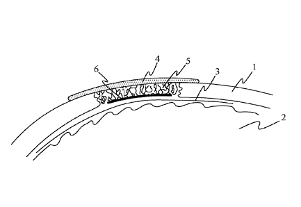
Figure 1 schematically shows an implant according to a first embodiment. The
implant is arranged in an opening in the skull 1 of a patient. The implant
consists of
a surface layer 4 and a porous part 5 attached to its underside. The porous
part 5
essentially fills the hole in the skull. The implant is arranged on the lamina
dura 3
and brain 2 of the patient. The surface layer 4 overlaps with the skull 1 and
hence
extends over each edge of the porous part 5.
Figure 2 schematically shows an implant according to a second embodiment. In
this
embodiment, the second, inner surface of the porous part 5 is further mostly
covered
by a membrane 6 made of collagen. This membrane 6 prevents penetration of the
Date Recue/Date Received 2023-03-09
14
dura mater to the porosities of the porous part 5 of the implant and may be
beneficial
in clinical cases where intracranial pressure is increased for a long period
of time.
Figure 3 schematically shows an implant according to a third embodiment. In
this
embodiment, the implant is used for replacing a missing part of the femur bone
after
a bone tumour surgery. The porous part 5 of the implant fills the bone cavity
and a
fibre reinforced surface layer 8 made of slowly biodegradable materials
reinforces
the implant and gives it an anatomical outer shape.
Figures 4A and 4B schematically illustrate a further embodiment. Figure 4A is
a side
view showing the surface layer 4, wherein the first surface of the surface
layer is the
surface shown as an upper surface in the Figure and the second surface is the
surface opposite to the first surface, namely the lower surface in the Figure.
The
porous part 5 is attached to the second surface of the porous part 4 and its
first
surface is also the surface that is shown as an upper surface in the Figure
and the
second surface is the lower surface in the Figure. Should a membrane made of
collagen be used, it would be attached to the lower surface of the porous part
but it
would not cover the lower surface of the porous part entirely.
Figure 4A further shows, in dashed line, openings 9 and 9' for attaching the
implant
to the bone of the patient. Figure 4B shows the implant of Figure 4A as a top
view.
The porous part 5 is shown in dashed lines underneath the surface layer 4 and
each
corner of the surface layer 4 is equipped with an opening 9, 9'. These
openings can
used for attaching the implant to the bone by screws.
Date Recue/Date Received 2023-03-09