Note: Descriptions are shown in the official language in which they were submitted.
CA 02991076 2017-12-28
' WO 2017/011763 PCMS2016/042543
1
METHODS FOR IDENTIFICATION, ASSESSMENT, PREVENTION, AND
TREATMENT OF METABOLIC DISORDERS USING SLIT2
Cross-Reference to Related Applications
This application claims the benefit of priority to U.S. Provisional
Application No.
62/193,359, filed 16 July 2015, the entire contents of said application is
incorporated herein
in its entirety by this reference.
Statement of Rights
This invention was made with government support under Grant DK031405 awarded
by the National Institutes of Health. The U.S. government has certain rights
in the
invention.
Background of the Invention
Metabolic disorders comprise a collection of health disorders or risks that
increase
the risk of morbidity and loss of qualify of life. For example, diabetes,
obesity, including
central obesity (disproportionate fat tissue in and around the abdomen),
atherogenic
dyslipidemia (including a family of blood fat disorders, e.g., high
triglycerides, low HDL
cholesterol, and high LDL cholesterol that can foster plaque buildups in the
vascular
system, including artery walls), high blood pressure (130/85 mmHg or higher),
insulin
resistance or glucose intolerance (the inability to properly use insulin or
blood sugar), a
chronic prothrombotic state (e.g., characterized by high fibrinogen or
plasminogen activator
inhibitor-1 levels in the blood), and a chronic proinflammatory state (e.g.,
characterized by
higher than normal levels of high-sensitivity C-reactive protein in the
blood), are all
metabolic disorders collectively afflicting greater than 50 million people in
the United
States.
Brown fat has attracted significant interest as an antidiabetic tissue owing
to its
ability to dissipate energy as heat (Cannon and Nedergaard (2004) Physiol.
Rev. 84:277-
359; Lowell and Spiegelman (2000) Nature 404:652-660). Activation of brown fat
thermogenesis involves the induction of a program of genes, including
uncoupling protein 1
(UCP1), which uncouples respiration and increases heat production in fat cells
(Kozak and
Harper (2000) Annu. Rev. Nutr. 20:339-363). Other non-UCP1 pathways may also
contribute to non-shivering thermogenesis (Kazak etal. (2015) Cell 163:643-
655). It is
CA 02991076 2017-12-28
, WO 2017/011763 PCT/US2016/042543
2
now recognized that at least two types of thermogenic fat cells exist ¨
classical
interscapular brown fat, as well as inducible brown-like adipocytes in white
fat (also known
as beige fat), which tends to be dispersed among white fat depots (Wu et al.
(2012) Cell
150:366-376; Shinoda et al. (2015) Nat. Med 4:389-394). BAT has high basal
levels of
UCP1, whereas beige fat has low basal levels that are highly inducible upon
stimulation
with cold or other agents (Wu et al. (2012) Cell 150:366-376). Despite their
common
ability to exhibit adaptive thermogenesis, brown and beige cells do not derive
from the
same lineage precursors (Lepper and Fan (2010) Genesis 48:424-436; Long et al.
(2014)
Cell Metabolism 19:810-820; Seale et al. (2008) Nature 454:961-967) and
express different
molecular signatures (Long etal. (2014) Cell Metabolism 19:810-820; Sharp et
al. (2012)
PLoS One 7:e49452; Wu etal. (2012) Cell 150:366-376; Harms and Seale (2013)
Nat.
Med 19:1252-1263). Mouse models resistant to weight gain through enhanced
brown and
beige fat content or activity have demonstrated that activation of
thermogenesis in fat can
be a powerful strategy to improve metabolic health and prevent weight gain
(Cederberg and
Enerback (2003) Curr. Mot Med. 3:107-125; Fisher et al. (2012) Genes Dev.
26:271-281;
Vegiopoulos et al (2010) Science 328:1158-1161; Ye et a/. (2012) Cell 151:96-
110).
Ablation of UCP1+ cells in transgenic mice have an increased propensity toward
obesity
and diabetes (Lowell et al. (1993) Nature 366:740-742), whereas UCP1 knockout
mice
develop obesity under thermoneutrality conditions when fed a high fat diet
(Feldmann etal.
(2009) Cell Metabolism 9:203-209).
A physiological stimulus for inducing active thermogenic fat in mice and
humans is
a cold environment, which causes the release of neurotransmitters, such as
catecholamines,
from nerve terminals or M2 macrophages (Morrison et al. (2012)Front.
EndocrinoL 3:5;
Nguyen etal. (2011) Nature 480:103-108). Brown fat has relatively recently
been found to
exist and be functional in adult humans based on studies observing increased
symmetrical
glucose uptake in supraclavicular regions upon exposure to cold environment
(Cypess et aL
(2009) N. Engl. J. Med 360:1509-1517; Virtanen et al. (2009) N. Engl. J. Med
360:1518-
1525; Yoneshiro et al. (2011) Obesity 19:13-16). Brown fat has also been shown
to be
activated by the p3-agonist, mirabegron, illustrating that the canonical cAMP
pathway for
adipose thermogenesis is likely to be function in humans and raising the
possibility of
additional, yet unknown pathways of activation (Cypess etal. (2014) Cell
Metab. 21:33-
38). The functional characteristics of human BAT has yet to be determined, but
several
papers have shown that supraclavicular human brown fat is most similar to the
beige fat of
CA 02991076 2017-12-28
WO 2017/011763 PCT/US2016/042543
3
rodents (Wu et al. (2012) Cell 150:366-376; Sharp etal. (2012) PLoS ONE
7:e49452;
Shinoda et al. (2015) Nat. Med. 4:389-394). Thus, it is believed that brown
and beige fat
likely have complementary and overlapping functions in the maintenance of
whole body
energy homeostasis.
The transcriptional regulator PRDM16 is critical to the development of both
brown
and beige fat (Seale etal. (2007) Cell Metabolism 6:38-54; Seale etal. (2008)
Nature
454:961-967; Kajimura etal. (2009) Nature 460:1154-1158; Seale etal. (2011)J.
Clin.Invest. 121:96-105). Mice with fat-specific ablation of PRDM16
demonstrate
significantly lower basal thermogenic gene expression in the subcutaneous fat:
these
animals are also resistant to browning of the white fat when stimulated with a
cold
environment or 03-agonism (Cohen etal. (2014) Cell 156:304-316). Conversely,
aP2-
PRDM16 transgenic mice show enhanced "browning" of their subcutaneous adipose
depots, leading to augmented energy expenditure, reduced weight gain on high
fat diet, and
improved glucose and insulin homeostasis (Seale etal. (2011) J. Clininvest.
121:96-105).
As the classical brown fat in this model was found to be relatively
unaffected, adiponectin
(aP)-driven deletion of PRDM16 mice provide the opportunity to specifically
study beige
fat function. These mice develop a moderate obese phenotype compared to
littermate
controls, which is accompanied by an expansion of the subcutaneous depots with
increased
infiltration of inflammatory immune cells.
Despite decades of scientific research, such factors have not been identified
and few
effective therapies have emerged to treat metabolic disorders. The various
metabolic
benefits of activating brown or beige fat have raised interest in the
discovery of hormones
and secreted proteins that can act on fat tissue locally or systemically to
induce browning.
Beige fat development occurs in distinct pockets of cells, consistent with the
possibility of a
paracrine regulatory factor at work. White adipose tissues secrete many
proteins factors
(adipokines) that influence local and systemic metabolism, including adipsin,
adiponectin,
leptin and TNFa (Rosen and Spiegelman (2014) Cell. 156:20-44; Blither and
Mantzoros
(2015)Metabolism. 64:131.45). However, there is a great need to identify
molecular
regulators of metabolic disorders, especially those unknown secretory proteins
from brown
and/or beige fat. Such molecular regulators would also be useful in the
generation of
diagnostic, prognostic, and therapeutic agents to effectively control
metabolic disorders in
subjects.
CA 02991076 2017-12-28
WO 2017/011763 PCT/1JS2016/042543
4
Summary of the Invention
The present invention is based in part on the discovery that Slit2 and
biologically
active fragments thereof are polypeptides secreted by beige fat cells that
have the ability to
modulate many metabolic processes, including modulating adipose thermogenesis,
energy
expenditure, and glucose homeostasis. Expression of Slit2 and its biologically
active
fragments is regulated by thermogenic stimuli (e.g., Prdm16 and cold
exposure), their
expression is downregulated in the white adipose tissue of obese animals, and
they induce
activation of PKA signaling, which is required for its pro-thermogenic
activity. Slit2 and
its biologically active fragments protect against diet-induced insulin
resistance when
circulating levels of Slit2 are increased in the blood, as it induces a
thermogenic gene
expression program in the subcutaneous white fat. Slit2 and its biologically
active
fragments act in a cell-autonomous manner to induce a cAMP cellular signaling
program,
induce thermogenic gene expression, and increase whole body energy
expenditure. Based
on this role in peripheral tissue for Slit and its biologically active
fragments to modulate
adipose tissue homeostasis and glucose metabolism, they have the therapeutic
ability to
treat metabolic disorders, especially obesity-induced metabolic disorders.
In one aspect, a use of an agent that modulates expression and/or activity of
Slit2 or
a biologically active fragment thereof in a subject for the preparation of a
medicament for
modulating a metabolic response in the subject is provided.
The compositions and methods of the present invention are characterized by
many
embodiments and each such embodiment can be applied to any combination of
embodiments described herein. For example, in one embodiment, the expression
and/or
activity of Slit2 or the biologically active fragment thereof is upregulated.
In another
embodiment, expression and/or activity of Slit2 or the biologically active
fragment thereof
is upregulated using an agent selected from the group consisting of a nucleic
acid molecule
encoding a Slit2 polypeptide or fragment thereof, and a Slit2 polypeptide or
fragment
thereof. In still another embodiment, the medicament further comprises an
additional
agent that increases the metabolic response. In yet another embodiment,
expression and/or
activity of Slit2 or the biologically active fragment thereof is
downregulated. In still
another embodiment, expression and/or activity of Slit2 or the biologically
active fragment
thereof is downregulated using an agent selected from the group consisting of
an anti-Slit2
antisense nucleic acid molecule, an anti-Slit2 RNA interference molecule, a
blocking anti-
Slit2 antibody, a non-activating form of Slit2 polypeptide or fragment
thereof, and a small
CA 02991076 2017-12-28
WO 2017/011763 PCT/US2016/042543
molecule that binds to Slit2. In yet another embodiment, the medicament
further
comprises an additional agent that decreases the metabolic response. In
another
embodiment, the metabolic response is selected from the group consisting of:
a) modified
expression of a marker selected from the group consisting of: cidea,
adiponectin, adipsin,
5 otopetrin, type II deiodinase, cig30, ppar gamma 2, pgcl a, ucpl, elov13,
cAMP, Prdm16,
cytochrome C, cox4i1, coxIII, cox5b, cox7al, cox8b, glut4, atpase b2, cox II,
atp5o,
ndufb5, ap2, ndufsl, GRP109A, acylCoA-thioesterase 4, EARA1, claudinl, PEPCK,
fgf21, acylCoA-thioesterase 3, dio2, fatty acid synthase (fas), leptin,
resistin, and nuclear
respiratory factor-1 (nrfl); b) modified thermogenesis in adipose cells; c)
modified
differentiation of adipose cells; d) modified insulin sensitivity of adipose
cells; e) modified
basal respiration or uncoupled respiration; 0 modified whole body oxygen
consumption; g)
modified obesity or appetite; h) modified insulin secretion of pancreatic beta
cells; i)
modified glucose tolerance; j) modified phosphorylation of EGFR, ERK, AMPK,
protein
kinase A (PKA) substrates having an RRX(S/T) motif, wherein the X is any amino
acid
and the (SIT) residue is a serine or threonine, HSL; and k) modified
expression of UCP1
protein. In still another embodiment, the metabolic response is upregulated.
In yet another
embodiment, the metabolic response is downregulated.
In another aspect, a method for modulating a metabolic response comprising
contacting a cell with an agent that modulates expression and/or activity of
Slit2 or a
biologically active fragment thereof to thereby modulate the metabolic
response is
provided.
As described above, the compositions and methods of the present invention are
characterized by many embodiments and each such embodiment can be applied to
any
combination of embodiments described herein. For example, in one embodiment,
expression and/or activity of Slit2 or the biologically active fragment
thereof is upregulated.
In another embodiment, expression and/or activity of Slit2 or the biologically
active
fragment thereof is upregulated using an agent selected from the group
consisting of a
nucleic acid molecule encoding a Slit2 polypeptide or fragment thereof, and a
Slit2
polypeptide or fragment thereof. In still another embodiment, the method
further comprises
contacting the cell with an additional agent that increases the metabolic
response. In yet
another embodiment, expression and/or activity of Slit2 or the biologically
active fragment
thereof is downregulated. In another embodiment, expression and/or activity of
Slit2 or the
biologically active fragment thereof is downregulated using an agent selected
from the
CA 02991076 2017-12-28
' W02017/011763 PCT/US2016/042543
6
group consisting of an anti-Slit2 antisense nucleic acid molecule, an anti-
Slit2 RNA
interference molecule, a blocking anti-Slit2 antibody, a non-activating form
of Slit2
polypeptide or fragment thereof, and a small molecule that binds to S11t2. In
still another
embodiment, the method further comprises contacting the cell with an
additional agent that
decreases the metabolic response. In yet another embodiment, the step of
contacting occurs
in vivo. In another embodiment, the step of contacting occurs in vitro. In
still another
embodiment, the cell is selected from the group consisting of fibroblasts,
adipoblasts,
preadipocytes, adipocytes, white adipocytes, brown adipocytes, and beige
adipocytes. In
yet another embodiment, the metabolic response is selected from the group
consisting of: a)
modified expression of a marker selected from the group consisting of: cidea,
adiponectin,
adipsin, otopetrin, type II deiodinase, cig30, ppar gamma 2, pgcla, ucpl,
elov13, cAMP,
Prdm16, cytochrome C, cox4i1, coxIII, cox5b, cox7al, cox8b, glut4, atpase b2,
cox
atp5o, ndufb5, ap2, ndufsl, GRP109A, acylCoA-thioesterase 4, EARA1, claudinl,
PEPCK,
fgf21, acylCoA-thioesterase 3, di o2, fatty acid synthase (fas), leptin,
resistin, and nuclear
respiratory factor-1 (nrfl); b) modified thermogenesis in adipose cells; c)
modified
differentiation of adipose cells; d) modified insulin sensitivity of adipose
cells; e) modified
basal respiration or uncoupled respiration; f) modified whole body oxygen
consumption; g)
modified obesity or appetite; h) modified insulin secretion of pancreatic beta
cells; i)
modified glucose tolerance; j) modified phosphorylation of EGFR, ERK, AlvIPK,
protein
kinase A (PKA) substrates having an RRX(S/T) motif, wherein the X is any amino
acid and
the (SIT) residue is a serine or threonine, HSL; and k) modified expression of
UCP1
protein. In another embodiment, the metabolic response is upregulated. In
still another
embodiment, the metabolic response is downregulated.
In still another aspect, a method of preventing or treating a metabolic
disorder in a
subject comprising administering to the subject an agent that promotes
expression and/or
activity of Slit2 or a biologically active fragment thereof in the subject,
thereby preventing
or treating the metabolic disorder in the subject is provided. In one
embodiment, the agent
is selected from the group consisting of a nucleic acid molecule encoding a
Slit2
polypeptide or fragment thereof, and a Slit2 polypeptide or fragment thereof.
In another
embodiment, the agent is administered by intravenous or subcutaneous
injection. In still
another embodiment, the agent is administered in a pharmaceutically acceptable
formulation. In yet another embodiment, the metabolic disorder is selected
from the group
consisting of insulin resistance, hyperinsulinemia, hypoinsulinemia, type IT
diabetes,
CA 02991076 2017-12-28
W02017/011763 PCT/US2016/042543
7
hypertension, hyperhepatosteatosis, hyperuricemia, fatty liver, non-alcoholic
fatty liver
disease, polycystic ovarian syndrome, acanthosis nigricans, hyperphagia,
endocrine
abnormalities, triglyceride storage disease, Bardet-Biedl syndrome, Lawrence-
Moon
syndrome, and Prader-Labhart-Willi syndrome. In another embodiment, the
subject is a
non-human animal or a human.
In yet another aspect, a method for preventing or treating a metabolic
disorder in a
subject comprising administering to the subject an agent that inhibits Slit2
expression
and/or activity in the subject, thereby preventing or treating the metabolic
disorder in the
subject is provided. In one embodiment, the agent is selected from the group
consisting of
an anti-Slit2 antisense nucleic acid molecule, an anti-Slit2 RNA interference
molecule, a
blocking anti-Slit2 antibody, a non-activating form of Slit2 polypeptide or
fragment thereof,
and a small molecule that binds to Slit2. In another embodiment, the agent is
administered
by intravenous or subcutaneous injection. In still another embodiment, the
agent is
administered in a pharmaceutically acceptable formulation. In yet another
embodiment, the
metabolic disorder is selected from the group consisting of obesity-associated
cancer,
anorexia, and cachexia. In another embodiment, the subject is a non-human
animal or a
human.
In another aspect, a cell-based assay for screening for agents that modulate a
metabolic response in a cell by modulating the expression and/or activity of
Slit2 or a
biologically active fragment comprising contacting the cell expressing Slit2
or the
biologically active fragment thereof with a test agent the modulates the
expression and/or
activity of Slit2 and determining the ability of the test agent to modulate a
metabolic
response in the cell is provided.
In still another aspect, a method for assessing the efficacy of an agent that
modulates Slit2 expression and/or activity for modulating a metabolic response
in a subject,
comprising a) detecting in a subject sample at a first point in time, the
expression and/or
activity of Slit2; b) repeating step a) during at least one subsequent point
in time after
administration of the agent; and c) comparing the expression and/or activity
detected in
steps a) and b), wherein a significantly lower expression and/or activity of a
marker listed in
Table 1 or 2 in the first subject sample relative to at least one subsequent
subject sample,
indicates that the agent increases the metabolic response in the subject
and/or wherein a
significantly higher expression and/or activity of a marker listed in Table 1
or 2 in the first
CA 02991076 2017-12-28
WO 2017/011763 PCT/US2016/042543
8
subject sample relative to at least one subsequent subject sample, indicates
that the test
agent decreases the metabolic response in the subject is provided.
As described above, the compositions, assays, and methods of the present
invention
are characterized by many embodiments and each such embodiment can be applied
to any
combination of embodiments described herein. For example, in one embodiment,
expression and/or activity of Slit2 or the biologically active fragment
thereof is upregulated.
In another embodiment, expression and/or activity of Slit2 or the biologically
active
fragment thereof is downregulated. In still another embodiment, the agent is
selected from
the group consisting of a nucleic acid molecule encoding a Slit2 polypeptide
or fragment
thereof, a Slit2 polypeptide or fragment thereof, a small molecule that binds
to Slit2, an
anti-Slit2 antisense nucleic acid molecule, an anti-Slit2 RNA interference
molecule, an anti-
Slit2 siRNA molecule, a blocking anti-Slit2 antibody, and a non-activating
form of Slit2
polypeptide or fragment thereof. In yet another embodiment, the subject has
undergone
treatment for the metabolic disorder, has completed treatment for the
metabolic disorder,
and/or is in remission from the metabolic disorder between the first point in
time and the
subsequent point in time. In another embodiment, the first and/or at least one
subsequent
sample is selected from the group consisting of ex vivo and in vivo samples.
In still another
embodiment, the first and/or at least one subsequent sample is obtained from
an animal
model of a metabolic disorder. In yet another embodiment, the first and/or at
least one
subsequent sample is selected from the group consisting of tissue, whole
blood, serum,
plasma, buccal scrape, saliva, cerebrospinal fluid, urine, stool, and bone
marrow. In
another embodiment, the first and/or at least one subsequent sample is a
portion of a single
sample or pooled samples obtained from the subject. In still another
embodiment, a
significantly higher expression and/or activity comprises upregulating the
expression and/or
activity by at least 25% relative to the second sample. In yet another
embodiment, a
significantly lower expression and/or activity comprises downregulating the
expression
and/or activity by at least 25% relative to the second sample. In another
embodiment, the
amount of the marker is compared. In still another embodiment, the amount of
the marker
is determined by determining the level of protein expression of the marker. In
yet another
embodiment, the presence of the protein is detected using a reagent which
specifically binds
with the protein. In another embodiment, the reagent is selected from the
group consisting
of an antibody, an antibody derivative, and an antibody fragment. In still
another
embodiment, the level of expression of the marker in the sample is assessed by
detecting
CA 02991076 2017-12-28
, W02017/011763 PCT/US2016/042543
9
the presence in the sample of a transcribed polynucleotide or portion thereof.
In yet another
embodiment, the transcribed polynucleotide is an mRNA or a cDNA. In another
embodiment, the step of detecting further comprises amplifying the transcribed
polynucleotide. In still another embodiment, the level of expression of the
marker in the
sample is assessed by detecting the presence in the sample of a transcribed
polynucleotide
which anneals with the marker or anneals with a portion of a polynucleotide
under stringent
hybridization conditions. In yet another embodiment, the metabolic response is
selected
from the group consisting of: a) modified expression of a marker selected from
the group
consisting of: cidea, adiponectin, adipsin, otopetrin, type II deiodinase,
cig30, ppar gamma
2, pgcla, ucpl, elov13, cAMP, Prdm16, cytochrome C, cox4i1, coxIII, cox5b,
cox7al,
cox8b, glut4, atpase b2, cox II, atp5o, ndufb5, ap2, ndufsl, GRP109A, acylCoA-
thioesterase 4, EARA1, claudinl, PEPCK, fgf21, acylCoA-thioesterase 3, dio2,
fatty acid
synthase (fas), leptin, resistin, and nuclear respiratory factor-1 (nrfl); b)
modified
thermogenesis in adipose cells; c) modified differentiation of adipose cells;
d) modified
insulin sensitivity of adipose cells; e) modified basal respiration or
uncoupled respiration;
f) modified whole body oxygen consumption; g) modified obesity or appetite; h)
modified
insulin secretion of pancreatic beta cells; i) modified glucose tolerance; j)
modified
phosphorylation of EGFR, ERK, AMPK, protein kinase A (PKA) substrates having
an
RRX(S/T) motif, wherein the X is any amino acid and the (SIT) residue is a
serine or
threonine, HSL; and k) modified expression of UCP1 protein. In another
embodiment, the
metabolic response is upregulated. In still another embodiment, the metabolic
response is
downregulated. In yet another embodiment, Slit2 is selected from the group of
Slit2
sequences shown in Table 1.
Brief Description of Figures
Figure 1 includes 7 panels, identified as panels A, B, C, D, E, F, and G which
show
that Slit2 is a PRDM16-regulated secreted protein in adipose cells. Panel A
representative
images from UCP1 immunohistochemistry on sections of inguinal subcutaneous
adipose
tissue from aP2-PRDM16 and wild type mice. Images are shown at 10x
magnification.
Scale bar, 100 pim. Panel B shows normalized thermogenic gene expression in
primary
inguinal cells from aP2-PRDMI6 and wild type mice at day 7 of differentiation.
Panel C
shows a heat map of relative protein levels in conditioned medium from wild
type or ap2-
PRDMI6 primary inguinal cells (n = 2 per group) as determined by TMT labeling
and mass
CA 02991076 2017-12-28
W02017/011763 PCT/US2016/042543
spectrometry. Shown is a short list of detected secreted proteins. The fold
change for each
individual sample is shade-coded according to the key. Panel D shows the
normalized
mRNA expression of Slid, Slit2 and S1i13 in BAT and iWAT from 6 week-old mice
chronically housed at 30 C thermoneutrality (TN) or exposed to a 4 C cold
challenge for
5 the indicated time points (n = 3 per group). Gene expression of Ap2,
Ucpl, Adipsin, F4/80,
S1it2 and S11t3 in iWAT (Panel E) and Slit2 and S1i13 in eWAT (Panel F) from
C57/b6 mice
fed a chow diet or a high fat diet for 16 weeks is shown. Panel G shows
primary inguinal
cells treated with forskolin for 4h before gene expression analysis of
Adiponeetin, Ucpl,
Slit2 and Slit3. Data are presented as mean SEM. *p <0.05, ** p < 0.01, ***
p < 0.001.
10 Figure 2 includes 7 panels, identified as panels A, B, C, D, E, F, and
G, which
further show that Slit2 is a PRDM16-regulated secreted protein in adipose
cells. Panel A
shows peptides (bold text) corresponding to mouse Slit2 and Slit3 detected in
conditioned
medium from aP2-PRDM16 inguinal cells. Panels B and C show the normalized mRNA
expression of Slit2, Slit3, and Prdm16 in brown fat tissue (BAT) from aP2-
PRDM16 mice
(Panel B) and adipocyte-specific deletion of PRDM16 (prdm1eh)0"1( ) (Panel C).
Panels D
and E show tissue mRNA expression of Slit2 (Panel D) and Slit3 (Panel E) in 6
week old
C57/b6 mice. Panel F shows normalized mRNA expression of S1112 and Ucpl in
iWAT,
eWAT and BAT after 3 days treatment with daily injections of CL 316,243 (1
mg/kg).
Panel G shows normalized mRNA expression of S11t2 and S11t3 in BAT in lean
mice or 16
weeks C57/b6 high fat diet mice.
Figure 3 includes 10 panels, identified as panels A, B, C, D, E, F, G, H, I,
and J
which show that Slit2 promotes a thermogenic program in cells and in mice.
Panels A and
B show thermogenic gene expression in primary inguinal cells treated for 24 h
with 1 g/ml
of Slit2 (Panel A) or lysyl oxidase (LOX1), glypicanl (GPC1), chordin-like 1
(CHIA) or C-
X-C motif chemokine 12 (CXCL12) recombinant proteins (Panel B) at day 6 of
differentiation. Panel C shows the results of Western blotting against Slit2
in primary
inguinal cells overexpressing full length Slit2 in adenoviral vectors. Panel D
shows
normalized thermogenic mRNA expression in primary inguinal cells
overexpressing
adenoviral full length Slit2 (Slit2-FL) or lacZ control. Panel E shows the
results of
C57/BL6 mice injected (i.v.) with adenoviral vectors Slit2-FL or LacZ (n = 3)
and Western
blotting against Slit2 from plasma of these mice obtained at day 7 post-
injection. Panel F
shows normalized iWAT mRNA expression of thermogenesis genes and white fat
selective
genes at day 7 post-injection. Panel G shows representative images from UCP1
CA 02991076 2017-12-28
WO 2017/011763 PCT/US2016/042543
11
immunohistochemistry on sections of inguinal subcutaneous adipose tissue from
mice
injected with Slit2-FL or LacZ at day 7. Images are shown at 10x
magnification. Scale bar,
100 um. Panel H shows Western blotting against Slit2 in primary inguinal cells
from
Slit2f1"41" mice transduced with LacZ virus (Slit211"/fl") or Cre virus
(Slit21(13). Panel I
shows gene expression in primary inguinal cells from Slit2f1"41" mice
transduced with
LacZ virus (Slit2n"ifl") or CRE virus (Slit2K ). Panel J shows gene expression
in BAT
tissue from Slit2flox/flox mice infected with with GFP-AAV8 (Slit2f1"41"-AAV8-
GFP) or
Cre virus (Slit2f1"/fl"-AAV8-CRE).
Figure 4 includes 6 panels, identified as panels A, B, C, D, E and F, which
further
show that Slit2 promotes a thermogenic program in cells and in mice. Panels A-
C show
mRNA expression in liver (Panel A), quadriceps (Panel B) and brown fat (Panel
C) in mice
overexpressing LacZ or Slit2-FL. Panel D shows representative images from UCP1
immunohistochemistry on sections of BAT from mice injected with Slit2-FL or
LacZ
control at day 7. Images are shown at 10x magnification. Scale bar, 100 m.
Panel E
shows normalized mRNA expression levels in iWAT (K) at day 7 postinjection.
Panel F
shows representative images from UCP1 immunohistochemistry of iWAT from C57/b6
mice injected with Slit2-FL or LacZ at day 7. Scale bar, 100 tim. Data are
presented as
mean SEM. * p < 0.05, ** p < 0.01, *** p < 0.001.
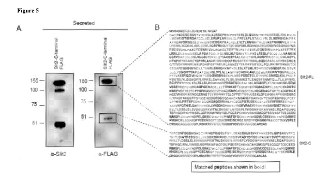
Figure 5 includes 6 panels, identified as panels A, B, C, D, E, and F, which
identify
and characterize a Slit2 cleavage fragment. Panel A shows a Western blot of
overexpressed
full-length C-terminal FLAG-tagged Slit2 detected with a Slit2 antibody (left)
and an anti-
FLAG antibody (right). Boxed immunoreactive bands were analyzed using mass
spectrometry. Panel B shows matched peptides to Slit2-FL or Slit2-C (bold
text) using C-
terminal FLAG-tagged Slit2 overexpression in primary inguinal cells. Panel C
shows a
cloning scheme for Slit2 full-length protein, Slit2-N, and Slit2-C protein
domains. Panel D
shows the results of Western blotting of overexpressed LacZ, Slit2-N, and
Slit2-C in
primary inguinal cells detected with a V5 antibody. Panel E shows Western
blotting results
for V5-expression in liver tissue after 6 days post-injection with LacZ, Slit2-
N, or Slit2-C
adenovirus. Panel F shows Western blotting results of mouse plasma after 6
days post-
injection with LacZ, Slit2-N or Slit2-C adenovirus.
Figure 6 includes 8 panels, identified as panels A, B, C, D, E, F, G, and H,
which
show that Slit2-C is sufficient to recapitulate the thermogenic activity of
full-length Slit2.
Panels A and B show normalized thermogenic mRNA expression in primary inguinal
cells
CA 02991076 2017-12-28
W02017/011763 PCT/US2016/042543
12
(Panel A) or primary brown fat cells (Panel B) overexpressing Ad-Slit2-N, Ad-
Slit2-C, or
Ad-lacZ control. Panels C and D show thermogenic mRNA expression in iWAT
(Panel C)
and BAT (Panel D) in mice overexpressing LacZ or Slit2-C. Panel E shows
representative
images from UCP1 immunohistochemistry on sections of inguinal subcutaneous
adipose
tissue (upper panel) and BAT (lower panel) from mice injected with Slit2-C or
LacZ
control at day 7. Images are shown at 10x magnification. Scale bar, 100 gm.
Panel F
shows 02 consumption in inguinal white fat tissue (left panel) and brown fat
tissue (right
panel) from 6 week-old mice fed a chow diet. Animal number, n = 10 per group.
Data are
presented as mean SEM. * p <0.05, ** p <0.01, *** p < 0.001. Panel G shows
UCP1
immunohistochemistry of iWAT (upper panel) and BAT (lower panel) from mice
injected
with Slit2-C or LacZ at day 7. Scale bar, 100 gm. Panel H shows 02 consumption
in
iWAT (left panel) and BAT (right panel) from mice injected with Slit2-C or
LacZ at day 7.
n =10 per group. Data are presented as mean SEM. * p < 0.05, ** p <0.01, ***
p <
0.001.
Figure 7 includes 4 panels, identified as panels A, B, C, and D, which further
show
that Slit2-C is sufficient to recapitulate the thermogenic activity of full-
length Slit2. Panel
A shows normalized mRNA expression of fatty acid synthase (fas) and hormone-
sensitive
lipase (hsl) in inguinal fat 7 days post-injection with LacZ or Slit2-C
adenovirus in DIO
mice. Panel B shows normalized mRNA expression of fatty acid synthase (fas),
adipose
triglyceride lipase (atgl), and hormone-sensitive lipase (hsl) in BAT 7 days
post-injection
with LacZ or Slit2-C adenovirus in DIO mice. Panel C shows normalized mRNA
expression of white fat selective genes, resistin and leptin, in BAT 7 days
post-injection
with LacZ or Slit2-C adenovirus. Panel D shows Western blot of UCP1 protein
(left) and
quantification of UCP1 protein intensities relative tubulin (right) in BAT 7
days post-
injection with LacZ or Slit2-C adenovirus in DIO mice.
Figure 8 includes 9 panels, identified as panels A, B, C, D, E, F, G, H, and
I, which
show that increased circulating Slit2-C augments whole body energy expenditure
and
improves glucose homeostasis in obese mice. Panels A-E shows the results of
whole body
energy expenditure measured in DIO mice 6 days after injection with LacZ or
Slit2-C
adenovirus. Oxygen (02) consumption (Panel A), respiratory exchange ratio
(Panel B),
locomotor activity (Panel C), food intake (Panel D), and body weight (Panel E)
were
measured at day 7. Panel F shows tissue weights of brown fat (BAT), inguinal
fat (Ing),
and epididymal fat (Epi) at day 7 post-njection with LacZ or Slit2-C
adenovirus. Panel G
CA 02991076 2017-12-28
, W02017/011763 PCT/US2016/042543
13
shows the results of intraperitoneal glucose tolerance tests in 16 weeks diet-
induced obese
mice injected with Slit2-C or LacZ performed at day 7 (n = 9-10). Data are
presented as
mean SEM. * p < 0.05, ** p <0.01, *** p <0.001. Panel H shows averaged
oxygen
consumption at days 5-7 in mice with no significant different in body weight
between the
groups. Panel I shows tissue weights of BAT, iWAT and eWAT at day 7 post-
injection
with LacZ or Slit2-C adenovirus.
Figure 9 includes 9 panels, identified as panels A, B, C, D, E, F, G, H, and
1, which
show that increased circulating full-length Slit2 (Slit2-FL) augments whole
body energy
expenditure and improves glucose homeostasis in obese mice. Panels A-E show
the results
of whole body energy expenditure measured in lean mice under 6 days after
injection with
with LacZ or Slit2-FL adenovirus. Oxygen (02) consumption (Panel A),
respiratory
exchange ratio (Panel B), food intake (Panel C), locomotor activity (Panel D),
and body
weight (Panel D) were measured at day 7. Panel F shows the results of
intraperitoneal
glucose tolerance tests in 16 weeks diet-induced obese mice injected with
Slit2-FL or LacZ
performed at day 7 (n = 9-10). Panels G-I show plasma levels of total
cholesterol (Panel
G), triglycerides (Panel H), and non-fasting insulin (Panel I) in mice 7 days
post-injection
with LacZ or Slit2-C adenovirus.
Figure 10 includes 14 panels, identified as panels A, B, C, D, E, F, G, H, I,
J, K, L,
M, and N, which show that Slit2-C induces a thermogenesis program through the
protein
kinase A (PKA) signaling pathway in adipocytes. Panels A and B show the
results of
primary inguinal cells treated with Slit2-C or LacZ control at day 2 of
differentiation (108
pfu/well), starved overnight at day 6, and analyzed at day 7 by Western
blotting for
phosphorylated (phospho-) and total protein amounts of epidermal growth factor
receptor
(EGFR), ERK1/2, and AMPK (Panel A), as well as PKA substrates, HSL, UCP1, a-
tubulin
protein (Panel B). As a positive control, similar samples were treated with
100 nM NE for
minutes. Panels C and D show the results of primary inguinal cells treated
with Slit2-C
or LacZ control at day 2 of differentiation (108 pfu/well) and then treated
with PKA
inhibitor, H89 (30 pM), for 2 h before either Western blot analysis for PKA
signaling
(Panel C) or gene expression analysis for aP2, Ucp1, and Dio2 (Panel D). Panel
E shows
30 primary cells treated as in Panel A and blotted for phospho-and total
ATGL and
phosphorylated PKC substrates. Panel F shows quantification of UCP1 protein
levels
relative a-tubulin in Panel B, n=3. Panel G shows Western blot analysis for
PKA substrate
phosphorylation upon acute treatment (30 min) with conditioned medium from
cells
CA 02991076 2017-12-28
. W02017/011763 PCT/US2016/042543
14
expressing LacZ, Slit2-FL or Slit2-C. Panels H and I show thermogenic gene
expression in
primary inguinal cells overexpressing Slit2-C or LacZ at day and treated with
a-receptor
antagonist propranolol (100 nM) for 24h (Panel H) or adenylyl cyclase
inhibitor SQ-22536
(10 M) for 24h (Panel I). Panel J shows silverstain of immunopurified Slit2-C
FLAG
protein compared with an albumin standard. Panel K shows Western blot of
immunopurified Slit2-C FLAG protein using antibodies for FLAG or Slit2. Panel
L shows
cell surface binding of FLAG peptide or Slit2-C protein to primary inguinal
adipocytes.
Panel M shows treatment of primary inguinal cells with 20 nM NE or 20 nM Slit2-
C
protein for 0, 5, 15, 30, 60 and 90 min. Panel N shows normalized gene
expression in
primary inguinal cells after treatment with Slit2-C protein for 2h.
Comparisons are
presented as Slit2-C vs. LacZ (*), LacZ vs. Slit2-C with drug treatment (#) or
LacZ vs. drug
treatment ($). Data are presented as mean SEM. * p <0.05, ** p < 0.01, *** p
<0.001.
Figure 11 includes 7 panels, identified as panels A, B, C, D, E, F, and G,
which
show that the EGFR and ERK pathways are activated by, but not required for,
Slit2-C
activity. Panel A shows the results of a phosphokinase array used to detect
phosphorylated
forms of proteins in LacZ or Slit2-C treated primary inguinal cells at day7 of
differentiation. Panel B show Western blot results of phosphorylated EGFR in
reposnse to
increasing concentrations of EGFR tyrosine kinase inhibitors, erlotinib and
lapatinib. Panel
C shows normalized mRNA expression in primary inguinal cells treated with LacZ
or Slit2-
C adenovirus in the presence or absence of the EGFR inhibitors, erlotinib and
lapatinib.
Panel D shows normalized mRNA expression in primary inguinal cells treated
with LacZ or
Slit2-C adenovirus in the presence or absence of the ERK inhibitor, PD0325901.
Panel E
shows cell surface binding of either FLAG peptide, PM20D1 protein (100 nM) or
Slit2-C
protein (100 nM) to primary inguinal adipocytes. Panel F shows Western blot of
phosphorylated PKA substrates after 60 min incubation with increasing
concentrations of
Slit2-C FLAG purified protein. Panel G shows quantification of phosphorylated
PKA
substrates in Figure 6, Panel L after incubation with Slit2-C FLAG purified
protein relative
time point 0.
Figure 12 includes 6 panels, identified as panels A, B, C, D, E, and F, which
show
that Slit2 promotes a thermogenesis program in cells and in mice. Panels A and
B show
normalized thermogenic mRNA expression (Panel A) and (Panel B) oxygen
consumption
measured by Seahorse in primary brown fat cells from Slit2t1"/fl" mice
transduced with
adenovirus expressing LacZ (Slit2f1"/f1") or CRE (Slit2K ). Panel C shows
total body
CA 02991076 2017-12-28
WO 2017/011763 PCT/US2016/042543
weight in Slit2f1"/fl" mice infected with with AAV8-GFP (Slit2f10/fl0x-AAV8-
GFP) or CRE
virus (Slit2f1"in"-AAV8-CRE) (n = 8). Panels D-F show normalized mRNA
expression of
vascular and neuronal markers in BAT (Panel D), iWAT (Panel E) and quadriceps
muscle
(Panel F) 7 days postinjection with LacZ or Slit2-FL adenovirus.
5 Figure 13 includes 3 panels, identified as panels A, B, and C showing
cellular
oxygen consumption measured by Seahorse in primary inguinal fat cells after
(Panel A)
acute treatment (4 minutes) (Panel A) or long term treatment (2 h) (Panels B
and C). Panel
C shows statistical analysis of basal and oligomycin induced respiration shown
in Panel B.
Note that for every figure containing a histogram, the bars from left to right
for each
10 discreet measurement correspond to the figure boxes from top to bottom
in the figure
legend as indicated.
Detailed Description of the Invention
The present invention is based in part on the discovery that Slit2 and
biologically
15 active fragments thereof are secreted polypeptides that have the ability
to modulate adipose
thermogenesis and related metabolic activity (e.g., modulate one or more
biological
activities of a) brown fat and/or beige fat gene expression, such as
expression of a marker
selected from the group consisting of: cidea, adiponectin, adipsin, otopetrin,
type II
deiodinase, cig30, ppar gamma 2, pgcla, ucpl, elov13, cAMP, Prdm16, cytochrome
C,
cox4i1, coxIII, cox5b, cox7al, cox8b, glut4, atpase b2, cox II, atp5o, ndufb5,
ap2, ndufsl,
GRP109A, acylCoA-thioesterase 4, EARA1, claudinl, PEPCK, fgf21, acylCoA-
thioesterase 3, dio2, fatty acid synthase (fas), leptin, resistin, and nuclear
respiratory factor-
1 (nrfl); b) thermogenesis in adipose cells; c) differentiation of adipose
cells; d) insulin
sensitivity of adipose cells; e) basal respiration or uncoupled respiration;
f) whole body
oxygen consumption; g) obesity or appetite; h) insulin secretion of pancreatic
beta cells; i)
glucose tolerance; j) modified phosphorylation of EGFR, ERK, AMPK, protein
kinase A
(PKA) substrates having an RRX(S/T) motif', wherein the X is any amino acid
and the (SIT)
residue is a serine or threonine, HSL; and k) modified expression of UCP1
protein.
It is demonstrated herein that Slit2 and its biologically active cleavage
products are
secreted by beige fat cells and can act systemically on cells in culture and
in vivo to
stimulate a broad program of brown fat-like development. Slit2 and its
biologically active
cleavage products is induced by natural stimuli, such as cold and Prdm16 gene
expression,
and they can cause an increase in energy expenditure in mice with no change in
movement
CA 02991076 2017-12-28
W02017/011763
PCT/US2016/042543
16
or food intake. This results in improvement in metabolic disorders (e.g.,
obesity and
glucose homeostasis).
In order that the present invention may be more readily understood, certain
terms
are first defined. Additional definitions are set forth throughout the
detailed description.
The term "amino acid" is intended to embrace all molecules, whether natural or
synthetic, which include both an amino functionality and an acid functionality
and capable
of being included in a polymer of naturally-occurring amino acids. Exemplary
amino acids
include naturally-occurring amino acids; analogs, derivatives and congeners
thereof; amino
acid analogs having variant side chains; and all stereoisomers of any of any
of the
foregoing. The names of the natural amino acids are abbreviated herein in
accordance with
the recommendations of IUPAC-IUB.
The term "antisense" nucleic acid refers to oligonucleotides which
specifically
hybridize (e.g., bind) under cellular conditions with a gene sequence, such as
at the cellular
mRNA and/or genomic DNA level, so as to inhibit expression of that gene, e.g.,
by
inhibiting transcription and/or translation. The binding may be by
conventional base pair
complementarity, or, for example, in the case of binding to DNA duplexes,
through specific
interactions in the major groove of the double helix.
The terms "beige fat" or "brite (brown in white) fat" or "iBAT (induced brown
adipose tissue)" or "recruitable BAT (brown adipose tissue)" or "wBAT (white
adipose
BAT)" refer to clusters of UCP1-expressing adipocytes having thermogenic
capacity that
develop in white adipose tissue (WAT). Beige fat can develop in subcutaneous
WAT, such
as in inguinal WAT, or in intra-abdominal WAT such as in epididymal WAT.
Similar to
adipocytes in brown adipose tissue (BAT), beige cells are characterized by a)
multilocular
lipid droplet morphology, b), high mitochondria' content, and/or c) expression
of a core set
of brown fat-specific genes, such as Ucpl, Cidea, Pgcla, and other listed in
Table 2. BAT
and beige fat both are able to undergo thermogenesis, but these are distinct
cell types since
beige cells do not derive from Myf5 precursor cells like BAT cells, beige fat
express
thermogenic genes only in response to activators like beta-adrenergic receptor
or
PPARgamma agonists unlike constitutive expression in BAT cells (Harms and
Seale (2013)
Nat Med 19:1252-1263).
The term "binding" or "interacting" refers to an association, which may be a
stable
association, between two molecules, e.g., between a polypeptide of the
invention and a
binding partner, due to, for example, electrostatic, hydrophobic, ionic and/or
hydrogen-
CA 02991076 2017-12-28
WO 2017/011763 PCT/US2016/042543
17
bond interactions under physiological conditions. Exemplary interactions
include protein-
protein, protein-nucleic acid, protein-small molecule, and small molecule-
nucleic acid
interactions.
The term "biological sample" when used in reference to a diagnostic assay is
intended to include tissues, cells and biological fluids isolated from a
subject, as well as
tissues, cells and fluids present within a subject.
The term "isolated polypeptide" refers to a polypeptide, in certain
embodiments
prepared from recombinant DNA or RNA, or of synthetic origin, or some
combination
thereof, which (1) is not associated with proteins that it is normally found
within nature, (2)
is isolated from the cell in which it normally occurs, (3) is isolated free of
other proteins
from the same cellular source, (4) is expressed by a cell from a different
species, or (5) does
not occur in nature.
The terms "label" or "labeled" refer to incorporation or attachment,
optionally
covalently or non-covalently, of a detectable marker into a molecule, such as
a polypeptide.
Various methods of labeling polypeptides are known in the art and may be used.
Examples
of labels for polypeptides include, but are not limited to, the following:
radioisotopes,
fluorescent labels, heavy atoms, enzymatic labels or reporter genes,
chemiluminescent
groups, biotinyl groups, predetermined polypeptide epitopes recognized by a
secondary
reporter (e.g., leucine zipper pair sequences, binding sites for secondary
antibodies, metal
binding domains, epitope tags). Examples and use of such labels are described
in more
detail below. In some embodiments, labels are attached by spacer arms of
various lengths
to reduce potential steric hindrance.
The terms "metabolic disorder" and "obesity related disorders" are used
interchangeably herein and include a disorder, disease or condition which is
caused or
characterized by an abnormal or unwanted metabolism (i.e., the chemical
changes in living
cells by which energy is provided for vital processes and activities) in a
subject. Metabolic
disorders include diseases, disorders, or conditions associated with aberrant
or unwanted
(higher or lower) thermogenesis or aberrant or unwanted levels (high or low)
adipose cell
(e.g., brown or white adipose cell) content or function. Metabolic disorders
can be
characterized by a misregulation (e.g., downregulation or upregulation) of PGC-
1 activity.
Metabolic disorders can detrimentally affect cellular functions such as
cellular proliferation,
growth, differentiation, or migration, cellular regulation of homeostasis,
inter- or intra-
cellular communication; tissue function, such as liver function, muscle
function, or
CA 02991076 2017-12-28
W02017/011763 PCT/US2016/042543
18
adipocyte function; systemic responses in an organism, such as hormonal
responses (e.g.,
insulin response). Examples of metabolic disorders include obesity, insulin
resistance, type
II diabetes, hypertension, hyperuricemia, fatty liver, non-alcoholic fatty
liver disease,
polycystic ovarian syndrome, acanthosis nigricans, hyperphagia, endocrine
abnormalities,
triglyceride storage disease, Bardet-Biedl syndrome, Lawrence-Moon syndrome,
Prader-
Labhart-Willi syndrome, anorexia, and cachexia.
As used herein, "obesity" refers to a body mass index (BMI) of 30 kg/m2 or
more
(National Institute of Health, Clinical Guidelines on the Identification,
Evaluation, and
Treatment of Overweight and Obesity in Adults (1998)). However, the present
invention is
also intended to include a disease, disorder, or condition that is
characterized by a body
mass index (BMI) of 25 kg/m2 or more, 26 kg/m2 or more, 27 kg/m2 or more, 28
kg/m2 or
more, 29 kg/m2 or more, 29.5 kg/m2 or more, or 29.9 kg/m2 or more, all of
which are
typically referred to as overweight (National Institute of Health, Clinical
Guidelines on the
Identification, Evaluation, and Treatment of Overweight and Obesity in Adults
(1998)).
The obesity described herein may be due to any cause, whether genetic or
environmental.
Examples of disorders that may result in obesity or be the cause of obesity
include
overeating and bulimia, polycystic ovarian disease, craniopharyngioma, the
Prader-Willi
Syndrome, Frohlich's syndrome, Type II diabetics, GH-deficient subjects,
normal variant
short stature, Turner's syndrome, and other pathological conditions showing
reduced
metabolic activity or a decrease in resting energy expenditure as a percentage
of total fat-
free mass, e.g., children with acute lymphoblastic leukemia.
As used herein, the term "Slit2" refers to the Slit2 family member of the slit
family
of secreted proteins and is intended to include fragments, variants (e.g.,
allelic variants) and
derivatives thereof unless otherwise specified. Slit proteins are secreted
extracellular
matrix proteins bound to the cell surface by the extracellular matrix (e.g.,
heparan sulfates)
(Liang etal. (1999)J. Biol. Chem. 274:17885-17892; Ronca et a/. (2001)J. Biol.
Chem.
276:29141-29147). Slit proteins have four leucine-rich repeat (LRR) domains
connected by
disulfide bonds, followed by six epidermal growth factor (EGF) repeats, a beta-
sandwich
domain similar to that of laminin G called a LamG domain, one to three
additional EGF
repeats, and a C-terminal cysteine knot (Holmes etal. (1998) Mech. Dev. 79:57-
72; Itoh et
al. (1998) Brain Res. MoL Brain Res. 62:175-186; Brose etal. (1999) Cell
96:795-806;
Rothberg and Artavanis-Tsakonas (1992)1. MoL Biol. 227:367-370; Hohenester
etal.
(1999)MoL Cell 4:783-792; Nguyen-Ba-Carvet and Chedotal (2002) Neuron 22:463-
473).
CA 02991076 2017-12-28
WO 2017/011763
PCT/US2016/042543
19
Slit2 is proteolytically cleaved within the EGF domain region (Brose etal.
(1999) Cell
96:795-806; Patel et al. (2001) Development 128:5031-5037; Condac etal. (2012)
Glycobiol. 22:1183-1192. Following proteolytic cleavage of Slit2, the
canonical 140 kDa
N-terminal fragment remains associated with the cell surface, whereas the 50-
60 kDa C-
terminal fragment can be detected in conditioned cell media (Brose etal.
(1999) Cell
96:795-806; Wang et al. (1999) Cell 96:771-784. Slit2 protein is known to
interact with the
transmembrane receptor Roundabout, also known as Robo, and is known to be
involved in
neuronal guidance, kidney development, blood cell migration, and osteoblast
differentiation. However, Slit2 has not heretofore been implicated in the
regulation of
cellular metabolism. Mature slit proteins lack a signal sequence and Slit2
sequences of the
present invention can comprise a signal sequence, as well as lack a signal
sequence. The
Slit2 signal sequence is generally the most N-terminal 20, 21, 22, 23, 24, 25,
26, 27, 28, 29,
or 30 amino acids. In one embodiment, the Slit2 signal sequence is
MSGIGWQTLSLSLGLVLSILNKVAP.
At least three splice variants encoding distinct human Slit2 isoforms exist.
Slit2
isoform 1 (NM_004787.2 and NP_004778.1), also referred to as Slit2A, is the
longest
human Slit2 protein and is encoded by the longest transcript. Slit2 isoform 2
(NM 001289135.1 and NP_001276064.1), also referred to as Slit2C, lacks an
alternate in-
frame exon in the 5' coding region relative to the Slit2 transcript variant 1
and therefore
encodes a smaller isoform relative to the Slit2 isoform 1. Slit2 isoform 3
(NM 001289136.1 and NP 001276065.1), also referred to as Slit2B, also lacks an
alternate
in-frame exon in the 5' coding region relative to the Slit2 transcript variant
1 and therefore
encodes a smaller isoform relative to the Slit2 isoform 1. The nucleic acid
and polypeptide
sequences for each transcript variant and isoform is provided herein as SEQ ID
NOs:1-6,
respectively. Nucleic acid and polypeptide sequences of Slit2 orthologs in
organisms other
than humans are well known and include, for example, Mus muscu/us Slit2
(NM_001291227.1, NP 001278156.1, NM_001291228.1, NP_001278157.1,
NM_178804.4, and NP 848919.3); Rattus norvegicus Slit2 (NM 022632.2 and
NP 072154.2); Canis lupus familiaris Slit2 (XM 005618749.1 and XP
005618806.1); Bos
taurus Slit2 (NM_001191516.2 and NP_001178445.2); and Gallus gallus Slit2
(NM 001267075.1 and NP 001254004.1).
In some embodiments, fragments of Slit2 having one or more biological
activities of
the full-length Slit2 protein are described and employed. Such fragments can
comprise or
CA 02991076 2017-12-28
W02017/011763
PCT/US2016/042543
consist of at least one domain of a Slit2 protein without containing the full-
length Slit2
protein sequence. In some embodiments, Slit2 fragments can comprise, or
consist of, an N-
terminal signal peptide sequence (SS) domain, a leucine-rich repeat (LRR)
domain, an EGF
domain, a LamG domain, and a C-terminal cysteine knot domain, without
containing the
5 full-length Slit2 protein sequence. As further indicated in the
Examples, Slit2 orthologs are
highly homologous and retain common structural domains well known in the art.
Biologically active fragments, such as Slit2-N and Slit2-C, are also described
herein.
Table 1
10 SEO ID NO: 1 Human Slit2 Transcript Variant 1 cDNA Sequence
1 atgcgcggcg ttggctggca gatgctgtcc ctgtcgctgg ggttagtgct ggcgatcctg
61 aacaaggtgg caccgcaggc gtgcccggcg cagtgctctt gctcgggcag cacagtggac
121 tgtcacgggc tggcgctgcg cagcgtgccc aggaatatcc cccgcaacac cgagagactg
181 gatttaaatg gaaataacat cacaagaatt acgaagacag attttgctgg tcttagacat
15 241 ctaagagttc ttcagcttat ggagaataag attagcacca ttgaaagagg
agcattccag
301 gatcttaaag aactagagag actgcgttta aacagaaatc accttcagct gtttcctgag
361 ttgctgtttc ttgggactgc gaagctatac aggcttgatc tcagtgaaaa ccaaattcag
421 gcaatcccaa ggaaagcttt ccgtggggca gttgacataa aaaatttgca actggattac
481 aaccagatca gctgtattga agatggggca ttcagggctc tccgggacct ggaagtgctc
20 541 actctcaaca ataacaacat tactagactt tctgtggcaa gtttcaacca
tatgcctaaa
601 cttaggactt ttcgactgca ttcaaacaac ctgtattgtg actgccacct ggcctggctc
661 tccgactggc ttcgccaaag gcctcgggtt ggtctgtaca ctcagtgtat gggcccctcc
721 cacctgagag gccataatgt agccgaggtt caaaaacgag aatttgtctg cagtggtcac
781 cagtcattta tggctccttc ttgtagtgtt ttgcactgcc ctgccgcctg tacctgtagc
841 aacaatatcg tagactgtcg tgggaaaggt ctcactgaga tccccacaaa tcttccagag
901 accatcacag aaatacgttt ggaacagaac acaatcaaag tcatccctcc tggagctttc
961 tcaccatata aaaagcttag acgaattgac ctgagcaata atcagatctc tgaacttgca
1021 ccagatgctt tccaaggact acgctctctg aattcacttg tcctctatgg aaataaaatc
1081 acagaactcc ccaaaagttt atttgaagga ctgttttcct tacagctcct attattgaat
1141 gccaacaaga taaactgcct tcgggtagat gcttttcagg atctccacaa cttgaacctt
1201 ctctccctat atgacaacaa gcttcagacc atcgccaagg ggaccttttc acctcttcgg
1261 gccattcaaa ctatgcattt ggcccagaac ccctttattt gtgactgcca tctcaagtgg
1321 ctagcggatt atctccatac caacccgatt gagaccagtg gtgcccgttg caccagcccc
1381 cgccgcctgg caaacaaaag aattggacag atcaaaagca agaaattccg ttgttcagct
1441 aaagaacagt atttcattcc aggtacagaa gattatcgat caaaattaag tggagactgc
1501 tttgcggatc tggcttgccc tgaaaagtgt cgctgtgaag gaaccacagt agattgctct
1561 aatcaaaagc tcaacaaaat cccggagcac attccccagt acactgcaga gttgcgtctc
1621 aataataatg aatttaccgt gttggaagcc acaggaatct ttaagaaact tcctcaatta
1681 cgtaaaataa actttagcaa caataagatc acagatattg aggagggagc atttgaagga
1741 gcatctggtg taaatgaaat acttcttacg agtaatcgtt tggaaaatgt gcagcataag
1801 atgttcaagg gattggaaag cctcaaaact ttgatgttga gaagcaatcg aataacctgt
1861 gtggggaatg acagtttcat aggactcagt tctgtgcgtt tgctttcttt gtatgataat
1921 caaattacta cagttgcacc aggggcattt gatactctcc attctttatc tactctaaac
1981 ctcttggcca atccttttaa ctgtaactgc tacctggctt ggttgggaga gtggctgaga
2041 aagaagagaa ttgtcacggg aaatcctaga tgtcaaaaac catacttcct gaaagaaata
2101 cccatccagg atgtggccat tcaggacttc acttgtgatg acggaaatga tgacaatagt
2161 tgctccccac tttctcgctg tcctactgaa tgtacttgct tggatacagt cgtccgatgt
2221 agcaacaagg gtttgaaggt cttgccgaaa ggtattccaa gagatgtcac agagttgtat
2281 ctggatggaa accaatttac actggttccc aaggaactct ccaactacaa acatttaaca
CA 02991076 2017-12-28
WO 2017/011763
PCT/US2016/042543
21
2341 cttatagact taagtaacaa cagaataagc acgctttcta atcagagctt cagcaacatg
2401 acccagctcc tcaccttaat tcttagttac aaccgtctga gatgtattcc tcctcgcacc
2461 tttgatggat taaagtctct tcgattactt tctctacatg gaaatgacat ttctgttgtg
2521 cctgaaggtg ctttcaatga tctttctgca ttatcacatc tagcaattgg agccaaccct
2581 ctttactgtg attgtaacat gcagtggtta tccgactggg tgaagtcgga atataaggag
2641 cctggaattg ctcgttgtgc tggtcctgga gaaatggcag ataaactttt actcacaact
2701 ccctccaaaa aatttacctg tcaaggtcct gtggatgtca atattctagc taagtgtaac
2761 ccctgcctat caaatccgtg taaaaatgat ggcacatgta atagtgatcc agttgacttt
2821 taccgatgca cctgtccata tggtttcaag gggcaggact gtgatgtccc aattcatgcc
2881 tgcatcagta acccatgtaa acatggagga acttgccact taaaggaagg agaagaagat
2941 ggattctggt gtatttgtgc tgatggattt gaaggagaaa attgtgaagt caacgttgat
3001 gattgtgaag ataatgactg tgaaaataat tctacatgtg tcgatggcat taataactac
3061 acatgccttt gcccacctga gtatacaggt gagttgtgtg aggagaagct ggacttctgt
3121 gcccaggacc tgaacccctg ccagcacgat tcaaagtgca tcctaactcc aaagggattc
3181 aaatgtgact gcacaccagg gtacgtaggt gaacactgcg acatcgattt tgacgactgc
3241 caagacaaca agtgtaaaaa cggagcccac tgcacagatg cagtgaacgg ctatacgtgc
3301 atatgccccg aaggttacag tggcttgttc tgtgagtttt ctccacccat ggtcctccct
3361 cgtaccagcc cctgtgataa ttttgattgt cagaatggag ctcagtgtat cgtcagaata
3421 aatgagccaa tatgtcagtg tttgcctggc tatcagggag aaaagtgtga aaaattggtt
3481 agtgtgaatt ttataaacaa agagtcttat cttcagattc cttcagccaa ggttcggcct
3541 cagacgaaca taacacttca gattgccaca gatgaagaca gcggaatcct cctgtataag
3601 ggtgacaaag accatatcgc ggtagaactc tatcgggggc gtgttcgtgc cagctatgac
3661 accggctctc atccagcttc tgccatttac agtgtggaga caatcaatga tggaaacttc
3721 cacattgtgg aactacttgc cttggatcag agtctctctt tgtccgtgga tggtgggaac
3781 cccaaaatca tcactaactt gtcaaagcag tccactctga attttgactc tccactctat
3841 gtaggaggca tgccagggaa gagtaacgtg gcatctctgc gccaggcccc tgggcagaac
3901 ggaaccagct tccacggctg catccggaac ctttacatca acagtgagct gcaggacttc
3961 cagaaggtgc cgatgcaaac aggcattttg cctggctgtg agccatgcca caagaaggtg
4021 tgtgcccatg gcacatgcca gcccagcagc caggcaggct tcacctgcga gtgccaggaa
4081 ggatggatgg ggcccctctg tgaccaacgg accaatgacc cttgccttgg aaataaatgc
4141 gtacatggca cctgcttgcc catcaatgcg ttctcctaca gctgtaagtg cttggagggc
4201 catggaggtg tcctctgtga tgaagaggag gatctgttta acccatgcca ggcgatcaag
4261 tgcaagcatg ggaagtgcag gctttcaggt ctggggcagc cctactgtga atgcagcagt
4321 ggatacacgg gggacagctg tgatcgagaa atctcttgtc gaggggaaag gataagagat
4381 tattaccaaa agcagcaggg ctatgctgct tgccaaacaa ccaagaaggt gtcccgatta
4441 gagtgcagag gtgggtgtgc aggagggcag tgctgtggac cgctgaggag caagcggcgg
4501 aaatactctt tcgaatgcac tgacggctcc tcctttgtgg acgaggttga gaaagtggtg
4561 aagtgcggct gtacgaggtg tgtgtcctaa
SEQ ID NO: 2 Human Slit Isoform 1 Amino Acid Sequence
1 mrgvgwqmls lslglvlail nkvapqacpa qcscsgstvd chglalrsvp rniprnterl
61 dlngnnitri tktdfaglrh lrvlqlmenk istiergafq dIkelerlrl nrnhlqlfpe
121 11flgtakly rldlsenqiq aiprkafrga vdiknlq1dy nclisciedga fralrdlevl
181 tlnnnnitrl svasfnhmpk lrtfrlhsnn lycdchlawl sdwlrqrprv glytqcmgps
241 hlrghnvaev qkrefvcsgh qsfmapscsv lhcpaactcs nnivdcrgkg lteiptnlpe
301 titeirleqn tikvippgaf spykklrrid lsnnqisela pdafqglrsl nslvlygnki
361 telpkslfeg lfslq1111n ankinclrvd afqdlhnlnl lslydnklqt iakgtfsplr
421 aiqtmhlaqn pficdchlkw ladylhtnpi etsgarctsp rrlankrigq ikskkfrcsa
481 keqyfipgte dyrsklsgdc fadlacpekc rcegttvdcs nqklnkipeh ipqytaelrl
541 nnneftvlea tgifkklpql rkinfsnnki tdieegafeg asgvneillt snrlenvqhk
601 mfkgleslkt lmlrsnritc vgndsfigls svrllslydn qittvapgaf dtlhslstln
661 llanpfncnc ylawlgewlr kkrivtgnpr cqkpyflkei piqdvaiqdf tcddgnddns
721 csplsrcpte ctcldtvvrc snkglkv1pk giprdvtely ldgnqftivp kelsnykhlt
781 lidlsnnris tlsnqsfsnm tqlltlilsy nrlrcipprt fdglkslrll slhgndisvv
CA 02991076 2017-12-28
MM02017/011763 PCT/US2016/042543
22
841 pegafndlsa lshlaiganp lycdcnmqwl sdwvkseyke pgiarcagpg emadkllltt
901 pskkftcqgp vdvnilakcn pclsnpcknd gtcnsdpvdf yrctcpygfk gqdcdvpiha
961 cisnpckhgg tchlkegeed gfwcicadgf egencevnvd dcedndcenn stcvdginny
1021 tcicppeytg elceekldfc aqdlnpcqhd skciltpkgf kcdctpgyvg ehcdidfddc
1081 qdnkckngah ctdavngytc icpegysglf cefsppmvlp rtspcdnfdc qngaqcivri
1141 nepicqclpg yggekceklv svnfinkesy lqipsakvrp qtnitlqiat dedsgillyk
1201 gdkdhiavel yrgrvrasyd tgshpasaiy svetindgnf hivellaldq slslsvdggn
1261 pkiitnlskq stlnfdsply vggmpgksnv aslrqapgqn gtsfhgcirn lyinselqdf
1321 qkvpmqtgil pgcepchkkv cahgtcqpss gagftcecqe gwmgplcdqr tndpclgnkc
1381 vhgtclpina fsysckcleg hggvlcdeee dlfnpcgaik ckhgkcrlsg lgqpycecss
1441 gytgdscdre iscrgerird yyqkqqgyaa cqttkkvsrl ecrggcaggq ccgplrskrr
1501 kysfectdgs sfvdevekvv kcgctrcvs
SE() ID NO: 3 Human Slit2 Transcript Variant 2 cDNA Sequence
1 atgcgcggcg ttggctggca gatgctgtcc ctgtcgctgg ggttagtgct ggcgatcctg
61 aacaaggtgg caccgcaggc gtgcccggcg cagtgctctt gctcgggcag cacagtggac
121 tgtcacgggc tggcgctgcg cagcgtgccc aggaatatcc cccgcaacac cgagagactg
181 gatttaaatg gaaataacat cacaagaatt acgaagacag attttgctgg tcttagacat
241 ctaagagttc ttcagcttat ggagaataag attagcacca ttgaaagagg agcattccag
301 gatcttaaag aactagagag actgcgttta aacagaaatc accttcagct gtttcctgag
361 ttgctgtttc ttgggactgc gaagctatac aggcttgatc tcagtgaaaa ccaaattcag
421 gcaatcccaa ggaaagcttt ccgtggggca gttgacataa aaaatttgca actggattac
481 aaccagatca gctgtattga agatggggca ttcagggctc tccgggacct ggaagtgctc
541 actctcaaca ataacaacat tactagactt tctgtggcaa gtttcaacca tatgcctaaa
601 cttaggactt ttcgactgca ttcaaacaac ctgtattgtg actgccacct ggcctggctc
661 tccgactggc ttcgccaaag gcctcgggtt ggtctgtaca ctcagtgtat gggcccctcc
721 cacctgagag gccataatgt agccgaggtt caaaaacgag aatttgtctg cagtgatgag
781 gaagaaggtc accagtcatt tatggctcct tcttgtagtg ttttgcactg ccctgccgcc
841 tgtacctgta gcaacaatat cgtagactgt cgtgggaaag gtctcactga gatccccaca
901 aatcttccag agaccatcac agaaatacgt ttggaacaga acacaatcaa agtcatccct
961 cctggagctt tctcaccata taaaaagctt agacgaattg acctgagcaa taatcagatc
1021 tctgaacttg caccagatgc tttccaagga ctacgctctc tgaattcact tgtcctctat
1081 ggaaataaaa tcacagaact ccccaaaagt ttatttgaag gactgttttc cttacagctc
1141 ctattattga atgccaacaa gataaactgc cttcgggtag atgcttttca ggatctccac
1201 aacttgaacc ttctctccct atatgacaac aagcttcaga ccatcgccaa ggggaccttt
1261 tcacctcttc gggccattca aactatgcat ttggcccaga acccctttat ttgtgactgc
1321 catctcaagt ggctagcgga ttatctccat accaacccga ttgagaccag tggtgcccgt
1381 tgcaccagcc cccgccgcct ggcaaacaaa agaattggac agatcaaaag caagaaattc
1441 cgttgttcag gtacagaaga ttatcgatca aaattaagtg gagactgctt tgcggatctg
1501 gcttgccctg aaaagtgtcg ctgtgaagga accacagtag attgctctaa tcaaaagctc
1561 aacaaaatcc cggagcacat tccccagtac actgcagagt tgcgtctcaa taataatgaa
1621 tttaccgtgt tggaagccac aggaatcttt aagaaacttc ctcaattacg taaaataaac
1681 tttagcaaca ataagatcac agatattgag gagggagcat ttgaaggagc atctggtgta
1741 aatgaaatac ttcttacgag taatcgtttg gaaaatgtgc agcataagat gttcaaggga
1801 ttggaaagcc tcaaaacttt gatgttgaga agcaatcgaa taacctgtgt ggggaatgac
1861 agtttcatag gactcagttc tgtgcgtttg ctttctttgt atgataatca aattactaca
1921 gttgcaccag gggcatttga tactctccat tctttatcta ctctaaacct cttggccaat
1981 ccttttaact gtaactgcta cctggcttgg ttgggagagt ggctgagaaa gaagagaatt
2041 gtcacgggaa atcctagatg tcaaaaacca tacttcctga aagaaatacc catccaggat
2101 gtggccattc aggacttcac ttgtgatgac ggaaatgatg acaatagttg ctccccactt
2161 tctcgctgtc ctactgaatg tacttgcttg gatacagtcg tccgatgtag caacaagggt
2221 ttgaaggtct tgccgaaagg tattccaaga gatgtcacag agttgtatct ggatggaaac
2281 caatttacac tggttcccaa ggaactctcc aactacaaac atttaacact tatagactta
CA 02991076 2017-12-28
WO 2017/011763
PCT/US2016/042543
23
2341 agtaacaaca gaataagcac gctttctaat cagagcttca gcaacatgac ccagctcctc
2401 accttaattc ttagttacaa ccgtctgaga tgtattcctc ctcgcacctt tgatggatta
2461 aagtctcttc gattactttc tctacatgga aatgacattt ctgttgtgcc tgaaggtgct
2521 ttcaatgatc tttctgcatt atcacatcta gcaattggag ccaaccctct ttactgtgat
2581 tgtaacatgc agtggttatc cgactgggtg aagtcggaat ataaggagcc tggaattgct
2641 cgttgtgctg gtcctggaga aatggcagat aaacttttac tcacaactcc ctccaaaaaa
2701 tttacctgtc aaggtcctgt ggatgtcaat attctagcta agtgtaaccc ctgcctatca
2761 aatccgtgta aaaatgatgg cacatgtaat agtgatccag ttgactttta ccgatgcacc
2821 tgtccatatg gtttcaaggg gcaggactgt gatgtcccaa ttcatgcctg catcagtaac
2881 ccatgtaaac atggaggaac ttgccactta aaggaaggag aagaagatgg attctggtgt
2941 atttgtgctg atggatttga aggagaaaat tgtgaagtca acgttgatga ttgtgaagat
3001 aatgactgtg aaaataattc tacatgtgtc gatggcatta ataactacac atgcctttgc
3061 ccacctgagt atacaggtga gttgtgtgag gagaagctgg acttctgtgc ccaggacctg
3121 aacccctgcc agcacgattc aaagtgcatc ctaactccaa agggattcaa atgtgactgc
3181 acaccagggt acgtaggtga acactgcgac atcgattttg acgactgcca agacaacaag
3241 tgtaaaaacg gagcccactg cacagatgca gtgaacggct atacgtgcat atgccccgaa
3301 ggttacagtg gcttgttctg tgagttttct ccacccatgg tcctccctcg taccagcccc
3361 tgtgataatt ttgattgtca gaatggagct cagtgtatcg tcagaataaa tgagccaata
3421 tgtcagtgtt tgcctggcta tcagggagaa aagtgtgaaa aattggttag tgtgaatttt
3481 ataaacaaag agtcttatct tcagattcct tcagccaagg ttcggcctca gacgaacata
3541 acacttcaga ttgccacaga tgaagacagc ggaatcctcc tgtataaggg tgacaaagac
3601 catatcgcgg tagaactcta tcgggggcgt gttcgtgcca gctatgacac cggctctcat
3661 ccagcttctg ccatttacag tgtggagaca atcaatgatg gaaacttcca cattgtggaa
3721 ctacttgcct tggatcagag tctctctttg tccgtggatg gtgggaaccc caaaatcatc
3781 actaacttgt caaagcagtc cactctgaat tttgactctc cactctatgt aggaggcatg
3841 ccagggaaga gtaacgtggc atctctgcgc caggcccctg ggcagaacgg aaccagcttc
3901 cacggctgca tccggaacct ttacatcaac agtgagctgc aggacttcca gaaggtgccg
3961 atgcaaacag gcattttgcc tggctgtgag ccatgccaca agaaggtgtg tgcccatggc
4021 acatgccagc ccagcagcca ggcaggcttc acctgcgagt gccaggaagg atggdtgggg
4081 cccctctgtg accaacggac caatgaccct tgcctt:ggaa ataaatgcgt acatggcacc
4141 tgcttgccca tcaatgcgtt ctcctacagc tgtaagtgct tggagggcca tggaggtgtc
4201 ctctgtgatg aagaggagga tctgtttaac ccatgccagg cgatcaagtg caagcatggg
4261 aagtgcaggc tttcaggtct ggggcagccc tactgtgaat gcagcagtgg atacacgggg
4321 gacagctgtg atcgagaaat ctcttgtcga ggggaaagga taagagatta ttaccaaaag
4381 cagcagggct atgctgcttg ccaaacaacc aagaaggtgt cccgattaga gtgcagaggt
4441 gggtgtgcag gagggcagtg ctgtggaccg ctgaggagca agcggcggaa atactctttc
4501 gaatgcactg acggctcctc ctttgtggac gaggttgaga aagtggtgaa gtgcggctgt
4561 acgaggtgtg tgtcctaa
SEC) ID NO: 4 Human Slit2 Isoform 2 Amino Acid Sequence
1 mrgvgwqmls lslglvlail nkvapqacpa qcscsgstvd chglalrsvp rniprnterl
61 dlngnnitri tktdfaglrh lrvlqlmenk istiergafq dlkelerlrl nrnhlqlfpe
121 llflgtakly rldlsengiq aiprkafrga vdiknlq1dy nqisciedga fralrdlevl
181 tlnnnnitrl svasfnhmpk lrtfrlhsnn lycdchlawl sdwlrgrprv glytqcmgps
241 hlrghnvaev qkrefvcsde eeghqsfmap scsvlhcpaa ctcsnnivdc rgkglteipt
301 nlpetiteir leqntikvip pgafspykkl rridlsnnqi selapdafqg lrslnslvly
361 gnkitelpks lfeglfslql 111nankinc lrvdafqdlh nlnllslydn klqtiakgtf
421 splraiqtmh laqnpficdc hlkwladylh tnpietsgar ctsprrlank rigqikskkf
481 rcsgtedyrs klsgdcfadl acpekgrceg ttvdcsnqkl nkipehipqy taelrinnne
541 ftvleatgif kklpqlrkin fsnnkitdie egafegasgv neilltsnrl envqhkmfkg
601 leslktlmlr snritcvgnd sfiglssvrl lslydngitt vapgafdtlh slstlnllan
661 pfncncylaw lgewlrkkri vtgnprcqkp yflkeipiqd vaiqdftcdd gnddnscspl
721 srcptectcl dtvvrcsnkg lkvlpkgipr dvtelyldgn qftivpkels nykhltlidl
781 snnristlsn gsfsnmtql1 tlilsynrlr cipprtfdgl kslrllslhg ndisvvpega
CA 02991076 2017-12-28
WO 20171011763 PCT/US2016/042543
24
841 fndlsalshl aiganplycd cnmqwlsdwv kseykepgia rcagpgemad klllttpskk
901 ftcqgpvdvn ilakcnpcls npckndgtcn sdpvdfyrct cpygfkgqdc dvpihacisn
961 pckhggtchl kegeedgfwc icadgfegen cevnvddced ndcennstcv dginnytcic
1021 ppeytgelce ekldfcaqdl npcqhdskci ltpkgfkcdc tpgyvgehcd idfddcqdnk
1081 ckngahctda vngytcicpe gysglfcefs ppmvlprtsp cdnfdcqnga qcivrinepi
1141 cgclpgygge kceklvsvnf inkesylqip sakvrpqtni tlqiatdeds gillykgdkd
1201 hiavelyrgr vrasydtgsh pasaiysvet indgnfhive llaldqs1s1 svdggnpkii
1261 tnlskqstln fdsplyvggm pgksnvaslr qapgqngtsf hgcirnlyin selqdfqkvp
1321 mqtgilpgce pchkkvcahg tcgpssciagf tcecciegwmg plcdqrtndp clgnkcvhgt
1381 clpinafsys ckcleghggv lcdeeedlfn pcqaikckhg kcrlsglgqp ycecssgytg
1441 dscdreiscr gerirdyyqk qqgyaacqtt kkvsrlecrg gcaggqccgp lrskrrkysf
1501 ectdgssfvd evekvvkcgc trcvs
SEO ID NO: 5 Human Slit2 Transcript Variant 3 cDNA Sequence
1 atgcgcggcg ttggctggca gatgctgtcc ctgtcgctgg ggttagtgct ggcgatcctg
61 aacaaggtgg caccgcaggc gtgcccggcg cagtgctctt gctcgggcag cacagtggac
121 tgtcacgggc tggcgctgcg cagcgtgccc aggaatatcc cccgcaacac cgagagactg
181 gatttaaatg gaaataacat cacaagaatt acgaagacag attttgctgg tcttagacat
241 ctaagagttc ttcagcttat ggagaataag attagcacca ttgaaagagg agcattccag
301 gatcttaaag aactagagag actgcgttta aacagaaatc accttcagct gtttcctgag
361 ttgctgtttc ttgggactgc gaagctatac aggcttgatc tcagtgaaaa ccaaattcag
421 gcaatcccaa ggaaagcttt ccgtggggca gttgacataa aaaatttgca actggattac
481 aaccagatca gctgtattga agatggggca ttcagggctc tccgggacct ggaagtgctc
541 actctcaaca ataacaacat tactagactt tctgtggcaa gtttcaacca tatgcctaaa
601 cttaggactt ttcgactgca ttcaaacaac ctgtattgtg actgccacct ggcctggctc
661 tccgactggc ttcgccaaag gcctcgggtt ggtctgtaca ctcagtgtat gggcccctcc
721 cacctgagag gccataatgt agccgaggtt caaaaacgag aatttgtctg cagtggtcac
781 cagtcattta tggctccttc ttgtagtgtt ttgcactgcc ctgccgcctg tacctgtagc
841 aacaatatcg tagactgtcg tgggaaaggt ctcactgaga tccccacaaa tcttccagag
901 accatcacag aaatacgttt ggaacagaac acaatcaaag tcatccctcc tggagctttc
961 tcaccatata aaaagcttag acgaattgac ctgagcaata atcagatctc tgaacttgca
1021 ccagatgctt tccaaggact acgctctctg aattcacttg tcctctatgg aaataaaatc
1081 acagaactcc ccaaaagttt atttgaagga ctgttttcct tacagctcct attattgaat
1141 gccaacaaga taaactgcct tcgggtagat gcttttcagg atctccacaa cttgaacctt
1201 ctctccctat atgacaacaa gcttcagacc atcgccaagg ggaccttttc acctcttcgg
1261 gccattcaaa ctatgcattt ggcccagaac ccctttattt gtgactgcca tctcaagtgg
1321 ctagcggatt atctccatac caacccgatt gagaccagtg gtgcccgttg caccagcccc
1381 cgccgcctgg caaacaaaag aattggacag atcaaaagca agaaattccg ttgttcaggt
1441 acagaagatt atcgatcaaa attaagtgga gactgctttg cggatctggc ttgccctgaa
1501 aagtgtcgct gtgaaggaac cacagtagat tgctctaatc aaaagctcaa caaaatcccg
1561 gagcacattc cccagtacac tgcagagttg cgtctcaata ataatgaatt taccgtgttg
1621 gaagccacag gaatctttaa gaaacttcct caattacgta aaataaactt tagcaacaat
1681 aagatcacag atattgagga gggagcattt gaaggagcat ctggtgtaaa tgaaatactt
1741 cttacgagta atcgtttgga aaatgtgcag cataagatgt tcaagggatt ggaaagcctc
1801 aaaactttga tgttgagaag caatcgaata acctgtgtgg ggaatgacag tttcatagga
1861 ctcagttctg tgcgtttgct ttctttgtat gataatcaaa ttactacagt tgcaccaggg
1921 gcatttgata ctctccattc tttatctact ctaaacctct tggccaatcc ttttaactgt
1981 aactgctacc tggcttggtt gggagagtgg ctgagaaaga agagaattgt cacgggaaat
2041 cctagatgtc aaaaaccata cttcctgaaa gaaataccca tccaggatgt ggccattcag
2101 gacttcactt gtgatgacgg aaatgatgac aatagttgct ccccactttc tcgctgtcct
2161 actgaatgta cttgcttgga tacagtcgtc cgatgtagca acaagggttt gaaggtcttg
2221 ccgaaaggta ttccaagaga tgtcacagag ttgtatctgg atggaaacca atttacactg
2281 gttcccaagg aactctccaa ctacaaacat ttaacactta tagacttaag taacaacaga
2341 ataagcacgc tttctaatca gagcttcagc aacatgaccc agctcctcac cttaattctt
CA 02991076 2017-12-28
WO 2017/011763
PCT/US2016/042543
2401 agttacaacc gtctgagatg tattcctcct cgcacctttg atggattaaa gtctcttcga
2461 ttactttctc tacatggaaa tgacatttct gttgtgcctg aaggtgcttt caatgatctt
2521 tctgcattat cacatctagc aattggagcc aaccctcttt actgtgattg taacatgcag
2581 tggttatccg actgggtgaa gtcggaatat aaggagcctg gaattgctcg ttgtgctggt
5 2641 cctggagaaa
tggcagataa acttttactc acaactccct ccaaaaaatt tacctgtcaa
2701 ggtcctgtgg atgtcaatat tctagctaag tgtaacccct gcctatcaaa tccgtgtaaa
2761 aatgatggca catgtaatag tgatccagtt gacttttacc gatgcacctg tccatatggt
2821 ttcaaggggc aggactgtga tgtcccaatt catgcctgca tcagtaaccc atgtaaacat
2881 ggaggaactt gccacttaaa ggaaggagaa gaagatggat tctggtgtat ttgtgctgat
10 2941 ggatttgaag
gagaaaattg tgaagtcaac gttgatgatt gtgaagataa tgactgtgaa
3001 aataattcta catgtgtcga tggcattaat aactacacat gcctttgccc acctgagtat
3061 acaggtgagt tgtgtgagga gaagctggac ttctgtgccc aggacctgaa cccctgccag
3121 cacgattcaa agtgcatcct aactccaaag ggattcaaat gtgactgcac accagggtac
3181 gtaggtgaac actgcgacat cgattttgac gactgccaag acaacaagtg taaaaacgga
15 3241 gcccactgca
cagatgcagt gaacggctat acgtgcatat gccccgaagg ttacagtggc
3301 ttgttctgtg agttttctcc acccatggtc ctccctcgta ccagcccctg tgataatttt
3361 gattgtcaga atggagctca gtgtatcgtc agaataaatg agccaatatg tcagtgtttg
3421 cctggctatc agggagaaaa gtgtgaaaaa ttggttagtg tgaattttat aaacaaagag
3481 tcttatcttc agattccttc agccaaggtt cggcctcaga cgaacataac acttcagatt
20 3541 gccacagatg
aagacagcgg aatcctcctg tataagggtg acaaagacca tatcgcggta
3601 gaactctatc gggggcgtgt tcgtgccagc tatgacaccg gctctcatcc agcttctgcc
3661 atttacagtg tggagacaat caatgatgga aacttccaca ttgtggaact acttgccttg
3721 gatcagagtc tctctttgtc cgtggatggt gggaacccca aaatcatcac taacttgtca
3781 aagcagtcca ctctgaattt tgactctcca ctctatgtag gaggcatgcc agggaagagt
25 3841 aacgtggcat
ctctgcgcca ggcccctggg cagaacggaa ccagcttcca cggctgcatc
3901 cggaaccttt acatcaacag tgagctgcag gacttccaga aggtgccgat gcaaacaggc
3961 attttgcctg gctgtgagcc atgccacaag aaggtgtgtg cccatggcac atgccagccc
4021 agcagccagg caggcttcac ctgcgagtgc caggaaggat ggatggggcc cctctgtgac
4081 caacggacca atgacccttg ccttggaaat aaatgcgtac atggcacctg cttgcccatc
4141 aatgcgttct cctacagctg taagtgcttg gagggccatg gaggtgtcct ctgtgatgaa
4201 gaggaggatc tgtttaaccc atgccaggcg atcaagtgca agcatgggaa gtgcaggctt
4261 tcaggtctgg ggcagcccta ctgtgaatgc agcagtggat acacggggga cagctgtgat
4321 cgagaaatct cttgtcgagg ggaaaggata agagattatt accaaaagca gcagggctat
4381 gctgcttgcc aaacaaccaa gaaggtgtcc cgattagagt gcagaggtgg gtgtgcagga
4441 gggcagtgct gtggaccgct gaggagcaag cggcggaaat actctttcga atgcactgac
4501 ggctcctcct ttgtggacga ggttgagaaa gtggtgaagt gcggctgtac gaggtgtgtg
4561 tcctaa
SEQ ID NO: 6 Human Slit2 Isoform 3 Amino Acid Sequence
1 mrgvgwqmls lslglvlail nkvapqacpa qcscsgstvd chglalrsvp rniprnterl
61 dlngnnitri tktdfaglrh lrvlqlmenk istiergafq dIkelerlrl nrnhlqlfpe
121 llflgtakly rldlsengig aiprkafrga vdiknlq1dy ngisciedga fralrdlevl
181 tlnnnnitrl svasfnhmpk lrtfrlhsnn lycdchlawl sdwlrqrprv glytqcmgps
241 hlrghnvaev qkrefvcsgh gsfmapscsv 1hcpaactcs nnivdcrgkg 1teiptnlpe
301 titeirleqn tikvippgaf spykklrrid lsnnqisela pdafgglrs1 nslvlygnki
361 telpkslfeg lfslq1111n ankinclrvd afqdlhnlnl lslydnklqt iakgtfsplr
421 aigtmhlacin pficdchlkw ladylhtnpi etsgarctsp rrlankrigq ikskkfrcsg
481 tedyrsklsg dcfadlacpe kcrcegttvd csnqklnkip ehipqytael rinnneftvl
541 eatgifkklp qlrkinfsnn kitdieegaf egasgvneil ltsnrlenvq hkmfkglesl
601 ktlmlrsnri tcvgndsfig lssvrllsly dnclittvapg afdtlhslst lnllanpfnc
661 ncylawlgew lrkkrivtgn prcqkpyflk eipiqdvaiq dftcddgndd nscsp1srcp
721 tectcldtvv rcsnkglkvl pkgiprdvte lyldgnqftl vpkelsnykh ltlidlsnnr
781 istlsnqsfs nmtqlltlil synrlrcipp rtfdglkslr llslhgndis vvpegafndl
841 salshlaiga nplycdcnmq wlsdwvksey kepgiarcag pgemadk111 ttpskkftcq
CA 02991076 2017-12-28
VO02011(011763 PCT/US2016/042543
26
901 gpvdvnilak cnpclsnpck ndgtcnsdpv dfyrctcpyg fkgqdcdvpi hacisnpckh .
961 ggtchlkege edgfwcicad gfegencevn vddcedndce nnstcvdgin nytcicppey
1021 tgelceekld fcaqdlnpcq hdskciltpk gfkcdctpgy vgehcdidfd dcgdnkckng
1081 ahctdavngy tcicpegysg lfcefsppmv lprtspcdnf dcqngaqciv rinepicgcl
1141 pgyqgekcek lvsvnfinke sylqipsakv rpqtnitlqi atdedsgill ykgdkdhiav
1201 elyrgrvras ydtgshpasa iysvetindg nfhivellal dqs1s1svdg gnpkiitnls
1261 kgstlnfdsp lyvggmpgks nvaslrqapg qngtsfhgci rnlyinselq dfqkvpmqtg
1321 ilpgcepchk kvcahgtcqp ssgagftcec qegwmgplcd qrtndpclgn kcvhgtc1pi
1381 nafsysckcl eghggvlcde eedlfnpcqa ikckhgkcrl sglgqpycec ssgytgdscd
1441 reiscrgeri rdyyqkqqgy aacqttkkvs rlecrggcag gqccgplrsk rrkysfectd
1501 gssfvdevek vvkcgctrcv s
SEC) ID NO: 7 Mouse Slit2 Transcript Variant 1 cDNA Sequence
1 atgagtggca ttggctggca gacactgtcc ctatcgctgg ggttagtgtt gtcgatcttg
61 aacaaggtgg cgccgcaggc gtgcccggcc cagtgctcct gttcaggcag cacggtggac
121 tgtcatgggc tggcactgcg cagtgtgccc aggaatatcc cccgcaacac cgagagactg
181 gatttgaatg gaaataacat cacgaggatc acgaagatag attttgctgg tctcaggcac
241 ctcagagttc ttcagctcat ggagaacaga atcagcacca tcgagagggg agcattccag
301 gatcttaagg agctggaaag actgcgttta aacagaaata accttcagtt gtttcctgag
361 ctgctgtttc tcgggactgc gaagctctac cggcttgatc tcagtgaaaa tcaaattcaa
421 gcaattccaa ggaaggcttt ccgtggggca gttgacatta aaaacctgca actggattac
481 aaccagatca gctgcattga agatggggcg ttcagagctc tacgagatct ggaagtgctc
541 actctgaaca ataacaatat tactagactt tcagtggcaa gtttcaacca tatgcctaaa
601 cttaggacat ttcgactcca ctcgaacaac ttgtactgcg actgccacct agcctggctc
661 tcagactggc ttcgccaaag gccacgggtg ggcttgtaca ctcagtgtat gggcccatcc
721 cacctgaggg gccacaatgt agcagaggtt caaaaacgag agtttgtctg cagtgatgag
781 gaagaaggtc accagtcatt catggctccc tcctgcagtg tgctgcactg ccccgctgct
841 tgtacctgta gcaacaacat tgtagactgc cgagggaaag gtctcactga gatccccaca
901 aatctgcctg agaccatcac agaaatacgt ttggaacaga actccatcag ggtcatccct
961 ccaggagcct tctcaccata caaaaagctt agacgactag acctgagcaa caaccagatc
1021 tctgaacttg caccagatgc cttccaagga ctgcgctctc tgaattcact tgtcctgtat
1081 ggaaataaaa tcacagaact cccaaaaagt ttattcgaag gactattttc cttgcagcta
1141 ctattattga atgccaacaa gataaactgc cttcgggtag atgcttttca ggacctgcac
1201 aacttgaacc ttctctcctt atatgacaat aagcttcaga cggttgccaa gggcaccttc
1261 tcagccctca gagccatcca aactatgcat ttggcccaga atcctttcat ttgtgactgc
1321 catctcaagt ggctagcgga ttatctccac accaacccaa ttgagaccag cggtgcccgt
1381 tgcaccagcc cccgccgcct ggcaaacaaa agaattggac agatcaaaag caagaaattc
1441 cgttgttcag ctaaagaaca gtatttcatt ccaggtacag aagattatcg atcaaaatta
1501 agtggagact gctttgcaga cttggcttgt cctgagaagt gtcgctgtga agggaccaca
1561 gtagactgct ccaatcaaag actcaacaaa atccctgacc atattcccca gtacacagca
1621 gagctgcgtc tcaataataa tgaattcaca gtgttagaag ccacgggaat atttaagaaa
1681 cttcctcagt tacgtaaaat caactttagc aacaataaga tcacggatat cgaggagggt
1741 gcatttgaag gcgcgtctgg tgtgaatgaa attcttctca ccagtaaccg tttggaaaat
1801 gttcagcata agatgttcaa aggactggag agcctcaaaa cattgatgct gagaagtaat
1861 cgaataagct gtgttgggaa cgacagtttc ataggactcg gctctgtgcg tctgctctct
1921 ttatatgaca atcaaattac cacagtggca ccaggagcat ttgattctct ccattcatta
1981 tccactctaa acctcttggc caatcctttc aactgtaact gtcacctggc atggctggga
2041 gaatggctca gaaggaaaag aattgtaaca ggaaatcctc gatgccaaaa accctacttc
2101 ctgaaggaaa tcccaatcca ggatgtagcc attcaggact tcacctgtga tgatggaaat
2161 gatgacaata gttgctctcc actctcccgt tgtccttctg aatgtacctg cttggataca
2221 gtggtacgat gtagcaacaa gggcttgaag gttttgccta aaggtattcc aaaagatgtc
2261 acagagctgt atctggatgg gaaccagttt acgctggtcc cgaaggaact ctctaactac
2341 aaacatttaa cacttataga cttaagtaac aaccgaataa gcaccctttc caatcaaagc
2401 ttcagcaaca tgacccagct tctcacctta atcctcagtt acaaccgtct gagatgtatc
CA 02991076 2017-12-28
Mi02017/011763
PCPUS2016/042543
27
2461 cctccacgaa cctttgatgg attgaagtct cttcggttac tgtctttaca tggaaatgac
2521 atttctgttg tgcctgaagg tgccttcaat gacttgtcag ccttgtcaca cttagcgatt
2581 ggagccaacc ctctttactg tgattgtaac atgcagtggt tatccgactg ggtgaagtcg
2641 gaatataagg aacctggaat tgcacgctgt gccggccctg gagaaatggc agataaatta
2701 ttactcacta ctccctccaa aaaatttaca tgtcaaggtc ccgtggatat cactattcaa
2761 gccaagtgta atccctgctt atcaaatcca tgtaaaaatg atggcacctg taacaatgac
2821 cccgttgatt tttatcgatg tacctgccca tatggattca agggtcagga ctgtgatgtc
2881 cccattcatg cttgtatcag taatccatgt aaacatggag gaacttgtca cttaaaggaa
2941 ggagagaatg ctggattctg gtgcacttgt gctgatgggt ttgaaggaga aaactgtgaa
3001 gtcaatattg atgattgtga agataatgat tgtgaaaata attctacatg cgttgatgga
3061 attaacaact acacatgtct ttgcccaccg gaatacacag ctgctaatct gaatgaggtg
3121 gaaaaaggtg aactgtgtga ggaaaagctg gacttctgtg cacaagactt gaatccctgc
3181 cagcatgact ccaagtgcat cctgactcca aagggattca agtgtgactg cactccagga
3241 tacattggtg agcactgtga cattgacttt gatgactgcc aagataacaa gtgtaaaaac
3301 ggtgctcact gcacagatgc cgtgaacgga tacacgtgcg tctgtcctga aggctacagt
3361 ggcttgttct gtgagttttc tccacccatg gtcctccctc gcaccagccc ctgtgataat
3421 tttgattgcc agaatggagc ccagtgtatc atcaggataa atgaaccaat atgccagtgt
3481 ttgcctggct acctgggaga gaagtgtgag aaattggtca gtgtgaattt tgtaaacaaa
3541 gagtcctatc ttcagattcc ttcagccaag gttcggcctc Agacaaacat cacacttcag
3601 attgccacag atgaagacag cggcatcctc ttgtataaag gtgacaaaga ccacattgcc
3661 gtggaactct atagagggcg agttcgagcc agctatgaca ccggctctca tccggcttct
3721 gccatttaca gtgtggagac aatcaatgat ggaaacttcc acattgtgga gctactgacc
3781 ctggattcca gtctttccct ctctgtggat ggaggaagcc ctaaagtcat caccaatttg
3841 tcaaaacaat ctactctgaa tttcgactct ccactctatg taggaggcat gcctgggaaa
3901 aataacgtgg catccctgcg ccaggcccct gggcaaaatg gcaccagctt ccatggctgt
3961 atccggaacc tttacattaa cagtgagctg caggacttcc ggaaaatgcc tatgcaaacc
4021 ggaattctgc ctggctgtga accatgccac aagaaagtat gtgcccatgg catgtgccag
4081 cccagcagcc aatcaggctt cacctgtgaa tgtgaggaag ggtggatggg gcccctctgt
4141 gaccagagaa ccaatgatcc ctgcctcgga aacaaatgtg tgcatgggac ctgcctgccc
4201 atcaatgcct tctcctatag ttgcaagtgc ctggagggcc atggcggtgt cctctgtgat
4261 gaagaagaag atctctttaa cccctgccag atgatcaagt gcaagcatgg gaagtgcagg
4321 ctttctggag tgggccagcc ctattgtgaa tgcaacagtg gattcaccgg ggacagctgt
4381 gatagagaaa tttcttgtcg aggggaacgg ataagggact attaccagaa gcagcagggt
4441 tacgctgcct gtcaaacaac taagaaagta tctcgcttgg aatgcagagg cgggtgcgct
4501 ggaggccagt gctgtggacc tctgagaagc aagaggcgga aatactcttt cgaatgcaca
4561 gatggctcct catttgtgga cgaggttgag aaagtggtga agtgcggctg cgcgagatgt
4621 gcctcctaa
SEQ ID NO: 8 Mouse Slit2 Isofonn 1 Amino Acid Sequence
1 msgigwqtls lslglvlsil nkvapqacpa qcscsgstvd chglalrsvp rniprnterl
61 dlngnnitri tkidfaglrh lrvlqlmenr istiergafq dlkelerlrl nrnnlqlfpe
121 llflgtakly rldlsengiq aiprkafrga vdiknlq1dy nclisciedga fralrdlevl
181 tlnnnnitrl svasfnhmpk lrtfrlhsnn lycdchlawl sdwlrqrprv glytqcmgps
241 hlrghnvaev qkrefvcsde eeghqsfmap scsvlhcpaa ctcsnnivdc rgkglteipt
301 nlpetiteir leqnsirvip pgafspykkl rrldlsnnqi selapdafqg lrslnslvly
361 gnkitelpks lfeglfslql 111nankinc lrvdafqdlh nlnllslydn klqtvakgtf
421 salraiqtmh laqnpficdc hlkwladylh tnpietsgar ctsprrlank rigqikskkf
481 rcsakeqyfi pgtedyrskl sgdcfadlac pekcrcegtt vdcsnqrink ipdhipqyta
541 elrinnneft vleatgifkk lpqlrkinfs nnkitdieeg afegasgvne illtsnrlen
601 vqhkmfkgle slktlmlrsn riscvgndsf iglgsvrlls lydnqittva pgafdslhsl
661 stlnllanpf ncnchlawlg ewlrrkrivt gnprcqkpyf lkeipiqdva iqdftcddgn
721 ddnscsplsr cpsectcldt vvrcsnkglk vlpkgipkdv telyldgnqf tivpkelsny
781 khltlidlsn nristlsnqs fsnmtql1t1 ilsynrlrci pprtfdglks lrllslhgnd
841 isvvpegafn dlsalshlai ganplycdcn mqwlsdwvks eykepgiarc agpgemadk1
CA 02991076 2017-12-28
W02017/011763 PCT/US2016/042543
= 28
901 llttpskkft cqgpvditiq akcnpclsnp ckndgtcnnd pvdfyrctcp ygfkgqdcdv
961 pihacisnpc khggtchlke genagfwctc adgfegence vniddcednd cennstcvdg
1021 innytcicpp eytaanlnev ekgelceekl dfcaqdlnpc qhdskciltp kgfkcdctpg
1081 yigehcdidf ddcqdnkckn gahctdavng ytcvcpegys glfcefsppm vlprtspcdn
1141 fdcqngaqci irinepicqc lpgylgekce klvsvnfvnk esylqipsak vrpqtnitlq
1201 iatdedsgil lykgdkdhia velyrgrvra sydtgshpas aiysvetind gnfhivellt
1261 ldsslslsvd ggspkvitnl skgstlnfds plyvggmpgk nnvaslrqap gqngtsfhgc
1321 irnlyinsel qdfrkmpmqt gilpgcepch kkvcahgmcq pssqsgftce ceegwmgplc
1381 dqrtndpclg nkcvhgtclp inafsysckc leghggvlcd eeedlfnpcq mikckhgkcr
1441 lsgvgqpyce cnsgftgdsc dreiscrger irdyyqkqqg yaacqttkkv srlecrggca
1501 ggqccgplrs krrkysfect dgssfvdeve kvvkcgcarc as
SEO ID NO: 9 Mouse Slit2 Transcript Variant 2 cDNA Sequence
1 atgagtggca ttggctggca gacactgtcc ctatcgctgg ggttagtgtt gtcgatcttg
61 aacaaggtgg cgccgcaggc gtgcccggcc cagtgctcct gttcaggcag cacggtggac
121 tgtcatgggc tggcactgcg cagtgtgccc aggaatatcc cccgcaacac cgagagactg
181 gatttgaatg gaaataacat cacgaggatc acgaagatag attttgctgg tctcaggcac
241 ctcagagttc ttcagctcat ggagaacaga atcagcacca tcgagagggg agcattccag
301 gatcttaagg agctggaaag actgcgttta aacagaaata accttcagtt gtttcctgag
361 ctgctgtttc tcgggactgc gaagctctac cggcttgatc tcagtgaaaa tcaaattcaa
421 gcaattccaa ggaaggcttt ccgtggggca gttgacatta aaaacctgca actggattac
481 aaccagatca gctgcattga agatggggcg ttcagagctc tacgagatct ggaagtgctc
541 actctgaaca ataacaatat tactagactt tcagtggcaa gtttcaacca tatgcctaaa
601 cttaggacat ttcgactcca ctcgaacaac ttgtactgcg actgccacct agcctggctc
661 tcagactggc ttcgccaaag gccacgggtg ggcttgtaca ctcagtgtat gggcccatcc
721 cacctgaggg gccacaatgt agcagaggtt caaaaacgag agtttgtctg cagtgatgag
781 gaagaaggtc accagtcatt catggctccc tcctgcagtg tgctgcactg ccccgctgct
841 tgtacctgta gcaacaacat tgtagactgc cgagggaaag gtctcactga gatccccaca
901 aatctgcctg agaccatcac agaaatacgt ttggaacaga actccatcag ggtcatccct
961 ccaggagcct tctcaccata caaaaagctt agacgactag acctgagcaa caaccagatc
1021 tctgaacttg caccagatgc cttccaagga ctgcgctctc tgaattcact tgtcctgtat
1081 ggaaataaaa tcacagaact cccaaaaagt ttattcgaag gactattttc cttgcagcta
1141 ctattattga atgccaacaa gataaactgc cttcgggtag atgcttttca ggacctgcac
1201 aacttgaacc ttctctcctt atatgacaat aagcttcaga cggttgccaa gggcaccttc
1261 tcagccctca gagccatcca aactatgcat ttggcccaga atcctttcat ttgtgactgc
1321 catctcaagt ggctagcgga ttatctccac accaacccaa ttgagaccag cggtgcccgt
1381 tgcaccagcc cccgccgcct ggcaaacaaa agaattggac agatcaaaag caagaaattc
1441 cgttgttcag gtacagaaga ttatcgatca aaattaagtg gagactgctt tgcagacttg
1501 gcttgtcctg agaagtgtcg ctgtgaaggg accacagtag actgctccaa tcaaagactc
1561 aacaaaatcc ctgaccatat tccccagtac acagcagagc tgcgtctcaa taataatgaa
1621 ttcacagtgt tagaagccac gggaatattt aagaaacttc ctcagttacg taaaatcaac
1681 tttagcaaca ataagatcac ggatatcgag gagggtgcat ttgaaggcgc gtctggtgtg
1741 aatgaaattc ttctcaccag taaccgtttg gaaaatgttc agcataagat gttcaaagga
1801 ctggagagcc tcaaaacatt gatgctgaga agtaatcgaa taagctgtgt tgggaacgac
1861 agtttcatag gactcggctc tgtgcgtctg ctctctttat atgacaatca aattaccaca
1921 gtggcaccag gagcatttga ttctctccat tcattatcca ctctaaacct cttggccaat
1981 cctttcaact gtaactgtca cctggcatgg ctgggagaat ggctcagaag gaaaagaatt
2041 gtaacaggaa atcctcgatg ccaaaaaccc tacttcctga aggaaatccc aatccaggat
2101 gtagccattc aggacttcac ctgtgatgat ggaaatgatg acaatagttg ctctccactc
2161 tcccgttgtc cttctgaatg tacctgcttg gatacagtgg tacgatgtag caacaagggc
2221 ttgaaggttt tgcctaaagg tattccaaaa gatgtcacag agctgtatct ggatgggaac
2281 cagtttacgc tggtcccgaa ggaactctct aactacaaac atttaacact tatagactta
2341 agtaacaacc gaataagcac cctttccaat caaagcttca gcaacatgac ccagettctc
2401 accttaatcc tcagttacaa ccgtctgaga tgtatccctc cacgaacctt tgatggattg
CA 02991076 2017-12-28
INT12017M11763
PCT/US2016/042543
29
2461 aagtctcttc ggttactgtc tttacatgga aatgacattt ctgttgtgcc tgaaggtgcc
2521 ttcaatgact tgtcagcctt gtcacactta gcgattggag ccaaccctct ttactgtgat
2581 tgtaacatgc agtggttatc cgactgggtg aagtcggaat ataaggaacc tggaattgca
2641 cgctgtgccg gccctggaga aatggcagat aaattattac tcactactcc ctccaaaaaa
2701 tttacatgtc aaggtcccgt ggatatcact attcaagcca agtgtaatcc ctgcttatca
2761 aatccatgta aaaatgatgg cacctgtaac aatgaccccg ttgattttta tcgatgtacc
2821 tgcccatatg gattcaaggg tcaggactgt gatgtcccca ttcatgcttg tatcagtaat
2881 ccatgtaaac atggaggaac ttgtcactta aaggaaggag agaatgctgg attctggtgc
2941 acttgtgctg atgggtttga aggagaaaac tgtgaagtca atattgatga ttgtgaagat
3001 aatgattgtg aaaataattc tacatgcgtt gatggaatta acaactacac atgtctttgc
3061 ccaccggaat acacaggtga actgtgtgag gaaaagctgg acttctgtgc acaagacttg
3121 aatccctgcc agcatgactc caagtgcatc ctgactccaa agggattcaa gtgtgactgc
3181 actccaggat acattggtga gcactgtgac attgactttg atgactgcca agataacaag
3241 tgtaaaaacg gtgctcactg cacagatgcc gtgaacggat acacgtgcgt ctgtcctgaa
3301 ggctacagtg gcttgttctg tgagttttct ccacccatgg tcctccctcg caccagcccc
3361 tgtgataatt ttgattgcca gaatggagcc cagtgtatca tcaggataaa tgaaccaata
3421 tgccagtgtt tgcctggcta cctgggagag aagtgtgaga aattggtcag tgtgaatttt
3481 gtaaacaaag agtcctatct tcagattcct tcagccaagg ttcggcctca gacaaacatc
3541 acacttcaga ttgccacaga tgaagacagc ggcatcctct tgtataaagg tgacaaagac
3601 cacattgccg tggaactcta tagagggcga gttcgagcca gctatgacac cggctctcat
3661 ccggcttctg ccatttacag tgtggagaca atcaatgatg gaaacttcca cattgtggag
3721 ctactgaccc tggattccag tctttccctc tctgtggatg gaggaagccc taaagtcatc
3781 accaatttgt caaaacaatc tactctgaat ttcgactctc cactctatgt aggaggcatg
3841 cctgggaaaa ataacgtggc atccctgcgc caggcccctg ggcaaaatgg caccagcttc
3901 catggctgta tccggaacct ttacattaac agtgagctgc aggacttccg gaaaatgcct
3961 atgcaaaccg gaattctgcc tggctgtgaa ccatgccaca agaaagtatg tgcccatggc
4021 atgtgccagc ccagcagcca atcaggcttc acctgtgaat gtgaggaagg gtggatgggg
4081 cccctctgtg accagagaac caatgatccc tgcctcggaa acaaatgtgt gcatgggacc
4141 tgcctgccca tcaatgcctt ctcctatagt tgcaagtgcc tggagggcca tggcggtgtc
4201 ctctgtgatg aagaagaaga tctctttaac ccctgccaga tgatcaagtg caagcatggg
4261 aagtgcaggc tttctggagt gggccagccc tattgtgaat gcaacagtgg attcaccggg
4321 gacagctgtg atagagaaat ttcttgtcga ggggaacgga taagggacta ttaccagaag
4381 cagcagggtt acgctgcctg tcaaacaact aagaaagtat ctcgcttgga atgcagaggc
4441 gggtgcgctg gaggccagtg ctgtggacct ctgagaagca agaggcggaa atactctttc
4501 gaatgcacag atggctcctc atttgtggac gaggttgaga aagtggtgaa gtgcggctgc
4561 gcgagatgtg cctcctaa
SEC) ID NO: 10 Mouse Slit2 Isoform 2 Amino Acid Sequence
1 msgigwqtls lslglvlsil nkvapqacpa qcscsgstvd chglalrsvp rniprnterl
61 dlngnnitri tkidfaglrh lrvlqlmenr istiergafq dlkelerlrl nrnnlqlfpe
121 llflgtakly rldlsenqiq aiprkafrga vdiknlq1dy nqisciedga fralrdlevl
181 tlnnnnitrl svasfnhmpk lrtfrlhsnn lycdchlawl sdwlrqrprv glytqcmgps
241 hlrghnvaev qkrefvcsde eeghqsfmap scsvlhcpaa ctcsnnivdc rgkglteipt
301 nlpetiteir leqnsirvip pgafspykkl rrldlsnnqi selapdafqg lrslnslvly
361 gnkitelpks lfeg1fslql 111nankinc lrvdafqdlh nlnllslydn klqtvakgtf
421 salraiqtmh laqnpficdc hlkwladylh tnpietsgar ctsprrlank rigqikskkf
481 rcsgtedyrs klsgdcfadl acpekcrceg ttvdcsnqrl nkipdhipqy taelrinnne
541 ftvleatgif kklpqlrkin fsnnkitdie egafegasgv neilltsnrl envqhkmfkg
601 leslktlmlr snriscvgnd sfiglgsvrl lslydnqitt vapgafds1h s1stlnllan
661 pfncnchlaw lgewlrrkri vtgnprcqkp yflkeipiqd vaiqdftcdd gnddnscspl
721 srcpsectcl dtvvrcsnkg lkvlpkgipk dvtelyldgn qftivpkels nykhltlidl
781 snnristlsn qsfsnmtqll tlilsynrlr cipprtfdgl kslrllslhg ndisvvpega
841 fndlsalshl aiganplycd cnmqwlsdwv kseykepgia rcagpgemad klllttpskk
901 ftcqgpvdit iqakcnpcls npckndgtcn ndpvdfyrct cpygfkgqdc dvpihacisn
CA 102991076 2017-12-28
WO 2017/011763
PCT/US2016/042543
961 pckhggtchl kegenagfwc tcadgfegen cevniddced ndcennstcv dginnytcic
1021 ppeytgelce ekldfcaqdl npcqhdskci ltpkgfkcdc tpgyigehcd idfddcgclnk
1081 ckngahctda vngytcvcpe gysglfcefs ppmvlprtsp cdnfdcqnga qciirinepi
1141 cgclpgylge kceklvsvnf vnkesylqip sakvrpqtni tlqiatdeds gillykgdkd
5 1201 hiavelyrgr
vrasydtgsh pasaiysvet indgnfhive lltldss1s1 svdggspkvi
1261 tnlskgstln fdsplyvggm pgknnvaslr qapgqngtsf hgcirnlyin selqdfrkmp
1321 mqtgilpgce pchkkvcahg mcgpssgsgf tceceegwmg plcdqrtndp clgnkcvhgt
1381 clpinafsys ckcleghggv lcdeeedlfn pcqmikckhg kcrlsgvgqp ycecnsgftg
1441 dscdreiscr gerirdyyqk qqgyaacqtt kkvsrlecrg gcaggqccgp lrskrrkysf
10 1501 ectdgssfvd evekvvkcgc arcas
SEO ID NO: 11 Mouse S1it2 Transcript Variant 3 cDNA Sequence
1 atgagtggca ttggctggca gacactgtcc ctatcgctgg ggttagtgtt gtcgatcttg
61 aacaaggtgg cgccgcaggc gtgcccggcc cagtgctcct gttcaggcag cacggtggac
15 121 tgtcatgggc
tggcactgcg cagtgtgccc aggaatatcc cccgcaacac cgagagactg
181 gatttgaatg gaaataacat cacgaggatc acgaagatag attttgctgg tctcaggcac
241 ctcagagttc ttcagctcat ggagaacaga atcagcacca tcgagagggg agcattccag
301 gatcttaagg agctggaaag actgcgttta aacagaaata accttcagtt gtttcctgag
361 ctgctgtttc tcgggactgc gaagctctac cggcttgatc tcagtgaaaa tcaaattcaa
20 421 gcaattccaa
ggaaggcttt ccgtggggca gttgacatta aaaacctgca actggattac
481 aaccagatca gctgcattga agatggggcg ttcagagctc tacgagatct ggaagtgctc
541 actctgaaca ataacaatat tactagactt tcagtggcaa gtttcaacca tatgcctaaa
601 cttaggacat ttcgactcca ctcgaacaac ttgtactgcg actgccacct agcctggctc
661 tcagactggc ttcgccaaag gccacgggtg ggcttgtaca ctcagtgtat gggcccatcc
25 721 cacctgaggg
gccacaatgt agcagaggtt caaaaacgag agtttgtctg cagtggtcac
781 cagtcattca tggctccctc ctgcagtgtg ctgcactgcc ccgctgcttg tacctgtagc
841 aacaacattg tagactgccg agggaaaggt ctcactgaga tccccacaaa tctgcctgag
901 accatcacag aaatacgttt ggaacagaac tccatcaggg tcatccctcc aggagccttc
961 tcaccataca aaaagcttag acgactagac ctgagcaaca accagatctc tgaacttgca
30 1021 ccagatgcct
tccaaggact gcgctctctg aattcacttg tcctgtatgg aaataaaatc
1081 acagaactcc caaaaagttt attcgaagga ctattttcct tgcagctact attattgaat
1141 gccaacaaga taaactgcct tcgggtagat gcttttcagg acctgcacaa cttgaacctt
1201 ctctccttat atgacaataa gcttcagacg gttgccaagg gcaccttctc agccctcaga
1261 gccatccaaa ctatgcattt ggcccagaat cctttcattt gtgactgcca tctcaagtgg
1321 ctagcggatt atctccacac caacccaatt gagaccagcg gtgcccgttg caccagcccc
1381 cgccgcctgg caaacaaaag aattggacag atcaaaagca agaaattccg ttgttcaggt
1441 acagaagatt atcgatcaaa attaagtgga gactgctttg cagacttggc ttgtcctgag
1501 aagtgtcgct gtgaagggac cacagtagac tgctccaatc aaagactcaa caaaatccct
1561 gaccatattc ccca-gtacac agcagagctg cgtctcaata ataatgaatt cacagtgtta
1621 gaagccacgg gaatatttaa gaaacttcct cagttacgta aaatcaactt tagcaacaat
1681 aagatcacgg atatcgagga gggtgcattt gaaggcgcgt ctggtgtgaa tgaaattctt
1741 ctcaccagta accgtttgga aaatgttcag cataagatgt tcaaaggact ggagagcctc
1801 aaaacattga tgctgagaag taatcgaata agctgtgttg ggaacgacag tttcatagga
1861 ctcggctctg tgcgtctgct ctctttatat gacaatcaaa ttaccacagt ggcaccagga
1921 gcatttgatt ctctccattc attatccact ctaaacctct tggccaatcc tttcaactgt
1981 aactgtcacc tggcatggct gggagaatgg ctcagaagga aaagaattgt aacaggaaat
2041 cctcgatgcc aaaaacccta cttcctgaag gaaatcccaa tccaggatgt agccattcag
2101 gacttcacct gtgatgatgg aaatgatgac aatagttgct ctccactctc ccgttgtcct
2161 tctgaatgta cctgcttgga tacagtggta cgatgtagca acaagggctt gaaggttttg
2221 cctaaaggta ttccaaaaga tgtcacagag ctgtatctgg atgggaacca gtttacgctg
2281 gtcccgaagg aactctctaa ctacaaacat ttaacactta tagacttaag taacaaccga
2341 ataagcaccc tttccaatca aagcttcagc aacatgaccc agcttctcac cttaatcctc
2401 agttacaacc gtctgagatg tatccctcca cgaacctttg atggattgaa gtctcttcgg
2461 ttactgtctt tacatggaaa tgacatttct gttgtgcctg aaggtgcctt caatgacttg
CA 02991076 2017-12-28
W02017/011763
PCT/US2016/042543
31
2521 tcagccttgt cacacttagc gattggagcc aaccctcttt actgtgattg taacatgcag
2581 tggttatccg actgggtgaa gtcggaatat aaggaacctg gaattgcacg ctgtgccggc
2641 cctggagaaa tggcagataa attattactc actactccct ccaaaaaatt tacatgtcaa
2701 ggtcccgtgg atatcactat tcaagccaag tgtaatccct gcttatcaaa tccatgtaaa
2761 aatgatggca cctgtaacaa tgaccccgtt gatttttatc gatgtacctg cccatatgga
2821 ttcaagggtc aggactgtga tgtccccatt catgcttgta tcagtaatcc atgtaaacat
2881 ggaggaactt gtcacttaaa ggaaggagag aatgctggat tctggtgcac ttgtgctgat
2941 gggtttgaag gagaaaactg tgaagtcaat attgatgatt gtgaagataa tgattgtgaa
3001 aataattcta catgcgttga tggaattaac aactacacat gtctttgccc accggaatac
3061 acaggtgaac tgtgtgagga aaagctggac ttctgtgcac aagacttgaa tccctgccag
3121 catgactcca agtgcatcct gactccaaag ggattcaagt gtgactgcac tccaggatac
3181 attggtgagc actgtgacat tgactttgat gactgccaag ataacaagtg taaaaacggt
3241 gctcactgca cagatgccgt gaacggatac acgtgcgtct gtcctgaagg ctacagtggc
3301 ttgttctgtg agttttctcc acccatggtc ctccctcgca ccagoccctg tgataatttt
3361 gattgccaga atggagccca gtgtatcatc aggataaatg aaccaatatg ccagtgtttg
3421 cctggctacc tgggagagaa gtgtgagaaa ttggtcagtg tgaattttgt aaacaaagag
3481 tcctatcttc agattccttc agccaaggtt cggcctcaga caaacatcac acttcagatt
3541 gccacagatg aagacagcgg catcctcttg tataaaggtg acaaagacca cattgccgtg
3601 gaactctata gagggcgagt tcgagccagc tatgacaccg gctctcatcc ggcttctgcc
3661 atttacagtg tggagacaat caatgatgga aacttccaca ttgtggagct actgaccctg
3721 gattccagtc tttccctctc tgtggatgga ggaagcccta aagtcatcac caatttgtca
3781 aaacaatcta ctctgaattt cgactctcca ctctatgtag gaggcatgcc tgggaaaaat
3841 aacgtggcat ccctgcgcca ggcccctggg caaaatggca ccagcttcca tggctgtatc
3901 cggaaccttt acattaacag tgagctgcag gacttccgga aaatgcctat gcaaaccgga
3961 attctgcctg gctgtgaacc atgccacaag aaagtatgtg cccatggcat gtgccagccc
4021 agcagccaat caggcttcac ctgtgaatgt gaggaagggt ggatggggcc cctctgtgac
4081 cagagaacca atgatccctg cctcggaaac aaatgtgtgc atgggacctg cctgcccatc
4141 aatgccttct cctatagttg caagtgcctg gagggccatg gcggtgtcct ctgtgatgaa
4201 gaagaagatc tctttaaccc ctgccagatg atcaagtgca agcatgggaa gtgcaggctt
4261 tctggagtgg gccagcccta ttgtgaatgc aacagtggat tcaccgggga cagctgtgat
4321 agagaaattt cttgtcgagg ggaacggata agggactatt accagaagca gcagggttac
4381 gctgcctgtc aaacaactaa gaaagtatct cgcttggaat gcagaggcgg gtgcgctgga
4441 ggccagtgct gtggacctct gagaagcaag aggcggaaat actctttcga atgcacagat
4501 ggctcctcat ttgtggacga ggttgagaaa gtggtgaagt gcggctgcgc gagatqtgcc
4561 tcctaa
SEC) ID NO: 12 Mouse Slit2 Isoform 3 Amino Acid Sequence
1 msgigwqtls lslglvlsil nkvapqacpa qcscsgstvd chglalrsvp rniprnterl
61 dlngnnitri tkidfaglrh lrvlqlmenr istiergafq dlkelerlrl nrnnlqlfpe
121 11flgtakly rldlsenqiq aiprkafrga vdiknlq1dy ngisciedga fralrdlevl
181 tlnnnnitr1 svasfnhmpk lrtfrlhsnn lycdchlawl sdwlrqrprv glytqcmgps
241 hlrghnvaev qkrefvcsgh qsfmapscsv lhcpaactcs nnivdcrgkg lteiptnlpe
301 titeirleqn sirvippgaf spykklrrld lsnnqisela pdafqglrsl nslvlygnki
361 telpkslfeg lfslq1111n ankinclrvd afqdlhnlnl lslydnklqt vakgtfsalr
421 aigtmhlaqn pficdchlkw ladylhtnpi etsgarctsp rrlankrigq ikskkfrcsg
481 tedyrsklsg dcfadlacpe kcrcegttvd csnqrinkip dhipqytael rinnneftvl
541 eatgifkklp qlrkinfsnn kitdieegaf egasgvneil ltsnrlenvq hkmfkglesl
601 kt1m1rsnri scvgndsfig lgsvrllsly dngittvapg afdslhslst lnllanpfnc
661 nchlawlgew lrrkrivtgn prcqkpyflk eipiqdvaiq dftcddgndd nscsplsrcp
721 sectcldtvv rcsnkglkv1 pkgipkdvte lyldgnqftl vpkelsnykh ltlidlsnnr
781 istlsnqsfs nmtqlltlil synrlrcipp rtfdglkslr llslhgndis vvpegafndl
841 salshlaiga nplycdcnmq wlsdwvksey kepgiarcag pgemadk111 ttpskkftcq
901 gpvditiqak cnpclsnpck ndgtcnndpv dfyrctcpyg fkgqdcdvpi hacisnpckh
961 ggtchlkege nagfwctcad gfegencevn iddcedndce nnstcvdgin nytcicppey
CA 02991076 2017-12-28
W132017M11763
PCT/US2016/042543
32
1021 tgelceekld fcaqdlnpcq hdskciltpk gfkcdctpgy igehcdidfd dcqdnkckng
1081 ahctdavngy tcvcpegysg lfcefsppmv lprtspcdnf dcqngaqcii rinepicgcl
1141 pgylgekcek lvsvnfvnke sylqipsakv rpqtnitlqi atdedsgill ykgdkdhiav
1201 elyrgrvras ydtgshpasa iysvetindg nfhivelltl dsslslsvdg gspkvitnls
1261 kgstlnfdsp lyvggmpgkn nvaslrqapg qngtsfhgci rnlyinselq dfrkmpmqtg
1321 ilpgcepchk kvcahgmcqp ssqsgftcec eegwmgplcd qrtndpclgn kcvhgtclpi
1381 nafsysckcl eghggvlcde eedlfnpcqm ikckhgkcrl sgvgqpycec nsgftgdscd
1441 reiscrgeri rdyyqkqqgy aacqttkkvs rlecrggcag gqccgplrsk rrkysfectd
1501 gssfvdevek vvkcgcarca s
SE() ID NO: 13 Rat Slit2 cDNA Sequence
1 atgagtggca ttggctggca gacactgtcc ctatctctgg cgttagtgtt gtcgatcttg
61 aaccaggtgg cgcctcaggc gtgcccggcc cagtgctcct gttcaggcag cacagtggac
121 tgtcatgggc tggcactgcg cagtgtgccc aggaatatcc cccgcaacac ggagagactg
181 gatttgaatg gaaataacat cacaaggatc acgaagacag attttgcggg tctcagacac
241 ctcagagttc ttcagctcat ggagaacaag atcagcacca tcgagagggg agcattccag
301 gatcttaagg agctagaaag actgcgttta aacagaaata accttcagtt gtttcctgag
361 ctgctgtttc ttgggactgc gaagctctac cggcttgatc tcagtgaaaa tcagattcaa
421 gcaattccaa ggaaggcttt ccgtggtgca gttgacatta aaaatctgca gttggattac
481 aaccagatca gctgcattga agatggggca ttccgagctc tgcgagatct ggaagtgctc
541 actctgaaca ataacaatat tactagactt tcagtggcaa gtttcaacca tatgcctaaa
601 cttaggacat ttcgactcca ctccaacaac ctatactgcg actgccacct ggcctggctc
661 tcggactggc ttcgccaaag gccacgggtg ggcttgtaca ctcagtgtat gggcccatcc
721 cacctgaggg gccataatgt agcagaggtt caaaaacgag agtttgtctg cagtgatgag
781 gaagaaggtc accagtcatt catggctccc tcctgcagtg tgctgcactg cccgattgct
841 tgtacctgta gcaacaacat tgtagactgc cgagggaaag gtctcactga gatccccaca
901 aatctgcctg agaccatcac agaaatacgt ttggaacaga actccataag ggtcatccct
961 ccaggagcat tctcaccata caaaaagctt cgacgactag acctgagtaa taaccagatc
1021 tcggaacttg ctccagatgc cttccaagga ctgcgttctc tgaattccct tgtcctgtat
1081 ggaaataaaa tcacagaact cccaaaaagt ttatttgaag gactgttttc cttacagcta
1141 ctattattga atgccaacaa gataaactgc cttcgggtag atgcttttca ggacctgcac
1201 aacttgaacc ttctctcctt atacgacaat aagcttcaga ctgttgccaa gggcaccttc
1261 tcagctctca gagccatcca aactatgcat ttggcccaga atcctttcat ttgtgactgc
1321 catctcaagt ggctagcgga ttatctccac accaacccaa ttgagaccag cggtgcccgt
1381 tgcaccagtc cccgccgcct ggctaacaaa agaattggac agatcaaaag caagaaattc
1441 cgttgttcag gtacagaaga ttatcgatca aaattaagtg gagactgctt tgcagacttg
1501 gcttgtcctg aaaaatgtcg ctgtgaaggg accacagtag actgctccaa tcaaaaactc
1561 aacaaaatcc cagaccatat tccccagtac acagcagagc tgcgtctcaa taataatgaa
1621 ttcacagtgt tagaagccac gggaatattt aagaaacttc ctcaattgcg taaaatcaac
1681 cttagcaaca ataagatcac tgatatcgag gagggggcat tcgaaggtgc gtctggtgtg
1741 aatgagattc tgcttaccag taaccgtttg gaaaatgttc agcataagat gttcaaagga
1801 ttggagagcc tcaaaacatt gatgctgaga agtaatcgaa taagctgtgt gggaaacgac
1861 agtttcacag gactcggttc tgtgcgtctg ctctctttat atgacaatca aattaccaca
1921 gttgcaccag gagcatttgg tactctccat tcattatcta cactaaacct cttggccaat
1981 cctttcaact gtaactgtca cctggcatgg cttggagaat ggctcagaag gaaaagaatt
2041 gtaacaggaa atcctcgatg ccaaaaaccc tacttcttga aggaaatacc aatccaggat
2101 gtagccattc aggacttcac ctgtgatgac ggaaacgatg ataatagctg ctctccactc
2161 tcccgttgtc cttcggaatg tacttgcttg gatacagtag tacgatgtag caacaagggc
2221 ttgaaggtct tacctaaagg cattccaaga gatgtcacag aactgtatct ggatgggaac
2281 cagtttacac tggtcccgaa ggaactctcc aactacaaac atttaacact tatagactta
2341 agtaacaaca gaataagcac cctttccaac caaagcttca gcaacatgac ccaacttctc
2401 accttaattc tcagttacaa ccgtctgaga tgtatccctc cacggacctt tgatggattg
2461 aaatctcttc gtttactgtc tctacatgga aatgacattt ctgtcgtgcc tgaaggtgcc
2521 tttggtgacc tttcagcctt gtcacactta gcaattggag ccaaccctct ttactgtgat
CA 02991076 2017-12-28
W02017/011763
PCT/US2016/042543
33
2581 tgtaacatgc agtggttatc cgactgggtg aagtcggaat ataaggaacc tggaattgcc
2641 cgctgtgccg gtcccggaga aatggcagat aaattgttac tcacaactcc ctccaaaaaa
2701 tttacatgtc aaggtcctgt ggatgttact attcaagcca agtgtaaccc ctgcttgtca
2761 aatccatgta aaaatgatgg cacctgtaac aatgacccgg tggattttta tcgatgcacc
2821 tgcccatatg gtttcaaggg ccaggactgt gatgtcccca ttcatgcctg tatcagtaat
2881 ccatgtaaac atggaggaac ttgccactta aaagaaggag agaatgatgg attctggtgt
2941 acttgtgctg atgggtttga aggagaaagc tgtgacatca atattgatga ttgcgaagat
3001 aatgattgtg aaaataattc tacatgcgtt gatggaatta acaactacac gtgtctttgc
3061 ccaccggaat acacaggcga actgtgtgag gaaaaactgg acttctgtgc acaagacctg
3121 aatccctgcc agcatgactc caagtgcatc ctgacgccaa agggattcaa gtgtgactgc
3181 actccgggat acattggtga gcactgtgac atcgactttg atgactgcca agataacaag
3241 tgcaaaaacg gtgctcattg cacagatgca gtgaacggat acacatgtgt ctgtcctgaa
3301 ggctacagtg gcttgttctg tgagttttct ccacccatgg tcctccctcg caccagcccc
3361 tgtgataatt ttgattgtca gaatggagcc cagtgtatca tcagggtgaa tgaaccaata
3421 tgccagtgtt tgcctggcta cttgggagag aagtgtgaga aattggtcag tgtgaatttt
3481 gtaaacaaag agtcctatct tcagattcct tcagccaagg ttcgacctca gacaaacatc
3541 acacttcaga ttgccacaga tgaagacagc ggcatcctct tgtacaaggg tgacaaggac
3601 cacattgctg tggaactcta tcgagggcga gttcgagcca gctatgacac cggctctcac
3661 ccggcttctg ccatttacag tgtggagaca atcaatgatg gaaacttcca cattgtagag
3721 ctactgaccc tggattcgag tctttccctc tctgtggatg gaggaagccc taaaatcatc
3781 accaatttgt caaaacaatc tactctgaat ttcgactctc cactttacgt aggaggtatg
3841 cctgggaaaa ataacgtggc ttcgctgcgc caggcccctg ggcagaacgg caccagcttc
3901 catggctgta tccggaacct ttacattaac agtgaactgc aggacttccg gaaagtgcct
3961 atgcaaaccg gaattctgcc tggctgtgaa ccatgccaca agaaagtgtg tgcccatggc
4021 acatgccagc ccagcagcca atcaggcttc acctgtgaat gtgaggaagg gtggatgggg
4081 cccctctgtg accagagaac caatgatccc tgtctcggaa acaaatgtgt acatgggacc
4141 tgcttgccca tcaacgcctt ctcctacagc tgcaagtgcc tggagggcca cggcggggtc
4201 ctctgtgatg aagaagaaga tctgtttaac ccctgccagg tgatcaagtg caagcacggg
4261 aagtgcaggc tctctgggct cgggcagccc tattgtgaat gcagcagtgg attcaccggg
4321 gacagctgtg acagagaaat ttcttgtcga ggggaacgga taagggatta ttaccaaaag
4381 cagcagggtt acgctgcctg tcaaacgact aagaaagtat ctcgcttgga gtgcagaggc
4441 gggtgtgctg gggggcagtg ctgtggacct ctgagaagca agaggcggaa atactctttc
4501 gaatgcacag atggatctt
SEQ ID NO: 14 Rat Slit2 Amino Acid Sequence
1 msgigwqtls lslalvlsil nqvapqacpa qcscsgstvd chglalrsvp rniprnterl
61 dlngnnitri tktdfaglrh lrvlqlmenk istiergafq dlkelerlrl nrnnlqlfpe
121 llflgtakly rldlsencliq aiprkafrga vdiknlq1dy ngisciedga fralrdlevl
181 tlnnnnitrl svasfnhmpk lrtfrlhsnn lycdchlawl sdwlrqrprv glytqcmgps
241 hlrghnvaev qkrefvcsde eeghqsfmap scsvlhcpia ctcsnnivdc rgkglteipt
301 nlpetiteir leqnsirvip pgafspykkl rrldlsnnqi selapdafqg lrslnslvly
361 gnkitelpks lfeglfslql 111nankinc lrvdafqdlh nlnllslydn klqtvakgtf
421 salraigtmh laqnpficdc hlkwladylh tnpietsgar ctsprrlank rigqikskkf
481 rcsgtedyrs klsgdcfadl acpekcrceg ttvdcsnqkl nkipdhipqy taelrinnne
541 ftvleatgif kklpqlrkin lsnnkitdie egafegasgv neilltsnrl envqhkmfkg
601 leslktlmlr snriscvgnd sftglgsvrl lslydngitt vapgafgtlh slstlnllan
661 pfncnchlaw lgewlrrkri vtgnprcqkp yflkeipiqd vaiqdftcdd gnddnscspl
721 srcpsectcl dtvvrcsnkg lkvlpkgipr dvtelyldgn qftivpkels nykhltlidl
781 snnristlsn gsfsnmtql1 tlilsynrlr cipprtfdgl kslrllslhg ndisvvpega
841 fgdlsalshl aiganplycd cnmqwlsdwv kseykepgia rcagpgemad klllttpskk
901 ftcqgpvdvt igakcnpcls npckndgtcn ndpvdfyrct cpygfkgqdc dvpihacisn
961 pckhggtchl kegendgfwc tcadgfeges cdiniddced ndcennstcv dginnytcic
1021 ppeytgelce ekldfcaqdl npcqhdskci ltpkgfkcdc tpgyigehcd idfddcgdnk
1081 ckngahctda vngytcvcpe gysglfcefs ppmvlprtsp cdnfdcqnga qciirvnepi
CA 02991076 2017-12-28
W02017/011763
PCT/US2016/042543
34
1141 cqclpgylge kceklvsvnf vnkesylqip sakvrpqtni tlqiatdeds gillykgdkd
1201 hiavelyrgr vrasydtgsh pasaiysvet indgnfhive lltldss1s1 svdggspkii
1261 tnlskqstln fdsplyvggm pgknnvaslr qapgqngtsf hgcirnlyin selqdfrkvp
1321 mqtgilpgce pchkkvcahg toqpsscisgf tceceegwmg plcdqrtndp clgnkcvhgt
1381 clpinafsys ckcleghggv lcdeeedlfn pcqvikckhg kcrlsglgqp ycecssgftg
1441 dscdreiscr gerirdyyqk qqgyaacqtt kkvsrlecrg gcaggqccgp lrskrrkysf
1501 ectdgssfvd evekvvkcgc trcas
SEQ ID NO: 15 Dog Slit2 cDNA Sequence
1 atgcgcgggg ccggccggcg ggcgctgccc gtgtcgctgg ggctcgtgct gctgatcctg
61 ggcgaggcgg cgccgcaggc gtgcccggcg cagtgctcct gctcgggcag caccgtggac
121 tgtcacgggc tggcgctgcg cagcgtgccc aggagcatcc cccgcaacac cgagaggctg
181 gatttgaatg gcaataacat cacacggatt accaagacag atttcgctgg tcttcgacac
241 ctaagagttc ttcagcttat ggagaataag attagcacca ttgaaagagg agcattccag
301 gatcttaagg aactggagag actgcgttta aacagaaatc accttcagct gtttcctgag
361 ttgctgtttc ttgggacttc gaagctgtac aggcttgatc tcagtgaaaa ccaaattcag
421 gcaattccaa ggaaggcttt ccgtggggca gttgacatta aaaatttgca actggattac
481 aaccagatca gctgtattga agatggggca tttagagctc tgcgggacct ggaagtgctc
541 actctcaaca ataacaacat tactagactt tctgtggcaa gtttcaacca tatgcctaaa
601 cttaggactt ttcggctgca ttcaaacaat ctgtattgcg actgccacct ggcctggctt
661 tctgactggc tgcgccaaag gccccgggtt ggtctctaca ctcagtgtat gggcccatcc
721 cacctgaggg gtcataacgt agccgaggtt caaaaacgcg aatttgtctg cagtggtaag
781 ggagaaagaa cctttctgtt gtcctattat cttatgctac tttgccacca gtccttcatg
841 gctccttctt gcagcgtcct gcattgtcca gccgcttgta cctgtagcaa caatatcgta
901 gactgtcgtg ggaaaggtct cactgagatc cccacgaacc tgccagagac catcacagaa
961 atacgtttgg aacagaactc aatcaaggtc atccctcctg gagctttctc accatataaa
1021 aagcttagaa gaattgacct gagcaataat cagatctctg aactagcacc ggacgctttc
1081 caaggactac gctctctgaa ttcacttgtc ctctatggaa ataaaatcac ggaactccca
1141 aaaagtttat ttgaaggact gttttcctta cagctgctat tattgaatgc caacaagata
1201 aactgccttc gggtagatgc ttttcaggat ctgcacaacc tgaatcttct ctccctgtac
1261 gacaacaagc tgcagaccat cgccaagggg accttctcac ctctccgggc cattcagacc
1321 atgcacctgg cccagaaccc ctttatttgt gactgccatc tcaagtggct ggcggactat
1381 ctccacacca accccatcga gaccagtggt gcccggtgca ccagcccccg gcgcctggca
1441 aacaaaagaa tcggacagat caaaagcaag aaattccgtt gttcagctaa agaacagtat
1501 ttcattccag gtacagaaga ttatcgatca aaattaagcg gggactgctt tgcagatctg
1561 gcttgccctg aaaagtgccg ctgtgaagga accacagtag attgctccaa tcaaaaactc
1621 accaaaatcc cagaccacat cccccagtac actgcagagc tgcgtctcaa taataatgaa
1681 ttcacagtgc tggaagctac aggaatcttc aagaaacttc cgcagttacg taaaataaac
1741 ttcagcaaca acaagatcac agacattgaa gaaggagcat ttgaaggagc agctggtgta
1801 aacgaaatcc ttctcacgag taaccgtttg gaaaatgttc agcataagat gttcaaggga
1861 ttggaaagcc tgaaaacgtt gatgttgcga agcaatcgca taagctgcgt tggcaacgat
1921 agcttcatag gcctgagctc tgtgcggttg ctttcgctgt acgataatca gatcgccacc
1981 atcgcgccgg gggcgttcga caccctgcac tcgttgtcca ccctaaacct gttggccaac
2041 ccttttaact gcaactgcta cctggcttgg ctgggcgagt ggctcaggaa gaaaagaatt
2101 gtaaccggaa atcctcgctg tcaaaaacca tacttcctca aagaaatccc catccaggac
2161 gtcgccattc aagacttcac gtgtgacgac ggaaatgacg acagtagctg ttctccactc
2221 tcgcgctgtc ccacggaatg cacgtgcttg gatacagttg tccgatgtag caacaagggc
2281 ctgaaggtct tgcccaaagg tattcccaga gacgtcactg aactgtatct ggatgggaac
2341 cactttacct tggttcccaa ggagctctat aactacaaac atctaacgct tatagacctg
2401 agcaacaacc gcataagcac tctttctaat cagagcttca gcaacatgac ccagctcctc
2461 accctaattc tcagttacaa ccgtttgaga tgtattcctc ctcgaacctt cgatggactc
2521 aagtctctcc gattactttc attacatgga aatgacattt ctgttgtgcc tgaaggtgct
2581 ttcagtgatc tctctgcatt atcacaccta gcaatcggag ccaaccccct ttactgtgat
2641 tgcaacatgc agtggttatc ggactgggta aagtcggaat acaaagaacc cgggattgct
CA 02991076 2017-12-28
V032017/011763
PCT/US2016/042543
2701 cgctgtgccg gccccggaga aatggcagat aaattattac tcacgactcc ctccaaaaaa
2761 tttacatgtc aaggtcctgt ggatatcaat attctagcta aatgtaatcc ctgcttatca
2821 aacccatgta agaatgatgg cacctgtaac aatgatccag tcgactttta tcgctgtacc
2881 tgtccgtatg gtttcaaggg gcaggactgt gatgtcccaa tccacgcatg catcagtaac
5 2941 ccgtgtacac atggaggaac ttgccactta aaggagggag aaaaagatgg attctggtgt
3001 atttgtgccg atggatttga aggagaaaat tgtgaagtca atgttgatga ctgtgaagat
3061 aatgactgtg aaaataactc tacgtgtgtc gatggaatta ataactacac atgcctttgt
3121 ccgcctgagt acacaggcga gttgtgtgag gagaagctgg acttctgcgc tcaggacctg
3181 aacccctgcc agcacgactc caagtgcatc ctgatgccca aaggattcaa atgcgactgc
10 3241 acgccggggt acgtgggcga gcactgcgac atcgacttcg acgactgcca ggatcacaag
3301 tgtaaaaacg gagcgcactg cacggacgcg gtgaacggct acacgtgcac ctgccccgaa
3361 ggctacagcg gcttgttctg tgaattctcc ccgcccatgg tcctcccacg caccagcccc
3421 tgtgacaact tcgactgtca gaacggggcg cagtgcatcg tcagggcggg cgagccaatc
3481 tgccagtgtc tgcccggcta ccagggggac aagtgtgaga agttggtcag cgtgaacttc
15 3541 gtgaacaaag agtcgtatct tcaaattcct tcagccaagg tccggcccca aacgaacatc
3601 accctgcaga ttgccaccga cgaagacagc gggatcctcc tgtacaaggg cgacaaggac
3661 cacattgccg tggagctgta tcggggacgg gtgcgcgcca gctacgacac cggctcgcac
3721 cccgcttctg ccatttacag cgtggagacg atcaatgatg gaaactttca cattgtggaa
3781 ctacttgccc tggatcagag cctgtccctc tccgtggatg gagggagccc caaaatcatc
20 3841 accaacttgt caaagcagtc cactctgaat tttgactctc cactctatgt aggaggcatg
3901 cccgggagga acaacgtggc cgcggccctg cgccaggccc cggggcacaa cggcaccagc
3961 ttccacggct gcatccggaa cctgtatatc aacagcgagc tccaggactt ccgccaggtg
4021 cccatgcaga ccggcatcct gcccggctgc gagccgtgcc acaggaaggt gtgtgcccac
4081 ggcgcgtgcc agcccagcag ccagtcgggc ttcacctgcg agtgcgagga gggctggacg
25 4141 gggcccctgt gtgaccagag gaccaacgac ccctgtctcg ggaacaaatg tgtgcacggc
4201 acctgcttgc ccatcaacgc cttctcctac agctgtaagt gtctggaggg ccacgggggc
4261 gtcctctgcg acgaagagga ggacctgttc aacccctgcc aggccatcag gtgcaagcac
4321 gggaaatgca ggctctcggg cctgggccag ccctactgcg aatgcagcag cgggtacacg
4381 ggggatagct gcgaccgaga agtgtcctgt cggggcgagc gcgtccggga ctactaccca
30 4441 aagcagcagg gctacgcggc ctgccagacc accaagaagg tgtcgcggct ggagtgcagg
4501 ggcggctgcg cggccgggca gtgctgcggg ccgctgcgga gcaagcggcg gaaatactcc
4561 ttcgagtgca cggacggctc gtcgttcgtg gacgaggtgg agaaggtggt caagtgcggc
4621 tgcagcaggt gcgccgcctg a
35 SEO ID NO: 16 Dog Slit2 Amino Acid Sequence
1 mrgagrralp vs1g1v1111 geaapqacpa qcscsgstvd chglalrsvp rsiprnterl
61 dlngnnitri tktdfaglrh lrylqlmenk istiergafq dlkelerlrl nrnhlqlfpe
121 llflgtskly rldlsenqiq aiprkafrga vdiknlq1dy nqisciedga fralrdlevl
181 tlnnnnitrl svasfnhmpk lrtfrlhsnn lycdchlawl sdwlrqrprv glytqcmgps
241 hlrghnvaev qkrefvcsgk gertfllsyy lmllchqsfm apscsvlhcp aactcsnniv
301 dcrgkgltei ptnlpetite irleqnsikv ippgafspyk klrridlsnn qiselapdaf
361 qglrslnslv lygnkitelp kslfeglfsl q1111nanki nclrvdafqd lhnlnllsly
421 dnklqtiakg tfsplraiqt mhlaqnpfic dchlkwlady lhtnpietsg arctsprrla
481 nkrigqiksk kfrcsakeqy fipgtedyrs klsgdcfadl acpekcrceg ttvdcsnqkl
541 tkipdhipqy taelrinnne ftvleatgif kklpqlrkin fsnnkitdie egafegaagv
601 neilltsnrl envqhkmfkg leslktlmlr snriscvgnd sfig1ssvr1 lslydnqiat
661 iapgafdtlh slstlnllan pfncncylaw lgewlrkkri vtgnprcqkp yflkeipiqd
721 vaiqdftcdd gnddsscspl srcptectcl dtvvrcsnkg lkvlpkgipr dvtelyldgn
781 hftivpkely nykhltlidl snnristlsn qsfsnmtqll tlilsynrlr cipprtfdgl
841 kslrllslhg ndisvvpega fsdlsalshl aiganplycd cnmqwlsdwv kseykepgia
901 rcagpgemad klllttpskk ftcqgpvdin ilakcnpcls npckndgtcn ndpvdfyrct
961 cpygfkgqdc dvpihacisn pcthggtchl kegekdgfwc icadgfegen cevnvddced
1021 ndcennstcv dginnytcic ppeytgelce ekldfcaqdl npcqhdskci lmpkgfkcdc
1081 tpgyvgehcd idfddcgdhk ckngahctda vngytctcpe gysglfcefs ppmvlprtsp
CA 02991076 2017-12-28
. W02017/011763
PCT/US2016/042543
36
1141 cdnfdcqnga qcivragepi cqclpgyqgd kceklvsvnf vnkesylqip sakvrpqtni
1201 tlqiatdeds gillykgdkd hiavelyrgr vrasydtgsh pasaiysvet indgnfhive
1261 llaldqs1s1 svdggspkii tnlskqstln fdsplyvggm pgrnnvaaal rqapghngts
1321 fhgcirnlyi nselqdfrqv pmqtgilpgc epchrkvcah gacqpssqsg ftceceegwt
1381 gplcdqrtnd pclgnkcvhg tclpinafsy sckcleghgg vlcdeeedlf npcgairckh
1441 gkcrlsglgq pycecssgyt gdscdrevsc rgervrdyyp kqqgyaacqt tkkvsrlecr
1501 ggcaagqccg plrskrrkys fectdgssfv devekvvkcg csrcaa
SEO ID NO: 17 Cow Slit2 cDNA Sequence
1 atgcacggcg tcggctggca gacgctgtcc ctgtctctgg ggttagtgct ggcgatcctg
61 aacgaggtgg cgccgcaagc gtgtccggcg cagtgctcct gctccgggag cacagtggac
121 tgtcacgggc tggcgttgcg cagtgtgccc aggaatatcc cccgcaacac cgagagattg
181 gatttgaatg gaaataacat cacaaggatt accaagacag attttgctgg tcttcgacac
241 ctaagagttc ttcagcttat ggagaataag attaccacca ttgaaagagg agcattccag
301 gatcttaaag aactggagag actgcgttta aacagaaatc accttcagct gtttcctgag
361 ttgctgtttc ttgggacttc gaagctatac aggcttgacc tcagtgaaaa ccagattcag
421 gcaattccaa ggaaagcttt tcgtggggca gttgatatta aaaatctgca actggattac
481 aaccacatca gctgtattga agatggggca ttcagggctc tccgggacct ggaagtgctc
541 actctcaaca ataacaacat tactagactt tctgtggcaa gtttcaacca tatgcctaaa
601 cttaggactt ttcgactcca ttcgaacaac ctatattgtg actgccacct ggcctggctc
661 tcggactggc tgcgccaaag gcctcgggtg ggcctctaca ctcagtgtat ggggccatct
721 cacctgaggg gccacaatgt agctgaggtt caaaaacgag aatttgtctg cagcgatgag
781 gaagaaggtc accagtcatt tatggctcct tcttgcagtg ttttgcactg cccagctgct
841 tgtacctgta gcaacaacat cgtagattgc cgtgggaaag gtctcactga gatccccacg
901 aatctgccag agaccatcac agaaatacgt ttggaacaga actcaatcaa ggtcatccct
961 cctggagctt tctcaccata taaaaagctt agaagaatcg acctgagcaa taatcagatc
1021 tctgagctag caccagatgc tttccaagga ctacgctctc tgaattcact tgtcctctat
1081 ggaaataaaa tcacagaact cccaaaaagt ttatttgaag gactgttttc cttacagtta
1141 ctattactga atgccaacaa gataaactgc ctccgggtag atgcttttca ggatctgcac
1201 aacctgaacc ttctctcctt atatgacaac aagcttcaga ccatcgccaa ggggaccttt
1261 tcacctctcc gggccattca aaccatgcat ttggcccaga acccctttat ttgtgactgc
1321 catctcaagt ggctggcgga ttatctccat accaacccaa tcgagaccag tggtgcccgc
1381 tgcaccagtc cccggcgact ggcaaacaaa agaatcggac agatcaaaag caagaaattc
1441 cgttgttcag ctaaagaaca gtatttcatt ccaggtacag aagattatcg atcaaaatta
1501 agtggggact gctttgccga tttggcttgc cctgaaaagt gccgctgcga agggaccaca
1561 gtagactgct ccaatcaaaa actcaccaaa atcccagatc acattcccca gtacactgca
1621 gagctgcgcc tcaacaataa tgaatttaca gtgttggaag ctaccgggat cttcaagaaa
1681 cttcctcagt tacgtaaaat aaactttagc aacaataaga tcacagacat tgaagaggga
1741 gcgtttgaag gagcatctgg tgtgaatgaa atacttctca cgagtaatcg tttggaaaat
1801 gttcagcata agatgttcaa gggcttggaa agcctcaaga ctttgatgtt gagaagtaat
1861 cgcataagct gtgtagggaa tgacagtttc ataggactca gctctgtgcg tttgctttct
1921 ttatatgata atcagattac taccattgca ccaggagctt ttgatactct ccattcttta
1981 tctactctaa acctcttggc caatcctttc aactgtaact gctacctggc ttggttggga
2041 gaatggctta ggaagaaaag aattgtaaca ggaaatcctc gatgtcagaa accctatttc
2101 ctcaaagaaa tccccatcca ggatgtggcc attcaagact tcacttgtga tgatggaaat
2161 gatgacaata gctgttcccc actctctcgc tgtcctgccg agtgtacctg cttggacaca
2221 gtggttcgat gtagcaacaa agccttgaag gtcttgccca aaggaattcc aagagatgtc
2281 actgaattgt atctggatgg gaaccagttt accttggttc ctaaggaact ctctaactac
2341 aaacatttaa cacttataga cttaagtaac aacagaataa gcaccctctc taatcagagc
2401 ttcagcaaca tgacccagct cctcacttta attcttagtt acaaccgttt gagatgtatt
2461 cctcctcgaa ccttcgatgg actgaagtct cttcggttac tttctttaca tggaaacgac
2521 atttctgttg tgcctgaagg tgctttcaat gatcttgctg cattatcaca cctagcaatt
2581 ggagccaacc ctctttactg tgattgtaac atgcagtggt tatccgactg ggtaaagtcg
2641 gaatacaaag agccgggaat tgctcgctgt gctggtcctg gagaaatggc agataaacta
CA 02991076 2017-12-28
=
MI02017/011763
PCT/US2016/042543
37
2701 cttctcacaa ctccctccaa aaaatttaca tgtcaaggtc ctgtggatgt caatattcta
2761 gctaaatgta atccctgctt atcaaatcca tgtaaaaatg atggcacctg taacaatgac
2821 ccagttgact tttatcgctg cacctgtcca tatggtttca aggggcagga ttgtgatgtt
2881 ccaattcatg cgtgcatcag caacccatgt aaacatggag gaacttgcca cttaaaagaa
2941 ggagaaaaag atggattctg gtgtatttgt gctgatggat ttgaaggaga aaattgtgaa
3001 atcaatgttg atgactgtga agataatgac tgtgaaaata actctacatg tgtcgatgga
3061 attaataact acacatgcct ttgcccacct gagtacacag gagagttgtg tgaggagaaa
3121 ctggacttct gtgcccagga cttgaacccc tgccagcatg actccaagtg catcctgacg
3181 ccaaagggat acaaatgtga ctgcactcca ggatacatag gcgaacattg tgacattgac
3241 ttcgatgact gccaagataa caagtgtaag aacggagccc actgcaccga tgcagtgaac
3301 ggttacacat gcacctgtcc tgaaggctac agtggcttgt tttgtgaatt ttctccacct
3361 atggttctcc ctcgtaccag cccctgtgat aattttgatt gtcagaatgg agctcaatgc
3421 atcatcagga tcaatgagcc aatatgccag tgtttgcctg gctaccaggg agaaaagtgt
3481 gaaaaactgg tcagtgtgaa ttttgtaaac aaagagtctt atcttcagat cccttccgcc
3541 aaggtccggc ctcaaacaaa catcactctt cagatcgcca cagatgaaga cagtggaatc
3601 ctcctgtata agggtgataa agaccatatt gctgtagaac tctaccgagg acgtgttcgt
3661 gccagctatg acaccggctc ccacccggct tctgccattt acagtgtgga gacaatcaat
3721 gacggaaatt ttcacattgt ggaactactt gccctggatc aaagtctctc cctctcagtg
3781 gatggaggga gccccaaaat cattaccaac ttgtcaaaac agtccactct gaattttgac
3841 tccccactct atgttggagg catgcccggg aagaacaacg tggccgcagc tctgcgccag
3901 gcccctgggc agaatggcac cagcttccac ggttgcatcc ggaaccttta catcaacagc
3961 gaacttcagg acttccggaa ggtgcccatg cagaccggca tcctgcctgg ctgtgaacca
4021 tgccacaaga aggtgtgtgc ccacggcaca tgccagccca gcagccaggc cggcttcacc
4081 tgcgagtgcg aggaaggatg gacagggccc ctctgtgatc agaggaccaa tgacccctgt
4141 cttggaaata aatgcgtcca cggcacctgc ctgcccatca atgcgttctc ctacagctgc
4201 aaatgcctag agggccatgg gggcgtcctc tgtgatgaag aggaggatct gtttaaccca
4261 tgccaggcga tcaagtgcaa gcatgggaaa tgcaggctct caggactggg gcagccctac
4321 tgtgaatgca gcagtggata caccggggac agctgtgatc gagaaatctc ttgtcgaggg
4381 gaacggataa gagattatta ccaaaagcag cagggctacg ccgcttgcca gacgaccaag
4441 aaggtgtctc ggttggaatg cagagggggc tgtgcaggcg ggcagtgctg cggacctctg
4501 aggagcaaga gaaggaaata ctctttcgaa tgcactgatg ggtcctcgtt tgtggacgag
4561 gtggagaagg tggtaaagtg tggctgtacc cgctgcgctt cctaa
SEO ID NO: 18 Cow Slit2 Amino Acid Sequence
1 mhgvgwqtls lslglvlail nevapqacpa qcscsgstvd chglalrsvp rniprnterl
61 dlngnnitri tktdfaglrh lrvlqlmenk ittiergafq dlkelerlrl nrnhlqlfpe
121 llflgtskly rldlsenqiq aiprkafrga vdiknlq1dy nhisciedga fralrdlevl
181 tlnnnnitrl svasfnhmpk lrtfrlhsnn lycdchlawl sdwlrqrprv glytqcmgps
241 hlrghnvaev qkrefvcsde eeghqsfmap scsvlhcpaa ctcsnnivdc rgkglteipt
301 nlpetiteir leqnsikvip pgafspykkl rridlsnnqi selapdafqg lrslnslvly
361 gnkitelpks lfeglfslql 111nankinc lrvdafqdlh nlnllslydn klqtiakgtf
421 splraiqtmh laqnpficdc hlkwladylh tnpietsgar ctsprrlank rigqikskkf
481 rcsakeqyfi pgtedyrskl sgdcfadlac pekcrcegtt vdcsnqkltk ipdhipqyta
541 elrinnneft vleatgifkk lpqlrkinfs nnkitdieeg afegasgvne illtsnrlen
601 vqhkmfkgle slktlmlrsn riscvgndsf iglssvrlls lydnqittia pgafdtlhsl
661 stlnllanpf ncncylawlg ewlrkkrivt gnprcqkpyf lkeipiqdva iqdftcddgn
721 ddnscsplsr cpaectcldt vvrcsnkalk vlpkgiprdv telyldgnqf tivpkelsny
781 khltlidlsn nristlsnqs fsnmtql1t1 ilsynrlrci pprtfdglks lrllslhgnd
841 isvvpegafn dlaalshlai ganplycdcn mqwlsdwvks eykepgiarc agpgemadkl
901 llttpskkft cqgpvdvnil akcnpclsnp ckndgtcnnd pvdfyrctcp ygfkgqdcdv
961 pihacisnpc khggtchlke gekdgfwcic adgfegence invddcednd cennstcvdg
1021 innytcicpp eytgelceek ldfcaqdlnp cqhdskcilt pkgykcdctp gyigehcdid
1081 fddcqdnkck ngahctdavn gytctcpegy sglfcefspp mvlprtspcd nfdcqngaqc
1141 iirinepicq clpgyqgekc eklvsvnfvn kesylqipsa kvrpqtnitl qiatdedsgi
CA 02991076 2017-12-28
WO 2017/011763
PCT/US2016/042543
= 38
1201 llykgdkdhi avelyrgrvr asydtgshpa saiysvetin dgnfhivell aldqs1s1sv
1261 dggspkiitn lskqstlnfd splyvggmpg knnvaaalrq apgqngtsfh gcirnlyins
1321 elqdfrkvpm qtgilpgcep chkkvcahgt ccipssqagft ceceegwtgp lcdqrtndpc
1381 lgnkcvhgtc lpinafsysc kcleghggvl cdeeedlfnp cciaikckhgk crlsglgqpy
1441 cecssgytgd scdreiscrg erirdyyqkq qgyaacqttk kvsrlecrgg caggqccgpl
1501 rskrrkysfe ctdgssfvde vekvvkcgct rcas
SEO ID NO: 19 Chicken Slit2 cDNA Sequence
1 atgatgtgcg cctgggggag gctccccctg gccctggggc tgctgctggt gctggcgggc
61 gaggcggcgc cgcagccgtg cccggcgcag tgctcctgct caggaagcac ggtggactgt
121 cacgggctgg cgctgcgcgg cgtcccgagg aacatccccc gcaacactga gcggctggac
181 cttaatggaa ataacatcac cagaatcacc aagaccgact ttgctggtct aaggcacctt
241 cgagttcttc agctcatgga gaacaagatt agcactattg agagaggagc attccaggat
301 ttaaaagaac tggagaggct gcgcctaaac agaaataacc tccagttgct ttctgaactg
361 ctctttctgg ggacgccgaa gttatacagg cttgatctta gtgaaaatca gattcaagcc
421 atacccagga aggcatttcg tggagcagta gacataaaaa atctgcaact ggattacaac
481 cagatcagct gtattgaaga tggggcattt agggctctac gcgacctgga agtgctcact
541 ctcaacaaca ataacattac tcgactgtcc gtcgcaagtt tcaatcatat gcccaaactc
601 agaacttttc gcctgcactc caacaacctc tactgtgact gccacctggc ctggctgtcg
661 gactggctgc ggcagcggcc acgtgtaggc ctctacactc agtgcatggg cccagcacac
721 ctgcggggcc ataacgtggc tgaggtccag aagcgggagt tcgtctgcag tggtcaccaa
781 tcatttatgg ctccatcctg cagtgtcttg cattgtcctg ctgcatgcac ctgtagtaac
841 aacattgtgg actgtcgtgg gaaaggcctt actgaaattc caacaaatct tccagaaacc
901 attactgaaa tacggttaga acaaaattca atcaaagtca tacctcctgg agctttctca
961 ccctataaaa agcttcgaag aattgacctg agcaataacc agatctctga agcagctcca
1021 gatgctttcc agggcttacg ttctctcaat tcacttgtcc tctatggcaa taaaattaca
1081 gaacttccaa aaggcctatt tgaaggactg ttttctctgc aattgctatt attaaatgcc
1141 aacaagatca attgcctgcg tgttgatgct tttcaagatc tgcacaactt gaatctccta
1201 tctttatatg acaacaagct tcagaccatt gcaaaaggca ccttttcacc tctacgtgca
1261 attcagacct tgcatttggc tcagaaccca tttatctgtg actgccatct gaagtggctg
1321 gcggattatc ttcatacaaa ccccattgag accagtggtg cccgctgcac cagcccccgc
1381 cgtctggcaa acaaaaggat cggccagatc aaaagcaaga aattccgctg ctcagctaaa
1441 gagcagtatt tcattccagg cactgaagat tacagatcca aattaagtgg tgactgcttt
1501 gcagatttgg cttgccctga gaaatgtcgc tgtgaaggga ccacagtgga ctgctccaat
1561 cagaaactca acaaaattcc tgatcacatc ccacagtaca cagcagagtt gcgactcaat
1621 aacaatgaat tttcagtcct ggaagctact gggatcttta agaagcttcc tcaactgcga
1681 aaaataaacc tgagcaataa caagattaca gatattgaag aaggtgcatt tgatggagcc
1741 tctggtgtca atgaactatt gctcactagc aatcgtttgg aaactgttag agacaaaatg
1801 ttcaaaggac tggaaagtct taaaacactg atgctgagga gtaaccgtgt gagctgtgtg
1861 gggaacgaca gtttcacagg cctgagctct gtccgtctgc tctcactata tgacaaccag
1921 atcaccaccg tggcacccgg ctccttcgat accctgcatt cactctctac attaaacctc
1981 ttggccaatc ctttcaactg caactgccat cttgcatggc ttggagattg gctaaggaag
2041 aaacgcattg tgacgggaaa ccctcgctgt cagaaacctt atttcctcaa agagattect
2101 atccaggatg tggcaattca ggattttaca tgtgatgatg gaaatgatga caatagctgc
2161 tctccgctgt cccgctgtcc tgcagaatgt acttgtctag acacagttgt tcgctgcagc
2221 aacaaaggcc taaaagcttt gcctaaaggc atcccaaaag atgtaactga actatatttg
2281 gatggaaacc agtttactct tgttcctaaa gagctctcca actacaaaca tttaacactt
2341 atagatttaa gtaacaacag aatcagcact ctttctaatc agagcttcag caacatgact
2401 cagctgctca ccttaattct tagttacaac cgcctgaggt gtatccctgc acggactttt
2461 gatgggttga aatcacttag gttgctgtct ttacatggca atgatatttc tgtggttcct
2521 gaaggagcct ttaatgatct ttcagcgtta tcacacctgg ctattggagc aaatcctctt
2581 tattgtgatt gtaacatgca atggctgtct gactgggtaa aatcagaata caaagaacct
2641 ggtattgcac gatgtgctgg ccctggagaa atggcagata aacttctact tacaactcca
2701 tctaaaaaat ttacttgcca agggcccgtg gatgtcaata ttcttgctaa gtgtaacccc
CA 02991076 2017-12-28
WO 2017/011763
PCT/US2016/042543
39
=
2761 tgcttatcaa atccatgtaa aaatgatgga acctgcaata atgatccagt tgacttctat
2821 agatgtactt gcccatatgg tttcaagggt caagactgtg atattcccat tcatgcctgc
2881 attagtaacc cttgcaacca tggtggaact tgtcatttga aagaaggaga aaaagatggt
2941 ttctggtgca cttgtgcaga tggatttgaa ggagaaaatt gtgaaataaa tgttgatgac
3001 tgtgaagaca atgactgtga aaataactct acttgtgtgg atggaattaa taattatact
3061 tgcctttgtc cacctgaata tacaggtgag ctctgtgagg agaaactaga tttctgtgct
3121 caaaacctga acccttgcca gcacgactca aagtgtatct tgactcccaa aggttacaag
3181 tgtgattgca cacctggata tgtaggtgaa cactgcgata ttgacttcga tgactgccag
3241 gacaataaat gtaaaaacgg agcacagtgt acggatgcag ttaacgggta tacttgtatt
3301 tgcccagagg gatacagtgg cttgttttgt gagttttcgc caccaatggt tttacctcgc
3361 accagccctt gtgataatta tgaatgccaa aatggagccc agtgtattgt aaaggagagt
3421 gaaccaatct gccagtgttt atcaggctac cagggtgaga aatgtgaaaa gctgatcagt
3481 ataaactttg tcaacaaaga atcctatcta caaatccctt cagctaagat acactcccaa
3541 accaatatca ctcttcagat tgccacagac gaagacagtg ggatcctgct ctacaaaggc
3601 gataaggatc atatagcagt agagctgtac cgtggtagag tgagggtcag ttatgacaca
3661 ggatcttatc cagcctctgc tatttacagt gtggaaacta ttaatgatgg caatttccac
3721 attgtggagc tgcttgccat ggatcagatt ctgtctttgt ctattgatgg aggaagcccc
3781 aagataatta ccaatttgtc caagcagtcc actttgaatt ttgattctcc actgtatgtc
3841 ggaggcatgc ctgtgaaaaa taacattgca gctctacgtc agtctccagg acagaatggc
3901 acaagcttcc atggctgcat ccgtaatctg tatatcaaca gcgaactcca ggacttcaga
3961 aatgtgccac tgcaagtggg aattctgcca ggttgcgagc cttgtcacaa gaaagtttgt
4021 gtgcatggaa catgccatgc taccagccag tcaagcttta cctgtgagtg tgaaggagga
4081 tggactggac ccctctgtga tcaacaaact aatgacccgt gtctcggaaa taaatgtgtg
4141 catggtacct gcttgccgat caatgcattt tcatacagtt gtaaatgcct gcagggacat
4201 gggggagtcc tctgtgatga agaggaaatg ctgtttaacc cctgccaatc catcaggtgt
4261 aaacatggca aatgcaggct ttcaggactt gggaaaccat attgcgaatg cagcagcgga
4321 tacacggggg acagctgtga taaagaaatc tcttgtcgag gggaacgaat ccgagattac
4381 taccaaaagc agcaagggta tgctgcgtgc cagacgacca agaaggtatc gagactagaa
4441 tgtaaaggag gatgttcaac cgggcagtgc tgtggaccac taaggagcaa gagacggaaa
4501 tactcttttg aatgcactga tgggtcgtca tttgtggacg agattgaaaa agtggtgaag
4561 tgtggctgta caaattgtcc ctcctaa
SEQ ID NO: 20 Chicken Slit2 Amino Acid Sequence
1 mmcawgrlpl alglllvlag eaapqpcpaq cscsgstvdc hglalrgvpr niprnterld
61 lngnnitrit ktdfaglrhl rvlqlmenki stiergafqd lkelerlrin rnnlqllsel
121 lflgtpklyr ldlsenqiqa iprkafrgav diknlq1dyn qisciedgaf ralrdlevlt
181 lnnnnitrls vasfnhmpkl rtfrlhsnnl ycdchlawls dwlrqrprvg lytqcmgpah
241 lrghnvaevq krefvcsghq sfmapscsvl hcpaactcsn nivdcrgkgl teiptnlpet
301 iteirleqns ikvippgafs pykklrridl snnqiseaap dafqglrsln slvlygnkit
361 elpkglfegl fslq1111na nkinclrvda fqdlhnlnll slydnklqti akgtfsplra
421 iqtlhlaqnp ficdchlkwl adylhtnpie tsgarctspr rlankrigqi kskkfrcsak
481 eqyfipgted yrsklsgdcf adlacpekcr cegttvdcsn qklnkipdhi pqytaelrin
541 nnefsvleat gifkklpqlr kinlsnnkit dieegafdga sgvnelllts nrletvrdkm
601 fkgleslktl mlrsnrvscv gndsftglss vrllslydnq ittvapgsfd tlhslstlnl
661 lanpfncnch lawlgdwlrk krivtgnprc qkpyflkeip iqdvaiqdft cddgnddnsc
721 splsrcpaec tcldtvvrcs nkglkalpkg ipkdvtelyl dgnqftivpk elsnykhltl
781 idlsnnrist lsnqsfsnmt qlltlilsyn rlrcipartf dglkslrlls lhgndisvvp
841 egafndlsal shlaiganpl ycdcnmqwls dwvkseykep giarcagpge madklllttp
901 skkftcqgpv dvnilakcnp clsnpckndg tcnndpvdfy rctcpygfkg qdcdipihac
961 isnpcnhggt chlkegekdg fwctcadgfe genceinvdd cedndcenns tcvdginnyt
1021 cicppeytge lceekldfca qnlnpcqhds kciltpkgyk cdctpgyvge hcdidfddcq
1081 dnkckngaqc tdavngytci cpegysglfc efsppmvlpr tspcdnyecq ngaqcivkes
1141 epicqclsgy qgekceklis infvnkesyl qipsakihsq tnit1qiatd edsgillykg
1201 dkdhiavely rgrvrvsydt gsypasaiys vetindgnfh ivellamdqi lslsidggsp
CA 02991076 2017-12-28
W02017/011763 PCT/US2016/042543
1261 kiitnlskqs tlnfdsplyv ggmpvknnia alrqspgqng tsfhgcirnl yinselqdfr
1321 nvplqvgilp gcepchkkvc vhgtchatsq ssftcecegg wtgplcdqqt ndpclgnkcv
1381 hgtclpinaf sysckclqgh ggvlcdeeem lfnpcgsirc khgkcrlsgl gkpycecssg
1441 ytgdscdkei scrgerirdy yqkqqgyaac qttkkvsrle ckggcstgqc cgplrskrrk
5 1501 ysfectdgss fvdeiekvvk cgctncps
SEQ ID NO: 21 Human Slit2-N Fragment Amino Acid Sequence
QACP AQC S C S GSTVD CHGLALRSVPRNIPRNTERLDLNGNNITRITKIDF AGLRHLR
VLQLMENRIST1ERGAFQDLKELERLRLNRNNLQLFPELLFLGTAKLYRLDLSENQI
10 QAIPRKAFRGAVDIKNLQLDYNQISCIEDGAFRALRDLEVLTLNNNNITRL S VA SFN
HMPKLRTFRLHSNNLYCDCHLAWLSDWLRQRPRVGLYTQCMGP SHLRGHNVAEV
QKREFVC S GHQ SFMAPS C S VLHCPAAC TC SNNIVD CRGKGLTEIPTNLPETITEIRLE
QNSIRVIPPGAF SPYKKLRRLDL SNNQISELAPDAFQGLRSLNSLVLYGNKITELPKS
LFEGLF SLQLLLLNANKINCLRVDAFQDLHNLNLLSLYDNKLQTVAKGTF SALRAI
= 15 QTMHLAQNPFICDCHLKWLADYLHTNPIETSGARCTSPRRLANKRIGQIKSKKFRC S
GTEDYRSKL S GDCFADLACPEKCRCEGTTVD C SNQRLNKIPDHIPQYTAELRLNNN
EFTVLEATGIFKKLPQLRKINF SNNKITDIEEGAFEGASG'VNEILLT SNRLENVQIIKM
FKGLESLKTLMLRSNRISCVGNDSFIGLGSVRLLSLYDNQITTVAPGAFDSLHSL STL
NLLANPFNCNCHLAWLGEWLRRKRIVTGNPRCQKPYFLKEIPIQDVAIQDFTCDDG
20 NDDNSCSPLSRCP SECTCLDTVVRCSNKGLKVLPKGIPKDVTELYLDGNQFTLVPK
EL SNYKHLTLIDL SNNRIS TL SNQ SF SNMTQLLTLILSYNRLRCIPPRTFDGLK SLRLL
SLHGNDISVVPEGAFNDLSAL SHLAIGANPLYCDCNMQWL SDWVK SEYKEPGIAR
CAGPGEMADKLLLTTPSKKFTCQGPVDITIQAKCNPCLSNPCKNDGTCNNDPVDFY
RCTCPYGFKGQDCDVPIHACISNPCKHGGTCHLKEGENAGFWCTCADGFEGENCE
25 VNIDDCEDNDCENNSTCVDGINNYTCLCPPEYTGELCEEKLDFCAQDLNPCQHDSK
CILTPKGFKCDCTPGYIGEHCDIDFDDCQDNKCKNGAHCTDAVNGYTCVCPEGYS
GLFCEF SPPMVLPR
SEQ ID NO: 22 Human Slit2-C Fragment Amino Acid Sequence
30 T SP CDNF'D CQNGAQC IIRINEPICQCLPGYLGEKCEKLVS VNFVNKE SYLQIP SAKVR
PQTNITLQIATDED SGILLYKGDKDHIAVELYRGRVRASYDTGSHPASAIYSVETIND
GNFHIVELLTLDSSL SL SVD GGSPKVITNL SKQ S TLNFD SPLYVGGMPGKNNVASLR
QAPGQNGT SFHGCIRNLYIN SELQDFRKMPMQTGILP GCEPCIIKKVCAHGMCQP SS
QSGFTCECEEGWMGPLCDQRTNDPCLGNKCVHGTCLP1NAF SYSCKCLEGHGGVL
35 CDEEEDLFNPCQMIKCKHGKCRL SGVGQPYCECNSGFTGDSCDREISCRGERIRDY
YQKQQGYAACQTTKKVSRLECRGGCAGGQCCGPLRSKRRKYSFECTDGSSFVDEV
EKVVKCGCARCAS
Included in Table 1 are variations of 1, 2, 3, 4, 5, 6, 7, 8, 9, 10, 11, 12,
13, 14, 15, 16, 17,
40 18, 19, 20, or more nucleotides or amino acids on the 5' (N-terminal)
end, on the 3' (C-
terminal) end, or on both the 5' (N-terminal) and 3' (C-terminal) ends, of the
domain
sequences as long as the sequence variations encode or maintain the recited
function and/or
homology
Included in Table 1 are nucleic acid and amino acid molecules comprising,
consisting
essentially of, or consisting of:
1) a nucleic acid or amino acid sequence having at least 80%, 81%, 82%, 83%,
84%, 85%,
86%, 87%, 88%, 89%, 90%, 91%, 92%, 93%, 94%, 95%, 96%, 97%, 98%, 99%, 99.5%,
or
more identity across their full length with a sequence of SEQ ID NO:1-22, or a
biologically
active fragment thereof;
CA 02991076 2017-12-28
WO 2017/011763 PCT/US2016/042543
41
2) a nucleic acid or amino acid sequence having at least 10, 15, 20, 25, 30,
35, 40, 45, 50,
55, 60, 65, 70, 75, 80, 85, 90, 95, 100, 105, 110, 115, 120, 125, 130, 135,
140, 145, 150,
155, 160, 165, 170, 175, 180, 185, 190, 195, 200, 205, 210, 215, 220, 225,
230, 235, 240,
245, 250, 255, 260, 265, 270, 275, 280, 285, 290, 295, 300, 305, 310, 315,
320, 325, 330,
335, 340, 345, 350, 400, 450, 500, 550, 600, 650, 700, 750, 800, 850, 900,
950, 1000, 1050,
1100, 1150, 1200, 1250, 1300, 1350, 1400, 1450, 1500, 1550, 1600, 1650, 1700,
1750,
1800, 1850, 1900, 1950, 2000, 2050, 2100, 2150, 2200, 2250, 2300, 2350, 2400,
2450,
2500, 2550, 2600, 2650, 2700, 2750, 2800, 2850, 2900, 2950, 3000, 3500, 4000,
4500,
5000, 5500, 6000, 6500, 7000, 7500, 8000, 8500, 9000, 9500, 10000, or more
nucleotides
or amino acids, or any range in between, inclusive such as between 110 and 300
nucleotides;
3) a biologically active fragment of a nucleic acid or amino acid sequence of
SEQ ID NO:1-
22 having at least 10, 15, 20, 25, 30, 35, 40, 45, 50, 55, 60, 65, 70, 75, 80,
85, 90, 95, 100,
105, 110, 115, 120, 125, 130, 135, 140, 145, 150, 155, 160, 165, 170, 175,
180, 185, 190,
195, 200, 205, 210, 215, 220, 225, 230, 235, 240, 245, 250, 255, 260, 265,
270, 275, 280,
285, 290, 295, 300, 305, 310, 315, 320, 325, 330, 335, 340, 345, 350, 400,
450, 500, 550,
600, 650, 700, 750, 800, 850, 900, 950, 1000, 1050, 1100, 1150, 1200, 1250,
1300, 1350,
1400, 1450, 1500, 1510, 1515, 1520, 1521, 1522, 1523, 1524, 1525, 1526, 1527,
1528,
1529, 1530, or more nucleotides or amino acids, or any range in between,
inclusive such as
between 110 and 300 nucleotides;
4) a biologically active fragment of a nucleic acid or amino acid sequence
sequence of SEQ
ID NO:1-22 having 10, 15, 20, 25, 30, 35, 40, 45, 50, 55, 60, 65, 70, 75, 80,
85, 90, 95, 100,
105, 110, 115, 120, 125, 130, 135, 140, 145, 150, 155, 160, 165, 170, 175,
180, 185, 190,
195, 200, 205, 210, 215, 220, 225, 230, 235, 240, 245, 250, 255, 260, 265,
270, 275, 280,
285, 290, 295, 300, 305, 310, 315, 320, 325, 330, 335, 340, 345, 350, 400,
450, 500, 550,
600, 650, 700, 750, 800, 850, 900, 950, 1000, 1050, 1100, 1150, 1200, 1250,
1300, 1350,
1400, 1450, 1500, 1510, 1515, 1520, 1521, 1522, 1523, 1524, 1525, 1526, 1527,
1528,
1529, 1530, or fewer nucleotides or amino acids, or any range in between,
inclusive such as
between 110 and 300 nucleotides;
5) one or more domains selected from the group consisting of an N-terminal
signal peptide
sequence (SS) domain, a leucine-rich repeat (LRR) domain, an EGF domain, a
LamG
domain, and a C-terminal cysteine knot domain, in any combination, inclusive
such as an
EGF domain and a C-terminal cysteine knot domain;
6) the ability to modulate one or more biological activities of a) brown fat
and/or beige fat
gene expression, such as expression of a marker selected from the group
consisting of:
cidea, adiponectin, adipsin, otopetrin, type II deiodinase, cig30, ppar gamma
2, pgcl ct,
ucpl, elov13, cAMP, Prdm16, cytochrome C, cox4i1, coxIII, cox5b, cox7al,
cox8b, glut4,
atpase b2, cox II, atp5o, ndufb5, ap2, ndufsl, GRP109A, acylCoA-thioesterase
4, EARA1,
claudinl, PEPCK, fgf21, acylCoA-thioesterase 3, dio2, fatty acid synthase
(fas), leptin,
resistin, and nuclear respiratory factor-1 (nrfl); b) thermogenesis in adipose
cells; c)
differentiation of adipose cells; d) insulin sensitivity of adipose cells; e)
basal respiration or
uncoupled respiration; f) whole body oxygen consumption; g) obesity or
appetite; h) insulin
secretion of pancreatic beta cells; i) glucose tolerance; j) modified
phosphorylation of
EGFR, ERK, AMPK, protein kinase A (PKA) substrates having an RRX(S/T) motif,
wherein the X is any amino acid and the (SIT) residue is a serine or
threonine, HSL; and k)
modified expression of UCP1 protein; and
7) any combination of 1) through 6), as well as those in the Examples and
Figures and
modified according to the descriptions provided herein, inclusive.
CA 02991076 2017-12-28
, WO 2017/011763 PCT/US2016/042543
42
It will be appreciated that specific sequence identifiers (SEQ ID NOs) have
been
referenced throughout the specification for purposes of illustration and
should therefore not
be construed to be limiting. Any marker of the invention, including, but not
limited to, the
markers described in the specification and markers described herein (e.g.,
cidea,
adiponectin (adipoq), adipsin, otopetrin, type II deiodinase, cig30, ppar
gamma 2, pgcla,
ucpl, elov13, cAMP, Prdm16, cytochrome C, cox4i1, coxIII, cox5b, cox7al,
cox8b, glut4,
atpase b2, cox II, atp5o, ndufb5, ap2, ndufsl, GRP109A, acylCoA-thioesterase
4, EARA1,
claudinl, PEPCK, fgf21, acylCoA-thioesterase 3, dio2, fatty acid synthase
(fas), leptin,
resistin, and nuclear respiratory factor-1 (nrf1)), are well known in the art
and can be used
in the embodiments of the invention.
There is a known and definite correspondence between the amino acid sequence
of a
particular protein and the nucleotide sequences that can code for the protein,
as defined by
the genetic code (shown below). Likewise, there is a known and definite
correspondence
between the nucleotide sequence of a particular nucleic acid and the amino
acid sequence
encoded by that nucleic acid, as defined by the genetic code.
GENETIC CODE
Alanine (Ala, A) GCA, GCC, GCG, GOT
Arginine (Arg, R) AGA, ACG, CGA, CGC, CGG, CGT
Asparagine (Asn, N) AAC, AAT
Aspartic acid (Asp, D) GAO, GAT
Cysteine (Cys, 0) TGC, TGT
Glutamic acid (Glu, E) GAA, GAG
Glutamine (Gin, Q) CAA, CAG
Glycine (Gly, G) GGA, GGC, GGG, GGT
Histidine (His, H) CAC, CAT
Isoleucine (Ile, I) ATA, ATC, ATT
Leucine (Leu, L) CTA, CTC, CTG, OTT, TTA, TTG
Lysine (Lys, K) AAA, AAG
Methionine (Met, M) ATG
Phenylalanine (Phe, F) TTC, TTT
Praline (Pro, P) CCA, CCC, COG, OCT
Serine (Ser, S) AGO, AGT, TCA, TCC, TOG, TCT
CA 02991076 2017-12-28
WO 2017/011763 PCT/US2016/042543
43
Threonine (Thr, T) ACA, ACC, ACG, ACT
Tryptophan (Trp, W) TGG
Tyrosine (Tyr, Y) TAC, TAT
Valine (Val, V) GTA, GTC, GTG, GTT
Termination signal (end) TAA, TAG, TGA
An important and well known feature of the genetic code is its redundancy,
whereby, for most of the amino acids used to make proteins, more than one
coding
nucleotide triplet may be employed (illustrated above). Therefore, a number of
different
nucleotide sequences may code for a given amino acid sequence. Such nucleotide
sequences are considered functionally equivalent since they result in the
production of the
same amino acid sequence in all organisms (although certain organisms may
translate some
sequences more efficiently than they do others). Moreover, occasionally, a
methylated
variant of a purine or pyrimidine may be found in a given nucleotide sequence.
Such
methylations do not affect the coding relationship between the trinucleotide
codon and the
corresponding amino acid.
In view of the foregoing, the nucleotide sequence of a DNA or RNA coding for a
fusion protein or polypeptide of the present invention (or any portion
thereof) can be used
to derive the fusion protein or polypeptide amino acid sequence, using the
genetic code to
translate the DNA or RNA into an amino acid sequence. Likewise, for a fusion
protein or
polypeptide amino acid sequence, corresponding nucleotide sequences that can
encode the
fusion protein or polypeptide can be deduced from the genetic code (which,
because of its
redundancy, will produce multiple nucleic acid sequences for any given amino
acid
sequence). Thus, description and/or disclosure herein of a nucleotide sequence
which
encodes a fusion protein or polypeptide should be considered to also include
description
and/or disclosure of the amino acid sequence encoded by the nucleotide
sequence.
Similarly, description and/or disclosure of a fusion protein or polypeptide
amino acid
sequence herein should be considered to also include description and/or
disclosure of all
possible nucleotide sequences that can encode the amino acid sequence.
I. Isolated Nucleic Acids
One aspect of the invention pertains to methods utilizing isolated nucleic
acid
molecules that encode Slit2 or biologically active portions thereof. As used
herein, the term
"nucleic acid molecule" is intended to include DNA molecules (i.e., cDNA or
genomic
CA 02991076 2017-12-28
, WO 2017/011763
PCT/US2016/042543
44
DNA) and RNA molecules (i.e., mRNA) and analogs of the DNA or RNA generated
using
nucleotide analogs. The nucleic acid molecule can be single-stranded or double-
stranded,
but preferably is double-stranded DNA. An "isolated" nucleic acid molecule is
one which
is separated from other nucleic acid molecules which are present in the
natural source of the
nucleic acid. Preferably, an "isolated" nucleic acid is free of sequences
which naturally
flank the nucleic acid (i.e., sequences located at the 5' and 3' ends of the
nucleic acid) in
the genomic DNA of the organism from which the nucleic acid is derived. For
example, in
various embodiments, the isolated Slit2 nucleic acid molecule can contain less
than about 5
kb, 4kb, 3kb, 2kb, 1 kb, 0.5 kb or 0.1 kb of nucleotide sequences which
naturally flank the
nucleic acid molecule in genomic DNA of the cell from which the nucleic acid
is derived
(i.e., a brown adipocyte). Moreover, an "isolated" nucleic acid molecule, such
as a cDNA
molecule, can be substantially free of other cellular material, or culture
medium when
produced by recombinant techniques, or chemical precursors or other chemicals
when
chemically synthesized.
A nucleic acid molecule of the present invention, e.g., a nucleic acid
molecule
having the nucleotide sequence of a sequence described in Table 1 or a
nucleotide sequence
which is at least about 50%, preferably at least about 60%, more preferably at
least about
70%, yet more preferably at least about 80%, still more preferably at least
about 90%, and
most preferably at least about 95% or more (e.g., about 98%) homologous or
identical to a
nucleotide sequence described in Table 1 or a portion thereof (i.e., 100, 200,
300, 400, 450,
500, or more nucleotides), can be isolated using standard molecular biology
techniques and
the sequence information provided herein. For example, a human Slit2 cDNA can
be
isolated from a human beige fat cell line (from Stratagene, LaJolla, CA, or
Clontech, Palo
Alto, CA) using all or portion of SEQ ID NOs: 1, 3, and 5, or fragment
thereof, as a
hybridization probe and standard hybridization techniques (i.e., as described
in Sambrook,
J., Fritsh, E. F., and Maniatis, T. Molecular Cloning: A Laboratory Manual.
2nd, ed., Cold
Spring Harbor Laboratory, Cold Spring Harbor Laboratory Press, Cold Spring
Harbor, NY,
1989). Moreover, a nucleic acid molecule encompassing all or a portion of a
sequence
described in Table 1 or a nucleotide sequence which is at least about 50%,
preferably at
least about 60%, more preferably at least about 70%, yet more preferably at
least about
80%, still more preferably at least about 90%, and most preferably at least
about 95% or
more homologous to a sequence described in Table 1, or fragment thereof, can
be isolated
by the polymerase chain reaction using oligonucleotide primers designed based
upon the
CA 02991076 2017-12-28
W02017/011763
PCT/US2016/042543
sequence described in Table 1, or fragment thereof, or the homologous
nucleotide
sequence. For example, mRNA can be isolated from muscle cells (i.e., by the
guanidinium-
thiocyanate extraction procedure of Chirgwin et al. (1979) Biochemistry 18:
5294-5299)
and cDNA can be prepared using reverse transcriptase (i.e., Moloney MLV
reverse
5 transcriptase,
available from Gibco/BRL, Bethesda, MD; or AMV reverse transcriptase,
available from Seikagaku America, Inc., St. Petersburg, FL). Synthetic
oligonucleotide
primers for PCR amplification can be designed based upon a sequence described
in Table 1,
or fragment thereof, or to the homologous nucleotide sequence. A nucleic acid
of the
present invention can be amplified using cDNA or, alternatively, genomic DNA,
as a
10 template and
appropriate oligonucleotide primers according to standard PCR amplification
techniques. The nucleic acid so amplified can be cloned into an appropriate
vector and
characterized by DNA sequence analysis. Furthermore, oligonucleotides
corresponding to
a Slit2 nucleotide sequence can be prepared by standard synthetic techniques,
i.e., using an
automated DNA synthesizer.
15 Probes based
on the Slit2 nucleotide sequences can be used to detect transcripts or
genomic sequences encoding the same or homologous proteins. In preferred
embodiments,
the probe further comprises a label group attached thereto, i.e., the label
group can be a
radioisotope, a fluorescent compound, an enzyme, or an enzyme co-factor. Such
probes
can be used as a part of a diagnostic test kit for identifying cells or tissue
which express a
20 Slit2
protein, such as by measuring a level of a Slit2-encoding nucleic acid in a
sample of
cells from a subject, i.e., detecting Slit2 mRNA levels.
Nucleic acid molecules encoding other Slit2 members and thus which have a
nucleotide sequence which differs from the Slit2 sequences of SEQ ID NOs: 1,
3, 5, 7, 9,
11, 13, 15, 17, and 19, or fragment thereof; are contemplated. Moreover,
nucleic acid
25 molecules
encoding Slit2 proteins from different species, and thus which have a
nucleotide
sequence which differs from the Slit2 sequences of SEQ 113 NOs: 1, 3 5, 7, 9,
11, 13, 15,
17, and 19 are also intended to be within the scope of the present invention.
For example,
chimpanzee Slit2 cDNA can be identified based on the nucleotide sequence of a
human
and/or mouse Slit2.
30 In one
embodiment, the nucleic acid molecule(s) of the invention encodes a protein
or portion thereof which includes an amino acid sequence which is sufficiently
homologous
to an amino acid sequence of a sequence described in Table 1, or fragment
thereof, such
that the protein or portion thereof modulates (e.g., enhance), one or more of
the following
CA 02991076 2017-12-28
W02017/011763 PCT/US2016/042543
46
biological activities: a) brown fat and/or beige fat gene expression, such as
expression of a
marker selected from the group consisting of: cidea, adiponectin, adipsin,
otopetrin, type II
deiodinase, cig30, ppar gamma 2, pgcl a, ucpl, elov13, cAMP, Prdm16,
cytochrome C,
cox4i1, coxIII, cox5b, cox7al, cox8b, glut4, atpase b2, cox II, atp5o, ndufb5,
ap2, ndufsl,
GRP109A, acylCoA-thioesterase 4, EARA1, claudinl, PEPCK, fgf21, acylCoA-
thioesterase 3, dio2, fatty acid synthase (fas), leptin, resistin, and nuclear
respiratory factor-
1 (nrfl); b) thermogenesis in adipose cells; c) differentiation of adipose
cells; d) insulin
sensitivity of adipose cells; e) basal respiration or uncoupled respiration;
t) whole body
oxygen consumption; g) obesity or appetite; h) insulin secretion of pancreatic
beta cells; i)
glucose tolerance; j) modified phosphorylation of EGFR, ERK, AMPK, protein
kinase A
(PKA) substrates having an RRX(S/T) motif, wherein the X is any amino acid and
the (SIT)
residue is a serine or threonine, HSL; k) modified expression of UCP1 protein;
and 1)
growth and effects of metabolic disorders, such as obesity-associated cancer,
cachexia,
anorexia, diabetes, and obesity.
As used herein, the language "sufficiently homologous" refers to proteins or
portions thereof which have amino acid sequences which include a minimum
number of
identical or equivalent (e.g., an amino acid residue which has a similar side
chain as an
amino acid residue in an amino acid sequence described in Table 1, or fragment
thereof)
amino acid residues to an amino acid sequence of an amino acid sequence
described in
Table 1, or fragment thereof, such that the protein or portion thereof
modulates (e.g.,
enhance) one or more of the following biological activities: a) brown fat
and/or beige fat
gene expression, such as expression of a marker selected from the group
consisting of:
cidea, adiponectin, adipsin, otopetrin, type II deiodinase, cig30, ppar gamma
2, pgcla,
ucpl, elov13, cAMP, Prdm16, cytochrome C, cox4i1, coxIII, cox5b, cox7al,
cox8b, glut4,
atpase b2, cox II, atp5o, ndufb5, ap2, ndufsl, GRP109A, acylCoA-thioesterase
4, EARA1,
claudinl, PEPCK, fgf21, acylCoA-thioesterase 3, dio2, fatty acid synthase
(fas), leptin,
resistin, and nuclear respiratory factor-1 (nrfl); b) thermogenesis in adipose
cells; c)
differentiation of adipose cells; d) insulin sensitivity of adipose cells; e)
basal respiration or
uncoupled respiration; f) whole body oxygen consumption; g) obesity or
appetite; h) insulin
secretion of pancreatic beta cells; i) glucose tolerance; j) modified
phosphorylation of
EGFR, ERK, AMPK, protein kinase A (PKA) substrates having an RRX(S/T) motif,
wherein the X is any amino acid and the (SIT) residue is a serine or
threonine, HSL; k)
CA 02991076 2017-12-28
WO 2017/011763 PCT/US2016/042543
47
modified expression of UCP1 protein; and 1) growth and effects of metabolic
disorders,
such as obesity-associated cancer, cachexia, anorexia, diabetes, and obesity.
In another embodiment, the protein is at least about 50%, preferably at least
about
60%, more preferably at least about 70%, 75%, 80%, 85%, 90%, 91%, 92%, 93%,
94%,
95%, 96%, 97%, 98%, 99% or more homologous to the entire amino acid sequence
of an
amino acid sequence described in Table 1, or a fragment thereof.
Portions of proteins encoded by the Slit2 nucleic acid molecule of the
invention are
preferably biologically active portions of the Slit2 protein. As used herein,
the term
"biologically active portion of Slit2" is intended to include a portion, e.g.,
a domain/motif,
of Slit2 that has one or more of the biological activities of the full-length
Slit2 protein.
Standard binding assays, e.g., immunoprecipitations and yeast two-hybrid
assays, as
described herein, or functional assays, e.g., RNAi or overexpression
experiments, can be
performed to determine the ability of a Slit2 protein or a biologically active
fragment
thereof to maintain a biological activity of the full-length Slit2 protein.
The invention further encompasses nucleic acid molecules that differ from a
sequence described in Table 1, or fragment thereof, due to degeneracy of the
genetic code
and thus encode the same Slit2 protein as that encoded by a nucleotide
sequence described
in Table 1, or a fragment thereof. In another embodiment, an isolated nucleic
acid molecule
of the invention has a nucleotide sequence encoding a protein having an amino
acid
sequence described in Table 1, or fragment thereof, or fragment thereof, or a
protein having
an amino acid sequence which is at least about 70%, 75%, 80%, 85%, 90%, 91%,
92%,
93%, 94%, 95%, 9-0,/0,
o 97%, 98%, 99% or more homologous to an amino acid
sequence
described in Table 1, or a fragment thereof, or differs by at least 1, 2, 3, 5
or 10 amino acids
but not more than 30, 20, 15 amino acids from an amino acid sequence described
in Table
1.
It will be appreciated by those skilled in the art that DNA sequence
polymorphisms
that lead to changes in the amino acid sequences of Slit2 may exist within a
population
(e.g., a mammalian population, e.g., a human population). Such genetic
polymorphism in
the Slit2 gene may exist among individuals within a population due to natural
allelic
variation. As used herein, the terms "gene" and "recombinant gene" refer to
nucleic acid
molecules comprising an open reading frame encoding a Slit2 protein,
preferably a
mammalian, e.g., human, Slit2 protein. Such natural allelic variations can
typically result
in 1-5% variance in the nucleotide sequence of the Slit2 gene. Any and all
such nucleotide
CA 02991076 2017-12-28
WO 2017/011763 PCT/US2016/042543
48
variations and resulting amino acid polymorphisms in Slit2 that are the result
of natural
allelic variation and that do not alter the functional activity of Slit2 are
intended to be
within the scope of the invention. Moreover, nucleic acid molecules encoding
Slit2
proteins from other species, and thus which have a nucleotide sequence which
differs from
the human or mouse sequences of a sequence described in Table 1, are intended
to be
within the scope of the invention. Nucleic acid molecules corresponding to
natural allelic
variants and homologues of the human or mouse Slit2 cDNAs of the invention can
be
isolated based on their homology to the human or mouse Slit2 nucleic acid
sequences
disclosed herein using the human or mouse cDNA, or a portion thereof, as a
hybridization
probe according to standard hybridization techniques under stringent
hybridization
conditions (as described herein).
In addition to naturally-occurring allelic variants of the Slit2 sequence that
may
exist in the population, the skilled artisan will further appreciate that
changes can be
introduced by mutation into a sequence described in Table 1, or fragment
thereof, thereby
leading to changes in the amino acid sequence of the encoded Slit2 protein,
without altering
the functional ability of the Slit2 protein. For example, nucleotide
substitutions leading to
amino acid substitutions at "non-essential" amino acid residues can be made in
a sequence
described in Table 1, or fragment thereof. A "non-essential" amino acid
residue is a residue
that can be altered from the wild-type sequence of Slit2 (e.g., an amino acid
sequence
described in Table 1) without altering the activity of Slit2, whereas an
"essential" amino
acid residue is required for Slit2 activity. Other amino acid residues,
however, (e.g., those
that are not conserved or only semi-conserved between mouse and human) may not
be
essential for activity and thus are likely to be amenable to alteration
without altering Slit2
activity. Furthermore, amino acid residues that are essential for Slit2
functions related to
thermogenesis and/or adipogenesis, but not essential for Slit2 functions
related to
gluconeogenesis, are likely to be amenable to alteration.
Accordingly, another aspect of the invention pertains to nucleic acid
molecules
encoding Slit2 proteins that contain changes in amino acid residues that are
not essential for
Slit2 activity. Such Slit2 proteins differ in amino acid sequence from those
amino acid
sequences described in Table 1, or fragment thereof, yet retain at least one
of the Slit2
activities described herein. In one embodiment, the isolated nucleic acid
molecule
comprises a nucleotide sequence encoding a protein, wherein the protein lacks
one or more
Slit2 domains.
CA 02991076 2017-12-28
WO 2017/011763 PCT/US2016/042543
49
"Sequence identity or homology", as used herein, refers to the sequence
similarity
between two polypeptide molecules or between two nucleic acid molecules. When
a
position in both of the two compared sequences is occupied by the same base or
amino acid
monomer subunit, e.g., if a position in each of two DNA molecules is occupied
by adenine,
then the molecules are homologous or sequence identical at that position. The
percent of
homology or sequence identity between two sequences is a function of the
number of
matching or homologous identical positions shared by the two sequences divided
by the
number of positions compared x 100. For example, if 6 of 10, of the positions
in two
sequences are the same then the two sequences are 60% homologous or have 60%
sequence
identity. By way of example, the DNA sequences ATTGCC and TATGGC share 50%
homology or sequence identity. Generally, a comparison is made when two
sequences are
aligned to give maximum homology. Unless otherwise specified "loop out
regions", e.g.,
those arising from, from deletions or insertions in one of the sequences are
counted as
mismatches.
The comparison of sequences and determination of percent homology
between two sequences can be accomplished using a mathematical algorithm.
Preferably, the alignment can be performed using the Clustal Method. Multiple
alignment parameters include GAP Penalty =10, Gap Length Penalty = 10. For
DNA alignments, the pairwise alignment parameters can be Htuple=2, Gap
penalty=5, Window=4, and Diagonal saved=4. For protein alignments, the
pairwise
alignment parameters can be Ktuple=1, Gap penalty=3, Window=5, and Diagonals
Saved=5.
In a preferred embodiment, the percent identity between two amino acid
sequences
is determined using the Needleman and Wunsch (.1 MoL Biol. (48):444-453
(1970))
algorithm which has been incorporated into the GAP program in the GCG software
package
(available online), using either a Blossom 62 matrix or a PAM250 matrix, and a
gap weight
of 16, 14, 12, 10, 8, 6, or 4 and a length weight of 1, 2, 3, 4, 5, or 6. In
yet another
preferred embodiment, the percent identity between two nucleotide sequences is
determined
using the GAP program in the GCG software package (available online), using a
NWSgapdna.CMP matrix and a gap weight of 40, 50, 60, 70, or 80 and a length
weight of
1, 2, 3, 4, 5, or 6. In another embodiment, the percent identity between two
amino acid or
nucleotide sequences is determined using the algorithm of E. Meyers and W.
Miller
(CABIOS, 4:11-17 (1989)) which has been incorporated into the ALIGN program
(version
CA 02991076 2017-12-28
WO 2017/011763 PCT/US2016/042543
2.0) (available online), using a PAM120 weight residue table, a gap length
penalty of 12
and a gap penalty of 4.
An isolated nucleic acid molecule encoding a Slit2 protein homologous to an
amino
acid sequence described in Table 1, or fragment thereof, can be created by
introducing one
5 or more nucleotide substitutions, additions or deletions into a
nucleotide sequence described
in Table 1, or fragment thereof, or a homologous nucleotide sequence such that
one or more
amino acid substitutions, additions or deletions are introduced into the
encoded protein.
Mutations can be introduced into the sequence described in Table 1, or
fragment thereof, or
the homologous nucleotide sequence by standard techniques, such as site-
directed
10 mutagenesis and PCR-mediated mutagenesis. Preferably, conservative amino
acid
substitutions are made at one or more predicted non-essential amino acid
residues. A
"conservative amino acid substitution" is one in which the amino acid residue
is replaced
with an amino acid residue having a similar side chain. Families of amino acid
residues
having similar side chains have been defined in the art. These families
include amino acids
15 with basic side chains (e.g., lysine, arginine, histidine), acidic side
chains (e.g., aspartic
acid, glutamic acid), uncharged polar side chains (e.g., glycine, asparagine,
glutamine,
serine, threonine, tyrosine, cysteine), nonpolar side chains (e.g., alanine,
valine, leucine,
isoleucine, proline, phenylalanine, methionine, tryptophan), branched side
chains (e.g.,
threonine, valine, isoleucine) and aromatic side chains (e.g., tyrosine,
phenylalanine,
20 tryptophan, histidine). Thus, a predicted nonessential amino acid
residue in Slit2 is
preferably replaced with another amino acid residue from the same side chain
family.
Alternatively, in another embodiment, mutations can be introduced randomly
along all or
part of a Slit2 coding sequence, such as by saturation mutagenesis, and the
resultant
mutants can be screened for a Slit2 activity described herein to identify
mutants that retain
25 Slit2 activity. Following mutagenesis of a sequence described in Table
1, or fragment
thereof, the encoded protein can be expressed recombinantly (as described
herein) and the
activity of the protein can be determined using, for example, assays described
herein.
Slit2 levels may be assessed by any of a wide variety of well-known methods
for
detecting expression of a transcribed molecule or protein. Non-limiting
examples of such
30 methods include immunological methods for detection of proteins, protein
purification
methods, protein function or activity assays, nucleic acid hybridization
methods, nucleic
acid reverse transcription methods, and nucleic acid amplification methods.
CA 02991076 2017-12-28
WO 2017/011763
PCT/US2016/042543
51
In preferred embodiments, Slit2 levels are ascertained by measuring gene
transcript
(e.g., mRNA), by a measure of the quantity of translated protein, or by a
measure of gene
product activity. Expression levels can be monitored in a variety of ways,
including by
detecting mRNA levels, protein levels, or protein activity, any of which can
be measured
using standard techniques. Detection can involve quantification of the level
of gene
expression (e.g., genomic DNA, cDNA, mRNA, protein, or enzyme activity), or,
alternatively, can be a qualitative assessment of the level of gene
expression, in particular in
comparison with a control level. The type of level being detected will be
clear from the
context.
In a particular embodiment, the Slit2 mRNA expression level can be determined
both by in situ and by in vitro formats in a biological sample using methods
known in the
art. The term "biological sample" is intended to include tissues, cells,
biological fluids and
isolates thereof, isolated from a subject, as well as tissues, cells and
fluids present within a
subject. Many expression detection methods use isolated RNA. For in vitro
methods, any
RNA isolation technique that does not select against the isolation of mRNA can
be utilized
for the purification of RNA from cells (see, e.g., Ausubel et al., ed.,
Current Protocols in
Molecular Biology, John Wiley & Sons, New York 1987-1999). Additionally, large
numbers of tissue samples can readily be processed using techniques well known
to those
of skill in the art, such as, for example, the single-step RNA isolation
process of
Chomczynski (1989, U.S. Patent No. 4,843,155).
The isolated mRNA can be used in hybridization or amplification assays that
include, but are not limited to, Southern or Northern analyses, polymerase
chain reaction
analyses and probe arrays. One preferred diagnostic method for the detection
of mRNA
levels involves contacting the isolated mRNA with a nucleic acid molecule
(probe) that can
hybridize to the mRNA encoded by the gene being detected. The nucleic acid
probe can be,
for example, a full-length cDNA, or a portion thereof, such as an
oligonucleotide of at least
7, 15, 30, 50, 100, 250 or 500 nucleotides in length and sufficient to
specifically hybridize
under stringent conditions to a mRNA or genomic DNA encoding Slit2. Other
suitable
probes for use in the diagnostic assays of the invention are described herein.
Hybridization
of an mRNA with the probe indicates that Slit2 is being expressed.
In one format, the mRNA is immobilized on a solid surface and contacted with a
probe, for example by running the isolated mRNA on an agarose gel and
transferring the
mRNA from the gel to a membrane, such as nitrocellulose. In an alternative
format, the
CA 02991076 2017-12-28
WO 2017/011763 PCT/US2016/042543
52
probe(s) are immobilized on a solid surface and the mRNA is contacted with the
probe(s),
for example, in a gene chip array, e.g., an AffymetrixTM gene chip array. A
skilled artisan
can readily adapt known mRNA detection methods for use in detecting the level
of the Slit2
mRNA expression levels.
An alternative method for determining the Slit2 mRNA expression level in a
sample
involves the process of nucleic acid amplification, e.g., by rtPCR (the
experimental
embodiment set forth in Mullis, 1987, U.S. Patent No. 4,683,202), ligase chain
reaction
(Barany, 1991, Proc. Natl. Acad. Sci. USA, 88:189-193), self-sustained
sequence replication
(Guatelli et al., 1990, Proc. Natl. Acad. Sci. USA 87:1874-1878),
transcriptional
amplification system (Kwoh etal., 1989, Proc. Natl. Acad. Sc!. USA 86:1173-
1177), Q-
Beta Replicase (Lizardi et al., 1988, Bio/Technology 6:1197), rolling circle
replication
(Lizardi et al.,U U.S. Patent No. 5,854,033) or any other nucleic acid
amplification method,
followed by the detection of the amplified molecules using techniques well-
known to those
of skill in the art. These detection schemes are especially useful for the
detection of nucleic
acid molecules if such molecules are present in very low numbers. As used
herein,
amplification primers are defined as being a pair of nucleic acid molecules
that can anneal
to 5' or 3' regions of a gene (plus and minus strands, respectively, or vice-
versa) and
contain a short region in between. In general, amplification primers are from
about 10 to 30
nucleotides in length and flank a region from about 50 to 200 nucleotides in
length. Under
appropriate conditions and with appropriate reagents, such primers permit the
amplification
of a nucleic acid molecule comprising the nucleotide sequence flanked by the
primers.
For in situ methods, mRNA does not need to be isolated from the cells prior to
detection. In such methods, a cell or tissue sample is prepared/processed
using known
histological methods. The sample is then immobilized on a support, typically a
glass slide,
and then contacted with a probe that can hybridize to the Slit2 mRNA.
As an alternative to making determinations based on the absolute Slit2
expression
level, determinations may be based on the normalized Slit2 expression level.
Expression
levels are normalized by correcting the absolute Slit2 expression level by
comparing its
expression to the expression of a non-Slit2 gene, e.g., a housekeeping gene
that is
constitutively expressed. Suitable genes for normalization include
housekeeping genes
such as the actin gene, or epithelial cell-specific genes. This normalization
allows the
comparison of the expression level in one sample, e.g., a subject sample, to
another sample,
e.g., a normal sample, or between samples from different sources.
CA 02991076 2017-12-28
WO 2017/011763 PCT/US2016/042543
53
The level or activity of a Slit2 protein can also be detected and/or
quantified by
detecting or quantifying the expressed polypeptide. The Slit2 polypeptide can
be detected
and quantified by any of a number of means well known to those of skill in the
art. These
may include analytic biochemical methods such as electrophoresis, capillary
electrophoresis, high performance liquid chromatography (HPLC), thin layer
chromatography (TLC), hyperdiffusion chromatography, and the like, or various
immunological methods such as fluid or gel precipitin reactions,
immunodiffusion (single
or double), immunoelectrophoresis, radioimmunoassay (RIA), enzyme-linked
immunosorbent assays (ELISAs), immunofluorescent assays, Western blotting, and
the
like. A skilled artisan can readily adapt known protein/antibody detection
methods for use
in determining whether cells express Slit2.
Also provided herein are compositions comprising one or more nucleic acids
comprising or capable of expressing at least 1, 2, 3, 4, 5, 10, 20 or more
small nucleic acids
or antisense oligonucleotides or derivatives thereof, wherein said small
nucleic acids or
antisense oligonucleotides or derivatives thereof in a cell specifically
hybridize (e.g., bind)
under cellular conditions, with cellular nucleic acids (e.g., small non-coding
RNAS such as
miRNAs, pre-miRNAs, pri-miRNAs, miRNA*, piwiRNA, anti-miRNA, a miRNA binding
site, a variant and/or functional variant thereof, cellular mRNAs or a
fragments thereof). In
one embodiment, expression of the small nucleic acids or antisense
oligonucleotides or
derivatives thereof in a cell can enhance or upregulate one or more biological
activities
associated with the corresponding wild-type, naturally occurring, or synthetic
small nucleic
acids. In another embodiment, expression of the small nucleic acids or
antisense
oligonucleotides or derivatives thereof in a cell can inhibit expression or
biological activity
of cellular nucleic acids and/or proteins, e.g., by inhibiting transcription,
translation and/or
small nucleic acid processing of, for example, one or more biomarkers of the
present
invention, including one or more biomarkers listed in Table 1, the Figures,
and the
Examples, or fragment(s) thereof In one embodiment, the small nucleic acids or
antisense
oligonucleotides or derivatives thereof are small RNAs (e.g., microRNAs) or
complements
of small RNAs. In another embodiment, the small nucleic acids or antisense
oligonucleotides or derivatives thereof can be single or double stranded and
are at least six
nucleotides in length and are less than about 1000, 900, 800, 700, 600, 500,
400, 300, 200,
100, 50, 40, 30, 25, 24, 23, 22, 21,20, 19, 18, 17, 16, 15, or 10 nucleotides
in length. In
another embodiment, a composition may comprise a library of nucleic acids
comprising or
CA 02991076 2017-12-28
WO 2017/011763 PCT/US2016/042543
54
capable of expressing small nucleic acids or antisense oligonucleotides or
derivatives
thereof, or pools of said small nucleic acids or antisense oligonucleotides or
derivatives
thereof. A pool of nucleic acids may comprise about 2-5, 5-10, 10-20, 10-30 or
more
nucleic acids comprising or capable of expressing small nucleic acids or
antisense
oligonucleotides or derivatives thereof.
In one embodiment, binding may be by conventional base pair complementarity,
or,
for example, in the case of binding to DNA duplexes, through specific
interactions in the
major groove of the double helix. In general, "antisense" refers to the range
of techniques
generally employed in the art, and includes any process that relies on
specific binding to
oligonucleotide sequences.
It is well known in the art that modifications can be made to the sequence of
a
miRNA or a pre-miRNA without disrupting miRNA activity. As used herein, the
term
"functional variant" of a miRNA sequence refers to an oligonucleotide sequence
that varies
from the natural miRNA sequence, but retains one or more functional
characteristics of the
miRNA (e.g., cancer cell proliferation inhibition, induction of cancer cell
apoptosis,
enhancement of cancer cell susceptibility to chemotherapeutic agents, specific
miRNA
target inhibition). In some embodiments, a functional variant of a miRNA
sequence retains
all of the functional characteristics of the miRNA. In certain embodiments, a
functional
variant of a miRNA has a nucleobase sequence that is a least about 60%, 65%,
70%, 75%,
80%, 85%, 90%, 91%, 92%, 93%, 94%, 95%, 96%, 97%, 98% or 99% identical to the
miRNA or precursor thereof over a region of about 5, 6, 7, 8, 9, 10, 11, 12,
13, 14, 15, 16,
17, 18, 19, 20, 21, 22, 23, 24, 25, 30, 35, 40, 45, 50, 55, 60, 65, 70, 75,
80, 85, 90, 95, 100
or more nucleobases, or that the functional variant hybridizes to the
complement of the
miRNA or precursor thereof under stringent hybridization conditions.
Accordingly, in
certain embodiments the nucleobase sequence of a functional variant is capable
of
hybridizing to one or more target sequences of the miRNA.
miRNAs and their corresponding stem-loop sequences described herein may be
found in miRBase, an online searchable database of miRNA sequences and
annotation,
found on the world wide web at microrna.sanger.ac.uk. Entries in the miRBase
Sequence
database represent a predicted hairpin portion of a miRNA transcript (the stem-
loop), with
information on the location and sequence of the mature miRNA sequence. The
miRNA
stem-loop sequences in the database are not strictly precursor miRNAs (pre-
miRNAs), and
may in some instances include the pre-miRNA and some flanking sequence from
the
CA 02991076 2017-12-28
WO 2017/011763 PCT/1JS2016/042543
presumed primary transcript. The miRNA nucleobase sequences described herein
encompass any version of the miRNA, including the sequences described in
Release 10.0 of
the miRBase sequence database and sequences described in any earlier Release
of the
miRBase sequence database. A sequence database release may result in the re-
naming of
5 certain miRNAs. A sequence database release may result in a variation of
a mature miRNA
sequence.
In some embodiments, miRNA sequences of the present invention may be
associated with a second RNA sequence that may be located on the same RNA
molecule or
on a separate RNA molecule as the miRNA sequence. In such cases, the miRNA
sequence
10 may be referred to as the active strand, while the second RNA sequence,
which is at least
partially complementary to the miRNA sequence, may be referred to as the
complementary
strand. The active and complementary strands are hybridized to create a double-
stranded
RNA that is similar to a naturally occurring miRNA precursor. The activity of
a miRNA
may be optimized by maximizing uptake of the active strand and minimizing
uptake of the
15 complementary strand by the miRNA protein complex that regulates gene
translation. This
can be done through modification and/or design of the complementary strand.
In some embodiments, the complementary strand is modified so that a chemical
group other than a phosphate or hydroxyl at its 5' terminus. The presence of
the 5'
modification apparently eliminates uptake of the complementary strand and
subsequently
20 favors uptake of the active strand by the miRNA protein complex. The 5'
modification can
be any of a variety of molecules known in the art, including NH2, NHCOCH3, and
biotin.
In another embodiment, the uptake of the complementary strand by the miRNA
pathway is reduced by incorporating nucleotides with sugar modifications in
the first 2-6
nucleotides of the complementary strand. It should be noted that such sugar
modifications
25 can be combined with the 5' terminal modifications described above to
further enhance
miRNA activities.
In some embodiments, the complementary strand is designed so that nucleotides
in
the 3' end of the complementary strand are not complementary to the active
strand. This
results in double-strand hybrid RNAs that are stable at the 3' end of the
active strand but
30 relatively unstable at the 5' end of the active strand. This difference
in stability enhances
the uptake of the active strand by the miRNA pathway, while reducing uptake of
the
complementary strand, thereby enhancing miRNA activity.
CA 02991076 2017-12-28
WO 2017/011763 PCT/US2016/042543
56
Small nucleic acid and/or antisense constructs of the methods and compositions
presented herein can be delivered, for example, as an expression plasmid
which, when
transcribed in the cell, produces RNA which is complementary to at least a
unique portion
of cellular nucleic acids (e.g., small RNAs, mRNA, and/or genomic DNA).
Alternatively,
the small nucleic acid molecules can produce RNA which encodes mRNA, miRNA,
pre-
miRNA, pri-miRNA, miRNA*, piwiRNA, anti-miRNA, or a miRNA binding site, or a
variant thereof. For example, selection of plasmids suitable for expressing
the miRNAs,
methods for inserting nucleic acid sequences into the plasmid, and methods of
delivering
the recombinant plasmid to the cells of interest are within the skill in the
art. See, for
example, Zeng etal. (2002), Molecular Cell 9:1327-1333; Tuschl (2002), Nat.
Biotechnol,
20:446-448; Brummelkamp etal. (2002), Science 296:550-553; Miyagishi etal.
(2002),
Nat. Biotechnol. 20:497-500; Paddison etal. (2002), Genes Dev. 16:948-958; Lee
etal.
(2002), Nat. Biotechnol. 20:500-505; and Paul etal. (2002), Nat. Biotechnol.
20:505-508,
the entire disclosures of which are herein incorporated by reference.
Alternatively, small nucleic acids and/or antisense constructs are
oligonucleotide
probes that are generated ex vivo and which, when introduced into the cell,
results in
hybridization with cellular nucleic acids. Such oligonucleotide probes are
preferably
modified oligonucleotides that are resistant to endogenous nucleases, e.g.,
exonucleases
and/or endonucleases, and are therefore stable in vivo. Exemplary nucleic acid
molecules
for use as small nucleic acids and/or antisense oligonucleotides are
phosphoramidate,
phosphothioate and methylphosphonate analogs of DNA (see also U.S. Patents
5,176,996;
5,264,564; and 5,256,775). Additionally, general approaches to constructing
oligomers
useful in antisense therapy have been reviewed, for example, by Van der Krol
etal. (1988)
BioTechniques 6:958-976; and Stein etal. (1988) Cancer Res 48:2659-2668.
Antisense approaches may involve the design of oligonucleotides (either DNA or
RNA) that are complementary to cellular nucleic acids (e.g., complementary to
biomarkers
listed in Table 1, the Figures, and the Examples,). Absolute complementarity
is not
required. In the case of double-stranded antisense nucleic acids, a single
strand of the
duplex DNA may thus be tested, or triplex formation may be assayed. The
ability to
hybridize will depend on both the degree of complementarity and the length of
the antisense
nucleic acid. Generally, the longer the hybridizing nucleic acid, the more
base mismatches
with a nucleic acid (e.g., RNA) it may contain and still form a stable duplex
(or triplex, as
CA 02991076 2017-12-28
WO 017/011763 PCT/US2016/042543
57
the case may be). One skilled in the art can ascertain a tolerable degree of
mismatch by use
of standard procedures to determine the melting point of the hybridized
complex.
Oligonucleotides that are complementary to the 5' end of the mRNA, e.g., the
5'
untranslated sequence up to and including the AUG initiation codon, should
work most
efficiently at inhibiting translation. However, sequences complementary to the
3'
untranslated sequences of mRNAs have recently been shown to be effective at
inhibiting
translation of mRNAs as well (Wagner, R. (1994) Nature 372:333). Therefore,
oligonucleotides complementary to either the 5' or 3' untranslated, non-coding
regions of
genes could be used in an antisense approach to inhibit translation of
endogenous mRNAs.
Oligonucleotides complementary to the 5' untranslated region of the mRNA may
include
the complement of the AUG start codon. Antisense oligonucleotides
complementary to
mRNA coding regions are less efficient inhibitors of translation but could
also be used in
accordance with the methods and compositions presented herein. Whether
designed to
hybridize to the 5', 3' or coding region of cellular mRNAs, small nucleic
acids and/or
antisense nucleic acids should be at least six nucleotides in length, and can
be less than
about 1000, 900, 800, 700, 600, 500, 400, 300, 200, 100, 50, 40, 30, 25, 24,
23, 22, 21,20,
19, 18, 17, 16, 15, or 10 nucleotides in length.
Regardless of the choice of target sequence, it is preferred that in vitro
studies are
first performed to quantitate the ability of the antisense oligonucleotide to
inhibit gene
expression. In one embodiment these studies utilize controls that distinguish
between
antisense gene inhibition and nonspecific biological effects of
oligonucleotides. In another
embodiment these studies compare levels of the target nucleic acid or protein
with that of
an internal control nucleic acid or protein. Additionally, it is envisioned
that results
obtained using the antisense oligonucleotide are compared with those obtained
using a
control oligonucleotide. It is preferred that the control oligonucleotide is
of approximately
the same length as the test oligonucleotide and that the nucleotide sequence
of the
oligonucleotide differs from the antisense sequence no more than is necessary
to prevent
specific hybridization to the target sequence.
Small nucleic acids and/or antisense oligonucleotides can be DNA or RNA or
chimeric mixtures or derivatives or modified versions thereof, single-stranded
or double-
stranded. Small nucleic acids and/or antisense oligonucleotides can be
modified at the base
moiety, sugar moiety, or phosphate backbone, for example, to improve stability
of the
molecule, hybridization, etc., and may include other appended groups such as
peptides
CA 02991076 2017-12-28
W02017/011763 PCT/US2016/042543
58
(e.g., for targeting host cell receptors), or agents facilitating transport
across the cell
membrane (see, e.g., Letsinger et al. (1989) Proc. Natl. Acad. Sci. U.S.A.
86:6553-6556;
Lemaitre et al. (1987) Proc. Natl. Acad. Sci. 84:648-652; PCT Publication No.
W088/09810, published December 15, 1988) or the blood-brain barrier (see,
e.g., PCT
Publication No. W089/10134, published April 25, 1988), hybridization-triggered
cleavage
agents. (See, e.g., Krol et al. (1988) BioTechniques 6:958-976) or
intercalating agents.
(See, e.g., Zon (1988), Pharm. Res. 5:539-549). To this end, small nucleic
acids and/or
antisense oligonucleotides may be conjugated to another molecule, e.g., a
peptide,
hybridization triggered cross-linking agent, transport agent, hybridization-
triggered
cleavage agent, etc.
Small nucleic acids and/or antisense oligonucleotides may comprise at least
one
modified base moiety which is selected from the group including but not
limited to 5-
fluorouracil, 5-bromouracil, 5-chlorouracil, 5-iodouracil, hypoxanthine,
xantine, 4-
acetylcytosine, 5-(carboxyhydroxytiethyl) uracil, 5-carboxymethylaminomethy1-2-
thiouridine, 5-carboxymethylaminomethyluracil, dihydrouracil, beta-D-
galactosylqueosine,
inosine, N6-isopentenyladenine, 1-methylguanine, 1-methylinosine, 2,2-
dimethylguanine,
2-methyladenine, 2-methylguanine, 3-methylcytosine, 5-methylcytosine, N6-
adenine,
7-methylguanine, 5-methylaminomethyluracil, 5-methoxyaminomethy1-2-thiouracil,
beta-
D-mannosylqueosine, 5'-methoxycarboxymethyluracil, 5-methoxyuracil, 2-
methylthio-N6-
isopentenyladenine, uracil-5-oxyacetic acid (v), wybutoxosine, pseudouracil,
queosine,
2-thiocytosine, 5-methyl-2-thiouracil, 2-thiouracil, 4-thiouracil, 5-
methyluracil, uracil-5-
oxyacetic acid methylester, uracil-5-oxyacetic acid (v), 5-methyl-2-
thiouracil, 3-(3-amino-
3-N-2-carboxypropyl) uracil, (acp3)w, and 2,6-diaminopurine. Small nucleic
acids and/or
antisense oligonucleotides may also comprise at least one modified sugar
moiety selected
from the group including but not limited to arabinose, 2-fluoroarabinose,
xylulose, and
hexose.
In certain embodiments, a compound comprises an oligonucleotide (e.g., a miRNA
or miRNA encoding oligonucleotide) conjugated to one or more moieties which
enhance
the activity, cellular distribution or cellular uptake of the resulting
oligonucleotide. In
certain such embodiments, the moiety is a cholesterol moiety (e.g.,
antagomirs) or a lipid
moiety or liposome conjugate. Additional moieties for conjugation include
carbohydrates,
phospholipids, biotin, phenazine, folate, phenanthridine, anthraquinone,
acridine,
fluoresceins, rhodamines, coumarins, and dyes. In certain embodiments, a
conjugate group
CA 02991076 2017-12-28
WO 2017/011763 PCT/US2016/042543
59
is attached directly to the oligonucleotide. In certain embodiments, a
conjugate group is
attached to the oligonucleotide by a linking moiety selected from amino,
hydroxyl,
carboxylic acid, thiol, unsaturations (e.g., double or triple bonds), 8-amino-
3,6-
dioxaoctanoic acid (ADO), succinimidyl 4-(N-maleimidomethyl) cyclohexane-1-
carboxylate (SMCC), 6-aminohexanoic acid (AHEX or ABA), substituted Cl-C10
alkyl,
substituted or unsubstituted C2-C10 alkenyl, and substituted or unsubstituted
C2-C10
alkynyl. In certain such embodiments, a substituent group is selected from
hydroxyl,
amino, alkoxy, carboxy, benzyl, phenyl, nitro, thiol, thioalkoxy, halogen,
alkyl, aryl,
alkenyl and alkynyl.
In certain such embodiments, the compound comprises the oligonucleotide having
one or more stabilizing groups that are attached to one or both termini of the
oligonucleotide to enhance properties such as, for example, nuclease
stability. Included in
stabilizing groups are cap structures. These terminal modifications protect
the
oligonucleotide from exonuclease degradation, and can help in delivery and/or
localization
within a cell. The cap can be present at the 5'-terminus (5'-cap), or at the
3'-terminus (3'-
cap), or can be present on both termini. Cap structures include, for example,
inverted
deoxy abasic caps.
Suitable cap structures include a 4',5'-methylene nucleotide, a 1-(beta-D-
_ erythrofuranosyl) nucleotide, a 4'-thio nucleotide, a carbocyclic
nucleotide, a 1,5-
anhydrohexitol nucleotide, an L-nucleotide, an alpha-nucleotide, a modified
base
nucleotide, a phosphorodithioate linkage, a threo-pentofuranosyl nucleotide,
an acyclic
3',4'-seco nucleotide, an acyclic 3,4-dihydroxybutyl nucleotide, an acyclic
3,5-
dihydroxypentyl nucleotide, a 3'-3'-inverted nucleotide moiety, a 3'-3'-
inverted abasic
moiety, a 3'-2'-inverted nucleotide moiety, a 3'-2'-inverted abasic moiety, a
1,4-butanediol
phosphate, a 3'-phosphoramidate, a hexylphosphate, an aminohexyl phosphate, a
3'-
phosphate, a 3'-phosphorothioate, a phosphorodithioate, a bridging
methylphosphonate
moiety, and a non-bridging methylphosphonate moiety 51-amino-alkyl phosphate,
a 1,3-
diamino-2-propyl phosphate, 3-aminopropyl phosphate, a 6-aminohexyl phosphate,
a 1,2-
aminododecyl phosphate, a hydroxypropyl phosphate, a 5'-5'-inverted nucleotide
moiety, a
5'-5'-inverted abasic moiety, a 5'-phosphoramidate, a 5'-phosphorothioate, a
5'-amino, a
bridging and/or non-bridging 5'-phosphoramidate, a phosphorothioate, and a 5'-
mercapto
moiety.
CA 02991076 2017-12-28
WO 2017/011763
PCT/US2016/042543
It is to be understood that additional well known nucleic acid architecture or
chemistry can be applied. Different modifications can be placed at different
positions to
prevent the oligonucleotide from activating RNase H and/or being capable of
recruiting the
RNAi machinery. In another embodiment, they may be placed such as to allow
RNase H
5 activation and/or recruitment of the RNAi machinery. The modifications
can be non-
natural bases, e.g. universal bases. It may be modifications on the backbone
sugar or
phosphate, e.g., 2'-0-modifications including LNA or phosphorothioate
linkages. As used
herein, it makes no difference whether the modifications are present on the
nucleotide
before incorporation into the oligonucleotide or whether the oligonucleotide
is modified
10 after synthesis.
Preferred modifications are those that increase the affinity of the
oligonucleotide for
complementary sequences, i.e. increases the tm (melting temperature) of the
oligonucleotide base paired to a complementary sequence. Such modifications
include 2'-
0-flouro, 2'-0-methyl, 2'-0-methoxyethyl. The use of LNA (locked nucleic acid)
units,
15 phosphoramidate, PNA (peptide nucleic acid) units or INA (intercalating
nucleic acid) units
is preferred. For shorter oligonucleotides, it is preferred that a higher
percentage of affinity
increasing modifications are present. If the oligonucleotide is less than 12
or 10 units long,
it may be composed entirely of LNA units. A wide range of other non-natural
units may
also be build into the oligonucleotide, e.g., morpholino, 2'-deoxy-2'-fluoro-
arabinonucleic
20 acid (FANA) and arabinonucleic acid (ANA). In a preferred embodiment,
the fraction of
units modified at either the base or sugar relatively to the units not
modified at either the
base or sugar is selected from the group consisting of less than less than
99%, 95%, less
than 90%, less than 85% or less than 75%, less than 70%, less than 65%, less
than 60%,
less than 50%, less than 45%, less than 40%, less than 35%, less than 30%,
less than 25%,
25 less than 20%, less than 15%, less than 10%, and less than 5%, less than
1%, more than
99%, more than 95%, more than 90%, more than 85% or more than 75%, more than
70%,
more than 65%, more than 60%, more than 50%, more than 45%, more than 40%,
more
than 35%, more than 30%, more than 25%, more than 20%, more than 15%, more
than
10%, and more than 5% and more than 1%.
30 Small nucleic
acids and/or antisense oligonucleotides can also contain a neutral
peptide-like backbone. Such molecules are termed peptide nucleic acid (PNA)-
oligomers
and are described, e.g., in Perry-O'Keefe et al. (1996) Proc. Natl. Acad. Sci.
U.S.A.
93:14670 and in Eglom et al. (1993) Nature 365:566. One advantage of PNA
oligomers is
CA 02991076 2017-12-28
WO 2017/011763 PCT/1JS2016/042543
61
their capability to bind to complementary DNA essentially independently from
the ionic
strength of the medium due to the neutral backbone of the DNA. In yet another
embodiment, small nucleic acids and/or antisense oligonucleotides comprises at
least one
modified phosphate backbone selected from the group consisting of a
phosphorothioate, a
phosphorodithioate, a phosphoramidothioate, a phosphoramidate, a
phosphordiamidate, a
methylphosphonate, an alkyl phosphotriester, and a formacetal or analog
thereof.
In a further embodiment, small nucleic acids and/or antisense oligonucleotides
are
a-anomeric oligonucleotides. An ct-anomeric oligonucleotide forms specific
double-
stranded hybrids with complementary RNA in which, contrary to the usual b-
units, the
strands run parallel to each other (Gautier etal. (1987) Nucl. Acids Res.
15:6625-6641).
The oligonucleotide is a 2'-0-methylribonucleotide (Inoue et al. (1987) Nucl.
Acids Res.
15:6131-6148), or a chimeric RNA-DNA analogue (Inoue etal. (1987) FEBS Lett.
215:327-330).
Small nucleic acids and/or antisense oligonucleotides of the methods and
compositions presented herein may be synthesized by standard methods known in
the art,
e.g., by use of an automated DNA synthesizer (such as are commercially
available from
Biosearch, Applied Biosystems, etc.). As examples, phosphorothioate
oligonucleotides
may be synthesized by the method of Stein etal. (1988) Nucl. Acids Res.
16:3209,
methylphosphonate oligonucleotides can be prepared by use of controlled pore
glass
polymer supports (Sarin etal. (1988) Proc. Natl. Acad. Sci. U.S.A. 85:7448-
7451), etc. For
example, an isolated miRNA can be chemically synthesized or recombinantly
produced
using methods known in the art. In some instances, miRNA are chemically
synthesized
using appropriately protected ribonucleoside phosphoramidites and a
conventional
DNA/RNA synthesizer. Commercial suppliers of synthetic RNA molecules or
synthesis
reagents include, e.g., Proligo (Hamburg, Germany), Dharmacon Research
(Lafayette,
Colo., USA), Pierce Chemical (part of Perbio Science, Rockford, Ill., USA),
Glen Research
(Sterling, Va., USA), ChemGenes (Ashland, Mass., USA), Cruachem (Glasgow, UK),
and
Exiqon (Vedbaek, Denmark).
Small nucleic acids and/or antisense oligonucleotides can be delivered to
cells in
vivo. A number of methods have been developed for delivering small nucleic
acids and/or
antisense oligonucleotides DNA or RNA to cells; e.g., antisense molecules can
be injected
directly into the tissue site, or modified antisense molecules, designed to
target the desired
CA 02991076 2017-12-28
, WO 2017/011763
PCT/US2016/042543
62
cells (e.g., antisense linked to peptides or antibodies that specifically bind
receptors or
antigens expressed on the target cell surface) can be administered
systematically.
In one embodiment, small nucleic acids and/or antisense oligonucleotides may
comprise or be generated from double stranded small interfering RNAs (siRNAs),
in which
sequences fully complementary to cellular nucleic acids (e.g., mRNAs)
sequences mediate
degradation or in which sequences incompletely complementary to cellular
nucleic acids
(e.g., mRNAs) mediate translational repression when expressed within cells. In
another
embodiment, double stranded siRNAs can be processed into single stranded
antisense
RNAs that bind single stranded cellular RNAs (e.g., microRNAs) and inhibit
their
expression. RNA interference (RNAi) is the process of sequence-specific, post-
transcriptional gene silencing in animals and plants, initiated by double-
stranded RNA
(dsRNA) that is homologous in sequence to the silenced gene. in vivo, long
dsRNA is
cleaved by ribonuclease III to generate 21- and 22-nucleotide siRNAs. It has
been shown
that 21-nucleotide siRNA duplexes specifically suppress expression of
endogenous and
heterologous genes in different mammalian cell lines, including human
embryonic kidney
(293) and HeLa cells (Elbashir et al. (2001) Nature 411:494-498). Accordingly,
translation
of a gene in a cell can be inhibited by contacting the cell with short double
stranded RNAs
having a length of about 15 to 30 nucleotides or of about 18 to 21 nucleotides
or of about
19 to 21 nucleotides. Alternatively, a vector encoding for such siRNAs or
short hairpin
RNAs (shRNAs) that are metabolized into siRNAs can be introduced into a target
cell (see,
e.g., McManus etal. (2002) RNA 8:842; Xia et al. (2002) Nature Biotechnology
20:1006;
and Brummelkamp et al. (2002) Science 296:550). Vectors that can be used are
commercially available, e.g., from OligoEngine under the name pSuper RNAi
System.
Ribozyme molecules designed to catalytically cleave cellular mRNA transcripts
can
also be used to prevent translation of cellular mRNAs and expression of
cellular
polypeptides, or both (See, e.g., PCT International Publication W090/11364,
published
October 4, 1990; Sarver et al. (1990) Science 247:1222-1225 and U.S. Patent
No.
5,093,246). While ribozymes that cleave mRNA at site specific recognition
sequences can
be used to destroy cellular mRNAs, the use of hammerhead ribozymes is
preferred.
Hammerhead ribozymes cleave mRNAs at locations dictated by flanking regions
that form
complementary base pairs with the target mRNA. The sole requirement is that
the target
mRNA have the following sequence of two bases: 5'-UG-3'. The construction and
production of hammerhead ribozymes is well known in the art and is described
more fully
CA 02991076 2017-12-28
W02017/011763 PCT/US2016/042543
63
in Haseloff and Gerlach (1988) Nature 334:585-591. The ribozyme may be
engineered so
that the cleavage recognition site is located near the 5' end of cellular
mRNAs; i.e., to
increase efficiency and minimize the intracellular accumulation of non-
functional mRNA
transcripts.
The ribozymes of the methods and compositions presented herein also include
RNA
endoribonucleases (hereinafter "Cech-type ribozymes") such as the one which
occurs
naturally in Tetrahymena thermophila (known as the IVS, or L-19 IVS RNA) and
which
has been extensively described by Thomas Cech and collaborators (Zaug, et al.
(1984)
Science 224:574-578; Zaug, etal. (1986) Science 231:470-475; Zaug, et al.
(1986) Nature
324:429-433; published International patent application No. W088/04300 by
University
Patents Inc.; Been, etal. (1986) Cell 47:207-216). The Cech-type ribozymes
have an eight
base pair active site which hybridizes to a target RNA sequence whereafter
cleavage of the
target RNA takes place. The methods and compositions presented herein
encompasses
those Cech-type ribozymes which target eight base-pair active site sequences
that are
present in cellular genes.
As in the antisense approach, the ribozymes can be composed of modified
oligonucleotides (e.g., for improved stability, targeting, etc.). A preferred
method of
delivery involves using a DNA construct "encoding" the ribozyme under the
control of a
strong constitutive pol III or poi II promoter, so that transfected cells will
produce sufficient
quantities of the ribozyme to destroy endogenous cellular messages and inhibit
translation.
Because ribozymes unlike antisense molecules, are catalytic, a lower
intracellular
concentration is required for efficiency.
Nucleic acid molecules to be used in triple helix formation for the inhibition
of
transcription of cellular genes are preferably single stranded and composed of
deoxyribonucleotides. The base composition of these oligonucleotides should
promote
triple helix formation via Hoogsteen base pairing rules, which generally
require sizable
stretches of either purines or pyrimidines to be present on one strand of a
duplex.
Nucleotide sequences may be pyrimidine-based, which will result in TAT and CGC
triplets
across the three associated strands of the resulting triple helix. The
pyrimidine-rich
molecules provide base complementarity to a purine-rich region of a single
strand of the
duplex in a parallel orientation to that strand. In addition, nucleic acid
molecules may be
chosen that are purine-rich, for example, containing a stretch of G residues.
These
molecules will form a triple helix with a DNA duplex that is rich in GC pairs,
in which the
CA 02991076 2017-12-28
WO 2017/011763 PCT/US2016/042543
64
majority of the purine residues are located on a single strand of the targeted
duplex,
resulting in CGC triplets across the three strands in the triplex.
Alternatively, the potential sequences that can be targeted for triple helix
formation
may be increased by creating a so called "switchback" nucleic acid molecule.
Switchback
molecules are synthesized in an alternating 5'-3', 3'-5' manner, such that
they base pair
with first one strand of a duplex and then the other, eliminating the
necessity for a sizable
stretch of either purines or pyrimidines to be present on one strand of a
duplex.
Small nucleic acids (e.g., miRNAs, pre-miRNAs, pri-miRNAs, miRNA*, piwiRNA,
anti-miRNA, or a miRNA binding site, or a variant thereof), antisense
oligonucleotides,
ribozymes, and triple helix molecules of the methods and compositions
presented herein
may be prepared by any method known in the art for the synthesis of DNA and
RNA
molecules. These include techniques for chemically synthesizing
oligodeoxyribonucleotides and oligoribonucleotides well known in the art such
as for
example solid phase phosphoramidite chemical synthesis. Alternatively, RNA
molecules
may be generated by in vitro and in vivo transcription of DNA sequences
encoding the
antisense RNA molecule. Such DNA sequences may be incorporated into a wide
variety of
vectors which incorporate suitable RNA polymerase promoters such as the T7 or
SP6
polymerase promoters. Alternatively, antisense cDNA constructs that synthesize
antisense
RNA constitutively or inducibly, depending on the promoter used, can be
introduced stably
into cell lines.
Moreover, various well-known modifications to nucleic acid molecules may be
introduced as a means of increasing intracellular stability and half-life. One
of skill in the
art will readily understand that polypeptides, small nucleic acids, and
antisense
oligonucleotides can be further linked to another peptide or polypeptide
(e.g., a
heterologous peptide), e.g., that serves as a means of protein detection. Non-
limiting
examples of label peptide or polypeptide moieties useful for detection in the
invention
include, without limitation, suitable enzymes such as horseradish peroxidase,
alkaline
phosphatase, beta-galactosidase, or acetylcholinesterase; epitope tags, such
as FLAG,
MYC, HA, or HIS tags; fluorophores such as green fluorescent protein; dyes;
radioisotopes;
digoxygenin; biotin; antibodies; polymers; as well as others known in the art,
for example,
in Principles of Fluorescence Spectroscopy, Joseph R. Lakowicz (Editor),
Plenum Pub
Corp, 2nd edition (July 1999).
CA 02991076 2017-12-28
WO 2017/011763 PCT/US2016/042543
The modulatory agents described herein (e.g., antibodies, small molecules,
peptides,
fusion proteins, or small nucleic acids) can be incorporated into
pharmaceutical
compositions and administered to a subject in vivo. The compositions may
contain a single
such molecule or agent or any combination of agents described herein. Based on
the
5 genetic pathway analyses described herein, it is believed that such
combinations of agents
is especially effective in diagnosing, prognosing, preventing, and treating
melanoma. Thus,
"single active agents" described herein can be combined with other
pharmacologically
active compounds ("second active agents") known in the art according to the
methods and
compositions provided herein. It is believed that certain combinations work
synergistically
10 in the treatment of particular types of melanoma. Second active agents
can be large
molecules (e.g., proteins) or small molecules (e.g., synthetic inorganic,
organometallic, or
organic molecules).
H. Recombinant Expression Vectors and Host Cells
15 Another aspect of the invention pertains to the use of vectors,
preferably expression
vectors, containing a nucleic acid encoding Slit2 (or a portion thereof). As
used herein, the
term "vector" refers to a nucleic acid molecule capable of transporting
another nucleic acid
to which it has been linked. One type of vector is a "plasmid", which refers
to a circular
double stranded DNA loop into which additional DNA segments can be ligated.
Another
20 type of vector is a viral vector, wherein additional DNA segments can be
ligated into the
viral genome. Certain vectors are capable of autonomous replication in a host
cell into
which they are introduced (e.g., bacterial vectors having a bacterial origin
of replication and
episomal mammalian vectors). Other vectors (e.g., non-episomal mammalian
vectors) are
integrated into the genome of a host cell upon introduction into the host
cell, and thereby
25 are replicated along with the host genome. Moreover, certain vectors are
capable of
directing the expression of genes to which they are operatively linked. Such
vectors are
referred to herein as "expression vectors." In general, expression vectors of
utility in
recombinant DNA techniques are often in the form of plasmids. In the present
specification, "plasmid" and "vector" can be used interchangeably as the
plasmid is the
30 most commonly used form of vector. However, the invention is intended to
include such
other forms of expression vectors, such as viral vectors (e.g., replication
defective
retroviruses, adenoviruses and adeno-associated viruses), which serve
equivalent functions.
In one embodiment, adenoviral vectors comprising a Slit2 nucleic acid molecule
are used.
CA 02991076 2017-12-28
WO 2017/011763 PCT/US2016/042543
66
The recombinant expression vectors of the invention comprise a nucleic acid of
the
invention in a form suitable for expression of the nucleic acid in a host
cell, which means
that the recombinant expression vectors include one or more regulatory
sequences, selected
on the basis of the host cells to be used for expression, which is operatively
linked to the
nucleic acid sequence to be expressed. Within a recombinant expression vector,
"operably
linked" is intended to mean that the nucleotide sequence of interest is linked
to the
regulatory sequence(s) in a manner which allows for expression of the
nucleotide sequence
(e.g., in an in vitro transcription/translation system or in a host cell when
the vector is
introduced into the host cell). The term "regulatory sequence" is intended to
include
promoters, enhancers and other expression control elements (e.g.,
polyadenylation signals).
Such regulatory sequences are described, for example, in Goeddel; Gene
Expression
Technology: Methods in Enzymology 185, Academic Press, San Diego, CA (1990).
Regulatory sequences include those which direct constitutive expression of a
nucleotide
sequence in many types of host cell and those which direct expression of the
nucleotide
sequence only in certain host cells (e.g., tissue-specific regulatory
sequences). It will be
appreciated by those skilled in the art that the design of the expression
vector can depend on
such factors as the choice of the host cell to be transformed, the level of
expression of
protein desired, etc. The expression vectors of the invention can be
introduced into host
cells to thereby produce proteins or peptides, including fusion proteins or
peptides, encoded
by nucleic acids as described herein.
The recombinant expression vectors of the invention can be designed for
expression
of Slit2 in prokaryotic or eukaryotic cells. For example, Slit2 can be
expressed in bacterial
cells such as E. coil, insect cells (using baculovirus expression vectors)
yeast cells or
mammalian cells. Suitable host cells are discussed further in Goeddel, Gene
Expression
Technology: Methods in=Enzymology 185, Academic Press, San Diego, CA (1990).
Alternatively, the recombinant expression vector can be transcribed and
translated in vitro,
for example using T7 promoter regulatory sequences and T7 polymerase.
Expression of proteins in prokaryotes is most often carried out in E. coli
with
vectors containing constitutive or inducible promoters directing the
expression of either
fusion or non-fusion proteins. Fusion vectors add a number of amino acids to a
protein
encoded therein, usually to the amino terminus of the recombinant protein.
Such fusion
vectors typically serve three purposes: 1) to increase expression of
recombinant protein; 2)
to increase the solubility of the recombinant protein; and 3) to aid in the
purification of the
CA 02991076 2017-12-28
, WO 2017/011763
PCT/US2016/042543
67
recombinant protein by acting as a ligand in affinity purification. Often, in
fusion
expression vectors, a proteolytic cleavage site is introduced at the junction
of the fusion
moiety and the recombinant protein to enable separation of the recombinant
protein from
the fusion moiety subsequent to purification of the fusion protein. Such
enzymes, and their
cognate recognition sequences, include Factor Xa, thrombin and enterokinase.
Typical
fusion expression vectors include pGEX (Pharmacia Biotech Inc; Smith, D.B. and
Johnson,
K.S. (1988) Gene 67:31-40), pMAL (New England Biolabs, Beverly, MA) and pRIT5
(Pharmacia, Piscataway, NJ) which fuse glutathione S-transferase (GST),
maltose E
binding protein, or protein A, respectively, to the target recombinant
protein. In one
embodiment, the coding sequence of the Slit2 is cloned into a pGEX expression
vector to
create a vector encoding a fusion protein comprising, from the N-terminus to
the C-
terminus, and/or GST-thrombin cleavage site-Slit2. The fusion protein can be
purified by
affinity chromatography using glutathione-agarose resin. Recombinant Slit2
unfused to
GST can be recovered by cleavage of the fusion protein with thrombin.
Examples of suitable inducible non-fusion E. coli expression vectors include
pTrc
(Amann et al., (1988) Gene 69:301-315) and pET lid (Studier et al., Gene
Expression
Technology: Methods in Enzymology 185, Academic Press, San Diego, California
(1990)
60-89). Target gene expression from the pTrc vector relies on host RNA
polymerase
transcription from a hybrid trp-lac fusion promoter. Target gene expression
from the pET
lld vector relies on transcription from a T7 gn10-lac fusion promoter mediated
by a
coexpressed viral RNA polymerase (T7 gni). This viral polymerase is supplied
by host
strains BL21(DE3) or HMS174(DE3) from a resident X, prophage harboring a T7
gni gene
under the transcriptional control of the lacUV 5 promoter.
One strategy to maximize recombinant protein expression in E co/i is to
express the
protein in a host bacteria with an impaired capacity to proteolytically cleave
the
recombinant protein (Gottesman, S., Gene Expression Technology: Methods in
Enzymology
185, Academic Press, San Diego, California (1990) 119-128). Another strategy
is to alter
the nucleic acid sequence of the nucleic acid to be inserted into an
expression vector so that
the individual codons for each amino acid are those preferentially utilized in
E. coli (Wada
et al. (1992) Nucleic Acids Res. 20:2111-2118). Such alteration of nucleic
acid sequences
of the invention can be carried out by standard DNA synthesis techniques.
In another embodiment, the Slit2 expression vector is a yeast expression
vector.
Examples of vectors for expression in yeast S. cerivisae include pYepSecl
(Baldari, et al.,
CA 02991076 2017-12-28
WO 2017/011763 PCT/US2016/042543
68
(1987) EMBO 1 6:229-234), pMFa (Kurjan and Herskowitz, (1982) Cell 30:933-
943),
ORY88 (Schultz et al., (1987) Gene 54:113-123), and pYES2 (Invitrogen
Corporation, San
Diego, CA).
Alternatively, Slit2 can be expressed in insect cells using baculovirus
expression
vectors. Baculovirus vectors available for expression of proteins in cultured
insect cells
(e.g., Sf 9 cells) include the pAc series (Smith et al. (1983)Mol. Cell Biol.
3:2156-2165)
and the pVL series (Lucklow and Summers (1989) Virology 170:31-39).
In yet another embodiment, a nucleic acid of the invention is expressed in
mammalian cells using a mammalian expression vector. Examples of mammalian
expression vectors include pCDM8 (Seed, B. (1987) Nature 329:840) and pMT2PC
(Kaufman et al. (1987) EMBO J. 6:187-195). When used in mammalian cells, the
expression vector's control functions are often provided by viral regulatory
elements. For
example, commonly used promoters are derived from polyoma, Adenovirus 2,
cytomegalovirus and Simian Virus 40. For other suitable expression systems for
both
prokaryotic and eukaryotic cells see chapters 16 and 17 of Sambrook, J.,
Fritsh, E. F., and
Maniatis, T. Molecular Cloning: A laboratory Manual. 2nd, ed., Cold Spring
Harbor
Laboratory, Cold Spring Harbor Laboratory Press, Cold Spring Harbor, NY, 1989.
In another embodiment, the recombinant mammalian expression vector is capable
of
directing expression of the nucleic acid preferentially in a particular cell
type (e.g., tissue-
specific regulatory elements are used to express the nucleic acid). Tissue-
specific
regulatory elements are known in the art. Non-limiting examples of suitable
tissue-specific
promoters include the albumin promoter (liver-specific; Pinkert et al. (1987)
Genes Dev.
1:268-277), lymphoid-specific promoters (Calame and Eaton (1988) Adv. Immunol.
43:235-
275), in particular promoters of T cell receptors (Winoto and Baltimore (1989)
EMBO J.
8:729-733) and immunoglobulins (Banerji et al. (1983) Cell 33:729-740; Queen
and
Baltimore (1983) Cell 33:741-748), neuron-specific promoters (e.g., the
neurofilament
promoter; Byrne and Ruddle (1989) Proc. Natl. Acad. Sci. USA 86:5473-5477),
pancreas-
specific promoters (Edlund et al. (1985) Science 230:912-916), and mammary
gland-
specific promoters (e.g., milk whey promoter; U.S. Patent No. 4,873,316 and
European
Application Publication No. 264,166). Developmentally-regulated promoters are
also
encompassed, for example the murine hox promoters (Kessel and Gruss (1990)
Science
249:374-379) and the oc-fetoprotein promoter (Campes and Tilghman (1989) Genes
Dev.
3:537-546).
CA 02991076 2017-12-28
WO 2017/011763 PCT/US2016/042543
69
The invention further provides a recombinant expression vector comprising a
nucleic acid molecule of the invention cloned into the expression vector in an
antisense
orientation. That is, the DNA molecule is operatively linked to a regulatory
sequence in a
manner which allows for expression (by transcription of the DNA molecule) of
an RNA
molecule which is antisense to Slit2 mRNA. Regulatory sequences operatively
linked to a
nucleic acid cloned in the antisense orientation can be chosen which direct
the continuous
expression of the antisense RNA molecule in a variety of cell types, for
instance viral
promoters and/or enhancers, or regulatory sequences can be chosen which direct
constitutive, tissue specific or cell type specific expression of antisense
RNA. The
antisense expression vector can be in the form of a recombinant plasmid,
phagemid or
attenuated virus in which antisense nucleic acids are produced under the
control of a high
efficiency regulatory region, the activity of which can be determined by the
cell type into
which the vector is introduced.
Another aspect of the invention pertains to host cells into which a
recombinant
expression vector of the invention has been introduced. The terms "host cell"
and
"recombinant host cell" are used interchangeably herein. It is understood that
such terms
refer not only to the particular subject cell but to the progeny or potential
progeny of such a
cell. Because certain modifications may occur in succeeding generations due to
either
mutation or environmental influences, such progeny may not, in fact, be
identical to the
parent cell, but are still included within the scope of the term as used
herein.
A host cell can be any prokaryotic or eukaryotic cell. For example, Slit2
protein can
be expressed in bacterial cells such as E. coil, insect cells, yeast or
mammalian cells (such
as Fao hepatoma cells, primary hepatocytes, Chinese hamster ovary cells (CHO)
or COS
cells). Other suitable host cells are known to those skilled in the art.
Vector DNA can be introduced into prokaryotic or eukaryotic cells via
conventional
transformation or transfection techniques. As used herein, the terms
"transformation" and
"transfection" are intended to refer to a variety of art-recognized techniques
for introducing
foreign nucleic acid (e.g., DNA) into a host cell, including calcium phosphate
or calcium
chloride co-precipitation, DEAE-dextran-mediated transfection, lipofection, or
electroporation. Suitable methods for transforming or transfecting host cells
can be found
in Sambrook, et al. (Molecular Cloning: A Laboratory Manual. 2nd, ed., Cold
Spring
Harbor Laboratory, Cold Spring Harbor Laboratory Press, Cold Spring Harbor,
NY, 1989),
and other laboratory manuals.
CA 02991076 2017-12-28
WO 2017/011763 PCT/US2016/042543
A cell culture includes host cells, media and other byproducts. Suitable media
for
cell culture are well known in the art. A Slit2 polypeptide or fragment
thereof, may be
secreted and isolated from a mixture of cells and medium containing the
polypeptide.
Alternatively, a Slit2 polypeptide or fragment thereof, may be retained
cytoplasmically and
5 the cells harvested, lysed and the protein or protein complex isolated. A
Slit2 polypeptide
or fragment thereof, may be isolated from cell culture medium, host cells, or
both using
techniques known in the art for purifying proteins, including ion-exchange
chromatography,
gel filtration chromatography, ultrafiltration, electrophoresis, and
inmmunoaffinity
= purification with antibodies specific for particular epitopes of Slit2 or
a fragment thereof.
10 In other embodiments, heterologous tags can be used for purification
purposes (e.g., epitope
tags and FC fusion tags), according to standards methods known in the art.
Thus, a nucleotide sequence encoding all or a selected portion of a Slit2
polypeptide
may be used to produce a recombinant form of the protein via microbial or
eukaryotic
cellular processes. Ligating the sequence into a polynucleotide construct,
such as an
15 expression vector, and transforming or transfecting into hosts, either
eukaryotic (yeast,
avian, insect or mammalian) or prokaryotic (bacterial cells), are standard
procedures.
Similar procedures, or modifications thereof, may be employed to prepare
recombinant
Slit2 polypeptides, or fragments thereof, by microbial means or tissue-culture
technology in
accord with the subject invention.
20 In another variation, protein production may be achieved using in vitro
translation
systems. In vitro translation systems are, generally, a translation system
which is a cell-free
extract containing at least the minimum elements necessary for translation of
an RNA
molecule into a protein. An in vitro translation system typically comprises at
least
ribosomes, tRNAs, initiator methionyl-tRNAMet, proteins or complexes involved
in
25 translation, e.g., eIF2, eIF3, the cap-binding (CB) complex, comprising
the cap-binding
protein (CBP) and eukaryotic initiation factor 4F (eIF4F). A variety of in
vitro translation
systems are well known in the art and include commercially available kits.
Examples of in
vitro translation systems include eukaryotic lysates, such as rabbit
reticulocyte lysates,
rabbit oocyte lysates, human cell lysates, insect cell lysates and wheat germ
extracts.
30 Lysates are commercially available from manufacturers such as Promega
Corp., Madison,
Wis.; Stratagene, La Jolla, Calif.; Amersham, Arlington Heights, Ill.; and
GIBCO/BRL,
Grand Island, N.Y. In vitro translation systems typically comprise
macromolecules, such as
enzymes, translation, initiation and elongation factors, chemical reagents,
and ribosomes. In
CA 02991076 2017-12-28
WO 2017/011763 PCT/US2016/042543
71
addition, an in vitro transcription system may be used. Such systems typically
comprise at
least an RNA polymerase holoenzyme, ribonucleotides and any necessary
transcription
initiation, elongation and termination factors. In vitro transcription and
translation may be
coupled in a one-pot reaction to produce proteins from one or more isolated
DNAs.
In certain embodiments, the Slit2 polypeptideõ or fragment thereof, may be
synthesized chemically, ribosomally in a cell free system, or ribosomally
within a cell.
Chemical synthesis may be carried out using a variety of art recognized
methods, including
stepwise solid phase synthesis, semi-synthesis through the conformationally-
assisted re-
ligation of peptide fragments, enzymatic ligation of cloned or synthetic
peptide segments,
and chemical ligation. Native chemical ligation employs a chemoselective
reaction of two
unprotected peptide segments to produce a transient thioester-linked
intermediate. The
transient thioester-linked intermediate then spontaneously undergoes a
rearrangement to
provide the full length ligation product having a native peptide bond at the
ligation site.
Full length ligation products are chemically identical to proteins produced by
cell free
synthesis. Full length ligation products may be refolded and/or oxidized, as
allowed, to
form native disulfide-containing protein molecules. (see e.g., U.S. Pat. Nos.
6,184,344 and
6,174,530; and T. W. Muir et al., Curr. Opin. Biotech. (1993): vol. 4, p 420;
M. Miller, et
al., Science (1989): vol. 246, p 1149; A. Wlodawer, et al., Science (1989):
vol. 245, p 616;
L. H. Huang, et al., Biochemistry (1991): vol. 30, p 7402; M. Sclmolzer, et
al., Int. J. Pept.
Prot. Res. (1992): vol. 40, p 180-193; K. Rajarathnam, et al., Science (1994):
vol. 264, p 90;
R. E. Offord, "Chemical Approaches to Protein Engineering", in Protein Design
and the
Development of New therapeutics and Vaccines, J. B. Hook, G. Poste, Eds.,
(Plenum Press,
New York, 1990) pp. 253-282; C. J. A. Wallace, et al., J. Biol. Chem. (1992):
vol. 267, p
3852; L. Abrahmsen, et al., Biochemistry (1991): vol. 30, p 4151; T. K. Chang,
et al., Proc.
Natl. Acad. Sci. USA (1994) 91: 12544-12548; M. Schnlzer, et al., Science
(1992): vol.,
3256, p 221; and K. Akaji, et at., Chem. Pharm. Bull. (Tokyo) (1985) 33: 184).
For stable transfection of mammalian cells, it is known that, depending upon
the
expression vector and transfection technique used, only a small fraction of
cells may
integrate the foreign DNA into their genome. In order to identify and select
these
integrants, a gene that encodes a selectable marker (e.g., resistance to
antibiotics) is
generally introduced into the host cells along with the gene of interest.
Preferred selectable
markers include those which confer resistance to drugs, such as G418,
hygromycin and
methotrexate. Nucleic acid encoding a selectable marker can be introduced into
a host cell
CA 02991076 2017-12-28
WO 2017/011763 PCT/US2016/042543
72
on the same vector as that encoding Slit2 or can be introduced on a separate
vector. Cells
stably transfected with the introduced nucleic acid can be identified by drug
selection (e.g.,
cells that have incorporated the selectable marker gene will survive, while
the other cells
die).
A host cell of the invention, such as a prokaryotic or eukaryotic host cell in
culture,
can be used to produce (i.e., express) Slit2 protein. Accordingly, the
invention further
provides methods for producing Slit2 protein using the host cells of the
invention. In one
embodiment, the method comprises culturing the host cell of invention (into
which a
recombinant expression vector encoding Slit2 has been introduced) in a
suitable medium
until Slit2 is produced. In another embodiment, the method further comprises
isolating
Slit2 from the medium or the host cell.
The host cells of the invention can also be used to produce nonhuman
transgenic
animals. The nonhuman transgenic animals can be used in screening assays
designed to
identify agents or compounds, e.g., drugs, pharmaceuticals, etc., which are
capable of
ameliorating detrimental symptoms of selected disorders such as glucose
homeostasis
disorders, weight disorders or disorders associated with insufficient insulin
activity. For
example, in one embodiment, a host cell of the invention is a fertilized
oocyte or an
embryonic stem cell into which Slit2 encoding sequences, or fragments thereof,
have been
introduced. Such host cells can then be used to create non-human transgenic
animals in
which exogenous Slit2 sequences have been introduced into their genome or
homologous
recombinant animals in which endogenous Slit2 sequences have been altered.
Such
animals are useful for studying the function and/or activity of Slit2, or
fragments thereof,
and for identifying and/or evaluating modulators of Slit2 activity. As used
herein, a
"transgenic animal" is a nonhuman animal, preferably a mammal, more preferably
a rodent
such as a rat or mouse, in which one or more of the cells of the animal
includes a transgene.
Other examples of transgenic animals include nonhuman primates, sheep, dogs,
cows,
goats, chickens, amphibians, etc. A transgene is exogenous DNA which is
integrated into
the genome of a cell from which a transgenic animal develops and which remains
in the
genome of the mature animal, thereby directing the expression of an encoded
gene product
in one or more cell types or tissues of the transgenic animal. As used herein,
a
"homologous recombinant animal" is a nonhuman animal, preferably a mammal,
more
preferably a mouse, in which an endogenous Slit2 gene has been altered by
homologous
recombination between the endogenous gene and an exogenous DNA molecule
introduced
CA 02991076 2017-12-28
' W02017/011763 PCT/US2016/042543
73
into a cell of the animal, e.g., an embryonic cell of the animal, prior to
development of the
animal.
A transgenic animal of the invention can be created by introducing nucleic
acids
encoding Slit2, or a fragment thereof, into the male pronuclei of a fertilized
oocyte, e.g., by
microinjection, retroviral infection, and allowing the oocyte to develop in a
pseudopregnant
female foster animal. The huma Slit2 cDNA sequence can be introduced as a
transgene
into the genome of a nonhuman animal. Alternatively, a nonhuman homologue of
the huma
Slit2 gene can be used as a transgene. Intronic sequences and polyadenylation
signals can
also be included in the transgene to increase the efficiency of expression of
the transgene.
A tissue-specific regulatory sequence(s) can be operably linked to the Slit2
transgene to
direct expression of Slit2 protein to particular cells. Methods for generating
transgenic
animals via embryo manipulation and microinjection, particularly animals such
as mice,
have become conventional in the art and are described, for example, in U.S.
Patent Nos.
4,736,866 and 4,870,009, both by Leder etal., U.S. Patent No. 4,873,191 by
Wagner etal.
and in Hogan, B., Manipulating the Mouse Embryo, (Cold Spring Harbor
Laboratory Press,
Cold Spring Harbor, N.Y., 1986). Similar methods are used for production of
other
transgenic animals. A transgenic founder animal can be identified based upon
the presence
of the Slit2 transgene in its genome and/or expression of Slit2 mRNA in
tissues or cells of
the animals. A transgenic founder animal can then be used to breed additional
animals
carrying the transgene. Moreover, transgenic animals carrying a transgene
encoding Slit2
can further be bred to other transgenic animals carrying other transgenes.
To create a homologous recombinant animal, a vector is prepared which contains
at
least a portion of a Slit2 gene into which a deletion, addition or
substitution has been
introduced to thereby alter, e.g., functionally disrupt, the Slit2 gene. The
Slit2 gene can be
a human gene, but more preferably, is a nonhuman homologue of a huma Slit2
gene. For
example, a mouse Slit2 gene can be used to construct a homologous
recombination vector
suitable for altering an endogenous Slit2 gene, respectively, in the mouse
genome. In a
preferred embodiment, the vector is designed such that, upon homologous
recombination,
the endogenous Slit2 gene is functionally disrupted (i.e., no longer encodes a
functional
protein; also referred to as a "knock out" vector). Alternatively, the vector
can be designed
such that, upon homologous recombination, the endogenous Slit2 gene is mutated
or
otherwise altered but still encodes functional protein (e.g., the upstream
regulatory region
can be altered to thereby alter the expression of the endogenous Slit2
protein). In the
CA 02991076 2017-12-28
WO 2017/011763 PCT/US2016/042543
74
homologous recombination vector, the altered portion of the Slit2 gene is
flanked at its 5'
and 3' ends by additional nucleic acid of the Slit2 gene to allow for
homologous
recombination to occur between the exogenous Slit2 gene carried by the vector
and an
endogenous Slit2 gene in an embryonic stem cell. The additional flanking Slit2
nucleic
acid is of sufficient length for successful homologous recombination with the
endogenous
gene. Typically, several kilobases of flanking DNA (both at the 5' and 3'
ends) are
included in the vector (see e.g., Thomas, K.R. and Capecchi, M. R. (1987) Cell
51:503 for a
description of homologous recombination vectors). The vector is introduced
into an
embryonic stem cell line (e.g., by electroporation) and cells in which the
introduced Slit2
gene has homologously recombined with the endogenous Slit2 gene are selected
(see e.g.,
Li, E. et al. (1992) Cell 69:915). The selected cells are then injected into a
blastocyst of an
animal (e.g., a mouse) to form aggregation chimeras (see e.g., Bradley, A. in
Teratocarcinomas and Embryonic Stem Cells: A Practical Approach, E.J.
Robertson, ed.
(IRL, Oxford, 1987) pp. 113-152). A chimeric embryo can then be implanted into
a
suitable pseudopregnant female foster animal and the embryo brought to term.
Progeny
harboring the homologously recombined DNA in their germ cells can be used to
breed
animals in which all cells of the animal contain the homologously recombined
DNA by
germline transmission of the transgene. Methods for constructing homologous
recombination vectors and homologous recombinant animals are described further
in
Bradley, A. (1991) Current Opinion in Biotechnology 2:823-829 and in PCT
International
Publication Nos.: WO 90/11354 by Le Mouellec et al.; WO 91/01140 by Smithies
etal.;
WO 92/0968 by Zijlstra et al.; and WO 93/04169 by Berns et al.
In another embodiment, transgenic nonhuman animals can be produced which
contain selected systems which allow for regulated expression of the
transgene. One
example of such a system is the cre/loxP recombinase system of bacteriophage
Pl. For a
description of the cre/loxP recombinase system, see, e.g., Lakso et al. (1992)
Proc. NatL
Acad. Sci. USA 89:6232-6236. Another example of a recombinase system is the
FLP
recombinase system of Saccharomyces cerevisiae (O'Gorman et al. (1991) Science
251:1351-1355. If a cre/loxP recombinase system is used to regulate expression
of the
transgene, animals containing transgenes encoding both the Cre recombinase and
a selected
protein are required. Such animals can be provided through the construction of
"double"
transgenic animals, e.g., by mating two transgenic animals, one containing a
transgene
encoding a selected protein and the other containing a transgene encoding a
recombinase.
CA 02991076 2017-12-28
W02017/011763 PCT/US2016/042543
Clones of the nonhuman transgenic animals described herein can also be
produced
according to the methods described in Wilmut, I. etal. (1997) Nature 385:810-
813 and
PCT International Publication Nos. WO 97/07668 and WO 97/07669. In brief, a
cell, e.g.,
a somatic cell, from the transgenic animal can be isolated and induced to exit
the growth
5 cycle and enter Go phase. The quiescent cell can then be fused, e.g.,
through the use of
electrical pulses, to an enucleated oocyte from an animal of the same species
from which
the quiescent cell is isolated. The reconstructed oocyte is then cultured such
that it
develops to morula or blastocyst and then transferred to pseudopregnant female
foster
animal. The offspring borne of this female foster animal will be a clone of
the animal from
10 which the cell, e.g., the somatic cell, is isolated.
III. Isolated Slit2 polypeptides and Anti-Slit2 Antibodies
The present invention provides soluble, purified and/or isolated forms of
Slit2
polypeptides, or fragments thereof, for use in the present methods or as
compositions.
15 In one aspect, a Slit2 polypeptide may comprise a full-length Slit2
amino acid
sequence or a full-length Slit2 amino acid sequence with 1 to about 20
conservative amino
acid substitutions. Amino acid sequence of any Slit2 polypeptide described
herein can also
be at least 50, 55, 60, 65, 70, 75, 80, 85, 90, 91, 92, 93, 94, 95, 96, 97,
98, 99, or 99.5%
identical to a Slit2 polypeptide sequence of interest, described herein, well
known in the art,
20 or a fragment thereof. In addition, any Slit2 polypeptide, or fragment
thereof, described
herein has modulates (e.g., enhance) one or more of the following biological
activities: a)
brown fat and/or beige fat gene expression, such as expression of a marker
selected from
the group consisting of: cidea, adiponectin, adipsin, otopetrin, type II
deiodinase, cig30,
ppar gamma 2, pgcl a, ucpl, elov13, cAMP, Prdm16, cytochrome C, cox4i1,
coxIII, cox5b,
25 cox7al, cox8b, glut4, atpase b2, cox II, atp5o, ndufb5, ap2, ndufsl,
GRP109A, acylCoA-
thioesterase 4, EARA1, claudinl, PEPCK, fgf21, acylCoA-thioesterase 3, dio2,
fatty acid
synthase (fas), leptin, resistin, and nuclear respiratory factor-1 (nrfl); b)
thermogenesis in
adipose cells; c) differentiation of adipose cells; d) insulin sensitivity of
adipose cells; e)
basal respiration or uncoupled respiration; 0 whole body oxygen consumption;
g) obesity
30 or appetite; h) insulin secretion of pancreatic beta cells; i) glucose
tolerance; j) modified
phosphorylation of EGFR, ERK, AMPK, protein kinase A (PKA) substrates having
an
RRX(S/T) motif, wherein the X is any amino acid and the (S/T) residue is a
serine or
threonine, HSL; k) modified expression of UCP1 protein; and I) growth and
effects of
CA 02991076 2017-12-28
WO 2017/011763 PCT/US2016/042543
76
metabolic disorders, such as obesity-associated cancer, cachexia, anorexia,
diabetes, and
obesity. In another aspect, the present invention contemplates a composition
comprising an
isolated Slit2 polypeptide and less than about 25%, or alternatively 15%, or
alternatively
5%, contaminating biological macromolecules or polypeptides.
The present invention further provides compositions related to producing,
detecting,
or characterizing a Slit2 polypeptide, or fragment thereof, such as nucleic
acids, vectors,
host cells, and the like. Such compositions may serve as compounds that
modulate a Slit2
polypeptide's expression and/or activity, such as antisense nucleic acids.
In certain embodiments, a Slit2 polypeptide of the invention may be a fusion
protein
containing a domain which increases its solubility and bioavilability and/or
facilitates its
purification, identification, detection, and/or structural characterization.
Exemplary
domains, include, for example, Fe, glutathione S-transferase (GST), protein A,
protein G,
calmodulin-binding peptide, thioredoxin, maltose binding protein, HA, myc,
poly arginine,
poly His, poly His-Asp or FLAG fusion proteins and tags. Additional exemplary
domains
include domains that alter protein localization in vivo, such as signal
peptides, type III
secretion system-targeting peptides, transcytosis domains, nuclear
localization signals, etc.
In various embodiments, a Slit2 polypeptide of the invention may comprise one
or more
heterologous fusions. Polypeptides may contain multiple copies of the same
fusion domain
or may contain fusions to two or more different domains. The fusions may occur
at the N-
terminus of the polypeptide, at the C-terminus of the polypeptide, or at both
the N- and C-
terminus of the polypeptide. It is also within the scope of the invention to
include linker
sequences between a polypeptide of the invention and the fusion domain in
order to
facilitate construction of the fusion protein or to optimize protein
expression or structural
constraints of the fusion protein. In one embodiment, the linker is a linker
described herein,
e.g., a linker of at least 8, 9, 10, 15, 20 amino acids. The linker can be,
e.g., an unstructured
recombinant polymer (URP), e.g., a URP that is 9, 10, 11, 12, 13, 14, 15, 20
amino acids in
length, i.e., the linker has limited or lacks secondary structure, e.g., Chou-
Fasman
algorithm. An exemplary linker comprises (e.g., consists of) the amino acid
sequence
GGGGAGGGG. In another embodiment, the polypeptide may be constructed so as to
contain protease cleavage sites between the fusion polypeptide and polypeptide
of the
invention in order to remove the tag after protein expression or thereafter.
Examples of
suitable endoproteases, include, for example, Factor Xa and TEV proteases.
CA 02991076 2017-12-28
WO 2017/011763
PCT/US2016/042543
77
In some embodiments, Slit2 polypeptides, or fragments thereof, are fused to an
antibody (e.g., IgG 1, IgG2, IgG3, Ig04) fragment (e.g., Fc polypeptides).
Techniques for
preparing these fusion proteins are known, and are described, for example, in
WO 99/31241
and in Cosman etal., 2001 Immunity 14:123 133. Fusion to an Fe polypeptide
offers the
additional advantage of facilitating purification by affinity chromatography
over Protein A
or Protein G columns.
In still another embodiment, a Slit2 polypeptide may be labeled with a
fluorescent
label to facilitate their detection, purification, or structural
characterization. In an
exemplary embodiment, a Slit2 polypeptide of the invention may be fused to a
heterologous
polypeptide sequence which produces a detectable fluorescent signal,
including, for
example, green fluorescent protein (GFP), enhanced green fluorescent protein
(EGFP),
Renilla Reniformis green fluorescent protein, GFPmut2, GFPuv4, enhanced yellow
fluorescent protein (EYFP), enhanced cyan fluorescent protein (ECFP), enhanced
blue
fluorescent protein (EBFP), citrine and red fluorescent protein from discosoma
(dsRED).
Another aspect of the invention pertains to the use of isolated Slit2
proteins, and
biologically active portions thereof, as well as peptide fragments suitable
for use as
immunogens to raise anti-Slit2 antibodies. An "isolated" or "purified" protein
or
biologically active portion thereof is substantially free of cellular material
when produced
by recombinant DNA techniques, or chemical precursors or other chemicals when
chemically synthesized. The language "substantially free of cellular material"
includes
preparations of Slit2 protein in which the protein is separated from cellular
components of
the cells in which it is naturally or recombinantly produced. In one
embodiment, the
language "substantially free of cellular material" includes preparations of
Slit2 protein
having less than about 30% (by dry weight) of non-S1it2 protein (also referred
to herein as a
"contaminating protein"), more preferably less than about 20% of non-Slit2
protein, still
more preferably less than about 10% of non-Slit2 protein, and most preferably
less than
about 5% non-Slit2 protein. When the Slit2 protein or biologically active
portion thereof is
recombinantly produced, it is also preferably substantially free of culture
medium, i.e.,
culture medium represents less than about 20%, more preferably less than about
10%, and
most preferably less than about 5% of the volume of the protein preparation.
The language
"substantially free of chemical precursors or other chemicals" includes
preparations of Slit2
protein in which the protein is separated from chemical precursors or other
chemicals which
are involved in the synthesis of the protein. In one embodiment, the language
"substantially
CA 02991076 2017-12-28
W02017/011763 PCT/US2016/042543
78
free of chemical precursors or other chemicals" includes preparations of Slit2
protein
having less than about 30% (by dry weight) of chemical precursors of non-Slit2
chemicals,
more preferably less than about 20% chemical precursors of non-Slit2
chemicals, still more
preferably less than about 10% chemical precursors of non-Slit2 chemicals, and
most
preferably less than about 5% chemical precursors of non-Slit2 chemicals. In
preferred
embodiments, isolated proteins or biologically active portions thereof lack
contaminating
proteins from the same animal from which the Slit2 protein is derived.
Typically, such
proteins are produced by recombinant expression of, for example, a huma Slit2
protein in a
nonhuman cell.
In preferred embodiments, the protein or portion thereof comprises an amino
acid
sequence which is sufficiently homologous to an amino acid sequence described
in Table 1,
such that the protein or portion thereof maintains one or more of the
following biological
activities: a) brown fat and/or beige fat gene expression, such as expression
of a marker
selected from the group consisting of: cidea, adiponectin, adipsin, otopetrin,
type II
deiodinase, cig30, ppar gamma 2, pgcla, ucpl, elov13, cAMP, Prdm16, cytochrome
C,
cox4i1, coxIII, cox5b, cox7al, cox8b, glut4, atpase b2, cox II, atp5o, ndufb5,
ap2, ndufsl,
GRP109A, acylCoA-thioesterase 4, EARA1, claudinl, PEPCK, fgf21, acylCoA-
thioesterase 3, dio2, fatty acid synthase (fas), leptin, resistin, and nuclear
respiratory factor-
1 (nrfl); b) thermogenesis in adipose cells; c) differentiation of adipose
cells; d) insulin
sensitivity of adipose cells; e) basal respiration or uncoupled respiration;
f) whole body
oxygen consumption; g) obesity or appetite; h) insulin secretion of pancreatic
beta cells; i)
glucose tolerance; j) modified phosphorylation of EGFR, ERK, AMPK, protein
kinase A
(PKA) substrates having an RRX(S/T) motif, wherein the X is any amino acid and
the (S/T)
residue is a serine or threonine, HSL; k) modified expression of UCP1 protein;
and 1)
growth and effects of metabolic disorders, such as obesity-associated cancer,
cachexia,
anorexia, diabetes, and obesity. The portion of the protein is preferably a
biologically
active portion as described herein. In another preferred embodiment, the Slit2
protein has
an amino acid sequence described in Table 1, or fragment thereof,
respectively, or an amino
acid sequence which is at least about 50%, 55%, 60%, 65%, 70%, 75%, 80%, 85%,
90%,
91%, 92%, 93%, 94%, 95%, 96%, 97%, 98%, 99% or more homologous to the amino
acid
sequence described in Table 1, or fragment thereof. In yet another preferred
embodiment,
the Slit2 protein has an amino acid sequence which is encoded by a nucleotide
sequence
which hybridizes, e.g., hybridizes under stringent conditions, to a nucleotide
sequence
CA 02991076 2017-12-28
WO 2017/011763 PCT1US2016/042543
79
described in Table 1, or fragment thereof, or a nucleotide sequence which is
at least about
50%, preferably at least about 60%, more preferably at least about 70%, yet
more
preferably at least about 80%, still more preferably at least about 90%, and
most preferably
at least about 95% or more homologous to a nucleotide sequence described in
Table 1, or
fragment thereof. The preferred Slit2 proteins of the present invention also
preferably
possess at least one of the Slit2 biological activities, or activities
associated with the
complex, described herein. For example, a preferred Slit2 protein of the
present invention
includes an amino acid sequence encoded by a nucleotide sequence which
hybridizes, e.g.,
hybridizes under stringent conditions, to a nucleotide sequence described in
Table 1, or
fragment thereof, and which can maintain one or more of the following
biological activities
or, in complex, modulates (e.g., enhance) one or more of the following
biological activities:
a) brown fat and/or beige fat gene expression, such as expression of a marker
selected from
the group consisting of: cidea, adiponectin, adipsin, otopetrin, type II
deiodinase, cig30,
ppar gamma 2, pgc I a, ucp I, elov13, cAMP, Prdm16, cytochrome C, cox4i1,
coxIII, cox5b,
cox7al, cox8b, glut4, atpase b2, cox II, atp5o, ndufb5, ap2, ndufsl, GRP109A,
acylCoA-
thioesterase 4, EARA I, claudinl, PEPCK, fgf21, acylCoA-thioesterase 3, dio2,
fatty acid
synthase (fas), leptin, resistin, and nuclear respiratory factor-1 (nrfl); b)
thermogenesis in
adipose cells; c) differentiation of adipose cells; d) insulin sensitivity of
adipose cells; e)
basal respiration or uncoupled respiration; 0 whole body oxygen consumption;
g) obesity
or appetite; h) insulin secretion of pancreatic beta cells; i) glucose
tolerance; j) modified
phosphorylation of EGFR, ERK, AMPK, protein kinase A (PKA) substrates having
an
RRX(S/T) motif, wherein the X is any amino acid and the (SIT) residue is a
serine or
threonine, HSL; k) modified expression of UCP1 protein; and 1) growth and
effects of
metabolic disorders, such as obesity-associated cancer, cachexia, anorexia,
diabetes, and
obesity.
Biologically active portions of the Slit2 protein include peptides comprising
amino
acid sequences derived from the amino acid sequence of the Slit2 protein,
e.g., an amino
acid sequence described in Table 1, or fragment thereof, or the amino acid
sequence of a
protein homologous to the Slit2 protein, which include fewer amino acids than
the full
length Slit2 protein or the full length protein which is homologous to the
Slit2 protein, and
exhibist at least one activity of the Slit2 protein, or complex thereof.
Typically,
biologically active portions (peptides, e.g., peptides which are, for example,
50, 55, 60, 65,
70, 75, 80, 85, 90, 95, 100 or more amino acids in length) comprise a domain
or motif, e.g.,
CA 02991076 2017-12-28
WO 2017/011763 PCT/US2016/042543
signal peptide, EGF repeat domain, C-terminal cysteine knot domain, etc.). In
a preferred
embodiment, the biologically active portion of the protein which includes one
or more the
domains/motifs described herein can modulate differentiation of adipocytes
and/or
thermogenesis in brown adipocytes. Moreover, other biologically active
portions, in which
5 other regions of the protein are deleted, can be prepared by recombinant
techniques and
evaluated for one or more of the activities described herein. Preferably, the
biologically
active portions of the Slit2 protein include one or more selected
domains/motifs or portions
thereof having biological activity. In an exemplary embodiment, a Slit2
fragment
comprises and/or consists of about 408, 407, 406, 405, 404, 403, 402, 401,
400, 399, 398,
10 397, 396, 395, 394, 393, 392, 391, 390, 389, 388, 387, 386, 385, 384,
383, 382, 381, 380,
379, 378, 377, 376, 375, 374, 373, 372, 371, 370, 365, 360, 355, 350, 345,
340, 335, 330,
325, 320, 315, 310, 305, 300, 295, 290, 285, 280, 275, 270, 265, 260, 255,
250, 245, 240,
235, 230, 225, 220, 215, 210, 205, 200, or fewer residues of a sequence
described in Table
1, or any range in between, inclusive, such as 275 to 408 amino acids in
length.
15 Slit2 proteins can be produced by recombinant DNA techniques. For
example, a
nucleic acid molecule encoding the protein is cloned into an expression vector
(as described
above), the expression vector is introduced into a host cell (as described
above) and the
Slit2 protein is expressed in the host cell. The Slit2 protein can then be
isolated from the
cells by an appropriate purification scheme using standard protein
purification techniques.
20 Alternative to recombinant expression, a Slit2 protein, polypeptide, or
peptide can be
synthesized chemically using standard peptide synthesis techniques. Moreover,
native Slit2
protein can be isolated from cells (e.g., brown adipocytes), for example using
an anti-Slit2
antibody (described further below).
The invention also provides Slit2 chimeric or fusion proteins. As used herein,
a
25 Slit2 "chimeric protein" or "fusion protein" comprises a Slit2
polypeptide operatively
linked to a non-Slit2 polypeptide. A "Slit2 polypeptide" refers to a
polypeptide having an
amino acid sequence corresponding to Slit2, whereas a "non-Slit2 polypeptide"
refers to a
polypeptide having an amino acid sequence corresponding to a protein which is
not
substantially homologous to the Slit2 protein, respectively, e.g., a protein
which is different
30 from the Slit2 protein and which is derived from the same or a different
organism. Within
the fusion protein, the term "operatively linked" is intended to indicate that
the Slit2
polypeptide and the non-Slit2 polypeptide are fused in-frame to each other.
The non-Slit2
polypeptide can be fused to the N-terminus or C-terminus of the Slit2
polypeptide,
CA 02991076 2017-12-28
WO 2017/011763 PCT/US2016/042543
81
respectively. For example, in one embodiment the fusion protein is a Slit2-GST
and/or
Slit2-Fc fusion protein in which the Slit2 sequences, respectively, are fused
to the N-
terminus of the GST or Fc sequences. Such fusion proteins can facilitate the
purification,
expression, and/or bioavailbility of recombinant Slit2. In another embodiment,
the fusion
protein is a Slit2 protein containing a heterologous signal sequence at its C-
terminus. In
certain host cells (e.g., mammalian host cells), expression and/or secretion
of Slit2 can be
increased through use of a heterologous signal sequence.
Preferably, a Slit2 chimeric or fusion protein of the invention is produced by
standard recombinant DNA techniques. For example, DNA fragments coding for the
different polypeptide sequences are ligated together in-frame in accordance
with
conventional techniques, for example by employing blunt-ended or stagger-ended
termini
for ligation, restriction enzyme digestion to provide for appropriate termini,
filling-in of
cohesive ends as appropriate, alkaline phosphatase treatment to avoid
undesirable joining,
and enzymatic ligation. In another embodiment, the fusion gene can be
synthesized by
conventional techniques including automated DNA synthesizers. Alternatively,
PCR
amplification of gene fragments can be carried out using anchor primers which
give rise to
complementary overhangs between two consecutive gene fragments which can
subsequently be annealed and reamplified to generate a chimeric gene sequence
(see, for
example, Current Protocols in Molecular Biology, eds. Ausubel et al. John
Wiley & Sons:
1992). Moreover, many expression vectors are commercially available that
already encode
a fusion moiety (e.g., a GST polypeptide). A Slit2-encoding nucleic acid can
be cloned into
such an expression vector such that the fusion moiety is linked in-frame to
the Slit2 protein.
The present invention also pertains to homologues of the Slit2 proteins which
function as either a Slit2 agonist (mimetic) or a Slit2 antagonist. In a
preferred
embodiment, the Slit2 agonists and antagonists stimulate or inhibit,
respectively, a subset of
the biological activities of the naturally occurring form of the Slit2
protein. Thus, specific
biological effects can be elicited by treatment with a homologue of limited
function. In one
embodiment, treatment of a subject with a homologue having a subset of the
biological
activities of the naturally occurring form of the protein has fewer side
effects in a subject
relative to treatment with the naturally occurring form of the Slit2 protein.
Homologues of the Slit2 protein can be generated by mutagenesis, e.g.,
discrete
point mutation or truncation of the Slit2 protein. As used herein, the term
"homologue"
refers to a variant form of the Slit2 protein which acts as an agonist or
antagonist of the
CA 02991076 2017-12-28
WO 2017/011763 PCT/US2016/042543
82
activity of the Slit2 protein. An agonist of the Slit2 protein can retain
substantially the
same, or a subset, of the biological activities of the Slit2 protein. An
antagonist of the Slit2
protein can inhibit one or more of the activities of the naturally occurring
form of the Slit2
protein, by, for example, competitively binding to a downstream or upstream
member of the
Slit2 cascade which includes the Slit2 protein. Thus, the mammalia Slit2
protein and
homologues thereof of the present invention can be, for example, either
positive or negative
regulators of adipocyte differentiation and/or thermogenesis in brown
adipocytes.
In an alternative embodiment, homologues of the Slit2 protein can be
identified by
screening combinatorial libraries of mutants, e.g., truncation mutants, of the
Slit2 protein
for Slit2 protein agonist or antagonist activity. In one embodiment, a
variegated library of
Slit2 variants is generated by combinatorial mutagenesis at the nucleic acid
level and is
encoded by a variegated gene library. A variegated library of Slit2 variants
can be
produced by, for example, enzymatically ligating a mixture of synthetic
oligonucleotides
into gene sequences such that a degenerate set of potential Slit2 sequences is
expressible as
individual polypeptides, or alternatively, as a set of larger fusion proteins
(e.g., for phage
display) containing the set of Slit2 sequences therein. There are a variety of
methods which
can be used to produce libraries of potential Slit2 homologues from a
degenerate
oligonucleotide sequence. Chemical synthesis of a degenerate gene sequence can
be
performed in an automatic DNA synthesizer, and the synthetic gene then ligated
into an
appropriate expression vector. Use of a degenerate set of genes allows for the
provision, in
one mixture, of all of the sequences encoding the desired set of potential
Slit2 sequences.
Methods for synthesizing degenerate oligonucleotides are known in the art
(see, e.g.,
Narang, S.A. (1983) Tetrahedron 39:3; Itakura et al. (1984) Annu. Rev.
Biochem. 53:323;
Itakura et al. (1984) Science 198:1056; Ike et al. (1983) Nucleic Acid Res.
11:477.
In addition, libraries of fragments of the Slit2 protein coding can be used to
generate
a variegated population of Slit2 fragments for screening and subsequent
selection of
homologues of a Slit2 protein. In one embodiment, a library of coding sequence
fragments
can be generated by treating a double stranded PCR fragment of a Slit2 coding
sequence
with a nuclease under conditions wherein nicking occurs only about once per
molecule,
denaturing the double stranded DNA, renaturing the DNA to form double stranded
DNA
which can include sense/antisense pairs from different nicked products,
removing single
stranded portions from reformed duplexes by treatment with Si nuclease, and
ligating the
resulting fragment library into an expression vector. By this method, an
expression library
CA 02991076 2017-12-28
WO 2017/011763 PCT/US2016/042543
83
can be derived which encodes N-terminal, C-terminal and internal fragments of
various
sizes of the Slit2 protein.
Several techniques are known in the art for screening gene products of
combinatorial libraries made by point mutations or truncation, and for
screening cDNA
libraries for gene products having a selected property. Such techniques are
adaptable for
rabid screening of the gene libraries generated by the combinatorial
mutagenesis of Slit2
homologues. The most widely used techniques, which are amenable to high
through-put
analysis, for screening large gene libraries typically include cloning the
gene library into
replicable expression vectors, transforming appropriate cells with the
resulting library of
vectors, and expressing the combinatorial genes under conditions in which
detection of a
desired activity facilitates isolation of the vector encoding the gene whose
product was
detected. Recursive ensemble mutagenesis (REM), a new technique which enhances
the
frequency of functional mutants in the libraries, can be used in combination
with the
screening assays to identify Slit2 homologues (Arkin and Youvan (1992) Proc.
Natl. Acad
Sci. USA 89:7811-7815; Delagrave etal. (1993) Protein Engineering 6(3):327-
331).
In another aspect, an isolated Slit2 protein, or a a fragment thereof, can be
used as
an immunogen to generate antibodies that bind Slit2, or the complex thereof,
using standard
techniques for polyclonal and monoclonal antibody preparation. The full-length
Slit2
protein can be used or, alternatively, antigenic peptide fragments of Slit2,
or peptides in
complex, can be used as immunogens. A Slit2 immunogen typically is used to
prepare
antibodies by immunizing a suitable subject, (e.g., rabbit, goat, mouse or
other mammal)
with the immunogen. An appropriate immunogenic preparation can contain, for
example,
recombinantly expressed Slit2 protein or a chemically synthesized Slit2
peptide. The
preparation can further include an adjuvant, such as Freund's complete or
incomplete
adjuvant, or similar immunostimulatory agent. Immunization of a suitable
subject with an
immunogenic Slit2 preparation induces a polyclonal anti-Slit2 antibody
response.
Accordingly, another aspect of the invention pertains to the use of anti-Slit2
antibodies. The term "antibody" as used herein refers to immunoglobulin
molecules and
immunologically active portions of immunoglobulin molecules, i.e., molecules
that contain
an antigen binding site which specifically binds (immunoreacts with) an
antigen, such as
Slit2. Examples of immunologically active portions of immunoglobulin molecules
include
F(ab) and F(ab')2 fragments which can be generated by treating the antibody
with an
enzyme such as pepsin. The invention provides polyclonal and monoclonal
antibodies that
CA 02991076 2017-12-28
WO 267/011763 PCT/US2016/042543
84
bind Slit2. The term "monoclonal antibody" or "monoclonal antibody
composition", as
used herein, refers to a population of antibody molecules that contain only
one species of an
antigen binding site capable of immunoreacting with a particular epitope of
Slit2. A
monoclonal antibody composition thus typically displays a single binding
affinity for a
particular Slit2 protein with which it immunoreacts.
Polyclonal anti-Slit2 antibodies can be prepared as described above by
immunizing
a suitable subject with a Slit2 immunogen, or fragment thereof. The anti-Slit2
antibody
titer in the immunized subject can be monitored over time by standard
techniques, such as
with an enzyme linked immunosorbent assay (ELISA) using immobilized Slit2. If
desired,
the antibody molecules directed against Slit2 can be isolated from the mammal
(e.g., from
the blood) and further purified by well known techniques, such as protein A
chromatography to obtain the IgG fraction. At an appropriate time after
immunization, i.e.,
when the anti-Slit2 antibody titers are highest, antibody-producing cells can
be obtained
from the subject and used to prepare monoclonal antibodies by standard
techniques, such as
the hybridoma technique originally described by Kohler and Milstein (1975)
Nature
256:495-497) (see also, Brown etal. (1981) J. Immunol. 127:539-46; Brown etal.
(1980)
J. Biol. Chem. 255:4980-83; Yeh etal. (1976) Proc. Natl. Acad. Sci. USA
76:2927-31; and
Yeh et al. (1982) Mt. i Cancer 29:269-75), the more recent human B cell
hybridoma
technique (Kozbor et al. (1983) lmmunol. Today 4:72), the EBV-hybridoma
technique
(Cole etal. (1985), Monoclonal Antibodies and Cancer Therapy, Alan R. Liss,
Inc., pp. 77-
96) or trioma techniques. The technology for producing monoclonal antibody
hybridomas
is well known (see generally R. H. Kenneth, in Monoclonal Antibodies: A New
Dimension
In Biological Analyses, Plenum Publishing Corp., New York, New York (1980); E.
A.
Lerner (1981) Yale J. Biol. Med., 54:387-402; M. L. Gefter etal. (1977)
Somatic Cell
Genet. 3:231-36). Briefly, an immortal cell line (typically a myeloma) is
fused to
lymphocytes (typically splenocytes) from a mammal immunized with a Slit2
immunogen as
described above, and the culture supernatants of the resulting hybridoma cells
are screened
to identify a hybridoma producing a monoclonal antibody that binds Slit2.
Any of the many well-known protocols used for fusing lymphocytes and
immortalized cell lines can be applied for the purpose of generating an anti-
Slit2
monoclonal antibody (see, i.e., G. Galfre et al. (1977) Nature 266:550-52;
Gefter etal.
Somatic Cell Genet., cited supra; Lerner, Yale I Biol. Med., cited supra;
Kenneth,
Monoclonal Antibodies, cited supra). Moreover, the ordinarily skilled worker
will
CA 02991076 2017-12-28
WO 2017/011763 PCT/US2016/042543
appreciate that there are many variations of such methods which also would be
useful.
Typically, the immortal cell line (e.g., a myeloma cell line) is derived from
the same
mammalian species as the lymphocytes. For example, murine hybridomas can be
made by
fusing lymphocytes from a mouse immunized with an immunogenic preparation of
the
5 present invention with an immortalized mouse cell line. Preferred
immortal cell lines are
mouse myeloma cell lines that are sensitive to culture medium containing
hypoxanthine,
aminopterin and thymidine ("HAT medium"). Any of a number of myeloma cell
lines can
be used as a fusion partner according to standard techniques, i.e., the P3-
NS1/1-Ag4-1, P3-
x63-Ag8.653 or Sp2/0-Ag14 myeloma lines. These myeloma lines are available
from
10 ATCC. Typically, HAT-sensitive mouse myeloma cells are fused to mouse
splenocytes
using polyethylene glycol ("PEG"). Hybridoma cells resulting from the fusion
are then
selected using HAT medium, which kills unfused and unproductively fused
myeloma cells
(unfused splenocytes die after several days because they are not transformed).
Hybridoma
cells producing a monoclonal antibody of the invention are detected by
screening the
15 hybridoma culture supernatants for antibodies that bind Slit2, i.e.,
using a standard ELISA
assay.
As an alternative to preparing monoclonal antibody-secreting hybridomas, a
monoclonal anti-Slit2 antibody can be identified and isolated by screening a
recombinant
combinatorial immunoglobulin library (e.g., an antibody phage display library)
with Slit2 to
20 thereby isolate immunoglobulin library members that bind Slit2. Kits for
generating and
screening phage display libraries are commercially available (e.g., the
Pharmacia
Recombinant Phage Antibody System, Catalog No. 27-9400-01; and the Stratagene
SurfZAPTm Phage Display Kit, Catalog No. 240612). Additionally, examples of
methods
and reagents particularly amenable for use in generating and screening
antibody display
25 library can be found in, for example, Ladner et al.0 U.S. Patent No.
5,223,409; Kang etal.
PCT International Publication No. WO 92/18619; Dower etal. PCT International
Publication No. WO 91/17271; Winter etal. PCT International Publication WO
92/20791;
Markland et al. PCT International Publication No. WO 92/15679; Breitling et
al. PCT
International Publication WO 93/01288; McCafferty etal. PCT International
Publication
30 No. WO 92/01047; Garrard etal. PCT International Publication No. WO
92/09690; Ladner
etal. PCT International Publication No. WO 90/02809; Fuchs etal. (1991)
Bio/Technology
9:1369-1372; Hay etal. (1992) Hum. Antibod Hybridomas 3:81-85; Huse etal.
(1989)
Science 246:1275-1281; Griffiths etal. (1993) EMBO J. 12:725-734; Hawkins
etal. (1992)
CA 02991076 2017-12-28
WO 2017/011763 PCT/US2016/042543
86
Mol. Biol. 226:889-896; Clackson etal. (1991) Nature 352:624-628; Gram et al.
(1992)
Proc. Natl. Acad. Sc!. USA 89:3576-3580; Garrard etal. (1991) Bio/Technology
9:1373-
1377; Hoogenboom et al. (1991) Nucleic Acids Res. 19:4133-4137; Barbas etal.
(1991)
Proc. Natl. Acad. Sc!. USA 88:7978-7982; and McCafferty etal. Nature (1990)
348:552-
554.
Additionally, recombinant anti-Slit2 antibodies, such as chimeric and
humanized
monoclonal antibodies, comprising both human and non-human portions, which can
be
made using standard recombinant DNA techniques, are within the scope of the
invention.
Such chimeric and humanized monoclonal antibodies can be produced by
recombinant
DNA techniques known in the art, for example using methods described in
Robinson etal.
International Application No. PCT/1JS86/02269; Akira, et al. European Patent
Application
184,187; Taniguchi, M., European Patent Application 171,496; Morrison etal.
European
Patent Application 173,494; Neuberger et al. PCT International Publication No.
WO
86/01533; Cabilly et al. U.S. Patent No. 4,816,567; Cabilly etal. European
Patent
Application 125,023; Better et al. (1988) Science 240:1041-1043; Liu et al.
(1987) Proc.
Natl. Acad. Sc!. USA 84:3439-3443; Liu et al. (1987)1 Immunot 139:3521-3526;
Sun et
al. (1987) Proc. Natl. Acad. Sc!. USA 84:214-218; Nishimura etal. (1987) Canc.
Res.
47:999-1005; Wood etal. (1985) Nature 314:446-449; and Shaw etal. (1988)1
Natl.
Cancer Inst. 80:1553-1559); Morrison, S. L. (1985) Science 229:1202-1207; Oi
etal.
(1986) BioTechniques 4:214; Winter U.S. Patent 5,225,539; Jones etal. (1986)
Nature
321:552-525; Verhoeyan etal. (1988) Science 239:1534; and Beidler etal.
(1988)J.
Immunol. 141:4053-4060.
An anti-Slit2 antibody (e.g., monoclonal antibody) can be used to isolate
Slit2 by
standard techniques, such as affinity chromatography or immunoprecipitation.
An anti-
Slit2 antibody can facilitate the purification of natural Slit2 from cells and
of recombinantly
produced Slit2 expressed in host cells. Moreover, an anti-Slit2 antibody can
be used to
detect Slit2 protein (e.g., in a cellular lysate or cell supernatant) in order
to evaluate the
abundance and pattern of expression of the Slit2 protein. Anti-Slit2
antibodies can be used
to monitor protein levels in a cell or tissue, e.g., adipose cells or tissue,
as part of a clinical
testing procedure, e.g., in order to monitor a safe dosage of an uncoupling
agent. Detection
can be facilitated by coupling (e.g., physically linking) the antibody to a
detectable
substance. Examples of detectable substances include various enzymes,
prosthetic groups,
fluorescent materials, luminescent materials, bioluminescent materials, and
radioactive
CA 02991076 2017-12-28
WO 2017/011763 PCT/US2016/042543
87
materials. Examples of suitable enzymes include horseradish peroxidase,
alkaline
phosphatase, 13-galactosidase, or acetylcholinesterase; examples of suitable
prosthetic group
complexes include streptavidin/biotin and avidin/biotin; examples of suitable
fluorescent
materials include umbelliferone, fluorescein, fluorescein isothiocyanate,
rhodamine,
dichlorotriazinylamine fluorescein, dansyl chloride or phycoerythrin; an
example of a
luminescent material includes luminol; examples of bioluminescent materials
include
luciferase, luciferin, and aequorin, and examples of suitable radioactive
material include
1251, 1311, 35S or 3H.
In vivo techniques for detection of Slit2 protein include introducing into a
subject a
labeled antibody directed against the protein. For example, the antibody can
be labeled
with a radioactive marker whose presence and location in a subject can be
detected by
standard imaging techniques.
IV. Identification of Compounds that Modulate Slit2
The Slit2 nucleic acid and polypeptide molecules described herein may be used
to
design modulators of one or more of biological activities of the complex or
complex
polypeptides. In particular, information useful for the design of therapeutic
and diagnostic
molecules, including, for example, the protein domain, structural information,
and the like
for polypeptides of the invention is now available or attainable as a result
of the ability to
prepare, purify and characterize the complexes and complex polypeptides, and
domains,
fragments, variants and derivatives thereof.
In one aspect, modulators, inhibitors, or antagonists against the polypeptides
of the
invention, biological complexes containing them, or orthologues thereof, may
be used to
treat any disease or other treatable condition of a patient (including humans
and animals),
including, for example, metabolic disorders.
Modulators of Slit2 nucleic acid and polypeptide molecules, may be identified
and
developed as set forth below using techniques and methods known to those of
skill in the
art. The modulators of the invention may be employed, for instance, to inhibit
and treat
Slit2-mediated diseases or disorders. The modulators of the invention may
elicit a change
in one or more of the following activities: (a) a change in the level and/or
rate of formation
of a Slit2-receptor complex, (b) a change in the activity of a Slit2 nucleic
acid and/or
polypeptide, (c) a change in the stability of a Slit2 nucleic acid and/or
polypeptide, (d) a
change in the conformation of a Slit2 nucleic acid and/or polypeptide, or (e)
a change in the
CA 02991076 2017-12-28
WO 2017/011763 PCT/US2016/042543
88
activity of at least one polypeptide contained in a Slit2 complex. A number of
methods for
identifying a molecule which modulates a Slit2 nucleic acid and/or polypeptide
are known
in the art. For example, in one such method, a Slit2 nucleic acid and/or
polypeptide, is
contacted with a test compound, and the activity of the Slit2 nucleic acid
and/or polypeptide
is determined in the presence of the test compound, wherein a change in the
activity of the
Slit2 nucleic acid and/or polypeptide in the presence of the compound as
compared to the
activity in the absence of the compound (or in the presence of a control
compound)
indicates that the test compound modulates the activity of the Slit2 nucleic
acid and/or
polypeptide.
Compounds to be tested for their ability to act as modulators of Slit2 nucleic
acids
and/or polypeptides, can be produced, for example, by bacteria, yeast or other
organisms
(e.g. natural products), produced chemically (e.g. small molecules, including
peptidomimetics), or produced recombinantly. Compounds for use with the above-
described methods may be selected from the group of compounds consisting of
lipids,
carbohydrates, polypeptides, peptidomimetics, peptide-nucleic acids (PNAs),
small
molecules, natural products, aptamers and polynucleotides. In certain
embodiments, the
compound is a polynucleotide. In some embodiments, said polynucleotide is an
antisense
nucleic acid. In other embodiments, said polynucleotide is an siRNA. In
certain
embodiments, the compound comprises a biologically active fragment of a Slit2
polypeptide (e.g., a dominant negative form that binds to, but does not
activate, a Slit2
receptor).
A variety of assay formats will suffice and, in light of the present
disclosure, those
not expressly described herein may nevertheless be comprehended by one of
ordinary skill
in the art based on the teachings herein. Assay formats for analyzing Slit2-
receptor
complex formation and/or activity of a Slit2 nucleic acid and/or polypeptide,
may be
generated in many different forms, and include assays based on cell-free
systems, e.g.
purified proteins or cell lysates, as well as cell-based assays which utilize
intact cells.
Simple binding assays can also be used to detect agents which modulate a
Slit2, for
example, by enhancing the formation of a Slit2, by enhancing the binding of a
Slit2 to a
substrate, and/or by enhancing the binding of a Slit2 polypeptide to a
substrate. Another
example of an assay useful for identifying a modulator of a Slit2 is a
competitive assay that
combines one or more Slit2 polypeptides with a potential modulator, such as,
for example,
polypeptides, nucleic acids, natural substrates or ligands, or substrate or
ligand mimetics,
CA 02991076 2017-12-28
W02017/011763 PCT/US2016/042543
89
under appropriate conditions for a competitive inhibition assay. Slit2
polypeptides can be
labeled, such as by radioactivity or a colorimetric compound, such that Slit2-
receptor
copmlex formation and/or activity can be determined accurately to assess the
effectiveness
of the potential modulator.
Assays may employ kinetic or thermodynamic methodology using a wide variety of
techniques including, but not limited to, microcalorimetry, circular
dichroism, capillary
zone electrophoresis, nuclear magnetic resonance spectroscopy, fluorescence
spectroscopy,
and combinations thereof. Assays may also employ any of the methods for
isolating,
preparing and detecting Slit2es, or complex polypeptides, as described above.
Complex formation between a Slit2 polypeptide, or fragment thereof, and a
binding
partner (e.g., Slit2 receptor) may be detected by a variety of methods.
Modulation of the
complex's formation may be quantified using, for example, detectably labeled
proteins such
as radiolabeled, fluorescently labeled, or enzymatically labeled polypeptides
or binding
partners, by immunoassay, or by chromatographic detection. Methods of
isolating and
identifying Slit2-receptor complexes described above may be incorporated into
the
detection methods.
In certain embodiments, it may be desirable to immobilize a Slit2 polypeptide
to
facilitate separation of Slit2 complexes from uncomplexed forms of one or both
of the
proteins, as well as to accommodate automation of the assay. Binding of a
Slit2
polypeptide to a binding partner may be accomplished in any vessel suitable
for containing
the reactants. Examples include microtitre plates, test tubes, and micro-
centrifuge tubes. In
one embodiment, a fusion protein may be provided which adds a domain that
allows the
protein to be bound to a matrix. For example, glutathione-S-
transferase/polypeptide
(GST/polypeptide) fusion proteins may be adsorbed onto glutathione sepharose
beads
(Sigma Chemical, St. Louis, Mo.) or glutathione derivatized microtitre plates,
which are
then combined with the binding partner, e.g. an 35S-labeled binding partner,
and the test
compound, and the mixture incubated under conditions conducive to complex
formation,
e.g. at physiological conditions for salt and pH, though slightly more
stringent conditions
may be desired. Following incubation, the beads are washed to remove any
unbound label,
and the matrix immobilized and radiolabel determined directly (e.g. beads
placed in
scintillant), or in the supernatant after the complexes are subsequently
dissociated.
Alternatively, the complexes may be dissociated from the matrix, separated by
SDS-PAGE,
CA 02991076 2017-12-28
= =
= =
WO 2017/011763 PCT/US2016/042543
and the level of Slit2 polypeptides found in the bead fraction quantified from
the gel using
standard electrophoretic techniques such as described in the appended
examples.
Other techniques for immobilizing proteins on matrices are also available for
use in
the subject assay. For instance, a Slit2 polypeptide may be immobilized
utilizing
5 conjugation of biotin and streptavidin. For instance, biotinylated
polypeptide molecules
may be prepared from biotin-NHS(N-hydroxy-succinimide) using techniques well
known
in the art (e.g., biotinylation kit, Pierce Chemicals, Rockford, Ill.), and
immobilized in the
wells of streptavidin-coated 96 well plates (Pierce Chemical). Alternatively,
antibodies
reactive with the polypeptide may be derivatized to the wells of the plate,
and polypeptide
10 trapped in the wells by antibody conjugation. As above, preparations of
a binding partner
and a test compound are incubated in the polypeptide presenting wells of the
plate, and the
amount of complex trapped in the well may be quantified. Exemplary methods for
detecting such complexes, in addition to those described above for the GST-
immobilized
complexes, include immunodetection of complexes using antibodies reactive with
the
15 binding partner, or which are reactive with the Slit2 polypeptide and
compete with the
binding partner; as well as enzyme-linked assays which rely on detecting an
enzymatic
activity associated with the binding partner, either intrinsic or extrinsic
activity. In the
instance of the latter, the enzyme may be chemically conjugated or provided as
a fusion
protein with the binding partner. To illustrate, the binding partner may be
chemically cross-
20 linked or genetically fused with horseradish peroxidase, and the amount
of Slit2
polypeptide trapped in the Slit2 complex may be assessed with a chromogenic
substrate of
the enzyme, e.g. 3,3'-diamino-benzadine terahydrochloride or 4-chloro-1-
napthol. =
Likewise, a fusion protein comprising the Slit2 polypeptide and glutathione-S-
transferase
may be provided, and Slit2 complex formation quantified by detecting the GST
activity
25 using 1-chloro-2,4-dinitrobenzene (Habig et al (1974) J Biol Chem
249:7130).
Antibodies against the Slit2 polypeptide can be used for immunodetection
purposes.
Alternatively, the Slit2 polypeptide to be detected may be "epitope-tagged" in
the form of a
fusion protein that includes, in addition to the polypeptide sequence, a
second polypeptide
for which antibodies are readily available (e.g. from commercial sources). For
instance, the
30 GST fusion proteins described above may also be used for quantification
of binding using
antibodies against the GST moiety. Other useful epitope tags include myc-
epitopes (e.g.,
see Ellison et al. (1991) J Biol Chem 266:21150-21157) which includes a 10-
residue
CA 02991076 2017-12-28
WO 2017/011763 PCT/US2016/042543
91
sequence from c-myc, as well as the pFLAG system (International
Biotechnologies, Inc.) or
the pEZZ-protein A system (Pharmacia, N.J.).
In certain in vitro embodiments of the present assay, the protein or the set
of
proteins engaged in a protein-protein, protein-substrate, or protein-nucleic
acid interaction
comprises a reconstituted protein mixture of at least semi-purified proteins.
By semi-
purified, it is meant that the proteins utilized in the reconstituted mixture
have been
previously separated from other cellular or viral proteins. For instance, in
contrast to cell
lysates, the proteins involved in a protein-substrate, protein-protein or
nucleic acid-protein
interaction are present in the mixture to at least 50% purity relative to all
other proteins in
the mixture, and more preferably are present at 90-95% purity. In certain
embodiments of
the subject method, the reconstituted protein mixture is derived by mixing
highly purified
proteins such that the reconstituted mixture substantially lacks other
proteins (such as of
cellular or viral origin) which might interfere with or otherwise alter the
ability to measure
activity resulting from the given protein-substrate, protein-protein
interaction, or nucleic
acid-protein interaction.
In one embodiment, the use of reconstituted protein mixtures allows more
careful
control of the protein-substrate, protein-protein, or nucleic acid-protein
interaction
conditions. Moreover, the system may be derived to favor discovery of
modulators of
particular intermediate states of the protein-protein interaction. For
instance, a
reconstituted protein assay may be carried out both in the presence and
absence of a
candidate agent, thereby allowing detection of a modulator of a given protein-
substrate,
protein-protein, or nucleic acid-protein interaction.
Assaying biological activity resulting from a given protein-substrate, protein-
protein
or nucleic acid-protein interaction, in the presence and absence of a
candidate modulator,
may be accomplished in any vessel suitable for containing the reactants.
Examples include
microtitre plates, test tubes, and micro-centrifuge tubes.
In yet another embodiment, a Slit2 polypeptide may be used to generate a two-
hybrid or interaction trap assay (see also, U.S. Pat. No. 5,283,317; Zervos et
al. (1993) Cell
72:223-232; Madura et al. (1993) J. Biol Chem 268:12046-12054; Bartel et al.
(1993)
Biotechniques 14:920-924; and Iwabuchi et al. (1993) Oncogene 8:1693-1696),
for
subsequently detecting agents which disrupt binding of the interaction
components to one
another.
CA 02991076 2017-12-28
WO 2017/011763 PCT/US2016/042543
92
In particular, the method makes use of chimeric genes which express hybrid
proteins. To illustrate, a first hybrid gene comprises the coding sequence for
a binding
domain of a transcriptional activator may be fused in frame to the coding
sequence for a
"bait" protein, e.g., a Slit2 polypeptide of sufficient length to bind to a
potential interacting
protein. The second hybrid protein encodes a transcriptional activation domain
fused in
frame to a gene encoding a "fish" protein, e.g., a potential interacting
protein of sufficient
length to interact with the protein-protein interaction component polypeptide
portion of the
bait fusion protein. If the bait and fish proteins are able to interact, e.g.,
form a protein-
protein interaction component complex, they bring into close proximity the two
domains of
the transcriptional activator. This proximity causes transcription of a
reporter gene which is
operably linked to a transcriptional regulatory site responsive to the
transcriptional
activator, and expression of the reporter gene may be detected and used to
score for the
interaction of the bait and fish proteins. The host cell also contains a first
chimeric gene
which is capable of being expressed in the host cell. The gene encodes a
chimeric protein,
which comprises (a) a binding domain that recognizes the responsive element on
the
reporter gene in the host cell, and (b) a bait protein (e.g., a Slit2
polypeptide). A second
chimeric gene is also provided which is capable of being expressed in the host
cell, and
encodes the "fish" fusion protein. In one embodiment, both the first and the
second
chimeric genes are introduced into the host cell in the form of plasmids.
Preferably,
however, the first chimeric gene is present in a chromosome of the host cell
and the second
chimeric gene is introduced into the host cell as part of a plasmid.
The binding domain of the first hybrid protein and the transcriptional
activation
domain of the second hybrid protein may be derived from transcriptional
activators having
separable binding and transcriptional activation domains. For instance, these
separate
binding and transcriptional activation domains are known to be found in the
yeast GAL4
protein, and are known to be found in the yeast GCN4 and ADR1 proteins. Many
other
proteins involved in transcription also have separable binding and
transcriptional activation
domains which make them useful for the present invention, and include, for
example, the
LexA and VP16 proteins. It will be understood that other (substantially)
transcriptionally-
inert binding domains may be used in the subject constructs; such as domains
of ACE1, XcI,
lac repressor, jun or fos. In another embodiment, the binding domain and the
transcriptional
activation domain may be from different proteins. The use of a LexA DNA
binding domain
provides certain advantages. For example, in yeast, the LexA moiety contains
no activation
CA 02991076 2017-12-28
WO 2017/011763 PCT/US2016/042543
93
function and has no known affect on transcription of yeast genes. In addition,
use of LexA
allows control over the sensitivity of the assay to the level of interaction
(see, for example,
the Brent et al. PCT publication W094/10300).
In certain embodiments, any enzymatic activity associated with the bait or
fish
proteins is inactivated, e.g., dominant negative or other mutants of a protein-
protein'
interaction component can be used.
Continuing with the illustrative example, formation of a complex between the
bait
and fish fusion proteins in the host cell, causes the activation domain to
activate
transcription of the reporter gene. The method is carried out by introducing
the first
chimeric gene and the second chimeric gene into the host cell, and subjecting
that cell to
conditions under which the bait and fish fusion proteins and are expressed in
sufficient
quantity for the reporter gene to be activated. The formation of a complex
results in a
detectable signal produced by the expression of the reporter gene.
In still further embodiments, the Slit2 polypeptide, or complex polypeptide,
of
interest may be generated in whole cells, taking advantage of cell culture
techniques to
support the subject assay. For example, the Slit2 polypeptide, or complex
polypeptide, may
be constituted in a prokaryotic or eukaryotic cell culture system. Advantages
to generating
the Slit2 polypeptide, or complex polypeptide, in an intact cell includes the
ability to screen
for modulators of the level and/or activity of the Slit2 polypeptide, or
complex polypeptide,
which are functional in an environment more closely approximating that which
therapeutic
use of the modulator would require, including the ability of the agent to gain
entry into the
cell. Furthermore, certain of the in vivo embodiments of the assay are
amenable to high
through-put analysis of candidate agents.
The Slit2 nucleic acids and/or polypeptide can be endogenous to the cell
selected to
support the assay. Alternatively, some or all of the components can be derived
from
exogenous sources. For instance, fusion proteins can be introduced into the
cell by
recombinant techniques (such as through the use of an expression vector), as
well as by
microinjecting the fusion protein itself or tnRNA encoding the fusion protein.
Moreover, in
the whole cell embodiments of the subject assay, the reporter gene construct
can provide,
upon expression, a selectable marker. Such embodiments of the subject assay
are
particularly amenable to high through-put analysis in that proliferation of
the cell can
provide a simple measure of the protein-protein interaction.
CA 02991076 2017-12-28
W02017/011763 PCT/US2016/042543
94
The amount of transcription from the reporter gene may be measured using any
method known to those of skill in the art to be suitable. For example,
specific mRNA
expression may be detected using Northern blots or specific protein product
may be
identified by a characteristic stain, western blots or an intrinsic activity.
In certain
embodiments, the product of the reporter gene is detected by an intrinsic
activity associated
with that product. For instance, the reporter gene may encode a gene product
that, by
enzymatic activity, gives rise to a detection signal based on color,
fluorescence, or
luminescence.
In many drug screening programs which test libraries of compounds and natural
extracts, high throughput assays are desirable in order to maximize the number
of
compounds surveyed in a given period of time. Assays of the present invention
which are
performed in cell-free systems, such as may be derived with purified or semi-
purified
proteins or with lysates, are often preferred as "primary" screens in that
they can be
generated to permit rapid development and relatively easy detection of an
alteration in a
molecular target which is mediated by a test compound. Moreover, the effects
of cellular
toxicity and/or bioavailability of the test compound can be generally ignored
in the in vitro
system, the assay instead being focused primarily on the effect of the drug on
the molecular
target as may be manifest in an alteration of binding affinity with other
proteins or changes
in enzymatic properties of the molecular target. Accordingly, potential
modulators of Slit2
may be detected in a cell-free assay generated by constitution of a functional
Slit2 in a cell
lysate. In an alternate format, the assay can be derived as a reconstituted
protein mixture
which, as described below, offers a number of benefits over lysate-based
assays.
The activity of a Slit2 or a Slit2 polypeptide may be identified and/or
assayed using
a variety of methods well known to the skilled artisan. For example, the
activity of a Slit2
nucleic acid and/or polypeptide may be determined by assaying for the level of
expression
of RNA and/or protein molecules. Transcription levels may be determined, for
example,
using Northern blots, hybridization to an oligonucleotide array or by assaying
for the level
of a resulting protein product. Translation levels may be determined, for
example, using
Western blotting or by identifying a detectable signal produced by a protein
product (e.g.,
fluorescence, luminescence, enzymatic activity, etc.). Depending on the
particular
situation, it may be desirable to detect the level of transcription and/or
translation of a
single gene or of multiple genes.
CA 02991076 2017-12-28
WO 2017/011763 PCT/US2016/042543
In other embodiments, the biological activity of a Slit2 nucleic acid and/or
polypeptide may be assessed by monitoring changes in the phenotype of a
targeted cell.
For example, the detection means can include a reporter gene construct which
includes a
transcriptional regulatory element that is dependent in some form on the level
and/or
5 activity of a Slit2 nucleic acid and/or polypeptide. The Slit2 nucleic
acid and/or
polypeptide may be provided as a fusion protein with a domain that binds to a
DNA
element of a reporter gene construct. The added domain of the fusion protein
can be one
which, through its binding ability, increases or decreases transcription of
the reporter gene.
Whichever the case may be, its presence in the fusion protein renders it
responsive to a
10 Slit2 nucleic acid and/or polypeptide. Accordingly, the level of
expression of the reporter
gene will vary with the level of expression of a Slit2 nucleic acid and/or
polypeptide.
Moreover, in the whole cell embodiments of the subject assay, the reporter
gene
construct can provide, upon expression, a selectable marker. A reporter gene
includes any
gene that expresses a detectable gene product, which may be RNA or protein.
Preferred
15 reporter genes are those that are readily detectable. The reporter gene
may also be included
in the construct in the form of a fusion gene with a gene that includes
desired transcriptional
regulatory sequences or exhibits other desirable properties. For instance, the
product of the
reporter gene can be an enzyme which confers resistance to an antibiotic or
other drug, or
an enzyme which complements a deficiency in the host cell (i.e. thymidine
kinase or
20 dihydrofolate reductase). To illustrate, the aminoglycoside
phosphotransferase encoded by
the bacterial transposon gene Tn5 neo can be placed under transcriptional
control of a
promoter element responsive to the level of a Slit2 nucleic acid and/or
polypeptide present
in the cell. Such embodiments of the subject assay are particularly amenable
to high
through-put analysis in that proliferation of the cell can provide a simple
measure of
25 inhibition of the Slit2 nucleic acid and/or polypeptide.
Similarly, individual cells or analyses of phenotypes in organisms can be
formed to
determine effects of test agents on the modulation (e.g., upregulation) of one
or more of the
following Slit2-mediated biological activities: a) brown fat and/or beige fat
gene
expression, such as expression of a marker selected from the group consisting
of: cidea,
30 adiponectin, adipsin, otopetrin, type II deiodinase, cig30, ppar gamma
2, pgcl a, ucpl,
elov13, cAMP, Prdm16, cytochrome C, cox4i1, coxIII, cox5b, cox7al, cox8b,
glut4, atpase
b2, cox II, atp5o, ndufb5, ap2, ndufsl, GRP109A, acylCoA-thioesterase 4,
EARA1,
claudinl, PEPCK, fgf21, acylCoA-thioesterase 3, dio2, fatty acid synthase
(fas), leptin,
CA 02991076 2017-12-28
WO 2017/011763 PCT/US2016/042543
96
resistin, and nuclear respiratory factor-1 (nrfl); b) thermogenesis in adipose
cells; c)
differentiation of adipose cells; d) insulin sensitivity of adipose cells; e)
basal respiration or
uncoupled respiration; f) whole body oxygen consumption; g) obesity or
appetite; h) insulin
secretion of pancreatic beta cells; i) glucose tolerance; j) modified
phosphorylation of
EGFR, ERK, AMPK, protein kinase A (PKA) substrates having an RRX(S/T) motif,
wherein the X is any amino acid and the (SIT) residue is a serine or
threonine, HSL; k)
modified expression of UCP1 protein; and 1) growth and effects of metabolic
disorders,
such as obesity-associated cancer, cachexia, anorexia, diabetes, and obesity.
V. Methods of the Invention
One aspect of the present invention relates to methods of selecting agents
(e.g.,
antibodies, fusion constructs, peptides, small molecules, and small nucleic
acids) which
bind to, upregulate, downregulate, or modulate one or more biomarkers of the
present
invention listed in Table 1, the Figures, and the Examples, and/or a metabolic
disorder.
Such methods can use screening assays, including cell-based and non-cell based
assays.
In one embodiment, the invention relates to assays for screening candidate or
test
compounds which bind to or modulate the expression or activity level of, one
or more
biomarkers of the present invention, including one or more biomarkers listed
in Table 1, the
Figures, and the Examples, or a fragment or ortholog thereof. Such compounds
include,
without limitation, antibodies, proteins, fusion proteins, nucleic acid
molecules, and small
molecules.
In one embodiment, an assay is a cell-based assay, comprising contacting a
cell
expressing one or more biomarkers of the present invention, including one or
more
biomarkers listed in Table 1, the Figures, and the Examples, or a fragment
thereof, with a
test compound and determining the ability of the test compound to modulate
(e.g., stimulate
or inhibit) the level of interaction between the biomarker and its natural
binding partners as
measured by direct binding or by measuring a parameter of cancer.
For example, in a direct binding assay, the biomarker polypeptide, a binding
partner
polypeptide of the biomarker, or a fragment(s) thereof, can be coupled with a
radioisotope
or enzymatic label such that binding of the biomarker polypeptide or a
fragment thereof to
its natural binding partner(s) or a fragment(s) thereof can be determined by
detecting the
labeled molecule in a complex. For example, the biomarker polypeptide, a
binding partner
polypeptide of the biomarker, or a fragment(s) thereof, can be labeled with
125 35 14
I, S, C,
or
CA 02991076 2017-12-28
WO 2017/011763 PCT/US2016/042543
97
3H, either directly or indirectly, and the radioisotope detected by direct
counting of
radioemmission or by scintillation counting. Alternatively, the polypeptides
of interest a
can be enzymatically labeled with, for example, horseradish peroxidase,
alkaline
phosphatase, or luciferase, and the enzymatic label detected by determination
of conversion
of an appropriate substrate to product.
It is also within the scope of this invention to determine the ability of a
compound to
modulate the interactions between one or more biomarkers of the present
invention,
including one or more biomarkers listed in Table 1, the Figures, and the
Examples, or a
fragment thereof, and its natural binding partner(s) or a fragment(s) thereof,
without the
labeling of any of the interactants (e.g., using a microphysiometer as
described in
McConnell, H. M. etal. (1992) Science 257:1906-1912). As used herein, a
"microphysiometer" (e.g., Cytosensor) is an analytical instrument that
measures the rate at
which a cell acidifies its environment using a light-addressable
potentiometric sensor
(LAPS). Changes in this acidification rate can be used as an indicator of the
interaction
between compound and receptor.
In a preferred embodiment, determining the ability of the blocking agents
(e.g.,
antibodies, fusion proteins, peptides, nucleic acid molecules, or small
molecules) to
antagonize the interaction between a given set of nucleic acid molecules
and/or
polypeptides can be accomplished by determining the activity of one or more
members of
the set of interacting molecules. For example, the activity of one or more
biomarkers of the
present invention, including one or more biomarkers listed in Table 1, the
Figures, and the
Examples, or a fragment thereof, can be determined by detecting induction of
cytokine or
chemokine response, detecting catalytic/enzymatic activity of an appropriate
substrate,
detecting the induction of a reporter gene (comprising a target-responsive
regulatory
element operatively linked to a nucleic acid encoding a detectable marker,
e.g.,
chloramphenicol acetyl transferase), or detecting a cellular response
regulated by the
biomarker or a fragment thereof (e.g., modulations of biological pathways
identified herein,
such as modulated proliferation, apoptosis, cell cycle, and/or ligand-receptor
binding
activity). Determining the ability of the blocking agent to bind to or
interact with said
polypeptide can be accomplished by measuring the ability of an agent to
modulate immune
responses, for example, by detecting changes in type and amount of cytokine
secretion,
changes in apoptosis or proliferation, changes in gene expression or activity
associated with
CA 02991076 2017-12-28
WO 2017/011763 PCT/US2016/042543
98
cellular identity, or by interfering with the ability of said polypeptide to
bind to antibodies
that recognize a portion thereof.
In yet another embodiment, an assay of the present invention is a cell-free
assay in
which one or more biomarkers of the present invention, including one or more
biomarkers
listed in Table 1, the Figures, and the Examples, or a fragment thereof, e.g.,
a biologically
active fragment thereof, is contacted with a test compound, and the ability of
the test
compound to bind to the polypeptide, or biologically active portion thereof,
is determined.
Binding of the test compound to the biomarker or a fragment thereof, can be
determined
either directly or indirectly as described above. Determining the ability of
the biomarker or
a fragment thereof to bind to its natural binding partner(s) or a fragment(s)
thereof can also
be accomplished using a technology such as real-time Biomolecular Interaction
Analysis
(BIA) (Sjolander, S. and Urbaniczky, C. (1991) Anal. Chem. 63:2338-2345 and
Szabo et al.
(1995) Curr. Opin. Struct. Biol. 5:699-705). As used herein, "BIA" is a
technology for
studying biospecific interactions in real time, without labeling any of the
interactants (e.g.,
BIAcore). Changes in the optical phenomenon of surface plasmon resonance (SPR)
can be
used as an indication of real-time reactions between biological polypeptides.
One or more
biomarkers polypeptide or a fragment thereof can be immobilized on a BIAcore
chip and
multiple agents, e.g., blocking antibodies, fusion proteins, peptides, or
small molecules,
can be tested for binding to the immobilized biomarker polypeptide or fragment
thereof.
An example of using the BIA technology is described by Fitz et al. (1997)
Oncogene
15:613.
The cell-free assays of the present invention are amenable to use of both
soluble
and/or membrane-bound forms of proteins. In the case of cell-free assays in
which a
membrane-bound form protein is used it may be desirable to utilize a
solubilizing agent
such that the membrane-bound form of the protein is maintained in solution.
Examples of
such solubilizing agents include non-ionic detergents such as n-
octylglucoside, n-
dodecylglucoside, n-dodecylmaltoside, octanoyl-N-methylglucamide, decanoyl-N-
methylglucamide, Triton X-100, Triton X-114, Thesit ,
Isotridecypoly(ethylene glycol
ether), 3-[(3-cholamidopropyl)dimethylamminio]-1-propane sulfonate (CHAPS), 3-
[(3-
cholamidopropyl)dimethylamminio]-2-hydroxy-1-propane sulfonate (CHAPSO), or N-
dodecy1=N,N-dimethy1-3-ammonio-1-propane sulfonate.
In one or more embodiments of the above described assay methods, it may be
desirable to immobilize either the biomarker nucleic acid and/or polypeptide,
the natural
CA 02991076 2017-12-28
WO 2017/011763 PCT/US2016/042543
99
binding partner(s) of the biomarker, or fragments thereof, to facilitate
separation of
complexed from uncomplexed forms of the reactants, as well as to accommodate
automation of the assay. Binding of a test compound in the assay can be
accomplished in
any vessel suitable for containing the reactants. Examples of such vessels
include
microtiter plates, test tubes, and micro-centrifuge tubes. In one embodiment,
a fusion
protein can be provided which adds a domain that allows one or both of the
proteins to be
bound to a matrix. For example, glutathione-S-transferase-base fusion
proteins, can be
adsorbed onto glutathione Sepharosee beads (Sigma Chemical, St. Louis, MO) or
glutathione derivatized microtiter plates, which are then combined with the
test compound,
and the mixture incubated under conditions conducive to complex formation
(e.g., at
physiological conditions for salt and pH). Following incubation, the beads or
microtiter
plate wells are washed to remove any unbound components, the matrix
immobilized in the
case of beads, complex determined either directly or indirectly, for example,
as described
above. Alternatively, the complexes can be dissociated from the matrix, and
the level of
binding or activity determined using standard techniques.
In an alternative embodiment, determining the ability of the test compound to
modulate the activity of one or more biomarkers of the present invention,
including one or
more biomarkers listed in Table 1, the Figures, and the Examples, or a
fragment thereof, or
of natural binding partner(s) thereof can be accomplished by determining the
ability of the
test compound to modulate the expression or activity of a gene, e.g., nucleic
acid, or gene
product, e.g., polypeptide, that functions downstream of the interaction. For
example,
cellular migration or invasion can be determined by monitoring cellular
movement,
matrigel assays, induction of invasion-related gene expression, and the like,
as described
further herein.
In another embodiment, modulators of one or more biomarkers of the present
invention, including one or more biomarkers listed in Table 1, the Figures,
and the
Examples, or a fragment thereof, are identified in a method wherein a cell is
contacted with
a candidate compound and the expression or activity level of the biomarker is
determined.
The level of expression of biomarker RNA or polypeptide or fragments thereof
in the
presence of the candidate compound is compared to the level of expression of
biomarker
RNA or polypeptide or fragments thereof in the absence of the candidate
compound. The
candidate compound can then be identified as a modulator of biomarker
expression based
on this comparison. For example, when expression of biomarker RNA or
polypeptide or
CA 02991076 2017-12-28
W02017/011763 PCT/US2016/042543
100
fragments thereof is greater (statistically significantly greater) in the
presence of the
candidate compound than in its absence, the candidate compound is identified
as a
stimulator of biomarker expression. Alternatively, when expression of
biomarker RNA or
polypeptide or fragments thereof is reduced (statistically significantly less)
in the presence
of the candidate compound than in its absence, the candidate compound is
identified as an
inhibitor of biomarker expression. The expression level of biomarker RNA or
polypeptide
or fragments thereof in the cells can be determined by methods described
herein for
detecting biomarker mRNA or polypeptide or fragments thereof.
In yet another aspect of the present invention, a biomarker of the present
invention,
including one or more biomarkers listed in Table 1, the Figures, and the
Examples, or a
fragment thereof, can be used as "bait" in a two-hybrid assay or three-hybrid
assay (see,
e.g., U.S. Pat. No. 5,283,317; Zervos et al. (1993) Cell 72:223-232; Madura
etal. (1993) J.
Biol. Chem. 268:12046-12054; Bartel etal. (1993) Biotechniques 14:920-924;
Iwabuchi et
al. (1993) Oncogene 8:1693-1696; and Brent W094/10300), to identify other
nucleic acids
and/or polypeptides which bind to or interact with the biomarker or fragments
thereof and
are involved in activity of the biomarkers. Such biomarker-binding proteins
are also likely
to be involved in the propagation of signals by the biomarker polypeptides or
biomarker
natural binding partner(s) as, for example, downstream elements of one or more
biomarkers
-mediated signaling pathway.
The two-hybrid system is based on the modular nature of most transcription
factors,
which consist of separable DNA-binding and activation domains. Briefly, the
assay utilizes
two different DNA constructs. In one construct, the gene that codes for one or
more
biomarkers polypeptide is fused to a gene encoding the DNA binding domain of a
known
transcription factor (e.g., GAL-4). In the other construct, a DNA sequence,
from a library
of DNA sequences, that encodes an unidentified polypeptide ("prey" or
"sample") is fused
to a gene that codes for the activation domain of the known transcription
factor. If the
"bait" and the "prey" polypeptides are able to interact, in vivo, forming one
or more
biomarkers -dependent complex, the DNA-binding and activation domains of the
transcription factor are brought into close proximity. This proximity allows
transcription of
a reporter gene (e.g., LacZ) which is operably linked to a transcriptional
regulatory site
responsive to the transcription factor. Expression of the reporter gene can be
detected and
cell colonies containing the functional transcription factor can be isolated
and used to
obtain the cloned gene which encodes the polypeptide which interacts with one
or more
CA 02991076 2017-12-28
WO 2017/011763 PCT/US2016/042543
101
biomarkers polypeptide of the present invention, including one or more
biomarkers listed in
Table 1, the Figures, and the Examples, or a fragment thereof.
In another aspect, the invention pertains to a combination of two or more of
the
assays described herein. For example, a modulating agent can be identified
using a cell-
based or a cell-free assay, and the ability of the agent to modulate the
activity of one or
more biomarkers polypeptide or a fragment thereof can be confirmed in vivo,
e.g., in an
animal such as an animal model for cellular transformation and/or
tumorigenesis.
This invention further pertains to novel agents identified by the above-
described
screening assays. Accordingly, it is within the scope of this invention to
further use an
agent identified as described herein in an appropriate animal model. For
example, an agent
identified as described herein can be used in an animal model to determine the
efficacy,
toxicity, or side effects of treatment with such an agent. Alternatively, an
agent identified
as described herein can be used in an animal model to determine the mechanism
of action
of such an agent. Furthermore, this invention pertains to uses of novel agents
identified by
the above-described screening assays for treatments as described herein.
In other aspects of the present invention, the biomarkers described herein,
including
the biomarkers listed in Table 1, the Figures, and the Examples, or fragments
thereof, can
be used in one or more of the following methods: a) screening assays; b)
predictive
medicine (e.g., diagnostic assays, prognostic assays, and monitoring of
clinical trials); and
c) methods of treatment (e.g., therapeutic and prophylactic, e.g., by up- or
down-
modulating the copy number, level of expression, and/or level of activity of
the one or more
biomarkers).
The biomarkers described herein or agents that modulate the expression and/or
activity of such biomarkers can be used, for example, to (a) express one or
more biomarkers
of the present invention, including one or more biomarkers listed in Table 1,
the Figures,
and the Examples, or a fragment thereof (e.g., via a recombinant expression
vector in a host
cell in gene therapy applications or synthetic nucleic acid molecule), (b)
detect biomarker
RNA or a fragment thereof (e.g., in a biological sample) or a genetic
alteration in one or
more biomarkers gene, and/or (c) modulate biomarker activity, as described
further below.
The biomarkers or modulatory agents thereof can be used to treat conditions or
disorders
characterized by insufficient or excessive production of one or more
biomarkers
polypeptide or fragment thereof or production of biomarker polypeptide
inhibitors. In
addition, the biomarker polypeptides or fragments thereof can be used to
screen for
CA 02991076 2017-12-28
WO 2017/011763 PCT/US2016/042543
102
naturally occurring biomarker binding partner(s), to screen for drugs or
compounds which
modulate biomarker activity, as well as to treat conditions or disorders
characterized by
insufficient or excessive production of biomarker polypeptide or a fragment
thereof or
production of biomarker polypeptide forms which have decreased, aberrant or
unwanted
activity compared to biomarker wild-type polypeptides or fragments thereof
(e.g.,
melanoma).
A. Screening Assays
In one aspect, the present invention relates to a method for preventing in a
subject, a
disease or condition associated with an unwanted, more than desirable, or less
than
desirable, expression and/or activity of one or more biomarkers described
herein. Subjects
at risk for a disease that would benefit from treatment with the claimed
agents or methods
can be identified, for example, by any one or combination of diagnostic or
prognostic
assays known in the art and described herein (see, for example, agents and
assays described
above in the section describing methods of selecting agents and compositions).
B. Predictive Medicine
The present invention also pertains to the field of predictive medicine in
which
diagnostic assays, prognostic assays, and monitoring of clinical trials are
used for
prognostic (predictive) purposes to thereby treat an individual
prophylactically.
Accordingly, one aspect of the present invention relates to diagnostic assays
for
determining the expression and/or activity level of biomarkers of the present
invention,
including biomarkers listed in Table 1, the Figures, and the Examples, or
fragments thereof,
in the context of a biological sample (e.g., blood, serum, cells, or tissue)
to thereby
determine whether an individual is afflicted with a disease or disorder, or is
at risk of
developing a disorder, associated with aberrant or unwanted biomarker
expression or
activity. The present invention also provides for prognostic (or predictive)
assays for
determining whether an individual is at risk of developing a disorder
associated with
biomarker polypeptide, nucleic acid expression or activity. For example,
mutations in one
or more biomarkers gene can be assayed in a biological sample.
Such assays can be used for prognostic or predictive purpose to thereby
prophylactically treat an individual prior to the onset of a disorder
characterized by or
associated with biomarker polypeptide, nucleic acid expression or activity.
For example,
Slit2 expression and activity is associated with increased thermogenesis and
metabolism
CA 02991076 2017-12-28
WO 2017/011763 PCT/US2016/042543
103
such that overexpression of Slit2 predicts treatment of metabolic disorders,
either alone or
in combination with additional agents, including nuclear receptor inhibitors.
Another aspect of the present invention pertains to monitoring the influence
of
agents (e.g., drugs, compounds, and small nucleic acid-based molecules) on the
expression
or activity of biomarkers of the present invention, including biomarkers
listed in Table 1,
the Figures, and the Examples, or fragments thereof, in clinical trials. These
and other
agents are described in further detail in the following sections.
The term "altered amount" of a marker or "altered level" of a marker refers to
increased or
decreased copy number of the marker and/or increased or decreased expression
level of a
particular marker gene or genes in a cancer sample, as compared to the
expression level or
copy number of the marker in a control sample. The term "altered amount" of a
marker
also includes an increased or decreased protein level of a marker in a sample,
e.g., a cancer
sample, as compared to the protein level of the marker in a normal, control
sample.
The "amount" of a marker, e.g., expression or copy number of a marker, or
protein
level of a marker, in a subject is "significantly" higher or lower than the
normal amount of a
marker, if the amount of the marker is greater or less, respectively, than the
normal level by
an amount greater than the standard error of the assay employed to assess
amount, and
preferably at least twice, and more preferably three, four, five, ten or more
times that
amount. Alternately, the amount of the marker in the subject can be considered
"significantly" higher or lower than the normal amount if the amount is at
least about two,
and preferably at least about three, four, or five times, higher or lower,
respectively, than
the normal amount of the marker. In some embodiments, the amount of the marker
in the
subject can be considered "significantly" higher or lower than the normal
amount if the
amount is 10%, 15%, 20%, 25%, 30%, 35%, 40%, 45%, 50% or more, higher or
lower,
respectively, than the normal amount of the marker.
The term "altered level of expression" of a marker refers to an expression
level or
copy number of a marker in a test sample e.g., a sample derived from a subject
suffering
from cancer, that is greater or less than the standard error of the assay
employed to assess
expression or copy number, and is preferably at least twice, and more
preferably three, four,
five or ten or more times the expression level or copy number of the marker or
chromosomal region in a control sample (e.g., sample from a healthy subject
not having the
associated disease) and preferably, the average expression level or copy
number of the
marker or chromosomal region in several control samples. The altered level of
expression
CA 02991076 2017-12-28
WO 2017/011763 PCT/US2016/042543
104
is greater or less than the standard error of the assay employed to assess
expression or copy
number, and is preferably at least twice, and more preferably three, four,
five or ten or more
times the expression level or copy number of the marker in a control sample
(e.g., sample
from a healthy subject not having the associated disease) and preferably, the
average
expression level or copy number of the marker in several control samples.
The term "altered activity" of a marker refers to an activity of a marker
which is
increased or decreased in a disease state, e.g., in a cancer sample, as
compared to the
activity of the marker in a normal, control sample. Altered activity of a
marker may be the
result of, for example, altered expression of the marker, altered protein
level of the marker,
altered structure of the marker, or, e.g., an altered interaction with other
proteins involved
in the same or different pathway as the marker, or altered interaction with
transcriptional
activators or inhibitors.
The term "altered structure" of a marker refers to the presence of mutations
or allelic
variants within the marker gene or maker protein, e.g., mutations which affect
expression or
activity of the marker, as compared to the normal or wild-type gene or
protein. For
example, mutations include, but are not limited to substitutions, deletions,
or addition
mutations. Mutations may be present in the coding or non-coding region of the
marker.
The term "altered cellular localization" of a marker refers to the
mislocalization of
the marker within a cell relative to the normal localization within the cell
e.g., within a
healthy and/or wild-type cell. An indication of normal localization of the
marker can be
determined through an analysis of cellular localization motifs known in the
field that are
harbored by marker polypeptides. For example, SLNCR is a nuclear transcription
factor
coordinator and naturally functions to present combinations of nuclear
transcription factors
within the nucleus such that function is abrogated if nuclear import and/or
export is
inhibited.
The term "body fluid" refers to fluids that are excreted or secreted from the
body
as well as fluids that are normally not (e.g., amniotic fluid, aqueous humor,
bile, blood and
blood plasma, cerebrospinal fluid, cerumen and earwax, cowper's fluid or pre-
ejaculatory
fluid, chyle, chyme, stool, female ejaculate, interstitial fluid,
intracellular fluid, lymph,
menses, breast milk, mucus, pleural fluid, peritoneal fluid, pus, saliva,
sebum, semen,
serum, sweat, synovial fluid, tears, urine, vaginal lubrication, vitreous
humor, vomit). In a
preferred embodiment, body fluids are restricted to blood-related fluids,
including whole
blood, serum, plasma, and the like.
CA 02991076 2017-12-28
WO 2017/011763 PCT/US2016/042543
105
The term "classifying" includes "to associate" or "to categorize" a sample
with a
disease state. In certain instances, "classifying" is based on statistical
evidence, empirical
evidence, or both. In certain embodiments, the methods and systems of
classifying use of a
so-called training set of samples having known disease states. Once
established, the
training data set serves as a basis, model, or template against which the
features of an
unknown sample are compared, in order to classify the unknown disease state of
the
sample. In certain instances, classifying the sample is akin to diagnosing the
disease state
of the sample. In certain other instances, classifying the sample is akin to
differentiating
the disease state of the sample from another disease state.
The term "control" refers to any reference standard suitable to provide a
comparison
to the expression products in the test sample. In one embodiment, the control
comprises
obtaining a "control sample" from which expression product levels are detected
and
compared to the expression product levels from the test sample. Such a control
sample may
comprise any suitable sample, including but not limited to a sample from a
control patient
(can be stored sample or previous sample measurement) with a known outcome;
normal
tissue or cells isolated from a subject, such as a normal patient or the
patient in need of
metabolism modulation, cultured primary cells/tissues isolated from a subject
such as a
normal subject or the patient patient in need of metabolism modulation,
adjacent normal
cells/tissues obtained from the same organ or body location of the patient in
need of
metabolism modulation, a tissue or cell sample isolated from a normal subject,
or a primary
cells/tissues obtained from a depository. In another preferred embodiment, the
control may
comprise a reference standard expression product level from any suitable
source, including
but not limited to housekeeping genes, an expression product level range from
normal
tissue (or other previously analyzed control sample), a previously determined
expression
product level range within a test sample from a group of patients, or a set of
patients with a
certain outcome (for example, survival for one, two, three, four years, etc.)
or receiving a
certain treatment. It will be understood by those of skill in the art that
such control samples
and reference standard expression product levels can be used in combination as
controls in
the methods of the present invention. In one embodiment, the control may
comprise normal
or non-cancerous cell/tissue sample. In another preferred embodiment, the
control may
comprise an expression level for a set of patients, such as a set of cancer
patients, or for a
set of cancer patients receiving a certain treatment, or for a set of patients
with one outcome
versus another outcome. In the former case, the specific expression product
level of each
CA 02991076 2017-12-28
WO 2017/011763 PCT/US2016/042543
106
patient can be assigned to a percentile level of expression, or expressed as
either higher or
lower than the mean or average of the reference standard expression level. In
another
preferred embodiment, the control may comprise normal cells, cells from
patients treated
with a therapeutic and cells from patients having modulated metabolism. In
another
embodiment, the control may also comprise a measured value for example,
average level of
expression of a particular gene in a population compared to the level of
expression of a
housekeeping gene in the same population. Such a population may comprise
normal
subjects, cancer patients who have not undergone any treatment (i.e.,
treatment naive),
cancer patients undergoing therapy, or patients having benign cancer. In
another preferred
embodiment, the control comprises a ratio transformation of expression product
levels,
including but not limited to determining a ratio of expression product levels
of two genes in
the test sample and comparing it to any suitable ratio of the same two genes
in a reference
standard; determining expression product levels of the two or more genes in
the test sample
and determining a difference in expression product levels in any suitable
control; and
determining expression product levels of the two or more genes in the test
sample,
normalizing their expression to expression of housekeeping genes in the test
sample, and
comparing to any suitable control. In particularly preferred embodiments, the
control
comprises a control sample which is of the same lineage and/or type as the
test sample. In
another embodiment, the control may comprise expression product levels grouped
as
percentiles within or based on a set of patient samples, such as all patients
with cancer. In
one embodiment a control expression product level is established wherein
higher or lower
levels of expression product relative to, for instance, a particular
percentile, are used as the
basis for predicting outcome. In another preferred embodiment, a control
expression
product level is established using expression product levels from cancer
control patients
with a known outcome, and the expression product levels from the test sample
are
compared to the control expression product level as the basis for predicting
outcome. As
demonstrated by the data below, the methods of the present invention are not
limited to use
of a specific cut-point in comparing the level of expression product in the
test sample to the
control.
The term "pre-determined" biomarker amount and/or activity measurement(s) may
be a biomarker amount and/or activity measurement(s) used to, by way of
example only,
evaluate a subject that may be selected for a particular treatment, evaluate a
response to a
treatment such as an anti-immune checkpoint inhibitor therapy, and/or evaluate
the disease
CA 02991076 2017-12-28
WO 2017/011763 PCT/US2016/042543
107
state. A pre-determined biomarker amount and/or activity measurement(s) may be
determined in populations of patients with or without cancer. The pre-
determined
biomarker amount and/or activity measurement(s) can be a single number,
equally
applicable to every patient, or the pre-determined biomarker amount and/or
activity
measurement(s) can vary according to specific subpopulations of patients. Age,
weight,
height, and other factors of a subject may affect the pre-determined biomarker
amount
and/or activity measurement(s) of the individual. Furthermore, the pre-
determined
biomarker amount and/or activity can be determined for each subject
individually. In one
embodiment, the amounts determined and/or compared in a method described
herein are
based on absolute measurements. In another embodiment, the amounts determined
and/or
compared in a method described herein are based on relative measurements, such
as ratios
(e.g., serum biomarker normalized to the expression of a housekeeping or
otherwise
generally constant biomarker). The pre-determined biomarker amount and/or
activity
measurement(s) can be any suitable standard. For example, the pre-determined
biomarker
amount and/or activity measurement(s) can be obtained from the same or a
different human
for whom a patient selection is being assessed. In one embodiment, the pre-
determined
biomarker amount and/or activity measurement(s) can be obtained from a
previous
assessment of the same patient. In such a manner, the progress of the
selection of the
patient can be monitored over time. In addition, the control can be obtained
from an
assessment of another human or multiple humans, e.g., selected groups of
humans, if the
subject is a human. In such a manner, the extent of the selection of the human
for whom
selection is being assessed can be compared to suitable other humans, e.g.,
other humans
who are in a similar situation to the human of interest, such as those
suffering from similar
or the same condition(s) and/or of the same ethnic group.
Outcome measures, such as overall survival, increased thermogenesis, and
weight
loss can be monitored over a period of time for subjects following therapy for
whom the
measurement values are known. In certain embodiments, the same doses of
therapeutic
agents are administered to each subject. In related embodiments, the doses
administered
are standard doses known in the art for therapeutic agents. The period of time
for which
subjects are monitored can vary. For example, subjects may be monitored for at
least 2, 4,
6, 8, 10, 12, 14, 16, 18, 20, 25, 30, 35, 40, 45, 50, 55, or 60 months or
longer. Biomarker
threshold values that correlate to outcome of a therapy can be determined
using methods
such as those described in the Examples section. Outcomes can also be measured
in terms
CA 02991076 2017-12-28
WO 2017/011763 PCT/US2016/042543
108
of a "hazard ratio" (the ratio of death rates for one patient group to
another; provides
likelihood of death at a certain time point), "overall survival" (OS), and/or
"progression
free survival." In certain embodiments, the prognosis comprises likelihood of
overall
survival rate at 1 year, 2 years, 3 years, 4 years, or any other suitable time
point. The
significance associated with the prognosis of poor outcome in all aspects of
the present
invention is measured by techniques known in the art. For example,
significance may be
measured with calculation of odds ratio. In a further embodiment, the
significance is
measured by a percentage. In one embodiment, a significant risk of poor
outcome is
measured as odds ratio of 0.8 or less or at least about 1.2, including by not
limited to: 0.1,
0.2, 0.3, 0.4, 0.5, 0.6, 01, 0.8, 1.2, 1.3, 1.4, 1.5, 1.6, 1.7, 1.8, 1.9, 2.0,
2.5, 3.0, 4.0, 5.0,
10.0, 15.0, 20.0, 25.0, 30.0 and 40Ø In a further embodiment, a significant
increase or
reduction in risk is at least about 20%, including but not limited to about
25%, 30%, 35%,
40%, 45%, 50%, 55%, 60%, 65%, 70%, 75%, 80%, 85%, 90%, 95% and 98%. In a
further
embodiment, a significant increase in risk is at least about 50%. Thus, the
present invention
further provides methods for making a treatment decision for a patient in need
of modulated
metabolism, comprising carrying out the methods for prognosing a patient
according to the
different aspects and embodiments of the present invention, and then weighing
the results in
light of other known clinical and pathological risk factors, in determining a
course of
treatment for the patient in need of modulated metabolism.
A "kit" is any manufacture (e.g., a package or container) comprising at least
one
reagent, e.g., a probe, for specifically detecting or modulating the
expression of a marker of
the present invention. The kit may be promoted, distributed, or sold as a unit
for
performing the methods of the present invention. Kits comprising compositions
described
herein are encompassed within the present invention.
1. Diagnostic Assays
The present invention provides, in part, methods, systems, and code for
accurately
classifying whether a biological sample is associated with a melanoma or a
clinical subtype
thereof. In some embodiments, the present invention is useful for classifying
a sample
(e.g., from a subject) as a sample that will respond to metabolic intervention
using a
statistical algorithm and/or empirical data (e.g., the presence or level of
one or biomarkers
described herein).
CA 02991076 2017-12-28
WO 2017/011763 PCT/US2016/042543
109
An exemplary method for detecting the level of expression or activity of one
or
more biomarkers of the present invention, including one or more biomarkers
listed in Table
1, the Figures, and the Examples, or fragments thereof, and thus useful for
classifying
whether a sample is associated with melanoma or a clinical subtype thereof,
involves
obtaining a biological sample from a test subject and contacting the
biological sample with
a compound or an agent capable of detecting the biomarker (e.g., polypeptide
or nucleic
acid that encodes the biomarker or fragments thereof) such that the level of
expression or
activity of the biomarker is detected in the biological sample. In some
embodiments, the
presence or level of at least one, two, three, four, five, six, seven, eight,
nine, ten, fifty,
hundred, or more biomarkers of the present invention are determined in the
individual's
sample. In certain instances, the statistical algorithm is a single learning
statistical classifier
system. Exemplary statistical analyses are presented in the Examples and can
be used in
certain embodiments. In other embodiments, a single learning statistical
classifier system
can be used to classify a sample as a cancer sample, a cancer subtype sample,
or a non-
cancer sample based upon a prediction or probability value and the presence or
level of one
or more biomarkers described herein. The use of a single learning statistical
classifier
system typically classifies the sample as a cancer sample with a sensitivity,
specificity,
positive predictive value, negative predictive value, and/or overall accuracy
of at least about
75%, 76%, 77%, 78%, 79%, 80%, 81%, 82%, 83%, 84%, 85%, 86%, 87%, 88%, 89%,
90%, 91%, 92%, 93%, 94%, 95%, 96%, 97%, 98%, or 99%.
Other suitable statistical algorithms are well known to those of skill in the
art.
For example, learning statistical classifier systems include a machine
learning algorithmic
technique capable of adapting to complex data sets (e.g., panel of markers of
interest) and
making decisions based upon such data sets. In some embodiments, a single
learning
statistical classifier system such as a classification tree (e.g., random
forest) is used. In
other embodiments, a combination of 2, 3, 4, 5, 6, 7, 8, 9, 10, or more
learning statistical
classifier systems are used, preferably in tandem. Examples of learning
statistical classifier
systems include, but are not limited to, those using inductive learning (e.g.,
decision/classification trees such as random forests, classification and
regression trees
(C&RT), boosted trees, etc.), Probably Approximately Correct (PAC) learning,
connectionist learning (e.g., neural networks (NN), artificial neural networks
(ANN), neuro
fuzzy networks (NFN), network structures, perceptrons such as multi-layer
perceptrons,
multi-layer feed-forward networks, applications of neural networks, Bayesian
learning in
CA 02991076 2017-12-28
WO 2017/011763 PCMJS2016/042543
110
belief networks, etc.), reinforcement learning (e.g., passive learning in a
known
environment such as naive learning, adaptive dynamic learning, and temporal
difference
learning, passive learning in an unknown environment, active learning in an
unknown
environment, learning action-value functions, applications of reinforcement
learning, etc.),
and genetic algorithms and evolutionary programming. Other learning
statistical classifier
systems include support vector machines (e.g., Kernel methods), multivariate
adaptive
regression splines (MARS), Levenberg-Marquardt algorithms, Gauss-Newton
algorithms,
mixtures of Gaussians, gradient descent algorithms, and learning vector
quantization
(LVQ). In certain embodiments, the method of the present invention further
comprises
sending the cancer classification results to a clinician, e.g., an oncologist
or hematologist.
In another embodiment, the method of the present invention further provides a
diagnosis in the form of a probability that the individual has a cancer, such
as melanoma, or
a clinical subtype thereof. For example, the individual can have about a 0%,
5%, 10%,
15%, 20%, 25%, 30%, 35%, 40%, 45%, 50%, 55%, 60%, 65%, 70%, 75%, 80%, 85%,
90%, 95%, or greater probability of having cancer or a clinical subtype
thereof. In yet
another embodiment, the method of the present invention further provides a
prognosis of
cancer in the individual. For example, the prognosis can be surgery,
development of
melanoma or a clinical subtype thereof, development of one or more symptoms,
development of malignant cancer, or recovery from the disease. In some
instances, the
method of classifying a sample as a cancer sample is further based on the
symptoms (e.g.,
clinical factors) of the individual from which the sample is obtained. The
symptoms or
group of symptoms can be, for example, those associated with the [PI. In some
embodiments, the diagnosis of an individual as having melanoma or a clinical
subtype
thereof is followed by administering to the individual a therapeutically
effective amount of
a drug useful for treating one or more symptoms associated with melanoma or a
clinical
subtype thereof.
In some embodiments, an agent for detecting biomarker RNA, genomic DNA, or
fragments thereof is a labeled nucleic acid probe capable of hybridizing to
biomarker RNA,
genomic DNA., or fragments thereof The nucleic acid probe can be, for example,
full-
length biomarker nucleic acid, or a portion thereof, such as an
oligonucleotide of at least 15,
30, 50, 100, 250 or 500 nucleotides in length and sufficient to specifically
hybridize under
stringent conditions well known to a skilled artisan to biomarker mRNA or
genomic DNA.
Other suitable probes for use in the diagnostic assays of the present
invention are described
CA 02991076 2017-12-28
WO 2017/011763 PCT/US2016/042543
111
herein. In some embodiments, the nucleic acid probe is designed to detect
transcript
variants (i.e., different splice forms) of a gene.
A preferred agent for detecting Slit2 bioimarkers in complex with biomarker
proteins is an antibody capable of binding to the biomarker, preferably an
antibody with a
detectable label. Antibodies can be polyclonal, or more preferably,
monoclonal. An intact
antibody, or a fragment thereof (e.g., Fab or F(ab')2) can be used. The term
"labeled", with
regard to the probe or antibody, is intended to encompass direct labeling of
the probe or
antibody by coupling (i.e., physically linking) a detectable substance to the
probe or
antibody, as well as indirect labeling of the probe or antibody by reactivity
with another
reagent that is directly labeled. Examples of indirect labeling include
detection of a
primary antibody using a fluorescently labeled secondary antibody and end-
labeling of a
DNA probe with biotin such that it can be detected with fluorescently labeled
streptavidin.
The term "biological sample" is intended to include tissues, cells, and
biological fluids
isolated from a subject, as well as tissues, cells, and fluids present within
a subject. That is,
the detection method of the present invention can be used to detect biomarker
mRNA,
polypeptide, genomic DNA, or fragments thereof, in a biological sample in
vitro as well as
in vivo. For example, in vitro techniques for detection of biomarker mRNA or a
fragment
thereof include Northern hybridizations and in situ hybridizations. In vitro
techniques for
detection of biomarker polypeptide include enzyme linked immunosorbent assays
(ELISAs), Western blots, immunoprecipitations and immunofluorescence. In vitro
techniques for detection of biomarker genomic DNA or a fragment thereof
include
Southern hybridizations. Furthermore, in vivo techniques for detection of one
or more
biomarkers polypeptide or a fragment thereof include introducing into a
subject a labeled
anti- biomarker antibody. For example, the antibody can be labeled with a
radioactive
marker whose presence and location in a subject can be detected by standard
imaging
techniques.
In one embodiment, the biological sample contains polypeptide molecules from
the test subject. Alternatively, the biological sample can contain RNA
molecules from the
test subject or genomic DNA molecules from the test subject. A preferred
biological
sample is a hematological tissue (e.g., a sample comprising blood, plasma, B
cell, bone
marrow, etc.) sample isolated by conventional means from a subject.
In another embodiment, the methods further involve obtaining a control
biological
sample from a control subject, contacting the control sample with a compound
or agent
CA 02991076 2017-12-28
WO 2017/011763 PCT/US2016/042543
112
capable of detecting polypeptide, RNA, cDNA, small RNAs, mature miRNA, pre-
miRNA,
pri-miRNA, miRNA*, piwiRNA, anti-miRNA, or a miRNA binding site, or a variant
thereof, genomic DNA, or fragments thereof of one or more biomarkers listed in
Table 1,
the Figures, and the Examples, such that the presence of biomarker
polypeptide, RNA,
genomic DNA, or fragments thereof, is detected in the biological sample, and
comparing
the presence of biomarker polypeptide, RNA, cDNA, small RNAs, mature miRNA,
pre-
miRNA, pri-miRNA, miRNA*, piwiRNA, anti-miRNA, or a miRNA binding site, or a
variant thereof, genomic DNA, or fragments thereof in the control sample with
the presence
of biomarker polypeptide, RNA, cDNA, small RNAs, mature miRNA, pre-miRNA, pri-
miRNA, miRNA*, piwiRNA, piwiRNA, anti-miRNA, or a miRNA binding site, or a
variant thereof, genomic DNA, or fragments thereof in the test sample.
The invention also encompasses kits for detecting the presence of a
polypeptide,
RNA, cDNA, small RNAs, mature miRNA, pre-miRNA, pri-miRNA, miRNA*, piwiRNA,
anti-miRNA, or a miRNA binding site, or a variant thereof, genomic DNA, or
fragments
thereof, of one or more biomarkers listed in Table 1, the Figures, and the
Examples, in a
biological sample. For example, the kit can comprise a labeled compound or
agent capable
of detecting one or more biomarkers polypeptide, RNA, cDNA, small RNAs, mature
miRNA, pre-miRNA, pri-miRNA, miRNA*, piwiRNA, anti-miRNA, or a miRNA binding
site, or a variant thereof, genomic DNA, or fragments thereof, in a biological
sample;
means for determining the amount of the biomarker polypeptide, RNA, cDNA,
small
RNAs, mature miRNA, pre-miRNA, pri-miRNA, miRNA*, piwiRNA, anti-miRNA, or a
miRNA binding site, or a variant thereof, genomic DNA, or fragments thereof,f
in the
sample; and means for comparing the amount of the biomarker polypeptide, RNA,
cDNA,
small RNAs, mature miRNA, pre-miRNA, pri-miRNA, miRNA*, piwiRNA, anti-miRNA,
or a miRNA binding site, or a variant thereof; genomic DNA, or fragments
thereof, in the
sample with a standard. The compound or agent can be packaged in a suitable
container.
The kit can further comprise instructions for using the kit to detect the
biomarker
polypeptide, RNA, cDNA, small RNAs, mature miRNA, pre-miRNA, pri-miRNA,
miRNA*, piwiRNA, anti-miRNA, or a miRNA binding site, or a variant thereof,
genomic
DNA, or fragments thereof.
In some embodiments, therapies tailored to treat stratified patient
populations
based on the described diagnostic assays are further administered, such as
melanoma
standards of treatment, immune therapy, and combinations thereof described
herein.
CA 02991076 2017-12-28
WO 2017/011763 PCT/US2016/042543
113
2. Prognostic Assays
The diagnostic methods described herein can furthermore be utilized to
identify
subjects having or at risk of developing a disease or disorder associated with
aberrant
expression or activity of one or more biomarkers of the present invention,
including one or
more biomarkers listed in Table 1, the Figures, and the Examples, or a
fragment thereof
As used herein, the term "aberrant" includes biomarker expression or activity
levels which
deviates from the normal expression or activity in a control.
The assays described herein, such as the preceding diagnostic assays or the
following assays, can be used to identify a subject that would benefit from
metabolic
interventions (e.g., low levels of plasma Slit2 indicates that Slit2
administration would be
differentially beneficial). Alternatively, the prognostic assays can be used
to identify a
subject having or at risk for developing a disorder associated with a
misregulation of
biomarker activity or expression. Thus, the present invention provides a
method for
identifying and/or classifying a disease associated with aberrant expression
or activity of
one or more biomarkers of the present invention, including one or more
biomarkers listed in
Table 1, the Figures, and the Examples, or a fragment thereof. Furthermore,
the prognostic
assays described herein can be used to determine whether a subject can be
administered an
agent (e.g., an agonist, antagonist, peptidomimetic, polypeptide, peptide,
nucleic acid, small
molecule, or other drug candidate) to treat a disease or disorder associated
with aberrant
biomarker expression or activity. For example, such methods can be used to
determine
whether a subject can be effectively treated with an agent for a melanoma.
Thus, the
present invention provides methods for determining whether a subject can be
effectively
treated with an agent for a disease associated with aberrant biomarker
expression or activity
in which a test sample is obtained and biomarker polypeptide or nucleic acid
expression or
activity is detected (e.g., wherein a significant increase or decrease in
biomarker
polypeptide or nucleic acid expression or activity relative to a control is
diagnostic for a
subject that can be administered the agent to treat a disorder associated with
aberrant
biomarker expression or activity). In some embodiments, significant increase
or decrease
in biomarker expression or activity comprises at least 1.1, 1.2, 1.3, 1.4,
1.5, 1.6, 1.7, 1.8,
1.9, 2.0, 2 2.1, 2.2, 2.3, 2.4, 2.5, 2.6, 2.7, 2.8, 2.9, 3, 3.5, 4, 4.5, 5,
5.5, 6, 6.5, 7, 7.5, 8, 8.5,
9, 9.5, 10, 10.5, 11, 12, 13, 14, 15, 16, 17, 18, 19, 20 times or more higher
or lower,
respectively, than the expression activity or level of the marker in a control
sample.
CA 02991076 2017-12-28
WO 2017/011763 PCT/US2016/042543
114
The methods of the present invention can also be used to detect genetic
alterations
in one or more biomarkers of the present invention, including one or more
biomarkers listed
in Table 1, the Figures, and the Examples, or a fragment thereof, thereby
determining if a
subject with the altered biomarker is at risk for melanoma characterized by
aberrant
biomarker activity or expression levels. In preferred embodiments, the methods
include
detecting, in a sample of cells from the subject, the presence or absence of a
genetic
alteration characterized by at least one alteration affecting the integrity of
a gene encoding
one or more biomarkers, or the mis-expression of the biomarker (e.g.,
mutations and/or
splice variants). For example, such genetic alterations can be detected by
ascertaining the
existence of at least one of 1) a deletion of one or more nucleotides from one
or more
biomarkers gene, 2) an addition of one or more nucleotides to one or more
biomarkers
gene, 3) a substitution of one or more nucleotides of one or more biomarkers
gene, 4) a
chromosomal rearrangement of one or more biomarkers gene, 5) an alteration in
the level of
a messenger RNA transcript of one or more biomarkers gene, 6) aberrant
modification of
one or more biomarkers gene, such as of the methylation pattern of the genomic
DNA, 7)
the presence of a non-wild type splicing pattern of an RNA transcript of one
or more
biomarkers gene, 8) a non-wild type level of one or more biomarkers
polypeptide, 9) allelic
loss of one or more biomarkers gene, and 10) inappropriate post-translational
modification
of one or more biomarkers polypeptide. As described herein, there are a large
number of
assays known in the art which can be used for detecting alterations in one or
more
biomarkers gene. A preferred biological sample is a tissue or serum sample
isolated by
conventional means from a subject.
In certain embodiments, detection of the alteration involves the use of a
probe/primer in a polymerase chain reaction (PCR) (see, e.g., U.S. Patents
4,683,195 and
4,683,202), such as anchor PCR or RACE PCR, or, alternatively, in a ligation
chain
reaction (LCR) (see, e.g., Landegran et al. (1988) Science 241:1077-1080; and
Nakazawa
etal. (1994) Proc. Natl. Acad. Sci. USA 91:360-364), the latter of which can
be particularly
useful for detecting point mutations in one or more biomarkers gene (see
Abravaya et al.
(1995) Nucleic Acids Res. 23:675-682). This method can include the steps of
collecting a
sample of cells from a subject, isolating nucleic acid (e.g., genomic DNA,
mRNA, cDNA,
small RNA, mature miRNA, pre-miRNA, pri-miRNA, miRNA*, piwiRNA, anti-miRNA,
or a miRNA binding site, or a variant thereof) from the cells of the sample,
contacting the
nucleic acid sample with one or more primers which specifically hybridize to
one or more
CA 02991076 2017-12-28
WO 2017/011763 PCT/US2016/042543
115
biomarkers gene of the present invention, including the biomarker genes listed
in Table 1,
the Figures, and the Examples, or fragments thereof, under conditions such
that
hybridization and amplification of the biomarker gene (if present) occurs, and
detecting the
presence or absence of an amplification product, or detecting the size of the
amplification
product and comparing the length to a control sample. It is anticipated that
PCR and/or
LCR may be desirable to use as a preliminary amplification step in conjunction
with any of
the techniques used for detecting mutations described herein.
Alternative amplification methods include: self-sustained sequence replication
(Guatelli, J. C. etal. (1990) Proc. Natl. Acad. Sci. USA 87:1874-1878),
transcriptional
amplification system (Kwoh, D. Y. etal. (1989) Proc. Natl. Acad. Sci. USA
86:1173-1177),
Q-Beta Replicase (Lizardi, P. M. etal. (1988) Bio-Technology 6:1197), or any
other
nucleic acid amplification method, followed by the detection of the amplified
molecules
using techniques well known to those of skill in the art. These detection
schemes are
especially useful for the detection of nucleic acid molecules if such
molecules are present in
very low numbers.
In an alternative embodiment, mutations in one or more biomarkers gene of the
present invention, including one or more biomarkers listed in Table 1, the
Figures, and the
Examples, or a fragment thereof, from a sample cell can be identified by
alterations in
restriction enzyme cleavage patterns. For example, sample and control DNA is
isolated,
amplified (optionally), digested with one or more restriction endonucleases,
and fragment
length sizes are determined by gel electrophoresis and compared. Differences
in fragment
length sizes between sample and control DNA indicates mutations in the sample
DNA.
Moreover, the use of sequence specific ribozymes (see, for example, U.S.
Patent 5,498,531)
can be used to score for the presence of specific mutations by development or
loss of a
ribozyme cleavage site.
In other embodiments, genetic mutations in one or more biomarkers gene of the
present invention, including a gene listed in Table 1, the Figures, and the
Examples, or a
fragment thereof, can be identified by hybridizing a sample and control
nucleic acids, e.g.,
DNA, RNA, mRNA, small RNA, cDNA, mature miRNA, pre-miRNA, pri-miRNA,
miRNA*, piwiRNA, anti-miRNA, or a miRNA binding site, or a variant thereof, to
high
density arrays containing hundreds or thousands of oligonucleotide probes
(Cronin, M. T. et
al. (1996) Hum. Mutat. 7:244-255; Kozal, M. J. etal. (1996) Nat. Med. 2:753-
759). For
example, genetic mutations in one or more biomarkers can be identified in two
dimensional
CA 02991076 2017-12-28
WO 2017/011763 PCT/US2016/042543
116
arrays containing light-generated DNA probes as described in Cronin et al.
(1996) supra.
Briefly, a first hybridization array of probes can be used to scan through
long stretches of
DNA in a sample and control to identify base changes between the sequences by
making
linear arrays of sequential, overlapping probes. This step allows the
identification of point
mutations. This step is followed by a second hybridization array that allows
the
characterization of specific mutations by using smaller, specialized probe
arrays
complementary to all variants or mutations detected. Each mutation array is
composed of
parallel probe sets, one complementary to the wild-type gene and the other
complementary
to the mutant gene.
In yet another embodiment, any of a variety of sequencing reactions known in
the
art can be used to directly sequence one or more biomarkers gene of the
present invention,
including a gene listed in Table 1, the Figures, and the Examples, or a
fragment thereof, and
detect mutations by comparing the sequence of the sample biomarker gene with
the
corresponding wild-type (control) sequence. Examples of sequencing reactions
include
those based on techniques developed by Maxam and Gilbert (1977) Proc. Natl.
Acad. Sci.
USA 74:560 or Sanger (1977) Proc. Natl. Acad Sci. USA 74:5463. It is also
contemplated
that any of a variety of automated sequencing procedures can be utilized when
performing
the diagnostic assays (Naeve, C. W. (1995) Biotechniques 19:448-53), including
sequencing by mass spectrometry (see, e.g., PCT International Publication No.
WO
94/16101; Cohen et al. (1996) Adv. Chromatogr. 36:127-162; and Griffin et al.
(1993)
App!. Biochem. Biotechnol. 38:147-159).
Other methods for detecting mutations in one or more biomarkers gene of the
present invention, including a gene listed in Table 1, the Figures, and the
Examples, or
fragments thereof, include methods in which protection from cleavage agents is
used to
detect mismatched bases in RNA/RNA or RNA/DNA heteroduplexes (Myers et al.
(1985)
Science 230:1242). In general, the art technique of "mismatch cleavage" starts
by
providing heteroduplexes formed by hybridizing (labeled) RNA or DNA containing
the
wild-type sequence with potentially mutant RNA or DNA obtained from a tissue
sample.
The double-stranded duplexes are treated with an agent which cleaves single-
stranded
regions of the duplex such as which will exist due to base pair mismatches
between the
control and sample strands. For instance, RNA/DNA duplexes can be treated with
RNase
and DNA/DNA hybrids treated with SI nuclease to enzymatically digest the
mismatched
regions. In other embodiments, either DNA/DNA or RNA/DNA duplexes can be
treated
CA 02991076 2017-12-28
WO 2017/011763 PCT/US2016/042543
117
with hydroxylamine or osmium tetroxide and with piperidine in order to digest
mismatched
regions. After digestion of the mismatched regions, the resulting material is
then separated
by size on denaturing polyacrylamide gels to determine the site of mutation.
See, for
example, Cotton et al. (1988) Proc. Natl. Acad. Sci. USA 85:4397 and Saleeba
etal. (1992)
Methods Enzymol. 217:286-295. In a preferred embodiment, the control DNA or
RNA can
be labeled for detection.
In still another embodiment, the mismatch cleavage reaction employs one or
more
proteins that recognize mismatched base pairs in double-stranded DNA (so
called "DNA
mismatch repair" enzymes) in defined systems for detecting and mapping point
mutations
in biomarker genes of the present invention, including genes listed in Table
1, the Figures,
and the Examples, or fragments thereof, obtained from samples of cells. For
example, the
mutY enzyme of E. coli cleaves A at G/A mismatches and the thymidine DNA
glycosylase
from HeLa cells cleaves T at G/T mismatches (Hsu et al. (1994) Carcinogenesis
15:1657-
1662). The duplex is treated with a DNA mismatch repair enzyme, and the
cleavage
products, if any, can be detected from electrophoresis protocols or the like.
See, for
example, U.S. Patent 5,459,039.
In other embodiments, alterations in electrophoretic mobility will be used to
identify
mutations in biomarker genes of the present invention, including genes listed
in Table 1, the
Figures, and the Examples, or fragments thereof. For example, single strand
conformation
polymorphism (SSCP) may be used to detect differences in electrophoretic
mobility
between mutant and wild type nucleic acids (Orita et al. (1989) Proc Natl.
Acad. Sci USA
86:2766; see also Cotton (1993) Mutat. Res. 285:125-144 and Hayashi (1992)
Genet. Anal.
Tech. Appl. 9:73-79). Single-stranded DNA fragments of sample and control
nucleic acids
will be denatured and allowed to renature. The secondary structure of single-
stranded
nucleic acids varies according to sequence, the resulting alteration in
electrophoretic
mobility enables the detection of even a single base change. The DNA fragments
may be
labeled or detected with labeled probes. The sensitivity of the assay may be
enhanced by
using RNA (rather than DNA), in which the secondary structure is more
sensitive to a
change in sequence. In a preferred embodiment, the subject method utilizes
heteroduplex
analysis to separate double stranded heteroduplex molecules on the basis of
changes in
electrophoretic mobility (Keen etal. (1991) Trends Genet. 7:5).
In yet another embodiment the movement of mutant or wild-type fragments in
polyacrylamide gels containing a gradient of denaturant is assayed using
denaturing
CA 02991076 2017-12-28
WO 2017/011763 PCT/US2016/042543
118
gradient gel electrophoresis (DGGE) (Myers etal. (1985) Nature 313:495). When
DGGE
is used as the method of analysis, DNA will be modified to ensure that it does
not
completely denature, for example by adding a GC clamp of approximately 40 bp
of high-
melting GC-rich DNA by PCR. In a further embodiment, a temperature gradient is
used in
place of a denaturing gradient to identify differences in the mobility of
control and sample
DNA (Rosenbaum and Reissner (1987) Biophys. Chem. 265:12753).
Examples of other techniques for detecting point mutations include, but are
not
limited to, selective oligonucleotide hybridization, selective amplification,
or selective
primer extension. For example, oligonucleotide primers may be prepared in
which the
known mutation is placed centrally and then hybridized to target DNA under
conditions
which permit hybridization only if a perfect match is found (Saiki etal.
(1986) Nature
324:163; Saiki etal. (1989) Proc. Natl. Acad. Sci. USA 86:6230). Such allele
specific
oligonucleotides are hybridized to PCR amplified target DNA or a number of
different
mutations when the oligonucleotides are attached to the hybridizing membrane
and
hybridized with labeled target DNA. In some embodiments, the hybridization
reactions can
occur using biochips, microarrays, etc., or other array technology that are
well known in the
art.
Alternatively, allele specific amplification technology which depends on
selective
PCR amplification may be used in conjunction with the instant invention.
Oligonucleotides
used as primers for specific amplification may carry the mutation of interest
in the center of
the molecule (so that amplification depends on differential hybridization)
(Gibbs etal.
(1989) Nucleic Acids Res. 17:2437-2448) or at the extreme 3' end of one primer
where,
under appropriate conditions, mismatch can prevent, or reduce polymerase
extension
(Prossner (1993) Tibtech 11:238). In addition it may be desirable to introduce
a novel
restriction site in the region of the mutation to create cleavage-based
detection (Gasparini et
a/. (1992) Mol. Cell Probes 6:1). It is anticipated that in certain
embodiments amplification
may also be performed using Taq ligase for amplification (Barany (1991) Proc.
Natl. Acad.
Sci USA 88:189). In such cases, ligation will occur only if there is a perfect
match at the 3'
end of the 5' sequence making it possible to detect the presence of a known
mutation at a
specific site by looking for the presence or absence of amplification.
The methods described herein may be performed, for example, by utilizing pre-
packaged diagnostic kits comprising at least one probe nucleic acid or
antibody reagent
described herein, which may be conveniently used, e.g., in clinical settings
to diagnose
CA 02991076 2017-12-28
= =
WO 2017/011763 PCT/US2016/042543
119
patients exhibiting symptoms or family history of a disease or illness
involving one or more
biomarkers of the present invention, including one or more biomarkers listed
in Table 1, the
Figures, and the Examples, or fragments thereof.
3. Monitoring of Effects During Clinical Trials
Monitoring the influence of agents (e.g., drugs) on the expression or activity
of one
or more biomarkers of the present invention, including one or more biomarkers
listed in
Table 1, the Figures, and the Examples, or a fragment thereof (e.g., the
modulation of a
metabolic state) can be applied not only in basic drug screening, but also in
clinical trials.
For example, the effectiveness of an agent determined by a screening assay as
described
herein to increase expression and/or activity of one or more biomarkers of the
present
invention, including one or more biomarkers listed in Table 1, the Figures,
and the
Examples, or a fragment thereof, can be monitored in clinical trials of
subjects exhibiting
decreased expression and/or activity of one or more biomarkers of the present
invention,
including one or more biomarkers of the present invention, including one or
more
biomarkers listed in Table 1, the Figures, and the Examples, or a fragment
thereof, relative
to a control reference. Alternatively, the effectiveness of an agent
determined by a
screening assay to decrease expression and/or activity of one or more
biomarkers of the
present invention, including one or more biomarkers listed in Table 1, the
Figures, and the
Examples, or a fragment thereof, can be monitored in clinical trials of
subjects exhibiting
decreased expression and/or activity of the biomarker of the present
invention, including
one or more biomarkers listed in Table 1, the Figures, and the Examples, or a
fragment
thereof relative to a control reference. In such clinical trials, the
expression and/or activity
of the biomarker can be used as a "read out" or marker of the phenotype of a
particular cell.
In some embodiments, the present invention provides a method for monitoring
the
effectiveness of treatment of a subject with an agent (e.g., an agonist,
antagonist,
peptidomimetic, polypeptide, peptide, nucleic acid, small molecule, or other
drug candidate
identified by the screening assays described herein) including the steps of
(i) obtaining a
pre-administration sample from a subject prior to administration of the agent;
(ii) detecting
the level of expression and/or activity of one or more biomarkers of the
present invention,
including one or more biomarkers listed in Table 1, the Figures, and the
Examples, or
fragments thereof in the preadministration sample; (iii) obtaining one or more
post-
administration samples from the subject; (iv) detecting the level of
expression or activity of
the biomarker in the post-administration samples; (v) comparing the level of
expression or
CA 02991076 2017-12-28
WO 2017/011763 PCT/US2016/042543
120
activity of the biomarker or fragments thereof in the pre-administration
sample with the that
of the biomarker in the post administration sample or samples; and (vi)
altering the
administration of the agent to the subject accordingly. For example, increased
administration of the agent may be desirable to increase the expression or
activity of one or
more biomarkers to higher levels than detected (e.g., to increase the
effectiveness of the
agent.) Alternatively, decreased administration of the agent may be desirable
to decrease
expression or activity of the biomarker to lower levels than detected (e.g.,
to decrease the
effectiveness of the agent). According to such an embodiment, biomarker
expression or
activity may be used as an indicator of the effectiveness of an agent, even in
the absence of
an observable phenotypic response.
C. Methods of Treatment
The present invention provides for both prophylactic and therapeutic methods
of
treating a subject at risk of (or susceptible to) a disorder characterized by
insufficient or
excessive production of biomarkers of the present invention, including
biomarkers listed in
Table 1, the Figures, and the Examples, or fragments thereof, which have
aberrant
expression or activity compared to a control. Moreover, agents of the present
invention
described herein can be used to detect and isolate the biomarkers or fragments
thereof,
regulate the bioavailability of the biomarkers or fragments thereof, and
modulate biomarker
expression levels or activity.
1. Prophylactic Methods
In one aspect, the present invention provides a method for preventing in a
subject, a
disease or condition associated with an aberrant expression or activity of one
or more
biomarkers of the present invention, including one or more biomarkers listed
in Table 1, the
Figures, and the Examples, or a fragment thereof, by administering to the
subject an agent
which modulates biomarker expression or at least one activity of the
biomarker. Subjects at
risk for a disease or disorder which is caused or contributed to by aberrant
biomarker
expression or activity can be identified by, for example, any or a combination
of diagnostic
or prognostic assays as described herein. Administration of a prophylactic
agent can occur
prior to the manifestation of symptoms characteristic of the biomarker
expression or
activity aberrancy, such that a disease or disorder is prevented or,
alternatively, delayed in
its progression.
CA 02991076 2017-12-28
WO 2017/011763 PCT/US2016/042543
121
2. Therapeutic Methods
Another aspect of the present invention pertains to methods of modulating the
expression or activity or interaction with natural binding partner(s) of one
or more
biomarkers of the present invention, including one or more biomarkers listed
in Table 1, the
Figures, and the Examples, or fragments thereof, for therapeutic purposes. The
biomarkers
of the present invention have been demonstrated to correlate with adipose
tissue
thermogenesis and modulation of metabolism. Accordingly, the activity and/or
expression
of the biomarker, as well as the interaction between one or more biomarkers or
a fragment
thereof and its natural binding partner(s) or a fragment(s) thereof can be
modulated in order
to modulate the immune response.
Modulatory methods of the present invention involve contacting a cell with one
or
more biomarkers of the present invention, including one or more biomarkers of
the present
invention, including one or more biomarkers listed in Table 1, the Figures,
and the
Examples, or a fragment thereof or agent that modulates one or more of the
activities of
biomarker activity associated with the cell. An agent that modulates biomarker
activity can
be an agent as described herein, such as a nucleic acid or a polypeptide, a
naturally.
occurring binding partner of the biomarker, an antibody against the biomarker,
a
combination of antibodies against the biomarker and antibodies against other
immune
related targets, one or more biomarkers agonist or antagonist, a
peptidomimetic of one or
more biomarkers agonist or antagonist, one or more biomarkers peptidomimetic,
other
small molecule, or small RNA directed against or a mimic of one or more
biomarkers
nucleic acid gene expression product.
An agent that modulates the expression of one or more biomarkers of the
present
invention, including one or more biomarkers of the present invention,
including one or
more biomarkers listed in Table 1, the Figures, and the Examples, or a
fragment thereof is a
nucleic acid molecule described herein, e.g., an antisense nucleic acid
molecule, RNAi
molecule, shRNA, mature miRNA, pre-miRNA, pri-miRNA, miRNA*, piwiRNA, anti-
miRNA, or a miRNA binding site, or a variant thereof, or other small RNA
molecule,
triplex oligonucleotide, ribozyme, or recombinant vector for expression of one
or more
biomarkers polypeptide. For example, an oligonucleotide complementary to the
area
around one or more biomarkers polypeptide translation initiation site can be
synthesized.
One or more antisense oligonucleotides can be added to cell media, typically
at 200 1.1g/ml,
or administered to a patient to prevent the synthesis of one or more
biomarkers polypeptide.
CA 02991076 2017-12-28
WO 2017/011763 PCT/US2016/042543
122
The antisense oligonucleotide is taken up by cells and hybridizes to one or
more biomarkers
mRNA to prevent translation. Alternatively, an oligonucleotide which binds
double-
stranded DNA to form a triplex construct to prevent DNA unwinding and
transcription can
be used. As a result of either, synthesis of biomarker polypeptide is blocked.
When
biomarker expression is modulated, preferably, such modulation occurs by a
means other
than by knocking out the biomarker gene.
Agents which modulate expression, by virtue of the fact that they control the
amount of biomarker in a cell, also modulate the total amount of biomarker
activity in a
cell.
In one embodiment, the agent stimulates one or more activities of one or more
biomarkers of the present invention, including one or more biomarkers listed
in Table 1, the
Figures, and the Examples, or a fragment thereof. Examples of such stimulatory
agents
include active biomarker polypeptides or a fragment thereof, such as Slit2
binding partners,
and/or a nucleic acid molecule encoding the biomarker or a fragment thereof
that has been
introduced into the cell (e.g., cDNA, mRNA, shRNAs, siRNAs, small RNAs, mature
miRNA, pre-miRNA, pri-miRNA, miRNA*, piwiRNA, anti-miRNA, or a miRNA binding
site, or a variant thereof, or other functionally equivalent molecule known to
a skilled
artisan). In another embodiment, the agent inhibits one or more biomarker
activities. In
one embodiment, the agent inhibits or enhances the interaction of the
biomarker with its
natural binding partner(s). Examples of such inhibitory agents include anti
sense nucleic
acid molecules, anti-biomarker antibodies, biomarker inhibitors, and compounds
identified
in the screening assays described herein.
These modulatory methods can be performed in vitro (e.g., by contacting the
cell
with the agent) or, alternatively, by contacting an agent with cells in vivo
(e.g., by
administering the agent to a subject). As such, the present invention provides
methods of
treating an individual afflicted with a condition or disorder that would
benefit from up- or
down-modulation of one or more biomarkers of the present invention listed in
Table 1, the
Figures, and the Examples, or a fragment thereof, e.g., a disorder
characterized by
unwanted, insufficient, or aberrant expression or activity of the biomarker or
fragments
thereof. In one embodiment, the method involves administering an agent (e.g.,
an agent
identified by a screening assay described herein), or combination of agents
that modulates
(e.g., upregulates or downregulates) biomarker expression or activity. In
another
embodiment, the method involves administering one or more biomarkers
polypeptide or
CA 02991076 2017-12-28
WO 2017/011763 PCT/US2016/042543
123
nucleic acid molecule as therapy to compensate for reduced, aberrant, or
unwanted
biomarker expression or activity.
Stimulation of biomarker activity is desirable in situations in which the
biomarker is
abnormally downregulated and/or in which increased biomarker activity is
likely to have a
beneficial effect. Likewise, inhibition of biomarker activity is desirable in
situations in
which biomarker is abnormally upregulated and/or in which decreased biomarker
activity is
likely to have a beneficial effect.
In addition, these modulatory agents can also be administered in combination
therapy with, e.g., metabolism enhancing agents, such as transplanted brown
and/or beige
fat cells, hormones, and the like. The preceding treatment methods can be
administered in
conjunction with other forms of conventional therapy (e.g., standard-of-care
treatments for
cancer well known to the skilled artisan), either consecutively with, pre- or
post-
conventional therapy. For example, these modulatory agents can be administered
with a
therapeutically effective dose of a metabolism modulatory agent.
The methods of the present invention relate to the expression and/or activity
of Slit2
sufficient to modulate (e.g., induce or repress) brown and/or beige fat cell
differentiation
and/or activity, wherein increases in differentiated brown and/or beige fat
cells or activity
increase energy expenditure and favorably affect other metabolic processes and
can
therefore be used to treat metabolic disorders such as obesity, diabetes,
decreased
thermogenesis and subjects in need of more excersise; and, wherein decreases
in
differentiated brown and/or beige fat cells or activity decrease energy
expenditure and can
therefore be used to treat the effects of such conditions as cachexia,
anorexia, and obesity-
associated cancer.
The invention also relates to methods for increasing energy expenditure in a
mammal comprising inducing expression and/or activity of Slit2 sufficient to
activate
brown and/or beige fat cell differentiation or activity in the mammal, wherein
the
differentiated and/or more active brown fat and/or beige fat cells promote
energy
expenditure thereby increasing energy expenditure in the mammal.
The term "sufficient to activate" is intended to encompass any increase in
expression and/or activity of Slit2 that promotes, activates, stimulates,
enhances, or results
in brown fat and/or beige fat differentiation or activity.
In another aspect, the invention relates to methods for treating metabolic
disorders
in a subject comprising administering to the subject an agent that induces
expression and/or
CA 02991076 2017-12-28
WO 2017/011763 PCT/US2016/042543
124
activity of Slit2, wherein expression and/or activity of Slit2 increases
respiration and energy
expenditure to thereby treat the metabolic disorder. In one embodiment, total
respiration is
increased following the expression and/or activity of Slit2. In another
embodiment,
uncoupled respiration is increased following the expression and/or activity of
Slit2.
Uncoupled respiration dissipates heat and thereby increases energy expenditure
in the
subject.
As used herein, the term "agent" and "therapeutic agent" is defined broadly as
anything that cells from a subject having a metabolic disorder may be exposed
to in a
therapeutic protocol. In one embodiment, the agent is a recombinant Slit2
protein, or
fragment thereof, or nucleic acid molecule encoding such a polyptpide. In
another
embodiment, the agent is an anti-sense nucleic acid molecule having a sequence
complementary to Slit2 (e.g., an RNAi, siRNA, or other RNA inhibiting nucleic
acid
molecule).
The term "administering" is intended to include routes of administration which
allow the agent to perform its intended function of modulating (e.g.,
increasing or
decreasing) expression and/or activity of Slit2. Examples of routes of
administration which
can be used include injection (subcutaneous, intravenous, parenterally,
intraperitoneally,
intrathecal, etc., such as in a subcutaneous injection into white fate
depots), oral, inhalation,
and transdermal. The injection can be bolus injections or can be continuous
infusion.
Depending on the route of administration, the agent can be coated with or
disposed in a
selected material to protect it from natural conditions which may
detrimentally affect its
ability to perform its intended function. The agent may be administered alone,
or in
conjunction with a pharmaceutically acceptable carrier. Further the agent may
be
coadministered with a pharmaceutically acceptable carrier. The agent also may
be
administered as a prodrug, which is converted to its active form in vivo. The
agent may
also be administered in combination with one or more additional therapeutic
agent(s) (e.g.,
before, after or simultaneously therewith).
The term "effective amount" of an agent that induces expression and/or
activity of
Slit2 is that amount necessary or sufficient to modulate (e.g., increase or
derease)
expression and/or activity of Slit2 in the subject or population of subjects.
The effective
amount can vary depending on such factors as the type of therapeutic agent(s)
employed,
the size of the subject, or the severity of the disorder.
CA 02991076 2017-12-28
WO 2017/011763 PCT/US2016/042543
125
It will be appreciated that individual dosages may be varied depending upon
the
requirements of the subject in the judgment of the attending clinician, the
severity of the
condition being treated and the particular compound being employed. In
determining the
therapeutically effective amount or dose, a number of additional factors may
be considered
by the attending clinician, including, but not limited to: the pharmacodynamic
characteristics of the particular agent and its mode and route of
administration; the desired
time course of treatment; the species of mammal; its size, age, and general
health; the
specific disease involved; the degree of or involvement or the severity of the
disease; the
response of the individual subject; the particular compound administered; the
mode of
administration; the bioavailability characteristics of the preparation
administered; the dose
regimen selected; the kind of concurrent treatment; and other relevant
circumstances.
Treatment can be initiated with smaller dosages which are less than the
effective
dose of the compound. Thereafter, in one embodiment, the dosage should be
increased by
small increments until the optimum effect under the circumstances is reached.
For
convenience, the total daily dosage may be divided and administered in
portions during the
day if desired.
The effectiveness of any particular agent to treat a metabolic disorder can be
monitored by comparing two or more samples obtained from a subject undergoing
anti-
obesity or obesity-related disorder treatment. In general, it is preferable to
obtain a first
sample from the subject prior to begining therapy and one or more samples
during
treatment. In such a use, a baseline of expression of cells from subjects with
obesity or
obesity-related disorders prior to therapy is determined and then changes in
the baseline
state of expression of cells from subjects with obesity or obesity-related
disorders is
monitored during the course of therapy. Alternatively, two or more successive
samples
obtained during treatment can be used without the need of a pre-treatment
baseline sample.
In such a use, the first sample obtained from the subject is used as a
baseline for
determining whether the expression of cells from subjects with obesity or
obesity-related
disorders is increasing or decreasing.
Another aspect of the invention relates to a method for inducing brown fat
and/or
beige fat cell differentiation and/or activity in a mammal comprising
expressing Slit2
nucleic acid and/or polypeptide molecules in a mammal and, optionally,
monitoring the
differentiation of brown fat cells in the mammal. Increased brown and/or beige
adipose
tissue in the mammal will warm up the body and blood of the mammal resulting
in an
CA 02991076 2017-12-28
WO 2017/011763 PCT/US2016/042543
126
increased energy expenditure from the cells. The increased energy expenditure
will
increase the metabolic rate of the subject and may be used for the treatment
and/or
prevention of obesity and obesity related disorders. The induction of brown
fat cells may be
monitored by analyzing a) brown fat and/or beige fat gene expression, such as
expression of
a marker selected from the group consisting of: cidea, adiponectin, adipsin,
otopetrin, type
II deiodinase, cig30, ppar gamma 2, pgcla, ucpl, elov13, cAMP, Prdm16,
cytochrome C,
cox4i1, coxIII, cox5b, cox7al, cox8b, glut4, atpase b2, cox II, atp5o, ndufb5,
ap2, ndufsl,
GRP109A, acylCoA-thioesterase 4, EARA1, claudinl, PEPCK, fgf21, acylCoA-
thioesterase 3, dio2, fatty acid synthase (fas), leptin, resistin, and nuclear
respiratory factor-
1 (nrfl); b) thermogenesis in adipose cells; c) differentiation of adipose
cells; d) insulin
sensitivity of adipose cells; e) basal respiration or uncoupled respiration; 0
whole body
oxygen consumption; g) obesity or appetite; h) insulin secretion of pancreatic
beta cells; i)
glucose tolerance; j) modified phosphorylation of EGFR, ERK, AMPK, protein
kinase A
(PKA) substrates having an RRX(S/T) motif, wherein the X is any amino acid and
the (SIT)
residue is a serine or threonine, HSL; k) modified expression of UCP1 protein;
and 1)
growth and effects of metabolic disorders, such as obesity-associated cancer,
cachexia,
anorexia, diabetes, and obesity.
In any method described herein, such as a diagnostic method, prognostic
method,
therapeutic method, or combination thereof, all steps of the method can be
performed by a
single actor or, alternatively, by more than one actor. For example, diagnosis
can be
performed directly by the actor providing therapeutic treatment.
Alternatively, a person
providing a therapeutic agent can request that a diagnostic assay be
performed. The
diagnostician and/or the therapeutic interventionist can interpret the
diagnostic assay results
to determine a therapeutic strategy. Similarly, such alternative processes can
apply to other
assays, such as prognostic assays.
Any means for the introduction of a polynucleotide into mammals, human or non-
human, or cells thereof may be adapted to the practice of this invention for
the delivery of
the various constructs of the invention into the intended recipient. In one
embodiment of
the invention, the DNA constructs are delivered to cells by transfection,
i.e., by delivery of
"naked" DNA or in a complex with a colloidal dispersion system. A colloidal
system
includes macromolecule complexes, nanocapsules, microspheres, beads, and lipid-
based
systems including oil-in-water emulsions, micelles, mixed micelles, and
liposomes, The
preferred colloidal system of this invention is a lipid-complexed or liposome-
formulated
CA 02991076 2017-12-28
WO 2017/011763 PCT/US2016/042543
127
DNA. In the former approach, prior to formulation of DNA, e.g., with lipid, a
plasmid
containing a transgene bearing the desired DNA constructs may first be
experimentally
optimized for expression (e.g., inclusion of an intron in the 5' untranslated
region and
elimination of unnecessary sequences (Feigner, et al., Ann NY Acad Sci 126-
139, 1995).
Formulation of DNA, e.g. with various lipid or liposome materials, may then be
effected
using known methods and materials and delivered to the recipient mammal. See,
e.g.,
Canonico et al, Am J Respir Cell Mol Biol 10:24-29, 1994; Tsan et al, Am J
Physiol 268;
Alton et al., Nat Genet. 5:135-142, 1993 and U.S. patent No. 5,679,647 by
Carson et al.
The targeting of liposomes can be classified based on anatomical and
mechanistic
factors. Anatomical classification is based on the level of selectivity, for
example, organ-
specific, cell-specific, and organelle-specific. Mechanistic targeting can be
distinguished
based upon whether it is passive or active. Passive targeting utilizes the
natural tendency of
liposomes to distribute to cells of the reticulo-endothelial system (RES) in
organs, which
contain sinusoidal capillaries. Active targeting, on the other hand, involves
alteration of the
liposome by coupling the liposome to a specific ligand such as a monoclonal
antibody,
sugar, glycolipid, or protein, or by changing the composition or size of the
liposome in
order to achieve targeting to organs and cell types other than the naturally
occurring sites of
localization.
The surface of the targeted delivery system may be modified in a variety of
ways.
In the case of a liposomal targeted delivery system, lipid groups can be
incorporated into
the lipid bilayer of the liposome in order to maintain the targeting ligand in
stable
association with the liposomal bilayer. Various linking groups can be used for
joining the
lipid chains to the targeting ligand. Naked DNA or DNA associated with a
delivery
vehicle, e.g., liposomes, can be administered to several sites in a subject
(see below).
Nucleic acids can be delivered in any desired vector. These include viral or
non-
viral vectors, including adenovirus vectors, adeno-associated virus vectors,
retrovirus
vectors, lentivirus vectors, and plasmid vectors. Exemplary types of viruses
include HSV
(herpes simplex virus), AAV (adeno associated virus), HIV (human
immunodeficiency
virus), BIV (bovine immunodeficiency virus), and MLV (murine leukemia virus).
Nucleic
acids can be administered in any desired format that provides sufficiently
efficient delivery
levels, including in virus particles, in liposomes, in nanoparticles, and
complexed to
polymers.
CA 02991076 2017-12-28
W02017/011763
PCT/US2016/042543
128
The nucleic acids encoding a protein or nucleic acid of interest may be in a
plasmid
or viral vector, or other vector as is known in the art. Such vectors are well
known and any
can be selected for a particular application. In one embodiment of the
invention, the gene
delivery vehicle comprises a promoter and a demethylase coding sequence.
Preferred
promoters are tissue-specific promoters and promoters which are activated by
cellular
proliferation, such as the thymidine kinase and thymidylate synthase
promoters. Other
preferred promoters include promoters which are activatable by infection with
a virus, such
as the a- and 13-interferon promoters, and promoters which are activatable by
a hormone,
such as estrogen. Other promoters which can be used include the Moloney virus
LTR, the
CMV promoter, and the mouse albumin promoter. A promoter may be constitutive
or
inducible.
In another embodiment, naked polynucleotide molecules are used as gene
delivery
vehicles, as described in WO 90/11092 and U.S. Patent 5,580,859. Such gene
delivery
vehicles can be either growth factor DNA or RNA and, in certain embodiments,
are linked
to killed adenovirus. Curiel et al., Hum. Gene. Ther. 3:147-154, 1992. Other
vehicles
which can optionally be used include DNA-ligand (Wu et al., J. Biol. Chem.
264:16985-16987, 1989), lipid-DNA combinations (Feigner et al., Proc. Natl.
Acad. Sci.
USA 84:7413 7417, 1989), liposomes (Wang et al., Proc. Natl. Acad. Sci.
84:7851-7855,
1987) and microprojectiles (Williams et al., Proc. Natl. Acad. Sci. 88:2726-
2730, 1991).
A gene delivery vehicle can optionally comprise viral sequences such as a
viral
origin of replication or packaging signal. These viral sequences can be
selected from
viruses such as astrovirus, coronavirus, orthomyxovirus, papovavirus,
paramyxovirus,
parvovirus, picornavirus, poxvirus, retrovirus, togavirus or adenovirus. In a
preferred
embodiment, the growth factor gene delivery vehicle is a recombinant
retroviral vector.
Recombinant retroviruses and various uses thereof have been described in
numerous
references including, for example, Mann et al., Cell 33:153, 1983, Cane and
Mulligan,
Proc. Nat'l. Acad. Sci. USA 81:6349, 1984, Miller et al., Human Gene Therapy
1:5-14,
1990, U.S. Patent Nos. 4,405,712, 4,861,719, and 4,980,289, and PCT
Application Nos.
WO 89/02,468, WO 89/05,349, and WO 90/02,806. Numerous retroviral gene
delivery
vehicles can be utilized in the present invention, including for example those
described in
EP 0,415,731; WO 90/07936; WO 94/03622; WO 93/25698; WO 93/25234; U.S. Patent
No. 5,219,740; WO 9311230; WO 9310218; Vile and Hart, Cancer Res. 53:3860-
3864,
1993; Vile and Hart, Cancer Res. 53:962-967, 1993; Ram et al., Cancer Res.
53:83-88,
CA 02991076 2017-12-28
WO 2017/011763 PCTIUS2016/042543
129
1993; Takamiya et al., J. Neurosci. Res. 33:493-503, 1992; Baba et al., J.
Neurosurg.
79:729-735, 1993 (U.S. Patent No. 4,777,127, GB 2,200,651, EP 0,345,242 and
W091/02805).
Other viral vector systems that can be used to deliver a polynucleotide of the
invention have been derived from herpes virus, e.g., Herpes Simplex Virus
(U.S. Patent No.
5,631,236 by Woo etal., issued May 20, 1997 and WO 00/08191 by Neurovex),
vaccinia
virus (Ridgeway (1988) Ridgeway, "Mammalian expression vectors," In: Rodriguez
R L,
Denhardt D T, ed. Vectors: A survey of molecular cloning vectors and their
uses.
Stoneham: Butterworth,; Baichwal and Sugden (1986) "Vectors for gene transfer
derived
from animal DNA viruses: Transient and stable expression of transferred
genes," In:
Kucherlapati R, ed. Gene transfer. New York: Plenum Press; Coupar et al.
(1988) Gene,
68:1-10), and several RNA viruses. Preferred viruses include an alphavirus, a
poxivirus, an
arena virus, a vaccinia virus, a polio virus, and the like. They offer several
attractive
features for various mammalian cells (Friedmann (1989) Science, 244:1275-1281;
Ridgeway, 1988, supra; Baichwal and Sugden, 1986, supra; Coupar et al., 1988;
Horwich et
al.(1990) J.Virol., 64:642-650).
In other embodiments, target DNA in the genome can be manipulated using well-
known methods in the art. For example, the target DNA in the genome can be
manipulated
by deletion, insertion, and/or mutation are retroviral insertion, artificial
chromosome
techniques, gene insertion, random insertion with tissue specific promoters,
gene targeting,
transposable elements and/or any other method for introducing foreign DNA or
producing
modified DNA/modified nuclear DNA. Other modification techniques include
deleting
DNA sequences from a genome and/or altering nuclear DNA sequences. Nuclear DNA
sequences, for example, may be altered by site-directed mutagenesis.
In other embodiments, recombinant Slit2 polypeptides, and fragments thereof,
can
be administered to subjects. In some embodiments, fusion proteins can be
constructed and
administered which have enhanced biological properties (e.g., Fe fusion
proteins discussed
above). In addition, the Slit2 polypeptides, and fragment thereof, can be
modified
according to well known pharmacological methods in the art (e.g., pegylation,
glycosylation, oligomerization, etc.) in order to further enhance desirable
biological
activities, such as increased bioavailability and decreased proteolytic
degradation.
CA 02991076 2017-12-28
WO 2017/011763 PCT/US2016/042543
130
VI. Pharmaceutical Compositions
In another aspect, the present invention provides pharmaceutically acceptable
compositions which comprise a therapeutically-effective amount of an agent
that modulates
(e.g., increases or decreases) Slit2 expression and/or activity, formulated
together with one
or more pharmaceutically acceptable carriers (additives) and/or diluents. As
described in
detail below, the pharmaceutical compositions of the present invention may be
specially
formulated for administration in solid or liquid form, including those adapted
for the
following: (1) oral administration, for example, drenches (aqueous or non-
aqueous
solutions or suspensions), tablets, boluses, powders, granules, pastes; (2)
parenteral
administration, for example, by subcutaneous, intramuscular or intravenous
injection as, for
example, a sterile solution or suspension; (3) topical application, for
example, as a cream,
ointment or spray applied to the skin; (4) intravaginally or intrarectally,
for example, as a
pessary, cream or foam; or (5) aerosol, for example, as an aqueous aerosol,
liposomal
preparation or solid particles containing the compound.
The phrase "therapeutically-effective amount" as used herein means that amount
of
an agent that modulates (e.g., enhances) Slit2 expression and/or activity, or
expression
and/or activity of the complex, or composition comprising an agent that
modulates (e.g.,
enhances) Slit2 expression and/or activity, or expression and/or activity of
the complex,
which is effective for producing some desired therapeutic effect, e.g., weight
loss, at a
reasonable benefit/risk ratio.
The phrase "pharmaceutically acceptable" is employed herein to refer to those
agents, materials, compositions, and/or dosage forms which are, within the
scope of sound
medical judgment, suitable for use in contact with the tissues of human beings
and animals
without excessive toxicity, irritation, allergic response, or other problem or
complication,
commensurate with a reasonable benefit/risk ratio.
The phrase "pharmaceutically-acceptable carrier" as used herein means a
pharmaceutically-acceptable material, composition or vehicle, such as a liquid
or solid
filler, diluent, excipient, solvent or encapsulating material, involved in
carrying or
transporting the subject chemical from one organ, or portion of the body, to
another organ,
or portion of the body. Each carrier must be "acceptable" in the sense of
being compatible
with the other ingredients of the formulation and not injurious to the
subject. Some
examples of materials which can serve as pharmaceutically-acceptable carriers
include: (1)
sugars, such as lactose, glucose and sucrose; (2) starches, such as corn
starch and potato
CA 02991076 2017-12-28
WO 2017/011763 PCT/US2016/042543
131
starch; (3) cellulose, and its derivatives, such as sodium carboxymethyl
cellulose, ethyl
cellulose and cellulose acetate; (4) powdered tragacanth; (5) malt; (6)
gelatin; (7) talc; (8)
excipients, such as cocoa butter and suppository waxes; (9) oils, such as
peanut oil,
cottonseed oil, safflower oil, sesame oil, olive oil, corn oil and soybean
oil; (10) glycols,
such as propylene glycol; (11) polyols, such as glycerin, sorbitol, mannitol
and
polyethylene glycol; (12) esters, such as ethyl oleate and ethyl laurate; (13)
agar; (14)
buffering agents, such as magnesium hydroxide and aluminum hydroxide; (15)
alginic acid;
(16) pyrogen-free water; (17) isotonic saline; (18) Ringer's solution; (19)
ethyl alcohol; (20)
phosphate buffer solutions; and (21) other non-toxic compatible substances
employed in
pharmaceutical formulations.
The term "pharmaceutically-acceptable salts" refers to the relatively non-
toxic,
inorganic and organic acid addition salts of the agents that modulates (e.g.,
enhances) Slit2
expression and/or activity, or expression and/or activity of the complex
encompassed by the
invention. These salts can be prepared in situ during the final isolation and
purification of
the agents, or by separately reacting a purified agent in its free base form
with a suitable
organic or inorganic acid, and isolating the salt thus formed. Representative
salts include
the hydrobromide, hydrochloride, sulfate, bisulfate, phosphate, nitrate,
acetate, valerate,
oleate, palmitate, stearate, laurate, benzoate, lactate, phosphate, tosylate,
citrate, maleate,
fumarate, succinate, tartrate, napthylate, mesylate, glucoheptonate,
lactobionate, and
laurylsulphonate salts and the like (See, for example, Berge et al. (1977)
"Pharmaceutical
Salts", J. Pharm. Sci. 66:1-19).
In other cases, the agents useful in the methods of the present invention may
contain
one or more acidic functional groups and, thus, are capable of forming
pharmaceutically-
acceptable salts with pharmaceutically-acceptable bases. The term
"pharmaceutically-
acceptable salts" in these instances refers to the relatively non-toxic,
inorganic and organic
base addition salts of agents that modulates (e.g., enhances) Slit2 expression
and/or activity,
or expression and/or activity of the complex. These salts can likewise be
prepared in situ
during the final isolation and purification of the agents, or by separately
reacting the
purified agent in its free acid form with a suitable base, such as the
hydroxide, carbonate or
bicarbonate of a pharmaceutically-acceptable metal cation, with ammonia, or
with a
pharmaceutically-acceptable organic primary, secondary or tertiary amine.
Representative
alkali or alkaline earth salts include the lithium, sodium, potassium,
calcium, magnesium,
and aluminum salts and the like. Representative organic amines useful for the
formation of
CA 02991076 2017-12-28
WO 2017/011763 PCT/US2016/042543
132
base addition salts include ethylamine, diethylamine, ethylenediamine,
ethanolamine,
diethanolamine, piperazine and the like (see, for example, Berge et al.,
supra).
Wetting agents, emulsifiers and lubricants, such as sodium lauryl sulfate and
magnesium stearate, as well as coloring agents, release agents, coating
agents, sweetening,
flavoring and perfuming agents, preservatives and antioxidants can also be
present in the
compositions.
Examples of pharmaceutically-acceptable antioxidants include: (1) water
soluble
antioxidants, such as ascorbic acid, cysteine hydrochloride, sodium bisulfate,
sodium
metabisulfite, sodium sulfite and the like; (2) oil-soluble antioxidants, such
as ascorbyl
palmitate, butylated hydroxyanisole (BHA), butylated hydroxytoluene (BHT),
lecithin,
propyl gallate, alpha-tocopherol, and the like; and (3) metal chelating
agents, such as citric
acid, ethylenediamine tetraacetic acid (EDTA), sorbitol, tartaric acid,
phosphoric acid, and
the like.
Formulations useful in the methods of the present invention include those
suitable
for oral, nasal, topical (including buccal and sublingual), rectal, vaginal,
aerosol and/or
parenteral administration. The formulations may conveniently be presented in
unit dosage
form and may be prepared by any methods well known in the art of pharmacy. The
amount
of active ingredient which can be combined with a carrier material to produce
a single
dosage form will vary depending upon the host being treated, the particular
mode of
administration. The amount of active ingredient, which can be combined with a
carrier
material to produce a single dosage form will generally be that amount of the
compound
which produces a therapeutic effect. Generally, out of one hundred per cent,
this amount
will range from about 1 per cent to about ninety-nine percent of active
ingredient,
preferably from about 5 per cent to about 70 per cent, most preferably from
about 10 per
cent to about 30 per cent.
Methods of preparing these formulations or compositions include the step of
bringing into association an agent that modulates (e.g., increases or
decreases) Slit2
expression and/or activity, with the carrier and, optionally, one or more
accessory
ingredients. In general, the formulations are prepared by uniformly and
intimately bringing
into association a agent with liquid carriers, or finely divided solid
carriers, or both, and
then, if necessary, shaping the product.
Formulations suitable for oral administration may be in the form of capsules,
cachets, pills, tablets, lozenges (using a flavored basis, usually sucrose and
acacia or
CA 02991076 2017-12-28
WO 2017/011763 PCT/1JS2016/042543
133
tragacanth), powders, granules, or as a solution or a suspension in an aqueous
or non-
aqueous liquid, or as an oil-in-water or water-in-oil liquid emulsion, or as
an elixir or syrup,
or as pastilles (using an inert base, such as gelatin and glycerin, or sucrose
and acacia)
and/or as mouth washes and the like, each containing a predetermined amount of
a agent as
an active ingredient. A compound may also be administered as a bolus,
electuary or paste.
In solid dosage forms for oral administration (capsules, tablets, pills,
dragees,
powders, granules and the like), the active ingredient is mixed with one or
more
pharmaceutically-acceptable carriers, such as sodium citrate or dicalcium
phosphate, and/or
any of the following: (1) fillers or extenders, such as starches, lactose,
sucrose, glucose,
mannitol, and/or silicic acid; (2) binders, such as, for example,
carboxymethylcellulose,
alginates, gelatin, polyvinyl pyrrolidone, sucrose and/or acacia; (3)
humectants, such as
glycerol; (4) disintegrating agents, such as agar-agar, calcium carbonate,
potato or tapioca
starch, alginic acid, certain silicates, and sodium carbonate; (5) solution
retarding agents,
such as paraffin; (6) absorption accelerators, such as quaternary ammonium
compounds; (7)
wetting agents, such as, for example, acetyl alcohol and glycerol
monostearate; (8)
absorbents, such as kaolin and bentonite clay; (9) lubricants, such a talc,
calcium stearate,
magnesium stearate, solid polyethylene glycols, sodium lauryl sulfate, and
mixtures
thereof; and (10) coloring agents. In the case of capsules, tablets and pills,
the
pharmaceutical compositions may also comprise buffering agents. Solid
compositions of a
similar type may also be employed as fillers in soft and hard-filled gelatin
capsules using
such excipients as lactose or milk sugars, as well as high molecular weight
polyethylene
glycols and the like.
A tablet may be made by compression or molding, optionally with one or more
accessory ingredients. Compressed tablets may be prepared using binder (for
example,
gelatin or hydroxypropylmethyl cellulose), lubricant, inert diluent,
preservative,
disintegrant (for example, sodium starch glycolate or cross-linked sodium
carboxymethyl
cellulose), surface-active or dispersing agent. Molded tablets may be made by
molding in a
suitable machine a mixture of the powdered peptide or peptidomimetic moistened
with an
inert liquid diluent.
Tablets, and other solid dosage forms, such as dragees, capsules, pills and
granules,
may optionally be scored or prepared with coatings and shells, such as enteric
coatings and
other coatings well known in the pharmaceutical-formulating art. They may also
be
formulated so as to provide slow or controlled release of the active
ingredient therein using,
CA 02991076 2017-12-28
WO 2017/011763
PCT/US2016/042543
134
for example, hydroxypropylmethyl cellulose in varying proportions to provide
the desired
release profile, other polymer matrices, liposomes and/or microspheres They
may be
sterilized by, for example, filtration through a bacteria-retaining filter, or
by incorporating
sterilizing agents in the form of sterile solid compositions, which can be
dissolved in sterile
water, or some other sterile injectable medium immediately before use. These
compositions
may also optionally contain opacifying agents and may be of a composition that
they
release the active ingredient(s) only, or preferentially, in a certain portion
of the
gastrointestinal tract, optionally, in a delayed manner. Examples of embedding
compositions, which can be used include polymeric substances and waxes. The
active
ingredient can also be in micro-encapsulated form, if appropriate, with one or
more of the
above-described excipients.
Liquid dosage forms for oral administration include pharmaceutically
acceptable
emulsions, microemulsions, solutions, suspensions, syrups and elixirs. In
addition to the
active ingredient, the liquid dosage forms may contain inert diluents commonly
used in the
art, such as, for example, water or other solvents, solubilizing agents and
emulsifiers, such
as ethyl alcohol, isopropyl alcohol, ethyl carbonate, ethyl acetate, benzyl
alcohol, benzyl
benzoate, propylene glycol, 1,3-butylene glycol, oils (in particular,
cottonseed, groundnut,
corn, germ, olive, castor and sesame oils), glycerol, tetrahydrofuryl alcohol,
polyethylene
glycols and fatty acid esters of sorbitan, and mixtures thereof.
Besides inert diluents, the oral compositions can also include adjuvants such
as
wetting agents, emulsifying and suspending agents, sweetening, flavoring,
coloring,
perfuming and preservative agents.
Suspensions, in addition to the active agent may contain suspending agents as,
for
example, ethoxylated isostearyl alcohols, polyoxyethylene sorbitol and
sorbitan esters,
microcrystalline cellulose, aluminum metahydroxide, bentonite, agar-agar and
tragacanth,
and mixtures thereof.
Formulations for rectal or vaginal administration may be presented as a
suppository,
which may be prepared by mixing one or more agents with one or more suitable
nonirritating excipients or carriers comprising, for example, cocoa butter,
polyethylene
glycol, a suppository wax or a salicylate, and which is solid at room
temperature, but liquid
at body temperature and, therefore, will melt in the rectum or vaginal cavity
and release the
active agent.
CA 02991076 2017-12-28
WO 2017/011763 PCT/US2016/042543
135
Formulations which are suitable for vaginal administration also include
pessaries,
tampons, creams, gels, pastes, foams or spray formulations containing such
carriers as are
known in the art to be appropriate.
Dosage forms for the topical or transdermal administration of an agent that
modulates (e.g., increases or decreases) Slit2 expression and/or activity
include powders,
sprays, ointments, pastes, creams, lotions, gels, solutions, patches and
inhalants. The active
component may be mixed under sterile conditions with a pharmaceutically-
acceptable
carrier, and with any preservatives, buffers, or propellants which may be
required.
The ointments, pastes, creams and gels may contain, in addition to a agent,
excipients, such as animal and vegetable fats, oils, waxes, paraffins, starch,
tragacanth,
cellulose derivatives, polyethylene glycols, silicones, bentonites, silicic
acid, talc and zinc
oxide, or mixtures thereof.
Powders and sprays can contain, in addition to an agent that modulates (e.g.,
increases or decreases) Slit2 expression and/or activity, excipients such as
lactose, talc,
silicic acid, aluminum hydroxide, calcium silicates and polyamide powder, or
mixtures of
these substances. Sprays can additionally contain customary propellants, such
as
chlorofluorohydrocarbons and volatile unsubstituted hydrocarbons, such as
butane and
propane.
The agent that modulates (e.g., increases or decreases) Slit2 expression
and/or
activity, can be alternatively administered by aerosol. This is accomplished
by preparing an
aqueous aerosol, liposomal preparation or solid particles containing the
compound. A
nonaqueous (e.g., fluorocarbon propellant) suspension could be used. Sonic
nebulizers are
preferred because they minimize exposing the agent to shear, which can result
in
degradation of the compound.
Ordinarily, an aqueous aerosol is made by formulating an aqueous solution or
suspension of the agent together with conventional pharmaceutically acceptable
carriers and
stabilizers. The carriers and stabilizers vary with the requirements of the
particular
compound, but typically include nonionic surfactants (Tweens, Pluronics, or
polyethylene
glycol), innocuous proteins like serum albumin, sorbitan esters, oleic acid,
lecithin, amino
acids such as glycine, buffers, salts, sugars or sugar alcohols. Aerosols
generally are
prepared from isotonic solutions.
Transdermal patches have the added advantage of providing controlled delivery
of a
agent to the body. Such dosage forms can be made by dissolving or dispersing
the agent in
CA 02991076 2017-12-28
WO 2017/011763 PCT/US2016/042543
136
the proper medium. Absorption enhancers can also be used to increase the flux
of the
peptidomimetic across the skin. The rate of such flux can be controlled by
either providing
a rate controlling membrane or dispersing the peptidomimetic in a polymer
matrix or gel.
Ophthalmic formulations, eye ointments, powders, solutions and the like, are
also
contemplated as being within the scope of this invention.
Pharmaceutical compositions of this invention suitable for parenteral
administration
comprise one or more agents in combination with one or more pharmaceutically-
acceptable
sterile isotonic aqueous or nonaqueous solutions, dispersions, suspensions or
emulsions, or
sterile powders which may be reconstituted into sterile injectable solutions
or dispersions
just prior to use, which may contain antioxidants, buffers, bacteriostats,
solutes which
render the formulation isotonic with the blood of the intended recipient or
suspending or
thickening agents.
Examples of suitable aqueous and nonaqueous carriers which may be employed in
the pharmaceutical compositions of the invention include water, ethanol,
polyols (such as
glycerol, propylene glycol, polyethylene glycol, and the like), and suitable
mixtures thereof,
vegetable oils, such as olive oil, and injectable organic esters, such as
ethyl oleate. Proper
fluidity can be maintained, for example, by the use of coating materials, such
as lecithin, by
the maintenance of the required particle size in the case of dispersions, and
by the use of
surfactants.
These compositions may also contain adjuvants such as preservatives, wetting
agents, emulsifying agents and dispersing agents. Prevention of the action of
microorganisms may be ensured by the inclusion of various antibacterial and
antifungal
agents, for example, paraben, chlorobutanol, phenol sorbic acid, and the like.
It may also be
desirable to include isotonic agents, such as sugars, sodium chloride, and the
like into the
compositions. In addition, prolonged absorption of the injectable
pharmaceutical form may
be brought about by the inclusion of agents which delay absorption such as
aluminum
monostearate and gelatin.
In some cases, in order to prolong the effect of a drug, it is desirable to
slow the
absorption of the drug from subcutaneous or intramuscular injection. This may
be
accomplished by the use of a liquid suspension of crystalline or amorphous
material having
poor water solubility. The rate of absorption of the drug then depends upon
its rate of
dissolution, which, in turn, may depend upon crystal size and crystalline
form.
CA 02991076 2017-12-28
WO 2017/011763 PCT/US2016/042543
137
Alternatively, delayed absorption of a parenterally-administered drug form is
accomplished
by dissolving or suspending the drug in an oil vehicle.
Injectable depot forms are made by forming microencapsule matrices of an agent
that modulates (e.g., increases or decreases) Slit2 expression and/or
activity, in
biodegradable polymers such as polylactide-polyglycolide. Depending on the
ratio of drug
to polymer, and the nature of the particular polymer employed, the rate of
drug release can
be controlled. Examples of other biodegradable polymers include
poly(orthoesters) and
poly(anhydrides). Depot injectable formulations are also prepared by
entrapping the drug
in liposomes or microemulsions, which are compatible with body tissue.
When the agents of the present invention are administered as pharmaceuticals,
to
humans and animals, they can be given per se or as a pharmaceutical
composition
containing, for example, 0.1 to 99.5% (more preferably, 0.5 to 90%) of active
ingredient in
combination with a pharmaceutically acceptable carrier.
Actual dosage levels of the active ingredients in the pharmaceutical
compositions of
this invention may be determined by the methods of the present invention so as
to obtain an
amount of the active ingredient, which is effective to achieve the desired
therapeutic
response for a particular subject, composition, and mode of administration,
without being
toxic to the subject.
The nucleic acid molecules of the invention can be inserted into vectors and
used as
gene therapy vectors. Gene therapy vectors can be delivered to a subject by,
for example,
intravenous injection, local administration (see U.S. Pat. No. 5,328,470) or
by stereotactic
injection (see e.g., Chen etal. (1994) Proc. Natl. Acad. Sci. USA 91:3054
3057). The
pharmaceutical preparation of the gene therapy vector can include the gene
therapy vector
in an acceptable diluent, or can comprise a slow release matrix in which the
gene delivery
vehicle is imbedded. Alternatively, where the complete gene delivery vector
can be
produced intact from recombinant cells, e.g., retroviral vectors, the
pharmaceutical
preparation can include one or more cells which produce the gene delivery
system.
Exemplification
This invention is further illustrated by the following examples, which should
not be
construed as limiting.
CA 02991076 2017-12-28
WO 2017/011763 PCT/US2016/042543
138
Example 1: Materials and Methods for Examples 1-7
A. Animals
All animal experiments were approved by the Institutional Animal Care and Use
Committee of the Beth Israel Deaconess Medical Center. Mice (Mus muscu/us)
were
obtained from Jackson Laboratories, maintained in 12 hour light-lark cycles (6
a.m.-6 p.m.)
at 22 C, and fed a standard irradiated rodent chow diet or a high-fat diet (60
% fat) for 12-
20 weeks. AAV-8 viruses (Penn Vector Core) and adenoviruses (Vector Biolabs or
constructed in-house) were injected at a titer of 1011 or 1010 per mouse,
respectively. All
experiments were done with male mice. The aP2-PRDM16 transgenic mice have been
previously described in Seale etal. (2011)1 Clin. Invest. 121:96-105. PRDM16-
fioxed
mice were crossed with adiponectin-Cre and were maintained on a pure C57BL/6
background (Cohen etal. (2014) Cell 156:304-316). The Slit2-floxed mice have
been
described previously in Rama et al. (2015) Nat. Med. 21:483-491. Lean C57BL/6
mice
were obtained from Jackson Laboratories and were fed a high-fat diet (60% fat)
for 12-20
weeks.
B. Metabolicphenotyping
Glucose tolerance tests were performed on mice 7 days post-injection with
adenovirus. No significant difference was seen in weight loss in any of the
groups upon
injection. Animals were fasted overnight and then received intraperitoneal
glucose at 1
mg/kg. Energy expenditure was analyzed using a Comprehensive Lab Animal
Monitoring
System (Columbus Instruments). Cold exposure and thermoneutrality experiments
were
performed in Balb/c mice at 4 C or 30 C, respectively. Total levels of
cholesterol, free
fatty acids, triglycerides and insulin were measured at the Core Facility at
Joslin Diabetes
Center.
C. Respiration
Tissue respiration was performed using a Clark electrode (Stathkelvin
Instruments).
Freshly isolated tissues were dissected from mice treated with LacZ or Slit2-C
adenovirus
for 7 days. Equally sized pieces of tissue were minced and placed in
respiration buffer
containing PBS supplemented with 2% (w/v) bovine serum albumin, 1% (w/v)
glucose,
and 1 mM Na pyruvate. Oxygen (02) consumption was normalized to tissue weight.
Cellular oxygen consumption rates were determined using an XF24 Extracellular
Flux
Analyzer (Seahorse Biosciences). Primary brown fat adipocytes were seeded at
15,000
CA 02991076 2017-12-28
WO 2017/011763 PCT/US2016/042543
139
cells/well, differentiation was induced the following day as previously
described, and the
cells were analyzed on day 5. On the day of analysis, the cells were washed
once with
Seahorse respiration buffer (8.3 g/1DMEM, 1.8 g/1 NaCl, 1 mM pyruvate, 20 mM
glucose,
pen/strep), placed in 0.5 ml Seahorse respiration buffer, and incubated in a
CO2-free
incubator for 1 hr. Port injection solutions were prepared as follows:
oligomycin (1 1.1M
final concentration), norepinephrine (1.1.1M final concentration), FCCP (0.2 M
final
concentration), and rotenone (3 p.M final concentration). Each cycle consisted
of the
following: mix 4 min, wait 0 mM, and measure 2 min. Data are presented as
S.E.M.
D. Primary white and brown adipocyte cultures
Inguinal and brown stromal-vascular fractions were obtained from 6 weeks old
male
or newborn mice (postnatal days 5-10) for white and brown fat cultures,
respectively.
Inguinal fat tissue was dissected and washed with PBS, minced and digested for
45 min at
37 C in PBS containing 10 mM CaC12, 2.4 U/ml dispase II (Roche) and 10 mg/ml
collagenase D (Roche). Brown fat tissue was dissected, washed with PBS, minced
and
digested for 45 min at 37 C in PBS containing 1.3 mM CaCl2, 123 mM NaCl, 5 mM
KC1,
5.0 mM glucose, 100 mM HEPES, 4% BSA and 1.5 mg/ml collagenase B (Roche).
Digested tissue was filtered through a 100- m cell strainer and centrifuged at
600 g for 10
min. Pelleted inguinal stromal-vascular cells were grown to confluence and
induced to
differentiate by an adipogenic cocktail containing 0.02 1AM insulin, 1 1.1M
rosiglitazone, 5
1.1M dexamethasone, 0.5 1AM isobuthylmethylxanthine. For differentiation of
brown fat
cells, 1 nM T3 and 125 jiM indomethacin were also added to the adipogenic
cocktail. Two
days after induction, cells were maintained in adipocyte culture medium
containing 0.02
jiM insulin and 1 jiM rosiglitazone. Where indicated, cells were treated with
forskolin (10
M), norepinephrine (100 nM) for 4h or with recombinant proteins (1 g/ml, R&D
systems)
for 24 h or for the indicated times. For adenoviral overexpression of Slit2-
FL, Slit2-N,
Slit2-C, LacZ or Cre, virus was added at day 2 of differentiation at a titer
of 108
particles/well and cells were analyzed at day 6-7. Where indicated, cells were
treated with
the drugs Erlotinib (SelleckChem), Lapatinib (Santa Cruz), PD0325901 (Santa
Cruz),
Propranolol (SelleckChem), H89 dihydrochloride (Santa Cruz), SQ-22536 (Santa
Cruz) for
indicated time points and concentrations.
CA 02991076 2017-12-28
WO 2017/011763 PCT/US2016/042543
140
E. Molecular studies
RNA was extracted from cultured cells or frozen tissue samples using TRIzol ,
purified with QIAGEN RNeasy minicolumns. Normalized RNA was reversed
transcribed using a high-capacity cDNA reverse transcription lot (Applied
Biosystems) and
cDNA was analyzed by qRT-CPR. Relative mRNA levels were calculated using the
comparative CT method and normalized to cyclophilin mRNA. All primers used are
listed
with their sequences in Table 3 as follows:
Table 3
Forward primer (5' to 3') Reverse primer (5' to 3')
Adiponectin TGTTCCTCTTAATCCTGCCCA CCAACCTGCACAAGTTCCCTT
AcsI1 GATCTGGTGGAACGAGGCAA CTTCGGGTTCTGGAGGCTTG
Acox GCCCAACTGTGACTTCCATTAA GTAGCACTCCCCTCGAGTGAT
Ap2 AAGGTGAAGAGCATCATAACCCT TCACGCCTTTCATAACACATTCC
Atgl CAG CAC All TAT CCC GGT GTA C AAA TGC CGC CAT CCA CAT AG
Atp5b CACAATGCAGGAAAGGATCA GGTCATCAGCAGGCACATAG
Atp6v0d2 ACTTTTGGTGTTGTTCTGGGAA GCATGAACAGGATCTCAGGC
Atp9b TCTGGTAGTGTCCTGCTCACAG TCGTAACGGCCAAAACAAAT
Cd31 ACGCTGGTGCTCTATGCAAG TCAGTTGCTGCCCATTCATCA
Cd34 AAGGCTGGGTGAAGACCCTTA TGAATGGCCGTTTCTGGAAGT
Cidea TGC TCT TCT GTA TCG CCC AGT GCC GTG TTA AGG AAT CTG CTG
Cox2 GCCGACTAAATCAAGCAACA CAATGGGCATAAAGCTATGG
Cox4 GCACATGGGAGTGTTGTGA CCTTCTCCTTCTCCTTCAGC
C0x5ct GGGTCACACGAGACAGATGA GGAACCAGATCATAGCCAACA
Cox8 GAACCATGAAGTCAACGACT GCGAAGTTCACAGTGGTTCC
Cytb CATTTATTATCGCGGCCCTA TGTTGGGTTGTTTGATCCTG
Cyclophilin GGAGATGGCACAGGAGGAA GCCCGTAGTGCTTCAGCTT
Dio2 CAGTGTGGTGCACGTCTCCAATC TGAACCAAAGTTGACCACCAG
Ear2 CCTGTAACCCCAGAACTCCA CAGATGAGCAAAGGTGCAAA
ElovI3 TCC GCG TIC TCA TGT AGG TCT GGA CCT GAT GCA ACC CTA TGA
Err-a GCAGGGCAGTGGGAAGCTA CCTCTTGAAGAAGGCTTTGCA
Eva1 CCACTTCTCCTGAGTTTACAGC GCATTTTAACCGAACATCTGTCC
FasN AGGTGGTGATAGCCGGTATGT TGGGTAATCCATAGAGCCCAG
Gatm GACCTGGTCTTGTGCTCTCC GGGATGACTGGTGTTGGAGG
Glut1 GGGCTGCCAGGTTCTAGTC CCTCCGAGGTCCTTCTCA
Glut4 AGAGTCTAAAGCGCCT CCGAGACCAACGTGAA
Hs! GCTGGAGGAGTG 1111111 GC AGTTGAACCAAGCAGGTCACA3
Leptin GAGACCCCTGTGTCGGTTC CTGCGTGTGTGAAATGTCATTG
Lxra AGGAGTGTCGACTTCGCAAA CTCTTCTTGCCGCTTCAGTTT
Lxr6 CTCCCACCCACGCTTACAC GCCCTAACCTCTCTCCACTCA
Ng2 GGGCTGTGCTGTCTGTTGA TGATTCCCTTCAGGTAAGGCA
Nnmt TTACAGCTTTGGGTCCAGACA GGAGTTCTCCCTTTACAGCAC
Nr11 GAACTGCCAACCACAGTCAC TTTGTTCCACCTCTCCATCA
Pepckm GTGTGTACTGGGAAGGCATTGA GCCACGAGGTTATGGTGACA
Pepckc CAGGATCGAAAGCAAGACAGT AAGTCCTCTTCCGACATCCAG
Pg c1 a-total TGATGTGAATGACTTGGATACAGACA GCTCATTGTTGTACTGGTTGGATATG
Prdm16 CAG CAC GGT GM GCC AU C GCG TGC ATC CGC TTG TG
Ramp3 GTGAGTGTGCCCAGGTATGC CGACAGGTTGCACCACTTC
CA 02991076 2017-12-28
WO 2017/011763 PCT/US2016/042543
141
Resistin CCAGAAGGCACAGCAGTCTT CCGACATCAGGAAGCGACC
Slitl CTGCTCCCCGGATATGAACC TAGCATGCACTCACACCTGG
Slit2 GATTCTGGTGCACTTGTGCTG TGTGTATTCCGGTGGGCAAA
GCTGTGAACCATGCCACAAG CACACATTTGTTTCCGAGGCA
S11t2-N GCAACACCGAGAGACTGGATT AGATCCTGGAATGCTCCCCT
Slit3 CCACGCTGATCCTGAGCTAC GCACTCGGAGGGATCTTAGC
Tgf-a CCACCTGCAAGACCATCGAC CTGGCGAGCCTTAGTTTGGAC
Tnf-ci CAGGCGGTGCCTATGTCTC CGATCACCCCGAAGTTCAGTAG
Tyrosine GTCTCAGAGCAGGATACCAAGC CTCTCCTCGAATACCACAGCC
. .
VE cadherin CACTGCTTTGGGAGCCTTC GGGGCAGCGATTCATTTTTCT
Ucpl AAGCTGTGCGATGTCCATGT AAGCCACAAACCCTTTGAAAA
Uqcrb AGGCTTCCTGAGGACCTTTA TCCTTAGGCAAGATCTGATGC
For Western blotting, homogenized tissues, whole cell lysates, or concentrated
serum free conditioned medium were lysed in RIPA buffer containing protease
inhibitor
cocktail (Thermo Scientific) and phosphatase inhibitor cocktail (Thermo
Scientific),
separated by SDS-PAGE and transferred to Immobilon-P membranes (Millipore).
For
Western blotting of plasma samples, 1 p.1 of plasma was prepared containing 2X
sample
buffer (Invitrogen) with reducing agent, boiled and analyzed using Western
blot against V5,
FLAG, or the indicated antibody. V5-antibody was from Life Technologies and
anti-Flag
M2-HRP (A8592) from Sigma Aldrich. Anti-Slit2 antibody used was from Abcam
(Abcam
ab134166). Phospho-PKA Substrate, phospho-PKC Substrate, phospho-ERK1/2, total
ERK, phospho-AKT, total AKT, phospho-AMPK, total AMPK, phospho-ATGL, ATGL,
phospho-EGFR and EGFR were from Cell Signaling. Protein array was from R&D
Systems
(Proteome Profiler Mouse Phospho-RTK Array Kit, ARY014). Silverstain
(SilverQuestTM
Silver Staining Kit, LC6070) was purchased from Thermo Fisher.
F. Immunohistochemistry
Tissues were fixed in 4 % paraformaldehyde. Paraffin embedding and sectioning
were done by the Dana-Farber/Harvard Cancer Center Research Pathology core
facility.
For UCP1 immunohistochemistry, slides were deparaffinized in xylene, hydrated
in
descending 95%, 80% and 70% ethanol, and rinsed in water before heat-mediated
antigen
retrieval in 10 mM, pH 6.0 sodium citrate buffer. Quenching of endogenous
peroxidases
was performed using peroxidase quenching solution (Invitrogen). Slides were
blocked in
10% goat serum and incubated with rabbit polyclonal UCP1 antibody (Abcam,
ab10983) at
2 mg/ml in PBS-T/1% BSA overnight at 4 C. Slides were washed in PBS-T and
incubated
with 1:500 donkey anti-rabbit IgG HRP-linked antibody (GE healthcare) before
developing
using a SuperPictureTM 3rd Gen IHC Detection Kit (Invitrogen). Hematoxylin was
used as
CA 02991076 2017-12-28
WO 2017/011763 PCT/US2016/042543
142
counterstain. Immunohistochemical stainings of different fat depots were
observed with a
Nikon 80i upright light microscope using a 10x objective lens. Digital images
were
captured with a Nikon Digital Sight DS-Fil color camera and NIS-Elements
acquisition
software.
G. Construction of the Slit2 adenoviral expression plasmid, viral
packagingõ
transduction, and Slit2-N and Slit2-C expression
Slit2 full-length (untagged and Myc-DDK tagged) expression plasmids and
corresponding LacZ control plasmids in adenovirus was purchased from Vector
Biolabs.
To construct the Slit2-N and Slit2-C ENTR clones, PCR primers were designed to
amplify
the signal peptide, N-terminal, and C-terminal Slit2 from mouse cDNA (OriGene
MR227608). To construct the Slit2-N and Slit2-C ENTR clone, the Slit2N gene
was
amplified from mouse Slit2 cDNA to create PCR fragments corresponding to Slit2-
signal
peptide and Slit2-N that were ligated into the pENTRla dual selection vector.
The Slit2-C
PCR fragment was sub-cloned into the pENTRla vector containing the signal
peptide. The
Slit2-N and Slit2-C expression clones in which the fragments are fused to a C-
terminal V5
tag were generated by performing the LR reaction between pENIR/D-TOPO-Slit2N
or
pENTR/D-TOPO-Slit2-C and pAD/CMVN5-DEST (Life Technologies). The expression
construct was cut with Pad l and transfected into BEK-293A cells to produce
crude
adenoviral stock. Amplified virus was purified and concentrated using the
Vivapure
adenopack 100 (Sartorius Stedim Biotech) and buffer exchanged to 10 mM Tris-Cl
at pH
8.0, 2 mM MgC12, 4 % w/v sucrose. Adenovirus titer was calculated using an
AdenoXTM
Rapid Titer kit (Clontech). For primary adipocytes a concentration of 108
pfu/well was
used and 1010 pfu/mouse were used for in vivo experiments. Expression levels
of Slit2-N
and Slit2-C were confirmed after 48 hours post infection by Western blot
analysis using a
V5 antibody (Life Technologies). Expression of Slit2-N and Slit2-C was
performed by
amplification from mouse Slit2 cDNA and ligated into the pENTR dual selection
vector
with a signal peptide sequence.
H. Cloning and purification of mammalian recombinant Slit2-C
The pENTR/D-TOPO-Slit2-C were shuttled with LR Clonase (Thermo Fisher
Scientific) into an in-house generated gateway compatible variant of pCLHCX-
DEST,
modified from pCLNCX (Novus), for mammalian expression with a C-terminal FLAG
tag. Protein was purified from mammalian cell culture medium. HEK293A cells
were
CA 02991076 2017-12-28
WO 2017/011763 PCT/US2016/042543
143
infected with retrovirus expressing Slit2-C-FLAG in the presence of polybrene
(8
gimp. After two days, cells were selected with hygromycin (150 gg/ml, Sigma
Aldrich).
The stable 293A cells were then grown in complete media. At confluence, the
media was
changed and harvested after 24 h. Media was centrifuged to remove debris (1000
x g, 10
min, 4 C) and the supernatant containing Slit2-C FLAG was transferred into a
new tube.
Slit2-C FLAG was immunoaffinity purified overnight at 4 C using magnetic Flag-
M2
beads (Sigma Aldrich). The beads were collected, washed three times in PBS,
eluted
with 3xFLAG peptide (0.1 .is/m1 in PBS, Sigma Aldrich) and used for
downstream applications. Purity and concentration was assessed using
silverstain with an
albumin standard as a reference.
I. Mass spectrometry analysis: protein extraction, digestion, and tandem
mass tagging
labeling
i. Sample preparation, protein digestion, and TMT-labeling
Secreted proteins from primary inguinal cells from wild type or ap2-PRDM16tg
mice (100 ml of serum free media, 24 hour (hr) incubation) were concentrated
by methanol
chloroform precipitation and analyzed by mass spectrometry analysis.
Immunoprecipitation of Slit2-FLAG was performed using conditioned serum free
medium
from primary inguinal cells expressing Slit2-FL-FLAG using anti-FLAG M2
magnetic
beads (Sigma Aldrich). Mass spectrometry for the detection of FLAG-reactive
bands was
performed by in-gel digestion of immunopurified Slit2-CTF separated on SDS-
page and
stained with SimplyBlueTM SafeStain (Invitrogen). Corresponding cell lysates
were scraped
down and snap frozen. Cultured adipocytes (biological duplicates for each
condition) were
lysed with a mechanical homogenizer, disulfide bonds were reduced with DTT and
cysteine
residues alkylated with iodoacetamide essentially as previously described in
Huttlin et al.
(2010) Cell 143:1174-1189. Protein from cultured medias was extracted by
methanol-
chloroform precipitation and protein pellets were solubilized in buffer
composed of 50 mM
HEPES pH 8.5, 50 mM p-glycerophosphate 2 mM sodium orthovanadate, 2 mM PMSF,
and EDTA-free protease inhibitor cocktail (Promega) in 8 M Urea. Protein
lysates were
purified by methanol-chloroform precipitation and pellets were resuspended in
50 mM
HEPES pH 8.5 in 8 M urea. Protein lysates were diluted to 4 M urea and
digested with
LysC (Wako) in a 1/200 enzyme/protein ratio overnight. Protein extracts were
diluted
further to a 1.0 M urea concentration and trypsin (Promega) was added to a
final 1/200
enzyme/protein ratio for 6 hours at 37 C. Digests were acidified with 200 [IL
of 20%
CA 02991076 2017-12-28
WO 2017/011763 PCT/US2016/042543
144
formic acid (FA) to a pH ¨ 2 and subjected to 50 mg C18 solid-phase extraction
(SPE)
(Waters). Tryptic peptides were labeled with six-plex tandem mass tag (11%/IT)
reagents
(Thermo Scientific). Reagents (0.8 mg) were dissolved in 42 p.1 acetonitrile
(ACN) and 20
1 of the solution was added to 150 Rg of peptides dissolved in 100 p.1 of 50
mM HEPES,
pH 8.5. After 1 hour, the reaction was quenched by adding 8 jil of 5%
hydroxylamine for
minutes. Peptides were labeled with 4 reagents (126-129), combined and
subjected to
C18 SPE (50 mg).
ii. Basic pH reversed-phase HPLC (bpHrp)
TMT-labeled peptides were subjected to orthogonal bpHrp fractionation. TMT-
10 labeled peptides were solubilized in 500 1 of buffer A (5% ACN 10 mM
ammonium
bicarbonate, pH 8.0) and separated by an Agilent 300 Extend C18 column (5 p.m
particles,
4.6 mm ID and 220 mm in length). Using an Agilent 1100 binary pump equipped
with a
degasser and a photodiode array (PDA) detector (Thermo Scientific), a 45
minute linear
gradient from 18% to 35% acetonitrile in 10 mM ammonium bicarbonate pH 8 (0.8
mL/min
15 flowrate) separated the peptide mixtures into a total of 96 fractions.
Fractions were
consolidation into 24 samples in a checkerboard manner, acidified with 20%
formic acid,
and vacuum dried. Samples were dissolved in 5% acetonitrile/5% formic acid,
desalted via
StageTip, dried by vacuum centrifugation, and reconstituted for LC-MS/MS
analysis.
iii. Liquid chromatography tandem mass spectrometry (LC-MS/MS)
All LC-MS/MS experiments were performed on a Velos-Orbitrap EliteTM hybrid
mass spectrometer (Thermo Scientific) equipped with a FAMOSTm autosampler (LC
Packings) and an Agilent 1200 binary HPLC pump (Agilent Technologies).
Peptides were
separated on a 100 p.m I.D. microcapillary column packed first with
approximately 1 cm of
Magic C4 resin (5 tim, 100 A, Michrom Bioresources) followed by 25 cm of
Maccel
C18AQ resin (3 i.tm , 200 A, Nest Group). Peptides were separated by applying
a gradient
from 10 to 35% ACN in 0.125% FA over 170 min. at approximately 250 nl/min.
Electrospray ionization was enabled through applying a voltage of 1.8 kV
through a PEEK
junction at the inlet of the microcapillary column.
The Velos-Orbitrap EliteTM hybrid mass spectrometer was operated in data-
2 3 2
dependent mode for both MS and MS scans. For the MS method, the survey scan
was
4
performed in the Orbitrap EliteTM in the range of 400-1400 m/z at a resolution
of 3 x 10 ,
2
followed by the selection of the ten most intense ions (TOP 10) for CID-MS
fragmentation
CA 02991076 2017-12-28
WO 2017/011763 PCT/US2016/042543
145
6
using a precursor isolation width window of 2 m/z. The AGC settings were 3 x
10 and 2.5
2 2
x 10 ions for survey and MS scans, respectively. Ions were selected for MS
when their
intensity reached a threshold of 500 counts and an isotopic envelope was
assigned.
Maximum ion accumulation times were set to 1,000 ms for survey MS scans and to
150 ms
2
5 for MS scans. Singly charged ion species and ions for which a charge
state could not be
2
determined were not subjected to MS . Ions within a 10 ppm m/z window around
ions
2
selected for MS were excluded from further selection for fragmentation for 60
s.
3
In general, the survey MS scan settings were identical for the MS method,
where
2
the ten most intense ions were first isolated for ion trap CID-MS at a
precursor ion
3
isolation width of 2 m/z, using an AGC setting of 2 x 10, a maximum ion
accumulation
2
time of 150 ms, and with wide band activation. Directly following each MS
experiment,
3 5
ions were selected with an isolation width 2.5 m/z, the MS AGC was 2 x 10 and
with a
maximum ion time of 250 ms. Normalized collision energy was set to 35% and 60%
at an
2 3
activation time of 20 ms and 50 ms for MS and MS scans, respectively
(McAlister et al.
(2014)Anal. Chem. 86:7150-7158).
iv. Data processing: MS2 spectra assignment, data filtering and quantitative
data
analysis
A suite of in-house developed software tools was used to convert mass
spectrometric data from the RAW-file to the mzXML format, as well as to
correct
inaccurate assignments of peptide ion charge state and monoisotopic m/z. The
ReAdW.exe
program was modified to include ion accumulation time in the output during
conversion to
the mzXML file format (available on the World Wide Web at
sashimi.syn.sourceforge.net/viewvc/sashimi/) that had been modified to export
ion
2
accumulation times and FT peak noise. Assignment of MS spectra was performed
using
the SEQUEST algorithm by searching the data against a protein sequence
database
containing all known translated proteins from the mouse UniProt database
(downloaded on
08//2013) and known contaminants (porcine trypsin and human keratin). The
forward
(target) database component was followed by a decoy component including all
listed
protein sequences in reversed order. Searches were performed using a 25 ppm
precursor
ion tolerance, where both peptide termini were required to be consistent with
trypsin
CA 02991076 2017-12-28
WO 2017/011763 PCT/US2016/042543
146
specificity and allowing up to two missed cleavages. TMT tags on lysine
residues and
peptide N termini (+ 229.1629 Da) and carbamido- methylation of cysteine
residues
(+57.0214 Da) were set as static modifications, oxidation of methionine
residues (+ 15.994
2
Da) as a variable modification. A MS spectral assignment false discovery rate
of less than
1% was achieved by applying the target- decoy database search strategy.
Filtering was
performed using a linear discrimination analysis method to create one combined
filter
2
parameter from the following peptide ion and MS spectra properties: SEQUEST
parameters XCorr and ACn, peptide ion mass accuracy, charge state and peptide
length.
2
Linear discrimination scores were used to assign probabilities to each MS
spectrum for
2
being assigned correctly and these probabilities were used to filter the
dataset with an MS
spectra assignment false discovery rate to obtain a protein identification
false discovery rate
of less than 1.0% (Huttlin et al. (2010) Cell 143:1174-1189). For
quantification, a 0.03 m/z
window centered on the theoretical m/z value of each reporter ion was
monitored for ions,
and the intensity of the signal closest to the theoretical m/z value was used.
Reporter ion
intensities were denormalized by multiplication with the ion accumulation time
for each
3
MS spectrum and adjusted based on the overlap of isotopic envelopes of all
reporter ions.
Intensity distributions of isotopic envelopes were as provided by the
manufacturer (Thermo
Scientific). The total signal to noise (S/N) intensities across all peptides
quantified were
summed for each TMT channel, and all intensity values were normalized to
account for
potentially uneven TMT labeling (total minimum of 100 S/N). The intensities
for all
peptides of a given protein were summed to derive an overall protein abundance
S/N value
for each TMT signal (Ting et al. (2011) Nat. Methods 8:937-940). Proteins were
filtered
based on the criteria >1.3 fold enrichment in Prdm16tg conditioned medium
(samples in
duplicates), >1.3 fold enrichment in Prdm16tg BAT tissues and the presence of
a signal
peptide (see Figure 1C for select genes). The values are expressed as fold
change over
control (wild type).
v. Mass spectrometry from Slit2-CTF by in-gel digestion
In-gel protein tryptic digests were resuspended in 10 1.1L 1% formic acid, and
4 1.1L
were analyzed by microcapillary liquid chromatography electrospray ionization
tandem
mass spectrometry (LC-MS/MS). Analyses were done on a LTQ Orbitrap Elite mass
spectrometer (Thermo Scientific), an Agilent 1100 Series binary HPLC pump, and
a
Famos autosampler. Peptides were separated on a 100 gm x 28 cm fused silica
CA 02991076 2017-12-28
W02017/011763 0 PCT/US2016/042543
147
microcapillary column with an in- house made needle tip. The column was packed
with
MagicC18AQ C18 reversed-phase resin (particle size, 3 gm; pore size, 200 A;
Michrom
Bioresources). Separation was achieved applying a 45 min gradient from 5 to 35
%
acetonitrile in 0.125 % formic acid. The mass spectrometer was operated in a
data
dependent mode essentially as described previously (Villen and Gygi (2008)
Nat. Protoc.
3:1630-1638) with a full MS scan acquired with the Orbitrap, followed by up to
20 LTQ
MS/MS spectra on the most abundant ions detected in the MS scan. Mass
spectrometer
settings were: full MS (AGC, 1x10⁶; resolution, 6x10⁴; m/z range, 375-1800;
maximum
ion time, 1000 ms); MS/MS (AGC, 5x10³; maximum ion time, 120 ms; minimum
signal threshold, 4x10³; isolation width, 2 Da; dynamic exclusion time
setting, 30 sec). For
peptide identification, RAW files were converted into mzXML format and
processed
using a suite of software tools developed in-house for analysis. All
precursors selected
for MS/MS fragmentation were confirmed using algorithms to detect and correct
errors in
monoisotopic peak assignment and refine precursor ion mass measurements. All
MS/MS
spectra were then exported as individual DTA files and searched using the
Sequest
algorithm (Eng etal. (1994) J. Am. Soc. Mass. Spectrom. 5:976-989). These
spectra were
then searched non-ttyptically against a database containing sequence of mouse
Slit2 in
both forward and reversed orientations. The following parameters were selected
to
identify the sequence coverage of slit2: 20 ppm precursor mass tolerance, 0.8
Da product
ion mass tolerance, fully tryptic digestion, and up to two missed cleavages.
Variable
modifications for oxidation of methionine (+15.994915) and a fixed
modification for the
carbamidomethylation for cysteine (+57.021464) was used as well.
J. Statistical analysis
All values in graphs are presented as mean +/- s.e.m. The Student's t-test was
used
for single comparisons. Two-way ANOVA with repeated-measures was used for the
GTT
studies. The error bars (s.e.m.) shown for all results were derived from
biological
replicates, not technical replicates. Significant differences between two
groups (* p> 0.05,
** p> 0.01, *** p> 0.001) were evaluated using a two-tailed, unpaired t-test
as the sample
groups displayed a normal distribution and comparable variance.
K. Representative brown and beige fat markers
Table 2 below provides representative gene expression markers for brown and/or
beige fat. In addition, assays for analyzing quantitative RT-PCR,
mitochondrial biogenesis,
CA 02991076 2017-12-28
WO 2017/011763 PCT/US2016/042543
148
oxygen consumption, glucose uptake, energy intake, energy expenditure, weight
loss,
multilocular lipid droplet morphology, mitochondrial content, and the like
modulated by
Slit2 and exhibited by brown and/or beige fat cells are well known in the art
(see, at least
Harms and Seale (2013) Nat. Med. 19:1252-1263 and U.S. Pat. Publ.
2013/0074199).
Table 2
Gene Gene Name GenBank Gene GenBank Protein Gene ID
Symbol Accession Number Accession Number
adipsin complement factor D e.g., NM_013459.2 and e.g.,
NP j38487.1 e.g., 11537
NM_001928.2 and NP j01919.2 and 1675
fatty acid fatty acid e.g., NM 007643.3 and e.g., NP
031669.2 e.g., 12491
NM 000072.3 and and NP -000063.2 and 948
transporter transporter/cd36
NM 0010015472 and and NP_001001547.1
cd36 NM 001001548.2 and and NP 001001548.1
NM 001127443.1 and and NP-j01120915.1
NM 001127444.1 and NP 001120916.1
adiponectin adiponectin e.g., NM_009605.4 and e.g., NP
j033735.3 e.g., 11450
NM 004797.2 and NP j04788.1 and 9370
UCP-1 uncoupling protein 1 e.g., NM_009463.3 and e.g.,
NP 033489.1 e.g., 22227
NM 021833.4 and NP -068605.1 and 7350
cidea cell death-inducing e.g., NM_007702.2 and e.g.,
NP_031728.1 e.g., 12683
DFFA-like effector a NM_001279.3 and and NP j01270.1 and 1149
NM 198289.2 and NP_938031.1
PGCla Peroxisome e.g., NM_008904.2 and e.g., NP
j32930.1 e.g., 19017
proliferative activated NM j13261.3 and NP 037393.1 and 10891
receptor, gamma,
coactivator 1 alpha
Elov13 elongation of very e.g., NM_007703.2 and e.g.,
NP 031729.1 e.g., 12686
long chain fatty acids
NM_152310.1 and NP 689523.1 and 83401
(FENUE1o2,
SUR4/E1o3, yeast)-
like 3
C/EBPbeta CCAAT/enhancer e.g., NM 009883.3 and e.g., NP
034013.1 e.g., 12608
binding protein beta NM 005-194.2 and NP -005185.2 and 1051
Cox7a1 cytochrome c oxidase e.g., NM_009944.3 and e.g.,
NP_034074.1 e.g., 12865
subunit VlIa
NM_001864.2 and NP_001855.1 and 1346
polypeptide 1
Otopetrin Otopetrin 1 e.g., NM_172709.3 and e.g.,
NP_766297.2 e.g., 21906
NM_177998.1 and NP_819056.1 and 133060
Type II Deiodinase, e.g., NM_010050.2 and e.g., NP
j34180.1 e.g., 13371
deiodinase iodothyronine, type H
NM 000793.4 and and NP_000784.2 and 1734
NM_001007023.2 and and NP j01007024.1
NM_013989.3 and NP_054644.1
cytochrome cytochrome c e.g., NM 009989.2 and e.g., NP
034119.1 e.g., 13067
NM 018-947.4 and NP -061820.1 and 54205
CA 02991076 2017-12-28
WO 2017/011763 PCT/US2016/042543
149
cox4i1 cytochrome c oxidase e.g., NM_009941.2 and
e.g., NP_034071.1 e.g., 12857
subunit IV isoform 1
NM 001861.2 and NP 001852.1 and
1327
coxIll mitochondrially e.g., NC_005089.1 and e.g., NP
904334.1 e.g., 17705
encoded cytochrome c
ENST00000362079 and and 4514
oxidase III
ENSP00000354982
cox5b cytochrome c oxidase e.g., NM_009942.2 and
e.g., NP_034072.2 e.g.,
subunit Vb
NM_001862.2 and NP 001853.2
12859and
1329
cox8b cytocluome c oxidase e.g., NM_007751.3 e.g.,
NP_031777.1 e.g., 12869
subunit 8B, and 404544
mitochondrial
precursor
glut4 solute carrier family 2 e.g., NM_009204.2 and e.g., NP
033230.2 e.g., 20528
(facilitated glucose
NM_001042.2 and NP 001033.1 and
6517
transporter), member 4
atpase b2 ATPase, H+ e.g., NM_057213.2 and e.g.,
NP_476561.1 e.g., 117596
transportying,
NM 001693.3 and NP 001684.2 and 526
lysosomal 56/5810a,
VI subunit B2
coxII mitochondrially e.g., NC_005089.1 and e.g., NP_904331
and e.g., 17709
encoded cytochrome c
ENST00000361739 ENSP00000354876 and
4513
oxidase II
atp5o ATP synthase, H+ e.g., NM_138597.2 and
e.g., NP_613063.1 e.g., 28080
transporting,
NM 001697.2 and NP 001688.1 and 539
mitochondrial Fl
complex, 0 subunit
ndufb5 NADH dehydrogenase e.g., NM_025316.2 and e.g.,
NP_079592.2 e.g., 66046
(ubiquinone) 1 beta
NM 002492.2 and NP 002483.1 and
4711
subcomplex, 5, 161cDa
Rarres2 retinoic acid receptor e.g., NM 027852.2 and
e.g., NP 082128.1 e.g., 71660
responder (tazarotene NM_00289.3 and NP -002880.1 and
5919
induced) 2
Car3 carbonic anhydrase 3 e.g., NM 007606.3 and
e.g., NP 031632.2 e.g., 12350
NM 005181.3 and NP -005172.1 and
761
Peg10 paternally expressed e.g., NM 001040611.1
e.g., e.g., 170676
and NM -001040152.1 NP 001035701.1 and and 23089
and NM:001172437.1 NP-001035242.1 and
and NM 001172438.1 NP_001165908.1 and
and NM-_015068.3 NP 001165909.1 and
NP 055883.2
Cidec Cidec cell death- e.g., NM 178373.3 and
e.g., NP 848460.1 e.g., 14311
inducing DFFA-like NM_022T)94.2 and NP -071377.2 and
63924
effector c
Cd24a CD24a antigen e.g., NM 009846.2 and e.g., NP
033976.1 e.g., 12484
NM_01330.2 and NP -037362.1 and
100133941
=
Nr1d2 nuclear receptor e.g., NM 011584.4 and
e.g., NP 035714.3 e.g., 353187
subfamily 1, group D, NM_001145425.1 and and NP -001138897.1 and
9975
member 2 NM 005126.4 and NP 005117.3
Dcbc17 DEAD (Asp-Glu-Ala- e.g., NM 001040187.1
e.g., e.g., 67040
Asp) box polypeptide and NM -001098504.1 NP_001035277.1 and and
10521
17 and NM:001098505.1 NP_001091974.1 and
and NM 006386.4 and NP 001091975.1 and
NM 030881.3 NP 006377.2 and
NP 112020.1
CA 02991076 2017-12-28
WO 2017/011763
PCT/US2016/042543
150
Ap1p2 amyloid beta (A4) e.g., NM 001102455.1 e.g.,
e.g., 11804
precursor-like protein and NM -001142276.1 NP_001095925.1 and and 334
2 and NM 001142277.1 NP_001135748.1 and
and NM_001142278.1 NP_001135749.1 and
and NM_001642.2 NP 001135750.1 and
NP 001633.1
Nr3c1 nuclear receptor e.g., NM 008173.3 and e.g., NP
032199.3 e.g., 14815
subfamily 3, group C, NM 006176.2 and and NP _-000167.1 and 2908
member! NM 001018074.1 and and NP 001018084.1
NM 001018075.1 and and NP_001018085.1
NM_001018076.1 and and NP_001018086.1
NM 001018077.1 and and NP_001018087.1
NM 001020825.1 and and NP_001018661.1
NM 001024094.1 and NP 001019265.1
Rybp RINGI and 'YY1 e.g., NM 019743.3 and e.g., NP
062717.2 e.g., 56353
binding protein NM 012234.4 and NP -036366.3 and 23429
Txnip thioredoxin interacting e.g., NM 001009935.2 e.g.,
e.g., 56338
protein and NM _006472.3 - NP 001009935.1 and and 10628
_
NP 006463.3
Cig30 Elongation of very e.g., e.g.,
e.g., 83401
long chain fatty acids- NM 152310.1 and NM_ NP_689523.1 and and 12686
like 3 00703.1 NP 031729.1k
Ppar gamma Peroxisome e.g., NM_015869.4 e.g., NP 056953 and e.g., 5468
2 proliferator-activated and NM_011146.2' NP 035276.11
and 19016
_
receptor ganuna 2
Prdm16 PR domain containing e.g., NM 022114.3 and e.g., NP -
071397.3 e.g., 63976
16 protein NM 199:454.2 and and NP
_955533.2 and 70673
NM 027504.3 and NP 081780.3
Ap2 Fatty acid binding e.g., NM_001442.2 and e.g., NP
001433.1 e.g., 2167
protein 4 NM 024406.1 and NP _-077717.1 and 11770
Ndufs2 NADH dehydrogenase e.g. ,-NM 001166159.1 e.g.,
e.g., 4720
(ubiquinone) Fe-S and NM -004550.4 and NP 001159631.1 and and
226646
protein 2, 491cDa NM_ 15064.4 NP-_004541.1 and
(NADH-coenzyme Q NP_ 694704.1
reductase
Grp109A Hydroxycarboxylic e.g., NM 177551 and e.g., NP
808219 and e.g., 338442
acid receptor 2 NM 030'701.3 NP 109&6.1 and 80885
AcylCoA- Acyl-coenzyme A e.g., NM_152331 and e.g., NP
689544 and e.g., 122970
thioesterase thioesterase 4 NM 134247.3 NP _5991308.3
and 171282
_
4
Claudinl Claudinl e.g., NM 021101.4 and e.g., NP
066924.1 e.g., 9076
NM 016Z74.4 and NP 057883.1 and 12737
PEPCK Phosphoenolpyruvate e.g., NM 001018073.1 e.g.,
e.g., 5106
carboxykinase and NM -004563.2 and NP 001018083.1 and and
74551
(mitochondrial) NM _ 02994.2 NP-_004554.2 and
NP 083270.1
Fg121 Fibroblast growth e.g., NM 019113 and e.g., NP
061986 and e.g., 26291
factor 21 NM 0201513.4 NP 064-3-97.1 and 56636
AcyCoA- Acyl-coenzyme A e.g., NM 001037161.1 e.g., e.g.,
641371
thioesterase thioesterase 4 and NM:134246.3 NP 001032238.1 and and
171281
3 NP 599007.1
Dio2 Type II iodothyronine e.g., NM 00793.5 and e.g., NP
000784.2 e.g., 1734
deiodinase NM 0101550.2 and NP -034180.1 and 13371
CA 02991076 2017-12-28
WO 2017/011763 PCT/US2016/042543
151
L. Cell surface staining of Slit2-C using confocal laser scanning
microscopy
Live, primary differentiated adipocytes were incubated with recombinant Slit2-
C
FLAG-tagged protein for lh at 4 C before fixation and staining with a
fluorescent antibody
for visualization of cell-surface bound proteins using a confocal laser
scanning microscope.
Experiments were performed using a Nikon Ti w/A1R confocal inverted microscope
equipped with a Nikon Plan Apo 60x/NA 1.4 oil immersion objective lens using
excitation
wavelengths of 405 and 561 nm. All experiments were performed under confocal
imaging
conditions (pinhole < 1 airy unit) and images taken with the same laser
settings. Image
analysis was performed using the Nikon Elements acquisition software. Primary
inguinal
cells differentiated until day 5 was gently trypsinized and seeded onto poly-D-
lysine-coated
coverslips (Corning Biocoat 12mm German Glass coverslips, #08-774-385) in a 6-
well
plate at a density of 10,000 cells per well in growth medium. On the next day,
cell surface
binding was performed by adding 1 g/ml purified protein to cells or FLAG
peptide in PBS
in serum- containing medium for lh at 4 C on ice. Cells were washed three
times in PBS,
fixed in 4% paraformaldehyde for 10 min at 4 C, and washed with PBS three
times before
blocking with 5% BSA in PBS for lh at room temperature. Cells treated with
protein or
FLAG peptide alone were stained using 1:200 anti-Flag M2-HRP overnight at 4 C.
Cells
were washed with 5% BSA in PBS three times 10 min and stained with Alexa Fluor
568
goat-anti-mouse (10 g/ml, A-11031, Invitrogen) and 1 Rg/m1 of nuclear stain
(Hoechst33342, Invitrogen) for 30 min at room temperature. Cells were washed
three times
in 5% BSA in PBS before being mounted on glass slides using a water-based
fluorescent
mounting medium.
Example 2: Slit2 is a factor secreted from beige adipose cells
In order to identify factors secreted from beige adipocytes, the aP2-PRDM16
transgenic mouse model was used as a discovery tool. As reported previously in
Seale et
al. (2011)]. Clin.Invest. 121:96-105, aP2-PRDM16 mice have much more beige fat
in vivo,
as indicated by the increased number of multilocular, UCP1-positive cells in
their inguinal
fat pad (iWAT) (Figure 1A). Primary cultures of inguinal adipocytes from aP2-
PRDM16
mice also show much higher expression of thermogenic genes such as Prdm16,
Ucpl and
Cox8. In addition, the previously identified beige and brown markers Eval,
Ear2 (Wu et al.
(2012) Cell 150:366-376) and the beige-enriched mitochondrial marker Gatm
(Kazak et al.
(2015) Cell 163:643-655) are elevated at the mRNA level compared to inguinal
cultures
from wild-type littermates (Figure 1B). These data indicate that primary aP2-
PRDM16
CA 02991076 2017-12-28
WO 2017/011763 PCT/US2016/042543
152
cultures are enriched in beige adipocytes. On day 6 of differentiation, when
cultures were
visibly differentiated more than 90%, serum-free conditioned media was
collected for 24 h
from aP2-PRDM16 and wild-type iWAT adipocytes. These supernatants were then
analyzed by unbiased quantitative proteomics, using the TMT tagging method
(see
Example 1I). A total of 5,360 proteins were identified in this experiment, of
which ¨1260
were enriched in aP2-PRDM16 by more than >1.3 fold versus the wild-type
adipocytes.
Several criteria were established for prioritizing these candidates, including
the presence of
a signal peptide in the annotated gene and regulation by PRDM16 in tissues
(see Example
1). This yielded a shortlist of 13 proteins of potential interest (Figure IC).
Of these
prioritized candidates, two belonged to the same family of Drosophila Slit
homologs of
extracellular proteins (Slit2 and Slit3). Multiple peptides from Slit2 and
Slit3 were detected
in conditioned medium from the beige cells (Figure 2A) and tissues from aP2-
PRDM16 and
adipocyte-specific deletion of PRDM16 also indicated that Slit2 was a factor
secreted from
thermogenic adipocytes both in vitro and in vivo (Figures 2B-2C).
The Slit family in mouse and humans comprises three members ¨ Slitl, Slit2 and
Slit3. Slits are all extracellular matrix proteins of approximately 180 lcDa
with a 29 amino
acid signal peptide for classical secretion. They have mainly been studied in
the context of
their important role in brain development (Brose et al. (1999) Cell 96:795-
806; Nguyen et
at. (1999) Neuron 22:463-473; Wang etal. (1999) Cell 96:771-784). Despite the
broad
tissue expression pattern of Slit2 and Slit3, none of the Slit proteins have
been described to
be present or functionally active in adult peripheral tissues. In order to
investigate the
function of the Slit members in the periphery, their expression and regulation
in adipose
tissues was analyzed. Slit2 and Slit3 mRNAs were present in all adipose
tissues (Figures
ID and 2D-2E). Moreover, the mRNA expression of Slit2, but not Slit3, is also
inducible
in fat by actue but not long-term cold exposure in BAT and iWAT and suppressed
by high
fat diet (Figures 1D-1F). There was a trend to an increase in Slit2 gene
expression in iWAT
after 3 days treatment with the p-adrenergic agonist CL316, 243, but this did
not reach
statistical significance (Figure 2F). This might be explained by a rapid
desensitization
mechanism upon long-term activation of cAMP, similar to the transient
upregulation of
Slit2 mRNA seen upon cold exposure (Figure ID). Interestingly, the expression
of Slit2 is
suppressed in iWAT in diet-induced obese mice that also presents very low Ucpl
and
Adipsin mRNA levels (Figure 1E). Slit2 mRNA is also downregulated in
epididymal WAT
(eWAT) (Figure 1F) but not in classical BAT (Figure 2G), suggesting distinct
mechanisms
CA 02991076 2017-12-28
WO 2017/011763 PCT/US2016/042543
153
of transcriptional regulation. In addition, Slit2 is induced in inguinal cells
upon stimulation
with the cyclic AMP-activator forskolin (Figure 1G). These data point to a
physiologic
regulation of Slit2 in adipose cells and tissues and are suggestive of a link
between Slit2
and thermogenic function.
Example 3: Slit2 promotes a thermogenic program in cells and in mice
In order to assess whether Slit2 promotes thermogenesis in cultured cells,
fully
differentiated primary inguinal adipocytes were treated with recombinant Slit2
protein (1
g/ml, 24 hours). Commercial recombinant Slit2 treatment induced an increase of-
3-fold
in Ucpl mRNA, as well as large increases in expression of other genes
associated with
thermogenesis, including Dio2 and Cidea (Figure 3A). Importantly, recombinant
protein
treatment using several of the other 13 high-priority candidates (as
commercially available
recombinant proteins) did not produce a thermogenic response (Figure 3B). As a
complementary approach for Slit2, primary inguinal adipocytes were treated on
day 2 of
differentiation with adenoviruses expressing full-length Slit2 or lacZ
control, and the cells
were analyzed on day 7. Consistent with the recombinant protein treatment,
ectopic
expression of Slit2 robustly induced a thermogenic gene program leading to an
8-fold
increase in Ucpl mRNA and 2- to 5-fold elevations in Dio2, Elov13, and cidea
(Figure 3D).
Western blotting using an antibody against Slit2 revealed the expression of
full-length Slit2
(180 kDa), but also several additional cleavage products, including prominent
bands
migrating at ¨50 kDa and ¨37 kDa (Figure 3C).
In order to determine whether Slit2 contributes to physiological browning,
floxed
SLIT2 mice were imported. These animals are on a mixed genetic background and
hence
are not suitable for metabolic analyses (Rama et al. (2015) Nat. Med. 21:483-
491).
Nevertheless, primary adipocytes from Slit2fl'efl" mice were generated and
both the full
length and the cleaved 50 kDa form of Slit2 were deleted using adenovirus-
mediated Cre
expression (Figure 3H). This resulted in a reduction in thermogenic gene
expression and
expression of mitochondrial genes in both primary inguinal fat cells and
primary brown fat
cells (Figures 31 and Figure 12A). In primary brown fat cells, loss of Slit2
results in
reduced oxygen consumption (Figure 12B). To understand the molecular relevance
of Slit2
in vivo, injection of Cre recombinase driven by an AAV vector (AAV-8-CRE) was
used for
3 weeks, which reduced endogenous Slit2 levels in the brown fat by 70%. This
resulted in
a significant reduction in Ucpl expression and also reductions in expression
of several
other mitochondrial genes in this tissue (Figure 3J) without any difference in
weight loss
CA 02991076 2017-12-28
WO 2017/011763 PCT/US2016/042543
154
between the groups (Figure 12C). Together these experiments suggest that Slit2
is involved
in regulation of thermogenic gene expression in vivo.
In order to investigate the capacity of pharmacological levels Slit2 to induce
browning in vivo, either LacZ or Slit2 was overexpressed by intravenous
delivery of
adenovirus to lean mice. This protocol resulted in robust expression and
secretion of Slit2
from the liver (Figures 3E and 4A). Western blotting of the plasma from LacZ-
or Slit2-
treated mice at 7-days post-injection demonstrated multiple Slit2 fragments
secreted into
the circulation, including a prominent ¨50 kDa fragment similar or identical
to the 50 kDa
band also observed in cultured cells (Figure 3E). No changes in lipolysis or
lipogenesis
gene expression were seen in the liver (Figure 4A). In skeletal muscle, no
gene expression
changes in glucose transporters Glutl and Glut4 or the inflammatory gene Tnfa
were
observed (Figure 4B). In contrast, and consistent with the in vitro data,
circulating Slit2
induced a thermogenic gene expression program in the iWAT, with a 2.5-fold
induction of
Ucp 1 in iWAT and 1.5-fold induction of Prdm16 (Figure 4E). Circulating Slit2
induced a
thermogenic gene expression program with a 2-fold induction of Ucpl and Elov13
in
inguinal adipose tissue (Figure 3F). By contrast, white fat selective genes,
including Leptin
and Resistin, were strongly suppressed by circulating Slit2 (Figure 4E). No
obvious
changes in hepatic lipolysis or lipogenesis gene expression was observed
(Figure 4A). In
skeletal muscle, no gene expression changes in glucose transporters Glutl and
Glut4 or the
inflammatory gene TNFa was seen (Figure 4B). Consistent with the increase of
Ucp 1
mRNA, iWAT UCP1 protein was also increased as shown in histological sections
stained
with an antibody against UCP1 (Figures 3G, 4D, and 4F). Circulating Slit2
induced
PRDM16 greater than 2-fold in brown fat without any changes in the other
thermogenic
genes or UCP1 protein (Figures 4D-4E); however the tissue had a more dense
looking
appearance (Figure 4C, 4D, 4F). Circulating Slit2 did not change any of the
vascular and
neuronal markers in fat or in skeletal muscle (Figures 12D-12F). Taken
together, these
results demonstrate that ectopic expressed Slit2 in circulation promotes a
thermogenic
program in cultured adipocytes and adipose tissues.
Example 4: Identification and characterization of a Slit2 cleavage fragment
It was believed that the ¨50 kDa cleavage product observed from full-length
Slit2
expression represented a bioactive, thermogenic fragment of full-length Slit2.
It was sought
to characterize its molecular identity in more detail. However, commercially
available anti-
Slit2 antibodies were not effective for immunoaffinity purification of Slit2
from the
CA 02991076 2017-12-28
WO 2017/011763 PCT/US2016/042543
155
conditioned media. As an alternative strategy, adenoviruses that express full-
length Slit2
with a FLAG-tagged at the C-terminus (Slit2-CTF) were generated. Primary
inguinal
cultures were transduced with Slit2-CTF on day 2 and serum-free conditioned
media was
collected between days 6 and 7. Western blotting of conditioned media from
Slit2-CTF-
transduced adipocytes showed secretion of full-length Slit2 (-180 kDa), as
well as
fragments corresponding to ¨140 kDa and ¨50 kDa when using an anti-Slit2
antibody
(Figure 5A, left panel). Notably, the ¨50 kDa fragment was also detected by an
anti-FLAG
antibody indicating that this band represents a C-terminal Slit2 fragment
(Figure 5A, right
panel).
In order to definitively establish the fragments' identity, immunoaffinity
purified,
FLAG-tagged Slit2-CTF bands were subjected to mass spectrometry analysis.
Peptides
identified from the 50 kDa fragment mapped exclusively to the C-terminus of
Slit2 (Figure
5B). In contrast, peptides identified from the ¨180 kDa band mapped to all
portions of the
Slit2 protein. Taken together, these results demonstrate that the smaller 50
kDa fragment of
Slit2 from fat cells contains the entire C-terminal region of Slit2. The same
or a similar
cleavage product has been observed previously (Brose etal. (1999) Cell 96:795-
806;
Nguyen etal. (2001) J Neurosci. 21:4281-4289), but has no established
function.
In order to examine the activity of the C-terminal fragment (hereinafter
referred to
as "Slit2-C"), adenoviral constructs containing Slit2-C, a signal peptide for
secretion, and a
C-terminal V5-tag, were generated (Figure 5C). As the N-terminus of this C-
terminal
fragment, the sequence encoding amino acids immediately downstream of the
putative
cleavage site beginning at TSP (Brose etal. (1999) Cell 96:795-806; Nguyen
etal. (2001)
J. Neurosci. 21:4281-4289) was chosen. A similar construct containing the N-
terminal
portion of Slit2 immediately upstream of the Slit2-C sequence (hereinafter
referred to as
"Slit2-N"), was also generated (Figure 5C). Primary inguinal adipocytes were
transduced
with lacZ, Slit2-N, and Slit2-C viruses on day 2, and the cells were harvested
on day 6.
Both Slit2-N and Slit2-C proteins were efficiently expressed in adipocytes at
the predicted
molecular sizes; ¨140 kDa and ¨50 kDa, respectively (Figure 5D). Both were
detected in
both the cells and conditioned media, indicating that these fragments are
efficiently secreted
from adipocytes (Figure 5D). Interestingly, only Slit2-C, but not Slit2-N, was
efficiently
secreted into the blood following intravenous delivery of adenoviruses into
mice (Figure
SF), despite efficient hepatic transduction for both constructs (Figure 5E).
Although the
experiments described below focus on the biological effects of S11t2-C in
subsequent
CA 02991076 2017-12-28
WO 2017/011763 PCT/US2016/042543
156
experiments in vitro and in vivo, Slit2-N also exhibits similar qualitative,
although
quantitatively lower, biological activity as Slit-C. Based on this data, the
biological effects
of Slit2-C was focused upon in subsequent experiments in vitro and in vivo.
Example 5: Slit2-C is sufficient to recapitulate the thermogenic activity of
full-length
Slit2
It was next determined whether Slit2-C possesses much or any of the
thermogenic
activity of full-length Slit2. Primary inguinal and brown fat cultures were
transduced with
the Slit2-C or LacZ control viruses, and thermogenic gene expression was
analyzed at day
7. Under these conditions, Slit2-C induced a thermogenic gene expression
comparable to
full-length Slit2 in primary inguinal cells, while primary brown fat cells
responded stronger
to Slit2-C (Figures 6A-6B). Next, lean mice were injected with Slit2-C or
control
adenovirus and their adipose tissues were analyzed by gene expression methods.
In the
iWAT, Ucpl mRNA was significantly induced 3-fold, and other mitochondrial
genes also
showed a modest, but significant, 1.5- to 2-fold increase (Figure 6C). The
classical brown
fat showed significant changes in the transcriptional regulators, Prchn16,
Nrfl , and Erra. In
addition, there was also an upregulation of expression of several
mitochondrial genes, such
as Atp5b, Uqcrb, Atp6v0a2, Atp9b, and Cox5a, indicative of an activation of
BAT (Figure
6D). Similar experiments using 16-week diet-induced obese (DIO) mice showed a
reduction in Fas in inguinal and brown fat, while Hsi and Atgl were unchanged
(Figures
7A-7B). There was also a marked reduction in brown fat levels of Leptin upon
Slit2-C
treatment while another white-selective marker, Resistin, was unchanged
(Figure 7C).
Consistent with this gene expression data, immunohistochemical analysis by
UCP1 staining
in the inguinal white fat depots showed multiple pockets of UCP1-positive
cells in Slit2-C
treated mice compared with control animals (Figures 6E, upper panel, and 6G).
In the
BAT, UCP1 staining in BAT was similar between the two groups. However, the
tissue in
Slit2-C treated animals had a more dense looking appearance with smaller lipid
droplets
(Figures 6E, lower panel, and 6G). Quantification of Ucpl protein expression
in BAT
showed a 1.3-fold induction in BAT in Slit2-C treated animals (Figure 7D).
In order to assess the physiological effect of Slit2-C expression on tissue
respiration,
oxygen (02) consumption was analyzed as a readout. Brown and white adipose
pads were
dissected at day 7 after adenovirus injection and respiration of minced
tissues was measured
using a Clark electrode. 02 consumption was elevated in both inguinal and BAT
receiving
Slit2-C mice compared to tissues from mice receiving LacZ, although this only
reached
CA 02991076 2017-12-28
=
WO 2017/011763 PCT/US2016/042543
157
significance in the BAT (Figures 6F and 611). The data are further described
in Figure 7.
Qualitatively similar increases were observed in the inguinal pad (Figures 6F,
left), though
this only reached significance in the BAT (Figures 6F and 6H, right).
Example 6: Increased circulating Slit2-C augments whole body energy
expenditure
and improves glucose homeostasis in obese mice
In order to study the metabolic effects of increased circulating Slit2-C, 16-
week
high fat diet-fed mice were injected with adenoviral vectors expressing Slit2-
C or a LacZ
control. Whole body energy expenditure was analyzed over the following 7 days
using a
comprehensive laboratory animal monitoring system (CLAMS). Slit2-C induced
whole-
body oxygen consumption with no observable difference in respiratory exchange
ratio
(RER), locomotor activity, food intake, or body weight (Figures 8A-8E and 8H).
These
oxygen consumption data were normalized to total body weight. The elevated
whole body
oxygen consumption in the Slit2-C animals was accompanied by a reduction in
the mass of
the brown and inguinal, but not epididymal, depots (Figures 8F and 81).
Importantly,
circulating Slit2-C was found to dramatically improve glucose tolerance in
diet-induced
obese mice (Figure 8G). Similar experiments performed with full-length Slit2
had
comparable results on energy expenditure and glucose tolerance (Figures 9A-
9F). Total
plasma cholesterol, plasma triglycerides and non-fasting insulin levels were
not affected
by Slit2-C treatment (Figures 9G-91). These data demonstrate a new function
for the C-
terminal fragment of the Slit2 protein in augmenting whole body energy
expenditure and
improving metabolic health.
Example 7: Slit2-C induces a thermogenesis program through the PKA signaling
pathway in adipocytes
Canonical Slit signaling in the central nervous system occurs by interaction
of the
N-terminus of Slit proteins with the Robo family of receptors, resulting in
signaling through
the small GTPase Cdc42 involved in neuronal migration (Wong et al. (2001) Cell
107:209-
221). No in vivo function for the C-terminal region of Slit proteins has been
described.
The Slit2-C fragment as defined here completely lacks this ROBO interaction
domain,
suggesting that other receptors might be involved in signaling from this
protein in
adipocytes. In order to understand the possible receptors and signaling
pathways by which
Slit2-C exerts its thermogenic effects, phospho-arrays were used to identify
the intracellular
signaling pathways activated in primary inguinal adipocytes transduced with
Slit2-C versus
CA 02991076 2017-12-28
WO 2017/011763 PCT/US2016/042543
158
lacZ adenovirus (see Example 1). Of the 39 receptor tyrosine kinases and
intracellular
kinases tested in these initial assays, robust phosphorylation changes were
observed in only
two proteins, phospho-EGFR and phospho-ERK1/2, together with changes in total
EGFR
upon Slit2-C overexpression (Figures 10A and 11A). The EGFR and ERK pathways
were
antagonized with specific inhibitors, but the treatments failed to reverse
Slit2-C-induced
thermogenic gene expression effects (Figures 11A-11D). These data indicate
that the
EGFR and ERK pathways are activated by, but not required for, the thermogenic
activity of
Slit2-C activity.
Analysis of PKA signaling was also performed since the PKA signaling pathway
is
known to be involved in the canonical thermogenic activation of fat cells.
Slit2-C-
transduced cells, but not lacZ-transduced cells, showed robust phosphorylation
of PKA
substrates (Figure 10B). This Slit2-C-induced pattern is similar to the direct
treatment of
adipocytes with norepinephrine (NE). These observations indicate that Slit2-C
activates an
overlapping, but distinct, pathway from the canonical beta-adrenergic receptor-
mediated
signaling in adipocytes. Consistent with PKA activation, phosphorylation of
hormone
sensitive lipase (HSL5660) was induced, while total HSL was unaffected (Figure
10B). As a
comparison, activation of protein kinase C (PKC) substrates and ATGLs4 6 by
Slit2-C was
minimal (Figure 10E). Under the same conditions, Slit2-C also increased the
protein levels
of UCP1, which result confirmed the gene expression levels upon Slit2-C
overexpression
(Figures 10B and 10F).
To exclude potential intracellular effects of adenoviral overexpression, serum-
free
conditioned media were generated from cells expressing LacZ, Slit2-FL, or
Slit2-C.
Treatment of primary inguinal cells with conditioned media also increased PKA
signaling
in a pattern similar to norepinephrine (Figure 10G). These data demonstrate
that
extracellular Slit2-C activates the canonical 13-adrenergic receptor- mediated
signaling
pathway in adipocytes through an unknown receptor. To more precisely map the
mechanism of Slit2-C induced PKA signaling, Slit2-C transduced adipocytes were
co-
treated with various inhibitors. Propranolol, a pan43-receptor antagonist did
not inhibit
Slit2-C induced thermogenesis (Figure 10H), indicating that P-adrenergic
signaling is not
required for Slit2-C activity.
In addition, the PKA inhibitor, H89, was also used to inhibit this pathway in
fat
cells. At 30 p.M concentration, H89 significantly reduced the phosphorylation
of PKA
substrates in primary inguinal cells (Figure 10C). Under the same conditions,
H89
CA 02991076 2017-12-28
WO 2017/011763 PCT/US2016/042543
159
significantly reduced Ucpl mRNA by 50% and Dio2 down to baseline levels in
cells
receiving Slit2-C, indicating that the PKA pathway is responsible for the
thermogenic
response induced by Slit2-C overexpression (Figure 10D). Similar effects were
seen using
the adenylyl cyclase inhibitor SQ-22536 that inhibits the formation of
intracellular cAMP
(Figure 10I). Therefore, Slit2-C induces an activation of PKA signaling, which
is required
for its pro-thermogenesis activity. Together these data indicate that the
generation of
cAMP and activation of PKA signaling are important for the thermogenic
activity of Slit2-
C. Based on the foregoing, the data presented herein demonstrate a previously
uncharacterized role for Slit2 and a C-terminal protein fragment of Slit2 in
fat biology and
glucose metabolism.
To provide direct evidence of a cell surface receptor for Slit2-C, small scale
purified
recombinant mammalian Slit2-C from HEK293 cells was generated. The purity and
quantification of the protein ,content (compared with an albumin standard of
known
concentration) was verified by silver stained SDS gel electrophoresis (Figure
10J). This
shows a 50 kDa band as well as a single FLAG-reactive and Slit2-reactive band
on a
Western blot (Figure 10K). Importantly, binding of nanomolar concentrations of
purified
Slit2-C to the cell surface on live adipocytes incubated at 4 C was observed,
suggesting the
presence of a Slit2-C cell surface receptor on adipocytes (Figure 10L). As a
control for
specific staining, side-by-side comparisons were performed using another FLAG-
tagged
protein secreted from thermogenic adipocytes, Pm20D1 (Long et al. (2014) Cell
metabolism 19:810-820), demonstrating very limited binding to the cell surface
of
adipocytes compared with Slit2-C (Figure 11E). Importantly, similarly to the
virus
overexpression experiments, a subset of PKA substrate phosphorylations was
increased
after Slit2-C protein treatment in a time-dependent (Figure 10M) and dose-
dependent
(Figure 11F) manner. In contrast with NE, which induces a full response by 5
minutes
(min) of treatment, Slit2-C induces PKA phosphorylation at a slightly delayed
time that
peaks around 60 to 90 min (Figures 10M and 11G). The purified protein also
induced
subsequent changes in thermogenic gene expression in both white and brown
adipocytes in
culture 2h after protein treatment (Figure 10N). Taken together, these data
suggests that
Slit2-C is directly inducing the PKA pathway in adipocytes to induce
thermogenesis by
direct (and likely receptor-mediated) interaction with the target cell.
Human and rodent brown and beige fat have multiple shared characteristics,
including a potent 13-adrenergic receptor/PKA pathway that activates a
thermogenic
CA 02991076 2017-12-28
WO 2017/011763 PCT/US2016/042543
160
program. Recent studies in humans subjected to the 03-adrenergic receptor
agonist
mirabegron demonstrate an increased resting metabolic rate as well as an
apparent
activation of brown fat (Cypess et al. (2015) Cell Metabolism 21:33-38). These
observations demonstrate that signaling through the p 3 adrenergic receptors,
which drive
cAMP synthesis, are functional in human BAT in vivo. However, f3 -adrenergic
receptor
agonists suffer from untoward effects, limiting their clinical use for the
treatment of obesity
and diabetes.
Based on the foreoing, it has been determined that the C-terminal fragment of
Slit2,
which is produced endogenously by adipose cells, has several properties that
make it of
translational interest. First, Slit2 expression is under the control of
PRDM16, an important
regulator of both brown and beige fat in rodents. PRDM16 is also selectively
expressed in
human brown fat cells and tissues (Jespersen et al. (2013) Cell Metabolism
17:798-805;
Shinoda et al. (2015) Nat. Med. 21:389-394). Secondly, and importantly, the
Slit2 C-
terminal fragment appears to function largely through the cAMP/PKA signaling
system.
Although the magnitude of induction is may be lower and delayed in time
compared with
direct p-adrenergic receptor activation, it has the advantage of not working
through the
widely distributed (3-adrenergic receptors. It is thus expected that this
molecule may
circumvent some or all of the existing side effects of direct I3-adrenergic
receptor agonism.
The transcriptional regulation of Slit2 suggests that cold exposure may
control its
expression in a manner not completely dependent on the -adrenergic systems in
iWAT and
BAT. The mechanism of transcriptional regulation of Ucpl is somewhat
independent of
the adrenergic receptors; hence, a parallel pathway of regulation may exist
(Figure 10H).
Furthermore, Slit2 mRNA is reduced in iWAT after high fat diet. Similar
reductions of
Slit2 mRNA in eWAT, but not in BAT, were observed in mice fed a high fat diet,
pointing
towards interesting and distinct regulation mechanisms in the different
adipose depots.
Moreover, the results reveal a functional specificity of Slit2 C-terminal
fragment
that is distinct from previous studies of Slit2. In brain, the actions of
Slit2 are principally
thought to occur via its N-terminal ROBO binding domain (Kidd et al. (1999)
Cell 96:785-
794; Wang et al. (1999) Cell 96:771-784). It has been determined herein that
Slit2-C,
which does not contain this ROBO binding motif, nevertheless possesses potent
anti-
diabetic effect in vivo. These data demonstrate that the biological effects of
Slit2 extend
well beyond its ROBO binding activity and N-terminal domain. It is worth
considering
that, Slit2-C may also be important in other areas of physiology. Even this 50
kDa Slit2-C
CA 02991076 2017-12-28
WO 2017/011763 PCT/US2016/042543
161
fragment has multiple domains and may activate other pathways. BAT and iWAT
responds
slightly differently to Slit2-C overexpression in terms of downstream
transcriptional targets.
This may be explained by, for example, differences in baseline levels of
thermogenic genes,
the presence and abundance of the receptor(s) or co-receptor(s) and also by
the fact that
there are preferential signaling pathways in BAT and iWAT induced upon
stimulation. It
has been determined that PKA signaling is one mechanism that at least in part
is
responsible for the thermogenic effects. Studies evaluating the physiological
relevance of
circulating Slit2 in plasma are important for its significance as an
endogenous endocrine
protein. To date, because of lack of specific reagents for the detection of
Slit2 protein in
plasma, absolute quantifications of the circulating levels are to be
determined. However,
multiple unique peptides of Slit2 from both the N-and C-terminal Slit2 have
been found in
an independent plasma proteomic study (Liu et al. (2007) J. Am. Soc. Mass.
Spectrom.
18:1249-1264). Thus, the 50kDa fragment of Slit2 is believed to function, at
least in part,
in an endocrine fashion. Moreover, the Slit2-C pathway is believed to be
promising for the
treatment of obesity and related metabolic disorders.
Example 8: Cellular oxygen consumption measured by Seahorse in primary
inguinal
fat cells after treatment with Slit2-C
As described above in Examples 5-7, in vitro and in vivo data on respiration
using
Slit2-C adenovirus overexpression models and loss-of-function analyses in
Seahorse assays
are described. In vitro confirmation of the results was determined using an
alternative
source of recombinant Slit2-C (Figure 13). Briefly, primary white and brown
adipocyte
cultures were prepared as described in Example 1D, except that, where
indicated in Figure
13, cells were treated with norepinephrine (100 nM) or with recombinant
proteins (1 ug/mL
Slit2-C, Calico/AbbVie) for the indicated times. Cellular oxygen consumption
rates were
determined as described in Example 1 Statstical analysis was performed as
described in
Example 1C above. The data shown in Figure 13 confirm the results described
above in
Examples 5-7.
Incorporation by Reference
The contents of all references, patent applications, patents, and published
patent
applications, as well as the Figures and the Sequence Listing, cited
throughout this
application are hereby incorporated by reference.
CA 02991076 2017-12-28
WO 2017/011763
PCT/US2016/042543
162
Equivalents
Those skilled in the art will recognize, or be able to ascertain using no more
than
routine experimentation, many equivalents to the specific embodiments of the
invention
described herein. Such equivalents are intended to be encompassed by the
following
claims.