Note: Descriptions are shown in the official language in which they were submitted.
TITLE
Compositions and Methods for Improved Encapsulation of Functional Proteins in
Polymeric Vesicles
RELATED APPLICATIONS
100011 This application claims the benefit of priority to U.S. Provisional
Patent
Application Serial No. 62/187,942, filed on July 2, 2015, and U.S. Non-
Provisional
Patent Application Serial No. 15/198,836, filed on June 30, 2016.
STATEMENT AS TO FEDERALLY SPONSORED RESEARCH
[0002] Work on this invention was supported by funds from the National
Institute of
Health (Study ID# 1R43CA159527-01A1 and Study ID# 1R43A1096605-01). The
United States Government therefore has certain rights in this invention.
BACKGROUND
[0003] Natural and synthetic proteins offer an incomparable array of unique
biological
functions that may be exploited for human therapeutic applications. Their
clinical
utility, however, is often limited by biochemical instability, poor
pharmacologic
properties, and potential to induce adverse immunogenicity. Incorporation of
biomolecules, such as proteins, in long-circulating vehicles with attached
polyethylene
glycol (PEG) polymer chains (i.e., PEGylated vehicles), such as nanoparticles,
may
mitigate such issues. However, the stable encapsulation of large quantities of
functional proteins in PEGylated vehicles has proven to be challenging.
Conventional
encapsulation techniques, which were originally developed for small-molecule
drug
delivery, require the input of high energies and/or the use of organic
solvents for
particle formation, and are therefore unsuitable for use with biologically
complex and
more labile macromolecules.
[0004] In particular, examples of conventional encapsulation techniques may
include
thin-film rehydration, direct-hydration, and electro-fonnation, which may be
used to
encapsulate small molecules and proteins with unique biological function into
1
Date Recue/Date Received 2022-10-28
CA 02991101 2017-12-28
WO 2017/004498 PCT/US2016/040657
polymersomes generated from poly(ethylene oxide)-block-poly(butadiene) (PEO-b-
PBD). For example, methylene blue (mBlue; Mw = 319.85 g/mol) may be used as a
model small molecule, and myoglobin (Mb; Mw =17,600 Da) may be used a model
protein with unique biological function (i.e., oxygen storage). The
efficiencies of
encapsulating methylene blue and myoglobin into PEO-b-PBD using the thin-film
rehydration and direct hydration techniques have been compared. In particular,
quantification of the maximum encapsulation of fully functional myoglobin was
based
on a number of characteristics, using these established techniques. For
example, the
concentration and the reduction¨oxidation reaction ("redox") state of iron in
the heme
group of myoglobin were respectively measured using inductively coupled plasma
optical emission spectroscopy (ICP-OES) and UV-Vis absorption spectroscopy
(also
referred to as spectrophotometry). The morphologies and stabilities of
polymersome-
encapsulated myoglobin (PEM) were respectively verified by cryogenic
transmission
electron microscopy (cryo-TEM) and by dynamic light scattering (DLS).
Equilibrium
oxygen binding and release at various partial pressures of oxygen were
measured
using a Hemeox analyzer. While the thin-film rehydration and direct hydration
techniques allowed for successful methylene blue encapsulation, encapsulation
of
myoglobin was uniformly poor. Therefore, improved methods for generating PEM
will be beneficial for human therapeutic applications.
SUMMARY
[0005] Various embodiments include methods of preparing polymersome-
encapsulated functional protein suspensions by thermally blending an amount of
a
block copolymer with an amount of a low molecular weight polyethylene glycol
(PEG) for at least 30 minutes, mixing and cooling a resulting PEG/polymer
formulation to room temperature, adding an aliquot of a solution of the
functional
protein to a sample containing the PEG/polymer formulation, and performing at
least
three dilution steps in which polymersomes that are generated are
progressively
saturated with the functional protein. In some embodiments, a ratio of the
added
aliquot to the PEG/polymer sample is around 0.5:1 to 1.5:1 by volume. In some
embodiments, the thermal blending is performed at 90-100 C. In some
2
CA 02991101 2017-12-28
WO 2017/004498 PCT/US2016/040657
embodiments, each dilution step includes adding to the sample an additional
amount
of the solution of the functional protein, mixing a resulting dispersion of
the
functional protein in the PEG/polymer formulation, and sonicating the
resulting
dispersion for at least 30 minutes.
[0006] In some embodiments, performing the at least three dilution steps
includes
performing a first, a second, and a third dilution step in a serial fashion.
In some
embodiments, adding the additional amount of the solution in the first step
includes
adding a first amount of the solution of the functional protein such that a
ratio of the
first amount to the PEG/polymer formulation is around 1:1 by volume. In some
embodiments, adding the additional amount of the solution in the second step
includes
adding a second amount of the solution of the functional protein such that a
ratio of
the second amount to the PEG/polymer formulation is around 2:1 by volume. In
some
embodiments, adding the additional amount of the solution in the third step
includes
adding a third amount of the solution of the functional protein such that a
ratio of the
third amount to the PEG/polymer formulation is around 5:1 by volume..
Embodiment
methods may also include performing a fourth dilution step in which adding the
additional amount of the solution in the fourth step includes adding a fourth
amount of
the solution of the functional protein such that a ratio of the fourth amount
to the
PEG/polymer formulation is around 5:1 by volume. Embodiment methods may
further also include removing surface-associated protein from polymersomes in
the
suspension of the polymersome-encapsulated functional protein using
proteolysis after
the at least three dilution steps. In some embodiments, using proteolysis
includes
treating the mixed PEG/polymer/protein sample with a 0.4 wt% pronase solution
for
at least 18 hours at room temperature, and allowing dialysis of the
PEG/polymer/protein sample at 4 C for at least twelve hours.
[0007] In some embodiments, the solution of the functional protein may be a
150
mg/mL solution of oxymyoglobin (oxyMb) in phosphate buffered saline (PBS).
Embodiment methods may also include preparing the solution of the functional
protein by combining a solution of 150 mg/mL melmyoglobin (metMb) in phosphate
buffered saline with sufficient amount of 1 wt% sodium dithionite (Na2S204) to
3
CA 02991101 2017-12-28
WO 2017/004498 PCT/US2016/040657
reduce to the metMb to oxyMb. In some embodiments, the block copolymer may be
an amphiphilic diblock copolymer. In some embodiments, the amphiphilic diblock
copolymer may be poly(ethylene oxide)-block-poly(butadiene) (PEO-b-PBD). In
some embodiments, theimally blending an amount of the amphiphilic diblock
copolymer with an amount of the low molecular weight PEG for at least 30
minutes
includes thermally blending 5-15 mg of poly(ethylene oxide)-block-
poly(butadiene)
(PEO-b-PBD) with 5-15 mg of 500 kDa PEG (PEG500) for at least one hour. In
some embodiments, thermally blending an amount of the amphiphilic diblock
copolymer with an amount of the low molecular weight PEG for at least 50
minutes
includes thermally blending 10 mg of poly(ethylene oxide)-block-
poly(butadiene)
(PEO-b-PBD) with 10 mg of 500 kDa PEG (PEG500) for one hour. In some
embodiments, theimally blending the amount of the amphiphilic diblock
copolymer
with the amount of the low molecular weight PEG for at least 30 minutes may
include
thermally blending 10 mg of poly(ethylene oxide)-block-poly(butadiene) (PEO-b-
PBD) with 10 mg of 500 kDa PEG (PEG500) for one hour. In some embodiments,
adding the aliquot of the solution of the functional protein may include
adding 10 p.1_,
of a solution of oxyMb to the sample containing the PEG/polymer formulation.
In
some embodiments, the thermal blending may be performed at around 95 C.
BRIEF DESCRIPTION OF THE DRAWINGS
[0008] The accompanying drawings, which are incorporated herein and constitute
part
of this specification, illustrate exemplary embodiments, and together with the
descriptions of various embodiments, serve to explain the features herein.
[0009] FIG. 1 is a table of results from encapsulating a variety of proteins
using
existing techniques.
[0010] FIG. 2 is a table of properties for two poly(ethylene oxide)-block-
poly(butadiene) (i.e., PEO-b-PBD) diblock copolymers and their polymersome
formulations used for small molecule and protein encapsulation.
100111 FIGs. 3A and 3B are graphs showing results from optimization of various
steps
in the direct hydration protocol to improve encapsulation of myoglobin in
4
CA 02991101 2017-12-28
WO 2017/004498
PCT/US2016/040657
polymersomes of a particular PEO-b-PBD formulation.
[0012] FIGs. 3C and 3D are graphs showing the rate of myoglobin oxidation and
the
loss of surface-associated myoglobin from proteolysis as functions of time.
[0013] FIG. 3E is a graph showing final weight percentage compared to
encapsulation
efficiency of myoglobin using various ratios of myoglobin to phosphate
buffered
saline solution.
[0014] FIG. 4 is a table showing encapsulation results from using the
progressive
saturation protocol and 0B29 polymersomes to encapsulate a range of proteins
according to the various embodiments.
[0015] FIG. 5A is a schematic representation of components of polymersomes
prepared according to the various embodiments.
[0016] FIG. 5B is schematic representation of the existing thin-film
rehydration
protocol for forming polymersome-encapsulated myoglobin.
[0017] FIG. 5C is a schematic representation of the existing direct hydration
protocol
for forming polymersome-encapsulated myoglobin.
[0018] FIGs. 6A and 6B are graphs showing results from encapsulation of
methylene
blue into polymersomes formed from a particular PEO-b-PBD folinulation using
existing protocols.
[0019] FIGs. 6C and 6D are graphs showing results from encapsulation of
myoglobin
into polymersomes formed from a particular PEO-b-PBD formulation using
existing
protocols.
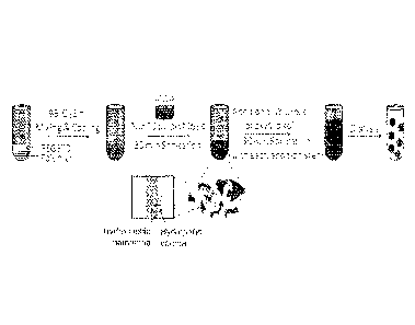
[0020] FIG. 7A is a schematic representation of the progressive saturation
protocol for
forming polymersome-encapsulated myoglobin in various embodiments.
[0021] FIGs. 7B and 7C are graphs illustrating results from encapsulation of
myoglobin into polymersomes formed from a particular PEO-b-PBD formulation
using the progressive saturation protocol of FIG. 7A.
CA 02991101 2017-12-28
WO 2017/004498
PCT/US2016/040657
[0022] FIG. 7D is a table summarizing the results shown in FIGs. 7B and 7C.
[0023] FIG. 7E is a table showing the partial pressure of oxygen required to
achieve
50% saturation (P50) obtained from 02 equilibrium curves for of free myoglobin
and
for polymersome-encapsulated myoglobin, prior to proteolysis and after pronase
treatment, that was prepared by progressive saturation.
100241 FIG. 8A is a graph showing results from encapsulation of myoglobin into
polymersomes formed from two particular PEO-b-PBD formulations using the
progressive saturation technique of the various embodiments.
[0025] FIGs. 8B and 8C are cryo-TEM images of vesicles in polymersome-
encapsulated myoglobin suspensions formed from each of the particular PEO-b-
PBD
formulations using the progressive saturation technique of the various
embodiments.
[0026] FIG. 8D is a graph showing the average hydrodynamic diameters of a
polymersome-encapsulated myoglobin suspension formed from a particular PEO-b-
PBD formulation using the progressive saturation technique of the various
embodiments.
[0027] FIG. 8E is a graph showing the oxygen equilibrium curves for free
oxymyoglobin and oxygenated polymersome-encapsulated myoglobin suspension
foirned from a particular PEO-b-PBD formulation using the progressive
saturation
technique of the various embodiments.
DETAILED DESCRIPTION
[0028] The various embodiments will be described in detail with reference to
the
accompanying drawings. Wherever possible, the same reference numbers will be
used throughout the drawings to refer to the same or like parts. References
made to
particular examples and implementations are for illustrative purposes, and are
not
intended to limit the scope of the claims.
[0029] The various embodiments include improved methods for generating PEM,
which may include identifying the key parameters of the thin-film rehydration
and
6
CA 02991101 2017-12-28
WO 2017/004498 PCT/US2016/040657
direct hydration protocols that prevent efficient uptake and/or compromise
protein
function, and performing iterative optimization of such key parameters. As a
result,
the various embodiments provide a generalizable technique that enables the
encapsulation of increased quantities of functional proteins within neutrally
charged
and fully PEGylated polymer vesicles. That is, the various embodiments include
a
new "progressive saturation" method for encapsulating myoglobin in
polymersomes.
[0030] Nanoparticle vehicles may overcome many challenges associated with the
delivery of functional proteins to enable the clinical development of diverse
macromolecular pharmaceuticals. Nanoparticle vehicles may include, for
example,
liposomes (i.e., self-assembled vesicles of natural phospholipids) and
polymersomes
(i.e., self-assembled polymer vesicles of block copolymers), as well as
micelles,
perfluorocarbon emulsions, and others. To that end, polymersomes, have
advantageous properties as compared to conventional liposomes, as liposomes
typically have high membrane permeability and low stability in vivo. There
have
been, however, few comparative studies to establish and validated a single,
scalable
and generalized strategy for encapsulating large amounts of protein in
neutrally
charged and/or PEGylated polymersomes. By optimizing and combining different
steps from various liposome-based encapsulation methods, the various
embodiments
provide a new progressive saturation technique that allows improved
encapsulation of
functional proteins in nanoscale polymer vesicles. The various embodiments
demonstrate a tradeoff between the degree of polymersome loading (i.e., weight
percentage of protein-to-polymer) and the encapsulation efficiency of protein
(with
respect to the initial quantity that was employed for polymersome formation)
that may
be achieved. Moreover, in the various embodiments, a proteolysis step
accurately
quantifies the amounts of both encapsulated protein (i.e., the desired
outcome) as well
as surface associated (i.e., non-specifically bound) product that may be
obtained in
polymersome suspensions formed by the progressive saturation protocol. While
there
are some reports of large amounts of protein loading within polymersomes at
high
efficiencies using existing liposome encapsulation techniques, such reports do
not
involve differentiating between encapsulated and surface-associated protein.
Therefore, the progressive saturation technique in the various embodiments may
7
CA 02991101 2017-12-28
WO 2017/004498 PCT/US2016/040657
provide a more robust, scalable, and generalizable strategy for encapsulation
of
proteins in fully PEGylated and neutrally charged polymersomes in quantities
and at
efficiencies that may enable further translational development.
[0031] Selective and potent modulation of protein function in mammalian cells
is the
principal activity of most molecular therapeutics, where the vast majority of
available
agents are organic small molecules (less than or equal to 800 Da in size).
Recent
studies suggest, however, that only a small percentage of the human proteome
is
susceptible to small molecule-based therapy. Moreover, the functional
diversity of
proteins that are successfully targeted by small molecules remains very low.
That is,
around 40% of all prescription drugs target a single class of proteins, namely
the G-
protein coupled receptors. However, use of small-molecule therapy is limited
since
small drug molecules are intrinsically unable to cope with the extended
contact
surfaces found at many biologically important interfaces.
[0032] Biomacromolecules such as proteins have recently shown significant
clinical
utility, in large part due to their ability to overcome these significant
limitations
associated with traditional small molecule therapies. When compared to the
interaction of a small-molecule with its biological target, macromolecular
therapeutics
have higher folding energies (typically around 7-20 kcal/mol) that allow for
the
adoption of larger and more precise three-dimensional configurations, which
are often
required for efficient binding and/or control of complex biological function.
As such,
macromolecules may achieve superior binding selectivity and more potent on-
target
activity. Currently, a small number of macromolecular therapies in use,
including the
approximately 200 protein drugs available worldwide, have demonstrated a high
potential as new leads in drug development. Nevertheless, several barriers
have
hindered the ready development of macromolecules as human therapeutics,
including:
(i) the difficulty and/or expense of commercial scale production, (ii)
biochemical
instability that occurs in pathophysiologic environments or with prolonged
storage,
(iii) short circulatory half-lives and large steric hindrance that prevent
effective tissue
penetration, and (iv) risks associated with their potential to promote severe
adverse
effects, such as the induction of anti-idiotypic antibodies and/or immune
complex
8
CA 02991101 2017-12-28
WO 2017/004498 PCT/US2016/040657
foimation. To overcome some of these limitations, most pharmaceutical
compounds
either employ biocompatible polymers (e.g., polyethylene glycol (PEG) or
hyaluronic
acid) or liposomes (i.e., lipid based vesicles) for protein complexation and
in vivo
delivery.
[0033] Synthetic nanoparticles may exhibit superior properties to enhance drug
delivery when compared to more conventional formulations. In particular, among
the
classes of nanoparticles, polymersomes (i.e., self-assembled polymer vesicles
comprised of amphiphilic block copolymers) may provide a beneficial nanoscale
delivery platfoun. While lipid-based vesicles (i.e., liposomes) have been
extensively
utilized in biomedical research, there are material limitations inherent to
these
phospholipid-based drug delivery vehicles, including compromised suspension
stability, premature drug release, and limited product shelf-life. In
contrast,
polymersomes are formed from higher molecular weight amphiphilic block
copolymers that impart a broad and tunable range of carrier properties. For
example,
polymersomes enable: (i) facile and stable loading of diverse therapeutic
payloads
through non-covalent interactions, (ii) mechanical stability that is 5 to 50
times greater
than that of liposomes or micellar structures constructed from similar
molecular
weight copolymers, (iii) economic and large scale production that removes the
need
for costly post-manufacturing purification, and (iv) diversity in biochemical
properties, which are imparted by their construction from a variety of
copolymer
compositions. Such properties may include fully PEGylated surfaces and tunable
in
vivo circulation times, site-specific targeting, environmental responsiveness,
and
complete biodegradation.
100341 The incorporation of proteins into nanoparticles may enhance their
pharmacologic performance and improve their on-target activity. Methods that
have
been developed for encapsulating proteins into nanoparticles have utilized
electrostatic interactions to incorporate a handful of highly anionic
proteins, or
chemical or genetic modification of the original protein for efficient and
reproducible
nanoparticle formation. Examples of such method include thin-film rehydration
(i.e.,
rehydration of dried polymer), which results in low yields of polymersome-
9
CA 02991101 2017-12-28
WO 2017/004498 PCT/US2016/040657
encapsulated protein. Another example method is direct hydration, the use of
which is
generally limited to small-scale preparations (e.g., less than one mL).
Another
example method is electro-formation, which provides useful results for only a
limited
number of proteins (i.e., highly charged proteins). Another example method is
hollow-fiber extrusion, which involves extrusion of preformed vesicles in the
presence
of protein solution. While the hollow-fiber extrusion technique has been used
for
large-scale preparations of liposome-encapsulated protein, elevated
temperatures and
pressures are required for polymersome formation, which has limited its
widespread
applicability.
[0035] Existing techniques require the input of thermal, electric, ultrasonic,
or
mechanical energy for particle formation, or alternatively the use of organic
cosolvents, which may damage the structure and/or function of the protein,
making
encapsulation more challenging and limited in utility. Therefore, in various
encapsulation techniques, a need exists for a generalized method that enables
the
incorporation of large quantities of native protein in neutrally charged
and/or
PEGylated nanoparticles.
[0036] While adoption of various liposome encapsulation techniques has enabled
facile incorporation of small molecules within polymersomes, these methods
cannot
directly be applied for scalable encapsulation of the functional proteins.
Often, there
is a trade-off in the maximum concentration of the aqueous protein that may be
encapsulated (i.e., mg protein/mL solution), the final loading ratio of
protein-to-
polymer that comprises the polymersome structure (i.e., w/w% protein/polymer),
and/or the protein encapsulation efficiency (i.e., the percentage of the
initial protein
suspension that is retained). Further, the value of each of these parameters
is highly
dependent on the nature of the protein, the exact block copolymer foimulation,
and the
encapsulation method that is utilized. For example, the table in FIG 1 shows
existing
results from encapsulation of various proteins into polymersomes. The various
embodiments provide an alternative, optimized and reproducible method to
efficiently
encapsulate increased quantities of functional proteins in polymersomes. The
newly
developed "progressive saturation" technique of the various embodiments is
readily
CA 02991101 2017-12-28
WO 2017/004498 PCT/US2016/040657
scalable, highly reproducible, and generalizable for producing increased
quantities of
polymersome-encapsulated protein that may enable new and diverse biomedical
applications.
[0037] In various embodiments, PEO-b-PBD copolymers are used to form
polymersomes that possess fully PEGylated surfaces. Such surfaces, being
uncharged
and nondegradable; provide an ideal system for ensuring vesicle integrity and
minimizing unwanted protein interactions or modifications. Two different
molecular
weight PEO-b-PBD polymers, "OB18" diblock copolymer and "0B29" diblock
copolymer, are employed to determine the generalizability of the results as
they
pertain to polymersomes of different minimal sizes, PEG lengths, and membrane
core
thicknesses. FIG. 2 provides a table showing a comparison of various
properties of
0B18 and 0B29. Methylene blue (mBlue; Mw = 319.85 g/mol), which is highly
stable in aqueous suspension and has a strong near-infrared absorbance
enabling ready
spectrophotometric detection, is used as a model small molecule to establish
various
baseline parameters for encapsulation using existing methods. Such baseline
parameters include aqueous suspension concentration, final weight percentage,
and
encapsulation efficiency. Myoglobin (Mb; Mw =17,600 Da), which has a size and
thermal stability (i.e., denaturation above 60 C) comparable to other small
proteins
with therapeutic potential, was used as a model protein. Myoglobin also has a
strong
ultraviolet (UV) absorbance that enables ready identification of its
functional status, as
determined by the redox state of its iron-containing heme group. Myoglobin has
additionally been employed in other studies, enabling comparisons of results
to other
encapsulations using existing techniques, as discussed above with respect to
FIG. 1.
Methylene blue is easily encapsulated in PEO-b-PBD polymersomes formed by thin-
film rehydrafion at elevated temperatures, yielding final weight ratios of
mBlue-to-
polymer of 4.1 and 5.0 w/w% when formed at 40 and 60 C, respectively.
However,
similar conditions only led to myoglobin degradation.
[0038] When vesicles are formed by thin-film rehydration, as the film of dry
copolymer is hydrated, lamellae (aka sponge-like structures) are first formed
as the
hydrophilic blocks in the film swell. Further swelling leads to transformation
into
11
CA 02991101 2017-12-28
WO 2017/004498 PCT/US2016/040657
hexagonally packed vesicles and finally into fully dispersed polymersomes.
When
thin-film rehydration is attempted in solutions of soluble small molecules (or
proteins), these water-soluble species adsorb to the surfaces of the budding
lamellae,
which subsequently adopt a spontaneous (or preferred) curvature. During
formation,
these membranes preferentially bend away from the aqueous compartment that
contains the higher concentration of adsorbing species, thereby excluding the
water-
soluble agents from vesicle encapsulation. Ultimately, the input of energy can
overcome this spontaneous surface tension in order to promote vesicle
encapsulation.
The amount of energy that is required scales with the size of the adsorbed
molecule
and the membrane thickness of the vesicle. Thus, while it is easy to disrupt
liposomes
and enable effective small molecule and protein loading by thin-film
rehydration by
the input of thermal (and/or sonic) energy, such input is only only enables
effective
encapsulation of small molecules into polymersome suspensions. In the direct
hydration method, which was developed as a hybrid of solvent dispersion and
homopolymer addition, the hydrophilic polymer PEG500 dimethyl ether (DME) is
used to disrupt the interactions of hydrophobic chains in the forming polymer
lamellae
. With subsequent additions of aqueous solution, self-assembly of vesicles
from
budding lamellae that have dispersed protein is promoted and results in
improvements
in aqueous encapsulation; encapsulation efficiencies as high as 37% have been
observed. Using direct hydration at 23 C, polymersome-encapsulated myoglobin
suspensions may have encapsulation efficiencies greater than 10%, with the
encapsulated myoglobin species exhibiting good suspension properties and the
characteristic absorption spectra of intact protein. The final loading of
myoglobin in
these polymersome-encapsulated suspensions, however, was found to be only
around
0.3 w/w% Mb/polymer. Upon addition of a protease solution to induce
proteolysis of
all surface associated (i.e., non-specifically bound) protein, the final Mb
composition
of PEM suspensions was found to be even lower __ that is, less than 0.1 w/w%
Mb/polymer. For translational therapeutic applications, the loading of
therapeutic
proteins within the aqueous cavities of polymersome vehicles is ultimately the
metric
that must be maximized in order to minimize the amount of associated carrier
that is
12
CA 02991101 2017-12-28
WO 2017/004498 PCT/US2016/040657
introduced to a subject. Therefore, such encapsulation using standard direct
hydration
is insufficient.
[0039] In various embodiments a progressive saturation protocol provides for
efficient
generation of PEM suspensions. The generalizability of progressive saturation
for
protein encapsulation is further established by utilizing a variety of
different proteins,
ranging from 17-450 kDa, yielding nanoscale polymersomes in quantities that
may
enable preclinical investigations of many novel therapeutic compositions. In
particular, a difference between the progressive saturation method and direct
hydration may involve adding five subsequent volumes of the functional protein
solution to dilute the PEG/polymer mixture in lieu of additional dilutions
with
phosphate buffered saline (PBS).
[0040] Specifically, the progressive saturation method of the various
embodiments
involves heating 10 mg of polymer and 10 mg of PEG at around 95 C for around
1 h.
The sample mixture may be centrifuged and cooled to room temperature. A
metmyoglobin (metMb) solution (e.g., 150 mg/mL in PBS) may be reduced to
oxymyoglobin (oxyMb) with sodium dithionite (Na2S204) (e.g., 1 wt%). From the
resulting oxyMb solution, a portion (i.e., aliquot) may be added to the sample
mixture
at a ratio of 1:1 by volume, and mixed thoroughly followed by sonication at
room
temperature for around 30 mm. In particular, the aliquot may be 10 1..t1_, of
the oxyMb
solution. The sample mixture may be further diluted with a number of dilution
steps.
Specifically, each dilution step may involve addition of a volume of the oxyMb
solution (e.g., 150 mg/mL in PBS), followed by thorough mixing and sonication
at
room temperature for around 30 minutes. The volumes of oxyMb solution used in
the
dilution steps may be amounts in which ratios of the oxyMb solution to the
original
sample mixture are 1:1, 2:1, 5:1, and 5:1 by volume 10 L, followed by 20, 50
and
100 L. After the dilution steps, the resulting sample may be sonicated for an
additional 30 mm at room temperature, followed by dialysis for at least 30 h
at around
4 C, employing a 1000 kDa molecular weight cutoff membrane. Surface
associated
myoglobin may be removed by proteolysis via treatment with 0.4 wt % pronase
solution, followed by dialysis for at least 12h at around 4 C (e.g.,
molecular weight
13
CA 02991101 2017-12-28
WO 2017/004498 PCT/US2016/040657
cutoff of 1000 kDa). In various embodiments, myoglobin encapsulation of the
resulting polymersome suspension may be measured before and after proteolysis.
Specifically, concentration of myoglobin may be measured using inductively
coupled
plasma optical emission spectroscopy (ICP-OES), while redox states of iron in
the
heme group of myoglobin may be quantified using UV-Vis absorption
spectroscopy.
[0041] These progressive saturation steps provided favorable results for
encapsulating
myoglobin in OB18 polymersomes, as shown in FIGs. 3A-3E. For example, FIG.
3A shows the final weight percentage of Mb-to-polymer (i.e., w/w% Mb/polymer)
in
polymersome-encapsulated myoglobin obtained when no sonication was used (as in
conventional techniques) and by using sonication, according to the progressive
saturation protocol (i.e., for around 30 min at room temperature after each
dilution
step). A shown in FIG. 3A, a final weight percentage of Mb-to-polymer of
around 6
(i.e., w/w% Mb/polymer) may be an achieved result from polymersome-
encapsulated
myoglobin created by a protocol that includes such sonication. Therefore,
sonicating
the sample for around 30 min at room temperature after each dilution step may
increase encapsulation efficiency by more than 30 times based on the final
weight
percentage resulting from polymersome-encapsulated myoglobin generated via
direct
hydration.
[0042] FIG. 3B shows the final weight percentage of Mb-to-polymer (i.e., w/w%
Mb/polymer) in polymersome-encapsulated myoglobin obtained using a
metmyoglobin solution (as in conventional techniques) and by using oxyMb as in
the
progressive saturation technique. FIG. 3C shows the rate of myoglobin
oxidation
(expressed as a percentage of metMb formed over time) as a function of
myoglobin
exposure to different solution conditions. FIG. 3D shows the amount of surface-
associated Mb removed (% Mb loss) as a function of proteolysis time for
various
oxyMb volumes. FIG. 3E shows the final weight percentage of Mb-to-polymer
(i.e.,
w/w% Mb/polymer) compared to encapsulation efficiency (% Mb EE) in
polymersome-encapsulated myoglobin generated using various ratios for the
volumes
of oxyMb solution to PBS used in the dilution steps. In particular, the
samples in FIG.
14
CA 02991101 2017-12-28
WO 2017/004498
PCT/US2016/040657
3E were proteolyzed for 18 h to remove surface-associated myoglobin, and
quantified
using UV-Vis absorption spectroscopy.
[0043] Therefore, the final Mb-to-polymer weight ratios that were obtained in
generating polymersome-encapsulated myoglobin using progressive saturation
according to various embodiments (i.e., 4-6 w/w% Mb/polymer) may be
significantly
improved compared to polymersome-encapsulated myoglobin generated using the
direct hydration protocol (i.e., 0.1-0.3 w/w% Mb/polymer). Without wishing to
be
bound to a particular theory, the loading capacity achieved using progressive
saturation steps may be due to incomplete polymersome formation during the
initial
dilution step, and further encapsulation being accomplished with each
subsequent
addition of protein solution.
[0044] Developing the progressive saturation protocol included optimizing and
combining various steps from multiple liposome formation methods. Factors
influencing the final concentrations of myoglobin, the relative loading levels
that
could be achieved within the OB18 polymersome carrier (i.e., w/w%
protein/polymer), and the efficiency of myoglobin encapsulation were
systematically
evaluated. Factors such as the molecular weight of the polymer, the oxidation
state
and concentration of the protein, the pH and nature of the buffered solution,
the exact
polymer hydration conditions (i.e., time, temperature, and blending
technique), the
number and duration of sonication steps, and the addition or avoidance of
freeze-thaw
cycles all had effects on the concentration and the fidelity of the final
polymersome-
encapsulated protein product.
[0045] Further, compared to polymersome-encapsulated myoglobin created using
existing techniques, polymersome-encapsulated myoglobin created by progressive
saturation also exhibits an increase in the final concentrations of Mb. For
example,
the final concentration of Mb in polymersome-encapsulated myoglobin generated
via
direct hydration is less than around 0.5 mg/ML in solution, while that of
polymersome-encapsulated myoglobin generated via progressive saturation in the
various embodiments may be greater than around 2.0 mg/mL in solution.
CA 02991101 2017-12-28
WO 2017/004498 PCT/US2016/040657
[0046] Using cryo-TEM to verify vesicle morphologies, suspensions of
polymersome-
encapsulated myoglobin developed using progressive saturation showed no signs
of
aggregate formation when maintained at 4 C, 23 C, and 37 C for longer than
one
month. The progressive saturation technique may be further utilized for the
successful
encapsulation of a variety of other proteins ranging in size from 17 to 450
kDa, within
PEO-b-PBD polymersomes.
[0047] Without wishing to be bound to a particular theory, there may be a
direct
tradeoff between Mb encapsulation efficiency and the final weight ratios of Mb-
to-
polymer that could be achieved based on the concentration of free Mb that was
used
for each addition step. Aqueous encapsulation of protein is preferred to
surface-
associated protein in order to assure that the final product meets the
objectives for
utilizing a polymersome delivery vehicle that is, to improve biochemical
stability, to
increase circulatory half-life, to minimize adverse side effects, and to
achieve
controlled release of the associated protein. The various embodiment
techniques may
be employed using different proteins that vary over a large range of molecular
weights
and sizes, including those associated with therapeutically relevant antibodies
and
enzymes. For example, the progressive saturation technique may be utilized to
encapsulate myoglobin in a PEO-b-PBD-based polymersome system comprised of the
0B29 diblock copolymer. In various embodiments, the progressive saturation
technique may be utilized to encapsulate any of a number of other proteins,
including,
but not limited to, antibodies (e.g., immunoglobulin G (IgG) and functional
enzymes
(e.g., catalase).
[0048] As described above with respect to FIG. 2, when compared to the 0B18
diblock copolymer, the 0B29 diblock copolymer has a smaller molecular weight
and
generates polymersomes with a shorter PEG brush border (1.3 vs. 3.9 kDa),
thinner
bilayer membrane (9.6 nm vs. 14.8 urn), and smaller average hydrodynamic
diameter
(130 vs. 200 urn). In various embodiments, using progressive saturation to
encapsulate myoglobin in 0B29 polymersomes provides similar results to those
from
OB18 polymersomes. In various embodiments, progressive saturation technique
may
be applied using any PEG-based polymersome-forming block copolymer, including
16
CA 02991101 2017-12-28
WO 2017/004498 PCT/US2016/040657
any amphiphilic polymer comprised of PEG and a hydrophobic block that is a
biodegradable polymer (e.g., a biodegradable polyester, poly(amide),
poly(peptide),
poly(nucleic acid), etc.). Examples of biodegradable polyesters that may form
the
hydrophobic block include, but are not limited to, poly(lactic acid),
poly(glycolic
acid), poly(lactic-co-glycolic acid), poly(caprolactone), poly(methyl
caprolactone),
poly(hydroxybutyrate), poly(hydroxyvalerate), poly(hdyroxyhexanoate),
poly(hydroxyoxtanoate), and poly(trimethylene carbonate).
[0049] The generalizability of the progressive saturation technique is further
demonstrated by analogous results from encapsulation of several larger
proteins using
0B29 polymersomes. FIG. 4 shows encapsulation results from using the
progressive
saturation protocol and 0B29 polymersomes to encapsulate myoglobin, hemoglobin
(Hb) (64 kDa), bovine serum albumin (BSA) (66 kDa), IgG (150 kDa), catalase
(250
kDa), fibrinogen (340 kDa), and apoferritin (450 kDa).
100501 The invention is intended to be illustrated but not limited by the
following
examples.
EXPERIMENTAL
[0051] Comparative and quantitative studies were performed in order to
establish a
generalizable method for producing scalable quantities of neutrally-charge and
fully
PEGylated polymersomes that encapsulate functional protein. Differences in
small
molecule and protein encapsulation were examined by employing polymersome
formulations comprised of OB18 and 0B29 diblock copolymers. As described above
with respect to FIG. 2, these two PEO-b-PBD polymers and the polymersomes
formed
therefrom differ with respect to molecular weight and, ultimately, vesicle
membrane
thicknesses. Methylene blue (mBlue; Mw = 319.85 gimol) was used as a model
small
molecule and myoglobin (Mb; Mw =17,600 Da) as a model protein with unique
biological function (i.e., oxygen storage and release). FIG. SA shows a
representation
of polymersomes made of amphiphilic diblock copolymers, as well as water-
insoluble
agents and water-soluble species that may be encapsulated in or attached to
polymersomes. For example, conventional vesicle formation techniques that were
17
CA 02991101 2017-12-28
WO 2017/004498 PCT/US2016/040657
employed to incorporate water-soluble agents within PEO-b-PBD polymersomes
included thin-film rehydration and direct hydration, protocols for which are
shown in
FIGs. 5B and 5C, respectively. The encapsulations of methylene blue and
myoglobin
in PEO-b-PBD polymersomes generated by each of the thin-film rehydration and
direct hydration methods were compared. In order to quantify the encapsulation
of
fully functional protein capable of oxygen binding, the iron concentration in
polymersome-encapsulated myoglobin was measured by ICP-OES, and the redox
states of iron in the heme group of myoglobin measured by UV-Vis absorption
spectroscopy.
[0052] Compared to PEM created using existing techniques, PEM created by
progressive saturation exhibit an increase in the final concentrations of Mb
(e.g., from
less than around 0.5 mg/mL in solution to greater than around 2.0 mg/mL in
solution), and an increase in the final weight ratio of Mb to polymer that
could be
reproducible obtained (from less than 1 w/w43/0 Mb/polymer to greater than
around 3-4
w/w% Mb/polymer). Further, PEM created by progressive saturation show an
increase in the overall efficiency of protein encapsulation (from less than
around 5%
to greater than around 90%) in the PEM suspensions. Using cryo-TEM to verify
vesicle morphologies, suspensions of PEM developed using progressive
saturation
display no signs of aggregate formation for longer than one month at 4 C, 23
C, and
37 C.
[0053] Materials
[0054] PEO(3900)-b-PBD(6500) (0B18) and PEO(1300)-b-PBD(2500) (0B29) were
purchased from Polymer Source (Dorval, Quebec, Canada). Horse skeletal muscle
Mb, bovine blood hemoglobin (Hb), bovine serum albumin (BSA), catalase (C),
fibrinogen (F), sodium hydrosulfite, poly(ethylene glycol) dimethyl ether
(PEG; Mn =
¨500), protease from Streptomyces griseus ("pronase"), and dichloromethane
(DCM)
were purchased from Sigma-Aldrich (St. Louis, USA). Horse spleen apoferritin
(aFr)
was purchased from Alfa Aesar (Ward Hill, USA). Immunoglobulin G (IgG) was
purchased from LEE Biosolutions (St. Louis, USA). Dialysis tubing and vials
were
purchased from Spectrum Laboratories (Rancho Dominguez, USA). Sodium chloride,
18
CA 02991101 2017-12-28
WO 2017/004498 PCT/US2016/040657
potassium chloride, sodium phosphate dibasic, potassium phosphate monobasic,
mBlue, and Triton X-100 were purchased from Fisher Scientific (Suwanee, USA).
All chemicals were of reagent grade unless otherwise stated.
[0055] The particle sizes were measured using DelsaTm Nano, a dynamic light
scattering (DLS) instrument (Beckman Coulter, Indianapolis, USA). Mb and mBlue
concentrations were determined by absorption spectroscopy using a GenesysTm
10S
UV-Vis spectrophotometer (Thermo Scientific, Suwanee, USA). The concentrations
of all proteins in polymersome suspension were further measured using a Micro
BCA
Protein Assay Kit, utilizing UV-Vis spectrophotometry and by following the
manufacturer's protocols (Pierce Biotechnology, Inc; Rockford, IL, USA). Iron
concentrations in polymersome-encapsulated Mb suspensions were determined
using
a Vista-PRO CCD ICP-OES (Varian, USA). Oxygen equilibrium binding was studied
using a HemoxTm- Analyzer (TCS Scientific Corp, New Hope, PA, USA). Electro-
formation was performed using Gene Pulser (Bio-Rad, Hercultes, CA, USA).
[0056] Methods
[0057] Thin-film rehydration method
[0058] 10 mg of 0B18 polymer was dissolved in 200 tut of DCM. The polymer
solution was deposited on Teflon wafers (15mm x 15mm) that were subsequently
dried for 30 min at room temperature (RT). The films were further kept under
vacuum
overnight at RT to ensure DCM evaporation. For methylene blue encapsulation,
polymer films were then hydrated with methylene blue solution (21 mg/mL) in
phosphate buffered saline (PBS; 10 mM, pH 7.4) for 24-48 h at 23, 40 or 60 C.
The
samples were sonicated for 30 min at room temperature, followed by (x10)
freeze-
thaw cycles using liquid nitrogen. The samples were dialyzed (MW cutoff = 100
kDa)
for 30 h at RT. For myoglobin encapsulation, polymer films were hydrated with
myoglobin solution (150 mg/mL) in PBS (10 mM, pH 7.4) for 60 h at 23, 40, and
60
C. The samples were then sonicated for 30 min at RT followed by dialysis (MW
cutoff= 1000 kDa) for 30 h at 4 C.
[0059] Direct hydration method
19
CA 02991101 2017-12-28
WO 2017/004498 PCT/US2016/040657
[0060] 10 mg of 0B18 and 10 mg of PEG were heated in a 1.5 mL centrifuge tube
for
20 min at 95 C. The samples were mixed and cooled to room temperature,
followed
by the addition of 10 ILIL of methylene blue solution (21 mg/mL) or myoglobin
solution (150 mg/mL) in PBS (10 mM, pH 7.4). The samples were then diluted
with
20, 70, and 900 ttL of PBS and well mixed after each addition/dilution (via
vortexing).
The samples were then dialyzed for 30 h at room temperature or at 4 C
(molecular
weight cutoff of 1000 kDa) to remove unencapsulated methylene blue or
myoglobin,
respectively.
[0061] Quantification of mBlue/Mb
[0062] The amounts of methylene blue or myoglobin that were encapsulated in
purified polymersome suspensions were determined by measuring solution
absorbance
at 665 urn (mBlue) or at 410 nm (Mb), using a UV-Vis spectrophotometer.
Calibration
curves for methylene blue and myoglobin were developed using serial dilutions
of
known concentrations. To measure the iron content in polymersome-encapsulated
myoglobin suspensions (as a corroboration of myoglobin concentration in that
sample), 5-10% (v/v) of Triton X-100 was added, the mixture was digested by
heating
in aqua regia for 3 h at 98 C, and was subsequently diluted with deionized
water.
ICP-OES was performed on experimental samples and their iron content was
determined in comparison to this standard calibration curve. The
concentrations of
myoglobin (as calculated by UV-Vis absorbance spectroscopy) were compared to
those obtained via ICP-OES or via the Micro BCA Assay (secondary UV-Vis
method)
for each suspension. Loading of aqueous encapsulants in the polymersomes was
quantified and expressed as the final weight percentages of encapsulant-to-
polymer
that comprised the vesicles in suspension (e.g., w/w% Mb/polymer).
[0063] Quantification of metMb
[0064] The amount of metinyoglobin (metMb, i.e., oxidized Mb with a Fe(III)-
heme
group) in polymersome suspensions was quantified using a modified UV-Vis
absorption protocol that was previously established for the measurement of
cyanomethemoglobin levels. In brief, the absorbance of myoglobin was measured
at
CA 02991101 2017-12-28
WO 2017/004498 PCT/US2016/040657
630 nm (L1) against a blank reference (deionized water). One drop of KCN
solution
(1 part 10% KCN and 1 part 50 mM phosphate, pH 7.6) was added and mixed with
the treated myoglobin samples. This reaction step was necessary to convert
metMb to
cyanometmyoglobin (cyanoMb), which does not absorb at 630 rim. After 2 min,
the
absorbance was measured at 630 nm (L2) against the deionized water, which
served as
the blank reference. The concentration of metMb was determined using Equation
1:
L -L2
[metMb] (mM) =¨ xDi (Eq. 1),
1 xE
where E = 3.7 (cm >< mM)-1 and is the extinction coefficient of metMb at 630
rim, and
DI is the dilution factor in this experiment (cuvette length = 1 cm).
[0065] To determine the concentration of myoglobin, one drop of 20%
K3(Fe(CN)6)
was added and mixed with the treated myoglobin sample. The solution was
allowed to
react for 2 min and an additional drop of 10% KCN was added and mixed. The
absorbance of the sample was then measured at 540 rim (L3). The concentration
of
total Mb was determined using Equation 2:
[total Mb] (mM) = x D (Eq. 2),
where E = 11.3 (cm x mA4)-1 and is the extinction coefficient for cyanometMb
at 540
nm; D2 is the dilution factor (cuvette length = 1 cm).
[0066] The percentage of metMb in the original solution was determined using
Equation 3:
[metMb] (%)¨ [metMb] x 100 (Eq. 3).
[metMb] + [total Mb]
[0067] Structural characterization of polymersomes
[0068] Polymersome suspensions were diluted in PBS solution and their
hydrodynamic diameters were measured by DLS using a standard 1.5 mL semi-micro
Plastibrand polystyrene cuvette (VWR, Atlanta, USA). The morphologies of blank
polymersomes and polymersome-encapsulated myoglobin were visualized by cryo-
TEM (JEOL 2100F, USA). In brief, polymersome samples were suspended in a
21
CA 02991101 2017-12-28
WO 2017/004498 PCT/US2016/040657
microperforated grid, rapidly vitrified using liquid ethane (-183 C), and
loaded onto a
cryogenic sample holder for cryo-TEM imaging at 200 kV.
100691 Encapsulation of mBlue and Mb in polymersome suspensions using
conventional methods
[0070] To establish a baseline for comparisons of small molecule and protein
encapsulation in polymersome suspensions, the final concentrations, weight
percentages (i.e., weight of encapsulated agent compared to the weight of the
polymer
that comprises the nanoparticle), and efficiencies of encapsulation for
methylene blue
were determined with OB18 polymersomes formed by the thin-film rehydration
technique. FIG. 6A shows the weight percentage results of the final
polymersome
composition for encapsulation of methylene blue at 40 C (i.e., 4.1 w/w%
mBlue/polymer) and 60 C. (i.e., 5.0 w/w% mBlue/polymer). When thin-film
rehydration was attempted at room temperature (i.e., 23 C), the encapsulation
of
methylene blue was found to be negligible (results not shown), possibly due to
the
observation that the polymer films did not swell after 48-72 h of hydration.
PEO-b-
PBD-based polymersomes require the input of energy for vesicle formation,
which is
usually supported by using elevated temperatures (e.g., greater than 45 C).
[0071] To improve the efficiency of encapsulation at lower temperatures, which
would
be necessary when employing labile proteins, encapsulation of mBlue was also
studied by the direct hydration method. FIG. 6B shows the final weight
percentage of
mBlue-to-polymer in polymersome suspensions created using direct hydration at
23
C (i.e., 1.2 w/w% mBlue/polymer).
[0072] Next, polymersome-encapsulated myoglobin suspensions formed at 23 C by
thin-film rehydration were initially found to be comprised of around 2.7 w/w%
Mb/polymer. After the addition of a proteolysis step to any remove surface-
associated
Mb (i.e., free protein that was nonspecifically bound), the final composition
of the
polymersomes was found to be only 0.5 w/v0/0 Mb/polymer, indicating that very
small
amounts of protein were being encapsulated within polymersomes. FIG. 6C shows
22
CA 02991101 2017-12-28
WO 2017/004498 PCT/US2016/040657
the final weight percentage of Mb-to-polymer in polymersome suspensions
created
using thin-film rehydration at 23 C both before and after the added
proteolysis step.
[0073] In order to improve the concentrations and the final weight percentages
of
myoglobin in polymersome-encapsulated myoglobin suspensions, polymersome
generation at higher temperatures was again attempted utilizing thin-film
rehydration
at 40 and 60 C. Such tests, however, only resulted in protein denaturation
and
aggregation. In contrast, polymersome-encapsulated myoglobin suspensions
prepared
by direct hydration at 23 C displayed good colloidal properties and the
characteristic
absorption spectra of intact myoglobin, yet the final loading ratio of Mb-to-
polymer in
these polymersome-encapsulated myoglobin suspensions was low. FIG. 6D shows
the
final weight percentage of Mb-to-polymer in polymersome suspensions created
using
direct hydration at 23 C both before proteolysis (i.e., showing 0.3 w/w%
Mb/polymer) and after proteolysis (i.e., 0.1 w/w43/0 Mb/polymer).
Modifications to Conventional Processes
[0074] Features of both the direct hydration and thin-film rehydration
techniques were
iteratively evaluated in experimental conditions in order to improve
polymersome-
encapsulation of functional protein.
[0075] Effects of sonication
[0076] Following the direct hydration protocol, upon addition of OB18 polymer
and
PEG, the sample was mixed, cooled to RT, and 10 iut of Mb solution (150 mg/mL)
in
PBS (10 mM, pH 7.4) was added. The sample was then further diluted with 10,
20, 50,
and 100 IA, of Mb solution, followed by mixing and sonication for either: A) 0
min or
B) 30 min after each additional dilution step. All samples were then dialyzed
for 30 h
at 4 C (molecular weight cutoff of 1000 kDa). The final Mb concentrations,
weight
percentages of Mb-to-polymer, and the efficiencies for Mb encapsulation in the
resultant polymersome suspensions were measured by UV-Vis absorption
spectroscopy, ICP-OES and compared.
23
CA 02991101 2017-12-28
WO 2017/004498 PCT/US2016/040657
[0077] In attempting encapsulation of Mb in OB18 polymersomes, and by
employing
the direct hydration protocol for vesicle formation, the weight ratios of Mb-
to-polymer
that were reproducibly obtained in the final PEM suspensions were found to
again be
very low (e.g., around 0.2 w/w% Mb/polymer). The encapsulation efficiency,
however, could be increased by more than 30 times if the samples were
sonicated for
30 mm at room temperature after each dilution step (i.e., sonicating after
introducing
additional volumes of aqueous solution to dilute the concentration of polymer
in
suspension). As discussed above with respect to FIG. 3A, the relative amount
of Mb
in PEM suspension could be increased to around 6.0 w/w% Mb/polymer, supporting
the addition of this sonication step to the original direct hydration
protocol.
100781 Effects of blending technique (dissolving polymer in organic solvent
vs.
addition of heat)
[0079] The effects of utilizing an organic solvent were compared to adding
heat to
blend OB18 with a PEG500 homopolymer to improve polymer dissolution during the
first step of the direct hydration protocol. These strategies were compared
with
respect to the final yield of polymersome formation and, ultimately, to the
concentrations and efficiencies of protein encapsulation that could be
obtained by
each method. If the two polymers were first mixed by dissolution in DCM
(followed
by polymersome formation after organic solvent evaporation), the final weight
ratio of
Mb-to-polymer in PEM suspensions was around 2 w/w% Mb/polymer. In comparison,
initial heating of dry OB18 and PEG500 to 95 C for 1 h improved mixing and
promoted more efficient polymersome generation, yielding a significantly
higher final
weight ratio of Mb-to-polymer in the final PEM suspensions (i.e., around 5
w/w%
Mb/polymer), corresponding to a greater amount of encapsulated protein.
[0080] Following the direct hydration protocol, 10 mg of 0B18 and 10 mg of PEG
were either blended by heating at 95 C for 1 h, or mixed by dissolution in
DCM (50
L) followed by drying under vacuum at room temperature overnight. Further
encapsulation was done using the same protocol with the addition of 30 mm of
sonication after each dilution step. Mb concentrations in the final
suspensions were
determined by UV-Vis absorption spectroscopy and ICP-OES and compared.
24
CA 02991101 2017-12-28
WO 2017/004498 PCT/US2016/040657
100811 Effects of Mb oxidation state (i.e., utilizing oxyMb vs. metMb for
polymersome-encapsulation)
[0082] Myoglobin encapsulation was found to be further augmented when the
starting
Mb stock solution was first reduced with sodium dithionite to convert all
metmyoglobin (i.e., metMb) to the oxmyoglobin (i.e., oxyMb) form. OxyMb
contains
a central heme group with iron in the ferrous state (i.e., Fe(II)), which
improves the
solubility of the protein when compared it its metMb form that contains
Fe(III). This
oxyMb solution was further desalted via dialysis prior to its utilization in
all of the
subsequent dilution steps in the direct hydration protocol, which was found to
be
necessary to increase the loading of Mb in PEM suspensions (i.e., the final
weight
ratio of Mb-to-polymer). As discussed above with respect to FIG. 3B, when
oxyMb
was used in the initial protocol step, PEM suspensions comprised of 6 w/w%
Mb/polymer were formed, which was a statistically significant improvement over
the
4 w/w% Mb/polymer obtained when metMb was utilized.
[0083] The direct hydration protocol was modified to expose the initial
mixture of
polymer and PEG to 1 h (instead of 20 min) of heating at 95 C. The effect of
the iron
oxidation state of the heme group of Mb on the efficiency of polymersome-
encapsulation was studied by using oxyMb (i.e., Fe(II)Mb) vs. metMb for each
dilution step. MetMb solution was prepared by dissolving lyophilized Mb in
PBS; the
same solution was treated with 1 wt% Na2S204 to obtain the reduced Mb form
(oxyMb). Mb encapsulation in polymersomes was measured by UV-Vis absorption
spectroscopy and ICP-OES, and compared.
[0084] Effects of sonication and temperature on Mb oxidation
[0085] More than 40% of the oxyMb that was used in the initial step for
polymersome-
encapsulation was found to be reoxidized to metMb within 2 h at 50 C. In
contrast,
only around 15% metMb was generated from the initial oxyMb solution if lower
temperatures were employed for polymersome founation (e.g., heating for 2 h at
40
C). The rate of Mb oxidation at 50 C was also significantly higher than that
at 40
C, regardless of the addition of sonication or the power that was utilized, as
discussed
CA 02991101 2017-12-28
WO 2017/004498 PCT/US2016/040657
above with respect to FIG. 3C. As such, it was determined that sonication had
no
effect on Mb oxidation and it was thus preferentially employed to both promote
polymer mixing and to provide interfacial energy to augment polymersome
formation.
[0086] The initial Mb solution (at 150 mg/mL) was reduced with Na2S204 and
subjected to various conditions, including heating at 40 C (with or without
sonication) or at 50 C for 2-5 h. Mb oxidation was determined by measuring
the
percentages of metMb in the total polymersome-encapsulated Mb suspensions,
using
the cyanomethemoglobin method.
[0087] Effects of proteolysis
[0088] Upon formation, PEM suspensions were treated with 0.4% pronase solution
for
up to 18 h at room temperature in order to examine the duration of time
required for
the complete digestion of any surface-associated (i.e., non-specifically
bound) Mb. It
was observed that all surface-associated Mb was digested in 2 h and that
neither
increasing pronase exposure time nor concentration further augmented Mb loss,
thus
indicating that only encapsulated Mb was retained, as discussed above with
respect to
FIG. 3D.
[0089] Mb was encapsulated in OB18 polymersomes using different initial
solution
concentrations of protein (i.e., 50, 75, and 150 mg/mL) followed by dialysis
for at
least 30 h at 4 C (molecular weight cutoff of 1000 kDa). The samples were
subsequently treated with 0.4 wt% pronase solution for 18 h at room
temperature and
again dialyzed overnight at 4 C. Mb encapsulation in polymersomes (before and
after
proteolysis) was measured by UV-Vis absorption spectroscopy and ICP-OES, and
compared.
[0090] Improvement of Mb encapsulation efficiency (i.e., %Mb EE)
[0091] Five sets of experiments were done with various Mb-to-PBS volume ratios
(i.e., "Mb:PBS") in order to establish the optimal Mb concentration to use in
each
subsequent dilution step in our modification of the original "direct
hydration"
protocol. Notably, when the Mb:PBS increased, the final w/w% Mb/polymer in the
26
CA 02991101 2017-12-28
WO 2017/004498 PCT/US2016/040657
PEM suspensions also increased; but, the Mb encapsulation efficiency (i.e.,
%Mb EE)
decreased as a result. In other words, the final Mb-to-polymer mass ratio was
maximized when all dilutions steps were conducted using a maximally
concentrated
Mb solution (i.e., Mb:PBS = 190:0 and 150 mg/mL oxyMb). As discussed above
with
respect to FIG. 3E, the %Mb EE was largest when the Mb:PBS was minimal (i.e.,
10:180). As the amount of protein in the final polymersome suspension is
ultimately
the metric that must be optimized for therapeutic administration (in order to
minimize
the amount of associated carrier polymer that is introduced to a subject), it
was
determined that a pure Mb solution (150 mg oxyMb/mL) would be used for each
dilution step in the ultimate encapsulation protocol, maximizing the fmal w/w%
Mb/polymer in PEM suspensions.
[0092] Following the basic direct hydration protocol, 10 mg polymer and 10 mg
of
PEG were initially heated in 1.5 mL microcentrifuge tubes for 1 h at 95 C and
subsequently cooled to RT. The mixtures were then diluted by adding 10, 10,
20, 50,
and 100 taL of diluents. Two different solutions were used and compared for
each of
the 5 dilution steps: PBS and/or Mb suspensions (i.e., 150 mg/mL Mb in PBS).
The
final (v/v) ratio of Mb to PBS (i.e., "Mb:PBS") used as diluents in steps 1,
2, 3, 4, and
were 10:180, 20:170, 40:150, 90:100, and 190:0, respectively. The samples were
then proteolyzed using 0.4 wt% pronase and again dialyzed overnight at 4 C
(molecular weight cutoff of 1000 kDa). Mb encapsulation was measured using UV-
Vis absorption spectroscopy. The Mb encapsulation efficiencies were calculated
using
Equation 4:
Mb Encapsulation Efficiency=11- __________ 21 x100 (Eq. 4),
Where vi = Initial volume of the unencapsulated Mb (mL), c1= Initial
concentration
of unencapsulated Mb (mg/mL), v2 = volume of polymersome-encapsulated Mb
obtained after dialysis and proteolysis (mL), and c2 = concentration of
encapsulated
Mb obtained after dialysis and proteolysis (mg/mL).
[0093] Using progressive saturation to generate polymersome-encapsulated
protein
suspensions.
27
CA 02991101 2017-12-28
WO 2017/004498 PCT/US2016/040657
[0094] By incorporating each of the steps in the various embodiments, a
progressive
saturation technique was established, represented in FIG. 7A, which vastly
improved
upon the results of the original direct hydration protocol discussed above
with respect
to FIG. 5C. Using the progressive saturation protocol, the final content of Mb
in
0B18-based PEM suspensions was found to be 6.1 and 3.2 w/w /0 Mb/polymer
before
and after proteolysis, respectively. Quantification of the iron content
(numbers of
intact heme groups) in each of the polymersome suspensions by ICP-OES
corroborated UV-Vis measurements of protein concentration. As shown in FIG.
7B,
the final loading ratios of Mb in the polymersomes were found to be 7.9 and
5.1
w/w% Mb/polymer before and after proteolysis, respectively. As shown in FIG.
7C,
the percentage of metMb (with respect to the total Mb content in these
suspensions)
was determined by UV-Vis absorbance spectroscopy and found to be around 8% and
6% in non-proteolyzed (PEM-SE) and proteolyzed (PEM-E) samples, respectively.
FIG. 7D is a table of the measured properties (i.e., results) for the 0B18-
based PEM
suspensions, as discussed with respect to FIGs. 7B and 7C.
[0095] Stability of polymersome-encapsulated protein suspensions
[0096] OB18-encapsulated Mb suspensions were prepared using the optimized
progressive saturation technique. The samples were stored at 4, 23, and 37 C
for 3
weeks. At predetermined time points, the samples were diluted with PBS and the
mean particle size and distributions were determined by DLS.
100971 Equilibrium binding of oxygen in polymersome-encapsulated Mb
suspensions
[0098] The equilibrium binding and dissociation curves for oxygen in
suspensions of
free and polymersome-encapsulated Mb were obtained at 37 C using a HemoxTm-
Analyzer. Samples were allowed to saturate to a p02 of 147 mmHg (using
compressed
air) and then deoxygenated (using a compressed nitrogen stream). The
absorbance of
oxygenated and deoxygenated free and polymersome-encapsulated Mb suspensions
was recorded as a function of p02 via dual wavelength spectroscopy. Oxygen
equilibrium curves were fit to a four-parameter (Ao, A, P50, n) Hill model
(Equation
5). In this model, Ao and AG represent the absorbance at 0 mmHg and at 147
mmHg,
28
CA 02991101 2017-12-28
WO 2017/004498 PCT/US2016/040657
respectively. The p02 represents the partial pressure of oxygen; and, P50
represents
the partial pressure of 02 where the sample is 50% saturated with oxygen.
Lastly, n
represents the cooperativity coefficient for the sample.
Abs-As
Y = (Eq. 5).
A¨ A0 p0121+ Prsio
[0099] FIG. 7E shows the P50 values (in mmHg) obtained for a free myoglobin
(Mb)
sample, a polymersome-encapsulated myoglobin sample prior to proteolysis (PEM-
SE) that was prepared using the progressive saturation technique, and a
polymersome-
encapsulated myoglobin sample after pronase treatment (PEM-E) that was
prepared
using the progressive saturation technique.
101001 Characterization of the final PEM suspensions
101011 The size distributions of the final OB18- and 0B29-based PEM
suspensions
were measured by both DLS and cryo-TEM. FIG. 8A provides the average
hydrodynamic diameter of particles in OB18-based and 0B29-based PEM
suspensions prepared using progressive saturation, as assessed by DLS. Cryo-
TEM
images of vesicles in OB18-based and 0B29-based PEM suspensions are shown in
FIGs. 8B and 8C, respectively. These results confirmed a mean particle
diameter of
approximately 200 nm for 0B18 polymersomes, and 130 nm for 0B29
polymersomes. The stability of the 0B18-based PEM suspensions were further
examined over three weeks and at various temperatures, with the polymersomes,
demonstrating no aggregation based on the consistent particle numbers and
stable size
distributions in suspension. FIG. 8D shows the average hydrodynamic diameters
of
particles, as determined by DLS, in OB18-based PEM suspensions that were
prepared
by progressive saturation at various temperatures (i.e., 4 C, 23 C, and 37
C) as a
function of time. Finally, the functional status of encapsulated Mb in the PEM
suspensions (i.e., retention of Mb's ability to bind and release oxygen) was
verified by
dual wavelength spectroscopy. FIG 8E shows oxygen equilibrium curves for free
oxyMb and oxygenated OB18-based PEM suspensions. Error bars denote standard
deviation of the mean. n > 3 experimental replicates per condition. The oxygen
29
CA 02991101 2017-12-28
WO 2017/004498 PCT/US2016/040657
equilibrium curves, P50 (i.e., the partial pressure of 02 where the Mb is 50%
saturated
with oxygen) of PEM were very similar to those of free Mb in solution.
101021 Polymersome-encapsulation using block copolymers and proteins of
varying
molecular weight
[0103] The generalizability of the progressive saturation technique was tested
using
proteins of various sizes: i.e., Mb (17 kDa), hemoglobin (Hb; 64 kDa), bovine
serum
albumin (BSA; 66 kDa), immunoglobulin G (IgG: 150 kDa), catalase (250 kDa),
fibrinogen (340 kDa), and apoferritin (450 kDa); each protein was dissolved in
PBS
(10 mM, pH 7.4) at its maximal solubility, corresponding to final suspension
concentrations of 150, 150, 40, 20, 50, 50, and 25 mg/mL, respectively. The
progressive saturation protocol was followed to encapsulate these proteins in
0B29
polymersomes. Free proteins were separated by dialysis for at least 30 h at 4
C
(molecular weight cutoff of 1000 kDa). Surface associated protein was removed
by
proteolysis via treatment with 0.4 wt% pronase solution followed by overnight
dialysis at 4 C (molecular weight cutoff of 1000 kDa). Protein encapsulation
(before
and after proteolysis) in polymersome suspensions was quantified via the micro-
BCA
assay, utilizing UV-Vis spectrophotometry and by following the manufacturer's
protocols (Pierce Biotechnology, Inc; Rockford, IL, USA). The final
concentrations
of protein were divided by those of polymer and expressed as the final weight
ratios of
protein-to-polymer that comprised the polymersomes in suspension (e.g., w/w%
Mb/polymer).
[0104] Statistical analysis
[0105] Data are presented as the mean the standard deviation of the mean
(SD). A
minimum of three experimental replicates was used for each condition. One-way
analysis of variance (ANOVA) was conducted using GraphPad software (San Diego,
USA). A p value of < 0.05 was considered statistically significant.