Note: Descriptions are shown in the official language in which they were submitted.
CA 02991484 2018-01-05
WO 2017/019117
PCT/US2015/065713
OPTICAL IMAGING AND MEASUREMENT SYSTEMS AND METHODS FOR
CATARACT SURGERY AND TREATMENT PLANNING
RELATED APPLICATIONS
[001] This application is a non-provisional application and claims the
benefit under 35
U.S.C. 119(e) of U.S. Provisional Application Serial No. 62/197,539, filed
July 27, 2015,
which is incorporated herein in its entirety by reference. Full Paris
Convention priority is
hereby expressly reserved.
BACKGROUND
[002] Cataract extraction is a frequently performed surgical procedure. A
cataract forms
through opacification of the eye's crystalline lens. The cataract scatters
light passing through
the lens, and may perceptibly degrade vision. Generally, a cataract can vary
in degree from
slight to complete opacity. Early in the development of an age-related
cataract, the power of
the lens may increase, causing near-sightedness (myopia). Over time, the
gradual yellowing
and opacification of the lens may reduce the perception of blue colors as
shorter wavelengths
are more strongly absorbed and scattered within the cataractous crystalline
lens. As the
cataract formation gradually progresses, the patient may experience
progressive vision loss.
[003] Cataract treatment may involve surgically removing the opaque
crystalline lens,
and replacing it with an artificial intraocular lens (IOL). Each year, an
estimated 15 million
cataract surgeries are performed worldwide. Cataract surgery can be performed
using a
technique called phacoemulsification in which an ultrasonic tip with
associated irrigation and
aspiration ports is used to sculpt the relatively hard nucleus of the lens to
facilitate removal
through an opening made in the anterior lens capsule. The nucleus of the
crystalline lens is
contained within an outer membrane of the lens referred to as the lens
capsule. To access the
lens nucleus, surgeons first perform a manual continuous curvilinear
capsulohexis (CCC)
procedure to form a circular hole in the anterior side of the lens capsule.
Alternatively,
surgeons may use a laser surgical system to perform the anterior capsulotomy
to gain access
to the lens nucleus. The surgical laser beam may also be used to fragment the
cataractous
crystalline lens before it is aspirated out of the eye. After the cataractous
lens is removed, a
1
CA 02991484 2018-01-05
WO 2017/019117
PCT/US2015/065713
synthetic foldable intraocular lens (IOL) can be inserted into the remaining
lens capsule of
the eye.
[004] Planning a cataract treatment can be challenging. There is
significant variation
between patients in many important eye biometric parameters, each of which may
affect
surgical planning, treatment, and outcome. Moreover, many patients may have
biometric
configurations, including for example, corneal lower order and higher order
aberrations,
extreme axial lengths, and/or previous conical refractive treatments such as
LASIK, which
may also affect surgical planning, treatment, and outcome. For example, with
respect to eye
aberrations, some patients have near-sightedness (myopia), far-sightedness
(hyperopia), or
astigmatism. Near-sightedness occurs when light focuses in front of the
retina, while far-
sightedness occurs when light refracts to a focus behind the retina.
Astigmatism occurs when
the corneal curvature is unequal in two or more directions. Various surgical
methods have
been developed and used to treat these types of aberrations. Ideally, for best
results and
outcome, a cataract surgeon would have access to not only ocular biometry
information, but
also to information on the eye's anterior corneal surface, posterior conical
surface, anterior
lens surface, posterior lens surface, lens tilt, lens thickness, and lens
position in order to plan
cataract treatment pre-operatively, and/or to assess the post-operative
refractive state of a
patient's eye with the implanted IOL.
[005] A variety of optical diagnostic systems have been developed, each of
which
provides a limited subset of the desired measurements. Thus, currently most
patients have
various measurements performed on different devices if the measurements are
taken at all.
There is a significant disadvantage, however, to using multiple measurement
devices in
cataract planning because the patient's eye may be in different positions
during each of the
measurements, and/or it may have changed between the different measurements,
or the
measurement may have been made under different conditions. Further, there may
be no way
to combine or fuse the data sets from different devices to obtain a single,
three-dimensional
model of the patient's eye. Hence, it can be often difficult to apply advanced
vision modeling
techniques, such as ray tracing, because the current diagnostic environment is
often
inadequate to reliably produce the three-dimensional models necessary for
accurate vision
modeling.
2
CA 02991484 2018-01-05
WO 2017/019117
PCT/US2015/065713
[006] As a result, there is an ongoing need for an improved optical
imaging,
measurement, and diagnostic system that can obtain most, if not all, of the
necessary
biometric and structural features of a patient's eye with the patient's eye in
a single
orientation within a brief period of time, that can fuse the data obtained
from various optical
techniques to achieve an accurate three-dimensional model of a patient's eye,
and that can
utilize advanced vision modeling techniques, such as ray tracing or other
power calculation
techniques, to improve cataract planning and outcome evaluation.
SUMMARY OF THE INVENTION
[007] This disclosure provides embodiments for improved optical measurement
systems
and methods for carrying out imaging and measurements used for diagnostics,
treatment
planning, and IOL placement for cataract treatment and surgery.
[008] An eye imaging and measurement system for planning a cataract
treatment in a
patient's eye according to one embodiment comprises: a Corneal Topography
Subsystem, a
wavefront aberrometer subsystem, and an eye structure imaging subsystem,
wherein the
subsystems have a shared optical axis, and each subsystem is operatively
coupled to the
others via a controller. The eye structure imaging subsystem is selected from
the group
consisting of an optical coherence tomographer (OCT), a Scheimpflug imager, a
fluorescence
imager, a structured lighting imager, a wavefront tomographer, and an
ultrasound imager.
The eye structure imaging subsystem is an optical coherence tomographer,
including for
instance, a Fourier domain optical coherence tomographer, a spectral domain
optical
coherence tomographer, or a swept source optical coherence tomographer.
[009] In many embodiments, the eye imaging and measurement system comprises
an iris
imaging subsystem operatively coupled to the controller.
[0010] In many embodiments, the eye imaging and measurement system comprises a
posterior corneal astigmatism imaging and measurement subsystem operatively
coupled to
the controller.
[0011] In many embodiments, the eye imaging and measurement device comprises a
fixation target subsystem operatively coupled to the controller. This target
allows for fixating
3
CA 02991484 2018-01-05
WO 2017/019117
PCT/US2015/065713
the eye during on axis measurements. In other embodiments, this target can be
used to
perform off-axis measurements at different eccentricities.
[0012] In many embodiments, the controller is coupled to an Optical
Coherence
Tomography (OCT) subsystem configured to sequentially scan the eye in a
plurality of OCT
scan patterns, each scan pattern being at a different axial depth of a
patient's eye. The
plurality of scan patterns comprise an anterior segment OCT scan pattern at or
near a location
of a cornea, a lenticular OCT scan pattern at or near a location of a lens,
and a retinal OCT
scan patter at or near a location of a retina. The plurality of imaging scan
patterns preferably
comprises an anterior segment OCT scan pattern suitable to measure a plurality
of an anterior
corneal surface, a corneal pachymetry, a central corneal thickness, and an
anterior chamber
depth of a patient's eye. The plurality of imaging scan patterns preferably
also comprises a
lenticular OCT scan segment scan pattern suitable to measure a plurality of a
lens thickness,
an anterior lens surface, a posterior lens surface, and a lens surface tilt
and decentration. The
plurality of imaging scan patterns comprises a retinal OCT segment scan
pattern suitable to
measure at least one of an axial length and a retinal layer thickness
information. These
measurements may also be done post-operatively, allowing the measurement of
IOL axial
position, tilt, and decentration, so that the instrument allows for evaluation
of postoperative
outcomes and secondary treatment planning, if needed.
[0013] In many embodiments, the eye imaging and measurement system comprises a
memory operable to store data acquired from each of the Corneal Topography
Subsystem, the
wavefront sensor subsystem, and the Optical Coherence Tomography subsystem,
wherein the
stored data includes a plurality of ocular biometry information, anterior
corneal surface
information, posterior corneal surface information, anterior lens surface
information, and
posterior lens surface information, lens tilt information, and lens position
information. The
ocular biometry information preferably comprises a plurality of a central
corneal thickness
(CCT), an anterior chamber depth (ACD), a pupil diameter (PD), a white to
white distance
(WTW), a lens thickness (LT), an axial length (AXL), and retinal layer
thickness.
[0014] In many embodiments, the eye imaging and measurement system further
comprises
a memory operable to store Intraocular Lens ("IOL") Data, the IOL data
including a plurality
4
CA 02991484 2018-01-05
WO 2017/019117
PCT/US2015/065713
of dioptic power, anterior and posterior radius, IOL thickness, refractive
index and
dispersion, asphericity, toricity, echelette features, haptic angulation, and
lens filter.
[0015] In many embodiments, the eye imaging and measurement system further
comprises
a memory operable to store intraocular lens ("IOL") model data for a plurality
of IOL
models, IOL model having associated with a plurality of predetermined
parameters selected
from the group consisting of dioptic power, anterior and posterior radius, IOL
thickness,
refractive index, asphericity, toricity, echelette features, haptic
angulation, and lens filter.
[0016] An improved system for selecting an intraocular lens (IOL) for
implantation,
comprises: a memory operable to store data acquired from each of the Corneal
Topography
Subsystem, the wavefront sensor subsystem, and the Optical Coherence
Tomography
subsystem, wherein the stored data includes a plurality of ocular biometry
information,anterior corneal surface information, posterior corneal surface
information,
anterior lens surface information, posterior lens surface information, lens
tilt information, and
lens position information; the memory further operable to store intraocular
lens ("IOL")
model data for a plurality of IOL models, the IOL model having associated with
it a plurality
of predetermined parameters selected from the group consisting of dioptic
power, anterior
and posterior radius, IOL thickness, refractive index, asphericity, toricity,
echelette features,
haptic angulation, and lens filter; and a processor coupled to the memory, the
processor
deriving the treatment of the eye of the patient applying, for each of the
plurality of identified
IOL models, to: (1) predict a position of one of the identified IOL models
when implanted in
the subject eye, based on the plurality of characteristics; (2) simulate the
subject eye by
means of ray tracing for a plurality of IOL predetermined parameters and the
predicted IOL
position; (3) based on that, select an IOL spherical equivalent (SE) and
cylinder (C) power, as
well as determine the optimum IOL orientation based on said eye model; (4)
propose the
selected IOL power for one or more IOL models from the plurality of IOLs
corresponding to
the optimized IOL(s) based on predetermined criteria; and (5) show the
simulated optical
quality and/or visual performance provided by each of the proposed IOL models
for distance
and/or for any other vergence or field angle.
[0017] An improved method for selecting an intraocular lens may include the
calculation
of the posterior corneal astigmatism and total corneal power of the eye. An
accurate method
CA 02991484 2018-01-05
WO 2017/019117
PCT/US2015/065713
to calculate these two quantities may be comprised of a first step of
measuring the anterior
corneal shape with a topographer, a second step of measuring a corneal
thickness map with a
scanning optical coherence tomographer, and a third step of adding the
thickness map to the
anterior surface shape to obtain the shape of the posterior surface. For
highest accuracy, the
bending of the optical coherence beam on the anterior corneal surface may be
calculated
using Snell's law at each location across the cornea prior to the step of
adding the thickness
map to the anterior cornea shape. After the determination of posterior corneal
shape, the
posterior corneal astigmatism and total corneal power may be calculated using
standard
optical ray tracing techniques.
[0018] A method of selecting an intraocular lens (IOL) to be implanted in a
subject eye, or
alternatively, a tangible computer-readable storage device storing computer
instructions
which, when read by a computer, cause the computer to perform the method,
comprises:
measuring a plurality of eye characteristics comprising ocular biometry
information, anterior
corneal surface information, posterior corneal surface information, anterior
lens surface
information, posterior lens surface information, lens tilt information, and
lens position
information; and for each of Intraocular Lens ("IOL") model having associated
with it a
plurality of predetermined parameters selected from the group consisting of
dioptic power,
refractive index, anterior and posterior radius, IOL thickness, asphericity,
toricity, echelette
design, haptic angulation, and lens filter: (1) modeling the subject eye with
the intraocular
lens; (2) simulating the subject eye based on the plurality of IOL
predetermined parameters
and the predicted IOL position; (3) performing a ray tracing and an IOL
spherical equivalent
(SE) and cylinder (C) power calculation, as well as determine the optimum IOL
orientation
based on said eye model; and (4) proposing one IOL power for one or more IOL
models from
the plurality of IOLs corresponding to the optimized IOL(s) based on
predetermined criteria;
and optionally, (5) show the simulated optical quality and/or visual
performance provided by
each of the proposed IOL models for distance and/or for any other vergence.
[0019] A method, or alternatively, a tangible computer-readable storage
device storing
computer instructions which, when read by a computer, cause the computer to
perform the
method, comprising: (1) receiving a plurality of eye characteristics
comprising ocular
biometry information, anterior corneal surface information, posterior corneal
surface
6
CA 02991484 2018-01-05
WO 2017/019117
PCT/US2015/065713
information, anterior lens surface information, and posterior lens surface
information, lens tilt
information and lens position information; (2) for each of Intraocular Lens
("IOL") model
having associated with it a plurality of predetermined parameters selected
from the group
consisting of dioptic power, refractive index, anterior and posterior radius,
IOL thickness,
asphericity, toricity, echelette design, haptic angulation, and lens filter:
simulating a geometry
of the subject eye with each of the plurality of intraocular lenses (IOL)
implanted, in
accordance with the plurality of eye characteristics; (3) performing a ray
tracing and an IOL
spherical equivalent (SE) and cylinder (C) power calculation, as well as
determine the
optimum IOL orientation based on said eye model; (4) proposing one IOL power
for one or
more IOL models from the plurality of IOLs corresponding to the optimized
IOL(s) based on
predetermined criteria; and, optionally (5) showing the simulated optical
quality and/or visual
performance provided by each of the proposed IOL models for distance and/or
for any other
vergence.
[0020] A method of predicting the intraocular lens position comprising:
determining a
plurality of eye characteristics before cataract surgery, comprising ocular
biometry
information, anterior corneal surface information, posterior corneal surface
information,
anterior lens surface information, posterior lens surface information, lens
tilt information, and
lens position information; determining a plurality of eye characteristics
after cataract
surgery, comprising ocular biometry information, anterior corneal surface
information,
posterior corneal surface information, anterior lens surface information, and
posterior lens
surface information, IOL tilt information and IOL position information;
calculating or
measuring, based on a mathematical relationship, a distance from the apex or
from the retina
to a plane of the intraocular lens after an ocular surgical procedure;
calculating an optical
power of the intraocular lens suitable for providing a predetermined
refractive outcome;
wherein a mathematical relationship is found between the preoperative and
postoperative eye
characteristics that accurately predicts the measured distance from the apex
or from the retina
to the plane where the intraocular lens is.
[0021] An improved system for planning a treatment of an eye of a patient,
the system
comprising: a memory operable to store eye measurement data comprising ocular
biometry
information, anterior corneal surface information, posterior corneal surface
information,
7
CA 02991484 2018-01-05
WO 2017/019117
PCT/US2015/065713
anterior lens surface information, posterior lens surface information, lens
tilt information, and
lens position information; a processor coupled to the memory, the processor
deriving the
treatment of the eye of the patient applying an effective treatment transfer
function, wherein
the effective treatment transfer function is derived from, for each of a
plurality of prior eye
treatments, a correlation between a pre-treatment vector characterizing the
eye measurement
data before treatment, and a post-treatment vector characterizing post-
treatment eye
measurement data of the associated eye; an output coupled to the processor so
as to transmit
the treatment to facilitate improving refraction and/or higher order
aberration and/or optical
quality of the eye of the patient for one or multiple vergences and/or field
angles. The
processor preferably comprises tangible media embodying machine readable
instructions for
implementing the derivation of the treatment.
[0022] An improved method for planning a refractive treatment of an eye of
a patient, the
method comprising: measuring a plurality of ocular biometry information,
anterior corneal
surface information, posterior corneal surface information, anterior lens
surface information,
and posterior lens surface information, lens tilt information, and lens
position information.
[0023] A method of customizing at least one parameter of an intraocular
lens, comprising:
measuring a plurality of eye characteristics comprising ocular biometry
information, anterior
corneal surface information, posterior corneal surface information, anterior
lens surface
information, posterior lens surface information, lens tilt information, and
lens position
information; determining a desired postoperative condition of the eye;
empirically calculating
a post-operative condition of the eye based at least partially on the measured
eye
characteristics; and predictively estimating, in accordance with an output of
said empirically
calculating the post-operative condition and the eye characteristics, the at
least one parameter
of the intraocular lens to obtain the desired postoperative condition.
[0024] A method of adjusting the refraction in an eye of a patient who has
undergone
cataract surgery comprising: measuring a plurality of post-operative eye
characteristics in an
eye of a patient who has previously undergone cataract surgery, the eye
characteristics
comprising ocular biometry information, anterior corneal surface information,
posterior
corneal surface information, anterior lens surface information, and posterior
lens surface
information, lens tilt information and lens position information; identifying
a plurality of
8
CA 02991484 2018-01-05
WO 2017/019117
PCT/US2015/065713
corrective procedure based at least partially on one of (1) a comparison of at
least one
measured pre-operative eye characteristic and the corresponding measured post-
operative eye
characteristic; and (2) a comparison of at least one predicted post-operative
eye characteristic
and the corresponding measured post-operative eye characteristic; for each of
a plurality of
corrective procedures: modeling the subject eye with the corrective procedure;
modeling the
subject eye based on the corrective procedure; performing one of a ray tracing
and a power
calculation based on said eye model; and selecting a corrective procedure from
the plurality
of IOL models corresponding to the optimized IOL based on a predetermined
criteria.
[0025] In some embodiments, the system further comprises a processor
configured to
execute an algorithm. The algorithm comprises, for each of the IOL models: (1)
modeling
the subject's eye with an intraocular lens corresponding to the IOL model and
the measured
characteristics of the subject's eye; (2) simulating the subject's eye based
on the plurality of
IOL predetermined parameters and the predicted IOL position; (3) performing
one of a ray
tracing and a power calculation based on said model of the subject's eye; and
(4) selecting an
IOL from the plurality of IOL models corresponding to the optimized IOL based
on a
predetermined criteria.
[0026] In some embodiments, the system further comprises a processor
configured to
execute an algorithm. The algorithm comprises: determining a desired
postoperative
condition of the subject's eye; empirically calculating a post-operative
condition of the
subject's eye based at least partially on the one or more measured
characteristics of the
subject's eye; and predictively estimating, in accordance with an output of
said empirically
calculating and the eye characteristics, at least one parameter of an
intraocular lens for
implantation into the subject's eye to obtain the desired postoperative
condition.
[0027] This summary and the following detailed description are merely
exemplary,
illustrative, and explanatory, and are not intended to limit, but to provide
further explanation
of the invention as claimed. Additional features and advantages of the
invention will be set
forth in the descriptions that follow, and in part will be apparent from the
description, or may
be learned by practice of the invention. The objectives and other advantages
of the invention
will be realized and attained by the structure particularly pointed out in the
written
description, claims and the appended drawings.
9
CA 02991484 2018-01-05
WO 2017/019117
PCT/US2015/065713
BRIEF DESCRIPTION OF THE DRAWINGS
[0028] The novel features of the invention are set forth with particularity
in the appended
claims. A better understanding of the features and advantages will be
facilitated by referring
to the following detailed description that sets forth illustrative embodiments
using principles
of the invention, as well as to the accompanying drawings, in which like
numerals refer to
like parts throughout the different views. Like parts, however, do not always
have like
reference numerals. Further, the drawings are not drawn to scale, and emphasis
has instead
been placed on illustrating the principles of the invention. All illustrations
are intended to
convey concepts, where relative sizes, shapes, and other detailed attributes
may be illustrated
schematically rather than depicted literally or precisely.
[0029] FIG. 1A illustrates a front perspective view showing an optical
measurement
system according to many embodiments.
[0030] FIG. 1B illustrates a rear perspective view showing an optical
measurement system
according to many embodiments.
[0031] FIG. 1C illustrates a side perspective view showing an optical
measurement system
according to many embodiments.
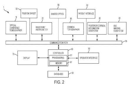
[0032] FIG. 2 is a block diagram of a system including an optical
measurement
instrument, and a position of an eye relative to the system according to one
or more
embodiments described herein which may be used by the optical measurement.
[0033] FIGs. 3A and 3B illustrate together an assembly illustrating a
suitable
configuration and integration of an optical coherence tomographer subsystem, a
wavefront
aberrometer subsystem, a corneal topographer subsystem, an iris imaging
subsystem, a
fixation target subsystem according to a non-limiting embodiment of the
present invention.
[0034] FIG. 4 is a block diagram of an OCT assembly according to many
embodiments of
the present invention.
[0035] FIG. 5 is a schematic drawing of a human eye.
CA 02991484 2018-01-05
WO 2017/019117
PCT/US2015/065713
[0036] FIG. 6A illustrates a preferred scanning region for the OCT
subsystem according
to many embodiments of the present invention.
[0037] Fig. 6B shows a representative graph of an intensity of an OCT
signal of an OCT
subsystem 190 according to many embodiments as a function of depth along the
axis defining
the axial length of the eye.
[0038] Fig. 6C shows a cross-section of an eye obtained by an optical
measurement
system of the present invention using an OCT subsystem according to the
present invention
[0039] FIG. 7 is a 3-dimensional representation of an anterior portion of
an eye obtained
using the optical measurement system according to many embodiments.
[0040] FIG. 8 is a flowchart of an example embodiment of a method for
performing
cataract diagnostics for an eye with an optical measurement instrument
according to one
embodiment described herein, including wavefront aberrometry, corneal
topography and
OCT measurements at various locations with the eye along the axial length of
the eye.
[0041] FIG. 9 is a flowchart of another example embodiment of a method for
performing
cataract diagnostics for an eye with an optical measurement instrument.
[0042] FIG. 10 is a flowchart of another example embodiment of a method for
performing
cataract diagnostics for an eye with an optical measurement instrument in
which OCT
measurements and iris imaging may be performed simultaneously.
[0043] FIG. 11 is a flowchart of yet another example embodiment of a method
for
performing cataract diagnostics for an eye with an optical measurement
instrument in which
OCT measurements and iris imaging may be performed simultaneously.
[0044] FIG. 12 illustrates another embodiment of a suitable configuration
and integration
of an optical coherence tomographer subsystem, a wavefront aberrometer
subsystem, a
corneal topographer subsystem, an iris imaging subsystem, a fixation target
subsystem and a
posterior corneal astigmatism subsystem according to a non-limiting embodiment
of the
present invention.
11
CA 02991484 2018-01-05
WO 2017/019117
PCT/US2015/065713
[0045] FIG. 13A illustrates an image obtained from a near detector of a
posterior conical
astigmatism subsystem according to a non-limiting embodiment of the present
invention.
FIG. 13B illustrates an image obtained from a far detector of an a posterior
corneal
astigmatism assembly according to a non-limiting embodiment of the present
invention.
[0046] FIG. 14 illustrates an alternate having near and far detectors that
can be used to
determine a total conical astigmatism.
[0047] FIG. 15 shows the far and near detectors operating as a separate
system to
determine a total corneal astigmatism.
[0048] FIG. 16 shows an embodiment of FIG. 15 in which a corneal topographer
has been
added.
DETAILED DESCRIPTION
[0049] Exemplary embodiments of optical measurement systems and methods for
cataract
diagnostics to illustrate various aspects and advantages of these devices and
methods are
described below. It should be understood, however, that these devices and
methods involve
principles that can be employed in a variety of other contexts, and therefore,
the novel
devices and method disclosed and claimed here should not be construed as being
limited to
the examplary embodiments described below.
[0050] As shown in Figures 1A-1C, an optical measurement system 1,
according to many
embodiments, is operable to provide for a plurality of measurements of the
human eye,
including measurements of the cornea, the lens capsule, the lens and the
retina. The main
unit 2 comprises a base 3 and includes many primary subsystems of many
embodiments of
the system 1. For example, externally visible subsystems include a touch-
screen display
control panel 7, a patient interface assembly 4 and a joystick 8.
[0051] The patient interface 4 preferably includes one or more structures
configured to
hold a patient's head in a stable, immobile and preferably comfortable
position during the
diagnostic measurements while also maintaining the eye of the patient in a
suitable alignment
with the diagnostic system. In a particularly preferred embodiment, the eye of
the patient
12
CA 02991484 2018-01-05
WO 2017/019117
PCT/US2015/065713
remains in substantially the same position relative to the diagnostic system
for all diagnostic
and imaging measurements performed by the system 1.
[0052] In one embodiment, the patient interface includes a chin support 6
and/or a
forehead rest 5 configured to hold the head of the patient in a single,
uniform position
suitably aligned with respect to the system 1 throughout the diagnostic
measurement. As
shown in Fig. IC, the optical measurement system 1 is preferably disposed so
that the patient
may be seated in a patient chair 9. The patient chair 9 can be configured to
be adjusted and
oriented in three axes (x, y, and z) so that the patent's head can be at a
suitable height and
lateral position for placement on the patient interface.
[0053] In many embodiments, the system 1 may include external communication
connections. For example, the system 1 can include a network connection (e.g.,
an RJ45
network connection) for connecting the system 1 to a network. The network
connection can
be used to enable network printing of diagnostic reports, remote access to
view patient
diagnostic reports, and remote access to perform system diagnostics. The
system 1 can
include a video output port (e.g., HDMI) that can be used to output video of
diagnostic
measurements performed by the system 2. The output video can be displayed on
an external
monitor for, for example, viewing by physicians or users. The output video can
also be
recorded for, for example, archival purposes. The system 2 can include one or
more data
output ports (e.g., USB) to enable export of patient diagnostic reports to,
for example, a data
storage device or a computer readable medium, for example a non-volatile
computer readable
medium, coupled to a laser cataract surgery device for use of the diagnostic
measurements in
conducting laser cataract surgeries. The diagnostic reports stored on the data
storage device
or computer readable medium can then be accessed at a later time for any
suitable purpose
such as, for example, printing from an external computer in the case where the
user without
access to network based printing or for use during cataract surgery, including
laser cataract
surgery.
[0054] FIG. 2 is a block diagram of a system including an optical
measurement instrument
1 according to one or more embodiments described herein. Optical measurement
instrument
1 includes: an optical coherence tomographer (OCT) subsystem 10, a wavefront
aberrometer
subsystem 20, and a corneal topographer subsystem 30 for measuring one or more
13
CA 02991484 2018-01-05
WO 2017/019117
PCT/US2015/065713
characteristics of a subject's eye. Optical measurement instrument 1 may
further include an
iris imaging subsystem 40, a fixation target subsystem 50, a controller 60,
including one or
more processor(s) 61 and memory 62, a display 70 and an operator interface 80.
Optical
measurement instrument 1 further includes a patient interface 4 for a subject
to present his or
her eye for measurement by optical measurement instrument 1.
[0055] The optical coherence tomography subsystem 10 is configured to
measure the
spatial disposition (e.g., three-dimensional coordinates such as X, Y, and Z
of points on
boundaries) of eye structures in three dimensions. Such structure of interest
can include, for
example, the anterior surface of the cornea, the posterior surface of the
cornea, the anterior
portion of the lens capsule, the posterior portion of the lens capsule, the
anterior surface of
the crystalline lens, the posterior surface of the crystalline lens, the iris,
the pupil, the limbus
and/or the retina. The spatial disposition of the structures of interest
and/or of suitable
matching geometric modeling such as surfaces and curves can be generated
and/or used by
the controller for a number of purposes, including, in some embodiment to
program and
control a subsequent laser-assisted surgical procedure. The spatial
disposition of the
structures of interest and/or of suitable matching geometric modeling can also
be used to
determine a wide variety of parameters.
[0056] As a non-limiting example, the system 1 can be configured to use a
swept source
OCT imaging system employing wavelengths of around 1060 nm with an 8 mm scan
depth.
The spatial disposition of the eye structures using optical coherence
tomography should
generally be measured while the patient is engaged with patient interface 4.
The OCT scan
depth is preferably between 8 and 50 mm, and the scan depth is preferably
greater than about
24 mm or even 30 mm to achieve a full eyescan depth. The swept source
wavelengths can be
centered at wavelengths from 840 nm to 1310 nm.Optical coherence tomographer
subsystem
is only one example of an eye structure imaging subsystem which may be
employed in
optical measurement instrument 1. In other embodiments, a different eye
structure imaging
subsystem may be employed, for example a Scheimpflug imager, a fluorescence
imager, a
structured lighting imager, a wavefront tomographer, an ultrasound imager, and
a plenoptic
imager.
14
CA 02991484 2018-01-05
WO 2017/019117
PCT/US2015/065713
[0057] The wavefront aberrometer subsystem 20 is configured to measure
ocular
aberrations, preferably including low and high order aberrations, by measuring
the wavefront
emerging from the eye by, for example a Shack Hartman sensor
[0058] The corneal topographer subsystem 30 may apply any number of modalities
to
measure the shape of the cornea including one or more of a keratometry reading
of the eye, a
corneal topography of the eye, an optical coherence tomography of the eye, a
Placido style
disc topography of the eye, a reflection of a plurality of points from the
corneal topography of
the eye, a grid reflected from the cornea of the eye topography, a Hartmann-
Shack
measurement of the eye, a Scheimpflug image topography of the eye, a confocal
tomography
of the eye, a Helmholtz source topographer, or a low coherence reflectometry
of the eye. The
shape of the cornea should generally be measured while the patient is engaged
with patient
interface 4.
[0059] Fixation target system 50 is configured to control the patient's
accommodation,
because it is often desired to measure the refraction and wavefront
aberrations when eye 101
is focused at its far point
[0060] Images captured by the corneal topographer subsystem 10, the
wavefront
aberrometer 20, the optical coherence tomographer subsystem 30 or the camera
40 may be
displayed with a display of the operator interface 80 of the optical
measurement system 2 or
the display 70 of the optical measurement system, respectively. The operator
interface may
also be used to modify, distort, or transform any of the displayed images.
[0061] The shared optics 55 provide a common propagation path that is
disposed between
the patient interface 4 and each of the optical coherence tomographer (OCT)
subsystem 10,
the wavefront aberrometer subsystem 20, the corneal topographer subsystem 30,
and in some
embodiments, an optional posterior corneal astigmatism subsystem 35, an iris
imaging
subsystem40, and a fixation target subsystem 50. In many embodiments, the
shared optics 55
may comprise a number of optical elements, including mirrrors, lenses and beam
combiners
to receive the emission from the respective subsystem to the patient's eye
and, in some cases,
to redirect the emission from a patient's eye along the common propagation
path to an
appropriate director.
CA 02991484 2018-01-05
WO 2017/019117
PCT/US2015/065713
[0062] The controller 60 controls the operation of the optical measurement
instrument 1
and can receive input from any of the optical coherence tomographer (OCT)
subsystem 10,
the wavefront aberrometer subsystem 20, the conical topographer subsystem 30
for
measuring one or more characteristics of the cornea of a subject's eye, the
optional posterior
corneal astigmatism subsystem, the iris imaging subsystem 40, the fixation
target 50, the
display 70 and the operator interface 80 via the communication paths 58. The
controller 60
can include any suitable components, such as one or more processor, one or
more field-
programmable gate array (FPGA), and one or more memory storage devices. In
many
embodiments, the controller 60 controls the display 70 to provide for user
control over the
laser eye surgery procedure for pre-cataract procedure planning according to
user specified
treatment parameters as well as to provide user control over the laser eye
surgery procedure.
The communication paths 58 can be implemented in any suitable configuration,
including
any suitable shared or dedicated communication paths between the controller 60
and the
respective system components.
[0063] The operator interface 80 can include any suitable user input device
suitable to
provide user input to the controller 60. For example, the user interface
devices 80 can
include devices such as joystick 8, a keyboard or a touchscreen display 70.
[0064] Figures 3A and 3B are simplified block diagrams illustrating an
assembly 100
according to many embodiments, which can be included in the system 1. The
assembly 100
is a non-limiting example of suitable configurations and integration of the
optical coherence
tomographer (OCT) subsystem 190, the wavefront aberrometer subsystem 150, the
conical
topographer subsystem 140 for measuring one or more characteristics of a
subject's eye, an
iris imaging subsystem 40, the fixation target subsystem 180 and the shared
optics.
[0065] The shared optics generally comprise one or more components of a
first optical
system 170 disposed along a central axis 102 passing through the opening or
aperture 114 of
the structure 110. A first optical system 170 directs light from the various
light sources along
the central axis 102 towards the eye and establishes a shared or common
optical path along
which the light from the various light sources travel to the eye 101. In one
embodiment,
optical system 170 comprises a quarter wave plate 171, a first beamsplitter
172, a second
beamsplitter 173, an optical element (e.g., a lens) 174, a second lens 175, a
third beamsplitter
16
CA 02991484 2018-01-05
WO 2017/019117
PCT/US2015/065713
176, and a structure including an aperture 178. Additional optical systems may
be used in
assembly 100 to direct light beams from one or more light sources to the first
optical system
170. For example, a second optical system 160 directs light to the first
optical system 170
from the wavefront aberrometer subsystem 150 and comprises mirror 153, beam
splitter 162
and beam splitter 183, and lens 185.
[0066] Other embodiments of suitable systems for the measurement of
refractive error,
and particularly to methods and techniques for compiling a top put graphic
mapping of
refractive errors include: U.S. Patent No. 6,550,917, filed October 20, 2000,
entitled
"Dynamic Range Extension Techniques For A Wavefront Sensor Including Use In
Ophthalmic Measurement"; U.S. Patent No. 6,908,196, filed February 21, 2003,
entitled
"System And Method For Performing Optical Corrective Procedures With Real-Time
Feedback"; U.S. Patent No. 7,455,407, filed April 21, 2004, entitled "System
And Method
Of Measuring And Mapping Three Dimensional Structures"; U.S. Patent No.
7,553,022,
filed July 27, 2007, entitled "System And Method Of Measuring And Mapping
Three
Dimensional Structures"; U.S. Patent No. 7,988,292, filed May 29, 2009,
entitled "System
And Method Of Measuring And Mapping Three Dimensional Structures"; and
W02001/058339, filed February 8, 2001, entitled "Dynamic Range Extension
Techniques
For A Wavefront Sensor." These references are hereby incorporated herein by
reference in
their entirety as if fully set forth.
[0067] Other configurations of the assembly 100, such as liquid lens
configurations, may
be possible and may be apparent to a person of skill in the art.
[0068] The corneal topographer subsystem 140 comprises a structure 110
having a
principal surface 112 with an opening or aperture 114 therein; a plurality of
first (or
peripheral) light sources 120 provided on the principal surface 112 of the
structure 110; a
Helmholz light source 130; and a detector, photodetector, or detector array
141.
[0069] In one embodiment, structure 110 has the shape of an elongated oval
or "zeppelin"
with openings or apertures at either end thereof. An example of such a
structure is disclosed
in Yobani Meji'a-Barbosa et al., "Object surface for applying a modified
Hartmann test to
measure corneal topography," APPLIED OPTICS, Vol. 40, No. 31 (Nov. 1, 2001)
("Meji'a-
17
CA 02991484 2018-01-05
WO 2017/019117
PCT/US2015/065713
Barbosa"). In some embodiments, principal surface 112 of structure 110 is
concave when
viewed from the cornea of eye 101, as illustrated in FIG. IA.
[0070] In one embodiment, where principal surface 112 is concave, principal
surface 112
has the shape of a conical frustum. Alternatively, principal surface 112 may
have a shape of
hemisphere or some other portion of a sphere, with an opening or aperture
therein. Also
alternatively, principal surface 112 may have the shape of a modified sphere
or conical
frustum, with a side portion removed. Beneficially, such an arrangement may
improve the
ergonomics of assembly 100 by more easily allowing structure 110 to be more
closely located
to a subject's eye 101 without being obstructed by the subject's nose. Of
course, a variety of
other configurations and shapes for principal surface 112 are possible.
[0071] In the embodiment of FIG. IA, the plurality of first light sources
120 are provided
on the principal surface 112 of structure 110 so as to illuminate the cornea
of eye 101. In one
embodiment, light sources 122 may comprise individual light generating
elements or lamps,
such as light emitting diodes (LEDs) and/or the tips of the individual optical
fibers of a fiber
bundle. Alternatively, principal surface 112 of structure 110 may have a
plurality of holes or
apertures therein, and one or more backlight lamps, which may include
reflectors and/or
diffusers, may be provided for passing lighting through the holes to form the
plurality of first
light sources 120 which project light onto the cornea of eye 101. Other
arrangements are
possible.
[0072] Other embodiments of suitable systems include: U.S. Patent No.
8,126,246, filed
January 8, 2009, entitled "Systems And Methods For Measuring Surface Shape";
U.S. Patent
No. 8,260,024, filed January 23, 2012, entitled "Systems And Methods For
Measuring
Surface Shape"; and European Patent Application No. 20090701204, filed January
8, 2008,
entitled "Systems And Methods For Measuring Surface Shape." These references
are hereby
incorporated herein by reference in their entirety as if fully set forth.
[0073] In another embodiment, structure 110 is omitted from assembly100,
and the first
light sources 120 may be independently suspended (e.g., as separate optical
fibers) to form a
group of first light sources 120 arranged around a central axis, the group
being separated
18
CA 02991484 2018-01-05
WO 2017/019117
PCT/US2015/065713
from the axis by a radial distance defining an aperture in the group
(corresponding generally
to the aperture 114 in the structure 110 illustrated in FIG. 1A).
[0074] In operation, a ray (solid line) from one of the first light sources
120 is reflected by
the cornea and passes through optical system 170 (including aperture 178) to
appear as a light
spot on detector array 141. It will be appreciated that this ray is
representative of a small
bundle of rays that make it through optical system 170 and onto detector array
141, all of
which will focus to substantially the same location on detector array 141.
Other rays from
that first light source 120 are either blocked by the aperture 178 or are
otherwise scattered so
as to not pass through the optical system 170. In similar fashion, light from
the other first
light sources 120 are imaged onto detector array 141 such that each one of
first light sources
120 is imaged or mapped to a location on detector array 141 that may be
correlated to a
particular reflection location on the cornea of eye 101 and/or the shape of
the cornea. Thus,
detector array 141 detects the light spots projected thereon and provides
corresponding output
signals to a processor of controller 60 (Fig. 2). The processor determines the
locations and/or
shape of the light spots on detector array 141, and compares these locations
and/or shapes to
those expected for a standard or model cornea, thereby allowing the processor
of controller
60 to determine the corneal topography. Alternatively, other ways of
processing the spot
images on detector array 141 may be used to determine the corneal topography
of eye 101, or
other information related to the characterization of eye 101.
[0075] Detector array 141 comprises a plurality of light detecting elements
arranged in a
two dimensional array. In one embodiment, detector array 141 comprises such a
charge-
coupled device (CCD), such as may be found in a video camera. However, other
arrangements such as a CMOS array, or another electronic photosensitive
device, may be
employed instead. Beneficially, the video output signal(s) of detector array
141 are provided
to processor 61 which processes these output signals as described in greater
detail below.
[0076] Assembly 100 also comprises a Helmholtz light source 130 configured
according
to the Helmholtz principle. As used herein, the term "Helmholtz source" or
"Helmholtz light
source" means one or a plurality of individual light sources disposed such
that light from
each of the individual light sources passes through an optical element having
optical power,
reflects off of a reference or test object, passes through the optical
element, and is received by
19
CA 02991484 2018-01-05
WO 2017/019117
PCT/US2015/065713
a detector, wherein light from the Helmholtz source is used to determine
geometric and/or
optical information of at least a portion of a surface of the reference or
test object. In general,
it is a characteristic of Helmholtz sources that the signal at the detector is
independent of the
relative position of the test or reference object relative to the Helmholtz
source. As used
herein, the term "optical element" means an element that refracts, reflects,
and/or diffracts
light and has either positive or negative optical power.
[0077] In such embodiments, the Helmholtz light source 130 is located at
optical infinity
with respect to eye 101. The Helmholtz principle includes the use of such
infinite sources in
combination with a telecentric detector system: i.e., a system that places the
detector array at
optical infinity with respect to the surface under measurement, in addition to
insuring that the
principal measured ray leaving the surface is parallel to the optical axis of
the instrument.
The Helmholtz corneal measurement principle has the Helmholtz light source at
optical
infinity and the telecentric observing system so that detector array 141 is
also optically at an
infinite distance from the images of the sources formed by the cornea. Such a
measurement
system is insensitive to axial misalignment of the corneal surface with
respect to the
instrument.
[0078] In one embodiment, the Helmholtz light source 130 comprises a second
light
source 132 which may comprise a plurality of lamps, such as LEDs or optical
fiber tips. In
one embodiment, second light source 132 comprises an LED and a plate 133 with
plurality of
holes or apertures in a surface that are illuminated by one or more backlight
lamps with an
optical element 131, which may comprise diffusers.
[0079] In one embodiment, second light sources 132 are located off the
central optical
axis 102 of assembly 100, and light from second light sources 132 is directed
toward optical
element 171 by third beamsplitter 176.
[0080] The operation of the topographer portion of system 100 may be
conducted with the
combined use of first light source 120 and the Helmholz light source 130. In
operation,
detector array 141 detects the light spots projected thereon from both
Helmholz light source
130 (detected at a central portion of detector array 141) and first light
sources 120 (detected
at a peripheral portion of detector array 141) and provides corresponding
output signals to
CA 02991484 2018-01-05
WO 2017/019117
PCT/US2015/065713
processor. In general, the images of first light sources 120 that appear on
detector array 140
emanate from an outer region of the surface of the cornea, and the images of
Helmholz light
source 130 that appear on detector array 141 emanate from a central or
paraxial region of the
surface of the cornea. Accordingly, even though information about the central
region of the
corneal surface (e.g., surface curvature) cannot be determined from the images
of first light
sources 120 on detector array 141, such information can be determined from the
images of
Helmholz light source 130 on detector array 141. A processor of controller 60
determines the
locations and/or shapes of the light spots on detector array 141, and compares
these locations
and/or shapes to those expected based for a standard or model cornea, thereby
allowing the
processor to determine the corneal topography of eye 101. Accordingly, the
topography of
the entire corneal surface can be characterized by system 100 without a "hole"
or missing
data from the central corneal region.
[0081] A fourth light source 201 off the central axis 102 may be directed
along optical
axis 102 by mirrors 177, 179 disposed on or near the aperture 178,
perpendicular to the
optical axis 102 are configured as a pupil retroreflection illuminator. The
pupil
retroreflecton illuminator is configured to direct a disc of light toward a
patient's eye,
whereby the disc of light may be reflected from reflective surfaces within the
eye, and the
reflected light is transmitted by optical path 170 to detector 141. The pupil
retroreflection
illuminators may optionally be configured such that, when a patient's pupil is
dilated, the disc
of light from light source 201 is reflected from an implanted IOL to image the
IOL, including
any fiducial marks; if IOL is imperfectly placed, detector 141 may be used to
determine IOL
edges are decentered. Also, images from detector 141 using the pupil
retroreflection
illuminator may see folds, for instance, unfolded edge if the IOL did not
unfold properly.
[0082] The wavefront aberrometer subsystem 150 of the assembly 100
comprises a third
light source 152 providing a probe beam and a wavefront sensor 155. The
wavefront
aberrometer subsystem 150 preferably further comprises a collimating lens 154,
a polarizing
beamsplitter 156, an adjustable telescope comprising a first optical element,
lens 163 and a
second optical element, lens 164, a movable stage or platform 166, and a
dynamic-range
limiting aperture 165 for limiting a dynamic range of light provided to
wavefront sensor 155
so as to preclude data ambiguity. Light from the wavefront aberrometer
subsystem is
21
CA 02991484 2018-01-05
WO 2017/019117
PCT/US2015/065713
directed to one of the constituent optical elements of the optical system 170
disposed along a
central axis 102 passing through the opening or aperture 114 of the structure
110. It will be
appreciated by those of skill in the art that the lenses 163, 164, or any of
the other lenses
discussed herein, may be replaced or supplemented by another type of
converging or
diverging optical element, such as a diffractive optical element.
[0083] Light source 152 is preferably an 840 nm SLD (super luminescent
laser diode). An
SLD is similar to a laser in that the light originates from a very small
emitter area. However,
unlike a laser, the spectral width of the SLD is very broad, about 40 nm. This
tends to reduce
speckle effects and improve the images that are used for wavefront
measurements.
[0084] Preferably, wavefront sensor 155 is a Shack-Hartmann wavefront
sensor
comprising a detector array and a plurality of lenslets for focusing received
light onto its
detector array. In that case, the detector array may be a CCD, a CMOS array,
or another
electronic photosensitive device. However, other wavefront sensors may be
employed
instead. Embodiments of wavefront sensors which may be employed in one or more
systems
described herein are described in U.S. Pat. No. 6,550,917, issued to Neal et
al. on Apr. 22,
2003, and U.S. Pat. No. 5,777,719, issued to Williams et al. on Jul. 7, 1998,
both of which
patents are hereby incorporated herein by reference in their entirety.
[0085] The aperture or opening in the middle of the group of first light
sources 120 (e.g.,
aperture 114 in principal surface 112 of structure 110) allows system 100 to
provide a probe
beam into eye 101 to characterize its total ocular aberrations. Accordingly,
third light source
152 supplies a probe beam through a light source polarizing beam splitter 156
and polarizing
beam splitter 162 to first beamsplitter 172 of optical system 170. First
beamsplitter 172
directs the probe beam through aperture 114 to eye 101. Preferably, light from
the probe
beam is scattered from the retina of eye 101, and at least a portion of the
scattered light
passes back through aperture 114 to first beamsplitter 172. First beamsplitter
172 directs the
back scattered light back through beam splitter 172 to polarizing beamsplitter
162, mirror
153, to wavefront sensor 155.
[0086] Wavefront sensor 155 outputs signals to a processor of controller 60
which uses
the signals to determine ocular aberrations of eye 101. Preferably, processor
141 is able to
22
CA 02991484 2018-01-05
WO 2017/019117
PCT/US2015/065713
better characterize eye 101 by considering the conical topography of eye 101
measured by
the Corneal Topography Subsystem, which may also be determined by processor
141 based
on outputs of detector array 141, as explained above.
[0087] In operation of the wavefront aberrometer subsystem 150, light from
light source
152 is collimated by lens 154. The light passes through light source
polarizing beam splitter
156. The light entering light source polarizing beam splitter 156 is partially
polarized. Light
source polarizing beam splitter 156 reflects light having a first, S,
polarization, and transmits
light having a second, P, polarization so the exiting light is 100% linearly
polarized. In this
case, S and P refer to polarization directions relative to the hypotenuse in
light source
polarizing beam splitter 156.
[0088] Light from light source polarizing beam splitter 156 enters
polarizing beamsplitter
162. The hypotenuse of polarizing beamsplitter 162 is rotated 90 degrees
relative to the
hypotenuse of light source polarizing beamsplitter 156 so the light is now S
polarized relative
the hypotenuse of polarizing beamsplitter 162 and therefore the light reflects
upwards. The
light from polarizing beamsplitter 162 travels upward and passes through
toward beam
splitter 172, retaining its S polarization, and then travels through quarter
wave plate 171.
Quarter wave plate 171 converts the light to circular polarization. The light
then travels
through aperture 114 in principal surface 112 of structure 110 to eye 101.
Preferably, the
beam diameter on the cornea is between 1 and 2 mm. Then, the light travels
through the
cornea and focuses onto the retina of eye 101.
[0089] The focused spot of light becomes a light source that is used to
characterize eye
101 with wavefront sensor 155. Light from the probe beam that impinges on the
retina of eye
101 scatters in various directions. Some of the light reflects back as a semi-
collimated beam
back towards assembly 100. Upon scattering, about 90% of the light retains its
polarization.
So the light traveling back towards assembly is substantially still circularly
polarized. The
light then travels through aperture 114 in principal surface 112 of structure
110, through
quarterwave plate 171, and is converted back to linear polarization.
Quarterwave plate 171
converts the polarization of the light from the eye's retina so that it is P
polarized, in contrast
to probe beam received from third light source 150 having the S polarization.
This P
polarized light then reflects off of first beamsplitter 172, and then reaches
polarizing
23
CA 02991484 2018-01-05
WO 2017/019117
PCT/US2015/065713
beamsplitter 162. Since the light is now P polarized relative the hypotenuse
of polarizing
beamsplitter 162, the beam is transmitted and then continues onto mirror 153.
After being
reflected by mirror 153, light is sent to an adjustable telescope comprising a
first optical
element 164 and a second optical element (e.g., lens) 163 and a movable stage
or platform
166. The beam is also directed through a dynamic-range limiting aperture 165
for limiting a
dynamic range of light provided to wavefront sensor 155 so as to preclude data
ambiguity.
[0090] When wavefront sensor 155 is a Shack-Hartmann sensor, the light is
collected by
the lenslet array in wavefront sensor 155 and an image of spots appears on the
detector array
(e.g., CCD) in wavefront sensor 155. This image is then provided to a process
of the
controller 60 and analyzed to compute the refraction and aberrations of eye
101.
[0091] An OCT subsystem 190 of assembly 100 preferably comprises an OCT
assembly
191, and a third optical path 192 which directs the OCT beam of the OCT light
source to the
first optical path 170. The third optical path 192 preferably comprises a
fiber optic line 196,
for conducting the OCT beam from the OCT light source, a z-scan device 193
operable to
alter the focus of the beam in the z-direction (i.e., along the direction of
propagation of the
OCT beam) under control of the controller, and x-scan device 195, and a y-scan
device 197
operable to translate the OCT beam in the x and y directions (i.e.,
perpendicular to the
direction of propagation of the of the OCT beam), respectively, under control
of the
controller. A first set 198 of polarization controllers may optionally be
included to change a
polarization property of the OCT light source. The OCT light source and
reference arm may
be incorporated into the main unit 4 of the optical measurement instrument 1
shown in FIG.
1A. Alternatively, the OCT assembly 191 may be housed in a second unit 200 and
the OCT
beam from the OCT source may be directed from the second housing 200 to the
main unit by
optical pathway 192.
[0092] The OCT systems and methods of the present invention are preferably FD-
OCT
(Fourier domain optical coherence tomography) systems, including either an SD-
OCT
(spectral domain optical coherence tomography) system or, more preferably, an
SS-OCT
(swept source optical coherence tomography) system. In conventional FD-OCT
systems, the
interference signal is distributed and integrated over numerous spectral
wavelength intervals,
and is inverse Fourier transformed to obtain the depth-dependent reflectivity
profile of the
24
CA 02991484 2018-01-05
WO 2017/019117
PCT/US2015/065713
sample. The profile of scattering as a function of depth is referred to as an
A-scan (Axial-
scan). The beam can be scanned laterally to produce a set of A-scans that can
be combined
together to form a tomogram of the sample (a B-scan).
[0093] In an SD-OCT system, various spectral wavelength intervals of the
combined
returned light from the reference and sample arms are spatially encoded using,
for instance, a
collimator, diffraction grating, and a linear detector array. Resampling of
the data obtained
from the linear detector array is performed in order to correct for the
nonlinear spatial
mapping of wavenumbers. After resampling and subtraction of the dc background,
the depth
profile structural information is obtained by performing the inverse Fourier
transform
operation. In swept-source OCT, the broad bandwidth optical source is replaced
by a rapid-
scanning laser source. By rapidly sweeping the source wavelength over a broad
wavelength
range, and collecting all the scattering information at each wavelength and at
each position,
the composition of the collected signal is equivalent to the spectral-domain
OCT technique.
The collected spectral data is then inverse Fourier transformed to recover the
spatial depth-
dependent information.
[0094] FD-OCT suffers from an inherent sample-independent limited depth
range,
typically between 1 and 5 mm. One limitation flows from the fact that FD-OCT
extracts
depth information from the inverse Fourier transform of a spectral
interferogram. Since the
spectral interferogram can only be recorded as a real signal, its Fourier
transform is
necessarily Hermitian symmetric about the zero path length difference (ZPD)
position. As a
result, the positive and negative displacements about the ZPD cannot be
unambiguously
resolved, which gives rise to mirror image artifacts and generally halves the
useable range.
This is referred to as the complex conjugate ambiguity. Another limitation is
a sensitivity
fall-off which results in reduced sensitivity with increasing depth. Moreover,
since the signal
in OCT is derived only from backscattered photons, optical attenuation from
absorption and
scattering generally result in a useable imaging depth of about 1-4 mm.
[0095] Several "full range" OCT techniques have been developed that
eliminate the
complex conjugate artifacts to effectively double the measurement range around
the ZPD
position. These full range OCT techniques result in useable imaging depths of
up to about 5
mm or even up to about 8 mm. Suitable full range techniques include methods
that dither the
CA 02991484 2018-01-05
WO 2017/019117
PCT/US2015/065713
reference leg length (M. Wijtkowski, et al, Opt. Lett. V27, #16, pg 1415,
2002), or that
exploit phase dispersion compensation (Kottig, et al, Opt. Express V20, #22,
pg 24925, 2012)
to break the phase ambiguity.
[0096] As shown in FIG. 4, the OCT assembly 191 of OCT subsystem 190 includes
a
broadband or a swept light source 202 that is split by a coupler 204 into a
reference arm 206
and a sample arm 210. The reference arm 206 includes a module 208 containing a
reference
reflection along with suitable dispersion and path length compensation. The
sample arm 210
of the OCT assembly 191 has an output connector 212 that serves as an
interface to the rest of
the optical measurement instrument. The return signals from both the reference
and sample
arms 206, 210 are then directed by coupler 204 to a detection device 220,
which employs one
of time domain, frequency, or single point detection techniques. In FIG. 4, a
swept source
technique is used with a laser wavelength of 1060 nm swept over a range of 8-
50 mm depth.
A second set 218 of polarization controllers may be used to change a
polarization property of
the reference beam of the reference arm.
[0097] FIG. 5 is a schematic drawing of a human eye 400. In many
embodiments, a light
beam 401 from a light source enters the eye from the left of FIG. 5, refracts
into the cornea
410, passes through the anterior chamber 404, the iris 406 through the pupil,
and reaches lens
402. After refracting into the lens, light passes through the vitreous chamber
412, and strikes
the retina 476, which detects the light and converts it to an electric signal
transmitted through
the optic nerve to the brain (not shown). The vitreous chamber 412 contains
the vitreous
humor, a clear liquid disposed between the lens 402 and retina 476. As
indicated in FIG. 5,
cornea 410 has corneal thickness (CT), here considered as the distance between
the anterior
and posterior surfaces of the cornea. Anterior chamber 404 has anterior
chamber depth
(ACD), which is the distance between the anterior surface of the cornea and
the anterior
surface of the lens. Lens 402 has lens thickness (LT) which is the distance
between the
anterior and posterior surfaces of the lens. The eye has an axial length (AXL)
which is the
distance between the anterior surface of the cornea and the retina 476. Fig. 5
also illustrates
that, in many subjects the lens, including the lens capsule, may be tilted at
one or more angles
relative to the optical axis, including an angle y relative to the optical
axis of the eye.
26
CA 02991484 2018-01-05
WO 2017/019117
PCT/US2015/065713
[0098] The optical system may also be arranged so that the movement pattern
of the scan
mirrors provides a lateral motion across the retina so that the shape of the
retina may be
determined. It is of particular interest to measure the shape and location of
the depressed
region of the retina named the foveal pit. When the patient is looking
directly into the
instrument, with their line of sight aligned to the fixation target, the
foveal pit will be in
center of the OCT lateral scan. This information is beneficial in that it
informs the instrument
operator if the patient was looking directly at the target when the
measurement was made.
Retinal scans are also useful in detecting disease conditions. In some cases,
there may be an
absence of a foveal pit that also is considered an indication of a corneal
abnormality.
[0099] The average axial length of the adult human eye is about 24 mm.
Since the full
range imaging depth of the OCT measurements are only about 5 mm to 8 mm, then
OCT
scanning of the invention may provide for OCT scans at different depths of the
eye that can
be combined together to form a combined OCT image of the eye. The OCT
measurements of
the present invention preferably includes OCT imaging at various depths of the
patient's eye
for imaging 1) at least a portion of the retina, 2) at least a portion of the
anterior portion of the
eye, including at least a portion of the cornea (anterior and posterior),
iris, and lens (anterior
and posterior) , and 3) performing axial eye length measurements. In a
preferred
embodiment, the coherence depth range of the OCT system to exceed the length
of the eye so
that the entire length of the eye may be measured at one time without the need
to combine
different depth ranges. In that case, however, it may still be beneficial to
change the focus of
the beam entering into the eye so that the strength of the captured light may
be optimized for
resolving different regions of the eye. For example, the beam may be focused
on the anterior
portion of the eye for increased resolution in that region while
simultaneously a measurement
of the length of the whole eye is being made. Similarly, the beam may be
focused on the
retina for high resolution measurements in that section while simultaneously
the whole eye
length is being measured. For both situations, the scan geometry may be
arranged so that
while the beam is scanning across on region, the beam is substantially
stationary on the other
region so that even though the beam is defocused there, the return signal
strength from the
defocus region is sufficient to provide a strong signal.
27
CA 02991484 2018-01-05
WO 2017/019117
PCT/US2015/065713
[00100] FIGS. 6A-6C illustrate various aspects of the OCT subsystem 190
according to
various aspects of the present invention. Fig. 6A illustrates a preferred
scanning region for
the OCT subsystem according to many embodiments of the present invention. The
scanning
region may be defined from starting point 301 to ending point 302 at the
anterior portion of
the eye extending in a direction transverse the direction of propagation of
the OCT beam and
also extending in a direction parallel to an axis defining the axial length of
the eye to the
posterior portion 304 of the eye. The lateral scanning region should generally
be sufficiently
large in the lateral direction to permit imaging of the central portion of the
cornea, at least a
portion of the iris, at least a portion of the lens and at least of the
retina. It should be noted
that a region 303 between the posterior portion of the lens and the surface of
the retina may
optionally not be scanned by OCT subsystem 190 because the portion 330 does
not contain
anatomical structure for 3D analysis.
[00101] Fig. 6B shows a representative graph of an intensity of an OCT signal
of an OCT
subsystem 190 according to many embodiments as a function of depth along the
axis defining
the axial length of the eye. The graph generally exhibits approximately four
peaks having a
complex structure: (1) a peak 310 having a doublet-like structure and
generally
corresponding to a location of the cornea; (2) a peak 320 having a doublet-
like structure and
generally corresponding to a location of an anterior surface of the lens; (3)
a peak 330 having
a complex structure generally corresponding to a location of a posterior
surface of the lens;
and (4) a peak 340 generally corresponding to a location of a retina. A
distance between peak
310 and peak 340 can be used to calculate the axial length (AL) of the eye.
Preferably, an
OCT scan by OCT subsystem 190, including both an A-scan and B-scan, is
conducted at least
one location in the anterior portion of the eye (e.g., a location of a cornea,
a location of an
anterior surface of a lens and/or a location of a posterior surface of the
lens) and at least one
location in the posterior portion of the eye (e.g., at a location of a
retina). In some
embodiments, an OCT scan by the OCT subsystem 190, including both an A-Scan
and a B-
scan is performed at a location corresponding to each of a location of the
cornea, a location of
an anterior surface of the lens, a location of a posterior surface of the
lens, and a location
corresponding to a retina.
28
CA 02991484 2018-01-05
WO 2017/019117
PCT/US2015/065713
[00102] It should be noted that because the OCT subsystem 190 provides for the
detection
of various structures of the eye, including a location of the cornea, the OCT
subsystem 190
may be used as a ranging system to precisely align the patient in relation to
the optical
measurement system 1 of the present invention. The use of the OCT as a ranging
system can
significantly improve accuracy of corneal topography measurements, including
keratometry
measurements, which are sensitive to misalignment of the corneal structures.
[00103] Fig. 6C shows a cross-section of an eye obtained by an optical
measurement
system of the present invention using an OCT subsystem according to the
present invention.
[00104] FIG. 7 shows a 3 dimensional view of an eye obtained by an optical
measurement
system of the present invention using an OCT subsystem according to the
present invention.
Fig. 7 evidences that the OCT subsystem of the present invention is operable
to obtain
biometry measurements according to the present invention, including the
central corneal
thickness (CCT), the anterior chamber depth (ACD), the radius of curvature of
the anterior
cornea (ROCAc), the radius of curvature of the Posterior cornea (ROCpc) and
the Radius of
curvature of the axial length (ROCAO=
[00105] Preferably, the OCT subsystem 190 provides sufficiently resolved
structural
information to provide a structural assessment that may provide a user with an
indication of
suitability of a particular patient for a laser cataract procedure. In one
embodiment, an OCT
scan performed by the OCT subsystem 190 at or near the retina (i.e., a retina
scan) is
sufficiently resolved to identify the foveal pit location and depth, wherein a
lack of
depression indicates an unhealthy retina.
[00106] In another embodiment, the optical measurement instrument 1 of the
present
invention provides one or more measurements sufficient to provide an
assessment of the tear
film of a patient. In one embodiment, the tear film assessment comprises a
comparison of a
wavefront aberrometry map and a corneal topography map or OCT map of the
patient's eye,
by, for instance, determining the irregular features in either the wavefront
aberrometery or
corneal topopgraphy maps This can be achieved by first fitting the surface
(either wavefront
or topography) to smooth functions such as Zemike or Taylor polynomials, and
then
subtracting this smooth surface from the original surface data. The resulting
map is the
29
CA 02991484 2018-01-05
WO 2017/019117
PCT/US2015/065713
residual of what does not fit a smooth surface and is highly correlated with
the tear film
(Haixia Liu, Larry Thibos, Carolyn G. Begley, Arthur Bradley, "MEASUREMENT OF
THE
TIME COURSE OF OPTICAL QUALITY AND VISUAL DETERIORATION DURING
TEAR BREAK-UP," Investigative Ophthalmology & Visual Science, June 2010, Vol.
51,
No. 6). A determination of whether the tear film is broken (if not smooth); an
assessment of
the tear film, including tear film breakup, can also be obtained by reviewing
the shape of
spots on the topographer. For instance, a finding or indication that the tear
film is disrupted,
or broken, may be based upon the shape of a spot in that, if the spots are not
round, and have,
for instance, an oblong or broken up shape, it indicates that tear film is
disrupted. The
existence of such a disrupted tear film may indicate that K value, and other
ocular
measurements may not be reliable. Further indications of the state of the tear
film may be
made by comparing the OCT and the topographer, or wavefront data (See Kob -
Simultaneous Measurement of Tear Film Dynamics IOVS, July 2010, Vol. 51, No.
7).
[00107] In operation, as shown in Fig. 3A, after exiting connector 212, the
OCT beam 214
is collimated, preferably using a collimating optical fiber 196. Following
collimating fiber
196 the OCT beam 214 is directed to an z-scan device 193 operable to change
the focal point
of the OCT beam in a z-direction, and x- and y-scan devices 195 and 197, which
are operable
to scan the OCT beam in x and y-directions perpendicular to the z-direction.
[00108] Following the collimating optical fiber 196, the OCT beam 214
continues through
a z-scan device 193, 194. Preferably, the z-scan device is a Z telescope 193,
which is
operable to scan focus position of the OCT beam 214 in the patient's eye 101
along the Z
axis. For example, the Z-telescope can include a Galilean telescope with two
lens groups
(each lens group includes one or more lenses). One of the lens groups moves
along the Z
axis about the collimation position of the Z-telescope 193. In this way, the
focus position in
the patient's eye 101 moves along the Z axis. In general, there is a
relationship between the
motion of lens group and the motion of the focus point. The exact relationship
between the
motion of the lens and the motion of the focus in the z axis of the eye
coordinate system does
not have to be a fixed linear relationship. The motion can be nonlinear and
directed via a
model or a calibration from measurement or a combination of both.
Alternatively, the other
lens group can be moved along the Z axis to adjust the position of the focus
point along the Z
CA 02991484 2018-01-05
WO 2017/019117
PCT/US2015/065713
axis. The Z-telescope 84 functions as a z-scan device for changing the focus
point of the
OCT beam 214 in the patient's eye 101. The Z-scan device can be controlled
automatically
and dynamically by the controller 60 and selected to be independent or to
interplay with the
X and Y scan devices described next.
[00109] After passing through the z-scan device, the OCT beam 214 is incident
upon an X-
scan device 195, which is operable to scan the OCT beam 214 in the X
direction, which is
dominantly transverse to the Z axis and transverse to the direction of
propagation of the OCT
beam 214. The X-scan device 195 is controlled by the controller 60, and can
include suitable
components, such as a lens coupled to a MEMS device, a motor, galvanometer, or
any other
well-known optic moving device. The relationship of the motion of the beam as
a function of
the motion of the X actuator does not have to be fixed or linear. Modeling or
calibrated
measurement of the relationship or a combination of both can be determined and
used to
direct the location of the beam.
[00110] After being directed by the X-scan device 196, the OCT beam 214 is
incident upon
a Y scan device 197, which is operable to scan the OCT beam 214 in the Y
direction, which
is dominantly transverse to the X and Z axes. The Y-scan device 197 is
controlled by the
controller 60, and can include suitable components, such as a lens coupled to
a MEMS
device, motor, galvanometer, or any other well-known optic moving device. The
relationship
of the motion of the beam as a function of the motion of the Y actuator does
not have to be
fixed or linear. Modeling or calibrated measurement of the relationship or a
combination of
both can be determined and used to direct the location of the beam.
Alternatively, the
functionality of the X-Scan device 195 and the Y-Scan device 197 can be
provided by an
XY-scan device configured to scan the OCT bean 214 in two dimensions
transverse to the Z
axis and the propagation direction of the OCT beam 214. The X-scan and Y scan
devices
195, 197 change the resulting direction of the OCT beam 214, causing lateral
displacements
of OCT beam 214 located in the patient's eye 101.
[00111] The OCT sample beam 214 is then directed to beam splitter 173 through
lens 175
through quarter wave plate 171 and aperture 114 and to the patient eye 101.
Reflections and
scatter off of structures within the eye provide return beams that retrace
back through the
patient interface quarter wave plate 171, lens 175, beam splitter 173, y-scan
device 197, x-
31
CA 02991484 2018-01-05
WO 2017/019117
PCT/US2015/065713
scan device 195, z-scan device 193, optical fiber 196 and beam combiner 204
(FIG. 3), and
back into the OCT detection device 220. The returning back reflections of the
sample arm
201 are combined with the returning reference portion 206 and directed into
the detector
portion of the OCT detection device 220, which generates OCT signals in
response to the
combined returning beams. The generated OCT signals that are in turn
interpreted by the
controller 60 to determine the spatial disposition of the structures of
interest in the patient's
eye 101. The generated OCT signals can also be interpreted by the controller
to determine
the spatial disposition of the structures of interest in the patient's eye
101. The generated
OCT signals can also be interpreted by the control electronics to align the
position and
orientation of the patient eye within the patient interface. As the OCT
information can be
obtained relatively rapidly (B-scans at 200-500 scans per second) this can be
used to provide
tracking information to the patient alignment system. That is, the center
offset of the corneal
vertex can by obtained in x, y and z by determining the highest point in the x
and y-slices,
and then determining an offset from the desired alignment point. The z value
is the
difference between the highest corneal point and the desired z location. This
information can
be fed to an XYZ tracker that aligns the systems either by moving the
instrument, the
patient's head, or internal mirrors and optical elements in the instrument.
[00112] The quarter wave plate 171 described above has the effect that light
returning into
the instrument will have its polarization rotated by ninety degrees relative
to the outgoing
polarization. This can result in a situation that the OCT reference beam and
signal light
incident on the detector 220 will have nearly orthogonal polarizations so that
the interference
signal generated is extremely weak. One effective method to maximize the
signal strength is
to set the relevant OCT reference and sample light beams to be linearly
polarized with, for
example, a polarizing controller in both the sample arm and the reference arm.
In one such
embodiment, a first set 198 of polarization controllers (FIG. 3A), for example
a set of
polarization rotating fiber paddle adjusters on the OCT source light output,
set the
polarization of the incident light on the beamsplitter 173 to be linearly
polarized on that
surface. Further, a second set 218 of polarization controllers (FIG. 4), such
as another set of
rotating fiber paddle adjusters, are placed in the reference fiber path
leading to the detector
220. Adjustment of the polarization controllers, such as the fiber paddles,
will maximize the
signal when the reference and signal polarizations match. This allows the
system to retain the
32
CA 02991484 2018-01-05
WO 2017/019117
PCT/US2015/065713
benefits of having the quarter wave plate 171 for the wavefront sensing
portion of the
instrument while having minimal impact on the OCT signal strength.
[00113] The quarter wave plate 171 may be zero order design at either the OCT
wavelength, the wavefront sensor wavelength, or an intermediate wavelength.
Practical zero
order wave plates made of crossed crystalline quartz plates are low cost and
will behave as
nearly as ideal over the wavelength range of interests, for instance if the
center wavefront
sensor wavelength is 840 nm and the center OCT wavelength is 1060 nm. Other
alternatives
are polymer waveplates or the more expensive achromatic quarter wave plates.
[00114] The optical measurement systems according to the present invention
preferably
comprise an iris imaging subsystem 40. The imaging subsystem 40 generally
comprises an
infrared light source, preferably infrared light source 152, and detector 141.
In operation
light from the light source 152 is directed along second optical path 160 to
first optical path
170 and is subsequently directed to eye 101 as described above. Light
reflected from the iris
of eye 101 is reflected back along first optical path 170 to detector 141. In
normal use, an
operator will adjust a position or alignment of system 100 in XY and Z
directions to align the
patient according to the image detector array 141. In one embodiment of the
iris imaging
subsystem, eye 101 is illuminated with infrared light from light source 152.
In this way, the
wavefront obtained by wavefront sensor 155 will be registered to the image
from detector
array 141.
[00115] The image that the operator sees is the iris of eye 101. The cornea
generally
magnifies and slightly displaces the image from the physical location of the
iris. So the
alignment that is done is actually to the entrance pupil of the eye. This is
generally the desired
condition for wavefront sensing and iris registration.
[00116] Iris images obtained by the iris imaging subsystem may be used for
registering
and/or fusing the multiple data sets obtained by the various subsystems of the
present
invention, by methods described for instance in "Method for registering
multiple data sets,"
U.S. Patent Appl. No. No. 12/418,841, which is incorporated herein by
reference. As set forth
in Appl. No. 12/418,841, wavefront aberrometry may be fused with corneal
topography,
optical coherence tomography and wavefront, optical coherence tomography and
topography,
33
CA 02991484 2018-01-05
WO 2017/019117
PCT/US2015/065713
pachymetry and wavefront, etc. For instance, with image recognition techniques
it is possible
to find the position and extent of various features in an image. Regarding
iris registration
images, features that are available include the position, size and shape of
the pupil, the
position, size and shape of the outer iris boundary (0IB), salient iris
features (landmarks) and
other features as are determined to be needed. Using these techniques, both
patient
movement between measurements (and/or during a measurement sequence) can be
identified,
as well as changes in the eye itself (including those induced by the
measurement, such as
changes in the size of the pupil, changes in pupil location, etc.).
[00117] In many embodiments, an optical measurement system according the
present
includes a target fixation subsystem 150 (Figure 1), and an assembly 100 shown
in Figures
3A and 3B includes fixation target subsystem 180 which includes a fixation
target 182 for the
patient to view. Fixation target subsystem 180 is used to control the
patient's
accommodation, because it is often desired to measure the refraction and
wavefront
aberrations when eye 101 is focused at its far point (e.g., because LASIK
treatments are
primarily based on this). Cylindrical correction and liquid lenses for the
target path may also
be used. In the target fixation subsystem, a projection of a target, for
instance a cross-hair
pattern is projected onto the eye of the patient, the cross hair pattern being
formed by a
backlit LED and a film. An alternative embodiment is to provide a video target
that allows
the projection of letters, charts, pictures or movies. One method to control
accommodation is
to provide the patient with a task "click a button each time you recognize a
real word" or
"click a button each time the target includes the color purple" in order to
insure that the
subject is really looking and concentrating on the target.
[00118] In operation, light originates from the light source 152 or,
alternatively, from video
target backlight 182 and lens 186. Lens 185 collects the light and forms an
aerial image T2.
This aerial image is the one that the patient views. The patient focus is
maintained on aerial
image 182 during measurement so as to maintain the eye in a fixed focal
position.
[00119] The operating sequence the optical measurement system and methods of
the
present is not particularly limited. A scan of the patient's eye may comprise
one or more of a
wavefront aberrometry measurement of a patient's eye utilizing the wavefront
aberrometry
subsystem, a corneal topography measurement of a patient's eye and an OCT scan
of the
34
CA 02991484 2018-01-05
WO 2017/019117
PCT/US2015/065713
patient's eye using the OCT substystem, wherein the OCT scan includes a scan
at each or one
or more locations within the eye of the patient. These locations of the OCT
scan may
correspond to the location of the cornea, the location of the anterior portion
of the lens, the
location of the posterior portion of the lens and the location of the retina.
In a preferred
embodiment, the operating sequence includes each of a wavefront aberrometry
measurement,
a corneal topography measurement and an OCT scan, wherein the OCT scan is
taken at least
at the retina, the cornea and one of anterior portion of the patient's lens.
Preferably, an iris
image is taken simultaneously with or sequentially with an each of
measurements taken with
wavefront aberrometry subsystem the Corneal Topography Subsystem and the OCT
subsystem, including an iris image take simultaneously with or sequentially
with the location
of each OCT scan. This results in improved accuracy in the 3-dimensional
modeling of the
patient's eye by permitting the various data sets to be fused and merged into
a 3-dimensional
model.
[00120] Figure 8 shows one embodiment of an operating sequence and method in
which
wavefront aberrometry measurements, corneal topography measurements and OCT
measurements are all taken. The optical measurement apparatus, including the
method of
Figure 8 may be used preoperatively, intra-operatively and/or postoperatively.
In the method
of Figure 8, a step 501 comprises aligning the optical measurement system to
the eye of the
patent. A step 505 comprises activating the Target Fixation subsystem for
patient fixation on
target. A step 510 comprises activating the wavefront aberrometer subsystem
such that the
wavefront aberrometer light source 510 is activated and the eye refraction is
measured via the
wavefront sensor. A step 515 comprises activating the target fixation system
to move the
target to an optimum position and activate the wavefront aberrometer subsystem
such that the
wavefront aberrometer light source 152 is activated and the eye refraction is
measured via the
wavefront sensor 155. A step 520 comprises obtaining an iris image using Iris
Imaging
Subsystem while infrared light source 152 is operating. A step 525 comprises
operating the
z-scan device to set OCT scan location at or near cornea, and performing an
OCT Scan with
the OCT Subsystem. A step 530 comprises operating the z-scan device to set the
OCT
location at a location at or near the lens anterior and performing an OCT Scan
with the OCT
Subsystem. A step 535 comprises operating the z-scan device to set the OCT
location at a
location at or near the lens posterior and performing an OCT Scan with the OCT
Subsystem.
CA 02991484 2018-01-05
WO 2017/019117
PCT/US2015/065713
A step 540 comprises operating the X-scan device and Y-scan device so no light
from OCT
reaches detector 141. A step 545 comprises obtaining an iris image using the
his Imaging
Subsystem while the infrared light source 152 flashes. A step 550 comprises
obtaining an iris
image using the Iris Imaging Subsystem while the light sources 120 and
helmholz source
flash. A step 550 comprises measuring the corneal topography with the Corneal
Topography
Subsystem. A step 555 comprises operating the z-scan device to set the OCT
location at a
location at or near the retina and performing an OCT Scan with the OCT
Subsystem. A step
560 comprises operating the X-scan device and Y-scan device so no light from
OCT reaches
detector 141. An optional step 565 comprises measure corneal topography with
Corneal
Topography Subsystem, which may provide for an improved 3D model of the
patient eye.
An optional step 570 comprises obtaining an iris image using Iris Imaging
Subsystem (for 3D
model).
[00121] Figure 9 shows one embodiment of an operating sequence and method in
which no
wavefront aberrometry measurements are taken. The optical measurement
apparatus,
including the method of Figure 8 may be used preoperatively, intra-operatively
and/or
postoperatively. In the embodiment of Figure 9, a step 601 comprises aligning
the optical
measurement system to the eye of the patent. A step 605 comprises activating
the Target
Fixation subsystem for patient fixation on target. A step 610 comprises
obtaining an iris
image using Iris Imaging Subsystem while infrared light source 152 is
operating. A step 615
comprises operating the z-scan device to set OCT scan location at or near
cornea, and
performing an OCT Scan with the OCT Subsystem. A step 620 comprises operating
the z-
scan device to set the OCT location at a location at or near the lens anterior
and performing
an OCT Scan with the OCT Subsystem. A step 625 comprises operating z-scan
device to set
the OCT location at a location at or near the lens posterior and performing an
OCT Scan with
the OCT Subsystem. A step 530 comprises operating the X-scan device and Y-scan
device
so no light from OCT reaches detector 141. A step 635 comprises obtaining an
iris image
using the his Imaging Subsystem while the infrared light source 152 flashes. A
step 640
comprises measuring the corneal topography with the Corneal Topography
Subsystem. A
step 645 comprises operating the z-scan device to set the OCT location at a
location at or near
the retina and performing an OCT Scan with the OCT Subsystem. A step 650
comprises
operating the X-scan device and Y-scan device so no light from OCT reaches
detector 141.
36
CA 02991484 2018-01-05
WO 2017/019117
PCT/US2015/065713
An optional step 655 comprises measuring corneal topography with Corneal
Topography
Subsystem, which may provide for an improved 3D model of the patient eye. An
optional
step 660 comprises obtaining an iris image using Iris Imaging Subsystem.
[00122] Figure 10 shows an embodiment of an operational sequence and method in
which
OCT measurements utilizing the OCT subsystem and his images using the iris
imaging
subsystem may be taken simultaneously in order to improve three dimensional
modeling of
the patient's eye and improved iris registration of the measurement data sets.
The operational
sequence of Figure 10 may be applied to or incorporated into either of the
operational
sequences and methods of Figures 8 or 9 as would be readily understood by
those ordinarily
skilled. In order to effectuate the operating sequence and method of Figure
10, a lens is
inserted into optical path 170 between beam splitter 173 and detector 141. The
inserted lens
is selected to preferentially pass infrared light used for iris imaging but to
block an OCT
beam from the OCT light source from reaching detector 141. In this
configuration, OCT
measurements and iris images may be taken simultaneously. Further, in the
embodiment of
Fig. 10 a regular speed global shutter iris camera is used operating at 12
frames/second. The
operating sequence and method of Figure 10 may be used preoperatively, intra-
operatively
and/or postoperatively.
[00123] In the embodiment of Figure 10, a step 701 comprises aligning the
optical
measurement system to the eye of the patent. A step 705 comprises activating
the Target
Fixation subsystem for patient fixation on target. A step 710 comprises
obtaining an iris
image using Iris Imaging Subsystem while infrared light source 152 is
operating. A step 715
comprises obtaining an iris image using Iris Imaging Subsystem while corneal
topography
light sources 120 and Helmholz light source 132 are operating. A step 720
comprises
operating the z-scan device to set OCT scan location at or near cornea, and
performing an
OCT Scan with the OCT Subsystem. A step 725 comprises operating the z-scan
device to
set the OCT location at a location at or near the lens anterior and performing
an OCT Scan
with the OCT Subsystem. A step 730 comprises operating z-scan device to set
the OCT
location at a location at or near the lens posterior and performing an OCT
Scan with the OCT
Subsystem. A step 735 comprises obtaining an iris image using his Imaging
Subsystem
while infrared light source 152 is operating. A step 740 comprises obtaining
an iris image
37
CA 02991484 2018-01-05
WO 2017/019117
PCT/US2015/065713
using Iris Imaging Subsystem while corneal topography light sources 120 and
Helmholz light
source 132 are operating. A step 745 comprises operating the z-scan device to
set the OCT
location at a location at or near the retina and performing an OCT Scan with
the OCT
Subsystem. A step 750 comprises obtaining an iris image using his Imaging
Subsystem
while corneal topography light sources 120 and Helmholz light source 132 are
operating. A
step 755 comprises obtaining an iris image using Iris Imaging Subsystem while
infrared light
source 152 is operating.
[00124] Figure 11 shows another embodiment of an operational sequence and
method in
which OCT measurements utilizing the OCT subsystem and his images using the
iris
imaging subsystem may be taken simultaneously in order to improve three
dimensional
modeling of the patient's eye and improved iris registration of the
measurement data sets.
The operational sequence of this embodiment may be applied to or incorporated
into either of
the operational sequence and methods of Figures 8 or 9 as would be readily
understood by
those ordinarily skilled. As with the method of Fig. 10, in order to
effectuate the operating
sequence and method of Figure 11, a lens is inserted into optical path 170
between beam
splitter 173 and detector 141. The inserted lens is selected to preferentially
pass infrared light
used for iris imaging but to block an OCT beam from the OCT light source from
reaching
detector 141. In this configuration, OCT measurements and iris images may be
taken
simultaneously. Further, in the embodiment of Fig. 10 a high speed global
shutter iris
camera, or fast frame rate, is used operating at 60 frames/second. Under the
fast frame rate
conditions of this embodiment, an infrared illumination source, such as a
wavefront
aberrometry source, may be used with a one or more second light sources, such
as a
combination of the corneal topography sources 120 and the Helmholz source, to
alternately
illuminate a patient's eye repeatedly at short intervals (i.e., alternative
short flashes). Under
these conditions, the iris imaging subsystem may be synched to the flash from
each source so
as to capture iris images under both illumination conditions. The operating
sequence and
method of Figure 11 may be used preoperatively, intra-operatively and/or
postoperatively.
[00125] In the embodiment of Figure 11, a step 801 comprises aligning the
optical
measurement system to the eye of the patient. A step 805 comprises activating
the Target
Fixation subsystem for patient fixation on target. A step 810 comprises
obtaining an iris
38
CA 02991484 2018-01-05
WO 2017/019117
PCT/US2015/065713
image using Iris Imaging Subsystem while infrared light source 152 is
operating and
obtaining an iris image using his Imaging Subsystem while corneal topography
light sources
120 and Helmholz light source 132 are operating. This is done by alternately
operating the
infrared light source and a combination of the conical topography/Helmholz
light sources so
as to alternately illuminate the patient's eye with the infrared light source
and the combined
light sources, preferably at a rate that a patient's eye cannot resolve the
"flicker." In this step,
the Iris imaging subsystem is in synch with the respective illuminate lights.
A step 815
comprises operating the z-scan device to set OCT scan location at or near
cornea, and
performing an OCT Scan with the OCT Subsystem. A step 820 comprises operating
the z-
scan device to set the OCT location at a location at or near the lens anterior
and performing
an OCT Scan with the OCT Subsystem. A step 825 comprises operating z-scan
device to set
the OCT location at a location at or near the lens posterior and performing an
OCT Scan with
the OCT Subsystem. A step 830 comprises operating the z-scan device to set the
OCT
location at a location at or near the retina and performing an OCT Scan with
the OCT
Subsystem. A step 835 comprises obtaining an iris image using his Imaging
Subsystem
while infrared light source 152 is operating and obtaining an iris image using
Iris Imaging
Subsystem while corneal topography light sources 120 and Helmholz light source
132 are
operating as described above for Step 810.
[00126] Placido style-based or spot-based topographers work by shining a
pattern of light
on the eye. If a patient is looking directly into an instrument, there is
often a portion of the
cornea that is not illuminated because of a shadow created by the patient's
nose. One
solution employed by some topographers is to have the patient look into the
instrument with
about a degree angle. This simply moves the nose relative to the instrument so
there is no
shadow on the cornea. To aid in orienting the patients head properly, the chin
rest often has
two ten degree indentations, one for the left eye and the other for the right
eye. This solution
works well for an instrument that is dedicated to only measuring corneal
topography. But it
has drawbacks with an instrument that is meant to measure more characteristics
of the eye
such as refractive state , gaze angle, angle kappa and iris features. In an
integrated system
that includes a corneal topographer and an OCT system, it is advantageous to
combine the
results from both into a single display map of corneal topography. In the
region where the
topographer image is illuminated, the highest accuracy characterization of the
optical surface
39
CA 02991484 2018-01-05
WO 2017/019117
PCT/US2015/065713
may be obtained. Then in extended regions where the OCT elevation data is
available, that
information can be used in the same map. The combined map may also include an
annular
zone that extends beyond the roughly circular region of corneal topographer
coverage,
[00127] Several methods may be used to join the OCT data set to the placido
style spot
based topographer data set. One method is to use as a reference an image taken
with the
same camera as the topographer but with the topographer pattern turned off and
simple
illumination from one or a few light sources turned on. Another is to have the
scan mirrors
from the OCT pause momentarily at certain locations so light from the OCT is
bright enough
to be seen on the camera. Another is to perform XY polynomial shape fits on
both OCT and
topographer data sets and join those together in best fit method. In that case
the OCT data
that is collected in the same region as the topographer data is being used to
assist in
performing the match. Another more direct method is simply to have done a step
at a
previous point in time, for instance during manufacture and calibration, where
the
relationship between the OCT scan pattern and image locations on the camera
have been
established. This may be done simply by placing a reflective target at the
measurement plane
and recording images of the scan pattern of the OCT. In theory, in an ideal
system the
entirety of the OCT beam would be going into the OCT measurement optical path,
but in
practice it is found that a very small amount of light leakage at the OCT
wavelength that
reaches the camera is sufficient to perform such a calibration.
[00128] FIG. 12 is a simplified block diagram illustrating an assembly 100
according to
another embodiment of the present invention that further comprises a posterior
corneal
astigmatism assembly 900. Except for the inclusion of the posterior corneal
astigmatism
assembly, the other components may be same as are described with respect to
FIGS. 1-11.
Specifically, the assembly 100 according to many embodiments includes an the
optical
coherence tomographer (OCT) subsystem 190, the wavefront aberrometer subsystem
150, the
corneal topographer subsystem 140 for measuring one or more characteristics of
a subject's
eye, an iris imaging subsystem 40, the fixation target subsystem 180 and the
shared optics 50
as described above with respect to FIGS 1-11.
[00129] The posterior corneal astigmatism assembly 900 generally comprises a
first
detector 910 at a first effective optical distance D1 from a predetermined
location anterior or
CA 02991484 2018-01-05
WO 2017/019117
PCT/US2015/065713
posterior to the patient's cornea and a second detector 920 at a second
effective optical
distance D2 from the predetermined location. In many embodiments ,the
effective optical
distance D1 is less than the effective optical distance D2. In these
embodiments, the first
detector 910 is referred to as the near detector and the second detector 920
is referred to as
the far detector. The first and second detectors 910, 920 are generally
detectors suitable for
detecting visible and/or infrared light, such as a CCD, and more specifically
capable of
detecting light reflected from the patient's eye.
[00130] Without being limited to theory, the posterior corneal astigmatism
assembly 900 is
based on the principle that the amount of distortion of an object by a toric
lens that is detected
by a detector depends on a distance of the detector from the toric lens. More
specifically, the
amount of detected distortion increases with increasing distance from the
toric lens. For
example, when a toric spectacle lens is placed in front of a patient's eye, it
introduces
distortion into the image the patient perceives. Because of the action of the
toric lens in these
instances, a patient may perceive a physically round object as a distorted
oval with the axis of
the oval pointing along the axis of the astigmatism. Further, the closer the
lens is to the eye,
the less distortion the patient perceives. For instance, a toric contact lens
on a patient's eye
may cause almost no perceivable distortion. FIG. 13A illustrates an image
detected of a
round object by a detector with a toric lens disposed between the round object
and the
detector when the detector is near the toric lens. FIG. 13B illustrates an
image detected of a
round object by a detector with a toric lens disposed between the round object
and the
detector when the detector is further from the toric lens.
[00131] The same principle can be applied to the situation where the cornea
itself replaces
the toric lens in the preceding example. In brief, the total astigmatism and
the posterior
corneal astigmatism of the patient's eye are obtained by measuring the effect
of the cornea on
light reflected from one or more structures posterior to the cornea within the
patient's eye. In
accordance with many embodiments, two detectors 910, 920 located at different
effective
optical distances D1, D2 from the eye, obtain simultaneous images of a
predetermined
structural feature posterior to the cornea within the patient's eye. Optical
elements 904, 902,
preferably beam splitters, deflect light from the optical axis 102 to the near
detector 910 and
the far detector 920, respectively. Light reflected from within the eye
provides structural
41
CA 02991484 2018-01-05
WO 2017/019117
PCT/US2015/065713
information regarding a predetermined structure in the patient's eye, passes
through the
cornea and is detected by both the near detector 910 and the far detector 920.
The near
detector 910 at the shorter effective optical distance D1 from the
predetermined structure
represents the less distorted image of the predetermined structure. In many
embodiments, the
near detector may be sufficiently close to the patient's eye that it may be
deemed an
undistorted image of the predetermined structure. The far detector 920 at the
longer effective
optical distance D2 is characterized has having a greater distorted image of
the target
structure. In connection with the posterior corneal astigmatism assembly of
many
embodiments, the amount of distortion at the far detector 920, preferably in
comparison to the
image from the first detector 910, reveals the total corneal astigmatism of
the eye.
[00132] The predetermined structure imaged by the near detector 910 and the
far detector
920 is preferably the iris 406, and more preferably, a boundary of the iris.
In principle, the
strongest distortion effect would be expected when the predetermined structure
being imaged
is at the focus point of the cornea. However, the focus point of the cornea
(410, FIG. 5) is the
retina (476, FIG. 5), and the crystalline lens (402, FIG. 5) of the eye is
between the cornea
and the lens. The presence of the lens significantly complicates any attempt
to look at retinal
features for corneal distortion analysis. Conversely, the iris of the eye lies
in between the
cornea and the lens, which eliminates the lens as a confounding factor. As a
result, in a
preferred embodiment, the predetermined structure to be imaged is the iris, or
more
specifically, a boundary thereof. To obtain a clear iris boundary, an infrared
light source is
directed onto the retina. In a preferred embodiment, back scatter from the
retina uniformly
back illuminates the pupil of the eye, passes the cornea and is detected
substantially
simultaneously by the near detector 910 and the far detector 920 to produce
iris images at the
first effective optical distance D1 and the second effective optical distance
D2.
[00133] In some embodiments, an effective optical distance is a physical
distance between
a predetermined location anterior to the iris, or anterior to the cornea, and
a detector,
preferably an entrance pupil of the detector. The predetermined location is
preferably in a
location at or near the apex 407 of the cornea. In some embodiments, the
predetermined
location is less than 2 mm from the apex 407 of the cornea or less than 1 mm
from the apex
of the cornea. Conversely, one or more optical elements 901, 902 may be used
to optically
42
CA 02991484 2018-01-05
WO 2017/019117
PCT/US2015/065713
relay the entrance point of the detector to a second predetermined location,
for instance to a
predetermined location at or near the apex of the cornea. When optical
elements 901, 902 are
used to relay a position of a detector, the effective optical distance is a
distance between the
predetermined location and the relayed position of the detector. The optical
relay the
detector's entrance pupil of may be relayed by a telescope, such as a 4F
telescope, or
holographic optical elements.
[00134] When clinically feasible, the near detector 910 may be physically
placed at or very
near to the apex of the cornea of the eye. However, in practice, this will
typically be
inconvenient clinically. Instead, it is advantageous to optically relay the
entrance pupil of the
near detector 910 to near the apex of the cornea by means of a telescope, such
as a 4F
telescope. Holographic optical elements can serve the same purpose.
[00135] In some embodiments, the relay of the near detector 910 by, for
instance, the 4F
telescope, also makes it possible to position the entrance pupil of the near
detector 910 to be a
few millimeters within the eye, at the eye's exit pupil instead of the corneal
apex. This plane
is the virtual image of the iris of the eye as seen underneath the cornea. In
human eyes, this
location varies over a narrow range of less than 2 mm. In either case, whether
the entrance
pupil is relayed to the corneal apex or iris, the near detector 910, such as a
camera, is focused
on the iris feature to obtain the image for the data analysis. A lens 903 may
be used to direct
the back reflected to the near detector 910.
[00136] The far detector 920 may generally be placed at any suitable effective
optical
distance. In many embodiments, a suitable effective optical distance for the
far detector 920
is between about 50 mm and 500 mm, or between about 100 mm and 300 mm or about
100
mm to 200 mm. Like the near detector 910, the far detector 920, such as a
camera, is
preferably focused on the iris when the image is obtained for the data
analysis. In some
embodiments, a camera lens 904 with a long zoom may be placed remotely from
the eye to
direct to the light to the far detector 920.
[00137] In some embodiments, images from the near detector 910 and far
detector 920 are
obtained at two or more eye pupil diameters. This can be achieved by changing
a target light
43
CA 02991484 2018-01-05
WO 2017/019117
PCT/US2015/065713
brightness to control pupil diameter. The near and far detectors can be
configured to acquire
image simultaneously at the different pupil diameters to obtain the best data
set for analysis.
[00138] The simultaneous imaging by near detector 910 and far detector 920
provides the
total corneal astigmatism. To find the posterior corneal astigmatism, the
anterior corneal
astigmatism is subtracted from the total corneal astigmatism. This can be
done, for instance,
with vectoral methods to get the axis correct as is known to those ordinarily
skilled. The
amount of distortion seen by the far detector 920 is proportional to the
distance that the iris is
from the apex of the cornea. As such, the accuracy of the total corneal
astigmatism and
posterior corneal astigmatism calculations can be improved if an accurately
measured
anterior chamber depth is included.
[00139] Preferably, the anterior corneal topographer needed to obtain the
anterior corneal
astigmatism is incorporated into the optical measurement system 1 described
herein that
includes a corneal topographer subsystem shown in FIGS. 2, 3A and 3B. However,
the
anterior corneal topography may be performed on a separate instrument and can
be based for
instance on a placido style-based or spot-based corneal topographer.
[00140] FIG. 14 shows an alternate arrangement exhibiting the near and far
detectors that
can be used to determine the total corneal astigmatism. The near detector 910
is located an
effective distance of D1 from the eye. The pair of lenses 901 and 175 together
behave as a
single effective lens so that the effective lens focal length may be
calculated according to the
well-known "lens maker equation" and the distance from the lens 175 to the eye
is greater
than that effective lens focal length. The distance of the detector 910 from
lens 901 is set so
that the detector is focused on the iris of the eye. In Figure 14, the
detector 141 is the far
detector. The lenses 175 and 174 are separated by the sum of their focal
lengths making them
an afocal system. Effectively, the light patterns received by the camera from
the eye is the
same as that obtained from a detector located far away.
[00141] FIG. 15 shows the far and near detectors operating as a separate
system for
determining the total corneal astigmatism of a patient's eye. Two cameras view
the eye. The
dashed lines show rays that indicate the imaging condition. The eye iris 406
eye is imaged
through the cornea 407. In the presence of astigmatism on the cornea, the
imaging condition
44
CA 02991484 2018-01-05
WO 2017/019117
PCT/US2015/065713
may not be exactly satisfied but the offset from the ideal best focus is small
enough that a
clear image of the iris still appears on the camera sensors 1604 and 1605. In
practice the
spherical power of the cornea is about 43 diopters and the cylindrical optical
power of the
cornea is typically about one to two diopters. The beam splitting element 1601
sends light
into both optical paths. The lens 1603 has a shorter focal length than the
lens 1602. So the
camera sensor 1605 is considered the near detector and camera sensor 1604 is
the far
detector. For the purpose of illustration, we can consider the case when the
iris 406 of the
eye has a circular shape. Then if the cornea 407 has a low strength of
astigmatism, the
image seen on both near and far cameras is circular. But when the cornea has
strong
astigmatism, the near camera sees a substantially circular iris as in Figure
13A while the far
camera sees an elliptical pattern as in Figure 13B. The orientation of the
ellipse also shows
the angle of the astigmatic axis. However, in most eyes, the iris 406 inside
the eye has a
slightly elliptical shape, so it is not possible to deduce the astigmatism of
the eye solely from
far camera image alone. The comparison of the ellipticity between the image
gives the total
corneal astigmatism. Determination of the strength of the cylinder may be
accomplished by
analyzing the short and long axes of the ellipse and applying the thin lens
imaging equation to
long and short axes independently.
[00142] FIG. 16 shows the addition of the corneal topographer to the total
corneal
astigmatism system depicted in FIG. 15. The topography data may be analyzed to
give the
anterior spherical power and astigmatism of the anterior of the cornea. Simple
subtraction of
the anterior cylinder power from the total corneal astigmatism gives the
posterior astigmatism
power.
[00143] The arrangement of the near detector 910 and far detector 920 also
makes it
possible to calculate range to an object by comparing object sizes by known
triangulation
techniques or by other ray tracing means known to those ordinarily skilled.
[00144] The posterior corneal astigmatism assembly 900 may be used in LASIK
surgery to
improve results by accurately measuring posterior corneal astigmatism.
[00145] The optical measurement instrument 1 and the optical measurements
obtained
therewith may be used pre-operatively, i.e. before a cataract surgery or other
surgical
CA 02991484 2018-01-05
WO 2017/019117
PCT/US2015/065713
procedure, for, e.g., eye biometry and other measurements, diagnostics and
surgical planning.
Surgical planning may include one or more predictive models. In the one or
more predictive
models, one or more characteristics of the postoperative condition of the
patient's eye or
vision is modeled based on one or more selected from the group consisting of
pre-operative
measurements obtained from the optical measurement instrument 1, a
contemplated surgical
intervention, and on or more algorithms or models stored in the memory of the
optical
measurement system 1 and executed by the processor. The contemplated surgical
intervention may include the selection of an IOL for placement, the selection
of an IOL
characteristic, the nature or type of incision to be used during surgery
(e.g., relaxation
incision), or one or more post-operative vision characteristics requested by
the patient.
[00146] The optical measurement instrument 1 and the optical measurements
obtained
therewith may be used intra-operatively, i.e., during a cataract surgery or
other surgical
procedure, for, e.g., intraoperative eye diagnostics, determining IOL position
and/or
orientation, surgical planning, and control/or of a laser surgical system. For
instance, in the
case of laser cataract surgical procedure, any measurement data obtained
preoperatively by
the optical measurement instrument may be transferred to a memory associated
with a
cataract laser surgical system for use before, during or after either the
placement of a
capsulotomy, fragmentation or a patient's lens or IOL position and/or
orientation during the
cataract surgery. In some embodiments, measurements using optical measurement
instrument 1 may be taken during the surgical procedure to determine whether
the IOL is
properly placed in the patient's eye. In this regard, conditions measured
during the surgical
procedure may be compared to a predicted condition of the patient's eye based
on pre-
operative measurements, and a difference between the predicted condition and
the actual
measured condition may be used to undertake additional or corrective actions
during the
cataract surgery or other surgical procedure. The corrective procedure may
also be merely
based on intraoperative measurements so that the actual measured condition
dictates the
action that is needed to provide the desired outcome.
[00147] The optical measurement instrument 1 and the optical measurements
obtained
therewith may be used postoperatively, i.e., after a cataract surgery or other
surgical
procedure, for, e.g., post-operative measurement, postoperative eye
diagnostics,
46
CA 02991484 2018-01-05
WO 2017/019117
PCT/US2015/065713
postoperative IOL position and/or orientation determinations, and corrective
treatment
planning if necessary. The postoperative testing may occur sufficiently after
the surgery that
the patient's eye has had sufficient time to heal and the patient's vision has
achieved a stable,
postsurgical state. A postoperative condition may be compared to one or more
predicted
condition performed pre-operatively, and a difference between the
preoperatively predicted
condition and the postoperatively measured condition may be used to plan
additional or
corrective actions during the cataract surgery or other surgical procedure.
The corrective
procedure may also be merely based on intraoperative measurements so that the
actual
measured condition dictates the action that is needed to provide the desired
outcome.
[00148] Instrument 1 stores all the biometric data and postoperative
information in an
embedded database, so that the data contained in this database can be used to
further optimize
or generate new algorithms to improve future patient's outcomes. In certain
embodiments,
these algorithms are related to optimize actual lens position prediction,
surgically induced
astigmatism, IOL constants or personalized regressions to account for corneal
spherical
aberration in IOL power calculations for post-LASIK eyes.
[00149] The optical measurement instrument 1, including the Corneal Topography
Subsystem, the OCT subsystem and the wavefront aberrometry subsystem,
utilizing a
suitable operating sequence as disclosed herein, is operable to measure one,
more than one or
all of the following: ocular biometry information, anterior corneal surface
information,
posterior corneal surface information, anterior lens surface information,
posterior lens surface
information, lens thickness information, lens tilt information and lens
position information.
In some embodiments, the ocular biometry information may include a plurality
of central
corneal thicknesses (CCT), an anterior chamber depth (ACT), a pupil diameter
(PD), a white
to white distance (WTW), a lens thickness (LT), an axial length (AL) and a
retinal layer
thickness. This measurement data may be stored in memory 62 associated with
controller 60.
The plurality of characteristics may be measured preoperatively, and where
appropriate,
intra-operatively, and postoperatively.
[00150] In some embodiments, memory 62 associated with controller 60 may store
intraocular lens (IOL) model data for a plurality of IOL models, each of the
IOL models
having associated with it a plurality of predetermined parameters selected
from the group
47
CA 02991484 2018-01-05
WO 2017/019117
PCT/US2015/065713
consisting of dioptic power, refractive index and dispersion, asphericity,
toricity, echellete
features, haptic angulation, and lens filter. The IOL data may be used by one
or more
processors of optical measurement instrument 1, in conjunction with
measurement data of a
subject's eye obtained by optical measurement instrument 1, for cataract
diagnostics or
cataract treatment planning, which may include specifying and/or selecting a
particular IOL
for a subject's eye. For example, one or more processors of optical
measurement instrument
1 may execute an algorithm which includes: accessing the plurality of IOL
models stored in,
and for each of the IOL models: (1) modeling the subject's eye with an
intraocular lens
corresponding to the IOL model and the measured characteristics of the
subject's eye; (2)
simulating the subject's eye based on the plurality of IOL predetermined
parameters and the
predicted IOL position; (3) performing one of a ray tracing and a power
calculation
based on said model of the subject's eye; and (4) selecting an IOL for the
subject's eye from
the plurality of IOL models corresponding to the optimized IOL based on a
predetermined
criteria.
[00151] In some embodiments, one or more processors of optical measurement
instrument
1 may execute an algorithm comprising: determining a desired postoperative
condition of the
subject's eye; empirically calculating a post-operative condition of the eye
based at least
partially on the measured eye characteristics; and predictively estimating, in
accordance with
an output of said empirically calculating and the eye characteristics, at
least one parameter of
an intraocular lens for implantation into the subject's eye to obtain the
desired postoperative
condition.
[00152] In many embodiments, the eye imaging and measurement system further
comprises
a memory operable to store Intraocular Lens ("IOL") Data, the IOL data
including a plurality
of dioptic power, anterior and posterior radius, IOL thickness, refractive
index and
dispersion, asphericity, toricity, echelette features, haptic angulation, and
lens filter.
[00153] In many embodiments, the eye imaging and measurement system further
comprises
a memory operable to store intraocular lens ("IOL") model data for a plurality
of IOL
models, IOL model having associated with a plurality of predetermined
parameters selected
from the group consisting of dioptic power, anterior and posterior radius, IOL
thickness,
48
CA 02991484 2018-01-05
WO 2017/019117
PCT/US2015/065713
refractive index and dispersion, asphericity, toricity, echelette features,
haptic angulation, and
lens filter.
[00154] An improved system for selecting an intraocular lens (IOL) for
implantation,
comprises: a memory operable to store data acquired from each of the Corneal
Topography
Subsystem, the wavefront sensor subsystem and the Optical Coherence Tomography
subsystem, wherein the stored data includes a plurality of ocular biometry
information,
anterior corneal surface information, posterior corneal surface information,
anterior lens
surface information, and posterior lens surface information, lens tilt
information, lens
thickness information, and lens position information; the memory further
operable to store
intraocular lens ("IOL") model data for a plurality of IOL models, IOL model
having
associated with it a plurality of predetermined parameters selected from the
group consisting
of dioptic power, anterior and posterior radius, IOL thickness, refractive
index and
dispersion, asphericity, toricity, echelette features, haptic angulation, and
lens filter; and a
processor coupled to the memory, the processor deriving the treatment of the
eye of the
patient applying, for each of the plurality of identified IOL Model, to: (1)
predict a position of
one of the identified IOL Models when implanted in the subject eye, based on
the plurality of
characteristics; (2) simulate the subject eye based on the plurality of IOL
predetermined
parameters and the predicted IOL position; (3) perform one or more of ray
tracing and an
IOL spherical equivalent (SE) and cylinder (C) power calculation, as well as
optionally, to
determine the optimum IOL orientation based on said eye model; and (4) propose
one IOL
power for one or more IOL models from the plurality of IOLs corresponding to
the optimized
IOL(s) based on predetermined criteria; and (5) show the simulated optical
quality and/or
visual performance provided by each of the proposed IOL models for distance
and/or for any
other vergence and/or field angle.
[00155] A method of selecting an intraocular lens (IOL) to be implanted in a
subject's eye,
comprising: measuring a plurality of eye characteristics comprising ocular
biometry
information, anterior corneal surface information, posterior corneal surface
information,
anterior lens surface information, and posterior lens surface information,
lens tilt information,
lens thickness information and lens position information; and for each of
Intraocular Lens
("IOL") model having associated with it a plurality of predetermined
parameters selected
49
CA 02991484 2018-01-05
WO 2017/019117
PCT/US2015/065713
from the group consisting of dioptic power, refractive index and dispersion,
anterior and
posterior radius, IOL thickness, asphericity, toricity, echelette design,
haptic angulation, and
lens filter: (1) modeling the subject eye with the intraocular lens; (2)
simulating the subject
eye based on the plurality of IOL predetermined parameters and the predicted
IOL position;
(3) performing a ray tracing and an IOL spherical equivalent (SE) and cylinder
(C) power
calculation, as well as determine the optimum IOL orientation based on said
eye model; and
(4) proposing one IOL power for one or more IOL models from the plurality of
IOLs
corresponding to the optimized IOL(s) based on predetermined criteria; and
optionally (5)
show the simulated optical quality and/or visual performance provided by each
of the
proposed IOL models for distance and/or for any other vergence and/or field
angle.
[00156] A tangible computer-readable storage device storing computer
instructions which,
when read by a computer, cause the computer to perform a method comprising:
receiving a
plurality of eye characteristics comprising ocular biometry information,
anterior corneal
surface information, posterior corneal surface information, anterior lens
surface information,
and posterior lens surface information, lens tilt information, lens thickness
information and
lens position information; for each of Intraocular Lens ("IOL") model having
associated with
it a plurality of predetermined parameters selected from the group consisting
of dioptic
power, refractive index and dispersion, anterior and posterior radius, IOL
thickness,
asphericity, toricity, echelette design, haptic angulation, and lens filter:
(1) simulating a
geometry of the subject eye with each of the plurality of intraocular lenses
(IOL) implanted,
in accordance with the plurality of eye characteristics; (2) performing a ray
tracing and an
IOL spherical equivalent (SE) and cylinder (C) power calculation, as well as
optionally
determining the optimum IOL orientation based on said eye model; (3) proposing
one IOL
power for one or more IOL models from the plurality of IOLs corresponding to
the optimized
IOL(s) based on predetermined criteria; and optionally (4) showing the
simulated optical
quality and/or visual performance provided by each of the proposed IOL models
for distance
and/or for any other vergence and/or field angle.
[00157] A method of predicting the intraocular lens position comprising:
determining a
plurality of eye characteristics before cataract surgery, comprising ocular
biometry
information, anterior corneal surface information, posterior corneal surface
information,
CA 02991484 2018-01-05
WO 2017/019117
PCT/US2015/065713
anterior lens surface information, and posterior lens surface information,
lens tilt information,
lens thickness information and lens position information; determining a
plurality of eye
characteristics after cataract surgery, comprising ocular biometry
information, anterior
corneal surface information, posterior corneal surface information, anterior
IOL surface
information, and posterior IOL surface information, IOL tilt information, and
IOL position
information; calculating or measuring, based on a mathematical relationship, a
distance from
the apex or from the retina to a plane of the intraocular lens after an ocular
surgical
procedure; calculating an optical power of the intraocular lens suitable for
providing a
predetermined refractive outcome; wherein a mathematical relationship is found
between the
preoperative and postoperative eye characteristics that accurately predict the
measured
distance from the apex or from the retina to the plane where the intraocular
lens is. In a
certain embodiment, the method herein described to predict the IOL position
may depend on
the IOL model and/or patient's biometric configurations.
[00158] An improved system for planning a refractive treatment of an eye of a
patient, the
system comprising: a memory operable to store eye measurement data comprising
ocular
biometry information, anterior corneal surface information, posterior corneal
surface
information, anterior lens surface information, and posterior lens surface
information, lens tilt
information and lens position information; a processor coupled to the memory,
the processor
deriving the treatment of the eye of the patient applying an effective
treatment transfer
function, wherein the effective treatment transfer function is derived from,
for each of a
plurality of prior eye treatments, a correlation between a pre-treatment
vector characterizing
the eye measurement data before treatment, and a post-treatment vector
characterizing post-
treatment eye measurement data of the associated eye; an output coupled to the
processor so
as to transmit the treatment to facilitate improving refraction and/or higher
order aberration
and/or optical quality of the eye of the patient for one or more multiple
vergences and/or field
angles. The processor preferably comprises tangible media embodying machine
readable
instructions for implementing the derivation of the treatment.
[00159] An improved method for planning a refractive treatment of an eye of a
patient, the
system comprises: measuring a plurality of ocular biometry information,
anterior corneal
surface information, posterior corneal surface information, anterior lens
surface information,
51
CA 02991484 2018-01-05
WO 2017/019117
PCT/US2015/065713
and posterior lens surface information, lens tilt information, lens thickness
information and
lens position information.
[00160] A method of customizing at least one parameter of an intraocular lens,
comprising:
measuring a plurality of eye characteristics comprising ocular biometry
information, anterior
corneal surface information, posterior corneal surface information, anterior
lens surface
information, and posterior lens surface information, lens tilt information and
lens position
information; determining a desired postoperative condition of the eye;
empirically calculating
a post-operative condition of the eye based at least partially on the measured
eye
characteristics; and predictively estimating, in accordance with an output of
said empirically
calculating and the eye characteristics, the at least one parameter of the
intraocular lens to
obtain the desired postoperative condition.
[00161] A method of adjusting the refractive refraction in an eye of a patient
who has
undergone cataract surgery comprising: measuring a plurality of post-operative
eye
characteristics in an eye of a patient who has previously undergone cataract
surgery, the eye
characteristics comprising ocular biometry information, anterior corneal
surface information,
posterior corneal surface information, anterior lens surface information, and
posterior lens
surface information, lens tilt information and lens position information;
identifying a plurality
of corrective procedure based at least partially on one of (1) a comparison of
at least one
measured pre-operative eye characteristic and the corresponding measured post-
operative eye
characteristic; and (2) a comparison of at least one predicted post-operative
eye characteristic
and the corresponding measured post-operative eye characteristic; for each of
a plurality of
corrective procedures: modeling the subject eye with the corrective procedure
; modeling the
subject eye based on the corrective procedure; performing one of a ray tracing
and a power
calculation based on said eye model; and selecting a corrective procedure from
the plurality
of IOL models and/or orientations corresponding to the optimized IOL model
and/or
orientation based on a predetermined criteria. In certain embodiments, the
adjustment is
merely based on postoperative measurements so that the actual measured
condition dictates
the action that is needed to improve the refraction of the patient.
[00162] In some embodiments, the system further comprises a processor
configured to
execute an algorithm. The algorithm comprises, for each of the IOL models: (1)
modeling
52
CA 02991484 2018-01-05
WO 2017/019117
PCT/US2015/065713
the subject's eye with an intraocular lens corresponding to the IOL model and
the measured
characteristics of the subject's eye; (2) simulating the subject's eye based
on the plurality of
IOL predetermined parameters and the predicted IOL position; (3) performing
one of a
ray tracing and a power calculation based on said model of the subject's eye;
and (4)
selecting an IOL from the plurality of IOL models corresponding to the
optimized IOL based
on a predetermined criteria.
[00163] This summary and the following detailed description are merely
exemplary,
illustrative, and explanatory, and are not intended to limit, but to provide
further explanation
of the invention as claimed. Additional features and advantages of the
invention will be set
forth in the descriptions that follow, and in part will be apparent from the
description, or may
be learned by practice of the invention. The objectives and other advantages
of the invention
will be realized and attained by the structure particularly pointed out in the
written
description, claims and the appended drawings.
[00164] In another embodiment, the systems and methods of the present include
methods of
determining an intraocular lens as described in U.S. Patent No. 8,696,120,
entitled "System
and Methods for Determining Intraocular Lens Power," the entire contents of
which are
incorporated herein by reference. As described in U.S. Patent No. 8,696,120, a
number of
ocular parameters are used in deriving an appropriate lens power for
implantation into the
eye. These parameters include axial length (AL), corneal radius (CR) or power
(K), and
anterior chamber depth prior to surgery (ACDpre), among others. In general,
one or more of
these parameters are used to provide the preoperative estimation of the
postoperative
effective lens position (ELP), which is related to the IOL's principal plane,
although it may
be modified depending on the surgeon through the optimization of the
corresponding IOL
constant. The ELP is then used in combination with one or more of these same
parameters to
provide an estimate of the correct lens power to provide a desired refractive
outcome
(typically emmetropia). As shown in U.S. Patent No. 8,696,120, the combined
measurements
of VLpre, ACDpre, and LT are highly predictive in calculating the
postoperative vitreous
length, from which the position of an implanted intraocular lens or optic can
be derived if its
thickness is known. The calculated position of optic will generally be given
in this
53
CA 02991484 2018-01-05
WO 2017/019117
PCT/US2015/065713
embodiment in terms of the "postoperative vitreous length" (VLpost), which is
defined herein
as the distance from the back of the IOL to the retina.
[00165] In certain embodiments, a highly predictive formulation of VLpost is
calculated
based on the following mathematical relationship which includes VLpre, ACDpre,
and LT:
[00166] VL post =C1+C2*VL pre +C3*ACD pre +C4*LT, (1)
[00167] where VLpre is the preoperative vitreous length of the eye measured as
the
difference between the AL and the ACDpre plus LT. ACDpre is the anterior
chamber depth
prior to an ocular surgical procedure as measured from the anterior corneal
surface to the
anterior lens surface, LT is the lens thickness, and C1-C4 are constants, that
may depend on
the IOL model. AL, ACDpre and LT may be measured with, for example the AC
Master or
other biometer and VLpre can be then be calculated from these measurements.
[00168] By way of non-limiting example, in certain 3 piece intraocular lens
embodiments,
constants for VLpost may be as follows: C1=-0.901; C2=0.982; C3=0.309; and
C4=0.545.
[00169] In some embodiments, AL may be used rather than VLpre according to the
following mathematical relationship: VLpost=AL¨(ACDpre+0.5LT). AL may be
measured, for
example, with the IOL Master. This illustrated embodiment was found to be
highly predictive
of VLpost with r2=0.86.
[00170] Another embodiment uses AL rather than VLpre according to the
following
mathematical relationship: VLpost=C1+C2*AL+C3*ACDp,+C4*LT where constants in
certain 1 piece intraocular lens embodiments may be as follows: C1=-2.042;
C2=0.944;
C3=0.396; and C4=0.203. This illustrated embodiment was found to be highly
predictive of
VLpost with r2=0.93. By way of non-limiting example, in certain 3 piece
intraocular lens
embodiments, constants for VLpost may be as follows: C1=-0.902; C2=0.983;
C3=0.673; and
C4=0.437. This illustrated embodiment was also found to be highly predictive
of VLpost with
r2=0.98.
[00171] In some embodiments, one or more of the measured variables may be left
out. For
example, the measurement of ACDpre may be left out and the coefficients for LT
and VLpre
54
CA 02991484 2018-01-05
WO 2017/019117
PCT/US2015/065713
may be evaluated according to the following mathematical relationship:
VLpost=¨C1+C2*VLpre+C3*LT where C1=1.63, C2=0.912, and C3=0.448. This
illustrated
embodiment was also found to be highly predictive of VLppst with r2=0.86.
[00172] Expanding further on this by leaving out LT, the coefficients for VLp,
may be
evaluated according to the following mathematical relationship:
VLpost=C1+C2*VLpre where
C1=4.734 and C2=0.842. This illustrated embodiment was also found to be highly
predictive
of VLppst with r2 =0.83. The preoperative vitreous length was found to be a
good predictor for
the postoperative total power of the eye with r2=0.71.
[00173] The systems and methods of the present invention may also incorporate
a
customized intraocular lens calculation such as is disclosed in U.S. Patent
No. 8,746,882,
entitled "Customized Intraocular Lens Power Calculation System and Method,"
which is
incorporated herein in its entirety. This embodiment generally includes
measuring anterior
and posterior corneal topography, an axial length (AXL), and an anterior
chamber depth
(ACD) of a subject eye, and for each of a plurality of intraocular lenses
(IOLs), simulating
the subject eye with the intraocular lens (IOL) implanted in accordance with
the measuring,
performing either monochromatic or polychromatic ray tracing through the
surfaces defining
the built eye model, calculating from the ray tracing a modulation transfer
function (MTF)-
based value, and selecting the IOL corresponding to a highest one of the MTF
value for
implanting in the subject eye. As used in this embodiment, the modulation
transfer function
(MTF) is one measurement of the quality of the system composed by the eye and
the
implanted IOL power. This function shows how an optical system transfers the
frequency
content from the object to the image. The higher the MTF value, the better the
optical
system. This function is closely related to contrast sensitivity measurements,
and is also
related to visual acuity when maximum contrast is considered. A human eye with
excellent
acuity can resolve about 30 sinusoidal cycles of black and white areas per
degree, expressed
in cycles per degree (cpd). Alternatively, MTF may be related to spatial
frequency in terms
of sinusoidal cycles of black and white areas distinguishable per millimeter,
expressed as
cycles per millimeter (cpmm), for example, 25, 50, or 100 cpmm. Spatial
frequencies like 25
cpmm are especially interesting in vision, because the peak of contrast
sensitivity related to
the visual system is in this region. In this embodiment, the ray tracing may
be performed
CA 02991484 2018-01-05
WO 2017/019117
PCT/US2015/065713
polychromatically or monochromatically, depending on the IOL material, at a
suitable
entrance pupil, such as at or at about a 4 mm entrance pupil, for example.
Further, the
polychromatic ray tracing may be performed at about six (6) wavelengths
weighted according
the spectral sensitivity curve in photonic or mesopic conditions, although
other suitable
numbers of wavelengths may be used according to the present invention,
calculating of the
radially averaged polychromatic modulation transfer function (RpMTF) (or its
monochromatic version (RMTF) if a monochromatic ray tracing is performed)
value may be
with regard to a single optical resolution, herein referred to as "point
values," such as with
respect to calculation of the RpMTF/RMTF at or at about 25 cpmm.
Alternatively,
Calculating from the ray tracing of the RpMTF/RMTF value may comprise
calculating the
area under a RpMTF/RMTF curve, wherein each curve pertains to the RpMTF/RMTF
at a
plurality of optical resolutions. Those skilled in the art will recognize that
MTF Volume,
Visual Strehl ratio or other suitable optical metrics for predicting the
optical quality for each
individual IOL model in the customized eye model may be used.
[00174] In this embodiment, the system and method may further include
measuring a
plurality of characteristics of a subject eye, and, with respect to at least
one characteristic for
each of a plurality of identified IOLs, predicting a position of the
identified IOL when
implanted in the subject eye, simulating the subject eye based on the
plurality of
characteristics, perform a ray tracing based on the customized eye model,
calculating from
the ray tracing a point from the RpMTF/RMTF value, and comparing a plurality
of
RpMTF/RMTF values corresponding to the plurality of considered IOLs to
identify a highest
one of RpMTF/RMTF values. Further, the method preferably including identifying
one IOL
from the plurality of IOLs corresponding to the highest one of RpMTF/RMTF
values, and
may include outputting the identified one of the IOLs.
[00175] Other systems and method that may be used in connection with the
present
invention include the following, all of which are incorporated herein by
reference in their
entirety: U.S. Patent No. 8,696,119, entitled "Systems and Method for
Determining
Intraocular Lens Power"; U.S. Patent Publ. No. 20014/0253877, entitled,
"Intraocular Lens
that Matches an Image Surface to a Retinal Shape and Method of Designing
Same"; U.S.
Patent Publ. No. 2013/0335701, entitled "Lenses, Systems and Method for
Providing Custom
56
CA 02991484 2018-01-05
WO 2017/019117
PCT/US2015/065713
Aberration Treatments and Monovision to Correct Presbyobpia"; U.S. Patent No.
8,623,081,
entitled "Apparatus, System and Method of Intraocular Lens Power Calculation
Using a
Regression Formula Incorporate Corneal Spherical Aberration"; U.S. Patent
Publ. No.
2013/08282116, entitled "Apparatus, System and Method to Account for Spherical
Aberration at the Iris Plane in the Design of an Intraocular Lens"; U.S.
Patent Publ. No.
2013/0226294, entitled "Apparatus, System and Methods for Optimizing
Peripheral Vision";
WO 2013/028992, entitled "Ophthalmic Devices, Systems and Method for
Optimizing
Peripheral Vision"; U.S. Patent No. 8,430,508, entitled "Single Microstructure
Lens, Systems
And Methods,"; U.S. Patent No. 8,848,0228, entitled "Limited Echelette Lens,
Systems And
Methods"; and U.S. Patent No. 8,444,267, entitled, "Ophthalmic Lens, Systems
And Methods
Having At Least One Rotationally Asymmetric Diffractive Structure.
[00176] All other patents and patent applications cited here are hereby
incorporated by
reference hereby reference in their entirety. Also, U.S. Patent Publication
No. 2009/0161090,
entitled "Systems and Methods for Measuring the Shape and Location of an
Object," is
hereby incorporated by reference in its entirety.
[00177] The use of the terms "a" and "an" and "the" and similar referents in
the context of
describing the invention (especially in the context of the following claims)
are to be
construed to cover both the singular and the plural, unless otherwise
indicated here or clearly
contradicted by context. The terms "comprising," "having," "including," and
"containing"
are to be construed as open-ended terms (i.e., meaning "including, but not
limited to,") unless
otherwise noted. The term "connected" is to be construed as partly or wholly
contained
within, attached to, or joined together, even if there is something
intervening. Recitation of
ranges of values here are merely intended to serve as a shorthand method of
referring
individually to each separate value falling within the range, unless otherwise
indicated herein,
and each separate value is incorporated into the specification as if it were
individually recited
herein. All methods described here can be performed in any suitable order
unless otherwise
indicated here or otherwise clearly contradicted by context. The use of any
and all examples,
or exemplary language (e.g., "such as") provided herein, is intended merely to
better
illuminate embodiments of the invention, and does not pose a limitation on the
scope of the
57
CA 02991484 2018-01-05
WO 2017/019117
PCT/US2015/065713
invention unless otherwise claimed. No language in the specification should be
construed as
indicating any non-claimed element as essential to the practice of the
invention.
[00178] While certain illustrated embodiments of this disclosure have been
shown and
described in an exemplary form with a certain degree of particularity, those
skilled in the art
will understand that the embodiments are provided by way of example only, and
that various
variations can be made and remain within the concept without departing from
the spirit or
scope of the invention. Such variations would become clear to one of ordinary
skill in the art
after inspection of the specification, drawings and claims herein. Thus, it is
intended that this
disclosure cover all modifications, alternative constructions, changes,
substitutions,
variations, as well as the combinations and arrangements of parts, structures,
and steps that
come within the spirit and scope of the invention as generally expressed by
the following
claims and their equivalents.
58