Note: Descriptions are shown in the official language in which they were submitted.
I
CA 02991603 2018-01-05
1 -
Method and Apparatus for Creating an Opening in the Calcified Shell in the
Region of the Blunt End of an Incubated Bird Egg with Embryo and for
Determining the Sex thereof
DESCRIPTION
The present invention relates to a method and an apparatus for creating an
opening
in the calcified shell in the region of the blunt end of an incubated bird egg
containing an embryo. Within the region of the blunt end there is an outer
membrane and an inner membrane with an air cell located therebetween, and the
embryo adjoins the inner membrane at the hatching egg stored with its pointed
end
facing downwards.
The present invention further relates to a method and an apparatus for
determining
the sex of such embryos using optical data collected by irradiating the embryo
through the open calcified shell.
In industrial poultry farming, hatching eggs of laying hen lines or broiler
lines are put
into so-called brooding trays at the beginning of the hatching process,
wherein the
eggs are placed in such a way that the blunt ends of the eggs and thus also
the air
cells within the eggs face upwards. Accordingly, the somewhat more pointed
ends
of the oval eggs face downwards. Subsequently, the brooding trays are
initially
inserted into a setter trolley or in tray shelves into the incubator. After a
prescribed
pre-hatching period, the hatching eggs are then relocated onto so-called
hatching
trays in order for the chicks to hatch.
Due to strong specializing in the field of chicken breeding (hybridization),
the sexing
of developing chicks is of extraordinary importance. At present, the sexing
takes
place manually and/or visually by means of cloaca morphology, the color of the
feathers, or the shape of the feathers of certain feather regions immediately
after
hatching. The identified chicks are separated and brought to the respective
breeding
establishments or production plants. Particularly, male chicks of the laying
hen lines
are required for breeding only in small numbers (parents / grandparents)
and/or are
- 2 -
not suitable for grow-out due to their genetics and are therefore sorted out
and killed
immediately after hatching (end product).
Killing male day-old chicks by default has increasingly been causing ethical
and
legal concerns according to the German Animal Welfare Act. To date, however,
practicable alternatives have not been available.
In conventional apparatuses for sexing incubated bird eggs, e.g., in WO
2011/088825 Al or DE 10 2007 013 107 Al, a hole is formed in the calcified
shell
outside of the region of the air cell by opening both membranes during working
on
the calcified shell, wherein a strong impact on the hatching egg is accepted,
and
harm often arises so that oftentimes no further development of the embryo is
possible after sexing, thus considerably reducing hatchability.
It is thus an object of the present invention to provide a method and an
apparatus
for creating an opening in the calcified shell in the region of the blunt end
of an
incubated bird egg with embryo, said method and apparatus being suitably
configured such that the opening in the calcified shell is formed as an open
access
to the air cell without defectively impacting the inner membrane of the
calcified shell
in order to ensure further development of the embryo. It is another object of
the
present invention to provide a method and an apparatus for determining the sex
of
an avian embryo with which the risk of damaging the embryo is reduced or
avoided.
The present invention allows opening incubated bird eggs as carefully as
possible
without thereby damaging the inner membrane. With the help of the opening it
is
possible to determine the sex of the embryo non-invasively, e.g., with the
help of
optical, biological, or chemical methods, for instance, spectroscopic methods
such
as Raman spectroscopy and/or fluorescence spectroscopy. According to the
invention, the bird eggs may be closed again after the sex has been
determined.
Thus, further development of the embryo is ensured.
Date Recue/Date Received 2023-01-30
I I
CA 02991603 2018-01-05
- 3 -
An essential advantage is that by preserving the inner membrane, the interior
of the
egg, i.e. the developing embryo, remains mostly unaffected and higher
hatchability
rates can be achieved in comparison to other opening methods.
According to an aspect of the present invention, a method for creating an
opening in
the calcified shell in the region of the blunt end of an incubated bird egg
with embryo
is provided, wherein within the region of the blunt end there is an outer
membrane
and an inner membrane with an air cell located therebetween. The method
comprises the following steps:
a) storing the incubated bird eggs with their pointed ends facing
downwards,
wherein the embryo adjoins the inner membrane;
b) candling the incubated bird egg and detecting the light transmitted
through
the incubated egg for detecting the position and the geometry of the air cell
at the
blunt end of the incubated bird egg; and
c) subsequently creating an opening in the calcified shell at the blunt end
of the
incubated bird egg above the taut inner membrane to the air cell in order to
obtain
an access to the air cell.
The method may further comprise a step for detecting the position and geometry
of
the incubated bird egg, wherein the incubated bird egg is preferably placed on
a
predetermined brooding tray.
The step for detecting the position and geometry of the air cell may comprise
a step
for determining a two-dimensional projection of the air cell with a central
point m
from the detected light transmitted through the incubated egg, wherein the two-
dimensional projection of the air cell comprises an essentially elliptic,
optionally
circular, shape with the point of intersection of intersecting major and minor
axes A,
B of the ellipse as a central point m.
The central point m may be used as the center for creating the opening,
wherein the
opening is preferably circular and has a radius R that at the most corresponds
to
half the minor axis of the ellipse.
CA 02991603 2018-01-05
- 4 -
According to the present invention, the creation of the opening may comprise a
step
for creating a predetermined breaking point in the calcified shell, wherein
the
creation of the opening may comprise a step for removing the region of the
calcified
shell defined by the predetermined breaking point.
The method may further comprise a further step for candling the incubated bird
egg
after creating the opening, and a step for detecting the light transmitted
through the
incubated bird egg, wherein light in the spectral range between 500 nm and 600
nm
is preferably used in order to capture the embryo-specific target structures.
Prior to detecting the light transmitted through the incubated bird egg, the
method
may comprise a step for determining the distance a of the inner membrane
starting
from the vertex of the egg's blunt end, and a step for focusing the inner
membrane
using the distance a.
The method may further comprise a step for detecting the position of the
embryo
using the detected light transmitted through the incubated bird egg.
Furthermore, the method may comprise a step for determining the sex of the
embryo, wherein the step for determining the sex of the embryo may comprise a
step for measuring optical data, preferably using absorption spectroscopy,
particularly Raman spectroscopy or fluorescence spectroscopy, or using
chemical or
biological data.
After the creation of the opening, the method may further comprise a step for
closing
the opening, preferably with a semipermeable membrane composed of a
biocompatible material.
The method may further comprise a step for disinfecting at least the blunt end
of the
incubated bird egg.
I
CA 02991603 2018-01-05
- 5 -
The method may also comprise a step for conveying the incubated bird eggs and
a
step for returning the incubated bird eggs.
According to another aspect of the present invention, an apparatus for
creating an
opening in the calcified shell of an incubated bird egg with embryo in the
region of
the blunt end of the incubated bird egg is provided, wherein within the region
of the
blunt end there is an outer membrane and an inner membrane with an air cell
located therebetween, wherein the apparatus is preferably adapted to carry out
the
above-described method. Accordingly, the apparatus comprises a holder,
preferably
a brooding tray, which is configured to store the incubated bird egg with its
pointed
end facing downwards, wherein the embryo adjoins the inner membrane. The
apparatus further comprises a first detection device configured to detect the
position
and geometry of the air cell, wherein the first detection device comprises a
first
candling device configured to transmit light through the incubated bird egg,
and a
first detector configured to record the light transmitted through the
incubated bird
egg. The apparatus further comprises an opening device configured to create an
opening in the calcified shell at the blunt end of the incubated bird egg
above the
taut inner membrane to the air cell so as to obtain an access to the air cell.
The first candling device may be arranged below the incubated bird egg, and
the
first detector may be arranged above the incubated bird egg and opposite the
first
candling device.
The first detection device may further be configured to detect the position
and
geometry of the incubated bird egg stored with its pointed end facing
downwards,
wherein the apparatus further preferably comprises a brooding tray on which
the
incubated bird egg is stored.
The first detection device may be a sensor or a sensor array and may
preferably
comprise a distance sensor or a triangulation sensor.
The apparatus may further comprise an analysis and control unit configured to
determine a two-dimensional projection of the air cell with a central point m
by using
CA 02991603 2018-01-05
- 6 -
the detected light, wherein said two-dimensional projection of the air cell
comprises
an essentially elliptic, optionally circular, shape with the point of
intersection of
intersecting major and minor axes A, B of the ellipse as a central point m.
The analysis and control unit may further be configured to determine a
substantially
circular opening, wherein the center of the opening corresponds to the central
point
m and preferably comprises a radius R that corresponds to at the most half the
minor axis of the ellipse.
The opening device may comprise a working device configured to create a
predetermined breaking point in the form of the opening, wherein the working
device
preferably is a laser-optical device configured to perforate the calcified
shell with a
laser beam.
The opening device may further comprise a removal device configured to remove
the region defined by the predetermined breaking point.
The apparatus may also comprise a second candling device arranged below the
incubated bird egg and configured to send light through the incubated bird
egg,
wherein light in the spectral range between 500 nm and 600 nm is preferably
used.
The apparatus may further comprise a second detector arranged above the
incubated bird egg and opposite the second candling device, wherein the second
detector is configured to record the light transmitted through the incubated
bird egg.
The analysis and control unit may further be configured to detect the position
of the
embryo by using the light transmitted through the incubated bird egg and
recorded
by the second detector.
The apparatus may further comprise a sex determination unit configured to
determine the sex of the embryo, wherein the sex determination unit may be an
optical measurement unit, preferably an absorption spectroscopy unit,
particularly a
CA 02991603 2018-01-05
- 7 -
Raman spectroscopy unit or a fluorescence spectroscopy unit, or a chemical
measurement unit or a biological measurement unit.
The apparatus may further comprise a closing device configured to close the
opening, preferably using a semipermeable membrane composed of a
biocompatible material.
The apparatus may also comprise a disinfection device configured to disinfect
at
least the blunt end of the incubated bird egg.
(L)
The apparatus may further comprise a transportation device configured to
transport
the incubated bird egg in a transporting direction, wherein the transportation
device
may further comprise a conveying device configured to convey the incubated
bird
egg to the apparatus, and wherein the transportation device may further
comprise a
returning device configured to return the incubated bird egg from the
apparatus.
According to the present invention, a method is provided for creating an
opening in
the calcified shell in the region of the blunt end of an incubated bird egg
with embryo
stored with its pointed end facing downwards. Within the region of the blunt
end
there is an outer membrane and an inner membrane with an air cell located
therebetween, wherein the embryo adjoins the inner membrane. The hatching eggs
to be investigated may rest in a predetermined enclosure. Furthermore, the
hatching
egg stored with its pointed end facing downwards may be measured, wherein the
position and dimensions of the air cell within the range of the blunt end and
the
location of the embryo below the inner membrane shielding the embryo from the
air
cell may be detected. A two-dimensional projection of the hatching egg with a
center
M and a two-dimensional projection of the air cell with a central point m
overlaying
therewith may be made, wherein the two-dimensional projection of the air cell
comprises, e.g., an elliptic shape with the point of intersection m of
intersecting
major and minor axes A, B of the ellipse. The central point of a projection of
the
breaking point to be assigned to the purposed opening may be assigned to the
point
of intersection m of the major and minor axes A, B of the ellipse. A calcified
shell lid
corresponding to the two-dimensional projection of the breaking point may be
lifted
I
CA 02991603 2018-01-05
- 8 -
and removed from the calcified shell. An opening and thus also an access to
the
detected air cell may in this way be obtained in the calcified shell and in
the outer
membrane adhering to the calcified shell.
The breaking point projection for the predetermined breaking point may
illustrate a
circular projection, wherein the radius R of the circular projection is
smaller than half
the length N2 of the minor axis A of the ellipse with R < N2.
Before the hatching eggs are measured, the hatching eggs in the enclosure are
disinfected, preferably in the region of their blunt ends.
Preferably, one or more of the following steps may subsequently be carried out
using an analysis and control unit: detecting the positions and geometries of
the
hatching eggs stored with their pointed ends facing downwards and resting on a
predetermined brooding tray; detecting the geometry of the air cell at the
blunt end
of the hatching egg; detecting and digitalizing the geometric data of the
volume of
the air cell; determining a two-dimensional outline in the form of an area
projected
onto a surface in a digitalized camera image from the volume projection of the
air
cell in the shape of an ellipse; calculating the point of intersection m of
the major
and minor axes A, B of the ellipse from the digitalized camera image;
calculating the
projection of the breaking point and the corresponding ditch-like
predetermined
breaking point with respect to the point of intersection m of the ellipse; as
well as
overlaying the projection of the breaking point with the projection of the air
cell;
creating an opening in the calcified shell in the projection of the breaking
point
located centrically to the point of intersection m of the ellipse via the
predetermined
breaking point above the taut inner membrane by means of a working device; and
removing the separated part of the calcified shell as a lid according to the
defined
breaking point and creating the opening.
The distance a of the inner membrane may be determined from the vertex of the
egg of the blunt end, wherein the distance a is used for focusing the color
camera
on a target structure in the region of the inner membrane.
I
CA 02991603 2018-01-05
- 9 -
After the calcified shell has been opened, the hatching egg may be candled
again
for a second time, and a picture of the detected region of the inner membrane
of the
air cell may be taken by means of the color camera for determining the target
structure for collecting measurement data of the target structure with respect
to the
egg and the embryo.
The egg-specific and embryo-specific measurement data of the target structure
may
be collected using absorption spectroscopy, e.g., Raman spectroscopy or
fluorescence spectroscopy.
An adjusted contrasting of the embryo-specific target structures lying beneath
the
inner membrane may be set using a light source, preferably in the spectral
range
between 500 nm and 600 nm with the second candling device.
After the egg-specific and embryo-specific features have been collected and
measured, the opening in the air cell may be closed by means of a locking
element
in the form of a semipermeable membrane.
At least one enclosure with hatching eggs in which the hatching eggs are held
in a
sorted manner may be assigned to the transportation device.
A marking may be chosen at an enclosure for the hatching eggs or at the
transportation device as a determinable region of the opening if the
positioning
device installed in the region of the conveying device is oriented so as to
determine
the position of the enclosure and if the first positioning device is arranged
above the
enclosure.
The step of creating an opening in the calcified shell region constitutes a
defined
treatment of the hatching egg, wherein the initial positioning of the hatching
egg is
carried out by means of a sensor or a sensor array, and the final positioning
of the
hatching egg which is assigned to the creation of the opening and which
corresponds to the associated setting position of the working device is
carried out
using the programmatic means saved in the analysis and control unit.
CA 02991603 2018-01-05
- 10 -
The working device may be positioned precisely at a predetermined and non-
stationary position for creating the opening with respect to the calcified
shell region
of the hatching egg that is to be worked on with the help of the data received
from a
distance sensor or a triangulation sensor or a grazing light sensor, wherein
the data
are assigned to the respective hatching egg.
By means of the transportation device, the enclosure in the form of a tray for
hatching eggs may be moved beneath the first detection device and the working
device for creating the opening, wherein by means of the first detection
device a 20
image section or a 3D image section of the top view of the hatching egg is
captured
with the data of said image section being transmitted via the electrical
connecting
lines to the analysis and control unit for further processing. In the analysis
and
control unit, the image data of the 2D image section can be processed together
with
the egg distance data from the distance sensor or the triangulation sensor by
means
of activated programmatic means, and the processed signals received may be
forwarded to the working device in order to create a predetermined breaking
point.
The opening of the calcified shell of the hatching egg in the region of the
air cell
created from the outside may also be created by means of mechanical, chemical,
or
water jet tools.
The following structural components may be part of the apparatus according to
the
present invention: a conveying device, a transportation device for
transporting the
enclosure, a positioning device for detecting the positions and the locations
of the
hatching eggs in the enclosure, a first detection device for determining the
dimension of the egg, a second detection device with a first candling device
and a
color camera for determining the dimensions of the air cell, and a working
device
which creates a predetermined breaking point in the calcified shell calculated
in an
analysis and control unit along the ditch-like predetermined breaking point
and
identifies it as a lid to be lifted. Furthermore, the following structural
components
may be part of the apparatus: a device for lifting and removing the lid and
for
creating an opening in the air cell, a color camera with a second candling
device for
I
CA 02991603 2018-01-05
1 1 -
capturing and focusing the target structure to be investigated in the region
of the
inner membrane within the air cell, a unit for collecting measurement data
with
respect to the egg and the embryo of the target structure with a measuring
probe in
the beam path that is directed to the target structure, a closing device that
closes
the open air cell with a locking element, a returning device and an analysis
and
control unit in signaling communication, e.g., via connecting lines, with the
aforementioned structural components and directing with an algorithm the
creation
of the opening to the air cell.
The unit for collecting measurement data with respect to the egg and the
embryo of
the target structure may be an absorption spectra collection unit, e.g., a
Raman
spectra collection unit or a fluorescence spectra collection unit.
The apparatus may further comprise a transportation device with the help of
which
at least one hatching egg is conveyed and returned within the apparatus, a
first
detection device for detecting a region relating to the hatching eggs and for
converting the data of said region into electrical data, a second detection
device for
determining the dimensions of the air cell with the first candling device, a
working
device for creating a predetermined breaking point for creating an opening in
the
calcified shell, said working device receiving working signals from the
analysis and
control unit for working, a device for lifting the lid along the predetermined
breaking
point thus creating the opening in the air cell, a color camera for focusing
on a target
structure of the inner membrane using the second candling device, a unit for
collecting measurement data with respect to the egg and the embryo of the
target
structure, a closing device, and an analysis and control unit.
The measurement data recorded in the unit for collecting measurement data with
respect to the egg and the embryo of the target structure may, e.g.,
constitute
optical measurement data, preferably measurement data from the absorption
spectroscopy used such as, e.g., Raman spectroscopy or fluorescence
spectroscopy, or chemical or biological measurement data.
114
CA 02991603 2018-01-05
- 12 -
A device for disinfecting at least the region of the blunt end may be arranged
at least
in front of the working device.
The deployed locking element may consist of a biocompatible material.
A sensor or a sensor array may be deployed as a first detection device to
which
optionally a distance sensor or a triangulation sensor is assigned.
At least one enclosure / tray / hatching tray with hatching eggs in which the
hatching
eggs are held in a sorted manner may be assigned to the transportation device.
A positioning device may be assigned to the standardized or predimensioned
enclosures with constant distances AB, AE, AS of the bulges for receiving the
eggs in
order to determine the positioning areas of the sorted hatching eggs, said
positioning device being geared only to the required determination of the
enclosure
so that the positioning areas of the hatching eggs may be determined from the
constant distances AB, AE, AS.
The positioning device may serve for the purpose of determining a region
relating to
the hatching eggs from the enclosure or a positioning area of at least one
hatching
egg in the enclosure, wherein the positioning device detects a predetermined
marking of the enclosure, and wherein the positioning area of the respective
hatching egg is determined in the analysis and control unit from the
predetermined
distances AB, AE, As of the bulges for receiving the eggs of the enclosure
with
respect to each other and to the marking of the enclosure.
The apparatus may further comprise at least one sensor or a sensor array
configured as a first detection device for capturing a 2D image section or a
3D
image section in top view of the calcified shell region of at least one
conveyed
hatching egg, and optionally at least one distance sensor or a triangulation
sensor,
wherein the sensor collects the position data of the conveyed hatching egg.
The
apparatus may further comprise an analysis and control unit which collects and
processes the egg position data and the position data, preferably the data of
the
CA 02991603 2018-01-05
- 13 -
predetermined breaking point, of the working device for creating the
predetermined
breaking point, wherein the working device for creating a predetermined
breaking
point in the calcified shell receives from the analysis and control unit the
executive
working signals that are necessary for working on the egg.
The working device for creating an opening in the predetermined breaking point
may
be a laser-optical device which perforates the calcified shell with its laser
beam and
forms a ditch-like predetermined breaking point.
The transportation device may be configured as a conveying device and as a
returning device in a continuous form.
The sensor or the sensor array as well as the working device may be arranged
above the transportation device and above the hatching eggs resting in the
transported enclosure.
In principle, the whole arrangement of the aforementioned structural
components
may constitute an automated production line.
As per the method according to the invention, the following steps may
preferably be
carried out: transmitted light radiation and projection for exposing the
position of the
air cell in the incubated bird egg with embryo, creating an opening in the
calcified
shell, wherein the opening is projected centrally onto the surface of the
inner
membrane, and closing the open air cell by means of at least one semipermeable
membrane in order to ensure the gas exchange of the air cell with the external
air
after the measurement recording, in particular the spectroscopic process, has
been
carried out, and maintaining sterility.
Since noncontact laser-optical procedures can ideally be incorporated in
poultry
industry processes, an apparatus based on a CO2 laser, more specifically, an
Nd:YAG laser or an Er:YAG laser, is preferably used for working on the
calcified
shell of hatching eggs. Other laser techniques may also be used.
I
CA 02991603 2018-01-05
- 14 -
The present invention is described in more detail by means of an exemplary
embodiment with the help of the Figures.
The Figures show:
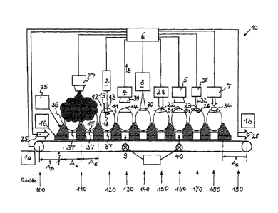
Fig. 1 a schematic illustration of an apparatus according to the
invention for
creating an opening in the calcified shell of an incubated bird egg
containing an embryo in the region of the blunt end thereof with a plurality
of structural components above an enclosure containing hatching eggs,
particularly for creating an opening in the direction to the air cell in the
region of the blunt end of an incubated bird egg with embryo, and for
determining the sex of the embryo,
Fig. 2 a schematic sectional view of bird eggs with avian embryos
with a
potential formation of the inner membrane, wherein the eggs stored on
brooding trays are candled from below, starting from the pointed end, by
light sources arranged below the brooding tray, preferably by means of
white light,
Fig. 3 a schematic illustration of a projection of the egg and the
projection of the
corresponding air cell,
Fig. 4 a schematic illustration according to Fig. 3 with a circular
opening
determined to be located in the center of the air cell projection as a
predetermined breaking point for creating the opening in the calcified
shell in order to create an access to the air cell,
Fig. 5 a schematic view of an egg dimensioned with the help of an
optical
distance meter for measuring the distance a of the inner membrane from
the vertex of the egg of the blunt end for focusing on the target structure
by means of collecting and detecting characteristic features of the target
structure, and
CA 02991603 2018-01-05
- 15 -
Fig. 6 a Raman spectrum that is measured at the intact inner membrane
of the
air cell in the region of the blunt end according to the present invention.
In the following, the functionality of the apparatus according to the present
invention
and the method according to the present invention are explained in more detail
with
reference to Fig. 1 to Fig. 6.
Fig. 1 shows an apparatus 10 for creating an opening 31 in the calcified shell
11 in
the region of the blunt end 14 of an incubated bird egg 12 containing an
embryo 18.
Within the region of the blunt end 14 there is an outer membrane 20 and an
inner
membrane 19 with an air cell 13 located therebetween. The embryo 18 adjoins
the
inner membrane 19 of the hatching egg 12 stored with its pointed end 15 facing
downwards. The hatching eggs 12 are held in a sorted manner in an enclosure
16.
The exemplary embodiment according to Fig. 1 comprises a transportation device
1
to which the enclosure 16 is assigned in the form of a tray in which the
hatching
eggs 12 are sorted with their blunt ends 14 facing upwards, wherein the
enclosure
16 is configured such that the hatching eggs 12 are stored at the positioning
areas
37 at equal distances AE, and a positioning device 35 which detects a marker
36 or
a marking or an edge at the enclosure 16 from which the predetermined
positioning
areas 37 of the hatching eggs 12 are determined by means of predetermined
distances A within the enclosure 16. The apparatus further comprises at least
one
detection device 2 which detects the dimensions (surface geometry) of the
hatching
egg 12, a detection device 4 for determining the dimensions of the air cell 13
with a
candling device 9 and a color camera 39, a working device 8 for creating a
predetermined breaking point 30 in the calcified shell 11, wherein after
working on
the calcified shell 11 the calcified shell 11 comprises ditch-like
predetermined
breaking points 30, and a device 28 for lifting and removing (removal device)
the lid
22 defined by the predetermined breaking point 30, and creating an opening 31
in
the air cell 13. Furthermore, the apparatus according to Fig. 1 comprises a
color
camera 5 with a second candling device 40 for detecting and focusing the
structure
26 to be investigated in the region of the inner membrane 19 within the air
cell 13, a
Raman spectra recording unit 38 with a measuring probe 32 in the beam path 23
being directed to the structure 26, a closing device 7 with locking elements
34, and
CA 02991603 2018-01-05
- 16 -
an analysis and control unit 6 that is connected with all structural
components via a
connecting line each.
In general, as is shown in Fig. 1, the enclosures / brooding tray 16 are,
e.g.,
configured pallet-like such that the distances AE between the hatching eggs 12
themselves and the distances AB (beginning of the enclosure) and the distances
AS
(end of the enclosure) are standardized between the hatching eggs 12 and,
e.g., an
edge of the enclosure 16 and that the standardized values have already been or
may already be saved and available in the analysis and control unit 6 so that
they
can be processed without signaling problems for setting the respective
position of
the opening device 8 for creating the opening 31 for calculating / determining
the
assigned calcified shell regions of the hatching eggs 12.
That is to say, a positioning device 35 may preferably be deployed for
standardized
or predimensioned brooding trays 16 with constant distances AB, AE, As of the
bulges for receiving the eggs in order to determine the positioning areas 37
of the
sorted hatching eggs 12, said positioning device being geared only to the
required
detection of the brooding tray 16 so that the positioning areas 37 of the
hatching
eggs 12 may be determined from the constant distances AB, AE, As.
In addition to or inclusive of the detection device 2 in the form of a sensor
or a
sensor array, a distance sensor or a triangulation sensor may optionally be
deployed. The sensors arranged in addition to the detection device 2
substantially
serve for ensuring the accuracy during a continuous course of the process.
The region of the air cell 13 detected by the detection device 4 with the help
of a
camera 39 in combination with a first device 9 for candling the egg 12 with
white
light may be a 2D image section 3 in the form of a two-dimensional projection,
or a
3D image section that is modified to be a two-dimensional projection.
The 2D image section 3 underlies the analysis and control unit 6 for
processing,
wherein in the analysis and control unit 6 the projection of the air cell 13
is assigned
to the projection of the egg 12, and the projection of the air cell 13 is
overlaid by a
CA 02991603 2018-01-05
- 17 -
circular projection 21, wherein the circular projection 21 determines the
dimensions
of the entrance area of the opening 31.
According to the invention, the apparatus 10 contains, e.g., a conveying
device I a,
a transportation device 1 for transporting the enclosure 16, a positioning
device 35
for determining the locations of the hatching eggs 12 in the enclosure 16, and
a
detection device 2 for detecting the dimensions of the eggs. The apparatus 10
may
further comprise a detection device 4 with a first candling device 9 and a
color
camera 39 for detecting the dimensions of the air cell 13, a working device 8
which
creates a predetermined breaking point 30 in the calcified shell 11 along the
predetermined breaking point 30 that was calculated in an analysis and control
unit
6 and which identifies said predetermined breaking point 30 as a lid 22 to be
lifted, a
device 28 for lifting and removing the lid 22 and for creating an opening 31
in the air
cell 13, and a color camera 5 with a second candling device 40 for detecting
and
focusing the structure 26 to be investigated in the region of the inner
membrane 19
within the air cell 13. Furthermore, the apparatus 10 may comprise a Raman
spectra
recording unit 38 with a measuring probe 32 in the beam path 23 being directed
to
the target structure 26, a closing device 7 which closes the open air cell 13
with a
locking element 34, a returning device lb and an analysis and control unit 6
which is
in signaling communication with all aforementioned structural components via
connecting lines and which directs with an algorithm the creation of the
opening 31
to the air cell 13.
A device 27 for disinfecting (disinfection device) at least the region of the
blunt end
14 may be arranged at least in front of the working device 8.
A closing device 7 with locking material for closing the created opening 31 of
the air
cell 13 is arranged downstream of the working device 8.
The locking element 34 may consist of a biocompatible material.
A sensor or a sensor array to which optionally a distance sensor or a
triangulation
sensor is assigned may be used as a first detection device 2.
I
CA 02991603 2018-01-05
- 18 -
At least one enclosure / tray / hatching tray 16 with hatching eggs 12 in
which the
hatching eggs are held in a sorted manner is assigned to the transportation
device
1, la, lb.
According to Fig. 1, the positioning device 35 detects a predetermined marking
36 of
the enclosure 16 and determines, with the help of the predetermined distances
AB,
AE, AS of the bulges for receiving the eggs of the enclosure 16, the positions
of the
eggs 12 to each other and determines in the analysis and control unit 6 the
respective positioning area 37 of the respective hatching egg 12 with respect
to the
marking 36 of the enclosure 16.
The apparatus 10 may comprise at least one sensor 2 or a sensor array
configured
as a detection device 18a for capturing a 2D image section 3 or a 3D image
section
in top view of the calcified shell region of at least one conveyed hatching
egg 12,
and optionally at least one distance sensor or a triangulation sensor, wherein
the
sensor 2 detects the positioning data of the conveyed hatching egg 12, and an
analysis and control unit 6 which collects and processes the data regarding
the
position of the egg as well as the positioning data of the working device 8
for
creating an opening 31, wherein the working device 8 receives from the
analysis
and control unit the working signals to be executed that are necessary for
processing in order to create the opening 31 in the calcified shell 11.
The transportation device 1 may be configured as a conveying device la and as
a
returning device lb in a continuous form.
The sensor 2 or the sensor array as well as the working device 8 may be
arranged,
e.g., above the transportation device 1, la, lb as well as above the hatching
eggs
12 resting in the transported enclosure 16.
In Fig. 1, the working device 8 for creating the predetermined breaking point
30 may
be a laser-optical device which creates a predetermined breaking point 30 in
the
calcified shell 11 by means of perforating the calcified shell 11 and a final
I II
CA 02991603 2018-01-05
- 19 -
breakthrough. The laser-optical device 8 may also be configured as a
retraceable,
movable device.
A device 28 for lifting the cut-free lid 22 is arranged thereafter.
This is followed by a color camera 5 of a second device 40 for candling the
hatching
egg 12 for a second time in order to determine the measuring location at the
inner
membrane 19.
A Raman measuring probe 32 absorbing a scattered radiation is assigned to the
downstream Raman spectra recording unit 38, said Raman measuring probe 32
recording the Raman scattered radiation via the beam path 23.
With the help of the closing device 7 arranged thereafter, a locking element
34 is
applied to the opening 31 for closing said opening 31.
In Fig. 1, the conveying device la and the returning device lb are part of a
consistent transportation device 1 comprising a predetermined running
direction 25.
For positioning and releasing the opening process, programmatic means are
saved
in the analysis and control unit 6, said programmatic means completing the
course
of the process as per a predetermined algorithm, wherein the analysis and
control
unit 6 is in electrical communication with all structural components of the
various
devices.
The sensor or the sensor array as the detection device 2 for dimensioning the
egg
sends the 2D image section 3 of the captured top view of the hatching egg 12
to the
analysis and control unit 6 via electrical connecting lines provided. The
sensor 2 or
the sensor array and the laser-optical opening device 8 are arranged above the
transportation device 1 as well as above the hatching eggs 12 resting in the
brooding tray / enclosure 16.
I
CA 02991603 2018-01-05
- 20 -
With the help of the detection device 4 for the air cell present in the form
of a
distance sensor or a triangulation sensor, the laser-optical working device 8
is
positioned exactly above the hatching egg 12. For this purpose, the laser-
optical
opening device 8 receives positioning and working signals from the analysis
and
control unit 6 which, for instance, can be a PC.
In general, the air cell 13 is located in the region of the blunt end 14. As
shown in
Fig. 4, a calcified shell lid 22 may be precisely removed by applying a
contour
corresponding to the predetermined breaking point 30.
According to Fig. 4, the opening created from the outside may comprise a
guideway
30 in the form of the predetermined breaking point substantially adapted to
the
inner-egg borders 17 of the air cell 13. It may be determined, e.g., using
transmitted
light techniques.
Thus, predetermined breaking points 30 may be created at different points
at/in the
calcified shell 11 in the region of the air cell 13.
In the following, the method according to the invention for creating an
opening 31 in
the calcified shell 11 in the region of the blunt end 14 of incubated bird
eggs 12 with
embryo 18 is described with the help of an exemplary embodiment. According to
the
method of the present invention, the hatching egg 12 stored with its pointed
end 15
facing downwards is dimensioned. The location and the dimensions of the air
cell 13
within the region of the blunt end 14 as well as the location of the embryo 18
below
the inner membrane shielding the embryo 18 from the air cell 13 are detected.
As
shown in Figures 2 and 3, a two-dimensional projection 3 of the hatching egg
12
with a center M and overlaying therewith a two-dimensional projection 29 of
the air
cell 13 with a central point m is carried out. The two-dimensional projection
29 of the
air cell 13 preferably has an elliptic shape with a point of intersection as a
central
point m of intersecting major and minor axes A, B of the ellipse. The central
point of
a circular projection 21 representing the predetermined breaking point 30 is
assigned to the point of intersection m of the major and minor axes A, B of
the
ellipse. The radius R of the circular projection 21 is preferably smaller than
half the
CA 02991603 2018-01-05
- 21 -
expansion A/2 of the minor axis A of the ellipse with R <N2. A calcified shell
lid 22
corresponding to the two-dimensional circular projection 21 is lifted and
removed
from the remaining body of the calcified shell 11. Thus, an opening 31 is
achieved in
the calcified shell 11 and thus also in the air cell 13.
Prior to the start of dimensioning the hatching eggs 12, the hatching eggs 12
are
disinfected preferably at least in the region of the blunt end 14.
The method according to the present invention preferably comprises the
following
steps: detecting the positions and geometries of the already disinfected
hatching
eggs 12 stored with their pointed ends 15 facing downwards and resting on a
predetermined brooding tray 16, detecting the geometry of the hatching eggs
12,
detecting the geometry of the air cell 13 at the blunt end 14 of the hatching
egg 12,
and detecting and digitalizing the geometry data of the volume of the air cell
13. The
method may further comprise the following steps: detecting a two-dimensional
outline 29 in the form of an area projected onto a surface from the volume
projection
of the air cell 13 in the shape of an ellipse, calculating the point of
intersection m of
the major and minor axes A, B of the ellipse 29 from a digitalized camera
image 33,
calculating the circular projection 21 and the corresponding predetermined
breaking
point 30 with respect to the point of intersection m of the ellipse, creating
an opening
31 in the calcified shell 11 in the circular projection 21 located centrically
to the
ellipse 29 via the predetermined breaking point 30 above the taut inner
membrane
19 with the help of the working device 8, as well as removing the separated
part of
the calcified shell 11 as a lid 22 in the defined circular projection 21.
In Fig. 5, the distance a of the inner membrane 19 is calculated starting from
the
vertex of the egg of the blunt end 14, wherein the detected distance a serves
for
focusing on the target structure 26 in the region of the inner membrane 19.
After the calcified shell 11 has been opened, the hatching egg 12 is candled
again
for a second time and an image of the detected region of the inner membrane 19
of
the air cell 13 is taken with a color camera 5 for determining the structure
26 for
determining the sex, e.g., by means of Raman spectroscopy.
1
CA 02991603 2018-01-05
- 22 -
With the help of the color camera 5 and using a light source 40, preferably in
the
spectral range between 500 nm and 600 nm, an adjusted contrasting of the
embryo-
specific target structures 26 laying beneath the inner membrane 19 may be set.
With the help of the data received from a distance sensor or a triangulation
sensor
or a grazing light sensor, wherein the data are assigned to the respective
hatching
egg 12, the working device 8 is exactly positioned at a position predetermined
for
the creation of the opening with respect to the calcified shell region 21 of
the
hatching egg 12 to be worked on.
The enclosure 16 in the form of a tray of the hatching eggs 12 at the
transportation
device 1 is moved beneath the detection device 2 and the working device 8 for
creating the opening 31, wherein by means of the detection device 2 a 2D image
section 3 or a 3D image section that is converted into a 2D image section is
captured of the top view of the hatching egg 12 with the data of said image
section
being transmitted via electrical connecting lines to the analysis and control
unit 6 for
further processing, wherein in the analysis and control unit 6 the image data
of the
2D image section are processed together with the egg distance data from the
distance sensor or the triangulation sensor by means of activated programmatic
means, and the processed signals received are forwarded to the working device
8 in
order to create the opening 31.
According to Fig. 1, the method for creating an opening 31 in the calcified
shell 11 in
the region of the blunt end 14 of an incubated bird egg 12 containing an
embryo 18
comprises the following steps: conveying the hatching eggs 12 using a
conveying
device 1a (step 100), disinfecting at least the blunt end 14 (step 110),
detecting at
least one position area of the conveyed hatching eggs 12 and/or an enclosure
16
containing hatching eggs 12 using a positioning device 35 which is stationed
at least
in the conveying region of the enclosure 16 containing hatching eggs 12, and
converting the respective position area that has been detected into electrical
data
signals as well as processing the electrical data signals in an analysis and
control
unit 6 (step 120), candling the hatching egg 12 in order to detect the
position of the
CA 02991603 2018-01-05
- 23 -
air cell 13 (step 130), positioning a working device 8 in order to create a
predetermined breaking point 30 in the calcified shell 11 of the hatching egg
12 as
well as working on the blunt end 14 for creating the opening 31 in the
calcified shell
11 of the hatching egg 12 (step 140), removing the lid 22 identified to be
located
alongside the predetermined breaking point 30 (step 150), focusing the color
camera 5 on the target structure 26 to be detected in the region of the inner
membrane 19 (step 160), measuring the Raman spectrum 24 of the target
structure
26 (embryo) according to Fig. 6 (step 170), closing the opening 31 with a
locking
element 34 (step 180) and returning the hatching eggs 12 using a returning
device
lb in the running direction 25 of the transportation device 1 (step 190).
The step of creating the opening 31 constitutes a defined, target-oriented
treatment
of the hatching egg 12, wherein the initial positioning by means of the sensor
32 or
the sensor array and the final positioning of the hatching egg 12 which is
assigned
to the creation of the opening and which corresponds to the associated setting
position of the working device 8 are carried out using programmatic means
saved in
the analysis and control unit 6.
In addition to a laser-optical treatment, the aforementioned target-oriented
treatment
of the calcified shell 11 of the hatching egg 12 may also be carried out by
means of
mechanical, chemical, or water jet tools.
It may also be advantageous that the treatment can be carried out at
incubation
temperatures. To this end, the apparatus 10 may, e.g., without the analysis
and
control unit 6, be incorporated inside a housing (not illustrated) that is
adapted to
incubating temperatures.
As an input parameter for obtaining the opening 31 in the calcified shell 11
and for
detecting the air cell 13 at the blunt end 14, the positions and geometries of
the
hatching eggs 12 that are typically stored with their pointed ends 15 facing
downwards are detected, e.g., by means of laser triangulation or grazing light
technologies. The eggs 12 which are, e.g., stored on standard brooding trays
16 are
subsequently candled, preferably using white light from below, i.e., starting
from the
CA 02991603 2018-01-05
- 24 -
pointed end 15, by means of the first detection device 2 for a first candling
step, as
is shown in Fig. 2. For this purpose, appropriate light sources 9 (e.g.,
halogen
lamps, LED or standard egg-candlers) are arranged below the brooding tray 16
(Fig.
1 and Fig. 2).
By means of the transmitting light, the geometry of the air cell 13 at the
blunt end 14
of the hatching egg 12 is made visible and detected and digitalized with the
help of a
camera 39 and processed as a 2D image section 3 in the analysis and control
unit
6. As indicated in Fig. 2, the geometries of the potential air cells 13 may be
of
io different sizes and be arranged in different inclinations to the
longitudinal axis of the
respective egg.
In practice, the outline of the area 29 (volume projection of the air cell 13)
obtained
in this way and projected onto the surface 3, as is shown in Fig. 3, in most
cases
corresponds to an ellipse.
With the aid of the collected surface data (laser triangulation) and by
calculating the
point of intersection m of the major and minor axes A, B of the ellipse from
the
digitalized camera image 33, the predetermined breaking point 30 and thus also
the
calcified shell lid 22 may then be precisely calculated centrically to the
point of
intersection m above the taut inner membrane 19, and the calcified shell 11
may
thus be opened. For this purpose, lasers for material machining such as, e.g.,
CO2
lasers are used, but mechanical processes such as milling come into operation
as
well.
According to Fig. 4, in order to create a sufficiently large opening 31 for
subsequent
investigations, the calcified shell material is cleared away and removed
circularly,
partially or in full, preferably with R <Al2 within the predetermined breaking
point 30
in the detected region 21 as the calcified shell lid 22. This step may be
carried out
with the help of either scanning laser optics or a defined movement of rigid
laser
optics along the desired outline 21 of the predetermined breaking point 30.
The lid
22 which is produced in this way may subsequently be removed mechanically,
thus
CA 02991603 2018-01-05
- 25 -
creating a free access to the interior of the air cell 13 and to the inner
membrane 19
adjoining the interior of the egg.
By way of a repeated use of the laser triangulation or using a simple optical
distance
meter, the distance a of the inner membrane 19 from the vertex of the egg 14
of the
blunt end may be exactly detected at the total height b of the egg 12
according to
Fig. 5. For subsequent, e.g., spectroscopic methods, this results in the
option of
exactly aligning their focus areas with target structures 26 that are to be
investigated
and that directly adjoin the inner membrane 19, as illustrated in Fig. 5, and
of
collecting gender-related information. In order to locate the inner target
structures 26
(e.g., embryonic blood vessels), the hatching eggs 12 are candled preferably
with
green light from the light source 40 starting from the pointed end 15, and an
image
of the exposed region of the inner membrane 19 of the air cell 13 is taken by
the
color camera 5 and transmitted via signal transfer to the analysis and control
unit 6.
By way of appropriately adjusting the focus, the analysis and control unit 6
may also
serve to detect the distance of the inner membrane 19 with target structures
26
located behind it.
In order to receive an optimized contrasting of the specific target structures
26 (for
instance, embryonic blood vessels) that are to be investigated and that are
located
below the inner membrane 19, the light source 40 may, for example, be used in
the
green spectral range between 500 and 600 nm for hemoglobin absorption.
With the help of the above-described apparatus 10 and the method according to
the
present invention, characteristic Raman spectra 24 of embryonic blood vessels
26
may be recorded, e.g., when using IR Raman spectroscopy, as is shown in Fig. 6
for a single Raman spectrum 24, by means of the measuring probe 32 pertaining
to
the Raman spectra recording unit 38 via the beam path 23 in order to determine
the
sex of the developing avian embryo 18.
In particular, characteristic Raman spectra 24 may be analyzed with the help
of well-
known methods of data analysis in order to determine the sex of the developing
avian embryo.
õ
CA 02991603 2018-01-05
- 26 -
For instance, the well-known cluster analysis may be used for analyzing
characteristic Raman spectra 24 in order to determine the sex of the
developing
avian embryo.
The cluster analysis substantially combines two methods, namely principal
component analysis (PCA) and k-nearest neighbors classification,
PCA serves for structuring and simplifying the recorded data (e.g.,
characteristic
Raman spectrum according to Fig. 6). In doing so, the measured signal (data,
i.e.,
characteristic Raman spectrum) is depicted by linear combinations in a number
that
is smaller than that of the measurement values of the signal. This step
suppresses
the noise during measurement and makes the measured signals more comparable.
After the signal has been dissected into its main components, it is possible
to set up
a mathematical space, i.e., the parameters of each linear combination of a
main
component correspond to a point in a two-dimensional space in which the axes
are
any potential combination of the parameters of a potential linear combination.
In this
way, a point cloud is formed. In the next step, said point cloud is used for k-
nearest
neighbors classification.
With respect to the signals analyzed here (characteristic Raman spectra), the
k-
nearest neighbors classification is k=1. This is a special case and is called
Voronoi
diagram. The obtained point cloud is divided into regions with every point
being the
center of one region. Different metrics, e.g., the Euclidean distance of the
centers to
each other, or the size of particular regions may be applied for this
construct.
Particular combinations of centers and regions are specific for the sex of the
developing avian embryo and differ significantly for male and female embryos.
Even if the cluster analysis was exemplarily described here, other data
analysis
methods may be used for determining the sex of the developing avian embryo on
the basis of the recorded signals (data, i.e., characteristic Raman spectra).
The
aforementioned well-known data analysis methods are also applicable for other
CA 02991603 2018-01-05
- 27 -
analysis methods such as fluorescence spectroscopy or further spectroscopic
methods, in particular for the analysis of characteristic spectra.
After the characteristic measurement data have been collected, the opening 31
created in the calcified shell 11 of the blunt end 14 of the hatching egg 12
is
mechanically closed again by means of a locking element 34, e.g., by means of
a
biocompatible plaster (e.g., by means of a medical 3M DuraPore plaster) or a
cap.
The properties of the locking material that is used are selected so as not to
affect
the physiology of the hatching egg 12, and particularly so as to avoid an
excessive
loss of liquid from the air cell 13 by evaporation.
In order to guarantee sterility right from the beginning, a device 27 is
provided to
disinfect at least the region of the blunt end 14.
While in the exemplary embodiments the methods and the apparatus have been
described as an example on the basis of the measured data of Raman
spectroscopy, an equivalent utilization is similarly possible with the help of
absorption-spectroscopic techniques, particularly fluorescence spectroscopy.
In
order to avoid a repetition of the above descriptions, reference is made to
the
above-described exemplary embodiments with respect to the use of absorption-
spectroscopic techniques, in particular fluorescence spectroscopy.
While the present invention has been described and shown with respect to its
preferred embodiments, it is obvious to the person skilled in the art that
different
modifications and alterations may be performed to it without departing from
the
scope of protection of the invention. It is intended in this way that the
present
invention covers the modifications and alterations of this invention provided
that they
are covered by the scope of protection of the accompanying patent claims and
their
equivalents.
Furthermore, different features that are described in connection with the
method
according to the invention may also form a basis for features of the apparatus
according to the invention, and vice versa. Even if specific features have
been
CA 02991603 2018-01-05
- 28 -
described in combination with other features, the present invention is not to
be
limited to these combinations. Rather, an arbitrary combination, as far as it
makes
technical sense, of the described features is possible.
List of reference signs
1 transportation device
la conveying device
lb returning device
2 detection device for detecting the dimensions of the egg
3 2D image section
4 detection device for detecting the dimensions of the air cell
5 color camera focusing on a target structure 26
6 analysis and control unit
7 closing device
8 working device
9 first light source for a first candling step / first candling
device
10 apparatus according to the invention
11 calcified shell
12 hatching egg
13 air cell within the egg
14 blunt end
15 pointed end
16 enclosure / brooding tray
17 linking border between air cell and inner membrane
18 embryo
19 inner membrane
20 outer membrane
21 breaking point projection
22 calcified shell lid
23 beam path for recording a Raman spectrum
24 Raman spectrum in the form of an intensity / wavenumber curve
CA 02991603 2018-01-05
- 29 -
25 running direction of the conveyor belt
26 target structure / measuring region of the inner membrane
27 device for disinfection
28 device for lifting the lid 22
29 air cell projection
30 predetermined breaking point
31 opening in the calcified shell
32 measuring probe
33 digitalized camera image
34 locking element
35 positioning device
36 marker
37 positioning area
38 Raman spectra recording unit / unit for recording measurement data
39 camera for first candling device 9
40 second light source for a second candling step / second candling
device
A first minor axis of the ellipse
second major axis of the ellipse
a distance between measuring point at the inner membrane and vertex of the
blunt end of the egg
height of the egg
coordinate
coordinate
M center of the egg projection
point of intersection of the air cell projection (ellipse)
radius of the circular projection with R <P12
100 process step of conveying
110 process step of disinfecting
120 process step of detecting
130 process step of candling
140 process step of opening
CA 02991603 2018-01-05
- 30 -
150 process step of removing
160 process step of focusing
170 process step of measuring
180 process step of closing
190 process step of returning